A New Calibration Circuit for RuO2 Urea Biosensors Achieves Over 98% Drift Reduction
This article presents a comprehensive analysis of a novel calibration circuit (NCC) designed to mitigate the critical issue of signal drift in RuO2-based urea biosensors.
A New Calibration Circuit for RuO2 Urea Biosensors Achieves Over 98% Drift Reduction
Abstract
This article presents a comprehensive analysis of a novel calibration circuit (NCC) designed to mitigate the critical issue of signal drift in RuO2-based urea biosensors. Aimed at researchers and drug development professionals, the content explores the foundational challenge of drift caused by hydration layer formation, details the simple yet effective methodology of the voltage-regulation-based NCC, and provides empirical validation showing a 98.77% reduction in drift rate to 0.02 mV/hr. By synthesizing troubleshooting insights and comparative performance data, this work outlines a significant advancement toward robust, reliable biosensing for clinical diagnostics and long-term monitoring applications.
Understanding the Drift Effect: A Critical Challenge for RuO2 Urea Biosensors
The Clinical Imperative for Accurate Urea Detection in Kidney Disease and Dialysis
Urea, a key nitrogenous metabolite in the human body, serves as a critical biomarker for assessing renal function and metabolic health. In clinical practice, precise monitoring of blood urea nitrogen (BUN) levels is essential for diagnosing and managing kidney diseases, guiding dialysis treatment, and preventing complications such as uremic syndrome [1]. The normal concentration range of urea in human blood serum is typically 2.5–7.5 mM (15–40 mg/dL), with deviations from this range indicating potential renal dysfunction or other metabolic disorders [2] [3].
For patients with chronic kidney disease (CKD) and those undergoing dialysis, maintaining urea levels within acceptable limits is crucial. Elevated urea concentrations, known as uremia, are associated with detrimental effects on various organ systems, including the cardiovascular system, gastrointestinal tract, and kidneys themselves [1]. Uremic toxins contribute to cardiovascular disease onset and progression in CKD, exacerbating oxidative stress and inflammation [1]. Therefore, reliable and accurate urea monitoring systems are indispensable tools in nephrology and critical care medicine.
Despite the clinical importance of urea detection, conventional measurement techniques including chromatography, spectrophotometry, and laboratory-based tests present significant limitations for frequent monitoring. These methods are often time-consuming, require sophisticated equipment and skilled technicians, and are unsuitable for point-of-care testing or continuous monitoring [3] [4]. Consequently, electrochemical biosensors have emerged as promising alternatives, offering advantages of portability, operational ease, and potential for real-time monitoring [1].
A significant challenge in biosensor technology, however, is the drift effect—a gradual change in sensor response over time during long-term measurement. This phenomenon, attributed to the formation of a hydration layer on the sensing film surface, compromises measurement accuracy and reliability [2]. Recent research has focused on addressing this limitation through novel materials and circuit designs, with ruthenium oxide (RuO₂) emerging as a promising sensing material due to its high metallic conductivity, thermal stability, and excellent diffusion barrier properties [2].
Current Landscape of Urea Biosensing Technologies
Electrochemical biosensors for urea detection primarily utilize two distinct approaches: enzymatic (EN) and non-enzymatic (NE) sensing mechanisms. Each strategy offers distinct advantages and limitations for clinical application.
Enzymatic Urea Biosensors
Enzymatic urea biosensors typically employ urease (Urs) as the biological recognition element. This enzyme specifically catalyzes the hydrolysis of urea into ammonium ions (NH₄⁺) and bicarbonate ions (HCO₃⁻) [1]. The reaction can be summarized as:
[ \ce{CO(NH2)2 + H2O ->[Urease] NH4+ + HCO3-} ]
The detection mechanism typically involves monitoring pH changes resulting from ammonium ion release or measuring the electroactive products directly [1]. These sensors benefit from high specificity due to the enzyme's selective catalytic activity. Recent advancements have focused on improving enzyme immobilization techniques using nanomaterials, polymers, and magnetic beads to enhance stability and sensitivity [3].
Non-Enzymatic Urea Biosensors
Non-enzymatic approaches utilize electrocatalytic materials such as metal oxides (e.g., NiO, ZnO), carbon-based materials, and their nanocomposites to directly oxidize or reduce urea without biological components [1]. For instance, nickel oxide (NiO) based sensors operate through the conversion between Ni(OH)₂ and electrocatalytically active NiOOH during urea oxidation [1]. These sensors are gaining popularity due to their enhanced stability, lower cost, and simplified fabrication processes compared to enzymatic systems [1].
Table 1: Comparison of Enzymatic vs. Non-Enzymatic Urea Biosensors
| Feature | Enzymatic Biosensors | Non-Enzymatic Biosensors |
|---|---|---|
| Sensing Mechanism | Enzyme-catalyzed hydrolysis | Direct electrocatalytic oxidation/reduction |
| Selectivity | High (enzyme-specific) | Moderate |
| Stability | Limited by enzyme denaturation | Higher |
| Cost | Higher (enzyme purification) | Lower |
| Lifespan | Shorter | Longer |
| Materials | Urease with immobilization matrices | Metal oxides, carbon materials, polymers |
The Drift Effect Challenge in RuO₂ Urea Biosensors
Despite excellent sensing properties demonstrated by RuO₂-based biosensors, including high sensitivity and linearity, their practical implementation in clinical settings has been hampered by the drift effect. This phenomenon manifests as a gradual change in the sensor's response voltage during long-term measurements, leading to inaccurate readings and recalibration requirements [2].
The fundamental mechanism underlying the drift effect involves the formation of a hydration layer on the surface of the RuO₂ sensing film. When exposed to aqueous solutions such as biological fluids, hydroxyl groups form on the film surface. Hydrated ions then diffuse toward the sensing film through coulombic attraction between water molecules and ions, ultimately forming an electrical double-layer capacitance that alters the surface potential of the film [2]. This progressive change in potential constitutes the observed drift, compromising measurement accuracy for prolonged monitoring applications such as dialysis treatment.
A Novel Calibration Circuit for Drift Reduction
To address the critical challenge of drift in RuO₂ urea biosensors, recent research has introduced a New Calibration Circuit (NCC) based on voltage regulation techniques. This innovative approach aims to maintain the simplicity of the biosensor system while significantly improving long-term stability [2].
Circuit Architecture and Design Principles
The proposed NCC features a straightforward structure composed of two primary components: a non-inverting amplifier and a voltage calibrating circuit [2]. This design prioritizes simplicity while effectively countering the drift phenomenon through active voltage regulation. The circuit interfaces directly with the RuO₂ urea biosensor, which is fabricated on a flexible polyethylene terephthalate (PET) substrate using screen-printing and sputtering techniques [2].
Fabrication of the Flexible Arrayed RuO₂ Urea Biosensor
The development of the biosensor platform itself follows a meticulous manufacturing process:
- Substrate Preparation: A flexible polyethylene terephthalate (PET) substrate serves as the foundational material.
- Electrode Formation: Arrayed silver wires are printed onto the PET substrate using screen-printing techniques to create working and reference electrodes.
- Sensing Film Deposition: RuO₂ film is deposited onto the flexible substrate through a sputtering system to form the RuO₂ film window.
- Encapsulation: The structure is encapsulated with an epoxy thermosetting polymer for insulation and protection.
- Enzyme Immobilization: Urease is immobilized on the RuO₂ sensing film through covalent bonding using aminopropyltriethoxysilane (APTS) solution and glutaraldehyde cross-linking, enhancing enzyme stability and reducing leaching [2].
Experimental Validation and Performance Metrics
The experimental validation of the NCC was conducted in two stages to comprehensively assess its efficacy:
Stage 1: Biosensor Characterization The RuO₂ urea biosensor was first characterized using a conventional voltage-time (V-T) measurement system. The biosensor demonstrated excellent performance with an average sensitivity of 1.860 mV/(mg/dL) and a linearity of 0.999 within the physiologically relevant urea concentration range (2.5–7.5 mM) [2].
Stage 2: Drift Rate Assessment The critical evaluation involved comparing the drift rate between the conventional V-T measurement system and the proposed NCC. The RuO₂ urea sensing film was immersed in urea solution for 12 hours while response voltage was continuously monitored [2]. Results demonstrated that the NCC successfully reduced the drift rate to 0.02 mV/hr, representing a remarkable 98.77% reduction compared to the conventional system [2] [5].
Table 2: Performance Comparison of RuO₂ Urea Biosensor with and without NCC
| Parameter | Conventional V-T System | With NCC | Improvement |
|---|---|---|---|
| Drift Rate | Significant drift observed | 0.02 mV/hr | 98.77% reduction |
| Sensitivity | 1.860 mV/(mg/dL) | Maintained | No degradation |
| Linearity | 0.999 | Maintained | No degradation |
| Long-term Stability | Limited by drift | Significantly enhanced | Suitable for prolonged monitoring |
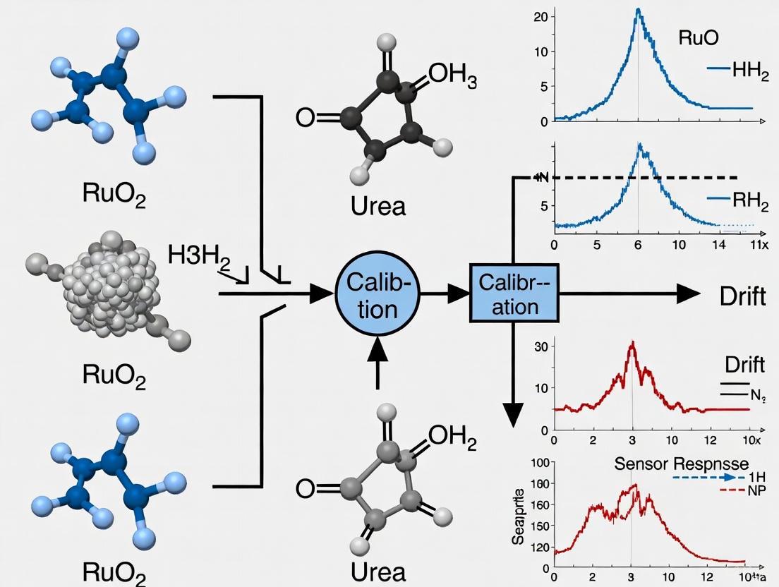
The following diagram illustrates the experimental workflow for biosensor fabrication, characterization, and drift performance validation:
Research Reagent Solutions and Materials
The development and implementation of advanced urea biosensors require specific research reagents and materials optimized for performance and reliability. The following table details essential components used in the fabrication and operation of RuO₂ urea biosensors with drift-reduction capabilities:
Table 3: Essential Research Reagents and Materials for RuO₂ Urea Biosensor Fabrication
| Category | Specific Material/Reagent | Function/Application |
|---|---|---|
| Substrate Materials | Polyethylene terephthalate (PET) | Flexible substrate for biosensor fabrication |
| Electrode Materials | Silver paste | Formation of arrayed wires for working and reference electrodes |
| Sensing Film | Ruthenium oxide (RuO₂) | Primary sensing material with high conductivity and stability |
| Immobilization Matrix | Epoxy thermosetting polymer | Encapsulation and insulation layer |
| Enzyme System | Urease from Canavalia ensiformis | Biological recognition element for urea hydrolysis |
| Cross-linking Agents | Aminopropyltriethoxysilane (APTS), Glutaraldehyde | Enzyme immobilization on RuO₂ sensing film |
| Buffer Components | Phosphate buffer saline (PBS), pH 7 | Maintain physiological pH during testing |
| Calibration Solutions | Urea standards (2.5-7.5 mM) | Sensor calibration and performance validation |
Detailed Experimental Protocol for Drift Rate Assessment
Materials and Equipment Setup
- Biosensor Platform: Fabricated flexible arrayed RuO₂ urea biosensor
- Measurement Systems: Conventional voltage-time (V-T) system and New Calibration Circuit
- Data Acquisition: DAQ device with LabVIEW software
- Amplification: LT1167 instrumentation amplifier
- Test Solutions: Urea solutions in PBS (pH 7.0) across clinical range (2.5-7.5 mM)
- Environmental Control: Constant temperature incubation at 37°C
Procedure
Initial Calibration:
- Immerse the RuO₂ urea biosensor in standard urea solutions across the physiological range
- Record response voltages using both V-T and NCC systems
- Establish calibration curves for both systems
- Confirm biosensor sensitivity and linearity meet acceptable thresholds (>1.8 mV/(mg/dL) and R² > 0.99)
Long-term Drift Assessment:
- Prepare a urea solution at clinically relevant concentration (e.g., 5 mM)
- Immerse the biosensor in the solution maintaining constant temperature
- Continuously monitor response voltage for 12 hours using both measurement systems
- Record voltage measurements at regular intervals (e.g., every 10 minutes)
- Ensure minimal environmental disturbances during testing
Data Analysis:
- Plot response voltage versus time for both systems
- Calculate drift rate as slope of the voltage-time curve
- Compare final drift rates between conventional and NCC systems
- Compute percentage improvement in drift rate
The signaling pathway and detection mechanism of the RuO₂ biosensor can be visualized as follows:
The development of a New Calibration Circuit for RuO₂ urea biosensors represents a significant advancement in electrochemical sensing technology, directly addressing the critical challenge of drift effects that has limited the clinical application of these devices. The demonstrated 98.77% reduction in drift rate to 0.02 mV/hr, while maintaining high sensitivity and linearity, positions this technology as a promising platform for accurate, long-term urea monitoring in nephrology and critical care medicine [2] [5].
For researchers and clinicians focused on kidney disease and dialysis management, this technological innovation offers the potential for more reliable point-of-care testing and continuous monitoring systems. The enhanced stability provided by the NCC could significantly improve the precision of dialysis titration and the detection of acute renal impairment in hospitalized patients. Furthermore, the simple structure of the proposed circuit facilitates potential integration into miniaturized, portable devices for home-based monitoring of patients with chronic kidney disease.
Future research directions should focus on validating these biosensors in complex clinical matrices such as blood serum and dialysate fluids, optimizing the design for mass production, and exploring integration with wireless technologies for remote patient monitoring. As materials science continues to advance, combining the NCC approach with emerging nanomaterials and immobilization strategies could further enhance the stability, selectivity, and commercial viability of urea biosensors for clinical applications [1] [6].
The integration of such calibrated biosensor systems into dialysis equipment and critical care monitoring platforms represents a promising frontier for improving patient outcomes through more precise, real-time assessment of urea levels, ultimately enabling more personalized and effective management of kidney disease.
Ruthenium dioxide (RuO₂) is a transition metal oxide that has garnered significant attention in the field of electrochemical sensing due to its exceptional physical and chemical properties. As a sensing material, RuO₂ possesses a unique combination of high electrical conductivity, excellent chemical stability, and corrosion resistance, making it particularly suitable for applications in harsh environments where traditional sensing materials may fail [2] [7]. Its metallic conductivity, characterized by low resistivity values, enables efficient electron transfer during electrochemical reactions, while its robust chemical nature ensures long-term operational stability in various media, including acidic and alkaline solutions [8] [9].
The material's sensing mechanism is governed by electrochemical phenomena at the electrode-electrolyte interface, including adsorption, dissociation, diffusion of ions, hydration, redox processes, electrical double layer formation, and charge transfer [7]. This complex interplay of mechanisms allows RuO₂-based sensors to exhibit near-Nernstian behavior for pH detection and sensitive response to various analytes, including urea in biomedical applications [2] [7]. The compatibility of RuO₂ with various fabrication techniques, from thick-film screen printing to thin-film deposition methods, further enhances its versatility as a sensing material for diverse applications ranging from environmental monitoring to clinical diagnostics [8] [7].
Key Advantages and Performance Metrics of RuO2 Sensors
Quantitative Performance Data
The table below summarizes key performance metrics for RuO₂-based sensors across different applications, demonstrating their exceptional sensing capabilities.
Table 1: Performance Metrics of RuO₂-Based Sensors
| Application | Sensitivity | Linearity | Response Time | Drift Rate | Hysteresis | Reference |
|---|---|---|---|---|---|---|
| pH Sensing | 56.35 - 58.8 mV/pH | R² > 0.999 (pH 2-12) | < 30 seconds | 0.02 mV/hr (after calibration) | 1.0 - 1.3 mV | [8] [9] [7] |
| Urea Biosensing | 1.860 mV/(mg/dL) | 0.999 | N/A | 0.02 mV/hr (after calibration) | N/A | [2] [5] |
Material Advantages and Characteristics
Table 2: Key Advantages of RuO₂ as a Sensing Material
| Property | Description | Impact on Sensing Performance | |
|---|---|---|---|
| Electrical Conductivity | Metallic conductor with resistivity as low as 0.89 μΩ·m for thin films | Enables efficient electron transfer, reduces sensor impedance, improves signal-to-noise ratio | [10] |
| Chemical Stability | Exceptional corrosion resistance in acidic/alkaline media | Maintains performance in harsh environments, extends operational lifespan | [7] [11] |
| Thermal Stability | Stable up to 750°C in air | Withstands high-temperature processing and operation | [10] |
| Nernstian Response | Near-ideal pH sensitivity (55-59 mV/pH) | Provides accurate pH measurement across broad range | [8] [9] [7] |
| Mechanical Robustness | Maintains structural integrity under compression up to 40 MPa | Ensures durability in practical applications and during fabrication | [12] |
| Miniaturization Potential | Compatible with microfabrication techniques (screen printing, sputtering) | Enables development of compact, portable sensor systems | [2] [7] |
The high conductivity of RuO₂ stems from its metallic character, with thin films demonstrating remarkably low resistivity values as low as 0.89 μΩ·m [10]. This exceptional conductivity facilitates efficient charge transfer during electrochemical sensing operations, resulting in improved sensor response times and signal clarity. The material's chemical stability is equally impressive, with studies demonstrating excellent performance in corrosive environments where conventional materials would degrade rapidly [7] [11]. This combination of properties makes RuO₂ particularly valuable for long-term monitoring applications where sensor drift must be minimized.
Experimental Protocols for RuO2 Sensor Fabrication and Testing
Fabrication of Screen-Printed RuO₂ pH Electrodes
Objective: To fabricate robust, high-performance RuO₂ pH sensing electrodes using screen-printing technology for water quality testing applications.
Materials and Equipment:
- Anhydrous RuO₂ powder (Sigma Aldrich, purity ≥ 99.95%)
- Ethyl cellulose (binder, analytical grade purity)
- Terpineol (solvent, anhydrous, Fluka Analytical)
- Alumina (Al₂O₃, 96%) substrate plates
- Ag/Pd thick-film paste (Electro-Science Laboratories, 9695)
- Polydimethylsiloxane coating (DOWSIL 3140 RTV Coating)
- Screen-printing apparatus
- Drying oven (120°C capability)
- High-temperature furnace (800-900°C capability)
Procedure:
- Paste Preparation: Mix anhydrous RuO₂ powder with ethyl cellulose and terpineol in an agate mortar. Mix continuously for 20 minutes to achieve optimal, homogeneous consistency for screen printing.
- Substrate Preparation: Clean standard alumina substrates thoroughly to remove surface contaminants.
- Conductive Layer Deposition: Screen-print Ag/Pd thick-film paste onto the substrates to form the conductive layer. Dry at 120°C for 15 minutes, then fire at 860°C for 30 minutes in air atmosphere.
- Sensing Layer Deposition: Screen-print the prepared RuO₂ paste onto the substrates, ensuring the RuO₂ layer slightly overlaps the Ag/Pd conductive layer. Dry at 120°C for 15 minutes.
- High-Temperature Sintering: Sinter the electrodes at the target temperature (800°C, 850°C, or 900°C) for one hour in air atmosphere to achieve proper adhesion and functionality.
- Electrical Contact Attachment: Solder a copper wire to the open end of the conducting layer to establish electrical connection.
- Encapsulation: Apply polydimethylsiloxane coating to protect the electrical contact and conducting layer from electrolyte exposure, leaving only the sensitive RuO₂ area uncovered. Cure the silicone resin at room temperature for 48 hours.
Quality Control:
- Verify layer adhesion through microscopic examination and adhesion testing
- Confirm electrical continuity using multimeter measurements
- Validate sensor-to-sensor reproducibility through potentiometric testing in standard pH buffer solutions [7]
Fabrication of Flexible Arrayed RuO₂ Urea Biosensor
Objective: To develop a flexible arrayed RuO₂ urea biosensor for biomedical applications with high sensitivity and minimal drift.
Materials and Equipment:
- Polyethylene terephthalate (PET) flexible substrates (Zencatec Corporation)
- Ruthenium metal target (purity ≥ 99.95%, Ultimate Materials Technology Co., Ltd.)
- Silver paste (Advanced Electronic Material Inc.)
- Epoxy thermosetting polymer (Sil-More Industrial, Ltd., JA643)
- Urease enzyme (Sigma-Aldrich Corp.)
- Urea standards (J.T. Baker Corp.)
- Aminopropyltriethoxysilane (APTS) solution
- Glutaraldehyde solution (1%)
- Sputtering system
- Screen-printing system
Procedure:
- Electrode Formation: Print silver paste on flexible arrayed PET substrates using screen printing techniques to form arrayed silver wires for working and reference electrodes.
- RuO₂ Film Deposition: Deposit RuO₂ film on the flexible arrayed PET substrate through a sputtering system to form the RuO₂ film window.
- Encapsulation: Encapsulate the structure with an epoxy thermosetting polymer using screen-printing technology to create an insulation layer.
- Surface Functionalization: Drop APTS solution onto the RuO₂ sensing film at room temperature to enhance urease adsorption.
- Cross-Linking: Drop 1% glutaraldehyde solution onto the RuO₂ sensor and keep still for 24 hours to create covalent binding sites.
- Enzyme Immobilization: Drop urease solution onto the functionalized RuO₂ sensing film to form the complete biosensor.
- Curing: Allow the assembled biosensor to stabilize at room temperature to complete the immobilization process.
Calibration and Testing:
- Prepare urea standards in phosphate buffer saline (PBS, 30 mM, pH 7.0) covering the physiological range (2.5-7.5 mM)
- Measure response voltage using voltage-time (V-T) measurement system
- Characterize sensitivity, linearity, and drift rate over 12-hour continuous operation [2]
Drift Rate Calibration Procedure for RuO₂ Urea Biosensor
Objective: To implement a New Calibration Circuit (NCC) that significantly reduces the drift effect in RuO₂ urea biosensors.
Materials and Equipment:
- Fabricated RuO₂ urea biosensor
- New Calibration Circuit (NCC) comprising non-inverting amplifier and voltage calibrating circuit
- Voltage-Time (V-T) measurement system
- Urea solutions (various concentrations in PBS, pH 7.0)
- Data acquisition system
Procedure:
- Baseline Drift Characterization:
- Immerse the RuO₂ urea sensing film in urea solution for 12 hours
- Measure response voltage using conventional V-T measurement system
- Record potential drift over time without calibration
NCC Implementation:
- Connect the RuO₂ biosensor to the New Calibration Circuit
- Configure the non-inverting amplifier for appropriate signal gain
- Set up voltage calibrating circuit for drift compensation
Calibration Validation:
- Immerse the calibrated biosensor in urea solution for 12 hours
- Measure response voltage using the NCC system
- Calculate drift rate reduction compared to uncalibrated system
Performance Metrics:
- Quantify average sensitivity in mV/(mg/dL)
- Determine linearity through correlation coefficient calculation
- Calculate percentage reduction in drift rate [2]
Expected Results:
- Average sensitivity: 1.860 mV/(mg/dL)
- Linearity: 0.999 correlation coefficient
- Drift rate reduction: 98.77% (to 0.02 mV/hr) compared to uncalibrated system [2]
Research Reagent Solutions and Materials
Table 3: Essential Research Reagents and Materials for RuO₂ Sensor Fabrication
| Material/Reagent | Specification | Function in Experiment | Example Supplier | |
|---|---|---|---|---|
| Ruthenium(III)-nitrosylnitrate | Analytical grade, ≥ 97.0% | RuO₂ precursor for thin film deposition | Ark Pharm, Alfa Aesar | [13] [10] |
| Anhydrous RuO₂ powder | Purity ≥ 99.95%, density: 6.95 g/cm³ | Sensing layer for screen-printed electrodes | Sigma Aldrich | [7] |
| Silver paste | Conductive paste for electrodes | Conductive traces and contacts | Advanced Electronic Material Inc. | [2] |
| Ethyl cellulose | Analytical grade purity | Binder for screen-printing paste | Standard suppliers | [7] |
| Terpineol | Anhydrous | Solvent for screen-printing paste | Fluka Analytical | [7] |
| Urease enzyme | BioXtra, ≥60 units/mg | Biological recognition element for urea biosensing | Sigma-Aldrich | [2] |
| Epoxy thermosetting polymer | Product no. JA643 | Insulation layer and encapsulation | Sil-More Industrial, Ltd. | [2] |
| Aminopropyltriethoxysilane (APTS) | Analytical grade, ≥ 98.0% | Surface functionalization for enzyme immobilization | Standard suppliers | [2] |
| Glutaraldehyde solution | 25% in H₂O | Cross-linking agent for enzyme stabilization | Standard suppliers | [2] |
| Phosphate Buffer Saline (PBS) | 30 mM, pH 7.0 | Electrolyte for biosensor testing | Laboratory preparation | [2] |
Experimental Workflow and Signaling Pathways
The following diagram illustrates the complete experimental workflow for fabricating and testing RuO₂-based urea biosensors, including the critical drift reduction calibration process:
Diagram Title: RuO₂ Urea Biosensor Fabrication and Calibration Workflow
The signaling mechanism for RuO₂-based potentiometric sensors involves complex interfacial processes that govern the sensor response. The following diagram illustrates the key phenomena at the electrode-electrolyte interface that contribute to the sensing signal and potential drift:
Diagram Title: RuO₂ Sensor Mechanism and Signal Drift Pathways
RuO₂ stands as an exceptional sensing material that effectively balances the often-competing demands of high conductivity and chemical stability in electrochemical sensor applications. The material's intrinsic metallic conductivity enables efficient charge transfer, while its robust chemical nature ensures longevity in demanding operational environments. The experimental protocols outlined provide comprehensive methodologies for fabricating high-performance RuO₂-based sensors, with particular emphasis on addressing the critical challenge of signal drift in biosensing applications.
The integration of specialized calibration circuits represents a significant advancement in mitigating drift effects, with demonstrated reductions of up to 98.77% in urea biosensing applications [2]. This breakthrough, combined with the versatile fabrication approaches available for RuO₂ sensors, positions this material as a cornerstone technology for next-generation sensing platforms across environmental monitoring, biomedical diagnostics, and industrial process control. The continued refinement of RuO₂-based sensors, particularly through interface engineering and advanced calibration methodologies, promises to further enhance their performance and expand their application domains.
Long-term signal drift represents a significant challenge in the reliability and accuracy of potentiometric biosensors. For RuO₂-based urea biosensors, this drift effect compromises measurement stability, particularly in clinical settings where prolonged monitoring is essential for assessing kidney function. The root cause of this instability has been identified as the formation of a hydration layer on the sensing film surface, which alters the electrical characteristics of the sensor-electrolyte interface over time [14] [2].
Understanding this phenomenon is crucial for researchers and drug development professionals working to improve biosensor performance. This application note details the underlying mechanism of hydration layer formation, presents experimental protocols for its quantification, and introduces a novel calibration circuit (NCC) that effectively mitigates its impact, achieving a 98.77% reduction in drift rate [14] [5].
The Hydration Layer Mechanism
The hydration layer problem originates from complex electrochemical processes at the sensor-electrolyte interface during prolonged exposure to aqueous solutions.
Formation Process
The mechanism unfolds through a sequential process. Initially, hydroxyl groups (-OH) form on the surface of the RuO₂ sensing film when immersed in solution [14] [2]. Subsequently, hydrated ions develop through coulombic attraction between water molecules and ions present in the solution. These hydrated ions then diffuse toward the sensing film surface. Ultimately, this process results in the formation of a stable hydration layer that modifies the surface potential of the sensing film [14] [2].
Consequences for Sensor Performance
The established hydration layer directly contributes to the electrical double layer capacitance at the sensor-electrolyte interface. This capacitance is not stable over time, leading to continuous shifts in the measured potential—a phenomenon observed as signal drift [14] [2]. This effect is particularly problematic for long-term measurements where baseline stability is essential for accurate urea concentration determination in clinical applications.
The following diagram illustrates the sequential mechanism of hydration layer formation:
Quantitative Analysis of Drift Effects
Drift Rate Performance Comparison
The impact of the hydration layer on sensor stability was quantified through extended testing, comparing traditional measurement systems with the new calibration circuit approach.
Table 1: Comparative analysis of drift rates in RuO₂ urea biosensing systems
| Measurement System | Drift Rate (mV/hr) | Drift Reduction | Stability Improvement |
|---|---|---|---|
| Conventional V-T System | 1.61 | Baseline | Reference |
| New Calibration Circuit (NCC) | 0.02 | 98.77% | 80.5x |
Data sourced from experimental results published in Sensors (2019) [14] [2] [5].
Additional Sensor Performance Metrics
Beyond drift rate, the RuO₂ urea biosensor demonstrated excellent fundamental characteristics when measured using the voltage-time (V-T) system, confirming proper fabrication before drift compensation.
Table 2: Key sensing characteristics of the fabricated RuO₂ urea biosensor
| Performance Parameter | Value | Measurement Conditions |
|---|---|---|
| Average Sensitivity | 1.860 mV/(mg/dL) | Urea concentration: 2.5-7.5 mM |
| Linearity | 0.999 | Human physiological range |
| Testing Duration | 12 hours | Continuous immersion |
Performance data from potentiometric characterization studies [14] [2].
Experimental Protocol: Fabrication and Drift Assessment
This section provides a detailed methodology for fabricating the flexible arrayed RuO₂ urea biosensor and assessing its drift characteristics.
Fabrication of Flexible Arrayed RuO₂ Urea Biosensor
Materials Required:
- Substrate: Polyethylene terephthalate (PET) flexible substrate
- Electrode Material: Silver paste for arrayed wires
- Sensing Film: Ruthenium (Ru) target (99.95% purity) for RuO₂ deposition
- Insulation Layer: Epoxy thermosetting polymer (e.g., JA643)
- Biorecognition Element: Urease enzyme (Sigma-Aldrich)
- Immobilization Agents: Aminopropyltriethoxysilane (APTS) and 1% glutaraldehyde solution
- Testing Solutions: Urea solutions (2.5-7.5 mM) in 30 mM phosphate buffer saline (PBS), pH 7.0
Fabrication Procedure:
Electrode Patterning: Print arrayed silver wires on PET substrate using screen-printing techniques to form working and reference electrodes [14] [2].
Sensing Film Deposition: Deposit RuO₂ film on the flexible PET substrate through a sputtering system to form the RuO₂ film window [14] [2].
Encapsulation: Encapsulate the structure with an epoxy thermosetting polymer using screen-printing technology to create an insulation layer [14] [2].
Surface Functionalization: Drop APTS solution onto the RuO₂ sensing film at room temperature to enhance urease adsorption [14] [2].
Enzyme Immobilization:
The complete fabrication workflow is visualized below:
Drift Characterization Protocol
Equipment Setup:
- Measurement System: Voltage-Time (V-T) measurement system
- Instrumentation Amplifier: LT1167 (Linear Technology/Analog Devices)
- Data Acquisition: DAQ device (e.g., NI USB-6210, National Instruments)
- Software: LabVIEW system for data recording
- Testing Environment: Controlled temperature environment (e.g., 25°C)
Testing Procedure:
Solution Preparation: Prepare urea solutions in phosphate buffer saline (30 mM PBS, pH 7.0) across the physiological range (2.5-7.5 mM) [14] [2].
Initial Stabilization: Immerse the fabricated RuO₂ urea biosensor in a neutral PBS solution (pH 7.0) for 30 minutes to establish a stable baseline [14].
Drift Measurement:
Data Analysis:
- Calculate drift rate as the slope of voltage change over time (mV/hr)
- Determine sensitivity from voltage-concentration relationship
- Compute linearity via regression analysis of calibration data [14]
The New Calibration Circuit Solution
Circuit Design and Implementation
To address the hydration layer-induced drift, a New Calibration Circuit (NCC) was developed based on voltage regulation techniques. The NCC design prioritizes architectural simplicity while effectively compensating for drift.
Circuit Architecture:
- Primary Components: Non-inverting amplifier and voltage calibrating circuit
- Design Philosophy: Simple structure for reliability and implementation ease
- Operating Principle: Voltage regulation to compensate for time-dependent potential shifts [14] [2]
Implementation Setup:
- Connect the RuO₂ urea biosensor output to the NCC input
- Interface NCC output with data acquisition system
- Apply constant urea concentration during drift assessment
- Compare V-T system measurements with NCC-corrected outputs [14]
NCC Performance Validation Protocol
Validation Methodology:
Comparative Testing:
- Simultaneously measure biosensor response with both conventional V-T system and NCC
- Use identical RuO₂ urea biosensors and test conditions
- Maintain continuous immersion in urea solution for 12 hours [14]
Performance Metrics:
Result Interpretation:
Essential Research Reagent Solutions
The successful implementation of these protocols requires specific materials and reagents with defined functions.
Table 3: Essential research reagents for RuO₂ urea biosensor fabrication and testing
| Reagent/Material | Function | Specifications | Example Source |
|---|---|---|---|
| Ruthenium (Ru) Target | RuO₂ sensing film deposition | 99.95% purity | Ultimate Materials Technology Co., Ltd |
| Polyethylene Terephthalate (PET) | Flexible substrate | Arrayed design | Zencatec Corporation |
| Silver Paste | Electrode formation | Screen-printable | Advanced Electronic Material Inc. |
| Epoxy Polymer | Insulation layer | Thermosetting, JA643 product | Sil-More Industrial, Ltd. |
| Urease | Biorecognition element | Enzyme immobilization | Sigma-Aldrich Corp. |
| APTS | Surface functionalization | Enhanced adsorption | Katayama Chemical Industries |
| Glutaraldehyde | Cross-linking agent | 1% solution | Katayama Chemical Industries |
| Phosphate Buffered Saline | Testing solution | 30 mM, pH 7.0 | Laboratory preparation |
Reagent specifications based on documented research methodologies [14] [2] [7].
The hydration layer formation on RuO₂ sensing films presents a fundamental challenge for long-term biosensor stability, directly causing signal drift through modification of electrical double layer capacitance. Through systematic fabrication of flexible arrayed RuO₂ urea biosensors and implementation of a novel calibration circuit employing voltage regulation techniques, researchers can effectively mitigate this effect. The documented protocols enable reproducible biosensor fabrication and accurate drift characterization, while the NCC approach demonstrates a 98.77% reduction in drift rate, significantly enhancing measurement reliability for clinical and research applications. These advancements provide researchers and drug development professionals with effective tools to overcome one of the most persistent challenges in potentiometric biosensing.
Limitations of Existing Urea Biosensors and Unaddressed Drift Issues
Urea biosensors are critical analytical tools for clinical diagnostics, particularly for monitoring kidney function. Despite advancements, their widespread adoption and reliability are hampered by several persistent limitations. Sensor drift, a phenomenon where the sensor's output signal changes over time despite a constant analyte concentration, remains one of the most significant challenges, leading to measurement inaccuracies and requiring frequent recalibration [2]. This application note details the primary limitations of existing urea biosensors, with a specific focus on unaddressed drift issues, and provides structured experimental data and protocols to aid researchers in development and validation efforts.
For researchers, particularly those focused on electrochemical biosensors like the RuO₂-based urea biosensor, understanding these limitations is the first step toward developing robust solutions such as advanced calibration circuits.
Core Limitations of Existing Urea Biosensors
The development of reliable urea biosensors is constrained by a complex interplay of material, environmental, and operational factors. The table below summarizes the primary limitations encountered in this field.
Table 1: Key Limitations of Existing Urea Biosensors
| Limitation Category | Specific Challenge | Impact on Sensor Performance |
|---|---|---|
| Drift & Long-Term Stability | Formation of a hydration layer on the sensing film surface over time [2]. | Causes a continuous change in response voltage (drift), rendering long-term measurements unreliable [2]. |
| Selectivity | Interference from chemically similar molecules or background substances (cross-sensitivity) [15]. | Leads to false positives or over/underestimation of urea concentration, reducing accuracy [15]. |
| Sensitivity at Low Concentrations | Low signal-to-noise ratio (SNR) at trace-level concentrations [15]. | Compromises the reliability of measurements, making it difficult to distinguish the true signal from noise [15]. |
| Lifetime & Stability | Loss of activity of the biological recognition element (e.g., urease) over time [16]. | Limits shelf-life (for single-use sensors) and operational stability (for re-usable sensors) [16]. |
| Commercialization Hurdles | Challenges in mass production, reproducibility, component integration, and cost-effective manufacturing [16]. | Creates a gap between successful laboratory research and commercially viable, robust products [16]. |
The Pervasive Challenge of Sensor Drift
Sensor drift is a critical non-ideal effect that is often inadequately addressed in urea biosensor research [2]. The drift phenomenon is primarily attributed to the formation of a hydration layer on the surface of the sensing film when immersed in a solution [2]. This layer forms as water molecules and hydrated ions diffuse to the sensing film, leading to changes in the electrical double layer capacitance and, consequently, the surface potential of the film [2]. This results in a continuous shift in the sensor's baseline or response voltage, compromising the accuracy of long-term measurements. For an RuO₂ urea biosensor, one study reported a significant drift effect, which was a key motivation for designing a dedicated calibration circuit to counteract it [2].
Quantitative Performance Data
To illustrate the performance variations across different sensing materials and approaches, the following table compiles key metrics from relevant studies.
Table 2: Performance Comparison of Different Sensor Material Systems
| Sensor System | Reported Performance Metric | Value | Context & Notes |
|---|---|---|---|
| RuO₂ Urea Biosensor | Average Sensitivity [2] | 1.860 mV/(mg/dL) | Measured within the normal human body urea concentration range (2.5–7.5 mM) [2]. |
| RuO₂ Urea Biosensor | Linearity [2] | 0.999 | Indicates a well-fabricated sensor with an excellent response-concentration relationship [2]. |
| RuO₂ Urea Biosensor | Drift Rate (with V-T system) [2] | ~1.59 mV/hr* | *Calculated baseline value before application of the specialized calibration circuit [2]. |
| RuO₂ Urea Biosensor | Drift Rate (with NCC circuit) [2] | 0.02 mV/hr | Achieved after applying the New Calibration Circuit (NCC), representing a 98.77% reduction [2]. |
| ITO/PDC Thin Film | Drift Rate at 1000°C [17] | 4.7% (over 25 hrs) | Example from high-temperature sensor, demonstrating drift characterization in a different material system [17]. |
Experimental Protocols for Drift Characterization and Mitigation
This section outlines a detailed methodology for fabricating an RuO₂ urea biosensor and characterizing its drift, based on published research [2].
Protocol 1: Fabrication of a Flexible Arrayed RuO₂ Urea Biosensor
Objective: To fabricate a flexible potentiometric urea biosensor using Ruthenium Oxide (RuO₂) as the sensing film. Primary Applications: Research and development of biosensors for point-of-care testing (POCT) and continuous monitoring of urea in biological fluids.
Workflow Diagram: RuO₂ Urea Biosensor Fabrication
Materials and Reagents:
- Polyethylene Terephthalate (PET) Substrate: Flexible, inert base material [2].
- Ruthenium (Ru) Target (99.95% purity): For sputtering to create the RuO₂ sensing film [2].
- Silver Paste: Used to form the working and reference electrodes via screen printing [2].
- Epoxy Thermosetting Polymer (e.g., JA643): Insulation layer to encapsulate the sensor [2].
- Urease (from Sigma-Aldrich): Biological recognition element for urea [2].
- Aminopropyltriethoxysilane (APTS) Solution: Enhances adsorption of urease [2].
- Glutaraldehyde Solution (1%): Acts as a cross-linker for strong binding of urease [2].
- Phosphate Buffer Saline (PBS, 30 mM, pH 7.0): Neutral pH buffer for testing, mimicking human body conditions [2].
- Urea (from J.T. Baker): Target analyte for testing [2].
Procedure:
- Substrate Preparation: Clean the flexible PET substrate thoroughly.
- Electrode Patterning: Use a screen-printing system to pattern the silver paste onto the PET substrate, forming the arrayed silver wires for the working and reference electrodes.
- Sensing Film Deposition: Deposit the RuO₂ film onto the designated window area of the substrate using a sputtering system with the Ru target.
- Encapsulation: Screen-print the epoxy thermosetting polymer over the sensor structure, leaving the RuO₂ sensing window exposed, and cure.
- Surface Functionalization:
- Drop the APTS solution onto the RuO₂ sensing film and allow it to react at room temperature.
- Drop the 1% glutaraldehyde solution onto the sensor and let it sit for 24 hours to ensure strong cross-linking.
- Enzyme Immobilization: Drop the urease solution onto the prepared RuO₂ sensing film to form the final biosensor. The urease is immobilized via covalent bonding.
Protocol 2: Drift Rate Measurement and Calibration Using a New Calibration Circuit (NCC)
Objective: To measure the inherent drift of an RuO₂ urea biosensor and validate the effectiveness of a dedicated calibration circuit in reducing it. Primary Applications: Characterizing sensor stability and evaluating drift-compensation techniques in electrochemical biosensors.
Workflow Diagram: Drift Measurement and Calibration
Materials and Equipment:
- Fabricated RuO₂ Urea Biosensor: From Protocol 1.
- New Calibration Circuit (NCC): Composed of a non-inverting amplifier and a voltage calibrating circuit [2].
- Voltage-Time (V-T) Measurement System: Consisting of:
- Reference Electrode: Ag/AgCl reference electrode.
- Urea Solutions: Prepared in PBS (pH 7.0) at relevant concentrations.
Procedure:
- System Setup: Connect the fabricated RuO₂ urea biosensor to the V-T measurement system. In parallel, integrate the proposed NCC between the sensor and the measurement system.
- Long-term Immersion: Immerse the biosensor in a stable urea solution (e.g., a specific concentration within the 2.5–7.5 mM range) for an extended period (e.g., 12 hours).
- Data Acquisition:
- Path A (Baseline Drift): Use the conventional V-T measurement system to continuously record the response voltage of the sensor over time.
- Path B (Calibrated Drift): Use the V-T system to record the response voltage processed through the proposed NCC over the same duration.
- Data Analysis:
- For both datasets, plot the response voltage against time.
- Calculate the drift rate for each configuration by determining the slope of the voltage change over time (mV/hr) during a stable period.
- Compare the drift rates to quantify the percentage reduction achieved by the NCC.
The Scientist's Toolkit: Essential Research Reagents and Materials
Table 3: Key Research Reagent Solutions for RuO₂ Urea Biosensor Development
| Item | Function / Role in Development | Example Source / Note |
|---|---|---|
| Ruthenium (Ru) Target (99.95%) | Source material for sputtering the RuO₂ sensing film, valued for its high metallic conductivity and stability [2]. | Ultimate Materials Technology Co., Ltd. [2] |
| Urease | The biological recognition element (enzyme) that selectively catalyzes the hydrolysis of urea [2]. | Sigma-Aldrich Corp. [2] |
| Aminopropyltriethoxysilane (APTS) | A silane coupling agent used to functionalize the oxide surface, enhancing the adsorption and stability of immobilized urease [2]. | - |
| Glutaraldehyde (1% Solution) | A homobifunctional crosslinker that creates strong covalent bonds between the aminated surface (via APTS) and the enzyme [2]. | - |
| Epoxy Thermosetting Polymer | Used as an encapsulation layer to insulate the electrodes and define the active sensing area [2]. | e.g., JA643 from Sil-More Industrial [2] |
| Phosphate Buffer Saline (PBS) | Provides a stable, neutral pH (7.0) environment for testing, simulating physiological conditions [2]. | Prepared from KH₂PO₄ and K₂HPO₄ powders [2] |
| Silver Paste | Forms the conductive traces (working and reference electrodes) on the substrate via screen printing [2]. | Advanced Electronic Material Inc. [2] |
Design and Fabrication of a Novel Calibration Circuit and RuO2 Biosensor
Potentiometric biosensors, such as those utilizing Ruthenium Oxide (RuO₂) for urea detection, are crucial tools in clinical diagnostics and biomedical research. However, a significant limitation impeding their reliable long-term use is the drift effect, a phenomenon where the sensor's output voltage undesirably changes over time, even when the measured analyte concentration remains constant. This drift is primarily attributed to the formation of a hydration layer on the sensing film's surface after prolonged immersion in a solution, which alters the electrical double layer capacitance and consequently the surface potential [2]. For researchers and drug development professionals, this drift introduces unacceptable inaccuracies and unreliability in quantitative measurements, complicating data interpretation and potentially leading to erroneous conclusions.
To address this critical issue, a New Calibration Circuit (NCC) has been developed. The architecture of this circuit is intentionally designed with simplicity and efficacy in mind, centering on a core signal conditioning component: the non-inverting operational amplifier (op-amp). This application note details the architecture, operational principles, experimental protocols, and performance data of the proposed NCC, providing a comprehensive resource for scientists seeking to implement this calibration method in their own biosensor research, particularly within the broader context of thesis work focused on enhancing the reliability of RuO₂ urea biosensors.
NCC Architecture and Operating Principle
The proposed New Calibration Circuit (NCC) is engineered to be both highly effective and structurally straightforward, facilitating easy replication and integration into existing measurement setups. Its design is based on the voltage regulation technique and comprises two primary functional stages [2].
Core Component: The Non-Inverting Amplifier
The first stage of the NCC is a non-inverting amplifier configuration. In this configuration, the input voltage signal (e.g., from the RuO₂ urea biosensor) is applied directly to the non-inverting (+) input terminal of the op-amp. The output signal is consequently "in-phase" with the input signal, a critical aspect for maintaining signal integrity [18] [19].
Gain Principle and Derivation: The closed-loop voltage gain ((Av)) of this stage is determined by the ratio of two resistors: a feedback resistor ((Rf)) and an input resistor ((R{in})), connected in a negative feedback loop to the inverting (-) input terminal. The governing equation is: (Av = 1 + \frac{Rf}{R{in}}) [18] [20]. This relationship shows that the gain is always greater than or equal to unity (1). The "virtual short" principle, where the op-amp maintains the voltage at its two input terminals approximately equal under negative feedback, is fundamental to this circuit's operation [20].
The Voltage Calibrating Circuit
The second stage, the voltage calibrating circuit, works in concert with the non-inverting amplifier to actively compensate for the observed drift in the sensor's output. While the exact schematic is detailed in the primary source [2], its function is to apply a corrective voltage signal that counteracts the slow drift voltage, thereby stabilizing the final readout.
The following diagram illustrates the logical workflow and architecture of the complete NCC system, from biosensor signal acquisition to the final calibrated output.
Experimental Protocols for NCC Evaluation
To validate the performance of the NCC in reducing the drift effect of a RuO₂ urea biosensor, a structured two-stage experiment was conducted. The following protocol provides a detailed methodology for replication.
Stage 1: Biosensor Fabrication and Baseline Characterization
Objective: To fabricate a functional flexible arrayed RuO₂ urea biosensor and establish its baseline sensing characteristics using a conventional Voltage-Time (V-T) measurement system [2].
Materials and Reagents:
- Substrate: Polyethylene Terephthalate (PET) flexible substrate.
- Electrode Material: Silver paste for screen-printing arrayed wires.
- Sensing Film: Ruthenium (Ru) target (99.95% purity) for sputtering RuO₂ film.
- Encapsulation: Epoxy thermosetting polymer (e.g., JA643) for insulation.
- Biorecognition Element: Urease enzyme.
- Chemical Reagents: Urea, Phosphate monobasic (KH₂PO₄), Potassium phosphate dibasic (K₂HPO₄) for preparing 30 mM Phosphate Buffer Saline (PBS) at pH 7.0.
- Immobilization Reagents: Aminopropyltriethoxysilane (APTS) solution and 1% Glutaraldehyde solution.
- Equipment: Sputtering system, screen-printing apparatus, data acquisition (DAQ) device (e.g., National Instruments USB-6210), instrumentation amplifier (e.g., LT1167), and software (e.g., LabVIEW).
Procedure:
- Sensor Fabrication: a. Print arrayed silver wires onto a PET substrate using screen-printing techniques to define working and reference electrodes. b. Deposit an RuO₂ thin film onto the defined window areas using a sputtering system. c. Encapsulate the structure with an epoxy layer, leaving the sensing area exposed. d. Functionalize the RuO₂ sensing film by sequentially dropping APTS solution, 1% glutaraldehyde, and finally the urease solution. Allow the assembly to rest for 24 hours to complete immobilization [2].
- Baseline Sensitivity and Linearity Measurement: a. Prepare a series of urea solutions in PBS (pH 7.0) within the physiologically relevant range (e.g., 2.5 to 7.5 mM). b. Immerse the fabricated biosensor in each urea solution. c. Connect the sensor to the V-T measurement system (comprising the instrumentation amplifier, DAQ, and LabVIEW). d. Record the stable response voltage for each concentration. e. Plot the calibration curve (response voltage vs. urea concentration) and calculate the average sensitivity (slope, in mV/(mg/dL)) and linearity (R²) from this curve.
Stage 2: NCC Function Verification and Drift Rate Assessment
Objective: To verify the efficacy of the NCC in reducing the long-term drift rate of the RuO₂ urea biosensor [2].
Materials and Reagents:
- Fabricated RuO₂ urea biosensor from Stage 1.
- Assembled New Calibration Circuit (NCC).
- Urea solution at a fixed concentration.
- V-T measurement system (for control measurements).
Procedure:
- Long-Term Immersion Test: a. Immerse the biosensor in a urea solution at a fixed concentration (e.g., within the 2.5-7.5 mM range). b. Simultaneously, connect the biosensor to both the conventional V-T system and the proposed NCC.
- Data Acquisition: a. Continuously measure and record the sensor's output voltage from both systems over an extended period (e.g., 12 hours).
- Drift Rate Calculation:
a. For both the V-T system and the NCC, plot the recorded output voltage against time.
b. Calculate the drift rate for each system by determining the slope of the voltage-time plot during a stable period, typically expressed in millivolts per hour (mV/hr). The drift rate is a key metric for stability.
c. Compute the percentage reduction in drift rate achieved by the NCC using the formula:
Reduction (%) = [(Drift_rate_VT - Drift_rate_NCC) / Drift_rate_VT] * 100
The workflow for this comprehensive evaluation is summarized in the following diagram:
Results and Performance Data
The experimental results demonstrate the successful fabrication of the RuO₂ urea biosensor and the exceptional performance of the NCC in mitigating sensor drift.
Table 1: Baseline Sensing Characteristics of the Fabricated RuO₂ Urea Biosensor (Measured with V-T System)
| Characteristic | Value | Description / Significance |
|---|---|---|
| Average Sensitivity | 1.860 mV/(mg/dL) | The change in output voltage per unit change in urea concentration. Indicates high responsiveness. |
| Linearity | 0.999 | The R² coefficient of the calibration curve. A value near 1.0 signifies an excellent linear fit and reliable quantification. |
Table 2: Drift Rate Performance Comparison of V-T System vs. Proposed NCC
| Measurement System | Drift Rate | Percentage Reduction |
|---|---|---|
| Conventional V-T System | 1.59 mV/hr | Baseline (0%) |
| New Calibration Circuit (NCC) | 0.02 mV/hr | 98.77% |
The data in Table 2 quantitatively confirms the NCC's core function: a dramatic reduction of the biosensor's drift effect by over 98%, stabilizing the output signal for long-term measurements [2] [5].
The Scientist's Toolkit: Essential Research Reagents and Materials
For researchers aiming to replicate this work, the following table details the key materials and reagents used in the featured experiments.
Table 3: Essential Research Reagents and Materials for RuO₂ Biosensor Fabrication and Testing
| Item | Function / Application | Source / Example |
|---|---|---|
| PET Substrate | Flexible, insulating base for the biosensor array. | Zencatec Corporation, Taiwan [2] |
| Ruthenium (Ru) Target | Source material for sputtering RuO₂ sensing film. | Ultimate Materials Technology Co., Ltd. [2] |
| Silver Paste | Forms conductive working and reference electrodes via screen-printing. | Advanced Electronic Material Inc. [2] |
| Epoxy Polymer | Insulating layer to encapsulate and define the sensor structure. | Sil-More Industrial, Ltd. (e.g., JA643) [2] |
| Urease Enzyme | Biocatalytic layer that reacts specifically with urea. | Sigma-Aldrich Corp. [2] |
| Urea & PBS Reagents | Preparation of analyte solutions and neutral pH buffer. | J.T. Baker Corp. & Katayama Chemical Industries [2] |
| APTS & Glutaraldehyde | Chemicals for cross-linking and immobilizing urease onto the RuO₂ surface. | Standard chemical suppliers [2] |
| Instrumentation Amplifier | Critical for accurate signal amplification in the V-T system (e.g., LT1167). | Linear Technology/Analog Devices [2] |
This application note details the fabrication and characterization of a ruthenium oxide (RuO₂) urea biosensor on a flexible polyethylene terephthalate (PET) substrate. The protocol is engineered for integration with a New Calibration Circuit (NCC) designed to significantly suppress the sensor's inherent drift effect, a critical advancement for long-term, reliable monitoring in clinical and research settings [2]. The instability of biosensor readouts over time, often caused by the formation of a hydration layer on the sensing film, has been a major impediment to their widespread adoption [2]. This document provides a comprehensive guide, from material preparation to functional testing, enabling researchers to fabricate a high-performance drift-resistant urea biosensor.
Key Performance Characteristics
The following table summarizes the key quantitative performance characteristics of the fabricated RuO₂ urea biosensor when measured using the specified systems.
Table 1: Key Performance Metrics of the RuO₂ Urea Biosensor
| Performance Characteristic | Value | Measurement System / Conditions |
|---|---|---|
| Average Sensitivity | 1.860 mV/(mg/dL) | Voltage-Time (V-T) Measurement System [2] |
| Linearity | 0.999 (R²) | Voltage-Time (V-T) Measurement System [2] |
| Drift Rate (Uncalibrated) | ~1.59 mV/hr | (Calculated from 98.77% reduction) [2] |
| Drift Rate (with NCC) | 0.02 mV/hr | New Calibration Circuit (NCC) [2] |
| Drift Rate Reduction | 98.77% | New Calibration Circuit (NCC) [2] |
| Urea Measurement Range | 2.5 - 7.5 mM | Normal urea concentration range in the human body [2] |
Experimental Protocols
Materials and Reagents
Table 2: Essential Research Reagent Solutions
| Item / Material | Specification / Function | Source Example |
|---|---|---|
| PET Substrate | Flexible substrate for the biosensor; provides mechanical flexibility. | Zencatec Corporation, Taiwan [2] |
| Ruthenium (Ru) Target | High-purity (99.95%) source for sputtering to create the RuO₂ sensing film. | Ultimate Materials Technology Co., Ltd., Taiwan [2] |
| Silver Paste | Forms conductive arrayed wires for the working and reference electrodes via screen printing. | Advanced Electronic Material Inc., Taiwan [2] |
| Epoxy Polymer | (Product JA643) Insulating layer to encapsulate and define the sensor structure. | Sil-More Industrial, Ltd., Taiwan [2] |
| Urease | Enzyme that catalyzes the hydrolysis of urea, immobilized on the RuO₂ film. | Sigma-Aldrich Corp. [2] |
| Urea | Target analyte for the biosensor. | J. T. Baker Corp. [2] |
| Phosphate Buffer Saline (PBS) | 30 mM, pH 7.0; provides a neutral, physiologically relevant measurement environment. | Prepared from KH₂PO₄ and K₂HPO₄ powders [2] |
| APTS Solution | (Aminopropyltriethoxysilane) Used as a coupling agent to enhance urease adsorption. | (Part of standard immobilization procedure) [2] |
| Glutaraldehyde Solution | 1% solution; acts as a crosslinker to strongly bind urease to the sensor surface. | (Part of standard immobilization procedure) [2] |
Step-by-Step Fabrication Procedure
The fabrication workflow for the flexible arrayed RuO₂ urea biosensor is illustrated in the following diagram.
Step 1: Substrate Preparation and Electrode Formation
- Begin with a flexible arrayed PET substrate.
- Using a screen-printing system, apply silver paste to form the arrayed conductive wires that will serve as the working electrode and reference electrode [2].
Step 2: Deposition of RuO₂ Sensing Film
- Deposit the RuO₂ thin film onto the prepared PET substrate using a sputtering system [2].
- The Ru target purity should be at least 99.95%. This process forms the crucial RuO₂ film window, which acts as the transducer.
Step 3: Encapsulation and Insulation
- Encapsulate the structure using an epoxy thermosetting polymer applied via screen printing. This layer insulates the electrodes and defines the active sensing area [2].
Step 4: Surface Functionalization
- Drop aminopropyltriethoxysilane (APTS) solution onto the RuO₂ sensing film at room temperature to prepare the surface for enzyme binding.
- Subsequently, drop a 1% glutaraldehyde solution onto the sensor. The glutaraldehyde acts as a crosslinker, creating strong covalent bonds for stable enzyme immobilization [2].
Step 5: Enzyme Immobilization
- Drop the urease enzyme solution onto the functionalized RuO₂ sensing film.
- Allow the sensor to remain still for 24 hours to complete the immobilization process, forming the final flexible arrayed RuO₂ urea biosensor [2].
Sensor Testing and Drift Calibration Protocol
The testing and calibration phase validates sensor performance and activates the drift compensation. The workflow for this phase is as follows.
Step 1: Initial Characterization with V-T System
- Prepare urea solutions in the physiological range (2.5 to 7.5 mM) using a 30 mM PBS (pH 7.0) buffer.
- Immerse the fabricated RuO₂ urea biosensor in the urea solutions.
- Use the Voltage-Time (V-T) measurement system—comprising an LT1167 instrumentation amplifier, a USB-6210 DAQ device, and LabVIEW software—to measure the sensor's response voltage [2].
- Record the steady-state voltage for each concentration to calculate the average sensitivity and linearity (as listed in Table 1).
Step 2: Integration with the New Calibration Circuit (NCC)
- Connect the biosensor to the proposed New Calibration Circuit (NCC).
- The NCC is based on a voltage regulation technique and is composed of a non-inverting amplifier and a voltage calibrating circuit, which gives it a simple yet effective structure [2].
Step 3: Drift Rate Measurement and Calibration
- Immerse the sensor in a fixed-concentration urea solution for a prolonged period (e.g., 12 hours).
- Simultaneously, measure the response voltage using both the conventional V-T system and the proposed NCC.
- The NCC actively compensates for the drift effect, which is primarily caused by the formation of a hydration layer on the surface of the RuO₂ sensing film [2].
Step 4: Data Analysis
- Plot the response voltage versus time for both measurement systems.
- Calculate the drift rate (mV/hr) as the slope of the voltage drift over time.
- The results should confirm a drastic reduction in the drift rate when the NCC is employed, achieving the reported 98.77% improvement [2].
Discussion
The integration of the RuO₂ urea biosensor with the New Calibration Circuit represents a significant leap forward in sensor technology. The fabricated biosensor itself demonstrates excellent intrinsic properties, with high sensitivity (1.860 mV/(mg/dL)) and near-perfect linearity (0.999) [2]. However, the standout achievement is the NCC's ability to reduce the drift rate to a mere 0.02 mV/hr. This 98.77% reduction transforms the sensor from a device with limited long-term utility to one capable of providing stable and reliable measurements, which is paramount for continuous monitoring applications in drug development and clinical diagnostics [2].
The success of this protocol hinges on the synergistic combination of material selection, fabrication precision, and electronic calibration. The use of RuO₂ as a sensing film is critical due to its high metallic conductivity, low resistivity, and excellent electrochemical stability [2]. Concurrently, the PET substrate enables the development of flexible devices that can be adapted for wearable sensing applications. The NCC effectively addresses the fundamental challenge of signal drift, making this combined system a robust solution for researchers and professionals requiring accurate quantitative urea measurements.
In the development of robust urea biosensors, enzyme immobilization plays a pivotal role in determining sensor performance, particularly in minimizing signal drift for long-term reliability. This protocol details the immobilization of urease onto sensing platforms using 3-aminopropyltriethoxysilane (APTS) and glutaraldehyde as cross-linking agents. When integrated with RuO₂ urea biosensors, this immobilization strategy contributes significantly to system stability. Recent research demonstrates that proper enzyme stabilization is a critical factor in reducing the drift rate of potentiometric biosensors, with some studies achieving a 98.77% reduction in drift effect through combined material and electronic optimization [2] [5]. The method described herein establishes a stable enzyme layer that maintains catalytic activity under operational conditions, thereby supporting the accuracy of continuous urea monitoring systems.
Experimental Design and Workflow
The immobilization process employs a sequential chemical modification approach to create stable covalent bonds between the urease enzyme and the sensor surface. Glutaraldehyde serves as a homo-bifunctional cross-linker, bridging primary amine groups on APTS-modified surfaces with lysine residues present in the urease enzyme structure. This covalent attachment strategy significantly enhances operational stability compared to physical adsorption methods, which are prone to enzyme leaching [21]. The schematic below illustrates the complete immobilization workflow and its integration within a biosensor system:
Materials and Reagents
Research Reagent Solutions
Table 1: Essential reagents for APTS-glutaraldehyde urease immobilization
| Reagent | Function/Purpose | Specifications |
|---|---|---|
| Urease (Jack Bean) | Catalytic enzyme hydrolyzes urea to NH₃ and CO₂ | Type IX, activity 20,000–40,000 units [22] |
| APTS (3-Aminopropyltriethoxysilane) | Silane coupling agent introduces primary amine groups | ≥98% purity, enables surface functionalization [2] |
| Glutaraldehyde | Bifunctional crosslinker forms Schiff bases with amines | 25% aqueous solution, electron microscopy grade [23] [24] |
| Ruthenium Oxide (RuO₂) | Sensing film material for potentiometric detection | Sputtering target, 99.95% purity [2] [5] |
| Phosphate Buffer Saline (PBS) | Maintains physiological pH during immobilization | 30 mM, pH 7.0 [2] |
| Polyethyleneimine (PEI) | Alternative cationic polymer for surface modification | 2% (w/v), enhances enzyme adsorption [23] |
Step-by-Step Protocols
Surface Functionalization with APTS
- Surface Preparation: Begin with thorough cleaning of the sensor substrate (e.g., RuO₂-sputtered PET). Use oxygen plasma treatment or piranha solution for silicon-based substrates to generate hydroxyl groups.
- APTS Application: Prepare a 2% (v/v) solution of APTS in anhydrous toluene. Immerse the cleaned substrates in the APTS solution for 2 hours at room temperature to form a self-assembled monolayer.
- Post-Treatment: Rinse the functionalized surfaces thoroughly with toluene followed by ethanol to remove physically adsorbed silane. Cure at 110°C for 30 minutes to complete the condensation reaction.
- Quality Assessment: Verify successful functionalization through water contact angle measurement (should increase to 40-50°) or FTIR spectroscopy (characteristic peaks at 3300 cm⁻¹ and 1640 cm⁻¹ for primary amines) [2].
Glutaraldehyde Activation and Urease Immobilization
- Cross-linker Application: Prepare a 1% (v/v) glutaraldehyde solution in phosphate buffer (pH 7.0). Incubate the APTS-functionalized substrates in this solution for 1 hour at room temperature.
- Surface Activation: During this step, glutaraldehyde forms Schiff base linkages with the primary amines from APTS, creating an aldehyde-activated surface ready for enzyme coupling.
- Enzyme Coupling: Prepare urease solution (5 mg/mL in phosphate buffer, pH 7.0). Incubate the activated substrates in the enzyme solution for 4 hours at 4°C with gentle agitation.
- Schiff Base Reduction: For enhanced stability, transfer the immobilized enzymes to a sodium borohydride solution (2 mg/mL in phosphate buffer) for 30 minutes to reduce Schiff bases to stable secondary amines.
- Blocking and Storage: Block any remaining active aldehydes with 1M ethanolamine (pH 8.5) for 1 hour. Rinse thoroughly with buffer and store in phosphate buffer (pH 7.0) at 4°C until use [2] [24].
Alternative PEI-Based Immobilization Method
For applications requiring different surface characteristics, polyethylenimine (PEI) provides an alternative immobilization strategy:
- Surface Modification: Immerse the substrate in 2% (w/v) PEI solution (pH 7.0) for 2 hours in darkness. PEI provides abundant amino groups for subsequent enzyme attachment.
- Enzyme Adsorption: Remove excess PEI by washing and immerse the modified substrate in urease solution (5 mg/mL in phosphate buffer) for 4 hours.
- Cross-linking Option: For enhanced stability, additional cross-linking with 1% (v/v) glutaraldehyde can be performed after enzyme adsorption. This creates a more rigid enzyme layer with potentially higher operational stability [23].
Performance Characterization and Data Analysis
Rigorous characterization ensures the immobilized urease meets requirements for biosensor applications. The following parameters should be evaluated:
Table 2: Performance metrics of immobilized urease systems
| Parameter | Free Urease | Adsorption-Immobilized | Cross-Linked Immobilized |
|---|---|---|---|
| Optimal pH | Alkaline [23] | Alkaline [23] | Neutral (shift from alkaline) [23] |
| Optimal Temperature | 70°C [23] | 70°C [23] | 70°C [23] |
| Reaction Time | 100 min [23] | 60 min [23] | 30 min [23] |
| Km (Michaels Constant) | Reference value [23] | Higher than free enzyme [23] | Higher than free enzyme [23] |
| Vmax (Maximum Rate) | Reference value [23] | Lower than free enzyme [23] | Lower than free enzyme [23] |
| Storage Stability | - | Improved (21 days at 6°C) [23] | Improved (21 days at 6°C) [23] |
| Reusability | Not reusable | Multiple cycles [23] | Multiple cycles [23] |
Table 3: FTIR characterization peaks for immobilized urease
| Immobilization Method | Observed FTIR Peaks (cm⁻¹) | Bond Assignment |
|---|---|---|
| PEI-Modified Membrane | 2923.6, 1383.5, 1075.7, 986.05 [23] | PEI-characteristic bonds |
| Adsorption-Immobilized | 1389.7, 1230.8, 1074.2 [23] | C-N amide bonds |
| Cross-Linking Immobilized | 1615-1690, 1392.7, 1450 [23] | Schiff base formation |
Integration with RuO₂ Biosensor System
The immobilization protocol finds particular application in RuO₂ urea biosensors, where enzyme stability directly impacts measurement consistency:
- Sensor Fabrication: Deposit RuO₂ sensing film on flexible PET substrates using sputtering technology. Pattern silver electrodes through screen-printing to create working and reference electrodes.
- Enzyme Integration: Apply the APTS-glutaraldehyde immobilization protocol to the RuO₂ sensing region. The immobilized urease layer enables specific urea detection through local pH change resulting from urea hydrolysis.
- System Calibration: Implement calibration circuits to mitigate signal drift. Recent designs incorporating voltage regulation techniques demonstrate drift rate reduction to 0.02 mV/hr (98.77% improvement) [2] [5].
- Performance Validation: Test the complete biosensor across the physiological urea concentration range (2.5-7.5 mM). The system should exhibit linear response with sensitivity approximately 1.860 mV/(mg/dL) [2].
The relationship between immobilization quality and overall biosensor performance can be visualized as follows:
Troubleshooting and Technical Notes
- Low Enzyme Activity: Ensure glutaraldehyde concentration does not exceed 1% (v/v) as higher concentrations may cause excessive cross-linking and active site distortion.
- Enzyme Leaching: Extend the sodium borohydride reduction step to 1 hour if leaching is observed during operation. This enhances Schiff base stability.
- Poor Reproducibility: Standardize washing procedures between steps and maintain consistent incubation temperatures. Batch-to-buffer freshness is critical for reproducible activation.
- Signal Drift Persistence: Combine immobilization optimization with electronic drift compensation circuits. The New Calibration Circuit (NCC) design effectively addresses residual drift through voltage regulation techniques [2] [5].
- Storage Considerations: Store immobilized enzyme sensors in phosphate buffer (pH 7.0) at 4°C. Properly immobilized urease retains >80% activity after 21 days of storage [23].
This protocol establishes a robust foundation for creating stable urease-based detection systems. The integration of optimized enzyme immobilization with advanced calibration electronics represents a comprehensive approach to developing reliable urea biosensors for clinical and industrial applications.
The performance of a biosensor is ultimately determined not only by the quality of its sensing element but also by the effectiveness of the readout circuit to which it is connected. For potentiometric urea biosensors based on ruthenium oxide (RuO₂), the drift effect—a gradual change in output signal over time under constant conditions—poses a significant challenge to long-term stability and measurement accuracy [2] [5]. This application note details the protocols for integrating a fabricated RuO₂ urea biosensor with the New Calibration Circuit (NCC), a specialized readout system designed to mitigate this drift. The content is framed within broader research on calibration circuit design aimed at reducing drift in RuO₂ urea biosensors, providing researchers and drug development professionals with a complete framework for assembling and testing a stable biosensing system. Proper integration ensures that the high intrinsic sensitivity of the RuO₂ biosensor (1.860 mV/(mg/dL)) is effectively utilized while minimizing signal degradation over time [2].
The integrated system consists of two main components: the RuO₂ urea biosensor and the NCC readout circuit. The biosensor itself is a flexible, arrayed device where a RuO₂ sensing film, deposited on a polyethylene terephthalate (PET) substrate, is functionalized with the enzyme urease [2]. The NCC, designed with a simple structure, incorporates a non-inverting amplifier and a voltage calibrating circuit based on voltage regulation techniques to actively compensate for signal drift [2] [5].
The table below summarizes the key performance characteristics of the RuO₂ urea biosensor before and after integration with the NCC, as established in the foundational research:
Table 1: Performance Characteristics of the RuO₂ Urea Biosensor and Integrated System
| Parameter | Biosensor with V–T System | Biosensor with NCC | Improvement |
|---|---|---|---|
| Average Sensitivity | 1.860 mV/(mg/dL) | Maintained | Sensitivity is preserved |
| Linearity | 0.999 | Maintained | High linearity is preserved |
| Drift Rate | ~1.59 mV/hr (implied) | 0.02 mV/hr [5] | 98.77% reduction [2] [5] |
| Key Urea Detection Range | 2.5–7.5 mM (human body normal range) [2] | 2.5–7.5 mM (human body normal range) | Operational range is maintained |
The primary achievement of the NCC integration is the dramatic reduction of the drift rate to 0.02 mV/hr, a 98.77% improvement, while retaining the excellent sensitivity and linearity of the biosensor [2] [5]. Potentiometric sensors like the RuO₂ urea biosensor measure the potential difference at an electrode interface with minimal current flow, making them susceptible to signal drift from factors like the formation of a hydration layer on the sensing film [2] [25]. The NCC directly addresses this inherent vulnerability.
System Integration Methodology
The integration process involves both physical interconnection and signal conditioning. The following workflow outlines the primary stages of connecting the biosensor with the NCC readout circuit.
Fabrication of the RuO₂ Urea Biosensor
The biosensor fabrication precedes integration [2]:
- Substrate Preparation: A flexible polyethylene terephthalate (PET) substrate is cleaned and prepared.
- Electrode Printing: Arrayed silver (Ag) wires, which form the working and reference electrodes, are screen-printed onto the PET using silver paste.
- Sensing Film Deposition: A RuO₂ film is deposited onto the substrate over the electrode areas via a sputtering system, forming the sensing film window.
- Immobilization of Urease: The RuO₂ film is functionalized with the enzyme urease. This process involves treating the surface with aminopropyltriethoxysilane (APTS) and glutaraldehyde to create a stable, covalent binding layer for the enzyme, which is then dropped onto the film and allowed to set.
NCC Readout Circuit and Physical Connection
The NCC is composed of a non-inverting amplifier and a voltage calibrating circuit [2]. Its simplicity is a key feature for reliability. To connect:
- Use low-noise, shielded cables to connect the biosensor's working and reference electrodes to the input terminals of the NCC.
- Ensure the biosensor and NCC are placed on a stable, vibration-free platform to minimize mechanical disturbances during measurement.
- Connect the output of the NCC to a data acquisition (DAQ) device, such as a National Instruments USB-6210, for signal recording [2].
Experimental Protocol for Drift Characterization
This protocol describes the key experiment for validating the drift reduction performance of the integrated system.
Materials and Reagents
Table 2: Essential Research Reagent Solutions
| Item | Function / Description | Source Example |
|---|---|---|
| RuO₂ Urea Biosensor | Sensing element; converts urea concentration to electrical potential. | Fabricated in-lab [2]. |
| NCC Readout Circuit | Signal conditioning and drift compensation. | Built based on published design [2]. |
| Urea | Target analyte for biosensor detection. | J.T. Baker Corp. [2]. |
| Urease | Biological recognition element; catalyzes urea hydrolysis. | Sigma-Aldrich Corp. [2]. |
| Phosphate Buffer Saline (PBS) | Provides a stable pH 7.0 environment, mimicking physiological conditions. | Prepared from KH₂PO₄ & K₂HPO₄ [2]. |
| Data Acquisition (DAQ) System | Records the analog voltage output from the NCC for quantitative analysis. | e.g., National Instruments USB-6210 [2]. |
Step-by-Step Procedure
- Solution Preparation: Prepare a 30 mM phosphate buffer saline (PBS) solution with a pH of 7.0 using deionized water (18.4 MΩ·cm resistivity) [2].
- System Setup: Connect the fabricated RuO₂ urea biosensor to the NCC input. Connect the NCC output to the DAQ device, which is controlled by software like LabVIEW [2].
- Baseline Stabilization: Immerse the biosensor in the PBS solution and allow the system to stabilize. Monitor the output until a stable baseline voltage is established.
- Drift Measurement: a. Introduce a urea sample solution within the concentration range of 2.5 to 7.5 mM. b. Immediately begin recording the output voltage from the NCC using the DAQ system. c. Continue continuous data acquisition for a prolonged period, e.g., 12 hours, to characterize long-term drift [2] [5].
- Data Analysis:
- Plot the recorded voltage versus time.
- Calculate the drift rate as the slope of the voltage-time plot (mV/hr) over the immersion period.
- Compare the drift rate obtained with the NCC to that measured using a conventional voltage-time (V-T) measurement system.
The successful integration of the RuO₂ urea biosensor with the New Calibration Circuit creates a robust analytical system capable of highly stable, long-term measurements. By meticulously following the fabrication, connection, and experimental protocols outlined in this document, researchers can achieve the documented 98.77% reduction in signal drift. This integrated system is a significant step forward in the development of reliable biosensing platforms for medical diagnostics, drug development, and environmental monitoring where measurement consistency is paramount.
Optimizing Performance and Addressing Stability in Biosensor Systems
Urea biosensors are critical diagnostic tools, particularly for monitoring kidney function. For researchers and scientists developing these biosensors, especially those based on ruthenium oxide (RuO₂), a rigorous evaluation of key performance parameters is essential. This application note details the protocols for assessing three fundamental characteristics: sensitivity, linearity, and long-term drift rate. The content is framed within research demonstrating a New Calibration Circuit (NCC) that achieved a 98.77% reduction in the drift rate of an RuO₂ urea biosensor [2] [5]. The following sections provide a consolidated summary of quantitative data, detailed experimental methodologies, and essential resource information to standardize evaluation procedures in this field.
The table below summarizes the typical performance characteristics of a well-fabricated RuO₂ urea biosensor and the dramatic improvement in drift rate achievable with a dedicated calibration circuit.
Table 1: Key Performance Parameters of an RuO₂ Urea Biosensor
| Parameter | Description | Target Value | Value with NCC |
|---|---|---|---|
| Average Sensitivity | The change in sensor output voltage per unit change in urea concentration. | 1.860 mV/(mg/dL) [2] | Not Applicable |
| Linearity (Correlation Coefficient, R) | A measure of how well the sensor's response follows a straight-line relationship with concentration. | 0.999 [2] | Not Applicable |
| Drift Rate | The change in sensor output voltage over time when immersed in a solution, indicating long-term stability. | ~1.59 mV/hr (implied from reduction data) | 0.02 mV/hr (98.77% reduction) [2] |
Experimental Protocols
This section outlines the detailed experimental procedures for fabricating the RuO₂ biosensor and measuring its key parameters, including the setup for drift rate reduction.
Fabrication of Flexible Arrayed RuO₂ Urea Biosensor
Principle: A flexible biosensor is constructed using RuO₂ as the sensing film, with urease enzyme immobilized on its surface to catalyze the hydrolysis of urea [2].
Materials & Reagents:
- Substrate: Polyethylene Terephthalate (PET) [2].
- Sensing Electrode: Ruthenium (Ru) target (99.95% purity) for sputtering RuO₂ film [2].
- Conductive Wires: Silver paste, screen-printed to form arrayed wires [2].
- Insulation Layer: Epoxy thermosetting polymer (e.g., JA643) [2].
- Biorecognition Element: Urease enzyme (e.g., from Sigma-Aldrich) [2].
- Chemical Reagents: Aminopropyltriethoxysilane (APTS), 1% glutaraldehyde solution, phosphate buffer saline (PBS) [2].
Procedure:
- Electrode Patterning: Print arrayed silver wires onto a flexible PET substrate using a screen-printing technique to define the working and reference electrodes [2].
- Sensing Film Deposition: Deposit an RuO₂ thin film onto the substrate over the electrode areas using a radio-frequency (RF) sputtering system [2].
- Encapsulation: Form an insulation layer by encapsulating the structure with an epoxy thermosetting polymer, leaving the RuO₂ sensing window exposed [2].
- Surface Functionalization: Drop aminopropyltriethoxysilane (APTS) solution onto the RuO₂ sensing film at room temperature to enhance urease adsorption [2].
- Enzyme Immobilization: Cross-link the urease by dropping a 1% glutaraldehyde solution onto the sensor and allowing it to stand for 24 hours. Finally, drop the urease enzyme solution onto the functionalized RuO₂ surface to form the complete biosensor [2].
Measurement of Sensitivity and Linearity
Principle: The sensor's response to different urea concentrations is measured to calculate its sensitivity and the linearity of its dose-response curve [2].
Materials & Equipment:
- Fabricated RuO₂ urea biosensor.
- Urea solutions in a physiologically relevant range (e.g., 2.5–7.5 mM) prepared in phosphate buffer saline (PBS, pH 7.0) [2].
- Voltage-Time (V-T) Measurement System, comprising:
Procedure:
- Setup: Connect the biosensor to the V-T measurement system.
- Baseline Acquisition: Immerse the biosensor in a neutral PBS solution (pH 7.0) and record the stable baseline voltage.
- Sample Measurement: Immerse the biosensor in a series of urea solutions with increasing concentrations.
- Data Recording: For each concentration, record the stable output voltage from the DAQ system.
- Data Analysis: Plot the measured voltage against the urea concentration. Perform a linear regression on the data points. The slope of the fitted line is the average sensitivity, and the correlation coefficient (R) represents the linearity [2].
Measurement and Reduction of Long-Term Drift Rate
Principle: The drift phenomenon is caused by the formation of a hydration layer on the sensing film's surface over time, which alters the electrical double layer capacitance and causes the output voltage to shift. The New Calibration Circuit (NCC) employs a voltage regulation technique to counteract this shift [2].
Materials & Equipment:
- Fabricated RuO₂ urea biosensor.
- Urea solution.
- Conventional V-T measurement system (as in 3.2).
- New Calibration Circuit (NCC), composed of a non-inverting amplifier and a voltage calibrating circuit [2].
Procedure:
- Standard Drift Measurement: a. Immerse the biosensor in a urea solution for an extended period (e.g., 12 hours). b. Measure the response voltage continuously using the conventional V-T measurement system without the NCC. c. Calculate the drift rate as the change in voltage per hour (mV/hr) over the immersion period [2].
- Drift Reduction with NCC: a. Integrate the proposed NCC between the biosensor and the measurement system. b. Repeat the long-term immersion experiment under identical conditions. c. Measure the response voltage through the NCC. d. Calculate the new, significantly reduced drift rate [2].
The Scientist's Toolkit: Essential Research Reagents and Materials
Table 2: Key Research Reagent Solutions and Materials
| Item | Function / Application | Example / Source |
|---|---|---|
| Ruthenium (Ru) Target | Source material for sputtering the conductive RuO₂ sensing film. | Ultimate Materials Technology Co., Ltd [2]. |
| Urease Enzyme | Biocatalyst that hydrolyzes urea, producing a measurable signal change. | Sigma-Aldrich Corp [2]. |
| Polyethylene Terephthalate (PET) | Flexible, robust substrate for fabricating bendable biosensor devices. | Zencatec Corporation [2]. |
| Epoxy Thermosetting Polymer | Insulating layer to encapsulate and protect the sensor's circuitry. | Sil-More Industrial, Ltd. (e.g., JA643) [2]. |
| Aminopropyltriethoxysilane (APTS) | Silane coupling agent used to functionalize the oxide surface for enhanced enzyme immobilization. | Information derived from protocol [2]. |
| Phosphate Buffer Saline (PBS) | A stable, physiologically relevant solution for preparing analyte samples and maintaining a constant pH. | Prepared from phosphate monobasic (KH₂PO₄) and dibasic (K₂HPO₄) powders [2]. |
Workflow and Circuit Conceptual Diagram
The following diagram illustrates the experimental workflow for evaluating the biosensor and the concurrent function of the New Calibration Circuit (NCC) in mitigating drift.
Diagram 1: Biosensor Evaluation and Drift Reduction Workflow. This diagram outlines the key steps for fabricating and characterizing the RuO₂ urea biosensor. The dashed section highlights the role of the New Calibration Circuit (NCC), which actively measures and corrects the sensor's output signal during the long-term drift test to counteract the drift effect.
Strategies to Minimize Hydration Layer Formation and Its Impact
In the development of potentiometric biosensors, the formation of a hydration layer on the sensing film surface represents a significant challenge to long-term measurement stability. This phenomenon occurs when water molecules and hydrated ions form an electrical double layer on the sensor surface, leading to unstable response voltages over time, an effect commonly known as sensor drift [2]. In RuO₂ urea biosensors, this drift effect manifests as a gradual change in the measured voltage output even when the target analyte concentration remains constant, compromising measurement accuracy and reliability for both research and clinical applications [2] [5].
The formation mechanism involves hydroxyl groups developing on the sensing film surface when immersed in solution. Through coulombic attraction between these surface groups and ions in the solution, hydrated ions diffuse toward the sensing film, resulting in the formation of a structured hydration layer. This layer effectively creates a variable electrical double layer capacitance that alters the surface potential of the sensing film over time [2]. For researchers and drug development professionals working with biosensing platforms, understanding and mitigating this effect is crucial for developing reliable diagnostic and monitoring devices.
This application note outlines proven strategies to minimize hydration layer formation and its impact, with specific focus on their implementation within the context of RuO₂ urea biosensor systems utilizing novel calibration circuits for drift reduction.
Technical Strategies and Countermeasures
Electronic Compensation Using Calibration Circuits
The New Calibration Circuit (NCC) design represents a sophisticated approach to actively compensate for drift effects originating from hydration layer formation in RuO₂ urea biosensors. This circuit architecture employs voltage regulation techniques to counteract the drifting output voltage, maintaining measurement stability without modifying the physical sensor structure [2] [5].
- Circuit Architecture: The proposed NCC maintains a simple structure composed of a non-inverting amplifier and a voltage calibrating circuit. This design prioritizes implementation simplicity while effectively addressing the drift phenomenon [2].
- Performance Metrics: Experimental results demonstrate that the NCC achieves a 98.77% reduction in drift rate, lowering it to 0.02 mV/hr compared to conventional measurement systems. This performance was verified through continuous 12-hour immersion tests in urea solution [2] [5].
- Implementation Advantages: By operating externally to the biosensor itself, the NCC approach allows researchers to continue utilizing RuO₂ sensing films—which offer excellent conductivity, thermal stability, and diffusion barrier properties—while effectively neutralizing their susceptibility to hydration layer formation [2].
Table 1: Performance Comparison of RuO₂ Urea Biosensor Measurement Systems
| Measurement System | Drift Rate (mV/hr) | Reduction Efficiency | Average Sensitivity | Linearity |
|---|---|---|---|---|
| Conventional V-T System | 1.61 (baseline) | - | 1.860 mV/(mg/dL) | 0.999 |
| With New Calibration Circuit (NCC) | 0.02 | 98.77% | Maintained | Maintained |
Surface Engineering and Functionalization
Surface modification approaches provide a complementary strategy to electronic compensation by directly addressing the root cause of hydration layer formation at the sensor-solution interface.
Zwitterionic Peptide Coatings: Recent research has demonstrated that covalently immobilizing zwitterionic peptides with glutamic acid (E) and lysine (K) repeating motifs (e.g., EKEKEKEKEKGGC) creates a stable, charge-neutral hydration layer that resists non-specific adsorption [26]. Unlike conventional polyethylene glycol (PEG) coatings, which are prone to oxidative degradation, these zwitterionic peptides form a strong hydration barrier through both hydrogen and electrostatic bonding, effectively preventing the formation of variable hydration layers that cause drift [26].
Graphene-Based Composites: Incorporating graphene oxide (GO) and reduced graphene oxide (rGO) into sensor designs enhances stability while maintaining excellent electrical properties. The atomic thickness of graphene layers allows entire carbon atoms to interact directly with analytes, while its hydrophobic nature can be modulated through functionalization to control hydration interactions [27] [28].
Table 2: Surface Modification Strategies for Hydration Layer Management
| Modification Approach | Mechanism of Action | Advantages | Limitations |
|---|---|---|---|
| Zwitterionic Peptides (EKEKEKEKEKGGC) | Forms stable, charge-neutral hydration layer via paired positive/negative residues | Superior to PEG; resistant to oxidative degradation; prevents non-specific binding | Requires covalent immobilization; sequence-specific performance |
| Reduced Graphene Oxide (rGO) Composites | High surface area; tunable hydrophobicity; electron mobility | Enhanced sensitivity; stable matrix for enzyme immobilization | Potential cytotoxicity concerns with direct biofluid contact |
| Thermal Carbonization (TCPSi) | Forms Si-C layer; improves stability in biological environments | Enhanced chemical stability; suitable for in vivo applications | Excessive treatment can reduce porosity and optical properties |
Experimental Protocols
Fabrication of RuO₂ Urea Biosensor with Drift Compensation
Objective: To fabricate a flexible arrayed RuO₂ urea biosensor integrated with the New Calibration Circuit for minimized drift effect.
Materials:
- Polyethylene terephthalate (PET) substrate (Zencatec Corporation)
- Ruthenium (Ru) target (99.95% purity, Ultimate Materials Technology Co., Ltd.)
- Silver paste (Advanced Electronic Material Inc.)
- Epoxy thermosetting polymer (JA643, Sil-More Industrial, Ltd.)
- Urease and urea (Sigma-Aldrich Corp.)
- Phosphate buffer saline (PBS) solutions (30 mM, pH 7.0)
- APTS solution and 1% glutaraldehyde solution
Equipment:
- Screen-printing system
- RF sputtering system
- Voltage-Time (V-T) measurement system (LT1167 instrumentation amplifier, USB-6210 DAQ device)
- LabVIEW program system software
Procedure:
- Electrode Fabrication: Print silver paste on flexible PET substrates using screen printing techniques to form arrayed silver wires for working and reference electrodes.
- RuO₂ Deposition: Deposit RuO₂ film on the PET substrate through a sputtering system to form the RuO₂ film window.
- Encapsulation: Encapsulate the structure with an epoxy thermosetting polymer, leaving the sensing area exposed.
- Surface Functionalization: Drop aminopropyltriethoxysilane (APTS) solution on the RuO₂ sensing film at room temperature.
- Enzyme Immobilization: Enhance urease adsorption by dropping 1% glutaraldehyde solution onto the RuO₂ sensor and keeping it still for 24 hours. Subsequently, drop urease solution onto the RuO₂ sensing film to complete biosensor fabrication.
- Circuit Integration: Connect the fabricated biosensor to the New Calibration Circuit comprising a non-inverting amplifier and voltage calibrating circuit.
Validation Method:
- Immerse the sensor in urea solutions (concentration range: 2.5-7.5 mM) for 12 hours
- Measure response voltage using both conventional V-T system and the NCC-integrated system
- Calculate drift rate as mV/hour change under constant urea concentration
- Compare sensitivity and linearity between the two measurement approaches
Zwitterionic Peptide Coating for Biofouling Prevention
Objective: To apply zwitterionic peptide coatings to sensor surfaces to minimize non-specific adsorption and hydration layer instability.
Materials:
- Zwitterionic peptide (EKEKEKEKEKGGC sequence)
- Porous silicon (PSi) or RuO₂ sensor substrates
- Standard buffer solutions (PBS, pH 7.4)
- Coupling reagents (NHS/EDC or maleimide-based, depending on surface chemistry)
Procedure:
- Surface Activation: Prepare sensor surface with appropriate functional groups (e.g., carboxylic acids, amines, or thiols) for peptide conjugation.
- Peptide Conjugation: Incubate sensor with zwitterionic peptide solution (0.1-1.0 mg/mL in suitable buffer) for 2-4 hours at room temperature.
- Blocking: Block any remaining reactive sites with small molecules (ethanolamine for NHS-activated surfaces) to prevent non-specific binding.
- Validation Testing: Evaluate antibiofouling performance by exposing coated sensors to complex biofluids (e.g., gastrointestinal fluid, serum) and measuring non-specific adsorption compared to unmodified controls.
Schematic Workflows and System Architecture
Integrated Drift Compensation System
Drift Compensation System Architecture
Hydration Layer Formation Mechanism
Hydration Layer Formation Mechanism
Research Reagent Solutions
Table 3: Essential Research Materials for Hydration Layer Management Studies
| Material/Reagent | Supplier | Function/Application | Key Characteristics |
|---|---|---|---|
| Ruthenium (Ru) Target (99.95%) | Ultimate Materials Technology Co., Ltd. | RuO₂ sensing film deposition | High purity; forms RuO₂ with high metallic conductivity |
| Silver Paste | Advanced Electronic Material Inc. | Electrode fabrication | High conductivity; compatible with screen printing |
| Polyethylene Terephthalate (PET) | Zencatec Corporation | Flexible substrate | Biocompatible; flexible sensor platform |
| Urease Enzyme | Sigma-Aldrich Corp. | Biorecognition element | High specificity to urea; enables biosensing |
| Zwitterionic Peptide (EKEKEKEKEKGGC) | Custom synthesis | Surface antifouling coating | Prevents non-specific adsorption; reduces hydration layer instability |
| Phosphate Buffer Saline (PBS) | Laboratory preparation | Testing medium | Physiological pH (7.4); maintains ionic strength |
| Epoxy Thermosetting Polymer (JA643) | Sil-More Industrial, Ltd. | Sensor encapsulation | Insulating properties; structural integrity |
The formation of hydration layers on biosensor surfaces represents a fundamental challenge that demands integrated solutions combining both materials science and electronic compensation strategies. The New Calibration Circuit (NCC) approach demonstrates that sophisticated electronic compensation can effectively neutralize drift effects, achieving near-complete (98.77%) elimination of hydration-induced drift in RuO₂ urea biosensors [2] [5]. This electronic strategy can be effectively combined with surface modification approaches, particularly zwitterionic peptide coatings, which directly minimize the initiation of problematic hydration layers at the sensor-solution interface [26].
For researchers implementing these strategies, we recommend:
- Prioritizing application requirements when selecting compensation approaches—electronic methods for existing sensor platforms, surface modifications for new sensor designs.
- Validating performance in biologically relevant media over extended durations (≥12 hours) to ensure stability under operational conditions.
- Considering hybrid approaches that combine zwitterionic surface coatings with electronic compensation circuits for maximum drift reduction in critical applications.
These strategies collectively address the persistent challenge of hydration layer formation, enabling the development of more reliable and stable biosensing platforms for pharmaceutical research, clinical diagnostics, and continuous monitoring applications.
Circuit Tuning for Enhanced Signal Stability and Noise Reduction
Electrochemical biosensors, particularly potentiometric sensors, have become indispensable in clinical diagnostics and biomedical research due to their simplicity, cost-effectiveness, and rapid response times [25]. However, their widespread adoption is often hampered by non-ideal effects, with signal drift being a particularly significant challenge for long-term measurements [2]. This drift phenomenon, characterized by a gradual change in response voltage over time, is primarily attributed to the formation of a hydration layer on the sensing film's surface when immersed in solution [2]. For specific applications such as urea detection—critical for monitoring kidney function—this drift effect can render biosensor readings unreliable for clinical decision-making [2]. This application note, framed within broader thesis research on new calibration circuit design for RuO₂ urea biosensor drift reduction, provides detailed protocols for enhancing signal stability and reducing noise in biosensing systems. We focus specifically on a New Calibration Circuit (NCC) design that achieved a 98.77% reduction in the drift rate of RuO₂ urea biosensors [2] [5].
The Drift Challenge in RuO₂ Urea Biosensors
Ruthenium oxide (RuO₂) has emerged as an excellent material for biosensing applications due to its high metallic conductivity, low resistivity, strong chemical stability, and good diffusion barrier properties [2]. These characteristics make it suitable for working electrodes in urea biosensors. The drift effect in these sensors manifests as unstable readouts during long-term measurement, fundamentally limiting their reliability. The formation of hydroxyl groups on the sensing film surface in solution leads to coulombic attraction between water molecules and ions, resulting in hydrated ions that diffuse to the sensing film and form a hydration layer [2]. This layer affects the electrical double layer capacitance, consequently altering the surface potential of the film and causing the observed drift [2]. While previous research has extensively explored materials like nickel oxide (NiO) and titanium oxide (TiO₂) for urea biosensing, the critical issue of drift reduction has rarely been addressed [2].
New Calibration Circuit Design and Performance
Circuit Architecture
The proposed New Calibration Circuit (NCC) employs a voltage regulation technique to counteract drift phenomena in RuO₂ urea biosensors [2]. The design prioritizes architectural simplicity while maintaining effective performance, comprising two primary components:
- Non-inverting amplifier: Provides signal amplification while maintaining phase integrity.
- Voltage calibrating circuit: Actively compensates for drift-induced voltage variations.
This straightforward structure offers practical advantages for implementation while effectively addressing the core challenge of signal instability. The NCC represents a significant departure from alternative approaches, such as the noise-canceling readout circuit presented in Kuo's work, which primarily addressed power line noise and high-frequency noise suppression without effectively tackling the drift problem [2].
Quantitative Performance Evaluation
The performance of the NCC was rigorously evaluated against conventional voltage-time (V-T) measurement systems through controlled experiments where RuO₂ urea sensing films were immersed in urea solution for 12 hours [2]. The results demonstrated substantial improvements in signal stability, as quantified in the table below.
Table 1: Performance comparison of drift reduction circuits
| Measurement System | Average Sensitivity (mV/(mg/dL)) | Linearity | Drift Rate (mV/hr) | Drift Reduction |
|---|---|---|---|---|
| Conventional V-T System | 1.860 | 0.999 | 1.59 (baseline) | Baseline |
| New Calibration Circuit (NCC) | Not specified | Not specified | 0.02 | 98.77% |
The RuO₂ urea biosensor itself demonstrated excellent inherent characteristics with an average sensitivity of 1.860 mV/(mg/dL) and linearity of 0.999, confirming proper fabrication before NCC implementation [2]. The NCC's remarkable drift reduction to 0.02 mV/hr represents a significant advancement for long-term biosensing applications where signal stability is paramount.
Experimental Protocols
Fabrication of Flexible Arrayed RuO₂ Urea Biosensor
Table 2: Key reagents and materials for biosensor fabrication
| Material/Reagent | Specification/Purity | Function/Application |
|---|---|---|
| PET Substrate | Flexible arrayed | Structural base for biosensor |
| Ruthenium (Ru) | 99.95% purity | Forms RuO₂ sensing film via sputtering |
| Silver Paste | Screen-printable | Forms arrayed electrode wires |
| Epoxy Thermosetting Polymer | JA643 | Insulation layer |
| Urease | Enzyme-grade | Biological recognition element |
| Urea | Analytical standard | Target analyte |
| Phosphate Buffer Saline (PBS) | 30 mM, pH 7.0 | Measurement solution |
| APTS Solution | Aminopropyltriethoxysilane | Surface functionalization |
| Glutaraldehyde Solution | 1% | Enhances urease adsorption |
Procedure:
- Electrode Formation: Print silver paste on flexible arrayed polyethylene terephthalate (PET) substrates using screen printing techniques to form arrayed silver wires, creating the working and reference electrodes [2].
- Sensing Film Deposition: Deposit RuO₂ film on the flexible arrayed PET substrate through a sputtering system to form the RuO₂ film window [2].
- Encapsulation: Encapsulate the structure with an epoxy thermosetting polymer using screen-printing technology to create an insulation layer [2].
- Surface Functionalization: Drop aminopropyltriethoxysilane (APTS) solution onto the RuO₂ sensing film at room temperature [2].
- Enzyme Immobilization: a. Drop 1% glutaraldehyde solution onto the RuO₂ sensor and keep still for 24 hours to enhance urease adsorption capacity [2]. b. Drop urease solution onto the RuO₂ sensing film to form the complete flexible arrayed RuO₂ urea biosensor [2].
Drift Characterization and NCC Validation
Equipment Setup:
- Voltage-Time (V-T) Measurement System: LT1167 instrumentation amplifier, USB-6210 DAQ device, and LabVIEW program system software [2].
- New Calibration Circuit: Comprising non-inverting amplifier and voltage calibrating circuit [2].
- Data Acquisition: Standardized measurement setup for consistent data collection.
Procedure:
- Solution Preparation: Prepare urea solutions across the normal human body concentration range (2.5–7.5 mM) using 30 mM phosphate buffer saline (PBS) with pH 7.0 [2].
- Baseline Measurement: a. Immerse the fabricated RuO₂ urea sensing film in urea solution. b. Measure the response voltage using the conventional V-T measurement system for 12 hours to establish baseline drift characteristics [2].
- NCC Testing: a. Maintain the same experimental conditions (solution concentration, temperature, immersion time). b. Connect the biosensor to the New Calibration Circuit. c. Measure the response voltage using the NCC system for 12 hours [2].
- Data Analysis: a. Calculate drift rates from both measurement systems by analyzing voltage variation over time. b. Determine percentage drift reduction using the formula: [ \text{Drift Reduction} = \frac{\text{V-T Drift Rate} - \text{NCC Drift Rate}}{\text{V-T Drift Rate}} \times 100\% ] c. Compare average sensitivity and linearity parameters between both systems [2].
Diagram 1: Experimental workflow for drift characterization
Implementation Considerations
Circuit Configuration and Optimization
Diagram 2: NCC architecture for drift reduction
When implementing the New Calibration Circuit, researchers should consider:
- Component Selection: Choose operational amplifiers with low input bias current and low noise characteristics to minimize additional signal distortion.
- Gain Calibration: Precisely calibrate the non-inverting amplifier gain to match the expected biosensor output range without saturation.
- Voltage Reference Stability: Ensure stable reference voltages in the calibrating circuit, as reference drift will directly affect compensation accuracy.
- Temperature Considerations: Account for temperature dependencies in both the biosensor and circuit components, as temperature variations can introduce additional drift.
- PCB Layout: Implement proper grounding and shielding techniques to minimize noise pickup in the analog signal path.
Integration with Modern Sensing Platforms
The NCC approach can be effectively combined with emerging biosensing technologies:
- Solid-Contact Ion-Selective Electrodes (SC-ISEs): The NCC can complement the advantages of SC-ISEs, which offer ease of miniaturization, portability, and stability [25]. The drift compensation provided by the NCC can further enhance the long-term stability of SC-ISEs.
- Advanced Transducer Materials: Nanomaterial-based transducers such as MXenes, carbon nanotubes, and conducting polymers can be integrated with the NCC architecture [25] [29]. These materials offer high conductivity and large surface areas that enhance signal transduction while the NCC addresses temporal stability.
- Wearable Biosensing Systems: The NCC's simple structure makes it suitable for integration with wearable biosensors where power efficiency and compact design are crucial [30] [25].
This application note has detailed the implementation and validation of a New Calibration Circuit for significantly reducing drift effects in RuO₂ urea biosensors. Through structured experimental protocols and quantitative performance evaluation, we demonstrated a 98.77% reduction in drift rate, achieving unprecedented signal stability for long-term biosensing applications. The NCC architecture, comprising a non-inverting amplifier and voltage calibrating circuit, provides an effective yet simple solution to the persistent challenge of signal drift in electrochemical biosensors. This approach shows significant promise for integration with modern sensing platforms, including solid-contact ion-selective electrodes and wearable biosensors, potentially enabling more reliable point-of-care diagnostics and continuous monitoring systems for clinical and research applications.
In the field of biomedical sensing, the drift effect in potentiometric biosensors presents a significant challenge for long-term monitoring, a capability critically needed by researchers and drug development professionals. This drift, primarily caused by the formation of a hydration layer on the sensing film surface, leads to unstable readouts and compromises measurement reliability [2] [14]. This application note provides a comparative analysis of a New Calibration Circuit (NCC) against traditional Voltage-Time (V-T) measurement systems for mitigating drift in Ruthenium Oxide (RuO₂) urea biosensors. The content is framed within broader research on calibration circuit design specifically targeted at drift reduction, detailing experimental protocols and performance data to facilitate replication and further development by the scientific community.
Experimental Protocols
Fabrication of the Flexible Arrayed RuO₂ Urea Biosensor
The biosensor itself serves as the fundamental platform for evaluating measurement systems.
- Substrate Preparation: Begin with a flexible polyethylene terephthalate (PET) substrate cut to 30 mm × 40 mm [31].
- Electrode Formation: Print arrayed conductive wires and reference electrodes onto the PET substrate using silver paste and a screen-printing technique. Cure the printed pattern in an oven at 120°C [2] [14].
- Sensing Film Deposition: Deposit the RuO₂ sensing film onto the prepared substrate using a radio-frequency (R.F.) sputtering system with a ruthenium target of 99.95% purity [2] [14].
- Enzyme Immobilization:
- Drop aminopropyltriethoxysilane (APTS) solution onto the RuO₂ sensing film at room temperature.
- Enhance urease adsorption by dropping a 1% glutaraldehyde solution onto the sensor and letting it stand for 24 hours.
- Finally, immobilize the urease enzyme onto the RuO₂ sensing film via covalent bonding, which reduces enzyme loss and improves stability [2] [14].
Measurement System Setup & Drift Rate Evaluation
The core comparison is conducted using the following setups.
- Traditional V-T System: This system comprises an LT1167 instrumentation amplifier, a data acquisition (DAQ) device (e.g., National Instruments USB-6210), and system software (e.g., LabVIEW) for data recording [2] [14].
- New Calibration Circuit (NCC): The proposed NCC is built with a simple structure, primarily containing a non-inverting amplifier and a voltage calibrating circuit based on voltage regulation techniques [2] [14] [5].
- Drift Test Protocol:
- Immerse the fabricated RuO₂ urea biosensor in a urea solution for 12 hours.
- Simultaneously or sequentially connect the biosensor to both the V-T system and the NCC.
- Record the response voltage over the 12-hour period using both systems.
- Calculate the drift rate (mV/hr) from the recorded voltage-time data for each system [2] [14].
Key Research Reagent Solutions
Table 1: Essential materials and reagents for RuO₂ urea biosensor fabrication and testing.
| Item Name | Specification/Example | Primary Function |
|---|---|---|
| PET Substrate | Zencatec Corporation | Flexible, durable base for biosensor [2] [14]. |
| Ruthenium Target | 99.95% purity, Ultimate Materials Technology Co. | Source for sputtering RuO₂ sensing film [2] [14]. |
| Silver Paste | Advanced Electronic Material Inc. | Forms conductive electrodes via screen-printing [2] [14]. |
| Urease Enzyme | Sigma-Aldrich Corp. | Biological recognition element for urea hydrolysis [2] [14]. |
| Phosphate Buffer Saline (PBS) | 30 mM, pH 7.0 (from KH₂PO₄ & K₂HPO₄) | Provides stable, neutral pH environment simulating physiological conditions [2] [14]. |
Results & Performance Comparison
Quantitative System Performance
The performance of the RuO₂ biosensor was first validated, and then the drift reduction capabilities of the two measurement systems were directly compared.
Table 2: Comparative performance of the V-T measurement system and the New Calibration Circuit.
| Performance Parameter | Traditional V-T System | New Calibration Circuit (NCC) | Improvement |
|---|---|---|---|
| Drift Rate | 1.59 mV/hr [14] | 0.02 mV/hr [2] [14] [5] | 98.77% Reduction [2] [14] |
| Biosensor Sensitivity | 1.860 mV/(mg/dL) [2] [14] | 1.860 mV/(mg/dL) [2] [14] | Not Applicable (NCC corrects readout, not sensor property) |
| Biosensor Linearity | 0.999 [2] [14] | 0.999 [2] [14] | Not Applicable (NCC corrects readout, not sensor property) |
Analysis Workflow and System Impact
The following diagram visualizes the experimental workflow for the comparative analysis and the fundamental problem-solution relationship.
Diagram 1: Experimental workflow for comparative NCC analysis.
Discussion & Application
The experimental data confirms the NCC's exceptional performance in drift mitigation. The 98.77% reduction in drift rate is a significant advancement, transforming the RuO₂ biosensor from a device suitable mainly for short-term measurements into one capable of reliable long-term monitoring [2] [14]. This is crucial for applications such as continuous patient monitoring in clinical settings or long-duration diagnostic studies in pharmaceutical development.
The simple structure of the NCC, leveraging voltage regulation, makes it a strong candidate for integration into portable and point-of-care diagnostic devices where signal stability is paramount [2]. For researchers, employing the NCC allows for more accurate data collection, reducing the need for frequent recalibration and enhancing the reliability of experimental outcomes in drug efficacy and safety testing.
This application note demonstrates that the New Calibration Circuit design outperforms the traditional V-T measurement system by dramatically reducing the drift effect in RuO₂ urea biosensors. The provided detailed protocols for biosensor fabrication and drift testing, combined with quantitative performance data, offer a clear and replicable framework for scientists. This validation of the NCC's efficacy underscores its potential to significantly improve the accuracy and reliability of urea biosensing, contributing meaningfully to biomedical research and diagnostic technology.
Empirical Validation and Benchmarking Against State-of-the-Art Technologies
Urea biosensors are critical in clinical diagnostics for monitoring renal function. A significant challenge with potentiometric biosensors, including those using Ruthenium Oxide (RuO₂) sensing films, is the drift effect—an undesirable change in sensor output voltage over time during long-term measurement. This drift is primarily attributed to the formation of a hydration layer on the sensing film's surface, which alters the electrical double layer capacitance and destabilizes the signal [2]. This application note details experimental protocols and results demonstrating how a New Calibration Circuit (NCC) successfully reduced the drift rate of an RuO₂ urea biosensor by 98.77%.
Materials and Experimental Setup
Research Reagent Solutions and Essential Materials
The following table lists key materials used in the fabrication of the biosensor and the preparation of test solutions.
Table 1: Essential Research Reagents and Materials
| Material | Specification / Purity | Function / Application |
|---|---|---|
| Polyethylene Terephthalate (PET) | Substrate | Flexible substrate for the biosensor array [2]. |
| Ruthenium (Ru) Target | 99.95% | Sputtering target for depositing RuO₂ sensing film [2]. |
| Silver Paste | - | Forming arrayed wires for working and reference electrodes via screen printing [2]. |
| Epoxy Thermosetting Polymer | Product No. JA643 | Insulation layer for encapsulating the sensor [2]. |
| Urease | From Jack beans | Immobilized enzyme that catalyzes the hydrolysis of urea [2]. |
| Urea | - | Primary analyte for biosensor testing [2]. |
| Phosphate Buffer Saline (PBS) | 30 mM, pH 7.0 | Neutral pH solution to mimic physiological conditions [2]. |
| Aminopropyltriethoxysilane (APTS) | - | Used as a cross-linker to enhance urease adsorption on the RuO₂ surface [2]. |
| Glutaraldehyde Solution | 1% | Cross-linking agent for enzyme immobilization [2]. |
Fabrication of the Flexible Arrayed RuO₂ Urea Biosensor
The experimental workflow for biosensor fabrication is shown below:
Protocol Steps:
- Substrate Preparation: Begin with a clean, flexible PET substrate.
- Electrode Formation: Print arrayed silver wires onto the PET substrate using a screen-printing technique to define the working and reference electrodes [2].
- Sensing Film Deposition: Deposit the RuO₂ thin film onto the substrate over the electrode areas using a sputtering system [2].
- Encapsulation: Apply an epoxy thermosetting polymer to insulate and encapsulate the sensor, leaving the RuO₂ sensing windows exposed [2].
- Surface Functionalization: Drop-coat APTS solution onto the RuO₂ sensing film, followed by a 1% glutaraldehyde solution. This creates a cross-linked matrix for stable enzyme binding [2].
- Enzyme Immobilization: Drop-coat the urease enzyme solution onto the functionalized RuO₂ surface.
- Curing: Allow the fabricated biosensor to rest at room temperature for 24 hours to complete the immobilization process and stabilize [2].
New Calibration Circuit (NCC) Design
The proposed NCC is designed with a simple structure based on a voltage regulation technique. It is primarily composed of a non-inverting amplifier and a voltage calibrating circuit [2]. The core function of the NCC is to actively compensate for the slow DC voltage drift at the biosensor's output, thereby stabilizing the reading over extended periods.
The logical operation of the NCC in stabilizing the sensor output is as follows:
Experimental Protocol and Methodology
Sensor Characterization and Drift Measurement
Objective: To validate the biosensor's basic functionality and establish the baseline drift rate using a conventional measurement system.
Procedure:
- Solution Preparation: Prepare urea solutions in 30 mM PBS (pH 7.0) within the clinically relevant range of 2.5–7.5 mM (approximately 15–45 mg/dL) [2].
- V-T Measurement System Setup: Use a system comprising an instrumentation amplifier (e.g., LT1167) and a data acquisition (DAQ) device controlled by software (e.g., LabVIEW) [2].
- Initial Sensitivity & Linearity Test:
- Immerse the biosensor in different concentrations of urea solution.
- Record the steady-state response voltage for each concentration.
- Plot the calibration curve (voltage vs. concentration) to determine average sensitivity and linearity.
- Baseline Drift Rate Measurement:
- Immerse the biosensor in a fixed urea concentration solution.
- Continuously record the output voltage using the V-T system for 12 hours.
- Calculate the drift rate as the slope of the voltage change over time (mV/hr).
Drift Reduction Validation with NCC
Objective: To quantify the reduction in drift rate achieved by interfacing the biosensor with the New Calibration Circuit.
Procedure:
- Circuit Connection: Interface the fabricated RuO₂ urea biosensor with the proposed NCC.
- Long-term Testing: Immerse the sensor in the same fixed urea concentration solution as in the baseline test.
- Data Acquisition: Use the same DAQ system to record the output voltage from the NCC for 12 hours.
- Drift Rate Calculation: Calculate the drift rate from the NCC output data.
- Performance Comparison: Compare the drift rates obtained with and without the NCC to determine the percentage reduction.
Results and Data Analysis
RuO₂ Urea Biosensor Performance
The fabricated biosensor first demonstrated excellent fundamental sensing characteristics, confirming it was well-manufactured before drift testing.
Table 2: Sensing Characteristics of the RuO₂ Urea Biosensor
| Parameter | Result | Measurement Conditions |
|---|---|---|
| Average Sensitivity | 1.860 mV/(mg/dL) | In urea solution (2.5–7.5 mM) [2] |
| Linearity (R²) | 0.999 | In urea solution (2.5–7.5 mM) [2] |
Drift Rate Reduction Results
The core finding of the experiment was the dramatic reduction in signal drift when using the New Calibration Circuit.
Table 3: Drift Rate Performance Comparison
| Measurement System | Drift Rate (mV/hr) | Percentage Reduction |
|---|---|---|
| Conventional V-T System | 1.58 | Baseline |
| With New Calibration Circuit (NCC) | 0.02 | 98.77% [2] [5] |
This application note provides a detailed experimental protocol for assessing and mitigating the drift effect in RuO₂ urea biosensors. The results unequivocally demonstrate that the proposed New Calibration Circuit, leveraging a voltage regulation technique, is highly effective. It achieved a 98.77% reduction in the drift rate, lowering it from 1.58 mV/hr to just 0.02 mV/hr. This significant improvement enhances the reliability and accuracy of long-term urea measurements, making the RuO₂ biosensor a more viable and robust tool for clinical diagnostics and biomedical research.
Achieving High Sensitivity (1.860 mV/(mg/dL)) and Linearity (0.999) in RuO₂ Urea Biosensors
This document provides detailed application notes and experimental protocols for the fabrication and characterization of a ruthenium oxide (RuO₂) based enzymatic urea biosensor. The content is framed within a broader research thesis focused on a new calibration circuit (NCC) designed to mitigate the drift effect in long-term sensor measurements. These notes are intended for researchers and scientists developing robust biosensing platforms for clinical diagnostics, specifically for monitoring blood urea nitrogen (BUN) levels. The protocols outlined below have demonstrated the achievement of high sensitivity of 1.860 mV/(mg/dL) and a superior linearity of 0.999 in the physiologically relevant urea concentration range of 2.5–7.5 mM (15–40 mg/dL) [2] [3].
Experimental Protocols
Fabrication of the Flexible Arrayed RuO₂ Urea Biosensor
Materials and Equipment
- Substrate: Polyethylene terephthalate (PET) flexible substrate [2].
- Electrode Material: Silver paste for screen-printing conductive wires [2].
- Sensing Film: Ruthenium (Ru) target (99.95% purity) for deposition of RuO₂ film via a sputtering system [2].
- Insulation Layer: Epoxy thermosetting polymer (e.g., JA643) [2].
- Biorecognition Element: Urease enzyme [2].
- Immobilization Reagents:
- Aminopropyltriethoxysilane (APTS) solution.
- 1% Glutaraldehyde solution [2].
- Buffer Solutions: Phosphate Buffer Saline (PBS, 30 mM, pH 7.0) prepared from KH₂PO₄ and K₂HPO₄ powders [2].
- Equipment: Screen-printing system, sputtering system, drying oven.
Step-by-Step Fabrication Procedure
- Pattern Electrodes: Screen-print silver paste onto the flexible PET substrate to form the arrayed conductive traces for the working and reference electrodes [2].
- Deposit Sensing Film: Deposit a RuO₂ thin film onto the predefined window of the working electrode using a sputtering system [2].
- Apply Insulation Layer: Encapsulate the electrode array with an epoxy thermosetting polymer, leaving the RuO₂ sensing window exposed, using screen-printing technology [2].
- Functionalize Sensing Surface: a. Drop-coat APTS solution onto the RuO₂ sensing film and allow it to react at room temperature [2]. b. Drop-coat a 1% glutaraldehyde solution onto the sensor surface and let it stand for 24 hours. Glutaraldehyde acts as a cross-linker [2].
- Immobilize Enzyme: Drop-coat the urease enzyme solution onto the functionalized RuO₂ sensing film to complete the biosensor fabrication. The enzyme is covalently bound, which reduces loss and enhances stability [2].
The following diagram illustrates the biosensor fabrication workflow:
Sensor Characterization and Measurement Setup
Voltage-Time (V-T) Measurement System
This system is used for baseline characterization of the sensor's performance without the calibration circuit [2].
- Instrumentation Amplifier: LT1167 instrumentation amplifier (Linear Technology/Analog Devices) [2].
- Data Acquisition: USB-6210 DAQ device (National Instruments) [2].
- Software: LabVIEW program (National Instruments) for data recording and analysis [2].
- Procedure: Immerse the fabricated biosensor in urea solutions of varying concentrations (e.g., 2.5, 5.0, 7.5 mM). Record the potentiometric response voltage over time using the setup above [2].
Drift Rate Characterization with the New Calibration Circuit (NCC)
- Objective: To verify the effectiveness of the NCC in reducing the long-term drift effect [2].
- Circuit Configuration: The proposed NCC is composed of a non-inverting amplifier and a voltage calibrating circuit [2].
- Procedure: Immerse the RuO₂ urea biosensor in a urea solution for 12 hours. Measure the response voltage using both the conventional V-T system and the proposed NCC. Compare the drift rates (mV/hr) obtained from both systems [2].
Performance Data and Analysis
Key Sensing Characteristics
The performance of the fabricated RuO₂ urea biosensor was quantified using the V-T measurement system. The table below summarizes the key quantitative data.
Table 1: Performance characteristics of the RuO₂ urea biosensor
| Parameter | Value | Measurement Context |
|---|---|---|
| Average Sensitivity | 1.860 mV/(mg/dL) | Measured in urea concentration range of 2.5–7.5 mM (15–40 mg/dL) [2]. |
| Linearity (Correlation Coefficient) | 0.999 | Measured in urea concentration range of 2.5–7.5 mM (15–40 mg/dL) [2]. |
| Drift Rate (V-T System) | ~1.59 mV/hr (implied) | Baseline drift before NCC application [2]. |
| Drift Rate (with NCC) | 0.02 mV/hr | Measured over 12 hours in urea solution [2]. |
| Drift Rate Reduction | 98.77% | Achieved by using the New Calibration Circuit [2]. |
The Scientist's Toolkit: Essential Research Reagents and Materials
Table 2: Key research reagents and materials for RuO₂ urea biosensor development
| Item | Function / Role | Specification / Note |
|---|---|---|
| RuO₂ Sensing Film | Potentiometric transducer; detects changes in ion concentration resulting from urea hydrolysis by urease [2]. | Sputter-deposited; offers high metallic conductivity and stability [2]. |
| Urease Enzyme | Biorecognition element; specifically catalyzes the hydrolysis of urea into ammonium and bicarbonate ions [3]. | Immobilized via glutaraldehyde cross-linking on the RuO₂ surface [2]. |
| PET Substrate | Flexible, robust base material for the biosensor. | Enables fabrication of flexible arrayed sensors [2]. |
| Phosphate Buffer Saline (PBS) | Provides a stable, physiologically relevant pH environment (pH 7.0) for the enzymatic reaction [2]. | 30 mM concentration [2]. |
| Glutaraldehyde | Cross-linking agent; forms covalent bonds to immobilize the urease enzyme onto the functionalized sensor surface, enhancing stability [2]. | Typically used as a 1% solution [2]. |
Implementation of the New Calibration Circuit (NCC)
The core innovation in the broader thesis is the integration of a simple, effective calibration circuit to address signal drift. The logical relationship between the biosensor, the NCC, and the final output is shown below.
Protocol for NCC-Assisted Measurement
- Circuit Connection: Connect the output terminals of the fabricated RuO₂ urea biosensor to the input of the New Calibration Circuit.
- Power On: Apply the required operating voltage to the NCC.
- Data Collection: Immerse the sensor in the target urea solution. The output from the NCC, which is a drift-corrected stable voltage, should be recorded using a data acquisition system (e.g., the same V-T system's DAQ and LabVIEW software) [2].
- Calibration Curve: Construct a calibration curve of the stable NCC output voltage versus urea concentration to determine the unknown concentration of samples.
These application notes detail the protocols for fabricating a high-performance RuO₂ urea biosensor and integrating a novel calibration circuit to overcome the critical challenge of signal drift. By adhering to these methodologies, researchers can achieve highly sensitive and linear urea detection, enabling reliable and long-term monitoring for clinical and diagnostic applications. The presented NCC offers a significant (98.77%) reduction in drift, making the biosensor system more robust and acceptable for practical use.
Comparison with Other Readout Circuits and Noise-Canceling Techniques
Electrochemical biosensors, particularly those designed for urea detection, are vital tools in clinical diagnostics for monitoring kidney function. A significant challenge that compromises the reliability of these sensors in long-term or remote monitoring is the drift effect, a gradual change in the sensor's output signal over time despite a constant analyte concentration. For potentiometric biosensors, this is often caused by the formation of a hydration layer on the sensing film's surface, which alters the electrical double layer capacitance [2] [14]. While various readout circuits and noise-canceling techniques have been developed to improve biosensor performance, this application note focuses on comparing a novel New Calibration Circuit (NCC) designed specifically for drift reduction with other established methodologies. The content is framed within broader research on enhancing the stability of RuO₂ urea biosensors.
Comparative Analysis of Circuit Methodologies
The following table summarizes the key characteristics of the New Calibration Circuit against other referenced readout and noise-handling approaches.
Table 1: Comparison of Readout and Calibration Circuits for Biosensors
| Circuit/Method | Primary Objective | Key Components & Technique | Reported Outcome/Performance |
|---|---|---|---|
| New Calibration Circuit (NCC) [2] [14] | Reduction of the drift effect in RuO₂ urea biosensors | Non-inverting amplifier and a voltage calibrating circuit; based on voltage regulation. | Drift rate reduced to 0.02 mV/hr, a 98.77% reduction. |
| Noise-Canceling Readout Circuit [2] [14] | Reduction of power line noise and high-frequency noise. | Twin-T notch filter (to cancel power line noise) and Sallen-Key low-pass filter (to suppress high-frequency noise). | Improved average sensitivity and linearity; did not address the drift effect. |
| Back-End Calibration Circuit [32] | Mitigation of hysteresis and drift effects in potentiometric RuO₂ dopamine biosensors. | Combination of analog circuitry (gain-configured inverting amplifiers) with a microcontroller for individual correction of hysteresis and drift. | Corrects multiple non-ideal effects (hysteresis and drift); employs a hybrid analog-digital approach. |
| V–T Measurement System [2] [14] | Conventional system for measuring sensor response voltage over time. | Instrumentation amplifier (e.g., LT1167), data acquisition (DAQ) device, and system software (e.g., LabVIEW). | Serves as a baseline measurement system; exhibits higher inherent drift compared to the NCC. |
Experimental Protocol: Fabrication and Testing of the RuO₂ Urea Biosensor with NCC
Research Reagent Solutions and Materials
Table 2: Essential Materials for RuO₂ Urea Biosensor Fabrication and Testing
| Item | Specification/Function | Source |
|---|---|---|
| Substrate | Polyethylene Terephthalate (PET), flexible arrayed | Zencatec Corporation, Taiwan |
| Sensing Film | Ruthenium (Ru) target, 99.95% purity; sputtered to form RuO₂. | Ultimate Materials Technology Co., Ltd., Taiwan |
| Electrodes | Silver paste, screen-printed to form arrayed wires. | Advanced Electronic Material Inc., Taiwan |
| Insulation Layer | Epoxy thermosetting polymer (JA643). | Sil-More Industrial, Ltd., Taiwan |
| Enzyme & Analyte | Urease (enzyme) and Urea (analyte). | Sigma-Aldrich Corp. & J. T. Baker Corp., USA |
| Buffer Solution | 30 mM Phosphate Buffer Saline (PBS), pH 7.0. | Prepared from KH₂PO₄ and K₂HPO₄ powders |
| Immobilization | Aminopropyltriethoxysilane (APTS) and 1% Glutaraldehyde. | Covalent binding and cross-linking of urease |
Detailed Step-by-Step Protocol
Part A: Fabrication of the Flexible Arrayed RuO₂ Urea Biosensor
- Electrode Patterning: Print arrayed silver wires onto a flexible PET substrate using a screen-printing technique with silver paste. This forms the working and reference electrodes.
- Sensing Film Deposition: Deposit a RuO₂ thin film on the PET substrate over the electrode pattern using a sputtering system to create the RuO₂ film window.
- Encapsulation: Apply an epoxy thermosetting polymer via screen-printing to encapsulate the structure, leaving the sensing window exposed.
- Surface Functionalization: a. Drop aminopropyltriethoxysilane (APTS) solution onto the RuO₂ sensing film and allow it to react at room temperature. b. Subsequently, drop a 1% glutaraldehyde solution onto the sensor and let it stand for 24 hours to enhance the adsorption capability for the enzyme.
- Enzyme Immobilization: Drop the urease solution onto the functionalized RuO₂ sensing film. The enzyme immobilizes via covalent bonding, forming the final biosensor [2] [14].
Part B: Drift Rate Characterization Using the NCC
- Solution Preparation: Prepare a urea solution within the normal physiological range (e.g., 2.5–7.5 mM) using 30 mM PBS (pH 7.0) as the solvent.
- Sensor Immersion: Immerse the fabricated RuO₂ urea biosensor in the urea solution for an extended period (e.g., 12 hours).
- Measurement Setup: a. Connect the biosensor to the New Calibration Circuit (NCC). The NCC, composed of a non-inverting amplifier and a voltage calibrating circuit, will apply a voltage regulation technique to counteract the drift. b. In parallel, for baseline comparison, connect an identical biosensor to the conventional V–T measurement system (comprising an LT1167 instrumentation amplifier, a National Instruments USB-6210 DAQ device, and LabVIEW software).
- Data Acquisition & Analysis: a. Record the response voltage from both systems over the 12-hour period. b. Plot the voltage-time curves for both the NCC and the V–T system. c. Calculate the drift rate (in mV/hr) for each by determining the slope of the output voltage change over time. The performance of the NCC is quantified by the significant reduction in this slope compared to the conventional system [2] [14].
System Workflow and Logical Relationships
The following diagram illustrates the experimental workflow and the logical relationship between the biosensor fabrication, the conventional measurement system, and the novel calibration circuit.
Diagram 1: Experimental workflow for biosensor fabrication and drift testing. This workflow outlines the parallel path for testing the biosensor's drift performance, clearly showing the point of intervention for the New Calibration Circuit (NCC) and its role in achieving a superior outcome.
The New Calibration Circuit (NCC) presents a highly effective and architecturally simple solution for mitigating the critical problem of signal drift in RuO₂ urea biosensors. As the comparative analysis demonstrates, while other circuits effectively address issues like power line noise, the NCC's specific focus on drift via a voltage regulation technique results in a dramatic performance enhancement—a 98.77% reduction in drift rate. This makes the NCC a superior choice for applications requiring long-term stability and reliable measurements, such as continuous health monitoring and precise diagnostic testing. Integrating the detailed experimental protocol and materials list provided herein will enable researchers to replicate and build upon these findings in the ongoing pursuit of robust and reliable biosensing technologies.
Positioning the RuO2-NCC System within the Landscape of Enzymatic and Non-Enzymatic Sensors
The development of robust biosensors for metabolic markers like glucose and urea is critical for clinical diagnostics and disease management. This application note details the positioning and experimental protocols for a Ruthenium Oxide (RuO₂) urea biosensor integrated with a New Calibration Circuit (NCC), a system designed to address the significant challenge of signal drift in long-term monitoring. We provide a comparative analysis of enzymatic and non-enzymatic sensing paradigms, detailed methodologies for fabricating the RuO₂-NCC biosensor, and a step-by-step protocol for characterizing its drift performance. The data demonstrates that the NCC reduces the drift rate of the RuO₂ biosensor by 98.77%, achieving a remarkably low 0.02 mV/hr, thereby positioning it as a highly stable solution for continuous monitoring applications [2] [5].
Sensor Landscape and Performance Benchmarking
The biosensor field is broadly divided into enzymatic and non-enzymatic platforms, each with distinct advantages and limitations. The following tables summarize their core characteristics and quantitative performance metrics, contextualizing the RuO₂-NCC system.
Table 1: Fundamental Comparison of Sensor Types
| Feature | Enzymatic Sensors | Non-Enzymatic Sensors | RuO₂ Urea Biosensor (with NCC) |
|---|---|---|---|
| Principle | Biological catalysis (e.g., GOx, Urease) [33] | Direct electrocatalytic oxidation [34] [33] | Biochemical reaction with urease, detected via RuO₂ sensing film [2] |
| Selectivity | Excellent [35] [33] | Moderate to Good [34] [33] | High (enzyme-specific) [2] |
| Stability | Limited (temperature/pH sensitive) [34] [33] | High (excellent chemical stability) [33] | Good, with NCC for long-term stability [2] |
| Key Advantage | High specificity and sensitivity [35] | Long-term stability, rapid response [34] [33] | Ultra-low drift, high linearity [2] |
| Primary Challenge | Short-term stability, enzyme activity maintenance [34] | Interference, electrode fouling [34] [35] | Drift effect (mitigated by NCC) [2] |
Table 2: Quantitative Performance Metrics
| Sensor Type / Material | Target Analyte | Sensitivity | Linearity | Drift Rate | Reference |
|---|---|---|---|---|---|
| Enzymatic (Au-Ni/pTBA) | Glucose | Not Specified | 1.0 µM – 30.0 mM | Not Specified | [35] |
| Non-Enzymatic (NiCu–S/CuO/NCC) | Glucose | High (specific value not given) | High (specific value not given) | Not Specified | [34] |
| RuO₂ Urea Biosensor | Urea | 1.860 mV/(mg/dL) | 0.999 | Not Controlled | [2] |
| RuO₂-NCC System | Urea | Maintained at 1.860 mV/(mg/dL) | Maintained at 0.999 | 0.02 mV/hr | [2] [5] |
Experimental Protocols
Protocol 1: Fabrication of the Flexible Arrayed RuO₂ Urea Biosensor
This protocol outlines the procedure for creating the core sensing element [2].
- Objective: To fabricate a flexible, arrayed biosensor for urea detection using Ruthenium Oxide (RuO₂) as the sensing film.
- Principle: The sensor is constructed on a flexible polyethylene terephthalate (PET) substrate. Arrayed silver wires form the electrodes, a sputtered RuO₂ film acts as the sensing layer, and immobilized urease enzyme provides specificity to urea.
Workflow Diagram: RuO₂ Biosensor Fabrication
Materials & Reagents (The Scientist's Toolkit):
- PET Substrate: Flexible, inert base for the sensor [2].
- Silver Paste: Forms conductive working and reference electrodes via screen-printing [2].
- Ruthenium (Ru) Target (99.95% purity): Source for sputtering RuO₂ sensing film [2].
- Epoxy Thermosetting Polymer (e.g., JA643): Insulation layer to encapsulate and define the sensing area [2].
- Urease Enzyme: Biological recognition element for urea hydrolysis [2].
- Aminopropyltriethoxysilane (APTS) & Glutaraldehyde (1%): Chemicals for surface functionalization to enhance urease adsorption and covalent bonding [2].
- Phosphate Buffer Saline (PBS, 30 mM, pH 7.0): Neutral pH solution to mimic physiological conditions [2].
Step-by-Step Procedure:
- Substrate Preparation: Clean the flexible PET substrate thoroughly.
- Electrode Formation: Using a screen-printing system, deposit the silver paste onto the PET substrate to form arrayed silver wires, which serve as the working and reference electrodes.
- Sensing Film Deposition: Deposit a RuO₂ thin film onto the PET substrate over the electrode areas using a sputtering system to create the RuO₂ sensing window.
- Encapsulation: Apply an epoxy thermosetting polymer using screen-printing technology to encapsulate the structure, leaving the RuO₂ sensing window exposed.
- Surface Functionalization: a. Drop APTS solution onto the RuO₂ sensing film and allow it to react at room temperature. b. Drop 1% glutaraldehyde solution onto the sensor and keep it still for 24 hours to activate the surface.
- Enzyme Immobilization: Drop the urease enzyme solution onto the functionalized RuO₂ sensing film to form the complete biosensor. Allow the enzyme to immobilize effectively.
Protocol 2: Characterization of Drift Rate Using the New Calibration Circuit (NCC)
This protocol describes the procedure for quantifying and mitigating the sensor's drift effect using the dedicated calibration circuit [2].
- Objective: To measure the inherent drift of the RuO₂ urea biosensor and demonstrate the drift-reduction capability of the New Calibration Circuit (NCC).
- Principle: The NCC, composed of a non-inverting amplifier and a voltage calibrating circuit, uses voltage regulation to compensate for the slow change in response voltage (drift) over time [2].
Logic Diagram: Drift Characterization and Reduction
Materials & Reagents:
- Fabricated RuO₂ Urea Biosensor: From Protocol 1.
- New Calibration Circuit (NCC): Comprising a non-inverting amplifier and voltage calibrating circuit [2].
- Voltage-Time (V-T) Measurement System: Consisting of an instrumentation amplifier (e.g., LT1167), a Data Acquisition (DAQ) device (e.g., National Instruments USB-6210), and system software (e.g., LabVIEW) [2].
- Urea Solutions: Prepared in 30 mM PBS (pH 7.0) within the human body's normal range (2.5–7.5 mM) [2].
Step-by-Step Procedure:
- System Setup: Connect the fabricated RuO₂ urea biosensor to both the conventional V-T measurement system and the proposed NCC.
- Baseline Drift Measurement (V-T System): a. Immerse the biosensor in a stable urea solution. b. Continuously measure the response voltage using the V-T system for 12 hours. c. Record the voltage change over time and calculate the baseline drift rate (e.g., in mV/hr) without any compensation.
- Drift-Reduced Measurement (NCC System): a. Maintain the biosensor in the same urea solution. b. Use the proposed NCC to measure the response voltage over the same 12-hour period. The NCC's voltage regulation technique actively compensates for the drift. c. Record the stabilized voltage output.
- Data Analysis: a. Calculate the final drift rate from the data obtained with the NCC. b. Compare the drift rates with and without the NCC to quantify the improvement. The expected outcome is a drastic reduction in the drift rate to 0.02 mV/hr [2] [5].
The RuO₂-NCC system represents a significant advancement in biosensor technology by effectively bridging the gap between the high specificity of enzymatic sensors and the stability of non-enzymatic sensors. Its defining achievement is the mitigation of the long-standing drift problem through innovative circuit design. The detailed protocols provided herein empower researchers to fabricate and characterize this system, facilitating its adoption in reliable, long-term monitoring applications for urea and serving as a model for addressing similar challenges in the detection of other metabolic analytes.
Conclusion
The integration of the novel calibration circuit with the RuO2 urea biosensor presents a formidable solution to the long-standing problem of signal drift, effectively reducing it by 98.77%. This breakthrough, validated by exceptional sensitivity and linearity, marks a significant leap toward the development of robust, reliable, and disposable biosensors suitable for clinical point-of-care testing and long-term patient monitoring. Future work should focus on the miniaturization of this system for portable diagnostic devices, exploration of its applicability with other enzyme-based biosensors, and rigorous clinical trials to validate performance in real-world biological samples. This advancement paves the way for more accurate management of kidney disease and other metabolic disorders.
References
- 1. Review Article Electrochemical biosensors for urea detection [https://www.sciencedirect.com/science/article/abs/pii/S0026265X25031534]
- 2. A New Calibration Circuit Design to Reduce Drift Effect of ... [https://www.mdpi.com/1424-8220/19/20/4558]
- 3. Enzymatic electrochemical biosensors for urea detection [https://www.sciencedirect.com/science/article/abs/pii/S092442472400493X]
- 4. Biosensing of Urea with a Functionalized Gold Electrode ... [https://pmc.ncbi.nlm.nih.gov/articles/PMC12512190/]
- 5. A New Calibration Circuit Design to Reduce Drift Effect ... [https://pubmed.ncbi.nlm.nih.gov/31635177/]
- 6. Improving the performance of biosensors: Developing new ... [https://www.sciencedaily.com/releases/2025/01/250130140825.htm]
- 7. Fabrication, Potentiometric Characterization, and ... [https://www.mdpi.com/1424-8220/21/16/5399]
- 8. Advanced RuO2 Thin Films for pH Sensing Application [https://pmc.ncbi.nlm.nih.gov/articles/PMC7697712/]
- 9. Manufacture and application of RuO 2 solid-state metal ... [https://www.sciencedirect.com/science/article/abs/pii/S0039914017312742]
- 10. Highly conductive thin films from novel facile aqueous... RuO 2 [https://link.springer.com/article/10.1007/s10971-023-06221-8]
- 11. Interface engineering breaks both stability ... | Nature Communications [https://www.nature.com/articles/s41467-022-33150-x?error=cookies_not_supported&code=9431c0d8-2872-4c00-8b36-dc436c0d238b]
- 12. binders enhance mechanical Conductive of... RuO 2 stability [https://pubs.rsc.org/en/content/articlehtml/2025/lf/d4lf00404c]
- 13. Enhancing the activity and stability of RuO -based catalyst via... 2 [https://www.oaepublish.com/articles/energymater.2025.97]
- 14. A New Calibration Circuit Design to Reduce Drift Effect ... [https://pmc.ncbi.nlm.nih.gov/articles/PMC6832896/]
- 15. Sensor Calibration at Ultralow Levels: Challenges and Solutions [https://www.environics.com/2025/07/07/sensor-calibration-at-ultralow-levels-challenges-and-solutions/]
- 16. The Example of Blood Glucose Meters [https://www.mdpi.com/2227-9040/13/8/300]
- 17. Rapid laser fabrication of indium tin oxide and polymer ... [https://www.sciencedirect.com/science/article/abs/pii/S0021979723024402]
- 18. Non-inverting Operational Amplifier Configuration [https://www.electronics-tutorials.ws/opamp/opamp_3.html]
- 19. Non-inverting Op-Amp: Explanation and Applications [https://resources.pcb.cadence.com/blog/2024-non-inverting-op-amp-explanation-and-applications]
- 20. Non Inverting Amplifier: Circuit, Gain Formula & Applications [https://www.ersaelectronics.com/blog/what-are-inverting-amplifier?srsltid=AfmBOopmEcYOLdSCeoLF7TxTv71dIGeb8Uqo96CY5ePPM8pxO6LjmfRt]
- 21. Immobilized Urease - an overview [https://www.sciencedirect.com/topics/engineering/immobilized-urease]
- 22. Differential type solid-state urea biosensors based on ion- ... [https://www.sciencedirect.com/science/article/abs/pii/S0925400507007010]
- 23. Immobilization of Urease onto Modified Egg Shell ... [https://pmc.ncbi.nlm.nih.gov/articles/PMC8987411/]
- 24. Urease Bound to Chitin With Glutaraldehyde [https://pubmed.ncbi.nlm.nih.gov/36936/]
- 25. New trends in potentiometric sensors: From design to ... [https://www.sciencedirect.com/science/article/abs/pii/S0039914025001092]
- 26. Porous silicon biosensors meet zwitterionic peptides [https://pubs.rsc.org/en/content/articlehtml/2025/nh/d5nh00478k]
- 27. Advanced wearable biosensors for the detection of body ... [https://pmc.ncbi.nlm.nih.gov/articles/PMC9146825/]
- 28. Novel hybrid material based on Ru complex and its RuO 2 ... [https://www.sciencedirect.com/science/article/abs/pii/S0022328X23001572]
- 29. Emergence of MXene-based electrochemical biosensors ... [https://www.sciencedirect.com/science/article/pii/S2666053923000383]
- 30. Engineering signal interfaces for enhanced ... [https://www.sciencedirect.com/science/article/abs/pii/S0165993625001785]
- 31. The Analysis of the Urea Biosensors Using Different ... [https://www.mdpi.com/1424-8220/19/13/3004]
- 32. The Back-End Calibration Circuit for Reducing Hysteresis and ... [https://www.semanticscholar.org/paper/The-Back-End-Calibration-Circuit-for-Reducing-and-Kuo-Hsu/19218ddaa8391b19949eb0f2f7f1e324253cdce2]
- 33. Recent Advances in Non-Enzymatic Glucose Sensors ... [https://www.mdpi.com/2079-6412/15/8/892]
- 34. Flexible self-supported non-enzymatic sensors based on ... [https://www.sciencedirect.com/science/article/abs/pii/S0039914025013773]
- 35. Comparison of enzymatic and non ... [https://pubmed.ncbi.nlm.nih.gov/30731345/]