Advanced Strategies to Minimize False Positives in Electrochemical Immunosensors: From Nanomaterial Design to Multi-Mode Validation
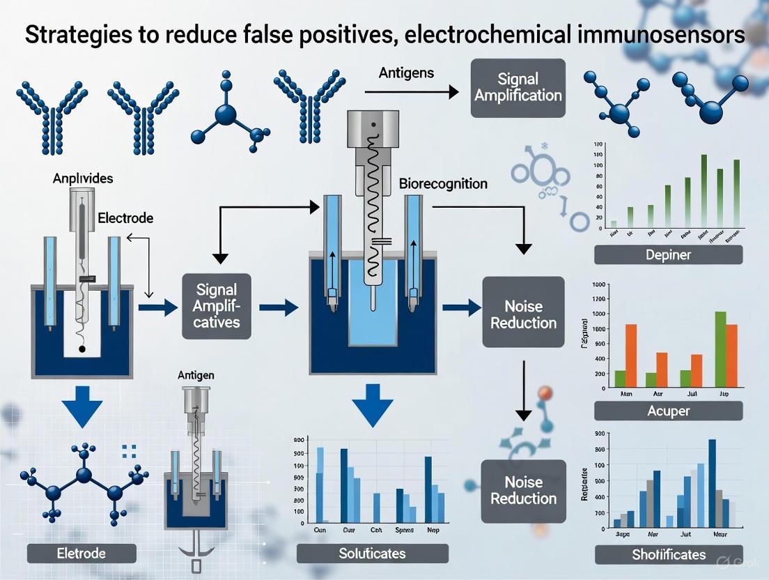
This article provides a comprehensive analysis of cutting-edge strategies to enhance the specificity and reliability of electrochemical immunosensors by mitigating false-positive results.
Advanced Strategies to Minimize False Positives in Electrochemical Immunosensors: From Nanomaterial Design to Multi-Mode Validation
Abstract
This article provides a comprehensive analysis of cutting-edge strategies to enhance the specificity and reliability of electrochemical immunosensors by mitigating false-positive results. Tailored for researchers, scientists, and drug development professionals, it explores the foundational causes of inaccuracies, details innovative methodological approaches involving nanomaterials and multi-mode detection, discusses optimization and troubleshooting for real-world application, and evaluates validation frameworks for comparative performance assessment. The synthesis of these core intents offers a roadmap for developing next-generation, highly accurate diagnostic tools for clinical and biomedical research.
Understanding the Root Causes of False Positives in Electrochemical Immunosensing
Frequently Asked Questions (FAQs)
1. What is a false positive in diagnostic testing? A false positive is a test result that incorrectly indicates the presence of a specific condition or analyte when it is not actually present [1]. In the context of electrochemical immunosensors, this means the device generates a positive signal despite the target biomarker being absent from the sample.
2. What are the main technical causes of false positives in electrochemical immunosensors? The primary technical causes include [1] [2] [3]:
- Cross-reactivity: The antibodies used in the immunosensor recognize and bind to non-target molecules (e.g., structurally similar proteins or other interfering substances) present in the sample matrix, such as serum.
- Non-specific binding (NSB): Undesirable proteins or molecules from a complex sample (e.g., blood, serum) adsorb onto the sensor's electrode surface without any specific biorecognition, leading to a false signal [3].
- Sample contamination: Even small traces of a contaminant, such as genetic material or a target analyte from another sample, can cause a false positive reading [1].
- Interference from reagents or the sample matrix: Components in the sample or expired/faulty chemical reagents can skew the electrochemical measurement [1].
3. Beyond technical issues, what other factors can lead to false positives? Other critical factors involve the sensor's design and operation [1]:
- Improper sensor calibration: Equipment that is not correctly calibrated can produce inaccurate and skewed results.
- Suboptimal sampling procedures: Errors during sample collection, storage, or preparation (e.g., sample degradation) can compromise the test's accuracy.
4. What are the real-world consequences of a false positive diagnosis? False positives have significant implications for patients, healthcare systems, and public health [1]:
- For patients: Unnecessary stress and anxiety, needless therapeutic interventions (including medications with potential side effects), and invasive procedures that carry inherent risks.
- For clinical decision-making: Delays in reaching the correct diagnosis and initiating the proper treatment, as clinical focus is diverted.
- For healthcare systems: Increased costs due to unnecessary follow-up tests, treatments, and hospital stays, leading to a mismanagement of valuable resources.
5. How can using multiple biomarkers reduce false positives? Relying on a single biomarker can be misleading, as its concentration can be elevated due to non-target conditions. For example, in sepsis diagnosis, measuring both Procalcitonin (PCT) and Interleukin-6 (IL-6) simultaneously provides a more specific diagnostic signature. A sensor that requires both biomarkers to be elevated for a positive result can effectively rule out conditions that only affect one of them, thereby reducing misdiagnosis [4].
Troubleshooting Guide: Mitigating False Positives
This guide addresses common experimental challenges and provides targeted strategies to enhance the specificity of your electrochemical immunosensors.
Challenge 1: High Background Signal from Non-Specific Binding (NSB)
Problem: Your sensor produces a significant signal in control samples (e.g., blank or negative samples), indicating that non-target molecules are interacting with the sensor surface.
Recommended Solutions:
| Solution | Underlying Principle | Key Experimental Considerations |
|---|---|---|
| Optimize Surface Blocking [3] | Coats remaining active sites on the electrode after antibody immobilization with inert proteins (e.g., BSA, casein) or polymers. | Test different blocking agents and concentrations. Incubation time and temperature are critical. |
| Employ Advanced Surface Chemistries [2] [3] | Creates a bio-inert layer that resists protein adsorption. Common strategies include Self-Assembled Monolayers (SAMs) of alkanethiols, and coatings with polyethylene glycol (PEG) or its derivatives. | The choice of chemistry depends on your electrode material (e.g., gold for SAMs). Ensure the layer does not impede electron transfer. |
| Utilize Nanomaterial-Based Electrodes [5] | Nanomaterials (e.g., gold nanoparticles, graphene) can provide a more oriented antibody immobilization, which reduces steric hindrance and can minimize NSB by presenting antibodies more efficiently. | The size, shape, and functionalization of nanomaterials significantly impact performance and reproducibility. |
Challenge 2: Signal Interference and Cross-Reactivity
Problem: The sensor signal is affected by non-target components in complex sample matrices (like serum), or the detection antibody binds to molecules similar to the target analyte.
Recommended Solutions:
| Solution | Underlying Principle | Key Experimental Considerations |
|---|---|---|
| Implement a Sandwich Assay Format [6] [3] | Requires two distinct antibodies to bind to different epitopes on the target analyte for a signal to be generated. This double recognition drastically improves specificity. | The pair of antibodies (capture and detection) must be carefully selected and validated for their specificity and lack of cross-reactivity. |
| Use High-Fidelity Biorecognition Elements | Employ highly specific monoclonal antibodies or aptamers to minimize the chance of binding to non-target molecules [7] [3]. | Source antibodies from reputable suppliers and validate their specificity against a panel of potential interferents. |
| Employ Label-Free Detection with EIS [2] [8] | Electrochemical Impedance Spectroscopy (EIS) directly measures the blocking of electron transfer upon immunocomplex formation. It avoids potential interference from enzyme labels or redox probes used in other methods. | EIS is highly sensitive to surface fouling. Excellent surface preparation and blocking are prerequisites. |
Challenge 3: Inconsistent Results and Poor Reproducibility
Problem: Signal output varies significantly between sensor batches or experimental runs.
Recommended Solutions:
| Solution | Underlying Principle | Key Experimental Considerations |
|---|---|---|
| Standardize Immobilization Protocols [2] | A consistent and optimized method for attaching antibodies to the electrode surface is fundamental for creating uniform sensor surfaces. | Systematically characterize each modification step using techniques like Cyclic Voltammetry (CV) and EIS to ensure layer-by-layer reproducibility [2]. |
| Introduce Rigorous Quality Control [1] | Using external quality assurance (EQA) programs and synthetic controls helps identify and eliminate false positives before reporting results. | Include multiple negative controls and low-positive controls in every experimental run to monitor performance. |
| Automate Sample Preparation [1] | Minimizes human error and operator-induced variability during sample handling, which is a common source of contamination and inconsistent results. | Explore automated liquid handling systems for sample and reagent dispensing, especially for high-throughput workflows. |
Experimental Protocol: A Representative Workflow for a Dual-Protein Sensor
The following protocol is adapted from a study that successfully developed an ultra-sensitive dual-protein electrochemical immunosensor for sepsis biomarkers (PCT and IL-6) to eliminate false positives from single-analyte analysis [4].
1. Sensor Fabrication and Surface Modification
- Synthesis of Nanocomposite: Prepare the
MXene@PANI@Aunanorod ternary heterostructure. The MXene provides high conductivity, PANI offers structural stability and prevents MXene stacking, and the Gold Nanorods (Au NRs) act as signal amplifiers [4]. - Electrode Modification: Drop-cast the
MXene@PANI@Au NRsnanocomposite onto the surface of pretreated screen-printed electrodes (SPEs). This creates a high-surface-area, conductive platform for antibody immobilization [4]. - Antibody Immobilization: Covalently immobilize specific capture antibodies for Procalcitonin (PCT) and Interleukin-6 (IL-6) onto the modified electrode surfaces, using different electrodes or individually addressable channels for each biomarker [4].
- Surface Blocking: Incubate the functionalized electrodes with a suitable blocking buffer (e.g., containing BSA) to passivate any remaining active sites and prevent non-specific binding [4] [3].
2. Assay Procedure and Measurement
- Sample Incubation: Apply 5 µL of the clinical sample (e.g., spiked or real human serum) onto the sensor and incubate to allow the antigens (PCT and IL-6) to bind to their respective capture antibodies [4].
- Washing: Gently rinse the electrode with a wash buffer to remove any unbound proteins and matrix components.
- Electrochemical Detection: For a sandwich-type assay, incubate with a detection antibody linked to a signal tag. Alternatively, for a label-free approach, directly measure the change in electrochemical impedance. In the referenced work, the intrinsic properties of the nanocomposite enabled direct, label-free quantification. Connect the sensor to a potentiostat and perform the measurement (e.g., Differential Pulse Voltammetry or EIS) [4].
- Data Analysis: Quantify the concentration of each biomarker based on the calibrated electrochemical signal. The use of two biomarkers allows for data validation; a positive sepsis diagnosis is only considered when both biomarkers are elevated beyond their respective clinical thresholds, thereby reducing the risk of a false positive from a single marker [4].
Experimental Workflow for Dual-Protein Sensor
Research Reagent Solutions
The following table details key materials used in the featured experiment and their functions in enhancing sensor performance and reducing false positives [4] [5].
| Research Reagent | Function in the Experiment |
|---|---|
| MXene (2D Material) | Serves as a highly conductive substrate, providing a large surface area for increased antibody loading and enhanced electron transfer. |
| Polyaniline (PANI) | A conductive polymer that coats MXene, preventing its oxidation and stacking, thereby improving the stability and reproducibility of the sensor. |
| Gold Nanorods (Au NRs) | Act as signal amplifiers due to their excellent conductivity and provide abundant sites for efficient antibody immobilization. |
| Screen-Printed Electrodes (SPEs) | Enable sensor miniaturization, portability, and mass production. They are ideal for point-of-care testing and can be disposable to prevent carryover contamination. |
| Specific Monoclonal Antibodies | The high-specificity biorecognition elements for PCT and IL-6. Their high affinity and specificity are crucial for minimizing cross-reactivity. |
| Principal Component Analysis (PCA) | A statistical tool used to analyze the electrochemical data from both biomarkers, helping to clearly discriminate between septic patients and healthy individuals. |
FAQ: Troubleshooting False Positives in Immunosensors
What are non-specific binding (NSB) and cross-reactivity, and how do they differ?
- Non-Specific Binding (NSB) occurs when an antibody or other assay component unintentionally adheres to surfaces or molecules other than the target analyte. This is often driven by hydrophobic interactions, electrostatic forces, or van der Waals forces, and leads to a high background signal [9] [10] [11].
- Cross-Reactivity happens when an antibody binds to an off-target molecule that shares structural similarities with the intended antigen, leading to false positives [12] [11] [13]. While NSB is a general attraction to surfaces or proteins, cross-reactivity is a specific, but undesired, molecular recognition event.
What are the primary causes of high background in my electrochemical immunosensor? High background signals primarily stem from NSB of proteins or other molecules to the electrode surface, cross-reactivity of detection antibodies, and inadequate blocking of unoccupied sites on the sensor surface [9] [10]. Other factors include sample contamination, use of the wrong enzyme substrate, insufficient washing steps, and poor water quality in buffers [10].
How can I minimize cross-reactivity from secondary antibodies in a multiplexed assay? Using cross-adsorbed secondary antibodies is a key strategy. These antibodies undergo an additional purification step to remove antibodies that bind to immunoglobulins from off-target species [14]. For example, a goat anti-mouse IgG antibody that is highly cross-adsorbed against bovine, rabbit, and human IgG will not recognize primary antibodies from those species, which is crucial for multiplexing experiments [14].
Experimental Protocols for Error Suppression
Protocol 1: Surface Blocking to Minimize Non-Specific Binding
Objective: To saturate unoccupied binding sites on the transducer surface after immobilization of the capture antibody.
- Surface Preparation: After the capture antibody is immobilized on the electrode (e.g., a screen-printed carbon electrode), wash the surface with an appropriate buffer (e.g., PBS).
- Blocking Solution Application: Incubate the electrode with a blocking solution for 1-2 hours at room temperature. Common blockers include [12] [10]:
- Bovine Serum Albumin (BSA) at 1-5%
- Casein sodium salt
- Non-fat dry milk
- Fish gelatin
- Commercial blocking formulations (e.g., StabilGuard, StabilBlock)
- Washing: Thoroughly wash the electrode with wash buffer (e.g., PBS with 0.05% Tween-20) to remove excess blocker.
- Validation: The effectiveness of blocking can be validated by running the immunosensor with a blank sample (without the target analyte) and confirming a low background signal [12].
Protocol 2: Sequential Staining to Prevent Cross-Species Antibody Binding
Objective: To prevent cross-reactivity between secondary antibodies and off-target primary antibodies in assays using multiple primaries (e.g., from mouse and rat).
- Incubate with First Primary Antibody: Apply the first primary antibody (e.g., mouse anti-NeuN) to the sample and incubate.
- Wash: Thoroughly wash to remove unbound antibody.
- Incubate with its Specific Secondary Antibody: Apply the secondary antibody specific to the first primary (e.g., goat anti-mouse IgG) and incubate. This "uses up" the binding sites for this secondary.
- Wash: Thoroughly wash again.
- Repeat for the Second Primary: Apply the second primary antibody (e.g., rat anti-GFAP), wash, and then apply its specific secondary antibody (e.g., goat anti-rat IgG) [15]. This sequential method prevents the secondary antibody for the first primary from encountering and binding to the second primary antibody from a different species.
Quantitative Data: Sensor Performance with Error Suppression
Table 1: Performance of Electrochemical Immunosensors Implementing NSB/Cross-Reactivity Suppression Strategies
| Target Analyte | Sensor Platform / Key Suppression Strategy | Limit of Detection (LOD) | Linear Range | Key Findings |
|---|---|---|---|---|
| Soybean Allergen (Gly m TI) [16] | Electrochemical immunosensor / Not specified | -- | 0.1 - 100,000 mg kg⁻¹ in food | Quantified down to 0.1 mg kg⁻¹ of soybean in complex food matrices without matrix interference. |
| Carcinoembryonic Antigen (CEA) [17] | SPCE modified with antifouling rGO and β-CD-COOH | 6.0 fg/mL | 10 fg/mL - 1.0 ng/mL | The antifouling layers enhanced sensing performance, achieving a low LOD in patient serum samples. |
| Prostate-Specific Antigen (PSA) [9] | ELISA sandwich immunoassay with redox polymer signal tag | 0.3 pg/mL | -- | Demonstrated an extremely low LOD for a sandwich-type electrochemical immunoassay. |
Research Reagent Solutions
Table 2: Essential Reagents for Minimizing False Positives
| Reagent / Material | Function / Purpose | Example Use Cases |
|---|---|---|
| Blocking Agents (BSA, Casein, Non-fat dry milk, Fish gelatin, Commercial blockers) [12] [10] | Saturates unused binding sites on the solid phase to prevent NSB of proteins. | Used after immobilization of capture antibodies on electrodes or microplates. |
| Cross-Adsorbed Secondary Antibodies [14] | Secondary antibodies purified to remove antibodies that bind to immunoglobulins of non-target species. | Essential for multiplex assays using primary antibodies from different species to prevent cross-reactivity. |
| Assay Diluents (e.g., Protein-containing or protein-free formulations) [10] [11] | Dilutes samples and reagents while containing components to block matrix interferences (e.g., HAMA, rheumatoid factor). | Used to dilute patient samples (like serum) to reduce false positives caused by interfering substances. |
| Nanostructured Materials (e.g., reduced Graphene Oxide, PEG, SAMs) [9] [17] | Modifies electrode surface to improve conductivity, provide more binding sites, and create a low-fouling or antifouling layer. | Used in electrochemical immunosensor fabrication to enhance signal and resist NSB from complex samples. |
| Wash Buffers (e.g., PBS with Tween-20) [10] | Removes unbound reagents and weakly adsorbed molecules from the assay surface through surfactants. | A critical step after every incubation period in an immunoassay to reduce background. |
Experimental Workflow for Error Minimization
The diagram below outlines a logical troubleshooting workflow for diagnosing and addressing the root causes of false positives in immunosensor development.
Diagram 1: A logical workflow for troubleshooting high background and false positives in immunosensors. Researchers can follow the path based on their experimental observations to identify the most likely cause and corresponding solution.
Interference from Complex Biological Matrices (e.g., Blood, Serum)
FAQs: Understanding and Identifying Interference
What are the common sources of interference in immunoassays from biological matrices? Interference in immunoassays can arise from various endogenous and exogenous substances present in complex biological matrices like blood and serum. Common sources include [18] [19]:
- Heterophile antibodies and human anti-animal antibodies that can bind to assay antibodies.
- Cross-reacting substances with structural similarities to the target analyte.
- Endogenous binding proteins, such as sex hormone-binding globulin or cortisol binding globulin, which can bind to the analyte and alter its measurable concentration.
- Matrix effects from components like lipids (lipemia), hemoglobin (hemolysis), or bilirubin (icterus).
- Pre-analytical factors, including the type of specimen tube anticoagulant (e.g., EDTA, heparin), sample storage conditions, and carryover from tube additives.
How can I tell if my experimental results are affected by interference? Clinical laboratorians and researchers should suspect interference when encountering the following scenarios [18] [19]:
- The test result is clinically implausible or inconsistent with the patient's clinical presentation.
- There is a discordance between results from different assay methods for the same analyte.
- The result shows a dramatic, unexpected change from a previous measurement without a clinical correlate.
- The analyte does not recover linearly upon serial dilution of the sample.
What are the consequences of not addressing interference? Unidentified interference can lead to [18]:
- Misdiagnosis or missed diagnosis, leading to the wrong course of treatment.
- Unnecessary follow-up laboratory tests and clinical investigations.
- Inaccurate data in research settings, compromising study validity.
- In the context of drug monitoring, interference could lead to incorrect drug dosing.
Troubleshooting Guides: Detecting and Confirming Interference
Guide 1: Systematic Workflow for Interference Investigation
Follow this logical pathway to systematically investigate potential interference.
Guide 2: Experimental Protocols for Detection
Protocol: Serial Dilution for Recovery Assessment This protocol tests whether the measured analyte concentration decreases proportionally with dilution, which is expected in the absence of interferents [19].
- Preparation: Obtain a sufficient volume of the patient sample with suspected interference. Prepare an appropriate diluent (the manufacturer's recommended diluent is ideal, or a validated alternative like non-immune serum or assay buffer).
- Dilution Series: Create a series of dilutions (e.g., 1:2, 1:4, 1:8) of the patient sample in the chosen diluent.
- Analysis: Measure the analyte concentration in each diluted sample using the standard immunoassay protocol.
- Calculation and Interpretation:
- Calculate the expected concentration for each dilution (e.g., the undiluted result multiplied by the dilution factor).
- Plot the measured concentration against the expected concentration.
- Interpretation: If the measured values are significantly lower or higher than expected (non-linear recovery), an interfering substance is likely present. The interference typically diminishes once the interferent is diluted to a non-effective concentration.
Protocol: Investigating Tube-Specific Interference Pre-analytical factors related to blood collection tubes are a common source of error [20].
- Hypothesis: A specific tube additive (e.g., gel separator, anticoagulant) is causing interference.
- Experimental Setup: During validation, collect paired samples from multiple donors using the standard tube and an alternative tube (e.g., a rapid serum tube instead of a plasma separator tube).
- Analysis: Run the target immunoassay on both sets of samples.
- Interpretation: A consistent, significant bias in results from one tube type indicates tube-specific interference. For example, using rapid serum tubes was shown to reduce false-positive troponin results by 50% compared to plasma separator tubes [20].
Data Presentation
Table 1: Common Interference Types and Their Effects on Immunoassays
This table summarizes key interferents and their impact on assay results, aiding in initial hypothesis generation.
| Interference Type | Description | Typical Effect on Result | Example Analytes Affected |
|---|---|---|---|
| Heterophile Antibodies [18] | Endogenous human antibodies that bind weakly to immunoglobulins from other species. | Falsely elevated or decreased | Human chorionic gonadotropin (hCG), cardiac troponin [18] |
| Human Anti-Animal Antibodies [18] | Antibodies against animal immunoglobulins (e.g., from exposure to pets). | Falsely elevated or decreased | Various, depending on assay antibodies |
| Cross-Reactivity [18] [21] | Non-target molecules with structural similarity to the analyte bind to the assay antibody. | Falsely elevated | Cortisol (cross-reaction with fludrocortisone), drugs of abuse [18] |
| Binding Proteins [18] | Endogenous proteins (e.g., SHBG) that bind the analyte, making it unavailable. | Falsely lowered | Sex hormones, cortisol, free thyroxine (FT4) |
| Lipemia (Lipids) [18] [20] | High lipid concentration causing turbidity. | Interferes with nephelometry/turbidimetry | Varies |
| Biotin [19] | High doses of vitamin B7 can interfere with biotin-streptavidin based assays. | Falsely lowered or elevated | Thyroid tests, cardiac troponin |
Table 2: Comparison of Methods for Investigating Interference
This table helps researchers select the most appropriate troubleshooting method based on performance characteristics.
| Investigation Method | Principle | Key Advantage | Key Limitation/Caveat |
|---|---|---|---|
| Serial Dilution [19] | Tests for linearity of analyte recovery upon sample dilution. | Powerful for detecting the presence of an interferent. | Must validate diluent and expected recovery in control samples first; some assays dilute non-linearly by design. |
| Alternate Method Comparison [19] | Comparing results from a different immunoassay that uses unique antibodies/reagents. | Can confirm if interference is method-specific. | Requires knowledge of the expected bias between methods; comparable results strongly rule out interference. |
| Blocking Reagents [19] | Pre-treating sample with reagents to neutralize heterophile antibodies or remove biotin. | Directly targets and removes specific interferents. | Must validate that the blocking reagent itself does not affect the assay using control samples. |
| Sample Re-collection [20] | Collecting a new sample using a different tube type or collection method. | Addresses pre-analytical errors and tube-specific interferences. | Logistically challenging; requires patient re-consent. |
The Scientist's Toolkit: Research Reagent Solutions
Table 3: Key Reagents and Materials for Mitigating Interference
Essential tools for the researcher's bench to prevent and resolve matrix interference issues.
| Reagent / Material | Function in Interference Mitigation | Application Notes |
|---|---|---|
| Heterophile Blocking Reagents [19] | Contains proprietary mixtures of animal immunoglobulins or inert blocking agents to neutralize heterophile antibodies in patient samples. | Add to sample prior to assay. Effectiveness should be verified with positive and negative controls. |
| Biotin Blocking Kits [19] | Contains reagents (e.g., streptavidin) to bind and neutralize excess biotin in the sample. | Critical for patients on high-dose biotin therapy. Useful when biotin-streptavidin chemistry is used in the assay. |
| Certified Metal-Free Tubes [20] | Specially manufactured collection tubes that do not leach trace metals like chromium or aluminum. | Essential for accurate trace metal testing (e.g., for monitoring metal-on-metal prosthetic devices). |
| Rapid Serum Tubes (RST) [20] | Contain thrombin to accelerate clotting, producing a clean serum sample quickly and reducing fibrin strand formation. | Shown to reduce false-positive rates in cardiac troponin testing by ~50% compared to standard plasma tubes. |
| Protein G [22] | A bacterial protein that binds the Fc region of antibodies. Used in sensor fabrication for oriented antibody immobilization. | Improves assay specificity and sensitivity by presenting antibodies in an optimal configuration, potentially reducing non-specific binding [22]. |
Visualizing Mitigation Strategies for Sensor Design
A strategic approach to minimizing interference begins at the sensor design and assay development stage.
Limitations of Conventional Single-Mode Detection Platforms
Frequently Asked Questions (FAQs)
1. What is the main limitation of conventional single-mode electrochemical immunosensors? The primary limitation is their susceptibility to false results (both positives and negatives), largely due to inter-electrode variations and inherent signal errors. Unlike ratiometric approaches that use an internal reference, single-mode sensors lack a self-correcting mechanism. This means that small, analyte-independent variations in the electrode surface, sample matrix, or experimental conditions can lead to significant inaccuracies in the reported concentration [25].
2. Why is my single-mode immunosensor giving inconsistent results between different electrode batches? This is a classic symptom of poor reproducibility caused by the manufacturing variability of base electrodes. Even with high-precision equipment, it is challenging to produce electrodes that are perfectly identical. In single-mode detection, this inherent inter-electrode variation directly translates into signal drift and inconsistent results, as there is no internal calibration to correct for it [25].
3. My label-free immunosensor shows a signal, but the control sample (without the target) also shows significant background. What could be the cause? This indicates a problem with nonspecific binding (NSB). In label-free formats, any molecule that adsorbs to the electrode surface can alter its electrical properties (e.g., impedance) and generate a signal, leading to false positives. This is a known challenge for label-free electrochemical immunosensors (ELFIs), as they lack a secondary, signal-generating antibody that can provide an additional layer of specificity [26] [27].
4. How can the sample matrix (like blood or wastewater) affect my single-mode sensor's performance? Complex sample matrices contain various interfering substances (e.g., proteins, salts, other electroactive compounds) that can:
- Cause Fouling: Adsorb to the electrode surface, blocking the active sites and reducing sensitivity [8] [27].
- Generate Non-Specific Signals: Directly participate in redox reactions, contributing to the background current and leading to false positives [28] [29]. Single-mode sensors struggle to distinguish these interfering signals from the specific antigen-antibody binding signal.
Troubleshooting Guides
Issue 1: High False Positive/Negative Rates
Problem: The sensor indicates the presence of the target analyte when it is absent (false positive) or fails to detect it when it is present (false negative).
| Potential Cause | Diagnostic Steps | Solution and Recommended Strategy |
|---|---|---|
| Nonspecific Binding (NSB) | Run a control with a non-target protein of similar size and charge. If a significant signal is observed, NSB is likely. | Optimize the blocking step using agents like BSA or casein. Incorporate a rigorous layer-by-layer electrochemical characterization (LbL-EC) to monitor and minimize NSB during sensor assembly [26]. |
| Inter-Electrode Variation | Test the same sample with multiple electrodes from different batches and observe signal variance. | Transition from a single-mode to a ratiometric sensing approach. This uses two signals (a sensing signal and an internal reference signal) to self-correct for variations in the base electrode, dramatically improving accuracy and reproducibility [25]. |
| Sample Matrix Interference | Perform a standard addition recovery experiment in the real sample matrix. | Improve sample pre-treatment (e.g., filtration, dilution) to remove interferents. Alternatively, use a sandwich-type immunosensor format, which requires two specific binding events, thereby enhancing selectivity against matrix interferents [29] [27]. |
Issue 2: Poor Reproducibility and Stability
Problem: The sensor's performance (signal output for a fixed concentration) varies significantly from one experiment to another or degrades rapidly over time.
| Potential Cause | Diagnostic Steps | Solution and Recommended Strategy |
|---|---|---|
| Unstable Bioreceptor Immobilization | Monitor the baseline signal of the modified electrode over time in a blank buffer. Signal drift suggests poor immobilization. | Use a more robust immobilization strategy, such as covalent binding onto a nanomaterial-modified electrode (e.g., graphene, AuNPs) instead of simple physical adsorption [25] [27]. |
| Dynamic Variations in Electrode Surface | Characterize the electrode surface before and after use with techniques like Cyclic Voltammetry (CV) and Electrochemical Impedance Spectroscopy (EIS). | Implement electrochemical ratiometry. This method overcomes the inherent errors and dynamic variations of the base electrode by relying on the ratio between two signals, providing significantly enhanced sensing stability [25]. |
| Degradation of Signal Labels (in labeled sensors) | Test the activity of the enzymatic label (e.g., HRP) separately from the immunosensor. | Ensure proper storage conditions (e.g., at 4°C). Consider using nanomimetic enzymes or stable redox probes (e.g., ferrocene derivatives) that offer better stability than biological enzymes [29]. |
Experimental Protocol: Implementing a Ratiometric Immunosensor
This protocol provides a methodology to construct a ratiometric electrochemical immunosensor, a key strategy to overcome the limitations of single-mode platforms and reduce false positives [25].
1. Objective: To fabricate a label-free ratiometric immunosensor for the detection of a model virus (e.g., SARS-CoV-2 pseudovirus) using a dual-probe system to enhance reproducibility and accuracy.
2. Materials and Reagents:
- Electrode: Screen-printed electrodes (SPEs)
- Probe 1 (Internal Reference): Thionin acetate (TA) or Ferrocene carboxylic acid (Fc)
- Probe 2 (Sensing Probe): Potassium ferricyanide (K₃[Fe(CN)₆])
- Nanomaterial: Electrochemically synthesized graphene (eG) dispersion
- Biorecognition Elements: Specific antibody against the target antigen
- Blocking Agent: Bovine Serum Albumin (BSA)
- Buffers: Phosphate Buffered Saline (PBS, 0.1 M, pH 7.4) for washing and dilution
3. Procedure: Step 1: Electrode Modification
- Clean the SPE according to the manufacturer's instructions.
- Perform in situ electrodeposition or drop-casting of graphene (eG) onto the working electrode surface to create an eG-SPE. This enhances the electrode's surface area and electrical conductivity.
- Characterize the modification using Cyclic Voltammetry (CV) in a solution containing both [Fe(CN)₆]³⁻/⁴⁻ and TA to confirm the presence of two well-defined, stable redox peaks.
Step 2: Antibody Immobilization
- Activate the eG-SPE surface as required for your chosen immobilization chemistry (e.g., EDC/NHS for covalent binding).
- Incubate the electrode with a solution of the specific capture antibody for a set period (e.g., 2 hours at room temperature).
- Wash thoroughly with PBS to remove physically adsorbed antibodies.
Step 3: Surface Blocking
- Incubate the modified electrode with a solution of BSA (e.g., 1% w/v) for at least 1 hour. This critical step blocks any remaining active sites on the electrode to minimize nonspecific binding.
- Wash again with PBS.
Step 4: Ratiometric Detection and Data Analysis
- Record the Differential Pulse Voltammetry (DPV) signal of the prepared immunosensor in a blank buffer solution containing both TA and [Fe(CN)₆]³⁻/⁴⁻. This gives the initial currents, I₀(TA) and I₀(Fe).
- Incubate the immunosensor with the sample containing the target antigen.
- After incubation and washing, record the DPV signal again in the same buffer, yielding the final currents, I(TA) and I(Fe).
- Data Analysis: The concentration of the target antigen is correlated not to the absolute change of a single signal, but to the ratio of the two signal changes (e.g., ΔI(Fe) / ΔI(TA) or ΔI(TA) / I₀(TA) ). This ratio is self-calibrating and corrects for inherent electrode-to-electrode variations.
Research Reagent Solutions
The following table details key materials used in advanced electrochemical immunosensing to mitigate false results.
| Item | Function/Benefit | Application Example |
|---|---|---|
| Screen-Printed Electrodes (SPEs) | Inexpensive, disposable, allow for mass production and miniaturization for point-of-care testing. | Used as the base transducer in most modern electrochemical immunosensor designs [26] [25]. |
| Electrochemically Synthesized Graphene (eG) | Nanomaterial with high surface area and excellent conductivity; enhances electron transfer and provides a platform for bioreceptor immobilization. | Serves as a modifier for SPEs to significantly boost the current amplitude and improve sensor sensitivity [25]. |
| Ratiometric Electrochemical Probes (e.g., Fc, TA, [Fe(CN)₆]³⁻/⁴⁻) | A pair of redox probes used to generate two independent signals. The ratio between these signals provides an internal calibration, overcoming electrode variability and reducing false results. | Fc/TA and [Fe(CN)₆]³⁻/⁴⁻/TA pairs are used to create a self-referencing system, drastically improving reproducibility and accuracy [25]. |
| Gold Nanoparticles (AuNPs) | Provide a high-surface-area, biocompatible substrate for antibody immobilization; can facilitate electron transfer and act as a label for signal amplification. | Used to modify electrode surfaces, improving the orientation and loading of capture antibodies [27]. |
| Blocking Agents (e.g., BSA, Casein) | Proteins used to passivate the electrode surface after antibody immobilization. They bind to non-specific sites, preventing interferents from causing false positive signals. | A critical step in all immunosensor protocols after the capture antibody is attached to the electrode [26] [29]. |
Signaling Pathway and Experimental Workflow
The following diagram illustrates the core logical relationship between the limitations of single-mode detection and the advanced strategy of ratiometric sensing.
Single Mode vs Ratiometric Detection Logic
The diagram below outlines the key stages in the experimental workflow for developing a ratiometric immunosensor, as described in the protocol.
Ratiometric Immunosensor Workflow
The Role of Biomarker Specificity and Concentration in Early-Stage Disease
Frequently Asked Questions (FAQs)
Q1: Why is biomarker specificity so critical for early-stage disease detection? High biomarker specificity ensures that the detected signal is generated only by the target biomarker and not by other interfering substances present in complex biological samples like blood or urine. A lack of specificity is a primary cause of false positives, where a test incorrectly indicates the presence of disease. For instance, a biomarker should be uniquely associated with a specific cancer type and not be elevated in non-cancerous conditions like inflammation or infection [30] [31].
Q2: How does low biomarker concentration in early-stage disease lead to false negatives? In the initial stages of a disease like cancer, the tumor burden is minimal, leading to extremely low concentrations of shed biomarkers, such as circulating tumor DNA (ctDNA) or specific proteins, in the bloodstream. These concentrations can fall below the detection limit (LOD) of the immunosensor. One study notes that early-stage tumors can shed as little as 0-1 copies of ctDNA per milliliter of blood, leading to 76–92% of Stage I cancers being missed by some ctDNA-based assays [30].
Q3: What are the main sources of non-specific binding in electrochemical immunosensors? Non-specific binding (NSB) occurs when molecules other than the target biomarker adhere to the sensor surface, generating a false signal. Key sources include:
- Complex Biological Matrices: Serum and blood contain high abundances of non-specific proteins, cells, and other molecules that can physically adsorb to the electrode [32] [2].
- Surface Properties: Variations in the electrode's surface chemistry, morphology, and composition can increase the likelihood of NSB [2].
- Insufficient Blocking: An inadequately optimized blocking step fails to cover all non-active sites on the sensor surface, leaving room for non-target molecules to bind [2].
Q4: What strategies can improve the reproducibility of immunosensor results? Reproducibility is hampered by inter-electrode variations and inconsistent surface modifications. Key strategies include:
- Electrochemical Ratiometry: Using an internal reference signal to self-calibrate and correct for variations in the base electrode, significantly enhancing reproducibility and accuracy [25].
- Layer-by-Layer Electrochemical Characterization (LbL-EC): Systematically monitoring each step of the electrode modification (e.g., immobilization, blocking) using techniques like EIS and CV to ensure consistent and optimized sensor assembly [2].
- Utilizing Nanomaterials: Nanomaterials like graphene can enhance electron transfer and provide a more uniform surface for bioreceptor immobilization [25].
Q5: How can AI and machine learning help reduce diagnostic errors? AI and machine learning algorithms can process complex data patterns that are difficult to discern manually. They can be trained to:
- Analyze Fluorescent Signals: AI can be integrated with smartphone-based microscopy to accurately quantify biomarker concentrations from fluorescent images, minimizing human error in interpretation [32].
- Create Diagnostic Embeddings: Machine learning can analyze a panel of multiple biomarker concentrations (e.g., amino acid signatures) to create a unique disease fingerprint, dramatically improving specificity and reducing the false positive rate [30].
Troubleshooting Guides
Guide 1: Addressing High False Positive Rates
Potential Cause: Non-Specific Binding and Interferents False positives often arise from non-specific adsorption of molecules or cross-reactivity with similar biomarkers.
| Troubleshooting Step | Protocol Details | Expected Outcome |
|---|---|---|
| 1. Optimize Blocking Agent | After antibody immobilization, incubate the electrode with a blocking solution (e.g., 1-3% BSA or casein in PBS) for 1 hour at room temperature. Test different agents and concentrations. | Reduction in signal from negative control samples. |
| 2. Introduce Stringency Washes | After sample incubation, perform washes with a buffer containing a mild detergent (e.g., 0.05% Tween-20 in PBS). Adjust ionic strength and pH to disrupt weak, non-specific interactions. | Lower background signal without significantly affecting the specific target signal. |
| 3. Validate Antibody Specificity | Use techniques like Western Blot or mass spectrometry to confirm that the capture/detection antibodies bind exclusively to the target biomarker and do not cross-react with other proteins in the sample matrix. | Identification and elimination of cross-reactive antibodies from the assay. |
| 4. Employ Ratiometric Sensing | Construct a sensor using two electrochemical probes (e.g., Fc and K3[Fe(CN)6]). The ratio of their signals is used for quantification, which self-corrects for background interference and electrode surface variations [25]. | Improved reproducibility and a significant reduction in false positives caused by non-specific effects. |
Guide 2: Addressing High False Negative Rates
Potential Cause: Insufficient Sensitivity for Low Biomarker Concentration False negatives occur when the sensor cannot detect the low levels of biomarker present in early-stage disease.
| Troubleshooting Step | Protocol Details | Expected Outcome |
|---|---|---|
| 1. Incorporate Signal-Amplifying Nanomaterials | Modify the electrode surface with nanomaterials such as gold nanoparticles (AuNPs), carbon nanotubes, or graphene oxide during fabrication. These materials enhance electron transfer and provide a high surface area for antibody loading, amplifying the detection signal [33] [25]. | A lower Limit of Detection (LOD), allowing the sensor to detect biomarker concentrations in the picogram or femtomolar range. |
| 2. Use Enzyme Labels | Employ an enzyme-labeled secondary antibody (e.g., Horseradish Peroxidase - HRP). After immunocomplex formation, add an enzyme substrate (e.g., TMB/H2O2) that produces an electroactive product, resulting in a cascading signal amplification. | A dramatic increase in the measured current, enhancing sensitivity by several orders of magnitude. |
| 3. Pre-concentrate the Sample | Implement a microfluidic platform with integrated filters or use magnetic beads functionalized with capture antibodies to isolate and concentrate the target biomarker from a larger sample volume before analysis. | An effective increase in the local biomarker concentration presented to the sensor. |
| 4. Leverage a Multi-Biomarker Panel | Instead of relying on a single biomarker, detect a panel of multiple biomarkers associated with the disease. Use machine learning to analyze the combined signature, which can be more sensitive to early-stage disease than any single biomarker alone [30]. | Increased probability of detecting the disease even if concentrations of individual biomarkers are low. |
Experimental Data & Protocols
Table 1: Performance of Advanced Sensing Strategies for Early Detection
This table summarizes quantitative data from recent studies on innovative approaches to improve detection limits and accuracy.
| Sensing Strategy / Platform | Target Biomarker(s) | Detection Limit | Key Performance Metric (False Positive/Negative Rate) | Reference |
|---|---|---|---|---|
| AI-enhanced Smartphone Microscopy | Carcinoembryonic Antigen (CEA) | 0.4 ng/mL | Not explicitly stated; high accuracy for multiplex detection | [32] |
| Electrochemical Ratiometric Immunosensor | SARS-CoV-2 Spike Pseudovirus | Not explicitly stated | Reproducibility: ~5x improvement vs non-ratiometric; Accuracy: Rivaled gold-standard PCR | [25] |
| Immunodiagnostic Amino Acid Signature (AACS) | Multi-Cancer Panel (Breast, Colorectal, etc.) | N/A (Pattern-based) | False Positive Rate: 0% (in N=97 cohort); Identified 78% of cancers | [30] |
| Competitive Electrochemical Immunosensor | Aβ42 Peptides (Alzheimer's) | 25.2 pM | Detection range: 0.056–13.7 nM | [29] |
Core Experimental Protocol: Fabrication of a Label-Free Electrochemical Immunosensor
This protocol outlines the key steps for constructing a basic label-free immunosensor, a common platform in research [2].
Electrode Pretreatment:
- Clean the working electrode (e.g., Glassy Carbon Electrode or screen-printed carbon electrode) by polishing with alumina slurry (0.05 µm) on a microcloth. Rinse thoroughly with deionized water and then with ethanol.
- Perform electrochemical activation via cyclic voltammetry (CV) in 0.5 M H2SO4 (e.g., 15 cycles from -0.2 V to +0.6 V at 50 mV/s).
Surface Modification (Nanomaterial Enhancement):
- Deposit a suspension of graphene oxide or other nanomaterials onto the electrode surface and allow it to dry. Alternatively, electrochemically reduce graphene oxide in situ by applying a fixed potential.
Antibody Immobilization:
- Apply a droplet (e.g., 10 µL) of the capture antibody solution (in PBS, pH 7.4) onto the modified electrode and incubate in a humidified chamber for 12-16 hours at 4°C.
- Wash the electrode gently with PBS to remove any physically adsorbed antibodies.
Surface Blocking:
- Incubate the electrode with a blocking agent (e.g., 1% BSA) for 1 hour at room temperature to passivate any remaining active sites on the electrode surface.
- Rinse with PBS to remove excess blocking agent. The immunosensor is now ready for use.
Detection via Electrochemical Impedance Spectroscopy (EIS):
- Measure the EIS spectrum of the sensor in a solution of 5 mM [Fe(CN)6]3−/4− as a redox probe. Record the charge transfer resistance (Rct) before and after exposure to the antigen.
- The increase in Rct is proportional to the amount of antigen bound to the surface, allowing for quantification.
Essential Visualizations
Diagram: Ratiometric Immunosensor Signaling Pathway
This diagram illustrates the working principle of a dual-probe ratiometric electrochemical immunosensor, a key method for reducing false results.
Diagram: Experimental Workflow for AACS Biomarker Analysis
This diagram outlines the novel immunodiagnostic workflow for detecting cancer via Amino Acid Concentration Signatures (AACS).
The Scientist's Toolkit: Research Reagent Solutions
| Item | Function in the Experiment | Technical Specification / Example |
|---|---|---|
| Screen-Printed Electrodes (SPEs) | Disposable, cost-effective electrochemical cells that facilitate mass production and miniaturization for point-of-care testing [25] [2]. | Carbon, gold, or platinum working electrodes; often used as a three-electrode system. |
| Bioorthogonal Fluorogenic Labels | Chemical probes that react specifically with target amino acid side-chains (e.g., Cysteine, Lysine) in neat plasma, becoming fluorescent only upon reaction. This eliminates the need for purification steps [30]. | Labels with specific excitation/emission profiles (e.g., 460 nm and 580 nm). |
| Signal-Amplifying Nanomaterials | Enhance sensor sensitivity by increasing the electroactive surface area and facilitating electron transfer. They can also serve as platforms for antibody immobilization [33] [25]. | Gold Nanoparticles (AuNPs), Graphene, Multi-walled Carbon Nanotubes (MWNTs), Quantum Dots (with toxicity considerations [33]). |
| Electrochemical Redox Probes | Molecules that undergo reversible redox reactions at the electrode surface, generating the measurable current in techniques like EIS and DPV. Essential for ratiometric sensing [25] [2]. | Potassium Ferricyanide (K3[Fe(CN)6]), Ferrocene derivatives, Thionin Acetate. |
| Blocking Agents | Proteins or other molecules used to cover non-specific binding sites on the sensor surface after antibody immobilization, thereby reducing background noise and false positives [2]. | Bovine Serum Albumin (BSA) at 1-3%, casein, or synthetic blocking reagents. |
Innovative Materials and Multi-Mode Assay Designs for Enhanced Specificity
Troubleshooting Guides
Guide 1: Addressing MXene Instability in Sensor Fabrication
Problem: Rapid degradation and restacking of MXene nanosheets, leading to decreased sensor conductivity and signal instability.
Solutions:
- Apply Conductive Polymer Coating: Use polyaniline (PANI) as an interfacial stabilizer. PANI prevents MXene stacking and oxidation while providing amine termini for biomolecule immobilization [4].
- Optimized Synthesis Protocol:
- Disperse 10 mg of synthesized MXene in 50 mL deionized water via ultrasonication [4].
- Mix 2 μL aniline solution and 6 mg ammonium persulfate in 10 mL of 1 M HCl [4].
- Slowly add the aniline mixture to the MXene dispersion while stirring [4].
- Stir the combined mixture at room temperature for 4 hours for complete PANI polymerization [4].
- Centrifuge the product and wash three times to remove unreacted precursors [4].
Guide 2: Mitigating False Positives in Complex Biological Samples
Problem: Non-specific binding and interference from complex sample matrices (e.g., serum) cause false positive signals.
Solutions:
- Implement Dual-Biomarker Detection: Develop sensors that simultaneously detect two specific biomarkers (e.g., PCT and IL-6 for sepsis). A true positive requires both biomarkers to be elevated, significantly reducing false positives from single-analyte tests [4].
- Surface Passivation: After antibody immobilization, incubate the sensor surface with 1% Bovine Serum Albumin (BSA) for 30-60 minutes to block non-specific active sites [34].
- Validate with Real Samples: Perform recovery tests using spiked human serum samples. Acceptable recovery rates (e.g., 96.0%–108.0%) confirm minimal matrix interference [4].
Guide 3: Achieving Consistent Noble Metal Nanoparticle Decoration
Problem: Inconsistent decoration of Gold Nanorods (Au NRs) on MXene-polymer composites, leading to variable signal amplification.
Solutions:
- Controlled Integration: After synthesizing the MXene@PANI composite, slowly add a precise volume of pre-synthesized Au NRs solution under constant stirring [4]. The amine groups on PANI provide nucleation sites for anchoring Au NRs [4].
- Verification with Electron Microscopy: Use SEM and TEM to confirm the uniform distribution of Au NRs on the MXene@PANI surface. Successful synthesis shows Au NRs anchored without aggregation [4].
Frequently Asked Questions (FAQs)
FAQ 1: Why is a dual-protein detection strategy more effective than a single-protein approach for reducing false positives? Many diseases lack a single, perfectly specific biomarker. For instance, procalcitonin (PCT) can elevate in non-infectious inflammation, and interleukin-6 (IL-6) can rise in various inflammatory conditions. Measuring both proteins in parallel creates a more specific diagnostic signature. A positive result is only confirmed when both biomarkers are elevated, effectively eliminating false positives caused by unrelated conditions that affect only one biomarker [4].
FAQ 2: What is the specific role of the conductive polymer (PANI) in the MXene@PANI@Au nanocomposite? Polyaniline serves multiple critical functions:
- Stability: It acts as a spacer to prevent MXene nanosheets from restacking and shields them from environmental oxidation [4].
- Immobilization: Its amine-termini offer active sites for the stable anchoring of gold nanorods and subsequent antibody immobilization [4].
- Synergistic Performance: It works synergistically with MXene; PANI provides redox activity while MXene offers high conductivity, together enhancing electron transfer efficiency [4].
FAQ 3: Our sensor performance is inconsistent between batches. What are the key factors to control? Batch-to-batch variability is a common challenge in nanomaterial-based sensors. Focus on standardizing:
- MXene Synthesis: Strictly control the etching time, temperature, and concentration of the etchant (e.g., HF) used on the MAX phase precursor [35].
- Nanocomposite Mixing: Ensure precise stoichiometric ratios of MXene, aniline, and Au NRs, and maintain consistent reaction times and stirring speeds during nanocomposite synthesis [4].
- Biorecognition Element Immobilization: Standardize the concentration of antibodies, incubation time, and the blocking procedure to ensure consistent sensor surface chemistry [34].
FAQ 4: How do noble metal nanoparticles like Gold Nanorods enhance sensor signal? Gold Nanorods function as excellent signal amplifiers due to their:
- High Electrical Conductivity: They facilitate faster electron transfer between the electrode surface and the biorecognition layer, increasing the current response [4].
- Large Surface Area: They provide abundant sites for immobilizing a high density of detection antibodies (Ab2), which enhances the binding capacity for the target analyte [4].
- Catalytic Properties: They can catalyze certain electrochemical reactions, leading to further signal enhancement [4].
The table below summarizes key performance metrics from recent studies utilizing MXene-based nanocomposites for electrochemical detection, highlighting the ultra-sensitive detection limits achievable.
Table 1: Performance Metrics of MXene-Based Electrochemical Immunosensors
| Target Analyte | Nanocomposite Used | Detection Limit | Linear Range | Application Context |
|---|---|---|---|---|
| Procalcitonin (PCT) | MXene@PANI@Au NRs [4] | 0.84 pg·mL⁻¹ [4] | 1 pg·mL⁻¹ – 1 μg·mL⁻¹ [4] | Sepsis Diagnosis [4] |
| Interleukin-6 (IL-6) | MXene@PANI@Au NRs [4] | 0.75 pg·mL⁻¹ [4] | 1 pg·mL⁻¹ – 1 μg·mL⁻¹ [4] | Sepsis Diagnosis [4] |
| Prostate-Specific Antigen (PSA) | PANI@MXene Quantum Dots-Au NPs [34] | 0.61 fg·mL⁻¹ [34] | 2 fg·mL⁻¹ – 2 pg·mL⁻¹ [34] | Prostate Cancer Detection [34] |
Table 2: Key Material Properties and Their Roles in False Positive Reduction
| Material | Key Property | Role in Reducing False Positives |
|---|---|---|
| MXene | High electrical conductivity, large surface area [36] | Enhances signal-to-noise ratio, allowing detection of low-abundance biomarkers and minimizing background signals [4]. |
| Conductive Polymer (PANI) | Prevents MXene oxidation, offers amine termini [4] | Creates a stable, reproducible sensing interface, reducing signal drift and variability that can lead to false readings [4]. |
| Noble Metal Nanoparticles (Au NRs) | Signal amplification effect, high conductivity [4] | Amplifies the specific signal from the target biomarker, making it easier to distinguish from non-specific background binding [4]. |
| Dual-Biomarker Strategy | Parallel detection channels [4] | Requires co-detection of two biomarkers, providing a built-in verification step that eliminates false positives from single-analyte interference [4]. |
Experimental Protocols
Protocol 1: Fabrication of a Dual-Protein MXene@PANI@Au NRs Immunosensor
This protocol details the creation of a sensor for parallel detection of PCT and IL-6, a strategy proven to reduce false positives in sepsis diagnosis [4].
Workflow Overview:
Materials & Reagents:
- MXene (Ti₃C₂): Serves as the highly conductive base substrate [4].
- Aniline & Ammonium Persulfate: Monomer and initiator for PANI polymerization [4].
- Gold Nanorods (Au NRs): Signal amplification agents [4].
- Specific Capture Antibodies: Anti-PCT and Anti-IL-6 for target recognition [4].
- Bovine Serum Albumin (BSA): Blocking agent to minimize false positives [34].
- Screen-Printed Electrodes (SPEs): Transducer platform for easy miniaturization [4].
Step-by-Step Procedure:
- MXene Synthesis: Etch Ti₃AlC₂ MAX phase powder in hydrofluoric acid (HF) to remove the Al layer, followed by washing and centrifugation until neutral pH to obtain few-layer MXene [4].
- MXene@PANI Composite Synthesis:
- Disperse 10 mg of synthesized MXene in 50 mL deionized water via ultrasonication [4].
- In a separate container, mix 2 μL of aniline solution with 6 mg of ammonium persulfate in 10 mL of 1 M HCl [4].
- Slowly add the aniline mixture to the MXene dispersion while stirring [4].
- Continue stirring at room temperature for 4 hours [4].
- Centrifuge the resulting MXene@PANI composite and wash three times to remove unreacted precursors [4].
- Au NRs Integration: Add a precise amount of pre-synthesized Au NRs solution to the MXene@PANI mixture. Stir for 4 hours at room temperature to allow anchoring of Au NRs onto the PANI surface via amine groups. Collect the final MXene@PANI@Au NRs nanocomposite via centrifugation and washing [4].
- Electrode Modification: Drop-cast the MXene@PANI@Au NRs nanocomposite onto the working area of a screen-printed electrode and allow it to dry [4].
- Antibody Immobilization: Incubate the modified electrode with solutions containing specific capture antibodies (Anti-PCT and Anti-IL-6). The antibodies immobilize onto the nanocomposite surface via interactions with Au NRs and PANI amine groups [4].
- Surface Blocking: Incubate the electrode with 1% BSA solution for 30-60 minutes to block any remaining non-specific binding sites, a critical step for reducing false positives [34].
Protocol 2: Validation of Sensor Specificity and Reduction of False Positives
Objective: To confirm that the sensor's signal is specific to the target biomarkers and not from interferents.
Procedure:
- Select Interferents: Choose proteins that are structurally similar or commonly found in the sample matrix (e.g., for sepsis detection, use Carcinoembryonic Antigen (CEA), Alpha-Fetoprotein (AFP), or Human Immunoglobulin G (IgG)) [34].
- Prepare Control Solutions: Create separate solutions containing a high concentration of each potential interfering substance (e.g., 100x the expected concentration of the target biomarker).
- Measure Response: Test each control solution individually using the fabricated immunosensor and record the electrochemical signal (e.g., via DPV or EIS).
- Analyze Data: The signal generated from the interferents should be negligible (typically < 5% of the signal from the target biomarker at its clinical cutoff) to confirm high specificity [34].
Research Reagent Solutions
Table 3: Essential Materials for Fabricating Advanced Electrochemical Immunosensors
| Research Reagent | Function / Rationale for Use | Key Experimental Consideration |
|---|---|---|
| MXene (Ti₃C₂Tx) | A highly conductive 2D base material that provides a large surface area for nanocomposite construction and enhances electron transfer [36] [35]. | Susceptible to oxidative degradation; must be stored in an inert atmosphere or used immediately after synthesis [4]. |
| Polyaniline (PANI) | A conductive polymer that stabilizes MXene, prevents restacking, and provides functional groups (-NH₂) for biomolecule attachment [4]. | The polymerization time (e.g., 4 hours) and acid concentration (e.g., 1 M HCl) must be optimized for consistent film formation [4]. |
| Gold Nanorods (Au NRs) | Noble metal nanoparticles that provide significant electrochemical signal amplification and facilitate antibody immobilization [4]. | The aspect ratio and concentration must be controlled for uniform and reproducible signal enhancement [4]. |
| Screen-Printed Electrodes (SPEs) | Disposable, miniaturized electrode platforms suitable for point-of-care testing and multiplexing (e.g., dual-protein detection) [4]. | The working electrode material (often carbon) may need pre-treatment (e.g., electrochemical cleaning) before nanocomposite modification. |
| Bovine Serum Albumin (BSA) | A blocking agent used to passivate unmodified surfaces on the sensor, critically reducing non-specific binding and false positives [34]. | A concentration of 1% (w/v) with an incubation time of 30-60 minutes is typical, but optimization for specific sensor designs is recommended [34]. |
Signaling Pathways and Experimental Workflows
The following diagram illustrates the strategic approach to reducing false positives, integrating material science with a dual-biomarker verification step.
Dual-Protein Detection Strategies to Cross-Verify Results and Reduce Misdiagnosis
In the field of medical diagnostics and therapeutic drug monitoring, electrochemical immunosensors have become indispensable tools due to their rapid analysis, high sensitivity, and potential for point-of-care testing [37] [28]. These devices combine the specificity of antibody-antigen interactions with the sensitivity of electrochemical transducers, enabling the detection of low-abundance protein biomarkers in complex biological samples [38] [39]. However, their inherent limitations, including susceptibility to false positives and false negatives, pose significant challenges for clinical implementation [28].
The consequences of diagnostic inaccuracies are far-reaching, potentially leading to misdiagnosis, inappropriate treatment, and compromised patient safety. The COVID-19 pandemic has particularly highlighted that no diagnostic tool is infallible, with both conventional and AI-powered biosensors demonstrating vulnerabilities to erroneous results [28]. Dual-protein detection strategies represent a sophisticated approach to mitigate these risks by incorporating internal verification mechanisms within a single assay system. This technical support center provides comprehensive guidance for researchers implementing these advanced methodologies to enhance the reliability of their electrochemical immunosensing platforms.
Understanding and Troubleshooting False Results
Frequently Asked Questions
Q1: What are the primary factors causing false positives in electrochemical immunosensors? False positive results primarily stem from nonspecific binding, where non-target molecules interact with the capture antibodies or sensor surface, generating signals indistinguishable from the target analyte [28]. This often occurs due to insufficient blocking of the sensor surface, cross-reactivity with structurally similar molecules, or interference from matrix components in complex samples like blood or urine [33]. Additional factors include electrode fouling, insufficient washing steps, and presence of heterophilic antibodies in patient samples that can bridge detection and capture antibodies without the target present.
Q2: How do false negatives typically occur? False negatives often result from the hook effect (antigen excess), where extremely high analyte concentrations saturate both capture and detection antibodies, preventing the formation of the characteristic "sandwich" complex [40]. Other causes include biomarker degradation during storage or processing, loss of antibody affinity due to improper immobilization or storage conditions, and the presence of interfering substances that mask detection epitopes or inhibit the electrochemical reaction [33] [28]. In some cases, sensor surface passivation or degradation of the electrochemical label over time can also diminish signal output below the detection threshold.
Q3: How can dual-protein detection strategies reduce these errors? Dual-protein detection incorporates an internal verification mechanism by simultaneously detecting two distinct biomarkers or two different epitopes on the same biomarker [41]. This approach enables cross-validation where the ratio or correlation between the two signals provides a reliability check. For instance, if one signal suggests a positive result while the other does not, this discrepancy flags a potential false result. Additionally, incorporating a control protein that shows consistent expression across samples normalizes for variations in sample matrix, processing, or sensor-to-sensor variability [40] [42].
Q4: What are the key considerations when selecting protein pairs for dual detection assays? Ideal protein pairs should have correlated biological expression patterns in the target pathology while maintaining sufficient structural differences to avoid antibody cross-reactivity [38]. The biomarkers should be present in comparable concentration ranges to prevent one analyte from dominating the signal. Additionally, they should have similar stability profiles in the sample matrix and preferably contain different epitope structures to facilitate simultaneous antibody binding without steric hindrance [40]. Prior knowledge of expected biomarker ratios in healthy versus diseased states provides an additional layer of validation.
Troubleshooting Common Experimental Issues
Table 1: Troubleshooting Guide for Common Experimental Challenges
| Problem | Potential Causes | Solutions |
|---|---|---|
| High background signal | Inadequate surface blocking; Nonspecific antibody binding; Sample matrix interference | Optimize blocking agent concentration (e.g., BSA, casein); Include additional washing steps; Implement sample pre-treatment or dilution [37] |
| Inconsistent results between replicates | Improper antibody immobilization; Electrode surface heterogeneity; Uneven sample distribution | Standardize antibody immobilization protocol; Characterize electrode surface uniformity; Incorporate microfluidic mixing [37] [28] |
| Poor correlation between dual signals | Different biomarker stability; Antibody cross-reactivity; Varying binding kinetics | Assess biomarker stability in sample matrix; Validate antibody specificity; Optimize incubation times and temperatures [40] |
| Signal degradation over time | Biomarker or antibody instability; Electrode fouling; Reporter enzyme degradation | Implement proper storage conditions; Regenerate electrode surface; Use stable signal amplification systems [37] |
| Reduced sensitivity in clinical samples | Matrix effects; Presence of interfering substances; Protein fouling | Optimize sample dilution factor; Include sample purification steps; Use nanostructured surfaces to minimize fouling [33] |
Advanced Dual-Mode Detection Methodologies
Electrochemiluminescence and Electrochemical Dual-Mode Biosensing
Recent advances in dual-mode detection systems have significantly enhanced the reliability of protein detection. A prime example is the development of an electrochemiluminescence and electrochemical (ECL-EC) dual-mode biosensing platform that enables highly sensitive monitoring with built-in cross-verification [41].
Experimental Protocol:
- Sensor Fabrication: Synthesize RuPCN-224 metal-organic framework (MOF) nanoparticles as dual-signal nanoprobes. Modify an ITO electrode with chitosan to enhance biocompatibility and adsorption capacity.
- Interface Construction: Immobilize capture DNA (HDNA) via glutaraldehyde coupling. Hybridize with aptamer sequences to create a double-strand DNA biosensing interface.
- Assay Implementation: Introduce tDNA-modified RuPCN-224 nanoparticles to form a sandwich structure. Upon target protein introduction (e.g., PFOA), the specific aptamer-target binding causes detachment of RuPCN-224 nanoparticles, reducing both ECL and electrochemical signals.
- Dual-Signal Measurement: Record ECL emission intensity and differential pulse voltammetry (DPV) current simultaneously. The combined signal changes provide cross-verified detection [41].
This approach demonstrated remarkably sensitive detection with limits of 0.97 ng/mL in ECL mode and 0.14 ng/mL in electrochemical mode, providing built-in validation through two independent measurement modalities [41].
Electrochemical Immunosensor with Internal Controls
Another sophisticated approach involves developing cost-effective electrochemical immunosensors with integrated quality controls, as demonstrated for COVID-19 diagnosis [37].
Experimental Protocol:
- Electrode Preparation: Modify pencil graphite electrodes (PGE) through electrochemical pretreatment in 0.10 M H₂SO₄ at -1.10 V for 100 seconds.
- Surface Modification: Perform electropolymerization of 4-hydroxybenzoic acid (4-HBA) using cyclic voltammetry (25 cycles, 0.0 to +1.4 V, 50 mV/s) in 0.50 M H₂SO₄.
- Nanoparticle Enhancement: Decorate with silver nanoparticles to improve conductivity and signal amplification.
- Antibody Immobilization: Optimize anti-SARS-CoV-2 antibody concentration (1:250 dilution) with 30-minute immobilization, followed by surface blocking with 0.01% BSA for 10 minutes.
- Sample Analysis: Dilute clinical samples 1:10 and measure charge transfer resistance (Rct) values via electrochemical impedance spectroscopy after 20-minute incubation [37].
This sensor achieved detection without cross-reactivity to Influenza A, Influenza B, HIV, or Vaccinia virus, demonstrating excellent specificity crucial for reliable diagnosis [37].
Figure 1: Dual-Protein Detection Cross-Verification Workflow. This diagram illustrates how simultaneous detection of two targets provides internal validation, flagging inconsistent signals for review.
Research Reagent Solutions
Table 2: Essential Research Reagents for Dual-Protein Detection Assays
| Reagent Category | Specific Examples | Function in Dual Detection | Optimization Tips |
|---|---|---|---|
| Capture Molecules | Monoclonal antibodies, aptamers, molecular imprinted polymers [38] [41] | Specifically bind target proteins; Different epitopes for same protein or different correlated proteins | Validate pairwise compatibility; Avoid steric hindrance; Confirm epitope non-competition |
| Signal Probes | Enzyme conjugates (HRP, ALP), metal nanoparticles, quantum dots, RuPCN-224 MOFs [41] [39] | Generate measurable signals; Enable multiplexing with distinct signals | Ensure spectral separation for optical detection; Use different metals for electrochemical stripping |
| Electrode Modifiers | Conductive polymers (poly(4-HBA)), graphene, carbon nanotubes, metal nanoparticles [37] [43] | Enhance electron transfer; Increase surface area; Provide functional groups | Characterize with EIS and CV; Optimize deposition method and thickness |
| Blocking Agents | BSA, casein, fish skin gelatin, commercial blocking blends | Minimize nonspecific binding; Reduce false positives | Test multiple agents; Optimize concentration and incubation time |
| Signal Amplification | Enzymatic substrates, nanostructures for catalytic activity, rolling circle amplification | Enhance detection sensitivity; Improve low-abundance protein detection | Match amplification system to detection modality; Optimize signal-to-noise ratio |
Experimental Design and Optimization Workflow
Implementing a robust dual-protein detection strategy requires systematic experimental design and optimization. The following workflow provides a step-by-step approach:
Figure 2: Dual-Protein Detection Assay Development Workflow. This systematic approach ensures robust assay performance with built-in verification mechanisms.
Validation and Error Control Strategies
Implementing rigorous validation protocols is essential for ensuring the reliability of dual-protein detection systems. Cross-linking mass spectrometry studies have demonstrated that context-sensitive data filtering combined with target-decoy fusion strategies can increase inter-protein link identifications by 75% while maintaining low error rates [42]. These principles can be adapted to electrochemical immunosensing:
Validation Protocol:
- Analytical Specificity: Test against structurally similar proteins, high-abundance proteins, and common medications to rule out cross-reactivity.
- Hook Effect Evaluation: Spike samples with high concentrations (100x expected maximum) of both target proteins to ensure the detection system remains linear.
- Sample Interference Testing: Test recovery in various clinical matrices (serum, plasma, urine) and at different dilution factors.
- Stability Studies: Evaluate biomarker stability under various storage conditions and time points.
- Correlation with Clinical Status: Establish expected signal ratios for healthy versus diseased populations to create clinically relevant decision thresholds.
By implementing these dual-protein detection strategies with rigorous validation, researchers can significantly enhance the reliability of electrochemical immunosensors, reducing misdiagnosis and advancing their translation to clinical applications.
Implementing Triple-Mode Biosensing (e.g., Electrochemical, Colorimetric, Fluorescent) for Internal Validation
In electrochemical immunosensor research, false positive results represent a significant challenge, potentially leading to incorrect diagnoses and misguided treatment decisions. These inaccuracies can arise from various sources, including nonspecific binding, matrix interference from complex samples, sensor surface fouling, and impurities that generate signals mimicking the target analyte [44] [28]. Internal validation through triple-mode biosensing provides a powerful strategy to mitigate these risks. By combining electrochemical, colorimetric, and fluorescent detection modalities on a single platform, researchers can cross-verify results, significantly enhancing diagnostic reliability. This multi-parametric approach leverages the distinct signal transduction mechanisms of each method, ensuring that a true positive analyte detection event is confirmed across multiple physical principles, thereby reducing the likelihood of false positives from any single interfering substance [28] [45].
Troubleshooting Guides and FAQs
This section addresses common challenges researchers encounter when developing and implementing triple-mode biosensing platforms for internal validation.
Frequently Asked Questions
Q1: Why is internal validation particularly important for electrochemical immunosensors? Electrochemical immunosensors are highly susceptible to false positives from impedance changes caused by nonspecific binding or surface impurities rather than specific antigen-antibody interactions [44] [28]. Internal validation with orthogonal detection methods (colorimetric and fluorescent) provides confirmation through different physical principles, ensuring signal specificity.
Q2: What are the major advantages of a triple-mode approach over single-mode biosensing? Triple-mode biosensing provides built-in verification, reducing false positives and increasing result confidence. It offers complementary information: electrochemical sensors deliver high sensitivity, colorimetric allows simple visual readout, and fluorescent detection provides high spatial resolution and potential for real-time monitoring [45] [46]. This multi-parameter approach also enables operation across different dynamic ranges and sample matrices.
Q3: How can I minimize cross-talk between different detection signals in an integrated platform? Careful sensor design is crucial. Use spatial separation of detection zones, implement sequential measurement protocols, select signal reporters with non-overlapping spectral properties (for optical methods), and employ appropriate membrane barriers. For electrochemical/optical integration, ensure optical components are electrically insulated [45].
Q4: What immobilization strategies help reduce nonspecific binding in multi-modal biosensors? Effective strategies include using high-purity capture antibodies, optimizing surface blocking agents (e.g., BSA, casein), implementing mixed self-assembled monolayers (SAMs) to resist protein adsorption, and incorporating antifouling polymer coatings like PEG or zwitterionic materials [44] [45].
Q5: How should I handle discrepancies between signals from different detection modes? First, verify assay conditions and reagent integrity. Systematically investigate potential interferents specific to each method. If discrepancies persist, prioritize the signal with the highest specificity for your target analyte, and consider additional confirmatory testing. Documenting these cases provides valuable data for future optimization [28].
Troubleshooting Common Experimental Issues
Table 1: Troubleshooting Common Issues in Triple-Mode Biosensing
| Problem | Possible Causes | Solutions |
|---|---|---|
| High background in fluorescent mode | Incomplete washing, autofluorescence of substrate, reagent impurities, or light leakage. | Increase wash stringency (e.g., add mild detergent); test substrate autofluorescence; use purified reagents; ensure proper sealing of detection chamber [45] [46]. |
| Poor colorimetric signal intensity | Insensitive chromogenic substrate, suboptimal enzyme-antibody conjugation, or low catalytic activity. | Test alternative substrates (e.g., TMB, ABTS); optimize enzyme-antibody ratio; check enzyme activity and storage conditions [47]. |
| Inconsistent electrochemical response | Electrode fouling, unstable reference electrode, or varying incubation conditions. | Implement electrode cleaning protocols (e.g., electrochemical cycling, polishing); check reference electrode integrity; standardize incubation time/temperature [44] [48]. |
| Low signal across all detection modes | Antibody degradation, improper storage, low antigen concentration, or insufficient incubation time. | Check antibody activity (e.g., via ELISA); ensure proper storage conditions; confirm antigen quality and concentration; optimize incubation time [28]. |
| High variation between replicates | Inconsistent surface modification, uneven washing, or pipetting errors. | Standardize surface modification protocols; automate washing steps; use calibrated pipettes and train operators on proper technique [44] [45]. |
Key Experimental Protocols
This section provides detailed methodologies for establishing a triple-mode biosensing platform with internal validation capabilities, focusing on the reduction of false positives.
Protocol: Fabrication of a Multi-Modal Immunosensor
This protocol outlines the development of an electrochemical immunosensor with integrated colorimetric and fluorescent validation capabilities, based on the thionine-labeled approach with enhancements for multi-modal detection [44].
Reagents and Materials:
- Capture antibody (Ab1) specific to target analyte
- Detection antibody (Ab2) for sandwich assay formation
- Thionine (Thi), N-hydroxysuccinimide (NHS), N-(3-Dimethylaminopropyl)-N′-ethylcarbodiimide hydrochloride (EDC)
- Fluorescent dye (e.g., Cy5 NHS ester)
- Horseradish peroxidase (HRP) for colorimetric signal generation
- Screen-printed gold electrode (SPGE) or pencil graphite electrode (PGE)
- Bovine serum albumin (BSA), ethanolamine, glutaraldehyde
- Chromogenic substrate (e.g., TMB for colorimetric readout)
- Phosphate buffered saline (PBS), blocking buffer
Procedure:
Step 1: Synthesis of Triple-Functionality Detection Probe (Ab2-Conjugate)
- Prepare 1 mg/mL solution of Ab2 in MES buffer (pH 6.0).
- Add EDC (20 mg/mL final concentration) and NHS (10 mg/mL) to the Ab2 solution to activate carboxyl groups. Incubate at 37°C with shaking at 650 rpm for 30 minutes [44].
- Divide the activated Ab2 solution into two equal portions.
- To portion A: Add thionine (1 mg/mL final concentration) for electrochemical labeling. Incubate at 37°C with shaking for 4 hours [44].
- To portion B: Add Cy5 NHS ester (following manufacturer's instructions) for fluorescent labeling. Incubate in the dark at room temperature for 2 hours.
- Purify both conjugates (Thi-Ab2 and Cy5-Ab2) using ultrafiltration with PBS buffer.
- Combine the purified Thi-Ab2 and Cy5-Ab2 conjugates with HRP-conjugated Ab2 (commercially obtained or prepared using standard conjugation protocols) at an optimal molar ratio determined experimentally (start with 1:1:1).
- The final mixture serves as the triple-functionality detection probe.
Step 2: Electrode Modification and Immunosensor Assembly
- Clean the working electrode (SPGE or PGE) electrochemically in 0.5 M H₂SO₄ [44].
- Modify the electrode with cysteamine hydrochloride (10 μL, 0.02 M) for 2 hours to form a self-assembled monolayer with terminal amine groups.
- Activate the amine-modified surface with glutaraldehyde (10 μL, 2.5%) for 15 minutes.
- Immobilize capture antibody (Ab1, 10 μL, 1 mg/mL) on the activated surface for 2 hours at 4°C.
- Block remaining active sites with ethanolamine (10 μL, 3.0%) for 15 minutes at 4°C, followed by BSA solution (10 μL, 1.0 wt%) for 15 minutes at 4°C to minimize nonspecific binding [44].
- Wash the modified electrode with PBS (pH 7.4) and store at -20°C until use.
Step 3: Triple-Mode Detection Assay
- Incubate the modified immunosensor with sample containing target analyte for 30 minutes at room temperature to form the Ab1-antigen complex.
- Wash thoroughly with PBS containing 0.05% Tween 20 to remove unbound substances.
- Apply the triple-functionality detection probe to the sensor and incubate for 30 minutes to form the sandwich complex.
- Wash again to remove unbound detection probes.
- Perform triple-mode detection:
- Electrochemical: Measure thionine redox peaks using differential pulse voltammetry or cyclic voltammetry in buffer solution [44].
- Colorimetric: Add TMB substrate and measure absorbance at 650 nm or observe visual color change [47].
- Fluorescent: Measure fluorescence emission at the appropriate wavelength for Cy5 (∼670 nm) with excitation at ∼650 nm [46].
Protocol: Internal Validation Procedure
This protocol describes how to use the triple-mode signals for internal validation to identify and reduce false positives.
Procedure:
- Data Normalization: Normalize signals from all three detection modes to a 0-100% scale based on positive and negative controls.
- Correlation Analysis: Calculate correlation coefficients between electrochemical-colorimetric, electrochemical-fluorescent, and colorimetric-fluorescent signals.
- Validation Threshold Setting: Establish threshold values for each mode independently based on negative control mean + 3 standard deviations.
- Result Interpretation:
- True Positive: Signal above threshold in all three detection modes with high correlation (e.g., r > 0.9).
- Potential False Positive: Signal above threshold in only one or two modes with poor correlation between modes.
- Inconclusive: Mixed signals requiring repeated measurement or further investigation.
Research Reagent Solutions
Table 2: Essential Reagents for Triple-Mode Biosensor Development
| Reagent/Category | Function/Purpose | Examples & Key Characteristics |
|---|---|---|
| Signal Reporters | Generate measurable signals upon target binding | Thionine: Electrochemical reporter with distinct redox peaks [44]. HRP Enzyme: Catalyzes colorimetric substrate conversion [47]. Cy5 Dye: High quantum yield fluorescent marker [46]. |
| Surface Chemistry | Enable controlled biomolecule immobilization | Cysteamine: Forms self-assembled monolayer on gold surfaces [44]. Glutaraldehyde: Crosslinker for covalent antibody attachment [44]. Poly(4-HBA): Conductive polymer for electrode modification [48]. |
| Blocking Agents | Reduce nonspecific binding and false positives | BSA: Standard blocking protein [44]. Ethanolamine: Quenches unreacted groups [44]. Casein & Synthetic Blockers: Alternative options for challenging matrices. |
| Nanomaterials | Enhance sensitivity and signal amplification | Silver Nanoparticles (AgNPs): Improve conductivity and enable SERS detection [48]. Gold Nanoparticles: Quench or enhance fluorescence. Graphene Oxide: Enhances electrochemical signals. |
| Recognition Elements | Provide target specificity | Monoclonal Antibodies: High specificity for immunoassays [44] [16]. Aptamers: Nucleic acid-based recognition elements. Nanobodies: Small, stable binding domains [46]. |
Signaling Pathways and Experimental Workflows
This section provides visual representations of key processes in triple-mode biosensing.
Triple-Mode Biosensing Internal Validation Workflow
Thionine-Based Electrochemical Immunosensor Mechanism
Binding-Activated Fluorescent Biosensor Principle
Advanced Bioreceptor Immobilization Techniques to Minimize Non-Specific Adsorption
Non-specific adsorption (NSA) is a persistent challenge in electrochemical immunosensors, leading to elevated background signals, false positives, and reduced sensor accuracy. NSA occurs when biomolecules adhere non-specifically to the sensor surface via physisorption, driven by hydrophobic forces, ionic interactions, or van der Waals forces [49]. The immobilization technique of the bioreceptor (e.g., antibody, enzyme, DNA) is a critical determinant in mitigating NSA, as it influences orientation, stability, and surface coverage. This guide details advanced immobilization strategies and troubleshooting protocols to minimize NSA, enhancing the reliability of your immunosensor data.
FAQs: Immobilization and Non-Specific Adsorption
1. What is non-specific adsorption and how does it affect my immunosensor?
Non-specific adsorption (NSA), or non-specific binding, occurs when molecules in a sample adhere indiscriminately to your sensor surface through physisorption (e.g., hydrophobic interactions, hydrogen bonding, van der Waals forces) rather than specific biorecognition [49]. This results in a high background signal, a lower signal-to-noise ratio, and false positives, which can compromise the limit of detection, dynamic range, and reproducibility of your assay [49].
2. How does bioreceptor immobilization influence NSA?
The method used to anchor your bioreceptor (e.g., an antibody) to the transducer surface directly impacts NSA. Poorly controlled immobilization can lead to:
- Incorrect Orientation: The active binding sites of the antibodies may be obstructed, reducing specific binding and creating unused surface areas prone to NSA [49].
- Denaturation: The bioreceptor may lose its activity and specificity due to harsh chemical treatments or surface interactions [49].
- Incomplete Coverage: Bare spots on the sensor surface become prime locations for non-specific binding of sample components [50]. Advanced immobilization techniques aim to create a dense, correctly oriented, and stable layer of bioreceptors while passivating the surrounding surface.
3. What are the main categories of NSA reduction methods?
NSA reduction strategies can be broadly classified into two categories [49]:
- Passive Methods: These aim to prevent NSA by coating the surface with physical or chemical barriers. This includes using protein blockers like BSA or creating antifouling polymer coatings [49] [51].
- Active Methods: These involve dynamically removing adsorbed molecules after they have bound, for example, by applying surface shear forces through electromechanical or acoustic transducers [49].
4. Can I use non-covalent methods for effective bioreceptor immobilization?
Yes, non-covalent strategies like affinity binding or immobilization via hydrogen bonding are simple and effective alternatives. One study demonstrated that immobilizing antibodies on a cysteamine-modified gold surface via hydrogen bonding resulted in a biosensor with excellent performance, high repeatability, and low interference from serum matrices [52]. This method avoids the need for additional cross-linking reagents, simplifying the functionalization process.
Troubleshooting Guides
Problem: High Background Signal in Complex Fluids
Potential Cause: Inadequate surface passivation, leading to non-specific adsorption of matrix components (e.g., proteins from serum or urine).
Solutions:
- Implement a Blocking Step: After immobilizing your bioreceptor, incubate the sensor with a solution of a non-interacting protein like Bovine Serum Albumin (BSA). A concentration of 0.01% to 1% BSA for 10-30 minutes is commonly effective at covering residual bare surfaces [48] [53].
- Use a 3D Nanocomposite Coating: Develop a multifunctional coating that combines antifouling properties with bioreceptor immobilization. For instance, a porous BSA matrix embedded with gold-coated silver nanowires (Au@AgNWs) has been shown to provide superior antifouling capabilities in human serum and urine, maintaining performance even after a month of incubation [51].
- Add Mild Detergents: Incorporate non-ionic surfactants like Tween 20 at low concentrations (e.g., 0.05%) into your running buffer. This disrupts hydrophobic interactions that are a major cause of NSA [53].
Problem: Inconsistent Sensor Response and Poor Reproducibility
Potential Cause: Uncontrolled, random orientation of immobilized antibodies or instability of the immobilization layer.
Solutions:
- Adopt an Oriented Immobilization Strategy: Instead of simple physisorption, use methods that promote uniform orientation.
- Covalent Binding via Short Spacers: Use a self-assembled monolayer (SAM) of linkers like cysteamine (for NH₂ terminal) or cysteine (for COOH terminal). Activate the terminal groups with EDC/NHS (for COOH) or glutaraldehyde (for NH₂) to form stable covalent bonds with the antibody [52].
- Affinity-Based Immobilization: Utilize Protein A or Protein G, which bind to the Fc region of antibodies, ensuring the antigen-binding sites are exposed to the solution.
- Cross-Link the Immobilized Layer: For multilayer assemblies, cross-linking with glutaraldehyde can stabilize the film. One study showed that cross-linked multilayer assemblies not only increased specific binding capacity but also reduced non-specific adsorption from human blood plasma threefold compared to a simple physisorbed monolayer [50].
Problem: Low Specific Signal and Apparent Loss of Sensitivity
Potential Cause: The immobilization process may have denatured the bioreceptors, or the density of active bioreceptors is too low.
Solutions:
- Optimize Immobilization pH and Buffer: The pH of your immobilization buffer should be optimized to maintain the stability and activity of your bioreceptor. Avoid pH values that cause precipitation or denaturation.
- Use Biocompatible Conductive Polymers: Modify your electrode surface with polymers like poly(4-hydroxybenzoic acid). These polymers offer functional groups for stable biomolecule immobilization and can facilitate electron transfer, improving overall sensitivity [48].
- Characterize the Immobilization Layer: Use techniques like electrochemical impedance spectroscopy (EIS) or X-ray photoelectron spectroscopy (XPS) to verify the successful and consistent modification of the electrode surface at each step of the functionalization process [52].
Experimental Protocols
Protocol 1: Covalent Antibody Immobilization on a Gold Electrode with BSA Blocking
This is a standard, highly reliable protocol for creating a stable, specifically reactive sensor surface.
Materials:
- Cysteamine (CT) or Cysteine (CS): Thiol-based linkers for forming a self-assembled monolayer (SAM) on gold [52].
- EDC & NHS: Cross-linkers for activating carboxyl groups to form covalent amide bonds with antibodies [52].
- Glutaraldehyde (GA): A bifunctional cross-linker for reacting with amine groups [52].
- Anti-Target Antibody: Your specific bioreceptor.
- Bovine Serum Albumin (BSA): A blocking protein to passivate unused surfaces [48] [52].
- Phosphate Buffered Saline (PBS), pH 7.4: A standard physiological buffer.
Method:
- Gold Electrode Pretreatment: Clean the gold electrode mechanically (e.g., with alumina slurry) and electrochemically (e.g., by cycling in sulfuric acid) to ensure a pristine surface.
- SAM Formation: Incubate the clean gold electrode in a 10 mM aqueous solution of cysteamine (for an NH₂-terminated surface) or cysteine (for a COOH-terminated surface) for 1 hour at room temperature. Rinse thoroughly with water to remove physically adsorbed linkers.
- Surface Activation:
- For NH₂-terminated surface (from cysteamine): Incubate with a 2.5% glutaraldehyde solution in PBS for 1 hour. Rinse with PBS.
- For COOH-terminated surface (from cysteine): Incubate with a fresh mixture of EDC (0.4 M) and NHS (0.1 M) in water for 30 minutes. Rinse with water.
- Antibody Immobilization: Incubate the activated electrode with a solution of your specific antibody (e.g., diluted 1:250 in a suitable buffer) for 30-60 minutes. Rinse with PBS to remove unbound antibody [48].
- Blocking: Incubate the electrode with a 0.01% BSA solution in PBS for 10 minutes to block any remaining active sites on the sensor surface [48] [52].
- Storage: The functionalized sensor can be stored in PBS at 4°C until use. One study showed that sensors stored at 4°C retained significantly more of their initial response than those stored at 25°C [48].
Protocol 2: Hydrogen Bonding-Assisted Antibody Immobilization
This protocol offers a simpler, reagent-free alternative for antibody anchoring, which has been shown to produce biosensors with excellent repeatability [52].
Materials:
- Cysteamine (CT): A short-chain molecule that forms a SAM with a terminal amine group.
- Anti-Target Antibody.
- PBS, pH 7.4.
Method:
- Gold Electrode Pretreatment and SAM Formation: Follow Steps 1 and 2 from Protocol 1 to create an NH₂-terminated SAM using cysteamine.
- Antibody Immobilization via Hydrogen Bonding: Without any further chemical activation, directly incubate the cysteamine-modified electrode with the antibody solution. The amine groups on the SAM and various functional groups on the antibody form multiple hydrogen bonds, securing the antibody to the surface. Incubate for 1 hour, then rinse with PBS [52].
- The resulting biosensor is ready for use. This method eliminates the need for cross-linking reagents like glutaraldehyde, simplifying the procedure and potentially preserving antibody activity better.
Research Reagent Solutions
Table 1: Key Reagents for Advanced Immobilization and NSA Reduction.
| Reagent | Function | Example Usage |
|---|---|---|
| Bovine Serum Albumin (BSA) | Passive blocking agent to cover unused surface areas and prevent NSA [48] [53]. | 0.01% solution, 10 min incubation post-immobilization [48]. |
| Tween 20 | Non-ionic surfactant to disrupt hydrophobic interactions in running buffers [53]. | 0.05% (v/v) addition to assay buffer. |
| Cysteamine / Cysteine | Short-chain linkers to form Self-Assembled Monolayers (SAMs) on gold surfaces [52]. | 10 mM aqueous solution, 1 hr incubation on clean gold. |
| EDC & NHS | Cross-linkers to activate carboxyl groups for covalent amine coupling [52]. | EDC (0.4 M) and NHS (0.1 M) mixture, 30 min activation. |
| Glutaraldehyde | Bifunctional cross-linker for covalent binding to amine groups [52]. | 2.5% solution in PBS, 1 hr incubation. |
| Conductive Polymers (e.g., poly(4-HBA)) | Electrode modifier for improved biomolecule immobilization and electron transfer [48]. | Electropolymerization via cyclic voltammetry. |
| Au@AgNWs in BSA Matrix | 3D nanocomposite for antifouling, enhanced charge transfer, and oriented immobilization [51]. | Embedded in a cross-linked BSA matrix on the electrode. |
Diagrams of Immobilization Strategies and NSA Impact
Immobilization Methods Comparison
NSA Reduction Strategy Map
Signal Amplification Using Nanozymes and Nanocomposites for Clearer Signal-to-Noise Ratios
In electrochemical immunosensors, the signal-to-noise ratio (SNR) is a critical performance parameter that compares the level of a desired signal to the level of background noise. A higher SNR indicates a clearer, more reliable detection signal, which is essential for accurate biomarker quantification. False positives often arise from insufficient SNR, where background interference is misinterpreted as a true target signal. Signal amplification strategies using nanozymes and nanocomposites directly address this challenge by enhancing the specific detection signal while suppressing non-specific background noise, thereby improving the overall fidelity and reliability of immunosensing platforms.
Research Reagent Solutions
Table 1: Key Research Reagents for Signal Amplification
| Reagent Type | Specific Examples | Primary Function in Signal Amplification |
|---|---|---|
| Nanocomposite Scaffolds | PEGDA hydrogel doped with AuNPs on Carbon Black SPE [54] | Provides a 3D biocompatible matrix for enhanced antibody immobilization and electron transfer. |
| Nanozymes | Black Phosphorus (BP) nanosheets [55] | Serves as a highly efficient fluorescence quencher to enable "on-off-on" sensing, reducing false positives. |
| Conductive Nanomaterials | Gold Nanowires (AuNWs) [56], Multi-Walled Carbon Nanotubes (MWCNTs) [57] | Enhances electron transfer kinetics and increases the electroactive surface area. |
| Signal Probes | Nitrogen-Doped Carbon Nanodots (N-CDs) [55], Ferrocyanide Redox Mediator [57] | Acts as an electrochemical or optical reporter whose signal is modulated by target binding. |
| Surface Chemistry Tools | 1-ethyl-3-(3-dimethylaminopropyl) carbodiimide (EDC) & N-hydroxysuccinimide (NHS) [54] [57] | Enables covalent, oriented immobilization of antibodies onto sensor surfaces. |
Troubleshooting Common Experimental Issues
Problem: High Background Noise and False Positives
Q: My immunosensor produces significant background signal even in the absence of the target analyte, leading to false positives. What are the primary causes and solutions?
A: High background noise often stems from non-specific adsorption of biomolecules or inefficient signal amplification. Solutions include:
- Implement Advanced Antifouling Coatings: Utilize micrometer-thick porous coatings. For instance, a nozzle-printed emulsion coating of cross-linked bovine serum albumin (BSA) with embedded gold nanowires (AuNWs) can create a conductive barrier that resists biofouling from complex fluids like serum for over a month. This coating reduces non-specific binding while maintaining excellent electron transfer, thereby enhancing SNR [56].
- Adopt "On-Off-On" Sensing Strategies: To circumvent false positives common in single-mode fluorescence assays, employ a system regulated by a nanozyme like Black Phosphorus (BP) nanosheets. The initial fluorescence of Nitrogen-doped carbon nanodots (N-CDs) is quenched by BP nanosheets ("off" state). In the presence of the target, the fluorescence is restored ("on" state), providing a built-in verification step that significantly improves result confidence [55].
- Leverage Machine Learning for Dynamic Response Analysis: Instead of relying solely on steady-state signals, use machine learning models trained on the dynamic response of the sensor. This approach can distinguish between the kinetic profiles of specific binding and non-specific interactions, allowing for the identification and rejection of false-positive results, even from the initial transient data [58].
Problem: Insufficient Signal Strength and Low Sensitivity
Q: The output signal from my immunosensor is too weak for low-concentration target detection. How can I amplify the signal effectively?
A: Weak signals can be overcome by integrating nanomaterials that enhance both the loading of recognition elements and the efficiency of signal transduction.
- Construct Hybrid Nanocomposite Transducers: Design a multi-layered transducing element. A highly effective configuration involves:
- Step 1: Modify a screen-printed electrode (SPE) with carbon black (CB) nanoparticles. CB is low-cost and provides a stable, conductive layer that prevents the "coffee ring effect" during subsequent polymerizations [54].
- Step 2: Photopolymerize a poly(ethylene glycol) diacrylate (PEGDA) hydrogel in situ on the CB layer. This hydrogel acts as a hydrophilic, 3D scaffold that facilitates the diffusion of the target analyte [54].
- Step 3: Dope the PEGDA hydrogel with gold nanoparticles (AuNPs). The AuNPs provide a large surface area for the oriented immobilization of antibodies and significantly enhance electron transfer between the biorecognition element and the electrode [54].
- This CB/PEGDA-AuNPs composite synergistically improves conductivity and antibody loading, leading to a much higher signal for the same target concentration [54].
- Utilize Nanozymes in Amplification-Free CRISPR/Cas Systems: For nucleic acid detection, integrate nanozymes with CRISPR/Cas systems. The Cas protein (e.g., Cas12a) provides the specific recognition and, upon activation, exhibits non-specific "trans-cleavage" activity that can be harnessed for signal generation. Nanozymes (nanomaterials with enzyme-like activity) can then be used to massively amplify this signal, either electrochemically or colorimetrically, enabling highly sensitive detection without the need for a pre-amplification step, which reduces complexity and time [59].
Problem: Inconsistent Performance in Complex Biological Samples
Q: My sensor performs well in buffer but fails in real samples like blood or serum due to matrix effects and fouling. How can I improve its robustness?
A: Matrix effects are a major hurdle for clinical applications. The key is to engineer surfaces that are both antifouling and conductive.
- Solution: Apply a Thick, Porous, and Conductive Nanocomposite Coating.
- Emulsion Formulation: Prepare an oil-in-water emulsion with a water phase containing BSA and AuNWs. Optimize the sonication time (e.g., 25 minutes) to create a stable emulsion with a small, uniform oil droplet size (~325 nm) and a high zeta potential (e.g., -75.5 mV), ensuring extended shelf-life [56].
- Nozzle-Printing and Curing: Use nozzle printing to deposit the emulsion precisely onto the working electrode. This method allows for the creation of a localized, micrometer-thick (~1 µm) coating. The addition of glutaraldehyde (GA) crosslinks the BSA matrix, and subsequent heating evaporates the oil, leaving behind a porous, interconnected network [56].
- Mechanism of Action: This thick, porous coating excels in complex biological fluids for two reasons. First, the cross-linked BSA matrix and the engineered pore structure provide exceptional resistance to biofouling. Second, the embedded AuNWs maintain rapid electron transfer across the thick layer. This combination results in a sensor that retains high sensitivity (3.75 to 17-fold enhancements reported) even after prolonged exposure to serum and nasopharyngeal secretions [56].
Experimental Protocols for Key Methodologies
Protocol: Fabrication of a PEGDA-AuNPs/CB-SPE Immunosensor
This protocol details the construction of a hydrogel-nanocomposite based immunosensor for the detection of protein biomarkers like hemoglobin [54].
- Materials: Screen-printed electrode (SPE), Carbon black (CB) nanoparticles, Poly(Ethylene Glycol) Diacrylate (PEGDA, Mn 10,000), Darocur 1173 photoinitiator, Tetrachloroauric acid (HAuCl₄), Trisodium citrate, 11-mercaptoundecanoic acid (MUA), 1-ethyl-3-(3-dimethylaminopropyl) carbodiimide (EDC), N-hydroxysuccinimide (NHS), Protein G, specific monoclonal antibody (e.g., anti-hemoglobin), Bovine Serum Albumin (BSA).
- Procedure:
- CB-Modification of SPE: Drop-cast 4 µL of a 1 mg/mL dispersion of CB nanoparticles onto the working electrode of the SPE and allow to dry [54].
- Synthesis of AuNPs: Prepare gold nanoparticles by the citrate reduction method of HAuCl₄ [54].
- Functionalization of AuNPs: Incubate the AuNPs with 11-mercaptoundecanoic acid (MUA) to form a self-assembled monolayer presenting carboxyl groups [54].
- Preparation of PEGDA Precursor Solution: Mix the MUA-functionalized AuNPs with the PEGDA polymer and the Darocur 1173 photoinitiator [54].
- Hydrogel Photopolymerization: Drop-cast the PEGDA-AuNPs precursor mixture onto the CB-SPE and expose to UV light (365 nm) for 300 seconds to form the cross-linked PEGDA-AuNPs hydrogel nanocomposite [54].
- Antibody Immobilization:
- Activate the carboxyl groups on the hydrogel surface using a mixture of EDC and NHS.
- Immobilize Protein G to facilitate oriented antibody binding.
- Incubate with the specific monoclonal antibody (e.g., anti-hemoglobin).
- Block non-specific sites with BSA [54].
- Detection: Perform electrochemical measurements (e.g., Differential Pulse Voltammetry) in the presence of a redox mediator like [Fe(CN)₆]³⁻/⁴⁻ to quantify the target antigen [54].
Protocol: "On-Off-On" Fluorescent Immunosensor for Reduced False Positives
This protocol uses BP nanosheets to control a fluorescence signal for highly specific detection of targets like β-amyloid oligomers [55].
- Materials: Black Phosphorus (BP) crystals, N-Methyl-2-pyrrolidone (NMP), Nitrogen-doped carbon nanodots (N-CDs), specific primary antibody (e.g., Anti-Aβ), target antigen (e.g., Aβ), EDC, NHS.
- Procedure:
- Preparation of BP Nanosheets: Liquid-phase exfoliate BP crystals in NMP under an inert atmosphere to produce BP nanosheets. Characterize by TEM and AFM to confirm a few-layer structure [55].
- Synthesis of N-CDs: Synthesize N-CDs via a hydrothermal method using cellulose as a carbon precursor and ammonia to introduce nitrogen doping [55].
- Conjugation of N-CDs with Antibody: Covalently conjugate the N-CDs with the specific antibody (Anti-Aβ) using EDC/NHS chemistry to form N-CDs@Anti-Aβ [55].
- Fluorescence Assay Procedure:
- "On" State 1: Measure the fluorescence of the N-CDs@Anti-Aβ solution; this is the initial "on" state.
- Introduction of Target: Incubate the N-CDs@Anti-Aβ with the sample containing the target antigen (Aβ).
- "On" State 2: The fluorescence remains "on" as the immunocomplex forms.
- "Off" State: Add BP nanosheets to the mixture. The BP nanosheets quench the fluorescence of the N-CDs, turning the signal "off".
- "On" State 3 (Detection): Remove the BP nanosheets from the solution via differential centrifugation. The fluorescence is restored only if the N-CDs@Anti-Aβ successfully bound to the target Aβ, which is then released from the BP surface. This final "on" signal is quantitatively related to the target concentration [55].
Performance Data and Comparison
Table 2: Analytical Performance of Featured Sensor Platforms
| Sensor Platform | Target Analyte | Detection Technique | Linear Range | Limit of Detection (LOD) | Key SNR/Fidelity Feature |
|---|---|---|---|---|---|
| PEGDA-AuNPs/CB-SPE [54] | Hemoglobin | Differential Pulse Voltammetry (DPV) | 0.005 - 0.1 mg/mL | 0.005 mg/mL | Low interference from glucose & ascorbic acid; 107% recovery in serum. |
| BP-N-CDs Fluorescent Switch [55] | β-amyloid₁₋₄₂ oligomers | Fluorescence ("on-off-on") | 0.25 - 15.0 ng/mL | 83 pg/mL | Effectively reduces false positives via a regenerable quenching mechanism. |
| Emulsion-Coated AuNWs Sensor [56] | SARS-CoV-2 Biomarkers | Multiplexed Electrochemistry | Not Specified | Not Specified | 3.75 to 17-fold sensitivity enhancement; maintains performance in biofluids for >1 month. |
| MWCNT-based Label-free Sensor [57] | C-Reactive Protein (CRP) | Differential Pulse Voltammetry (DPV) | 1.25 - 80 μg/mL | 0.745 μg/mL (PBS) / 0.177 μg/mL (blood) | Successfully detects CRP in human whole blood with high specificity. |
Optimizing Sensor Fabrication and Operational Protocols for Real-World Reliability
Strategies to Mitigate Batch-to-Batch Variability in Nanomaterial Synthesis
Troubleshooting Guide: Common Scenarios and Solutions
Batch-to-batch variability in nanomaterial synthesis is a significant challenge that can lead to inconsistent research data, including false positives in electrochemical immunosensors. This guide addresses specific issues and provides targeted solutions to enhance reproducibility.
Table: Troubleshooting Common Batch-to-Batch Variability Issues
| Problem Scenario | Root Cause | Solution | Preventive Measures |
|---|---|---|---|
| Inconsistent nanoparticle size distribution [60] | Uncontrolled reaction conditions (e.g., temperature, mixing) during synthesis. | Implement continuous flow synthesis instead of batch processing to improve reaction control [60]. | Standardize and meticulously document all synthesis parameters (temperature, mixing speed, addition rate). |
| Variable electrochemical signal in biosensors [61] [33] | Changes in nanomaterial surface chemistry or agglomeration state. | Enhance characterization of nanomaterial surface area and dispersion stability [62] [60]. | Adopt a standardized protocol for nanomaterial functionalization and store materials in optimized conditions to prevent agglomeration. |
| Poor correlation between in vitro and in vivo data [61] | Weak link between nanomaterial's measured physicochemical attributes and its actual performance in vivo. | Shift focus from core design alone to an integrated formulation strategy (e.g., embedding NPs in hydrogels or implants) [61]. | Prioritize the measurement of Critical Quality Attributes (CQAs) that have a demonstrated impact on biological performance. |
| Unpredictable protein adsorption ("protein corona") [60] | Minor, often unmeasured, variations in surface properties like charge and topography. | Conduct thorough surface structural and compositional analysis for every new batch [60]. | Control surface chemistry rigorously during synthesis and use techniques like DLS and SMPS to characterize batches in biological fluids [60]. |
Frequently Asked Questions (FAQs)
Q1: Why is batch-to-batch variability a particularly critical issue for electrochemical immunosensors?
Variability in the nanomaterials used to modify the sensor's electrode directly impacts its analytical performance. Inconsistent size, shape, or surface chemistry of nanoparticles can lead to fluctuations in key parameters like conductivity, electrocatalytic activity, and the efficiency of biomolecule immobilization [33] [63]. This manifests as an unstable baseline, changes in sensitivity, and an increased risk of false positives or negatives, thereby compromising the reliability of the entire diagnostic platform.
Q2: Beyond basic size measurement, what are the most critical physicochemical properties to characterize for each batch?
While size is fundamental, a comprehensive characterization is vital. Key properties include:
- Surface Chemistry: Determine the composition, charge (zeta potential), and functional groups present [60].
- Surface Area: BET surface area measurements can be more relevant than size alone for understanding reactivity [62].
- Morphology: Analyze the shape (spherical, rod-like, etc.) and structure using electron microscopy [60].
- Dispersion Stability: Assess the agglomeration and aggregation state in the relevant dispersion medium over time [62] [60].
- Concentration: Use metrics beyond weight percentage, such as particle number concentration, for precise dosing [62].
Q3: Our synthesis protocol is highly standardized, yet we still see variability. What are we missing?
A: The issue may lie in the inherent limitations of batch synthesis reactors, where slight gradients in temperature or reagent concentration can occur. Furthermore, impurities in raw materials or solvents can significantly influence nucleation and growth kinetics. To address this, consider moving to continuous flow reactors for superior control over reaction parameters [60]. Additionally, establish strict quality control for all starting materials and document their sources and batch numbers.
Q4: How can we design a quality control protocol that is both thorough and practical for a research laboratory?
A: Develop a tiered approach:
- Minimal Characterization (For every batch): Size (DLS), zeta potential, and a quick functional assay (e.g., electrochemical response to a standard solution).
- Extended Characterization (For new batches and periodically): Use SEM/TEM for morphology, BET for surface area, and spectroscopic techniques for surface chemistry.
- Stability Monitoring: Track the dispersion stability of a master batch over its intended shelf life [62].
Experimental Protocols for Reproducibility
Protocol 1: Systematic Characterization of Nanomaterial Batches
This protocol is based on the extensive work by the OECD Working Party on Manufactured Nanomaterials (WPMN) and subsequent multi-laboratory studies [60].
Objective: To comprehensively characterize a batch of synthesized nanomaterials to ensure consistency and identify sources of variability.
Materials:
- Nanomaterial batch to be characterized
- Appropriate dispersion medium (e.g., water, PBS)
- Ultrasonic bath or probe sonicator
- Dynamic Light Scattering (DLS) instrument
- Zeta potential analyzer
- Scanning or Transmission Electron Microscope (SEM/TEM)
- Surface area analyzer (BET)
- Spectroscopic tools (e.g., FTIR, XPS) for surface chemistry
Procedure:
- Sample Preparation: Disperse the nanomaterial in the chosen medium using a standardized sonication protocol (defined time, energy, and temperature).
- Size and Agglomeration Analysis:
- Measure the hydrodynamic diameter and polydispersity index (PDI) via DLS.
- Critical Step: For agglomerated materials, confirm DLS data with mobility spectrometry (SMPS) or electron microscopy for greater reliability [60].
- Surface Charge Analysis: Measure the zeta potential in the relevant dispersion medium to predict colloidal stability.
- Morphological Analysis: Use SEM or TEM to visually confirm primary particle size, shape, and degree of agglomeration in the dry state.
- Surface Area Measurement: Perform BET analysis on the dry powder to determine the specific surface area.
- Surface Chemistry Verification: Use techniques like FTIR or XPS to confirm the presence of intended surface functional groups or coatings.
- Documentation: Record all data, including instrument settings and sample preparation details, in a batch record.
Protocol 2: Validating Nanomaterial Performance in an Electrochemical Immunosensor
Objective: To verify that a new nanomaterial batch performs identically to a validated batch in the final application.
Materials:
- New and validated (control) batches of nanomaterial
- Electrode (e.g., Glassy Carbon, Pencil Graphite)
- Electrochemical cell and potentiostat
- Standard redox probe (e.g., Ferri/Ferrocyanide)
- Target analyte at a known concentration
Procedure:
- Electrode Modification: Modify separate electrodes with the new and control nanomaterial batches using an identical, standardized procedure (e.g., drop-casting volume, drying time).
- Electrochemical Characterization:
- Functional Assay:
- Use the modified electrodes to detect the target analyte at a fixed concentration.
- Compare the analytical signal (e.g., change in current, Rct) and the calculated sensitivity between the two batches.
- Acceptance Criteria: Define acceptable deviations (e.g., <10% variation in sensitivity or Rct) before the experiment. The new batch must meet these criteria to be approved for use.
Workflow and Signaling Pathways
The following diagram illustrates a systematic workflow for managing nanomaterial variability, from synthesis to application, highlighting critical control points.
The Scientist's Toolkit: Essential Research Reagent Solutions
Table: Key Materials and Methods for Mitigating Variability
| Item / Method | Function / Purpose | Key Considerations |
|---|---|---|
| Continuous Flow Reactors [60] | Provides superior control over reaction parameters (mixing, temperature) compared to batch reactors, leading to higher reproducibility. | Ideal for scaling up production while maintaining consistency; reduces inherent batch-to-batch gradients. |
| Standardized Reference Materials [60] | Serves as a positive control to calibrate synthesis methods and analytical techniques. | Use materials with well-characterized properties (e.g., OECD sponsorship program materials) for method validation. |
| Dynamic Light Scattering (DLS) | Measures hydrodynamic size distribution and polydispersity index of nanoparticles in suspension. | Can be unreliable for highly agglomerated or non-spherical particles; confirm with microscopy [60]. |
| BET Surface Area Analysis | Quantifies the specific surface area of nanomaterial powders, a critical property influencing reactivity and biomolecule loading. | More predictive than size alone for applications where surface interactions are key [62]. |
| Zeta Potential Analysis | Assesses the surface charge and predicts the colloidal stability of nanomaterial dispersions. | Low absolute values indicate a tendency to agglomerate, which can alter performance. |
| Systematic Data Documentation | Tracking all synthesis parameters, raw material sources, and characterization data for every batch. | Enables root cause analysis of variability and is fundamental for establishing a robust process [62] [60]. |
Optimizing Blocking Agents and Surface Passivation to Prevent Non-Specific Binding
FAQs on Blocking Agents and Surface Passivation
Q1: What is the fundamental purpose of a blocking step in biosensing?
The blocking step is critical to prevent nonspecific binding (NSB) of detection reagents, such as antibodies, to surfaces other than the target antigen. If omitted or inadequate, these reagents can bind to various sites on the tissue sample or sensor surface via simple adsorption, charge-based, hydrophobic, or other non-covalent interactions. This nonspecific binding significantly increases background noise and can lead to false-positive results, compromising the assay's reliability [64].
Q2: What are the common categories of blocking agents, and when should I use them?
Common blocking agents can be broadly classified into a few categories, each with its own strengths and considerations:
- Protein Solutions: This includes Bovine Serum Albumin (BSA), gelatin, or non-fat dry milk, typically used at 1-5% (w/v). These proteins compete with your antibodies for nonspecific binding sites. A crucial note is that non-fat dry milk contains biotin and should be avoided if your detection system uses streptavidin or other biotin-binding proteins [64].
- Normal Serum: Serum (e.g., from goat, horse) at 1-5% (w/v) is often used because it contains antibodies and other proteins that bind to reactive sites. It is critical to use serum from the same species as your secondary antibody to prevent the secondary antibody from recognizing nonspecifically-bound serum proteins [64].
- Small Molecule Thiols: For gold electrode surfaces, alkanethiols like 6-mercapto-1-hexanol (MCH) are a gold standard. They form a self-assembled monolayer that displaces nonspecifically adsorbed probes and creates a hydrophilic, non-fouling surface [65].
- Polymer Surfactants: Pluronic F127 is a triblock copolymer that has shown superior performance in passivating hydrophobic surfaces. It self-assembles with a central polypropylene oxide (PPO) moiety attaching to the surface and polyethylene oxide (PEO) tails forming a dense, hydrated brush layer that repels biomolecules [66].
Q3: My electrochemical immunosensor has high background. How can I optimize my blocking protocol?
High background often stems from insufficient blocking. Optimization requires a systematic approach:
- Empirical Testing: There is no single best blocking agent for all systems. You must test different blockers (e.g., BSA vs. normal serum vs. MCH vs. Pluronic F127) and evaluate both the background signal (negative control) and the specific signal (positive control) [64] [67].
- Signal-to-Noise Ratio: Choose the blocking agent and condition that yields the highest signal-to-noise ratio [64].
- Buffer Consistency: For optimal results, use the same blocking buffer to dilute your antibodies as you used for the initial blocking step. This prevents the displacement of your blocking layer [64].
- Consider Advanced Agents: If classical blockers fail, investigate alternatives. For instance, sodium diethyldithiocarbamate (DEDTC) has been shown to provide excellent signal stabilization and significantly reduce unspecific protein adsorption on DNA biosensors compared to MCH [65].
Q4: How does surface passivation with Pluronic F127 work, and what are its advantages?
Pluronic F127 passivation works through a straightforward self-assembly process on hydrophobic surfaces. The PPO block adsorbs onto the surface, while the two PEO blocks extend into the solution, creating a physical and energetic barrier that biomolecules cannot easily penetrate [66].
Its key advantages include:
- Superior Performance: It often outperforms traditional BSA and mPEG passivation, resulting in the lowest nonspecific adhesion for various biomolecular condensates and single molecules [66].
- Robustness: The passivation is stable across a wide range of pH (4-9), salt concentrations (0-1 M), and extensive buffer washing [66].
- Simplicity and Speed: The entire procedure takes less than 3 hours with under an hour of active handling, compared to over 15 hours for standard mPEG/BSA treatment [66].
Q5: Why is the choice of diluent and blocking agent critical when working with peptide-modified surfaces?
Research has demonstrated that the combination of diluents and blocking agents can dramatically alter the electrochemical response to protein adsorption. For example, on peptide-modified gold surfaces:
- Using a combination of n-butylamine (blocking agent) and hexanethiol (diluent) caused a dramatic decrease in charge transfer resistance (Rct) upon protein adsorption.
- In contrast, more polar surfaces induced minimal change in Rct. This shows that the chemical nature of the surface, tailored by diluents and blockers, can either promote or prevent protein adsorption, directly impacting the sensor's signal [68].
Troubleshooting Guide for Common Experimental Issues
| Problem | Possible Causes | Recommended Solutions |
|---|---|---|
| High Background Signal | - Inadequate or incomplete blocking.- Use of an inappropriate blocking agent (e.g., biotin-rich milk in a streptavidin system).- Incorrect serum species for secondary antibody. | - Extend blocking time (30 min to overnight).- Increase concentration of blocking agent (1-5% is common).- Switch blocking agents (e.g., from BSA to serum or Pluronic F127).- Ensure serum is from the secondary antibody species [64] [66]. |
| Low Specific Signal | - Over-blocking, which can mask the target antigen.- Blocking agent interfering with antibody-antigen binding. | - Titrate the concentration of your blocking agent.- Test a different, less obstructive blocking agent (e.g., switch from a large protein to a small molecule thiol like MCH) [64] [65]. |
| Poor Reproducibility | - Inconsistent surface functionalization.- Unoptimized or unstable blocking layer. | - Standardize the incubation time and temperature for blocking.- Use commercial, pre-formulated blocking buffers for consistency.- Consider robust agents like Pluronic F127 or DEDTC that form stable layers [64] [65] [66]. |
| Nonspecific Protein Adsorption | - Hydrophobic or charged surfaces attracting proteins. | - Implement a combined blocking strategy (e.g., MCH followed by BSA).- Use advanced polymer passivants like Pluronic F127 [66].- Tailor surface hydrophobicity with specific diluents and blocking agents [68]. |
Experimental Protocols for Key Methodologies
Protocol 1: Standard Blocking Procedure for Immunohistochemistry (IHC)
This protocol is adapted from standard IHC practices and can be generalized to other affinity-based assays [64].
- Sample Preparation: Complete all prior sample preparation steps (fixation, embedding, sectioning, de-paraffinization, and antigen retrieval).
- Apply Blocking Buffer: Incubate the sample with an appropriate volume of your chosen blocking buffer (e.g., 1-5% normal serum, BSA, or a commercial blocker).
- Incubate: Incubate for 30 minutes to overnight at either ambient temperature or 4°C. The optimal time and temperature should be determined empirically for each antibody and target.
- Wash (Optional): Sufficiently wash the sample with buffer to remove excess blocking protein that might interfere with antigen detection. Note: Many researchers skip this wash step if they dilute their primary antibodies in the same blocking buffer.
- Proceed: Continue with the application of the primary antibody.
Protocol 2: Efficient Surface Passivation with Pluronic F127
This protocol describes a simple and highly effective method for passivating hydrophobic surfaces (e.g., Sigmacote-treated glass) for studying biomolecular condensates or reducing NSB in sensitive assays [66].
- Surface Treatment: Ensure your surface is hydrophobic (e.g., by treating glass with Sigmacote).
- Prepare PF127 Solution: Prepare a 0.1-1% (w/v) solution of Pluronic F127 in your desired buffer (e.g., PBS or Tris).
- Incubate: Apply the PF127 solution to cover the surface entirely. Incubate for 30-60 minutes at room temperature.
- Wash: Gently remove the PF127 solution and rinse the surface thoroughly with a large volume (e.g., 10 mL for a 3.3 x 3.3 mm area) of your assay buffer to remove any unbound polymer.
- Use: The surface is now ready for use. The passivation layer is stable across a wide range of pH and salt conditions.
Protocol 3: Functionalizing a Gold Electrode with a Binary Monolayer
This is a classic protocol for preparing a gold surface for DNA or immunosensing, using MCH as a diluent and blocking agent [65].
- Electrode Cleaning: Clean the gold electrode thoroughly via chemical (piranha solution) and/or electrochemical (cycling in H₂SO₄ and KOH) methods.
- Receptor Immobilization: Incubate the clean electrode with a solution containing your thiolated receptor (e.g., DNA probe or antibody) for a set period to form a dense monolayer.
- Backfilling with MCH: Incubate the electrode with a 1-10 mM solution of 6-mercapto-1-hexanol (MCH) for 20-60 minutes. This step is critical to:
- Displace any nonspecifically adsorbed receptor molecules.
- "Stand up" the receptor molecules to make them more accessible.
- Block remaining vacant sites on the gold surface to prevent nonspecific adsorption of proteins or other assay components.
- Rinse and Dry: Rinse the electrode with solvent (e.g., ethanol) and water, then dry under a stream of inert gas (e.g., N₂).
Research Reagent Solutions: Essential Materials
| Reagent | Function/Benefit | Example Application Context |
|---|---|---|
| Bovine Serum Albumin (BSA) | Inexpensive, readily available protein that competes for nonspecific binding sites. | General blocking agent for IHC, ELISA, and Western Blotting [64]. |
| 6-Mercapto-1-hexanol (MCH) | Forms a self-assembled monolayer on gold, displacing nonspecifically adsorbed probes and blocking the electrode. | Standard diluent and blocking agent for gold electrode-based electrochemical DNA sensors and immunosensors [65]. |
| Pluronic F127 | Triblock copolymer that self-assembles on hydrophobic surfaces, forming a dense, hydrated brush layer that minimizes nonspecific binding. | Superior passivation for microscopy studies of biomolecular condensates and for reducing background in single-molecule imaging [66]. |
| Normal Serum | Contains antibodies and other proteins that bind to reactive sites; prevents nonspecific binding of secondary antibodies. | Blocking in IHC and immunoassays. Must be from the same species as the secondary antibody [64]. |
| Sodium Diethyldithiocarbamate (DEDTC) | An alternative electrode blocking agent that can provide improved biosensor working parameters and reduced unspecific protein adsorption compared to MCH. | Electrode blocking agent in stem-loop based electrochemical DNA biosensors [65]. |
| Tween 20 | Non-ionic surfactant that reduces hydrophobic interactions and is often used as an additive in wash and blocking buffers. | Enhancing blocking efficiency when used in combination with other agents like BSA or MCH [67]. |
Performance Data of Blocking Agents
Table 1: Comparison of Blocking Agent Efficacy in Electrochemical Biosensors
| Blocking Agent | Target System | Key Performance Metric | Result / Limit of Detection (LOD) | Reference |
|---|---|---|---|---|
| DEDTC | Stem-loop DNA biosensor (Methylene Blue label) | Current strength & signal stabilization | Significantly improved current strength and stabilization vs. MCH; Reduced unspecific protein adsorption. | [65] |
| BSA + Tween 20 | Gold impedimetric biosensor (for S. aureus) | Nonspecific bacterial attachment | One of the most effective combinations for blocking bacterial attachment. | [67] |
| Mercaptoundecanol (MCU) + Tween 20 | Gold impedimetric biosensor (for S. aureus) | Nonspecific bacterial attachment | One of the most effective combinations for blocking bacterial attachment. | [67] |
| Polyethylene Glycol (5kPEG) | Gold impedimetric biosensor (for S. intermedius) | Nonspecific bacterial attachment | Actually enhanced bacterial attachment, highlighting need for optimization. | [67] |
| Pluronic F127 | Hydrophobic glass (for biomolecular condensates) | Contact angle / Nonspecific adhesion | Highest contact angle (lowest adhesion) for Dhh1, Nck/N-WASP, and polySUMO/polySIM condensates vs. BSA/mPEG. | [66] |
Table 2: Impact of Surface Chemistry on Protein Adsorption
This table summarizes the electrochemical response to protein adsorption on peptide-modified gold surfaces with different chemical treatments, demonstrating how surface tailoring affects performance [68].
| Peptide Sequence | Blocking Agent | Diluent | Protein | Change in Charge Transfer Resistance (ΔRct) |
|---|---|---|---|---|
| VLGXE-Au | n-Butylamine | Hexanethiol | CD13 | Increase |
| VLGXE-Au | n-Butylamine | Hexanethiol | Mucin | Dramatic Decrease |
| VLGXE-Au | n-Butylamine | Hexanethiol | BSA | Dramatic Decrease |
| YNGRT-Au | n-Butylamine | Hexanethiol | CD13 | Dramatic Decrease |
| YNGRT-Au | n-Butylamine | Hexanethiol | Mucin | Dramatic Decrease |
| YNGRT-Au | n-Butylamine | Hexanethiol | BSA | Dramatic Decrease |
| Polar Surfaces | Various | Various | All Proteins Tested | Minimal Change |
Visual Diagrams
Diagram 1: Mechanism of Non-Specific Binding and Blocking
Diagram 2: Surface Passivation with Pluronic F127
Addressing Scalability and Manufacturing Challenges for Consistent Performance
FREQUENTLY ASKED QUESTIONS (FAQS)
Q1: What are the most significant manufacturing challenges affecting the consistency of electrochemical immunosensors?
The primary challenges include batch-to-batch variability in nanomaterial synthesis, achieving reproducible electrode surface modification, and the high cost of traditional fabrication methods like physical vapor deposition (PVD) which requires cleanroom facilities [33] [69]. Inconsistent surface chemistry and morphology directly impact the immobilization of biorecognition elements (e.g., antibodies), leading to variable sensor performance and false positives due to non-specific binding [2].
Q2: How can my lab transition from a research-grade sensor to a design suitable for mass production?
Adopting scalable fabrication techniques is key. Screen-printing is a widely used, cost-effective method for mass-producing planar electrodes [69]. Recent research also demonstrates promising alternatives such as laser ablation of laminated gold leaf and low-cost electrodeposition, which can create highly conductive electrodes with customizable geometries without expensive equipment [69]. The core principle is to simplify the sensor architecture and material requirements without compromising the key function of the signal amplification platform [70].
Q3: Why do my sensors exhibit high background noise or false positives, and how can this be minimized during manufacturing?
High background often stems from non-specific binding or incomplete optimization of the immunological chain [2]. This can be addressed by:
- Systematic Layer-by-Layer Characterization: Use electrochemical techniques like Electrochemical Impedance Spectroscopy (EIS) and Cyclic Voltammetry (CV) after depositing each layer (nanomaterial, antibody, blocking agent) to monitor changes in surface properties and optimize each step [2].
- Effective Blocking: After antibody immobilization, use adequate blocking agents (e.g., Bovine Serum Albumin - BSA) to cover any remaining reactive sites on the electrode surface [71].
- Precise Control of Antibody Immobilization: Ensure consistent antibody concentration, incubation time, and pH during immobilization to form a uniform and robust recognition layer [2].
Q4: Which nanomaterials help balance performance, cost, and manufacturing scalability?
Gold Nanoparticles (AuNPs) are prominent due to their excellent conductivity, biocompatibility, and ease of functionalization for antibody binding [72] [73] [71]. Bimetallic nanostructures (e.g., Au-Cu, Co-Cu) and composites with conductive polymers or graphene derivatives can enhance signal amplification and stability while potentially lowering costs compared to pure noble metals [72] [70]. The choice should be guided by the required sensitivity versus the complexity and cost of nanomaterial synthesis [33].
TROUBLESHOOTING GUIDE
Use the following table to diagnose and resolve common experimental issues.
| Problem & Phenomenon | Potential Root Cause | Recommended Solution | Supporting Experimental Protocol |
|---|---|---|---|
| Low SensitivityPoor signal amplitude, high limit of detection. | Inefficient electron transfer; poor antibody immobilization; suboptimal nanomaterial performance. | Modify electrode with a composite nanomaterial to enhance surface area and conductivity. | Protocol: GCE Modification with Nanocomposite [71]:1. Polish bare GCE with alumina slurry and rinse.2. Deposit a layer of Sodium Alginate (SA).3. Decorate with Gold Nanoparticles (AuNPs).4. Coat with γ-MnO₂-Chitosan nanocomposite.5. Immobilize anti-CEA antibody on the modified surface. |
| High Background Signal / False PositivesSignificant signal in absence of target analyte. | Non-specific binding; insufficient blocking; uneven electrode surface. | Implement rigorous blocking and optimize the immunological chain step-by-step. | Protocol: Layer-by-Layer Electrochemical Characterization (LbL-EC) [2]:1. After each modification step (nanomaterial, Ab, blocking agent), characterize the electrode using EIS and CV in a [Fe(CN)₆]³⁻/⁴⁻ probe.2. An increasing impedance signal confirms successful layer deposition. Optimize steps that show poor reproducibility. |
| Poor ReproducibilityHigh variance between sensor batches or replicate measurements. | Manual fabrication inconsistencies; variable nanomaterial properties; unstable antibody immobilization. | Employ automated fabrication (e.g., laser ablation, screen-printing) and standardize nanomaterial synthesis. | Protocol: Fabrication of Gold Leaf Electrodes (GLEs) [69]:1. Laminate a gold leaf foil onto a PVC adhesive sheet.2. Use a laser ablation system to precisely pattern the electrode geometry.3. Electrochemically characterize GLEs in a ferri/ferrocyanide solution via CV and EIS to ensure consistency. |
| Signal InstabilitySignal degradation over time or during measurement. | Unstable sensing layer; weak attachment of antibodies; detachment of nanomaterials. | Use bimetallic doping strategies to strengthen the sensing platform and improve antibody binding. | Protocol: Creating a Stable AuCu-Vertical Graphene Electrode [70]:1. Grow Au–Cu co-doped vertical graphene (VG) on a substrate using chemical vapor deposition.2. Electrodeposit additional Au nanoparticles using doped bimetallic NPs as nucleation sites for firm antibody binding. This creates a robust, long-term stable sensing interface. |
RESEARCH REAGENT SOLUTIONS: ESSENTIAL MATERIALS AND THEIR FUNCTIONS
The table below lists key materials used in advanced electrochemical immunosensors, as featured in recent studies.
| Research Reagent | Primary Function in the Immunosensor |
|---|---|
| Gold Nanoparticles (AuNPs) [72] [71] | Enhances electrical conductivity; provides a high-surface-area platform for stable antibody immobilization via thiol groups or physical adsorption. |
| Bimetallic Sulfides (e.g., CuCo₂S₄) [72] | Acts as a highly conductive and electrocatalytic nanostructure; the synergistic effect between metals boosts the redox signal and improves sensitivity. |
| Vertical Graphene (VG) [70] | Serves as a highly conductive 2D nanomaterial with a large surface area, facilitating excellent electron transfer and serving as a scaffold for metal doping. |
| Chitosan (CS) [71] | A biocompatible polymer used to form a 3D network that retains other nanocomposites (e.g., MnO₂) and helps immobilize biomolecules on the electrode. |
| Magnetic Beads (MBs) [69] | Used for efficient target capture, preconcentration, and separation from complex samples, thereby reducing interference and improving sensitivity. |
| Sodium Alginate (SA) [71] | A biopolymer used to form a stable, porous matrix on the electrode surface, improving the loading capacity for nanoparticles and biomolecules. |
| Polyhedral Hollow CoCu Bimetallic Sulfide [72] | The hollow structure provides a vast surface area for antibody loading, while the bimetallic composition enhances electrocatalytic activity for signal amplification. |
EXPERIMENTAL WORKFLOW AND SIGNALING PATHWAY
The following diagram illustrates the core signaling mechanism of a label-free electrochemical immunosensor and the key steps involved in its fabrication and measurement, integrating elements that are critical for scalable manufacturing.
The diagram shows how a scalable fabrication process (yellow nodes) feeds into a reliable signaling pathway (green nodes). The critical outcome—Consistent Performance & Reduced False Positives—is directly dependent on the quality of the measured signal, which itself is a product of both a robust manufacturing process and a well-understood detection mechanism.
Evaluating Biocompatibility and Toxicity of Nanomaterials to Prevent Sample Interference
Frequently Asked Questions (FAQs)
FAQ 1: Why is evaluating nanomaterial biocompatibility and toxicity critical for electrochemical immunosensor accuracy?
Assessing nanomaterial biocompatibility and toxicity is fundamental to preventing false positives and ensuring sensor reliability. Non-biocompatible nanomaterials can interact unpredictably with biological samples, leading to:
- Non-specific Binding: Toxic nanomaterials may denature proteins or cause unintended interactions between biomolecules and the sensor surface, generating signal interference [33] [73].
- Sample Matrix Interference: In complex matrices like blood or serum, nanomaterials can adsorb interfering compounds (e.g., albumin, lipids) instead of the target analyte, skewing results [71] [74].
- Signal Instability: Cytotoxic effects can degrade the biological recognition elements (e.g., antibodies, aptamers) immobilized on the sensor, leading to signal drift and inaccurate quantification [33].
FAQ 2: What are the most common nanomaterial-related causes of false positives in immunosensing?
The primary causes stem from the nanomaterials' intrinsic properties and their interaction with the sensor's ecosystem.
- Cytotoxicity of Material Components: Certain nanomaterials, like some cadmium-based quantum dots, are known to be toxic. Their leaching of heavy metal ions can damage biomolecules and create artificial signals [33].
- Agglomeration and Inhomogeneous Films: Nanomaterials such as carbon nanotubes (CNTs) have a tendency to agglomerate due to strong van der Waals forces. This can result in non-uniform sensor surfaces, creating hotspots for non-specific adsorption and inconsistent electrochemical responses, which severely impacts reproducibility [75].
- Inadequate Surface Passivation: After immobilizing the biorecognition element, any remaining active sites on the nanomaterial must be "blocked" (e.g., with Bovine Serum Albumin - BSA). Incomplete blocking allows non-target molecules to bind directly to the nanomaterial, producing a false signal [71] [73].
FAQ 3: Which characterization techniques are essential for pre-validation of nanomaterial biocompatibility?
A multi-technique approach is required to fully characterize nanomaterials before their use in sensors. The following table summarizes the key techniques and their specific roles in assessing properties related to biocompatibility and performance.
Table 1: Essential Characterization Techniques for Nanomaterial Biocompatibility
| Technique | Primary Function in Biocompatibility Assessment | Relevant Parameters Measured |
|---|---|---|
| Dynamic Light Scattering (DLS) | Evaluates hydrodynamic size and colloidal stability in biological buffers [71]. | Size distribution, aggregation state in solution. |
| Zeta Potential Analysis | Measures surface charge, predicting interaction with charged biomolecules and cell membranes [71]. | Colloidal stability, propensity for non-specific binding. |
| Field-Emission Scanning Electron Microscopy (FE-SEM) | Visualizes surface morphology and uniformity of the nanomaterial coating on the electrode [71]. | Surface homogeneity, porosity, layer structure. |
| Atomic Force Microscopy (AFM) | Provides topographical data and measures surface roughness at the nanoscale [71]. | Surface roughness (a key factor in non-specific adsorption). |
| Fourier Transform Infrared (FTIR) Spectroscopy | Confirms successful functionalization and identifies chemical groups on the nanomaterial surface [71]. | Successful bioreceptor immobilization, surface chemistry. |
| X-ray Diffraction (XRD) | Analyzes crystalline structure and phase composition of the nanomaterial [71]. | Purity, crystalline phase, which can influence reactivity. |
FAQ 4: How can I select nanomaterials that minimize toxicity and sample interference?
Selection should prioritize materials with a proven history of biocompatibility and functionalization potential.
- Carbon Nanomaterials: Graphene, graphene oxide (GO), and carbon nanotubes (CNTs) are widely used due to their high conductivity, large surface area, and generally good biocompatibility, especially when properly functionalized [75] [74].
- Gold Nanoparticles (AuNPs): AuNPs are excellent candidates because of their high biocompatibility, ease of functionalization with thiol groups, and strong conductivity, which enhances signal amplification without significant toxicity [71] [73] [76].
- Biocompatible Polymers: Integrating natural polymers like chitosan (CS) and sodium alginate (SA) can improve biocompatibility and provide a 3D porous scaffold that enhances biomolecule immobilization while reducing non-specific interactions [71].
Troubleshooting Guides
Problem: High Background Signal or Non-Specific Binding
This is a classic symptom of interference, where a signal is detected even when the target analyte is absent.
Table 2: Troubleshooting High Background Signal
| Symptoms | Potential Root Cause | Corrective Action |
|---|---|---|
| High signal in blank/control samples | Incomplete blocking of non-specific sites on the nanomaterial surface. | Optimize the concentration and incubation time of blocking agents (e.g., BSA, casein). Test different blocking agents to find the most effective one for your specific nanomaterial and sample matrix [71] [73]. |
| Inconsistent signal between replicates | Non-uniform nanomaterial film on the electrode surface (agglomeration). | Improve nanomaterial dispersion protocols using appropriate solvents and surfactants. Optimize the deposition method (e.g., drop-casting vs. electrodeposition) to achieve a homogeneous layer [75]. |
| Signal varies with different sample matrices (e.g., serum vs. buffer). | Non-specific adsorption of matrix components (e.g., proteins, salts). | Incorporate a rigorous washing step with a buffer containing a mild detergent (e.g., Tween 20). Dilute the complex sample matrix if analytically permissible. Use a ratiometric sensing approach to correct for background effects [71]. |
Experimental Protocol: Optimizing a Blocking Step to Reduce Non-Specific Binding
This protocol follows methodologies used in developing a CEA immunosensor [71].
- Sensor Fabrication: Prepare your nanomaterial-modified electrode and immobilize the primary antibody as per your standard protocol.
- Blocking Solution Preparation: Prepare a 1-5% (w/v) solution of Bovine Serum Albumin (BSA) in your working phosphate buffer (PB, pH 7.4).
- Blocking Incubation: Pipette 50-100 µL of the BSA solution onto the modified electrode surface. Incubate in a humidified chamber at room temperature for 1 hour.
- Washing: Gently rinse the electrode with PB to remove unbound BSA. Perform a final wash with PB containing 0.05% Tween 20 to disrupt weak non-specific interactions.
- Validation: Test the blocked sensor against a blank solution and a negative control sample (lacking the target analyte) using Differential Pulse Voltammetry (DPV). The measured current should be minimal and stable, indicating successful blocking.
Problem: Inconsistent Sensor Performance and Poor Reproducibility
This often points to issues with the physicochemical stability of the nanomaterial interface.
- Root Cause 1: Nanomaterial Agglomeration. As noted in carbon nanotube-based sensors, agglomeration creates a heterogeneous surface, leading to variable immobilization efficiency and electrochemical response [75].
- Corrective Action: Implement ultrasonication protocols to disperse nanomaterials prior to modification. Use surface functionalization (e.g., carboxylation of CNTs) to improve hydrophilicity and stability. Characterize the dispersion quality using DLS and Zeta Potential [71] [75].
- Root Cause 2: Unstable Immobilization of Bioreceptors. Antibodies or aptamers may be weakly or irregularly attached to the nanomaterial.
- Corrective Action: Employ covalent crosslinking strategies (e.g., using EDC/NHS chemistry for carboxylated nanomaterials) instead of physical adsorption. For AuNPs, utilize the strong Au-Thiol chemistry for stable immobilization [71] [73].
Experimental Protocol: Assessing Colloidal Stability via Zeta Potential
- Sample Preparation: Dilute your functionalized nanomaterial in the same buffer used for biosensing (e.g., 1 mM PBS) to a suitable concentration.
- Measurement: Load the sample into a folded capillary cell and place it in a Zeta potential instrument.
- Data Acquisition & Analysis: Run the measurement at 25°C. A zeta potential value greater than +30 mV or less than -30 mV typically indicates good colloidal stability. Values between -10 mV and +10 mV suggest a high tendency for agglomeration, necessitating re-optimization of surface functionalization [71].
The Scientist's Toolkit: Research Reagent Solutions
Table 3: Essential Materials for Nanomaterial-Based Immunosensor Development
| Reagent / Material | Critical Function | Application Example |
|---|---|---|
| Gold Nanoparticles (AuNPs) | Enhance electron transfer; provide a stable, biocompatible platform for immobilizing biomolecules via thiol linkages [71] [73]. | Electrodeposited on electrodes to anchor capture antibodies [71] [76]. |
| Chitosan (CS) | A biocompatible polymer that forms a 3D hydrogel scaffold, improving biomolecule retention and stability on the electrode [71]. | Used as a matrix to retain gamma-MnO₂ and immobilize antibodies in a CEA sensor [71]. |
| Sodium Alginate (SA) | A biodegradable polysaccharide that provides a stable and sensitive matrix for biomolecule immobilization [71]. | Used as an initial modifier on a glassy carbon electrode to create a functional base layer [71]. |
| Bovine Serum Albumin (BSA) | The standard blocking agent for passivating unoccupied binding sites on the sensor surface to minimize non-specific adsorption [71] [73]. | Used at a 1-5% concentration after antibody immobilization to "block" the sensor [71]. |
| Tween 20 | A non-ionic detergent that reduces hydrophobic interactions, used in wash buffers to remove weakly adsorbed contaminants [71]. | Added at 0.05% to phosphate buffer for post-incubation washing steps. |
| Carboxylated Nanomaterials | Carbon nanotubes or graphene with surface -COOH groups allow for efficient covalent antibody immobilization via EDC/NHS chemistry [75] [74]. | Creates a stable and oriented antibody conjugation layer, enhancing reproducibility. |
Experimental Workflow Visualization
The following diagram illustrates a logical, step-by-step workflow for evaluating nanomaterial biocompatibility to prevent sample interference, integrating the FAQs and troubleshooting guides outlined above.
Nanomaterial Biocompatibility Evaluation Workflow
Troubleshooting Common Experimental Challenges
FAQ 1: How can I minimize non-specific binding and false positives when transitioning from buffer to clinical samples?
Non-specific binding (NSB) is a primary source of false positives in complex clinical matrices like serum, blood, or saliva. This occurs when matrix components (e.g., proteins, lipids, cells) interact non-specifically with the sensor surface, generating a false signal.
Solutions:
- Implement Robust Surface Blocking: After immobilizing the capture bioreceptor (antibody, aptamer), incubate the sensor with inert proteins or polymers to cover any remaining reactive sites on the electrode surface. Common blocking agents include Bovine Serum Albumin (BSA), casein, gelatin, or polyvinylpyrrolidone [77].
- Utilize Protective Membranes and Hydrogels: Coat the sensor with a permselective membrane (e.g., Nafion) or a hydrogel. These layers can filter out interfering compounds based on size and charge while allowing the target analyte to pass. They also minimize surface fouling from plasma proteins and lipids [77] [2].
- Employ Advanced Nanomaterial Coatings: Use coatings like polyethylene glycol (PEG) on the electrode surface. PEG creates a hydrophilic, anti-fouling barrier that repels proteins and other biomolecules, thereby reducing NSB [77].
- Incorporate Sample Dilution and Pre-treatment: Simple sample preparation steps, such as diluting the sample with a suitable buffer, can reduce the concentration of interferents below a critical threshold. For complex samples like blood, filtration or centrifugation can remove particulate matter and cells that cause clogging or fouling [77].
FAQ 2: My sensor's sensitivity drops significantly in clinical samples. What amplification strategies can I use?
The complex matrix can shield the target, impede electron transfer, or dilute the effective analyte concentration, leading to a loss of sensitivity.
Solutions:
- Leverage Nanomaterials for Signal Enhancement: Integrate nanomaterials into your electrode design or as tracer labels. Their high surface area and excellent conductive properties can dramatically enhance the electrochemical signal.
- Use Enzymatic Signal Amplification: In sandwich-type immunosensors, label the detection antibody with an enzyme (e.g., Horseradish Peroxidase - HRP). The enzyme catalyzes a reaction that produces a large amount of electroactive product, leading to a significantly amplified signal [78].
- Apply Redox Cycling: Certain systems can be designed so that the electroactive product generated at the electrode surface is recycled back to its original state in solution, generating multiple electrons per analyte binding event and greatly increasing sensitivity [78].
FAQ 3: How can I improve the reproducibility and stability of my immunosensor?
Variations in sensor fabrication and degradation of biological components are common hurdles.
Solutions:
- Standardize Immobilization Chemistry: Use well-controlled, covalent immobilization strategies (e.g., EDC/NHS chemistry) instead of physical adsorption to ensure a stable and uniform layer of bioreceptors on the electrode surface [5].
- Adopt a Layer-by-Layer Electrochemical Characterization (LbL-EC): After depositing each layer (electrode modifier, bioreceptor, blocking agent), characterize the electrode electrochemically using techniques like Cyclic Voltammetry (CV) or Electrochemical Impedance Spectroscopy (EIS). This allows for real-time monitoring and optimization of each fabrication step, ensuring consistency and high performance [2].
- Ensure Proper Storage: Store fabricated sensors in a dry, stable environment, often at 4°C, to preserve the activity of biological recognition elements.
Table 1: Advanced Nanomaterials for Signal Amplification and Their Functions
| Nanomaterial | Function in Immunosensor | Key Advantage | Example Application |
|---|---|---|---|
| Gold Nanoparticles (AuNPs) [78] [5] | Electrode scaffold; tracer label; nanocarrier | High conductivity, biocompatibility, facile bioconjugation | Prostate-Specific Antigen (PSA) detection [78] |
| Graphene [78] [5] | Electrode material | High surface area, fast electron transfer | Carcinoembryonic Antigen (CEA) detection [78] |
| Carbon Nanotubes (CNTs) [78] [5] | Electrode scaffold | Excellent electrocatalytic properties, enhances electron transfer | Cytokine detection [78] |
| Magnetic Nanoparticles [5] | Nanocarrier; separation tool | Enables easy washing and concentration of analytes using an external magnet | Efficient sample preparation and Ab immobilization [5] |
Experimental Protocols for Key Validation Experiments
Protocol: Optimization of a Blocking Agent to Reduce Non-Specific Binding
Objective: To identify the most effective blocking agent for minimizing false positives in a clinical serum sample.
Materials:
- Fabricated electrochemical immunosensors with immobilized capture antibody.
- Blocking solutions: 1-3% BSA, 1% casein, 0.5% gelatin, 1% polyvinylpyrrolidone.
- Negative control sample (e.g., analyte-free serum).
- Washing buffer (e.g., PBS with 0.05% Tween 20).
- Electrochemical reader.
Method:
- Divide the sensors into groups (n=3 for each blocking agent).
- Incubate each group with a different blocking solution for 30-60 minutes at room temperature.
- Wash the sensors thoroughly with washing buffer.
- Incubate all sensors with the negative control serum sample for 20 minutes.
- Wash again to remove unbound serum components.
- Measure the electrochemical signal (e.g., via EIS or amperometry) for all sensors.
- The blocking agent that yields the lowest signal with the negative control sample is the most effective, as it indicates minimal non-specific adsorption.
Protocol: Layer-by-Layer Electrochemical Characterization of an Immunosensor
Objective: To monitor the successful fabrication of an immunosensor and ensure high surface loading with minimal non-specific binding.
Materials:
- Bare working electrode (e.g., Glassy Carbon Electrode, Screen-Printed Electrode).
- Electrochemical workstation.
- Modification reagents (nanomaterials, linkers), bioreceptors, blocking agents.
- Redox probe solution (e.g., 5mM Ferri-/Ferrocyanide).
Method:
- Baseline Measurement: Record a Cyclic Voltammogram (CV) or EIS spectrum of the bare electrode in the redox probe solution.
- Post-Modification Measurement: After modifying the electrode with a nanomaterial (e.g., graphene), record CV/EIS again. An increase in current (CV) or a decrease in electron transfer resistance (EIS) indicates improved conductivity.
- Post-Bioreceptor Immobilization: After immobilizing the antibody/aptamer, record CV/EIS. A decrease in current or an increase in resistance is expected, as the biological layer hinders electron transfer of the redox probe. This confirms successful immobilization.
- Post-Blocking Measurement: After applying the blocking agent, record CV/EIS. The signal should remain relatively unchanged or show a slight further decrease. A large change might indicate undesired adsorption of the blocking agent.
- Final Verification: After exposure to the target analyte, a significant change in the signal (e.g., a large increase in EIS resistance for a label-free sensor) confirms target binding.
This LbL-EC approach provides quantitative data for each step, enabling precise optimization of concentrations, incubation times, and material choices [2].
The Scientist's Toolkit: Essential Research Reagents & Materials
Table 2: Key Reagent Solutions for Electrochemical Immunosensor Development
| Reagent / Material | Function | Key Consideration |
|---|---|---|
| Screen-Printed Electrodes (SPEs) [2] | Disposable, miniaturized electrochemical cells. | Enable mass production and portability. Ideal for point-of-care formats. |
| Bioreceptors (Antibodies, Aptamers) [79] [2] | Biological recognition elements that bind the target analyte with high specificity. | Affinity and specificity are paramount. Aptamers can offer better stability than antibodies. |
| Blocking Agents (BSA, Casein) [77] [2] | Reduce non-specific binding by covering unused surface areas on the electrode. | Must be optimized for the specific sensor surface and sample matrix. |
| Cross-linking Reagents (EDC/NHS) [5] | Form covalent bonds for stable immobilization of bioreceptors onto sensor surfaces. | Preferable to physical adsorption for better reproducibility and sensor stability. |
| Electroactive Nanomaterials (AuNPs, Graphene) [78] [5] | Enhance electron transfer, increase surface area, and can be used as signal labels. | Functionalization (e.g., with amine or carboxyl groups) is often needed for bioconjugation. |
| Redox Probes (Ferricyanide) [2] [8] | Provide a measurable electrochemical signal in techniques like EIS and CV. | Used for label-free detection; signal change reflects immunocomplex formation. |
| Enzymatic Labels (HRP, ALP) [78] | Catalyze reactions that generate an amplified electrochemical signal in sandwich assays. | Offer high sensitivity through catalytic signal amplification. |
Benchmarking Performance: Validation Frameworks and Comparative Analysis of New Strategies
Establishing Standardized Protocols for Clinical Sample Validation
Technical Support Center: FAQs & Troubleshooting Guides
Frequently Asked Questions (FAQs)
Q1: What is the fundamental difference between method verification and validation in a clinical setting?
A1: Verification and validation are distinct but complementary processes. Verification is the confirmation through objective evidence that specified requirements have been fulfilled—it answers "Are we doing the test correctly?" In practice, this often involves checking that a previously established method works as intended in your laboratory. Validation is the confirmation through objective evidence that the requirements for a specific intended use have been fulfilled—it answers "Are we doing the correct test?" This is a more extensive process, proving that the test is fit for its specific diagnostic purpose [80].
Q2: Our electrochemical immunosensor shows high background signal, leading to false positives. What are the primary areas to investigate?
A2: A high background signal is a common issue. We recommend a systematic investigation focusing on these areas, which can be visualized in the troubleshooting logic diagram below:
Q3: How can I assess the selectivity of my immunosensor to ensure it doesn't cross-react with similar biomolecules?
A3: Selectivity is critical for reducing false positives. A comprehensive assessment involves testing against structurally similar analogs and substances likely present in the sample matrix (e.g., serum proteins) [80]. The tested concentrations should be clinically relevant. A well-defined selectivity study should include the potential interferents listed in the table below.
Table 1: Key Potential Interferents for Selectivity Assessment
| Interferent Category | Examples | Suggested Concentration | Impact on False Positives |
|---|---|---|---|
| Structural Analogs | Carbohydrate Antigen 125 (for CA199), Carcinoembryonic Antigen (CEA) | 2-5x physiological upper limit | High: Direct cross-reactivity can cause significant false signals. |
| Matrix Components | Human Serum Albumin (HSA), Immunoglobulins (IgG), Bilirubin | Physiological maximum (e.g., HSA at 60 g/L) | Medium: Can cause nonspecific binding and baseline drift. |
| Common Medications | Anticoagulants (heparin), Analgesics (paracetamol) | Peak plasma concentration | Variable: Depends on electrochemical activity of the drug. |
Q4: What statistical measures are essential for validating the analytical performance of a new immunosensor?
A4: A robust validation requires quantifying several key performance metrics through replicate analysis of samples with known concentrations. The data should be summarized for easy comparison against acceptable criteria, often derived from clinical requirements [80].
Table 2: Essential Statistical Metrics for Analytical Validation
| Performance Metric | Definition & Protocol | Target Value for Reduction of False Positives |
|---|---|---|
| Limit of Detection (LOD) | Measure blank sample (n=20). LOD = Mean(blank) + 3SD(blank). | Sufficiently low to detect early disease, but high enough to avoid noise. |
| Limit of Quantification (LOQ) | Measure low-concentration sample (n=20). LOQ = Mean + 10SD. Confirm precision (CV < 20%) and accuracy (bias < ±20%). | Establishes the reliable reporting limit, minimizing false positives from low-end noise. |
| Intra-assay Precision | Analyze at least 3 concentration levels (low, medium, high) with 20 replicates each in a single run. Report as Coefficient of Variation (CV %). | CV < 10-15%. Low precision increases result variability and false positive risk. |
| Inter-assay Precision | Analyze at least 3 concentration levels over 5-10 different days. Report as CV %. | CV < 15%. Ensures consistency across different runs and operators. |
| Accuracy/Recovery | Spike a known amount of analyte into a real sample matrix (e.g., serum). Measure and calculate % recovery = (Measured Concentration / Spiked Concentration) * 100. | 85-115% recovery. Ensures the sensor measures the true value correctly. |
Troubleshooting Common Experimental Issues
Problem: Inconsistent signal output in a sandwich-type electrochemical immunosensor.
Symptoms: High variation between replicate measurements (high CV%), making results unreliable.
Investigation & Resolution: Follow the systematic workflow below to diagnose and resolve the issue.
1. Probe Electrode Surface Preparation:
- Root Cause: Inconsistent modification of the electrode surface (e.g., drop-casting of nanomaterials like rGO-TEPA/AuNPs) leads to varying amounts of captured primary antibody (Ab1) [81].
- Action: Standardize the preparation protocol. This includes precise volumes for nanomaterial dispersion, controlled drying conditions (e.g., in a desiccator at room temperature), and fixed incubation times for Ab1 immobilization.
2. Probe Immunoreaction and Washing Steps:
- Root Cause: Non-uniform incubation temperatures or agitation during antigen/Ab2 binding, combined with inconsistent washing, cause variable amounts of the label being captured.
- Action: Use a temperature-controlled incubator/shaker for all reaction steps. Implement a strict and reproducible washing procedure—fixed volume, number of washes, and wash duration.
3. Probe Signal Amplification System:
- Root Cause: Instability of the signal-generating label (e.g., decomposition of H₂O₂ used with Au@CuxOS catalysts) or uneven mixing of the substrate solution [81] [73].
- Action: Prepare substrate solutions immediately before use. Ensure solutions are mixed thoroughly and gently before addition to the electrochemical cell.
The Scientist's Toolkit: Key Research Reagent Solutions
The following materials are essential for constructing high-performance electrochemical immunosensors, as featured in recent literature.
Table 3: Essential Reagents for Electrochemical Immunosensor Development
| Reagent/Material | Function in the Immunosensor | Example from Featured Research |
|---|---|---|
| Reduced Graphene Oxide (rGO) composites | Platform for electrode modification. Provides a large, conductive surface area for high antibody loading and enhances electron transfer [81] [73]. | rGO-TEPA (tetraethylene pentamine) used as a support for Au nanoparticles, improving hydrophilicity and providing amine groups for bioconjugation [81]. |
| Gold Nanoparticles (AuNPs) | Biocompatible nanostructures for immobilizing antibodies. Improve electrical conductivity and can catalyze certain reactions [81] [73]. | AuNPs were layered on rGO-TEPA to create a robust, high-surface-area platform for capturing primary antibodies (Ab1) [81]. |
| Specialized Nanostructured Labels | Signal amplification tags. Used to label the detection antibody (Ab2) and generate the electrochemical readout, crucial for sensitivity. | Au@CuxOS yolk-shell nanostructures with porous shells served as non-enzymatic labels, catalyzing the reduction of H₂O₂ for ultrasensitive detection [81]. |
| Electrochemical Mediators | Molecules that shuttle electrons between the electrode surface and the recognition element, facilitating the measurement. | [Fe(CN)₆]³⁻/⁴⁻ is a common redox probe in label-free detection. Mediators like thionine or Prussian blue can also be immobilized on the electrode [73]. |
| Blocking Agents (e.g., BSA) | Used to cover unsaturated binding sites on the electrode surface after antibody immobilization, thereby reducing nonspecific binding and false positives. | Bovine Serum Albumin (BSA, 1-5%) is routinely used to block nonspecific sites to minimize background noise [81]. |
The accurate detection of clinical biomarkers is fundamental to disease diagnosis, prognosis, and therapeutic monitoring. Electrochemical immunosensors, which leverage the specific binding between an antibody and its target antigen, have emerged as powerful tools in this domain due to their high sensitivity, potential for miniaturization, and cost-effectiveness [82] [27]. However, a significant challenge that persists in this field is the risk of false positives and false negatives, which can lead to misdiagnosis and subsequent clinical consequences [28]. These inaccuracies can arise from various factors, including signal noise, environmental variability, cross-reactivity, matrix effects from complex samples, and calibration drift in miniaturized devices [83] [84].
To address these limitations, biosensing platforms have evolved from single-mode detection to more sophisticated dual-mode and triple-mode systems. This progression is fundamentally driven by the need for higher reliability, self-validation capability, and robust performance in real-world clinical settings [83] [84]. This guide provides a technical comparison of these platforms, focusing on their design principles, operational protocols, and their specific role in mitigating false results, thereby supporting researchers in selecting and optimizing the most appropriate sensing strategy for their work.
Technical Comparison of Detection Platforms
The following table summarizes the core characteristics, advantages, and limitations of single, dual, and triple-mode biosensing platforms.
Table 1: Comparative Analysis of Single, Dual, and Triple-Mode Biosensing Platforms
| Feature | Single-Mode Biosensors | Dual-Mode Biosensors | Triple-Mode Biosensors |
|---|---|---|---|
| Core Principle | Relies on a single transduction mechanism (e.g., amperometry or EIS) to generate a signal [83]. | Integrates two independent transduction mechanisms (e.g., ECL and Electrochemical) for signal generation [83] [85]. | Merges three distinct detection mechanisms into a single analytical platform [84]. |
| Key Advantage | Simplicity, low cost, ease of miniaturization [83]. | Cross-validation reduces false positives/negatives; wider dynamic range [83] [41]. | High self-validation, ultra-high reliability, and robustness in complex matrices [84]. |
| Primary Limitation | Susceptible to false results from interference, signal noise, and matrix effects [83] [84]. | More complex fabrication and instrumentation than single-mode [83]. | High integration complexity and challenging data standardization [84]. |
| Typical LOD | Variable, can be very high (e.g., femtomolar) but less reliable [82]. | Excellent (e.g., pg L⁻¹ level demonstrated for ECL-EC sensors) [85]. | Ultra-sensitive, designed for ultra-trace detection [84]. |
| Role in Reducing False Results | Limited; results from a single signal are vulnerable to error [28]. | Two signals provide internal cross-validation, enhancing accuracy [83] [41]. | Three signals offer redundant validation, drastically reducing the probability of diagnostic errors [84]. |
Detailed Experimental Protocols
Protocol 1: Fabrication of a Single-Mode Multiplexed Electrochemical Immunosensor
This protocol outlines the construction of a sandwich-type immunosensor on a multi-electrode array for detecting multiple cancer biomarkers (e.g., CEA and AFP), as derived from recent literature [82].
1. Reagents and Materials:
- Screen-printed carbon electrodes (SPCEs) with multiple working electrodes.
- Chitosan (Chit) and gold nanoparticles (AuNPs).
- Capture antibodies (Ab1) for CEA and AFP.
- Antigens (CEA and AFP standards).
- Detection antibodies (Ab2) conjugated with AuNPs.
- Silver enhancement solution.
- Phosphate buffer saline (PBS, pH 7.4) for washing and dilution.
2. Step-by-Step Procedure: 1. Electrode Pretreatment: Clean and activate the surface of each working electrode on the SPCE array via electrochemical cycling or plasma treatment. 2. Surface Modification: Deposit a composite layer of Chit and AuNPs onto each working electrode. Chit provides a biocompatible matrix, while AuNPs enhance conductivity and provide a surface for antibody immobilization. 3. Probe Immobilization: Spot the specific capture antibodies (Anti-CEA Ab1 and Anti-AFP Ab1) onto different working electrodes. Incubate in a humidified chamber at 4°C for several hours, then wash with PBS to remove unbound antibodies. 4. Blocking: Treat the electrodes with a blocking agent (e.g., BSA solution) to cover any remaining active sites on the electrode surface and minimize non-specific binding. 5. Antigen Incubation: Introduce the sample (e.g., serum) containing the target antigens (CEA and AFP) to the sensor. Incubate to allow the formation of the Ab1-Antigen complex. Wash thoroughly. 6. Signal Generation and Detection: Incubate the sensor with the detection antibodies (Ab2) conjugated with AuNPs to form a sandwich complex (Ab1-Antigen-Ab2-AuNP). Subsequently, expose the sensor to a silver enhancement solution. The AuNPs catalyze the reduction of silver ions, depositing metallic silver onto the nanoparticle surfaces, which dramatically amplifies the electrochemical signal. 7. Measurement: Perform Linear Sweep Anodic Stripping Voltammetry (LSASV) to measure the dissolved silver signal. The peak current is proportional to the concentration of the target antigen [82].
Protocol 2: Developing an ECL-Electrochemical Dual-Mode Immunosensor
This protocol details the construction of a dual-mode sensor for an oncoprotein like c-Myc, using an indirect competition format to achieve ultra-sensitive detection [85].
1. Reagents and Materials:
- Magnetic glassy carbon electrode (MGCE).
- Gold nanoparticles-magnetic reduced graphene oxide (AuMrGO) nanocomposite.
- Ruthenium(II)tris(bipyridine) (Ru) doped trimetallic nanocomposite (Tri-Ru) as a bifunctional label.
- Target antigen (OPc-Myc) and specific antibody.
- Electrochemiluminescence (ECL) coreactant (e.g., Tripropylamine, TPrA).
2. Step-by-Step Procedure: 1. Platform Preparation: Immobilize the AuMrGO nanocomposite onto the surface of the MGCE using its magnetic properties. This platform offers a large surface area and high conductivity. 2. Competitive Assay Setup: * Pre-incubate a fixed concentration of the specific antibody with the sample containing the target OPc-Myc. * The free antibodies (not bound to the antigen in the sample) are then captured by the AuMrGO platform on the electrode. 3. Label Binding: Introduce the Tri-Ru nanocomposite, which is conjugated with a secondary antibody or protein that binds to the captured primary antibody. In this competitive format, a higher concentration of the target antigen in the sample results in fewer captured primary antibodies and, consequently, a lower signal from the Tri-Ru label. 4. Dual-Mode Detection: * ECL Mode: Place the electrode in a solution containing the ECL coreactant. Apply a voltage to initiate the ECL reaction. The emitted light intensity is measured and is inversely related to the OPc-Myc concentration. * Electrochemical (EC) Mode: Using the same electrode, perform a voltammetric technique like Differential Pulse Voltammetry (DPV). The electrochemical current generated by the Tri-Ru label is measured, which is also inversely related to the target concentration [85].
Protocol 3: Designing a Photothermal-Colorimetric-Fluorescence Triple-Mode Biosensor
This protocol describes a generalized approach for a non-enzymatic triple-mode biosensor, a cutting-edge strategy for achieving maximum detection accuracy [84].
1. Reagents and Materials:
- Multi-functional nanozyme (e.g., a carbon-based nanomaterial with enzyme-mimicking activity).
- Specific biorecognition element (aptamer or antibody).
- Substrate for the nanozyme (e.g., TMB for peroxidase-like activity).
- A near-infrared (NIR) laser source and thermal camera.
- Microplate reader or spectrophotometer.
2. Step-by-Step Procedure: 1. Bioconjugation: Immobilize the biorecognition element (e.g., an aptamer) onto the surface of the multi-functional nanozyme. 2. Target Recognition: Incubate the conjugated nanozyme with the sample. The binding of the target analyte triggers a change in the catalytic activity of the nanozyme or a change in the physical state of the assay system. 3. Triple-Mode Readout: * Colorimetric Mode: Add a chromogenic substrate (like TMB). The catalytic activity of the nanozyme produces a color change, which can be quantified by measuring the absorbance with a UV-Vis spectrometer or even a smartphone camera. * Fluorescence Mode: If the reaction produces or quenches a fluorophore, measure the change in fluorescence intensity using a fluorometer. * Photothermal Mode: Expose the solution to an NIR laser. The nanozyme-catalyzed product (e.g., the oxidized form of TMB) often has strong photothermal conversion efficiency. Use a thermal camera to record the temperature change of the solution. The temperature rise is directly correlated to the concentration of the target analyte [84]. 4. Data Correlation: The three independent signals provide cross-validated results, and the combination of their dynamic ranges can yield an extremely wide overall detection range.
Troubleshooting Guide and FAQs
Table 2: Frequently Asked Questions and Troubleshooting
| Question / Issue | Possible Cause | Solution |
|---|---|---|
| Q: My sensor shows high background noise. | Non-specific adsorption of proteins or other molecules to the sensing interface. | Optimize the blocking step using agents like BSA, casein, or PEG-based blockers. Increase wash stringency (e.g., add mild detergent like Tween-20 to washing buffer) [86]. |
| Q: I observe low reproducibility between replicates. | Inconsistent probe immobilization across electrodes. | Standardize the immobilization protocol (concentration, time, temperature). Use a quality control step (e.g., EIS) to check each electrode before the assay [28]. |
| Q: The signal in my dual-mode assay is discordant between the two modes. | One detection mode may be more susceptible to a specific interferent in the sample matrix. | Treat the two signals as independent validators. Re-run the assay. If discordance persists, investigate potential cross-reactivities or optimize the sample pre-treatment process [83] [41]. |
| Q: How can I validate a positive result in a single-mode sensor to ensure it's not false? | Single-mode sensors lack built-in validation. | Confirm results using a standard technique (e.g., ELISA) or by spiking the sample with a known concentration of the analyte to check for recovery. Transition to a dual-mode design for future experiments [28]. |
| Q: The dynamic range of my sensor is too narrow for clinical application. | The sensing chemistry may saturate at high analyte concentrations. | Incorporate a dilution step for samples. Alternatively, design a multi-mode sensor where each mode covers a different segment of the concentration range, effectively broadening the overall dynamic window [84]. |
Essential Research Reagent Solutions
Table 3: Key Research Reagents and Their Functions
| Reagent / Material | Function in Biosensing | Example Use Case |
|---|---|---|
| Gold Nanoparticles (AuNPs) | Signal amplification; platform for biomolecule immobilization; enhances electron transfer [82] [27]. | Used as labels in electrochemical immunosensors, often with silver enhancement for signal amplification [82]. |
| Graphene Oxide & Reduced Graphene Oxide | Provides a large surface area for probe immobilization; excellent electrical conductivity [82] [85]. | Used in composites (e.g., AuMrGO) to create a highly conductive sensing platform [85]. |
| Ru(bpy)₃²⁺ and its derivatives | Acts as an electrochemiluminescent (ECL) emitter [41]. | Incorporated into metal-organic frameworks (e.g., RuPCN-224) to create highly efficient ECL labels for dual-mode sensing [41]. |
| Magnetic Nanoparticles | Enable easy separation and concentration of analytes; simplify washing steps [82] [87]. | Used in magneto-immunosensors to immobilize the immunocomplex on the electrode surface via an applied magnetic field [82]. |
| Peptide Nucleic Acids (PNA) | Synthetic DNA analog with a neutral backbone; used as a probe for higher specificity and stability [86]. | Serve as capture probes for miRNA detection, reducing non-specific binding due to neutral charge [86]. |
| Trimetallic Nanocomposites | Serve as bifunctional or trifunctional labels due to synergistic effects, enhancing both ECL and electrochemical signals [85]. | Used as a single label (e.g., Tri-Ru) to generate signals for two different detection modes in a dual-mode immunosensor [85]. |
Signaling Pathways and Experimental Workflows
The following diagrams illustrate the core concepts and workflows discussed in this guide.
Logical Flow for Biosensor Selection
This diagram outlines the decision-making process for selecting an appropriate biosensing platform based on the application's requirements for reliability and complexity.
Dual-Mode Sensing Validation Pathway
This diagram visualizes how a dual-mode biosensor internally cross-validates results to reduce the likelihood of false positives and negatives.
Generalized Triple-Mode Biosensor Workflow
This flowchart depicts the operational sequence for a typical triple-mode biosensor that integrates photothermal, colorimetric, and fluorescence detection.
Cross-Validation with Gold-Standard Methods like ELISA and LC-MS/MS
For researchers developing electrochemical immunosensors, cross-validation with established gold-standard methods is not merely a procedural formality—it is the cornerstone of analytical credibility. Within the context of reducing false positives in immunosensor research, cross-validation provides the objective benchmark against which new methods must be proven. This process ensures that the innovative, rapid detection capabilities of electrochemical immunosensors do not come at the cost of analytical reliability, particularly in minimizing misleading false positive signals that could derail diagnostic applications.
This technical support center addresses the specific experimental challenges you may encounter when validating your novel immunosensors against techniques such as Enzyme-Linked Immunosorbent Assay (ELISA) and Liquid Chromatography with Tandem Mass Spectrometry (LC-MS/MS). The following guides, protocols, and troubleshooting resources are designed to help you design robust validation studies that convincingly demonstrate the precision and accuracy of your methods to the scientific community.
Quantitative Comparison of Analytical Methods
The following table summarizes key performance characteristics of common gold-standard methods used for cross-validation of electrochemical immunosensors, based on data from recent validation studies.
Table 1: Performance Metrics of Reference Analytical Methods
| Method | Typical Linear Range | Inter-Assay Precision (CV%) | Intra-Assay Precision (CV%) | Key Advantages |
|---|---|---|---|---|
| Multiplex LC-MS/MS [88] [89] | 2 - 100 µg/mL (for mAbs) | < 13.1% | < 14.6% | High specificity, multiplexing capability, wide linear range |
| ELISA [90] | Varies by analyte | Typically < 10% | Typically < 10% | High sensitivity, well-established protocols, widely accessible |
| Electrochemical Immunosensors [91] | Varies by design (e.g., for NSE) | Data dependent on validation | Data dependent on validation | Potential for portability, rapid analysis, and point-of-care use |
The success of a cross-validation study is often judged by the percentage bias between the new method and the reference method. For instance, a cross-validation study of a multiplex LC-MS/MS method against reference methods for monoclonal antibodies showed a mean absolute bias of 10.6% (range: 3.0-19.9%), which is within accepted bioanalytical guidelines [88] [89]. Similar performance targets should be set for immunosensor validation.
Detailed Experimental Protocols for Cross-Validation
Protocol 1: Cross-Validation of a Multiplex LC-MS/MS Method
This protocol is adapted from a GPCO-UNICANCER study for simultaneous quantification of therapeutic monoclonal antibodies (mAbs) in plasma [88] [89].
1. Sample Preparation:
- Use the
mAbXmisekit or equivalent for mAb extraction from plasma. - Employ full-length stable-isotope-labeled antibodies as internal standards (IS) to correct for sample preparation variability and matrix effects.
2. Instrumentation and Analysis:
- Liquid Chromatography: Utilize a suitable LC system to separate analytes. Correct separation is critical to resolve interferences, as was specifically required for the Nivolumab peptide (ASGI) in the referenced study [89].
- Mass Spectrometry: Operate the tandem mass spectrometer (MS/MS) in positive ion electrospray mode using multiple reaction monitoring (MRM).
3. Validation Parameters:
- Linearity: Establish a calibration curve from 2 to 100 µg/mL. The regression coefficient (r²) should be >0.994 [89].
- Precision and Accuracy: Assess both within-run and between-run performance. Inter-assay accuracy should be within 91.3–107.1%, with precision (CV) <15% [89].
- Matrix Effect: Evaluate in five different lots of human plasma. Report the matrix effect as a percentage for each analyte [89].
4. Cross-Validation:
- Analyze 16-28 real patient samples (e.g., from cancer patients) with both the new multiplex method and the respective reference methods (ELISA or other LC-MS/MS).
- Compare the calculated concentrations and determine the percentage bias to establish comparability.
Protocol 2: Inter-Laboratory Cross-Validation
This protocol is crucial for methods intended for use in multi-center clinical trials [92].
1. Method Validation at Each Site:
- Each participating laboratory first validates the method locally following accepted bioanalytical guidelines (e.g., EMA, FDA).
2. Sample Exchange and Analysis:
- Prepare and share a common set of quality control (QC) samples and blinded clinical study samples among all laboratories.
- Each site assays the samples using their validated method.
3. Data Comparison:
- Determine the accuracy of QC samples across sites (e.g., within ±15% of the nominal concentration).
- Calculate the percentage bias for clinical study samples. In a lenvatinib study, this bias was within ±11.6% across laboratories [92].
Troubleshooting Guides and FAQs
FAQ: Core Principles of Method Validation
Q: Why is cross-validation specifically important for reducing false positives in electrochemical immunosensor research? A: Cross-validation directly tests the specificity of your immunosensor. A high rate of false positives often indicates cross-reactivity or matrix interference. By running the same samples with a gold-standard method like LC-MS/MS—known for its high specificity due to separation by chromatography and mass—you can identify whether positive signals from your immunosensor are genuine or artifactual [28] [90].
Q: What are the core parameters I must validate for my method? A: According to regulatory guidelines (FDA, EMA), you must characterize the following parameters [90]:
- Specificity/Sensitivity: The ability to detect only the target analyte.
- Precision: The reproducibility of measurements (both intra- and inter-assay).
- Accuracy: The closeness of your measurement to the true value.
- Linearity/Range: The range of concentrations over which the method provides accurate results.
- Robustness: The reliability of the method under small, deliberate changes in conditions.
Troubleshooting Common Cross-Validation Issues
Problem: Inconsistent Results Between Your Method and the Reference Method
| Possible Cause | Recommended Solution |
|---|---|
| Matrix Effects | - Dilute the sample to minimize interference.- Use a stable-isotope-labeled internal standard (for LC-MS/MS) to correct for suppression or enhancement [89]. |
| Calibration Discrepancies | - Ensure the standard curve of your method demonstrates parallelism with the dilution series of a real sample. A lack of parallelism indicates matrix effects or a difference between the reference standard and the native protein [90]. |
| Insufficient Specificity | - For immunosensors, test a panel of related substances to evaluate cross-reactivity [90]. This is critical for ruling out false positives. |
| Sample Degradation | - Document and standardize sample handling procedures (collection, storage temperature, freeze-thaw cycles) across all analyses [90] [93]. |
Problem: High Background or False Positives in ELISA Validation Methods
When using ELISA as a reference method, it must itself be optimized to avoid generating misleading data.
| Possible Cause | Recommended Solution |
|---|---|
| Insufficient Washing | - Follow recommended washing procedures rigorously. Increase the number of washes or add a 30-second soak step between washes to reduce non-specific binding [94] [95]. |
| Cross-Contamination | - Use a fresh plate sealer for each incubation step; do not reuse sealers or reagent reservoirs [94]. |
| Non-Specific Binding | - Optimize the blocking buffer and ensure the blocking step was performed correctly. Test different blocking buffers and concentrations [90] [93]. |
| Improper Reagent Preparation | - Double-check calculations for dilutions. Make fresh buffers for each assay to avoid contamination [96] [95]. |
Essential Research Reagent Solutions
Table 2: Key Reagents for Cross-Validation Experiments
| Reagent / Material | Critical Function | Considerations for Use |
|---|---|---|
| Stable-Isotope-Labeled Internal Standard | Normalizes for variability in extraction efficiency and matrix effects in LC-MS/MS [89]. | Essential for achieving high accuracy and precision in quantitative mass spectrometry. |
| High-Affinity Capture Antibodies | Forms the basis of specific analyte detection in ELISA and immunosensors [90]. | Titrate the antibody to find the concentration that gives the best signal-to-noise ratio and minimizes non-specific binding. |
| Validated Calibration Standards | Creates the standard curve for quantitative analysis [90]. | The standard diluent should match the sample matrix as closely as possible to avoid matrix effects. |
| Optimized Blocking Buffer | Prevents non-specific binding of reagents to the plate or sensor surface, reducing background [90] [93]. | Test different blocking buffers (e.g., BSA, casein) for optimal performance with your specific assay. |
Workflow Visualization for Cross-Validation
The following diagram illustrates the logical workflow for cross-validating a novel electrochemical immunosensor, integrating the key experimental and troubleshooting steps discussed.
Assessing Sensitivity, Specificity, and Accuracy in Multicenter Studies
Frequently Asked Questions (FAQs)
Q1: What is the primary purpose of a multicenter diagnostic accuracy study? Multicenter studies are designed to evaluate a new diagnostic test's performance (its sensitivity, specificity, and accuracy) across multiple, independent clinical sites. This methodology is crucial for assessing how the test performs in diverse patient populations and under varying real-world conditions, thereby providing more generalizable and reliable evidence of its clinical validity compared to a single-center study [97] [98].
Q2: How can verification bias impact the results of my diagnostic study? Verification bias occurs when the results of the index test (the new test you are evaluating) influence whether a participant receives the reference standard test (the best available method for diagnosis). For example, if only participants with a positive result on your immunosensor undergo the confirmatory reference test, the estimates of sensitivity and specificity will be skewed. This bias can be mitigated by ensuring that all participants in the study undergo both the index test and the reference standard [99].
Q3: Why is a standardized protocol critical in a multicenter study for electrochemical immunosensors? Inconsistent procedures across different study sites introduce variability that can compromise the integrity of the results. A standardized protocol ensures uniformity in every step, including electrode modification, sample processing, incubation times, and measurement techniques. This minimizes site-to-site variability, a common source of false positives and negatives, and is essential for producing reliable, reproducible data on the immunosensor's sensitivity and specificity [97] [98].
Q4: What are the key challenges in using nanoparticles for electrochemical immunosensors, and how do they relate to false positives? While nanoparticles like gold nanoparticles (AuNPs) are excellent for signal amplification, they present challenges such as:
- Batch-to-batch variability: Inconsistent synthesis can lead to variations in size and shape, causing unpredictable sensor performance and potential false signals [33].
- Non-specific binding: Nanomaterials can sometimes adsorb non-target molecules from complex samples like serum, leading to increased background noise and false positives [33] [71]. Rigorous optimization of blocking agents and washing steps is required to mitigate this.
Q5: How should I handle situations where a perfect gold standard test is not available or ethical to apply? The absence of a perfect gold standard is a common methodological challenge. In such cases, several statistical approaches can be employed to evaluate your test's accuracy, including:
- Latent Class Models (LCMs): These models can estimate sensitivity and specificity when no single reference standard is perfect [99].
- Composite Reference Standards: Combining multiple test results or clinical follow-up data to create a more robust reference standard [99]. It is recommended to consult with a methodologist early in the study design phase to address this issue appropriately.
Troubleshooting Guides
Issue 1: High Background Signal or False Positives
Potential Causes and Solutions:
Cause: Inadequate Blocking
- Solution: Ensure thorough blocking of all non-specific binding sites on the electrode surface after antibody immobilization. Bovine Serum Albumin (BSA) is commonly used, but other blockers like casein or synthetic blocking peptides may be more effective for specific applications. Increase the concentration of the blocking agent or extend the incubation time [71] [100].
Cause: Non-specific Adsorption from Complex Samples
- Solution: Optimize the composition and pH of the washing buffers used between steps. Adding a mild detergent (e.g., Tween 20) to the buffer can help reduce non-specific interactions. Furthermore, employing oriented antibody immobilization techniques (e.g., using boronic acid to bind to antibody carbohydrate chains) can improve antigen-binding efficiency and reduce non-specific surface interactions [100].
Cause: Contaminated or Inconsistent Nanomaterials
Issue 2: Low Signal or False Negatives
Potential Causes and Solutions:
Cause: Loss of Bioactivity of Immobilized Antibodies
Cause: Inefficient Electron Transfer
- Solution: Verify the functionality of your nanocomposite. The integration of highly conductive materials like AuNPs and graphene is meant to enhance electron transfer. Re-characterize the modified electrode surface using techniques like cyclic voltammetry (CV) and electrochemical impedance spectroscopy (EIS) in a standard redox probe (e.g., [Fe(CN)₆]³⁻/⁴⁻) to ensure the nanocomposite layer is not hindering electron flow [71] [102].
Issue 3: Poor Reproducibility Between Study Sites
Potential Causes and Solutions:
Cause: Protocol Deviations
- Solution: Develop a detailed, step-by-step manual of procedures (MOP) for all sites. Conduct training sessions for all personnel to ensure the protocol is understood and followed identically. This includes standardizing sample collection, storage, and preparation methods [98].
Cause: Variation in Instrument Calibration
Experimental Protocols for Key Experiments
Protocol 1: Electrode Modification and Immunosensor Fabrication
This protocol is adapted from recent research on immunosensors for cancer biomarker detection [71] [100].
Objective: To create a reproducible, high-sensitivity immunosensor surface on a glassy carbon electrode (GCE) using a nanocomposite for the detection of a target analyte.
Materials:
- Glassy Carbon Electrode (GCE)
- Alumina polishing slurry
- Sodium Alginate (SA) solution
- Gold Nanoparticles (AuNPs), synthesized
- γ-Manganese Dioxide/Chitosan (γ-MnO₂-CS) nanocomposite
- Phosphate Buffer Saline (PBS), pH 7.4
- Target-specific antibody (e.g., Anti-CEA, Rituximab)
- Bovine Serum Albumin (BSA)
Procedure:
- Electrode Pretreatment: Polish the GCE sequentially with 1.0, 0.3, and 0.05 μm alumina slurry on a microcloth. Rinse thoroughly with distilled water and ethanol, then dry at room temperature.
- Initial Coating: Deposit a 10 μL drop of Sodium Alginate (SA) solution onto the clean GCE surface and allow it to dry.
- Nanocomposite Modification: Apply a 10 μL drop of the synthesized AuNPs onto the SA-coated GCE.
- Biomaterial Layer: Further modify the electrode by applying a 10 μL drop of the γ-MnO₂-CS nanocomposite dispersion.
- Antibody Immobilization: Incubate the modified electrode with a 10 μL drop of the specific antibody solution (e.g., 100 μg/mL) for 4 hours at 4°C.
- Blocking: To minimize non-specific binding, treat the electrode with a 0.05-1% (w/v) BSA solution for 1 hour at room temperature.
- Storage: The fabricated immunosensor should be rinsed with PBS and stored at 4°C when not in use.
Protocol 2: Analytical Validation using Spiked Samples
This protocol outlines a standard method for establishing a calibration curve and determining key performance metrics [71].
Objective: To determine the sensitivity, linear range, and limit of detection (LOD) of the immunosensor.
Materials:
- Fabricated immunosensor (from Protocol 1)
- Purified target antigen at known concentrations
- PBS or analyte-free human serum (for spiking)
- Potentiostat with a three-electrode system
Procedure:
- Sample Preparation: Prepare a series of standard solutions by spiking the target antigen into PBS or artificial serum at concentrations covering the expected physiological and pathological range.
- Measurement: Incubate the immunosensor with each standard solution for a fixed, optimized time (e.g., 15-30 minutes).
- Electrochemical Detection: Perform the electrochemical measurement (e.g., Differential Pulse Voltammetry (DPV) or EIS) in a solution containing a redox probe like [Fe(CN)₆]³⁻/⁴⁻.
- Data Analysis: Record the change in electrochemical signal (e.g., peak current in DPV or charge-transfer resistance, Rct, in EIS). Plot the signal against the logarithm of the antigen concentration.
- Calculation: The limit of detection (LOD) is typically calculated as 3.3 × (Standard Deviation of the blank response) / (Slope of the calibration curve).
Data Presentation
Table 1: Key Performance Metrics from Recent Multicenter and Immunosensor Studies
This table summarizes quantitative data to illustrate typical performance benchmarks and the impact of study design.
| Study / Sensor Focus | Sensitivity (%) | Specificity (%) | Clinical Context / Notes | Reference |
|---|---|---|---|---|
| SureStatus SARS-CoV-2 Ag Test (Multicenter) | 82.4 (Overall)90.7 (First 7 days post-symptom) | 98.5 | Performance was higher during early infection when viral loads are high. Highlights the importance of patient selection in study design. [97] | |
| SPR Immunosensor for COMP Biomarker | N/A (Ultra-sensitive concentration detection) | N/A (Excellent accuracy reported) | Detected concentrations from 2.80 to 680.54 fM. LOD: 0.15 fM. Demonstrates the potential for extremely high sensitivity with advanced signal amplification. [101] | |
| Electrochemical Immunosensor for CEA | N/A (Ultra-sensitive concentration detection) | N/A | Linear range: 10 fg/mL to 0.1 µg/mL. LOD: 9.57 fg/mL. Showcases the application of nanomaterials for detecting low-abundance biomarkers. [71] | |
| Impedimetric Immunosensor for Lymphoma Cells | N/A (Cell-based detection) | N/A | Linear range: 100 to 50,000 cells/mL. LOD: 64 cells/mL. Highlights a label-free approach for detecting whole cancer cells. [100] |
Table 2: Research Reagent Solutions for Electrochemical Immunosensor Development
This table details essential materials and their functions in constructing high-performance immunosensors.
| Research Reagent | Function in Immunosensor Development |
|---|---|
| Gold Nanoparticles (AuNPs) | Enhance electrical conductivity and provide a high-surface-area scaffold for stable immobilization of antibodies or other biorecognition elements. [71] [100] |
| Chitosan (CS) | A biocompatible polymer used to form a 3D hydrogel matrix on the electrode, facilitating the retention of other nanomaterials and biomolecules. [71] |
| Sodium Alginate (SA) | A biodegradable polysaccharide used to form a stable, porous matrix on the electrode surface, improving the platform for subsequent modifications. [71] |
| Boronic Acid (BA) | Used for oriented antibody immobilization by specifically binding to carbohydrate chains in the antibody's Fc region. This can improve antigen-binding efficiency and reduce non-specific binding. [100] |
| Bovine Serum Albumin (BSA) | The most common blocking agent used to passivate any remaining active sites on the electrode surface after antibody immobilization, thereby reducing non-specific adsorption and false positives. [71] [100] |
| Manganese Dioxide (MnO₂) Nanocomposites | Used in hybrid nanocomposites to increase electrode surface area and enhance catalytic properties, leading to improved sensor sensitivity. [71] |
| 3,3'-dithiodipropionic acid di(N-hydroxysuccinimide ester) (DTSP) | A crosslinker containing NHS esters that facilitates the covalent attachment of biomolecules (like antibodies) to amine-functionalized surfaces or nanoparticles. [100] |
Mandatory Visualization
Multicenter Study Workflow
Electrode Modification Process
The Role of Artificial Intelligence in Data Processing and Error Correction
Frequently Asked Questions (FAQs)
Q1: How significantly can AI improve the sensitivity and specificity of my electrochemical immunosensor?
AI integration leads to substantial improvements in key performance metrics. The table below summarizes the performance gains of AI-optimized aptasensors (a type of immunosensor) compared to conventional versions.
Table 1: Performance Comparison of Conventional vs. AI-Optimized Electrochemical Aptasensors [103]
| Performance Metric | Conventional Aptasensors | AI-Optimized Aptasensors |
|---|---|---|
| Sensitivity | 60 - 75% | 85 - 95% |
| Specificity | 70 - 80% | 90 - 98% |
| False Positive/Negative Rate | 15 - 20% | 5 - 10% |
| Response Time | 10 - 15 seconds | 2 - 3 seconds |
| Data Processing Speed | 10 - 20 minutes per sample | 2 - 5 minutes per sample |
| Calibration Accuracy | 5 - 10% margin of error | < 2% margin of error |
Q2: What specific sensor-related errors can AI help correct?
AI algorithms are particularly effective at mitigating errors from several common sources:
- Signal Overlap in Multiplexed Detection: AI can resolve overlapping voltammetric peaks from multiple electroactive species, a common challenge in complex samples [104].
- Batch-to-Batch Variation: Machine learning models can compensate for minor inconsistencies between different batches of screen-printed electrodes (SPEs) by learning from a broad dataset, enhancing reproducibility [105].
- Environmental and Matrix Interference: AI models can be trained to recognize and filter out signal noise caused by complex sample matrices (e.g., blood, food, or environmental samples) or fluctuating experimental conditions [106] [104].
- Non-Specific Binding: By identifying subtle patterns in the electrochemical data that are characteristic of non-specific interactions, AI can help distinguish them from specific antibody-antigen binding signals, reducing false positives [28] [105].
Q3: What is a real-world example of an AI-powered error correction workflow?
A robust methodology was demonstrated for detecting Staphylococcal enterotoxin B (SEB) [105]. The following diagram illustrates the integrated workflow of electrochemical analysis and AI-based data processing for error correction.
Q4: What are the hardware requirements for implementing AI with my existing electrochemical setup?
The requirements are often minimal. The SEB detection study [105] used a standard CHI660e Electrochemical Workstation and commercially available, low-cost screen-printed electrodes (SPEs). The AI model was trained on a computer using the data collected from this setup. For real-time analysis, a connection to a computing device (e.g., a laptop or a single-board computer like a Raspberry Pi) running the pre-trained AI model is sufficient, making integration into existing labs highly feasible.
Troubleshooting Guides
Issue: High False Positive Rates in Complex Samples
Problem: Your sensor is producing signals even when the target analyte is absent, especially when testing complex biological or environmental samples.
Solution: Implement an AI model trained for classification and signal resolution.
- Root Cause: The signal from your target analyte may be obscured or mimicked by interference from other electroactive species in the sample matrix [104].
- Investigation Steps:
- Perform a control test with a sample known to be free of the target analyte but containing common interferents.
- Compare the cyclic voltammetry (CV) or square wave voltammetry (SWV) curves of the control sample and a pure target sample. Look for overlapping peaks or subtle shape differences.
- Resolution Protocol:
- Data Collection: Record voltammetric data (preferably CV or SWV) for your target analyte and common interferents, both individually and in mixtures, across a range of concentrations.
- Model Training: Use a machine learning approach, such as the Gramian Angular Field (GAF) transformation coupled with a Convolutional Neural Network (CNN), to transform the voltammetric data into images that the AI can analyze for qualitative classification [104].
- Validation: Test the trained AI model on new, blinded mixture samples to validate its ability to correctly identify and quantify the target while ignoring interferents.
- Prevention Tips: Continuously add new experimental data from your specific application to re-train and fine-tune the AI model, improving its robustness over time [106].
Issue: Poor Reproducibility Between Sensor Batches
Problem: Results vary significantly when using different batches of commercially purchased or lab-fabricated electrodes.
Solution: Use AI to correct for batch-to-batch variations.
- Root Cause: Inevitable minor differences in electrode surface morphology, composition, and modification efficiency can lead to signal drift [105].
- Investigation Steps:
- Run a standard solution of a redox probe (e.g., ferricyanide) with multiple electrodes from different batches.
- Compare the CV curves, noting variations in peak current, peak potential, and shape.
- Resolution Protocol:
- Feature Selection: Instead of relying on a single data point (e.g., peak current), extract multiple features from the entire voltammogram. The SEB detection study successfully used eight key parameters from the CV curve [105].
- Multivariate Modeling: Train a multivariate regression model (e.g., Linear Regression, Support Vector Regression) on data collected from multiple electrode batches. The model learns the underlying relationship between the feature set and the analyte concentration, becoming robust to minor batch-related noise.
- Implementation: Once trained, this model can be deployed to analyze data from new electrode batches, automatically correcting for variations and providing a consistent, accurate concentration readout.
The Scientist's Toolkit: Research Reagent Solutions
Table 2: Essential Materials for AI-Enhanced Electrochemical Immunosensing [105]
| Reagent / Material | Function in the Experiment |
|---|---|
| Screen-Printed Electrodes (SPEs) | Low-cost, disposable platforms that integrate working, reference, and counter electrodes for consistent and portable measurements. |
| Potassium Ferricyanide (K₃[Fe(CN)₆]) | A redox probe used in the electrolyte solution to monitor electron transfer efficiency and generate the electrochemical signal during CV. |
| Phosphate-Buffered Saline (PBS) | A standard buffer used to prepare antibody, antigen, and other reagent solutions, maintaining a stable pH and ionic strength. |
| Glutaraldehyde | A crosslinking agent used to create strong covalent bonds for immobilizing antibodies onto the electrode surface. |
| β-Mercaptoethylamine | A chemical used in electrode pretreatment to form a self-assembled monolayer on gold surfaces, facilitating subsequent antibody attachment. |
| Specific Antibodies | The biological recognition element that binds selectively to the target analyte (e.g., SEB antibody), providing the sensor's specificity. |
Conclusion
The concerted advancement of nanomaterial design, multi-mode detection strategies, and rigorous validation protocols is fundamentally reshaping the landscape of electrochemical immunosensors. By systematically addressing the root causes of false positives—from material-level engineering to system-level cross-verification—researchers are paving the way for diagnostic tools of unparalleled reliability. Future directions will inevitably involve the deeper integration of AI for real-time analytics, the development of standardized, scalable manufacturing processes, and the execution of large-scale clinical trials. These efforts promise to translate sophisticated laboratory research into robust, point-of-care diagnostics that can truly transform personalized medicine and critical disease management.
References
- 1. The Implications of False Positives in Diagnostic Testing [https://www.apbiocode.com/false-positives/]
- 2. Label-free electrochemical immunosensors: A practical guide [https://www.sciencedirect.com/science/article/abs/pii/S0165993624004321]
- 3. Electrochemical Immunosensor | Encyclopedia MDPI [https://encyclopedia.pub/entry/16648]
- 4. Ultra-sensitive dual-protein detection using MXene@PANI ... [https://www.sciencedirect.com/science/article/abs/pii/S0026265X25030243]
- 5. Nanostructure-Based Electrochemical Immunosensors as ... [https://www.mdpi.com/2673-3293/2/1/2]
- 6. Recent Advances in Enhancement Strategies for ... [https://pmc.ncbi.nlm.nih.gov/articles/PMC6069457/]
- 7. Electrochemical immunosensors – A powerful tool for ... [https://www.sciencedirect.com/science/article/abs/pii/S0956566317307583]
- 8. Prospects and challenges of using electrochemical ... [https://pmc.ncbi.nlm.nih.gov/articles/PMC7934662/]
- 9. Suppressing Non-Specific Binding of Proteins onto ... [https://pmc.ncbi.nlm.nih.gov/articles/PMC6468902/]
- 10. What Causes High Background in ELISA Tests? [https://shop.surmodics.com/elisa-test-results-high-background-causes]
- 11. Luminex HP Assays: Overcoming Multiplexing Challenges [https://www.rndsystems.com/blog/luminex-hp-assays-101-overcoming-multiplexing-challenges]
- 12. Non-Specific Binding and Cross-Reaction of ELISA [https://pmc.ncbi.nlm.nih.gov/articles/PMC8394222/]
- 13. Specificity and Cross-Reactivity - NCBI - NIH [https://www.ncbi.nlm.nih.gov/books/NBK2396/]
- 14. Secondary Antibody Cross-Adsorption and Cross Reactivity [https://www.thermofisher.com/us/en/home/life-science/antibodies/antibodies-learning-center/antibodies-resource-library/antibody-methods/secondary-antibody-cross-adsorption-cross-reactivity.html]
- 15. Blocking Cross-Species Secondary Binding When ... [https://www.frontiersin.org/journals/neuroscience/articles/10.3389/fnins.2021.579859/full]
- 16. Electrochemical immunosensor for point-of-care detection ... [https://www.sciencedirect.com/science/article/pii/S0039914023010354]
- 17. A low-fouling electrochemical immunosensor based on ... [https://www.sciencedirect.com/science/article/abs/pii/S0026265X25002826]
- 18. Interferences in Immunoassay - PMC - PubMed Central - NIH [https://pmc.ncbi.nlm.nih.gov/articles/PMC1904417/]
- 19. Investigating Immunoassay Interferences [https://myadlm.org/cln/articles/2022/april/investigating-immunoassay-interferences]
- 20. Keys to curbing tube interference with test results [https://www.captodayonline.com/keys-curbing-tube-interference-test-results/]
- 21. Solutions to immunoassay interference, cross reactivity and ... [https://www.gyrosproteintechnologies.com/spinblog/immunoassay-interference-crossreactivity-challenges]
- 22. On-chip-based electrochemical biosensor for the sensitive ... [https://www.nature.com/articles/s41598-022-10765-0]
- 23. Multidimensional signal amplification architectures in ... [https://pmc.ncbi.nlm.nih.gov/articles/PMC12426757/]
- 24. Electrochemical biosensors based on the 3D immobilization of ... [https://pubs.rsc.org/en/content/articlehtml/2025/ra/d5ra03744a]
- 25. The value of electrochemical ratiometry in immunosensing [https://www.sciencedirect.com/science/article/abs/pii/S0956566324008248]
- 26. Label-free electrochemical immunosensors: A practical guide [https://www.sciencedirect.com/science/article/pii/S0165993624004321]
- 27. Electrochemical protein biosensors for disease marker ... [https://www.nature.com/articles/s41378-024-00700-w]
- 28. Biosensors, Artificial Intelligence Biosensors, False Results ... [https://pmc.ncbi.nlm.nih.gov/articles/PMC12025796/]
- 29. The Applications of Electrochemical Immunosensors in ... [https://pmc.ncbi.nlm.nih.gov/articles/PMC10144570/]
- 30. Immunodiagnostic plasma amino acid residue biomarkers ... [https://www.nature.com/articles/s41467-025-61685-2]
- 31. Types of biomarkers and their applications [https://www.abcam.com/en-us/knowledge-center/immunology-and-infectious-disease/types-of-biomarkers-and-their-applications]
- 32. Simultaneous detection of trace protein biomarkers from a ... [https://www.sciencedirect.com/science/article/abs/pii/S0956566325001332]
- 33. Electrochemical detection techniques and usage of ... [https://link.springer.com/article/10.1007/s44373-025-00066-2]
- 34. Ultrasensitive electrochemical biosensor based on MXene ... [https://www.nature.com/articles/s41598-025-26754-y]
- 35. MXene-Based Electrochemical Biosensors - PubMed Central [https://pmc.ncbi.nlm.nih.gov/articles/PMC11940519/]
- 36. MXenes in biosensing: Enhancing sensitivity and flexibility [https://www.sciencedirect.com/science/article/pii/S2214180424001144]
- 37. Development and Optimization of a Cost-Effective ... [https://www.mdpi.com/2079-6374/15/2/67]
- 38. Protein recognition methods for diagnostics and therapy - PMC [https://pmc.ncbi.nlm.nih.gov/articles/PMC11889627/]
- 39. Sensors | Special Issue : Electrochemical Immunosensors [https://www.mdpi.com/journal/sensors/special_issues/electrochemical_immunosensors]
- 40. Pushing the detection limits: strategies towards highly ... [https://link.springer.com/article/10.1007/s00216-021-03566-3]
- 41. Electrochemiluminescence and electrochemical dual- ... [https://www.sciencedirect.com/science/article/abs/pii/S0003267025012607]
- 42. Redesigning error control in cross-linking mass ... [https://pmc.ncbi.nlm.nih.gov/articles/PMC11696718/]
- 43. Emerging nano sensing technologies for the detection of ... [https://pubs.rsc.org/en/content/articlehtml/2025/ra/d5ra06960b?page=search]
- 44. Ultrasensitive Electrochemical Immunosensor for Multiplex ... [https://pmc.ncbi.nlm.nih.gov/articles/PMC10975641/]
- 45. A Closer Look at Biosensors: A Practical Tutorial for ... [https://www.spectroscopyonline.com/view/a-closer-look-at-biosensors-a-practical-tutorial-for-spectroscopists]
- 46. Starting a fluorescent biosensor revolution - Wyss Institute [https://wyss.harvard.edu/news/starting-a-fluorescent-biosensor-revolution/]
- 47. Colorimetric Sensors: Methods and Applications - PMC [https://pmc.ncbi.nlm.nih.gov/articles/PMC10747148/]
- 48. Development and Optimization of a Cost-Effective ... [https://pmc.ncbi.nlm.nih.gov/articles/PMC11853419/]
- 49. Non-Specific Adsorption Reduction Methods in Biosensing [https://www.mdpi.com/1424-8220/19/11/2488]
- 50. Immobilisation of multilayer bioreceptor assemblies on ... [https://www.sciencedirect.com/science/article/abs/pii/S0956566397001073]
- 51. Electrochemical immunosensor utilizing a multifunctional ... [https://www.sciencedirect.com/science/article/abs/pii/S0925400523003362]
- 52. Bioreceptors' immobilization by hydrogen bonding ... [https://pmc.ncbi.nlm.nih.gov/articles/PMC11473665/]
- 53. 4 Ways to Reduce Non-Specific Binding in SPR Experiments [https://nicoyalife.com/blog/4-ways-reduce-non-specific-binding-spr/]
- 54. Design of an electrochemical hydrogel nanocomposite ... [https://www.sciencedirect.com/science/article/pii/S0026265X24021349]
- 55. A regenerable and reducing false-positive fluorescent ... [https://www.sciencedirect.com/science/article/abs/pii/S0039914022002570]
- 56. Micrometer-thick and porous nanocomposite coating for ... [https://www.nature.com/articles/s41467-024-44822-1]
- 57. A label-free electrochemical immunosensor on gold- ... [https://link.springer.com/article/10.1007/s00604-025-07252-4]
- 58. Reduction of Biosensor False Responses and Time Delay ... [https://pubmed.ncbi.nlm.nih.gov/37931911/]
- 59. Nanozyme-assisted amplification-free CRISPR/Cas system ... [https://pmc.ncbi.nlm.nih.gov/articles/PMC10796770/]
- 60. Characterization of Nanoparticle Batch-To-Batch Variability [https://www.mdpi.com/2079-4991/8/5/311]
- 61. Bridging the Gap: The Role of Advanced Formulation ... [https://pmc.ncbi.nlm.nih.gov/articles/PMC12579833/]
- 62. 2025 Nanomaterial SDS Requirements: Practical Guide [https://www.sdscopilot.com/blog/Nanomaterials]
- 63. Research Progress of Electrochemical Biosensors for ... [https://www.mdpi.com/2079-6374/15/4/231]
- 64. Blocking Strategies for IHC [https://www.thermofisher.com/us/en/home/life-science/protein-biology/protein-biology-learning-center/protein-biology-resource-library/pierce-protein-methods/blocking-strategies-ihc.html]
- 65. Further insight into the possible influence of electrode ... [https://www.sciencedirect.com/science/article/pii/S0925400521016543]
- 66. Advanced surface passivation for high-sensitivity studies of ... [https://pmc.ncbi.nlm.nih.gov/articles/PMC11145189/]
- 67. Optimizing blocking of nonspecific bacterial attachment to ... [https://www.sciencedirect.com/science/article/pii/S2214180416300356]
- 68. Electrochemical Characterization of Protein Adsorption ... [https://www.mdpi.com/1424-8220/15/8/19429]
- 69. A Cost-Effective and Rapid Manufacturing Approach for ... [https://pmc.ncbi.nlm.nih.gov/articles/PMC11944730/]
- 70. Dual-channel, real-time, long-term stable electrochemical ... [https://www.sciencedirect.com/science/article/abs/pii/S0003267025004118]
- 71. A novel electrochemical immunosensor based on ... [https://www.nature.com/articles/s41598-025-09547-1]
- 72. Label-free electrochemical immunoassay for ultra-sensitive ... [https://www.sciencedirect.com/science/article/abs/pii/S0141813025008566]
- 73. Recent Advances in Electrochemical Immunosensors with ... [https://pmc.ncbi.nlm.nih.gov/articles/PMC9855954/]
- 74. The Application of Functional Nanomaterials-Based ... [https://pmc.ncbi.nlm.nih.gov/articles/PMC12251326/]
- 75. Carbon Nanomaterial-Based Electrochemical Biosensors ... [https://www.mdpi.com/2079-6374/15/10/684]
- 76. Nanomaterial-enhanced electrochemical immunosensor ... [https://www.sciencedirect.com/science/article/abs/pii/S0026265X25025378]
- 77. Combining Electrochemical Sensors with Miniaturized Sample ... [https://pmc.ncbi.nlm.nih.gov/articles/PMC4327035/]
- 78. Current Technologies of Electrochemical Immunosensors [https://pmc.ncbi.nlm.nih.gov/articles/PMC5796447/]
- 79. Maximising the translation potential of electrochemical ... [https://pmc.ncbi.nlm.nih.gov/articles/PMC12355650/]
- 80. A standardized framework for the validation and verification ... [https://pmc.ncbi.nlm.nih.gov/articles/PMC3002854/]
- 81. An electrochemical immunosensor for ultrasensitive ... [https://www.sciencedirect.com/science/article/abs/pii/S0956566314005090]
- 82. Multiplexed Electrochemical Immunosensors for Clinical Biomarkers - PMC [https://www.ncbi.nlm.nih.gov/pmc/articles/PMC5464191/]
- 83. Recent advancements in dual modality biosensors for ... [https://www.sciencedirect.com/science/article/abs/pii/S016599362500322X]
- 84. Triple-mode biosensing strategies: A critical review of ... [https://www.sciencedirect.com/science/article/abs/pii/S0026265X25023550]
- 85. A dual-mode immunosensor for detection of oncoprotein c ... [https://pubmed.ncbi.nlm.nih.gov/39984205/]
- 86. Highly sensitive dual mode electrochemical platform for ... [https://www.nature.com/articles/srep36719]
- 87. Biosensing strategies for the electrochemical detection of viruses and viral diseases – A review - PMC [https://pmc.ncbi.nlm.nih.gov/articles/PMC9186435/]
- 88. Cross-Validation of a Multiplex LC-MS/MS Method for ... [https://pubmed.ncbi.nlm.nih.gov/34451893/]
- 89. Cross-Validation of a Multiplex LC-MS/MS Method for ... [https://pmc.ncbi.nlm.nih.gov/articles/PMC8401780/]
- 90. ELISA Assay Validation: Best Practices, Methods, and Key ... [https://www.nebiolab.com/elisa-assay-validation-guide/]
- 91. Advances and Challenges in Nanomaterial-Based ... [https://pmc.ncbi.nlm.nih.gov/articles/PMC10785636/]
- 92. Method validation studies and an inter-laboratory cross ... [https://www.sciencedirect.com/science/article/pii/S2352551718300258]
- 93. Common ELISA Problems and Solutions [https://www.myassays.com/common-elisa-problems-and-solutions.html]
- 94. ELISA Troubleshooting Guide [https://www.thermofisher.com/us/en/home/life-science/protein-biology/protein-biology-learning-center/protein-biology-resource-library/pierce-protein-methods/overview-elisa/elisa-troubleshooting-guide.html]
- 95. Troubleshooting Guide: ELISA [https://www.rndsystems.com/resources/troubleshooting-guide-elisa-development]
- 96. Common errors in manual ELISA assays and how ... [https://automata.tech/case-studies/common-errors-in-manual-elisa-assays-and-how-automation-can-help]
- 97. A Multicenter Clinical Diagnostic Accuracy Study of ... - PubMed [https://pubmed.ncbi.nlm.nih.gov/36066256/]
- 98. Study designs for comparative diagnostic test accuracy [https://www.sciencedirect.com/science/article/pii/S0895435621001359]
- 99. Diagnostic test evaluation methodology: A systematic review ... [https://journals.plos.org/plosone/article?id=10.1371/journal.pone.0223832]
- 100. Direct detection of lymphoma cancer cells based on ... [https://pubs.rsc.org/en/content/articlehtml/2025/ra/d4ra07801b?page=search]
- 101. An ultra-sensitive SPR immunosensor for quantitative ... [https://www.sciencedirect.com/science/article/abs/pii/S0956566323003123]
- 102. Electrochemical Biosensors Driving Model Transformation ... [https://www.mdpi.com/2304-8158/14/15/2669]
- 103. AI-optimized electrochemical aptasensors for stable, ... [https://www.sciencedirect.com/science/article/pii/S2405844024173690]
- 104. Artificial intelligence-assisted electrochemical sensors for ... [https://pubs.rsc.org/en/content/articlehtml/2025/dd/d4dd00318g]
- 105. Artificial intelligence enhanced electrochemical ... [https://www.nature.com/articles/s41598-025-03930-8]
- 106. Artificial intelligence−powered electrochemical sensor [https://pmc.ncbi.nlm.nih.gov/articles/PMC11425101/]