Biofouling in Electrochemical Biosensors: Signal Degradation Mechanisms, Advanced Antifouling Strategies, and Clinical Application
This article provides a comprehensive analysis of how biofouling critically impairs electrochemical biosensor signals, leading to passivation, specificity loss, and performance degradation in complex biological media.
Biofouling in Electrochemical Biosensors: Signal Degradation Mechanisms, Advanced Antifouling Strategies, and Clinical Application
Abstract
This article provides a comprehensive analysis of how biofouling critically impairs electrochemical biosensor signals, leading to passivation, specificity loss, and performance degradation in complex biological media. It explores the fundamental mechanisms of nonspecific protein adsorption and biofilm formation, evaluates a spectrum of antifouling strategies from zwitterionic materials to smart polymers, and discusses optimization and validation methodologies. Tailored for researchers and drug development professionals, the content synthesizes current research and future directions to guide the development of reliable biosensors for clinical diagnostics and point-of-care testing.
The Fundamental Challenge: How Biofouling Compromises Biosensor Signal Integrity
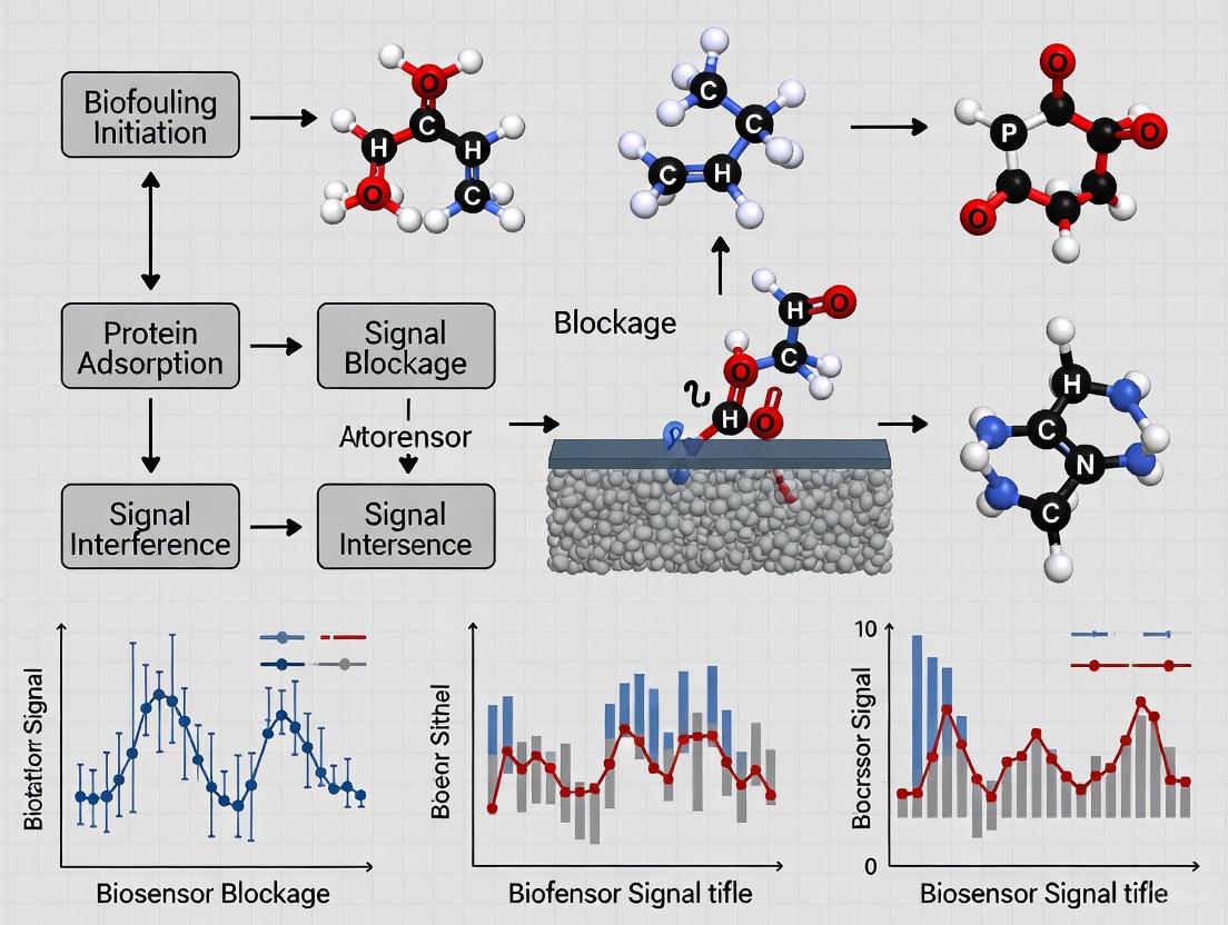
Biofouling refers to the uncontrolled adsorption of biomolecules (such as proteins), cells, or microorganisms onto surfaces exposed to complex biological environments [1]. In the context of electrochemical biosensors, this phenomenon presents a fundamental challenge, particularly for devices operating in biological media like blood, saliva, or serum [2] [3]. When a biosensor is introduced into a biological fluid, its sensing interface is immediately coated with a layer of nonspecifically adsorbed proteins and other biomolecules [1]. This biofouling layer can severely compromise biosensor performance by causing electrode passivation, reducing electrochemical activity, diminishing sensitivity, and ultimately leading to a loss of detection specificity and accuracy [2] [4]. For implantable and wearable biosensors intended for continuous monitoring, biofouling is a primary factor limiting their functional service life and clinical applicability [4].
The formation of a biofouling layer is not a static event but a dynamic, competitive process. The Vroman effect describes this phenomenon, where abundant, highly mobile proteins initially adsorb to a surface but are later displaced by proteins with higher surface affinity, albeit lower mobility [1]. The final composition and conformation of the adsorbed protein layer are influenced by numerous factors, including the physicochemical properties of the sensor surface, the protein concentration and source, and environmental conditions such as ionic strength, pH, and temperature [1].
Mechanisms of Signal Interference in Electrochemical Biosensors
Biofouling interferes with electrochemical biosensor signals through multiple physical and electrochemical mechanisms, which are summarized in the diagram below.
The core interference mechanisms include:
- Creating a Physical Diffusion Barrier: The accumulated layer of proteins and cells acts as a physical barrier that hinders the diffusion of the target analyte from the bulk solution to the active electrode surface [1] [3]. This increases the time for the analyte to reach the sensor and can reduce the measured current in amperometric or voltammetric sensors.
- Hindering Electron Transfer: The fouling layer can introduce a significant resistance to electron transfer between the solution-based redox species and the electrode surface. This is readily observed in techniques like Electrochemical Impedance Spectroscopy (EIS), where an increase in charge-transfer resistance (R~ct~) is a classic signature of surface fouling [4].
- Passivating the Electrode Surface: Nonspecific adsorption directly onto the electrode material can block active sites crucial for the electrocatalytic processes that generate the sensor's signal. This leads to a progressive decline in signal strength over time [4].
- Masking or Blocking Bioreceptors: When fouling proteins adsorb directly onto the immobilized biorecognition elements (e.g., antibodies, aptamers), they can sterically hinder the binding of the target analyte, leading to false negatives and a drastic reduction in sensor sensitivity and specificity [2].
Quantitative Impacts of Biofouling on Sensor Performance
The detrimental effects of biofouling can be quantified through various performance metrics, as illustrated by data from recent studies.
Table 1: Quantitative Impacts of Biofouling on Sensor Parameters
| Performance Parameter | Impact of Biofouling | Experimental Evidence |
|---|---|---|
| Electrochemical Activity | Significant decrease | Noise measurement techniques quantified the loss of electrochemical activity on gold surfaces due to albumin adsorption [4]. |
| Sensor Sensitivity / Signal Strength | Progressive reduction | Electrode passivation from nonspecific adsorption weakens electrochemical signals and can lead to signal loss [2] [3]. |
| Detection Limit | Increased (worsened) | Even low levels of fouling can interfere with ultra-trace detection, raising the practical limit of detection [2]. |
| Stability & Reproducibility | Severe degradation | Signal drift and instability occur due to the dynamic nature of the fouling layer [1] [4]. |
| Sensor Lifespan | Shortened | Biofouling is a primary reason for the failure of implantable and wearable sensors, particularly due to biofilm formation [2] [4]. |
Advanced Strategies for Biofouling Mitigation
Developing effective antifouling strategies is a central focus in electrochemical biosensor research. The goal is to create a surface that resists the nonspecific adsorption of proteins and cells while still allowing the specific capture of the target analyte.
Material-Based Antifouling Strategies
The most common approach involves functionalizing the electrode surface with low-fouling materials.
- Zwitterionic Materials: These materials, such as peptides with alternating positively and negatively charged residues (e.g., EKEKEKEK), form a dense hydration layer via electrostatic interactions with water molecules. This hydrated layer creates a physical and energetic barrier that prevents protein adhesion [2]. They are noted for their high biocompatibility and effectiveness.
- Polyethylene Glycol (PEG) and Derivatives: PEG is a long-standing and widely used antifouling polymer. It works by forming a steric barrier and exhibiting high chain mobility in aqueous environments, which makes it energetically unfavorable for proteins to adsorb [2] [3].
- Antifouling Peptides: Short peptide sequences designed to be highly hydrophilic and neutrally charged can effectively resist nonspecific binding. They are easier to modify and prepare compared to synthetic polymers [2].
- Multifunctional Peptides: An advanced strategy involves designing branched peptides that combine multiple functions. For example, a single peptide can incorporate a zwitterionic antifouling sequence, a hydrophobic antibacterial sequence, and a specific recognition aptamer [2]. This integrated approach simultaneously addresses nonspecific protein adsorption, bacterial colonization, and target sensing.
Measurement and Operational Strategies
Beyond material coatings, innovative measurement techniques can help monitor and compensate for fouling.
- Stochastic Electrochemical Noise Measurement: This technique analyzes intrinsic current and potential fluctuations at the electrode-electrolyte interface without applying an external electrical bias. Statistical analysis of this noise can quantify the formation of a biofouling layer and the consequent loss of electrochemical activity in real-time, offering a pathway for sensor recalibration [4].
Detailed Experimental Protocols for Evaluation
To rigorously evaluate the antifouling performance of a modified electrode, a standard set of experiments is employed. The workflow for a comprehensive assessment is outlined below.
Protocol 1: Electrochemical Assessment of Nonspecific Protein Adsorption
This protocol uses Electrochemical Impedance Spectroscopy (EIS) and Cyclic Voltammetry (CV) to quantify fouling.
- Sensor Preparation: Fabricate the electrochemical biosensor with the antifouling surface modification. A standard configuration uses a three-electrode system: a modified working electrode (e.g., glassy carbon), a platinum counter electrode, and a Ag/AgCl reference electrode [2] [5].
- Baseline Electrochemical Measurement: Perform EIS and CV measurements in a standard redox probe solution, typically 5 mM K~3~[Fe(CN)~6~]/K~4~[Fe(CN)~6~] in PBS (pH 7.4). EIS is conducted over a frequency range from 100 kHz to 0.1 Hz at a formal potential. CV is typically scanned between -0.2 and 0.6 V. Record the charge-transfer resistance (R~ct~) from EIS and the peak current from CV [2].
- Fouling Challenge: Incubate the modified electrode in the fouling solution. Common challenges include:
- Post-Fouling Electrochemical Measurement: Gently rinse the electrode with PBS to remove loosely adsorbed molecules. Repeat the EIS and CV measurements in the same redox probe solution from Step 2.
- Data Analysis: Calculate the percentage change in R~ct~ and peak current. A superior antifouling surface will show minimal change (< 10% is often considered excellent) after exposure to the fouling solution, indicating effective resistance to nonspecific adsorption [2].
Protocol 2: Quantitative Validation of Protein Adsorption
This protocol uses a quartz crystal microbalance with dissipation monitoring (QCM-D) to directly measure the mass of adsorbed protein.
- Sensor Mounting: Mount the modified sensor surface (coated on a QCM-D crystal) in the flow cell chamber.
- Baseline Establishment: Flow a buffer (e.g., PBS) over the sensor until a stable frequency (f) and energy dissipation (D) baseline is achieved.
- Protein Adsorption Phase: Introduce the protein solution (e.g., serum or a single-protein solution) into the flow cell for a predetermined time.
- Rinsing Phase: Switch back to the buffer flow to remove any non-adsorbed proteins.
- Data Analysis: The change in resonance frequency (Δf) is directly related to the mass of adsorbed protein on the surface (using the Sauerbrey equation). A smaller frequency shift indicates a better antifouling surface. This technique was used to validate that a multifunctional peptide-coated surface had minimal protein adsorption [2].
The Scientist's Toolkit: Essential Research Reagents and Materials
Table 2: Key Reagents and Materials for Antifouling Biosensor Research
| Item | Function/Application | Specific Examples |
|---|---|---|
| Zwitterionic Peptides | Forms a hydration layer to resist protein adsorption | EKEKEKEK sequence peptides [2] |
| Antibacterial Peptides (AMPs) | Kills bacteria to prevent biofilm formation | KWKWKWKW sequence peptides [2] |
| Polyethylene Glycol (PEG) | Classic polymer for creating a steric antifouling barrier | PEG-based thiols for self-assembled monolayers on gold [2] [3] |
| Gold Nanoparticles (AuNPs) | Enhances electrode surface area and facilitates biomolecule immobilization | Colloidal AuNPs electrodeposited on PEDOT:PSS [2] |
| Electrode Materials | Platform for sensor fabrication and modification | Glassy carbon electrode (GCE), screen-printed carbon electrodes (SPCE), gold disk electrode [2] [5] |
| Redox Probes | For electrochemical characterization of surface fouling | Potassium ferricyanide/ferrocyanide ([Fe(CN)₆]³⁻/⁴⁻) [2] |
| Fouling Challenge Agents | To test sensor robustness in complex media | Bovine Serum Albumin (BSA), Fibrinogen, human serum, saliva [1] [2] |
| Quartz Crystal Microbalance (QCM-D) | For label-free, quantitative measurement of adsorbed protein mass | QCM-D sensors with gold or silica coatings [2] |
Biofouling, the non-specific adsorption of biomolecules onto sensor surfaces, presents a fundamental barrier to the reliability and deployment of electrochemical biosensors. Occurring in complex biofluids such as blood, saliva, or urine, this process directly compromises sensor function through three primary mechanisms: passivation of the electrode surface, a consequent loss of sensitivity, and a significant increase in background noise. These effects collectively degrade the signal-to-noise ratio, impair detection limits, and ultimately shorten the functional lifespan of biosensors, particularly for in vivo and continuous monitoring applications. This whitepaper details the underlying mechanisms, provides quantitative data on these impacts, outlines advanced measurement protocols for their characterization, and discusses emerging mitigation strategies critical for the development of robust biosensing platforms in clinical and biopharmaceutical settings.
Electrochemical biosensors combine a biological recognition element (e.g., an enzyme, antibody, or DNA strand) with an electrochemical transducer to convert a biological interaction into a quantifiable electrical signal [6] [7]. Their high sensitivity, potential for miniaturization, and low cost make them exceptionally attractive for clinical diagnostics, environmental monitoring, and food safety [6]. However, the analytical performance of these sensors is critically dependent on the integrity of the interface between the electrode and the sample solution.
When deployed in complex biological matrices—such as whole blood, plasma, or saliva—sensor surfaces are rapidly coated with a layer of proteins, lipids, and other biomolecules in a process known as biofouling [8]. This fouling layer acts as a physical and chemical barrier, interfering with the fundamental processes required for electrochemical detection. For researchers and drug development professionals, understanding and quantifying these interferences is not merely an academic exercise but a prerequisite for developing assays and devices that can perform reliably in real-world conditions. The following sections dissect the specific impacts of this fouling layer on electrochemical signals.
Core Mechanisms: How Biofouling Degrades Sensor Performance
The degradation of sensor performance due to biofouling is multifaceted, impacting both the faradaic (charge-transfer) and non-faradaic processes at the electrode-electrolyte interface.
Passivation: The Insulating Barrier
Passivation refers to the formation of an insulating layer on the electrode surface that blocks electron transfer. Albumin, a prevalent protein in blood, is a common culprit [4].
- Mechanism: The adsorbed biomolecules form a non-conductive film that physically separates the electroactive species in the solution from the electrode surface. This layer increases the effective distance for electron tunneling and creates a dielectric barrier.
- Impact on Electrochemistry: Passivation directly increases the charge-transfer resistance (Rₐ), a parameter readily measured by Electrochemical Impedance Spectroscopy (EIS). This leads to a dampening of the current response in techniques such as amperometry and voltammetry.
Loss of Sensitivity: Diminished Analytical Response
Sensitivity loss is a direct consequence of passivation. As the fouling layer builds up, the electrode's ability to drive and measure redox reactions of the target analyte is progressively diminished.
- Mechanism: The fouling layer hinders the diffusion of both the target analyte and any redox mediators to and from the electrode surface. Furthermore, if the bio-recognition element (e.g., an antibody) is itself fouled, its ability to bind the target is sterically hindered.
- Impact: The sensor's calibration curve shifts, resulting in a smaller electrical signal for the same concentration of analyte. This can lead to false negatives or a significant underestimation of the analyte concentration, which is catastrophic in contexts like cardiac troponin detection for heart attack diagnosis [7].
Increased Background Noise: Masking the Signal
An often-overlooked impact is the increase in non-faradaic background current and noise, which obscures the analytical signal.
- Mechanism: The fouling layer alters the electrical double layer at the electrode interface. The dynamic adsorption and desorption of charged biomolecules on this layer generate intrinsic current and potential fluctuations, observed as electrochemical noise [4].
- Impact: This increased noise reduces the signal-to-noise ratio (SNR), effectively raising the limit of detection (LOD) and making it difficult to distinguish low-abundance targets from the background, a critical factor for detecting low-level biomarkers.
The following diagram illustrates the interconnected nature of these degradation mechanisms.
Quantitative Data: Measuring the Impact of Biofouling
The following tables summarize key quantitative findings from research on the effects of biofouling, providing a reference for the expected magnitude of signal degradation.
Table 1: Impact of Albumin-Induced Biofouling on Gold Electrodes
| Measured Parameter | Measurement Technique | Change Due to Biofouling | Functional Consequence |
|---|---|---|---|
| Electrochemical Activity | Stochastic Noise Analysis [4] | Quantifiable decrease | Correlates with formation of an insulating protein layer |
| Charge-Transfer Resistance (Rₐ) | Electrochemical Impedance Spectroscopy (EIS) [4] | Significant increase | Passivation of the electrode surface, leading to signal damping |
| Non-Faradaic Current Noise | Statistical Analysis of Current Fluctuations [4] | Increased magnitude | Elevated background noise, reducing signal-to-noise ratio |
Table 2: Performance Degradation in Complex Biofluids
| Sample Matrix | Target Analyte | Reported Impact | Reference |
|---|---|---|---|
| Whole Blood | Cancer Biomarkers | Decreased sensitivity; performance challenges without antifouling strategies [8] | [8] |
| Unprocessed Saliva | SARS-CoV-2 S1 Spike Protein | Limits long-term stability and complicates detection without robust surface modifications [8] | [8] |
| General Biofluids | Various | Shortened service life of implantable and wearable biosensors [4] | [4] |
Experimental Protocols: Characterizing Biofouling
To develop effective countermeasures, researchers must accurately characterize the extent and kinetics of biofouling. Below is a detailed protocol for a novel method that quantifies biofouling in real-time.
Stochastic Electrochemical Noise Measurement for Biofouling Quantification
This technique, introduced by Jamali et al., uniquely allows for the appraisal of the electrode surface in its innate state without applying an external electrical bias, preventing perturbation of the system [4].
1. Objective: To quantify the formation of a biofouling layer on a gold electrode surface in real-time by analyzing intrinsic current and potential fluctuations.
2. Materials and Reagents:
- Working Electrode: Polished gold disk electrode.
- Reference Electrode: Ag/AgCl (3M KCl).
- Counter Electrode: Platinum wire.
- Test Solution: Phosphate Buffered Saline (PBS), pH 7.4, with and without a model fouling agent (e.g., Bovine Serum Albumin (BSA) at physiologically relevant concentrations).
3. Instrumentation:
- A potentiostat with a low-current preamplifier is required, capable of measuring and recording current and potential with high temporal resolution. The instrument must be housed in a Faraday cage to minimize external electromagnetic interference.
4. Experimental Workflow: The step-by-step procedure and data flow for this experiment are outlined below.
5. Data Analysis:
- Statistical Analysis: Calculate the standard deviation and variance of the recorded current or potential noise over a defined time window.
- Model Fitting: Fit the statistical parameters to the extended analytical model described in the source literature [4]. The model interprets the changes in noise characteristics in terms of the coverage and insulating properties of the adsorbed fouling layer.
- Corroboration: Validate the findings from the noise analysis with complementary techniques such as EIS, which should show a correlated increase in charge-transfer resistance.
Key Advantage: This method provides thermodynamic and kinetic information on the fouling process without applying an external potential that could alter the adsorption process, offering a more native view of biofouling.
The Scientist's Toolkit: Key Research Reagents and Materials
The following table lists essential materials used in the featured stochastic experiment and the broader field of biofouling research.
Table 3: Research Reagent Solutions for Biofouling Studies
| Reagent / Material | Function in Experiment | Specific Example |
|---|---|---|
| Gold Electrode | Provides a clean, well-defined, and reproducible surface for studying fundamental fouling mechanisms. | Polished gold disk electrode [4] |
| Bovine Serum Albumin (BSA) | A model protein used to simulate proteinaceous biofouling in a controlled laboratory setting. | BSA in PBS buffer [4] |
| Phosphate Buffered Saline (PBS) | Provides a physiologically relevant ionic strength and pH, serving as the base electrolyte for experiments. | 1X PBS, pH 7.4 [4] |
| Antifouling Polymers | Used to create surface modifications that resist non-specific adsorption. | Zwitterionic polymers, PEG-based coatings [8] |
| Nanobodies | Robust biological recognition elements that can be used in receptor design to maintain function in fouling environments. | Anti-SARS-CoV-2 nanobodies for detection in saliva [8] |
Biofouling is an inevitable challenge that directly and detrimentally impacts electrochemical signals through passivation, sensitivity loss, and increased background noise. Acknowledging and systematically addressing these impacts is paramount for the transition of biosensors from laboratory proof-of-concept to viable commercial products, especially in point-of-care diagnostics [7].
Future research is focused on developing more sophisticated antifouling surface modifications [8] and stimuli-responsive surfaces that can offer dynamic control over biointeractions. Furthermore, integrating real-time fouling assessment techniques, like stochastic noise analysis, could pave the way for sensors capable of self-diagnosing performance decay and initiating recalibration. For academic and industrial researchers alike, embedding the consideration of biofouling from the earliest stages of biosensor design—aligned with translational frameworks like the REASSURED criteria—will be crucial to maximizing the commercial and clinical impact of this promising technology [7].
The Role of Protein Adsorption and Biofilm Formation in Electrode Fouling
Electrochemical biosensors are powerful tools for detecting biomarkers in medical diagnostics and biological research. However, their performance in complex biological environments is severely compromised by electrode fouling, a process primarily driven by the non-specific adsorption of proteins and the formation of biofilms [2] [9] [10]. This passive adsorption of biomolecules (biofouling) and subsequent microbial colonization fundamentally alter the electrochemical properties of the sensing interface [11]. In the context of biosensor research, understanding these mechanisms is paramount, as fouling leads to sensor passivation, loss of specificity, and significant signal degradation, ultimately resulting in analytical failure and unreliable data [2] [10]. This technical guide delves into the mechanisms by which protein adsorption and biofilm formation impact electrochemical signals, providing a framework for developing robust antifouling strategies.
Mechanisms and Impacts of Fouling on Sensor Signals
Electrode fouling imposes a significant threat to sensing probes used in vivo and in complex biofluids like blood, saliva, and serum [2] [11]. The fouling process begins instantly upon exposure to a biological medium, with proteins adsorbing to the electrode surface, forming a conditioning film. This layer then facilitates the attachment of bacteria, which can proliferate and form structured biofilms [12] [13].
Fundamental Mechanisms of Signal Interference
The adsorbed layers of proteins and biofilms interfere with electrochemical signals through several physical and electrochemical mechanisms:
- Physical Barrier Formation: The fouling layer acts as a diffusion barrier, limiting the access of target analytes to the electroactive electrode surface. This reduces perfusion and can drastically deteriorate detection limits [11].
- Electroactive Surface Area Blocking: Fouling substances accumulate on the electrode surface, reducing its active area and hindering electron transfer reactions [13].
- Alteration of Electrode Kinetics: The fouling layer can alter the electrochemical properties of the electrode surface, directly impacting the electron transfer kinetics for specific redox probes [11].
Differential Impact on Redox Probes
The effect of biofouling is highly dependent on the nature of the redox reaction. Studies on carbon surfaces show that fouling affects outer-sphere and inner-sphere redox probes differently [11].
- Outer Sphere Redox (OSR) Probes: The electron transfer kinetics of OSR probes like Ru(NH₃)₆³⁺ can be largely unaffected by protein fouling from BSA or fetal bovine serum (FBS). However, negatively charged OSR probes like IrCl₆²⁻ can be affected due to electrostatic repulsion from adsorbed, negatively charged proteins [11].
- Inner Sphere Redox (ISR) Probes: In contrast, the electron transfer kinetics of ISR probes like dopamine are heavily affected by fouling on all surfaces. For instance, the peak separation (ΔEₚ) for dopamine can increase dramatically (30–451%) after fouling, indicating severely slowed electron transfer, as the reaction requires specific interaction sites on the electrode surface that are blocked by proteins [11].
Table 1: Quantifying the Impact of Fouling on Electrochemical Biosensor Performance
| Performance Parameter | Impact of Fouling | Experimental Evidence |
|---|---|---|
| Detection Limit | Severe deterioration | Dopamine detection limit worsened from 50 nM in PBS to 50 μM in a biological environment [11]. |
| Electron Transfer Kinetics | Slowed for inner-sphere probes | ΔEₚ for dopamine increased by 30% to 451% after fouling with BSA/FBS [11]. |
| Sensitivity | Significant reduction | General consequence of reduced active surface area and increased impedance [9] [13]. |
| Selectivity & Reliability | Compromised | Increased noise, interference, and false-positive/negative results [10] [13]. |
| Signal Stability | Decreased over time | Biofilm formation leads to continuous signal drift and eventual sensor failure [2] [12]. |
Experimental Methodologies for Fouling Studies
To systematically study fouling and develop mitigation strategies, robust and reproducible experimental protocols are essential. Below are detailed methodologies for simulating fouling and evaluating antifouling surfaces.
Protocol 1: Investigating Biofouling on Working Electrodes
This protocol assesses the impact of biofouling and chemical fouling on carbon fiber micro-electrodes (CFMEs), commonly used in neurotransmitter detection [9].
- Objective: To evaluate the effects of biofouling and chemical fouling on the sensitivity and voltammetric signals of a CFME working electrode.
- Materials:
- Fabricated CFMEs and Ag/AgCl reference electrodes [9].
- Bovine Serum Albumin (BSA) solution (40 g L⁻¹) or cell culture medium (e.g., F12-K Gibco Nutrient Mix) as biofouling agents.
- Neurotransmitters: Serotonin (5-HT, 25 μM) or Dopamine (DA, 1 mM) as chemical fouling agents.
- Tris buffer (15 mM, pH 7.4).
- Fast-scan cyclic voltammetry (FSCV) setup (e.g., National Instruments or WINCS Harmoni system).
- Procedure:
- Stabilization: Stabilize the CFME in Tris buffer by applying the relevant voltage waveform for at least 30 minutes until a stable background current is achieved.
- Baseline Measurement: Record FSCV scans in clean Tris buffer to establish a baseline for neurotransmitter detection.
- Fouling Phase:
- For biofouling: Immerse the electrode in BSA solution or nutrient mix while continuously applying a triangular waveform (-0.4 V to 1.0 V, 400 V/s, 10 Hz) for 2 hours [9].
- For chemical fouling with serotonin: Immerse the electrode in 25 μM serotonin solution and apply the "Jackson" waveform (0.2 V → 1.0 V → -0.1 V → 0.2 V, 1000 V/s) for 5 minutes [9].
- For chemical fouling with dopamine: Immerse the electrode in 1 mM dopamine solution and apply a triangular waveform (-0.4 V to 1.0 V, 400 V/s) for 5 minutes [9].
- Post-Fouling Measurement: Rinse the electrode gently with Tris buffer and repeat the FSCV measurements as in Step 2.
- Data Analysis: Compare the post-fouling and baseline voltammograms. Analyze changes in oxidation/ reduction peak currents (sensitivity), peak potential shifts (ΔEₚ), and background charging current.
Experimental Workflow for Electrode Fouling Studies
Protocol 2: Evaluating a Multifunctional Antifouling Biosensor
This protocol details the fabrication and testing of a biosensor with a peptide-based interface designed to resist fouling, as demonstrated for detecting the SARS-CoV-2 RBD protein in saliva [2].
- Objective: To fabricate a low-fouling electrochemical biosensor and validate its antifouling and antibacterial performance in complex biofluids.
- Materials:
- Glassy Carbon Electrode (GCE).
- Monomers: 3,4-Ethylenedioxythiophene (EDOT) and poly(sodium 4-styrenesulfonate) (PSS).
- Gold salt (e.g., HAuCl₄) for electrodepositing Au nanoparticles (AuNPs).
- Synthetic multifunctional branched peptide (PEP) with antifouling (EKEKEKEK), antibacterial (KWKWKWKW), and recognition (KSYRLWVNLGMVL) sequences [2].
- Saliva samples (artificial or human).
- Quartz Crystal Microbalance with Dissipation monitoring (QCM-D), Laser Confocal Microscopy.
- Biosensor Fabrication:
- Electrode Preparation: Polish the GCE sequentially with 0.3 µm and 0.05 µm alumina slurry and rinse thoroughly with water.
- Polymer Deposition: Electrodeposit the conductive polymer PEDOT:PSS onto the GCE from an aqueous solution containing 7.4 mM EDOT and 1.0 mg mL⁻¹ PSS.
- Nanostructuring: Electrodeposit AuNPs onto the PEDOT:PSS-modified surface to create a high-surface-area substrate.
- Peptide Immobilization: Immobilize the multifunctional branched peptide (PEP) onto the AuNP surface via gold-sulfur (Au-S) chemistry to form the final biosensor (PEP/AuNP/PEDOT/GCE).
- Performance Evaluation:
- Antifouling Test: Immerse the modified electrode in undiluted human saliva or a protein solution (e.g., 40 g L⁻¹ BSA) for a set period. Use QCM-D to quantify the amount of non-specifically adsorbed protein and fluorescence imaging to visualize adsorption.
- Antibacterial Test: Use an Electrical Bacterial Growth Sensor (EBGS) or similar method to demonstrate that the modified interface inhibits bacterial growth and reproduction [2].
- Analytical Performance: Perform electrochemical measurements (e.g., EIS, DPV) in spiked saliva samples to determine the detection limit, linear range, and correlation with standard methods like ELISA.
Visualizing Fouling Mechanisms and Defenses
The following diagram illustrates the molecular-level interaction between a multifunctional peptide interface and fouling agents, a key advanced antifouling strategy.
Multifunctional Peptide Interface Mechanism
The Scientist's Toolkit: Key Research Reagents and Materials
The table below catalogues essential materials used in fouling research and the development of antifouling biosensors, as cited in the literature.
Table 2: Key Research Reagent Solutions for Fouling and Antifouling Studies
| Research Reagent / Material | Function in Experimentation | Technical Notes & Examples |
|---|---|---|
| Bovine Serum Albumin (BSA) | Model protein for simulating biofouling; used to create a conditioning film on electrodes. | Used at 40 g L⁻¹ to simulate protein adsorption [9]. A major component of blood plasma responsible for fouling [10]. |
| Cell Culture Medium (e.g., F12-K) | Complex solution for realistic biofouling simulation; contains nutrients, proteins, and salts. | Provides a more realistic insight compared to single-protein models [9] [11]. |
| Neurotransmitters (Dopamine, Serotonin) | Serve as both target analytes and chemical fouling agents due to irreversible adsorption of by-products. | Dopamine (1 mM) and Serotonin (25 μM) solutions used for chemical fouling studies [9]. |
| Multifunctional Branched Peptides | Engineered surface modifier providing antifouling, antibacterial, and specific recognition capabilities. | e.g., PEP sequence with EKEKEKEK (antifouling), KWKWKWKW (antibacterial), and a target-specific aptamer [2]. |
| Zwitterionic Peptides (e.g., EKEKEKEK) | Create a hydrophilic, neutral surface that forms a hydration layer, acting as a physical and electrostatic barrier to fouling. | Classical zwitterionic peptides exhibit excellent resistance to biofouling in complex media [2]. |
| Antibacterial Peptides (e.g., KWKWKWKW) | Positively charged sequences that interact with negatively charged bacterial membranes, causing cell death and preventing biofilm formation. | Integrated into multifunctional peptides to enhance long-term sensor stability [2]. |
| PEDOT:PSS | Conductive polymer used as an electrode coating; improves conductivity and can be modified to enhance antifouling properties. | Serves as a substrate for further nanomaterial and peptide modification [2] [9]. |
| Gold Nanoparticles (AuNPs) | Used to nanostructure electrode surfaces; increase electroactive surface area and facilitate biomolecule immobilization via Au-S chemistry. | Electrodeposited on polymer-modified electrodes to create a high-surface-area platform [2]. |
| Tetrahedral Amorphous Carbon (ta-C) | Electrode material with high sp³ carbon content; demonstrates variable resistance to fouling based on surface chemistry. | Material choice influences the extent and impact of protein adsorption on electron transfer [11]. |
| Sulfide Ions (S²⁻) | Chemical fouling agent for Ag/AgCl reference electrodes; decreases open circuit potential and causes peak voltage shifts. | Added to buffer solution to simulate reference electrode poisoning in vivo [9]. |
Protein adsorption and biofilm formation are formidable adversaries in electrochemical biosensing, directly causing signal degradation through physical blocking, kinetic interference, and introduction of noise. Research unequivocally shows that fouling drastically alters the performance of both working and reference electrodes. The development of effective antifouling strategies, such as engineered multifunctional interfaces that combine zwitterionic antifouling motifs with antimicrobial agents, is crucial to achieving accurate and reliable sensing in complex biological media like whole blood, saliva, and in vivo. A comprehensive understanding of these fouling mechanisms, coupled with robust experimental methodologies for testing new materials and coatings, is foundational to advancing the field of electrochemical biosensors for clinical diagnostics and long-term monitoring.
Electrochemical biosensors represent a powerful tool for the detection of biomarkers in fields ranging from clinical diagnostics to environmental monitoring. However, their operational accuracy in real-world biological environments is critically compromised by biofouling—the nonspecific, uncontrolled adsorption of proteins, cells, and other biomolecules onto the sensor surface [14] [15]. This fouling layer acts as a physical and chemical barrier, instigating a cascade of detrimental effects that fundamentally undermine sensor function. Within the context of biosensor research, biofouling is not merely a surface nuisance but a core phenomenon that directly induces three primary failure modes: signal drift, reduced specificity, and ultimate sensor failure. This whitepaper provides an in-depth technical analysis of these consequences, detailing the underlying mechanisms and presenting the latest validated strategies to mitigate them, thereby equipping researchers and drug development professionals with the knowledge to design more robust and reliable sensing platforms.
Core Mechanisms: How Biofouling Compromises Sensor Function
The adverse effects of biofouling manifest through several interconnected physical and electrochemical pathways. The diagram below illustrates the core mechanisms through which biofouling compromises biosensor accuracy.
Signal Drift
Signal drift refers to the unidirectional, time-dependent change in the sensor's baseline signal, which is unrelated to the concentration of the target analyte. Biofouling contributes to drift through two main mechanisms. First, the accumulation of a non-conductive protein layer on the electrode surface passivates the interface, progressively inhibiting electron transfer kinetics and leading to a signal decay over time [16] [14]. Second, in field-effect transistor (FET)-based biosensors (BioFETs), the fouling layer can alter the gate capacitance and threshold voltage ((V_T)). As noted in studies of carbon nanotube-based BioFETs, electrolytic ions from the solution slowly diffuse into the sensing region, which is exacerbated by a fouling layer, leading to a continuous shift in the electrical characteristics that can falsely mimic or obscure a true biomarker binding event [16].
Reduced Specificity
The specificity of a biosensor is determined by the selective binding of its immobilized biorecognition element (antibody, aptamer, etc.) to the target analyte. Biofouling directly impairs this by creating a layer prone to nonspecific adsorption of other biomolecules present in complex media like serum, saliva, or blood [2] [15]. This nonspecific binding creates a competing signal that is chemically indistinguishable from the specific binding event, leading to false positives and an overestimation of the target concentration. Furthermore, a dense fouling layer can sterically hinder the access of the target analyte to the capture probe, reducing the assay's sensitivity and increasing the limit of detection [17].
Sensor Failure
In the most severe cases, biofouling leads to complete and irreversible sensor failure. This can occur through extreme passivation, where the fouling layer becomes so thick and impermeable that electron transfer is completely blocked, rendering the electrode electrochemically silent [14] [15]. Additionally, the adsorption and proliferation of bacteria on the sensor interface can lead to the formation of stable biofilms [2]. These biofilms are communities of microorganisms encased in a polymeric matrix that not only create a severe diffusion barrier but also actively metabolize and alter the local chemical environment, permanently degrading the sensor's function [2].
Quantitative Impact: Data from Recent Studies
The following tables summarize quantitative findings on the impact of biofouling and the performance enhancements achieved by various antifouling strategies, as reported in recent literature.
Table 1: Documented Impacts of Biofouling on Sensor Performance
| Performance Metric | Impact of Biofouling | Experimental Context |
|---|---|---|
| Electron Transfer Kinetics | Significant slowing after prolonged exposure | Slower electron transfer and reduced faradaic current after exposure to serum and nasopharyngeal secretions [17]. |
| Signal Stability | >50% signal loss due to drift | CNT-based BioFETs exhibited debilitating signal drift in high ionic strength solutions, obscuring biomarker detection [16]. |
| Detection Limit | Increase of several orders of magnitude | Nonspecific adsorption in complex media weakened signal-to-noise ratio, raising the practical detection limit [14]. |
| Operational Lifespan | Failure within hours in whole blood | Without antifouling protection, sensors experienced rapid passivation and biofilm-induced failure in complex fluids [2] [18]. |
Table 2: Efficacy of Antifouling Strategies in Recent Experimental Studies
| Antifouling Strategy | Reported Performance Enhancement | Test Medium & Duration |
|---|---|---|
| POEGMA Polymer Brush [16] | Enabled stable, sub-femtomolar detection in 1X PBS; mitigated signal drift. | Undiluted ionic solution (1X PBS); repeated measurements. |
| Multifunctional Branched Peptide [2] | LOD of 0.28 pg mL⁻¹ for SARS-CoV-2 RBD protein; excellent antifouling/antibacterial properties. | Human saliva samples; validated vs. commercial ELISA. |
| Zwitterionic SBMA@PDA Coating [19] | Reduced signal drift; high robustness to pH, temperature, and mechanical stress. | Diverse biological fluids; wearable microneedle patch in artificial ISF. |
| Porous Albumin Nanocomposite [17] | Maintained rapid electron transfer for >1 month; 3.75 to 17-fold sensitivity enhancement. | Serum and nasopharyngeal secretions; long-term stability test. |
| eRapid Nanocomposite [18] | Sensing capabilities maintained over weeks of continuous use in blood. | Whole blood; multiplexed detection of 30+ biomarkers. |
Experimental Protocols for Mitigating Biofouling Consequences
To combat the dire consequences of biofouling, researchers have developed sophisticated surface chemistry and materials science protocols. The following section details specific experimental methodologies for implementing two of the most promising strategies: polymer brush interfaces and multifunctional peptides.
Protocol 1: Implementing a POEGMA Polymer Brush for Drift Mitigation
This protocol is adapted from the development of the "D4-TFT" BioFET, which overcame signal drift and Debye screening to achieve attomolar-level detection in physiologically relevant ionic strength (1X PBS) [16].
Principle: Grafting a poly(oligo(ethylene glycol) methyl ether methacrylate) (POEGMA) polymer brush above the transducer establishes a hydrated layer that resists nonspecific protein adsorption. Furthermore, through the Donnan potential effect, this layer effectively extends the Debye length, allowing for the detection of large antibody-antygen immune complexes beyond the typical screening distance in high ionic strength solutions [16].
Materials:
- Substrate: Carbon nanotube (CNT) thin-film transistor (TFT).
- Polymer: Poly(oligo(ethylene glycol) methyl ether methacrylate) (POEGMA).
- Biorecognition Element: Target-specific capture antibodies (cAb).
- Passivation Layer: Appropriate material for device passivation alongside the polymer brush.
- Testing Configuration: Stable electrical setup with a palladium (Pd) pseudo-reference electrode.
Step-by-Step Workflow:
- Surface Preparation: Clean and prepare the CNT-TFT channel surface to ensure uniform polymer grafting.
- POEGMA Grafting: Grow the POEGMA polymer brush interface on the high-κ dielectric of the device. This is typically done via surface-initiated atom transfer radical polymerization (SI-ATRP) to achieve a dense, brush-like conformation [16].
- Antibody Immobilization: Inkjet-print the capture antibodies (cAb) into the POEGMA matrix above the CNT channel. A control device with no antibodies must be prepared in parallel on the same chip.
- Device Encapsulation: Passivate the device to minimize leakage current and enhance overall stability.
- Electrical Measurement:
- Use a stable testing configuration with a Pd pseudo-reference electrode to avoid bulky Ag/AgCl.
- Enforce a rigorous testing methodology that relies on infrequent DC sweeps rather than static or AC measurements to minimize the influence of drift on the recorded signal.
- The detection of the target biomarker is confirmed by a shift in the on-current ((I_{on})) and must be absent in the control device.
Protocol 2: Fabricating a Multifunctional Peptide-Based Biosensor
This protocol outlines the construction of an electrochemical biosensor using a designed branched peptide to achieve antifouling, antibacterial, and recognition capabilities simultaneously, as demonstrated for SARS-CoV-2 RBD protein detection in saliva [2].
Principle: A single, branched peptide (PEP) is synthesized to integrate three distinct sequences: a zwitterionic antifouling motif (e.g., EKEKEKEK), a positively charged antibacterial peptide (AMP, e.g., KWKWKWKW), and a specific recognition aptamer (e.g., KSYRLWVNLGMVL for SARS-CoV-2 RBD). This design creates a multifunctional interface that resists fouling, kills bacteria, and specifically captures the target analyte [2].
Materials:
- Electrode: Glassy carbon electrode (GCE).
- Conductive Polymer: Poly(3,4-ethylenedioxythiophene) doped with poly(styrenesulfonate) (PEDOT:PSS).
- Nanomaterial: Gold nanoparticles (AuNPs).
- Custom Peptide: Synthesized multifunctional branched peptide (PEP) with a terminal thiol group for gold-sulfur binding.
Step-by-Step Workflow:
- Electrode Polishing: Polish the GCE sequentially with 0.3 µm and 0.05 µm alumina slurry on a polishing pad, followed by rinsing with ultrapure water.
- Conductive Polymer Deposition: Electrodeposit PEDOT:PSS onto the clean GCE from an aqueous solution containing the EDOT monomer and PSS dopant. This creates a rough, high-surface-area foundation.
- Gold Nanoparticle Decoration: Deposit AuNPs uniformly onto the PEDOT:PSS-modified substrate to enhance conductivity and provide binding sites for the peptide.
- Peptide Self-Assembly: Incubate the electrode in a solution of the thiolated, multifunctional branched peptide (PEP) to form a stable, self-assembled monolayer via gold-sulfur (Au-S) bonds.
- Validation and Sensing:
- Validate the antifouling and antibacterial properties using techniques like quartz crystal microbalance (QCM-D), fluorescence imaging, and electrochemical impedance spectroscopy (EIS) in complex media like saliva or serum.
- For detection, incubate the biosensor with the sample and measure the electron transfer resistance using differential pulse voltammetry (DPV) or electrochemical impedance spectroscopy (EIS).
The experimental workflow for this sensor construction is visualized below.
The Scientist's Toolkit: Key Reagent Solutions
The advancement of antifouling biosensors relies on a specific set of materials and reagents. The following table catalogs key solutions used in the featured experiments and the broader field.
Table 3: Key Research Reagent Solutions for Antifouling Biosensors
| Reagent / Material | Function & Mechanism | Exemplary Use Case |
|---|---|---|
| POEGMA Polymer Brush | Extends Debye length via Donnan potential; hydrated layer resists nonspecific adsorption [16]. | Ultra-sensitive BioFETs for detection in undiluted biological buffers [16]. |
| Zwitterionic Peptides (e.g., EKEKEKEK) | Forms a strong hydration layer via zwitterionic residues; neutral charge reduces electrostatic fouling [2]. | Low-fouling electrochemical biosensors for direct detection in saliva [2]. |
| Antibacterial Peptides (e.g., KWKWKWKW) | Disrupts negatively charged bacterial cell membranes, preventing biofilm formation [2]. | Sensors for long-term operation in bacteria-rich environments [2]. |
| Porous Albumin Nanocomposite | Micrometer-thick, porous cross-linked BSA matrix with AuNWs; resists fouling while enhancing mass transport and sensitivity [17]. | Multiplexed sensors for simultaneous detection of SARS-CoV-2 nucleic acid, antigen, and antibody [17]. |
| Zwitterionic Polymers (pSBMA, pCBMA) | Mimics cell membrane surfaces; forms a robust hydrophilic coating with superior antifouling properties [19] [15]. | Wearable sensors and therapeutic drug monitoring platforms [19]. |
| PEDOT:PSS Conducting Polymer | Provides high electronic conductivity and porosity; its amphiphilic nature helps repel fouling agents [2] [15]. | Base conductive layer for functionalization in complex media [2]. |
The challenges posed by biofouling—signal drift, reduced specificity, and sensor failure—are significant but not insurmountable barriers to the deployment of reliable electrochemical biosensors. As detailed in this whitepaper, the research community has moved beyond simple PEG-based coatings to sophisticated, multifunctional solutions. These include polymer brushes that manipulate interfacial physics, smart peptides that combine recognition with active antifouling and antibacterial actions, and nanostructured porous coatings that enhance both sensitivity and stability. The quantitative data and detailed protocols provided underscore a clear path forward. For researchers and drug developers, the integration of these advanced antifouling strategies is no longer an optional enhancement but a fundamental requirement for creating next-generation biosensors capable of accurate, continuous, and reliable operation in the complex biological milieus where they are most needed.
The development of implantable electrochemical biosensors represents a frontier in continuous health monitoring, offering the potential to transform the management of chronic diseases and improve our understanding of physiological processes. However, the performance and longevity of these devices remain severely limited by the host's reaction to the implanted foreign material, a process known as the foreign body response (FBR). This complex, multi-stage biological reaction to implanted sensors compromises analytical accuracy and ultimately leads to device failure [20] [21]. Within the context of research on how biofouling affects electrochemical biosensor signals, understanding the FBR is fundamental. This whitepaper provides an in-depth examination of the FBR sequence, its specific impacts on sensor signal integrity, the strategies being developed to mitigate it, and detailed experimental approaches for studying this critical phenomenon.
The Biological Sequence of the Foreign Body Response
The foreign body response is a coordinated biological reaction that unfolds in a series of overlapping stages, each contributing to sensor degradation.
Table 1: Stages of the Foreign Body Response and Impact on Sensors
| Stage | Timeframe | Key Biological Events | Impact on Sensor Function |
|---|---|---|---|
| Protein Adsorption | Minutes to Hours | Formation of a provisional matrix; adsorption of proteins, cytokines, and other biomolecules. | Unpredictable signal reduction (>50%); requires frequent calibration [20]. |
| Acute Inflammation | Days | Infiltration of leukocytes; mast cell degranulation; histamine release; fibrinogen adsorption. | Reduced analyte diffusion; consumption of oxygen/glucose by immune cells; local pH drop degrading sensor enzymes [20]. |
| Chronic Inflammation | Weeks | Presence of macrophages, monocytes, lymphocytes; formation of Foreign Body Giant Cells (FBGCs). | Frustrated phagocytosis; enhanced degradation of the sensor surface [20]. |
| Granulation Tissue | Weeks | Proliferation of blood vessels and connective tissue; infiltration of fibroblasts. | Development of a vascularized tissue layer around the sensor [20]. |
| Fibrous Encapsulation | Weeks to Months | Deposition of collagen to form an avascular, fibrous capsule isolating the implant. | Significantly diminished transport of glucose and oxygen; increased sensor lag time and eventual failure [20] [22]. |
The process begins immediately upon implantation, with the adsorption of biomolecules (e.g., proteins, cytokines, growth factors) to the sensor surface, forming a "provisional matrix" [20]. This initial layer of biofouling can cause a substantial and unpredictable decrease in sensor response, often exceeding 50%, primarily due to molecules smaller than 15 kDa [20]. The subsequent infiltration of inflammatory cells, such as neutrophils and macrophages, leads to a hostile local environment. These cells consume oxygen and glucose, produce reactive oxygen species, and acidify the local environment (pH can drop to as low as 3.6), which can degrade the enzymatic components of biosensors [20]. When macrophages are unable to phagocytose the large implant, they fuse to form foreign body giant cells (FBGCs), which persist at the sensor-tissue interface and contribute to the degradation of the underlying material [20]. The final and most detrimental stage is the formation of a fibrous capsule, an avascular collagenous sheath that severely impedes the transport of analytes (e.g., glucose and oxygen) to the sensor surface, leading to increased lag times and a progressive loss of signal [20] [22].
Diagram 1: The Foreign Body Response Sequence
Sensor Signal Degradation: Connecting Biofouling to Analytical Failure
The FBR directly impairs the core function of electrochemical biosensors through physical, chemical, and metabolic pathways. The accumulated fouling layer and resulting fibrous capsule act as a diffusion barrier, hindering the transport of the target analyte (e.g., glucose) to the sensing electrode and delaying the sensor's response to concentration changes in the surrounding tissue [20] [22]. Furthermore, the metabolic activity of immune cells clustered at the sensor-tissue interface consumes analytes like glucose and oxygen, creating a local concentration that is not representative of the systemic or bulk tissue level, leading to inaccurate readings [20]. The inflammatory process also generates a chemically hostile environment, characterized by local acidification and the production of reactive oxygen and nitrogen species that can degrade or denature sensitive recognition elements (e.g., enzymes) and damage electrode materials [20] [21]. Finally, the non-specific adsorption of proteins and other biomolecules can increase background noise or directly passivate the electrode surface, reducing the signal-to-noise ratio and the sensitivity of the sensor over time [22] [23].
Strategies to Mitigate the Foreign Body Response
Research efforts to combat the FBR and biofouling can be broadly categorized into passive, active, and material-based strategies, often used in combination.
Passive and Material-Based Strategies
Passive strategies focus on modifying the physicochemical properties of the sensor surface to make it less recognizable as foreign or to resist the initial adhesion of biomolecules and cells.
- Biomimetic Coatings: These materials are designed to mimic biological surfaces. Phospholipid polymers, such as 2-methacryloyloxyethyl phosphorylcholine (MPC), imitate the outer membrane of cells and have been shown to reduce protein adsorption and the production of pro-inflammatory cytokines [20]. Similarly, hydrogel coatings (e.g., based on poly(ethylene glycol) - PEG or poly(2-hydroxyethyl methacrylate) - PHEMA) create a hydrophilic, tissue-like interface that can reduce fibrous encapsulation and improve tissue integration [20] [22].
- Surface Topography and Porosity: The physical architecture of the implant surface significantly influences the healing response. Introducing micro- and nano-scale pores (5-500 μm) has been demonstrated to promote angiogenesis and disrupt the formation of a dense, avascular fibrous capsule, thereby improving analyte transport [20].
- Advanced Nanomaterials and Antifouling Peptides: Nanomaterials like graphene oxide and zwitterionic peptides are being explored for their inherent antifouling properties. Their high hydrophilicity enables the formation of a protective hydration layer that effectively resists non-specific protein adsorption [2] [23]. Multifunctional peptides that combine zwitterionic antifouling sequences with antibacterial properties represent a promising advanced strategy to combat multiple failure modes simultaneously [2].
Table 2: Common Anti-Fouling Materials and Their Mechanisms
| Material/Strategy | Key Examples | Proposed Mechanism of Action | Reported Limitations |
|---|---|---|---|
| Hydrogels | PEG, PHEMA | Hydrophilic interface; mimics tissue mechanics; reduces protein adsorption & fibrous encapsulation [20] [22]. | Stability concerns (leaching, delamination); low mechanical strength [20]. |
| Biomimetic Membranes | Phospholipid Polymers (e.g., MPC) | Mimics cell membrane; appears "self" to immune system; reduces protein adhesion & inflammatory cytokine release [20] [22]. | Often provides only short-term benefits; stability issues when grafted [20]. |
| Porous Materials | Sphere-templated Hydrogels | Promotes vascularization; disrupts fibrous tissue deposition; optimal pore size (e.g., ~35 μm) enhances angiogenesis [20]. | Non-optimal pore sizes can promote fibrosis [20]. |
| Nanomaterial Coatings | Graphene Oxide, Gold Nanoparticles | Unique physicochemical properties (e.g., hydrophobicity, catalytic activity); can be functionalized with antifouling agents like PEG [23]. | Potential for nanoparticle aggregation; long-term biocompatibility under investigation [23]. |
| Antifouling Peptides | Zwitterionic (EKEKEKEK), Multifunctional Branched Peptides | Forms a hydrated layer; neutral charge reduces electrostatic attraction; some possess antibacterial properties [2]. | Complex synthesis; cost of peptide production [2]. |
Active Release Strategies
Active strategies involve the local and controlled release of bioactive agents from the sensor coating to directly modulate the immune response.
- Anti-inflammatory Agents: The release of potent anti-inflammatory drugs, such as dexamethasone, aims to suppress the activity of inflammatory cells at the implant site [20].
- Anti-fibrotic Agents: Compounds like the tyrosine kinase inhibitor masitinib can target specific pathways in the FBR. Masitinib inhibits the c-KIT receptor on mast cells, preventing their degranulation and subsequent initiation of fibrosis, which has been shown to reduce capsule thickness around model implants [24].
- Pro-angiogenic Factors: Releasing molecules like vascular endothelial growth factor (VEGF) encourages the formation of new blood vessels around the implant, preventing the formation of an avascular fibrous capsule and ensuring a more reliable supply of analyte [20].
Diagram 2: FBR Mitigation Strategies
Detailed Experimental Protocol: Local Drug Delivery to Modulate FBR
The following protocol details a method for evaluating the efficacy of an actively released drug, using the tyrosine kinase inhibitor Masitinib as a representative example, to mitigate the FBR in a rodent model [24].
Objective
To assess the ability of locally released Masitinib, delivered from a polymer-coated model implant, to reduce fibrous capsule thickness and inflammatory cell density in a murine subcutaneous implant model.
Materials and Reagents
Table 3: Research Reagent Solutions for FBR Drug Delivery Studies
| Reagent/Material | Function/Description | Supplier Example |
|---|---|---|
| Masitinib | Active pharmaceutical ingredient; tyrosine kinase inhibitor targeting mast cell c-KIT receptor. | Selleck Chemicals [24] |
| PLGA (50:50) | Biodegradable polymer for fabricating drug-eluting microspheres; provides controlled release. | Lakeshore Biomaterials (Evonik) [24] |
| Poly(vinyl alcohol) (PVA) | Surfactant used in the formation of PLGA microspheres via emulsion. | Sigma-Aldrich [24] |
| PEG/PEO Blend | Water-soluble polymer matrix to transiently hold microspheres on implant during insertion. | Sigma-Aldrich [24] |
| Dichloromethane (DCM) | Organic solvent for dissolving PLGA in microsphere fabrication. | Sigma-Aldrich [24] |
Methodology
Fabrication of Drug-Loaded Microspheres:
- Prepare a solution of PLGA (e.g., intrinsic viscosity 0.15–0.25 dL/g) in Dichloromethane (DCM).
- Dissolve Masitinib directly in the polymer solution.
- Emulsify this organic phase into an aqueous Poly(vinyl alcohol) (PVA) solution using homogenization to form a water-in-oil-in-water (w/o/w) double emulsion.
- Stir the emulsion for several hours to evaporate the DCM, allowing the solid microspheres to form.
- Collect the microspheres by centrifugation, wash to remove residual PVA, and lyophilize for storage. Characterize the microspheres for size (target 5-20 μm) and drug loading efficiency [24].
Coating of Model Implants:
- Create model sensor implants (e.g., polymer fibers).
- Prepare a transient coating by dispersing the drug-loaded PLGA microspheres in an aqueous solution of a PEG/PEO blend.
- Dip-coat or spray-coat the model implants with this microsphere suspension to create a uniform layer. The PEG/PEO matrix is designed to dissolve rapidly upon implantation, leaving the drug-releasing microspheres at the tissue site [24].
In Vivo Implantation and Evaluation:
- Perform subcutaneous implantation of coated and control (uncoated or empty microsphere-coated) model sensors in the backs of mice (e.g., C57BL/6J strain).
- Sacrifice animals at predetermined time points (e.g., 14, 21, and 28 days post-implantation).
- Excise the implant and surrounding tissue, and process for histology (e.g., paraffin embedding, sectioning, and staining with Hematoxylin & Eosin (H&E) for general morphology and Masson's Trichrome for collagen/fibrous capsule).
- Perform quantitative histomorphometric analysis on the tissue sections. Key metrics include fibrous capsule thickness (measured at multiple points around the implant) and inflammatory cell density in the tissue adjacent to the implant [24].
Data Analysis
- Compare the average capsule thickness and cell density between Masitinib-releasing implants and control implants using appropriate statistical tests (e.g., t-test or ANOVA).
- A statistically significant reduction in these parameters for the Masitinib group indicates successful modulation of the FBR.
The foreign body response remains the most significant obstacle to the long-term reliability and widespread clinical adoption of implantable electrochemical biosensors. While strategies such as biomimetic coatings, advanced nanomaterials, and local drug delivery have shown promise in mitigating specific aspects of the FBR, a universally effective solution remains elusive. The future of the field lies in the development of multi-faceted "smart" coatings that combine passive antifouling properties with the active release of multiple bioactive agents tailored to sequentially address different stages of the immune response [25] [2]. Furthermore, the exploration of novel targets within the complex immunologic cascade of the FBR, such as specific macrophage phenotypes or other immune modulators, continues to be a fertile area of research. Success in this endeavor is critical for realizing the full potential of implantable biosensors to provide accurate, continuous physiological monitoring for weeks, months, or even years, thereby advancing both clinical medicine and fundamental physiological research.
Combating Signal Interference: A Toolkit of Advanced Antifouling Materials and Methods
Biofouling presents a fundamental challenge to the reliability and longevity of electrochemical biosensors. This phenomenon refers to the non-specific adsorption of proteins, lipids, cells, and other biomolecules onto sensor surfaces when deployed in complex biological environments such as blood, serum, or sweat [26] [27]. The consequences for sensor signal integrity are severe and multifaceted. The accumulation of fouling agents forms an impermeable barrier on the electrode surface, which hampers electron transfer kinetics, increases background noise, and physically blocks target analytes from reaching recognition elements [26]. This signal degradation begins immediately upon contact with biological fluids, with the most dramatic deterioration often occurring within the first few hours of exposure [26]. For continuous monitoring devices, such as those used for therapeutic drug monitoring or biomarker tracking, biofouling can lead to progressively inaccurate readings and eventual sensor failure, necessitating costly and invasive replacement procedures [27]. The development of effective passive antifouling strategies—those that prevent adsorption through material properties rather than active release mechanisms—is therefore crucial for advancing electrochemical biosensing technologies, particularly for implantable and point-of-care diagnostic applications.
Established Antifouling Materials and Mechanisms
Passive antifouling materials function by creating a physical and thermodynamic barrier that is unfavorable for the adsorption of biomolecules. The most established strategies utilize hydrophilic, neutral surfaces that form a tightly bound hydration layer through hydrogen bonding or electrostatic interactions with water molecules. This hydrated interface presents a physical and energetic barrier that repels the approach of other biomolecules, as displacement of the bound water molecules is thermodynamically unfavorable [27]. The following sections detail the three primary material classes employed for this purpose.
Poly(Ethylene Glycol) and Derivatives
Poly(ethylene glycol) (PEG) has long been considered the "gold standard" passive antifouling material. Its effectiveness stems from a combination of high hydrophilicity, molecular mobility, and neutral charge. PEG chains form a dense, hydrated brush-like layer on surfaces, creating a steric and energetic barrier that prevents protein adhesion [27]. The hypothesized mechanism involves the formation of a tight hydration layer through hydrogen bonding with water, which biomolecules cannot easily displace [27]. However, PEG coatings suffer from significant drawbacks, including susceptibility to auto-oxidation and hydrolytic degradation in biological environments, which can produce reactive oxygen species and lead to a loss of antifouling performance over time [27]. This limitation has motivated the search for more robust alternatives.
Zwitterionic Peptides and Polymers
Zwitterionic materials represent a powerful alternative to PEG, characterized by the presence of balanced positive and negative charges within the same molecule. Common zwitterionic peptides incorporate alternating positively charged lysine (K) and negatively charged glutamic acid (E) residues, forming sequences such as EKEKEKEK [2]. These peptides exhibit remarkable antifouling properties due to their extreme hydrophilicity and electrically neutral net charge. The zwitterionic groups attract water molecules more strongly than PEG via electrostatically induced hydration, forming a very stable and dense hydration layer that effectively resists the adsorption of nonspecific proteins [2]. This superior hydration creates a physical and energetic barrier that prevents fouling agents from adhering to the sensor surface. Furthermore, zwitterionic peptides can be engineered to include additional functionalities, such as antibacterial sequences (e.g., KWKWKWKW) and specific recognition elements, creating multifunctional surfaces that combine antifouling capabilities with specific analyte detection [2].
Antifouling Hydrogels
Hydrogels are three-dimensional, cross-linked polymer networks that can absorb and retain large amounts of water, making them exceptionally effective for antifouling applications. Their antifouling mechanism combines strong hydration with a physical barrier effect. The water-rich environment within hydrogels creates a thermodynamic barrier against protein adsorption, while the mesh-like network structure can physically block the approach of larger fouling agents [28] [27]. Recent innovations have focused on enhancing the functionality of antifouling hydrogels. For instance, conducting polymer hydrogels, such as polyaniline (PANI) hydrogel, combine antifouling properties with electronic conductivity, enabling their direct use as sensor transducing elements [28]. Similarly, nanocomposite hydrogels incorporate conductive materials like gold nanowires (AuNWs) into a cross-linked protein (e.g., bovine serum albumin) matrix, creating a porous, conductive, and antifouling coating that maintains electron transfer while resisting biofouling [17]. The porosity of these coatings can be engineered to enhance mass transport of target analytes while excluding larger, interfering species [17].
Table 1: Comparison of Key Passive Antifouling Materials
| Material Class | Key Mechanism | Advantages | Limitations |
|---|---|---|---|
| PEG and Derivatives | Hydration layer via hydrogen bonding [27] | Established history, ease of application [27] | Susceptible to oxidation and hydrolysis [27] |
| Zwitterionic Peptides | Electrostatically induced hydration; balanced charge [2] | High hydrophilicity; can be multifunctional [2] | Requires precise peptide design and synthesis |
| Polyacrylamide Hydrogels | Hydration & physical barrier; tunable mechanics [27] | Combinatorial discovery potential; tissue-like stiffness [27] | Performance depends on monomer composition |
| Conductive Nanocomposite Hydrogels | Porous hydrated barrier with conductive pathways [17] | Combines antifouling with enhanced sensitivity [17] | More complex fabrication process (e.g., nozzle printing) |
Quantitative Performance of Antifouling Strategies
Evaluating the efficacy of antifouling coatings requires rigorous testing in complex biological media over relevant timeframes. Performance is typically quantified by the extent of signal preservation when the sensor is exposed to fouling environments, the long-term stability of the electrochemical response, and the demonstrated detection limits for specific analytes in real samples.
Performance Metrics and Longevity
Advanced antifouling strategies have demonstrated exceptional capability in preserving sensor function. For instance, a micrometer-thick porous nanocomposite coating composed of cross-linked albumin and gold nanowires maintained rapid electron transfer kinetics for over one month when continuously exposed to serum and nasopharyngeal secretions [17]. Similarly, a sol-gel silicate antifouling layer, while experiencing a 50% signal reduction within the first 3 hours, still provided a measurable signal after 6 weeks of constant incubation in cell culture medium [26]. In comparative studies, certain non-intuitive polyacrylamide-based copolymer hydrogels discovered via high-throughput screening exhibited superior anti-biofouling properties over current "gold standard" materials like PEG, better preserving the function of electrochemical biosensors during in vivo implantation [27].
Sensing Performance in Complex Media
The ultimate test of an antifouling biosensor is its analytical performance in real biological samples. Multiple studies have validated their sensors in such conditions with impressive results. A zwitterionic peptide hydrogel-based biosensor achieved reliable detection of prostate specific antigen (PSA) in human serum with a low detection limit of 5.6 pg mL⁻¹ [29]. A multifunctional peptide-based biosensor detected the SARS-CoV-2 RBD protein in human saliva with a wide linear range (1.0 pg mL⁻¹ to 1.0 μg mL⁻¹) and a detection limit of 0.28 pg mL⁻¹ [2]. Furthermore, a wearable cortisol sensor based on a conducting PANI hydrogel demonstrated reliable detection in artificial sweat, with results correlating well with commercial ELISA kits, confirming its utility for real-time physiological monitoring [28].
Table 2: Exemplary Antifouling Biosensor Performance in Biological Media
| Sensor Platform | Target Analyte | Sample Matrix | Detection Limit | Linear Range |
|---|---|---|---|---|
| Zwitterionic Peptide Hydrogel [29] | Prostate Specific Antigen (PSA) | Human Serum | 5.6 pg mL⁻¹ | 0.1 - 100 ng mL⁻¹ |
| Multifunctional Branched Peptide [2] | SARS-CoV-2 RBD Protein | Human Saliva | 0.28 pg mL⁻¹ | 1.0 pg mL⁻¹ - 1.0 μg mL⁻¹ |
| Conductive PANI Hydrogel [28] | Cortisol | Artificial Sweat | 33 pg mL⁻¹ | 10⁻¹⁰ - 10⁻⁶ g/mL |
| Porous Nanocomposite Coating [17] | SARS-CoV-2 Biomarkers | Clinical Specimens | Not Specified | High Sensitivity & Specificity |
Experimental Protocols for Antifouling Biosensor Fabrication
The successful implementation of an antifouling strategy requires a meticulous, step-wise fabrication process. Below are detailed protocols for creating two prominent types of antifouling biosensors, as derived from the literature.
Protocol 1: Fabrication of a Multifunctional Peptide-Based Biosensor
This protocol outlines the construction of an electrochemical biosensor using a branched peptide with antifouling, antibacterial, and recognition capabilities for detecting proteins in saliva [2].
- Electrode Pretreatment: Begin with a glassy carbon working electrode (GCE). Polish it sequentially with 0.3 µm and 0.05 µm alumina aqueous slurry on a polishing pad. Rinse thoroughly with ultrapure water to remove all polishing residues.
- Conductive Polymer Deposition: Prepare a 5 mL aqueous solution containing 7.4 mM 3,4-ethylenedioxythiophene (EDOT) and 1.0 mg mL⁻¹ poly(sodium 4-styrenesulfonate) (PSS) as a dopant. Immerse the pretreated electrode in this solution and perform electropolymerization via cyclic voltammetry (CV) to deposit a layer of poly(3,4-ethylenedioxythiophene) doped with poly(styrenesulfonate) (PEDOT:PSS).
- Gold Nanoparticle (AuNP) Modification: Electrochemically deposit AuNPs onto the PEDOT:PSS-modified surface. This is typically done from a chloroauric acid (HAuCl₄) solution using amperometry or potential cycling. The deposited AuNPs provide a high-surface-area platform for subsequent peptide immobilization via gold-sulfur chemistry.
- Peptide Immobilization: Design and synthesize a multifunctional branched peptide (PEP) that integrates: a) a zwitterionic antifouling sequence (e.g., EKEKEKEK), b) a positively charged antibacterial sequence (e.g., KWKWKWKW), and c) a specific recognition peptide aptamer (e.g., KSYRLWVNLGMVL for SARS-CoV-2 RBD protein). Incubate the AuNP-modified electrode in a solution of the synthesized peptide, allowing the thiol groups (e.g., from terminal cysteine residues) to form stable Au-S bonds, anchoring the peptide to the sensor surface.
- Validation and Assay: Validate the assembly process using techniques like scanning electron microscopy (SEM) and electrochemical impedance spectroscopy (EIS). For the assay, incubate the fabricated biosensor with the sample (e.g., saliva) containing the target protein. The specific binding event between the recognition aptamer and the target can be measured electrochemically using techniques like differential pulse voltammetry (DPV) or EIS.
Protocol 2: Nozzle-Printing of a Thick Porous Nanocomposite Coating
This protocol describes an advanced method for creating a thick, porous, and conductive antifouling coating specifically on the working electrode of a multiplexed sensor array [17].
- Emulsion Preparation: Create an oil-in-water emulsion. The water phase consists of phosphate buffer saline (PBS) containing Bovine Serum Albumin (BSA) and conductive gold nanowires (AuNWs). The oil phase is hexadecane. Combine the phases and sonicate for a optimized duration (e.g., 25 minutes) to create a stable emulsion with a narrow droplet size distribution (e.g., ~325 nm average diameter).
- Coating Formulation: Immediately before printing, add glutaraldehyde (GA) to the emulsion. Glutaraldehyde acts as a cross-linker for the BSA protein.
- Precision Nozzle Printing: Use a nozzle-printing system to deposit the emulsion precisely onto the working electrode(s) of a multiplexed gold electrode array. This localized deposition is crucial to avoid coating the reference and counter electrodes, which would compromise their function.
- Cross-Linking and Pore Formation: After printing, heat the sensor to simultaneously initiate two processes: a) the cross-linking of BSA by glutaraldehyde to form a stable protein matrix, and b) the evaporation of the oil phase (hexadecane). The evaporation of the oil droplets leaves behind an interconnected, porous network within the ~1 µm thick coating.
- Sensor Functionalization and Use: The resulting coating is a micrometer-thick, porous nanocomposite with exceptional antifouling and electroconducting properties. The surface can then be further functionalized with specific biorecognition elements (e.g., antibodies, DNA probes) for detecting targets like SARS-CoV-2 nucleic acid, antigen, or host antibodies directly in clinical specimens.
The Scientist's Toolkit: Essential Research Reagents
The following table lists key reagents and materials required for the development and fabrication of antifouling electrochemical biosensors as described in the featured research.
Table 3: Research Reagent Solutions for Antifouling Biosensors
| Reagent/Material | Function/Application | Specific Example |
|---|---|---|
| Zwitterionic Peptides | Forms a hydration layer to resist non-specific protein adsorption [2]. | EKEKEKEK sequence [2]; CFEFKFC hydrogel [29]. |
| Acrylamide-based Monomers | Base materials for creating combinatorial copolymer hydrogel libraries with tunable properties [27]. | [tris(hydroxymethyl)methyl]-acrylamide [27]. |
| Gold Nanoparticles (AuNPs) | Enhances surface area for immobilization; facilitates electron transfer; enables Au-S bonding [2] [29]. | Electrochemically deposited on PEDOT:PSS [2]. |
| Conductive Polymers | Serves as a conductive substrate for subsequent modifications [2] [29]. | PEDOT:PSS [2] [29]; Polyaniline (PANI) Hydrogel [28]. |
| Cross-linkers | Stabilizes hydrogel matrices and protein-based coatings [17]. | Glutaraldehyde for BSA matrix [17]. |
| Gold Nanowires (AuNWs) | Provides conductive pathways within thick, porous antifouling coatings [17]. | Impregnated in cross-linked albumin nanocomposite [17]. |
Visualizing Workflows and Material Interactions
The following diagrams illustrate the core concepts and experimental workflows discussed in this whitepaper.
Antifouling Mechanism and Sensor Failure
This diagram contrasts a functioning antifouling sensor with a failed sensor due to biofouling, illustrating the key mechanisms of signal degradation.
Multifunctional Peptide Biosensor Fabrication
This workflow outlines the key steps involved in constructing an electrochemical biosensor based on a multifunctional branched peptide.
The detrimental impact of biofouling on electrochemical biosensor signals is a central challenge that must be overcome to realize the full potential of these devices in continuous monitoring and complex media analysis. Passive antifouling strategies, particularly those employing advanced hydrogels, zwitterionic peptides, and their composites, offer a powerful solution by creating a robust hydrated barrier that resists non-specific adsorption. The quantitative data and detailed protocols presented herein demonstrate that these materials can preserve sensor sensitivity and specificity over extended periods in demanding biological environments like blood, serum, and saliva. As research progresses, the integration of high-throughput material discovery, precision manufacturing like nozzle printing, and multifunctional material design will continue to enhance the translational potential of these antifouling strategies, paving the way for more reliable and long-lasting electrochemical biosensors in both clinical and point-of-care settings.
Electrochemical biosensors have emerged as powerful tools for detecting biomarkers, pathogens, and analytes across healthcare, environmental monitoring, and drug development. However, their performance in complex biological fluids is severely compromised by biofouling—the nonspecific adsorption of proteins, cells, microorganisms, and other biomolecules onto sensor surfaces [8] [15]. This fouling layer forms an impermeable barrier that passivates the electrode, leading to signal drift, reduced sensitivity, and ultimately, unreliable measurements [15]. The problem is particularly acute for applications requiring continuous monitoring or operation in undiluted biological samples such as blood, saliva, or urine [2] [30]. Beyond molecular fouling, bacterial adhesion and subsequent biofilm formation present an additional challenge, as biofilms can permanently degrade sensor function and pose infection risks [31] [32]. Traditional biosensor designs that incorporate only recognition elements cannot overcome these limitations, creating an urgent need for multifunctional interfaces that integrate antifouling, antibacterial, and molecular recognition capabilities into a unified sensing platform.
Integrated Multifunctional Material Strategies
Innovative material designs have emerged that concurrently address fouling resistance, bacterial killing, and specific target capture. These systems typically combine multiple functional components through sophisticated engineering at the molecular and nano-scale.
Peptide-Based Composite Materials
Zwitterionic peptide sequences have demonstrated exceptional antifouling performance due to their ability to form strong hydration layers via electrostatic interactions [2]. Their neutral charge minimizes nonspecific adsorption through electrostatic repulsion. Researchers have ingeniously designed branched peptide architectures that incorporate distinct functional domains:
- Antifouling domain: Typically composed of alternating positively and negatively charged amino acids (e.g., EKEKEKEK) [2]
- Antibacterial domain: Comprising sequences with cationic and hydrophobic residues (e.g., RWRWRW or KWKWKWKW) that disrupt bacterial membranes [33] [2]
- Recognition domain: Specific peptide aptamer sequences for target binding (e.g., CPPPPKCLHYEVY for aminopeptidase N or KSYRLWVNLGMVL for SARS-CoV-2 RBD protein) [33] [2]
This modular design was demonstrated in a biosensor for detecting the receptor-binding domain (RBD) of SARS-CoV-2 in saliva, where the multifunctional peptide interface enabled a wide linear range (1.0 pg mL⁻¹ to 1.0 μg mL⁻¹) with a remarkably low detection limit of 0.28 pg mL⁻¹, while maintaining functionality in complex biofluids [2].
Biomaterial Hybrid Systems
Amyloid-like bovine serum albumin (ALBSA) represents another promising platform, where heat-denatured BSA forms a robust antifouling matrix with embedded antimicrobial peptides (AMPs) [33]. In one implementation, ALBSA-AMP composites were crosslinked with polyaniline (PANI) on electrode surfaces, creating a sensing interface that could detect aminopeptidase N (APN) cancer biomarkers in human urine with a detection limit of 24.25 pg mL⁻¹ across a linear range of 100.0 pg mL⁻¹ to 10.0 μg mL⁻¹ [33]. This approach synergized the exceptional antifouling properties of ALBSA with the membrane-lytic activity of AMPs against both Gram-positive and Gram-negative bacteria.
Layered Filtering-Antifouling Architectures
For particularly challenging applications like blood or sweat sensing, researchers have developed multilayer "filtering-sensing" systems that combine size-exclusion filtration with molecular-level antifouling [34] [30]. One innovative design featured:
- A superhydrophilic TiO₂/PVDF nanofilter membrane that blocks larger foulants like keratinocytes and bacteria while resisting oil accumulation
- An underlying reduced graphene oxide/polypeptide hydrogel (rGO/PEPG) sensing layer providing zwitterionic antifouling against smaller molecules [34]
This architecture incorporated an additional self-cleaning functionality through TiO₂ nanoparticles that generate reactive oxygen species under UV light, mineralizing accumulated oils into CO₂ and H₂O to regenerate sensor function [34]. Similarly, a cortisol biosensor for blood analysis employed a dialysis-inspired hydrophilic membrane to filter cells and bacteria, combined with an rGO/PEPG hydrogel sensing interface that maintained stable performance in undiluted blood [30].
Table 1: Performance Comparison of Multifunctional Biosensing Platforms
| Material Platform | Target Analyte | Sample Matrix | Linear Range | Detection Limit | Antifouling/Antibacterial Efficiency |
|---|---|---|---|---|---|
| ALBSA-AMP-PANI [33] | Aminopeptidase N (APN) | Human urine | 100.0 pg mL⁻¹ - 10.0 μg mL⁻¹ | 24.25 pg mL⁻¹ | >90% reduction in protein adsorption; effective against E. coli and S. aureus |
| Multifunctional Branched Peptide [2] | SARS-CoV-2 RBD protein | Human saliva | 1.0 pg mL⁻¹ - 1.0 μg mL⁻¹ | 0.28 pg mL⁻¹ | 90-95% reduction in non-specific adsorption; >90% bacterial killing |
| rGO/Peptide Hydrogel + TiO₂/PVDF [34] | Uric acid | Human sweat | Not specified | Comparable to ELISA | Effective filtration of keratinocytes; photocatalytic self-cleaning |
| PEP/AuNP/PEDOT [2] | SARS-CoV-2 RBD protein | Complex media | 1.0 pg mL⁻¹ - 1.0 μg mL⁻¹ | 0.28 pg mL⁻¹ | Excellent antifouling in serum/urine; prevents bacterial growth |
Experimental Protocols and Methodologies
Fabrication of Peptide-Based Multifunctional Biosensors
Protocol 1: Branched Peptide Biosensor for Protein Detection [2]
Electrode Pretreatment: Polish glassy carbon electrode (GCE) sequentially with 0.3 μm and 0.05 μm alumina slurry on a polishing pad. Rinse thoroughly with ultrapure water and dry under nitrogen stream.
Conductive Polymer Deposition: Prepare aqueous solution containing 7.4 mM 3,4-ethylenedioxythiophene (EDOT) and 1.0 mg mL⁻¹ poly(sodium 4-styrenesulfonate) (PSS) as dopant. Electrodeposit PEDOT:PSS layer on GCE using chronoamperometry at 1.0 V for 300 s.
Gold Nanoparticle Modification: Immerse PEDOT-modified electrode in 5 mL aqueous solution containing 0.5 mM HAuCl₄ and 50 mM HCl. Deposit AuNPs by cycling potential between -0.2 and +1.0 V (vs. saturated calomel electrode) at 100 mV/s for 10 cycles.
Peptide Immersion: Incubate AuNP/PEDOT-modified electrode in 1.0 mM solution of multifunctional branched peptide (dissolved in PBS, pH 7.4) for 12 hours at 4°C to form stable Au-S bonds.
Sensor Characterization: Validate stepwise assembly using scanning electron microscopy (SEM), electrochemical impedance spectroscopy (EIS), and cyclic voltammetry (CV) in 5 mM Fe(CN)₆³⁻/⁴⁻ solution.
Protocol 2: ALBSA-AMP Composite Biosensor [33]
ALBSA Preparation: Heat native BSA solution (10 mg mL⁻¹ in PBS) at 65°C for 30 minutes to form amyloid-like BSA (ALBSA) fibrils.
AMP Incorporation: Mix ALBSA solution with antimicrobial peptide (RWRWRW-NH₂) at 2:1 molar ratio and incubate at room temperature for 2 hours with gentle stirring.
Electrode Modification: Electrodeposit polyaniline (PANI) on GCE from solution containing 0.1 M aniline and 0.5 M H₂SO₄ using cyclic voltammetry between -0.2 to 0.9 V for 20 cycles.
Composite Immobilization: Crosslink ALBSA-AMP composite to PANI-modified electrode using 2.5% glutaraldehyde vapor phase exposure for 30 minutes.
Aptamer Functionalization: Immobilize peptide aptamer (CPPPPKCLHYEVY) on ALBSA-AMP surface via EDC/NHS chemistry for 2 hours at room temperature.
Assessment Methods for Antifouling and Antibacterial Performance
Antifouling Evaluation [33] [2]
Fluorescence Imaging: Incubate modified surfaces with fluorescein-isothiocyanate (FITC)-labeled bovine serum albumin or human serum proteins for 2 hours. Quantify non-specific adsorption using confocal laser scanning microscopy or fluorescence intensity measurements.
Electrochemical Analysis: Monitor charge transfer resistance (Rₑₜ) in Fe(CN)₆³⁻/⁴⁻ solution before and after exposure to fouling solutions (e.g., 100% serum, urine, saliva). Calculate fouling ratio as (Rₑₜafter - Rₑₜbefore)/Rₑₜ_before × 100%.
Quartz Crystal Microbalance (QCM): Measure frequency shifts to quantify mass of non-specifically adsorbed proteins on modified surfaces exposed to complex biofluids.
Antibacterial Assessment [33] [2]
Bacterial Growth Assays: Incubate modified surfaces with Gram-positive (S. aureus) and Gram-negative (E. coli) bacteria in LB medium for 12-24 hours. Measure optical density at 600 nm or plate on agar for colony counting.
Live/Dead Staining: Use BacLight Bacterial Viability Kit with confocal microscopy to visualize and quantify live (green) versus dead (red) bacteria on surfaces after 4-6 hours of contact.
Electrical Bacterial Growth Sensing (EBGS): Monitor impedance changes in bacterial cultures in contact with modified surfaces to assess bacterial growth inhibition in real-time.
Table 2: Essential Research Reagent Solutions for Multifunctional Biosensor Development
| Reagent Category | Specific Examples | Function/Purpose | Key Characteristics |
|---|---|---|---|
| Antifouling Materials | Zwitterionic peptides (EKEKEKEK) [2], PEG derivatives [15], ALBSA [33] | Resist non-specific adsorption of proteins and biomolecules | Form hydration layers; electrically neutral; highly hydrophilic |
| Antibacterial Agents | Antimicrobial peptides (RWRWRW, KWKWKWKW) [33] [2], silver nanoparticles [32] | Kill bacteria and prevent biofilm formation | Cationic and hydrophobic; disrupt bacterial membranes |
| Recognition Elements | Peptide aptamers (KSYRLWVNLGMVL, CPPPPKCLHYEVY) [33] [2], nanobodies [8] | Specific target binding and capture | High affinity and specificity; stable in complex fluids |
| Conductive Materials | Polyaniline (PANI) [33], PEDOT:PSS [2], reduced graphene oxide (rGO) [34] [30] | Enhance electron transfer and signal transduction | High conductivity; biocompatible; modifiable surface |
| Crosslinkers/Immobilization | Glutaraldehyde [33], EDC/NHS chemistry [2] | Stable attachment of biomaterials to surfaces | Form stable covalent bonds; preserve bioactivity |
The Scientist's Toolkit: Key Research Reagent Solutions
The development of multifunctional biosensing interfaces requires carefully selected materials and reagents that provide specific functionalities. This toolkit summarizes essential components identified from recent research advances.
Design and Signaling Workflows
The integration of antifouling, antibacterial, and recognition elements follows systematic design principles that can be visualized through standardized workflows. The diagrams below illustrate both the conceptual framework and experimental implementation of these multifunctional systems.
Diagram 1: Multifunctional Material Design Framework illustrating the integration of three core functionalities into a unified biosensing interface and their corresponding performance outcomes.
Diagram 2: Experimental Fabrication Workflow for Peptide-Based Biosensors showing the stepwise manufacturing process from electrode preparation to performance evaluation.
The integration of antifouling, antibacterial, and recognition elements represents a paradigm shift in electrochemical biosensor design, directly addressing the critical challenge of biofouling that has long limited real-world applications. The multifunctional material strategies discussed—from engineered peptide architectures to layered filtering-antifouling systems—demonstrate that it is possible to create sensing interfaces that maintain reliability in the most complex biological environments. These advances are particularly crucial for the development of continuous monitoring systems, point-of-care diagnostics, and implantable sensors that must operate for extended periods in fouling-rich media. As research progresses, the integration of stimuli-responsive materials, artificial intelligence-driven design, and scalable manufacturing methods will further enhance the capabilities of these multifunctional platforms, ultimately translating laboratory innovations into clinical and commercial applications that improve healthcare outcomes and advance diagnostic capabilities.
Biofouling presents a fundamental challenge for electrochemical biosensors, significantly compromising their analytical performance and operational longevity in complex biological environments. In electrochemical sensing platforms, the nonspecific adsorption of proteins, biomolecules, and microorganisms onto electrode surfaces leads to electrode passivation, signal drift, and ultimately, sensor failure [35] [2]. This fouling layer acts as a physical barrier, impeding electron transfer kinetics and reducing faradaic currents and charge transfer efficiency critical for accurate measurement. The problem is particularly acute in continuous monitoring applications such as in vivo drug tracking or chronic disease management, where even minimal biofouling accumulation over time can drastically skew calibration curves and measurement precision [35].
The core challenge stems from the fundamental conflict between the sensor's need for a stable, reproducible interface and the inherently dynamic, adhesive nature of biological fluids. As sensor technology advances toward implantable and wearable formats for long-term biomarker monitoring, developing effective anti-biofouling strategies has become a critical research frontier. This whitepaper examines two promising active approaches: stimuli-responsive polymers that dynamically alter their properties to resist and release foulants, and mechanical actuation systems that physically disrupt biofilm formation.
Stimuli-Responsive Polymer Platforms
Fundamental Mechanisms and Material Designs
Stimuli-responsive polymers represent a versatile class of "smart" materials capable of undergoing reversible changes in their physical properties or chemical structure in response to external environmental triggers [36]. These transformations occur at the molecular level through mechanisms including isomerization, cyclization, ionization, and bond cleavage, which manifest as macroscopic changes in surface wettability, conformation, or solvation state [36].
The most extensively researched stimuli-responsive systems for anti-biofouling applications include:
- pH-responsive polymers: Weak polyelectrolytes such as poly(acrylic acid) (PAAc) undergo reversible swelling/deswelling transitions driven by protonation/deprotonation of functional groups (e.g., -COOH, -NH2). The abrupt variation in film thickness and hydration in response to environmental pH changes enables controlled adsorption and release of foulants [37].
- Thermo-responsive polymers: Exhibiting temperature-dependent solubility transitions, polymers with lower critical solution temperature (LCST) behavior, such as those incorporating dimethylaminoethyl methacrylate (DMAEMA) segments, switch from hydrophilic to hydrophobic states upon passing a specific thermal threshold, facilitating foulant release [38].
- Photo-responsive polymers: Functionalized with chromophores like azobenzene, spiropyran, or diarylethenes, these materials undergo reversible conformational changes or polarity switches upon light exposure, enabling spatiotemporally controlled anti-fouling effects [36].
Table 1: Key Stimuli-Responsive Moieties and Their Switching Mechanisms
| Stimulus | Responsive Moieties | Switching Mechanism | Response Outcome |
|---|---|---|---|
| pH | Poly(acrylic acid), DMAEMA | Protonation/Deprotonation | Swelling/Deswelling, Charge reversal |
| Temperature | DMAEMA, PNIPAAm | LCST/UCST Transition | Hydrophilicity/Hydrophobicity switch |
| Light | Azobenzene, Spiropyran | Cis-Trans Isomerization, Ring-opening | Conformational change, Polarity switch |
| Redox | Disulfides, Ferrocene | Bond cleavage/formation, Oxidation state | Degradation, Hydrophilicity change |
| Ionic Strength | Quaternary ammonium salts | Screening of electrostatic repulsion | Chain collapse/extension |
Multi-Stimuli Responsive Systems and "Resistance–Kill–Release" Mechanism
Advanced systems combine multiple responsive elements to create synergistic anti-biofouling effects. A prominent example is the "resistance–kill–release" coating, which integrates fouling resistance, bactericidal activity, and triggered release capabilities into a single platform [38] [39]. These multifunctional coatings typically incorporate:
- Fouling resistance through hydrophilic polymer segments that form a hydration barrier.
- Contact-killing via cationic groups such as quaternary ammonium compounds that disrupt bacterial membranes.
- Stimuli-triggered release mechanisms that shed killed bacteria and debris to regenerate active sites [38].
Research demonstrates the effectiveness of amphiphilic copolymers containing DMAEMA and adamantyl methacrylate (AdaMMA). After quaternization, these polymers (PAdaM3QA−X) exhibited enhanced hydrophilicity (water contact angle changes from 118.2° to 122.7°), achieving a 78.4% bovine serum albumin desorption rate and 96.8% sterilization rate [38]. The LCST of these systems demonstrated tunable sensitivity to pH and ionic strength, with quaternized variants showing increased pH responsiveness due to stronger intramolecular repulsion [38].
Experimental Protocol: Fabrication of pH/Temperature-Responsive Hydrogel Coatings
Materials: Acrylic acid (AAc), poly(ethylene glycol) diacrylate (PEGDA575, MW 575 g/mol), photo-initiator (Irgacure 2959), functionalized glass substrates, dopamine hydrochloride, tris(hydroxymethyl)aminomethane buffer [37].
Method:
- Substrate Preparation: Clean glass coverslips thoroughly. Functionalize with polydopamine by immersing in 2 mg/mL dopamine solution in 10 mM Tris-HCl buffer (pH 8.5) for 30 minutes to form an adhesive self-assembled monolayer [37].
- Reaction Mixture Preparation: Prepare photosensitive mixtures with varying AAc:PEGDA575 mole ratios (1:1, 5:1, 10:1, 15:1) and 1 wt% Irgacure 2959 relative to total monomers [37].
- Film Deposition: Deposit mixture via spin-coating (1000-3000 rpm, 30-60 seconds) onto functionalized substrates [37].
- Photopolymerization: Expose to UV light (λ = 365 nm, 10-30 mW/cm²) under inert atmosphere for 5-15 minutes to form crosslinked networks [37].
- Post-treatment: Apply vacuum, UV, or plasma treatments to spontaneously form microwrinkled surface patterns, which enhance antibacterial properties [37].
Characterization: Evaluate pH responsiveness by measuring film thickness/swelling ratio variation across pH 3-10. Assess adsorption/desorption capacity using methylene blue as a cationic dye model. Test antibacterial efficacy against Gram-positive (S. aureus) and Gram-negative (E. coli) strains via LIVE/DEAD assays [37].
Diagram: Experimental workflow for fabricating stimuli-responsive hydrogel coatings with microwrinkled surfaces for enhanced antibacterial properties.
Mechanical Actuation Approaches
Thin-Film Magnetic Microactuators
Mechanical anti-biofouling approaches employ physical motion to disrupt and dislodge biofouling accumulation. A prominent example is polyimide-based flexible magnetic actuators developed for implantable catheters [40]. These devices leverage externally applied magnetic fields to generate out-of-plane deflection, creating shear forces that remove adherent biofouling.
Fabrication Protocol:
- Sacrificial Layer: Deposit 500 nm silicon dioxide via plasma enhanced chemical vapor deposition on silicon wafer [40].
- Structural Layer: Spin-coat 12 μm polyimide (PI-2525) and cure [40].
- Conduction Layer: Evaporate chromium (20 nm)/gold (50 nm) bilayer [40].
- Magnetic Elements: Electroplate 10 μm nickel through photolithographically defined plating mold [40].
- Patterning: Define structural plate via oxygen plasma etching [40].
- Release: Remove sacrificial oxide layer in buffered oxide etchant for 12 hours [40].
- Biocompatibility: Conformally deposit 500 nm Parylene C coating [40].
These actuators demonstrated remarkable durability, withstanding up to 300 million actuation cycles in physiological conditions (37°C PBS) with minimal performance degradation, making them suitable for chronic implantation [40]. Testing demonstrated significant reduction in bovine serum albumin and bioconjugated microbead fouling upon actuation [40].
Table 2: Performance Characteristics of Anti-Biofouling Approaches
| Technology | Key Performance Metrics | Testing Conditions | Efficacy Results |
|---|---|---|---|
| Stimuli-Responsive Copolymer (PAdaM3QA-10%) | BSA desorption, Bacterial sterilization | Fluorescence intensity, Spread plate method | 78.4% BSA desorption, 96.8% sterilization [38] |
| Magnetic Microactuators | Fatigue resistance, Fouling removal | 37°C PBS, 300 million cycles | Significant reduction in BSA and microbead fouling [40] |
| Multifunctional Peptide Biosensor | Detection limit, Linear range | Human saliva samples | LOD: 0.28 pg mL⁻¹, Range: 1.0 pg mL⁻¹ to 1.0 μg mL⁻¹ [2] |
| SLIPS | Bacterial attachment prevention | Pseudomonas aeruginosa PAO1 | Superior effectiveness in preventing bacterial attachment [41] |
| Wear-Resistant Composite Coating | Surface roughness, Tensile strength, Self-cleaning | Marine field tests, Abrasion cycles | 33% roughness reduction, 85% tensile strength improvement, >97.1% self-cleaning [42] |
Slippery Liquid-Infused Porous Surfaces (SLIPS)
SLIPS represent an alternative physical approach that creates a dynamic liquid interface to prevent biofouling attachment. These surfaces are fabricated by infusing lubricating liquids into micro/nanostructured substrates, creating a continuously renewing interface that minimizes adhesion points for fouling organisms [41].
Molecular Anti-Biofouling Mechanism: Advanced characterization combining all-atom molecular dynamics simulations with experimental validation reveals that SLIPS inhibit bacterial adhesion through nanoscale interfacial phenomena. The flexible siloxane backbone and non-polar nature of silicone oil molecules enhance water diffusivity at the liquid-liquid interface, creating continuous nanoscale fluctuations that suppress pilin adhesion effectiveness [41]. This molecular-level understanding explains the exceptional anti-biofouling performance observed experimentally.
Application to Electrochemical Biosensors
Mitigating Signal Drift in Complex Media
For electrochemical biosensors, biofouling-induced signal drift presents a fundamental limitation for long-term deployment. Research investigating electrochemical aptamer-based (EAB) sensors in whole blood identified two primary signal loss mechanisms: electrochemically driven desorption of self-assembled monolayers and fouling by blood components [35]. These findings directly inform targeted stabilization strategies for biosensor interfaces.
Multifunctional coatings integrating antifouling, antibacterial, and recognition elements offer promising solutions. A demonstrated approach incorporates branched peptides with zwitterionic antifouling sequences (EKEKEKEK), antibacterial peptides (KWKWKWKW), and specific recognition aptamers on biosensor surfaces [2]. This design achieved detection of SARS-CoV-2 RBD protein in human saliva with a low detection limit (0.28 pg mL⁻¹) and wide linear range (1.0 pg mL⁻¹ to 1.0 μg mL⁻¹), while maintaining excellent antifouling and antibacterial properties in complex biological media [2].
Diagram: Biofouling challenges in electrochemical biosensors and active mitigation strategies.
The Scientist's Toolkit: Essential Research Reagents and Materials
Table 3: Key Research Reagents for Anti-Biofouling Sensor Development
| Material/Reagent | Function/Application | Representative Examples |
|---|---|---|
| DMAEMA | Temperature/pH-responsive monomer, LCST behavior | PAdaMX copolymer synthesis [38] |
| AdaMMA | Host-guest interaction via adamantyl groups, hydrophobicity control | β-CD surface immobilization [38] |
| Poly(acrylic acid) | pH-responsive swelling, carboxylic groups for adsorption | Microwrinkled hydrogel films [37] |
| PEGDA575 | Crosslinker for hydrogel networks, control of mesh size | Poly(AAc-co-PEGDA) composites [37] |
| 12-Hydroxystearic acid | Precursor for multifunctional anchoring material | N,N'-Bis(12-hydroxystearoyl)-1,3-phenylenediamine synthesis [42] |
| Zwitterionic peptides | Antifouling sequences for biosensor interfaces | EKEKEKEK branches [2] |
| Antibacterial peptides | Bacterial membrane disruption, contact-killing | KWKWKWKW sequences [2] |
| Polyimide PI-2525 | Flexible substrate for microactuators | Magnetic actuator structural layer [40] |
Active anti-biofouling strategies employing stimuli-responsive polymers and mechanical actuation mechanisms offer powerful solutions to the persistent challenge of biofouling in electrochemical biosensors. The "resistance–kill–release" paradigm demonstrated by multi-stimuli responsive copolymers provides a biomimetic approach to maintaining sensor functionality through continuous surface regeneration. Meanwhile, mechanical approaches including magnetic microactuators and SLIPS technologies leverage physical principles to disrupt foulant adhesion without chemical biocides. As electrochemical biosensors evolve toward implantable and continuous monitoring platforms, integrating these active anti-biofouling strategies will be essential for achieving reliable long-term performance in complex biological environments. Future research directions include developing more sophisticated multi-stimuli responsive systems, improving the mechanical durability of active coatings, and creating integrated solutions that combine chemical and mechanical approaches for synergistic anti-biofouling efficacy.
Electrochemical biosensors represent a powerful class of analytical tools that combine the specificity of biological recognition elements with the sensitivity of electrochemical transducers. These devices function by converting the concentration of a target analyte into a measurable electrical signal—such as current, potential, or impedance—enabling quantitative analysis for applications ranging from medical diagnostics to environmental monitoring [43]. A significant bottleneck, however, jeopardizes their performance, particularly in real-world applications: biofouling. This phenomenon refers to the non-specific, uncontrolled adsorption of biomolecules (e.g., proteins, cells, lipids) from complex biological fluids like blood, plasma, or saliva onto the sensor's surface [44] [45].
Biofouling directly and detrimentally affects electrochemical biosensor signals through several mechanisms. The fouling layer acts as an insulating barrier, impeding electron transfer between the analyte and the electrode surface, which leads to a significant degradation of the signal [46] [45]. This manifests as a decrease in current, an increase in impedance, or a drift in the baseline signal. Furthermore, non-specific adsorption occludes the biorecognition elements (e.g., aptamers, antibodies) immobilized on the sensor, blocking access for the target analyte and reducing the effective sensitivity of the assay [44]. The cumulative effect is a biosensor with compromised reliability, including a poorer limit of detection (LOD), a diminished signal-to-noise ratio, and unsatisfactory reproducibility, ultimately limiting its utility in clinical and point-of-care settings [46] [8] [44]. To overcome this pervasive challenge, the field has turned to advanced interfacial engineering, with zwitterionic and biomimetic materials emerging as particularly promising solutions.
Zwitterionic Interfaces: Creating an Invisible Hydration Shield
Fundamental Principles and Mechanisms
Zwitterionic materials are characterized by the presence of both positive and negative charged groups within the same molecule, creating a net-neutral surface with an exceptionally strong affinity for water molecules [44]. Their superior anti-fouling performance stems from their ability to form a tight, stable hydration layer via electrostatic interactions. This water layer acts as a physical and energetic barrier, effectively repelling the approach of biomolecules which must displace this bound water to adsorb, a process that is thermodynamically unfavorable [44]. This mechanism offers enhanced stability and performance compared to traditional poly(ethylene glycol) (PEG) coatings, which are prone to oxidative degradation in biological media [44].
Showcase: The "Zwitter-Repel" Copolymer Coating
A groundbreaking example is the "Zwitter-repel" coating, a multifunctional zwitterionic copolymer synthesized via free-radical polymerization and applied as a thin (~16 nm) film on gold electrodes via dip-coating [46]. This polymer is engineered with sulfobetaine, carboxylic, aldehyde, and thiol groups, providing both effective fouling resistance and a means for direct attachment to the electrode surface.
Table 1: Quantitative Performance of the "Zwitter-Repel" Coating [46]
| Performance Metric | Bare Gold Electrode | Zwitter-Repel Coated Electrode |
|---|---|---|
| Protein Adsorption | Baseline (100%) | ~67% reduction |
| Anodic Current Signal after 1h in 1% HSA | 83% decrease | 5% increase |
| LOD for Redox-labeled DNA in Buffer | Not specified | 23 nM |
| LOD for Redox-labeled DNA in Undiluted Plasma | Not specified | 21 nM |
| Detection of SARS-CoV-2 in 50% Saliva | Challenging with PEG-based systems | 104 cp mL-1 within 5 minutes |
The experimental protocol for validating this coating involved incubating the modified electrodes in radiolabeled human serum albumin (HSA) protein-spiked human plasma. The zwitterionic coating reduced protein adsorption by approximately 67% compared to the bare gold surface. Furthermore, when subjected to cyclic voltammetry after incubation in 1% HSA for one hour, the coated electrode maintained its performance with a mere 5% increase in anodic current, starkly contrasting the 83% decrease observed with the bare gold electrode [46]. This platform facilitated the detection of COVID-19 in unfiltered 50% saliva with improved reproducibility, demonstrating its potential to eliminate sample pre-processing.
Showcase: Zwitterionic Peptides on Porous Silicon
Another innovative approach involves the covalent immobilization of zwitterionic peptides on porous silicon (PSi) biosensors. Systematic screening identified a specific peptide sequence, EKEKEKEKEKGGC, which exhibited superior anti-fouling properties [44]. The peptide features alternating glutamic acid (E, negatively charged) and lysine (K, positively charged) repeats, terminated with a cysteine residue for surface anchoring.
Table 2: Performance of Zwitterionic Peptide-Modified PSi Biosensor [44]
| Aspect | PEG-Passivated Sensor | Zwitterionic Peptide Sensor |
|---|---|---|
| Anti-fouling Agent | Polyethylene Glycol (PEG, 750 Da) | EKEKEKEKEKGGC peptide |
| Target Analyte | Lactoferrin (LF) | Lactoferrin (LF) |
| Detection Matrix | Gastrointestinal (GI) fluid | Gastrointestinal (GI) fluid |
| Improvement over PEG | Baseline | >1 order of magnitude improvement in LOD and Signal-to-Noise |
| Broad-Spectrum Protection | Limited | Effective against proteins, biofilm-forming bacteria, and mammalian cells |
The experimental workflow for this sensor is as follows. First, the PSi film is functionalized to present reactive groups for bioconjugation. The zwitterionic peptide is then covalently immobilized onto the surface via its terminal cysteine. Subsequently, an aptamer specific for lactoferrin is coupled to the passivated surface. This design allows the sensor to operate reliably in clinically relevant, complex environments like GI fluid, protecting against both molecular and cellular fouling [44].
Biomimetic Interfaces: Learning from Nature's Design
Fundamental Principles and Mechanisms
Biomimetic strategies seek to imitate structures and functionalities found in nature to solve complex engineering problems. In the context of anti-fouling coatings, this involves creating synthetic surfaces that mimic the non-fouling properties of biological cell membranes or extracellular matrices. These designs often focus on engineering physical topographies and chemical compositions that are unfavorable for protein adhesion and cell attachment.
Showcase: Electrospun Biomimetic Fiber Membranes
A prominent example is the development of electrospun membranes based on polyurethane (PU) and gelatin (GE) as biomimetic coatings for implantable glucose biosensors. These membranes create a fibro-porous, tissue-like interface at the sensor-tissue boundary [47]. The core innovation lies in a coaxial fiber design, where a PU core provides mechanical stability, and a gelatin shell mimics the natural extracellular matrix.
Table 3: In Vivo Performance of Biomimetic Electrospun Coatings [47]
| Coating Type | Structure | Key Feature | In Vivo Outcome |
|---|---|---|---|
| Solvent-cast PU Film | Non-porous | Traditional control | Formation of a dense fibrous capsule |
| Electrospun PU (12PU) | Microporous fibers | Prevents cell infiltration | Prevents fibrous capsule but forms a barrier cell layer |
| Coaxial PU-GE (6PU10GE) | Core(PU)-Shell(GE) fibers | Biomimetic, cell-infiltratable | Prevents fibrous capsule; improves in vivo sensitivity for 3+ weeks |
The experimental protocol for assessing these coatings involved implanting sensor variants in a rat subcutaneous model. The key finding was that the biomimetic 6PU10GE coaxial fiber membrane, with its specific fiber diameter and pore size, allowed fibroblast infiltration and collagen deposition within its pores. This integration prevented the formation of a dense, isolating fibrous capsule that typically forms around foreign bodies. By mitigating this foreign body response, the sensor maintained a more stable microenvironment and demonstrated improved in vivo sensitivity for at least three weeks compared to traditional designs [47].
The Scientist's Toolkit: Essential Reagents and Materials
Table 4: Key Research Reagents for Zwitterionic and Biomimetic Interfaces
| Reagent / Material | Function / Role | Example / Note |
|---|---|---|
| Sulfobetaine Methacrylate | Monomer for creating zwitterionic polymers with cationic and anionic groups. | Key component in the "Zwitter-repel" copolymer [46]. |
| EK-repeat Peptides | Zwitterionic peptide for surface passivation. | Sequence: EKEKEKEKEKGGC; C-terminal cysteine for anchoring [44]. |
| Thermoplastic Polyurethane | Base polymer for creating durable, flexible electrospun fibers. | Used as core material in coaxial biomimetic membranes [47]. |
| Gelatin | Natural polymer mimicking the extracellular matrix. | Used as shell material in coaxial fibers to promote biointegration [47]. |
| Polyethylene Glycol | Traditional "gold-standard" anti-fouling polymer; used for comparison. | Prone to oxidative degradation; benchmark for new materials [44]. |
| Glutaraldehyde | Crosslinking agent for stabilizing polymeric coatings. | Used to crosslink gelatin shells in electrospun fibers [47]. |
The relentless challenge of biofouling, which severely compromises signal fidelity in electrochemical biosensors, is being met with sophisticated material solutions. Zwitterionic interfaces, through the formation of a robust hydration shield, and biomimetic coatings, which promote harmonious tissue integration, have demonstrated remarkable performance in moving biosensing from controlled laboratory settings into complex, real-world biological fluids. The quantitative data showcases their ability to significantly reduce non-specific adsorption, maintain electron transfer efficiency, and enable sensitive detection of targets in unprocessed samples like plasma, saliva, and GI fluid. As research continues to refine the design, stability, and functionalization of these advanced interfaces, they hold the definitive promise of unlocking a new generation of reliable, reproducible, and commercially viable biosensors for transformative applications in clinical diagnostics and personalized medicine.
Biofouling—the nonspecific, spontaneous accumulation of proteins, cells, and other biological materials on sensor surfaces—represents a fundamental barrier to reliable electrochemical biosensing [48]. This process creates an impermeable layer that physically blocks analyte access, degrades electrode function, increases background noise, and ultimately causes signal drift and sensor failure [35] [48]. The economic and clinical implications are substantial, ranging from inaccurate diagnostic results to the need for frequent sensor replacement.
The strategic approach to combating biofouling diverges significantly based on the intended sensor application. For implantable sensors, designed for long-term continuous monitoring within the body, the focus is on stability and longevity, requiring sophisticated material solutions and active cleaning mechanisms. In contrast, single-use diagnostic sensors, designed for short-term or one-time measurements, prioritize cost-effectiveness and rapid functionality, often employing simpler barrier strategies. This whitepaper provides a technical guide to these tailored strategies, framing them within the broader research context of understanding and mitigating biofouling's impact on electrochemical signal integrity.
Fundamental Biofouling Mechanisms and Their Impact on Sensor Signals
The Process of Foreign Body Response and Signal Interference
Upon implantation or contact with biological fluids, a cascade of events leads to sensor fouling. The process begins with the rapid adsorption of proteins (e.g., albumin, fibrinogen) onto the sensor surface, forming a conditioning film [48]. This protein layer facilitates the attachment of inflammatory cells and fibroblasts, which can lead to the formation of a fibrous capsule that isolates the sensor from the surrounding environment, severely limiting analyte diffusion [48] [49]. For electrochemical sensors, this biofilm introduces several signal-disrupting phenomena:
- Reduced Sensitivity: The fouling layer acts as a physical diffusion barrier, slowing the transport of the target analyte to the recognition element and transducer surface [48].
- Signal Drift: The gradual buildup of foulants causes a continuous change in the baseline signal, a key challenge for electrochemical aptamer-based (EAB) sensors and other continuous monitoring platforms [35].
- Increased Noise: Nonspecific binding of interfering molecules and cells can generate stochastic electrical noise, obscuring the signal from the target analyte [48] [23].
Visualizing the Biofouling Mechanism
The following diagram illustrates the sequential mechanism of biofouling on an implantable biosensor and its direct consequence on electrochemical signal integrity.
Strategic Imperatives: A Comparative Framework
The core of this guide lies in understanding that the sensor's application dictates its design philosophy. The following table summarizes the divergent strategic imperatives for implantable versus single-use diagnostic sensors.
Table 1: Strategic Imperatives for Implantable vs. Single-Use Sensors
| Design Parameter | Implantable Sensors | Single-Use Sensors |
|---|---|---|
| Primary Goal | Long-term stability & reliability (>30 days) [48] | High accuracy for a single measurement |
| Key Challenge | Mitigating chronic foreign body response & signal drift [35] [48] | Preventing acute fouling during short measurement window |
| Fouling Strategy | Multi-faceted: combine passive & active methods [48] [50] | Primarily passive barrier layers & surface chemistry |
| Material Cost | Secondary concern; performance is paramount | Primary driver; must be low-cost |
| Biocompatibility | Critical for long-term integration [51] [52] | Important, but for short-term contact |
| Calibration | Requires in-situ recalibration/compensation [53] | Factory calibration; minimal user intervention |
| Lifetime Focus | Years of functional stability [51] | Minutes to hours of reliable operation |
Material and Methodological Strategies
Advanced Material Solutions for Implantable Sensors
For sensors intended for long-term implantation, material innovation is the first line of defense against biofouling.
- Hydrophilic and Zwitterionic Polymers: Materials such as poly(ethylene glycol) (PEG) and zwitterionic polymers create a hydration layer via strong repulsive hydration forces, providing a physical and energetic barrier to protein adsorption [48] [26]. Their non-toxic and biocompatible nature makes them ideal for implantation [26].
- Biomimetic and Smart Materials: These materials mimic biological surfaces to evade immune recognition. This category includes albumin-based coatings, which leverage albumin's natural resistance to adhesion, and stimuli-responsive polymers that change properties in response to environmental triggers like pH or temperature [48] [49].
- Drug-Eluting Materials: Coatings that release anti-inflammatory drugs or antibiotics from a polymer matrix can actively suppress the foreign body response and prevent microbial biofilm formation at the implantation site [48] [49].
- Nanostructured Carbon Materials: Graphene, carbon nanotubes, and other carbon-based nanomaterials provide a combination of high surface area, excellent conductivity, and inherent antifouling properties due to their specific surface chemistries (e.g., hydrophobicity of pristine graphene) [23] [26].
Active Compensation and Cleaning Mechanisms
Beyond passive materials, implantable sensors can integrate active systems to maintain signal fidelity.
- Electrochemical Activation (Cleaning): This method involves applying specific electrical potentials or a train of pulses to the working electrode to electrochemically desorb fouling materials. Techniques can include single anodic/cathodic potentials or complex potential sweeps to trigger surface reactions that remove the fouling layer [50] [26].
- Machine Learning-Based Compensation: For analytes like propofol, where fouling is predictable, machine learning models can be trained to compensate for signal drift. For instance, a Gaussian Radial Basis Function Support Vector Classifier (RBF-SVC) has been used to correctly classify propofol concentration with >98.9% accuracy in serum, despite continuous electrode fouling [53].
Pragmatic Strategies for Single-Use Sensors
The strategy for single-use, disposable sensors is fundamentally different, emphasizing simplicity and cost-control.
- Permselective Membranes: Polymers like Nafion are often used to coat electrodes. These membranes selectively allow the target analyte (e.g., a neutral molecule like H₂O₂) to pass while excluding larger, charged interferents commonly found in blood or other matrices, thus reducing fouling and noise [48] [54].
- Self-Assembled Monolayers (SAMs): SAMs of alkanethiols on gold electrodes can create a dense, well-ordered, and tunable surface that minimizes nonspecific protein adsorption during the sensor's short operational window [48] [23].
- Hydrophobic Barriers: Materials like poly-L-lactic acid (PLLA) can form a protective layer that preserves the electrochemical properties of the sensor's catalyst for a sufficient duration to complete a measurement, even in complex media [26].
Table 2: Performance Comparison of Selected Anti-Fouling Materials
| Material/Strategy | Sensor Type | Key Advantage | Reported Performance & Limitations |
|---|---|---|---|
| Sol-Gel Silicate [26] | Implantable | High mechanical/thermal stability | Signal preserved after 6 weeks in cell culture; initial signal drop |
| PEG/Zwitterions [48] [26] | Both | Strong repulsive hydration layer | High biocompatibility; can be prone to oxidation |
| Albumin-Graphene Coating [49] | Implantable | Combines physical barrier & bio-inertness | Enabled functional sensing for >3 weeks in plasma |
| Poly-L-lactic Acid [26] | Single-Use | Effective short-term barrier | Complete signal deterioration after 72h |
| Nafion Membrane [48] [54] | Single-Use | Excellent permselectivity | Common for excluding anions; can be used in implants |
Experimental Protocols for Key Strategies
Protocol 1: Evaluating Anti-Fouling Layers with an Adsorbed Redox Mediator
This protocol is designed to screen the protective ability of various coatings without damaging the underlying sensor catalyst [26].
- Electrode Preparation: Fabricate working electrodes. Carbon-based electrodes (e.g., glassy carbon, pencil lead in a glass capillary) are polished and characterized.
- Catalyst Adsorption: Modify the electrode surface by immersing it in a 0.5 mg/mL solution of a model redox mediator (e.g., syringaldazine) in ethanol for 60 seconds. Dry under ambient conditions.
- Baseline Measurement: Perform electrochemical measurements (e.g., Cyclic Voltammetry from -0.2 V to +0.8 V vs. Ag/AgCl) in a buffer solution to establish the initial signal of the mediator.
- Layer Application: Apply the candidate antifouling layer (e.g., sol-gel silicate, PLLA, PEG) onto the modified electrode using its specific fabrication protocol (e.g., spin-coating, dip-coating).
- Post-Coating Verification: Re-test the coated electrode in a buffer to ensure the coating itself does not degrade the mediator's signal.
- Fouling Challenge: Incubate the coated electrode in a complex biological medium (e.g., cell culture medium, human serum) at 37°C for extended periods (hours to weeks).
- Performance Monitoring: At predetermined time points, remove the electrode, rinse, and measure the retained electrochemical signal of the mediator in a clean buffer solution. Compare the signal decay to an uncoated control electrode.
Protocol 2: Machine Learning Compensation for Signal Drift
This methodology uses computational means to correct for fouling-induced drift, rather than preventing fouling itself [53].
- Data Acquisition: Use a potentiostat to acquire a large dataset of voltammetric signals (e.g., Staircase Cyclic Voltammetry) from the sensor during continuous exposure to the target analyte (e.g., propofol) in a fouling medium (e.g., human serum) over time. The dataset must include signals across the target concentration range.
- Feature Extraction: From each voltammogram (current vs. potential curve), extract relevant features (e.g., peak current, peak potential, charge, full width at half maximum).
- Model Training: Use a majority of the dataset to train a machine learning classifier, such as a Support Vector Classifier (SVC) with a Gaussian Radial Basis Function (RBF) kernel. The model learns to correlate the features of the voltammogram with the known analyte concentration, even as the signal shape drifts due to fouling.
- Model Validation & Optimization: Test the trained model on a withheld subset of the data. Optimize model parameters (e.g., regularization parameter
C, kernel coefficientgamma) to maximize classification accuracy for the target concentration levels. - Implementation: Deploy the optimized model in the sensor's software/firmware. During real-time operation, new voltammograms are continuously analyzed by the model, which outputs the fouling-compensated analyte concentration.
The Scientist's Toolkit: Essential Research Reagents
Table 3: Key Research Reagents for Anti-Biofouling Studies
| Reagent / Material | Function in Research | Example Application |
|---|---|---|
| Syringaldazine [26] | Adsorbed redox mediator to test coating integrity. | Model catalyst for evaluating protective effects of anti-fouling layers. |
| Poly(ethylene glycol) (PEG) [48] [26] | Gold-standard polymer for creating anti-adhesive surfaces. | Forming hydrophilic layers that repel protein adsorption. |
| Zwitterionic Polymers [48] [23] | Create super-hydrophilic surfaces via mixed positive/negative charges. | Grafting to sensor surfaces to resist cell and protein adhesion. |
| Bovine Serum Albumin (BSA) [49] | Natural "passivating" protein; base for composite coatings. | Cross-linking with graphene to form a bio-inert composite coating. |
| Nafion [48] [54] | Cation-exchange polymer for selective filtration. | Coating electrodes to exclude anionic interferents in biological fluids. |
| Gold Nanoparticles [23] | Nanostructured electrode material with facile bio-functionalization. | Enhancing conductivity and serving as a platform for SAMs in NEGS. |
| Sol-Gel Silicate Precursors [26] | Forming stable, porous inorganic layers. | Creating a long-term protective barrier on implantable sensors. |
The divergence in strategies for managing biofouling in implantable versus single-use diagnostic sensors is a direct consequence of their operational requirements and economic constraints. As research into the mechanisms of biofouling progresses, the development of novel materials and intelligent compensation algorithms will continue to push the boundaries of what is possible. Future directions will likely see a greater convergence of these strategies, with smart, multifunctional coatings that combine long-term passive resistance with on-demand active cleaning, and the widespread integration of machine learning for real-time signal correction. This will be crucial for realizing the full potential of electrochemical biosensors in personalized medicine, advanced diagnostics, and long-term health monitoring.
Optimizing Sensor Performance: Practical Solutions for Fouling Resistance and Longevity
Biofouling, the non-specific adsorption of biomolecules (e.g., proteins, cells) and microorganisms onto surfaces, represents a fundamental challenge in the development of reliable electrochemical biosensors. Within the context of biosensor research, biofouling in complex biological matrices such as blood, saliva, or sweat leads to electrode passivation, reduced sensitivity, and erroneous readings, ultimately compromising diagnostic accuracy [2] [55]. This technical guide provides an in-depth evaluation of three core analytical techniques—Quartz Crystal Microbalance (QCM), Electrochemical Impedance Spectroscopy (EIS), and Confocal Microscopy—for the quantitative and qualitative assessment of antifouling strategies. The efficacy of antifouling materials, such as zwitterionic peptides, polymer brushes, and porous nanocomposites, is critical for advancing durable biosensors capable of functioning in real-world applications [56] [2] [17].
Core Techniques for Antifouling Evaluation
The following table summarizes the primary techniques used for evaluating antifouling efficacy, detailing their principle of measurement and key outputs.
Table 1: Core Analytical Techniques for Antifouling Assessment
| Technique | Fundamental Principle | Key Measurable Outputs | Primary Application in Antifouling Evaluation |
|---|---|---|---|
| Quartz Crystal Microbalance (QCM) | Piezoelectric mass sensing: oscillation frequency (ƒ) shifts in response to mass adsorption/desorption on the sensor surface. | Frequency shift (Δƒ), Dissipation (ΔD), Calculated mass adsorption. | Label-free, real-time quantification of non-specific biomolecule adsorption and biofilm formation [56] [2] [57]. |
| Electrochemical Impedance Spectroscopy (EIS) | Electromechanical sensing: measures the impedance (resistance to current flow) of an electrode interface as a function of frequency. | Charge Transfer Resistance (Rct), Solution Resistance (Rs), Double Layer Capacitance (Cdl). | Detection of insulating biofilm layers and fouling-induced changes in electron transfer kinetics [58] [59] [60]. |
| Confocal Laser Scanning Microscopy | Optical imaging: uses a laser to scan specific depths within a fluorescently labeled sample, constructing high-resolution 3D images. | Biofilm thickness, cell viability (via Live/Dead staining), 3D structure visualization, surface coverage. | Qualitative and quantitative analysis of biofilm viability, morphology, and spatial distribution on surfaces [2] [60]. |
Quartz Crystal Microbalance (QCM)
QCM operates on the inverse piezoelectric effect, where an oscillating quartz crystal resonator experiences a decrease in its resonant frequency when mass adsorbs to its surface. The relationship between frequency shift (Δƒ) and mass change (Δm) is described by the Sauerbrey equation, which is most accurate for rigid, evenly distributed films in air [58] [57]: Δf = -C_f × Δm
For experiments in liquid and with viscoelastic layers like biofilms, the Kanazawa and Gordon equation and dissipation monitoring (ΔD) provide a more accurate assessment, as they account for the viscosity and density of the liquid and the viscoelastic properties of the adsorbed layer [57].
Experimental Protocol: QCM for Protein Adsorption Evaluation This protocol is adapted from studies evaluating antifouling polymer brush coatings and multifunctional peptides [56] [2].
- Sensor Preparation: Use AT-cut quartz crystals with gold electrodes. Clean crystals thoroughly with a hot basic Piranha solution (NH₄OH:H₂O₂:H₂O, 1:5:1 v/v at 70°C) followed by rinsing with copious water and ethanol, then dry under a nitrogen stream [58].
- Surface Functionalization: Immerse the clean crystal in a solution containing the antifouling material (e.g., terpolymer brush solution, multifunctional peptide) to form a nano-coating on the gold surface.
- Baseline Establishment: Mount the crystal in a flow cell and perfuse with a suitable buffer (e.g., Phosphate Buffered Saline - PBS) at a constant flow rate (e.g., 50 µL/min) until a stable frequency baseline is achieved.
- Sample Exposure: Introduce the challenging solution (e.g., 100% serum, diluted saliva, or bacterial culture) to the sensor surface via continuous flow.
- Real-Time Monitoring: Record the frequency (ƒ) and dissipation (D) shifts throughout the exposure period. A stable frequency indicates minimal fouling.
- Regeneration (Reusability Test): Wash the sensor surface with buffer to remove loosely bound material. A significant frequency return towards the original baseline indicates a reusable sensor. The sensor's reusability can be demonstrated over multiple cycles (e.g., 60 sequential injections of complex samples) [56].
- Data Analysis: Use the recorded Δƒ and ΔD to model the adsorbed mass, considering the viscoelastic properties of the adlayer.
Diagram 1: QCM antifouling assessment workflow.
Electrochemical Impedance Spectroscopy (EIS)
EIS probes the dielectric and conductive properties of an electrode-electrolyte interface by applying a small sinusoidal AC potential over a wide frequency range (e.g., 0.1 Hz to 100 kHz) and measuring the current response. Biofouling typically increases the Charge Transfer Resistance (Rct), as the accumulated layer hinders the access of redox probes to the electrode surface, and alters the Double Layer Capacitance (Cdl) [59] [60] [61]. Data is commonly interpreted by fitting to an equivalent circuit model, such as the Randles circuit.
Experimental Protocol: EIS for Fouling Detection on Electrodes This protocol is informed by studies on low-fouling electrochemical biosensors and biofilm monitoring [2] [60] [61].
- Electrode Setup: Utilize a standard three-electrode system: Gold or glassy carbon as the Working Electrode (WE), Ag/AgCl as the Reference Electrode (RE), and a platinum wire as the Counter Electrode (CE).
- Surface Modification: Modify the WE with the antifouling coating (e.g., multifunctional branched peptides, PCL/PEO membrane, or porous nanocomposite) [2] [55] [17].
- Initial EIS Measurement: In an electrolyte solution containing a redox probe (e.g., 5 mM [Fe(CN)₆]³⁻/⁴⁻ in PBS), perform an EIS scan. This is the baseline measurement.
- Fouling Challenge: Incubate the modified WE in the complex biofluid (e.g., undiluted human plasma, sweat) for a predetermined period (e.g., 1-24 hours).
- Post-Fouling EIS Measurement: Gently rinse the electrode with buffer and perform another EIS scan under identical conditions to the baseline.
- Data Analysis: Fit the Nyquist plots from both measurements to an equivalent circuit model. The primary indicator of fouling is a significant increase in the fitted Rct value. A stable Rct indicates excellent antifouling performance.
Diagram 2: EIS assessment setup and process.
Confocal Laser Scanning Microscopy
Confocal microscopy provides direct visual evidence of fouling, offering insights that complement the quantitative data from QCM and EIS. Its key advantage is the ability to generate high-resolution 3D reconstructions of biofilms and quantify cell viability on the surface.
Experimental Protocol: Confocal Microscopy for Biofilm Analysis This protocol is based on methods used to validate sensor antifouling and antibacterial properties [2] [60].
- Substrate Preparation: Coat substrates (e.g., glass slides, gold chips) with the antifouling material, using the same method as for sensor fabrication.
- Biofilm Formation: Expose the coated substrates to a bacterial suspension (e.g., Pseudomonas aeruginosa, Staphylococcus aureus) under conditions conducive to biofilm growth (e.g., in a flow cell or static culture for 24-48 hours).
- Fluorescent Staining: After incubation, carefully rinse the substrate to remove non-adherent cells. Stain with a fluorescent viability kit, typically containing SYTO 9 (labels all cells green) and propidium iodide (labels dead cells with compromised membranes red).
- Image Acquisition: Image the stained biofilm using a confocal microscope. Collect Z-stacks at multiple random locations on the substrate to obtain a representative view.
- Image Analysis: Use image analysis software (e.g., ImageJ) to determine key parameters:
- Biofilm Biovolume: Total volume of the biofilm per unit area.
- Average Thickness: Mean thickness of the biofilm.
- Live/Dead Ratio: Ratio of green to red fluorescence, indicating antibacterial efficacy of the coating.
Comparative Performance and Data Integration
Quantitative Comparison of Techniques
A direct comparison of QCM and EIS for the same analyte reveals key differences in their operational performance, as demonstrated in a study detecting the small molecule morphine [57].
Table 2: Performance Comparison: QCM vs. EIS for Morphine Detection
| Parameter | QCM Aptasensor | EIS Aptasensor | QCM with NanoMIPs | EIS with NanoMIPs |
|---|---|---|---|---|
| Limit of Detection (LOD) | 0.07 pg/mL [58] | 132 ng/mL [58] | 0.19 µg/mL [57] | 0.11 ng/mL [57] |
| Assay Format | Direct, label-free | Direct, label-free | Requires AuNP signal amplification | Direct, label-free |
| Measurement Type | Real-time binding | End-point measurement | Real-time binding | End-point measurement |
| Data Complexity & Handling | Simpler, real-time visualization | Complex, requires fitting and processing | Simpler, real-time visualization | Complex, requires fitting and processing |
| Key Advantage | Exceptional sensitivity for mass change; real-time visualization. | Ultra-sensitive for small molecules; miniaturization potential. | Automated, rapid, and multiplexable. | Superior sensitivity for small molecules without amplification. |
Case Study: Integrated Sensor Analysis
A powerful approach involves using these techniques in concert. For instance, a multifunctional peptide-based biosensor was characterized using multiple methods [2]:
- QCM-D: Quantified the minimal non-specific protein adsorption on the peptide-coated surface.
- EIS: Demonstrated the retained electron transfer capability and stability of the modified electrode in saliva.
- Confocal Microscopy: Visually confirmed the reduction in adhered bacteria and the antibacterial effect of the coating via Live/Dead staining.
This multi-faceted validation provides a comprehensive picture of the coating's antifouling efficacy, from mass uptake and electrochemical performance to direct visual evidence of biofilm prevention.
The Scientist's Toolkit: Essential Research Reagents
The following table details key reagents and materials used in the featured experiments for developing and evaluating antifouling surfaces.
Table 3: Key Research Reagents for Antifouling Experiments
| Reagent / Material | Function / Application | Specific Example from Literature |
|---|---|---|
| Zwitterionic Peptides (e.g., EKEKEKEK) | Forms a strong hydration layer via electrostatic interactions, resisting non-specific protein adsorption. Used as antifouling sequences in biosensor interfaces [2]. | Multifunctional branched peptide with antifouling (EKEKEKEK), antibacterial (KWKWKWKW), and recognizing sequences [2]. |
| Antifouling Polymer Brushes (e.g., Terpolymers) | Creates a dense, hydrophilic brush layer that forms a physical and energetic barrier to biomolecule adhesion. | Stable bio-functional antifouling nanocoating on QCM sensors for detection in food samples [56]. |
| Porous Nanocomposites (e.g., BSA+AuNWs) | Micrometer-thick, porous coatings that combine molecular sieving, fouling resistance, and maintained electrical conductivity. | Nozzle-printed emulsion of cross-linked albumin and gold nanowires for multiplexed electrochemical sensors [17]. |
| Polymer Membranes (e.g., PCL/PEO) | Acts as a physical barrier/filter to prevent foulants from reaching the sensor interface while allowing analyte permeation. | PCL/PEO membrane integrated into a microfluidic sweat sensor to mitigate electrode biofouling [55]. |
| Redox Probes (e.g., [Fe(CN)₆]³⁻/⁴⁻) | Essential for Faradaic EIS measurements. The change in their electron transfer kinetics at the electrode surface is used to detect fouling. | Used in EIS to monitor the charge transfer resistance (Rct) on peptide-modified electrodes [2] [57]. |
| Fluorescent Viability Stains (e.g., SYTO 9/PI) | Differentiates between live and dead bacterial cells in a biofilm, enabling quantitative assessment of antibacterial coatings via Confocal Microscopy. | Live/Dead BacLight bacterial viability kit used for fluorescence imaging after biofilm formation on sensor surfaces [2] [60]. |
The fight against biofouling is critical for the deployment of robust electrochemical biosensors in clinical, environmental, and food safety monitoring. QCM, EIS, and Confocal Microscopy are not mutually exclusive but are complementary pillars of a rigorous antifouling assessment strategy. QCM excels in real-time, label-free quantification of adsorbed mass; EIS is highly sensitive to the slightest changes in electron transfer kinetics caused by fouling; and Confocal Microscopy provides unparalleled visual confirmation and viability analysis of biofilms. Employing this multi-technique approach, as demonstrated in recent literature, allows researchers to thoroughly validate new antifouling materials and coatings, accelerating the development of next-generation, reliable biosensing platforms.
Biofouling—the non-specific adsorption of proteins, cells, and other biomolecules onto sensor surfaces—poses a formidable challenge to the reliability of electrochemical biosensors in real-world applications. This phenomenon significantly compromises analytical performance by deteriorating key performance metrics, including signal preservation, limit of detection (LOD), and selectivity [14] [62] [15]. When fouling occurs, it passivates the electrode surface, creating a physical barrier that impedes electron transfer and leads to signal drift, false positives, or false negatives [62] [15]. Research within the field is therefore critically focused on developing and validating antifouling strategies that can protect the sensing interface, enabling accurate and reliable detection of target analytes in complex biological media such as blood, serum, and saliva [14] [5]. This guide details the core performance metrics and methodologies essential for rigorously assessing these antifouling strategies within the context of biosensor development.
Core Performance Metrics and Assessment Methodologies
The evaluation of an antifouling electrochemical biosensor necessitates a multi-faceted approach, centering on three interdependent metrics: the ability to preserve the analytical signal, the ultimate sensitivity, and the capability to distinguish the target from interferents.
Signal Preservation and Stability
Signal preservation quantifies the sensor's ability to maintain its output signal integrity against non-specific adsorption in complex environments. A primary method for assessment is the relative signal recovery test, which involves comparing the sensor's response in a clean buffer versus a complex biofluid [2] [63].
Experimental Protocol:
- Measure Baseline Response: Record the electrochemical signal (e.g., peak current in DPV or charge transfer resistance in EIS) for the sensor in a standard buffer solution.
- Expose to Complex Media: Incubate the sensor in the undiluted complex medium (e.g., human serum, whole blood, saliva) for a predetermined time (e.g., 30-60 minutes) at a physiologically relevant temperature (e.g., 37°C).
- Measure Post-Exposure Response: After incubation and a gentle rinse, measure the electrochemical signal again in the standard buffer.
- Calculate Signal Recovery: Determine the percentage of signal recovery using the formula:
(Post-exposure signal / Baseline signal) × 100%.
A high signal recovery percentage (e.g., >90%) indicates excellent antifouling properties and signal preservation [63]. Furthermore, the stability of the signal can be validated by performing multiple consecutive measurements in complex media or by monitoring the signal over an extended period to evaluate drift [64] [14].
Limit of Detection (LOD) in Complex Media
The LOD defines the lowest concentration of an analyte that can be reliably detected by the sensor and must be established in the presence of biofouling agents to be clinically relevant.
Experimental Protocol:
- Calibration Curve in Complex Media: Spike the target analyte at a range of known concentrations into the complex biofluid (e.g., serum, blood).
- Sensor Measurement: Measure the sensor's response for each spiked concentration.
- Data Analysis: Plot the signal (e.g., current, impedance change) against the logarithm of the analyte concentration. The LOD is typically calculated using the formula:
LOD = 3.3 × (Standard Deviation of the Blank) / (Slope of the Calibration Curve), where the "blank" is the signal from the unspiked complex medium.
It is crucial to compare the LOD obtained in complex media with the LOD measured in a clean buffer. A minimal difference between the two values demonstrates that the antifouling strategy effectively preserves the sensor's sensitivity [64]. For instance, a sensor for Salmonella typhimurium reported a LOD of 3 CFU/mL in buffer and maintained a similarly low LOD in milk and orange juice, proving its robustness [64].
Selectivity and Anti-Interference Ability
Selectivity confirms that the sensor's signal originates specifically from the target analyte and not from other structurally similar molecules or matrix components.
Experimental Protocol:
- Challenge with Interferents: Expose the sensor to the complex sample matrix (e.g., serum) containing high concentrations of potential interferents. Common interferents include:
- Proteins: Bovine Serum Albumin (BSA), lysozyme, immunoglobulin G (IgG).
- Cells: Red blood cells, epithelial cells.
- Metabolites: Uric acid, ascorbic acid.
- Structurally similar molecules: Other proteins from the same family as the target.
- Measure Specificity: For immunosensors, a key test is to measure the signal in a sample containing the target and then in a separate sample containing only the interferents. The signal from the interferent-only sample should be negligible compared to the target signal.
- Quantify Cross-Reactivity: The signal from high concentrations of interferents is expressed as a percentage relative to the signal from the target analyte at its relevant physiological concentration.
A highly selective sensor will show a strong signal for its target while generating minimal response even in the presence of high concentrations of competing molecules [2] [63]. Molecular docking studies can also be used a priori to probe and validate the specific binding interactions between the recognition element (e.g., peptide, aptamer) and the target, further supporting selectivity claims [2].
Table 1: Key Experimental Protocols for Assessing Antifouling Performance
| Performance Metric | Core Experimental Method | Key Calculation/Analysis | Interpretation of Success |
|---|---|---|---|
| Signal Preservation | Signal recovery test before/after exposure to complex media | % Recovery = (Post-exposure signal / Baseline signal) × 100 | High signal recovery (>90%); Low signal drift over time |
| Limit of Detection (LOD) | Calibration curve in spiked complex media | LOD = 3.3 × (SD of blank) / (Slope of calibration curve) | LOD in complex media is close to LOD in buffer |
| Selectivity | Challenge with non-target proteins and molecules in complex media | % Cross-reactivity = (Signal from interferent / Signal from target) × 100 | Low cross-reactivity (<5%); High signal for target vs. interferents |
Advanced Antifouling Strategies and Their Validation
Innovative material science and engineering approaches are at the forefront of combating biofouling. The following strategies have been rigorously validated using the performance metrics above.
Hydrophilic Polymer Brushes and Peptides
Materials that form a strong hydration layer via hydrogen bonding are highly effective at repelling the hydrophobic and electrostatic interactions that drive protein adsorption.
- Polyethylene Glycol (PEG): The "gold standard" antifouling polymer. Its ethylene glycol units bind water molecules to form a hydrated, steric barrier that repels biomolecules [62] [15].
- Zwitterionic Peptides: Peptide sequences (e.g., EKEKEKEK) with alternating positively and negatively charged residues. They are overall electrically neutral and form an even more robust hydration layer than PEG, leading to superior antifouling performance [2] [62].
- Chondroitin Sulfate (CS): A heteropolysaccharide with massive carboxyl, amide, and hydroxyl groups. These functional groups endow CS with a strong proton acceptance ability, enhancing hydration and providing effective antifouling properties [64].
Combined and Multi-Functional Materials
Integrating different antifouling mechanisms can yield a synergistic enhancement in performance.
- PEG-Peptide Conjugates: A biosensor for HER2 demonstrated that a surface functionalized with both PEG and an antifouling peptide exhibited superior antifouling performance compared to surfaces modified with either material alone. This combination led to an ultralow LOD of 0.44 pg mL⁻¹ for HER2 in human blood and serum [63].
- Multifunctional Branched Peptides: A single peptide can be designed with distinct domains for anchoring (e.g., Cys for gold-sulfur bond), antifouling (e.g., zwitterionic sequence), and specific target recognition. This design streamlines sensor fabrication while integrating multiple functions [2].
Physical Separation and Filtering Strategies
A paradigm shift from modifying the electrode itself involves physically separating the recognition event from the electrode transducer.
- Magnetic Beads: The immunorecognition process occurs on the surface of magnetic beads modified with antibodies and antifouling materials. After capturing the target and forming a sandwich complex in the sample solution, the beads are magnetically captured on the electrode surface for signal readout. This prevents the complex sample matrix from ever contacting the electrode [62].
- Filtering Membranes: A recent innovation uses a hydrophilic filtering membrane placed over the sensor. This membrane filters out large-size fouling agents like cells and bacteria while allowing the analyte and smaller molecules to pass through, working in tandem with an underlying antifouling hydrogel on the electrode [30].
The following diagram illustrates the logical decision-making process for selecting and evaluating these antifouling strategies.
The Scientist's Toolkit: Essential Research Reagents and Materials
The development and validation of antifouling electrochemical biosensors rely on a specific set of reagents and materials, each serving a critical function in constructing the sensing interface and evaluating its performance.
Table 2: Key Research Reagent Solutions for Antifouling Biosensor Development
| Reagent/Material | Function | Example Use Case |
|---|---|---|
| 4-arm Amine-terminated PEG | Multifunctional antifouling polymer; provides anchoring points for further modification. | Conjugated with peptides to create a synergistic antifouling layer on AuNP/PEDOT electrodes [63]. |
| Designed Multifunctional Peptide | Integrates sensor anchoring, antifouling, and target recognition into a single molecule. | A peptide with sequence (CPPPPKSESKSESHLTVSPWY) was used for specific HER2 detection in blood [63]. |
| Chondroitin Sulfate (CS) | Heteropolysaccharide antifouling agent; forms a hydrated barrier via functional groups. | Covalently modified on a polydopamine-coated electrode to resist fouling in food samples (milk, juice) [64]. |
| Poly-xanthurenic Acid (PXA) | Self-signal polymer; generates intrinsic electrochemical signal, eliminating need for external probes. | Electrodeposited on electrode to provide a stable signal for direct detection of S. typhimurium [64]. |
| Poly(3,4-ethylenedioxythiophene) (PEDOT) | Conducting polymer substrate; enhances electron transfer and provides a platform for modification. | Served as a conductive base layer for depositing AuNPs and subsequent antifouling materials [2] [63]. |
| Au Nanoparticles (AuNPs) | Nanomaterial transducer; increases surface area, improves conductivity, and facilitates biomolecule immobilization. | Electrodeposited on PEDOT to create a high-surface-area substrate for attaching PEG and peptides [2] [63]. |
| Magnetic Beads | Mobile solid support; separates immunorecognition (on bead) from signal readout (on electrode). | Used to immobilize antibodies and capture target analyte in solution, preventing electrode fouling [62]. |
The path to developing electrochemical biosensors that function reliably outside the controlled laboratory environment hinges on the rigorous assessment of signal preservation, limit of detection, and selectivity directly in complex, fouling-inducing media. The experimental frameworks and advanced strategies detailed in this guide provide a roadmap for researchers to validate their antifouling approaches. As the field progresses, the combination of novel materials, engineered biomolecules, and innovative physical separation techniques will continue to push the boundaries, enabling a new generation of biosensors for real-time clinical diagnostics, food safety monitoring, and environmental sensing.
Comparative Analysis of Coating Durability and Long-Term Stability
The functional longevity of electrochemical biosensors is paramount for their reliable application in continuous monitoring, point-of-care diagnostics, and personalized medicine. A significant impediment to their long-term stability is biofouling, a process defined as the spontaneous accumulation of biological materials such as proteins, cells, and bacteria on the sensor surface [48]. This phenomenon directly compromises biosensor function by physically blocking the diffusion of target analytes and by triggering a foreign body response (FBR), which can lead to the sensor's encapsulation by fibrotic tissue and eventual failure [49] [48]. The ensuing signal drift and degradation severely limit the operational lifespan of implantable and wearable biosensors, restricting their utility in chronic disease management and long-term physiological monitoring [48]. This analysis examines the durability and stability of various advanced coating technologies developed to mitigate biofouling, providing a quantitative comparison of their performance and detailing the experimental methodologies used for their evaluation.
Anti-biofouling strategies for biosensors can be broadly categorized into passive and active approaches. Passive coatings aim to create a surface that inherently resists the adhesion of biomolecules and cells, while active strategies may incorporate mechanisms to remove fouling or respond to environmental changes.
Table 1: Summary of Anti-Biofouling Coating Technologies for Electrochemical Biosensors
| Coating Technology | Main Components | Anti-Biofouling Mechanism | Reported Long-Term Performance | Key Advantages |
|---|---|---|---|---|
| BSA-Graphene Composite [49] | Bovine Serum Albumin (BSA), Functionalized Graphene | Forms a cross-linked lattice barrier; prevents non-specific binding and includes antibiotic drugs. | Retained detection capabilities and resisted cell/biofilm adhesion for over 3 weeks in complex human plasma. | Prevents fibroblast adhesion and immune cell activation; low-cost, scalable fabrication. |
| Conducting Polymer Hydrogel [28] | Polyaniline (PANI) Hydrogel, Antifouling Peptides | Hydrogel's water retention and 3D structure create a physical barrier to non-specific adsorption. | Demonstrated outstanding stability and accurate cortisol detection in artificial sweat. | Superior antifouling property; wearable form factor; excellent selectivity. |
| Hydrophilic Polymer Coating [65] | Polyvinyl Alcohol (PVA) Hydrogel | Hydrophilic layer reduces biofouling and associated signal drift. | Reduced sensitivity degradation, following an approximately linear trend over time versus sigmoidal degradation without coating. | Reduces open-circuit potential drift; enables easier and more accurate measurements. |
| Ultra-Slippery Surfaces [66] | Lubricant-Infused Porous Surfaces (SLIPS), Aerophilic Surfaces (APhS) | Maintains a protective, liquid overlayer or trapped air plastron that prevents organism settlement. | Slippery surfaces can delay bacterial accumulation and prevent attachment of complex fluids like blood and marine organisms. | Nontoxic; self-healing; effective against a wide spectrum of foulants. |
| Cell Membrane Coating [67] | Right-Side-Out-Oriented Red Blood Cell Membranes | Uses native cell membrane structures to create a biologically stealthy surface. | Biosensors maintained conformation and stability of membrane proteins, enabling sensitive drug evaluation. | Preserves biological activity of membrane proteins; high sensitivity. |
| Engineered Self-Assembled Monolayer (SAM) [68] | Flexible Trihexylthiol Anchor (Letsinger-type) | Dense packing of anchor molecules on electrode surface enhances stability. | Retained 75% of original signal after 50 days of storage in buffer. | Enhanced stability without sacrificing electron transfer efficiency. |
The following diagram illustrates the core mechanisms by which biofouling occurs and how the different categories of coatings work to mitigate it.
Diagram 1: Biofouling Mechanisms and Coating Strategies. This diagram outlines the sequential process of biofouling and how different coating strategies intervene to prevent sensor failure.
Quantitative Analysis of Coating Performance
A critical evaluation of coating performance relies on quantitative metrics that directly relate to sensor functionality. Key parameters include signal retention over time, sensitivity degradation, and the ability to resist specific foulants like proteins and bacteria.
Table 2: Quantitative Performance Metrics of Anti-Biofouling Coatings
| Coating Technology | Experimental Duration | Key Quantitative Metrics | Performance against Biofouling Agents |
|---|---|---|---|
| BSA-Graphene Composite [49] | > 3 weeks | Continuous detection of inflammatory biomarkers; inhibition of fibroblast adhesion and P. aeruginosa biofilm. | Resisted primary human fibroblast adhesion; prevented Pseudomonas aeruginosa biofilm formation. |
| Flexible Trithiol SAM [68] | 50 days | Retained 75% of original signal after storage in buffer; electron transfer rates of 40–70 s⁻¹. | Robust performance in 50% blood serum, demonstrating selectivity in complex matrices. |
| PVA Hydrogel Coating [65] | Long-term operation (specific days not given) | Reduced OCP drift; sensitivity degradation followed a more stable linear trend. | Effective in reducing biofouling-induced sensitivity degradation. |
| Graphene Oxide (GO) Membranes [69] | 45 days | Sustainable water purification; complete rejection of biopolymers; biofilm structure varied with GO membrane type. | Surface properties dictated biofilm structure; V-GO formed a thick but porous biofilm, while P-GO formed a thin, dense, high-resistance biofilm. |
The data reveals that multi-functional coatings, such as the BSA-Graphene composite, which combines a physical barrier with active antibiotic components, are particularly effective for complex in vivo environments [49]. Furthermore, the stability of the underlying sensor interface is as crucial as the surface coating; the use of advanced anchors like the flexible trithiol SAM can significantly enhance the operational lifetime of the entire biosensor construct by ensuring the biorecognition elements remain firmly attached to the electrode [68].
Detailed Experimental Protocols for Coating Evaluation
To ensure the reproducibility and reliability of durability data, standardized experimental protocols are essential. The following section details key methodologies used to evaluate coating stability and anti-biofouling efficacy.
In Vitro Biofouling and Stability Assessment
Long-term storage and challenge tests in biologically relevant media are fundamental for initial coating validation.
- Solution Storage Stability Test: Sensors are stored in complex biological buffers (e.g., phosphate-buffered saline) or directly in solutions like 50% blood serum at room temperature. Their baseline signal (e.g., background current) and response to target analytes are measured at regular intervals over periods extending from weeks to months. A successful coating, such as the flexible trithiol SAM, will exhibit minimal signal loss (e.g., <30% over 50 days) and retain its specific binding capabilities after regeneration cycles [68].
- Biofilm Adhesion Assay: Coatings are exposed to cultures of relevant bacterial strains, such as Pseudomonas aeruginosa, for defined periods. The extent of biofilm formation is quantified using techniques like confocal microscopy or crystal violet staining. As demonstrated with the BSA-Graphene coating, an effective anti-biofouling layer will show significant inhibition of biofilm growth compared to an uncoated control [49].
- Protein and Cell Adhesion Tests: Coatings are incubated with solutions containing proteins (e.g., bovine serum albumin, fibrinogen) or primary human cells (e.g., fibroblasts). The amount of protein adsorbed or the number of adhered cells is measured, often using fluorescence-based techniques. Effective coatings dramatically reduce this non-specific adsorption [49] [66].
Electrochemical and Physicochemical Characterization
These tests evaluate the coating's impact on the core sensor function and its physical properties.
- Electron Transfer Rate Measurement: The efficiency of electron transfer through the coating is critical for electrochemical signal generation. This is typically measured using square wave voltammetry (SWV) at various frequencies. The peak current (Ip) is plotted against frequency (f) to calculate the apparent electron transfer rate. High rates (e.g., 40-70 s⁻¹), as seen with trithiol SAMs, indicate that the coating provides stability without impeding sensor performance [68].
- Surface Roughness and Hydrophilicity Analysis: Atomic Force Microscopy (AFM) is used to measure the average surface roughness (Ra) of the coating, a factor known to influence biofilm structure and fouling resistance [69]. Water contact angle measurements determine the hydrophilicity of the surface, with highly hydrophilic surfaces generally exhibiting better anti-fouling properties against proteins and cells [48].
- Electrochemical Impedance Spectroscopy (EIS) for In-Situ Monitoring: EIS is a powerful, non-destructive method to track coating degradation and biofouling in real-time. Parameters such as internal resistance and double-layer capacitance are sensitive to the buildup of biological material on the sensor surface. Studies on PVA-coated pH sensors have established a correlation between changes in EIS parameters and the degradation of sensor sensitivity, paving the way for in-situ calibration protocols [65].
The following diagram outlines a generalized workflow for developing and validating a durable biosensor coating, integrating the key experiments described above.
Diagram 2: Biosensor Coating Validation Workflow. This diagram charts the key experimental stages for developing and validating a durable biosensor coating.
The Scientist's Toolkit: Essential Reagents and Materials
The development and implementation of advanced anti-biofouling coatings require a specific set of reagents and materials. The following table details key components used in the featured research.
Table 3: Key Research Reagent Solutions for Anti-Biofouling Coatings
| Reagent / Material | Function in Coating Development | Specific Example from Research |
|---|---|---|
| Bovine Serum Albumin (BSA) | Serves as a natural barrier protein in a cross-linked lattice to prevent non-specific binding. | Used as a key component with functionalized graphene in a composite coating [49]. |
| Functionalized Graphene | Provides an efficient electrical signaling pathway within a composite coating matrix. | Cross-linked with BSA to create a stable, conductive, and anti-fouling layer [49]. |
| Polyaniline (PANI) Hydrogel | A conducting polymer hydrogel that provides a 3D structure with water retention capabilities to prevent fouling. | Formed the core of a wearable sensor for cortisol detection in sweat [28]. |
| Polyvinyl Alcohol (PVA) Hydrogel | A hydrophilic polymer coating used to create a barrier that reduces biofouling and signal drift. | Coated onto iridium oxide-based pH sensors to improve stability [65]. |
| Trihexylthiol Anchors | Used to create stable Self-Assembled Monolayers (SAMs) on gold electrode surfaces. | A flexible Letsinger-type trithiol anchor was used to significantly improve sensor stability over 50 days [68]. |
| Fluorinated Surfactants/ Lubricants | Used to create ultra-slippery surfaces (SLIPS, APhS) by forming a stable liquid or air layer. | Fluorinated phosphate ester surfactants create hydrophobic, aerophilic surfaces [66]. |
| Red Blood Cell Membranes | Provides a biologically stealthy, biomimetic coating that can help evade the immune response. | Used in a right-side-out orientation to create biosensors for evaluating Alzheimer's drugs [67]. |
The pursuit of long-term stability for electrochemical biosensors necessitates a multi-faceted approach to coating design. No single solution is universally optimal; the choice of coating depends on the specific application environment, target analyte, and required sensor lifetime. The evidence indicates that hybrid strategies—such as combining a stable conductive anchor [68] with a robust, foulant-repelling top layer like a BSA-graphene composite [49] or a hydrophilic hydrogel [28] [65]—hold the greatest promise for achieving functional longevity over weeks or months. Furthermore, the adoption of standardized, rigorous experimental protocols, including long-term challenge tests and in-situ impedance monitoring, is critical for generating comparable and reliable data on coating durability. As these advanced coatings evolve, they will unlock the full potential of biosensors for long-term implantable and wearable applications, fundamentally advancing personalized medicine and continuous health monitoring.
Biofouling presents a fundamental barrier to the reliable operation of electrochemical biosensors, particularly in complex biological environments. This phenomenon refers to the non-specific adsorption of proteins, cells, and other biomolecules onto sensor surfaces, which severely compromises analytical performance. Fouling layers physically block analyte access to recognition elements, increase background noise, and generate signals indistinguishable from specific binding events, ultimately reducing sensitivity, specificity, and sensor longevity [70] [44]. For researchers investigating how biofouling affects electrochemical biosensor signals, the central challenge lies in developing strategies that effectively minimize fouling while preserving the high biorecognition efficiency necessary for accurate target detection. This technical guide examines advanced antifouling materials and mechanisms, provides structured performance data, and details experimental methodologies to achieve this critical balance for robust biosensing in complex media.
Mechanisms of Biofouling and Its Impact on Sensor Signals
Biofouling in biosensors arises from complex interactions between sensor surface properties and biomolecules in the sample matrix. Proteins, possessing both hydrophilic and hydrophobic regions, dynamically reorient at interfaces to expose compatible regions to surface properties—hydrophilic areas to hydrophilic surfaces and hydrophobic regions to hydrophobic surfaces [44]. Similarly, charged proteins interact electrostatically with oppositely charged surfaces. In electrochemical systems, the resulting fouling layer negatively impacts signals through multiple pathways: increased impedance to charge transfer, reduced diffusion of redox species to the electrode surface, and blocked access to immobilized biorecognition elements [70] [71]. These effects manifest experimentally as decreased peak currents, increased charge transfer resistance, and overall diminished signal-to-noise ratios, fundamentally altering the sensor's response characteristics and potentially leading to false positives or negatives.
Strategic Approaches to Fouling Control
Material-Based Antifouling Strategies
Advanced materials that form highly hydrated layers create physical and energetic barriers to non-specific adsorption. The following table summarizes key material classes and their mechanisms of action:
Table 1: Material-Based Antifouling Strategies for Electrochemical Biosensors
| Material Class | Specific Examples | Antifouling Mechanism | Key Performance Findings |
|---|---|---|---|
| Zwitterionic Peptides | EKEKEKEKEKGGC [44]Alternating Lysine (K)/Glutamic Acid (E) [71] | Forms a charge-neutral surface with a tightly bound water layer via electrostatic and hydrogen bonding | >1 order of magnitude improvement in LOD and SNR over PEG [44]; Interference coefficient <12.76% in serum [71] |
| Conducting Hydrogels | Polyaniline (PANI) hydrogel with peptides [28] | Water retention and 3D structure prevent non-specific adsorption | Detection range: 10⁻¹⁰ to 10⁻⁶ g/mL cortisol in sweat; LOD: 33 pg/mL [28] |
| Macrocycle Hosts | Water-soluble quaterphen[4]arene sulfate (WQP[4]S) [72] | Molecular encapsulation of recognition element to resist protease hydrolysis and fouling | LOD of 0.81 U/L for Furin detection in blood; accurate in patient samples [72] |
| Polymer Coatings | Polyethylene Glycol (PEG) [44]Hyperbranched Polyglycerol (HPG) [44] | Formation of a hydrophilic hydration barrier | PEG is "gold-standard" but prone to oxidative degradation; HPG offers superior stability [44] |
Mechanism-Based Antifouling Strategies
Beyond material selection, innovative sensing mechanisms can inherently confer fouling resistance:
Conformational Change-Based Sensing: Electrochemical DNA (E-DNA) sensors utilize a redox-tagged DNA probe immobilized on the electrode. Target binding induces a conformational change that moves the redox tag relative to the electrode surface, altering electron transfer efficiency. Since signal generation depends on this specific structural change, the sensor remains largely unaffected by non-specific adsorption [70]. This approach has enabled direct detection of miRNA-29c in whole human serum with high selectivity against mismatched sequences [70].
Reference Signal Integration: Ratiometric sensors incorporating an internal reference molecule (e.g., methylene blue) correct for signal drift and environmental variability, enhancing measurement accuracy in fouling-prone environments [72].
The following diagram illustrates the operational principle of a conformational change-based electrochemical biosensor, which is inherently resistant to fouling.
Quantitative Performance of Antifouling Biosensors
Recent research demonstrates significant advances in fouling-resistant biosensors capable of operating in complex biofluids. The following table compares the performance of various platforms targeting different analytes:
Table 2: Performance Comparison of Advanced Antifouling Biosensors in Complex Media
| Sensor Platform / Recognition Element | Target Analyte | Sample Matrix | Linear Range | Limit of Detection (LOD) | Fouling Resistance Metric |
|---|---|---|---|---|---|
| E-DNA Sensor [70](Conformational change) | miRNA-29c | Whole human serum | 0.1–100 nM | Not specified | High selectivity; retains function in serum |
| Zwitterionic Peptide Aptasensor [44](EKEKEKEKEKGGC) | Lactoferrin | Gastrointestinal fluid | Not specified | >1 order of magnitude improvement vs. PEG | Prevents nonspecific adsorption from GI fluid/bacterial lysate |
| Macrocycle-Peptide Sensor [72](WQP[4]S encapsulation) | Furin | Blood (healthy & diabetic patients) | Not specified | 0.81 U/L | Accurate detection in patient blood samples |
| MF-Peptide Sensor [71](Alternating K/E sequence) | β-amyloid aggregates | 10% human serum | 0.3 fM–0.5 pM | 0.1 fM | Interference coefficient <12.76%; works in serum |
| Wearable Hydrogel Sensor [28](PANI-Pep hydrogel) | Cortisol | Human sweat | 10⁻¹⁰ to 10⁻⁶ g/mL | 33 pg/mL | Prevents nonspecific adsorption in sweat |
Experimental Protocols for Fabricating Antifouling Biosensors
Protocol 1: Zwitterionic Peptide-Modified Porous Silicon (PSi) Aptasensor
This protocol details the creation of a PSi biosensor modified with zwitterionic peptides for lactoferrin detection in gastrointestinal fluid [44].
Step 1: Surface Preparation and Peptide Immobilization
- Prepare PSi films using standard electrochemical etching.
- Oxidize PSi surfaces to create a hydroxyl-terminated surface.
- Functionalize with (3-aminopropyl)triethoxysilane (APTES) to create an amine-terminated surface.
- React with N-(3-Dimethylaminopropyl)-N′-ethylcarbodiimide (EDC) and N-Hydroxysuccinimide (NHS) to activate carboxyl groups.
- Immerse in a 0.1 mg/mL solution of the selected zwitterionic peptide (e.g., EKEKEKEKEKGGC) in PBS for 2 hours at room temperature to facilitate covalent binding via the terminal cysteine.
Step 2: Aptamer Immobilization
- Activate the passivated surface with EDC/NHS chemistry.
- Incubate with amino-modified lactoferrin-specific aptamer (100 μM in PBS) for 12 hours at 4°C to enable covalent attachment.
- Rinse thoroughly with PBS to remove unbound aptamers.
Step 3: Blocking and Validation
- Block any remaining reactive sites with 1% bovine serum albumin (BSA) for 30 minutes.
- Validate antifouling performance by exposing the sensor to undiluted gastrointestinal fluid or bacterial lysate for 1 hour, then measuring non-specific adsorption via optical reflectometry or fluorescence microscopy.
Protocol 2: Multifunctional Peptide-Based Electrochemical Sensor for β-Amyloid
This protocol creates an electrochemical sensor for β-amyloid aggregates in serum using a multifunctional peptide [71].
Step 1: Electrode Modification with Gold Nanoparticles (AuNPs)
- Polish and clean a gold working electrode.
- Immerse in 6 mM HAuCl₄ solution containing 0.1 M KNO₃.
- Perform electrodeposition via cyclic voltammetry (35 cycles, -0.2 V to -1.2 V, 50 mV/s) to create a nanostructured AuNP surface.
Step 2: MF-Peptide Self-Assembly
- Synthesize or procure the MF-peptide (C-terminus recognition sequence + (EK)₄ antifouling segment).
- Incubate AuNP-modified electrode in 2.0 μM MF-peptide solution for 12 hours to allow Au-S self-assembly via terminal cysteine.
Step 3: Surface Blocking and Electrochemical Characterization
- Block residual AuNP sites with 1 mM 6-mercapto-1-hexanol (MCH) for 20 minutes.
- Characterize using electrochemical impedance spectroscopy (EIS) and differential pulse voltammetry (DPV) in 0.1 M PBS containing 5.0 mM [Fe(CN)₆]³⁻/⁴⁻.
- Validate antifouling performance in 10% human serum, measuring signal change after 30 minutes of incubation.
The Scientist's Toolkit: Essential Research Reagents
Successful implementation of antifouling strategies requires specific reagents and materials, as detailed below.
Table 3: Essential Research Reagents for Antifouling Biosensor Development
| Reagent / Material | Function / Purpose | Example Application |
|---|---|---|
| Zwitterionic Peptides(e.g., EKEKEKEKEKGGC) | Forms a charge-neutral, hydration-rich barrier against non-specific adsorption [44]. | Surface passivation for PSi aptasensors [44]. |
| Gold Nanoparticles (AuNPs) | Increases electrode surface area and provides sites for thiol-based conjugation [71]. | Nanostructuring electrode surfaces for improved sensitivity [71]. |
| Methylene Blue (MB) | Redox reporter for electron transfer measurements; can serve as internal reference [70] [72]. | Tag for E-DNA sensors [70]; reference molecule in ratiometric sensors [72]. |
| Water-Soluble Macrocycles(e.g., WQP[4]S) | Molecular encapsulation to shield recognition elements from proteolysis and fouling [72]. | Enhancing stability of peptide-based Furin sensors [72]. |
| Conducting Hydrogels(e.g., PANI-Pep) | Provides a 3D hydrated matrix that resists fouling while maintaining electrical conductivity [28]. | Wearable cortisol sensors in sweat [28]. |
| EDC/NHS Chemistry | Standard carbodiimide crosslinking for covalent immobilization of biomolecules on surfaces. | Covalent attachment of peptides and aptamers to sensor surfaces [44]. |
Achieving the critical balance between minimizing fouling and maintaining high biorecognition efficiency is paramount for advancing electrochemical biosensors toward real-world applications. Current research demonstrates that integrating innovative materials like zwitterionic peptides with intelligent sensing mechanisms, such as conformational change detection, creates powerful synergies for fouling-resistant detection in complex biological fluids. Future directions will likely focus on multi-modal antifouling strategies, the development of increasingly stable synthetic biological receptors, and the integration of machine learning for signal processing to distinguish fouling artifacts from specific binding events. As these technologies mature, they will significantly enhance the reliability of biosensors for long-term monitoring, point-of-care diagnostics, and accurate biomarker quantification in clinically relevant samples.
Biofouling—the uncontrolled accumulation of microorganisms, proteins, and other biological materials on surfaces—poses a fundamental challenge to the reliability and longevity of electrochemical biosensors. In complex biological media such as blood, saliva, or serum, nonspecific adsorption onto sensing interfaces causes electrode passivation, significantly weakening electrochemical signals and leading to loss of specificity and sensitivity over time [2]. This fouling layer acts as a physical barrier to electron transfer, increases electrical resistance, and can cause signal drift, ultimately compromising the accuracy of biomarker detection essential for diagnostic and research applications [2] [73]. For researchers and drug development professionals, these biofouling effects translate into unreliable data, reduced sensor lifespan, and potential false positives/negatives in critical assays. This technical guide outlines an integrated system design approach, combining physical barriers, chemical modification, and advanced surface engineering to develop biofouling-resistant electrochemical biosensing platforms.
Integrated System Design Framework
An effective antifouling strategy requires a multi-layered defense system that addresses different stages of biofouling, from initial protein adsorption to mature biofilm formation. The most robust platforms integrate physical, chemical, and surface engineering approaches to create synergistic protection.
Physical Barrier Approaches
Physical barriers function primarily by creating a protective layer that minimizes direct contact between fouling agents and the sensor surface. These barriers can be engineered with specific morphological and structural characteristics to resist adhesion.
Nanoparticle-Based Barriers: Incorporating nanoparticles into sensor interfaces enhances physical protection while maintaining electrochemical functionality. Gold nanoparticles (AuNPs) electrodeposited onto conductive polymer substrates like poly(3,4-ethylenedioxythiophene) doped with poly(styrenesulfonite) (PEDOT:PSS) create a rough, high-surface-area morphology that can be further functionalized with antifouling molecules while facilitating electron transfer [2]. The composite structure of AuNP/PEDOT forms a dense physical matrix that restricts the approach and attachment of fouling organisms.
Polymer Mesh Architectures: Designing polymer networks with controlled pore sizes represents another physical barrier strategy. Zwitterionic polymer hydrogels with optimized mesh sizes can create a steric hindrance effect, physically blocking larger biofouling agents like bacteria and eukaryotic cells while allowing smaller target analytes to reach the sensing interface [74].
Chemical Modification Strategies
Chemical modification focuses on altering the surface chemistry to make it inherently resistant to biofouling through molecular-level interactions.
Zwitterionic Materials: Surfaces modified with zwitterionic compounds, which contain both positive and negative charged groups, demonstrate exceptional resistance to protein adsorption and bacterial adhesion [2] [74]. These highly hydrophilic structures form a tight hydration layer via water molecule interactions, creating an energy barrier that prevents foulant adhesion. Common implementations include:
- Zwitterionic peptides with alternating positively charged lysine (K) and negatively charged glutamic acid (E) residues (e.g., EKEKEKEK sequences) [2]
- Zwitterionic polymers such as poly(carboxybetaine) and poly(sulfobetaine) grafted onto sensor surfaces
- 3-aminopropyldimethylamine oxide zwitterion modified on sensing substrates to resist interface adsorption of interfering substances in serum [75]
PEG and Hydrophilic Polymers: Polyethylene glycol (PEG) and its derivatives remain widely used chemical modifiers that resist protein adsorption through steric repulsion and hydration effects. However, PEG can undergo oxidative degradation in biological environments, limiting long-term stability [2].
Surface Engineering Innovations
Surface engineering combines physical and chemical approaches with advanced fabrication techniques to create multifunctional interfaces with topographical and chemical antifouling properties.
Biomimetic Topographies: Nature-inspired surface designs replicate the micro- and nanoscale topographies of naturally antifouling organisms. These include:
- Shark skin-inspired patterns with microscopic ribs that disrupt bacterial settlement and biofilm formation [74]
- Lotus leaf-inspired superhydrophobic surfaces that minimize contact area with foulants through air pocket formation [74]
- Cicada wing-inspired nanopillars that mechanically rupture bacterial membranes through physical interactions [74]
Multifunctional Peptide Engineering: Advanced surface engineering incorporates multifunctional branched peptides that combine antifouling, antibacterial, and specific recognition capabilities in a single molecular structure [2]. These peptides typically feature:
- Zwitterionic antifouling sequences (e.g., EKEKEKEK) to resist nonspecific protein adsorption
- Antibacterial sequences (e.g., KWKWKWKW) with positively charged residues that interact with negatively charged bacterial membranes
- Specific recognition aptamers (e.g., KSYRLWVNLGMVL for SARS-CoV-2 RBD protein) for target binding [2]
Table 1: Quantitative Performance Comparison of Antifouling Strategies in Electrochemical Biosensors
| Strategy Type | Material/Approach | Test Medium | Fouling Reduction | Key Performance Metrics |
|---|---|---|---|---|
| Chemical Modification | Zwitterionic peptide (EKEKEKEK) | Human saliva | ~90% reduction in nonspecific adsorption | Detection limit: 0.28 pg mL⁻¹ for RBD protein [2] |
| Surface Engineering | Multifunctional branched peptide | Complex biological media | Excellent antifouling & antibacterial properties | Wide linear range: 1.0 pg mL⁻¹ to 1.0 μg mL⁻¹ [2] |
| Chemical Modification | Zn/Ru-MOF with zwitterion | Serum | Significant interface antifouling | Detection limit: 4.7 fg mL⁻¹ for alpha-fetoprotein [75] |
| Physical Barrier | Silver nanoparticle-modified PES membrane | Water/wastewater | Strong antimicrobial properties | ~70% permeability retention; >95% bacterial reduction [76] |
Experimental Protocols and Methodologies
Fabrication of Multifunctional Peptide-Based Biosensors
Materials Required:
- Glassy carbon working electrode (GCE)
- 3,4-Ethylenedioxythiophene (EDOT) and poly(sodium 4-styrenesulfonate) (PSS)
- Chloroauric acid (HAuCl₄) for AuNP electrodeposition
- Synthetic multifunctional branched peptide (antifouling + antibacterial + recognition sequences)
- Phosphate buffer saline (PBS, pH 7.4) and other biological buffers
Step-by-Step Protocol:
Electrode Pretreatment: Polish the GCE sequentially with 0.3 μm and 0.05 μm alumina aqueous slurry on a polishing pad. Rinse thoroughly with ultrapure water and dry under nitrogen stream [2].
PEDOT:PSS Electrodeposition: Soak the pretreated electrode in 5 mL aqueous solution containing 7.4 mM EDOT and 1.0 mg mL⁻¹ PSS. Perform electrochemical deposition using potentiostatic methods (e.g., +0.9 V for 20-30 minutes) to form a uniform PEDOT:PSS conductive polymer layer [2].
AuNPs Deposition: Immerse the PEDOT-modified electrode in HAuCl₄ solution (typically 0.5-1.0 mM in 0.1 M KNO₃). Apply cyclic voltammetry scans (e.g., -0.2 to +1.0 V vs. SCE) or potentiostatic deposition to form a dense, homogeneous layer of AuNPs on the PEDOT surface [2].
Peptide Immobilization: Incubate the AuNP/PEDOT-modified electrode in a solution of the multifunctional branched peptide (typically 0.1-1.0 mM in PBS, pH 7.4) for 4-12 hours. The thiol groups in the peptide sequence spontaneously form gold-sulfur (Au-S) bonds with the AuNP surface, creating a stable, oriented molecular layer [2].
Sensor Characterization:
- Electrochemical Analysis: Use electrochemical impedance spectroscopy (EIS) and cyclic voltammetry (CV) in Fe(CN)₆³⁻/⁴⁻ solution to verify each modification step and assess electron transfer efficiency.
- Surface Morphology: Characterize using scanning electron microscopy (SEM) to confirm AuNP distribution and layer uniformity.
- Antifouling Validation: Test antifouling performance using fluorescence microscopy with labeled proteins and electrochemical measurements in complex media like undiluted serum or saliva [2].
Assessment of Antifouling Performance
Quantitative Fouling Analysis Using Quartz Crystal Microbalance (QCM):
Sensor Calibration: Calibrate QCM-D sensors with known masses to establish baseline frequency (f) and energy dissipation (D) values.
Protein Adsorption Measurement: Expose functionalized sensors to complex biological media (e.g., 100% serum, saliva) for predetermined time periods while monitoring frequency shifts. The Sauerbrey equation correlates frequency change (Δf) with mass adsorption (Δm):
Δm = -C × Δf/n
where C is the mass sensitivity constant (17.7 ng cm⁻² Hz⁻¹ for 5 MHz crystals) and n is the overtone number [2].
Data Interpretation: Compare mass adsorption on modified versus unmodified surfaces. Effective antifouling surfaces typically show >90% reduction in adsorbed mass compared to control surfaces.
Antibacterial Efficacy Testing:
Bacterial Culture: Prepare suspensions of model bacteria (e.g., E. coli, S. aureus) in appropriate growth media to standardized concentrations (typically 10⁵-10⁶ CFU/mL).
Surface Exposure: Incubate functionalized sensors with bacterial suspensions for 12-48 hours under controlled temperature with mild agitation.
Viability Assessment:
- Live/Dead Staining: Use fluorescent dyes (SYTO 9 for live cells, propidium iodide for dead cells) and confocal laser scanning microscopy to visualize and quantify bacterial viability on surfaces.
- Colony Counting: After incubation, gently rinse surfaces and sonicate in buffer to detach bacteria. Plate serial dilutions on agar plates, incubate overnight, and count colony-forming units (CFU) [2].
EBGS Monitoring: Implement Electrical Bacterial Growth Sensor (EBGS) systems to continuously monitor bacterial growth and reproduction inhibition on modified interfaces through impedance changes [2].
The Scientist's Toolkit: Research Reagent Solutions
Table 2: Essential Research Reagents for Biofouling-Control Biosensor Development
| Reagent/Material | Function | Example Application | Key Characteristics |
|---|---|---|---|
| Zwitterionic Peptides (EKEKEKEK) | Antifouling sequence | Surface modification to resist nonspecific protein adsorption | Highly hydrophilic; forms hydration layer; neutral charge [2] |
| Antibacterial Peptides (KWKWKWKW) | Bacterial membrane disruption | Integrated into multifunctional peptides to prevent biofilm formation | Positively charged; interacts with negative bacterial membranes [2] |
| Gold Nanoparticles (AuNPs) | Nanostructured physical barrier & conjugation platform | Electrodeposited on electrodes for surface area enhancement & thiol chemistry | High surface area; biocompatible; enables Au-S bonding [2] |
| PEDOT:PSS | Conductive polymer substrate | Electrode modification for improved electron transfer & stability | High conductivity; stability; rough morphology for nanoparticle attachment [2] |
| Zwitterionic Monomers (e.g., carboxybetaine methacrylate) | Chemical antifouling coating | Surface grafting for ultralow fouling interfaces | Superhydrophilicity; charged balance; water structuring [74] |
| Bimetallic Organic Frameworks (Zn/Ru-MOF) | Enhanced luminophores with antifouling | Electrochemiluminescence biosensors for serum analysis | Coordination-induced enhancement; integratable with antifouling zwitterions [75] |
| Silver Nanoparticles | Antimicrobial agent | Membrane and surface modification for bactericidal activity | Broad-spectrum antimicrobial; multiple synthesis methods [76] |
Performance Validation and Analytical Assessment
Rigorous validation of integrated antifouling biosensors requires comprehensive testing in biologically relevant conditions to assess both fouling resistance and analytical performance.
Signal Stability in Complex Media
Long-term signal stability represents a critical validation metric for antifouling efficacy. Researchers should compare sensor responses in clean buffers versus complex biological media over extended periods (hours to days). Effective integrated systems demonstrate <10% signal variation in 100% serum or saliva over 1-2 hours of continuous operation, while unmodified sensors may show >50% signal attenuation due to fouling [2].
Detection Performance in Biological Matrices
The ultimate test of an integrated antifouling system is its ability to maintain detection performance in realistic sample matrices. Key analytical figures of merit should be established:
- Limit of Detection (LOD): The lowest analyte concentration that can be reliably distinguished from background noise. Advanced systems with multifunctional peptides have achieved LODs of 0.28 pg mL⁻¹ for SARS-CoV-2 RBD protein in human saliva [2].
- Dynamic Range: The concentration range over which the sensor responds quantitatively. Integrated systems should maintain wide linear ranges (e.g., 1.0 pg mL⁻¹ to 1.0 μg mL⁻¹) even in complex media [2].
- Selectivity: The ability to distinguish target analytes from interferents. This can be validated through control experiments with structurally similar molecules and potential interfering substances found in biological samples.
Correlation with Standard Methods
Validation against established reference methods provides crucial evidence of practical utility. For clinical biomarkers, correlation with commercial ELISA kits demonstrates reliability. Research has shown strong correlation (R² > 0.95) between multifunctional peptide-based biosensors and commercial ELISA for detecting SARS-CoV-2 RBD protein in clinical saliva samples [2].
Integrated system design combining physical barriers, chemical modification, and surface engineering represents a powerful paradigm for addressing the persistent challenge of biofouling in electrochemical biosensors. By creating multi-layered defense systems that operate across different length scales and temporal sequences, these integrated approaches enable reliable biomarker detection in complex biological matrices that would otherwise rapidly foul conventional sensor interfaces.
Future developments in this field will likely focus on adaptive surfaces that can respond to changing environmental conditions, intelligent coatings with self-healing capabilities, and AI-driven fouling prediction models that optimize antifouling strategies based on real-time monitoring [73]. The integration of big data analytics with sensor systems already shows promise for optimizing maintenance schedules and predicting fouling patterns based on environmental parameters [77]. Additionally, the growing emphasis on environmental sustainability will drive innovation in green antifouling chemistries and biodegradable materials that maintain performance while reducing ecological impact [78].
For researchers and drug development professionals, these advanced integrated systems offer the potential for more reliable, longer-lasting biosensing platforms that can provide accurate data in complex biological environments, ultimately accelerating diagnostic development and therapeutic monitoring while reducing false readings and instrument downtime.
From Bench to Bedside: Validating Sensor Reliability in Real-World Biomedical Applications
Electrochemical biosensors have emerged as promising tools for the detection of biomarkers, offering high sensitivity, portability, and the potential for point-of-care testing [79]. However, their performance in complex biological fluids such as serum, saliva, and whole blood is severely compromised by biofouling—the nonspecific adsorption of proteins, lipids, cells, and other biomolecules onto the sensor surface [10] [26]. This fouling layer acts as a physical and chemical barrier, impeding electron transfer, increasing background noise, and reducing the signal from the target analyte [10]. The consequence is a marked deterioration in key analytical figures of merit: diminished sensitivity, a raised limit of detection, poor reproducibility, and unreliable results in real-world samples [10] [26]. This technical guide examines the core challenges of biofouling, explores advanced antifouling strategies, and provides a detailed framework for the rigorous validation of electrochemical biosensors in these critical biofluids, contextualized within the broader research on how biofouling affects sensor signals.
The Biofouling Challenge in Complex Biofluids
The composition of biological fluids directly dictates the nature and severity of the fouling challenge. Each fluid presents a unique set of interferents that can passivate the electrode surface and compromise sensor function.
Serum and Plasma: These fluids represent a high-protein challenge. Human Serum Albumin (HSA) constitutes approximately 60% of total plasma proteins (35–50 mg mL⁻¹), followed by Immunoglobulin G (IgG) (6–16 mg mL⁻¹) and fibrinogen (2 mg mL⁻¹) [10]. These proteins are highly adhesive to metallic and carbon-based electrode surfaces, primarily through hydrophobic interactions, leading to the rapid formation of an impermeable fouling layer that occludes the sensor surface [10] [26].
Whole Blood: In addition to the protein-rich plasma, whole blood introduces cellular components such as red blood cells, white blood cells, and platelets [10]. These cells can directly adsorb onto the sensor, and their adhesion can trigger further biological responses, exacerbating fouling. The challenge is twofold: preventing protein adsorption and minimizing cell adhesion to achieve reliable analysis in undiluted whole blood [10].
Saliva: As a non-invasive diagnostic fluid, saliva contains a complex mixture of organic compounds (e.g., glucose, lactate, uric acid), inorganic ions, enzymes (e.g., α-amylase), DNA, and proteins [80] [81]. While less complex than blood, the presence of mucins and glycoproteins can still lead to significant sensor fouling, hindering accurate detection of low-concentration biomarkers [80].
Advanced Antifouling Strategies for Reliable Sensing
Overcoming biofouling requires sophisticated surface chemistries and material designs that create a bio-inert interface. The following table summarizes the primary classes of antifouling materials, their mechanisms of action, and their demonstrated performance in different biofluids.
Table 1: Antifouling Strategies for Electrochemical Biosensors in Complex Biofluids
| Antifouling Material Class | Key Examples | Mechanism of Action | Reported Performance |
|---|---|---|---|
| Zwitterionic Peptides & Polymers | EKEKEKEK sequences [2], zwitterionic peptides [10] | Forms a strong hydration layer via electrostatic interactions; neutral charge minimizes nonspecific protein adsorption. | ~90% reduction in protein adsorption; detection of RBD protein in saliva with LOD of 0.28 pg mL⁻¹ [2]. |
| Hydrogels & Hybrid Materials | OxBC/QCS hydrogel [82], PEG-based hydrogels [26] | Creates a physical, highly hydrated barrier; repels fouling agents via hydrophilicity and neutral charge. | Detected involucrin in wound exudate; LOD of 0.45 pg mL⁻¹; >90% antibacterial efficiency [82]. |
| Nanostructured & Porous Surfaces | Porous gold [10], nano-engineered carbon [26] | Acts as a size-exclusion diffusion filter; large proteins are excluded while small analytes diffuse to the sensor. | Enabled detection in undiluted blood; improved stability and sensitivity [10]. |
| Self-Assembled Monolayers (SAMs) | Oligo(ethylene glycol) (OEG) [10], sp³ hybridized carbon [26] | Presents a dense, ordered, and hydrophilic surface that is sterically and entropically unfavorable for protein adhesion. | Extended sensor stability in biological fluids from hours to days [10]. |
| Sol-Gel Silicates | Silicate layers [26] | Forms a stable, porous, and biocompatible inorganic matrix that limits the access of large fouling agents. | Maintained electrode signal for up to 6 weeks in cell culture medium [26]. |
Illustrative Antifouling Strategies
Multifunctional Branched Peptides: A state-of-the-art approach involves designing a single peptide that integrates multiple functions. One such design incorporates a zwitterionic antifouling sequence (EKEKEKEK), a positively charged antibacterial peptide (KWKWKWKW), and a specific recognition aptamer [2]. This design allows for simultaneous fouling resistance, bacterial killing, and specific target capture, enabling the detection of the SARS-CoV-2 RBD protein directly in human saliva with exceptional sensitivity (LOD: 0.28 pg mL⁻¹) [2].
Hydrogel-Based Composite Materials: Combining materials can yield synergistic antifouling effects. A composite hydrogel of oxidized bacterial cellulose (OxBC) and quaternized chitosan (QCS) was engineered to be electrically neutral and highly hydrophilic [82]. This material demonstrated exceptional antifouling and inherent antimicrobial properties, allowing for the detection of involucrin in complex wound exudate, a fluid with high fouling potential [82].
The following diagram illustrates the general workflow for developing and validating an antifouling electrochemical biosensor, integrating the strategies discussed above.
Diagram Title: Biosensor Development and Validation Workflow
Experimental Protocols for Validation
Rigorous validation is paramount to establish the credibility of a biosensor's performance in complex media. The following protocols detail key experiments.
Protocol: Quartz Crystal Microbalance with Dissipation (QCM-D) for Antifouling Assessment
Objective: To quantitatively measure the mass of nonspecific proteins adsorbed onto the modified sensor surface in real-time [2].
- Sensor Preparation: Modify the gold-coated QCM-D sensor crystal with the intended antifouling layer (e.g., the multifunctional branched peptide or hydrogel).
- Baseline Establishment: Flow a phosphate buffer saline (PBS, pH 7.4) solution over the sensor until a stable frequency (F) and dissipation (D) baseline is achieved.
- Protein Exposure: Introduce a solution of a relevant protein (e.g., 1 mg/mL HSA or human serum diluted in PBS) to the sensor surface for a set period (e.g., 30-60 minutes).
- Washing: Revert to flowing PBS buffer to remove loosely bound proteins.
- Data Analysis: Calculate the adsorbed mass (ng/cm²) using the Sauerbrey equation, which relates the change in resonance frequency to mass uptake. A superior antifouling surface will show a minimal frequency shift.
Protocol: Electrochemical Impedance Spectroscopy (EIS) for Signal Stability
Objective: To evaluate the integrity and fouling resistance of the modified electrode interface after exposure to biofluids [2] [83].
- Setup: Perform EIS in a solution containing a redox probe (e.g., 5 mM [Fe(CN)₆]³⁻/⁴⁻ in PBS) using a three-electrode system.
- Initial Measurement: Record the Nyquist plot of the freshly modified electrode.
- Incubation: Incubate the working electrode in the target biofluid (e.g., 100% human serum, undiluted saliva) for a predetermined fouling period (e.g., 1 hour, 24 hours).
- Post-Fouling Measurement: Gently rinse the electrode with PBS and record a new Nyquist plot in the same redox probe solution.
- Analysis: Compare the charge transfer resistance (Rct) before and after incubation. A minimal change in Rct indicates excellent antifouling properties and signal stability.
Protocol: Analytical Validation in Complex Media
Objective: To determine the key analytical parameters of the biosensor (sensitivity, LOD, LOQ) directly in the biofluid.
- Standard Addition in Biofluid: Prepare a series of standard solutions of the target analyte spiked into the relevant biofluid (e.g., serum, saliva). The biofluid should be used as the dilution matrix.
- Calibration Curve: Measure the electrochemical response (e.g., peak current, Rct change) for each spiked concentration. Plot the response versus concentration.
- Data Fitting: Perform linear regression on the calibration data.
- Calculation:
- Sensitivity: The slope of the calibration curve.
- Limit of Detection (LOD): Calculated as 3.3 × (Standard Error of the regression / Sensitivity).
- Limit of Quantification (LOQ): Calculated as 10 × (Standard Error of the regression / Sensitivity).
Performance Data and Comparative Analysis
The table below consolidates performance metrics from recent research, demonstrating the efficacy of advanced antifouling strategies across different biofluids.
Table 2: Performance Summary of Antifouling Biosensors in Complex Biofluids
| Target Analyte | Antifouling Strategy | Biofluid | Linear Range | Limit of Detection (LOD) | Reference |
|---|---|---|---|---|---|
| SARS-CoV-2 RBD Protein | Multifunctional Branched Peptide | Human Saliva | 1.0 pg mL⁻¹ to 1.0 μg mL⁻¹ | 0.28 pg mL⁻¹ | [2] |
| Involucrin (IVL) | OxBC/QCS Hydrogel | Wound Exudate | 1.0 pg mL⁻¹ to 1.0 μg mL⁻¹ | 0.45 pg mL⁻¹ | [82] |
| Glycine | Chitosan/Nafion Bilayer | Serum, Urine, Sweat | 25–500 μM | ~μM range (not specified) | [84] |
| NADH | 4'-mercapto-N-phenylquinone diamine (NPQD) monolayer | Mouse Whole Blood | Not Specified | 3.5 μM | [83] |
The Scientist's Toolkit: Essential Research Reagents and Materials
The development of robust antifouling biosensors relies on a specific set of materials and reagents.
Table 3: Essential Research Reagent Solutions for Antifouling Biosensor Development
| Reagent/Material | Function in Development | Specific Example Use-Case |
|---|---|---|
| Zwitterionic Peptides (e.g., EKEKEKEK) | Creates a highly hydrated, neutral surface that resists nonspecific protein adsorption. | Used as a core antifouling sequence in branched peptides for saliva sensing [2]. |
| Antibacterial Peptides (e.g., KWKWKWKW) | Disrupts bacterial cell membranes, preventing biofilm formation on the sensor. | Integrated with antifouling peptides to create multifunctional sensing interfaces [2]. |
| Quaternized Chitosan (QCS) | Provides inherent antimicrobial activity and forms hydrogels with tunable charge. | Combined with oxidized bacterial cellulose (OxBC) to create a neutral, antifouling hydrogel for wound exudate sensing [82]. |
| Gold Nanoparticles (AuNPs) | Increases electroactive surface area, enhances electron transfer, and provides a substrate for thiol-based chemistry. | Electrodeposited on PEDOT:PSS to create a high-surface-area platform for peptide immobilization [2]. |
| Poly(ethylene glycol) (PEG) derivatives | Classic antifouling polymer that forms a hydrated, sterically repulsive layer. | Used in various forms (e.g., SAMs, copolymers) to minimize protein adsorption [10] [26]. |
| Sol-Gel Silicates | Forms a stable, porous, and biocompatible inorganic layer that acts as a diffusion filter. | Demonstrated to protect electrode signals for up to 6 weeks in cell culture medium [26]. |
| Nafion | A perfluorosulfonated ionomer used as a permselective membrane to repel negatively charged interferents (e.g., ascorbic acid, uric acid). | Used as an outer layer on a glycine biosensor to suppress matrix interference in serum, urine, and sweat [84]. |
The path to reliable electrochemical biosensing in complex biofluids like serum, saliva, and whole blood is paved with sophisticated antifouling strategies. Moving beyond simple surface modifications, the field is increasingly adopting multifunctional materials that combine fouling resistance with antimicrobial properties and specific recognition. The validation of these sensors requires a rigorous, multi-faceted approach that goes beyond standard calibration in buffer, necessitating direct proof of fouling resistance and analytical performance in the target matrix. As these technologies mature, the integration of robust antifouling interfaces will be the cornerstone for the development of next-generation point-of-care diagnostics, implantable monitors, and other biosensing platforms that can perform reliably in the challenging environment of real biological samples.
Electrochemical biosensors represent a promising technology for the rapid, point-of-care detection of pathogens like SARS-CoV-2. However, their application to complex biological matrices such as saliva is significantly hindered by biofouling—the nonspecific adsorption of proteins, lipids, microorganisms, and other biomolecules onto the sensor surface [85] [86]. This fouling layer acts as an insulating barrier, physically impeding electron transfer between the electrode surface and the solution, thereby diminishing sensor sensitivity, increasing the false-negative rate, and reducing operational reliability [86]. The antifouling properties of a biosensor are therefore not merely supplementary but are fundamental to achieving accurate detection in real-world saliva samples.
This case study examines an innovative solution: an electrochemical biosensor constructed with a custom-designed, multifunctional branched peptide. This approach integrates distinct molecular capabilities—target recognition, antifouling, and antibacterial activity—into a single interface to directly address the biofouling challenge while enabling specific detection of the SARS-CoV-2 Receptor-Binding Domain (RBD) protein in human saliva [85].
The Multifunctional Peptide: An Integrated Solution
The core innovation of this biosensing platform is a branched peptide engineered to perform multiple functions simultaneously. Its design incorporates three critical elements:
- A Recognizing Sequence: This segment is specifically designed to bind with high affinity to the SARS-CoV-2 Spike glycoprotein's RBD, facilitating the specific capture and detection of the target viral antigen [85].
- An Antifouling Sequence: This component forms a highly hydrophilic layer that resists the nonspecific adsorption of other proteins and biomolecules present in saliva, ensuring that the signal originates primarily from the target-analyte interaction [85].
- An Antibacterial Sequence: This element provides activity against microorganisms in saliva, preventing bacterial colonization on the sensor surface that could contribute to fouling and sensor degradation [85].
Molecular docking simulations confirmed stable interactions, including hydrogen bonds and electrostatic forces, between the recognizing peptide sequence and the RBD, with computational analyses showing strong binding affinity [87] [85]. The branched structure allows these sequences to operate cooperatively without interference, creating a sophisticated biointerface.
Experimental Protocol: Biosensor Fabrication and Measurement
Biosensor Fabrication Workflow
The construction of the biosensor involves a series of precise steps to modify the electrode surface, as illustrated in the following workflow:
Detailed Fabrication Steps:
- Electrode Pretreatment: A screen-printed gold electrode (SPAuE) is first cleaned and activated using cyclic voltammetry (CV) in a 0.1 M H₂SO₄ solution, scanning between +1.6 V and -0.2 V for approximately 10 cycles until the voltammogram stabilizes. This step ensures a clean, reproducible gold surface [88].
- Peptide Immobilization: The synthetic, thiolated multifunctional peptide is reduced with tris(2-carboxyethyl)phosphine (TCEP) to break any disulfide bonds and maintain free thiol groups. The pretreated electrode is then incubated with 6 µL of this peptide solution (e.g., 1.59 mM) for one hour at room temperature. The thiol groups chemisorb onto the gold surface, forming a stable self-assembled monolayer [88] [85].
- Surface Blocking: To passivate any remaining exposed gold and ensure the peptide recognition sites are optimally oriented, the electrode is further incubated with 6 µL of 0.01 mM 6-mercapto-1-hexanol (MCH) for 10 minutes. This step is critical for minimizing nonspecific binding [88].
Measurement and Detection Protocol
Sample Preparation: Saliva samples are centrifuged at 3000 rpm for 15 minutes at 4°C to remove debris and cells. The supernatant is collected for analysis [87] [89]. For quantitative analysis, serial dilutions of the SARS-CoV-2 RBD protein in processed saliva or a buffer matrix are prepared.
Target Incubation and Detection:
- The modified biosensor is incubated with 5 µL of the prepared saliva sample for 15 minutes in a humidified chamber at 37°C with gentle shaking (50 rpm) to facilitate binding between the peptide and the RBD protein [88].
- Electrochemical Impedance Spectroscopy (EIS) is performed using a 5 mM K₃[Fe(CN)₆]/K₄[Fe(CN)₆] (1:1) redox couple in 0.5 M KCl solution [88] [89]. The measurements are typically taken at a DC potential of +0.19 V (vs. Ag pseudo-reference electrode) with an AC voltage amplitude of 0.010 V, sweeping frequencies from 50 kHz to 0.05 Hz.
- The charge transfer resistance (Rₑₜ), derived from fitting the EIS data to a modified Randles circuit, serves as the primary signal. The binding of the RBD protein to the peptide receptor hinders electron transfer to the redox probe, causing an increase in Rₑₜ that is proportional to the target concentration [88].
Performance and Validation Data
The multifunctional peptide-based biosensor was rigorously characterized. Its key analytical figures of merit are summarized in the table below.
Table 1: Analytical Performance of the Multifunctional Peptide-Based Biosensor for SARS-CoV-2 RBD Detection.
| Performance Parameter | Reported Value | Experimental Conditions |
|---|---|---|
| Detection Principle | EIS signal increase (Rₑₜ) upon RBD binding | Redox couple: [Fe(CN)₆]³⁻/⁴⁻ [88] |
| Linear Detection Range | 1.0 pg mL⁻¹ to 1.0 μg mL⁻¹ | RBD protein in saliva [85] |
| Limit of Detection (LOD) | 0.28 pg mL⁻¹ | Calculated as 3σ/slope [85] |
| Assay Time | ~15 minutes | Incubation and measurement [88] |
| Antifouling Capability | Excellent resistance to nonspecific adsorption | Validated vs. proteins and bacteria in saliva [85] |
| Clinical Correlation | Good correlation with commercial ELISA | Results from patient saliva samples [85] |
The biosensor's exceptional sensitivity (LOD of 0.28 pg mL⁻¹) is matched by its robust performance in a complex biofluid. The integrated antifouling and antibacterial sequences were proven effective via fluorescence imaging and electrochemical tests, showing a significant reduction in the adsorption of non-target biomolecules and microorganisms compared to non-antifouling surfaces [85]. This directly translates to more reliable and accurate detection in real saliva samples, as confirmed by a strong correlation with results from standard ELISA kits [85].
The Scientist's Toolkit: Essential Research Reagents
The development and implementation of this biosensor rely on a set of key materials and reagents, whose functions are critical for reproducibility and performance.
Table 2: Key Research Reagent Solutions for Peptide-Based Biosensor Fabrication.
| Reagent / Material | Function and Role in the Biosensor |
|---|---|
| Screen-Printed Gold Electrode (SPAuE) | Provides a robust, disposable electrochemical platform with a gold working electrode suitable for thiol-based chemisorption [88]. |
| Multifunctional Branched Peptide | The core biorecognition element; its thiol group anchors it to the gold, while its various sequences provide target binding, antifouling, and antibacterial properties [85]. |
| Tris(2-carboxyethyl)phosphine (TCEP) | A reducing agent used to cleave disulfide bonds in the synthetic peptide, ensuring free thiols are available for binding to the gold electrode surface [88]. |
| 6-Mercapto-1-hexanol (MCH) | A short-chain alkanethiol used to backfill and passivate the electrode surface, displacing non-specifically adsorbed peptide and orienting the receptor for better accessibility [88]. |
| Potassium Ferri-/Ferrocyanide ([Fe(CN)₆]³⁻/⁴⁻) | A redox probe used in the EIS and CV measurements. Changes in the electron transfer kinetics of this couple are used to quantify target binding [88] [89]. |
This case study demonstrates that the strategic use of multifunctional peptides is a highly effective strategy for overcoming the persistent challenge of biofouling in electrochemical biosensors. By integrating recognition, antifouling, and antibacterial properties into a single molecular layer, the developed sensor achieves highly sensitive and specific detection of the SARS-CoV-2 RBD protein directly in human saliva—a complex and fouling-rich medium.
The success of this platform validates a new design paradigm for biosensors intended for point-of-care diagnostics. It moves beyond simply appending antifouling moieties and instead integrates functionality at the molecular level. This approach ensures that antifouling is not a secondary coating but an intrinsic property of the sensing interface. The principles established here—combining computational peptide design with multifunctional engineering—provide a versatile and powerful framework that can be adapted for the detection of a wide array of pathogens and disease biomarkers in saliva, blood, and other challenging biological fluids, thereby accelerating the development of reliable, real-world diagnostic tools.
Biofouling, the undesirable accumulation of microorganisms, plants, algae, and animals on submerged surfaces, presents a significant challenge to electrochemical biosensors across marine and industrial applications. This accumulation severely compromises sensor accuracy, signal-to-noise ratio, and operational longevity by introducing non-specific interference and altering the electrochemical interface characteristics [73] [90]. Within the context of electrochemical biosensor research, biofouling manifests as signal drift, decreased sensitivity, and increased false-positive rates due to the non-specific adsorption of proteins, cells, and other biological materials onto the sensor's active surface [90]. This review provides a systematic comparison of fouling control sensor technologies deployed in harsh marine engineering environments versus controlled industrial production settings, examining their operating principles, mitigation strategies, and performance under distinct operational constraints. The analysis specifically frames these technological solutions within the broader research imperative to preserve electrochemical signal integrity against biofouling-induced degradation.
Biofouling Impact on Sensor Signals
Biofouling impacts electrochemical biosensors through multiple mechanisms that directly interfere with signal generation and transduction. The initial formation of a conditioning film and subsequent bacterial adhesion create a physical diffusion barrier, impeding analyte access to the electrode surface [73] [91]. As biofilm maturation progresses, fouling organisms and adsorbed biological materials introduce parasitic currents, increase electrical noise, and alter the charge transfer kinetics at the electrode-electrolyte interface [90]. In marine environments, barnacles, mussels, and microbial biofilms directly foul sensor surfaces, while in industrial and clinical settings (e.g., whole blood analysis), proteins and cells nonspecifically adsorb to sensing interfaces [73] [90]. Research demonstrates that biofouling can increase wave buoy data errors by over 30% and cause Conductivity-Temperature-Depth (CTD) sensor failures within two weeks during peak fouling seasons [73]. For biosensors specifically, this fouling leads to a remarkable drop in electrical signal and signal-to-noise ratio, potentially causing false positives and rendering the sensors unreliable for critical monitoring and diagnostic applications [90].
Figure 1: Biofouling Impact Pathway on Electrochemical Sensor Signals. This diagram illustrates the progressive stages of biofouling and its specific mechanisms of interference on electrochemical biosensor performance.
Fouling Control Sensor Technologies
Detection and Monitoring Technologies
Fouling control sensors employ diverse technologies to detect biofilm formation at various stages, enabling proactive mitigation before signal degradation occurs. Electrochemical sensors dominate the market, representing a major segment due to their ability to provide real-time, continuous data on fouling events through electrochemical reactions [92]. These systems detect changes in electrical properties (impedance, capacitance, or current) resulting from microbial colonization on sensor surfaces. The ALVIM Biofilm Monitoring System exemplifies this approach, detecting bacterial settlement in early phases (as low as 1% surface coverage) through electrochemical changes at the interface, allowing optimization of cleaning treatments in industrial water systems, cooling towers, and pure water lines [91].
Alternative technologies include optical systems that monitor fouling through signal attenuation or scattering, and ultrasonic sensors that detect surface changes through acoustic measurements. Emerging approaches integrate AI-powered optical coherence tomography for high-resolution imaging of biofouling on membrane surfaces, enabling precise monitoring of fouling progression [92]. These detection systems form the critical first component in comprehensive anti-fouling strategies, providing the temporal data necessary to implement targeted, efficient cleaning protocols only when needed, thereby reducing maintenance costs and chemical usage.
Anti-Fouling Strategies for Sensor Protection
Once detection occurs, multiple anti-fouling strategies protect sensor functionality. These approaches can be categorized into physical, chemical, and interface engineering methods:
Physical anti-fouling technologies include mechanical wipers, ultrasonic cleaning, and in-situ cleaning systems that physically remove accumulated biofouling. For example, the Antifouling Wiper system employs a dual-sided wiper designed to clean both sensor faces and restrictor end caps [92]. These systems provide non-chemical alternatives suitable for sensitive environments but may require periodic maintenance and consume additional power.
Chemical anti-fouling approaches utilize biocides, disinfectants, or antifouling coatings to prevent organism attachment. Chlorine generation through seawater electrolysis represents one chemical approach, where removed probes undergo cleaning procedures to eliminate macroscopic biofouling [92]. While effective, these methods face increasing regulatory scrutiny and environmental concerns, driving development of more eco-friendly alternatives.
Interface engineering focuses on creating surfaces that inherently resist biofouling through material properties. Biomimetic biointerfaces using zwitterionic polymers mimicking cell membranes create hydration layers that hinder protein adhesion [90]. Polydopamine (pDA) coatings provide strong adhesive primers for attaching anti-fouling molecules through Michael addition, creating surfaces that resist nonspecific adsorption of proteins or cells in complex media like whole blood [90]. These advanced material approaches offer promising directions for maintaining sensor signal integrity without chemical biocides.
Table 1: Fouling Control Sensor Technologies Comparison
| Technology Type | Operating Principle | Detection Capability | Key Advantages | Primary Limitations |
|---|---|---|---|---|
| Electrochemical Systems | Measures changes in electrical properties due to biofilm formation | Early detection (as low as 1% surface coverage) [91] | Real-time monitoring; Continuous data; High sensitivity | Requires calibration; Subject to electrode drift |
| Optical Coherence Tomography | AI-powered high-resolution imaging of membrane surfaces [92] | Visualizes fouling layer structure and thickness | High-resolution images; Non-invasive monitoring | Higher cost; Complex implementation |
| Zwitterionic Biointerfaces | Biomimetic hydration layer prevents nonspecific adsorption [90] | Prevents fouling rather than detecting it | Excellent anti-adhesion properties; Chemical-free | Does not detect fouling; Primarily preventive |
| Ultrasonic Cleaning Systems | Physical removal via acoustic energy and mechanical wiping [92] | Can be combined with various detection methods | Non-chemical; Suitable for sensitive environments | May require mechanical components; Energy consumption |
Comparative Analysis: Marine vs. Industrial Environments
Application Scenario Requirements
The operational requirements for fouling control sensors differ substantially between marine engineering and industrial production environments, driving distinct technological approaches. Marine sensors must withstand extremely harsh conditions including high salinity, strong hydrodynamic forces, variable temperatures, and immense pressure at depth, while simultaneously resisting highly diverse fouling communities ranging from microbial films to macrofouling organisms like barnacles and mussels [73] [92]. In contrast, industrial sensors typically operate in more controlled environments but face challenges related to process contamination, regulatory compliance, and the need for continuous operation with minimal downtime [92] [91].
This application divergence directly influences sensor design priorities. Marine sensors prioritize durability, corrosion resistance, and long-term deployment capability with minimal maintenance interventions, as evidenced by specialized sensors for offshore oil rigs and subsea equipment that must maintain functionality for extended periods despite extreme conditions [73] [92]. Industrial sensors emphasize integration with existing processes, cleaning cycle optimization, and compliance with industry-specific regulations, particularly in sectors like food processing and pharmaceuticals where product purity is paramount [91] [93].
Table 2: Environmental and Operational Requirements Comparison
| Parameter | Marine Engineering Applications | Industrial Production Applications |
|---|---|---|
| Primary Fouling Challenges | Diverse communities: microbes to macrofouling (barnacles, mussels) [73] | Mainly microbial biofilms; process-specific contaminants [91] |
| Environmental Conditions | High salinity; Variable temperature/pressure; Strong hydrodynamic forces [73] | Controlled but process-specific conditions; Chemical exposure [91] |
| Key Performance Metrics | Durability; Long-term deployment; Corrosion resistance; Minimal maintenance [92] | Integration with processes; Cleaning optimization; Regulatory compliance [93] |
| Dominant Sensor Technologies | Electrochemical systems with robust housings; ATEX-certified models for Oil & Gas [91] | Electrochemical systems with hygienic connections; Zwitterionic biointerfaces [90] |
| Regulatory Framework | International Maritime Organization (IMO) guidelines [92] | Industry-specific standards (e.g., FDA, EPA) for product quality and safety [93] |
Market Analysis and Adoption Trends
The global biofouling control clean sensor market reflects these application differences, with distinct growth patterns across sectors. The market size was valued at USD 129.03 million in 2024 and is projected to reach approximately USD 284.03 million by 2034, expanding at a compound annual growth rate (CAGR) of 8.21% [92]. The marine engineering segment dominated the market in 2024, driven by extensive use in ship hulls, offshore oil rigs, and subsea equipment protection [92]. Meanwhile, the industrial production segment is expected to grow at the fastest CAGR during the projection period, fueled by increasing adoption in water treatment, food and beverage processing, and pharmaceutical manufacturing [92].
Geographically, Asia Pacific dominated the global market in 2024, largely due to expanding industrialization and growing shipbuilding industries in the region, particularly in China, Japan, and South Korea [92]. Europe is expected to expand at the highest CAGR during the forecast period, reflecting stringent environmental regulations and advanced industrial infrastructure [92]. This regional variation underscores how industrial priorities and regulatory environments shape fouling control sensor adoption across different applications and markets.
Experimental Methodologies for Anti-Fouling Sensor Research
Biomimetic Biointerface Development Protocol
The development of anti-fouling biosensors with biomimetic interfaces involves a multi-step fabrication and validation process, as exemplified by research on zwitterionic coatings for whole blood detection [90]:
Surface Preparation and Coating:
- Clean substrate surfaces (e.g., Indium Tin Oxide electrodes) through sequential ultrasonication in acetone, ethanol, and ultrapure water for 30 minutes each, followed by drying under nitrogen stream.
- Form polydopamine (pDA) adhesive primer by immersing substrates in Tris-HCl buffer (10 mM, pH 8.5) containing 2 mg/mL dopamine hydrochloride, allowing oxidative self-polymerization at room temperature for specified duration.
- Rinse thoroughly with ultrapure water to remove unbound PDA or dopamine monomers.
- Anchor zwitterionic molecules (e.g., DMAPS - 3-dimethyl(methacryloyloxyethyl) ammonium propane sulfonate) and specific aptamers onto pDA-modified surface through Michael addition reaction.
Characterization and Validation:
- Analyze chemical modification success using X-ray Photoelectron Spectroscopy (XPS) to confirm elemental composition changes, particularly monitoring for sulfur (S2p) peak appearance at 168.9 eV indicating DMAPS anchoring.
- Evaluate anti-fouling performance through exposure to complex biological media (e.g., whole blood, serum) with subsequent electrochemical impedance spectroscopy or quartz crystal microbalance measurements to quantify non-specific adsorption.
- Assess sensor functionality through dose-response experiments with target analytes in fouling media to determine detection limit, linear range, and signal-to-noise ratio preservation.
This methodology successfully created an aptasensor that achieved a limit of detection of 7.11 U/mL for CA72-4 in 5×-diluted whole blood, demonstrating effective operation in fouling environments that typically compromise electrochemical signals [90].
Marine Sensor Field Testing Protocol
Validating fouling control sensors for marine applications requires rigorous field testing under realistic conditions:
Test Deployment Design:
- Select deployment sites representing varied marine environments (harbors, open ocean, deep sea) with different fouling pressures and environmental conditions.
- Install test sensors alongside reference instruments to establish baseline performance metrics and enable comparative analysis.
- Implement continuous monitoring systems to track sensor performance metrics (signal stability, noise levels, response characteristics) over extended deployment periods (typically 3-12 months).
Performance Evaluation:
- Conduct periodic in-situ verification measurements to assess data accuracy against manual samples analyzed by standard methods.
- Document fouling progression through photographic documentation and manual inspection during retrieval events.
- Quantify performance metrics including data quality, maintenance frequency, operational longevity, and failure modes.
Data Analysis:
- Correlate environmental parameters (temperature, nutrient levels, chlorophyll) with fouling progression rates and sensor performance degradation.
- Evaluate anti-fouling effectiveness by comparing fouled versus protected sensor surfaces and analyzing impact on measurement accuracy.
Studies utilizing this approach have demonstrated that advanced fouling control systems can maintain sensor functionality for extended periods, with one offshore oil platform deployment reporting sustained performance over a year despite extreme conditions [93].
Figure 2: Anti-fouling Biosensor Development Workflow. This experimental workflow outlines the key stages in developing and validating biomimetic biointerfaces for electrochemical biosensors, from surface preparation to real-sample testing.
The Researcher's Toolkit: Essential Materials and Reagents
Table 3: Key Research Reagents and Materials for Anti-fouling Sensor Development
| Material/Reagent | Function | Application Context |
|---|---|---|
| Polydopamine (pDA) | Strong adhesive primer for surface modification | Forms robust base layer for subsequent anti-fouling chemistry on various substrates [90] |
| Zwitterionic Molecules (DMAPS) | Creates biomimetic anti-fouling interface | Generates hydration layer that resists non-specific protein/cell adsorption [90] |
| Specific Aptamers | Target recognition elements | Provides molecular specificity while maintaining stability in fouling environments [90] |
| Electrochemical Probes (e.g., Ferrocene derivatives) | Signal generation and amplification | Enables sensitive detection in complex media through redox activity [90] |
| Click Chemistry Reagents (DBCO, Azides) | Bioconjugation and signal amplification | Facilitates efficient biomolecule attachment and enhances detection signals [90] |
| ATEX-certified Sensor Housings | Explosion-proof containment | Enables safe operation in hazardous environments (Oil & Gas) [91] |
| Hygienic Connection Systems (VARIVENT) | Aseptic process integration | Maintains sterility in food, pharmaceutical, and pure water applications [91] |
The convergence of fouling control sensor technologies with artificial intelligence represents the most promising future direction for both marine and industrial applications. AI-enabled systems can analyze complex datasets from multiple sensor inputs (water quality parameters, temperature, operational history) to predict biofouling events before they compromise sensor function [92]. This predictive capability enables transition from scheduled maintenance to condition-based interventions, significantly reducing downtime and operational costs. The integration of AI-powered optical coherence tomography for high-resolution membrane surface imaging exemplifies this trend, offering unprecedented visualization of fouling progression [92].
Advanced materials research continues to drive innovation, particularly in developing multi-functional interfaces that combine fouling resistance with self-cleaning capabilities or self-healing properties. Biomimetic approaches inspired by marine organisms that naturally resist fouling in their native environments offer particularly promising avenues for marine sensor protection [73] [78]. Additionally, the development of standardized evaluation protocols for cross-scenario antifouling performance would address a significant current limitation in the field, enabling more direct comparison between technologies and accelerating adoption [73].
In conclusion, while marine and industrial environments present distinct challenges for fouling control sensors, the fundamental research imperative remains consistent: preserving electrochemical signal integrity against biofouling-induced degradation through advanced detection, prevention, and interface engineering strategies. The ongoing miniaturization of sensors, improvement in biofouling resistance, and enhancement of integration capabilities with existing infrastructure will continue to expand application possibilities across both domains, ultimately enabling more reliable monitoring and detection in even the most challenging environments.
Within the broader research on how biofouling affects electrochemical biosensor signals, demonstrating a strong correlation with established clinical standards is not merely a procedural step but a critical validation of the sensor's reliability in real-world conditions. Biofouling—the nonspecific adsorption of proteins, cells, and other biomolecules onto a sensor interface—can lead to signal drift, passivation, and ultimately, analytical failure in complex media like blood or saliva. [2] This technical guide details the protocols and benchmarks for validating the performance of antifouling electrochemical biosensors against gold-standard methods such as the Enzyme-Linked Immunosorbent Assay (ELISA), ensuring data credibility for researchers and clinicians.
The development of electrochemical biosensors for clinical diagnostics aims to provide rapid, sensitive, and point-of-care alternatives to traditional laboratory assays. However, their translation to clinical use is hindered by the challenge of biofouling in complex biological matrices. A biosensor might exhibit excellent performance in clean buffer solutions, but its signal can be severely compromised in serum, saliva, or blood due to fouling. [2] This makes benchmarking against standard methods in relevant clinical matrices an indispensable part of the development process. Correlating results with ELISA, a widely accepted and robust immunoassay, provides a concrete measure of the biosensor's accuracy and resistance to fouling, validating that its signal derives from specific target recognition rather than nonspecific interference. [2] [94]
Experimental Protocols for Correlation Studies
A robust correlation study involves running the same clinical samples through both the novel electrochemical biosensor and the reference method, then statistically comparing the results. The following protocol outlines the key steps, using the detection of a model analyte, the SARS-CoV-2 RBD protein, as described in the literature. [2]
Protocol 1: Biosensor Fabrication and Measurement
This protocol details the creation of an antifouling biosensor and its use for quantifying target analytes in biological samples.
Key Materials:
- Multifunctional Branched Peptide: A peptide integrating zwitterionic antifouling sequences (e.g., EKEKEKEK), antibacterial sequences (e.g., KWKWKWKW), and a specific recognition aptamer (e.g., KSYRLWVNLGMVL for SARS-CoV-2 RBD protein). [2]
- Electrode Substrate: Glassy carbon electrode (GCE).
- Conductive Polymer: Poly(3,4-ethylenedioxythiophene) doped with poly(styrenesulfonate) (PEDOT:PSS).
- Gold Nanoparticles (AuNPs): For enhancing surface area and facilitating peptide immobilization via gold-sulfur bonds.
- Electrochemical Cell: Potentiostat for performing electrochemical measurements.
Step-by-Step Procedure:
- Electrode Pretreatment: Polish the GCE with alumina slurry (0.3 and 0.05 µm successively), rinse thoroughly with ultrapure water, and dry. [2]
- Conductive Polymer Deposition: Electrodeposit PEDOT:PSS onto the clean GCE from an aqueous solution containing EDOT and PSS. [2]
- Nanostructuring: Electrodeposit AuNPs onto the PEDOT-modified electrode to create a high-surface-area 3D scaffold. [2]
- Probe Immobilization: Immobilize the multifunctional branched peptide onto the AuNP/PEDOT/PSS surface via spontaneous gold-sulfur (Au-S) bonding. The zwitterionic and antibacterial sequences self-assemble to form the antifouling layer, while the recognition sequence is exposed. [2]
- Sample Measurement: Incubate the modified biosensor with the clinical sample (e.g., diluted saliva). After a set incubation time, perform an electrochemical measurement (e.g., Electrochemical Impedance Spectroscopy (EIS) or Differential Pulse Voltammetry (DPV)) in a suitable redox probe solution (e.g., [Fe(CN)₆]³⁻/⁴⁻). [2]
- Quantification: The change in electrochemical signal (e.g., charge transfer resistance, peak current) is measured. The analyte concentration is determined from a calibration curve constructed using standard solutions with known concentrations. [2]
Protocol 2: Reference ELISA Measurement
ELISA serves as the benchmark method due to its high specificity and widespread use in clinical laboratories.
Key Materials:
- Commercial ELISA kit specific for the target analyte (e.g., SARS-CoV-2 RBD protein).
- Microplate reader (spectrophotometer).
Step-by-Step Procedure:
- Sample Preparation: Use the same clinical sample aliquots analyzed with the biosensor.
- Assay Execution: Follow the manufacturer's instructions for the commercial ELISA kit. This typically involves:
- Incubating samples in wells coated with a capture antibody.
- Washing to remove unbound material.
- Adding an enzyme-linked detection antibody.
- Washing again.
- Adding a substrate solution that reacts with the enzyme to produce a colored product.
- Signal Measurement: Measure the absorbance of the solution in each well using a microplate reader. [2]
- Quantification: Calculate the analyte concentration from a standard curve generated with known standards provided in the kit. [2]
Protocol 3: Data Correlation and Statistical Analysis
- Procedure:
- For a set of patient samples (n > 10 is statistically meaningful), obtain concentration values from both the biosensor ([Y]~Biosensor~) and ELISA ([Y]~ELISA~).
- Plot [Y]~Biosensor~ against [Y]~ELISA~ to create a correlation scatter plot.
- Perform linear regression analysis on the data to obtain a correlation equation (y = mx + c) and the coefficient of determination (R²).
- A strong correlation is demonstrated by an R² value close to 1 (e.g., >0.98), a slope (m) close to 1, and an intercept (c) close to 0. [2]
The following diagram illustrates the integrated workflow for biosensor validation against a standard method like ELISA, highlighting the parallel processing of samples and the final correlation analysis.
Performance Data and Comparative Analysis
The ultimate test of an antifouling biosensor is its performance in complex biological fluids. The table below summarizes quantitative data from a reported biosensor benchmarked against ELISA, demonstrating excellent correlation despite the challenging saliva matrix.
Table 1: Benchmarking Performance of an Antifouling Electrochemical Biosensor for SARS-CoV-2 RBD Protein Detection in Human Saliva. [2]
| Performance Metric | Electrochemical Biosensor | Commercial ELISA Kit | Correlation Results | |
|---|---|---|---|---|
| Linear Detection Range | 1.0 pg mL⁻¹ to 1.0 μg mL⁻¹ | Not Specified | Correlation Coefficient (R²) | >0.98 |
| Limit of Detection (LOD) | 0.28 pg mL⁻¹ | Not Specified | Slope of Fitted Line | ~1.0 |
| Sample Matrix | Human saliva | Human saliva | Intercept of Fitted Line | ~0.0 |
| Antifouling Validation | Fluorescence imaging, QCM-D, molecular dynamics simulations | Not Applicable | Conclusion | Good agreement with ELISA |
This high degree of correlation, achieved in a fouling-prone medium like saliva, provides compelling evidence that the biosensor's multifunctional peptide interface effectively mitigates biofouling, allowing for accurate quantification comparable to a standardized clinical assay. [2]
The Scientist's Toolkit: Essential Reagents and Materials
The following table lists key materials used in developing and validating advanced electrochemical biosensors, as exemplified in the cited research.
Table 2: Key Research Reagent Solutions for Antifouling Biosensor Development. [2] [95] [96]
| Material / Reagent | Function in Biosensor Development | Technical Role |
|---|---|---|
| Multifunctional Branched Peptide | Core sensing interface. | Integrates target recognition with antifouling and antibacterial properties to ensure signal specificity and stability. [2] |
| Gold Nanoparticles (AuNPs) | Electrode nanostructuring. | Increases electroactive surface area for higher probe density and enhances electron transfer kinetics. [2] [95] |
| Zwitterionic Peptides (e.g., EKEKEK) | Antifouling layer formation. | Creates a strong hydration layer via electrostatic interactions, resisting nonspecific protein adsorption. [2] |
| Carbon Nanomaterials (CNTs, Graphene) | Transducer material. | Provides high conductivity, large surface area, and versatile chemistry for probe immobilization and signal amplification. [96] |
| PEDOT:PSS | Conductive polymer layer. | Serves as a stable, biocompatible substrate for the subsequent deposition of nanomaterials and biorecognition elements. [2] |
| Electrochemical Redox Probes (e.g., [Fe(CN)₆]³⁻/⁴⁻) | Signal generation. | Acts as a mediator in solution; its electrochemical activity is modulated by the biorecognition event on the electrode surface. [2] |
The pathway to clinical adoption of electrochemical biosensors inevitably traverses the landscape of rigorous benchmarking. As research into biofouling progresses, demonstrating a strong correlation with established methods like ELISA in relevant biological matrices transitions from a best practice to a fundamental requirement. The experimental frameworks and data analysis protocols outlined in this guide provide a roadmap for researchers to validate the accuracy and robustness of their biosensing platforms, thereby building the necessary confidence for their eventual application in drug development, diagnostics, and patient monitoring.
Biofouling, the nonspecific adsorption of biomolecules, cells, or microorganisms onto sensor surfaces, represents a formidable barrier to the reliability and widespread adoption of electrochemical biosensors. In complex biological matrices such as blood, serum, or saliva, fouling causes electrode passivation, significantly weakening electrochemical signals, diminishing sensor sensitivity, and leading to a loss of specificity and signal drift over time [30] [2] [97]. This degradation poses a particularly acute problem for applications requiring long-term monitoring or measurements in undiluted samples, directly impacting the accuracy of data critical for clinical diagnostics, therapeutic drug monitoring, and biomedical research. The pressing need to overcome these limitations has catalyzed significant innovation in fouling-control solutions, driving both technological advances and a growing market for robust sensing platforms. This article examines the trends in this dynamic field, focusing on the interplay between novel antifouling strategies, market forces, and regulatory pressures that are shaping the future of electrochemical biosensing.
Technical Innovations in Antifouling Electrochemical Biosensors
Recent research has yielded sophisticated materials and engineering strategies designed to confer robust antifouling properties to electrochemical sensors. These approaches often employ physical barriers, chemical modifications, or a combination of both to prevent nonspecific adsorption.
Integrated Filtering and Antifouling Strategies
Inspired by dialysis technology, one innovative platform employs a multilayer filtering-sensing sandwich patch. This design integrates two key functional layers:
- A filtering-mass transfer hydrophilic membrane: This membrane features a heterogeneous nanostructure that filters large-size substances such as cells, bacteria, and microorganisms, while allowing the rest of the biological fluid (including proteins, metabolites, and inorganic salts) to pass through continuously [30].
- A polypeptide composite hydrogel (rGO/PEPG): Modified onto the screen-printed electrode surface, this hydrogel is engineered with -COOH and -NH2 groups to form a strong hydrophilic, electrically neutral layer that further enhances the antifouling ability. A significant advantage of this material is its self-healing property, which relies on physical π-π stacking forces, making the sensor more durable for practical applications [30].
This integrated system has been successfully applied for the sensitive detection of cortisol in human blood, demonstrating performance comparable to the standard enzyme-linked immunosorbent assay (ELISA) method [30].
Multifunctional Material Interfaces
Another powerful strategy involves the design of interfaces with multiple capabilities, combining antifouling, antibacterial, and recognition functions.
Multifunctional Branched Peptides: Researchers have synthesized a peptide that integrates distinct sequences for different tasks:
- A zwitterionic antifouling sequence (EKEKEKEK) forms a hydrated layer that resists protein adsorption.
- An antibacterial peptide sequence (KWKWKWKW) interacts with and disrupts negatively charged bacterial cell membranes.
- A specific recognition aptamer (KSYRLWVNLGMVL) binds the target analyte, in this case, the receptor-binding domain (RBD) of the SARS-CoV-2 spike protein [2]. This design enables the construction of biosensors capable of assaying the RBD protein directly in human saliva with excellent selectivity and stability, achieving a remarkably low detection limit of 0.28 pg mL⁻¹ [2].
Nanostructured Films for Size-Exclusion: The use of vertically-ordered mesoporous silica films (VMSF) represents another effective antifouling approach. These films feature uniform, perpendicular nanochannels that act as a physical sieve. They block large macromolecules like proteins while allowing small analyte molecules (e.g., the chemotherapeutic drug paclitaxel) to diffuse to the electrode surface. When combined with a nanocomposite of multiwalled carbon nanotubes and an ionic liquid (MWCNTs-BMIMPF₆) to ensure rapid electron transfer, this architecture creates a sensor with exceptional antifouling capability and clinically relevant performance for therapeutic drug monitoring in undiluted human serum [98].
Advanced Sensing Mechanisms and Coatings
Further innovations focus on novel sensing mechanisms and coatings to minimize fouling impact.
- Coordination-Induced Enhancement and Antifouling: One biosensor for alpha-fetoprotein employs a bimetallic organic framework (Zn/Ru-MOF) that exhibits a coordination-induced enhancement effect for a strong electrochemiluminescence (ECL) signal. This luminophore is modified with a zwitterionic compound to resist the adsorption of interfering substances in serum, creating a biosensor with an ultra-low detection limit of 4.7 fg/mL [75].
- Systematic Evaluation Protocols: The efficacy of these antifouling strategies is validated using a combination of techniques, including electrochemical impedance spectroscopy, fluorescence imaging, quartz crystal microbalance, and molecular dynamics simulations, which provide a comprehensive assessment of nonspecific adsorption and interfacial properties [2] [97].
The diagram below illustrates the core mechanisms and experimental workflow for developing and validating an antifouling biosensor.
Market Analysis and Growth Drivers for Fouling-Control Solutions
The global market for biofouling control clean sensors is experiencing significant growth, fueled by the increasing demand for reliable sensing across diverse industries. This expansion reflects a broader recognition of the operational and economic costs associated with biofouling.
Market Size and Projections
Recent market analyses indicate a robust and accelerating trajectory for fouling-control sensor technologies.
Table 1: Global Biofouling Control Clean Sensor Market Size and Forecast
| Metric | 2024 Value | 2025 Value | 2034 Projected Value | CAGR (2025-2034) |
|---|---|---|---|---|
| Market Size | USD 128.3 - 129.03 Million [99] [92] | USD 139.62 Million [92] | USD 283.5 - 284.03 Million [99] [92] | 8.21% - 8.4% [99] [92] |
This growth is underpinned by several key drivers, including stringent environmental regulations, the push for operational efficiency in maritime and industrial sectors, and advancements in sensor technology itself [99] [92].
Market Segmentation and Regional Trends
The market can be segmented by technology type and application, with specific segments showing particularly strong growth.
Table 2: Biofouling Control Clean Sensor Market Segmentation and Key Trends
| Segment | Leading Category | Key Trends and Drivers |
|---|---|---|
| By Type | Electrochemical Fouling Control System Sensor [99] [92] | Demand for real-time, continuous data in shipping, offshore, and water treatment industries [99]. |
| By Application | Marine Engineering [99] [92] | Protection of ship hulls, offshore oil rigs, and subsea equipment; driven by performance and maintenance cost reduction [99] [92]. |
| Fastest-Growing Application | Industrial Production (e.g., water treatment, food & beverage) [92] | Need for process optimization, equipment protection, and compliance with safety and quality standards [92]. |
| Dominant Region | Asia Pacific [99] [92] | Rapid industrialization, growing shipbuilding, strict environmental regulations, and government initiatives [99] [92]. |
| Fastest-Growing Region | Europe [99] [92] | Investments in offshore renewable energy, burgeoning industrial activity, and expanding maritime traffic [99] [92]. |
The North American market is also a significant contributor, with companies prioritizing innovation, sustainability, and the integration of digital capabilities like AI and data analytics into their sensor offerings [100]. The regional market is forecast to grow at a CAGR of about 12% from 2026 to 2033, potentially reaching a valuation of over USD 400 million [100].
The Regulatory and Compliance Landscape
A major force propelling the adoption of advanced fouling-control solutions is the increasing stringency of environmental and operational regulations worldwide.
- Maritime Environmental Protection: The International Maritime Organization (IMO) has adopted guidelines for biofouling management to minimize the transfer of invasive aquatic species, creating a direct regulatory need for monitoring and control technologies [92]. Furthermore, regional bodies, including the US Coast Guard, enforce regulations related to ballast water management and other anti-fouling measures [100].
- Industrial and Clinical Standards: In sectors like water treatment and clinical diagnostics, regulatory pressure on enterprises to reduce their environmental impact is a key market driver [99]. For clinical biosensors, the ability to provide accurate and reliable results in complex matrices like blood and serum is paramount for regulatory approval and clinical adoption [30] [98] [97].
Compliance with these evolving regulations is no longer just a legal obligation but a critical factor for market entry and sustained operation, pushing industries to invest in more sophisticated and reliable fouling-control sensors [100].
The Scientist's Toolkit: Key Reagents and Materials for Antifouling Research
The development of advanced antifouling biosensors relies on a specific set of materials and reagents, each serving a distinct function in constructing the sensing interface.
Table 3: Essential Research Reagents for Antifouling Electrochemical Biosensor Development
| Reagent/Material | Function in Biosensor Development | Representative Use Case |
|---|---|---|
| Zwitterionic Peptides (e.g., EKEKEKEK) | Forms a strong hydrophilic, charge-neutral hydrated layer that resists nonspecific protein adsorption [2]. | Low-fouling biosensor for SARS-CoV-2 RBD protein detection in saliva [2]. |
| Antibacterial Peptides (e.g., KWKWKWKW) | Disrupts negatively charged bacterial cell membranes, preventing biofilm formation on the sensor [2]. | Multifunctional biosensor with integrated antifouling and antibacterial properties [2]. |
| Vertically-Ordered Mesoporous Silica Films (VMSF) | Provides size-exclusion and charge-selective filtration; blocks macromolecules while allowing analyte diffusion [98]. | Nanostructured anti-fouling sensor for paclitaxel detection in clinical serum [98]. |
| Multifunctional Branched Peptide (PEP) | Integrates antifouling, antibacterial, and specific target recognition sequences into a single molecule [2]. | Interface for specific protein detection in complex biological media [2]. |
| Reduced Graphene Oxide/Polypeptide Hydrogel (rGO/PEPG) | Creates a conductive, hydrophilic, and self-healing antifouling layer on the electrode surface [30]. | Multilayer filtering-sensing patch for cortisol detection in blood [30]. |
| Bimetallic Organic Frameworks (e.g., Zn/Ru-MOF) | Serves as a high-performance luminophore with enhanced electrochemiluminescence (ECL) efficiency [75]. | ECL biosensor for trace analysis of alpha-fetoprotein in serum [75]. |
The field of fouling-control for electrochemical biosensors is evolving rapidly, with several key trends shaping its future:
- Integration of Artificial Intelligence: AI and machine learning are being leveraged for real-time monitoring, predictive maintenance, and optimized cleaning strategies. AI algorithms can analyze sensor data to predict biofouling risks, enabling proactive intervention and reducing downtime [92] [100].
- Advanced Robotics and Automation: The use of Autonomous Underwater Vehicles (AUVs) equipped with sensors is transforming the inspection and maintenance of submerged structures and vessel hulls, providing comprehensive data while minimizing human risk [100].
- Material Science Innovations: Ongoing research is focused on developing more environmentally benign and sustainable antifouling materials, as well as exploring miniaturization and wireless communication for wider application across diverse settings [100].
In conclusion, the demand for reliable fouling-control solutions is intrinsically linked to the advancement and real-world application of electrochemical biosensors. The convergence of innovative material science, cross-disciplinary engineering, and data-driven technologies, all reinforced by a stricter regulatory environment, is creating a dynamic and rapidly growing market. For researchers and drug development professionals, these advancements pave the way for a new generation of robust, reliable, and clinically viable biosensing platforms capable of operating accurately in the most challenging biological environments.
Conclusion
Biofouling remains a paramount challenge that directly dictates the reliability and clinical adoption of electrochemical biosensors. A thorough understanding of fouling mechanisms is foundational to developing effective countermeasures. The current landscape of antifouling strategies, particularly multifunctional materials like zwitterionic peptides and smart polymers, shows significant promise in preserving signal integrity by creating bio-inert or actively defensive interfaces. Successful optimization and validation of these sensors in clinically relevant media are critical steps toward their translation into practical point-of-care diagnostics and implantable devices. Future research must focus on enhancing the longevity and robustness of these antifouling systems, integrating them with advanced transducers, and demonstrating their cost-effectiveness in large-scale clinical trials to fully realize their potential in personalized medicine and drug development.
References
- 1. Controlling Experimental Parameters to Improve ... [https://pmc.ncbi.nlm.nih.gov/articles/PMC7759637/]
- 2. A low-fouling electrochemical biosensor based on ... [https://www.sciencedirect.com/science/article/abs/pii/S0925400524000510]
- 3. Low fouling strategies for electrochemical biosensors ... [https://pubs.rsc.org/en/content/articlelanding/2019/ay/c8ay02674b]
- 4. Stochastic Electrochemical Measurement of a Biofouling ... [https://pubmed.ncbi.nlm.nih.gov/38702865/]
- 5. Electrochemical protein biosensors for disease marker ... [https://www.nature.com/articles/s41378-024-00700-w]
- 6. Fundamentals of bio-electrochemical sensing [https://www.sciencedirect.com/science/article/pii/S266682112300073X]
- 7. Maximising the translation potential of electrochemical ... [https://pmc.ncbi.nlm.nih.gov/articles/PMC12355650/]
- 8. Recent advances in surface modification and antifouling ... [https://www.sciencedirect.com/science/article/pii/S2451910323001126]
- 9. Understanding the different effects of fouling on working... mechanisms [https://pmc.ncbi.nlm.nih.gov/articles/PMC11648937/]
- 10. Do not let electrode fouling be the enemy of bioanalysis [https://www.sciencedirect.com/science/article/abs/pii/S1567539423001160]
- 11. Biofouling affects the redox kinetics of outer and inner ... [https://pubs.rsc.org/en/content/articlehtml/2020/cp/d0cp02251a]
- 12. Hyperhalophilic archaeal biofilms : growth , structure, and... kinetics [https://pubmed.ncbi.nlm.nih.gov/24417227/]
- 13. : Causes and Consequences Electrode Fouling [https://www.numberanalytics.com/blog/electrode-fouling-causes-consequences]
- 14. Beyond Sensitive and Selective Electrochemical Biosensors [https://pmc.ncbi.nlm.nih.gov/articles/PMC7348748/]
- 15. Antifouling (Bio)materials for Electrochemical (Bio)sensing [https://www.mdpi.com/1422-0067/20/2/423]
- 16. Addressing Signal Drift and Screening for Detection of ... [https://pmc.ncbi.nlm.nih.gov/articles/PMC12327215/]
- 17. Micrometer-thick and porous nanocomposite coating for ... [https://www.nature.com/articles/s41467-024-44822-1]
- 18. eRapid: Multiplexed Electrochemical Detection of Complex ... [https://wyss.harvard.edu/technology/erapid-multiplexed-electrochemical-detection-of-complex-diseases/]
- 19. Antifouling zwitterionic coating enhances electrochemical ... [https://www.sciencedirect.com/science/article/pii/S1748013225002646]
- 20. Glucose Sensor Membranes for Mitigating the Foreign ... [https://pmc.ncbi.nlm.nih.gov/articles/PMC3208860/]
- 21. In Vivo Chemical Sensors: Role of Biocompatibility on ... [https://pmc.ncbi.nlm.nih.gov/articles/PMC6773264/]
- 22. Methods for reducing biosensor membrane biofouling [https://www.sciencedirect.com/science/article/abs/pii/S0927776599001484]
- 23. Biofouling-resistant nanomaterials for non-enzymatic ... [https://pmc.ncbi.nlm.nih.gov/articles/PMC12020842/]
- 24. Modulation of the foreign body response to implanted ... [https://www.sciencedirect.com/science/article/abs/pii/S0142961213010685]
- 25. Implantable electrochemical biosensors: Challenges, ... [https://www.sciencedirect.com/science/article/pii/S2451910325001048]
- 26. Methods of Protection of Electrochemical Sensors against ... [https://pmc.ncbi.nlm.nih.gov/articles/PMC10831843/]
- 27. Combinatorial Polyacrylamide Hydrogels for Preventing ... [https://pmc.ncbi.nlm.nih.gov/articles/PMC9793805/]
- 28. A wearable electrochemical biosensor based on ... [https://www.sciencedirect.com/science/article/pii/S1001841725000713]
- 29. Antifouling zwitterionic peptide hydrogel based ... [https://pubmed.ncbi.nlm.nih.gov/36628704/]
- 30. A robust nonfouling electrochemical biosensor with both ... [https://www.sciencedirect.com/science/article/abs/pii/S1001841725007806]
- 31. Electrochemical biosensors on microfluidic chips as ... [https://www.frontiersin.org/journals/cellular-and-infection-microbiology/articles/10.3389/fcimb.2024.1419570/full]
- 32. Electrochemical Biosensing for Antibiotic-Resistant Bacteria [https://pmc.ncbi.nlm.nih.gov/articles/PMC12471839/]
- 33. An antifouling and antibacterial electrochemical biosensor ... [https://www.sciencedirect.com/science/article/abs/pii/S0925400522013661]
- 34. An Electrochemical Sensor with Both Antifouling and Self ... [https://www.sciencedirect.com/science/article/abs/pii/S0003267025010359]
- 35. Elucidating the Mechanisms Underlying the Signal Drift of ... [https://pubmed.ncbi.nlm.nih.gov/34491055/]
- 36. Stimuli-responsive structure–property switchable polymer ... [https://pubs.rsc.org/en/content/articlehtml/2023/me/d3me00002h]
- 37. Wrinkling on Stimuli-Responsive Functional Polymer ... [https://www.mdpi.com/2073-4360/13/23/4262]
- 38. A Multiple-Stimuli-Responsive Amphiphilic Copolymer for ... [https://pmc.ncbi.nlm.nih.gov/articles/PMC9416764/]
- 39. Recent advances in stimuli-responsive antibacterial coatings [https://www.sciencedirect.com/science/article/abs/pii/S0300944023005192]
- 40. Anti-biofouling implantable catheter using thin-film magnetic ... [https://pmc.ncbi.nlm.nih.gov/articles/PMC8281922/]
- 41. Unraveling the anti-biofouling mechanisms of slippery ... [https://www.sciencedirect.com/science/article/abs/pii/S0021979725003340]
- 42. A wear-resistant coating with combined mechanical and ... [https://www.frontiersin.org/journals/chemistry/articles/10.3389/fchem.2025.1666881/full]
- 43. Electrochemical Signal Amplification Strategies and Their ... [https://pmc.ncbi.nlm.nih.gov/articles/PMC9394270/]
- 44. Porous silicon biosensors meet zwitterionic peptides [https://pubs.rsc.org/en/content/articlehtml/2025/nh/d5nh00478k]
- 45. Antifouling strategies in advanced electrochemical sensors ... [https://pubmed.ncbi.nlm.nih.gov/31916551/]
- 46. Zwitter-repel: An anti-fouling coating promoting ... [https://www.sciencedirect.com/science/article/pii/S1385894724050113]
- 47. Biomimetic electrospun coatings increase the in vivo ... [https://pmc.ncbi.nlm.nih.gov/articles/PMC5826864/]
- 48. Anti-Biofouling Strategies for Long-Term Continuous Use ... [https://www.mdpi.com/2227-9040/8/3/66]
- 49. Implantable biosensors get a major longevity boost [https://wyss.harvard.edu/news/implantable-biosensors-get-a-major-longevity-boost/]
- 50. Devices and methods to detect and compensate for fouling ... [https://patents.google.com/patent/WO2025075851A1/en]
- 51. Implantable biosensors: Advancements and applications [https://www.sciencedirect.com/science/article/pii/S1877117325000870]
- 52. Current state of the art and future directions for implantable ... [https://pmc.ncbi.nlm.nih.gov/articles/PMC10539032/]
- 53. Continuous monitoring of propofol in human serum with ... [https://www.sciencedirect.com/science/article/abs/pii/S0956566320306552]
- 54. Recent Advances in Electrochemical Sensors for the ... [https://pmc.ncbi.nlm.nih.gov/articles/PMC12563010/]
- 55. PCL/PEO Polymer Membrane Prevents Biofouling in ... [https://pmc.ncbi.nlm.nih.gov/articles/PMC10456225/]
- 56. A reusable QCM biosensor with stable antifouling nano- ... [https://www.sciencedirect.com/science/article/pii/S0956713524004122]
- 57. A Comparison of EIS and QCM NanoMIP-Based Sensors ... [https://www.mdpi.com/2079-4991/11/12/3360]
- 58. Comparative Analysis of QCM and Electrochemical ... [https://pmc.ncbi.nlm.nih.gov/articles/PMC11429642/]
- 59. Mitigating the Effects of Electrode Biofouling-Induced ... [https://pmc.ncbi.nlm.nih.gov/articles/PMC8281608/]
- 60. Online monitoring of biofilm growth and activity using a ... [https://www.sciencedirect.com/science/article/abs/pii/S0956566313001747]
- 61. Integrating biofouling sensing with fouling mitigation in a ... [https://www.sciencedirect.com/science/article/abs/pii/S1383586622002398]
- 62. Anti-Fouling Strategies of Electrochemical Sensors for ... [https://www.mdpi.com/1424-8220/23/11/5202]
- 63. An electrochemical biosensor for HER2 detection in ... [https://www.sciencedirect.com/science/article/abs/pii/S0003267023002969]
- 64. An antifouling electrochemical biosensor using self-signal ... [https://www.sciencedirect.com/science/article/abs/pii/S0308814624011865]
- 65. Effect of Biofouling on Electrochemical Impedance Parameters ... [https://ui.adsabs.harvard.edu/abs/2025ITIM...74T3877S/abstract]
- 66. Anti‐Biofouling Coatings Based on Ultra‐Slippery Surfaces [https://pmc.ncbi.nlm.nih.gov/articles/PMC12519937/]
- 67. Electrochemical biosensors with right-side-out-oriented cell ... [https://www.sciencedirect.com/science/article/pii/S2211383525006367]
- 68. Improving the Stability and Sensing of Electrochemical ... [https://pmc.ncbi.nlm.nih.gov/articles/PMC3956047/]
- 69. Long-term operation and biofouling of graphene oxide ... [https://www.sciencedirect.com/science/article/abs/pii/S0376738823004179]
- 70. Conformational change-based electrochemical biosensor for ... [https://pubs.rsc.org/en/content/articlehtml/2025/ra/d5ra06247k?page=search]
- 71. A reagent-free, fouling-resistant electrochemical ... [https://pmc.ncbi.nlm.nih.gov/articles/PMC12281522/]
- 72. A Superb Antifouling Electrochemical Biosensor Based on ... [https://pubmed.ncbi.nlm.nih.gov/41034151/]
- 73. Biofouling prevention and control technology for marine ... [https://www.sciencedirect.com/science/article/pii/S0025326X25014006]
- 74. Nature-inspired surface modification strategies for ... [https://pmc.ncbi.nlm.nih.gov/articles/PMC11925587/]
- 75. A Dual-Functional Signal "On-Off-On" Biosensor Based ... [https://pubmed.ncbi.nlm.nih.gov/41103270/]
- 76. Mitigation of membrane biofouling through surface ... [https://www.sciencedirect.com/science/article/abs/pii/S1385894715004830]
- 77. Biofouling Control Clean Sensor Market Size [https://www.consegicbusinessintelligence.com/biofouling-control-clean-sensor-market]
- 78. Strategies for Biofouling Control: A Review from an ... [https://www.mdpi.com/2079-6412/15/10/1185]
- 79. Electrochemical Biosensors: A Prospective Insight to ... [https://www.sciencedirect.com/science/article/abs/pii/S2451910325001358]
- 80. Advancements and Trends in Electrochemical Biosensors for ... [https://pmc.ncbi.nlm.nih.gov/articles/PMC12197901/]
- 81. Saliva-Based Biosensors: Noninvasive Monitoring Tool for ... [https://pmc.ncbi.nlm.nih.gov/articles/PMC4172994/]
- 82. An antifouling electrochemical biosensor based on ... [https://www.sciencedirect.com/science/article/abs/pii/S0003267024006226]
- 83. Disposable electrocatalytic sensor for whole blood NADH ... [https://www.nature.com/articles/s41598-022-20995-x]
- 84. Electrochemical biosensor for glycine detection in ... [https://www.sciencedirect.com/science/article/pii/S0956566321001913]
- 85. A low-fouling electrochemical biosensor based on ... [https://ui.adsabs.harvard.edu/abs/2024SeAcB.40535322L/abstract]
- 86. Electrochemical Biosensor with Enhanced Antifouling ... [https://pubmed.ncbi.nlm.nih.gov/33797892/]
- 87. Artificial-Intelligence Bio-Inspired Peptide for Salivary ... [https://pmc.ncbi.nlm.nih.gov/articles/PMC11853606/]
- 88. Peptide-based simple detection of SARS-CoV-2 with ... [https://pmc.ncbi.nlm.nih.gov/articles/PMC8935448/]
- 89. Artificial-Intelligence Bio-Inspired Peptide for Salivary ... [https://www.mdpi.com/2079-6374/15/2/75]
- 90. An anti-fouling aptasensor of biomimetic biointerface for ... [https://www.sciencedirect.com/science/article/abs/pii/S0925400525012031]
- 91. ALVIM Biofilm Sensors [https://www.alvimcleantech.com/cms/en/biofilmsensors]
- 92. Biofouling Control Clean Sensor Market Size 2025 to 2034 [https://www.precedenceresearch.com/biofouling-control-clean-sensor-market]
- 93. Evaluation Criteria for Biofouling Control Clean Sensors [https://www.linkedin.com/pulse/top-biofouling-control-clean-sensor-companies-9yo2f/]
- 94. Advances in electrochemical biosensors for the detection ... [https://www.frontiersin.org/journals/chemistry/articles/10.3389/fchem.2025.1556595/full]
- 95. Electrochemical biosensors based on the 3D immobilization of ... [https://pubs.rsc.org/en/content/articlehtml/2025/ra/d5ra03744a]
- 96. Carbon Nanomaterial-Based Electrochemical Biosensors ... [https://pmc.ncbi.nlm.nih.gov/articles/PMC12562738/]
- 97. Promising Solutions to Address the Non-Specific ... [https://www.mdpi.com/2227-9040/13/3/92]
- 98. Nanostructured Anti-Fouling Electrochemical Sensor ... [https://www.sciencedirect.com/science/article/abs/pii/S092540052501559X]
- 99. Biofouling Control Clean Sensor Market Analysis and ... [https://www.insightaceanalytic.com/report/biofouling-control-clean-sensor-market/3016]
- 100. [2026] North America Biofouling Control Clean Sensor ... [https://www.linkedin.com/pulse/2026-north-america-biofouling-control-clean-awkec/]