Bioreceptor Elements in Biosensors: A Comprehensive Guide to Enzymes, Antibodies, Aptamers, and Cells for Research and Diagnostics
This article provides a comprehensive analysis of the core bioreceptor elements—enzymes, antibodies, aptamers, and cells—that are pivotal to modern biosensor technology.
Bioreceptor Elements in Biosensors: A Comprehensive Guide to Enzymes, Antibodies, Aptamers, and Cells for Research and Diagnostics
Abstract
This article provides a comprehensive analysis of the core bioreceptor elements—enzymes, antibodies, aptamers, and cells—that are pivotal to modern biosensor technology. Tailored for researchers, scientists, and drug development professionals, it explores the fundamental binding mechanisms and inherent characteristics of each element. The scope extends to methodological integration across electrochemical and optical biosensors, troubleshooting for stability and real-world performance, and a direct comparative analysis of sensitivity, cost, and reusability. By synthesizing foundational knowledge with advanced applications and validation strategies, this review serves as a strategic resource for selecting and optimizing bioreceptors to bridge the gap between laboratory research and clinical or point-of-care diagnostics.
The Building Blocks of Biosensing: Understanding Bioreceptor Mechanisms and Characteristics
Defining Bioreceptor Elements and Their Role in Analytical Devices
In the realm of analytical devices, bioreceptor elements serve as the fundamental biological recognition component that confers specificity to a biosensor. A biosensor is defined as an analytical device that combines a biological component with a physicochemical detector to measure the presence or concentration of a chemical substance [1] [2]. The bioreceptor, often called the biological recognition element, is designed to interact specifically with the target analyte of interest, producing an effect measurable by the transducer component of the biosensor [1]. This specific interaction forms the cornerstone of biosensing technology, enabling the detection and quantification of target substances across diverse fields including clinical diagnostics, environmental monitoring, food safety, and drug discovery [2].
The crucial function of a bioreceptor is to provide high selectivity for the analyte among a matrix of other chemical or biological components [1]. This selective binding is analogous to a lock and key mechanism, where the bioreceptor (lock) only interacts with its specific target analyte (key), even when present in complex sample matrices like blood, urine, or environmental samples [1] [2]. Following the biorecognition event, the transducer component transforms this biological interaction into a quantifiable signal, typically optical or electrical, which is then processed and displayed for interpretation [1] [2]. The performance of any biosensor is fundamentally governed by the properties of its bioreceptor, making the selection and optimization of these elements a critical focus in biosensor research and development [3].
Classification and Mechanisms of Bioreceptors
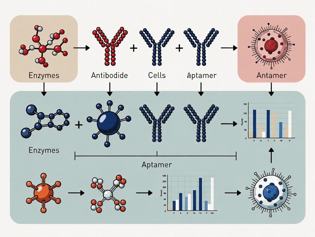
Bioreceptors can be broadly classified into several categories based on their biological origin and mechanism of action. The primary classes include antibodies, enzymes, nucleic acids, aptamers, cells, and molecularly imprinted polymers, each with distinct recognition paradigms and applications [1] [3].
Antibody-Based Bioreceptors
Antibodies are naturally occurring three-dimensional protein structures, typically ~150 kDa in size, that function within the immune system [3]. As bioreceptors, they operate through affinity-based binding, where the biosensor signal depends on the formation of an antibody-antigen immunocomplex [3]. The 3D protein structure of antibodies creates a unique recognition pattern with high specificity for the target bioanalyte, often referred to as an antigen [1] [3]. Antibodies share a general structural trend of a "Y" shaped conformation, comprised of light and heavy chains, with analyte binding domains located on the arms of the "Y" [3]. While antibodies provide exceptional specificity, their production requires animal experimentation, which is costly and time-consuming, and their binding capacity can be strongly dependent on assay conditions such as pH and temperature [1] [3].
Enzymatic Bioreceptors
Enzymes achieve bioanalyte specificity through binding cavities buried within their three-dimensional structure, utilizing hydrogen-bonding, electrostatics, and other non-covalent interactions to form recognition patterns [3]. Unlike antibodies, enzymatic biosensors are biocatalytic in nature, meaning the enzyme captures and catalytically converts the target bioanalyte into a measurable product [1] [3]. This process often involves the formation of an intermediate complex before the release of the measurable end product [3]. Enzymes are popular bioreceptors due to their ability to catalyze a large number of reactions, their potential to detect a group of analytes (substrates, products, inhibitors, and modulators), and their suitability with various transduction methods [1]. Since enzymes are not consumed in reactions, biosensors utilizing enzymatic bioreceptors can often be used continuously, though the sensor's lifetime is limited by enzyme stability [1].
Nucleic Acid Interactions
Nucleic acid-based bioreceptors can be divided into two main categories: genosensors and aptasensors [1]. Genosensors rely on complementary base pairing interactions (adenine:thymine and cytosine:guanine in DNA) to detect specific nucleic acid sequences [1] [3]. If the target nucleic acid sequence is known, complementary sequences can be synthesized, labeled, and immobilized on the sensor, with the hybridization event being optically or electrochemically detected [1]. Recent advances in this category include locked nucleic acids (LNA) and peptide nucleic acids (PNA), which offer improved binding stability [3]. Aptasensors utilize aptamers, which are single-stranded oligonucleotides (DNA or RNA) selected through Systematic Evolution of Ligands by Exponential Enrichment (SELEX) to bind specific targets with high affinity [1] [3]. Aptamers fold into complex three-dimensional structures that enable specific binding to a wide range of targets, including proteins, small molecules, ions, and whole cells [1] [4].
Cellular and Tissue-Based Bioreceptors
Cells and tissues represent more complex bioreceptor systems that leverage intact biological structures for sensing applications [1]. Cells are often used because they are sensitive to their surrounding environment and can respond to various stimuli [1]. Compared to isolated organelles, cells remain active for longer periods and offer good reproducibility, making them reusable [1]. They are commonly used to detect global parameters like stress conditions, toxicity, and organic derivatives, as well as to monitor drug treatment effects [1]. Tissue-based biosensors offer advantages including easier immobilization compared to cells, higher enzyme activity and stability maintained in the natural environment, availability, low cost, and avoidance of tedious enzyme extraction and purification processes [1]. However, tissues may lack specificity due to interference from other enzymes and have longer response times due to transport barriers [1].
Molecularly Imprinted Polymers
Molecularly imprinted polymers (MIPs) represent a class of synthetic bioreceptors that use a templated polymer matrix to achieve analyte specificity through patterns of non-covalent bonding, electrostatic interactions, or size inclusion/exclusion [3]. MIPs are designed to encapsulate the target bioanalyte, effectively forming synthetic recognition patterns between the bioanalyte and polymer matrix [3]. The tunability of MIPs comes from the choice of functional monomer, crosslinker, target bioanalyte, and solvent [3]. A significant advantage of MIPs is that a specific bioreceptor-bioanalyte pairing does not need to be biochemically identified, as the polymer is synthetically fabricated around the template bioanalyte [3].
Table 1: Comparative Analysis of Major Bioreceptor Types
| Bioreceptor Type | Recognition Mechanism | Target Examples | Binding Constant | Key Advantages | Key Limitations |
|---|---|---|---|---|---|
| Antibodies | Affinity-based immunocomplex formation | Proteins, viruses, cells | >10⁸ L/mol [1] | High specificity, mature protocols | Animal production, costly, environmental sensitivity [1] [3] |
| Enzymes | Catalytic conversion of substrate | Metabolites, inhibitors, toxins | Varies with enzyme | Signal amplification, continuous use | Limited by substrate specificity, stability [1] [3] |
| Nucleic Acids (Genosensors) | Complementary base pairing | DNA, RNA, genetic markers | N/A (hybridization) | High sequence specificity, design flexibility | Limited to nucleic acid targets [1] [3] |
| Aptamers | 3D structure-based binding | Ions, small molecules, proteins, cells | Varies with selection | Chemical synthesis, stability, modification ease | SELEX process costly, fewer standardized protocols [3] [4] |
| Molecularly Imprinted Polymers | Synthetic template-based binding | Small molecules, proteins | Varies with template | Synthetic production, stability, wide applicability | Optimization complexity, potential non-specific binding [3] |
| Cells/Tissues | Metabolic/functional response | Global parameters, toxins, drugs | Functional response | Holistic response, no purification needed | Lower specificity, longer response times [1] |
Bioreceptor Integration in Biosensor Systems
Fundamental Biosensor Architecture
The integration of a bioreceptor into a complete biosensor system requires careful consideration of the overall architecture. A typical biosensor consists of three main components: the bioreceptor (biological recognition element), the transducer, and the electronic system with display [1] [2]. The bioreceptor, as previously discussed, is responsible for specific analyte recognition. The transducer transforms the biological interaction into a measurable signal, working through physicochemical principles such as optical, piezoelectric, electrochemical, or electrochemiluminescence methods [1]. The electronic system processes the transduced signal through amplification and conditioning, ultimately displaying the results in a user-friendly format [2].
The general aim of biosensor design is to enable quick, convenient testing at the point of concern or care where the sample was procured [1]. This requires optimization of both the bioreceptor immobilization and its interface with the transducer surface. Bioreceptors are typically immobilized via covalent linkage to the sensor surface, forming organized arrays that maximize binding accessibility while minimizing non-specific interactions [3]. The spatial orientation and density of bioreceptors on the sensor surface significantly impact biosensor performance, including sensitivity, limit of detection, and dynamic range [4].
Diagram Title: Fundamental Biosensor Architecture
Transduction Mechanisms
The transduction mechanism employed in a biosensor determines how the bioreceptor-analyte interaction is converted into a measurable signal. Electrochemical transducers measure changes in electrical properties (current, potential, impedance) resulting from the biorecognition event [5] [6]. These are among the most commonly used transduction methods due to their high sensitivity, portability, and compatibility with miniaturization [6]. Optical transducers utilize light-based detection methods including surface plasmon resonance (SPR), localized surface plasmon resonance (LSPR), fluorescence, and colorimetry [4]. These methods offer advantages such as label-free detection, real-time monitoring, and high sensitivity, with some techniques capable of detecting analytes at femtomolar levels [4]. Other transduction methods include piezoelectric systems that measure mass changes through frequency variations, and thermal sensors that detect enthalpy changes from biochemical reactions [2].
The choice of transduction method is often dictated by the specific application requirements and the nature of the bioreceptor-analyte interaction. For instance, catalytic bioreceptors like enzymes often pair well with electrochemical transducers that can detect reaction products, while affinity-based bioreceptors like antibodies may be better suited to optical transducers that directly measure binding events [3] [2].
Performance Characteristics and Optimization Strategies
The performance of bioreceptor-based biosensors is evaluated through several key characteristics that determine their practical utility and reliability. Understanding and optimizing these parameters is essential for developing effective biosensing platforms.
Critical Performance Metrics
Selectivity is perhaps the most important feature of a biosensor, referring to the ability of a bioreceptor to detect a specific analyte in a sample containing other admixtures and contaminants [2]. High selectivity ensures that the biosensor responds only to the target analyte, ignoring potential interferents in complex sample matrices [1] [2]. Sensitivity defines the minimum amount of analyte that can be detected by the biosensor, often referred to as the limit of detection (LOD) [2]. In many medical and environmental applications, biosensors are required to detect analyte concentrations as low as ng/ml or even fg/ml to confirm the presence of trace analytes [2]. Reproducibility is the ability of the biosensor to generate identical responses for a duplicated experimental setup, characterized by the precision and accuracy of the transducer and electronics [2]. Reproducible signals provide high reliability and robustness to the inferences made from biosensor responses [2].
Stability represents the degree of susceptibility to ambient disturbances in and around the biosensing system, which can cause drift in the output signals [2]. Stability is particularly crucial in applications requiring long incubation steps or continuous monitoring [2]. Factors influencing stability include temperature sensitivity of transducers and electronics, as well as the degradation profile of the bioreceptor over time [2]. Linearity indicates the accuracy of the measured response to a straight-line relationship with analyte concentration, mathematically represented as y=mc, where c is the analyte concentration, y is the output signal, and m is the sensitivity [2]. The linear range defines the span of analyte concentrations over which the biosensor response changes linearly with concentration [2].
Table 2: Performance Requirements for Different Application Fields
| Application Field | Required Sensitivity | Key Selectivity Challenges | Stability Requirements | Sample Matrix Complexity |
|---|---|---|---|---|
| Medical Diagnostics | fg/ml - ng/ml for biomarkers [2] [7] | High (serum proteins, cells, drugs) [2] | Single-use or short-term stability often sufficient [2] | High (blood, urine, saliva) [2] [8] |
| Environmental Monitoring | ng/ml - μg/ml for contaminants [2] | Moderate (other pollutants, minerals) | Long-term stability (days to months) [2] | Moderate to high (water, soil extracts) [2] |
| Food Safety | pg/ml - ng/ml for pathogens/toxins [6] | High (food components, additives) | Varies by application [6] | High (complex food matrices) [6] |
| Biodefense | fg/ml - pg/ml for threat agents [2] | Very high (avoid false positives) | Long-term readiness with rapid response [2] | Varies (air, water, surfaces) [2] |
Bioreceptor Engineering and Immobilization Strategies
Optimizing bioreceptor performance often involves engineering the bioreceptors themselves and developing effective immobilization strategies to maintain their functionality on the sensor surface. For antibody-based receptors, researchers have engineered recombinant binding fragments (Fab, Fv, or scFv) or domains (VH, VHH) to overcome limitations of full antibodies, such as high molecular weight, limited stability, and the presence of essential disulfide bonds [1]. Similarly, artificial binding proteins (AgBPs) smaller than 100 amino-acid residues have been developed with strong stability, no disulfide requirements, and high yield expression in bacterial systems [1].
For nucleic acid-based bioreceptors, modifications include locked nucleic acids (LNA) that "lock" the ribose in the 3'-endo conformation, reducing conformational flexibility and improving binding with complementary targets [3]. Peptide nucleic acids (PNA) incorporate a repeating aminoethyl-glycine unit linked by peptide bonds, creating an uncharged oligonucleotide with higher stability in nucleic acid binding [3]. Aptamer engineering has focused on introducing specific chemical modifications to improve binding affinity, resistance to nucleases, and stability in various environmental conditions [4] [7].
Immobilization strategies significantly impact bioreceptor functionality by controlling orientation, density, and stability on the transducer surface. Common approaches include covalent attachment through functional groups (amine, carboxyl, thiol), affinity-based immobilization (e.g., biotin-streptavidin), physical adsorption, and entrapment within polymer matrices [3] [4]. Optimal immobilization maintains the bioreceptor's native conformation and binding activity while minimizing non-specific adsorption and providing stability under operational conditions [4].
Diagram Title: Bioreceptor Integration Workflow
Experimental Protocols for Bioreceptor Evaluation
SELEX Protocol for Aptamer Development
The Systematic Evolution of Ligands by Exponential Enrichment (SELEX) process is a fundamental protocol for generating aptamer bioreceptors with high affinity and specificity for target molecules [3]. The step-by-step methodology is as follows:
Library Preparation: Create a random oligonucleotide library containing approximately 10^14-10^15 different sequences with a central randomized region (20-70 base pairs) flanked by constant primer binding regions [3].
Incubation: Incubate the oligonucleotide library with the target analyte under optimized buffer conditions, temperature, and incubation time to allow binding [3].
Partitioning: Separate bound sequences from unbound sequences through partitioning methods such as filtration, affinity chromatography, or capillary electrophoresis [3].
Elution and Amplification: Elute the target-bound sequences and amplify them using polymerase chain reaction (PCR) for DNA aptamers or reverse transcription-PCR (RT-PCR) for RNA aptamers [3].
Stringency Adjustment: Increase selection stringency in successive rounds by reducing target concentration, increasing wash stringency, or adding counter-selection steps against related molecules to improve specificity [3].
Cloning and Sequencing: After 8-15 selection rounds, clone and sequence the enriched pool to identify individual aptamer candidates [3].
Characterization: Evaluate binding affinity (typically measured by dissociation constant, Kd) and specificity of individual aptamers using techniques such as surface plasmon resonance, isothermal titration calorimetry, or electrophoretic mobility shift assays [3] [7].
The entire SELEX process typically requires 2-8 weeks to complete, depending on the target complexity and selection strategy employed [3].
Electrochemical Biosensor Fabrication and Characterization
For researchers developing electrochemical biosensors utilizing various bioreceptors, the following standardized protocol provides a framework for sensor fabrication and performance evaluation [6]:
Electrode Pretreatment:
- Clean the working electrode (typically gold, glassy carbon, or screen-printed carbon) through mechanical polishing (with alumina slurry) and/or electrochemical cycling in sulfuric acid.
- Rinse thoroughly with deionized water and dry under nitrogen stream.
Bioreceptor Immobilization:
- For antibodies: Incubate electrode with protein A/G solution for oriented immobilization, or use covalent coupling through EDC/NHS chemistry to activated carboxyl groups.
- For aptamers: Utilize thiol-gold chemistry for thiol-modified aptamers, or avidin-biotin interaction for biotinylated sequences.
- For enzymes: Employ cross-linking with glutaraldehyde or encapsulation in polymer matrices like Nafion.
- Wash thoroughly with appropriate buffer to remove unbound bioreceptors.
Blocking:
- Incubate modified electrode with blocking agent (e.g., BSA, casein, or ethanolamine) to minimize non-specific binding.
- Wash with buffer to remove excess blocking agent.
Electrochemical Measurement:
- Utilize techniques such as Cyclic Voltammetry (CV), Electrochemical Impedance Spectroscopy (EIS), or Differential Pulse Voltammetry (DPV).
- Perform measurements in appropriate electrolyte solution with controlled temperature.
- Record baseline signal in analyte-free buffer.
Analytical Performance Evaluation:
- Sensitivity: Measure sensor response across a range of analyte concentrations. Calculate limit of detection (LOD) as 3×standard deviation of blank/slope of calibration curve.
- Selectivity: Test against potential interfering substances with similar structure or commonly co-existing compounds.
- Reproducibility: Assess response variability between different sensor batches (inter-assay) and within the same batch (intra-assay).
- Stability: Monitor signal response over time under storage conditions and during continuous operation.
This protocol typically requires 2-3 days for complete sensor fabrication and characterization, with variations depending on the immobilization chemistry and bioreceptor type [6].
Research Reagent Solutions for Bioreceptor Development
Table 3: Essential Research Reagents for Bioreceptor-Based Biosensing
| Reagent Category | Specific Examples | Function in Research | Application Notes |
|---|---|---|---|
| Immobilization Chemicals | EDC (1-Ethyl-3-(3-dimethylaminopropyl)carbodiimide), NHS (N-hydroxysuccinimide), Sulfo-SMCC | Covalent coupling of bioreceptors to sensor surfaces | EDC/NHS activates carboxyl groups; Sulfo-SMCC for thiol-maleimide chemistry [3] [4] |
| Blocking Agents | BSA (Bovine Serum Albumin), casein, ethanolamine, SuperBlock | Minimize non-specific binding on sensor surfaces | Choice depends on bioreceptor and sample matrix; testing multiple agents recommended [4] [6] |
| Signal Generating Reagents | Horseradish peroxidase (HRP), Alkaline phosphatase (ALP), fluorescent dyes (FITC, Cy dyes), electroactive tags (methylene blue, ferrocene) | Enable detection of bioreceptor-analyte binding | Enzyme-substrate systems offer amplification; direct tags simplify assay design [2] [6] |
| Nucleic Acid Modification Kits | Thiol-modification, biotinylation, amino-modification, click chemistry kits | Introduce functional groups for oriented immobilization | Commercial kits available from suppliers like Sigma-Aldrich, Thermo Fisher [3] [7] |
| Regeneration Solutions | Glycine-HCl (pH 2.0-3.0), NaOH (10-100mM), SDS (0.1-1%) | Dissociate analyte from bioreceptor for sensor reuse | Must be optimized for each bioreceptor-analyte pair to maintain activity [4] |
Emerging Trends and Future Perspectives
The field of bioreceptor development is rapidly evolving, driven by advances in biotechnology, nanotechnology, and materials science. Several emerging trends are shaping the future of bioreceptor elements in analytical devices. Next-generation aptamer development is focusing on overcoming current limitations through novel selection methods, expanded chemical modifications, and computational design approaches [9] [7]. These efforts aim to generate aptamers with enhanced binding affinity, improved stability in complex matrices, and reduced production costs [7]. The integration of artificial intelligence and machine learning in bioreceptor design is accelerating the discovery and optimization process, enabling predictive modeling of binding interactions and structural properties [5].
Multimodal and multiplexed biosensing platforms represent another significant trend, where multiple bioreceptors with different specificities are integrated into a single device for parallel detection of multiple analytes [9] [8]. This approach is particularly valuable for comprehensive diagnostic panels and environmental monitoring applications where multiple biomarkers or contaminants need simultaneous assessment [8]. Advances in nanomaterial-enhanced biosensors continue to push the limits of detection, with materials such as graphene, carbon nanotubes, metal-organic frameworks, and various nanoparticles being employed to enhance signal transduction and improve bioreceptor stability [5] [6].
The convergence of bioreceptor technology with point-of-care and wearable devices is expanding the applications of biosensors into personalized medicine and continuous health monitoring [9] [5]. These developments require bioreceptors that maintain stability and functionality under varied environmental conditions and in complex biological matrices like sweat, tears, or interstitial fluid [7]. Finally, the growing emphasis on sustainability and regulatory compliance is driving research into environmentally friendly biosensor manufacturing processes and robust validation protocols to ensure reliability and safety in real-world applications [5].
As these trends continue to evolve, bioreceptor elements will play an increasingly critical role in addressing global challenges in healthcare, environmental protection, and food safety through advanced sensing capabilities. The interdisciplinary nature of bioreceptor research ensures that continued innovation in this field will yield increasingly sophisticated solutions for analytical detection across diverse application domains.
Enzymes, as specialized biocatalysts, are fundamental components within the broader ecosystem of bioreceptor elements, which also includes antibodies, aptamers, and whole cells [10] [11]. Their unparalleled catalytic efficiency and substrate specificity make them indispensable for converting biochemical interactions into quantifiable signals in analytical devices [10]. This foundational role is critical across numerous applications, from medical diagnostics and therapeutic monitoring to environmental sensing and food safety [10] [12] [11].
The operational prowess of enzymes stems from their ability to accelerate biochemical reactions under mild conditions, a property that has been harnessed and enhanced through various engineering strategies [13]. The discovery of ribozymes expanded the definition of biocatalysts beyond proteins to include nucleic acids [14]. More recently, the field has been revolutionized by the development of artificial enzymes, including synthetic mimics and nanozymes—nanomaterials with intrinsic enzyme-like activity that offer enhanced stability and design flexibility [14]. These advancements have collectively pushed the boundaries of biocatalysis, enabling its application under increasingly diverse and challenging conditions [14].
This technical guide delves into the molecular intricacies of enzyme catalysis, explores the mechanistic basis of signal generation, and details contemporary experimental approaches. By framing these discussions within the context of modern bioreceptor research, this review provides scientists and drug development professionals with a comprehensive resource for leveraging enzymatic principles in cutting-edge diagnostic and therapeutic applications.
Catalytic Mechanisms of Enzymes
The extraordinary catalytic power of enzymes originates from their precise three-dimensional structures and their ability to interact with substrates through well-defined molecular mechanisms. These interactions lower the activation energy of reactions, facilitating rapid conversion of substrates to products.
Fundamental Principles of Enzyme Catalysis
Enzyme catalysis is governed by several key principles, with transition state stabilization representing a central paradigm. As initially proposed by Linus Pauling and later refined by Warshel through multiscale simulations, enzymes achieve remarkable rate accelerations by pre-organizing their electrostatic environments to preferentially stabilize the transition state over the ground state [13]. This pre-organization creates an electric field that can be quantitatively measured using techniques like vibrational Stark Shift spectroscopy and modeled through molecular simulations using Coulomb's law or higher-order multipole expansions [13].
Molecular modeling techniques, including quantum mechanics (QM) and molecular mechanics (MM), have become indispensable tools for elucidating these catalytic mechanisms at atomic resolution [13]. These physics-based approaches allow researchers to visualize and quantify the electrostatic contributions, hydrogen bonding networks, and structural dynamics that collectively enable efficient catalysis. The integration of artificial intelligence with these modeling techniques further enhances our ability to identify catalytically relevant conformations and predict the effects of structural modifications on enzyme function [13].
Engineering and Optimization Strategies
Rational engineering of enzyme properties relies on deep understanding of structure-function relationships across several domains:
Structure and Topology: Engineering active sites for improved shape complementarity to specific substrates can significantly enhance catalytic efficiency and selectivity. This approach has been successfully applied to improve substrate specificity in acyltransferases and O-methyl transferases [13]. Beyond the active site, engineering access tunnels can modulate the diffusion of substrates, products, and water molecules, thereby influencing catalytic rates [13].
Electrostatics: Optimization of electric fields and electrostatic potential surfaces enables precise manipulation of chemical reactivity involving charge separation or changes in ionic states [13]. Computational analysis of enzyme electrostatics provides quantitative parameters for guiding mutagenesis campaigns to enhance transition state stabilization.
Stability Engineering: Enhancing enzyme robustness against thermal denaturation, pH extremes, and organic solvents expands their application potential. Strategies include introducing disulfide bridges, engineering salt bridges, and optimizing surface charge distributions [13].
Nanozymes: The emergence of nanozymes represents a paradigm shift in biocatalysis. These nanomaterials exhibit intrinsic enzyme-like activity while offering advantages in stability, cost-effectiveness, and multifunctionality [14]. Unlike traditional enzymes, nanozymes possess multiple nanostructure-confined active sites and can maintain catalytic function under extreme conditions where protein-based enzymes would denature [14].
Table 1: Comparison of Natural Enzymes and Nanozymes
| Property | Natural Enzymes | Nanozymes |
|---|---|---|
| Composition | Proteins or RNA | Various nanomaterials (metals, metal oxides, MOFs, carbon) |
| Catalytic Sites | Defined active centers | Multiple, nanostructure-confined active sites |
| Stability | Sensitive to temperature, pH, proteolysis | High stability under extreme conditions |
| Production | Biological synthesis, often costly | Chemical synthesis, scalable |
| Design Flexibility | Limited by protein folding | Highly tunable size, morphology, surface |
| Multifunctionality | Typically single function | Integrated catalytic and physicochemical properties |
Signal Generation in Biosensing Systems
In biosensor applications, enzymes function as critical signal transducers that convert the presence of a target analyte into a measurable physical output. The exceptional catalytic efficiency of enzymes provides significant signal amplification, enabling detection of targets at ultralow concentrations.
Electrochemical Signal Transduction
Electrochemical biosensors represent a dominant platform where enzymes generate measurable electrical signals through catalytic reactions [15]. These systems typically employ oxidoreductases such as glucose oxidase (GOx), lactate oxidase (LOx), or cholesterol oxidase (ChOx) that produce or consume electroactive species during their catalytic cycles [10] [12].
A prominent signal generation strategy involves enzyme cascade catalysis, which couples multiple enzymatic reactions to achieve exponential signal amplification [16]. For example, in a system developed for detecting circulating tumor cells (CTCs), GOx immobilized on covalent organic frameworks (COFs) catalyzes the oxidation of glucose to generate hydrogen peroxide (H₂O₂). This H₂O₂ then serves as a substrate for a horseradish peroxidase mimic (HPM) constructed from bimetallic CuAu nanowires and metal-organic frameworks (MOFs), which catalyzes the oxidation of TMB (3,3',5,5'-tetramethylbenzidine), producing a strong electrochemical readout [16]. This synergistic amplification mechanism enables ultrasensitive detection at clinically relevant levels.
The general working principle of such enzymatic biosensors is illustrated below:
Diagram 1: Enzyme Biosensor Working Principle
Optical and Other Transduction Modalities
Beyond electrochemical systems, enzymes drive signal generation in multiple sensing modalities:
Optical Biosensors: Enzymatic reactions that produce colored, fluorescent, or luminescent products enable detection through absorbance, fluorescence, or chemiluminescence measurements [10] [11]. For instance, urease-catalyzed hydrolysis of urea generates ammonia, causing pH changes that can be detected with pH-sensitive dyes [10].
Thermometric Biosensors: Enzymatic reactions that release significant heat (exothermic reactions) enable detection through temperature changes measured using thermistors [10].
Piezoelectric Biosensors: Enzymes immobilized on crystal surfaces can detect mass changes resulting from catalytic conversion of substrates, producing frequency shifts in quartz crystal microbalances [10].
Table 2: Common Enzyme Types and Their Signal Generation Mechanisms in Biosensing
| Enzyme | Reaction Catalyzed | Signal Generated | Primary Applications |
|---|---|---|---|
| Glucose Oxidase (GOx) | β-D-glucose + O₂ → gluconic acid + H₂O₂ | Amperometric (H₂O₂ detection) | Glucose monitoring, food analysis [10] |
| Urease | Urea + H₂O → CO₂ + 2NH₃ | Potentiometric (pH change) | Kidney function, environmental monitoring [10] |
| Lactate Oxidase (LOx) | L-lactate + O₂ → pyruvate + H₂O₂ | Amperometric | Sports medicine, critical care [10] |
| Cholesterol Oxidase (ChOx) | Cholesterol + O₂ → cholest-4-en-3-one + H₂O₂ | Electrochemical/Optical | Cardiovascular health, food science [10] |
| Acetylcholinesterase (AChE) | Acetylcholine → choline + acetate | Inhibition-based amperometric | Pesticide detection, neurotoxin monitoring [10] |
| Horseradish Peroxidase (HRP) | H₂O₂ + donor → 2H₂O + oxidized donor | Colorimetric/Electrochemical | Signal amplification in immunoassays [16] |
Experimental Protocols and Methodologies
Robust experimental characterization is essential for understanding enzymatic mechanisms and developing effective biosensing platforms. The following protocols represent standardized approaches for evaluating enzyme activity and implementing enzymatic biosensors.
Protocol: Measuring α-Amylase Activity (INFOGEST Optimized Method)
This optimized protocol from the INFOGEST international research network demonstrates a standardized approach for quantifying enzyme activity with improved precision and reproducibility [17].
Principle: α-Amylase catalyzes the hydrolysis of starch into reducing sugars (maltose equivalents), which are quantified using colorimetric methods.
Reagents:
- Potato starch solution (0.5% w/v in phosphate buffer, pH 6.9)
- Maltose standard solutions (0-3 mg/mL for calibration curve)
- DNS reagent (3,5-dinitrosalicylic acid) or similar reducing sugar detection reagent
- Enzyme samples (properly diluted in appropriate buffer)
Procedure:
- Preparation: Prepare maltose calibrators (0, 0.3, 0.6, 0.9, 1.2, 1.5, 1.8, 2.1, 2.4, 2.7, 3.0 mg/mL) [17].
- Incubation: In separate tubes, add 500 μL starch solution to 100 μL enzyme solution. Incubate at 37°C for exactly 3 minutes [17].
- Reaction Termination: Add 1.0 mL DNS reagent to stop the reaction and develop color.
- Measurement: Measure absorbance at 540 nm using a spectrophotometer or microplate reader.
- Kinetics: Perform measurements at four time points (e.g., 1, 2, 3, 4 minutes) to establish linear reaction rate [17].
- Calculation: Generate maltose standard curve. Calculate enzyme activity where one unit liberates 1.0 mg of maltose from starch in 3 minutes at pH 6.9 at 37°C [17].
Validation: This optimized protocol demonstrates significantly improved interlaboratory reproducibility (CVs 16-21%) compared to traditional single-point measurements at 20°C [17].
Protocol: Cascade Catalysis Sensor for Ultrasensitive CTC Detection
This protocol details a sophisticated biosensing platform that exemplifies advanced signal generation through enzyme cascade amplification [16].
Principle: This sensor employs a dual-enzyme cascade system where glucose oxidase (GOx) generates H₂O₂, which is subsequently utilized by a peroxidase mimic (HPM) to oxidize TMB, producing a strong electrochemical signal specifically amplified at captured circulating tumor cells.
Reagents and Materials:
- HPM nanocomposite (CuAu nanowires combined with MOFs)
- GOx-Covalent Organic Frameworks-Au-Aptamer (GOx-CAA) conjugate
- Anti-EpCAM antibody for CTC capture
- TMB substrate solution
- Glucose solution
- Electrochemical cell with three-electrode system
Experimental Workflow:
Diagram 2: CTC Sensor Experimental Workflow
Procedure:
- Electrode Preparation: Modify electrode surface with HPM nanocomposite, which provides superior catalytic activity, conductivity, and binding sites for anti-EpCAM antibodies [16].
- Bioreceptor Immobilization: Immobilize anti-EpCAM antibodies on the HPM-modified electrode for specific CTC capture [16].
- Sample Incubation: Incubate blood sample with the functionalized electrode to capture CTCs via antibody-antigen interactions.
- Signal Probe Binding: Introduce GOx-CAA conjugates, which bind to captured CTCs via MUC1 aptamer recognition [16].
- Cascade Catalysis Reaction: Add solution containing glucose and TMB. GOx catalyzes glucose oxidation to generate H₂O₂, which is subsequently reduced by HPM while simultaneously oxidizing TMB [16].
- Electrochemical Measurement: Apply potential and measure current generated by oxidized TMB product using amperometry or differential pulse voltammetry [16].
Performance: This cascade catalysis sensor achieves exceptional sensitivity for CTC detection, reaching detection limits relevant for clinical applications (1 cell/mL) through synergistic signal amplification [16].
The Scientist's Toolkit: Essential Research Reagents
Successful implementation of enzymatic biosensing platforms requires carefully selected materials and reagents. The following table catalogs essential components for developing cascade catalysis systems similar to the CTC sensor described above.
Table 3: Essential Research Reagents for Advanced Enzymatic Biosensing
| Reagent/Material | Function | Example Application |
|---|---|---|
| Bimetallic Nanomaterials (CuAu nanowires) | Enhance electron transfer, provide catalytic sites | HPM construction for peroxidase-like activity [16] |
| Metal-Organic Frameworks (MOFs) | High surface area support for enzyme immobilization | Structural component of HPM nanocomposite [16] |
| Covalent Organic Frameworks (COFs) | Porous substrate for enzyme confinement and stabilization | GOx immobilization platform [16] |
| TMB Substrate (3,3',5,5'-tetramethylbenzidine) | Chromogenic/electroactive enzyme substrate | Peroxidase substrate for signal generation [16] |
| Glucose Oxidase (GOx) | Primary enzyme for H₂O₂ generation | Signal initiation in cascade systems [16] |
| Aptamers (e.g., MUC1-specific) | Molecular recognition elements for cell targeting | Specific binding to target cells [18] |
| Anti-EpCAM Antibodies | Immunorecognition for cell capture | CTC isolation from complex samples [16] |
| Nanozymes (e.g., Fe₃O₄ nanoparticles) | Nanomaterial-based enzyme mimics | Stable, tunable alternatives to natural enzymes [14] |
Enzymes represent sophisticated catalytic machinery whose mechanisms can be harnessed for powerful signal generation in analytical systems. From their fundamental catalytic principles based on transition state stabilization and electrostatic pre-organization to their implementation in complex cascade architectures, enzymes provide the critical link between molecular recognition and detectable signals in biosensing platforms.
The continued evolution of enzyme engineering—through rational design, directed evolution, and the development of nanozymes—promises to further expand the capabilities of enzymatic biosensors. The integration of artificial intelligence with physics-based modeling approaches offers particularly exciting prospects for predicting and optimizing enzyme function [13]. Furthermore, the discovery of natural nanozymes within biological systems opens new avenues for understanding physiological and pathological processes [14].
As the field advances, the synergy between natural enzymes and their synthetic counterparts will likely yield increasingly sophisticated sensing systems with enhanced sensitivity, specificity, and operational stability. These developments will solidify the role of enzymes as indispensable components within the broader repertoire of bioreceptor elements, enabling new breakthroughs in medical diagnostics, environmental monitoring, and therapeutic development.
Antibodies, or immunoglobulins, are Y-shaped proteins produced by B cells of the adaptive immune system and represent a dominant class of protein therapeutics. Their function as exquisite bioreceptors stems from an unparalleled ability to bind a vast array of target molecules (antigens) with high specificity and affinity. This specificity is not defined by a single, uniform mechanism, but is rather a direct consequence of their three-dimensional (3D) structure, which creates unique molecular surfaces for binding. The interactions that promote antigen binding are determined by the structures of six hypervariable loops, known as Complementarity-Determining Regions (CDRs), which form the antigen-binding site, or paratope [19]. Within the broader context of bioreceptor elements—which include enzymes, antibodies, aptamers, and whole cells—antibodies stand out for their native biological role in molecular recognition. This technical guide delves into the structural architecture of antibodies, the atomic-level details of antigen recognition, and the cutting-edge computational methods now enabling their de novo design. It also provides a detailed overview of the experimental protocols and reagent tools essential for research and development in this field.
Structural Architecture of Antibodies
The fundamental architecture of an antibody is a multi-domain protein, typically depicted as Y-shaped. It is composed of two identical heavy chains (HCs) and two identical light chains (LCs). Each chain consists of a series of domains: a variable domain (VH or VL) at the amino terminus and one or more constant domains (CH1, CH2, CH3, and CL) [19].
- Fragment Antigen-Binding (Fab) Region: This region, comprising one constant and one variable domain from each of the heavy and light chains (VH-CH1 and VL-CL), is responsible for antigen recognition. The Fab region contains the paratope.
- Fragment Crystallizable (Fc) Region: This region, formed by the paired constant domains of the heavy chains (CH2 and CH3), mediates immune effector functions such as complement activation and antibody-dependent cellular cytotoxicity (ADCC) by interacting with Fc receptors on immune cells [19].
- Hinge Region: A flexible polypeptide segment connects the Fab and Fc regions, providing the conformational flexibility needed for optimal antigen binding and effector function.
The binding surface of the antibody, the paratope, is located within the variable domains of the heavy and light chains (VH and VL). These domains each contain three hypervariable loops—CDR-H1, CDR-H2, CDR-H3 on the heavy chain, and CDR-L1, CDR-L2, CDR-L3 on the light chain—which are flanked by more conserved framework regions (FRs) [19]. The CDR loops, particularly CDR-H3, vary considerably in length, sequence, and structure, generating an enormous diversity of binding surfaces that enable the immune system to recognize virtually any pathogen. Table 1 summarizes the structural elements of a conventional antibody.
Table 1: Structural Elements of a Conventional IgG Antibody
| Structural Element | Description | Functional Role |
|---|---|---|
| Heavy Chain (HC) | One variable (VH) and three constant (CH1, CH2, CH3) domains | Determines antibody class/isotype; VH contributes to antigen binding. |
| Light Chain (LC) | One variable (VL) and one constant (CL) domain | VL contributes to antigen binding. |
| Complementarity-Determining Regions (CDRs) | Six hypervariable loops (3 in VH, 3 in VL) | Form the paratope; primary determinants of antigen specificity and affinity. |
| Framework Regions (FRs) | Four conserved sequences flanking CDRs in VH and VL | Provide a structural scaffold for the CDR loops. |
| Fab Region | Composed of VH-CH1 and VL-CL domains | Binds antigen; contains the paratope. |
| Fc Region | Composed of paired CH2 and CH3 domains | Mediates immune effector functions (e.g., ADCC, complement activation). |
| Hinge Region | Flexible peptide between CH1 and CH2 domains | Provides flexibility for antigen binding and avidity. |
A unique class of antibodies, known as single-domain antibodies (VHHs or nanobodies, is found in camelids (e.g., llamas, alpacas) and cartilaginous fish. These are composed only of a heavy chain variable domain (VHH), forming the smallest known antigen-binding fragment at ~12-15 kDa, compared to ~150 kDa for a full-length IgG. Despite having only three CDR loops, their interaction surface area is similar to that of conventional antibodies, and they can access buried epitopes that traditional antibodies cannot [19] [20].
The Structural Basis of Specificity and Immunoaffinity
The specificity of an antibody is quantified by its affinity—the strength of a single interaction between a paratope and its epitope on the antigen. High-affinity binding results from complementary molecular surfaces and favorable intermolecular forces.
Antibody-Antigen Interactions
The interface between an antibody and its antigen is a complex 3D surface where non-covalent forces stabilize the binding. A computational analysis of over 850,000 atom-atom contacts from 1,833 non-redundant antibody-antigen complexes has revealed clear patterns in the number of contacts and amino acid frequencies at the paratope [21]. The key interactions include:
- Hydrogen Bonds: Form between polar residues on the antibody and antigen.
- Electrostatic Interactions: Occur between charged amino acid side chains (e.g., salt bridges).
- Van der Waals Forces: Rely on precise shape complementarity between the paratope and epitope.
- Hydrophobic Interactions: Drive the burial of non-polar surfaces away from the aqueous environment.
The CDR-H3 loop is often the most critical for binding affinity and specificity due to its exceptional diversity in length, sequence, and conformational flexibility. Its conformation is also influenced by the relative orientation of the VH and VL domains, adding another layer of complexity to structure prediction and engineering [19].
Key Experimental Findings on Interface Trends
Large-scale structural analyses have provided critical insights into the molecular rules governing antibody-antigen interfaces. These studies compare conventional antibodies with single-domain antibodies (sdAbs) to understand how sdAbs compensate for their smaller size and reduced number of CDR loops.
- Amino Acid Frequencies: Specific amino acids are statistically overrepresented in paratopes. These "hotspot" residues are often found at the binding interface and are critical for interaction stability [21].
- Interaction Mechanisms of sdAbs: Single-domain antibodies employ distinct mechanisms to achieve high-affinity binding with only three CDR loops. This often involves a more convex paratope surface and a greater reliance on elongated CDR3 loops to access concave epitopes [21].
These findings have direct applications in antibody engineering, guiding the design of improved antibody libraries and therapeutic candidates with enhanced specificity and affinity.
Computational Design of Antibodies
Traditionally, antibody discovery has relied on animal immunization or screening of large random libraries. However, a revolutionary advance now allows for the de novo design of novel, epitope-specific antibodies entirely in silico.
The RFdiffusion Platform
A groundbreaking method involves a fine-tuned RFdiffusion network, a generative artificial intelligence (AI) model based on protein structure prediction [20]. This platform enables the design of antibody variable heavy chains (VHHs), single-chain variable fragments (scFvs), and full antibodies that bind to user-specified epitopes with atomic-level precision.
Workflow of De Novo Antibody Design:
- Input Specification: The user provides the 3D structure of the target antigen and specifies the desired epitope. A highly stable antibody framework (e.g., a humanized VHH framework) is selected as a structural scaffold.
- Conditional Diffusion: The fine-tuned RFdiffusion network is conditioned on the fixed framework and the target epitope. It then iteratively "de-noises" a random distribution of CDR loop backbones and rigid-body orientations, generating novel CDR loop conformations that are predicted to bind the target.
- Sequence Design: ProteinMPNN, a deep learning-based protein sequence design tool, is used to design the amino acid sequences for the generated CDR loop structures that are compatible with both the structure and the target binding.
- In Silico Validation: A separate fine-tuned RoseTTAFold2 (RF2) network is used to re-predict the structure of the designed antibody-antigen complex. Designs where the predicted structure closely matches the original RFdiffusion model ("self-consistent") are prioritized for experimental testing [20].
- Experimental Screening and Maturation: Designed antibodies are synthesized and screened for binding, typically using yeast surface display or surface plasmon resonance (SPR). Initial designs with modest affinity (nanomolar Kd) can be further improved through affinity maturation to achieve single-digit nanomolar or picomolar binders while maintaining epitope specificity [20].
This approach was successfully used to design VHH binders against disease-relevant targets like influenza haemagglutinin and Clostridium difficile toxin B (TcdB). Cryo-electron microscopy structures confirmed atomic-level accuracy in the designed CDR loops and the intended binding pose [20].
Diagram: AI-Driven Antibody Design Workflow. This flowchart outlines the key steps for de novo antibody design using fine-tuned RFdiffusion.
Experimental Protocols for Characterization
Validating the structure and function of antibodies, whether naturally derived or computationally designed, requires a suite of sophisticated experimental techniques.
Determining Binding Affinity and Kinetics with Surface Plasmon Resonance (SPR)
SPR is a label-free technique used to quantify biomolecular interactions in real-time by measuring changes in the refractive index on a sensor chip surface.
Detailed Protocol:
- Immobilization: The antigen is covalently immobilized onto a dextran-coated gold sensor chip via amine coupling. The surface is activated with a mixture of EDC (1-ethyl-3-(3-dimethylaminopropyl)carbodiimide) and NHS (N-hydroxysuccinimide). The antigen in a low-pH buffer (e.g., acetate buffer, pH 4.5-5.5) is flowed over the surface, forming amide bonds. Remaining active groups are deactivated with ethanolamine.
- Binding Kinetics Analysis: A series of concentrations of the purified antibody (the analyte) are injected over the antigen-coated surface and a reference surface at a constant flow rate (e.g., 30 μL/min) in HBS-EP buffer (10 mM HEPES, 150 mM NaCl, 3 mM EDTA, 0.05% surfactant P20, pH 7.4).
- Data Collection and Processing: The SPR instrument records a sensorgram (response units vs. time) for each analyte concentration. The data is double-referenced (reference surface and blank buffer injection subtracted).
- Curve Fitting: The kinetic data is fitted to a 1:1 Langmuir binding model using evaluation software (e.g., Biacore Evaluation Software) to determine the association rate constant (kon), dissociation rate constant (koff), and the equilibrium dissociation constant (KD = koff/kon).
Structural Validation with Cryo-Electron Microscopy (Cryo-EM)
Cryo-EM is powerful for determining the high-resolution structure of antibody-antigen complexes, especially for large or flexible targets.
Detailed Protocol:
- Sample Vitrification: The purified antibody-antigen complex (at ~0.5-3 mg/mL) is applied to a holey carbon grid. The grid is blotted with filter paper to create a thin liquid film and plunged into a cryogen (typically liquid ethane) cooled by liquid nitrogen. This rapidly freezes the sample in amorphous ice, preserving its native state.
- Data Collection: The vitrified grid is loaded into a cryo-electron microscope. Using automated software, thousands of micrographs are collected at a high defocus range (e.g., -1.5 to -3.0 μm) under low-dose conditions to minimize radiation damage.
- Image Processing: Micrographs are motion-corrected and the contrast transfer function (CTF) is estimated. Particles are automatically picked, extracted, and subjected to 2D classification to remove junk particles. An initial 3D model is generated ab initio or from a homologous structure, and then refined through 3D classification and high-resolution refinement.
- Model Building and Refinement: For high-resolution maps (<~3.5 Å), an atomic model of the antibody-antigen complex can be built de novo or by docking and refining existing crystal structures. The model is refined against the cryo-EM map using programs like Coot and Phenix [20].
The Scientist's Toolkit: Research Reagent Solutions
Research and development in antibody engineering rely on a suite of essential reagents, technologies, and computational tools. Table 2 details key materials and their functions.
Table 2: Essential Research Reagents and Tools for Antibody R&D
| Category | Item/Technology | Function and Application |
|---|---|---|
| Discovery Technologies | Yeast Surface Display | High-throughput screening of antibody libraries for binders. |
| Phage Display | In vitro screening of antibody fragment libraries displayed on phage. | |
| Hybridoma Technology | Production of monoclonal antibodies from immortalized B cells. | |
| Characterization Reagents | SPR Chip (e.g., CMS) | Gold sensor chip with a carboxymethylated dextran matrix for immobilizing biomolecules. |
| HBS-EP Buffer | Standard running buffer for SPR; provides stable pH and ionic strength, and reduces non-specific binding. | |
| Anti-His/C-myc/FLAG Tag Antibodies | Detection and purification of recombinant His-tagged or other tagged antibodies. | |
| Production & Purification | CHO/HEK293 Cell Lines | Mammalian expression systems for producing full-length, glycosylated antibodies. |
| Protein A/G/L Resins | Affinity chromatography resins that bind the Fc region of antibodies for purification. | |
| Computational Tools | RFdiffusion (fine-tuned) | Generative AI for de novo design of antibody structures targeting specific epitopes [20]. |
| ProteinMPNN | Deep learning-based protein sequence design for generating sequences that fold into a given backbone structure [20]. | |
| RoseTTAFold2 (fine-tuned) | Deep learning network for protein structure prediction, used for in silico validation of designed antibodies [20]. | |
| Rosetta | Software suite for protein structure prediction, design, and docking; used for energy calculations (ddG) [20]. |
Antibodies achieve their remarkable specificity through a sophisticated interplay of 3D structure and immunoaffinity. The precise architecture of the CDR loops, supported by their framework, creates a unique paratope that engages the antigen through a network of atomic interactions. The field is undergoing a transformation, driven by structural bioinformatics and artificial intelligence. The ability to design antibodies de novo with atomic-level precision, as demonstrated by platforms like RFdiffusion, marks a paradigm shift from empirical discovery to rational design. This progress, built upon a deep understanding of antibody structure and validated by rigorous experimental protocols, promises to accelerate the development of next-generation therapeutics, diagnostics, and research reagents.
Aptamers are short, single-stranded DNA or RNA oligonucleotides that bind to specific target molecules with high affinity and specificity by folding into defined three-dimensional structures [22] [23]. The term "aptamer" derives from the Latin words aptus (to fit) and meros (part), reflecting their lock-and-key relationship with targets [24]. Selected through an iterative Systematic Evolution of Ligands by EXponential enrichment (SELEX) process, aptamers function as synthetic molecular recognition elements with dissociation constants (Kd) typically ranging from nanomolar to picomolar levels [22]. Their programmable folding capabilities enable them to recognize diverse targets, including small molecules, proteins, whole cells, and pathogens [23].
As bioreceptor elements, aptamers are increasingly considered competitive alternatives to traditional antibodies and enzymes due to several distinct advantages. Table 1 compares key characteristics of aptamers against antibodies, highlighting features such as their smaller molecular weight, superior stability, easier modification, and entirely in vitro selection process that avoids animal use [23] [24]. These properties make aptamers particularly valuable for therapeutic development, diagnostic applications, and as targeting agents in drug delivery systems [25] [26].
Table 1: Comparative Analysis of Aptamers and Antibodies as Bioreceptors
| Feature | Aptamers | Antibodies |
|---|---|---|
| Molecular Nature | Short ssDNA or RNA oligonucleotides | Large proteins (~150 kDa) |
| Production Method | Chemical synthesis via SELEX in vitro | Biological production in vivo (animals/hybridoma) |
| Development Time | Weeks to months | Several months |
| Molecular Weight | 5–15 kDa | 150–170 kDa |
| Batch-to-Batch Variation | Low (chemical synthesis) | High (biological production) |
| Stability | Thermostable; reversible denaturation | Sensitive to heat/pH; irreversible denaturation |
| Modification | Easily and precisely modified | Limited and complex modification |
| Target Range | Proteins, small molecules, toxins, ions, non-immunogenic targets | Primarily immunogenic proteins and large antigens |
| Production Cost | Relatively low | High |
| Immunogenicity | Generally low | May trigger immune responses |
The SELEX Process: Methodology and Technological Innovations
The Systematic Evolution of Ligands by EXponential enrichment (SELEX) is the foundational technology for aptamer discovery, first developed in 1990 by Tuerk, Gold, Ellington, and Szostak [22] [24]. This iterative biochemical process screens combinatorial nucleic acid libraries to isolate sequences with high binding affinity and specificity to a target molecule.
Fundamental SELEX Workflow
The canonical SELEX methodology comprises three core stages repeated over 8-15 selection rounds [22] [23]:
- Incubation: A synthetic oligonucleotide library containing 10^14-10^15 random sequences (typically 20-80 nucleotides flanked by constant primer binding sites) is incubated with the target molecule under controlled buffer conditions [22] [24].
- Partitioning: Target-bound sequences are separated from unbound oligonucleotides through various separation techniques.
- Amplification: Enriched target-binding sequences are amplified by PCR (for DNA aptamers) or RT-PCR (for RNA aptamers) to generate an enriched pool for subsequent selection rounds [22].
Selection stringency typically increases with successive rounds through adjustments in buffer composition, washing stringency, and target concentration to favor the highest-affinity binders [23]. Following final selection rounds, the enriched pool is sequenced, and individual aptamer candidates are characterized for binding affinity, specificity, and structure.
Advanced SELEX Methodologies
Traditional SELEX approaches have evolved into specialized techniques that enhance selection efficiency and success rates for challenging targets. Key innovations include:
Capillary Electrophoresis SELEX (CE-SELEX) employs capillary electrophoresis under high voltage to separate target-aptamer complexes from unbound sequences based on migration differentials [23] [27]. This technique offers superior separation resolution, enables affinity constant determination during selection, and typically requires only 1-4 selection rounds, significantly accelerating aptamer discovery [23] [27]. Kinetic CE-SELEX methods like nonequilibrium capillary electrophoresis of equilibrium mixtures (NECEEM) and equilibrium capillary electrophoresis of equilibrium mixtures (ECEEM) further enable isolation of aptamers with controlled binding parameters (Kd, Kon, Koff) [27].
Cell-SELEX utilizes whole living cells as targets, enabling aptamer selection against native cell surface biomarkers without prior knowledge of molecular targets [22] [26]. This approach is particularly valuable for identifying disease-specific aptamers for cancer cell targeting. Counter-selection against control cells (e.g., non-malignant cells) is incorporated to enhance specificity [26].
Microfluidic SELEX integrates the SELEX process onto microfluidic platforms, enabling automation, reduced reagent consumption, and improved partitioning efficiency through precise fluidic control [23] [28]. These systems facilitate faster selection cycles and can be combined with other detection methods for real-time monitoring.
In Silico SELEX leverages computational approaches and predictive algorithms to simulate aptamer-target interactions, helping to identify minimal functional sequences and guide rational aptamer design, thereby reducing experimental rounds [23].
The following diagram illustrates the standard SELEX workflow with integration points for advanced methodologies:
Programmable Folding and Structural Diversity
Aptamers achieve molecular recognition through their capacity to fold into specific three-dimensional architectures that complement the surface features of their targets. This programmable folding is governed by nucleotide sequence and occurs through intrachain base pairing and stacking interactions, stabilized by hydrogen bonding, van der Waals forces, and electrostatic interactions [23].
Common Structural Motifs
Aptamers exhibit diverse secondary and tertiary structures that enable precise target recognition:
- Stem-loops (Hairpins): Single-stranded regions loop back to form double-stranded stems, creating binding pockets [27].
- G-quadruplexes: Guanine-rich sequences form planar tetrads stabilized by Hoogsteen hydrogen bonding, stacking into four-stranded structures [27].
- Pseudoknots: Complex structures with nested double-stranded regions formed by loop-base pair interactions [27].
- Bulges and internal loops: Unpaired regions within double-stranded segments that provide structural flexibility and recognition surfaces [23].
The folding pathway and final conformation are influenced by environmental factors including ionic strength, pH, temperature, and the presence of specific cations (e.g., Mg²⁺, K⁺) that stabilize particular structures [23]. For large target molecules, aptamers form adaptive structures that conform to clefts and gaps on the target surface, while for small molecules, they typically wrap around and encapsulate the target [23].
Structure-Function Relationships
The relationship between aptamer structure and binding function enables rational design approaches. Table 2 summarizes key structural motifs and their functional significance in molecular recognition. Post-SELEX optimization through truncation studies can identify minimal functional domains that maintain binding affinity while reducing synthesis costs and improving pharmacokinetic properties [23]. Computational modeling and predictive algorithms further assist in determining essential structural elements by simulating aptamer-target interaction processes [23].
Table 2: Aptamer Structural Motifs and Functional Characteristics
| Structural Motif | Description | Representative Targets | Functional Significance |
|---|---|---|---|
| Stem-Loop/Hairpin | Base-paired stem with unpaired loop | Proteins, small molecules | Creates binding pockets; versatile recognition |
| G-Quadruplex | Four-stranded structure with G-tetrads | Porphyrins, proteins, ions | Provides stable platform; electrochemical sensing |
| Pseudoknot | Nested double-stranded regions | Viral RNA, ribosomal frameshifting | Complex recognition surfaces; high specificity |
| Kissing Loop | Loop-loop interactions between strands | Dimeric proteins, RNA complexes | Multivalent binding; enhanced affinity |
| Bulge/Internal Loop | Unpaired regions within duplex | Proteins, cell surfaces | Structural flexibility; adaptive recognition |
Experimental Protocols for Key Applications
CE-SELEX Protocol for Protein Targets
This protocol outlines the CE-SELEX procedure for selecting high-affinity DNA aptamers against purified protein targets, based on methodologies described in search results [23] [27].
Materials and Reagents:
- Initial ssDNA library: 5'-GGGAGACAAGAATAAACGCTCA-N40-TGTGGTGGTGGTGGTG-3' (N40 = 40 random nucleotides)
- Target protein in appropriate storage buffer
- PCR components: primers, dNTPs, DNA polymerase
- Capillary electrophoresis system with UV/fluorescence detection
- Elution buffer: 8 M urea, 20 mM EDTA, 0.5 M NaCl
- Binding buffer optimized for target protein
Procedure:
- Library Preparation: Resuspend initial ssDNA library (10 nmol) in 500 μL binding buffer, denature at 95°C for 5 min, and slowly cool to room temperature for proper folding.
- Incubation: Mix folded library (100 pmol) with target protein (10 pmol) in 50 μL binding buffer, incubate at 37°C for 30 min.
- CE Separation: Inject mixture into CE capillary (50 μm ID, 50 cm effective length) with applied voltage of 15 kV. Monitor separation at 260 nm.
- Complex Collection: Collect protein-ssDNA complex peak (typically migrating faster than free library) within precise time window (5-10 s collection).
- Elution and Recovery: Add 2 volumes of elution buffer to collected fraction, incubate at 65°C for 10 min to dissociate complexes.
- PCR Amplification: Amplify recovered ssDNA using asymmetric PCR (forward:primer ratio 1:50) to regenerate ssDNA pool.
- Stringency Adjustment: For subsequent rounds, decrease protein concentration (to 5, 2, then 1 pmol) and reduce incubation time (to 20, 15, then 10 min).
- Cloning and Sequencing: After 3-4 rounds, clone final pool into sequencing vector, pick 20-50 colonies for Sanger sequencing.
Critical Steps:
- Precisely control capillary temperature (±0.5°C) to maintain complex stability
- Include negative selection rounds without target to remove non-specific binders
- Monitor enrichment by measuring complex peak area increase across rounds
Cell-SELEX Protocol for Cancer Cell Biomarkers
This protocol describes aptamer selection against native cell surface targets using whole cells, enabling discovery of disease-specific aptamers without prior target identification [22] [26].
Materials and Reagents:
- Target cells (e.g., cancer cell line)
- Control cells (e.g., non-malignant counterpart)
- ssDNA library with 40-60 nt random region
- Binding buffer: PBS with 1 mM MgCl₂, 5 mM glucose, 0.1 mg/mL yeast tRNA
- Cell culture equipment and reagents
- FACS sorting capability (optional)
Procedure:
- Cell Preparation: Culture target and control cells to 80% confluence, harvest using non-enzymatic dissociation buffer to preserve surface epitopes.
- Negative Selection: Incubate ssDNA library (1 nmol) with control cells (10⁶ cells) in binding buffer (1 mL) at 4°C for 60 min with gentle rotation. Collect unbound supernatant.
- Positive Selection: Incubate pre-cleared library with target cells (10⁶ cells) under identical conditions.
- Washing: Wash cells 3-5 times with ice-cold binding buffer (1 mL) to remove weakly bound sequences.
- Elution: Resuspend cell pellet in 200 μL elution buffer (binding buffer + 8 M urea), heat at 95°C for 10 min, centrifuge and collect supernatant.
- Amplification: PCR-amplify recovered ssDNA, generate single-stranded product for next selection round.
- Counter-Selection: Incorporate negative selection steps every 2-3 rounds to enhance specificity.
- Monitoring: Assess enrichment by measuring fluorescence intensity of aptamer pool binding to target vs. control cells using flow cytometry after rounds 8, 10, and 12.
- Identification: Sequence final pool (round 12-15), cluster sequences by homology, select representatives from dominant families for characterization.
Critical Steps:
- Maintain cell viability >95% throughout selection process
- Use identical cell numbers for target and control cells
- Progressively increase washing stringency (volume, duration) across rounds
- Validate specific binding of individual aptamers by flow cytometry
Research Reagent Solutions and Materials
Successful aptamer selection and application requires specialized reagents and materials. The following table details essential components for SELEX experiments and their functional significance.
Table 3: Essential Research Reagents for Aptamer Selection and Application
| Reagent/Material | Function | Specifications | Application Notes |
|---|---|---|---|
| Initial Oligonucleotide Library | Source of sequence diversity for selection | 40-80 nt random region flanked by 18-22 nt constant primer binding sites | DNA libraries more stable; RNA requires reverse transcription; chemical modifications enhance nuclease resistance |
| Target Molecules | Selection target for aptamer binding | Proteins, small molecules, cells, or pathogens | Purity critical for protein targets; viability essential for cell targets |
| Partitioning Matrix | Separation of bound and unbound sequences | Nitrocellulose filters, magnetic beads, capillary electrophoresis, microfluidic devices | Choice depends on SELEX variant; significantly impacts selection efficiency |
| Amplification Reagents | PCR/RT-PCR amplification of selected sequences | Polymerases, primers, dNTPs, buffers | Asymmetric PCR for ssDNA generation; precautions needed to prevent amplification bias |
| Binding Buffers | Maintain optimal conditions for target-aptamer interaction | Physiological pH, ionic strength, divalent cations (Mg²⁺) | Buffer optimization critical for success; may include carrier molecules (tRNA, BSA) to reduce non-specific binding |
| Modification Reagents | Post-SELEX optimization of aptamer properties | 2'-fluoro, 2'-O-methyl, PEGylation, inverted dT | Enhance nuclease resistance, prolong circulation half-life, improve bioavailability |
Applications in Biomedical Research and Drug Development
Aptamers have demonstrated significant utility across multiple domains of biomedical research and therapeutic development, leveraging their molecular recognition capabilities and advantageous physicochemical properties.
Diagnostic and Biosensing Applications
Aptamer-based biosensors (aptasensors) employ aptamers as recognition elements coupled with various transduction mechanisms for detecting diverse analytes [28] [24]. These platforms offer rapid, sensitive, and cost-effective alternatives to antibody-based assays with applications in:
- Infectious Disease Detection: Aptamers against pathogenic microorganisms including Escherichia coli O157:H7 (Kd = 107.6 ± 67.8 pM), Salmonella typhimurium (Kd = 6.33 ± 0.58 nM), and Mycobacterium tuberculosis enable specific pathogen identification [22] [24]. For viral detection, aptamers targeting SARS-CoV-2 spike protein RBD domain (Kd in picomolar range after multimerization) and influenza virus hemagglutinin demonstrate neutralizing capabilities alongside diagnostic utility [22].
- Cancer Biomarker Detection: Aptamers recognizing cancer-specific biomarkers including nucleolin, tenascin-C, prostate-specific antigen (PSA), mucin 1 (MUC1), and human epidermal growth factor receptor 2 (HER2) facilitate early cancer diagnosis and monitoring [24].
- Point-of-Care Testing: Aptamer stability and re-folding capability enable biosensor regeneration and field-based detection in diverse climates without cold chain requirements [26] [24].
Therapeutic Applications
Aptamers function as therapeutic agents through multiple mechanisms, including blocking molecular interactions, inhibiting target functions, and facilitating targeted degradation [25] [29]:
- Direct Therapeutics: Pegaptanib (Macugen), an FDA-approved RNA aptamer targeting vascular endothelial growth factor (VEGF), treats age-related macular degeneration by inhibiting pathological angiogenesis [30] [24]. Clinical studies investigating aptamers for diabetic macular edema (NCT01487044) and geographic atrophy (NCT02686658) demonstrate ongoing therapeutic development [30].
- Antiviral Applications: Aptamers against HIV proteins (gp120, Tat) and SARS-CoV-2 spike protein show viral neutralization capabilities in cellular models, with dissociation constants in nanomolar to picomolar ranges [22].
- Targeted Drug Delivery: Aptamer-drug conjugates and aptamer-functionalized nanoparticles enable cell-specific delivery of therapeutic payloads, minimizing off-target effects and improving therapeutic indices [26] [27]. These systems leverage aptamer specificity to direct chemotherapeutic agents, siRNA, or toxins to disease cells while sparing healthy tissues.
The following diagram illustrates key therapeutic applications and mechanisms of action for aptamers:
Quantitative Data and Performance Metrics
Table 4 presents binding affinity data for representative aptamers against various target classes, demonstrating their high-affinity interactions across diverse applications.
Table 4: Binding Affinities of Selected Aptamers Against Various Targets
| Aptamer Name | Target | Nucleotide Type | Dissociation Constant (Kd) | Application |
|---|---|---|---|---|
| Anti-E. coli O157:H7 | Escherichia coli O157:H7 | ssDNA | 107.6 ± 67.8 pM | Pathogen detection |
| ST2P | Salmonella typhimurium | ssDNA | 6.33 ± 0.58 nM | Food safety monitoring |
| NK2 | Mycobacterium tuberculosis H37Rv | ssDNA | Not specified | Therapeutic inhibition |
| A9/B4 | H9N2 avian influenza virus | DNA | Nanomolar range | Viral detection and inhibition |
| UCLA1 | HIV-1 gp120 | RNA | 0.15 nM | Viral neutralization |
| RBD-PB6 | SARS-CoV-2 spike RBD | 2'-fluoro RNA | Picomolar range (after multimerization) | COVID-19 diagnosis/therapy |
| nCoV-S1-Apts | SARS-CoV-2 spike protein | DNA | 0.118 ± 0.033 to 85.610 ± 14.219 nM | COVID-19 neutralization |
| Anti-VEGF | Vascular endothelial growth factor | RNA (Pegaptanib) | Not specified | AMD treatment (FDA-approved) |
The quantitative performance of aptamer-based detection systems demonstrates their analytical utility. Aptasensors routinely achieve detection limits comparable or superior to antibody-based assays, with examples including detection of cardiac troponin I (cTnI) at clinically relevant concentrations for cardiovascular disease monitoring and sensitive detection of cancer biomarkers like prostate-specific antigen [28] [24]. The combination of high affinity, specificity, and stability makes aptamers particularly valuable for applications requiring precise molecular recognition under challenging conditions.
Whole Cells and Tissues as Complex, Integrated Sensing Units
Whole-cell and tissue biosensors represent a sophisticated class of analytical devices that utilize living biological components as integrated sensing units. Unlike biosensors that employ isolated biological molecules like enzymes or antibodies, these systems leverage the intrinsic metabolic pathways, regulatory networks, and signaling capabilities of intact cellular structures for detection purposes [31] [32]. The fundamental principle underpinning these biosensors is their ability to convert biological or chemical information into measurable signals through the coordinated activity of living entities, ranging from individual microorganisms to complex tissue constructs [31].
The utilization of whole cells and tissues as biosensing elements has gained significant momentum in recent years, particularly in fields such as environmental monitoring, food safety, drug discovery, and biomedical diagnostics [31] [32]. These biosensing platforms offer distinct advantages over their molecular counterparts, including the preservation of native biological context, inherent signal amplification through cellular metabolic processes, and the ability to detect bioactive compounds that require cellular uptake or processing [32]. Furthermore, the self-replicating nature of cellular biosensors enables cost-effective production and regeneration of sensing elements without the need for complex purification processes [32].
Within the broader context of bioreceptor elements, whole cells and tissues occupy a unique position by integrating multiple recognition and transduction events into a unified response. While enzyme-based biosensors capitalize on catalytic specificity and antibody-based immunosensors exploit molecular affinity, cellular biosensors harness the complexity of biological pathways to provide functional information about analyte bioavailability, toxicity, or biological activity [31] [2]. This comprehensive sensing capability makes them particularly valuable for assessing complex biological responses that cannot be reduced to single molecular interactions.
Fundamental Principles and Biosensor Architecture
Core Components of Biosensing Systems
A typical biosensor, regardless of its specific design, consists of three fundamental components: a biological recognition element (bioreceptor), a transducer, and a signal processing system [2]. In whole-cell and tissue biosensors, the bioreceptor comprises living biological entities that interact with target analytes through natural cellular processes. This interaction triggers a physiological response that is subsequently converted into a quantifiable signal by the transducer [31]. The signal processing system then amplifies, processes, and presents this information in a user-interpretable format.
The biological recognition mechanism in whole-cell biosensors operates through two primary modalities: catalytic and affinity-based sensing. Catalytic biosensors utilize the metabolic activities of cells, where the analyte serves as a substrate for cellular enzymes or triggers specific metabolic pathways [31]. This approach is commonly employed in microbial biosensors for environmental monitoring, where viable cells metabolize various organic compounds aerobically or anaerobically, producing detectable end products like ammonia, carbon dioxide, or acids [31]. Affinity-based biosensors, in contrast, rely on molecular recognition events such as receptor-ligand interactions that trigger intracellular signaling cascades, ultimately leading to measurable cellular responses [31].
Transduction Mechanisms in Cellular Biosensing
The transduction component of whole-cell and tissue biosensors converts the biological response into a measurable signal, typically through electrochemical, optical, or acoustic means [31]. Electrochemical transducers are particularly prominent in cellular biosensing due to their sensitivity, miniaturization potential, and compatibility with biological systems [33]. These can be further classified into conductometric, amperometric, and potentiometric types, each with distinct operating principles and applications [31].
Optical transduction methods leverage the inherent optical properties of cells or employ genetic engineering to introduce reporter genes that produce fluorescent, luminescent, or colorimetric signals in response to analyte exposure [32]. Advanced optical biosensors may incorporate nanoscale components such as quantum dots—highly fluorescent semiconductor nanocrystals—to enhance detection sensitivity for specific proteins or DNA sequences [31]. Acoustic and calorimetric transducers, while less common, offer alternative sensing modalities based on mass changes or thermal effects resulting from cellular activities [31].
Table 1: Comparison of Transduction Methods in Whole-Cell and Tissue Biosensors
| Transduction Method | Principle of Operation | Detection Limit | Applications | Advantages |
|---|---|---|---|---|
| Amperometric | Measures current from redox reactions | ng/mL range [33] | Metabolic activity, toxin detection | High sensitivity, real-time monitoring |
| Potentiometric | Measures potential changes at electrode interfaces | Varies with application | Ion concentration, cellular respiration | Simple instrumentation, miniaturization |
| Optical (Fluorescence) | Detects light emission from reporter systems | Varies with reporter | Gene expression, protein localization | High spatial resolution, multiplexing capability |
| Acoustic | Measures frequency changes from mass adsorption | pg/cm² range | Cell adhesion, biofilm formation | Label-free, real-time monitoring |
| Calorimetric | Measures heat production from metabolic activity | ≥10³ cells | Cytotoxicity, drug screening | Label-free, non-invasive |
Working Principles of Whole Cells and Tissues as Bioreceptors
Cellular Sensing Mechanisms
Whole cells function as sophisticated sensing units through their innate ability to perceive and respond to environmental stimuli via complex signal transduction networks. Microbial cells, particularly in viable form, have gained significant importance as biological sensing components due to their remarkable adaptability to diverse conditions and capacity to metabolize a wide range of molecules [31]. These cellular biosensors typically operate through global parameter detection, responding to stress conditions, toxicity, organic derivatives, and pharmaceutical treatments [31]. The attachment characteristics of cells to surfaces facilitate their immobilization on transducer interfaces, enabling stable biosensor configuration [31].
Eukaryotic cells offer additional sensing capabilities through specialized organelles and more complex regulatory pathways. Mitochondria, for instance, function as natural calcium sensors, with calcium concentration directly influencing the opening of mitochondrial calcium channels [31]. This bioinspired strategy has been exploited in biosensing applications, including environmental monitoring for water pollution detection [31]. Similarly, chloroplasts in plant cells provide photosynthetic activity as a measurable parameter for environmental assessment.
Tissue-Based Sensing Systems
Tissues represent a higher level of biological organization in biosensing applications, offering advantages over isolated cellular systems through the preservation of native tissue architecture and cell-cell interactions [31]. Tissue-based biosensors typically utilize tissue sections or engineered tissue constructs containing necessary cofactors and enzyme systems that may be lost in isolated cellular preparations [31]. The inherent stability of tissue structures, combined with easier immobilization protocols and cost-effectiveness, makes them attractive for specific biosensing applications [31].
However, tissue-based biosensors face challenges related to specificity due to the presence of multiple enzyme systems that can complicate reaction pathways and potentially yield ambiguous results [31]. Despite this limitation, tissue biosensors have found applications in neurotransmitter detection, metabolic monitoring, and toxin screening, where the integrated function of multiple cell types more accurately reflects physiological responses.
Synthetic Biology Approaches to Cellular Biosensing
Recent advances in synthetic biology have dramatically expanded the capabilities of whole-cell biosensors through the intentional design and implementation of genetic circuits that program cellular behavior [32]. Synthetic biological whole-cell biosensors incorporate engineered sensing elements, such as transcription factors and riboswitches, coupled to reporting elements that generate detectable optical, electrical, or magnetic signals [32].
The construction process typically involves identifying or engineering transcription factors that respond to target substances, then using the corresponding inducible promoters to control expression of reporter genes [32]. This genetic circuit is transferred to chassis cells via plasmid vectors, enabling the cellular biosensor to exhibit specific characteristics in the presence of the target analyte [32]. More sophisticated designs incorporate additional genetic modules for signal amplification, logic operations, memory functions, and time-delayed responses, significantly enhancing biosensor performance and application scope [32].
Diagram 1: Architecture of a synthetic biology-based whole-cell biosensor showing the pathway from analyte detection to signal generation.
Performance Characteristics and Applications
Key Performance Metrics
The analytical performance of whole-cell and tissue biosensors is evaluated through several critical parameters that determine their practical utility. Selectivity remains perhaps the most crucial characteristic, defined as the ability of the cellular biosensor to respond specifically to the target analyte while ignoring potential interferents present in complex samples [2]. Cellular biosensors achieve selectivity through the inherent specificity of biological recognition elements such as membrane receptors, transcription factors, or metabolic pathways [32].
Sensitivity refers to the minimum detectable amount of analyte, typically expressed as the limit of detection (LOD) [2]. Whole-cell biosensors often exhibit exceptional sensitivity due to intrinsic biological amplification mechanisms, where a single molecular recognition event can trigger the production of numerous reporter molecules through gene expression or metabolic cascades [32]. Reproducibility characterizes the consistency of biosensor responses across repeated measurements and is influenced by both the stability of the biological component and the precision of the transduction system [2].
Stability represents the biosensor's susceptibility to environmental disturbances and operational duration, particularly important for applications requiring continuous monitoring or extended incubation periods [2]. Cellular biosensors face unique stability challenges related to microbial contamination, nutrient depletion, and genetic mutation of the sensing cells [32]. Linearity indicates the concentration range over which the biosensor response changes proportionally with analyte concentration, mathematically represented as y=mc, where c is analyte concentration, y is output signal, and m is sensitivity [2].
Table 2: Performance Characteristics of Representative Whole-Cell and Tissue Biosensors
| Biosensor Type | Target Analytic | Detection Mechanism | Sensitivity/LOD | Response Time | Stability |
|---|---|---|---|---|---|
| Microbial BOD Sensor | Organic Pollutants | Respiratory activity | 0.5-5 mg/L BOD | 15-30 minutes | 30-60 days [31] |
| Cytotoxicity Biosensor | Toxic Compounds | Cell viability markers | 10³-10⁴ cells | 1-24 hours | Single use [31] |
| Engineered Eukaryotic Sensor | Heavy Metals | Transcription factor activation | nM-μM range | 1-4 hours | Weeks with culture [32] |
| Tissue-Based Sensor | Neurotransmitters | Receptor binding | μM range | Seconds-minutes | Hours-days [31] |
| Synthetic Biology Sensor | Small Molecules | Riboswitch-mediated expression | pM-nM range | 30-90 minutes | Indefinite with freezing [32] |
Current Applications Across Fields
Whole-cell and tissue biosensors have found diverse applications across multiple domains, leveraging their unique capabilities for functional assessment. In environmental monitoring, microbial biosensors are extensively employed for biological oxygen demand (BOD) measurement, toxicity screening, and detection of specific pollutants including heavy metals and organic contaminants [31] [32]. The ability of microorganisms to integrate responses to complex mixtures makes them particularly valuable for assessing overall environmental quality and pollutant bioavailability.
In the medical diagnostics field, cellular biosensors contribute to disease detection through pathogen identification, metabolic disorder monitoring, and biomarker discovery [2]. Engineered mammalian cell biosensors enable functional characterization of therapeutic compounds, including receptor activation profiling and cytotoxicity assessment [31]. The development of microelectrode arrays (MEAs) integrated with stem cell systems further expands opportunities for monitoring cellular responses to electrical stimulation in tissue engineering and regenerative medicine applications [34].
The food safety sector utilizes whole-cell biosensors for rapid detection of contaminants, pathogens, and spoilage indicators [32]. Synthetic biology-based approaches have led to the creation of cellular biosensors that can be deployed in various formats, including test strips, kits, and even wearable devices such as masks and clothing for on-site monitoring [32]. These applications capitalize on the reproducibility, cost-effectiveness, and user-friendly operation of cellular biosensing platforms.
Engineering Methodologies and Experimental Protocols
Biosensor Construction and Optimization
The development of high-performance whole-cell and tissue biosensors follows systematic engineering approaches that balance biological function with analytical requirements. For microbial biosensors, the process typically begins with selection of appropriate chassis organisms based on their native sensory capabilities, genetic tractability, and robustness in the intended application environment [32]. Natural sensory mechanisms are then characterized and often enhanced through genetic modification to improve sensitivity, specificity, and dynamic range.
Advanced engineering strategies include transcription factor engineering through truncation, chimerism, functional domain mutation, whole-protein mutation, and de novo design [32]. Truncation approaches modify transcription factor performance by systematically shortening protein length, as demonstrated by Tao et al., who optimized specificity for cadmium and mercury ions by removing 10 and 21 amino acids from the C-terminal of the CadR transcription factor [32]. Chimerism creates novel biospecificity by combining target recognition domains from one transcription factor with gene expression regulation domains from another, exemplified by Mendoza et al.'s development of a mercury-specific biosensor through fusion of recognition and regulatory domains from different transcription factors [32].
Immobilization Techniques for Cellular Biosensors
Effective immobilization of whole cells and tissues represents a critical step in biosensor fabrication, balancing the preservation of biological function with operational stability and convenience. Physical adsorption represents the simplest approach, relying on van der Waals forces, electrostatic interactions, or hydrophobic binding to attach cells to transducer surfaces [31]. While minimally invasive to cellular function, this method often suffers from limited stability due to reversible binding.
Entrapment within polymer matrices (e.g., alginate, polyacrylamide, silica gel) or membrane assemblies provides enhanced retention while maintaining nutrient and analyte permeability [31]. Covalent attachment through cross-linking agents creates stable biosensor configurations but requires careful optimization to avoid compromising cellular viability and function [31]. Recent advances include microencapsulation techniques that create protective microenvironments for sensing cells, significantly extending operational lifetime while maintaining biosensor responsiveness.
Detailed Experimental Protocol: NGAL Detection Biosensor
The following protocol outlines the development of an electrochemical whole-cell biosensor for detection of Neutrophil Gelatinase-Associated Lipocalin (NGAL), a biomarker for acute kidney injury, demonstrating key principles in cellular biosensor implementation [33]:
Materials and Reagents:
- Screen-printed electrodes (SPEs) or thin-film gold electrodes
- Anti-human Lipocalin-2 monoclonal antibody (MAB17571)
- Biotin-conjugated anti-human Lipocalin-2 polyclonal antibody (BAF1757)
- Recombinant human Lipocalin-2 (NGAL standard)
- Streptavidin-conjugated horseradish peroxidase (Strep-HRP)
- 11-mercaptoundecanoic acid (MUA, 95%)
- N-(3-dimethylaminopropyl)-N'-ethylcarbodiimide hydrochloride (EDC, ≥99%)
- N-hydroxysuccinimide (NHS, 98%)
- Bovine serum albumin (BSA, ≥96%)
- Phosphate buffer saline (PBS), pH 7.3
- 1-Step Turbo TMB-ELISA Substrate Solution
Instrumentation:
- Potentiostat for electrochemical measurements
- Atomic force microscope for surface characterization
- ChemiDoc Imaging System for chemiluminescence detection
Functionalization Procedure:
Electrode Pretreatment:
- Clean electrode surfaces using cyclic voltammetry (CV) in 50 mM H₂SO₄ (10 cycles from 0 to +1.5 V)
- Rinse thoroughly with Milli-Q water and ethanol
- Dry under nitrogen stream
Self-Assembled Monolayer (SAM) Formation:
- Incubate electrodes in 1 mM MUA ethanol solution for 12-16 hours
- Wash with ethanol to remove physically adsorbed thiols
- Dry under nitrogen stream
Antibody Immobilization:
- Activate carboxyl groups with EDC/NHS (400 mM/100 mM in MES buffer, pH 5.5) for 30 minutes
- Incubate with anti-human Lipocalin-2 monoclonal antibody (50 μg/mL in PBS) for 2 hours
- Block nonspecific sites with 2% BSA for 1 hour
- Wash with PBS-T (PBS with 0.1% TWEEN20) followed by PBS
Electrochemical Detection:
- Incubate functionalized electrodes with sample/standard for 1 hour
- Wash with PBS-T and PBS
- Incubate with biotin-conjugated detection antibody (1 μg/mL) for 1 hour
- Wash with PBS-T and PBS
- Incubate with Strep-HRP (1:200 dilution) for 1 hour
- Wash with PBS-T and PBS
- Add TMB substrate solution
- Measure amperometric response at -0.1 V vs. reference electrode
Performance Characteristics:
- Detection limit: 1 ng/mL
- Linear range: 3.5-80 ng/mL
- Coefficient of variation: 8%
- Total assay time: <4 hours
The Scientist's Toolkit: Essential Research Reagents and Materials
Table 3: Essential Research Reagents for Whole-Cell and Tissue Biosensor Development
| Reagent Category | Specific Examples | Function in Biosensor Development | Key Considerations |
|---|---|---|---|
| Chassis Organisms | E. coli, S. cerevisiae, B. subtilis | Cellular platform for genetic circuit implementation | Genetic tractability, growth characteristics, application compatibility [32] |
| Genetic Parts | Transcription factors, promoters, reporter genes | Construction of sensing and reporting genetic circuits | Compatibility, expression levels, orthogonality [32] |
| Immobilization Matrices | Alginate, polyacrylamide, silica sol-gel, chitosan | Entrapment and stabilization of cellular components | Biocompatibility, permeability, mechanical stability [31] |
| Electrode Materials | Screen-printed electrodes, gold thin-film electrodes, ITO | Signal transduction platform | Surface chemistry, conductivity, functionalization compatibility [33] |
| Surface Chemistry Reagents | MUA, EDC/NHS, glutaraldehyde, cysteamine | Interface engineering between biological and electronic components | Reaction efficiency, stability, minimal nonspecific binding [33] |
| Reporter Systems | Fluorescent proteins, luciferases, HRP, β-galactosidase | Generation of measurable signals from biological recognition events | Sensitivity, detection modality, compatibility with host system [32] |
| Cell Culture Components | Growth media, serum, antibiotics, induction agents | Maintenance and preparation of cellular sensing elements | Reproducibility, viability maintenance, minimal background interference [31] |
Diagram 2: Decision framework for selecting appropriate components and configurations when developing whole-cell and tissue biosensors.
Whole cells and tissues represent increasingly sophisticated sensing platforms that leverage the intricate complexity of biological systems for analytical applications. As our understanding of cellular signaling pathways advances and synthetic biology tools become more powerful, the design possibilities for cellular biosensors continue to expand. The integration of computational approaches, including machine learning and deep learning, with experimental biosensor development promises to accelerate the optimization of sensing elements and enhance biosensor performance [35].
Future developments in whole-cell and tissue biosensing will likely focus on several key areas: enhanced multiplexing capabilities for simultaneous detection of multiple analytes, improved stability for long-term environmental monitoring applications, miniaturization for point-of-care diagnostic devices, and integration with wireless technologies for remote sensing [32]. Additionally, the convergence of tissue engineering with biosensor technology opens new possibilities for creating more physiologically relevant in vitro models that can provide functional readouts of cellular responses to pharmaceutical compounds or environmental stimuli [31] [34].
As these technologies mature, whole-cell and tissue biosensors are poised to make significant contributions across diverse fields including personalized medicine, environmental protection, food safety, and basic biological research. Their unique ability to provide functional information about biological activity, rather than mere presence or concentration of analytes, ensures their continued relevance in the expanding repertoire of analytical techniques available to researchers and industry professionals.
Bioreceptor elements are the cornerstone of biosensing technology, determining the specificity, sensitivity, and overall performance of diagnostic assays, therapeutic applications, and research tools. These elements function by selectively binding to target analytes through precise molecular recognition, enabling the detection and quantification of biomarkers, pathogens, and other molecules of interest. The fundamental characteristics of bioreceptors—specifically their affinity, specificity, and molecular size—directly influence their effectiveness in both research and clinical settings, dictating their suitability for applications ranging from point-of-care diagnostics to targeted drug delivery systems [3].
Understanding the core characteristics of different bioreceptor classes is essential for researchers and drug development professionals seeking to select optimal reagents for their specific applications. This technical guide provides a systematic comparison of five principal bioreceptor categories: antibodies, enzymes, aptamers, nucleic acids, and whole cells, with a focused analysis of their binding strengths, target discrimination capabilities, and physical dimensions. The comparative data presented herein aims to inform strategic decision-making in bioreceptor selection for biomedical research and diagnostic development [3] [36].
Core Characteristics Comparison
The performance of bioreceptor elements is fundamentally governed by their molecular properties. The following section provides a detailed comparative analysis of the core characteristics of major bioreceptor classes, with quantitative data summarized for direct comparison.
Table 1: Comparative Analysis of Bioreceptor Characteristics
| Bioreceptor | Molecular Size | Affinity Range (Kd) | Specificity Mechanism | Production Method |
|---|---|---|---|---|
| Antibodies | ~150 kDa [3] [37] | 10 pM - 10 nM [37] | 3D structural complementarity to epitopes [3] | Hybridoma/Recombinant [3] |
| Aptamers | 6-30 kDa [37] | 10 pM - 10 µM [37] | 3D folding creating binding pockets [38] [36] | SELEX (in vitro) [3] [37] |
| Enzymes | Variable (typically >50 kDa) | Varies by substrate | Active site complementarity [3] | Biological/Recombinant |
| Nucleic Acids | Variable by sequence | Varies by complementarity | Watson-Crick base pairing [3] | Chemical synthesis |
| Whole Cells | Cellular scale (µm) | Variable | Receptor-ligand interactions [11] | Cell culture |
Table 2: Performance and Practical Characteristics
| Bioreceptor | Temperature Stability | pH Stability | Production Cost | Batch-to-Batch Variation |
|---|---|---|---|---|
| Antibodies | -80°C to 4°C [37] | Sensitive to changes [37] | High [36] [37] | Moderate (monoclonal) [37] |
| Aptamers | -80°C to 100°C [37] | Less sensitive to changes [37] | Low after development [36] [37] | Low [36] [37] |
| Enzymes | Varies (often moderate) | Narrow optimal range | Moderate to High | Moderate |
| Nucleic Acids | High (renaturable) | Wide range | Low | Very Low |
| Whole Cells | Narrow (viability-dependent) | Narrow range | Moderate to High | High |
Analysis of Comparative Data
The data reveals significant trade-offs between different bioreceptor classes. Antibodies provide high affinity and well-established specificity but suffer from larger molecular size, higher production costs, and greater sensitivity to environmental conditions [37]. Their ~150 kDa size can limit tissue penetration in therapeutic applications but provides ample surface area for multiple binding interactions [3].
Aptamers demonstrate a compelling balance of characteristics, with smaller size (6-30 kDa) enabling better tissue penetration, excellent stability across extreme temperatures, and low batch-to-batch variation due to chemical synthesis [36] [37]. Their affinity range (10 pM - 10 µM) overlaps with antibodies, while their specificity can distinguish between targets differing by only a single functional group [36].
Enzymes and nucleic acids serve more specialized roles. Enzymes excel in catalytic applications but often have narrower stability parameters. Nucleic acids provide exceptional specificity for complementary sequences through Watson-Crick base pairing [3]. Whole cells offer the most complex recognition capabilities but present challenges in standardization and stability [11].
Experimental Protocols for Bioreceptor Evaluation
Standardized methodologies are essential for characterizing bioreceptor properties and ensuring experimental reproducibility. This section details fundamental protocols for assessing the critical parameters of affinity, specificity, and molecular size.
Surface Plasmon Resonance (SPR) for Affinity Measurement
Surface Plasmon Resonance provides label-free, real-time analysis of biomolecular interactions, enabling precise determination of binding affinity and kinetics.
Protocol:
- Immobilization: Covalently immobilize the bioreceptor (e.g., antibody or aptamer) on a SPR sensor chip using appropriate chemistry (e.g., amine coupling for antibodies, thiol chemistry for aptamers) [3] [39].
- Baseline Establishment: Flow running buffer (e.g., HBS-EP: 10 mM HEPES, 150 mM NaCl, 3 mM EDTA, 0.005% surfactant P20, pH 7.4) until a stable baseline is achieved.
- Association Phase: Inject serial dilutions of analyte across a concentration gradient (typically covering three orders of magnitude) at a constant flow rate (30 µL/min) for 3-5 minutes.
- Dissociation Phase: Replace analyte solution with running buffer and monitor dissociation for 10-15 minutes.
- Regeneration: Remove bound analyte using a regeneration solution (e.g., 10 mM glycine-HCl, pH 2.0) without damaging the immobilized bioreceptor.
- Data Analysis: Fit the resulting sensorgrams to a 1:1 Langmuir binding model using SPR evaluation software to calculate association rate (kₐ), dissociation rate (kḍ), and equilibrium dissociation constant (Kd = kḍ/kₐ) [39].
Cross-Reactivity Testing for Specificity Assessment
Specificity evaluation determines the bioreceptor's ability to distinguish between target and non-target molecules with similar structures.
Protocol:
- Panel Preparation: Prepare a panel of related analogs and structurally similar interferents at physiologically relevant concentrations (typically 10-100× expected target concentration).
- Assay Setup: Implement the bioreceptor in its intended format (e.g., ELASA, biosensor platform) following standard procedures [37].
- Cross-Reactivity Testing: Test each analog individually against the bioreceptor using identical conditions to target detection.
- Signal Measurement: Quantify binding signals for each analog relative to the target signal.
- Calculation: Calculate cross-reactivity percentage as (SignalAnalog / SignalTarget) × 100%. Specific bioreceptors should demonstrate <1% cross-reactivity with structurally similar molecules [36].
Dynamic Light Scattering (DLS) for Molecular Size Determination
Dynamic Light Scattering provides a rapid, non-invasive method for determining hydrodynamic diameter and size distribution of bioreceptors in solution.
Protocol:
- Sample Preparation: Clarify bioreceptor solution through 0.02 µm filtration or centrifugation at 14,000 × g for 10 minutes to remove dust and aggregates.
- Instrument Calibration: Validate instrument performance using a standard reference material of known size and polydispersity.
- Measurement: Transfer clean sample to disposable microcuvette, place in instrument chamber, and equilibrate to 25°C for 2 minutes.
- Data Acquisition: Perform minimum 10 measurements per sample with duration adjusted based on sample concentration.
- Analysis: Use cumulants analysis to determine Z-average diameter and polydispersity index (PDI). For monodisperse samples, PDI should be <0.1.
Diagram 1: Bioreceptor characterization workflow.
Bioreceptor Selection Pathways
The optimal choice of bioreceptor depends on application-specific requirements. The following decision pathway provides a systematic approach to selection based on primary performance needs.
Diagram 2: Bioreceptor selection decision tree.
Research Reagent Solutions Toolkit
Successful implementation of bioreceptor-based assays requires specific reagents and materials with optimized properties for research and diagnostic applications.
Table 3: Essential Research Reagents for Bioreceptor Applications
| Reagent/Material | Function | Key Characteristics | Application Examples |
|---|---|---|---|
| SPR Sensor Chips | Bioreceptor immobilization for binding studies | Gold surface with carboxylated dextran matrix | Affinity measurements, kinetic studies [39] |
| SELEX Library | Starting pool for aptamer development | 10^14-10^15 random oligonucleotide sequences | Aptamer selection against novel targets [38] [37] |
| Chemical Modification Kits | Enhanced nuclease resistance | 2'-fluoro/2'-O-methyl ribose, phosphorothioate backbones | Stabilization of aptamers for serum applications [36] |
| Immobilization Matrices | Surface conjugation | Carboxyl, amine, thiol, or streptavidin-functionalized | Biosensor development, affinity columns [3] [37] |
| Signal Transduction Elements | Detect binding events | Electrochemical tags, fluorophores, enzymes (HRP) | Biosensor development, diagnostic assays [37] |
The comparative analysis of bioreceptor elements reveals that each class possesses distinct advantages and limitations rooted in their fundamental characteristics. Antibodies remain the established standard for many applications requiring high affinity and specificity for complex protein targets, though their larger size, production complexity, and environmental sensitivity present limitations. Aptamers emerge as versatile alternatives with comparable affinity, superior stability, and smaller size enabling enhanced tissue penetration, though they require modifications for nuclease resistance in biological applications [36] [37].
The selection of an appropriate bioreceptor must be guided by application-specific requirements including target characteristics, operational environment, and scalability needs. Emerging trends in bioreceptor development include the engineering of hybrid molecules combining advantageous properties from different classes, advanced modification strategies to enhance stability and functionality, and integration with novel transduction platforms for improved diagnostic sensitivity. As biosensing technologies continue to evolve, the strategic selection and engineering of bioreceptor elements will remain crucial for advancing biomedical research, diagnostic capabilities, and therapeutic development [3] [11].
From Theory to Practice: Integrating Bioreceptors into Functional Biosensing Platforms
Immobilization Strategies for Optimal Bioreceptor Orientation and Activity
The performance of a biosensor is fundamentally dictated by the careful design of its biorecognition interface. Immobilizing bioreceptors—such as enzymes, antibodies, aptamers, and nucleic acids—onto a transducer surface is a critical step that directly influences the analytical sensitivity, selectivity, stability, and overall reliability of the device [40] [41]. A primary challenge lies in controlling the orientation and preserving the biological activity of these receptors during the immobilization process. Non-specific or random attachment can lead to the masking of active sites, reduced binding capacity, and conformational changes that diminish functionality [42]. Consequently, the choice of an appropriate immobilization strategy is not merely a technical detail but a central determinant in the development of effective biosensors for clinical diagnostics, environmental monitoring, and food safety [40] [43]. This guide provides an in-depth examination of contemporary immobilization techniques, with a specific focus on methodologies that promote optimal bioreceptor orientation and activity, framed within the broader context of advanced bioreceptor research.
Core Immobilization Techniques: A Comparative Analysis
Various immobilization strategies are employed in biosensor development, each with distinct advantages, limitations, and implications for bioreceptor orientation. The selection of a method depends on the nature of the bioreceptor, the transducer surface, and the specific application requirements [40] [42].
Table 1: Comparison of Core Immobilization Techniques
| Immobilization Method | Core Principle | Impact on Orientation | Advantages | Major Limitations |
|---|---|---|---|---|
| Physical Adsorption | Attachment via weak bonds (e.g., electrostatic, hydrophobic) [40]. | Random | Simple, rapid, no chemical modification [40]. | Low stability, poor reproducibility, potential receptor denaturation [40] [41]. |
| Covalent Binding | Formation of stable covalent bonds between receptor and activated support [40]. | Typically random, unless directed by specific chemistries. | High stability, strong binding, suitable for flow systems [40] [42]. | Risk of active site damage, requires specific functional groups, can reduce activity [40]. |
| Entrapment | Encapsulation within a porous matrix (e.g., polymer, silica gel) [40]. | N/A (receptor is free within matrix). | Protects bioreceptor, allows co-immobilization of mediators [40]. | Diffusion limitations for analyte, potential leaching, slow response time [40]. |
| Cross-Linking | Use of bifunctional agents (e.g., glutaraldehyde) to create a network [40]. | Random | Strong binding, high reagent density [40]. | High risk of significant activity loss due to rigidification and site blocking [40]. |
| Affinity Immobilization | Specific, non-covalent interaction between a tag on the receptor and a capture agent on the surface [40] [42]. | Controlled and oriented | Preserves activity, excellent orientation, can be reversible [40] [44]. | Requires receptor modification/tagging, more complex surface preparation [42]. |
Advanced Strategies for Oriented Immobilization
To overcome the limitations of random immobilization, advanced strategies focusing on controlled orientation have been developed. These methods ensure the bioreceptor's active site is maximally accessible to the target analyte.
Affinity-Based Oriented Immobilization
This approach utilizes high-affinity biological pairs to anchor the bioreceptor in a predefined orientation.
- Protein A/G for Antibody Orientation: Proteins A and G, derived from bacteria, bind specifically to the Fc region of antibodies. By immobilizing Protein A/G on the sensor surface first, antibodies can be captured in a manner that orient their antigen-binding (Fab) regions outward, towards the solution [44]. A study demonstrated that using a Protein A intermediate layer on a photonic ring resonator sensor significantly enhanced the sensitivity for detecting Bovine Serum Albumin (BSA), achieving a detection limit of 1 ng/mL [44].
- Streptavidin-Biotin System: This is one of the strongest non-covalent interactions in nature. Biotin can be chemically attached to bioreceptors like antibodies or DNA. Immobilizing streptavidin on the sensor surface allows for a highly stable and oriented capture of the biotinylated receptor [40] [42]. This method is particularly useful for acidic or acid-sensitive proteins that may not tolerate the conditions of standard covalent chemistry [42].
- Tag-Based Capture Systems: Bioengineered receptors can be produced with specific affinity tags (e.g., polyhistidine, GST, myc). The sensor surface is functionalized with a capture agent complementary to the tag, such as Ni-NTA for histidine tags. This provides a generic, oriented method for immobilizing a wide range of proteins [42].
Covalent Chemistries for Site-Directed Immobilization
While standard covalent coupling is often random, it can be tailored for orientation by targeting unique functional groups on the receptor.
- Thiol (-SH) Coupling: By targeting free cysteine residues, which are less abundant in proteins than amines, a more controlled and oriented immobilization can be achieved. For instance, antibodies can be reduced to generate free thiols in the hinge region, allowing for site-specific covalent attachment to a maleimide-activated surface [42].
- Oxidation of Glycans: Antibodies are glycoproteins with carbohydrate groups located predominantly in the Fc region. These sugars can be oxidized to generate aldehydes, which can then be covalently linked to a hydrazide-activated surface. This approach effectively orients the antibody with its antigen-binding sites available [42].
Diagram: Key Affinity-Based Oriented Immobilization Strategies
Key Affinity-Based Oriented Immobilization Strategies
The Role of Nanomaterials and Additives
The incorporation of nanomaterials and other additives can significantly enhance immobilization.
- Nanomaterials: The use of nanoparticles, carbon nanotubes, and conducting polymer nanowires provides a large surface area for immobilization, which can increase the loading capacity and sensitivity of the biosensor [40]. Certain nanomaterials, like metal nanoparticles, can also enhance electron transfer in electrochemical biosensors and help preserve the biological activity of enzymes by preventing denaturation that can occur on bulk metal surfaces [40].
- Blocking Agents: After the primary immobilization step, it is crucial to "block" any remaining reactive or non-specific sites on the sensor surface. Proteins like Bovine Serum Albumin (BSA) or cold-water fish skin gelatin are commonly used for this purpose. They prevent the non-specific adsorption of sample components, thereby reducing background signal and improving assay accuracy [44].
Experimental Protocols for Key Methodologies
This protocol details the process of creating a biosensor surface with oriented antibodies for the specific and sensitive detection of an antigen, using the anti-BSA/BSA model system.
Research Reagent Solutions
| Reagent | Function in the Protocol |
|---|---|
| Protein A | Affinity capture agent; binds the Fc region of antibodies to ensure oriented immobilization. |
| Phosphate-Buffered Saline (PBS) | Buffer solution; provides a physiologically compatible pH and ionic strength for biological interactions. |
| Cold Water Fish Skin (CWFS) Gelatin | Blocking agent; passivates uncovered areas of the sensor surface to minimize non-specific binding. |
| Specific Antibody (e.g., antiBSA) | Bioreceptor; specifically binds the target analyte. |
| Target Antigen (e.g., BSA) | Analyte; the molecule to be detected. |
| Glycine-HCl Buffer (pH 2.5) | Regeneration solution; breaks the antibody-Protein A interaction, allowing sensor surface reuse. |
Step-by-Step Procedure:
- Surface Preparation: The sensor chip (e.g., SPR, photonic ring resonator) is cleaned and prepared according to the manufacturer's instructions.
- Protein A Immobilization: A solution of Protein A (e.g., 10 µg/mL in PBS, pH 7.5) is flowed over the sensor surface for a set period. This results in the physical adsorption of Protein A, creating the intermediate layer. The process is monitored in real-time until a stable signal indicates sufficient coverage.
- Surface Blocking: A solution of CWFS gelatin (e.g., 100 µg/mL in PBS) is flowed over the surface to block any remaining bare sites. A negligible signal change during this step confirms a consistent Protein A layer.
- Antibody Immobilization: The specific antibody (e.g., antiBSA at 30 µg/mL in PBS with 10 µg/mL gelatin) is flowed over the Protein A layer. The antibodies bind specifically via their Fc regions, resulting in an oriented monolayer. A significant positive signal shift is observed.
- Antigen Detection & Specificity Test: The sample containing the target antigen (BSA) is flowed over the functionalized surface. Binding is indicated by a real-time signal increase. To confirm specificity, a non-specific protein (e.g., Ovalbumin) can be tested in a separate run, which should yield minimal signal.
- Surface Regeneration (Optional): For reusable sensors, a brief flow of glycine-HCl buffer (pH 2.5) is used to dissociate the antibodies from Protein A, resetting the surface for a new experiment.
Diagram: Experimental Workflow for Oriented Antibody Immobilization
Workflow for Oriented Antibody Immobilization
Performance Metrics of Different Biosensor Platforms
The effectiveness of immobilization strategies is ultimately reflected in the analytical performance of the biosensor. The following table summarizes reported data for different biosensor types and applications.
Table 2: Performance Metrics of Biosensors Utilizing Different Bioreceptors
| Bioreceptor | Target Analyte | Biosensor Platform | Limit of Detection (LOD) | Key Advantage | Reference Context |
|---|---|---|---|---|---|
| Antibody (via Protein A) | Bovine Serum Albumin (BSA) | Photonic Ring Resonator | 1 ng/mL | Oriented immobilization enhances sensitivity. | [44] |
| Antibody | Pathogenic Bacteria | Electrochemical | Varies by target; can distinguish live/dead cells. | High selectivity, can be coupled with portability. | [43] |
| DNA Probe | Genomic Biomarkers | Optical (Label-Free) | Not Specified | Chemically synthesized, high stability and reusability. | [41] |
| Aptamer | Various (Proteins, Small Molecules) | Multiple | Varies by target and selection. | Chemical synthesis, good stability, smaller than antibodies. | [41] |
| Enzyme | Glucose | Amperometric (Commercial) | Not Specified (High) | High catalytic activity, widely commercialized. | [40] [43] |
The immobilization of bioreceptors is a foundational step in biosensor design that transcends a mere technical procedure. As this guide has detailed, the strategy employed directly governs the analytical outcome by influencing bioreceptor orientation, loading, stability, and ultimate activity. While classical methods like adsorption and random covalent coupling are simple, they often fall short in demanding applications where maximum sensitivity and specificity are required. The adoption of advanced, oriented immobilization strategies—such as those leveraging affinity interactions (Protein A, streptavidin-biotin) or site-specific covalent chemistry—is critical for developing next-generation biosensors. These methods ensure the bioreceptor is presented in its most active and accessible form, which is a prerequisite for detecting low-abundance biomarkers, pathogens, and contaminants with high reliability. Future progress in this field will be intertwined with advances in bioengineering, nanomaterials, and surface chemistry, further enabling the creation of robust, commercially viable, and clinically impactful biosensing platforms.
Electrochemical biosensors have established themselves as powerful analytical tools that combine the specificity of biological recognition with the sensitivity of electrochemical transducers. These devices are integral to advancements in medical diagnostics, environmental monitoring, and food safety. By converting biochemical interactions into quantifiable electrical signals, they provide a platform for rapid, sensitive, and often label-free detection of a wide array of analytes, from small molecules and proteins to whole cells [45] [15]. The core of this technology lies in the synergy between a bioreceptor element—such as an enzyme, antibody, aptamer, or cell—and an electrochemical transduction method that monitors the binding event.
This technical guide focuses on three principal electrochemical transduction techniques: amperometric, voltammetric, and impedimetric. Each method offers distinct advantages and is suited to particular applications. Amperometry measures current from redox reactions at a constant potential, offering high sensitivity. Voltammetry applies a varying potential to study redox processes, providing rich qualitative and quantitative information. Impedimetry measures the opposition to current flow in an electrochemical system, enabling label-free and real-time monitoring of binding events [45] [46] [47]. The selection of an appropriate transduction method is critically dependent on the nature of the bioreceptor and the target analyte, forming the foundation for designing effective biosensing platforms. This review delves into the technical specifics of each method, provides structured experimental protocols, and situates them within the broader context of bioreceptor research.
Core Transduction Mechanisms
The operational principles of the three transduction methods are distinct, each with unique signaling pathways and output parameters.
Amperometric Transduction
Amperometric biosensors operate by applying a constant potential to the working electrode and measuring the resulting current generated from the oxidation or reduction of an electroactive species. This current is directly proportional to the concentration of the analyte. The method is often coupled with enzyme-based bioreceptors, where the enzyme catalyzes a reaction that produces or consumes an electroactive product, such as hydrogen peroxide (H₂O₂) [48] [45].
A classic example is a glucose biosensor using glucose oxidase (GOx). GOx catalyzes the oxidation of glucose to gluconolactone, simultaneously reducing its FAD cofactor to FADH₂. The enzyme is re-oxidized by oxygen, producing H₂O₂, which is then oxidized at the electrode surface. The current generated from H₂O₂ oxidation is measured and correlates to the glucose concentration [45]. Recent configurations use electron mediators to shuttle electrons directly from the enzyme to the electrode, enhancing efficiency and reducing dependence on oxygen [49].
Voltammetric Transduction
Voltammetric techniques involve applying a time-varying potential to the working electrode and monitoring the current response. The resulting plot of current versus potential (a voltammogram) provides information about the redox properties of the electroactive species. Common techniques include Cyclic Voltammetry (CV), Differential Pulse Voltammetry (DPV), and Square Wave Voltammetry (SWV) [45] [46].
CV is highly effective for characterizing the redox properties of an aptamer-modified electrode and studying binding interactions. For quantitative analysis, DPV and SWV are often preferred due to their superior sensitivity and lower limits of detection, as they minimize the contribution of capacitive current. Voltammetric biosensors can be label-free, where the binding of the target directly alters the redox signal of the electrode interface, or label-based, using an external redox probe whose signal changes upon binding [50] [46]. For instance, the binding of an antibody to its antigen on an electrode surface can hinder the access of a redox probe like [Fe(CN)₆]³⁻/⁴⁻ to the electrode, leading to a measurable decrease in current [50].
Impedimetric Transduction
Impedimetric biosensors use Electrochemical Impedance Spectroscopy (EIS) to measure the impedance (the resistance to current flow) of an electrode interface. A small-amplitude sinusoidal AC potential is applied across a range of frequencies, and the system's impedance is recorded. The data is often presented in a Nyquist plot and fitted to an equivalent circuit model to extract parameters like charge transfer resistance (Rct) and double-layer capacitance (Cdl) [51] [47].
These sensors are predominantly label-free, as the binding of a target biomarker (e.g., a protein or DNA) to the bioreceptor on the electrode surface alters the interfacial properties, typically increasing the Rct. This change can be monitored in real-time without the need for secondary labels. EIS can operate in faradaic mode, using a redox probe, or non-faradaic mode, relying on changes in the capacitance of the electrode-electrolyte interface [47]. Impedimetric biosensors are versatile and can be used with various bioreceptors, including antibodies, aptamers, and enzymes [51] [47].
The diagram below illustrates the core signaling pathways for these three electrochemical transduction methods.
Experimental Protocols
This section provides detailed methodologies for implementing each transduction technique, based on recent and relevant research applications.
Protocol 1: Amperometric Biosensor for Alanine Aminotransferase (ALT) Detection
This protocol details the development of an amperometric biosensor for detecting Alanine Aminotransferase (ALT), a key biomarker for liver health, using pyruvate oxidase (POx) as the biorecognition element [48].
1. Electrode Pretreatment and Modification:
- Begin with a polished platinum (Pt) disc working electrode.
- To enhance selectivity, modify the electrode surface with a semi-permeable poly(meta-phenylenediamine) (PPD) membrane. This is achieved via electrochemical polymerization by immersing the electrode in a solution of 5 mM m-phenylenediamine in 10 mM phosphate buffer (pH 6.5) and applying cyclic voltammetry (0–0.9 V, scan rate 0.02 V/s) for 10-20 cycles [48].
2. Enzyme Immobilization:
- For POx immobilization, prepare an enzyme gel containing 10% glycerol, 5% BSA, and 4.86 U/µL POx in 25 mM HEPES buffer (pH 7.4).
- Mix this gel with a 19.8% polyvinyl alcohol bearing steryl pyridinium groups (PVA-SbQ) photopolymer in a 1:2 ratio. The final mixture should contain 1.62 U/µL POx and 13.2% PVA-SbQ.
- Apply 0.15 µL of this mixture to the surface of the PVD-modified Pt electrode.
- Photopolymerize the membrane by exposing it to UV light (365 nm) for approximately 8 minutes until an energy dose of 2.4 J is delivered.
- Rinse the fabricated biosensor with working buffer before measurements [48].
3. Amperometric Measurement:
- Use a standard three-electrode system with the prepared biosensor as the working electrode, a Pt counter electrode, and an Ag/AgCl reference electrode.
- Perform measurements in a stirred cell at room temperature.
- Apply a constant potential of +0.6 V vs. Ag/AgCl.
- Introduce the sample containing ALT. The enzymatic reaction produces H₂O₂, which is oxidized at the electrode, generating a current.
- Monitor the change in current over time. The rate of current change or the steady-state current is proportional to the ALT activity in the sample. This biosensor has a reported linear range of 1–500 U/L and a limit of detection (LOD) of 1 U/L [48].
Protocol 2: Voltammetric Biosensor for SARS-CoV-2 Antibodies
This protocol outlines the construction of a voltammetric biosensor for the label-free detection of antibodies against SARS-CoV-2 (anti-rS) using Square Wave Voltammetry (SWV) [50].
1. Electrode Functionalization:
- Use an Indium Tin Oxide (ITO) coated glass slide as the working electrode.
- Activate the ITO surface by treating it with argon plasma for 5 minutes under a vacuum.
- Incubate the activated ITO slides in a 1% (v/v) ethanol solution of (3-aminopropyl)triethoxysilane (APTES) overnight at room temperature to form an amine-terminated self-assembled monolayer (ITO/APTES).
- Rinse the slides with ethanol and dry them at 40°C for 30 minutes [50].
2. Antigen Immobilization:
- Divide the ITO/APTES slide into separate working areas.
- Apply 10 µL of a 10 µg/mL solution of recombinant SARS-CoV-2 spike glycoprotein (SCoV2-rS) in phosphate buffer (PBS, pH 7.4) to each working area.
- Allow the protein to immobilize via air-drying for about 90 minutes. The SCoV2-rS covalently attaches to the APTES layer, likely through glutaraldehyde crosslinking or direct adsorption [50].
3. Voltammetric Measurement and Detection:
- Assemble a three-electrode system: SCoV2-rS-modified ITO as the working electrode, Ag/AgCl reference electrode, and a Pt wire counter electrode.
- Use a solution of 0.1 M PBS (pH 7.4) as the electrolyte. No external redox probe is required, as the signal is based on the intrinsic electrochemical properties of the immune complex.
- Perform Square Wave Voltammetry (SWV) measurements.
- Record the voltammogram after the electrode is incubated with a sample containing the target anti-rS antibodies. The formation of the antibody-antigen complex on the ITO surface alters the current density.
- Quantify the antibody concentration based on the relationship between the current density and the anti-rS concentration. This biosensor has a reported LOD of 113 ng/mL (0.75 nM) [50].
Protocol 3: Impedimetric Aptasensor for General Protein Detection
This is a generalized protocol for developing a label-free impedimetric aptasensor, a common configuration for detecting proteins and other biomarkers [51] [46] [47].
1. Electrode and Aptamer Preparation:
- Use a gold disk electrode or a gold interdigitated electrode (Au-IDE).
- Clean the gold electrode surface thoroughly (e.g., with piranha solution and electrochemical cycling).
- Synthesize or purchase a thiolated aptamer specific to the target of interest. Reduce any disulfide bonds in the aptamer solution prior to use.
2. Aptamer Immobilization:
- Incubate the clean gold electrode with the thiolated aptamer solution (e.g., 1-5 µM in a suitable buffer) for several hours (e.g., overnight) at room temperature. This allows a self-assembled monolayer (SAM) of aptamers to form on the gold surface via gold-thiol bonds.
- After incubation, rinse the electrode to remove unbound aptamers.
- Incubate the electrode with a passivating agent (e.g., 6-mercapto-1-hexanol) to backfill any uncovered gold surfaces and create a well-ordered SAM, which minimizes non-specific adsorption.
3. Impedimetric Measurement (EIS):
- Use a three-electrode system with the aptamer-modified gold electrode as the working electrode.
- The measurement solution is typically a buffer containing a redox probe, such as 2.5 mM K₄[Fe(CN)₆]/K₃[Fe(CN)₆].
- Perform EIS measurements by applying a small AC voltage amplitude (e.g., 5-10 mV) over a frequency range (e.g., 0.1 Hz to 100 kHz) at the formal potential of the redox couple.
- Measure the impedance spectrum first for the bare aptasensor (baseline).
- Then, incubate the aptasensor with the sample containing the target analyte.
- Measure the impedance spectrum again after binding. The binding of the target to the aptamer typically hinders electron transfer of the redox probe, resulting in an increase in the charge transfer resistance (Rct).
- The change in Rct (ΔRct) is proportional to the concentration of the target analyte. Data is analyzed by fitting the Nyquist plot to an equivalent circuit model [51] [47].
The following diagram summarizes the key steps common to the fabrication of biosensors using these different bioreceptors.
Comparative Analysis of Transduction Techniques
The following tables provide a consolidated comparison of the three electrochemical transduction methods, highlighting their key performance metrics and characteristics to guide selection for specific applications.
Table 1: Performance comparison of amperometric, voltammetric, and impedimetric biosensors.
| Parameter | Amperometry | Voltammetry | Impedimetry |
|---|---|---|---|
| Measured Quantity | Current | Current vs. Potential | Impedance (Resistance & Capacitance) |
| Detection Limit | Generally Low (nM-µM) [47] | Very High (pM-fM) [46] | High (pM-nM) [47] |
| Linearity | Wide range | Wide range | Limited range |
| Label Requirement | Often requires enzyme label | Label-free or label-based | Primarily label-free |
| Simplicity & Cost | Simple, low-cost instrumentation | Moderately complex | Complex data analysis |
Table 2: Characteristics and typical applications of different transduction methods.
| Parameter | Amperometry | Voltammetry | Impedimetry |
|---|---|---|---|
| Key Advantage | High sensitivity, simplicity | Rich information, high sensitivity | Label-free, real-time monitoring |
| Main Disadvantage | Interference from electroactive species | More complex than amperometry | Complex data interpretation |
| Best Suited For | Enzyme-substrate reactions, metabolic markers [48] [49] | Detection of proteins, DNA, small molecules [50] [46] | Affinity biosensing (Ab/Ag, aptamer/target), kinetic studies [51] [47] |
| Bioreceptor Compatibility | Enzymes | Aptamers, Antibodies | Antibodies, Aptamers, Cells |
The Scientist's Toolkit: Essential Research Reagents and Materials
The development and fabrication of high-performance electrochemical biosensors rely on a suite of critical reagents and materials. The following table details key components, their functions, and examples of their use in typical experimental workflows.
Table 3: Key research reagents and materials used in electrochemical biosensor development.
| Reagent/Material | Function | Application Example |
|---|---|---|
| Electrode Materials (Pt, Au, ITO, Glassy Carbon) [50] [47] | Serves as the solid support for bioreceptor immobilization and the platform for electrochemical transduction. | ITO electrodes provide a transparent, conductive surface for voltammetric immunosensors [50]. |
| Bioreceptors (Enzymes, Antibodies, Aptamers) [48] [50] [46] | Provides the specific molecular recognition for the target analyte. | Pyruvate oxidase (POx) is used as a biorecognition element in amperometric ALT biosensors [48]. |
| Crosslinkers & Immobilization Agents (Glutaraldehyde, APTES, PVA-SbQ) [48] [50] | Facilitates the stable attachment of bioreceptors to the electrode surface. | APTES is used to silanize ITO surfaces for subsequent protein immobilization [50]. |
| Redox Probes (K₃[Fe(CN)₆]/K₄[Fe(CN)₆], [Ru(NH₃)₆]³⁺) [50] [47] | Acts as an electron shuttle in solution for faradaic electrochemical measurements, enhancing signal. | The [Fe(CN)₆]³⁻/⁴⁻ couple is used in EIS to monitor charge transfer resistance changes [47]. |
| Blocking Agents (Bovine Serum Albumin - BSA, casein, 6-mercapto-1-hexanol - MCH) [48] [51] | Reduces non-specific binding of non-target molecules to the sensor surface, improving specificity. | MCH is used to backfill aptamer-modified gold electrodes to create a well-ordered SAM [51]. |
Amperometric, voltammetric, and impedimetric transduction methods form the cornerstone of modern electrochemical biosensing. The choice of technique is pivotal and must be aligned with the nature of the bioreceptor (enzyme, antibody, aptamer), the target analyte, and the required analytical performance. Amperometry excels in simplicity and sensitivity for catalytic systems, voltammetry offers versatile and highly sensitive detection, and impedimetry provides a powerful label-free platform for monitoring affinity-based binding events in real-time.
The future of this field is being shaped by the integration of advanced nanomaterials like graphene and metal-organic frameworks (MOFs) to enhance signal amplification and stability [46] [47]. Furthermore, the convergence of these biosensors with artificial intelligence for data analysis, microfluidics for automated sample handling, and flexible electronics for wearable and point-of-care devices is set to revolutionize their application in clinical diagnostics, environmental monitoring, and food safety [15] [47] [52]. By understanding the core principles and methodologies outlined in this guide, researchers and drug development professionals can effectively harness the power of electrochemical biosensors for their specific needs.
Optical biosensors have emerged as transformative analytical devices that combine a biological recognition element with a physicochemical detector to measure specific analytes by leveraging light-matter interactions. These sensors function by converting biological binding events into quantifiable optical signals through various transduction mechanisms including changes in refractive index, absorbance, scattering, reflectance, and fluorescence emission. The growing significance of optical biosensors in biomedical research and clinical diagnostics stems from their exceptional ability to provide rapid, sensitive, and often label-free detection of biological molecules in real-time, making them indispensable tools for researchers and drug development professionals working with complex biological systems [4] [53].
The fundamental architecture of all optical biosensors comprises three essential components: a biorecognition element, an optical transduction system, and a signal processing unit. The biorecognition element, which may consist of enzymes, antibodies, aptamers, or whole cells, provides specific binding affinity for the target analyte. This biological interaction subsequently induces measurable changes in the properties of light within the optical transduction system, which are finally converted into interpretable data by the signal processing unit. The performance of these biosensors is evaluated through critical parameters including sensitivity (response per unit change in analyte concentration), selectivity (ability to discriminate between different analytes), dynamic range (concentration range over which the sensor provides a linear response), detection limit (lowest detectable analyte concentration), and response time [54].
Recent technological advancements have significantly expanded the capabilities of optical biosensors, enabling their application across diverse fields including medical diagnostics, environmental monitoring, food safety, and drug discovery. The integration of optical biosensors with emerging technologies such as microfluidics, nanotechnology, and artificial intelligence has further enhanced their analytical performance, portability, and accessibility, positioning them as powerful tools for addressing complex analytical challenges in both research and clinical settings [53] [55].
Core Optical Transduction Techniques
Surface Plasmon Resonance (SPR)
Surface Plasmon Resonance (SPR) operates on the principle of evanescent wave propagation at metal-dielectric interfaces under total internal reflection conditions with consequent photonic energy attenuation. This plasmonic excitation occurs when incident p-polarized light strikes a thin metal film (typically gold or silver) at a specific angle and wavelength, causing resonant energy transfer to surface plasmon waves. The resonance condition is highly sensitive to changes in the refractive index within the evanescent field region, typically extending 100-300 nm from the metal surface, making it exceptionally responsive to biomolecular binding events occurring at the sensor interface [56].
The fundamental SPR phenomenon can be mathematically described by the resonance condition where the parallel wave-vector of incident light (kₓ) matches the surface plasmon wave-vector (k₀). This relationship is defined by the equation kₓ = k₀, where kₓ = (ω√εɡ/c) sinθ, representing the dependence on incident angle (θ), angular frequency (ω), and the dielectric constants of both the glass prism (εɡ) and metal film. In practical SPR biosensing, biomolecular binding increases the local refractive index at the sensor surface, leading to measurable shifts in resonance angle, wavelength, or intensity that can be quantified in real-time without requiring molecular labels [56].
SPR biosensors are particularly valued for their ability to provide detailed kinetic information about biomolecular interactions, including association (kₐₙ) and dissociation (kₒff) rate constants, and equilibrium binding constants (KD). This capability has made SPR an indispensable tool in pharmaceutical research for characterizing drug-target interactions, antibody affinity maturation, and epitope mapping. Modern SPR platforms have evolved to include multiplexed configurations that enable simultaneous monitoring of multiple interactions, significantly enhancing throughput for screening applications in drug discovery workflows [4] [56].
Interferometric Biosensors
Interferometric biosensors operate by splitting light into two paths: a sensing arm where biological interactions occur and a reference arm that remains stable. When the beams recombine, they create an interference pattern that is exquisitely sensitive to phase differences induced by refractive index changes in the sensing arm. As target molecules bind to the biorecognition elements immobilized on the sensor surface, they alter the optical path length, resulting in measurable phase shifts in the interference pattern that correlate with analyte concentration [53].
Recent advancements in interferometric biosensing have demonstrated exceptional sensitivity through innovative designs such as Fano resonance-based sensors with high Q-factors. One notable development incorporates a quasi-bound state in the continuum (BIC) mechanism, achieving remarkable sensing capabilities with a quality factor of 3668, sensitivity of 350 nm/RIU, and figure of merit (FOM) of 1000. Such performance metrics make interferometric sensors particularly suitable for detecting low-abundance biomarkers where extreme sensitivity is required [53].
Interferometric biosensors have been successfully implemented in various configurations including Mach-Zehnder interferometers (MZI), Young interferometers, and back-scattering interferometers. Their label-free operation, high sensitivity, and compatibility with miniaturization make them increasingly valuable for clinical diagnostics applications. Additionally, the ability to fabricate interferometric sensors on chip-scale platforms enables development of portable point-of-care devices capable of providing robust quantitative analysis in resource-limited settings [53].
Fluorescence-Based Biosensors
Fluorescence-based biosensors utilize the distinctive photophysical properties of fluorophores to detect and quantify biomolecular interactions through various mechanisms including fluorescence intensity, polarization, lifetime, and energy transfer. These biosensors typically employ biorecognition elements (antibodies, aptamers, or enzymes) labeled with fluorescent dyes whose emission properties change upon binding to target analytes. The exceptional sensitivity of fluorescence detection, often reaching attomolar concentrations for advanced implementations, makes this approach particularly valuable for detecting low-abundance biomarkers in complex biological matrices [54] [57].
Several sophisticated fluorescence-based techniques have been developed to enhance biosensing capabilities. Fluorescence Resonance Energy Transfer (FRET) utilizes distance-dependent energy transfer between donor and acceptor fluorophores, typically occurring within 1-10 nm proximity, enabling monitoring of conformational changes or binding events in real-time. Single Molecule Array (SIMOA) technology represents another significant advancement, employing microwell arrays to isolate individual paramagnetic beads coupled with target molecules, enabling digital counting of single enzyme-labeled complexes with femtomolar sensitivity. More recently, CRISPR-based biosensors have been adapted for diagnostic applications, utilizing Cas enzyme-mediated cleavage of fluorescently-labeled nucleic acids to achieve attomolar sensitivity with high specificity guided by complementary RNA sequences [57].
The versatility of fluorescence detection has facilitated its integration into various biosensing platforms including microarrays, flow cytometers, and microfluidic devices. Furthermore, the compatibility of fluorescence with multiplexed detection using spectrally distinct fluorophores enables simultaneous monitoring of multiple analytes, significantly enhancing analytical throughput for comprehensive biomarker profiling in both research and clinical applications [54] [57].
Table 1: Performance Comparison of Major Optical Biosensing Techniques
| Technique | Detection Limit | Key Applications | Real-Time Monitoring | Multiplexing Capability |
|---|---|---|---|---|
| SPR | ~1 pM [56] | Kinetic binding studies, drug discovery | Excellent | Moderate (array-based systems) |
| Interferometry | <1 fM (advanced designs) [53] | Label-free biomarker detection, clinical diagnostics | Good | Limited |
| Standard Fluorescence | ~1 pM [57] | Immunoassays, cellular imaging | Moderate to Good | Excellent (multicolor) |
| SIMOA | <1 fM [57] | Ultrasensitive protein quantification, biomarker validation | No | Moderate |
| CRISPR-Based | ~1 aM [57] | Nucleic acid detection, pathogen identification | No (endpoint) | Moderate |
Bioreceptor Integration and Functionalization
The performance of optical biosensors is fundamentally dependent on the effective integration of biorecognition elements that provide specific molecular affinity for target analytes. Antibodies represent the most established class of bioreceptors, leveraging the immune system's exquisite specificity for antigen recognition. The high affinity and maturity of production protocols make antibodies particularly suitable for clinical diagnostics applications. However, challenges including batch-to-batch variability, cross-reactivity, environmental sensitivity, and production costs have prompted exploration of alternative biorecognition molecules [4].
Aptamers, often described as "chemical antibodies," are single-stranded DNA or RNA molecules that fold into specific three-dimensional structures capable of binding targets with high affinity and specificity. These oligonucleotide-based recognition elements offer several distinct advantages including ease of synthetic production, superior chemical stability, minimal batch-to-batch variation, and amenability to chemical modifications for enhanced target binding and sensor integration. The Systematic Evolution of Ligands by Exponential Enrichment (SELEX) process enables in vitro selection of aptamers against diverse targets ranging from small molecules to whole cells, significantly expanding the repertoire of detectable analytes for biosensing applications [4] [56].
The immobilization strategy for anchoring bioreceptors to sensor surfaces critically influences biosensor performance by controlling orientation, density, and accessibility of recognition elements. For SPR platforms utilizing gold films, thiol-based self-assembled monolayers (SAMs) provide robust conjugation chemistry for both antibodies and aptamers. Alternative functionalization approaches include physical adsorption, covalent bonding via amine, carboxyl, or epoxy groups, and biological affinity-based systems such as biotin-streptavidin interactions. Optimal surface chemistry must maintain bioreceptor functionality while minimizing non-specific binding, which can generate significant background noise and compromise detection accuracy in complex biological samples [4] [58].
Table 2: Comparison of Major Bioreceptor Types in Optical Biosensors
| Property | Antibodies | Aptamers | Enzymes | Molecularly Imprinted Polymers (MIPs) |
|---|---|---|---|---|
| Specificity | High | High | Moderate (substrate-specific) | Moderate to High |
| Production | Biological (animals/hybridomas) | Chemical synthesis | Biological/Recombinant | Chemical synthesis |
| Stability | Moderate (sensitive to temperature, pH) | High (thermally stable, refoldable) | Variable (often sensitive) | Excellent (robust) |
| Cost | High | Moderate to Low | Variable | Low |
| Modification | Limited (possible denaturation) | Extensive (easy chemical modification) | Limited | Designed during synthesis |
| Key Applications | Clinical diagnostics, immunoassays | Label-free sensing, harsh environments | Metabolite detection, food safety | Small molecules, environmental monitoring |
Experimental Protocols and Methodologies
SPR Aptasensor Development Protocol
The development of a robust SPR aptasensor requires meticulous attention to each step of the fabrication and assay process. Begin with sensor chip preparation using a gold-coated glass substrate, which should be thoroughly cleaned through successive sonication in acetone, ethanol, and deionized water (5 minutes each), followed by drying under nitrogen stream and plasma treatment for 2-5 minutes to achieve a clean, hydrophilic surface. For surface functionalization, immerse the clean chip in a 1-10 mM solution of thiol-terminated linker molecules (such as carboxylated alkanethiols for antibody immobilization or thiol-modified aptamers) for 12-24 hours at room temperature to form a self-assembled monolayer (SAM) [58] [56].
For aptamer immobilization, dilute thiol-modified aptamers (typically 1-10 µM) in appropriate buffer (often PBS with Mg²⁺ for structural stability) and incubate with the SAM-functionalized chip for 2-4 hours. In the case of antibody immobilization, activate the carboxylated SAM with a mixture of 0.4 M EDC and 0.1 M NHS for 30 minutes, then incubate with antibody solution (10-100 µg/mL in 10 mM acetate buffer, pH 5.0) for 1-2 hours. Remaining activated groups should be blocked with 1 M ethanolamine-HCl (pH 8.5) for 30 minutes. The functionalized sensor chip must be thoroughly rinsed with appropriate buffer between each step to remove physically adsorbed molecules [56].
For analyte detection, assemble the functionalized chip in the SPR instrument and establish a stable baseline with running buffer (typically HBS-EP: 10 mM HEPES, 150 mM NaCl, 3 mM EDTA, 0.005% surfactant P20, pH 7.4). Introduce samples containing target analyte at controlled flow rates (10-50 µL/min), followed by dissociation monitoring during buffer flow. Between measurement cycles, regenerate the sensor surface using conditions that disrupt analyte-bioreceptor binding without damaging the immobilized layer (typically mild acid/base or high salt solutions). For kinetic analysis, measure binding responses at multiple analyte concentrations and fit the resulting sensorgrams to appropriate interaction models (1:1 Langmuir binding for simple systems) to determine association (kₐₙ) and dissociation (kₒff) rate constants, and equilibrium dissociation constant (KD = kₒff/kₐₙ) [56].
Fluorescence-Based CRISPR Biosensor Protocol
The development of a fluorescence-based CRISPR biosensor begins with guide RNA (gRNA) design complementary to the target DNA or RNA sequence. Synthesize or purchase the appropriate Cas protein (commonly Cas12a for DNA targets or Cas13a for RNA targets) and in vitro transcribe the gRNA using T7 RNA polymerase or acquire synthetic gRNA. Prepare the reporter molecule by synthesizing short single-stranded DNA or RNA oligonucleotides (typically 4-8 nucleotides) labeled with a fluorophore (e.g., FAM) at the 5' end and a quencher (e.g., BHQ1) at the 3' end [57].
For the detection assay, prepare the reaction mixture containing 50-100 nM Cas protein, 50-100 nM gRNA, 100-200 nM reporter molecule, and the target nucleic acid in appropriate reaction buffer (typically 20 mM HEPES, 100 mM NaCl, 5 mM MgCl₂, pH 6.5-7.0). Incubate the reaction at 37°C for 30-90 minutes to allow for target recognition and subsequent collateral cleavage of the reporter molecules. Include appropriate controls: negative control without target nucleic acid, positive control with known target concentration, and potentially inhibition controls if testing for inhibitors in complex samples [57].
Measure fluorescence emission using a plate reader, real-time PCR instrument, or portable fluorometer with appropriate excitation/emission filters for the fluorophore used (e.g., 485/535 nm for FAM). Quantify target concentration by comparing fluorescence signals to a standard curve generated with known target concentrations. For enhanced sensitivity, pre-amplify target nucleic acids using recombinase polymerase amplification (RPA) or loop-mediated isothermal amplification (LAMP) before CRISPR detection, enabling attomolar sensitivity in various sample matrices including serum, urine, and saliva [57].
Interferometric Biosensor Fabrication Protocol
Fabrication of high-sensitivity interferometric biosensors begins with substrate selection and preparation. For silicon-based photonic sensors, use silicon-on-insulator (SOI) wafers with 220-250 nm device layer and 2-3 µm buried oxide layer. Clean wafers using standard RCA protocol followed by dehydration baking at 180°C for 5 minutes. Apply photoresist via spin coating and pattern waveguide structures using UV or electron-beam lithography. Transfer patterns to silicon device layer using reactive ion etching (RIE) with appropriate chemistry (typically HBr/Cl₂ for silicon). Remove residual photoresist with oxygen plasma and solvent cleaning [53].
For functionalization, treat the sensor surface with oxygen plasma to create hydroxyl groups, then vapor-phase deposit aminosilane (e.g., 3-aminopropyltriethoxysilane) by exposing sensors to silane vapor under reduced pressure for 2-4 hours. Alternatively, use liquid-phase deposition in 2% silane solution in anhydrous toluene for 30-60 minutes. Subsequently, cross-link biorecognition elements (antibodies, aptamers, or DNA probes) using heterobifunctional crosslinkers such as SMCC for thiol-containing probes or glutaraldehyde for amine-containing probes. Optimize bioreceptor density by controlling concentration during immobilization (typically 10-100 µg/mL for antibodies, 1-10 µM for aptamers) and incubation time (1-4 hours) [53].
Characterize sensor performance by measuring sensitivity to refractive index changes using ethanol/water or sodium chloride solutions of known refractive indices. Determine detection limit for target analytes using dilution series in appropriate buffer. For biosensing applications, establish a stable baseline with running buffer, introduce samples containing target analyte, monitor phase shifts in real-time, and regenerate surface between measurements. For Fano resonance sensors, optimize structural parameters (asymmetry, gap sizes) through simulation before fabrication to achieve high Q-factors and sensitivity [53].
The Scientist's Toolkit: Essential Research Reagents and Materials
Table 3: Essential Research Reagents for Optical Biosensor Development
| Category | Specific Items | Function/Purpose | Key Considerations |
|---|---|---|---|
| Substrate Materials | Gold-coated glass/silicon chips, Silicon-on-Insulator (SOI) wafers, Plastic optical fibers | Provides base for sensor fabrication and waveguiding | Surface roughness, thickness uniformity, optical properties |
| Biorecognition Elements | Monoclonal/polyclonal antibodies, Thiol-modified aptamers, Functional enzymes, CRISPR-Cas systems | Target recognition and binding specificity | Affinity, specificity, stability, labeling compatibility |
| Surface Chemistry Reagents | Alkanethiols, Aminosilanes, EDC/NHS crosslinkers, Glutaraldehyde, Biotin-streptavidin systems | Immobilization of bioreceptors to sensor surface | Binding efficiency, orientation control, non-fouling properties |
| Signal Transduction Components | Fluorophores (FAM, Cy dyes, Quantum dots), Quenchers (BHQ, TAMRA), Plasmonic nanoparticles (Au, Ag) | Generation of measurable optical signals | Brightness, photostability, spectral characteristics |
| Buffer Components | HEPES, PBS, Saline solutions, Surfactants (Tween-20), Blocking agents (BSA, casein) | Maintain optimal assay conditions and reduce non-specific binding | pH stability, ionic strength, compatibility with bioreceptors |
Emerging Trends and Future Perspectives
The field of optical biosensing is undergoing rapid transformation driven by several convergent technological advances. The integration of artificial intelligence and machine learning with optical biosensors represents a particularly promising direction, enabling enhanced analytical performance through intelligent signal processing, pattern recognition, and automated decision-making. AI algorithms can significantly improve sensitivity and specificity by distinguishing subtle signal patterns from complex background noise, facilitating multiplexed detection, and enabling real-time data analysis for point-of-care applications. Furthermore, the coupling of optical biosensors with Internet of Things (IoT) platforms and cloud computing creates opportunities for remote monitoring, data sharing, and centralized analysis of distributed sensing networks [55].
Nanomaterial-enabled signal enhancement continues to push the detection limits of optical biosensors. Engineered nanomaterials including plasmonic nanoparticles, quantum dots, carbon-based nanomaterials, and metal-organic frameworks provide exceptional optical properties that dramatically improve sensing capabilities. These nanomaterials facilitate enhanced signal transduction through mechanisms such as localized surface plasmon resonance (LSPR), surface-enhanced Raman scattering (SERS), and plasmon-enhanced fluorescence, enabling single-molecule detection in some configurations. The development of hybrid systems combining multiple optical transduction mechanisms, such as LSPR-fluorescence systems, further expands the analytical capabilities of optical biosensors for complex sample analysis [53] [54].
The future trajectory of optical biosensors points toward increasingly miniaturized, portable, and cost-effective platforms suitable for point-of-care testing in resource-limited settings. Innovations in smartphone-integrated optical biosensors leverage the ubiquitous availability of mobile technology to create decentralized diagnostic capabilities. Additionally, the development of wearable and implantable optical biosensors enables continuous, real-time monitoring of biomarkers for personalized healthcare applications. Despite these promising advances, challenges remain in achieving fully integrated platforms capable of automated sample processing, standardized manufacturing for commercial viability, and comprehensive clinical validation to demonstrate diagnostic utility across diverse patient populations [53] [58] [55].
Lateral Flow Assays (LFAs) represent a cornerstone of point-of-care diagnostics, leveraging the principles of immunochromatography to provide rapid, user-friendly, and cost-effective detection of target analytes. For decades, the design and function of these assays have been intrinsically linked to the use of antibodies as the primary biorecognition element. [59] [60] However, a significant transition is underway, with aptamers—short, single-stranded DNA or RNA oligonucleotides—emerging as powerful alternative bioreceptors. This shift is driven by the need for more stable, reproducible, and design-flexible components in diagnostic tools. [61] [62] Aptamers are synthetically produced molecules that bind to specific targets with high affinity and specificity, at least comparable to traditional antibodies, but they offer distinct advantages that address several limitations inherent to antibody-based systems. [62] [63] This technical guide explores the core principles of LFAs, provides a detailed comparison between antibodies and aptamers, and outlines the experimental protocols and considerations essential for successfully transitioning to aptamer-based lateral flow biosensors, all within the broader context of optimizing bioreceptor elements for diagnostic applications.
Core Principles of Lateral Flow Assay Architecture
Fundamental Components and Workflow
A typical LFA is composed of several overlapping membranes mounted on a backing card for stability. [64] [63] [60] The liquid sample moves through these components via capillary action, without the need for external forces. The core components include:
- Sample Pad: This is the point of sample application. It is often made of cellulose or glass fiber and may be impregnated with buffer salts, proteins, and surfactants to optimize the sample pH, ionic strength, and viscosity for subsequent reactions while filtering out potential interferents like red blood cells. [63] [60]
- Conjugate Pad: This pad stores the labeled biorecognition element (e.g., an antibody or aptamer conjugated to a detector particle such as colloidal gold). The conjugate is dried here and must resolubilize efficiently upon contact with the migrating sample. [64] [59] The pad composition and the conjugate buffer often include stabilizers like sucrose to preserve bioreceptor functionality during storage. [60]
- Nitrocellulose Membrane: This is the critical reaction zone where test and control lines are printed. Its porous structure facilitates capillary flow and provides a surface for immobilizing capture molecules. The membrane's capillary flow time is a key parameter affecting assay kinetics. [64] [60]
- Absorbent Pad: Acting as a wick, this pad collects the excess fluid after it has traversed the membrane, ensuring continuous flow and preventing backflow. [63] [59]
The assay procedure begins when a liquid sample is applied to the sample pad. The fluid migrates to the conjugate pad, dissolving the labeled conjugate. If the target analyte is present, it binds to the conjugate, forming a complex. This complex continues its flow along the strip until it reaches the test line, where a second, immobilized bioreceptor captures it, leading to the accumulation of the detection label and a visible signal. The fluid continues to the control line, which captures excess labeled conjugate to confirm the assay has functioned correctly, before being wicked into the absorbent pad. [59] [60]
Assay Formats: Sandwich vs. Competitive
LFAs are broadly categorized into two main formats, chosen based on the size and epitope availability of the target analyte.
The sandwich (or non-competitive) format is used for larger analytes with multiple antigenic determinants, such as proteins, hormones, or whole cells. [64] [63] In this format, the intensity of the test line signal is directly proportional to the concentration of the target analyte. A positive result is indicated by the appearance of the test line.
The competitive format is typically employed for detecting small molecules with a single antigenic determinant, which cannot be bound simultaneously by two different bioreceptors. [64] [63] [59] In this format, the target analyte in the sample competes with a labeled competitor (either a labeled analog of the analyte or an immobilized competitor on the test line) for binding sites on a limited amount of bioreceptor. Consequently, the signal intensity at the test line decreases as the target analyte concentration increases. A positive result is therefore indicated by the absence or diminished intensity of the test line, which can be counterintuitive for users. [64]
Antibodies vs. Aptamers: A Technical Comparison
The core of the transition in LFA technology lies in the fundamental properties of the biorecognition elements. The following table provides a structured, quantitative comparison between traditional antibodies and emerging aptamers.
Table 1: Technical comparison of antibodies and aptamers as biorecognition elements
| Parameter | Antibodies | Aptamers | Technical Implications for LFA Development |
|---|---|---|---|
| Production Process | In vivo (animal hosts); can be complex and time-consuming. [62] [63] | In vitro (SELEX); chemical synthesis. [62] [65] | Aptamers enable rapid, animal-free production with no batch-to-batch variation. [63] |
| Development Timeline | Several months. [63] | Weeks to a few months. [62] | Faster iteration and optimization of LFA designs. |
| Thermal Stability | Sensitive to denaturation; often requires cold chain. [63] | High; can typically withstand temperatures >80°C and undergo reversible denaturation. [62] [63] | Extended shelf-life for aptamer-based LFAs; no cold storage requirement, ideal for resource-limited settings. [63] |
| Modification & Labeling | Limited sites; can affect antigen-binding site. [63] | Precise chemical modification at 5'/3' ends or backbone; easy incorporation of labels (e.g., biotin, thiol, fluorophores). [62] [63] | Flexible and oriented immobilization on nanoparticles and membranes, improving LFA consistency and signal strength. |
| Affinity (Dissociation Constant, Kd) | Picomolar to nanomolar. [62] | Picomolar to nanomolar, with some demonstrating 10,000-fold specificity over closely related analogs. [62] | Aptamers can achieve comparable or superior sensitivity and high specificity in LFAs. |
| Target Range | Primarily immunogenic molecules. [62] | Broad, including proteins, small molecules, cells, ions, and toxins. [62] [66] | Aptamers expand LFA applications to targets for which generating antibodies is difficult. |
| Cost & Reproducibility | High cost; significant batch-to-batch variation. [63] | Low-cost synthesis; high reproducibility between batches. [62] [63] | Reduces LFA manufacturing cost and ensures consistent performance. |
Aptamers possess a remarkable ability to discriminate between closely related molecules. For instance, a theophylline-binding aptamer showed a 10,000-fold higher binding capacity for theophylline compared to caffeine, which differs only by a single methyl group. [62] Furthermore, their reusability after a simple denaturation-renaturation cycle presents a unique advantage for certain sensor applications, although this is less critical for single-use LFAs. [62]
The primary limitation of aptamers is the incomplete understanding of the structure-function relationship, which makes de novo rational design challenging and currently relies on empirical selection methods like SELEX. [62] However, advances in bioinformatics are helping to clarify this relationship for targeted optimization. [62]
Experimental Protocols for Aptamer-Based LFA Development
The SELEX Process: Generating Target-Specific Aptamers
The Systematic Evolution of Ligands by EXponential enrichment (SELEX) is the foundational in vitro process for generating high-affinity aptamers. The following protocol outlines the critical steps, with CELL-SELEX used as a specific example for discovering cell-surface biomarkers. [66]
Table 2: Key reagents and materials for CELL-SELEX
| Reagent/Material | Function/Description |
|---|---|
| Synthetic ssDNA/RNA Library | A vast pool (10^14-10^15 sequences) of random oligonucleotides flanked by fixed primer binding sites. Serves as the starting material. |
| Target Cells | Cells expressing the disease-specific membrane markers of interest (e.g., cancer cells). |
| Counter/Control Cells | Cells used for negative selection (e.g., healthy or non-target cells of the same lineage). |
| PCR Reagents | For amplification of bound oligonucleotide sequences between selection rounds. |
| Flow Cytometer | For monitoring the enrichment progress of the aptamer pool by measuring binding affinity. |
| Cloning & Sequencing Tools | For identifying individual aptamer sequences from the final enriched pool. |
Procedure:
- Incubation & Positive Selection: The initial oligonucleotide library is incubated with the target cells. Sequences that bind to surface markers on the target cells are retained. [66]
- Washing: Unbound and weakly bound sequences are removed by rigorous washing. [66]
- Elution: The bound sequences are recovered, typically by heating or using denaturing conditions. [66]
- Amplification: The eluted sequences are amplified by PCR (for DNA aptamers) or RT-PCR (for RNA aptamers) to create an enriched library for the next selection round. [62] [66]
- Negative Selection: The enriched library is incubated with counter/control cells. Sequences that bind to these cells are discarded, while the unbound fraction (containing sequences specific to the target cells) is collected. [66] This critical step enhances the specificity of the selected aptamers.
- Repetition: Steps 1-5 are repeated for multiple rounds (typically 10-20) until the pool is sufficiently enriched for high-affinity binders. [66]
- Cloning and Sequencing: The final pool is cloned, and individual sequences are identified via Sanger or Next-Generation Sequencing. [66]
- Characterization: Candidate aptamers are synthesized and their binding affinity (Kd), specificity, and optimal binding conditions are rigorously characterized, often using techniques like ELASA or surface plasmon resonance (SPR). [67] [66]
Protocol: Developing an Aptamer-Based Lateral Flow Biosensor
The following detailed methodology is adapted from a recent study for detecting the Largemouth bass virus (LMBV), illustrating the integration of aptamers with signal amplification. [67]
Objective: To construct a sensitive and specific lateral flow biosensor (LFB) for the rapid detection of a viral pathogen using two specific aptamers.
Reagents and Materials:
- Aptamers: Two truncated DNA aptamers (LA38s and LA13s) specific to LMBV particles, with dissociation constants (Kd) of 3.42 nM and 2.34 nM, respectively. [67] LA38s is biotinylated for capture; LA13s is extended for amplification.
- Oligonucleotides: Primers for Strand Displacement Amplification (SDA), thiol-modified DNA probe for gold nanoparticle (AuNP) conjugation.
- Enzymes: Nt.BbvCI nicking enzyme and Klenow Fragment exo- DNA polymerase for SDA.
- Nanoparticles: Colloidal gold (AuNPs) synthesized from HAuCl4.
- LFA Materials: Nitrocellulose membrane, sample pad, conjugate pad, absorbent pad, and backing card. [67]
Procedure: Part A: Sample Processing and Amplification
- Target Capture: Incubate the sample (containing LMBV) with biotinylated capture aptamer (LA38s) conjugated to streptavidin magnetic beads.
- SDA Reaction: Add the amplification aptamer (LA13s) to the captured virus. The bound LA13s then serves as a template for isothermal SDA.
- The SDA reaction mixture includes dNTPs, specific primers, the nicking enzyme (Nt.BbvCI), and the polymerase (Klenow Fragment exo-).
- This enzymatic process generates a large number of short, single-stranded DNA amplicons from the aptamer template. [67]
Part B: Lateral Flow Biosensor Assembly and Detection
- AuNP Probe Conjugation:
- Synthesize colloidal AuNPs (~20 nm) by reducing tetrachloroauric acid (HAuCl4) with trisodium citrate. [67]
- Centrifuge the AuNPs to remove excess solvent and resuspend the pellet.
- Add a thiol-modified DNA probe (complementary to a region of the SDA amplicon) to the concentrated AuNPs and incubate overnight at 4°C to form AuNP-probe conjugates via Au-S bonds.
- Block the conjugates with BSA or other stabilizing agents to prevent non-specific binding.
- Strip Assembly:
- Test Line: Immobilize a biotinylated capture probe on the nitrocellulose membrane by applying streptavidin at the test line zone.
- Control Line: Immobilize a DNA probe complementary to the AuNP-probe itself.
- Assemble the strip by overlapping the sample pad, conjugate pad (containing the dried AuNP-probes), nitrocellulose membrane, and absorbent pad on a backing card. [67]
- Detection:
- Apply the SDA amplicon product to the sample pad.
- As the solution migrates, the amplicons hybridize with the AuNP-probes on the conjugate pad.
- The complex flows to the test line, where it is captured via hybridization with the immobilized probe, generating a red color.
- The excess AuNP-probes continue to the control line, where they are captured, validating the assay function.
- Results can be visually interpreted within 5 minutes of applying the sample. [67]
This protocol achieved a detection limit as low as 8 × 10¹ infected cells/mL, demonstrating the high sensitivity afforded by combining aptamers with enzymatic signal amplification. [67]
Performance Data and Applications of Aptamer-Based LFAs
The transition to aptamers is supported by compelling performance metrics and a growing range of applications. The table below summarizes quantitative data and applications highlighted in recent literature.
Table 3: Performance and applications of aptamer-based lateral flow assays
| Application Field | Target Analyte | Key Performance Metrics | Significance / Advantage |
|---|---|---|---|
| Medical Diagnostics / Biomarker Discovery | Protein Tyrosine Kinase 7 (PTK7) on leukemia cells. [66] | Identified via CELL-SELEX using aptamer Sgc8. [66] | Demonstrates aptamer capability for discovering novel cell-surface biomarkers without prior target knowledge. |
| Infectious Disease (Virology) | Largemouth Bass Virus (LMBV). [67] | LOD: 8 × 10¹ virus-infected cells/mL; detection time: ~1 hour (including amplification). [67] | Provides a rapid, sensitive, and user-friendly POC test for aquaculture, preventing economic losses. |
| Food Safety & Environmental Monitoring | Pesticides, heavy metals, toxins, antibiotics. [61] [64] [63] | High specificity for small molecules (e.g., oxytetracycline aptamer distinguishes it from tetracycline). [62] | Aptamer stability allows for detection in complex matrices and harsh environments where antibodies may degrade. |
| Technology Enhancement | General signal amplification. [68] | Nanoparticle-enhanced LFAs can achieve LOD as low as 0.01 pg/mL (100-fold improvement). [68] | Aptamers' ease of modification facilitates integration with advanced nanomaterials and amplification strategies. |
| Result Interpretation | Low-contrast LFA results. [68] | AI algorithms can reduce interpretation errors by 40%. [68] | Digital readers and AI complement aptamer-based LFAs, enabling quantitative, objective results. |
The transition from antibodies to aptamers in lateral flow assays marks a significant evolution in bioreceptor technology, aligning with the broader pursuit of optimal molecular recognition elements in diagnostics. Aptamers offer a compelling combination of high affinity, stability, design flexibility, and cost-effectiveness, which directly addresses critical challenges in LFA development and deployment, particularly for point-of-care use in resource-limited settings. [61] [63]
Future trajectories for aptamer-based LFAs are promising and interdisciplinary. They include the deeper integration of CRISPR-based systems for multiplexed detection, the application of artificial intelligence not only for result interpretation but also to aid in the in silico design and optimization of aptamer sequences, thereby mitigating the current reliance on empirical selection. [62] [68] Furthermore, the push for sustainable diagnostics will drive the adoption of biodegradable materials for strip components. [68] As these innovations mature, aptamer-based lateral flow assays are poised to become even more powerful, democratizing access to precise, rapid, and affordable diagnostic tools across healthcare, food safety, and environmental monitoring.
The evolution of biosensing technologies has been fundamentally shaped by the strategic integration of advanced bioreceptor elements, which serve as the molecular recognition core of these analytical devices. These elements—including enzymes, antibodies, aptamers, and whole cells—are engineered to achieve unparalleled specificity towards target analytes across diverse fields [58]. The selection and optimization of these bioreceptors directly influence the sensitivity, accuracy, and real-world applicability of biosensing platforms, determining their effectiveness in addressing critical challenges in healthcare, environmental protection, and food safety [69] [70].
This technical guide examines the operational frameworks, performance parameters, and implementation protocols for these bioreceptors within three pivotal domains: pathogen detection, clinical biomarker analysis, and environmental monitoring. By synthesizing recent scientific advances, we provide researchers and drug development professionals with a comprehensive resource for selecting appropriate sensing architectures, understanding their mechanistic principles, and applying standardized experimental methodologies to advance diagnostic and monitoring capabilities across sectors.
Bioreceptor Elements: Core Principles and Comparative Analysis
Bioreceptors form the foundational recognition layer in biosensors, enabling selective binding to target molecules through biochemical mechanisms. The four primary classes—enzymes, antibodies, aptamers, and cellular receptors—each offer distinct advantages and limitations tailored to specific application requirements.
Aptamers, synthetic single-stranded DNA or RNA oligonucleotides, have gained significant prominence as robust alternatives to traditional antibodies [38]. Selected via Systematic Evolution of Ligands by EXponential Enrichment (SELEX), aptamers achieve high-affinity binding by folding into specific three-dimensional structures that complement their targets through mechanisms involving shape complementarity, hydrogen bonding, van der Waals interactions, and electrostatic forces [38]. Their notable advantages include ease of synthesis, minimal immunogenicity, thermal stability, and reversible denaturation capabilities, allowing for sensor regeneration and multiple usage cycles [38].
Antibodies remain widely utilized for their exceptional specificity, particularly for protein targets, though their production complexity, batch-to-batch variability, and limited stability present challenges for some applications [38]. Enzymes provide catalytic amplification for signal generation but often require cofactors and stable conditions. Whole-cell biosensors leverage innate biological pathways for environmental sensing but may have slower response times and maintenance requirements [69].
Table 1: Comparative Analysis of Core Bioreceptor Elements
| Bioreceptor | Target Range | Binding Affinity | Stability | Production | Key Advantages | Primary Limitations |
|---|---|---|---|---|---|---|
| Aptamers | Proteins, cells, small molecules, ions | pM - nM range [38] | High thermal stability; reversible denaturation [38] | Chemical synthesis | Small size, modifiable, low cost [38] | Susceptibility to nuclease degradation [46] |
| Antibodies | Proteins, antigens | nM - pM range [38] | Sensitive to temperature and pH [38] | Biological systems | High specificity, well-established | Batch variability, immunogenicity [38] |
| Enzymes | Substrates, inhibitors | Varies with substrate | Dependent on optimal conditions | Extraction or recombinant | Catalytic amplification | Cofactor requirements, stability issues |
| Whole Cells | Nutrients, toxins, environmental signals | Varies with pathway | Requires viability maintenance | Cell culture | Functional pathway sensing | Longer response times, complex maintenance [69] |
Domain-Specific Applications and Experimental Protocols
Pathogen Detection
Infectious diseases continue to pose significant global health threats, with foodborne pathogens alone causing approximately 600 million illnesses and 420,000 deaths annually [71]. The persistent emergence of novel pathogens and drug-resistant strains has created an urgent need for diagnostic tools that are not only accurate but also rapid, scalable, and deployable across diverse settings [69].
3.1.1 Optical Biosensing Platforms
Optical biosensors represent a promising technological solution for pathogen identification, leveraging mechanisms such as colorimetric, fluorescence, surface-enhanced Raman scattering (SERS), and surface plasmon resonance (SPR) to convert biorecognition events into measurable signals [71]. These platforms offer advantages including high throughput, sensitivity, rapid response times, and multiplexing capabilities essential for comprehensive pathogen screening [71].
A notable application involves a slidable paper-embedded plastic optical biosensor utilizing colorimetric detection for foodborne pathogens. This platform employs specific primer sets for Salmonella, Staphylococcus aureus, and Escherichia coli O157:H7 with loop-mediated isothermal amplification (LAMP) technology [71]. The experimental workflow proceeds as follows:
- Primer Immobilization: Specific primer mixtures for different pathogens are pre-stored on discrete paper zones.
- Sample Introduction: The sliding device contacts paper zones with the sample sequentially.
- Amplification: Simultaneous amplification of multiple pathogens occurs via LAMP at constant temperature (60-65°C) for 30-60 minutes.
- Detection: Color change to magenta indicates pathogen presence, observable by naked eye or smartphone camera.
This methodology achieves detection limits comparable to conventional PCR while requiring minimal instrumentation, making it suitable for point-of-care settings [71].
3.1.2 Electrochemical Aptasensing Platforms
Electrochemical biosensors integrate the molecular recognition capabilities of aptamers with electrochemical transducers, enabling sensitive pathogen detection through techniques including amperometry, voltammetry, and electrochemical impedance spectroscopy (EIS) [46] [58]. These platforms benefit from miniaturization potential, portability, and compatibility with complex sample matrices.
A representative protocol for SARS-CoV-2 detection using an electrochemical aptasensor follows this procedure:
Electrode Modification:
- Clean gold electrode with piranha solution and electrochemical cycling.
- Immobilize thiol-modified spike protein aptamer via gold-thiol self-assembled monolayers (16 hours at room temperature).
- Block non-specific sites with 6-mercapto-1-hexanol (1 hour).
Sample Incubation:
- Apply clinical sample (nasopharyngeal swab in transport media) to modified electrode (15-30 minutes incubation).
- Wash with PBS to remove unbound analytes.
Electrochemical Measurement:
- Perform differential pulse voltammetry (DPV) in ferricyanide/ferrocyanide solution.
- Parameters: Potential range -0.2 to +0.6V, pulse amplitude 50mV, pulse width 50ms.
- Measure current decrease proportional to viral load.
This approach has demonstrated detection limits reaching femtomolar concentrations for viral RNA, with minimal cross-reactivity against related coronaviruses [46].
Table 2: Performance Metrics of Pathogen Detection Platforms
| Pathogen | Bioreceptor | Transduction Method | Detection Limit | Assay Time | Multiplexing Capacity |
|---|---|---|---|---|---|
| Salmonella, S. aureus, E. coli O157:H7 [71] | DNA primers | Colorimetric LAMP | ~10 CFU/mL [71] | <60 min [71] | 3-plex demonstrated [71] |
| SARS-CoV-2 [46] | RNA aptamer | Electrochemical (DPV) | 0.5 fM (RNA) [46] | <30 min [46] | Not reported |
| HIV, Tuberculosis [58] | Antibodies | Electrochemical impedance | ~10 pg/mL [58] | <20 min [58] | 2-plex in development |
| Listeria monocytogenes [71] | Aptamer | Fluorescence | 10 CFU/mL [71] | <2 hours [71] | 5-plex with pattern recognition |
Pathogen Detection Workflows
Clinical Biomarker Detection
The accurate quantification of disease-specific biomarkers in complex biological matrices represents a cornerstone of modern diagnostics and therapeutic monitoring. Biosensing platforms targeting clinical biomarkers must demonstrate exceptional sensitivity, specificity, and reliability to support clinical decision-making.
3.2.1 Extracellular Vesicle (EV) Analysis
Extracellular vesicles, including exosomes and microvesicles, have emerged as valuable biomarkers for cancer and other diseases due to their role in intercellular communication and their presence in readily accessible biofluids [18]. Traditional EV isolation methods like ultracentrifugation face limitations in sensitivity, specificity, and efficiency [18].
An aptamer-based biosensing platform for EV detection employs this protocol:
Aptamer Selection:
- Perform SELEX against EV surface markers (e.g., CD63, HER2).
- Inciliate randomized oligonucleotide library (10^14-10^16 sequences) with target EVs.
- Recover bound sequences and amplify via PCR (8-15 cycles).
- Repeat selection under increasing stringency conditions.
Sensor Fabrication:
- Immobilize thiol-modified EV-specific aptamers on gold electrodes.
- Characterize surface density using chronocoulometry with redox markers.
EV Detection:
- Incubate serum sample with aptamer-functionalized surface (60 minutes).
- Wash to remove unbound vesicles.
- Measure charge transfer resistance (Rct) increase via EIS.
- Parameters: Frequency range 0.1-100,000 Hz, amplitude 10mV.
This approach achieves detection limits of approximately 10^4 EVs/μL, enabling cancer subtype discrimination with superior stability compared to antibody-based platforms [18].
3.2.2 Cardiovascular Disease Biomarkers
Electrochemical aptasensors functionalized with nanomaterials demonstrate exceptional performance for detecting cardiac biomarkers including cardiac troponin I (cTnI), myoglobin, and NT-proBNP [46]. A representative troponin detection protocol incorporates these steps:
Electrode Modification:
- Deposit graphene oxide-gold nanoparticle composite on screen-printed carbon electrode.
- Functionalize with amine-modified cTnI aptamer via EDC-NHS chemistry.
- Block with bovine serum albumin (1% w/v).
Sample Analysis:
- Incubate with serum or whole blood sample (10 minutes).
- Perform square wave voltammetry in methylene blue solution.
- Monitor current decrease proportional to cTnI concentration.
This platform achieves detection limits of 0.8 pg/mL for cTnI in human serum, with a linear range from 1 pg/mL to 10 ng/mL, covering clinically relevant concentrations for acute myocardial infarction diagnosis [46].
Table 3: Clinical Biosensor Performance Characteristics
| Biomarker | Disease Association | Bioreceptor | Detection Platform | Linear Range | Detection Limit | Sample Matrix |
|---|---|---|---|---|---|---|
| Cardiac Troponin I [46] | Acute myocardial infarction | DNA aptamer | Electrochemical (SWV) | 1 pg/mL - 10 ng/mL [46] | 0.8 pg/mL [46] | Human serum |
| Prostate-Specific Antigen [46] | Prostate cancer | RNA aptamer | Electrochemical (EIS) | 0.1 pg/mL - 10 ng/mL [46] | 0.05 pg/mL [46] | Whole blood |
| Extracellular Vesicles [18] | Various cancers | DNA aptamer | Electrochemical (EIS) | 10^4 - 10^8 particles/μL [18] | 8.3×10^3 particles/μL [18] | Plasma, urine |
| Amyloid-Beta [46] | Alzheimer's disease | DNA aptamer | Graphene-modified EIS | 0.1 pM - 10 nM [46] | 0.05 pM [46] | Cerebrospinal fluid |
Environmental Monitoring
The pervasive contamination of environmental systems with emerging organic pollutants (EOPs)—including pharmaceuticals, pesticides, endocrine disruptors, and industrial chemicals—poses significant threats to ecosystem integrity and human health [72]. Biosensing platforms offer rapid, on-site alternatives to traditional analytical methods like liquid chromatography-mass spectrometry, which remain laboratory-bound and resource-intensive [72].
3.3.1 Pharmaceutical Contaminant Detection
Electrochemical aptasensors have demonstrated remarkable efficacy in detecting pharmaceutical residues in aquatic environments at trace concentrations. A representative application targets non-steroidal anti-inflammatory drugs (NSAIDs) using this methodology:
Aptamer Functionalization:
- Immobilize carbon nanotubes on glassy carbon electrode via drop-casting.
- Modify with carboxylic acid-functionalized NSAID aptamer using carbodiimide chemistry.
- Characterize using cyclic voltammetry in potassium ferricyanide.
Field Sampling:
- Collect water samples (river, lake, or wastewater).
- Filter through 0.45μm membrane to remove particulate matter.
- Adjust pH to 7.4 with phosphate buffer.
Detection Protocol:
- Incubate prepared sample with functionalized electrode (15 minutes).
- Perform DPV measurement in 5mM Tris buffer.
- Quantify concentration from calibration curve (typically 0.1-100 μg/L).
This approach achieves detection limits of 0.05 μg/L for diclofenac and ibuprofen, sufficient for monitoring environmental water quality against regulatory thresholds [73] [72].
3.3.2 Persistent Organic Pollutant Sensing
Perfluorooctanoic acid (PFOA), a stable synthetic compound with documented carcinogenic and reproductive toxicity risks, presents particular challenges for environmental monitoring due to its persistence and mobility [74]. An innovative aptapipette sensor technology enables ultrasensitive PFOA detection:
Nanopipette Fabrication:
- Pull borosilicate glass capillaries to ~500nm diameter tip.
- Functionalize with silica nanowire substrates via chemical etching.
- Aminate surface using (3-aminopropyl)triethoxysilane (APTES).
- Immobilize PFOA-specific aptamer through glutaraldehyde linkage.
Measurement Principle:
- Utilize PFOA-aptamer binding-induced changes in ion mobility.
- Acquire current signals via linear sweep voltammetry (+0.1 to +1.0V).
- Monitor current reduction proportional to PFOA concentration.
Validation:
- Test in real environmental samples (river water, lake water, tap water).
- Calculate recovery rates (typically 95-105%).
- Verify specificity against structurally similar perfluorinated compounds.
This platform demonstrates a linear response from 1 ng/L to 10 μg/L with a remarkable detection limit of 0.35 ng/L, overcoming the operational complexities of traditional methods while offering exceptional specificity and stability over one-week storage periods [74].
Environmental and Clinical Biosensing Workflows
The Scientist's Toolkit: Essential Research Reagents and Materials
Successful implementation of biosensing platforms requires careful selection of reagents and materials that optimize sensor performance, stability, and reproducibility. The following toolkit summarizes critical components referenced across the applications discussed.
Table 4: Essential Research Reagents and Materials for Biosensing Applications
| Category | Specific Examples | Function/Purpose | Application Examples |
|---|---|---|---|
| Nanomaterials | Gold nanoparticles (AuNPs) [46] | Enhanced surface area, electron transfer, signal amplification | Electrochemical aptasensors [46] |
| Graphene oxide, carbon nanotubes [46] | High conductivity, large surface area, biocompatibility | Cardiac biomarker detection [46] | |
| Metal-organic frameworks (MOFs) [72] | Porosity, tunable functionality, encapsulation | Environmental contaminant sensors [72] | |
| Immobilization Chemistry | Thiol-gold self-assembled monolayers [58] | Covalent aptamer/antibody attachment to gold surfaces | Pathogen sensors [58] |
| EDC-NHS chemistry [46] | Carboxyl-amine coupling for biomolecule immobilization | EV detection platforms [18] | |
| (3-aminopropyl)triethoxysilane (APTES) [74] | Surface amination for subsequent functionalization | Aptapipette sensors [74] | |
| Signal Transduction Elements | Methylene blue, ferricyanide [46] | Redox mediators for electrochemical detection | Clinical biomarker quantification [46] |
| Horseradish peroxidase (HRP) [58] | Enzymatic signal amplification | Optical and electrochemical platforms [58] | |
| Quantum dots, fluorescent dyes [71] | Fluorescent signaling for optical detection | Multiplexed pathogen detection [71] | |
| Stabilization Agents | Bovine serum albumin (BSA) [46] | Blocking agent to reduce non-specific binding | Serum-based assays [46] |
| Locked nucleic acids (LNAs) [46] | Aptamer modification for nuclease resistance | In vivo applications [46] | |
| Polyethylene glycol (PEG) [46] | Conjugation to enhance aptamer stability and half-life | Therapeutic monitoring [46] |
Bioreceptor-based biosensing platforms have established transformative capabilities across pathogen detection, clinical diagnostics, and environmental monitoring applications. The continuous refinement of enzymes, antibodies, aptamers, and cellular receptors—coupled with advanced transduction mechanisms and nanomaterial integration—has enabled unprecedented analytical performance in terms of sensitivity, specificity, and multiplexing capacity.
Future developments in this field will likely focus on several critical areas: enhancing multiplexing capabilities for comprehensive pathogen or biomarker panels; improving stability and reliability in complex sample matrices; integrating machine learning algorithms for data analysis and interpretation; and advancing miniaturization and connectivity for distributed sensing networks. Additionally, the growing incorporation of synthetic biology principles promises to expand the repertoire of programmable bioreceptors with tunable properties, further blurring the boundaries between biological recognition and engineering design.
As these technologies mature and transition from research laboratories to widespread implementation, they hold immense potential to reshape diagnostic paradigms, enable precision medicine, and strengthen environmental surveillance systems globally. The interdisciplinary convergence of molecular biology, materials science, and engineering will continue to drive innovation in this dynamic field, addressing emerging challenges in public health, environmental protection, and personalized healthcare.
The Rise of Point-of-Care (POC) and Miniaturized Diagnostic Devices
Point-of-Care Testing (POCT) represents a fundamental transformation in diagnostic medicine, moving clinical laboratory testing from centralized facilities to the immediate site of patient care where treatment is provided [75]. This paradigm shift enables rapid turnaround of test results, facilitating quicker clinical decision-making and implementation of appropriate treatments that can improve clinical and economic outcomes compared to traditional laboratory testing [75]. Technological advances, particularly in the miniaturization of electronics and improved instrumentation, have revolutionized POCT by enabling the development of smaller, more accurate devices that can be operated by various healthcare professionals and even patients themselves [75].
The convergence of POCT with miniaturization technologies has created a new diagnostic landscape characterized by devices that are not only portable but also increasingly sophisticated in their analytical capabilities. Miniaturization, defined as the process of scaling down assays to reduce total volume, provides numerous advantages over traditional experimental workflows, including decreased consumption of reagents and samples, reduced costs, lower risk of manual error through automation, and enhanced experiment scalability through improved cost-efficiency and data reproducibility [76]. This technical evolution has positioned POCT as an indispensable tool in modern healthcare, particularly valuable in resource-limited settings, emergency situations, and for managing chronic diseases requiring continuous monitoring.
Technical Foundations of POC Devices
Core Technologies and Operational Principles
POC diagnostic devices operate through several technological platforms, each with distinct mechanisms and applications. The three primary stages in the POCT process are the pre-analytical, analytical, and post-analytical phases [75]. The pre-analytical phase involves specimen collection, transport, preparation, and loading, representing the most critical controllable variable in POCT. The analytical phase encompasses the actual testing sequence, while the post-analytical phase involves result communication, interpretation, and guiding appropriate clinical actions [75].
Testing strips and lateral-flow assays represent one of the most widespread POC technologies. These typically consist of dried, porous matrices with impregnated carrier elements that interact with analytes when exposed, often producing a color change that can be interpreted visually or via readers [75]. Lateral-flow tests utilize a supporting material containing capillary beds to transport fluid samples to specific locations with reactive substances, as exemplified by pregnancy tests that detect human chorionic gonadotropin (hCG) in urine [75].
Immunoassays form another crucial technological category, relying on the specific binding between antibodies and target analytes. These can be configured as direct assays, where the analyte is directly bound by a specific antibody, or competitive immunoassays used when direct detection is not feasible [75]. The binding event is typically detected through various signal transduction methods, including optical or electrochemical sensors.
Molecular diagnostics based on nucleic acid amplification techniques have also been miniaturized for POC applications, enabling detection of specific DNA or RNA sequences with high sensitivity and specificity. These platforms often integrate sample preparation, amplification, and detection into single, automated devices suitable for near-patient testing.
The Miniaturization Revolution
Miniaturization technologies have dramatically transformed diagnostic capabilities, enabling the development of compact, portable, and efficient POC devices. Lab-on-a-chip (LOC) technology represents one of the most significant advancements, utilizing microfluidics to control the movement of very small volumes of reactants and samples through miniature channels [76]. These devices integrate multiple laboratory functions onto a single chip, enabling comprehensive diagnostic applications with minimal reagent consumption and sample requirements.
The market impact of these technological advances is substantial. The point of care diagnostics market is projected to grow from USD 15.05 billion in 2024 to USD 22.63 billion by 2029, at a compound annual growth rate (CAGR) of 8.5% [77]. Another analysis projects even more dramatic growth from US$ 50.08 billion in 2024 to US$ 105.22 billion by 2033, at a CAGR of 8.60% between 2025 and 2033 [78].
Table 1: Point-of-Care Diagnostics Market Projections
| Source | Base Year | Projection Year | Market Size (Base) | Market Size (Projected) | CAGR |
|---|---|---|---|---|---|
| MarketsandMarkets [77] | 2024 | 2029 | USD 15.05 billion | USD 22.63 billion | 8.5% |
| Renub Research [78] | 2024 | 2033 | US$ 50.08 billion | US$ 105.22 billion | 8.60% |
Three-dimensional printing (3DP) has emerged as a transformative manufacturing technology for producing miniaturized POC devices. Also known as additive manufacturing, 3DP enables the creation of complex geometries with lower material wastage [79]. Key 3DP technologies include material extrusion/fused deposition modeling (FDM), which builds objects from thermoplastic filaments layer by layer; vat photopolymerization/stereolithography (SLA), which uses light-based polymerization of liquid resins to create high-precision objects; and powder bed fusion techniques [79]. These technologies facilitate the production of compact, portable, and patient-specific diagnostic devices suitable for applications ranging from infectious disease detection to chronic disease monitoring.
Bioreceptor Elements: The Molecular Recognition Core
Fundamental Bioreceptor Classes
Bioreceptor elements serve as the molecular recognition core of biosensors, providing the specificity necessary for targeted analyte detection. These biological recognition components interact specifically with target analytes, and this interaction is subsequently transduced into a measurable signal [15].
Table 2: Major Bioreceptor Classes in POC Diagnostics
| Bioreceptor Class | Recognition Mechanism | Key Advantages | Common Applications |
|---|---|---|---|
| Antibodies [15] | Specific antigen-antibody binding | High specificity and sensitivity; well-established conjugation methods | Infectious disease tests (HIV, COVID-19), cardiac markers, hormone detection |
| Aptamers [15] | Folding into specific 3D structures for target binding | Thermal stability; batch-to-batch consistency; reusability | Small molecule detection (mycotoxins), therapeutic drug monitoring |
| Enzymes [15] | Catalytic activity with substrate specificity | Catalytic amplification; continuous monitoring capability | Glucose monitoring, metabolite detection, organophosphate sensors |
| Nucleic Acids [75] [15] | Sequence-specific hybridization | Programmable; predictable interactions; signal amplification | Pathogen detection, genetic markers, viral load monitoring |
Antibodies represent one of the most extensively utilized bioreceptor classes in POC diagnostics. As immunoglobulins exhibiting high specificity for binding target antigens, antibodies constitute a primary class of natural biorecognition molecules [15]. Their mechanism involves recognizing distinct antigenic epitopes, enabling rapid and precise identification of target analytes within complex biological matrices. Antibodies are characterized by high efficiency, specificity, and sensitivity, facilitating their integration with diverse functional materials such as nanoparticles and graphene [15]. However, limitations include resource-intensive production processes, significant time requirements, high costs, and batch-to-batch variability that can impact assay reproducibility [15].
Aptamers represent another important class of recognition elements, consisting of short single-stranded DNA or RNA oligonucleotides that fold into specific three-dimensional structures capable of binding to various targets with high affinity and specificity [15]. Aptamers are selected through Systematic Evolution of Ligands by Exponential Enrichment (SELEX) and offer advantages including superior thermal stability, batch-to-batch consistency, and reusability compared to protein-based receptors [15]. Their susceptibility to nuclease degradation can be mitigated through chemical modifications, making them increasingly valuable for POC applications.
Enzymes function as recognition elements in biosensing systems by selectively binding target analytes and catalyzing biochemical reactions that generate detectable signals [15]. Diverse enzyme classes, including oxidases (e.g., glucose oxidase), hydrolases (e.g., urease), and peroxidases, have been engineered for analytical applications. Key advantages include intrinsic catalytic amplification and application versatility, though limitations encompass stringent operational requirements and implementation costs [15].
Advanced and Emerging Bioreceptors
Beyond traditional bioreceptors, several advanced molecular recognition elements are emerging for specialized POC applications. DNAzymes (catalytic DNA molecules) combine the recognition properties of nucleic acids with catalytic activity, enabling signal amplification without protein enzymes [15]. Molecularly imprinted polymers (MIPs) represent synthetic alternatives to biological receptors, offering enhanced stability and lower production costs, though typically with lower specificity than their biological counterparts.
The integration of nanobodies (single-domain antibodies derived from camelids) and affibodies (small engineered binding proteins) provides additional options for stable, compact recognition elements suitable for miniaturized diagnostic platforms. These emerging bioreceptors offer advantages in terms of size, stability, and production scalability, addressing some limitations of conventional antibodies.
Biosensor Transduction Mechanisms and Platforms
Electrochemical Biosensors
Electrochemical biosensors represent a predominant category in POC diagnostics, utilizing electrochemical transducers to convert specific bio-recognition events into quantifiable electrical signals [15]. This integration of biorecognition elements with electrochemical transducers offers significant advantages, including high sensitivity, precision, robustness, simpler instrumentation, faster response times, and reduced requirements for sample pretreatment [15]. A key strength lies in their ability to amplify minute molecular interactions into readily measurable electrical outputs. Furthermore, electrochemical technology facilitates straightforward miniaturization and integration with portable systems such as microfluidic chips and smartphones, enhancing their suitability for point-of-care testing across diverse environments [15].
Electrochemical biosensors can be further categorized based on their specific measurement approaches:
- Amperometric sensors measure current resulting from electrochemical oxidation or reduction at a constant applied potential
- Potentiometric sensors detect potential differences between working and reference electrodes under zero-current conditions
- Impedimetric sensors monitor changes in electrical impedance resulting from binding events
- Voltammetric sensors measure current while varying the applied potential
Optical Biosensors
Optical biosensors utilize various light-transduction mechanisms to detect and quantify biological interactions. These include surface plasmon resonance (SPR) platforms that measure changes in refractive index near a metal surface; fluorescence-based systems that detect light emission from labeled molecules; colorimetric sensors that produce visible color changes measurable by eye or simple readers; and chemiluminescence systems that generate light through chemical reactions [52]. Optical biosensors provide label-free, real-time analysis of biomolecular interactions, making them ideal for investigating antigen-antibody interactions and immune cell recognition processes [52].
Mechanical and Other Biosensing Platforms
Mechanical biosensors, including piezoelectric and cantilever-based systems, detect mass changes or surface stress resulting from molecular binding events. Thermometric biosensors measure the heat generated or absorbed during biochemical reactions, while magnetic biosensors utilize magnetic particles or labels for detection.
Recent advances have also led to the development of wearable biosensors for continuous monitoring of health parameters, enabling real-time tracking of vital signs, glucose levels, and other biomarkers [80] [81]. These devices represent the convergence of POC diagnostics with digital health technologies, facilitating proactive healthcare management and personalized medicine.
Biosensor Mechanism Overview: This diagram illustrates the fundamental components and signal transduction pathways in biosensor systems, from sample introduction to result generation.
Experimental Protocols: Methodologies for POC Development
Fabrication of 3D-Printed Microfluidic Devices
Protocol Objective: To create customized microfluidic devices for POC applications using vat photopolymerization (SLA) 3D printing.
Materials and Equipment:
- SLA 3D printer (e.g., Formlabs Form 3+)
- Biocompatible photopolymer resin (e.g., Dental SG or Biomed Clear)
- Isopropyl alcohol (≥99%) for post-processing
- UV curing chamber (405 nm wavelength)
- Design software (e.g., AutoCAD, SolidWorks, or specialized microfluidic design tools)
Procedure:
- Device Design: Create a 3D model of the microfluidic device using CAD software, incorporating channel dimensions of 100-500 μm, mixing chambers, and inlet/outlet ports.
- Support Structure Addition: Generate necessary support structures using the printer's preprocessing software to prevent deformation during printing.
- Printing Parameters: Set layer height to 25-100 μm based on resolution requirements. Adjust exposure times according to resin manufacturer specifications.
- Printing Execution: Initiate the printing process, ensuring the build platform is properly leveled and the resin tank is sufficiently filled.
- Post-Processing:
- Transfer the printed device to an IPA bath for 5-10 minutes with gentle agitation to remove uncured resin.
- Rinse with fresh IPA to ensure complete residue removal.
- Carefully remove support structures using flush cutters.
- Post-Curing: Place the device in a UV curing chamber for 15-30 minutes to achieve optimal mechanical properties and biocompatibility.
- Quality Control: Inspect channels under magnification for defects or blockages. Perform flow testing with distilled water to verify proper functionality.
Validation: Characterize device performance by measuring flow rates, assessing mixing efficiency, and testing with actual samples spiked with target analytes.
Development of Electrochemical Aptasensor for Mycotoxin Detection
Protocol Objective: To fabricate an electrochemical biosensor for detection of aflatoxin B1 (AFB1) using gold nanoparticles and specific aptamers.
Materials and Reagents:
- Screen-printed carbon electrodes (SPCEs)
- Gold chloride solution (HAuCl₄)
- Thiol-modified AFB1-specific aptamer sequence
- 6-Mercapto-1-hexanol (MCH)
- Potassium ferricyanide/ferrocyanide redox probe
- Aflatoxin B1 standards
- Phosphate buffer saline (PBS, 0.1 M, pH 7.4)
Procedure:
- Electrode Modification:
- Clean SPCEs electrochemically by cyclic voltammetry in 0.5 M H₂SO₄ (-0.2 to +1.2 V, 10 cycles).
- Electrodeposit gold nanoparticles by chronoamperometry at -0.2 V for 60 seconds in 1 mM HAuCl₄ solution.
Aptamer Immobilization:
- Incubate AuNP-modified electrodes with 1 μM thiolated aptamer solution for 16 hours at 4°C.
- Backfill with 1 mM MCH for 1 hour to block nonspecific binding sites.
- Rinse thoroughly with PBS to remove unbound aptamers.
Electrochemical Characterization:
- Perform electrochemical impedance spectroscopy (EIS) in 5 mM [Fe(CN)₆]³⁻/⁴⁻ solution.
- Apply frequency range of 0.1 Hz to 100 kHz with 10 mV amplitude.
- Record charge transfer resistance (Rₑₜ) values before and after aptamer immobilization.
Sample Detection:
- Incubate functionalized electrodes with AFB1 standards or samples for 30 minutes at room temperature.
- Wash electrodes gently with PBS to remove unbound analytes.
- Measure EIS response and calculate Rₑₜ changes.
Data Analysis:
- Plot ΔRₑₜ against AFB1 concentration to generate calibration curve.
- Calculate limit of detection (LOD) using 3σ/slope method.
Performance Metrics: This protocol typically yields detection limits in the low pg/mL range with high specificity against other mycotoxins, making it suitable for food safety monitoring at the point of need.
Aptasensor Development Workflow: This diagram outlines the step-by-step process for fabricating and using an electrochemical aptasensor for mycotoxin detection, from electrode preparation to data analysis.
Essential Research Reagent Solutions
The development and implementation of POC and miniaturized diagnostic devices rely on specialized research reagents and materials that enable precise, reproducible, and sensitive detection of target analytes.
Table 3: Essential Research Reagents for POC Device Development
| Reagent Category | Specific Examples | Function | Application Notes |
|---|---|---|---|
| Biorecognition Elements | Monoclonal antibodies, aptamers, DNA probes, molecularly imprinted polymers | Molecular recognition of target analytes | Selection based on stability, specificity, and compatibility with transduction method [15] |
| Signal Transduction Materials | Screen-printed electrodes, gold nanoparticles, quantum dots, enzymatic substrates, redox mediators | Convert biological recognition events into measurable signals | Critical for sensitivity and signal-to-noise ratio optimization [15] |
| Microfluidic Components | PDMS, photopolymer resins, thermoplastic polymers, surface modification reagents | Create fluidic pathways and reaction chambers | Biocompatibility and minimal nonspecific binding are essential [79] |
| Signal Amplification Reagents | Horseradish peroxidase, alkaline phosphatase, nanoparticles with catalytic activity, nucleic acid amplification reagents | Enhance detection sensitivity | Particularly important for low-abundance targets [15] |
| Stabilization Formulations | Trehalose, sucrose, surfactants, antioxidants, protease inhibitors | Maintain reagent stability during storage | Essential for shelf-stable POC devices, especially in resource-limited settings [75] |
Market Applications and Implementation Considerations
Clinical Application Segments
The POC diagnostics market encompasses diverse product segments addressing various clinical needs. Glucose monitoring products dominate the market, primarily due to the growing global burden of diabetes and the consistent need for convenient glucose monitoring devices for effective disease management [77]. Infectious disease testing products represent another major segment, driven by the persistent burden of diseases such as HIV, malaria, COVID-19, and sexually transmitted infections, alongside increasing antimicrobial resistance concerns [77]. Cardiometabolic monitoring products address the significant global prevalence of cardiovascular diseases, with tests for cardiac markers, cholesterol, and coagulation parameters enabling rapid assessment in emergency settings [77].
Additional application segments include pregnancy and fertility testing, tumor/cancer marker testing, urinalysis, and drugs-of-abuse testing. Each segment presents unique technical requirements regarding sensitivity, specificity, and operational parameters that influence device design and implementation strategies.
End-User Adoption Patterns
POC diagnostic adoption varies across different healthcare settings, reflecting distinct operational requirements and clinical priorities:
Hospital Settings: Implement POC testing in emergency departments, critical care units, and operating rooms where rapid results are essential for time-sensitive clinical decisions. Common applications include blood gas analysis, cardiac marker testing, and coagulation monitoring [75] [77].
Clinical Laboratories and Diagnostic Centers: Utilize POC platforms to expand testing menus, reduce turnaround times, and alleviate workload on central laboratory facilities [78].
Ambulatory Care and Physician Offices: Deploy POC devices for immediate diagnostic information during patient consultations, enabling timely treatment decisions and reducing follow-up visits [77].
Home Care Settings and Self-Testing: Represent the fastest-growing segment, driven by patient empowerment trends, the increasing prevalence of chronic diseases requiring continuous monitoring, and technological advances that improve the usability and reliability of self-testing devices [77].
Challenges and Future Directions
Current Technical and Implementation Challenges
Despite significant advances, POC and miniaturized diagnostic devices face several persistent challenges. Standardization remains a major concern, as variations in device calibration, assay performance, and result interpretation across different POC platforms can complicate consistency with centralized laboratory standards [77]. This limitation affects the full integration of POC testing into routine clinical workflows.
Regulatory hurdles present another significant challenge, with stringent approval processes acting as a restraint on the POC diagnostics market [77]. Compliance with rigorous safety, accuracy, and performance standards required by regulatory bodies such as the FDA, CE marking, and other regional authorities involves comprehensive testing, clinical trials, detailed documentation, and ongoing audits. This process can be time-consuming and complex, potentially delaying product launches and increasing development costs [77].
Analytical performance limitations compared to centralized laboratory equipment, particularly regarding sensitivity and specificity, can restrict adoption for certain applications. Factors such as user error, environmental conditions, and interfering substances may affect the accuracy and reliability of POC test results, especially when operated by non-laboratory personnel [77].
Emerging Innovations and Future Trends
Several technological innovations are poised to address current limitations and expand the capabilities of POC and miniaturized diagnostic devices:
Artificial Intelligence Integration: AI-powered technologies are increasingly being utilized to assist healthcare providers in diagnosing conditions more accurately and efficiently [80]. These systems analyze vast amounts of data from sources like electronic health records and real-time patient monitoring, offering valuable insights for clinical decision-making. AI integration enhances diagnostic accuracy, reduces the likelihood of human error, and improves patient safety [80].
Advanced Wearable Biosensors: Next-generation wearable devices will integrate more seamlessly with digital health platforms, enabling continuous monitoring of an expanding range of biomarkers [80]. These systems support preventative care, chronic disease management, and patient engagement, representing a significant step toward proactive, tech-enabled healthcare.
Multiplexed Detection Platforms: Emerging technologies enable simultaneous detection of multiple analytes from a single sample, providing comprehensive diagnostic information. These systems are particularly valuable for differential diagnosis, comorbidity assessment, and personalized treatment planning.
Sustainable Device Engineering: Increasing emphasis on environmental, social, and governance (ESG) goals is driving the development of more sustainable POC devices through approaches such as reprocessing single-use devices, utilizing biodegradable materials, and implementing more sustainable packaging and recycling options [80].
The continued convergence of miniaturization technologies, advanced bioreceptors, innovative transduction mechanisms, and digital health platforms will further transform the POC diagnostics landscape, enabling more accessible, efficient, and personalized healthcare delivery across diverse clinical and non-clinical settings.
Enhancing Performance and Stability: Overcoming Biosensor Limitations
Addressing Matrix Effects and Non-Specific Adsorption in Complex Samples
The accurate detection of target analytes in complex biological samples represents a significant hurdle in diagnostic and pharmaceutical development. Matrix effects (ME) and non-specific adsorption (NSA) are two predominant forms of interference that can severely compromise the sensitivity, specificity, and reliability of analytical methods [82] [83]. Matrix effects occur when compounds co-eluting with the analyte alter the ionization efficiency in mass spectrometry, leading to signal suppression or enhancement [82] [84]. Non-specific adsorption refers to the unwanted accumulation of non-target sample components on the biosensing interface, which can obscure signals, passivate the sensor surface, and cause false positives or negatives [83].
Within the context of bioreceptor research—encompassing enzymes, antibodies, aptamers, and cells—these challenges become particularly pronounced. Each class of bioreceptor interacts uniquely with complex sample matrices such as blood, serum, urine, or environmental samples, necessitating tailored strategies to mitigate interference [3] [85]. This guide provides a comprehensive technical overview of the mechanisms, evaluation methods, and solutions for addressing ME and NSA, with a specific focus on applications involving established and emerging bioreceptor elements.
Fundamental Mechanisms and Impact on Bioreceptors
Origins and Consequences of Matrix Effects
In liquid chromatography-mass spectrometry (LC-MS), matrix effects stem from the competition between the analyte and interfering compounds during the ionization process. Interferents can include phospholipids, salts, metabolites, and proteins, which are prevalent in biological fluids [82] [84]. The proposed theories suggest that less-volatile compounds may affect charged droplet formation or evaporation in electrospray ionization (ESI), while basic compounds might deprotonate and neutralize analyte ions [82]. The consequences are dire, affecting key validation parameters such as accuracy, reproducibility, sensitivity, and linearity [82] [84].
Mechanisms and Impact of Non-Specific Adsorption
NSA, also known as biofouling, is primarily driven by physical adsorption facilitated by electrostatic, hydrophobic, and van der Waals interactions, as well as hydrogen bonding [83]. Its impact varies with the bioreceptor technology, as illustrated below:
- Immunosensors (Antibody-based): NSA can lead to false signals in surface plasmon resonance (SPR) as the adsorption of foulants causes similar reflectivity changes to specific antibody-antigen binding [83].
- Electrochemical Aptamer-Based (E-AB) Biosensors: Non-specifically adsorbed molecules can restrict the large conformational change required for structure-switching aptamers, thereby suppressing the specific signal [83].
- Enzyme-Based Biosensors: Adsorbed sample components may be electrochemically transformed, masking the signal from the enzymatic reaction, or they may inhibit the enzyme or sterically block substrate access [83].
Table 1: Impact of Interference on Different Bioreceptor Classes
| Bioreceptor | Primary Interference | Key Impact on Signal | Notable Vulnerabilities |
|---|---|---|---|
| Antibodies [3] [85] | NSA | False positives in label-free detection (e.g., SPR); reduced antigen binding capacity. | Sensitivity to assay conditions (pH, temperature) [1]. |
| Aptamers [3] [4] | NSA | Conformational restriction of structure-switching aptamers; signal suppression. | Susceptibility to nucleases in complex matrices. |
| Enzymes [3] [85] | NSA, ME (if detected via MS) | Passivation of sensor surface; inhibition of catalytic activity; masking of product signal. | Intrinsic stability limits sensor lifetime. |
| Whole Cells [85] [1] | NSA | Fouling of cell membrane; disruption of cellular respiration or reporting function. | Viability maintenance in complex samples. |
| Molecularly Imprinted Polymers (MIPs) [3] [86] | NSA | Binding site occlusion by non-target molecules; reduced specificity and sensitivity. | Heterogeneity of binding sites. |
Figure 1: Core Mechanisms and Impacts of Matrix Effects and Non-Specific Adsorption. ME primarily affects MS-based detection by altering ionization, while NSA physically fouls the sensor surface, impacting a wider range of platforms.
Evaluation Methods: Quantifying the Interference
Assessing Matrix Effects in LC-MS
Three principal techniques are employed to evaluate ME, each providing complementary information.
Post-Column Infusion Method: This qualitative method involves injecting a blank sample extract into the LC system while infusing a standard of the analyte post-column. A stable signal indicates no ME, whereas a depression or enhancement in the baseline identifies the retention time zones affected by ion suppression or enhancement [82] [84]. It is excellent for initial method development but is laborious for multi-analyte methods.
Post-Extraction Spike Method: This quantitative approach compares the MS response of an analyte in a neat solution to its response when spiked into a blank, pre-processed sample matrix at the same concentration. The matrix effect (ME%) is calculated as
(Response of spiked sample / Response of neat solution) * 100[82] [84]. A value of 100% indicates no ME, <100% indicates suppression, and >100% indicates enhancement.Slope Ratio Analysis: A semi-quantitative extension of the post-extraction spike method, this technique involves creating calibration curves in both neat solution and the post-extracted blank matrix. The ratio of the slopes (matrix/neat) provides an average measure of ME across a concentration range [84].
Assessing Non-Specific Adsorption
For NSA, the evaluation is often integrated into the biosensor development and validation process. A common quantitative metric is the signal-to-noise ratio, where the "noise" is the signal generated from a sample containing potential interferents but not the target analyte. A significant noise signal indicates substantial NSA [83]. In coupled detection systems like electrochemical-surface plasmon resonance (EC-SPR), the contributions of NSA and specific binding can be deconvoluted in real-time, providing a powerful tool for evaluating antifouling strategies [83].
Strategic Solutions: A Multi-Pronged Approach
Sample Preparation and Clean-up
A strategic sample clean-up is the first line of defense against both ME and NSA.
- Dilution: Simple sample dilution can reduce the concentration of interfering compounds below the threshold where they cause significant ME or NSA. This is only feasible for assays with very high sensitivity [82] [83].
- Selective Extraction: Techniques like dispersive micro solid-phase extraction (DµSPE) can selectively remove matrix components while leaving the analyte in solution. For instance, a mercaptoacetic acid-modified magnetic adsorbent (MAA@Fe3O4) was successfully used to eliminate matrix effects for primary aliphatic amines in skin moisturizers without adsorbing the analytes themselves [87].
- Protein Precipitation and Filtration: For biological fluids like serum or plasma, protein precipitation followed by centrifugation and filtration is a standard method to remove proteins that cause interference [83].
Chromatographic and MS Parameter Optimization
Modifying LC and MS conditions can effectively separate the analyte from co-eluting interferents.
- Improved Chromatographic Separation: Extending run times, changing gradient profiles, or using alternative stationary phases can shift the analyte's retention time away from regions of high ionization suppression identified by post-column infusion [82] [84].
- Source Parameter Tuning: Optimizing desolvation temperature, gas flows, and source voltages in the MS can improve ionization efficiency and robustness against matrix components [84].
- Switching Ionization Sources: Since atmospheric pressure chemical ionization (APCI) is often less prone to ME than electrospray ionization (ESI), switching sources can be a viable strategy [84].
Surface Engineering and Antifouling Coatings
Preventing NSA requires designing biosensor interfaces that resist the adhesion of non-target molecules.
- Antifouling Coatings: A wide range of materials, including peptides, cross-linked protein films, and hybrid polymers, can be applied to form a hydrophilic, neutral surface that minimizes hydrophobic and electrostatic interactions with foulants [83]. These coatings must be tailored to the transducer, considering requirements for conductivity (for EC) and thickness (for SPR) [83].
- Bioreceptor Orientation Control: Random orientation of immobilized antibodies or aptamers can lead to reduced antigen-binding capacity and increased interstitial space for NSA. Strategies that control orientation, such as using specific linker chemistries or external electric fields to align molecules, can enhance sensitivity and reduce fouling [83] [4].
- MIP Optimization: For molecularly imprinted polymers, integrating charged surfactants like sodium dodecyl sulfate (SDS) into the polymer network (e.g., polypyrrole or polyaniline) can electrostatically repel non-target molecules, significantly reducing NSA [86]. Optimizing the polymer thickness also limits diffusion into non-specific sites [86].
Calibration and Data Analysis Techniques
When interference cannot be fully eliminated, data correction methods are essential.
- Stable Isotope-Labeled Internal Standards (SIL-IS): This is the gold-standard for compensating ME in LC-MS. The SIL-IS co-elutes with the analyte, experiences nearly identical ionization suppression/enhancement, and its response is used to normalize the analyte's signal, ensuring accurate quantification [82] [84].
- Standard Addition Method: This technique involves spiking known concentrations of the analyte into aliquots of the sample. The resulting calibration curve accounts for the matrix-induced signal modification and is particularly useful for endogenous analytes where a blank matrix is unavailable [82].
- Structural Analog Internal Standards: If SIL-IS are unavailable or too costly, a co-eluting structural analogue can be used as an internal standard, though it is generally less effective than SIL-IS [82].
Table 2: Comparison of Major Strategies for Mitigating ME and NSA
| Strategy Category | Specific Technique | Primary Application | Key Advantage | Key Limitation |
|---|---|---|---|---|
| Sample Clean-up [82] [87] | DµSPE with MAA@Fe3O4 | General / LC-MS | Highly selective matrix removal | Requires method development |
| Chromatography [82] [84] | Gradient Optimization | LC-MS | Moves analyte away from ME zones | Increases analysis time |
| Surface Modification [83] [86] | SDS-modified MIPs | Biosensors (EC, SPR) | Reduces NSA via electrostatic repulsion | Polymer synthesis optimization needed |
| Internal Standard [82] [84] | Stable Isotope-Labeled (SIL-IS) | LC-MS | Compensates for ME effectively | Expensive; not always available |
| Calibration Method [82] | Standard Addition | General / LC-MS | No blank matrix required | Labor-intensive; low throughput |
Experimental Protocols
Objective: To qualitatively identify regions of ionization suppression/enhancement in an LC-MS chromatogram.
Materials:
- LC-MS system with a post-column T-piece
- Syringe pump for infusion
- Standard solution of the target analyte
- Blank sample extract (e.g., processed plasma, urine)
Procedure:
- Connect the syringe pump loaded with the analyte standard to a T-piece installed between the HPLC column outlet and the MS ion source.
- Initiate a constant infusion of the standard at a low flow rate (e.g., 10 µL/min).
- Inject the blank sample extract onto the LC column and run the chromatographic method as usual.
- Monitor the MS signal of the infused analyte. A stable signal indicates no ME. A dip (suppression) or peak (enhancement) in the signal indicates the retention time windows affected by matrix interferents co-eluting from the column.
- Use this information to adjust the chromatographic method, shifting the analyte's retention time away from the identified suppression zones.
Objective: To remove matrix components from a complex sample (e.g., skin moisturizer) prior to analysis of primary aliphatic amines.
Materials:
- Synthesized mercaptoacetic acid-modified magnetic Fe3O4 adsorbent (MAA@Fe3O4)
- Vortex mixer
- Strong magnet
- Sample solution
Procedure:
- Adsorbent Conditioning: Disperse a pre-optimized amount of MAA@Fe3O4 adsorbent (e.g., 10 mg) in the sample solution (e.g., 5 mL).
- Matrix Removal: Vortex the mixture vigorously for a set time to allow the adsorbent to interact with and bind matrix interferents.
- Phase Separation: Use a strong magnet to hold the magnetic adsorbent at the bottom of the vial.
- Supernatant Collection: Carefully collect the supernatant, which now contains the target analytes with significantly reduced matrix effects.
- The cleaned supernatant can then be subjected to further analysis, such as derivatization and GC-FID.
The Scientist's Toolkit: Essential Reagents and Materials
Table 3: Key Research Reagent Solutions for Addressing ME and NSA
| Reagent/Material | Function | Example Application |
|---|---|---|
| Mercaptoacetic acid-modified magnetic adsorbent (MAA@Fe3O4) [87] | Selective adsorption of matrix interferents | DµSPE clean-up of skin moisturizers for amine analysis. |
| Stable Isotope-Labeled Internal Standards (SIL-IS) [82] [84] | Normalization of analyte signal to compensate for ME | Quantitative LC-MS bioanalysis of pharmaceuticals in plasma. |
| Sodium Dodecyl Sulfate (SDS) [86] | Charged surfactant to reduce NSA in polymer films | Incorporating into MIPs (e.g., polypyrrole) to repel non-specific species. |
| O-Phenylenediamine (o-PD) / Dopamine (DA) [86] | Monomers for electropolymerization of non-conductive polymer films | Creating thin, controlled, low-NSA films for molecular imprinting. |
| Flexible Linker Peptides [88] | Spacers for surface display of bioreceptors | Optimizing spatial conformation of displayed proteins (e.g., CadR) for enhanced specificity. |
Figure 2: Integrated Workflow for Mitigating Interference. A multi-pronged strategy combining sample preparation, separation, surface engineering, and data correction is most effective for ensuring accurate quantification.
Successfully addressing matrix effects and non-specific adsorption is not a matter of applying a single universal solution but rather of implementing a holistic, integrated strategy. The choice of bioreceptor—be it the high specificity of an antibody, the tunability of an aptamer, the catalytic power of an enzyme, or the synthetic robustness of a MIP—dictates specific vulnerabilities and required countermeasures [3] [85] [86]. The future of interference mitigation lies in the continued development of smart materials for advanced antifouling coatings [83], the refinement of high-throughput and selective sample preparation techniques like DµSPE [87], and the adoption of rigorous assessment and calibration protocols throughout the method development lifecycle [82] [84]. By systematically applying the principles and protocols outlined in this guide, researchers can significantly enhance the accuracy and reliability of their analyses in even the most challenging complex samples.
In molecular diagnostics and therapeutic development, the stability of biorecognition elements is a critical determinant of success. Among these elements, proteins (particularly antibodies) and aptamers represent two fundamental classes, each with distinct behaviors under thermal stress. Antibodies, as protein-based reagents, are inherently susceptible to irreversible denaturation when exposed to elevated temperatures, leading to permanent loss of structure and function. In contrast, aptamers—single-stranded DNA or RNA oligonucleotides—possess a remarkable capacity for thermal recovery, able to renature into their active conformation after heat-induced denaturation [89] [90]. This fundamental difference stems from their biochemical nature: proteins rely on complex tertiary structures stabilized by diverse interactions, while aptamers, as nucleic acids, depend on predictable base pairing and stacking interactions that can be readily reformed. This technical guide examines the mechanisms behind these divergent behaviors, provides experimental frameworks for their characterization, and discusses the implications for research and development workflows involving these critical bioreceptors.
Fundamental Mechanisms and Properties
The Nature of Aptamer Thermal Recovery
Aptamers are short oligonucleotides (typically 20-100 nucleotides) that fold into specific three-dimensional structures capable of high-affinity target binding [90]. Their thermal stability and recovery capability are rooted in the predictable nature of nucleic acid biochemistry:
- Reversible Denaturation: The secondary structures in aptamers, such as hairpins, G-quadruplexes, and pseudoknots, are stabilized primarily by hydrogen bonding between complementary bases and base stacking interactions [90]. When heated, these interactions weaken, causing the aptamer to unfold. However, upon cooling, the same interactions readily reform, allowing the aptamer to refold into its original, functional conformation.
- Predictable Renaturation: Unlike proteins, whose folding pathways are complex and often require chaperone assistance, nucleic acid folding is primarily determined by sequence, making aptamer refolding highly predictable and efficient [90].
- Structural Flexibility: The aptamer's binding domain may remain unstructured until ligand binding, adopting its specific tertiary structure through an "induced fit mechanism" [90]. This structural flexibility contributes to their ability to recover functionality after thermal stress.
The Mechanism of Protein Denaturation
In contrast to aptamers, proteins experience irreversible denaturation under thermal stress due to several factors:
- Complex Stabilization: Protein tertiary structures are stabilized by diverse interactions including disulfide bridges, hydrophobic interactions, hydrogen bonds, and salt bridges [89]. Thermal stress disrupts these interactions collectively and cooperatively.
- Aggregation Tendency: As proteins unfold, exposed hydrophobic regions can interact with similar regions on other unfolded proteins, leading to irreversible aggregation that prevents proper refolding [89].
- Oxidative Damage: Elevated temperatures can accelerate oxidative processes that permanently damage amino acid side chains, further preventing recovery of native structure.
Table 1: Fundamental Properties Influencing Thermal Behavior
| Property | Aptamers | Proteins (Antibodies) |
|---|---|---|
| Primary Structure | Nucleotides (A, T, C, G) | Amino acids (20 types) |
| Stabilizing Forces | Hydrogen bonding, base stacking | Disulfide bridges, hydrophobic interactions, hydrogen bonds |
| Typical Renaturation Time | Minutes [90] | Hours (if possible) |
| Renaturation Efficiency | High (near 100%) | Variable to low |
| Aggregation Upon Denaturation | No | Yes |
Quantitative Comparison of Thermal Stability
Experimental Measurement Techniques
The thermal stability of aptamers and their recovery capacity can be quantitatively assessed using several biophysical methods:
- Thermofluorimetric Analysis (TFA): This method utilizes DNA intercalating dyes (e.g., SYBR Green) to monitor aptamer melting transitions. The dye fluoresces when intercalated with double-stranded regions of the aptamer, with fluorescence decreasing as the aptamer denatures. The derivative of the fluorescence signal (-dF/dT) reveals melting temperatures (Tm) and can distinguish between protein-bound and unbound aptamers based on their differential stability [91].
- Circular Dichroism (CD) Spectroscopy: CD measures changes in the secondary structure of aptamers as a function of temperature, providing information about conformational transitions.
- Differential Scanning Calorimetry (DSC): This technique directly measures the heat capacity changes during thermal denaturation, providing thermodynamic parameters such as enthalpy (ΔH) and entropy (ΔS) of unfolding.
Comparative Stability Data
Research has demonstrated significant differences in the thermal stability profiles between aptamers and protein-based antibodies:
Table 2: Quantitative Comparison of Thermal Stability Parameters
| Parameter | Aptamers | Proteins (Antibodies) |
|---|---|---|
| Typical Melting Temperature (Tm) | 55-85°C [91] | 45-70°C |
| Effect of Repeated Thermal Cycling | Minimal performance loss [90] | Progressive activity reduction |
| Room Temperature Storage Stability | Months to years [89] | Weeks (often requires cold chain) |
| Binding Affinity Recovery Post-Thermal Stress | >90% after renaturation [90] | Variable, often <50% |
| Effect on Dissociation Constant (Kd) | Unchanged after thermal recovery [90] | Permanent increase (reduced affinity) |
Aptamers demonstrate remarkable resilience, with studies showing they can withstand temperatures up to 95°C for short periods and still recover full binding activity upon cooling [90]. This property enables their use in applications requiring stringent washing conditions or where elevated temperature incubation is necessary to enhance binding kinetics.
Experimental Protocols for Stability Assessment
Protocol for Aptamer Thermal Recovery Analysis
Objective: To quantify the recovery of aptamer binding function after exposure to elevated temperatures.
Materials:
- Purified aptamer (DNA or RNA)
- Target molecule (protein or small molecule)
- Buffer solution (appropriate for the specific aptamer)
- Thermofluorimetric dye (SYBR Green I)
- Quantitative PCR instrument with thermal cycling capability
- Fluorescence spectrometer
Procedure:
- Prepare aptamer solutions at working concentration (typically 100-500 nM) in appropriate buffer.
- Divide samples into aliquots for different thermal stress conditions.
- Expose aptamer samples to predetermined temperatures (e.g., 65°C, 75°C, 85°C, 95°C) for 10 minutes.
- Cool samples gradually to room temperature (approximately 1°C per minute).
- Add DNA intercalating dye (e.g., 1X SYBR Green I) to each sample.
- Perform thermal melting analysis in qPCR instrument:
- Set temperature gradient from 25°C to 95°C with 1°C increments
- Hold at each temperature for 30 seconds before fluorescence measurement
- Monitor fluorescence decrease as aptamer denatures
- Calculate melting temperature (Tm) from the derivative of the fluorescence curve (-dF/dT).
- Compare Tm values of thermally stressed aptamers with unstressed controls.
- Validate functional recovery through binding assays (e.g., EMSA, ELISA-style assays).
Data Analysis: The recovery efficiency can be calculated as: % Recovery = (Tm-stressed / Tm-control) × 100
Full functional recovery is typically indicated by minimal change in Tm and maintenance of binding affinity.
Protocol for Protein Denaturation Assessment
Objective: To evaluate the irreversible denaturation of antibody binding capability after thermal stress.
Materials:
- Purified antibody
- Antigen
- Buffer solution (PBS or similar)
- Differential scanning calorimeter
- ELISA plates and detection reagents
Procedure:
- Prepare antibody solutions at working concentration.
- Divide into aliquots for thermal stress conditions.
- Expose antibody samples to elevated temperatures (e.g., 45°C, 55°C, 65°C, 75°C) for 10 minutes.
- Cool samples gradually to room temperature.
- For DSC:
- Load sample and reference cells
- Set temperature ramp from 25°C to 95°C at 1°C/minute
- Measure heat flow difference between sample and reference
- For functional assessment:
- Use thermally stressed antibodies in standard ELISA
- Compare signal intensity with unstressed controls
- Quantitate binding activity reduction.
Research Reagent Solutions
The following table outlines essential reagents and their applications in stability research for aptamers and proteins:
Table 3: Key Research Reagents for Thermal Stability Studies
| Reagent/Material | Function/Application | Examples/Specifications |
|---|---|---|
| DNA Intercalating Dyes | Detection of aptamer secondary structure in thermal assays | SYBR Green I, SYTOX Orange, Thiazole Orange [91] |
| FFPE DNA Repair Reagents | Repair of DNA damage in aptamers from archival samples | Hieff NGS FFPE DNA Repair Reagent [92] |
| Stability-Enhanced Buffer Systems | Maintain optimal pH and ionic strength during thermal stress | Phosphate buffers, Tris-based buffers with magnesium |
| Nuclease Inhibitors | Protect aptamers from enzymatic degradation during assays | DNase inhibitors, RNase inhibitors |
| Chemical Chaperones | Stabilize protein structure against thermal denaturation | Glycerol, trehalose, proline |
| Rapid Trypsin/Lys-C | Protein digestion for mass spectrometry analysis after stability tests | Promega Rapid Trypsin/Lys-C [93] |
| Protein Denaturants | Positive controls for denaturation studies | Urea, guanidine hydrochloride |
| TripleTOF Mass Spectrometer | High-resolution analysis of protein denaturation products | SCIEX TripleTOF 6600 [93] |
Technological Applications and Implications
Diagnostic and Therapeutic Applications
The thermal recovery properties of aptamers enable several applications where antibodies would fail:
- Point-of-Care Diagnostics: Aptamer-based tests can be designed for regions with limited cold-chain infrastructure, as they maintain stability at ambient temperatures for extended periods [89].
- Reusable Biosensors: The regenerability of aptamers allows creation of biosensor platforms that can be repeatedly used after a simple regeneration step [90].
- Automated High-Throughput Systems: Aptamer-based assays can incorporate stringent washing conditions at elevated temperatures to reduce background signal without compromising reagent integrity.
- In Vivo Applications: The small size and thermal stability of aptamers make them suitable for therapeutic applications where they need to withstand physiological temperatures for extended periods [90].
Workflow Integration
The integration of aptamers into research and diagnostic workflows capitalizes on their stability advantages:
Aptamer thermal recovery workflow.
For comparison, typical antibody-based workflows lack the regeneration and reuse cycle:
Antibody workflow with denaturation risk.
The fundamental difference in thermal behavior between aptamers and proteins—specifically, the ability of aptamers to recover functionality after thermal stress while most proteins undergo irreversible denaturation—represents a paradigm shift in bioreceptor selection for research and diagnostic applications. This thermal recovery property, coupled with aptamers' superior shelf stability, minimal batch-to-batch variation, and cost-effectiveness, positions them as powerful alternatives to traditional antibodies [89] [90]. As the field advances, the integration of aptamers into diagnostic platforms, therapeutic agents, and research tools will likely expand, particularly in applications where stability, regenerability, and cost considerations are paramount. Future developments in aptamer chemistry and modification will further enhance these inherent advantages, potentially establishing new standards for bioreceptor performance in challenging environments.
Combating Biofouling with Advanced Surface Chemistries and Antifouling Coatings
Biofouling, the undesirable accumulation of microorganisms, plants, algae, and animals on submerged surfaces, presents a formidable challenge across maritime, medical, and industrial sectors [94]. This complex process begins within hours of surface immersion, initiating with the formation of a conditioning film of organic macromolecules, followed by bacterial colonization and biofilm formation, ultimately culminating in the attachment of macrofouling organisms such as barnacles and mussels [95]. The economic and operational consequences are severe; biofouling on ship hulls can increase hydrodynamic resistance by up to 40%, leading to fuel consumption spikes of up to 62.5% in extreme cases and potentially increasing annual fuel costs by 10–20% [96]. Beyond maritime transport, biofouling compromises the efficiency of desalination membranes, medical implants, and pipeline systems, resulting in increased energy consumption, maintenance costs, and safety risks [97].
The global biofouling management service market, valued at USD 4.26 billion in 2024 and projected to reach USD 7.85 billion by 2032, reflects the growing economic importance and technological advancement in this field [98]. Environmental regulations have progressively restricted biocide use since the 2001 International Convention on the Control of Harmful Anti-Fouling Systems, which banned organotin compounds [99]. This regulatory landscape, combined with sustainability imperatives, has accelerated research into non-toxic, functional, and sustainable antifouling solutions [94]. This whitepaper examines advanced surface chemistries and coatings within the broader context of bioreceptor research, highlighting how innovations in materials science, combined with biological principles, are revolutionizing biofouling control.
Fundamental Biofouling Mechanisms and Processes
Temporal Progression of Surface Colonization
The biofouling process follows a well-defined temporal sequence characterized by distinct stages of surface colonization. Initially, within minutes of immersion, water-soluble organic compounds including proteins and polysaccharides form an invisible conditioning film that alters surface properties and facilitates subsequent biological attachment [97]. Within hours, pioneering microorganisms including bacteria and single-celled algae begin to adhere to this conditioned surface through physical and chemical interactions, eventually forming complex biofilm communities encapsulated within extracellular polymeric substances (EPS) [95]. This microfouling stage establishes a biologically modified surface that promotes the settlement of macrofouling organisms such as barnacle larvae, algal spores, and mussels, which typically appear within days to weeks, culminating in complex, multi-species fouling communities [94].
The spatial and temporal dynamics of contaminant adhesion exhibit characteristic patterns across different applications. In marine biofouling, microorganisms secrete metabolic products that alter surface morphology and provide nutrients and adhesion sites for other marine organisms [97]. Similarly, in medical applications, thrombus formation on device surfaces follows a sequential process initiated by protein adsorption, followed by platelet adhesion and activation through specific receptor-ligand interactions involving glycoprotein IB-IX-V, P-selectin, and von Willebrand factor [97]. Understanding these orchestrated adhesion processes is fundamental to designing effective antifouling strategies that target specific stages of the fouling sequence.
Molecular Interactions in Surface Adhesion
The adhesion of contaminants to interfaces is governed by complex chemical, physical, and mechanical interactions that operate across multiple length scales. Chemically, fouling organisms employ covalent bonds, ionic interactions, and coordination chemistry to achieve robust adhesion; mussels, for example, achieve remarkable underwater adhesion through dopamine-iron ion coordination and cross-linked protein networks [97]. Microorganisms and organic compounds additionally exploit physical interactions including hydrogen bonding, van der Waals forces, and hydrophobic interactions to attach to surfaces; lipids and organic compounds frequently displace interfacial water through hydrophobic interactions before binding via van der Waals forces to form tenacious films [97]. Mechanical interlocking further enhances adhesion, particularly on rough or porous surfaces where fouling organisms can penetrate surface irregularities or pores, as demonstrated in the blockage of polyvinylidene fluoride (PVDF) membranes during wastewater treatment where macromolecular proteins and inorganic ions physically obstruct membrane pores [97].
Table 1: Molecular Interactions Governing Biofouling Adhesion
| Interaction Type | Molecular Mechanisms | Representative Examples |
|---|---|---|
| Chemical Interactions | Covalent bonds, ionic bonds, coordination chemistry | Gold-thiol bonds from cysteine residues in proteins; dopamine-iron coordination in mussel adhesion; Schiff base formation between amino and aldehyde groups |
| Physical Adsorption | Hydrogen bonding, van der Waals forces, hydrophobic interactions | Hydrophobic expulsion of interfacial water by lipids; hydrogen bonding between hydrophilic amino acids and surfaces |
| Mechanical Interlocking | Physical entrapment in surface irregularities | Blockage of PVDF membrane pores by proteins and inorganic ions; settlement of fouling organisms in surface microtextures |
Advanced Antifouling Coating Technologies
Non-toxic Polymeric and Biomimetic Coatings
Traditional biocidal antifouling coatings are increasingly being replaced by advanced non-toxic alternatives that prevent fouling through physical and chemical surface properties rather than toxicity. Fouling-release coatings, typically based on silicone elastomers or fluoropolymers, create low surface energy surfaces that facilitate the easy removal of attached organisms under hydrodynamic forces [100] [95]. These coatings, characterized by their smooth, hydrophobic nature, do not prevent initial settlement but strongly reduce adhesion strength, enabling attached organisms to be removed at vessel speeds of 15-20 knots or through gentle cleaning operations [100]. Biomimetic approaches have yielded surfaces inspired by naturally antifouling organisms including sharks, dolphins, and marine invertebrates; these designs frequently incorporate microtopographical patterns that impede settlement or adhesion through physical surface features rather than chemical toxicity [94]. For instance, surfaces patterned with specific microtopographies can effectively reduce barnacle adhesion strength by creating unstable attachment points that mimic the antifouling properties of marine animal skins [94].
Polymer hydrogels represent another promising non-toxic approach, creating highly hydrated interfaces that mimic natural surfaces and resist protein adsorption and cell attachment [95]. Polyethylene glycol (PEG)-based hydrogels demonstrate exceptional resistance to biofouling through steric repulsion mechanisms and the formation of a physical barrier that prevents molecular and cellular attachment [95]. Zwitterionic polymers, containing balanced positive and negative charges within their molecular structure, create superhydrophilic surfaces that strongly bind water molecules, forming a hydration layer that effectively resists protein adsorption and subsequent biofilm formation [97]. These advanced materials are increasingly being applied beyond marine contexts to medical devices and desalination membranes where non-toxicity is paramount [97] [99].
Bio-based and Sustainable Coating Solutions
The development of truly sustainable antifouling solutions has led to the emergence of bio-coatings derived from renewable biological resources including polysaccharides, proteins, and other biopolymers [97]. These materials offer significant advantages over conventional coatings, including biodegradability, renewable sourcing, low toxicity, and compatibility with human-related applications such as medical devices [97]. Bio-coatings can be engineered to incorporate multiple functionalities including inherent antibacterial and antiviral properties, further expanding their application potential [97]. Research in this area has accelerated dramatically over the past decade, driven by tightening environmental regulations and growing industry commitment to sustainable practices [99].
Innovative biological approaches are also being developed for membrane cleaning in desalination systems, leveraging microorganisms that naturally counter biofilms through targeted enzyme secretion, production of antifouling metabolites, or predatory behaviors that disrupt established biofilms [99]. These biological cleaning strategies represent a paradigm shift from chemical-intensive approaches to nature-inspired solutions that harness ecological interactions for fouling control. The integration of these bio-based coatings and cleaning technologies aligns with global sustainability initiatives and circular economy principles while addressing the pressing need for effective fouling control across multiple industries [97] [99].
Table 2: Advanced Antifouling Coating Technologies and Their Mechanisms
| Coating Technology | Primary Materials | Antifouling Mechanism | Key Advantages |
|---|---|---|---|
| Fouling-release Coatings | Silicone elastomers, fluoropolymers | Low surface energy reduces adhesion strength | Non-toxic, effective for mobile structures, long service life |
| Biomimetic Surfaces | Various polymers with microtexturing | Microtopographies impede settlement | Environmentally benign, energy-independent operation |
| Hydrogel Coatings | PEG-based polymers, zwitterionic polymers | Hydration layer creates physical barrier | Highly effective against protein adsorption, suitable for medical applications |
| Bio-based Coatings | Polysaccharides, proteins, biopolymers | Combination of anti-adhesive and biodegradable properties | Renewable materials, low environmental impact, multifunctional |
| Hybrid Polymer Blends | Silicone-based resins, modified polybenzoxazines | Combines multiple antifouling mechanisms | Enhanced durability, adaptable to specific environmental conditions |
Experimental Methodologies for Antifouling Research
Laboratory-Scale Coating Evaluation Protocols
Standardized testing methodologies are essential for evaluating antifouling coating performance under controlled conditions. Static immersion testing involves submerging coated substrates in natural seawater for predetermined periods, typically following established standards such as ASTM D3623 for evaluating antifouling panels [94]. This method allows for the assessment of coating performance under realistic environmental conditions while controlling variables including water temperature, salinity, and fouling pressure [94]. Specimens are regularly monitored and periodically extracted for quantitative analysis of fouling accumulation using techniques including biomass quantification, microscopic examination, and DNA analysis to characterize the fouling community composition [94]. The biological efficacy of coatings is further evaluated through specific bioassays that measure the settlement and adhesion strength of target organisms including barnacle larvae (Amphibalanus amphitrite), diatom cultures (Navicula incerta), and bacterial strains (Cobetia marina); these standardized assays provide quantitative data on coating performance against representative fouling species [94].
Advanced analytical techniques enable detailed characterization of coating properties relevant to antifouling performance. Surface analysis methods including X-ray photoelectron spectroscopy (XPS), atomic force microscopy (AFM), and water contact angle measurements provide critical information about surface chemistry, topography, and wettability [97]. Spectroscopic ellipsometry and quartz crystal microbalance with dissipation (QCM-D) monitoring allow researchers to measure protein adsorption and biofilm formation in real-time, providing insights into the initial stages of the fouling process [97]. Mechanical properties including elastic modulus, hardness, and adhesion to substrates are typically characterized using standardized tensile tests, nanoindentation, and adhesion pull-off tests according to ASTM D4541 and related standards [95].
Field Testing and Performance Validation
Field testing under real-world conditions provides critical validation of antifouling coating performance that cannot be fully replicated in laboratory settings. Dynamic exposure testing utilizes specially designed test platforms such as rotating drums or submerged arrays with controlled water flow to simulate conditions experienced by ship hulls or tidal turbine blades [101]. A representative study employed a vertical axis impeller platform where samples mounted at different radial positions experienced controlled hydrodynamic shear stresses ranging from 50 Pa to 250 Pa, effectively mimicking flow conditions over submerged structures [101]. Results demonstrated significant differences in fouling communities based on stress levels, with static samples developing more diverse fouling and dynamic samples showing a transition from macrofouling dominance below approximately 100 Pa threshold stress to biofilm-dominated fouling at higher stresses [101].
Full-scale ship trials represent the ultimate validation of antifouling coating performance, typically conducted according to standardized protocols such as the ISO 19030 framework for measuring hull and propeller performance [100]. These trials monitor vessel parameters including fuel consumption, ship speed, and engine power over extended periods, often comparing multiple coating systems on different vessel sections or sister ships [96]. Advanced monitoring techniques including hull-mounted sensors, drone-based imaging, and autonomous inspection vehicles provide detailed data on fouling accumulation and distribution patterns [100]. The data collected enables quantitative assessment of coating impact on vessel energy efficiency and operational costs, with studies demonstrating that high-performance antifouling coatings can limit biofouling-related fuel consumption increases to just 5% per year compared to 20% annually for standard coatings [96].
The Scientist's Toolkit: Research Reagent Solutions
Table 3: Essential Research Reagents and Materials for Antifouling Studies
| Reagent/Material | Function in Research | Specific Applications |
|---|---|---|
| Silicone Elastomers | Fouling-release coating matrix | Creating low surface energy surfaces for adhesion reduction studies |
| Zwitterionic Polymers | Superhydrophilic coating component | Investigating hydration layer mechanisms against protein adsorption |
| PEG-based Hydrogels | Non-fouling material platform | Studying steric repulsion mechanisms and medical device applications |
| Biocides (Cu₂O, Zn-pyrithione) | Toxic antifouling reference | Comparative studies of environmental impact and efficacy |
| Barnacle Larvae (A. amphitrite) | Model macrofouling organism | Standardized settlement and adhesion bioassays |
| Diatom Cultures (N. incerta) | Model microfouling organism | Algal adhesion studies and biofilm formation assays |
| Extracellular Vesicles | Biofilm communication molecules | Studying intercellular signaling in biofilm development |
| Aptamer-based Biosensors | Detection of fouling organisms | Early biofilm detection and specific microorganism identification |
Integration with Bioreceptor Research and Detection Technologies
The field of antifouling research increasingly intersects with advanced detection technologies, particularly those leveraging bioreceptor elements including enzymes, antibodies, and aptamers for early fouling detection and monitoring. Aptamers—short, single-stranded DNA or RNA molecules selected for high-affinity binding to specific targets—offer significant advantages for fouling detection including enhanced stability, lower production costs, and easier modification compared to antibody-based detection systems [18]. These molecular recognition elements can be incorporated into aptasensors for specific detection of fouling organisms or biofilm biomarkers, enabling early intervention before fouling becomes established [18]. The selection process (Systematic Evolution of Ligands by EXponential enrichment, or SELEX) identifies aptamers with exceptional specificity and sensitivity against diverse targets including proteins, nucleic acids, and whole microorganisms relevant to biofouling communities [18].
Extracellular vesicles (EVs), membrane-bound nanoparticles released by cells including bacteria and microalgae, have emerged as crucial mediators of intercellular communication during biofilm formation [18]. These vesicles facilitate the exchange of proteins, RNA, and lipids between microorganisms, coordinating the development and maintenance of complex biofilm communities [18]. Detection and characterization of EVs using bioreceptor-based technologies provides insights into early biofilm formation processes and enables development of targeted disruption strategies [18]. The integration of these advanced detection methodologies with antifouling surface design creates synergistic systems where surfaces not only resist fouling but also provide diagnostic information about fouling pressure and coating performance, enabling predictive maintenance strategies and optimized cleaning schedules [100].
Future Perspectives and Research Directions
The future of antifouling surface chemistries lies in the development of multifunctional, responsive, and intelligent coating systems that adapt to changing environmental conditions and fouling pressures. Emerging research focuses on stimuli-responsive materials that alter their surface properties in response to environmental triggers including pH, temperature, or light, enabling on-demand activation of antifouling mechanisms [97]. The integration of digital technologies including AI-powered monitoring systems, robotic cleaning platforms, and real-time sensors is transforming biofouling management from scheduled maintenance to condition-based interventions [100] [96]. These systems enable predictive maintenance strategies based on actual fouling rates and performance data, optimizing operational efficiency and regulatory compliance [100].
Advanced manufacturing techniques including additive manufacturing and microfabrication enable the creation of complex surface topographies with precise control over feature size and distribution, facilitating the development of optimized biomimetic antifouling surfaces [94]. Meanwhile, the ongoing exploration of marine natural products continues to yield novel antifouling compounds with unique mechanisms of action, expanding the arsenal of environmentally benign antifouling agents [94]. As regulatory frameworks evolve toward stricter controls on biocide emissions and greater emphasis on invasive species management, the development of effective, sustainable, and commercially viable antifouling technologies will remain a critical research priority at the intersection of materials science, marine biology, and environmental engineering [102] [98]. The convergence of these disciplines, coupled with advances in bioreceptor-based detection and monitoring, promises to deliver next-generation antifouling solutions that minimize environmental impact while maximizing operational efficiency across maritime, medical, and industrial applications.
Signal amplification strategies are fundamental to advancing the sensitivity, specificity, and detection limits of modern biosensors. These strategies are particularly crucial when detecting low-abundance biomarkers, pathogens, or small molecules in complex biological matrices such as serum, plasma, or cell lysates. The necessity for amplification arises from the ultralow concentrations of target analytes (e.g., disease biomarkers, pathogens, or environmental contaminants) that often fall below the detection threshold of conventional analytical techniques. Effective amplification transforms minute molecular recognition events into robust, measurable signals, enabling early disease diagnosis, food safety monitoring, and environmental surveillance.
Within the context of bioreceptor elements—including enzymes, antibodies, aptamers, and cells—amplification strategies enhance the output signal without compromising the specificity of the primary biorecognition event. The integration of functional nanomaterials, enzyme-free nucleic acid amplification techniques (e.g., Hybridization Chain Reaction), and CRISPR-Cas systems has revolutionized biosensor design. These approaches can be broadly categorized into sample amplification (increasing the absolute quantity of the target itself, as seen in nucleic acid amplification techniques) and signal amplification (enhancing the output signal per recognition event using nanomaterials or catalytic labels). The strategic combination of multiple amplification mechanisms, such as nucleic acid techniques with nanomaterial-assisted signal enhancement, has demonstrated great potential for achieving attomolar (aM) or even single-molecule detection sensitivity, pushing the frontiers of analytical science toward real-time, point-of-care diagnostic applications.
Nanomaterial-Based Signal Amplification
Nanomaterials serve as powerful tools for signal amplification in electrochemical, optical, and piezoelectric biosensors. Their unique physicochemical properties—including high surface-to-volume ratios, exceptional electrical conductivity, and tunable surface chemistry—make them ideal for enhancing biorecognition events, improving immobilization capacity, and facilitating electron transfer.
Key Nanomaterials and Their Roles
Carbon Nanomaterials: Graphene, graphene oxide (GO), reduced graphene oxide (rGO), and carbon nanotubes (CNTs) provide extensive surface areas for biomolecule immobilization and enhance electron transfer kinetics. For instance, nanocomposites like rGO-TiO₂ and GO/Prussian blue have been used to develop aptasensors for detecting Salmonella and exosomes, respectively, achieving detection limits as low as 10 colony-forming units per milliliter (CFU mL⁻¹) [103]. Their excellent mechanical and electrical properties, coupled with the ease of functionalization, make them versatile matrix supports.
Gold Nanoparticles (AuNPs): AuNPs exhibit high surface-to-volume ratios, excellent biocompatibility, and facile modification with biorecognition elements such as aptamers and antibodies. Raspberry-shaped gold (RC-Au) nanoprisms, for example, have been synthesized with exposed high-index crystal faces to enhance catalytic activity, enabling ultrasensitive detection of targets like flufenpyr in food samples [103]. AuNPs can also act as carriers for multiple enzyme molecules or electroactive tags, significantly amplifying the output signal.
Metal-Organic Frameworks (MOFs) and Covalent Organic Frameworks (COFs): These crystalline porous materials feature ultrahigh surface areas, tunable porosity, and modular functionalization. MOFs and COFs improve biosensor performance by increasing probe-loading capacity, enhancing mass transport, and providing catalytic activity. Although traditionally less explored in immunosensing than carbon or metal nanomaterials, their integration is growing due to their ability to strengthen signal amplification and serve as excellent carrier platforms [104].
Quantum Dots (QDs): Semiconductor quantum dots offer size-tunable electrochemiluminescence and efficient charge transfer, making them valuable for optical and electrochemical signal amplification. Their integration with other nanomaterials (e.g., in Au-His-GQD-G hybrids) can yield ultrahigh catalytic activity through unique interfacial structures and Schottky heterojunctions [103].
Experimental Protocol: Nanomaterial-Enhanced Electrochemical Aptasensor
Aim: To detect E. coli O157:H7 using a glassy carbon electrode (GCE) modified with a reduced graphene oxide–polyvinyl alcohol and gold nanoparticle composite (AuNPs/rGO–PVA/GCE).
Materials:
- Reduced Graphene Oxide (rGO): Provides a high-surface-area conductive base.
- Polyvinyl Alcohol (PVA): Serves as a stabilizing polymer to form the composite.
- Gold Nanoparticles (AuNPs): Enhance conductivity and provide a surface for aptamer immobilization.
- Aptamer Probe: A single-stranded DNA sequence specific to E. coli O157:H7.
- Electrochemical Cell: Includes working (modified GCE), reference (e.g., Ag/AgCl), and counter electrodes (e.g., Pt wire).
- Buffer Solutions: Phosphate-buffered saline (PBS) for washing and as an electrolyte.
Procedure:
- Nanocomposite Synthesis: Prepare the AuNPs/rGO–PVA nanocomposite via chemical reduction or in-situ growth methods.
- Electrode Modification: Drop-cast the AuNPs/rGO–PVA suspension onto a polished GCE surface and allow it to dry, forming a stable nanocomposite film.
- Aptamer Immobilization: Incubate the modified electrode with the thiolated or amine-functionalized aptamer probe to facilitate self-assembly or covalent bonding onto the AuNPs/rGO–PVA surface.
- Blocking: Treat the electrode with a blocking agent (e.g., bovine serum albumin or mercaptohexanol) to minimize nonspecific binding sites.
- Target Incubation: Expose the functionalized electrode to samples containing varying concentrations of E. coli O157:H7. The binding event between the aptamer and bacteria forms a physical barrier on the electrode surface.
- Electrochemical Measurement: Use differential pulse voltammetry (DPV) or electrochemical impedance spectroscopy (EIS) to measure the signal change. The formation of the aptamer-E. coli complex hinders electron transfer, resulting in a measurable reduction in current or increase in impedance.
- Data Analysis: Plot the signal response (e.g., current decrease) against the logarithm of bacterial concentration. The sensor achieves a detection limit of 9.34 CFU mL⁻¹ in optimal conditions [103].
Hybridization Chain Reaction (HCR) and Nucleic Acid Amplification
Hybridization Chain Reaction (HCR) is an enzyme-free, isothermal nucleic acid amplification technique that enables sensitive detection of DNA, RNA, and proteins. Upon initiation by a target analyte (e.g., a specific miRNA sequence), HCR triggers a cascade of hybridization events between two stable DNA hairpin monomers, forming a long nicked double-stranded DNA polymer. This extended structure serves as an ideal scaffold for attaching multiple signal probes (e.g., fluorophores or electroactive molecules), dramatically amplifying the output signal.
Principles and Advantages of HCR
In a typical HCR system, two metastable hairpin DNA probes (H1 and H2) remain stable in the absence of the target. When the target miRNA is present, it hybridizes to the toehold domain of H1, opening the hairpin and exposing a new domain that subsequently hybridizes with H2. This interaction opens the H2 hairpin, in turn exposing a domain complementary to H1. The process continues autonomously, generating a long double-stranded DNA nanowire with numerous repeating units. The key advantages of HCR include:
- Isothermal Operation: No need for thermal cyclers, making it suitable for point-of-care settings.
- Enzyme-Free Amplification: Reduces costs and improves stability compared to enzyme-dependent methods.
- High Sensitivity: The chain reaction can generate hundreds to thousands of repeats per initiation event, significantly lowering detection limits to the femtomolar (fM) range.
- Versatility: HCR can be integrated with electrochemical, fluorescent, or colorimetric readouts and combined with other amplification strategies or nanomaterials for enhanced performance [105].
Experimental Protocol: HCR-Based Fluorescent miRNA Detection
Aim: To detect microRNA-21 (miR-21) using HCR for signal amplification and fluorescent readout.
Materials:
- DNA Hairpins (H1 and H2): Designed with complementary sticky ends and fluorophore labels (e.g., FAM at the 5' end of H1 and a quencher at the 3' end of H2, or alternatively, use intercalating dyes like SYBR Green).
- Target miRNA: Synthetic miR-21 or extracted RNA samples.
- Buffer: Saline solution (e.g., 4× SSC buffer: 600 mM sodium chloride and 60 mM sodium citrate) to promote hybridization.
- Fluorometer or Microplate Reader: For measuring fluorescence intensity.
Procedure:
- Hairpin Design and Preparation: Design H1 and H2 hairpins such that the initiator region of H1 is complementary to the target miRNA. Pre-treat hairpins by heating to 95°C for 2 minutes and slowly cooling to room temperature to ensure proper folding.
- HCR Reaction Setup: In a microcentrifuge tube, mix H1 and H2 (each at 50–100 nM) with the target miRNA at varying concentrations in the appropriate hybridization buffer.
- Incubation: Incubate the reaction mixture at 37°C for 60–90 minutes to allow the HCR cascade to proceed.
- Signal Detection: Add a DNA intercalating dye (e.g., SYBR Green) if not using pre-labeled hairpins. Measure the fluorescence emission at the appropriate wavelength (e.g., 520 nm for FAM excitation/emission).
- Data Analysis: Plot fluorescence intensity against miRNA concentration. The HCR-based assay can achieve detection limits as low as 23.5 femtomolar (fM) for miR-21, with a linear range from 1 picomolar (pM) to 10 nanomolar (nM) [106].
Table 1: Performance Comparison of Nucleic Acid Amplification Strategies in Biosensing
| Amplification Method | Detection Principle | Typical Detection Limit | Advantages | Example Application |
|---|---|---|---|---|
| Hybridization Chain Reaction (HCR) | Enzyme-free, self-assembling DNA nanowire formation | 23.5 fM (for miRNA) [106] | Isothermal, enzyme-free, high amplification efficiency | miRNA detection, cancer biomarker screening |
| Rolling Circle Amplification (RCA) | Enzymatic synthesis of long single-stranded DNA using a circular template | 10 attomolar (aM) (for miRNA let-7-5p) [106] | High processivity, generates long repeats for probe docking | Detection of S. aureus (7 CFU mL⁻¹) [106] |
| Catalytic Hairpin Assembly (CHA) | Target-catalyzed hairpin opening and assembly | 3.45 fM (for miRNA) [106] | Enzyme-free, catalytic turnover of target | miRNA imaging and detection in cells |
| CRISPR-Cas12a | crRNA-guided target recognition and trans-cleavage of reporter | 4 copies μL⁻¹ (for SARS-CoV-2) [106] | High specificity, programmable, multiple readout options | Virus detection, pathogen identification |
Figure 1: Mechanism of Hybridization Chain Reaction (HCR). The target miRNA initiates the reaction by opening hairpin H1, which then catalyzes the opening and assembly of hairpins H1 and H2 into a long DNA polymer, enabling signal amplification.
CRISPR-Cas Systems for Signal Amplification
CRISPR-Cas (Clustered Regularly Interspaced Short Palindromic Repeats and CRISPR-associated proteins) systems have emerged as revolutionary tools for biosensing and molecular diagnostics. These systems leverage the programmable nucleic acid recognition and collateral cleavage activities of Cas effector proteins to achieve unprecedented specificity and sensitivity.
CRISPR-Cas Effector Proteins and Mechanisms
Class 2 CRISPR-Cas systems, which utilize a single effector protein, are primarily used in diagnostic applications due to their simplicity and efficiency. Key effectors include:
Cas9: A type II effector that targets double-stranded DNA (dsDNA) and requires a protospacer adjacent motif (PAM: 5′-NGG). While primarily used for gene editing, catalytically inactive "dead" Cas9 (dCas9) has been repurposed for target binding without cleavage, enabling applications in biosensing and imaging [107].
Cas12 (e.g., Cas12a): A type V effector that targets DNA and exhibits both specific cis-cleavage of target DNA and nonspecific trans-cleavage of single-stranded DNA (ssDNA) upon activation. This collateral cleavage activity is triggered by the formation of a ternary complex (Cas12-crRNA-target DNA), making it ideal for signal amplification in diagnostic assays [107].
Cas13 (e.g., Cas13a): A type VI effector that targets RNA and exhibits collateral cleavage of nonspecific single-stranded RNA (ssRNA). Like Cas12, Cas13's trans-cleavage activity enables amplified detection of RNA targets, including viral genomes and mRNA biomarkers [107].
Cas14: A compact type V effector that targets single-stranded DNA (ssDNA) without requiring a PAM sequence. Its small size and high activity on ssDNA make it suitable for detecting small nucleic acid targets and mutations [107].
The general biosensing workflow involves:
- Recognition: A guide RNA (crRNA or sgRNA) directs the Cas protein to a specific nucleic acid target.
- Activation: Target binding activates the Cas protein's collateral cleavage activity.
- Signal Generation: The activated Cas protein cleaves reporter molecules (e.g., fluorescent or electroactive ssDNA/RNA probes), generating a measurable signal.
Experimental Protocol: CRISPR-Cas12a-Based Fluorescent DNA Detection
Aim: To detect a specific DNA sequence (e.g., from a pathogen) using Cas12a's trans-cleavage activity and a fluorescent reporter.
Materials:
- Cas12a Protein: Purified LbCas12a or AsCas12a enzyme.
- crRNA: Designed to be complementary to the target DNA sequence.
- Reporter Probe: A ssDNA oligonucleotide labeled with a fluorophore (e.g., FAM) at the 5' end and a quencher (e.g., BHQ1) at the 3' end.
- Target DNA: The dsDNA or ssDNA sequence to be detected.
- Reaction Buffer: Typically containing Tris-HCl, MgCl₂, and NaCl to support Cas12a activity.
- Fluorescence Reader: Real-time PCR machine or fluorometer for kinetic measurements.
Procedure:
- crRNA Design: Design a crRNA with a 20–24 nt guide sequence complementary to the target DNA, including a PAM sequence (5′-TTTN for Cas12a) for dsDNA targets.
- Complex Formation: Pre-incubate Cas12a (50–100 nM) with crRNA (60–120 nM) in the reaction buffer at 25°C for 10 minutes to form the ribonucleoprotein (RNP) complex.
- Reaction Setup: To the RNP complex, add the reporter probe (100–200 nM) and the target DNA at various concentrations. Include a no-target control.
- Incubation and Measurement: Transfer the reaction to a qPCR tube or plate and monitor fluorescence in real-time at 37°C for 30–60 minutes (measure every 1–2 minutes with excitation/emission at 485/535 nm for FAM).
- Data Analysis: Calculate the rate of fluorescence increase or the endpoint fluorescence. The assay can detect targets down to attomolar (aM) concentrations, with a linear range over several orders of magnitude [107].
Table 2: Key CRISPR-Cas Effector Proteins for Diagnostic Applications
| Cas Protein | Class/Type | Target | PAM Requirement | Collateral Cleavage Activity | Primary Applications |
|---|---|---|---|---|---|
| Cas9 | II/II | dsDNA | 5′-NGG | None (unless engineered) | Gene editing, DNA imaging, dCas9-based sensing |
| Cas12a | II/V | dsDNA, ssDNA | 5′-(T)TTN | Trans-cleavage of ssDNA | DNA detection (e.g., viruses, bacteria), DETECTR assay |
| Cas13a | II/VI | RNA | None (but requires a protospacer flanking site) | Trans-cleavage of ssRNA | RNA detection (e.g., SARS-CoV-2), SHERLOCK assay |
| Cas14 | II/V | ssDNA | None | Trans-cleavage of ssDNA | Mutation detection, single-nucleotide polymorphism (SNP) genotyping |
Figure 2: CRISPR-Cas Biosensing Workflow. The Cas protein complexed with crRNA binds to the target nucleic acid, activating its trans-cleavage activity, which then cleaves reporter probes to generate a detectable signal.
Integrated Approaches and The Scientist's Toolkit
The convergence of multiple signal amplification strategies often yields synergistic improvements in sensitivity, specificity, and practicality. Integrating nucleic acid amplification techniques (e.g., HCR, RCA) with functional nanomaterials and CRISPR-Cas systems creates powerful biosensing platforms capable of detecting ultralow analyte concentrations in point-of-care settings.
Hybrid Strategies: Nanomaterials, HCR, and CRISPR
HCR with Nanomaterials: Combining HCR with conductive or catalytic nanomaterials enhances signal output. For instance, DNA nanowires generated by HCR can be decorated with gold nanoparticles or horseradish peroxidase (HRP) enzymes, enabling dual amplification for electrochemical detection. This approach has been used to detect miRNA-21 down to femtomolar levels [105] [103].
CRISPR with HCR or RCA: Nucleic acid amplification methods like HCR or RCA can generate numerous activator DNA sequences that subsequently trigger CRISPR-Cas trans-cleavage. This multi-stage amplification strategy dramatically lowers detection limits. For example, an RCA-CRISPR-Cas12a assay detected E. coli O157:H7 with a limit of detection of 1.87 fM [106].
Aptamer-CRISPR Sensors: Aptamers specific to non-nucleic acid targets (e.g., proteins, small molecules) can be integrated with CRISPR-Cas systems. Upon target binding, the aptamer undergoes a conformational change, releasing an activator DNA that triggers Cas12/Cas13 collateral cleavage. This modular design extends CRISPR-based detection to a wide range of biomarkers, including prostate-specific antigen (PSA) and mycotoxins [108] [109].
The Scientist's Toolkit: Essential Research Reagents
Table 3: Key Research Reagent Solutions for Signal Amplification Experiments
| Reagent / Material | Function | Example Application |
|---|---|---|
| Gold Nanoparticles (AuNPs) | Signal carriers; enhance electron transfer; immobilize bioreceptors | Electrochemical aptasensors; colorimetric assays [103] |
| Graphene Oxide (GO) / Reduced GO | High-surface-area electrode modifier; quencher in fluorescence assays | Nanocomposite-based electrodes; FET biosensors [103] |
| DNA Hairpins (H1, H2) | Metastable probes for HCR amplification | Enzyme-free miRNA detection [105] |
| Cas12a/Cas13a Protein | CRISPR effector with collateral cleavage activity | Nucleic acid detection (e.g., SARS-CoV-2) [107] |
| Fluorophore-Quencher Reporters | Cleavable probes for real-time fluorescence detection | CRISPR-based assays (e.g., SHERLOCK) [107] |
| T7 RNA Polymerase | In vitro transcription for RNA synthesis | Cas13a RNA target generation [107] |
| Phi29 DNA Polymerase | High-processivity enzyme for RCA | Circular template amplification [106] |
Signal amplification strategies centered on nanomaterials, hybridization chain reaction, and CRISPR-Cas systems have profoundly enhanced the capabilities of modern biosensors. These technologies enable the detection of biomarkers, pathogens, and contaminants with exceptional sensitivity and specificity, often down to attomolar concentrations. The integration of these approaches—such as combining enzyme-free nucleic acid amplification with nanomaterial-enhanced transduction or CRISPR-Cas collateral cleavage—creates powerful, multi-stage amplification cascades suitable for point-of-care diagnostics, environmental monitoring, and food safety analysis.
Future developments will likely focus on improving the portability, multiplexing, and real-world applicability of these sensors. The incorporation of artificial intelligence for sensor design and data analysis, the development of sustainable and biodegradable nanomaterials, and the engineering of novel Cas effectors with enhanced properties will further advance the field. As these amplification strategies continue to evolve and converge, they will unlock new possibilities for early disease diagnosis, personalized medicine, and global health security.
Aptamers are short, single-stranded DNA or RNA oligonucleotides that bind to specific target molecules, such as proteins, small molecules, or whole cells, with high affinity and specificity [23] [110]. These nucleic acid ligands, often termed "chemical antibodies," combine the advantageous properties of small molecules and antibodies, exhibiting strong affinity comparable to monoclonal antibodies while remaining mostly non-immunogenic and demonstrating high tissue penetration capability [111]. The traditional method for aptamer discovery, Systematic Evolution of Ligands by Exponential Enrichment (SELEX), involves iterative rounds of selection and amplification from large random nucleic acid libraries [23] [112]. While effective, SELEX is labor-intensive, time-consuming, and resource-demanding, typically requiring 6-15 rounds over several weeks to months [113] [110]. Practical limitations constrain library sizes to approximately 10^15 candidates, covering only a minute fraction of the theoretical sequence space available to a typical 40-base aptamer library [114].
Computational approaches have emerged as powerful alternatives or complements to traditional SELEX, significantly accelerating aptamer discovery and optimization while reducing reliance on experimental trial-and-error [113] [115]. In silico methods leverage artificial intelligence (AI), molecular modeling, and bioinformatics to predict aptamer structures, binding affinities, and interactions with target molecules [115] [110]. The integration of machine learning (ML) with advanced computational approaches has created robust frameworks for aptamer optimization, enabling researchers to navigate the vast aptamer sequence space more efficiently and identify high-affinity binders that might be missed by conventional experimental methods [113] [114]. These computational aids are particularly valuable within the broader context of bioreceptor research, offering complementary tools to traditional antibody and enzyme-based recognition systems [23].
Computational Approaches for Aptamer Modeling and Design
Structure Prediction Methods
Accurate prediction of aptamer structure is fundamental to computational design, as the three-dimensional conformation determines binding capability. Secondary structure prediction represents the first critical step, with algorithms primarily utilizing free energy minimization or sequence alignment approaches [110]. RNAfold, for instance, predicts secondary structure based on minimum free energy given a nucleic acid sequence, while CentroidFold uses sequence alignment to identify overlapping regions in multiple RNA sequences [110]. These tools identify key structural motifs such as hairpins, G-quadruplexes, bulges, and internal loops that are crucial for binding functionality [23].
Tertiary structure prediction builds upon secondary structure information to model three-dimensional conformations. Several specialized servers have been developed for this purpose, employing different methodological approaches as illustrated in the table below [110].
Table 1: Computational Tools for Aptamer Tertiary Structure Prediction
| Tool Name | Methodology | Application Example |
|---|---|---|
| RNAComposer | Fragment assembly based on secondary structure | Prediction of RNA aptamers targeting angiopoietin-2 [110] |
| 3dRNA | Template-based assembly of helices and loops | Tertiary structure prediction of RNA aptamers binding to Streptococcus agalactiae membrane proteins [110] |
| Vfold3D | Motif identification and template assembly with energy calculation | RNA aptamer structure modeling for various targets [110] |
| SimRNA | Energy-based prediction method | Tertiary structure modeling without predefined fragments [110] |
These structure prediction methods enable researchers to move from sequence information to three-dimensional models that can subsequently be analyzed for potential binding interactions with target molecules [110] [116].
Molecular Docking and Dynamics Simulations
Molecular docking simulations predict the binding orientation and affinity between aptamers and their targets. This process involves sampling multiple possible binding conformations and scoring them based on binding energy calculations [115]. Specialized docking approaches account for the flexibility of both aptamer and target, with tools like HDOCK successfully predicting binding modes for complexes such as red blood cell-targeting aptamers with surface proteins [117].
Molecular dynamics (MD) simulations extend beyond static docking by modeling the physical movements of atoms and molecules over time, providing insights into complex stability, conformational changes, and binding strength [115] [116]. MD simulations typically employ software packages such as AMBER, GROMACS, or NAMD to solve Newton's equations of motion for all atoms in the system [115]. These simulations can reveal binding mechanisms and quantify interaction energies, as demonstrated in studies of aptamers targeting HIV1 integrase, where the T30695 aptamer showed higher interaction energy (-116.4 kcal mol⁻¹) compared to the 93del aptamer (-103.4 kcal mol⁻¹) [115].
Advanced implementations combine multiple computational techniques to create comprehensive workflows. For example, the CAAMO (Computer-Aided Aptamer Modeling and Optimization) framework integrates RNA structure prediction, ensemble docking, MD simulations, steered molecular dynamics (SMD), and binding free energy calculations to identify optimal binding modes and guide sequence optimization [111].
Figure 1: CAAMO Framework Workflow - A comprehensive computational-experimental approach for aptamer optimization
Machine Learning and AI-Driven Aptamer Optimization
Machine Learning Frameworks for Aptamer Discovery
Machine learning approaches have revolutionized aptamer discovery by enabling intelligent navigation of the vast sequence space and predicting sequence-function relationships. Neural network models have demonstrated particular success in this domain, with architectures capable of processing sequence and structural features to predict binding affinities [114]. For instance, convolutional neural networks (CNNs) can identify local sequence patterns and motifs, while bidirectional long short-term memory (BiLSTM) networks capture long-range dependencies in nucleotide sequences [118].
The MLPD (Machine Learning-guided Particle Display) methodology represents an advanced integration of experimental and computational approaches [114]. This framework begins with traditional library synthesis, followed by particle display to measure relative affinity of aptamer candidates at multiple threshold levels. Next-generation sequencing data from these experiments trains neural network models to predict affinity based on sequence features. The trained models then guide mutation strategies to generate improved aptamer variants, which are subsequently validated experimentally [114].
DeepAptamer exemplifies specialized deep learning applications for aptamer development, employing a hybrid neural network that combines CNNs and BiLSTM to predict binding affinities and identify potential binding motifs [118]. This approach can identify high-affinity sequences from unenriched early SELEX rounds, potentially obviating the need for 20-30 iterative selection rounds typically required for full enrichment [118].
AI-Driven Optimization Pipelines
Comprehensive AI-driven pipelines integrate multiple computational approaches to streamline aptamer optimization. AIoptamer (Artificial Intelligence-driven Aptamer Optimization Pipeline) exemplifies this trend, combining AI-based ranking with advanced classical computational methods [113]. The workflow begins with a known aptamer-host complex and systematically generates all possible sequence variants targeting the same host. AI models screen these variants based on sequence features and predicted binding affinity, with top candidates undergoing structural modeling through specialized tools like CHIMERA_NA [113].
The pipeline further evaluates selected candidates using PredPRBA (a deep learning-based scoring function for RNA-protein binding affinity prediction) and PDA-Pred (a machine learning model for DNA-protein binding affinity prediction) [113]. The highest-ranking complexes then undergo molecular dynamics simulations to assess structural stability and interaction strength at the atomic level. This integrated approach has demonstrated effectiveness across both RNA and DNA aptamer complexes, offering a generalized framework for aptamer optimization that significantly reduces reliance on traditional experimental methods [113].
Table 2: Machine Learning Applications in Aptamer Development
| Method | AI/ML Approach | Key Features | Performance |
|---|---|---|---|
| MLPD [114] | Neural Networks (Fully connected and CNN) | Combines particle display with ML; iterative mutation guided by model predictions | Generated novel high-affinity aptamers at greater rate than particle display alone |
| DeepAptamer [118] | Hybrid CNN-BiLSTM | Integrates sequence composition and structural features; identifies key nucleotides | Outperformed existing models in accuracy; identified high-affinity sequences from early SELEX rounds |
| AIoptamer [113] | AI ranking with deep learning scoring functions | Combines sequence variant generation, structural modeling, and MD simulations | Effective for both RNA and DNA aptamers; significantly reduces experimental trial-and-error |
| CAAMO [111] | Free Energy Pertigation with MD | Multi-strategy binding mode determination; rational structure-based design | 83% success rate (5/6 designed candidates showed improved binding) |
Experimental Protocols and Validation Methods
Integrated Computational-Experimental Workflows
Validating computationally designed aptamers requires robust experimental methodologies to confirm binding affinity and specificity. Electrophoretic Mobility Shift Assay (EMSA) provides a straightforward method to assess aptamer-target binding by measuring retardation of nucleic acid migration in a gel matrix due to complex formation [111]. This technique was successfully employed in the CAAMO framework, where five out of six computationally designed aptamer candidates showed enhanced binding affinities compared to the original sequence [111].
Surface Plasmon Resonance (SPR) and Biolayer Interferometry (BLI) enable real-time, label-free monitoring of biomolecular interactions, providing quantitative data on binding kinetics including association (kₐ) and dissociation (k_d) rates [116]. These methods have been applied to characterize aptamer-target interactions, such as in the analysis of Staphylococcus aureus IsdA surface protein binding, where SPR aptasensing combined with computational analysis validated binding specificity and affinity [116].
Microscale Thermophoresis (MST) measures binding affinity by detecting changes in molecular movement in response to temperature gradients, requiring small sample volumes and enabling rapid characterization [117] [116]. This technique was utilized in the characterization of human red blood cell binding aptamers, revealing dissociation constants in the nanomolar to low micromolar range [117].
Competitive Binding Assays
Competitive binding assays assess the relative performance of aptamers compared to other recognition elements, particularly antibodies. In one notable study, researchers directly compared RNA aptamers to neutralizing antibodies targeting the SARS-CoV-2 spike protein's receptor binding domain (RBD) [111]. The designed aptamer TaG34C demonstrated comparable binding affinity to the RBD relative to all tested neutralizing antibodies, highlighting its potential as an alternative to existing COVID-19 antibodies [111]. Such comparative studies are valuable for positioning aptamers within the broader landscape of bioreceptor elements.
Figure 2: Experimental Validation Workflow - Multi-method approach for confirming aptamer binding properties
Research Reagent Solutions and Computational Tools
Successful implementation of computational aptamer optimization requires both bioinformatics tools and experimental reagents. The table below summarizes key resources mentioned across the reviewed literature.
Table 3: Essential Research Reagents and Computational Tools for Aptamer Optimization
| Category | Resource | Specific Function | Application Example |
|---|---|---|---|
| Structure Prediction | RNAfold / Mfold | Secondary structure prediction based on minimum free energy | Tetracycline aptamer development [110] |
| RNAstructure | Secondary structure prediction with maximum expected accuracy | PSMA aptamer development [110] | |
| Vfold2D | Loop energy calculation for secondary structure prediction | RNA motif energy calculation [110] | |
| 3D Modeling | RNAComposer | Tertiary structure prediction via fragment assembly | Angiopoietin-2 aptamer modeling [110] |
| 3dRNA | Template-based tertiary structure assembly | Membrane protein aptamer prediction [110] | |
| Vfold3D | Tertiary structure prediction via motif template assembly | 3D RNA structure modeling [110] | |
| Molecular Docking | HDOCK | Protein-aptamer docking prediction | Red blood cell protein-aptamer complex prediction [117] |
| MDock | Nucleic acid-ligand docking | Aptamer-target binding mode identification [115] | |
| Dynamics Simulation | AMBER | Molecular dynamics simulation package | HIV1 integrase-aptamer interaction studies [115] |
| GROMACS | Molecular dynamics simulation package | Thrombin-aptamer complex stability analysis [115] | |
| NAMD | Molecular dynamics simulation package | DNA-coated nanopore protein detection [115] | |
| Experimental Validation | SPR/BLI Instruments | Label-free binding kinetics measurement | Staphylococcus aureus IsdA protein-aptamer interaction [116] |
| MST Systems | Solution-based affinity measurement | Red blood cell aptamer characterization [117] | |
| EMSA Reagents | Gel-based binding confirmation | SARS-CoV-2 RBD aptamer validation [111] |
The integration of computational aids, particularly machine learning and in silico modeling, has fundamentally transformed aptamer optimization strategies. These approaches address critical limitations of traditional SELEX by enabling intelligent navigation of sequence space, rational structure-based design, and predictive affinity assessment [113] [114]. Frameworks such as AIoptamer, CAAMO, and MLPD demonstrate the powerful synergy between computational prediction and experimental validation, significantly reducing development timelines while improving success rates [113] [111] [114].
As computational methods continue to advance, we anticipate several emerging trends in aptamer optimization. First, the integration of more sophisticated deep learning architectures will enhance prediction accuracy for complex binding interactions [118]. Second, multi-scale modeling approaches that combine quantum mechanical calculations with molecular dynamics may provide unprecedented insights into binding mechanisms [115]. Finally, the growing availability of high-quality experimental data will enable training of more robust and generalizable machine learning models [114] [118].
Within the broader context of bioreceptor research, computational aptamer optimization represents a paradigm shift toward rational design of recognition elements. By complementing traditional antibody and enzyme-based systems, these advanced aptamer selection methods expand the toolbox available to researchers and drug development professionals [23]. The continued refinement of these computational aids promises to accelerate the development of novel diagnostic, therapeutic, and sensing applications across biomedical fields.
Strategies for Scalable Production and Ensuring Batch-to-Batch Consistency
In the rapidly advancing field of biodetection, bioreceptor elements—including enzymes, antibodies, aptamers, and whole cells—serve as the foundational components for diagnostic devices, therapeutic monitoring systems, and environmental sensors. The performance and reliability of these technologies are critically dependent on the consistent production of their core recognition elements. However, researchers and drug development professionals often face significant challenges in scaling up production while maintaining batch-to-batch consistency, where variations between production runs can compromise data integrity, regulatory approval, and clinical outcomes [119].
Batch-to-batch variability introduces substantial risks in both research and clinical translation. In the biopharma industry, such variability refers to the variations between different product batches, which becomes particularly crucial during early-stage product development where stringent management is required to ensure a smooth transition to large-scale manufacturing [119]. The "golden batch" concept—a production run that achieves peak yield, quality, and cost-efficiency—remains elusive for many processes, as static recipes often miss how raw-material variations, environmental factors, and small setup changes interact [120].
This technical guide examines comprehensive strategies and detailed methodologies for achieving scalable production while ensuring consistent batch-to-batch performance of bioreceptor elements, with particular attention to aptamers as emerging alternatives to traditional antibodies. By implementing Advanced Manufacturing Technologies (AMT), data-centric approaches, and robust quality control systems, researchers can overcome the persistent challenge of variability, enhancing both the translational potential and reliability of their bioreceptor-based technologies [121].
Foundations of Batch Consistency in Bioreceptor Production
The pursuit of batch consistency extends beyond mere quality control—it represents a fundamental requirement for scientific reproducibility, regulatory compliance, and commercial viability. For bioreceptor elements, consistency encompasses multiple attributes including molecular structure, binding affinity, specificity, and functional activity across production batches.
Traditional approaches to bioreceptor production, particularly for antibodies, often rely on biological systems that introduce inherent variability due to factors such as cell line instability, culture conditions, and purification efficiency. Aptamers, as synthetic oligonucleotides, offer theoretical advantages in consistency due to their chemical production nature, yet still face challenges in folding reproducibility and selection efficiency that can impact batch uniformity [122].
A holistic management approach to batch-to-batch variability requires addressing several interconnected elements [119]:
- Sequence Criteria and Process Optimization: Establishing precise sequence criteria and optimizing production processes are critical components in minimizing variability.
- Early Validation and CQA Identification: Early validation and identification of Critical Quality Attributes (CQAs) contribute to a proactive approach, laying the groundwork for successful scale-up.
- QbD Principles and Design of Experiments (DoE): Implementing Quality by Design (QbD) principles, mainly through Design of Experiments, allows systematic exploration of variable effects on product quality.
- Raw Material Characterization: Understanding the impact of raw materials on variability is essential for maintaining product consistency.
The consequences of poor batch consistency extend throughout the research and development pipeline. In pharmaceutical manufacturing, variations can affect drug efficacy and patient safety, requiring rigorous testing protocols to ensure medications meet precise standards before release [123]. In diagnostic applications, inconsistent bioreceptor performance can lead to variable assay sensitivity and specificity, potentially impacting clinical decision-making.
Strategic Frameworks for Scalable Production
Data-Centric Approaches and Quality by Design
Implementing a data-centric approach throughout the pharmaceutical lifecycle, from early R&D to manufacturing, can expedite production efficiently without compromising quality [119]. This involves monitoring and analyzing data at each stage in R&D to identify trends, detect deviations early, and enable proactive process adjustments to minimize variability.
Quality by Design (QbD) principles form a systematic framework for understanding and controlling the production process. Through the application of Design of Experiments (DoE), researchers can systematically explore the effects of multiple variables on product quality attributes, identifying critical process parameters and establishing design spaces that ensure consistent output [119]. This approach shifts quality management from reactive testing to proactive design, building quality into the product from the earliest development stages.
For bioreceptor production, particularly with complex elements like aptamers, QbD begins with defining the Quality Target Product Profile (QTPP) followed by identification of Critical Quality Attributes (CQAs). These may include:
- Binding affinity constants
- Structural conformation metrics
- Purity specifications
- Stability indicators
The relationship between material attributes, process parameters, and these CQAs can be systematically mapped through designed experiments, creating a knowledge base that supports both scalability and consistency.
Advanced Process Control and Automation
Industrial Artificial Intelligence (AI) technologies offer powerful tools for maintaining batch consistency in complex bioprocesses. By building on existing distributed control systems (DCS) and creating a continuous feedback loop that collects live data, analyzes it in real-time, and automatically adjusts setpoints, AI systems can balance quality, throughput, energy use, and emissions simultaneously [120]. These systems learn the subtle interactions that define optimal production runs, then nudge setpoints in real time so every campaign hits that mark.
Seven key AI strategies have demonstrated significant improvements in batch-to-batch consistency [120]:
Predictive Quality Modeling: AI-powered soft-sensor models learn from thousands of historical runs, live sensor feeds, and laboratory sample results to anticipate deviations hours in advance, allowing proactive adjustments before quality slips.
Dynamic Recipe Adjustments: Intelligent systems process thousands of sensor readings in real-time, learning complex relationships between feed properties and critical outcomes, then calculating fresh setpoints for parameters like catalyst dosing, temperature ramps, and solvent ratios.
Multivariable Control: Reinforcement learning models analyze historical data alongside live sensor readings to predict how adjusting parameters in one area ripples through the entire process, balancing dozens of constraints without forcing operators to choose between production rate and efficiency.
Automated Anomaly Detection: Real-time pattern recognition spots trouble long before lab results or manual trend charts catch it, comparing each new datapoint against a living fingerprint of optimal performance.
Optimization of Batch Transitions: Intelligent systems minimize waste during start-ups or grade changes by learning the unique dynamics of each transition and adjusting factors like temperature and catalyst feeds in real-time.
Real-Time Feedback Loops: Neural-network controllers tuned for real-time feedback learn the plant's rhythm and self-correct within seconds, keeping each run on optimal targets with transparent explanations for operational adjustments.
Continuous Performance Monitoring: Intelligent dashboards stream live data, measuring every production run against the plant's best-ever performance, then dissecting thousands of variables to reveal which parameters drove superior results.
Table 1: AI-Driven Strategies for Batch Consistency
| Strategy | Key Mechanism | Application in Bioreceptor Production |
|---|---|---|
| Predictive Quality Modeling | Early deviation detection using historical data and sensor feeds | Predict aptamer folding efficiency or antibody affinity drift |
| Dynamic Recipe Adjustments | Real-time parameter optimization based on feed properties | Adjust nucleotide ratios in aptamer synthesis or culture conditions |
| Multivariable Control | Holistic process optimization balancing multiple constraints | Coordinate bioreactor parameters for consistent cell-based receptor production |
| Automated Anomaly Detection | Pattern recognition against optimal performance fingerprints | Identify subtle deviations in purification chromatograms |
| Batch Transition Optimization | Learning transition dynamics to minimize off-spec material | Reduce waste during scale-up or process changeovers |
| Real-Time Feedback Loops | Self-correcting controllers with transparent reasoning | Maintain optimal conditions for enzymatic receptor activity |
| Continuous Performance Monitoring | Golden batch analysis and reproduction | Identify critical factors for consistent bioreceptor performance |
Process Analytical Technology and Real-Time Monitoring
The implementation of Process Analytical Technology (PAT) frameworks enables real-time monitoring of critical quality attributes during production processes. Unlike traditional quality checks that occur after each step is completed, PAT systems integrate analytical capabilities directly into the production workflow, providing continuous data streams for immediate process adjustment [124].
For bioreceptor production, relevant PAT applications include:
- Inline spectroscopy (e.g., NIR, Raman) for concentration monitoring
- Bioactivity sensors for functional assessment
- Structural analysis probes for conformation monitoring
Tools like the MicroNIR PAT-W exemplify this approach, enabling non-destructive testing and real-time validation without halting production [125]. This plug-and-play online NIR device allows researchers to scan product uniformity in real-time, fitting seamlessly into existing processes without infrastructure changes. The ease of use means more data points, better insights, and fewer surprises through validation of every batch in seconds, without needing a lab tech or QA pause.
Aptamers as a Case Study in Scalable Production
Aptamer Advantages for Consistent Production
Aptamers, as short, single-stranded DNA or RNA molecules created to selectively bind to specific targets, offer particularly compelling advantages for scalable production with high batch consistency [18]. Unlike antibodies, which require biological production systems with inherent variability, aptamers can be synthesized chemically without the use of animals, offering a consistent and cost-effective alternative to antibody production and usage [122]. Their thermal stability, structural malleability, and synthetic accessibility make them particularly appealing for incorporation into next-generation biosensors and reproducible production [122].
Additional characteristics that favor batch consistency include:
- Predictable synthesis through established phosphoramidite chemistry
- Renaturable structures that can regain functionality after denaturation
- Precise sequence control through synthetic processes
- Reduced lot-to-lot variation compared to biological products
These attributes position aptamers as ideal bioreceptor elements for applications requiring high consistency and scalability, particularly in regulated environments like diagnostic testing and therapeutic monitoring.
SELEX Methodologies for Consistent Aptamer Selection
The Systematic Evolution of Ligands by Exponential Enrichment (SELEX) process represents the foundational method for aptamer development, but traditional approaches often suffer from long iteration cycles and limited success against complex targets [122]. Recent innovations have significantly improved the consistency and efficiency of aptamer selection, addressing key variability challenges.
Table 2: Advanced SELEX Methodologies for Consistent Aptamer Production
| Methodology | Key Features | Consistency Advantages | Target Applications |
|---|---|---|---|
| Magnetic Bead-Based SELEX | Target immobilization on magnetic beads; efficient partitioning | Simplified, reproducible separation; adjustable selectivity | Protein targets with available tags (His-tag, biotin) |
| Capture SELEX | Oligonucleotide library immobilization; target in solution | Preservation of target native conformation; structure-switching aptamers | Small molecules with limited binding epitopes |
| Capillary Electrophoresis SELEX | Separation by electrophoretic mobility differences | High efficiency (2-4 rounds); precise partitioning based on affinity | Targets with significant charge/size differences from library |
| Microfluidic SELEX | Automated microscale fluid handling; high surface-to-volume ratio | Reduced reagent consumption; improved selection efficiency | High-throughput aptamer discovery |
| Toggle SELEX | Alternating multiple related targets between rounds | Selection of cross-reactive or broad-spectrum aptamers | Targets with multiple variants or isoforms |
Diagram 1: SELEX Process Overview
Computational Approaches for Aptamer Optimization
Recent advances in computational tools, particularly machine learning (ML), deep learning (DL), and structure-based modeling, are transforming aptamer research by accelerating discovery and enhancing development consistency [122]. These approaches address limitations in conventional SELEX by enabling:
- Predictive algorithms that model aptamer-target interactions
- In silico sequence optimization before synthesis
- Structural stability prediction for consistent folding
- High-throughput virtual screening of candidate sequences
The integration of computational and experimental approaches creates a virtuous cycle where experimental data improves computational models, which in turn guide more efficient experimental designs. This convergence is particularly valuable for addressing the persistent challenge of batch-to-batch variability in bioreceptor production.
Practical Implementation and Quality Systems
Standardization of Operating Procedures
Standardized operator procedures represent a fundamental yet often overlooked strategy for improving batch consistency. Different operators often have different "styles"—even when following the same SOPs—and these subtle variances can lead to inconsistencies in mixing, sampling, or quality checks [125]. Implementing visual guides, updated training refreshers, and automated prompts within existing systems can reinforce consistency across shifts and operators.
For bioreceptor production, critical procedures requiring standardization include:
- Sample handling and preparation protocols
- Instrument calibration and verification
- Data documentation practices
- Quality testing methodologies
Regular audits of sampling techniques are particularly important, as manual sampling can be unreliable—and inconsistency in how, when, or where a sample is taken introduces variation that skews batch validation [125]. Standardizing container types, sampling locations, and timing helps avoid misleading quality checks.
Raw Material Control and Management
Even slight inconsistencies in ingredient storage, transfer, or pre-mixing can cause noticeable batch variation [125]. Implementing rigorous raw material characterization and control strategies is essential for maintaining product consistency. Key approaches include:
- Labeling raw materials with timestamps to ensure proper rotation
- Implementing FIFO (First-In, First-Out) systems to prevent material degradation
- Conducting spot checks for caking, settling, or other physical changes
- Documenting any ingredient prep steps to reduce variation
- Qualifying suppliers based on consistent quality metrics
For aptamer production, this extends to the quality control of nucleotides, enzymes, and solid supports used in synthesis, as impurities or variations in these materials can significantly impact the final product quality and performance.
The Scientist's Toolkit: Essential Research Reagents
Table 3: Key Research Reagent Solutions for Consistent Bioreceptor Production
| Reagent/Material | Function | Consistency Considerations |
|---|---|---|
| Magnetic Beads (Streptavidin/Ni-NTA) | Target immobilization for SELEX | Lot-to-lot consistency in binding capacity and uniformity |
| Modified Nucleotides | Aptamer synthesis with functional groups | Purity and modification efficiency between batches |
| Polymerase Enzymes | PCR amplification during SELEX | Consistent fidelity and amplification efficiency |
| Solid Support Matrices | Oligonucleotide synthesis | Consistent loading and cleavage efficiency |
| Buffer Systems | Reaction environments and storage | pH stability and lot-to-lot consistency |
| Purification Resins | Separation of target biomolecules | Consistent binding capacity and selectivity |
| Cell Culture Media | Production of cellular bioreceptors | Nutrient consistency and growth support performance |
Scaling Strategies for Different Organizational Sizes
The approach to scaling batch process manufacturing varies significantly based on organizational size and resources. Understanding where your business stands enables appropriate strategy selection [126]:
- Small Businesses (<$3M revenue): Often operate with tight budgets and rely heavily on owners and small teams. Scaling focus should be on process assessment, initial documentation, and strategic technology investments.
- Medium Businesses ($5M-$10M revenue): Begin developing formal processes and specialization. Scaling priorities include systematic standardization, expanded technology integration, and cultural development.
- Large Businesses ($50M+ revenue): Focus on fine-tuning established processes and leveraging advanced technologies. Scaling emphasizes continuous optimization, advanced analytics, and strategic innovation.
The cultural shift required for growth represents a significant barrier to scaling from small to medium-sized operations. Small businesses often thrive on a "get-it-done" mentality where roles overlap and processes are informal, but this approach can limit growth [126]. Transitioning from reliance on individual knowledge to documented, repeatable processes ensures that knowledge is accessible to everyone, not just held by a few key individuals.
Diagram 2: Business Scaling Roadmap
Ensuring batch-to-batch consistency while scaling production represents a multifaceted challenge in bioreceptor development, requiring integrated strategies spanning process design, quality systems, advanced technologies, and organizational culture. The convergence of experimental and computational approaches—particularly for promising receptors like aptamers—creates unprecedented opportunities to overcome traditional variability challenges.
As the biopharma industry moves toward increased adoption of Advanced Manufacturing Technologies (AMT) and faces potential regulatory shifts including enhanced focus on Annex 1 compliance and possible AI-powered inspections, the imperative for robust consistency strategies will only intensify [121]. By implementing the comprehensive framework outlined in this guide—encompassing data-centric development, advanced process control, standardized operations, and size-appropriate scaling strategies—research organizations can position themselves to meet these evolving demands while accelerating the translation of bioreceptor technologies from bench to bedside.
The future of consistent bioreceptor production lies in intelligent, self-optimizing systems powered by artificial intelligence and machine learning, enabling predictive analytics that anticipate deviations before they occur [120]. By beginning the journey toward these capabilities today, researchers and drug development professionals can build the foundational systems and cultural mindset needed to make exceptional batch consistency the norm rather than the exception.
Benchmarking Bioreceptors: A Data-Driven Comparison for Informed Selection
In the development of biosensors, the selection of a biorecognition element—such as an enzyme, antibody, aptamer, or whole cell—is a foundational decision that directly dictates analytical performance [3]. Among the most critical performance metrics are sensitivity, the Limit of Detection (LOD), and the dynamic range. These parameters collectively define a biosensor's capability to detect minute quantities of an analyte, respond proportionally across a concentration span, and function effectively in real-world applications, from clinical diagnostics to environmental monitoring [2] [127]. The intimate contact between the bioreceptor and the transducer means that the inherent properties of the bioreceptor profoundly influence these metrics [3]. For instance, while antibodies are celebrated for their high specificity, aptamers offer advantages in stability and synthetic production, and enzymes provide catalytic amplification [3] [24]. This guide provides an in-depth technical examination of these core metrics, framed within the context of bioreceptor selection, to inform researchers, scientists, and drug development professionals in their pursuit of robust biosensing platforms.
Fundamental Concepts and Definitions
Core Performance Metrics
- Sensitivity is formally defined as the magnitude of the biosensor's output signal change per unit change in analyte concentration [3]. In practice, it is the slope of the calibration curve. A highly sensitive biosensor produces a large, measurable signal for a small change in analyte concentration, which is crucial for detecting low-abundance biomarkers.
- Limit of Detection (LOD) is the lowest concentration of an analyte that can be reliably distinguished from a blank sample (no analyte present) [2]. It is a paramount indicator of a biosensor's capability, especially for applications like early-stage disease diagnosis where biomarkers are present at trace levels [127]. The LOD is typically determined statistically, often as the concentration corresponding to a signal that is three times the standard deviation of the blank measurements (the 3σ method) [128].
- Dynamic Range describes the span of analyte concentrations over which the biosensor provides a quantifiable response. It is bounded at the lower end by the LOD and at the upper end by the point where the sensor signal saturates and no longer changes with increasing concentration [128]. A wide dynamic range is essential for applications where the analyte concentration can vary over several orders of magnitude, such as monitoring disease progression or detecting environmental pollutants.
The Interplay of Metrics and Bioreceptor Influence
The relationship between sensitivity, LOD, and dynamic range is deeply intertwined with the choice of biorecognition element. Each type of bioreceptor possesses unique characteristics that shape the biosensor's performance profile [3].
Sensitivity is influenced by the affinity of the bioreceptor for its target and the efficiency of the signal transduction mechanism. For example, enzymatic biosensors can exhibit high sensitivity because a single binding event catalyzes the turnover of many reporter molecules, leading to signal amplification [3] [129].
The LOD is governed by the sensor's intrinsic noise level and its sensitivity. A high-affinity bioreceptor, such as a monoclonal antibody or a carefully selected aptamer, enables a lower LOD by ensuring stable binding even at minimal analyte concentrations [3] [24]. However, an excessive focus on achieving an ultra-low LOD can be counterproductive if it falls well below the clinically or environmentally relevant concentration range, adding unnecessary complexity without practical benefit [127].
The Dynamic Range is directly related to the binding capacity of the sensor surface and the binding affinity (Kd) of the bioreceptor. Bioreceptors with very high affinity can lead to a narrow dynamic range, as the sensor saturates quickly. Designing systems with a mix of bioreceptors having varying affinities, or using competitive assay formats, can help extend the dynamic range [130].
The following diagram illustrates the logical relationship between bioreceptor properties and the resulting key performance metrics of a biosensor.
Bioreceptor-Specific Performance Profiles
The choice of biorecognition element fundamentally shapes the performance characteristics of a biosensor. The table below provides a comparative overview of the key metrics and properties across major bioreceptor classes.
Table 1: Performance Metrics and Characteristics of Common Biorecognition Elements
| Bioreceptor | Typical LOD Range | Key Influencing Factors | Impact on Dynamic Range | Notable Advantages & Disadvantages |
|---|---|---|---|---|
| Antibodies [3] [85] | Low to sub-nanomolar | High affinity & specificity; precise 3D binding pocket [3]. | Can be limited by very high affinity leading to early saturation. | Adv: High specificity, well-established protocols [4].Dis: Animal production (costly, ethical concerns), batch-to-batch variation, sensitivity to denaturation [3] [85] [24]. |
| Aptamers [3] [24] | Nanomolar to picomolar | Binding affinity from SELEX selection; stable, defined 3D structure [3] [24]. | Can be tuned via SELEX; often wide dynamic range [129]. | Adv: Chemical synthesis (low cost, high batch uniformity), stability, reversible denaturation [24] [4].Dis: RNA aptamers susceptible to nucleases [24]. |
| Enzymes [3] | Varies with catalytic activity | Catalytic turnover provides signal amplification [3]. | Limited by substrate depletion or product inhibition at high [analyte]. | Adv: Signal amplification via catalysis.Dis: Specificity often for a class of molecules (e.g., substrates with certain functional groups) rather than a single analyte [85]. |
| Nucleic Acids (for genosensing) [3] [130] | Varies; can be very low with amplification | Specificity from Watson-Crick base pairing; strength of hybridization [3] [130]. | Can be extended using depletion probes to capture wild-type sequences [130]. | Adv: High specificity for complementary sequences.Dis: Application range is largely limited to nucleic acid targets [3]. |
Experimental Protocols for Metric Characterization
Accurately determining sensitivity, LOD, and dynamic range requires meticulous experimental design and execution. The following protocols outline standardized approaches for these characterizations.
General Calibration and LOD Determination Protocol
This protocol is applicable to a wide range of biosensor platforms to establish a calibration curve and calculate the LOD [2] [128].
- Preparation of Analyte Solutions: Prepare a series of standard solutions with known analyte concentrations. This series should span a range from below the expected LOD to above the expected saturation point, typically using logarithmic dilutions (e.g., 1 pM, 10 pM, 100 pM, 1 nM, 10 nM, 100 nM).
- Signal Measurement: For each concentration, including a blank (zero analyte), measure the biosensor's response (e.g., current, voltage, frequency shift, optical intensity). Each measurement should be replicated multiple times (n ≥ 3) to ensure statistical significance.
- Calibration Curve Plotting: Plot the average sensor response (y-axis) against the logarithm of the analyte concentration (x-axis). A sigmoidal relationship is common, with a linear central region.
- Sensitivity Calculation: Calculate the sensitivity as the slope of the linear portion of the calibration curve.
- LOD Calculation: Using the data from the blank measurements, calculate the standard deviation (σ) of the response. The LOD is typically determined as the analyte concentration that corresponds to a signal equal to the average blank response plus 3σ (LOD = Y_blank + 3σ) [128].
Protocol for Hybridization-Based Dynamic Range Extension
This specific protocol, used for nucleic acid detection (e.g., for circulating tumor DNA), demonstrates how probe design can be used to actively manage the dynamic range and improve LOD for single-nucleotide variants [130].
- Probe Design: Design two types of DNA probes immobilized on the sensor surface:
- Reference Probes: Perfectly complementary to the wild-type DNA sequence.
- Depletion Probes: Designed with specific thermodynamic properties to intentionally and efficiently capture and deplete the abundant wild-type targets from the sample solution.
- Sample Incubation: Incubate the clinical sample (containing a mixture of wild-type and mutant DNA) with the sensor surface functionalized with both probe types.
- Competitive Hybridization: The depletion probes sequester a significant portion of the wild-type DNA, thereby reducing its availability to hybridize with the reference probes. This "depletion" effectively enriches the relative concentration of the low-abundance mutant target at the sensing surface.
- Signal Acquisition and Analysis: Measure the hybridization signal. The use of depletion probes has been shown to increase the sensitivity for detecting mutations by a factor of 10, significantly lowering the LOD and extending the usable dynamic range for accurate quantification of mutant allele frequency [130].
The workflow for this advanced genosensing protocol is detailed below.
The Scientist's Toolkit: Research Reagent Solutions
The following table catalogues essential materials and reagents critical for developing and characterizing biosensors based on different bioreceptors.
Table 2: Essential Research Reagents for Biosensor Development and Characterization
| Item Name | Function/Application | Bioreceptor Context |
|---|---|---|
| SELEX Kit | Provides reagents and protocols for the Systematic Evolution of Ligands by Exponential Enrichment to generate high-affinity aptamers against a specific target [3] [24]. | Aptamers |
| Monoclonal Antibody | Highly specific immunoglobulin produced from a single clone of B-cells; serves as a high-performance, consistent bioreceptor for immunosensors [85] [129]. | Antibodies |
| Molecularly Imprinted Polymer (MIP) | A synthetic polymer with cavities templated for a specific analyte; acts as a stable, robust biomimetic bioreceptor [3]. | Biomimetic Receptors |
| Electrochemical Redox Mediator (e.g., Ferrocene) | A molecule that shuttles electrons between the bioreceptor/analyte and the electrode surface, facilitating amperometric or voltammetric signal transduction [129]. | Transducer Component |
| Screen-Printed Electrode (SPE) | A disposable, low-cost, mass-producible electrode used as the transducer platform, often for point-of-care biosensor designs [129]. | Transducer Component |
| Locked Nucleic Acid (LNA) | A modified nucleic acid analog with a locked ribose conformation, used in probes to enhance binding affinity and stability for genosensors [3]. | Nucleic Acid Probes |
| Signal Amplification Nanoparticle (e.g., AuNP, Graphene Oxide) | Nanomaterials used to label bioreceptors (e.g., secondary antibodies) to enhance the measured signal, thereby improving sensitivity and LOD [129]. | Signal Amplification |
Within the development of diagnostics, therapeutics, and biosensors, the selection of an appropriate biorecognition element is paramount. Among the critical operational characteristics influencing this choice are storage stability, renaturation capability, and shelf-life. These parameters directly impact a reagent's practicality, cost-effectiveness, and reliability in both research and commercial applications. This whitepaper provides a technical comparison of these operational characteristics across three primary classes of biorecognition elements: enzymes, antibodies, and aptamers. Framed within a broader examination of bioreceptor technologies, this guide equips researchers and drug development professionals with the data and methodologies needed to make informed decisions for their specific applications.
The following table summarizes the key operational characteristics of enzymes, antibodies, and aptamers, providing a high-level overview for quick comparison.
Table 1: Operational Comparison of Biorecognition Elements
| Characteristic | Enzymes | Antibodies | Aptamers |
|---|---|---|---|
| Thermal Stability | Susceptible to irreversible denaturation at moderate temperatures [131] | Sensitive to temperature; require refrigeration; denaturation is often irreversible [132] [133] | High stability; can be heated to 80-95°C and reversibly refolded [132] [133] [134] |
| Renaturation Capability | Possible under specific conditions (e.g., sol-gel, removal of detergent), but often inefficient and target-dependent [131] [135] | Irreversible denaturation is common; limited renaturation capability [132] | High; can be repeatedly denatured and renatured without functional loss [132] [133] |
| Shelf-Life & Storage | Limited shelf-life; often requires strict cold-chain storage [136] | Limited shelf-life; requires frozen or refrigerated storage (-20°C); susceptible to freeze-thaw cycles [132] | Long shelf-life (>2 years); can be stored lyophilized at room temperature; no cold chain required [132] [137] |
| Production Impact | Biological production leads to risk of contamination and batch-to-batch variation [132] | Biological production leads to significant batch-to-batch variation; complex purification [132] [134] | Chemical synthesis ensures high batch-to-batch consistency and simple scale-up [132] [133] [134] |
Detailed Analysis and Experimental Evidence
Enzymes
Stability Profile: Enzymes are proteins and, as such, are susceptible to denaturation under a variety of stresses, including heat, pH shifts, and the presence of denaturants. Their shelf-life is limited, and they typically require refrigerated or frozen storage to maintain activity over weeks to months [136].
Renaturation Capability: While enzyme denaturation is often considered irreversible, several experimental protocols have demonstrated successful renaturation.
- Sol-Gel Renaturation: A landmark study on Bovine Carbonic Anhydrase (CAB) demonstrated a novel renaturation route. Thermally denatured CAB was subjected to an alumina sol-gel transition. Remarkably, the enzyme not only regained activity but achieved 180% of the native enzyme's activity. The proposed mechanism involves the alumina nanoparticles binding to the denatured protein, preventing aggregation and providing a scaffold that supports refolding as the gel forms [131].
- Polyacrylamide Gel Renaturation: Another established method involves renaturing enzymes after electrophoresis in sodium dodecyl sulfate (SDS)-polyacrylamide gels. After electrophoresis, the SDS is removed by diffusion, allowing many monomeric enzymes to refold and regain activity, which can be detected by substrate-specific staining. However, this method is inefficient for oligomeric enzymes [135].
Antibodies
Stability Profile: Antibodies are large (~150-170 kDa) protein structures that are inherently unstable at elevated temperatures. Their functionality is compromised by repeated freeze-thaw cycles and prolonged storage at room temperature. Consequently, they require a consistent cold chain for storage and transportation, typically at -20°C or 4°C [132] [133]. Long-term availability relies on maintaining frozen hybridoma cell stocks, which are prone to genetic instability and potential loss [132].
Renaturation Capability: The denaturation of antibodies is typically irreversible [132] [133]. The complex tertiary and quaternary structure of immunoglobulins, stabilized by disulfide bonds, makes refolding to the native, active state exceptionally difficult once unfolded. This lack of renaturation capability is a significant operational disadvantage compared to other biorecognition elements.
Aptamers
Stability Profile: Aptamers, being single-stranded DNA or RNA oligonucleotides, exhibit superior intrinsic stability. They are stable at ambient temperature for extended periods, with manufacturers often guaranteeing a shelf-life of two years or more when stored in a neutral buffer at -20°C [137]. Lyophilized aptamers can be stored and shipped globally at room temperature, eliminating the need for a cold chain [132] [137]. This stability also extends to their use in biosensors, where they demonstrate excellent performance in complex matrices like blood plasma [137].
Renaturation Capability: A defining advantage of aptamers is their reversible folding. Aptamers can be heated to 80-90°C (denatured) and, upon cooling, will refold into their active, three-dimensional conformation [133]. This property allows researchers to easily "reset" an aptamer reagent that has been exposed to suboptimal conditions. The renaturation process is straightforward, typically involving a brief heat treatment followed by a slow cooling to room temperature in an appropriate binding buffer.
Experimental Protocols for Stability and Renaturation
Thermally Accelerated Ageing for Shelf-Life Prediction
This protocol is used to predict the long-term shelf-life of biosensors and biological reagents, such as those containing enzymes or antibodies, by employing elevated temperatures to accelerate the ageing process [136].
Principle: The Arrhenius equation models the relationship between temperature and the rate of a chemical reaction, including degradation processes. By measuring the decay of activity or signal at elevated temperatures, one can extrapolate the stability at standard storage temperatures.
Methodology:
- Fabricate multiple identical biosensors or prepare identical reagent aliquots.
- Incubate batches at different elevated temperatures (e.g., 4°C, 25°C, 37°C, 45°C).
- At predetermined time intervals, measure the functional response (e.g., enzymatic activity, antibody binding affinity) of samples from each temperature group.
- Fit the degradation data to a kinetic model (e.g., Arrhenius, or a linear Schaal model) [136].
- Extrapolate the data to determine the expected shelf-life at the desired storage temperature (e.g., -20°C or 4°C).
Sol-Gel Renaturation of Denatured Enzymes
This protocol details the method for renaturing a thermally denatured enzyme via entrapment in an alumina sol-gel matrix [131].
Principle: The sol-gel process provides a nanostructured environment that prevents protein aggregation and acts as a scaffold, facilitating the correct refolding of the denatured polypeptide chain.
Reagents:
- Enzyme of interest (e.g., Bovine Carbonic Anhydrase)
- Alumina sol (e.g., Boehmite nanoparticles in water)
- Appropriate assay buffers and substrates for activity measurement
Methodology:
- Thermal Denaturation: Denature the enzyme solution by heating it to a predetermined denaturing temperature (e.g., 65°C for CAB) for a set time (e.g., 10 minutes).
- Sol Preparation: Mix the denatured enzyme solution with a freshly prepared alumina sol.
- Gelation and Renaturation: Allow the mixture to gel spontaneously at room temperature for a defined period (e.g., 24 hours). The renaturation occurs during this gelation process.
- Activity Assay: Determine the enzymatic activity of the renatured enzyme entrapped within the xerogel and compare it to the native and denatured enzyme activities.
Methodological Workflow Visualization
The following diagram illustrates the key experimental pathways discussed for evaluating and ensuring the functionality of biorecognition elements.
The Scientist's Toolkit: Essential Research Reagents
This table lists key reagents and materials required for the experiments and applications discussed in this guide.
Table 2: Key Research Reagent Solutions
| Reagent/Material | Function/Application | Specific Examples & Notes |
|---|---|---|
| Alumina Sol (Boehmite) | Scaffold for enzyme renaturation; forms a porous gel matrix for entrapping and refolding denatured enzymes [131]. | Used in sol-gel renaturation protocol for carbonic anhydrase [131]. |
| Screen-Printed Electrodes (SPEs) | Versatile, disposable platforms for electrochemical biosensors; used in stability testing of biosensor components [136]. | Often modified with Prussian Blue, nanomaterials, and biorecognition elements for glucose sensing [136]. |
| Nuclease-Free Buffers (e.g., Tris-EDTA) | Storage and handling of aptamers; prevents enzymatic degradation by chelating metal ions required for nuclease activity [137]. | Recommended for suspending DNA aptamers for long-term storage at -20°C [137]. |
| Chemical Modification Reagents | Enhance aptamer stability in biological fluids; modifications include 2'-Fluoro, 2'-O-Methyl ribose, and phosphorothioate backbones [137] [134]. | Macugen uses 2'-F and 2'-O-Me modifications. Phosphorothioate grants stability in blood matrices [137] [134]. |
| Magnetic Beads (e.g., Streptavidin) | Solid support for SELEX procedures and various bioassays; enable efficient separation of bound and unbound molecules [122]. | Used in magnetic bead-based SELEX for aptamer selection [122]. |
The efficacy of a biosensor is fundamentally determined by its biorecognition element, the biological component that confers specificity for the target analyte. Among the most prominent bioreceptors are enzymes, antibodies, aptamers, and whole cells. Each class possesses distinct characteristics that directly impact the economic and practical aspects of biosensor development and deployment. The selection of a bioreceptor influences not only the performance of the final diagnostic device but also the entire lifecycle from research and development to commercial-scale production and real-world application. Key considerations include the time and financial investment required to develop a viable receptor, the scalability and reproducibility of its manufacturing process, and its stability during storage and transportation, particularly concerning cold-chain requirements. This analysis provides a comparative examination of these critical parameters for enzymes, antibodies, and aptamers, framing them within the context of modern biosensor research and drug development.
Comparative Analysis of Development and Production
The journey from conceptual research to a commercially viable bioreceptor involves significant investment in time and resources. The development pathway, cost structure, and stability profile vary considerably across different types of bioreceptors.
Table 1: Comparative Development Timelines and Production Costs of Bioreceptors
| Parameter | Enzymes | Antibodies | Aptamers |
|---|---|---|---|
| Typical Development Timeline | Months to years (dependent on source and optimization) [11] | Several months (hybridoma technology) [38] | A few days to weeks (SELEX process) [38] |
| Development Process | Discovery, isolation, purification, and potential protein engineering for stability or activity. | Immunization of animals or use of phage display libraries, followed by screening and purification [4]. | Systematic Evolution of Ligands by EXponential enrichment (SELEX), an in vitro iterative selection process [18] [38]. |
| Production Method | Extraction from biological sources or recombinant expression in microbial or cell systems. | Mammalian cell culture (e.g., CHO cells) is standard for monoclonal antibodies [4]. | Chemical synthesis (e.g., phosphoramidite chemistry), which is highly scalable and automatable [4] [38]. |
| Relative Cost of Production | Variable; can be high for complex enzymes requiring post-translational modifications. | High; requires expensive cell culture media, bioreactors, and complex purification [4]. | Low; synthetic production is cost-effective, with minimal batch-to-batch variation [4] [38]. |
| Batch-to-Batch Variability | Can be significant if sourced from different organisms or production batches. | Can occur due to the biological nature of production [4]. | Very low; synthetic process ensures high consistency and reproducibility [38]. |
Table 2: Stability and Cold-Chain Requirements of Bioreceptors
| Parameter | Enzymes | Antibodies | Aptamers |
|---|---|---|---|
| Thermal Stability | Generally low; susceptible to denaturation at elevated temperatures. | Moderate; can be sensitive to higher temperatures, leading to aggregation or loss of function. | High; can undergo reversible denaturation and regain function after thermal stress [38]. |
| Long-Term Shelf Stability | Often requires strict storage conditions; limited shelf life. | Requires refrigerated storage; limited shelf life. | Long shelf life; structurally robust with minimal activity degradation during storage [38]. |
| Cold-Chain Requirement | Typically required (often -20°C or below). | Typically required (often 2-8°C) to maintain activity and prevent aggregation [4]. | Not typically required; can often be stored at ambient temperatures, facilitating distribution [38]. |
| Key Stability Challenges | Denaturation, proteolytic degradation, and loss of cofactors. | Sensitivity to freeze-thaw cycles, aggregation, and fragmentation. | Susceptibility to nuclease degradation in biological fluids, though this can be mitigated via chemical modification [38]. |
Experimental Protocols for Bioreceptor Development and Integration
A critical understanding of bioreceptors requires insight into the experimental methods used for their creation and implementation in sensing platforms.
SELEX Protocol for Aptamer Development
The Systematic Evolution of Ligands by EXponential enrichment (SELEX) is a foundational in vitro protocol for generating target-specific aptamers [18] [38]. The process is iterative, designed to selectively amplify oligonucleotides with high affinity and specificity from a vast random library.
- Library Preparation: A synthetic oligonucleotide library is constructed, typically containing 10^14 to 10^16 unique sequences. Each sequence comprises a central randomized region (e.g., 30-50 nucleotides) flanked by constant primer binding sites for amplification [38].
- Incubation with Target: The single-stranded DNA or RNA library is incubated with the target molecule (e.g., a protein, small molecule, or whole cell). The incubation conditions (buffer, temperature, time) are optimized to promote binding.
- Partitioning of Bound Sequences: After incubation, the oligonucleotides bound to the target are separated from the unbound sequences. This can be achieved through various methods, including filtration (if the target is immobilized on a membrane or bead), affinity chromatography, or capillary electrophoresis [18].
- Elution and Amplification: The bound oligonucleotides are eluted from the target. DNA aptamers are directly amplified using the Polymerase Chain Reaction (PCR). For RNA aptamers, the eluted sequences are first reverse-transcribed into DNA, amplified by PCR, and then transcribed back into RNA [38].
- Purification and Iteration: The amplified pool is purified to yield a single-stranded library for the next round of selection. Steps 2-5 are repeated, typically for 8-15 rounds, with increasing stringency (e.g., using wash steps with counter-targets or decreasing incubation times) to enrich for the highest-affinity binders [18].
- Cloning and Sequencing: The final enriched pool is cloned, and individual sequences are identified through DNA sequencing.
- Characterization and Optimization: The binding affinity (Kd) and specificity of the selected aptamer candidates are characterized using techniques like surface plasmon resonance (SPR) or fluorescence polarization. The sequence may be truncated to its minimal functional length [38].
Antibody Immobilization on Electrochemical Biosensors
The performance of an antibody-based biosensor is highly dependent on the method used to immobilize the antibody onto the transducer surface. The following is a generalized protocol for covalent immobilization on a gold electrode, a common setup in electrochemical biosensors [12].
- Electrode Pretreatment: The gold working electrode is cleaned to remove organic contaminants. This is typically done via physical polishing with alumina slurry followed by chemical/electrochemical cleaning in sulfuric acid solution using cyclic voltammetry.
- Self-Assembled Monolayer (SAM) Formation: The cleaned electrode is immersed in a solution of a thiolated linker molecule (e.g., 11-mercaptoundecanoic acid). The thiol groups chemisorb onto the gold surface, forming a densely packed SAM. The terminal carboxylic acid groups of the SAM provide a functional handle for subsequent coupling.
- Surface Activation: The carboxylic acid groups on the SAM are activated using a cross-linking agent. A common approach is to use a mixture of N-Hydroxysuccinimide (NHS) and 1-Ethyl-3-(3-dimethylaminopropyl)carbodiimide (EDC). EDC facilitates the formation of an amine-reactive NHS ester.
- Antibody Coupling: The activated electrode is incubated with a solution of the specific antibody. The primary amines (lysine residues) on the antibody react with the NHS esters on the surface, forming stable amide bonds and covalently tethering the antibody.
- Quenching and Blocking: After coupling, any remaining activated esters are quenched by reacting with a small amine-containing molecule like ethanolamine. The surface is then "blocked" with an inert protein (e.g., Bovine Serum Albumin) to minimize non-specific adsorption of other molecules during the assay.
- Biosensor Assay: The functionalized biosensor is ready for use. Upon introduction of a sample containing the target antigen, the binding event occurs, which alters the electrochemical properties at the electrode-solution interface, enabling detection [12].
Visualizing Workflows and Relationships
The following diagrams illustrate the key developmental pathways for aptamers and antibodies, highlighting the logistical and operational differences.
The Scientist's Toolkit: Essential Research Reagents and Materials
The development and application of bioreceptors rely on a suite of specialized reagents and instruments.
Table 3: Key Research Reagent Solutions for Bioreceptor Development and Biosensing
| Reagent / Material | Function and Application in Research |
|---|---|
| SELEX Oligonucleotide Library | A synthetic pool of a vast number of random DNA or RNA sequences serving as the starting point for the in vitro selection of aptamers [38]. |
| NHS/EDC Crosslinker Kit | A standard chemical toolkit for activating carboxyl groups on sensor surfaces to covalently immobilize antibodies or other biomolecules via their amine groups [12]. |
| Gold Electrodes / SPR Chips | Common transducer surfaces in biosensors. Gold allows for easy functionalization with thiol chemistry and is compatible with optical (SPR) and electrochemical detection [12] [4]. |
| Monoclonal Antibody (mAb) | Produced from hybridoma cells, mAbs are highly specific bioreceptors used in immunoassays and biosensors for detecting antigens. They are a benchmark for specificity [4]. |
| Aptamer with Chemical Modifications | Synthetically modified aptamers (e.g., with 2'-Fluoro ribose, phosphorothioate backbone) to enhance resistance to nuclease degradation and improve serum stability for in vivo applications [38]. |
| PCR Reagents for SELEX | Enzymes (e.g., Taq polymerase), nucleotides (dNTPs), and primers essential for amplifying the selected oligonucleotide pools between rounds of the SELEX process [18] [38]. |
| Blocking Agents (e.g., BSA) | Used to passivate unused binding sites on a functionalized biosensor surface, thereby minimizing non-specific adsorption and reducing background signal [12]. |
The selection of an optimal bioreceptor element is a foundational decision in the development of diagnostics, therapeutics, and research assays. For decades, antibodies have been the predominant molecular recognition element, prized for their high specificity and well-characterized behavior. However, the emergence of aptamers—single-stranded DNA or RNA oligonucleotides that bind specific molecular targets with high affinity—has presented a powerful alternative. While often compared to antibodies, aptamers possess distinct biochemical properties that can significantly influence assay performance. This whitepaper synthesizes evidence from recent head-to-head comparative studies conducted under equivalent assay conditions to provide a rigorous, data-driven analysis of the relative strengths and limitations of aptamers and antibodies across key performance parameters. The findings are critical for researchers, scientists, and drug development professionals seeking to make informed decisions for their specific applications.
Performance Comparison: Quantitative Data Synthesis
Direct comparisons under standardized conditions reveal context-dependent advantages for each bioreceptor. Key findings from major studies are synthesized in the table below.
Table 1: Summary of Head-to-Head Performance Comparisons in Key Studies
| Study Focus & Context | Key Performance Metrics | Aptamer Performance | Antibody Performance | Implications |
|---|---|---|---|---|
| Quantifying CKD Biomarkers(AASK Cohort, Serum Samples) [138] | Correlation with immunoassay (Gold Standard);Association with clinical outcomes | Variable correlation (r=0.23 to 0.94);Weaker association with outcomes for some biomarkers | Gold standard;Stronger association with adverse outcomes on average | SOMAscan reliable for specific proteins (e.g., IL-8, TNFRSF1B); Immunoassays may retain superior prognostic value. |
| Tumor Penetration & Retention(Colorectal Cancer Xenograft Model) [139] | Penetration depth in tumorspheres;Signal retention in tumors | Effective penetration to tumorsphere cores;~4.3x longer signal retention vs. antibody | Limited penetration past 4h;Faster clearance from tumor site | Aptamers superior for targeting dense solid tumors and achieving homogeneous drug distribution. |
| Target Specificity & Validation(Standardized Flow Cytometry on 11 Cell Lines) [140] | Binding specificity to reported target;In vivo tumor localization | Only 5 of 15 tested aptamers showed receptor-specific activity; One (anti-hTfR "Waz") localized to tumors in vivo | Used as a binding correlation control; N/A for localization comparison | Highlights critical need for rigorous, standardized post-selection validation of aptamer function. |
| Clinical Sample Quantification(Tobramycin in Human Serum) [141] | Correlation with LC-MS/MS (Gold Standard) | Label-free SPR aptasensor showed good correlation with LC-MS/MS (R²=0.9471) | Fluorescence Polarisation Immunoassay (FPIA) also correlated well | Aptamers can achieve clinically relevant quantification in complex matrices, rivaling standard immunoassays. |
| Proteomic Platform Reproducibility(CSF Samples, ACE Cohort) [142] | Intra- and inter-platform reproducibility | SOMAscan (aptamer-based) showed high reproducibility for 2428 CSF measures; >600 proteins correlated with Olink | Olink (antibody-based) provided a reproducible comparator | Both high-throughput platforms are reliable, with aptamer-based SOMAscan covering a broader proteome. |
Experimental Protocols for Critical Comparative Studies
Protocol 1: Evaluating Tumor Penetration in 3D Tumorspheres
Objective: To compare the penetration depth and retention duration of an EpCAM aptamer versus an EpCAM antibody in an in vitro model of solid tumors [139].
Materials:
- HT-29 cells (human colorectal adenocarcinoma)
- Fluorescently-labeled EpCAM aptamer (DY647)
- Fluorescently-labeled EpCAM antibody (FITC)
- Confocal microscopy equipment
- Three-dimensional cell culture substrates
Methodology:
- Tumorsphere Formation: Seed HT-29 cells in low-adherence plates with appropriate media to encourage formation of three-dimensional multicellular tumorspheres over 5-7 days.
- Staining: Incubate mature tumorspheres with a standardized concentration (e.g., 250 nM) of either the DY647-labeled EpCAM aptamer or the FITC-labeled EpCAM antibody. A negative control aptamer/antibody should be included.
- Penetration Analysis: After 4 hours of incubation, wash the tumorspheres thoroughly to remove unbound agents. Fix the spheres and image using confocal microscopy. Acquire Z-stack images at regular intervals (e.g., 10-20 µm) from the periphery to the core of the sphere.
- Retention Analysis: For retention studies, incubate tumorspheres with the agents for 4 hours, wash, and then re-culture in fresh media. Image the spheres at multiple time points post-washing (e.g., 4h, 12h, 24h) to monitor signal decay.
- Quantification: Use image analysis software to quantify fluorescence intensity as a function of distance from the tumorsphere periphery and to calculate signal half-life.
Protocol 2: Validating Cell-Surface Binding Specificity
Objective: To standardize the validation of aptamer and antibody binding specificity and target engagement on live cells using flow cytometry [140].
Materials:
- Panel of relevant cancer cell lines (e.g., 11 lines with varying target expression)
- Site-specifically fluorescently-labeled aptamers and antibodies
- Non-targeting control aptamer/antibody sequence
- Sheared salmon sperm DNA (or other non-specific competitor)
- Flow cytometer with appropriate lasers and detectors
- siRNA for target gene knockdown
Methodology:
- Cell Preparation: Culture and harvest cells, ensuring high viability. Aliquot cells for test, control, and knockdown samples.
- Target Knockdown (Specificity Control): Transferct cells with siRNA targeting the purported aptamer receptor and a non-targeting siRNA control. Confirm knockdown via Western blot or qPCR after 48-72 hours.
- Staining Optimization: Titrate the concentration of fluorescent aptamer/antibody and the amount of non-specific competitor (e.g., 1 mg/mL sheared salmon sperm DNA) to minimize non-specific binding while retaining specific signal. Determine optimal incubation time (e.g., 30-60 minutes on ice).
- Flow Cytometry Staining:
- Resuspend cell pellets in binding buffer containing non-specific competitor.
- Incubate with the recommended concentration of fluorescent aptamer or antibody for the determined time on ice.
- Wash cells thoroughly with cold buffer to remove unbound reagent.
- Resuspend in buffer and analyze immediately on a flow cytometer.
- Data Analysis: Compare the median fluorescence intensity (MFI) of cells stained with the specific aptamer/antibody to those stained with the non-targeting control. Confirm specificity by demonstrating a significant reduction in MFI in the siRNA-knockdown cells compared to the non-targeting siRNA control.
Protocol 3: Quantifying Biomarkers in Clinical Samples
Objective: To benchmark the performance of an aptamer-based sensor against gold-standard methods for quantifying a small molecule drug in human serum [141].
Materials:
- Patient serum samples
- Biotinylated DNA aptamer specific for the target (e.g., Tobramycin)
- Streptavidin-coated SPR sensor chips
- Surface Plasmon Resonance (SPR) instrument
- LC-MS/MS system
- FPIA kit for the target molecule
Methodology:
- Sample Preparation: Collect and anonymize patient serum samples according to clinical standards. Dilute samples in running buffer to reduce matrix complexity.
- Aptamer Immobilization: Immobilize the biotinylated aptamer onto a streptavidin-coated SPR chip surface via standard amine-coupling or direct capture.
- SPR Binding Assay:
- Use a standardized running buffer for all analyses.
- Inject diluted serum samples over the aptamer-functionalized and reference sensor surfaces.
- Monitor the binding response in real-time.
- Regenerate the sensor surface between cycles with a mild regeneration solution (e.g., mild acid or base) that does not denature the aptamer.
- Calibration and Quantification: Generate a calibration curve by spiking known concentrations of the pure target analyte into control serum. Use this curve to interpolate the concentration in unknown patient samples from the SPR response.
- Method Comparison: Analyze the same set of patient samples using the reference methods (LC-MS/MS and/or FPIA). Perform correlation analysis (e.g., Pearson correlation, Passing-Bablok regression) to compare the concentrations obtained by the aptasensor against the gold standard.
Visualizing Experimental Workflows
The following diagrams illustrate the core experimental and conceptual workflows described in this whitepaper.
Tumor Penetration and Retention Assay
Specificity Validation Workflow
The Scientist's Toolkit: Essential Research Reagents
The following table details key reagents and their functions as derived from the methodologies of the cited comparative studies.
Table 2: Essential Reagents for Head-to-Head Aptamer/Antibody Studies
| Reagent / Material | Function / Rationale | Example from Literature |
|---|---|---|
| Site-Specifically Labeled Aptamers | Fluorescent labeling (e.g., DY647, FAM) at a specific terminus ensures consistent function and enables accurate quantification without impairing binding affinity. | EpCAM RNA aptamer labeled with DY647 at 5' end [139]; Anti-EGFR aptamer E07 [140]. |
| Validated Antibody Controls | High-quality, fluorescently-labeled monoclonal antibodies are essential as benchmark controls for specificity and performance comparisons. | FITC-conjugated EpCAM antibody [139]; Antibodies for PSMA, EGFR, TfR for flow cytometry [140]. |
| Non-Targeting Control Oligos | Scrambled or irrelevant sequence aptamers are critical for distinguishing specific binding from non-specific, charge-mediated uptake by cells. | C36 sequence used as non-specific control aptamer [140]. |
| Non-Specific Competitor DNA | Sheared salmon sperm DNA or tRNA is used in binding buffers to block non-specific interactions of nucleic acid-based aptamers with cellular components. | 1 mg/mL sheared salmon sperm DNA in flow cytometry binding buffer [140]. |
| 3D Cell Culture Substrates | Low-adherence plates and matrices enable the formation of multicellular tumorspheres, which better mimic the diffusion barriers of solid tumors than 2D cultures. | Used for in vitro penetration studies with HT-29 cells [139]. |
| SPR Sensor Chips | Streptavidin-coated chips allow for immobilization of biotinylated aptamers to create a label-free biosensing surface for kinetic and concentration analysis. | Used for label-free quantification of Tobramycin in serum [141]. |
| siRNA for Target Knockdown | Validated siRNA pools are used to knock down the expression of the putative target protein, providing definitive evidence of binding specificity. | Used to confirm aptamer binding was receptor-specific across 15 aptamers [140]. |
Head-to-head comparisons under equivalent conditions reveal that the choice between aptamers and antibodies is not a matter of simple superiority but of strategic application. Aptamers demonstrate definitive advantages in tumor penetration [139], manufacturing consistency, and the ability to target small molecules and toxins [132]. Conversely, antibodies remain a robust and well-understood technology, with some studies indicating potentially stronger associations with clinical outcomes in specific diagnostic contexts [138]. A critical finding is that not all commercially reported aptamers perform as advertised when subjected to rigorous, standardized validation, underscoring the need for independent verification of function [140]. Ultimately, the optimal bioreceptor choice depends on the specific application requirements, including the target, the sample matrix, the required sensitivity, and whether the assay will be conducted in vitro or in vivo. Researchers are empowered by this comparative data to make evidence-based selections for their development pipelines.
The foundation of any high-performance biosensor lies in its biorecognition element—the biological or biomimetic component that confers specificity for the target analyte. This critical interface between the biological world and the transducer defines the fundamental capabilities and limitations of the sensing platform. For researchers developing next-generation diagnostic tools, the initial selection of an appropriate bioreceptor represents one of the most consequential decisions in the biosensor design process, influencing everything from analytical performance to commercial viability [3] [143].
Biosensors have evolved dramatically since the first enzyme-based electrochemical sensor was introduced by Clark in 1962, expanding from natural biological elements like antibodies and enzymes to include synthetic molecules such as aptamers and molecularly imprinted polymers (MIPs) [52] [5]. This diversification offers researchers an extensive toolbox but simultaneously complicates the selection process, requiring careful consideration of the complex trade-offs between sensitivity, specificity, stability, and manufacturability. Within the context of a broader thesis on bioreceptor elements, this review provides a systematic framework for selecting the optimal recognition element based on target analyte characteristics and application requirements, empowering researchers to make informed decisions during the preliminary design phase of biosensor development [3].
Fundamental Characteristics of Major Bioreceptor Classes
Bioreceptors can be broadly categorized into natural biological elements (antibodies, enzymes, nucleic acids, cells) and synthetic recognition molecules (aptamers, MIPs). Each class exhibits distinct characteristics that directly influence biosensor performance across multiple parameters.
Natural Biorecognition Elements
Antibodies: These immunoglobulin proteins form the foundation of numerous immunoassays due to their high specificity and strong affinity for target antigens. Their natural "Y-shaped" structure creates unique three-dimensional recognition patterns capable of distinguishing between closely related molecules [3]. Antibodies excel in applications requiring exceptional specificity, such as clinical diagnostics for disease biomarkers and therapeutic drug monitoring [144] [52]. However, they present significant limitations, including batch-to-batch variability, time-consuming and costly production processes requiring animal hosts, and relatively poor stability under non-physiological conditions [3] [143].
Enzymes: As biological catalysts, enzymes achieve specificity through binding cavities buried within their three-dimensional structure that utilize hydrogen bonding, electrostatic interactions, and other non-covalent forces for molecular recognition [145] [3]. Enzyme-based biosensors typically operate through biocatalytic mechanisms, where the enzyme captures and converts the target analyte into a measurable product [3]. This catalytic amplification enables high sensitivity, as a single enzyme molecule can process numerous substrate molecules, significantly amplifying the detection signal [145]. However, enzymes share similar limitations with antibodies regarding production costs and stringent operational requirements for maintaining catalytic activity [145].
Nucleic Acids: DNA and RNA sequences function as recognition elements through the fundamental principle of complementary base pairing, offering exceptional programmability and predictability [145] [5]. Nucleic acid-based biosensors (genosensors) are ideally suited for detecting complementary DNA or RNA strands, making them invaluable tools for genetic disorder screening, pathogen detection, and microRNA analysis [5]. The structural predictability of double-stranded DNA ensures consistent signal generation, while their inherent molecular recognition fidelity enables ultralow concentration detection [145]. Limitations include susceptibility to nuclease degradation and strict requirements for hybridization conditions (temperature, pH, ionic strength) to maintain binding efficiency [145].
Whole Cells and Tissues: Utilizing microorganisms, tissues, or entire cells as recognition elements provides functional information about sample toxicity, drug effects, and physiological responses [52]. These systems measure dynamic biological responses rather than static molecular measurements, offering insights into ligand-receptor interactions and pharmacological effects [5]. While they may lack the environmental robustness of molecule-based biosensors, they provide invaluable functional information for toxicological screening and drug discovery [52].
Synthetic and Engineered Biorecognition Elements
Aptamers: These single-stranded DNA or RNA oligonucleotides are selected in vitro through Systematic Evolution of Ligands by Exponential Enrichment (SELEX) to bind specific targets with high affinity and specificity [3] [146]. Aptamers represent a powerful pseudo-natural modality that combines the programmability of nucleic acids with the molecular recognition capabilities traditionally associated with antibodies [3]. Their notable advantages include superior stability compared to protein-based receptors, batch-to-batch consistency, reversibility enabling continuous monitoring, and the ability to be chemically synthesized, eliminating biological variability [3] [146]. Additionally, aptamers can be selected against diverse targets, including metal ions, small molecules, proteins, and whole cells [3].
Molecularly Imprinted Polymers (MIPs): These fully synthetic biorecognition elements utilize a templated polymer matrix to achieve analyte specificity through patterns of non-covalent bonding, electrostatic interactions, or size exclusion [3]. MIPs are created by polymerizing functional monomers around a template molecule (the target analyte), forming synthetic recognition cavities that complement the template in size, shape, and functional group orientation [3]. Their primary advantage lies in exceptional stability under harsh chemical and physical conditions, low production costs, and the ability to be designed for targets where natural receptors are unavailable [3].
Table 1: Comparative Analysis of Major Bioreceptor Classes
| Bioreceptor | Source | Binding Mechanism | Key Advantages | Primary Limitations |
|---|---|---|---|---|
| Antibodies | Biological (animal hosts) | 3D structural complementarity to antigen | High specificity & affinity; well-established protocols | Batch-to-batch variability; costly production; limited stability |
| Enzymes | Biological (microbial/animal) | Catalytic conversion of substrate | Signal amplification; high sensitivity | Environmental sensitivity; reagent costs; limited target scope |
| Nucleic Acids | Synthetic/Biological | Complementary base pairing | High programmability; predictable binding; chemical stability | Limited to nucleic acid targets; requires controlled hybridization conditions |
| Aptamers | Synthetic (SELEX) | 3D structure complementarity | Tunable affinity; excellent stability; reversible binding | SELEX process can be lengthy; potential susceptibility to nucleases |
| MIPs | Synthetic (polymerization) | Shape complementarity & chemical interactions | High stability; low cost; wide applicability | Challenges with homogeneity & reproducibility; possible non-specific binding |
Decision Framework: Selecting Bioreceptors Based on Application Requirements
The optimal bioreceptor selection depends on a balanced consideration of performance requirements, operational constraints, and economic factors. The following decision framework provides systematic guidance based on primary application needs.
Framework for Bioreceptor Selection
Application-Specific Selection Guidelines
Clinical Diagnostics and Therapeutic Drug Monitoring: For applications demanding maximum specificity where false positives carry significant consequences, antibodies remain the gold standard due to their exquisitely precise molecular recognition [144] [52]. Their well-characterized performance in detecting disease-specific autoantibodies, cytokines, and therapeutic antibodies makes them ideal for clinical applications where regulatory acceptance is important [144]. However, for monitoring small molecule drugs or toxins, aptamer-based sensors offer advantages for continuous monitoring due to their reversible binding properties [146].
Environmental Monitoring and Food Safety: In scenarios requiring detection of small molecules (mycotoxins, pesticides, heavy metals) often in challenging environmental conditions, MIPs provide exceptional durability and chemical resistance [145]. Similarly, aptamers selected for specific environmental contaminants offer excellent stability for field deployment while maintaining high sensitivity [145] [146]. Enzyme-based biosensors are particularly effective for detecting substrates like organophosphates through inhibition assays, leveraging their catalytic amplification for sensitive detection [145].
Infectious Disease Diagnosis and Pathogen Detection: For nucleic acid targets from pathogens, DNA-based biosensors naturally provide the most specific recognition through complementary hybridization [6]. When detecting whole cells or viral particles, antibody-based sensors offer robust performance, though aptamers selected against cell-surface markers are increasingly competitive due to their stability and lower production costs [6] [146]. Phage-display derived recognition elements represent an emerging alternative with particular promise for bacterial detection [6].
Point-of-Care and Resource-Limited Settings: Applications requiring field deployment, extended shelf-life, or operation by non-specialists benefit from the stability of synthetic receptors like aptamers and MIPs [3]. Their resistance to thermal degradation eliminates cold-chain requirements, while their consistency manufacturing ensures reproducible performance across production batches [3] [146].
Multiplexed Detection Platforms: Systems designed to simultaneously measure multiple analytes benefit from the programmability and minimal cross-reactivity of nucleic acid-based receptors (DNA sensors and aptamers) [146]. The ability to design multiple orthogonal recognition elements that function under identical operational conditions makes these platforms ideal for comprehensive biomarker panels and complex sample analysis [146].
Table 2: Bioreceptor Performance Across Critical Application Parameters
| Bioreceptor | Sensitivity | Specificity | Stability | Development Time | Production Cost | Reusability |
|---|---|---|---|---|---|---|
| Antibodies | High (pM-fM) | Very High | Moderate (limited shelf-life) | Months (animal host) | High | Low |
| Enzymes | Very High (catalytic amplification) | High | Moderate (environmentally sensitive) | Weeks-Months | Moderate-High | Moderate |
| Nucleic Acids | High (nM-pM) | Very High (sequence-dependent) | High (chemical stability) | Weeks (synthesis) | Low | Moderate |
| Aptamers | High (pM-fM) | High (SELEX-dependent) | High (thermal stability) | Weeks (SELEX + synthesis) | Low (after selection) | High (reversible) |
| MIPs | Moderate (nM-μM) | Moderate-High | Very High (harsh conditions) | Days-Weeks (polymerization) | Very Low | High |
Experimental Protocols and Methodologies
Successful implementation of bioreceptor-based biosensors requires standardized methodologies for evaluation and optimization. Below are essential protocols for key bioreceptor types.
Immobilization Protocol for Electrochemical Aptasensors
Electrochemical aptasensors represent a prominent platform combining the molecular recognition of aptamers with the sensitivity and portability of electrochemical detection [146]. The immobilization procedure critically influences sensor performance.
Materials Required:
- Working electrode: Gold, screen-printed carbon, or indium tin oxide (ITO)
- Thiol-modified aptamer: DNA or RNA sequence with 5' or 3' thiol modification
- Clean reagents: Piranha solution (H₂SO₄:H₂O₂ 3:1) or oxygen plasma for gold electrodes
- Self-assembled monolayer (SAM) reagents: 6-mercapto-1-hexanol (MCH) in deoxygenated buffer
- Electrochemical cell: Standard three-electrode setup with reference and counter electrodes
- Electrochemical analyzer: Potentiostat capable of EIS, CV, and DPV measurements
Step-by-Step Procedure:
- Electrode Pretreatment: Clean the working electrode to remove organic contaminants. For gold electrodes, use piranha solution (CAUTION: extremely corrosive) or oxygen plasma treatment for 2-5 minutes, followed by thorough rinsing with deionized water and drying under nitrogen stream [146].
- Aptamer Immobilization: Prepare thiol-modified aptamer solution (typically 0.1-5 μM concentration) in deoxygenated phosphate buffer (pH 7.4). Apply 10-50 μL to the cleaned electrode surface and incubate in a humidified chamber for 12-16 hours at room temperature to facilitate covalent Au-S bond formation [146].
- Backfilling: Rinse the electrode with buffer to remove physically adsorbed aptamers. Incubate with 1-2 mM 6-mercapto-1-hexanol (MCH) for 30-60 minutes to form a mixed SAM that minimizes non-specific adsorption and orientates aptamers upright on the surface [146].
- Characterization: Perform electrochemical impedance spectroscopy (EIS) in 5 mM [Fe(CN)₆]³⁻/⁴⁻ solution (0.1 M KCl) to confirm aptamer immobilization through increased charge transfer resistance. Use cyclic voltammetry (CV) to validate stable electrode behavior [6] [146].
- Signal Measurement: Employ square wave voltammetry (SWV) or differential pulse voltammetry (DPV) with a redox probe (e.g., methylene blue) to measure conformational changes in aptamers upon target binding [146].
Characterization Protocol for Bioreceptor Binding Affinity
Quantifying bioreceptor-target interaction strength is essential for predicting biosensor performance.
Materials Required:
- Purified bioreceptor: Antibodies, aptamers, or other recognition elements at known concentration
- Target analyte: In purified form across a concentration series
- Binding buffer: Optimized for specific bioreceptor-target pair
- Detection system: Surface plasmon resonance (SPR), microscale thermophoresis (MST), or fluorescence polarization instrumentation
- Data analysis software: For curve fitting and equilibrium constant calculation
Step-by-Step Procedure:
- Experimental Setup: Prepare a dilution series of the target analyte spanning at least three orders of magnitude in concentration. Use appropriate controls to account for non-specific binding [3] [146].
- Binding Measurement: For label-free analysis using SPR, immobilize the bioreceptor on the sensor chip surface. Inject target analyte solutions in increasing concentrations while monitoring the binding response in real-time. For solution-based methods like MST, label one interaction partner with a fluorescent tag and measure binding-induced changes in thermophoretic behavior [3].
- Data Processing: Plot binding response versus analyte concentration. Fit the data to appropriate binding models (e.g., 1:1 Langmuir binding for most antibody-antigen interactions, more complex models for cooperative binding) [146].
- Equilibrium Constant Calculation: Extract the equilibrium dissociation constant (KD) from the fitted curve, representing the analyte concentration at which half of the bioreceptor binding sites are occupied [146]. Lower KD values indicate higher binding affinity.
Table 3: Research Reagent Solutions for Bioreceptor Development and Characterization
| Reagent/Category | Specific Examples | Primary Function | Application Notes |
|---|---|---|---|
| Bioreceptor Sources | Monoclonal antibodies, DNA aptamer libraries, restriction enzymes, polymer precursors | Molecular recognition element | Selection depends on target analyte; commercial libraries available for aptamer development |
| Immobilization Reagents | Thiol-modified nucleic acids, EDC/NHS chemistry, glutaraldehyde, protein A/G, SAM reagents | Surface functionalization & bioreceptor attachment | Critical for maintaining bioreceptor activity and orientation on transducer surface |
| Signal Transduction | Redox probes (ferrocene, methylene blue), fluorescent dyes (FAM, Cy3), enzymatic labels (HRP, AP) | Convert binding events to measurable signals | Choice depends on detection method (electrochemical, optical, etc.) |
| Electrode Materials | Gold, glassy carbon, screen-printed carbon, ITO, nanomaterials (graphene, CNTs) | Signal transduction platform | Nanomaterials enhance surface area and electron transfer kinetics |
| Characterization Tools | SPR, EIS, CV, fluorescence spectrometers, AFM | Binding affinity measurement & surface characterization | Essential for quantifying bioreceptor performance and optimization |
Emerging Trends and Future Perspectives
The field of bioreceptor development is rapidly evolving, with several emerging technologies poised to address current limitations and expand application possibilities.
Machine Learning-Enhanced Biosensing represents a paradigm shift, particularly for biosensors lacking highly specific bioreceptors. ML algorithms including principal component analysis (PCA), support vector machines (SVM), and artificial neural networks (ANNs) can effectively replace traditional specificity by detecting subtle patterns in sensor response data [147]. This approach has demonstrated particular utility for electronic nose/tongue systems and surface-enhanced Raman spectroscopy (SERS) platforms, where ML reintroduces specificity during data analysis rather than at the recognition interface [147]. Advanced techniques like Gaussian Process Regression (GPR) and stacked ensemble methods are increasingly applied to model nonlinear relationships between biosensor fabrication parameters and performance outcomes, enabling predictive optimization without exhaustive experimental trials [148].
Nanomaterial Integration continues to enhance bioreceptor performance through increased surface area, improved immobilization efficiency, and enhanced signal transduction. Graphene, polyaniline, carbon nanotubes, and MXenes have demonstrated remarkable capabilities for improving electron transfer rates in electrochemical biosensors [5]. Gold nanoparticles and mesoporous silica structures facilitate signal amplification strategies while providing stable platforms for bioreceptor attachment [145] [5]. These nanomaterial-enhanced interfaces are particularly valuable for detecting low-abundance biomarkers, where signal amplification critically determines clinical utility [144] [5].
Multiplexed and Multi-analyte Platforms represent the future of comprehensive diagnostic systems, with aptamers offering particular advantages due to their programmability and minimal cross-reactivity [146]. The ability to measure multiple biomarkers simultaneously provides a more complete picture of complex biological states, enabling more accurate disease stratification and therapeutic monitoring [146]. Integration with microfluidic and lab-on-a-chip technologies further enhances multiplexing capabilities while minimizing sample and reagent requirements [52].
Continuous Monitoring and Implantable Biosensors represent a growing application area where the reversible binding characteristics of aptamers provide significant advantages over irreversible antibody-based detection [146]. The development of continuous monitoring platforms for biomarkers beyond glucose represents a frontier in personalized medicine, with synthetic bioreceptors offering the stability and reusability required for long-term implantation [52] [146].
Selecting the optimal bioreceptor represents a critical decision point in biosensor development that balances competing priorities of sensitivity, specificity, stability, and cost. This decision framework provides researchers with a systematic approach to matching bioreceptor characteristics with application requirements, recognizing that increasingly there is no universal solution but rather an optimized pairing for each specific context.
While antibodies remain indispensable for applications demanding the highest specificity, synthetic receptors like aptamers and MIPs offer compelling advantages for challenging environments, point-of-care applications, and cost-sensitive implementations. The emerging integration of machine learning with biosensing platforms promises to overcome traditional limitations by introducing specificity computationally rather than molecularly, particularly valuable when specific bioreceptors are unavailable.
As biosensor technology continues to evolve, the ideal bioreceptor selection will increasingly depend on the specific use case, with the most sophisticated future platforms likely incorporating multiple receptor types in integrated systems that leverage the unique advantages of each. Through thoughtful application of this decision framework, researchers can strategically select bioreceptors that maximize the likelihood of technical success while satisfying the practical constraints of their specific diagnostic, monitoring, or research applications.
Performance validation of analytical methods is a critical process that ensures the reliability, accuracy, and reproducibility of data across diverse sectors. This process is particularly crucial when deploying technologies centered on key bioreceptor elements—enzymes, antibodies, aptamers, and cells—which form the foundation of modern biosensing platforms. These elements enable the specific recognition and detection of target analytes, from disease biomarkers and foodborne pathogens to environmental contaminants. The core challenge, and the focus of this technical guide, lies in establishing that a method performs consistently and meets predefined acceptance criteria when applied to the complex, real-world matrices encountered in clinical, food safety, and environmental settings. This guide provides researchers and drug development professionals with a structured framework for the design, execution, and interpretation of rigorous performance validation studies for bioreceptor-based assays.
Core Bioreceptor Elements in Detection Assays
The selection of an appropriate bioreceptor is the first critical step in developing a robust detection assay. Each class of bioreceptor offers distinct advantages and faces specific challenges that must be considered during validation.
Antibodies are proteins produced by the immune system that exhibit high specificity and affinity for target antigens. Their well-established production protocols make them a preferred choice in clinical diagnostics and food safety testing, such as in ELISA-based methods for pathogen detection [4]. However, challenges include potential cross-reactivity, batch-to-batch variability, and sensitivity to environmental conditions, which can impact assay reproducibility [4] [145].
Aptamers are short, single-stranded DNA or RNA oligonucleotides that bind to specific targets, from small molecules to whole cells. They are synthesized in vitro through the Systematic Evolution of Ligands by Exponential enrichment (SELEX) process [4] [145]. Key advantages include ease of synthesis, high chemical stability, and the ability to be chemically modified, which makes them promising alternatives to antibodies in challenging environments [4]. A relative drawback is that, as a newer technology, there are fewer standardized protocols compared to antibody-based methods [4].
Enzymes function as biorecognition elements by catalyzing specific biochemical reactions, generating a detectable signal. They offer intrinsic catalytic amplification and high substrate specificity [145]. Their application, however, requires stringent control over operational conditions like temperature and pH to maintain catalytic activity [145].
Cells are used as biosensitive elements, often responding to changes in their microenvironment, such as the presence of toxins. They provide functional, holistic information but can be more challenging to maintain and standardize.
Table 1: Comparison of Core Bioreceptor Elements
| Bioreceptor | Composition | Key Advantages | Key Limitations | Common Applications |
|---|---|---|---|---|
| Antibodies | Proteins | High specificity & affinity; Mature protocols | Batch variability; Sensitive to environment; Costly production | Clinical diagnostics (ELISA), Food pathogen detection |
| Aptamers | Single-stranded DNA/RNA | Chemical stability; Ease of synthesis & modification; Small size | Fewer standardized protocols; Susceptibility to nucleases | Emerging biosensors for toxins, environmental monitoring |
| Enzymes | Proteins | Catalytic signal amplification; High substrate specificity | Stringent requirement for environmental conditions | Metabolite detection, Quality control in food/pharma |
| Cells | Whole living systems | Functional response; Holistic information | Complex maintenance; Lower specificity | Environmental toxicity screening |
Validation Parameters and Statistical Frameworks
A robust validation study must assess a defined set of performance parameters using sound statistical methods. The specific acceptance criteria for these parameters should be established a priori based on the assay's intended use and regulatory guidelines.
Accuracy and Precision are fundamental. Accuracy, often measured as percent recovery in spike-and-recovery experiments, indicates the closeness of agreement between a test result and the true value. Precision, the closeness of agreement between independent test results, is typically evaluated at repeatability (within-lab, same day) and intermediate precision (within-lab, different days/analysts) levels, expressed as relative standard deviation (RSD) [149].
Sensitivity defines the lowest amount of an analyte that can be reliably detected (Limit of Detection, LOD) and quantified (Limit of Quantification, LOQ). These are frequently determined based on the standard deviation of the response for a blank sample and the slope of the calibration curve [149].
Specificity is the ability of the assay to distinguish the target analyte from other closely related substances that may be present in the sample matrix. This is a critical parameter for assessing cross-reactivity, especially for antibody- and aptamer-based assays [4].
Dynamic Range and Linearity describe the interval between the upper and lower concentrations of an analyte for which the assay provides results with an acceptable level of accuracy, precision, and linearity. The coefficient of determination (R²) is commonly used to evaluate linearity [149].
Statistical metrics are vital for interpreting validation data. Sensitivity (true positive rate) and Specificity (true negative rate) are used for qualitative tests. For quantitative assays, Receiver Operating Characteristic (ROC) curves and the Area Under the Curve (AUC) evaluate the biomarker's ability to discriminate between groups, such as diseased and healthy states [149]. A value of 0.5 indicates performance equivalent to a coin flip, while 1.0 represents perfect discrimination [149].
Table 2: Key Validation Parameters and Statistical Metrics
| Parameter | Definition | Common Measurement Methods | Key Statistical Metrics |
|---|---|---|---|
| Accuracy | Closeness to true value | Spike-and-recovery study | Percent Recovery (%) |
| Precision | Closeness of repeated measures | Repeated analysis of QC samples | Relative Standard Deviation (RSD) |
| Sensitivity | Lowest detectable quantity | Signal-to-noise ratio, standard deviation | LOD, LOQ |
| Specificity | Ability to distinguish target | Cross-reactivity testing | Percent Cross-Reactivity |
| Dynamic Range | Validated concentration interval | Calibration curve analysis | Coefficient of Determination (R²) |
| Discrimination | Ability to classify groups | ROC curve analysis | Area Under the Curve (AUC) |
Domain-Specific Validation Protocols
Clinical Sample Validation
In clinical settings, the primary focus is on validating biomarkers for disease detection, diagnosis, prognosis, and prediction of response to therapy [149]. The intended use must be defined early, as it dictates the validation pathway. For instance, a prognostic biomarker (e.g., an indicator of overall patient outcome) can be identified through a main effect test in a statistical model using retrospectively collected specimens, provided the patient population directly reflects the target population [149]. In contrast, a predictive biomarker (e.g., one that informs treatment selection) must be identified through an interaction test between the treatment and the biomarker using data from a randomized clinical trial (RCT) [149]. A classic example is the IPASS study, which established EGFR mutation status as a predictive biomarker for response to gefitinib in lung cancer by demonstrating a statistically significant interaction (p<0.001) between treatment and mutation status [149].
To mitigate bias, which is a major cause of validation failure, randomization and blinding are essential. Specimens from cases and controls should be randomly assigned to testing plates to control for batch effects, and personnel generating the biomarker data should be blinded to the clinical outcomes [149]. When dealing with high-dimensional data (e.g., from genomics), controlling the false discovery rate (FDR) is necessary to ensure findings are reproducible [149].
Food Safety Sample Validation
Food safety validation emphasizes the detection of contaminants like pathogens, mycotoxins, and chemicals. Regulatory standards, such as those from the FDA's Human Foods Program (HFP) and international bodies like AFNOR, provide clear frameworks [150] [151]. For example, the NF VALIDATION mark, awarded to 141 food microbiology methods in April 2025, requires validation according to the EN ISO 16140-2:2016 protocol [151]. This demonstrates the importance of adherence to internationally recognized validation standards.
The FDA's HFP prioritizes deliverables that strengthen regulatory oversight, such as finalizing guidance on action levels for contaminants like lead in foods for infants and young children under its "Closer to Zero" initiative [150]. Method validation in this domain must account for complex food matrices (e.g., dairy, meats, produce) and often requires multi-laboratory validation to establish reproducibility. Recent advancements include the extension of validated methods for Listeria and Salmonella detection to new sample types (e.g., 125g dairy samples) and simplified enumeration protocols (e.g., single-plate enumeration for ALOA COUNT) [151].
Environmental Sample Validation
Validating methods for environmental samples (e.g., water, soil) presents unique challenges due to the vast diversity of potential contaminants and the often uncontrolled nature of the sample matrices. Key considerations include assessing the impact of variable factors like pH, ionic strength, and the presence of humic acids or other interferents on assay performance. Robustness testing becomes paramount.
A prime example is the monitoring of Per- and Polyfluoroalkyl Substances (PFAS). The FDA is expanding the use of new analytical methods to understand PFAS exposure, as these chemicals can enter the food chain through the uptake of crops and animals raised in contaminated areas [150]. Validation for environmental methods must demonstrate that the assay can accurately quantify the analyte at relevant regulatory levels despite the complex background. The use of internal standards and extensive sample cleanup procedures are often critical components of a validated environmental method.
Experimental Protocols and Workflows
General Validation Workflow for a Novel Biosensor
The following workflow outlines the key steps for validating a new biosensor, such as an electrochemical aptasensor for mycotoxin detection.
Step 1: Bioreceptor Immobilization. The chosen bioreceptor (e.g., an anti-mycotoxin aptamer) is immobilized onto the transducer surface (e.g., a gold electrode). A common strategy involves functionalizing the surface with a self-assembled monolayer (e.g., of thiolated compounds) to which the aptamer can be attached via covalent chemistry. The surface is then blocked with a non-reactive protein (e.g., BSA) to minimize non-specific binding [4] [145].
Step 2: Assay Optimization. Critical parameters are optimized, including the concentration of the immobilized bioreceptor, incubation time and temperature for the sample, buffer composition (pH, ionic strength), and the type and concentration of the detection probe (if used in a sandwich format). A design of experiments (DoE) approach can be used to efficiently optimize multiple factors simultaneously.
Step 3: Preparation of Calibrators and QC Samples. A series of calibrators spanning the expected dynamic range is prepared in a pristine buffer. Quality Control (QC) samples at low, medium, and high concentrations within the range are prepared in the same biological, food, or environmental matrix that the assay is designed for.
Step 4: Analysis of Performance Parameters.
- Precision & Accuracy: Analyze replicates (n≥5) of each QC level in a single run for repeatability and over multiple days for intermediate precision. Calculate mean concentration, accuracy as %Recovery, and precision as %RSD.
- LOD/LOQ: Analyze a minimum of 10 independent blank matrix samples. LOD is typically calculated as the mean signal of the blank + 3 standard deviations, and LOQ as the mean signal of the blank + 10 standard deviations, converted to concentration via the calibration curve.
- Specificity: Test the assay against structurally similar analogs and common interferents in the matrix to evaluate cross-reactivity.
Step 5: Data Analysis and Reporting. All data is collated, and the pre-defined acceptance criteria are checked. A formal validation report is generated, detailing the protocol, results, and conclusions for each performance parameter.
Protocol: Validating an Antibody-Based ELISA for Clinical Use
This protocol provides a detailed methodology for validating a quantitative ELISA for a serum protein biomarker.
Materials:
- Microtiter Plates: 96-well plates coated with capture antibody.
- Reference Standard: Purified analyte of known concentration and purity.
- QC Samples: Pooled human serum spiked with analyte at low, mid, and high concentrations.
- Controls: Blank (analyte-free) serum and zero calibrator (assay buffer).
- Detection Reagents: Biotinylated detection antibody, streptavidin-HRP conjugate, and TMB substrate.
- Equipment: Microplate washer, microplate reader capable of measuring 450nm absorbance.
Procedure:
- Calibration Curve: Prepare a minimum of a 6-point standard curve by serially diluting the reference standard in the assay buffer. Run the standard curve in duplicate on every plate.
- Precision & Accuracy: Assay five replicates of each QC sample (low, mid, high) within a single run for intra-assay precision, and once daily over five separate days for inter-assay precision.
- Parallelism: Prepare a series of dilutions of a sample with a high endogenous level of the analyte. The calculated concentrations, when adjusted for dilution, should be constant, demonstrating that the matrix does not cause non-linearity.
- Stability: Subject QC samples to various stress conditions (e.g., freeze-thaw cycles, room temperature storage) and compare the results to freshly prepared controls to establish sample stability.
Data Analysis:
- Generate a 4- or 5-parameter logistic (4PL/5PL) curve fit from the standard curve data.
- Back-calculate the concentrations of the QC samples and controls.
- For precision, calculate the %RSD for intra- and inter-assay results.
- For accuracy, calculate the %Recovery for each QC level ((Mean Observed Concentration/Nominal Concentration) * 100).
The Scientist's Toolkit: Research Reagent Solutions
A successful validation study relies on a suite of critical reagents and tools. The following table details essential items for a bioreceptor-based assay validation pipeline.
Table 3: Essential Research Reagents and Materials for Validation
| Item/Category | Function/Purpose | Examples & Notes |
|---|---|---|
| Validated Bioreceptors | Core element for specific target recognition | Certified antibodies (e.g., for ELISA); SELEX-validated aptamers; purified enzymes. |
| Reference Standards | Define the calibration curve and assign quantitative value | Purified analyte of known identity and purity (e.g., aflatoxin B1, CRP protein). |
| Matrix-Matched QC Samples | Assess accuracy and precision in the target sample type | Pooled human serum (clinical); powdered food extracts (safety); synthetic water/soil eluates (environmental). |
| Signal Generation System | Transduces binding event into measurable signal | HRP/TMB (colorimetric); Ruthenium complexes (electrochemiluminescent); Fluorophores. |
| Surface Chemistry Kits | For immobilizing bioreceptors on sensor surfaces | Thiol- or amine-functionalization kits for gold/silica surfaces; PEG-based blocking reagents. |
| Data Analysis Software | For statistical analysis and curve fitting | Software for 4/5PL curve fitting, RSD calculation, and ROC analysis (e.g., R, GraphPad Prism). |
Advanced Technologies and Future Perspectives
The field of performance validation is being transformed by the integration of advanced technologies. Artificial Intelligence (AI) and machine learning are enhancing data analysis and signal detection. The FDA's Human Foods Program, for instance, is implementing the Warp Intelligent Learning Engine (WILEE), an AI-powered horizon-scanning tool for signal detection and surveillance of the food supply [150]. This facilitates a more risk-informed approach to post-market assessment.
New Approach Methodologies (NAMs) are also gaining traction. These include tools like the Expanded Decision Tree (EDT), which uses structure-based questions to sort chemicals into classes of toxic potential, reducing reliance on animal testing and accelerating safety assessments [150].
The convergence of these technologies with robust validation frameworks ensures that novel bioreceptor-based assays—whether employing traditional antibodies, novel aptamers, or engineered cells—can be confidently deployed to address pressing challenges in human health, food safety, and environmental protection.
Conclusion
The strategic selection of a bioreceptor element is paramount to the success of any biosensing platform, with no single element providing a universal solution. Enzymes offer catalytic power, antibodies bring a legacy of high specificity, aptamers provide unparalleled synthetic flexibility and stability, and cells deliver holistic functional responses. The future of biosensing lies in the intelligent integration of these elements, guided by computational design and nanomaterials, to create next-generation devices. Key directions include the development of multiplexed sensors, wearable continuous monitors, and robust point-of-care platforms that can reliably overcome the challenges of real-world sample matrices. By leveraging their respective strengths, researchers can drive innovation in personalized medicine, advanced diagnostics, and global health monitoring.
References
- 1. Biosensor [https://en.wikipedia.org/wiki/Biosensor]
- 2. Introduction to biosensors - PMC [https://pmc.ncbi.nlm.nih.gov/articles/PMC4986445/]
- 3. Guide to Selecting a Biorecognition Element for Biosensors [https://pmc.ncbi.nlm.nih.gov/articles/PMC6416154/]
- 4. Aptamers and antibodies in optical biosensing [https://link.springer.com/article/10.1007/s44371-025-00094-2]
- 5. Advancing protein biosensors: redefining detection through ... [https://pubs.rsc.org/en/content/articlehtml/2025/ra/d4ra06791f]
- 6. Bioreceptor modified electrochemical biosensors for the ... [https://pubs.rsc.org/en/content/articlehtml/2024/ra/d4ra04038d]
- 7. challenges and advances in aptamer-based biosensors [https://pubs.rsc.org/en/content/articlehtml/2020/ma/d0ma00639d]
- 8. Recent advances in bioreceptor-based sensing for ... [https://www.sciencedirect.com/science/article/abs/pii/S0956566325003069]
- 9. Innovative Biorecognition Elements for Next-Generation ... [https://www.frontiersin.org/research-topics/75267/innovative-biorecognition-elements-for-next-generation-biosensors]
- 10. Advances in Enzyme-Based Biosensors: Emerging Trends ... [https://www.mdpi.com/2673-4591/106/1/5]
- 11. Emerging applications of biorecognition elements-based ... [https://link.springer.com/article/10.1007/s44397-025-00003-3]
- 12. Point-of-care biosensors for infectious disease diagnosis [https://pubs.rsc.org/en/content/articlehtml/2025/ra/d5ra03897a?page=search]
- 13. Physics-based Modeling in the New Era of Enzyme Engineering [https://pmc.ncbi.nlm.nih.gov/articles/PMC12239909/]
- 14. Nanozymes expanding the boundaries of biocatalysis [https://www.nature.com/articles/s41467-025-62063-8]
- 15. Advanced biosensor technology for mycotoxin detection [https://pmc.ncbi.nlm.nih.gov/articles/PMC12603913/]
- 16. Cascade catalysis sensor for ultrasensitive detection of ... [https://www.sciencedirect.com/science/article/abs/pii/S0925400525014649]
- 17. Interlaboratory validation of an optimized protocol for ... [https://www.nature.com/articles/s41598-025-12561-y]
- 18. Recent advances in aptamer-based biosensing technology ... [https://www.frontiersin.org/journals/cell-and-developmental-biology/articles/10.3389/fcell.2025.1555687/full]
- 19. Review of Antibody Structure Prediction-Based on Artificial ... [https://link.springer.com/article/10.1007/s11831-025-10404-7]
- 20. Atomically accurate de novo design of antibodies with ... [https://www.nature.com/articles/s41586-025-09721-5]
- 21. Structural trends in antibody-antigen binding interfaces [https://www.sciencedirect.com/science/article/pii/S2001037023004725]
- 22. Advances in Aptamer-Based Biosensors and Cell ... [https://pmc.ncbi.nlm.nih.gov/articles/PMC9687594/]
- 23. Recent Advances in the Optimization of Nucleic Acid ... [https://pmc.ncbi.nlm.nih.gov/articles/PMC12564743/]
- 24. Nucleic Acid Aptamer-Based Biosensors: A Review - PMC [https://pmc.ncbi.nlm.nih.gov/articles/PMC10741014/]
- 25. Therapeutic Applications of Aptamers [https://pubmed.ncbi.nlm.nih.gov/38928448/]
- 26. Aptamers in Drug Discovery & Development [https://basepairbio.com/drug-discovery-development/]
- 27. Advancements in SELEX Technology for Aptamers and ... [https://www.mdpi.com/2218-273X/15/6/818]
- 28. Applications of Aptamers in Pharmaceutical Analysis [https://www.sciencedirect.com/science/article/pii/S0731708525005205]
- 29. Advances in aptamer technology for target-based drug ... [https://www.sciencedirect.com/science/article/pii/S2095177925001868]
- 30. TL Aptamers and Clinical Applications [https://www.trilinkbiotech.com/blog/aptamers-clinical-applications/]
- 31. Recent Advances in Application of Biosensors in Tissue ... [https://pmc.ncbi.nlm.nih.gov/articles/PMC4140114/]
- 32. Advances in Synthetic-Biology-Based Whole-Cell Biosensors [https://www.mdpi.com/1422-0067/24/9/7989]
- 33. Amperometric Biosensor for Quantitative Measurement ... [https://pmc.ncbi.nlm.nih.gov/articles/PMC10216821/]
- 34. Integrated sensors for electric stimulation of stem cells [https://www.sciencedirect.com/science/article/pii/S2666053924000808]
- 35. Designing the Future of Biosensing: Advances in Aptamer ... [https://www.mdpi.com/2079-6374/15/10/637]
- 36. Aptamers and the Next Generation of Diagnostic Reagents [https://pmc.ncbi.nlm.nih.gov/articles/PMC4021847/]
- 37. Aptamers [https://www.antibodies.com/aptamers]
- 38. Aptamers: Design, Theory, and Applications to Diagnosis ... [https://pmc.ncbi.nlm.nih.gov/articles/PMC12086380/]
- 39. Biosensors, Volume 15, Issue 9 (September 2025) [https://www.mdpi.com/2079-6374/15/9]
- 40. Immobilization strategies to develop enzymatic biosensors [https://www.sciencedirect.com/science/article/abs/pii/S073497501100156X]
- 41. Biochemistry strategies for label-free optical sensor ... [https://pmc.ncbi.nlm.nih.gov/articles/PMC9242939/]
- 42. Immobilization theory [https://www.sprpages.nl/immobilization/theory]
- 43. Bioreceptor modified electrochemical biosensors for the ... [https://pmc.ncbi.nlm.nih.gov/articles/PMC11378029/]
- 44. Experimental Study of the Oriented Immobilization ... [https://www.mdpi.com/1424-8220/18/4/1012]
- 45. Electrochemical Biosensors - an overview [https://www.sciencedirect.com/topics/engineering/electrochemical-biosensors]
- 46. Advances in aptamer-based electrochemical biosensors for ... [https://pubs.rsc.org/en/content/articlehtml/2025/nh/d5nh00368g?page=search]
- 47. Advances in Impedimetric Biosensors: Current Applications ... [https://www.mdpi.com/2072-666X/16/11/1244]
- 48. Development and Comparative Evaluation of Two Enzyme ... [https://pmc.ncbi.nlm.nih.gov/articles/PMC12566202/]
- 49. Amperometric immunosensors and enzyme electrodes for ... [https://www.sciencedirect.com/science/article/abs/pii/S0003267098000877]
- 50. Indium Tin Oxide-Based Voltammetric Biosensor for the ... [https://pmc.ncbi.nlm.nih.gov/articles/PMC12610070/]
- 51. Impedimetric Aptasensors: Emerging Tools for Sensitive ... [https://www.sciencedirect.com/science/article/abs/pii/S245191032500095X]
- 52. Immune Cell Dynamics and Diagnostic Capabilities [https://link.springer.com/article/10.1007/s44174-025-00490-3]
- 53. Advances in Optical Biosensors for Biomedical Applications [https://pmc.ncbi.nlm.nih.gov/articles/PMC11161368/]
- 54. Emerging Cytokine Biosensors with Optical Detection ... [https://www.mdpi.com/1424-8220/17/2/428]
- 55. Emerging trends in AI-integrated optical biosensors for point ... [https://pubs.rsc.org/en/content/articlehtml/2025/cc/d5cc04899k]
- 56. Surface Plasmon Resonance Aptasensors: Emerging ... [https://www.mdpi.com/2079-6374/15/6/359]
- 57. Optical biosensors for diagnosing neurodegenerative ... [https://www.nature.com/articles/s44328-025-00040-3]
- 58. Point-of-care biosensors for infectious disease diagnosis [https://pmc.ncbi.nlm.nih.gov/articles/PMC12377050/]
- 59. An Introduction to the Lateral Flow Test: Strengths ... [https://www.technologynetworks.com/diagnostics/articles/an-introduction-to-the-lateral-flow-test-strengths-limitations-and-applications-370382]
- 60. Lateral flow assays - PMC [https://pmc.ncbi.nlm.nih.gov/articles/PMC4986465/]
- 61. Chapter 3 - Lateral flow assays using aptamers [https://www.sciencedirect.com/science/article/pii/B9780443238833000040]
- 62. New Insights into Aptamers: An Alternative to Antibodies in ... [https://pmc.ncbi.nlm.nih.gov/articles/PMC11240909/]
- 63. -Based Aptamer : Lateral Trends in Clinical... Flow Assays Current [https://pmc.ncbi.nlm.nih.gov/articles/PMC8781427/]
- 64. A comprehensive review of competitive lateral flow assays ... [https://pubs.rsc.org/en/content/articlehtml/2025/lc/d4lc01075b]
- 65. Replacing antibodies with aptamers in lateral immunoassay. flow [https://scispace.com/papers/replacing-antibodies-with-aptamers-in-lateral-flow-1dxpk8bi5m]
- 66. Advances in Aptamer-Based Biomarker Discovery [https://www.frontiersin.org/journals/cell-and-developmental-biology/articles/10.3389/fcell.2021.659760/full]
- 67. Aptamer based lateral flow biosensor for rapid detection of ... [https://www.frontiersin.org/journals/microbiology/articles/10.3389/fmicb.2025.1643764/full]
- 68. Advancements and Applications of Lateral Flow Assays ... [https://www.mdpi.com/1424-8220/25/17/5414]
- 69. Engineering biosensing platforms for rapid and accurate ... [https://www.sciencedirect.com/science/article/abs/pii/S095656632500822X]
- 70. Nano-enabled biosensors in early detection of plant diseases [https://www.frontiersin.org/journals/nanotechnology/articles/10.3389/fnano.2025.1545792/full]
- 71. Research Progress on Multiplexed Pathogen Detection ... [https://www.mdpi.com/2079-6374/15/6/378]
- 72. Electrochemical aptasensing strategies for emerging ... [https://pubmed.ncbi.nlm.nih.gov/40990921/]
- 73. Electrochemical and photoelectrochemical aptasensors for ... [https://www.sciencedirect.com/science/article/pii/S0013468625007571]
- 74. High-performance aptapipette sensor for ultrasensitive and ... [https://pubs.rsc.org/en/content/articlehtml/2025/ay/d5ay01259g]
- 75. Point-of-Care Testing - StatPearls - NCBI Bookshelf [https://www.ncbi.nlm.nih.gov/books/NBK592387/]
- 76. Miniaturization in Life Sciences [https://dispendix.com/blog/miniaturization-in-life-sciences]
- 77. Point of Care Diagnostics Market Growth, Drivers, and ... [https://www.marketsandmarkets.com/Market-Reports/point-of-care-diagnostic-market-106829185.html]
- 78. Point of Care Diagnostics Market Size and Forecast 2025–2033 [https://vocal.media/longevity/point-of-care-diagnostics-market-size-and-forecast-2025-2033]
- 79. A Review on 3D-Printed Miniaturized Devices for Point-of ... [https://pmc.ncbi.nlm.nih.gov/articles/PMC12191180/]
- 80. Top MedTech Trends in 2025 [https://medtechintelligence.com/viewpoint/top-medtech-trends-in-2025/]
- 81. The Role of Point-of-Care Technology in Healthcare - Vim [https://getvim.com/blog/the-role-of-point-of-care-technology-in-healthcare/]
- 82. Strategies for the Detection and Elimination of Matrix ... [https://www.chromatographyonline.com/view/strategies-detection-and-elimination-matrix-effects-quantitative-lc-ms-analysis]
- 83. Promising Solutions to Address the Non-Specific ... [https://www.mdpi.com/2227-9040/13/3/92]
- 84. Compensate for or Minimize Matrix Effects? Strategies ... [https://pmc.ncbi.nlm.nih.gov/articles/PMC7412464/]
- 85. Biorecognition Element - an overview [https://www.sciencedirect.com/topics/engineering/biorecognition-element]
- 86. Improving MIP-based electrochemical sensors by ... [https://www.sciencedirect.com/science/article/abs/pii/S0026265X25024634]
- 87. Decreasing matrix effects for accurate analysis of primary ... [https://www.nature.com/articles/s41598-025-92919-4]
- 88. Advancing cadmium bioremediation: future directions for ... [https://www.frontiersin.org/journals/bioengineering-and-biotechnology/articles/10.3389/fbioe.2025.1720570/full]
- 89. Aptahistochemistry (AHC): The Next-Generation Molecular ... [https://communities.springernature.com/posts/aptahistochemistry-ahc-the-next-generation-molecular-disruptor-in-tissue-diagnostics]
- 90. New Insights into Aptamers: An Alternative to Antibodies in ... [https://www.mdpi.com/1422-0067/25/13/6833]
- 91. Quantifying Aptamer-Protein Binding via ... [https://pmc.ncbi.nlm.nih.gov/articles/PMC4562029/]
- 92. Overcoming FFPE Challenges: The Power of High-Efficiency ... [https://www.yeasenbio.com/blogs/ngs/overcoming-ffpe-challenges-the-power-of-high-efficiency-dna-repair-reagents]
- 93. The effect of storage time and temperature on ... [https://pmc.ncbi.nlm.nih.gov/articles/PMC11796064/]
- 94. Progress in anti-biofouling materials and coatings for the ... [https://www.sciencedirect.com/science/article/abs/pii/S1001074225003109]
- 95. Polymers for anti-fouling applications: a review - RSC Publishing [https://pubs.rsc.org/en/content/articlehtml/2025/va/d5va00034c]
- 96. AI Takes on Biofouling Prevention and Removal in 2025 [https://maritime-innovations.com/ai-takes-on-biofouling-prevention-and-removal-in-2025/]
- 97. Design and Application of Antifouling Bio-Coatings [https://www.mdpi.com/2073-4360/17/6/793]
- 98. Biofouling Management Service Market Size & Share 2025 ... [https://www.360iresearch.com/library/intelligence/biofouling-management-service]
- 99. Current Trends and Biotechnological Innovations in ... [https://www.mdpi.com/2077-0375/15/9/270]
- 100. The Maritime Industry's Biofouling Management Focus in ... [https://evac.com/blog/how-maritime-industry-manages-biofouling-2025/]
- 101. A method of testing biofouling growth over surfaces ... [https://www.sciencedirect.com/science/article/pii/S0029801825021572]
- 102. Biofouling [https://www.imo.org/en/ourwork/environment/pages/biofouling.aspx]
- 103. Research Progress on Aptamer Electrochemical ... [https://pmc.ncbi.nlm.nih.gov/articles/PMC12300297/]
- 104. Multidimensional signal amplification architectures in ... [https://pmc.ncbi.nlm.nih.gov/articles/PMC12426757/]
- 105. Integrating amplification strategies and functional ... [https://www.sciencedirect.com/science/article/abs/pii/S1567539425002737]
- 106. Important applications of DNA nanotechnology combined with ... [https://pubs.rsc.org/en/content/articlehtml/2025/ra/d4ra08325c]
- 107. Recent Advances in the CRISPR/Cas-Based Nucleic Acid ... [https://pmc.ncbi.nlm.nih.gov/articles/PMC11507162/]
- 108. Aptamer-Based PSA Biosensing Technology [https://www.sciencedirect.com/science/article/abs/pii/S0039914025013621]
- 109. Aptamer-based CRISPR-Cas powered diagnostics of diverse ... [https://applbiolchem.springeropen.com/articles/10.1186/s13765-023-00771-9]
- 110. Design and Prediction of Aptamers Assisted by In Silico ... [https://www.mdpi.com/2227-9059/11/2/356]
- 111. In silico design and validation of high-affinity RNA ... [https://elifesciences.org/reviewed-preprints/107785]
- 112. Applications of electrochemical biosensor of aptamers- ... [https://www.sciencedirect.com/science/article/pii/S2214180421000210]
- 113. Artificial Intelligence-Driven Aptamer Optimization Pipeline ... [https://pubmed.ncbi.nlm.nih.gov/40531093/]
- 114. Machine learning guided aptamer refinement and discovery [https://www.nature.com/articles/s41467-021-22555-9]
- 115. Methods and Applications of In Silico Aptamer Design and ... [https://pmc.ncbi.nlm.nih.gov/articles/PMC7698023/]
- 116. Analyzing aptamer structure and interactions: in silico ... [https://link.springer.com/article/10.1007/s12551-024-01252-z]
- 117. In silico and in vitro characterisation and affinity maturation ... [https://pubs.rsc.org/en/content/articlelanding/2025/ra/d5ra00645g]
- 118. DeepAptamer: Advancing high-affinity aptamer discovery ... [https://www.sciencedirect.com/science/article/pii/S2162253124003238]
- 119. Navigating Batch-to-Batch Variability with a Data-Centric ... [https://www.zaether.com/resources/navigating-batch-to-batch-variability-with-a-data-centric-approach/]
- 120. 7 AI Strategies That Improve Batch-to-Batch Consistency [https://imubit.com/article/batch-to-batch-consistency/]
- 121. 2025 Predictions for Pharma Facilities and Their Suppliers [https://hygenixinc.com/2025-predictions-pharma-facilities-and-their-suppliers]
- 122. Designing the Future of Biosensing: Advances in Aptamer ... [https://pmc.ncbi.nlm.nih.gov/articles/PMC12562710/]
- 123. How Batch Processing Enhances Product Quality and ... [https://www.proconexdirect.com/blog/2025/how-batch-processing-enhances-product-quality-and-consistency/]
- 124. Continuous vs. Batch Processing for Biopharma ... [https://hp-ne.com/blog/continuous-vs-batch-processing-for-biopharma-manufacturing/]
- 125. 5 Ways to Improve Batch Consistency Without Changing ... [https://www.scimed.co.uk/education/5-ways-to-improve-batch-consistency-without-changing-your-process/]
- 126. The Roadmap To Scaling Batch Process Manufacturers [https://mar-kov.com/the-roadmap-to-scaling-batch-process-manufacturers-from-small-to-medium-and-beyond/]
- 127. When lower isn't always better in biosensor research and ... [https://www.sciencedirect.com/science/article/abs/pii/S0956566324006766]
- 128. How to Characterize Limit of Detection (LOD) and Dynamic ... [https://eureka.patsnap.com/report-how-to-characterize-limit-of-detection-lod-and-dynamic-range-for-resonant-biosensors]
- 129. A review of antibody, aptamer, and nanomaterials ... [https://www.frontiersin.org/journals/bioengineering-and-biotechnology/articles/10.3389/fbioe.2024.1361469/full]
- 130. Hybridization-based sensor with large dynamic range for ... [https://www.sciencedirect.com/science/article/abs/pii/S0956566325002167]
- 131. Enzyme renaturation to higher activity driven by the sol-gel ... [https://pmc.ncbi.nlm.nih.gov/articles/PMC4585781/]
- 132. Aptamers vs. Antibodies [https://basepairbio.com/aptamers-vs-antibodies/]
- 133. Aptamers vs Antibodies [https://novaptech.com/en/aptamers-vs-antibodies-advantages/]
- 134. Aptamers as targeted therapeutics: current potential and ... [https://pmc.ncbi.nlm.nih.gov/articles/PMC5700751/]
- 135. Renaturation of enzymes after polyacrylamide gel ... [https://www.sciencedirect.com/science/article/pii/S0021925820797279]
- 136. Determination of stability characteristics for electrochemical ... [https://www.sciencedirect.com/science/article/abs/pii/S0039914017304253]
- 137. The Stability Of Aptamers [https://aptamergroup.com/the-stability-of-aptamers/]
- 138. Comparison of Aptamer-Based and Antibody-Based Assays ... [https://pmc.ncbi.nlm.nih.gov/articles/PMC8975030/]
- 139. Superior Performance of Aptamer in Tumor Penetration over Antibody: Implication of Aptamer-Based Theranostics in Solid Tumors [https://www.thno.org/v05p1083.htm]
- 140. A comparative analysis of cell surface targeting aptamers [https://www.nature.com/articles/s41467-021-26463-w]
- 141. Comparison against current standards of a DNA aptamer ... [https://www.sciencedirect.com/science/article/abs/pii/S0731708518309348]
- 142. Head-to-Head Comparison of Aptamer [https://www.mdpi.com/1422-0067/26/1/286]
- 143. Guide to Selecting a Biorecognition Element for Biosensors [https://pubmed.ncbi.nlm.nih.gov/30216055/]
- 144. Next-generation clinically relevant antibody detection [https://www.sciencedirect.com/science/article/pii/S2211383525005672]
- 145. Advanced biosensor technology for mycotoxin detection [https://www.frontiersin.org/journals/nutrition/articles/10.3389/fnut.2025.1596690/full]
- 146. Aptamer and Oligonucleotide-Based Biosensors for Health ... [https://pmc.ncbi.nlm.nih.gov/articles/PMC12109747/]
- 147. Machine Learning Enhances the Performance of Bioreceptor ... [https://pmc.ncbi.nlm.nih.gov/articles/PMC8401027/]
- 148. Machine learning-based prediction and interpretation of ... [https://www.sciencedirect.com/science/article/abs/pii/S0026265X25030048]
- 149. Biomarker Discovery and Validation: Statistical Considerations [https://pmc.ncbi.nlm.nih.gov/articles/PMC8012218/]
- 150. Human Food Program (HFP) FY 2025 Priority Deliverables [https://www.fda.gov/about-fda/human-foods-program/human-food-program-hfp-fy-2025-priority-deliverables]
- 151. NF VALIDATION (Food): what's new for April 2025! [https://nf-validation.afnor.org/en/food-industry/nf-validation-food-whats-new-for-april-2025/]