Bioreceptors in Biosensors: Engineering Specificity for Advanced Diagnostics and Drug Development
This article provides a comprehensive analysis of the critical role bioreceptors play in determining the specificity and overall performance of biosensors, a topic of paramount importance for researchers, scientists, and...
Bioreceptors in Biosensors: Engineering Specificity for Advanced Diagnostics and Drug Development
Abstract
This article provides a comprehensive analysis of the critical role bioreceptors play in determining the specificity and overall performance of biosensors, a topic of paramount importance for researchers, scientists, and drug development professionals. It explores the foundational principles of various bioreceptors—including antibodies, aptamers, nucleic acids, peptides, lectins, and molecularly imprinted polymers (MIPs)—and their unique binding mechanisms. The scope extends to methodological innovations in integrating these receptors with electrochemical and optical transducer platforms for applications in disease diagnostics, therapeutic monitoring, and bioprocessing. The content further addresses key challenges such as stability, heterogeneity, and sensor drift, while evaluating optimization strategies like machine learning integration and nanotechnology. Finally, it offers a comparative assessment of validation techniques and the translation of biosensor technology from research to clinical and industrial settings.
The Specificity Blueprint: Understanding Bioreceptor Fundamentals and Binding Mechanisms
In the realm of analytical devices, biosensors represent a powerful synergy of biological recognition and physicochemical detection. The core component that grants these devices their remarkable specificity is the bioreceptor [1] [2]. A bioreceptor is a biological or biomimetic element that possesses the unique ability to specifically identify and bind to a target analyte—the substance of interest—within a sample [3] [4]. This bio-recognition event generates a signal that is subsequently converted into a measurable output by a transducer, forming the fundamental operating principle of every biosensor [1].
The critical importance of the bioreceptor lies in its role as the primary determinant of a biosensor's selectivity [1] [5]. It is this element that allows the sensor to discriminate the target analyte from a complex matrix of other chemicals and biological components, ensuring the analytical reliability of the result [1]. The performance, stability, and overall viability of a biosensor are therefore intrinsically linked to the properties of its immobilized bioreceptor [1] [3]. As research pushes the boundaries of diagnostics, environmental monitoring, and food safety, the innovative development and application of bioreceptors are paramount for advancing biosensor specificity and capability [6].
Classification and Mechanisms of Bioreceptors
Bioreceptors are derived from a diverse array of biological sources and synthetic biomimetics. They can be broadly categorized based on their composition and mechanism of interaction with the target analyte. The table below summarizes the primary classes of bioreceptors, their recognition mechanisms, and key applications.
Table 1: Major Classes of Bioreceptors and Their Characteristics
| Bioreceptor Class | Recognition Element | Mechanism of Action | Common Applications | Key Advantages | Key Limitations |
|---|---|---|---|---|---|
| Antibodies [2] [5] | Immunoglobulins (Proteins) | High-affinity, lock-and-key binding to a specific antigen (e.g., surface protein) [2]. | Detection of pathogens, protein biomarkers (e.g., for cancer, infectious diseases) [7] [5]. | High specificity and affinity; well-established immobilization methods [5]. | Susceptible to denaturation; expensive production; batch-to-batch variation [5]. |
| Enzymes [2] [5] | Catalytic Proteins (e.g., Glucose Oxidase) | (1) Catalyzes analyte conversion to detectable product; (2) Analyte inhibits/activates enzyme activity [2]. | Detection of substrates (e.g., glucose), inhibitors (e.g., pesticides, toxins) [1] [5]. | Signal amplification via catalysis; reusable as not consumed [2]. | Stability dependent on environment; specificity can be for a functional group, not always a single compound [2] [5]. |
| Nucleic Acids [2] [5] | DNA, RNA, Aptamers | Genosensors: Complementary base-pair hybridization [2].Aptasensors: Folding into 3D structure for specific target binding [2]. | Genetic disorder screening, pathogen detection (viruses, bacteria), detection of small molecules [2] [8]. | High specificity; aptamers are chemically stable and synthetically produced [5]. | Genetic methods may not distinguish between live and dead cells [5]. |
| Cells & Tissues [2] | Microorganisms, Organelles, Whole Tissues | Response to global stimuli (e.g., toxicity, stress); metabolic activity detection [2]. | Toxicity and pollution monitoring, drug effect monitoring, herbicide detection [2]. | Longer functional activity; easier immobilization (tissues); contain natural enzyme co-factors [2]. | Longer response time; can lack specificity due to multiple interacting enzymes [2]. |
| Biomimetic Receptors [6] [5] | Molecularly Imprinted Polymers (MIPs), Artificial Binding Proteins | Synthetic polymers form cavities with shape and functional group memory of the target molecule [6]. | Detection of small molecules (pesticides, toxins) where raising antibodies is difficult [6] [5]. | High stability and reusability; can be produced for targets lacking natural receptors [5]. | Optimization of polymer synthesis and binding kinetics can be complex. |
The following diagram illustrates the fundamental operational principle shared by all biosensors, centered on the bioreceptor-analyte interaction.
Experimental Protocols: Immobilization and Detection
The practical implementation of a biosensor requires robust experimental protocols to ensure the bioreceptor remains stable and functional on the transducer surface. The following section details common methodologies.
Bioreceptor Immobilization Techniques
The method of immobilization is critical for maintaining the bioreceptor's activity and orientation, directly impacting the sensor's sensitivity and stability [3].
- Covalent Bonding: Bioreceptors are attached to an activated sensor surface via strong covalent bonds (e.g., amine-carboxyl group linkages). This method often involves cross-linkers like EDC (1-Ethyl-3-(3-dimethylaminopropyl) carbodiimide) and NHS (N-Hydroxy succinimide) to form stable amide bonds, as demonstrated in the functionalization of an Au-Ag nanostar SERS platform for α-fetoprotein detection [9]. This approach provides a stable, irreversible attachment with low leaching.
- Physical Adsorption: Relies on non-covalent interactions such as hydrophobic interactions, van der Waals forces, or ionic bonding to adsorb the bioreceptor onto the surface. While simple and cost-effective, this method can lead to random orientation and desorption under changing conditions [3] [5].
- Entrapment within Polymers: The bioreceptor is physically enclosed within a polymeric matrix (e.g., polypyrrole, sol-gel) that allows the analyte to diffuse in and interact. This protects the bioreceptor but can introduce diffusional limitations [5].
- Affinity-Based Immobilization: Uses high-affinity pairs like biotin-streptavidin, where one molecule is attached to the surface and the other is conjugated to the bioreceptor. This allows for controlled, oriented immobilization, which is particularly beneficial for antibodies and aptamers [5].
Example Protocol: SERS-Based Immunoassay for α-Fetoprotein (AFP)
This protocol, adapted from a recent study, details the steps for creating an immunosensor using antibodies as bioreceptors [9].
- Substrate Preparation: Synthesize or procure plasmonically active nanostructures (e.g., Au-Ag nanostars) to serve as the SERS-active substrate.
- Surface Functionalization:
- Incubate the nanostars with Mercaptopropionic Acid (MPA), which forms a self-assembled monolayer via thiol groups on the metal surface, presenting carboxyl groups.
- Activate the carboxyl groups using a solution of EDC and NHS, converting them into amine-reactive esters.
- Antibody Immobilization: Introduce the monoclonal anti-α-fetoprotein antibody (AFP-Ab) to the activated surface. The primary amines (lysine residues) on the antibody covalently couple with the NHS esters, immobilizing the antibody.
- Blocking: Treat the surface with an inert protein (e.g., Bovine Serum Albumin) to block any remaining reactive sites and minimize non-specific adsorption.
- Detection Assay:
- Expose the functionalized sensor to a sample containing the analyte (AFP antigen).
- Allow time for the antigen-antibody binding to occur.
- Wash to remove unbound material.
- The intrinsic Raman vibrational modes of the captured AFP are detected and enhanced by the nanostars, providing a quantitative readout without the need for a separate Raman reporter [9].
Table 2: Key Reagents for Bioreceptor-Based Biosensor Development
| Research Reagent / Material | Function in Experimental Protocol |
|---|---|
| Au-Ag Nanostars [9] | Plasmonic substrate for Surface-Enhanced Raman Scattering (SERS); amplifies the Raman signal of the captured analyte. |
| Mercaptopropionic Acid (MPA) [9] | A linker molecule that forms a self-assembled monolayer on gold/silver, presenting carboxyl groups for further functionalization. |
| EDC & NHS [9] | Cross-linking agents that activate carboxyl groups, enabling covalent immobilization of amine-containing bioreceptors (e.g., antibodies). |
| Monoclonal Antibodies [9] [5] | The highly specific bioreceptor that recognizes and binds to the target antigen (e.g., α-fetoprotein). |
| Aptamers [6] [5] | Single-stranded DNA or RNA oligonucleotides that act as synthetic bioreceptors; selected for high affinity to targets from ions to whole cells. |
| Molecularly Imprinted Polymers (MIPs) [6] [5] | Biomimetic synthetic receptors with tailor-made cavities for specific analyte recognition; offer high stability. |
| Graphene / Carbon Nanotubes [8] | Nanomaterials used to modify electrodes; provide a large surface area for bioreceptor immobilization and enhance electron transfer in electrochemical sensors. |
| Polydopamine [9] | A versatile polymer that facilitates surface modification and bioreceptor immobilization, known for its adhesive properties and biocompatibility. |
The workflow for a typical bioreceptor integration and detection process is summarized in the following diagram.
Bioreceptors are the cornerstone of biosensor technology, defining the specificity and enabling the detection of a vast range of analytes critical in healthcare, environmental monitoring, and security. From classic antibodies and enzymes to innovative aptamers and biomimetic polymers, the evolution of bioreceptors continues to push the limits of analytical science. The ongoing research, exemplified by recent advancements in extracellular vesicle profiling [6] and ultrasensitive pathogenic bacteria detection [7], underscores a clear trend: the future of biosensing lies in engineering bioreceptors with enhanced stability, affinity, and multiplexing capabilities. As these biological recognition elements become more sophisticated, they will undoubtedly unlock new frontiers in precision medicine, point-of-care diagnostics, and the real-time monitoring of complex biological systems, solidifying their indispensable role in scientific and clinical progress.
Within the field of biosensor specificity research, the selection of an appropriate bioreceptor is paramount for achieving high sensitivity, selectivity, and reliability. For decades, antibodies have been the cornerstone molecular recognition element in diagnostic assays and therapeutic applications. However, the emergence of aptamers, often termed "chemical antibodies," presents a powerful alternative with a distinct set of advantages and challenges. This whitepaper provides an in-depth technical comparison of antibodies and aptamers, framing their characteristics within the context of biosensor development. It aims to equip researchers and drug development professionals with a clear understanding of the structural, functional, and practical considerations for selecting the optimal high-affinity receptor for their specific applications, supported by quantitative data, experimental protocols, and visual workflows.
Structural and Functional Characteristics
Antibodies and aptamers differ fundamentally in their biochemical composition, origin, and molecular properties, which directly influences their performance as bioreceptors.
Antibodies are large, Y-shaped glycoproteins (∼150-170 kDa) produced in vivo by the immune system of vertebrates in response to foreign antigens [10]. Their binding affinity and specificity are derived from complex protein folding and disulfide bridge formation, creating defined antigen-binding sites.
Aptamers are short, single-stranded DNA or RNA oligonucleotides (typically 15-100 nucleotides, ∼5-15 kDa) selected in vitro through the Systematic Evolution of Ligands by EXponential enrichment (SELEX) process [11] [10]. Their binding capability arises from their sequence-dependent ability to fold into specific three-dimensional architectures—such as loops, G-quadruplexes, hairpins, and pseudoknots—that form complementary surfaces for their targets [11].
Table 1: Fundamental Characteristics of Antibodies and Aptamers
| Characteristic | Aptamers | Antibodies |
|---|---|---|
| Molecular Type | DNA or RNA oligonucleotides | Proteins (Immunoglobulins) |
| Molecular Weight | 5–15 kDa [10] | 150–170 kDa [10] |
| Production Process | In vitro chemical synthesis (SELEX) [10] | In vivo immune response (animals) or in vitro phage display [10] |
| Generation Time | Weeks to months [12] | Several months [10] |
| Batch-to-Batch Variability | Low (chemical synthesis) [12] [10] | High (biological production) [12] [10] |
Comparative Advantages and Limitations in Biosensing
The intrinsic properties of antibodies and aptamers translate into distinct performance profiles in biosensor design and operation.
Advantages of Aptamers
- Stability and Shelf Life: Aptamers exhibit exceptional thermal stability and can typically be heat-denatured and refolded to restore function, whereas antibody denaturation is often irreversible [12]. This allows aptamers to be stored lyophilized at room temperature for extended periods, eliminating the need for a cold chain [12].
- Chemical Modifiability: Aptamers can be easily and precisely modified during chemical synthesis with functional groups (e.g., amines, thiols), fluorescent dyes, redox reporters, or linkers without affecting their binding properties [12] [11]. This facilitates their oriented immobilization on sensor surfaces.
- Target Range: The in vitro SELEX process allows for the selection of aptamers against a broad spectrum of targets, including toxins, small molecules, and non-immunogenic targets that may not elicit a robust immune response for antibody production [12] [11].
- Reusability and Cost: Aptamer-based sensors can often be regenerated by dissociating the target, enabling multiple uses [11]. Their chemical synthesis is generally more scalable and cost-effective than the biological production of antibodies [12].
Advantages of Antibodies
- Proven Track Record: Antibodies have a long history of use in clinical diagnostics and therapeutics, with a vast repertoire of commercially available, validated reagents and established protocols [12] [13].
- High Specificity and Affinity: Well-characterized monoclonal antibodies can exhibit extremely high specificity and nanomolar to picomolar affinities for their targets, making them highly reliable for many applications [12].
- Robust Commercial Availability: A wide array of validated antibody pairs for sandwich assays and other formats is readily accessible to the research community.
Table 2: Performance and Practical Considerations
| Feature | Aptamers | Antibodies |
|---|---|---|
| Binding Affinity (K_D) | Micromolar to picomolar [11] | Nanomolar to picomolar [12] |
| Stability | High thermal stability; can renature [12] [10] | Sensitive to pH, temperature; denaturation is often irreversible [10] |
| Shelf Life | Long (months to years at room temp) [12] | Short (requires cold chain: 2–8 °C) [12] |
| Production Cost | Lower [12] [10] | Higher [12] [10] |
| Ethical Concerns | None (chemical synthesis) [10] | Present (dependent on animal use) [10] |
| Nuclease Susceptibility | Present (especially for RNA aptamers) [10] | Absent [10] |
Experimental Biosensor Platforms and Protocols
The evaluation of antibody and aptamer binding kinetics is critical for biosensor development. Various label-free optical biosensor platforms are routinely used for this characterization.
Key Biosensor Platforms for Kinetic Analysis
Surface Plasmon Resonance (SPR) platforms, such as the Biacore T100 and ProteOn XPR36, are considered the "gold standard" for real-time kinetic analysis [14] [15]. These instruments measure changes in the refractive index at a sensor surface upon biomolecular binding. Bio-Layer Interferometry (BLI), implemented in platforms like the Octet RED384, operates by analyzing the interference pattern of white light reflected from a biosensor tip, which shifts upon binding events [14] [15]. A comparative study evaluating ten high-affinity anti-PCSK9 monoclonal antibodies on four different platforms (Biacore T100, ProteOn XPR36, Octet RED384, and IBIS MX96) found that while all platforms could determine kinetic rankings, there was a trade-off between throughput and data reliability. The Biacore T100 and ProteOn XPR36 demonstrated superior data quality and consistency, whereas the Octet RED384 and IBIS MX96 offered higher throughput with some compromises in accuracy and reproducibility [15].
Detailed Protocol: Impedance Biosensor for Thrombin Detection
The following methodology exemplifies a direct comparative study of an antibody and an RNA aptamer for the specific detection of thrombin using a nanogap-impedance biosensor [16].
1. Sensor Fabrication:
- Nanogap biosensors with an electrode separation of 75 nm are fabricated using standard optical lithography and a sacrificial layer technique. This nanoscale gap creates a large surface-to-volume ratio, enhancing sensitivity and reducing electronic noise [16].
2. Surface Functionalization:
- A self-assembled monolayer (SAM) with carboxylic functionality is established on the gold electrodes using 11-mercapto-undecanoic acid.
- The SAM is activated in situ with a standard 1-ethyl-3-(3-dimethylaminopropyl)carbodiimide (EDC) and N-hydroxy-succinimide (NHS) process to create amine-reactive esters [16].
3. Ligand Immobilization:
- Either an anti-thrombin antibody (150 kDa) or an anti-thrombin RNA aptamer (8.5 kDa) is immobilized onto the activated SAM surface. The smaller size of the aptamer results in a lower initial immobilization signal compared to the antibody [16].
4. Binding Measurement:
- Thrombin (33.6 kDa) is introduced to the sensor.
- Real-time impedance measurements are performed in the microwave frequency range. The binding of thrombin to either receptor causes a measurable change in impedance.
- Reference sensors are used in parallel to minimize non-specific binding and buffer effects [16].
5. Results and Discussion:
- The study concluded that both the antibody and the RNA aptamer were equally suitable for the specific detection of thrombin in this biosensor format [16].
- The aptamer's smaller size allows for higher packing density on the sensor surface, which can be advantageous for sensitivity.
Diagram 1: Thrombin detection workflow.
The Scientist's Toolkit: Research Reagent Solutions
Table 3: Essential Reagents and Materials for Bioreceptor-Based Experiments
| Reagent/Material | Function/Description | Example Use Case |
|---|---|---|
| Carboxylated SAM (e.g., 11-mercapto-undecanoic acid) | Forms a self-assembled monolayer on gold surfaces, providing functional groups (COOH) for subsequent bioreceptor immobilization. | Surface functionalization for EDC/NHS coupling in SPR and impedance biosensors [16]. |
| EDC & NHS | Cross-linking reagents that activate carboxyl groups to form amine-reactive esters for covalent bonding. | Immobilization of antibodies or amine-modified aptamers onto sensor surfaces [16] [14]. |
| CM5 Sensor Chip | A carboxymethylated dextran-coated gold sensor chip used in SPR-based platforms like Biacore. | Common surface for immobilizing bioreceptors via amine coupling for kinetic studies [14]. |
| Protein A/G | Bacterial proteins that bind the Fc region of antibodies, allowing for oriented immobilization. | Used to capture antibodies on sensor surfaces in a specific orientation, improving antigen binding capacity [14]. |
| HBS-EP Buffer (10 mM HEPES, pH 7.4, 150 mM NaCl, 3 mM EDTA, 0.005% P20) | A standard running buffer for biosensor assays; provides physiological pH and ionic strength, while surfactant reduces non-specific binding. | Used as running and dilution buffer in SPR and BLI experiments to maintain consistent conditions [14]. |
| Regeneration Solution (e.g., Glycine-HCl, pH 1.5-2.5) | A low-pH solution that disrupts antibody-antigen or aptamer-target binding without damaging the immobilized receptor. | Regeneration of the sensor surface between binding cycles for re-use and multiple analyses [14]. |
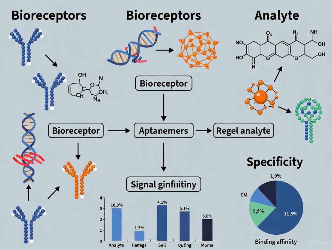
Bioreceptor Generation Workflows
The processes for generating antibodies and aptamers are fundamentally different, involving in vivo and in vitro systems, respectively.
Diagram 2: Bioreceptor generation workflows.
Both antibodies and aptamers serve as high-affinity receptors with the capacity to significantly enhance biosensor specificity. The choice between them is not a matter of superiority but of strategic application. Antibodies remain the established choice for many clinical applications due to their proven track record and high specificity. However, aptamers offer compelling advantages in stability, cost, modifiability, and target range, making them ideal for point-of-care diagnostics, environmental monitoring, and detecting novel or small-molecule targets. Future research in biosensor specificity will likely focus on leveraging the strengths of both receptors, potentially through hybrid approaches, and on overcoming the remaining challenges in aptamer commercialization and integration. As the toolkit for bioreceptor design expands, so too will the precision and capability of biosensors across healthcare, diagnostics, and biotechnology.
The performance of any biosensor is fundamentally governed by the specificity and affinity of its biorecognition element. Bioreceptors are the molecular components responsible for selectively binding to the target analyte, and their evolution is central to advancements in diagnostic sensitivity and specificity. Among the various bioreceptors available, nucleic acids and peptides represent two classes of emerging biomolecules that are redefining the frontiers of targeted detection. Their unique structural and functional properties, such as the programmable base-pairing of nucleic acids and the diverse molecular recognition capabilities of peptides, make them exceptionally suitable for developing next-generation biosensors [8] [17]. This whitepaper provides an in-depth technical guide to these emerging bioreceptors, framing their development and application within the broader thesis of achieving unparalleled specificity in biosensor research. It details the underlying mechanisms, presents comparative performance data, outlines experimental protocols, and visualizes key workflows to equip researchers and drug development professionals with the knowledge to leverage these tools effectively.
Nucleic Acid Bioreceptors: Programmable Probes for Precision Detection
Nucleic acid-based bioreceptors exploit the inherent, programmable specificity of Watson-Crick base pairing for the detection of complementary DNA, RNA, and specific non-nucleic acid targets.
Mechanisms and Types
The core mechanism involves the hybridization of a single-stranded nucleic acid probe (the bioreceptor) to its target sequence. This binding event is then transduced into a measurable signal [8]. Key types of nucleic acid bioreceptors include:
- DNA/RNA Probes: Used for the detection of complementary genetic sequences. They are fundamental in identifying pathogens, genetic mutations, and gene expression markers (e.g., microRNAs) [8].
- Aptamers: Single-stranded DNA or RNA oligonucleotides selected in vitro to bind with high affinity and specificity to a wide range of targets, including small molecules, proteins, and even whole cells [8] [18]. Their development is independent of biological systems, allowing for targets against which traditional antibodies are difficult to raise.
- CRISPR-based Systems: Adapted from bacterial immune systems, CRISPR-Cas systems provide both recognition and signal amplification. The Cas enzyme, guided by a CRISPR RNA (crRNA), cleaves a target nucleic acid sequence. This activity can be coupled with reporter molecules, enabling highly sensitive and specific detection, as demonstrated during the COVID-19 pandemic [8] [19].
Experimental Protocol: Electrochemical Aptasensor for NAD(H) Detection
The following protocol summarizes a representative methodology for constructing a nucleic acid-based biosensor, as detailed in recent literature [18].
- 1. Bioreceptor Immobilization: A thiol-modified DNA aptamer, specific for NAD(H), is chemisorbed onto a gold disk working electrode via gold-sulfur bonds. The electrode is typically incubated in a solution of the aptamer for several hours.
- 2. Surface Passivation: To minimize non-specific adsorption, the remaining gold surface is passivated with a self-assembled monolayer of a small-chain mercaptan, such as 6-mercapto-1-hexanol.
- 3. Electrochemical Measurement and Readout: The functionalized electrode is integrated into a standard three-electrode electrochemical cell. Detection is performed using electrochemical impedance spectroscopy (EIS) or a comparable technique. The binding of NAD(H) to the surface-immobilized aptamer causes a measurable change in charge transfer resistance (Rct) at the electrode-solution interface. The Rct value is proportional to the target concentration [18].
Performance Data for Nucleic Acid Bioreceptors
Table 1: Performance metrics of selected nucleic acid-based biosensors.
| Bioreceptor Type | Target Analyte | Transduction Method | Limit of Detection (LOD) | Assay Time | Key Application |
|---|---|---|---|---|---|
| DNA Aptamer [18] | NAD(H) | Electrochemical (EIS) | Low nM range | ~1-2 hours | Metabolic monitoring |
| CRISPR-cas System [19] | SARS-CoV-2 RNA | Photonic (Waveguide) | Single Molecule | ~30 minutes | Infectious disease diagnostics |
| Molecular Beacon [8] | microRNA | Fluorescence | Not Specified | Fast hybridization | Cancer biomarker detection |
Peptide-Based Bioreceptors: Versatile Molecular Recognizers
Peptide-based bioreceptors offer a combination of high stability and versatile molecular recognition capabilities, making them strong alternatives to traditional antibodies.
Mechanisms and Types
Peptides interact with their targets through a combination of electrostatic forces, hydrogen bonding, and hydrophobic interactions [20]. Their key forms include:
- Peptide Nucleic Acids (PNAs): Synthetic oligomers where the natural sugar-phosphate backbone is replaced by an N-(2-aminoethyl)-glycine backbone. PNAs are achiral, neutral, and exhibit high chemical and thermal stability. They hybridize with complementary DNA/RNA with higher affinity and specificity than natural nucleic acids, even discriminating single-base mismatches [17].
- Antimicrobial Peptides (AMPs): Naturally occurring peptides that form part of the innate immune response. They can be integrated into biosensors to detect whole pathogenic cells based on their affinity for microbial membranes [20].
- Synthetic Peptides from Phage Display: Peptides identified through biopanning of phage display libraries. This in vitro technique allows for the discovery of high-affinity peptide ligands for virtually any protein target [20].
Experimental Protocol: PNA-Based Electrochemical Biosensor for Single-Nucleotide Polymorphism (SNP) Detection
This protocol outlines the use of a PNA probe for the highly specific detection of a DNA point mutation [17].
- 1. PNA Probe Design and Immobilization: A PNA probe, complementary to the target DNA sequence encompassing the mutation site, is designed. The PNA is typically synthesized with a terminal amino or thiol group. A gold electrode is functionalized with a carboxylated SAM (e.g., mercaptoundecanoic acid). The PNA probe is then covalently immobilized onto the SAM via carbodiimide crosslinking chemistry.
- 2. Hybridization and Mismatch Discrimination: The functionalized electrode is exposed to a sample containing the target DNA. Hybridization is performed under low ionic strength conditions, which, due to the neutral PNA backbone, does not destabilize the PNA-DNA duplex, providing a key advantage over DNA probes. A single base mismatch in the target DNA results in a significant destabilization of the duplex, which is readily detected.
- 3. Electrochemical Readout: The hybridization event is detected using a redox-active mediator like methylene blue. The mediator's access to the electrode surface is hindered when a PNA-DNA duplex forms, leading to a measurable decrease in current in square wave voltammetry. The signal loss is directly correlated with the amount of perfectly matched target DNA hybridized [17].
Performance Data for Peptide Bioreceptors
Table 2: Performance metrics of selected peptide-based biosensors.
| Bioreceptor Type | Target Analyte | Transduction Method | Limit of Detection (LOD) | Assay Time | Key Application |
|---|---|---|---|---|---|
| PNA Probe [17] | Mutated DNA sequence | Electrochemical (Voltammetry) | High specificity for SNP | Fast hybridization | Genetic disorder screening |
| Antimicrobial Peptide [20] | E. coli O157:H7 | Optical (Colorimetric) | 10-100 CFU/mL | < 6 hours | Food safety monitoring |
| Phage Display Peptide [20] | Salmonella spp. | Electrochemical (Impedance) | 10 CFU/mL | ~2 hours | Pathogen detection |
The Scientist's Toolkit: Essential Research Reagent Solutions
The development and implementation of these advanced biosensors rely on a suite of specialized reagents and materials.
Table 3: Key reagents and materials for biosensor development based on nucleic acids and peptides.
| Reagent/Material | Function and Role in Biosensing |
|---|---|
| Gold Electrodes | Provides an excellent substrate for forming self-assembled monolayers (SAMs) for immobilizing thiol-modified nucleic acids or peptides [17] [18]. |
| Thiol/Mercaptan Chemistry | Enables the covalent attachment of bioreceptors (e.g., thiolated aptamers or PNAs) to gold surfaces. Shorter-chain mercaptans like 6-mercapto-1-hexanol are used for passivation [18]. |
| CRISPR-cas Reagents | Includes the Cas nuclease (e.g., Cas12a, Cas13) and custom-designed crRNA. These form the core of the biological recognition and amplification system for nucleic acid targets [19]. |
| High-Contrast Probes (e.g., Au NPs) | Gold nanoparticles (Au NPs) serve as labels in optical and electrochemical biosensors. Their cleavage, as in the HCCD technique, generates a strong, measurable signal contrast [19]. |
| Peptide Nucleic Acids (PNAs) | Synthetic bioreceptors that provide superior hybridization properties and stability against nucleases and proteases, crucial for detecting DNA/RNA targets in complex matrices [17]. |
| Redox Mediators (e.g., Methylene Blue) | Small molecules that shuttle electrons in electrochemical biosensors. Changes in their electrochemical behavior upon target binding form the basis of the detection signal [17]. |
Workflow and Mechanism Visualization
The following diagrams, generated using DOT language and compliant with the specified color and contrast rules, illustrate core concepts and experimental workflows.
CRISPR-Cas Enhanced Photonic Detection Workflow
PNA vs. DNA Hybridization Specificity
Nucleic acids and peptides have firmly established themselves as powerful and versatile bioreceptors, each offering distinct advantages for achieving high specificity in biosensing. Nucleic acids provide programmable, predictable recognition, exemplified by the revolutionary CRISPR-Cas technology. Peptides, particularly PNAs, offer exceptional stability and discrimination capabilities. The convergence of these bioreceptors with advanced transduction methods, nanomaterials, and computational design—such as the inverse design of photonic waveguides—is setting the stage for a new era of diagnostic tools [8] [19]. These tools are poised to meet the growing demands for precision medicine, point-of-care testing, and global health diagnostics, ultimately fulfilling the core thesis that the strategic selection and engineering of the bioreceptor is paramount to unlocking new levels of specificity and performance in biosensor research.
Biosensor performance is fundamentally governed by the biological recognition element (bioreceptor) which confers specificity for the target analyte. In complex biological samples—from serum and urine to environmental mixtures—the presence of numerous interfering components places extraordinary demands on this recognition capability. Traditional bioreceptors, particularly antibodies, face limitations including limited stability, high production costs, and restricted applicability for certain targets. Within this context, lectins and molecularly imprinted polymers (MIPs) have emerged as powerful complementary technologies that significantly expand the bioreceptor toolkit. Lectins provide natural carbohydrate recognition capabilities essential for glycan-based profiling, while MIPs offer synthetic antibody mimics with superior stability and customizability. This technical review examines the principles, applications, and implementation methodologies for these two distinct yet complementary bioreceptor classes, framing their development within the broader thesis that advancing bioreceptor technology is pivotal to achieving the specificity required for next-generation biosensing in real-world samples.
Lectin-Based Bioreceptors: Principles and Applications
Fundamental Recognition Mechanisms
Lectins are non-immunoglobulin proteins that recognize and reversibly bind to specific carbohydrate structures without altering them. This unique recognition capability stems from their specific affinity for glycan motifs present on cell surfaces, proteins, and other biological structures. In biosensing applications, lectins serve as cross-linkers that agglutinate cells or precipitate glycoconjugates by binding to their target sugar configurations, making them particularly valuable for detecting glycosylation patterns that serve as disease biomarkers [21] [6]. Their stability and specificity make them suitable for various transducer interfaces, where they can capture glycoconjugates through well-defined carbohydrate-protein interactions.
Performance Characteristics and Limitations
The implementation of lectins in biosensors presents distinct advantages and challenges that must be considered during assay design.
Table 1: Performance Characteristics of Lectin Bioreceptors
| Characteristic | Advantages | Limitations |
|---|---|---|
| Specificity | High specificity for carbohydrate motifs | May exhibit cross-reactivity with similar glycan structures |
| Stability | Generally robust; more stable than antibodies | Less stable than synthetic polymers like MIPs |
| Production Cost | Lower cost than monoclonal antibodies | Higher cost than synthetic MIPs |
| Sensitivity | Suitable for clinical detection ranges | Generally lower sensitivity compared to antibodies [22] |
| Application Scope | Ideal for glycan profiling and extracellular vesicle analysis | Limited to carbohydrate-containing targets |
As detailed in recent extracellular vesicle (EV) research, lectins enable precise detection of EV subpopulations by targeting specific surface glycans, supporting multiplexed and real-time analysis while preserving structural integrity [6]. However, their relatively lower sensitivity compared to antibodies remains a constraint in applications requiring ultra-trace detection [22].
Experimental Implementation Protocols
Protocol 1: Lectin Immobilization for Electrochemical Biosensors This standard protocol details the procedure for functionalizing transducer surfaces with lectin bioreceptors for carbohydrate detection [23].
- Surface Preparation: Clean electrode surfaces (gold, carbon, or ITO) via oxygen plasma treatment or piranha solution to generate reactive functional groups.
- Linker Application: Incubate with a crosslinker solution (e.g., glutaraldehyde or carbodiimide) for 1 hour at room temperature to create amine-reactive groups.
- Lectin Immobilization: Apply lectin solution (typically 0.1-1 mg/mL in phosphate buffer) and incubate for 12-16 hours at 4°C to preserve bioactivity.
- Surface Blocking: Treat with bovine serum albumin (BSA, 1% w/v) or ethanolamine to passivate unreacted sites and minimize non-specific binding.
- Validation: Confirm immobilization efficiency through electrochemical impedance spectroscopy (EIS) or quartz crystal microbalance (QCM) measurements.
Protocol 2: Lectin-Based EV Capture Assay This protocol outlines the specific application of lectins for isolating and analyzing extracellular vesicles from biofluids, a rapidly advancing application area [6].
- Sample Preparation: Isolate EVs from biofluids (serum, urine) using size-exclusion chromatography or ultrafiltration to remove interfering components.
- Microfluidic Functionalization: Pattern lectins (e.g., Con A, WGA) within microfluidic channels using covalent immobilization chemistry.
- EV Capture: Introduce prepared EV samples to the functionalized surface at controlled flow rates (typically 5-20 μL/min) to enable specific glycan-mediated capture.
- Signal Detection: Employ label-free detection methods such as surface plasmon resonance (SPR) or electrochemical impedance to quantify captured EVs.
- Characterization: Perform secondary analysis using antibody staining for specific tetraspanins (CD63, CD81) to confirm EV identity and subpopulations.
Figure 1: Lectin-Based Biosensing Mechanism. This diagram illustrates the molecular recognition interface where lectin bioreceptors, immobilized on a transducer surface, specifically bind to carbohydrate structures on target analytes such as extracellular vesicles, generating a measurable signal.
Molecularly Imprinted Polymers: Synthetic Recognition Elements
Fundamental Recognition Mechanisms
Molecularly imprinted polymers (MIPs) are synthetic biomimetic materials customized with target-oriented specificity, often termed "artificial antibodies" [24]. The core principle involves creating complementary cavities within a polymer network using template molecules, enabling highly specific recognition of target molecules during detection [25]. The MIP fabrication process represents a chemical evolution that mimics the immune response through three fundamental steps: (1) formation of a complex between the template and functional monomers via molecular interactions; (2) fixation of the functional monomers surrounding the template through cross-linking; and (3) removal of the template to yield imprinting cavities with shape complementarity and interactive sites [24]. This process creates synthetic recognition sites that rival the specificity of natural biological receptors while offering significantly enhanced stability.
Performance Characteristics and Limitations
MIPs present a compelling alternative to natural receptors, particularly in challenging analytical environments where stability and cost are significant factors.
Table 2: Performance Characteristics of Molecularly Imprinted Polymers
| Characteristic | Advantages | Limitations |
|---|---|---|
| Specificity | High template specificity; customizable | Potential cross-reactivity with structurally similar compounds |
| Stability | Excellent chemical/thermal stability; reusable | Brittleness in certain polymer formulations |
| Production Cost | Low cost and short preparation time | Optimization can be resource-intensive |
| Sensitivity | Can achieve high sensitivity with optimization | Sensitivity depends on template-monomer interaction strength |
| Application Scope | Broad applicability across molecular classes | Challenges with small gas molecule detection [22] |
MIPs demonstrate particular utility in diagnostics for cancers, viral diseases, and other pathologies where they enable precise recognition in complex biological samples such as serum and urine [24]. Their unique features include lower production cost, shorter preparation time, higher stability, reusability, and greater diversity compared to natural antibodies [24]. However, challenges remain in regulating selectivity and expanding recognition capabilities for novel targets.
Experimental Implementation Protocols
Protocol 3: Non-Covalent Molecular Imprinting This protocol, founded by Professor Klaus Mosbach, represents the most widely used approach for MIP synthesis, particularly suitable for small molecule targets [24] [25].
- Template-Monomer Complexation: Dissolve template molecule (0.1-1 mmol) and functional monomers (e.g., methacrylic acid, 4-vinylpyridine) in porogenic solvent (acetonitrile, toluene). Allow pre-assembly for 30-60 minutes through non-covalent interactions (hydrogen bonding, ionic interactions).
- Polymerization Initiation: Add cross-linker (ethylene glycol dimethacrylate, trimethylolpropane trimethacrylate) and thermal initiator (AIBN, 1% w/w). Purge with nitrogen or argon to remove oxygen.
- Polymerization: Heat at 60-70°C for 12-24 hours to form rigid, cross-linked polymer monolith.
- Template Extraction: Grind polymer and Soxhlet extract with methanol:acetic acid (9:1 v/v) for 24-48 hours to remove template molecules.
- Validation: Confirm template removal and binding capacity through HPLC or LC-MS analysis of extraction washes and subsequent rebinding experiments.
Protocol 4: MIP Nanoparticle Synthesis for Protein Recognition This advanced protocol addresses the challenge of creating MIPs for large biomolecular targets, which is essential for diagnostic applications [24].
- Epitope Approach: Identify and synthesize a peptide sequence representing a surface-exposed epitope of the target protein.
- Surface Functionalization: Modify silica nanoparticles (100-200 nm) with polymerizable groups (e.g., methacrylate silane) to create solid supports.
- Monomer Assembly: Mix epitope template with functional monomers (acrylamide, vinylphenylboronic acid) in aqueous buffer to establish specific interactions.
- Surface-Initiated Polymerization: Add cross-linker and initiator to form thin polymer shell around functionalized nanoparticles.
- Template Removal: Treat with denaturing conditions (SDS, acetic acid) to extract epitope templates while preserving complementary cavities.
- Characterization: Validate using dynamic light scattering, electron microscopy, and binding assays with fluorescently labeled target protein.
Figure 2: MIP Fabrication and Recognition Process. This workflow illustrates the key stages in creating molecularly imprinted polymers, from initial template-monomer complex formation through polymerization and template extraction to yield synthetic receptors with specific binding capabilities.
Comparative Analysis and Implementation Guidelines
Bioreceptor Selection Framework
Selecting between lectins and MIPs requires careful consideration of the analytical problem, sample matrix, and performance requirements. The following decision framework provides guidance for bioreceptor selection:
Table 3: Bioreceptor Selection Guide for Complex Sample Analysis
| Application Context | Recommended Bioreceptor | Rationale | Implementation Considerations |
|---|---|---|---|
| Glycan Profiling | Lectins | Natural specificity for carbohydrate structures | Combine multiple lectins for pattern recognition; optimize pH and ion dependence |
| Small Molecule Detection | MIPs | Excellent for <1000 Da molecules; high stability | Use dummy templates for toxic compounds; optimize monomer-template interactions |
| Complex Biological Samples | MIPs | Superior resistance to denaturation in serum/urine | Incorporate hydrophobic monomers for enhanced fouling resistance |
| Multiplexed Detection | Both | Complementary recognition capabilities | Employ spatial patterning techniques for array configurations |
| Point-of-Care Applications | MIPs | Enhanced shelf-life and thermal stability | Integrate with electrochemical transducers for portable detection |
| Budget-Constrained Projects | MIPs | Lower production costs and reusability | Balance development time against long-term usage needs |
Research Reagent Solutions
Successful implementation of lectin and MIP-based biosensing requires specific materials and reagents optimized for each technology.
Table 4: Essential Research Reagents for Lectin and MIP Biosensing
| Reagent Category | Specific Examples | Function | Technical Notes |
|---|---|---|---|
| Common Lectins | Concanavalin A (Con A), Wheat Germ Agglutinin (WGA) | Specific glycan recognition (mannose/glucose, N-acetylglucosamine) | Varying pH and ion requirements; check inhibition profiles |
| Functional Monomers | Methacrylic acid, Acrylamide, Vinylpyridine | Establish interactions with template molecules | Select based on template functional groups; computational screening available |
| Cross-linkers | Ethylene glycol dimethacrylate, N,N'-methylenebisacrylamide | Create rigid polymer matrix | Optimal cross-linker:monomer ratio typically 5:1 for proper cavity formation |
| Immobilization Chemistry | (3-Aminopropyl)triethoxysilane, Glutaraldehyde | Surface attachment of bioreceptors | Silicon photonic sensors require silane-based (not thiol) chemistry [26] |
| Signal Transducers | Interdigitated electrodes, Microring resonators, Screen-printed electrodes | Convert binding events to measurable signals | SiP sensors have ~40-200 nm evanescent field penetration depth [26] |
Lectins and MIPs represent complementary and rapidly evolving technologies that significantly expand the bioreceptor toolkit for complex sample analysis. While lectins provide unmatched capabilities for glycan profiling and extracellular vesicle characterization, MIPs offer unprecedented stability and customizability for diverse molecular targets. The future development of these technologies points toward several promising directions, including the creation of MIPs as Fab fragment mimics that retain selective recognition while removing non-functional segments [24], the establishment of specialized MIP databases to accelerate rational design, and the application of AI-assisted design tools for both MIPs and lectin-based assay optimization [24]. Furthermore, the integration of both technologies with advanced transducer platforms including silicon photonic sensors [26] and impedimetric biosensors [27] will enable new capabilities in multiplexed detection and point-of-care diagnostics. As the field progresses, the strategic combination of lectins and MIPs within integrated sensing platforms will undoubtedly overcome current limitations and unlock new possibilities for specific detection in the most challenging sample matrices.
The functional core of any biosensor is the analyte-bioreceptor interaction, a specific biochemical recognition event that confers the sensor's identity and dictates its performance. This interaction, central to a broader thesis on bioreceptor specificity, involves the selective binding of a target molecule (analyte) by a biological recognition element (bioreceptor) [1]. The fundamental mechanisms governing this interaction—ranging from molecular complementarity to binding kinetics—are the primary determinants of biosensor specificity, sensitivity, and overall reliability [23]. Within the context of modern biosensor research, which increasingly leverages artificial intelligence and nanotechnology, a deep understanding of these core principles is not merely academic but essential for the rational design of next-generation diagnostic tools for precision medicine and global health [28] [8]. This guide provides an in-depth technical examination of these key mechanisms, the methodologies for their study, and the advanced materials shaping their future.
Fundamental Principles of Analyte-Bioreceptor Interactions
The bio-recognition process is a specific binding event between a bioreceptor and its target analyte. This event is the primary source of a biosensor's selectivity, as the bioreceptor is designed to interact only with a particular analyte or a closely related group of analytes, even within complex sample matrices like blood or urine [1]. The subsequent conversion of this binding event into a quantifiable signal is the function of the transducer.
Core Components of a Biosensor
A typical biosensor comprises several key components that work in concert:
- Analyte: The substance of interest that requires detection (e.g., glucose, a viral antigen, a cancer biomarker) [1].
- Bioreceptor: A biological molecular species that specifically recognizes the analyte (e.g., enzyme, antibody, nucleic acid, aptamer) [1].
- Transducer: An element that converts the biological recognition event into a measurable signal (e.g., optical, electrochemical, piezoelectric) [1].
- Electronics: The system that processes the transduced signal (e.g., amplification, analog-to-digital conversion) [1].
- Display: The user interface that presents the final result in an interpretable format [1].
Types and Properties of Bioreceptors
Different bioreceptors leverage distinct biochemical principles to achieve specificity. Their inherent properties directly influence the design and application of the biosensor.
Table 1: Key Bioreceptor Types and Their Specificity Mechanisms
| Bioreceptor Type | Specificity Mechanism | Key Characteristics | Common Transduction Methods |
|---|---|---|---|
| Antibodies [1] | High-affinity binding to a specific antigenic epitope | Immunological; high specificity and diversity; can be monoclonal or polyclonal | Optical (SPR, RIfS), Electrochemical [29] |
| Enzymes [1] | Catalytic activity towards a specific substrate | Catalytic; generates a product; sensitivity can be affected by inhibitors | Electrochemical (amperometric), Optical |
| Nucleic Acids [8] | Base-pair complementary hybridization (e.g., DNA, RNA, aptamers) | Sequence-dependent; synthetic aptamers can bind proteins, cells, etc. | Optical (fluorescence), Electrochemical |
| Cells/Tissues [1] | Functional response to analytes (e.g., metabolites, toxins) | Utilizes intrinsic metabolic or signaling pathways | Thermal, Electrochemical |
| Aptamers [8] | Three-dimensional structure complementarity to a target (from small molecules to cells) | Synthetic oligonucleotides/peptides; high stability and selectivity | Optical, Electrochemical |
The critical performance characteristics of a biosensor, all of which are directly influenced by the analyte-bioreceptor interaction, include:
- Selectivity: The ability of the bioreceptor to detect a specific analyte in a sample containing adulterants and contaminants. This is the most critical feature of a biosensor [1].
- Sensitivity: The minimum detectable amount of analyte, also known as the limit of detection (LOD) [1].
- Stability: The degree to which the biosensor is susceptible to ambient disturbances, often linked to the degradation of the bioreceptor over time [1].
- Reproducibility: The ability to generate identical responses for a duplicated experimental setup, reflecting the precision and accuracy of the system [1].
The following diagram illustrates the fundamental workflow and core components involved in the analyte-bioreceptor interaction and signal generation within a biosensor.
Quantitative Analysis of Binding Kinetics
The interaction between an analyte (A) and a bioreceptor (R) to form a complex (AR) is governed by the law of mass action and can be described by the equation: A + R ⇌ AR The association rate constant (kₐₛₛ) and dissociation rate constant (kdᵢₛₛ) define the kinetics of this interaction, while the ratio kdᵢₛₛ/kₐₛₛ yields the equilibrium dissociation constant (K_D), a measure of affinity [29].
Fractal Dimension and Binding Kinetics
The surface geometry of the biosensor, where the bioreceptor is immobilized, is a primary parameter in determining reaction rates and performance [30]. Real-world biosensor surfaces are not smooth, and their heterogeneity can be quantitatively described by a fractal dimension (Df). A higher fractal dimension indicates a more heterogeneous surface. The binding rate coefficient (k) exhibits a power-law dependence on the fractal dimension, expressed as k = k' Df^p, where k' is a constant and p is the order of dependence [30]. This relationship highlights that surface roughness directly and significantly impacts the binding kinetics.
Table 2: Fractal Kinetics Analysis in Biosensor Binding
| Analyte-Receptor System | Analysis Type | Order of Dependence (p) | Impact on Binding Rate Coefficient (k) |
|---|---|---|---|
| 1 μM BSA to anti-BSA [30] | Single-fractal | 5.535 | Extreme sensitivity to surface heterogeneity |
| m-xylene to microorganism [30] | Single-fractal | 3.314 | High sensitivity to surface heterogeneity |
| PCR-amplified DNA to DNA capture protein [30] | Dual-fractal (k₂) | 3.399 | High sensitivity in later binding phase |
Methodologies for Kinetic Analysis
Quantitative biomolecular interaction analysis (BIA) requires careful experimental design and data evaluation to avoid artifacts such as mass transport limitation [29]. The one-to-one (1:1) interaction model, or pseudo-first-order kinetics, is commonly applied when the analyte flows over a surface with immobilized ligand, maintaining a constant analyte concentration [29].
Experimental Protocol: Determining kₐₛₛ and k_dᵢₛₛ via Reflectometric Interference Spectroscopy (RIfS) [29]
- Surface Functionalization: Immobilize the ligand (bioreceptor) on the transducer surface using amine-coupling chemistry. This involves cleaning and activating the transducer, modifying it with a silane (e.g., GOPTS), covalently binding a polymer layer (e.g., PEG), and finally coupling the ligand via its amino groups.
- Baseline Establishment: Flush the functionalized transducer surface with a running buffer (e.g., PBS, pH 7.4) until a stable baseline is achieved.
- Association Phase: Inject a series of analyte solutions at different known concentrations over the surface at a constant flow rate (e.g., 0.5 μL/s). Monitor the binding-induced change in optical thickness in real-time for a fixed period (e.g., 600 s).
- Dissociation Phase: Switch the flow to running buffer without analyte and monitor the decrease in signal as the analyte dissociates from the ligand for a fixed period (e.g., 900 s).
- Surface Regeneration: Inject a regeneration solution (e.g., 6 M guanidine hydrochloride, pH 1.5) to break the analyte-ligand bonds and prepare the surface for the next measurement cycle.
- Data Fitting: Fit the resulting sensorgrams (binding curves) for all concentrations globally using numerical integration algorithms to extract kₐₛₛ and k_dᵢₛₛ. This step avoids reliance on "black box" commercial software and ensures model correctness [29].
The following diagram outlines the key steps and decision points in this experimental workflow for kinetic analysis.
The Scientist's Toolkit: Research Reagent Solutions
The experimental study and application of analyte-bioreceptor interactions rely on a suite of specialized reagents and materials. The table below details key components used in the featured RIfS kinetic experiment and the broader field.
Table 3: Essential Research Reagents for Bioreceptor Immobilization and Analysis
| Research Reagent / Material | Function / Explanation | Experimental Context |
|---|---|---|
| 3-glycidyloxypropyl-trimethoxysilane (GOPTS) | A silane coupling agent that forms a self-assembled epoxy-functionalized layer on oxide surfaces, enabling subsequent covalent attachment of biomolecules [29]. | Surface functionalization of RIfS glass transducers [29]. |
| Poly(ethylene glycol) (PEG) | A polymer used as a spacer or barrier; reduces non-specific binding and can provide flexibility for oriented immobilization of bioreceptors [29]. | Polymer mixture (PEG-DA/PEG-MA) covalently bound to GOPTS layer [29]. |
| N-hydroxysuccinimide (NHS) / N,N'-diisopropyl-carbodiimide (DIC) | Cross-linking agents that activate carboxyl groups, allowing them to form stable amide bonds with primary amines on proteins or other ligands [29]. | Activated ester chemistry for covalent immobilization of the ligand (NRT) [29]. |
| Redox-Active Metal-Organic Frameworks (MOFs) [31] | Engineered porous crystalline materials that act as "wires" to facilitate efficient electron transfer between enzymes and electrodes in electrochemical biosensors. | Enhances reaction efficiency and long-term stability in enzyme-based electrochemical biosensors [31]. |
| Aquacyanocobinamide (ACCbi) [32] | A vitamin B12 derivative that serves as a colorimetric indicator; changes color from orange to violet upon interaction with hydrogen cyanide (HCN). | Bioreceptor and signal generator in a colorimetric biosensor for detecting Pseudomonas aeruginosa via its HCN biomarker [32]. |
| Metal Nanoparticles (e.g., Gold NPs) [28] [8] | Nanomaterials with unique optoelectronic properties; used for signal amplification and enhancing the immobilization of bioreceptors due to high surface-to-volume ratio. | Used in label-free immunosensors and for improving signal transmission in electrochemical biosensors [28] [8]. |
Advanced Materials and Future Perspectives
The pursuit of greater specificity and performance is driving innovation at the interface of chemistry, materials science, and biology.
- Nanomaterial-Enhanced Biosensors: The integration of nanomaterials like graphene, carbon nanotubes (CNTs), and MXenes is a key advancement. Their large surface area, excellent electrical conductivity, and tunable surface chemistry enhance bioreceptor loading, improve orientation, and amplify the transduced signal, thereby boosting both sensitivity and specificity [33] [28] [8].
- AI-Enhanced Interfacial Chemistry: Artificial intelligence (AI) and machine learning (ML) are revolutionizing biosensor design. AI models can predict optimal surface architectures, simulate analyte-receptor interactions at the atomic level, and analyze complex spectroscopic data to optimize functionalization strategies. This data-driven approach moves beyond traditional trial-and-error methods, accelerating the development of highly specific biosensing interfaces [28].
- Novel Transduction Platforms: Innovations such as CRISPR-based biosensors and advanced quartz crystal microbalance (QCM) systems are providing new pathways for achieving highly specific, real-time, and label-free detection of analytes, which is crucial for managing infectious diseases and cancer [8].
- Addressing Stability and Reproducibility: A significant challenge remains the stability of the bioreceptor layer and the reproducibility of its immobilization. Strategies such as the use of engineered metal-organic frameworks (MOFs) to entrap and stabilize enzymes, along with advanced polymer coatings and optimized cross-linking protocols, are being developed to enhance operational lifespan and measurement reliability [28] [31].
From Theory to Practice: Integrating Bioreceptors into Functional Sensing Platforms
The performance of a biosensor—its sensitivity, specificity, and stability—is profoundly dictated by the critical interface where biological recognition meets physical signal transduction. This interface is engineered through bioreceptor immobilization, the process of attaching biological recognition elements (such as antibodies, enzymes, nucleic acids, or aptamers) onto the surface of a transducer [1] [34]. The robustness of such sensors is significantly influenced by the ability to control the density and orientation of the bioreceptor at the surface, making the chemistry used for immobilization a paramount parameter [35]. Within the broader context of biosensor specificity research, the immobilization strategy is not merely a technical step but a fundamental determinant of the bioreceptor's functionality and accessibility, thereby directly impacting the sensor's analytical performance and reliability [36]. This guide provides an in-depth technical examination of prevalent immobilization techniques, their applications across different transducer materials, and the experimental protocols that underpin their successful implementation.
Foundational Immobilization Strategies
Several core methodologies are employed to conjugate bioreceptors to surfaces, each with distinct mechanisms, advantages, and limitations. The choice of method depends on the nature of the bioreceptor, the transducer material, and the intended application.
Table 1: Core Bioreceptor Immobilization Techniques
| Immobilization Technique | Mechanism of Attachment | Key Advantages | Key Limitations |
|---|---|---|---|
| Physical Adsorption [37] | Weak bonds (Van der Waals, electrostatic, hydrophobic) | Simple, inexpensive, less destructive to bioreceptor activity | Poor reproducibility, prone to leakage and desorption, non-specific binding |
| Covalent Bonding [35] [37] | Formation of robust covalent bonds between functional groups on the bioreceptor and the activated surface | Stable complexes, strong binding, high uniformity, good control over density | Can affect bioreceptor activity, requires additional reagents and surface activation |
| Entrapment [37] | Bioreceptor physically confined within a polymeric matrix (e.g., hydrogel, polymer) | High stability, minimizes leaching of bioreceptor | Gel matrix can hinder substrate diffusion, low loading capacity |
| Cross-linking [37] | Intermolecular covalent bonds formed between bioreceptors using a cross-linking reagent (e.g., glutaraldehyde) | Forms a stable 3D complex, improves efficiency and stability | Can lead to significant loss of activity due to harsh modification |
| Affinity Binding [35] [36] | High-affinity biological interactions (e.g., avidin-biotin, His-tag chelation) | Controlled orientation, reversible under certain conditions, high specificity | Requires genetic or chemical modification of the bioreceptor, additional cost |
Material-Specific Immobilization Chemistries
The chemical composition of the transducer surface dictates the most effective immobilization pathways. Selecting the appropriate chemistry for the material is crucial for creating a stable and functional biosensor.
Metallic Surfaces (Gold, Platinum)
- Gold Surfaces: The most extensively studied strategy for gold electrodes is the formation of a self-assembled monolayer (SAM) using thiolated bioreceptors [35]. The strong gold-sulfur bond allows for the creation of a well-ordered, dense monolayer. Alternative strategies include the use of poly adenine (polyA) motifs as anchoring tags, which leverage the affinity of adenine for gold, or physical adsorption through electrostatic interactions [35].
- Platinum and Other Metals: While less common than gold, platinum electrodes can be functionalized using molecules like isocyanide to form SAMs, analogous to thiol-on-gold chemistry [35].
Carbon-Based Surfaces
Carbon materials (glassy carbon, graphene, carbon nanotubes) possess complex surface chemistry and a wide working potential window [35]. Immobilization often requires initial surface activation to introduce functional groups. A widely used method involves the electrochemical reduction of aryldiazonium salts, which form robust covalent bonds with carbon surfaces, creating a layer that can be further functionalized with bioreceptors [35]. Another common approach is the activation of native carboxylic acid groups on carbon surfaces using carbodiimide chemistry (e.g., EDC/NHS) to form amide bonds with amine-containing bioreceptors [8].
Universal and Assisted Strategies
- Avidin-Biotin System: This is a versatile affinity strategy applicable to most materials. Avidin (or its analogues streptavidin/neutravidin) can physically adsorb to various surfaces, providing a universal anchor for any biotinylated bioreceptor [35]. This system offers strong binding and controlled orientation.
- Extra Structure-Assisted Immobilization: Polymers, hydrogels, nanoparticles, and magnetic beads can be used on top of an electrode to increase the surface area or introduce more functional groups, thereby enhancing sensitivity and bioreceptor loading capacity [35]. For instance, conducting polymers like polyaniline and polypyrrole have shown great potential in electrochemical sensing by improving electron transport and providing a matrix for bioreceptor entrapment or covalent attachment [8] [38].
Experimental Protocols for Key Techniques
Protocol: Covalent Immobilization of an Antibody on a Gold Electrode via SAM
This is a classic and highly reliable method for creating a stable biosensing interface [35] [37].
- Surface Cleaning: Clean the gold electrode surface thoroughly with piranha solution (a 3:1 mixture of concentrated sulfuric acid and 30% hydrogen peroxide) or via electrochemical cycling in sulfuric acid. Caution: Piranha solution is extremely corrosive and must be handled with extreme care.
- SAM Formation: Incubate the clean, dry gold electrode in a 1-10 mM solution of a thiolated molecule (e.g., 11-mercaptoundecanoic acid) in ethanol for 12-24 hours. This forms a SAM with terminal carboxylic acid groups.
- Surface Activation: Rinse the electrode with ethanol and water to remove physically adsorbed thiols. Then, immerse the electrode in an aqueous solution containing a carbodiimide cross-linker, most commonly a mixture of 1-Ethyl-3-(3-dimethylaminopropyl)carbodiimide (EDC) and N-Hydroxysuccinimide (NHS), for 30-60 minutes. This activates the carboxylic acid groups to form amine-reactive NHS esters.
- Bioreceptor Conjugation: Rinse the electrode to remove excess EDC/NHS. Incubate the activated surface with a solution of the antibody (typically 10-100 µg/mL in a mild buffer like PBS, pH 7.4) for 1-2 hours. The primary amine groups (lysine residues) on the antibody will form stable amide bonds with the NHS esters on the surface.
- Surface Passivation (Backfilling): To minimize non-specific adsorption, incubate the functionalized electrode with a solution of a passivating agent (e.g., 1-2 mM 6-mercapto-1-hexanol) for at least 1 hour. This step displaces any remaining non-specific adsorption sites and creates a non-fouling background.
- Storage: The prepared biosensor should be stored in a suitable buffer (e.g., PBS) at 4°C until use.
Protocol: Entrapment of an Enzyme within a Polypyrrole Hydrogel
This method is particularly useful for creating stable, 3D matrices for enzymatic biosensors [37] [38].
- Solution Preparation: Prepare an aqueous solution containing the enzyme (e.g., glucose oxidase), the monomer (e.g., 0.1 M pyrrole), and a supporting electrolyte (e.g., 0.1 M KCl).
- Electropolymerization: Immerse the working electrode (e.g., a carbon or platinum electrode) into the prepared solution. Apply a constant potential or use cyclic voltammetry to initiate the electrochemical polymerization of pyrrole. As the polypyrrole film grows on the electrode surface, the enzyme molecules become physically entrapped within the developing polymer matrix.
- Rinsing and Conditioning: After polymerization, remove the electrode and rinse it thoroughly with buffer to remove any unentrapped enzyme and monomer residue.
- Storage: Store the biosensor in an appropriate buffer at 4°C.
Table 2: Essential Research Reagent Solutions for Immobilization Protocols
| Reagent/Material | Function in Immobilization | Common Examples / Notes |
|---|---|---|
| Thiolated Linkers [35] | Forms a self-assembled monolayer (SAM) on gold surfaces, providing a foundation for further conjugation. | 11-mercaptoundecanoic acid, 16-mercaptohexadecanoic acid |
| Carbodiimide Cross-linkers [8] [9] | Activates carboxylic acid groups on surfaces or bioreceptors to facilitate amide bond formation with amines. | EDC (1-Ethyl-3-(3-dimethylaminopropyl)carbodiimide) |
| N-Hydroxysuccinimide (NHS) [8] [9] | Used with EDC to form a more stable, amine-reactive ester intermediate, improving coupling efficiency. | Sulfo-NHS (water-soluble variant) is also commonly used. |
| Glutaraldehyde [37] | A homobifunctional cross-linker that reacts with amine groups, used for cross-linking proteins or anchoring to aminated surfaces. | Can lead to loss of activity; concentration and reaction time must be optimized. |
| Avidin/Streptavidin [35] | A protein that binds with extremely high affinity to biotin, used in universal affinity-based immobilization. | Neutravidin is another common variant with lower non-specific binding. |
| Biotinylation Reagents | Chemically modifies bioreceptors (proteins, nucleic acids) with a biotin tag for subsequent avidin-based capture. | NHS-PEG4-Biotin, maleimide-PEG-biotin (for thiol groups) |
| Aryldiazonium Salts [35] | Forms a robust covalent layer on carbon-based electrodes, which can be pre-functionalized with desired groups. | 4-Nitrobenzenediazonium tetrafluoroborate |
Optimization and Performance Metrics
Successful immobilization is not just about attaching the bioreceptor; it is about preserving its function and maximizing sensor performance.
- Controlling Probe Density and Orientation: Overcrowded surface coverage can lead to steric hindrance, reducing hybridization efficiency and accessibility for larger analytes [35]. Probe density can be controlled by varying the concentration during immobilization and using backfilling agents. Orientation can be improved by using site-specific conjugation, such as biotin tagging at the Fc region of an antibody or using engineered cysteine residues.
- Surface Passivation: This is a critical step to prevent non-specific binding of non-target molecules to the electrode surface, which causes fouling and false signals [35]. Common passivating agents include bovine serum albumin (BSA), casein, and small mercaptoalkanol molecules (for gold surfaces).
- Key Performance Indicators: The success of an immobilization protocol is evaluated by the resulting biosensor's performance, including its sensitivity (limit of detection), selectivity (against interferents), linearity (dynamic range), reproducibility, and stability over time [1] [34].
The following workflow diagram summarizes the logical decision-making process for selecting and optimizing an immobilization strategy.
Figure 1. Immobilization Strategy Workflow - A logical flowchart for selecting and optimizing a bioreceptor immobilization strategy, highlighting the iterative nature of biosensor development.
The conjugation of bioreceptors to transducer surfaces is a sophisticated and critical discipline within biosensor research. The choice of immobilization technique—be it physical adsorption, covalent bonding, entrapment, or affinity-based binding—directly governs the analytical specificity, sensitivity, and operational stability of the final device. As the field advances towards multiplexed, point-of-care, and continuous monitoring diagnostics, the precision and robustness of these surface engineering strategies will only grow in importance. Future developments will likely focus on novel materials, smarter stimuli-responsive interfaces, and even more controlled bioorthogonal chemistries to further enhance the role of immobilization in achieving ultimate biosensor specificity.
Electrochemical biosensors have emerged as a cornerstone technology in modern analytical science, particularly for applications demanding rapid, sensitive, and specific detection of target analytes in complex biological matrices. The core of these devices lies in the bioreceptor, a biological or biomimetic recognition element that confers unparalleled specificity by selectively binding to a target molecule. This specific interaction occurs without the need for labels, such as fluorescent or radioactive tags, defining the powerful paradigm of label-free detection [23] [39]. The significance of bioreceptors extends beyond simple binding; they are the fundamental component that determines the analytical performance, robustness, and practical applicability of the entire biosensing system. Within the broader context of biosensor specificity research, understanding the structure-function relationship, immobilization techniques, and operational stability of various bioreceptors is paramount for advancing the field toward reliable point-of-care diagnostics and continuous monitoring devices [23] [40].
The working principle of an electrochemical biosensor hinges on the transduction of a biorecognition event into a quantifiable electrical signal. When a bioreceptor captures its target analyte, it induces a physicochemical change at the surface of an electrode. This change, which can be an alteration in charge distribution, mass, or conductivity, is then measured using electrochemical techniques such as electrochemical impedance spectroscopy (EIS), amperometry, or potentiometry [41] [39]. The absence of a label simplifies the assay design, reduces preparation time and cost, and allows for direct, real-time monitoring of binding events. This makes label-free electrochemical biosensors exceptionally suited for a wide range of applications, from clinical diagnosis of infectious diseases and cancer to environmental monitoring and food safety control [41] [7]. The continuous evolution of bioreceptor engineering and material science promises to further overcome existing challenges in sensitivity, stability, and multiplexing, solidifying their role in the future of analytical devices.
Bioreceptor Types and Their Mechanisms
Bioreceptors are the linchpin of biosensor specificity, and their selection is critical for assay design. The following table summarizes the key characteristics of major bioreceptor classes used in label-free electrochemical biosensing.
Table 1: Comparison of Bioreceptors for Label-Free Electrochemical Biosensing
| Bioreceptor Type | Composition & Origin | Recognition Principle | Key Advantages | Inherent Challenges |
|---|---|---|---|---|
| Antibodies [41] [39] | Proteins (Immunoglobulin G, etc.); produced by immune systems. | High-affinity, lock-and-key binding to specific antigens (proteins, etc.). | Exceptional specificity and high binding affinity. | Susceptible to denaturation; limited shelf-life; large size can hinder dense immobilization. |
| Aptamers [41] [7] | Short, single-stranded DNA or RNA oligonucleotides; selected in vitro (SELEX). | Folding into distinct 3D structures that bind targets (ions, small molecules, proteins). | Small size, thermal stability, ease of chemical synthesis and modification. | In-vitro selection process can be time-consuming; potential susceptibility to nuclease degradation. |
| Enzymes [41] [42] | Proteins; natural biological catalysts. | Catalytic conversion of a specific substrate; detection of reaction products or consumed co-factors. | Inherent signal amplification through catalytic turnover. | Sensitivity to environmental conditions (pH, temperature); activity can be inhibited. |
| Nucleic Acids (DNA/RNA) [41] [42] | Single-stranded DNA or RNA probes. | Complementary base-pairing (hybridization) with a target nucleic acid sequence. | High predictability of interactions; stable and cost-effective to produce. | Typically limited to nucleic acid targets unless combined with aptamers. |
| Peptides [43] [6] | Short sequences of amino acids; derived from natural proteins or designed in silico. | Molecular interaction via specific amino acid side chains with the target. | Smaller than antibodies; more stable; design flexibility. | Can exhibit lower binding affinity compared to antibodies. |
| Molecularly Imprinted Polymers (MIPs) [39] [40] | Synthetic polymers with tailor-made cavities. | Affinity binding to a template molecule used during polymerization (a "plastic antibody"). | High chemical and thermal stability; cost-effective production. | Can suffer from heterogeneity in binding sites and lower specificity in complex biological fluids. |
The following diagram illustrates the general workflow for selecting and integrating a bioreceptor into a functional biosensor platform, from initial design to signal generation.
Principles of Label-Free Electrochemical Transduction
Label-free electrochemical biosensing operates by directly measuring the electrochemical changes that occur when a bioreceptor binds its target analyte at the electrode-solution interface. Unlike labeled methods, this approach avoids additional reagents and steps, enabling direct, real-time monitoring of the binding event [41] [39]. The key transduction mechanisms include electrochemical impedance spectroscopy (EIS), field-effect transduction, and techniques based on blocking electron transfer.
Electrochemical Impedance Spectroscopy (EIS) is arguably the most prominent technique for label-free detection. It measures the impedance (resistance to current flow) of the electrode interface, often represented by the charge transfer resistance (Rₜ). When a bioreceptor on the electrode surface captures a target molecule (e.g., an antibody binding a protein, or a DNA probe hybridizing with its complement), it creates a physical and electrostatic barrier. This barrier hinders the flow of redox probes (e.g., ³⁻/⁴⁻) in the solution to the electrode surface, leading to an increase in Rₜ that can be quantitatively correlated with the target concentration [41] [7]. EIS is highly sensitive to surface modifications and is particularly effective for detecting larger analytes like proteins, cells, and nucleic acids.
Field-Effect Transduction forms the basis of devices like field-effect transistors (FETs). Here, the bioreceptor is immobilized on the gate of the transistor. The binding of a charged target analyte alters the local electric field, which in turn modulates the conductance of the transistor channel. This change in conductance can be measured as a shift in the source-drain current, providing a highly sensitive signal that is directly related to the analyte concentration [41]. This method is exceptionally suited for detecting charged molecules and offers potential for miniaturization and integration into high-density arrays.
A third common principle involves Blocking Access to the Electrode Surface. In this mechanism, the binding of the target analyte physically blocks the access of a redox probe to the electrode, resulting in a measurable decrease in the Faradaic current in techniques such as amperometry or cyclic voltammetry [41]. The following diagram illustrates these three primary signaling mechanisms.
Experimental Protocol: Developing a Peptide-Based Cortisol Sensor
This protocol details the development of an electrochemical biosensor using a computationally designed peptide bioreceptor for the label-free detection of cortisol, a steroid hormone stress biomarker. It serves as a representative template for de novo bioreceptor integration [43].
In Silico Bioreceptor Design and Screening
- Candidate Protein Identification: Search the Protein Data Bank (PDB) using keywords related to the target (e.g., "cortisol-binding"). Select proteins with known structures and filter out duplicates based on high sequence similarity scores (e.g., >75% via BLAST) [43].
- Molecular Docking for Primary Screening:
- Software: AutoDock Vina.
- Preparation: Process candidate protein files (remove water, add hydrogens and Kollman charges) and the target ligand file. Save in PDBQT format.
- Execution: Perform blind docking by placing the ligand in a grid box large enough to enclose the entire protein. Set an appropriate exhaustiveness level.
- Analysis: Rank the candidate proteins based on the computed binding affinity (score in kcal/mol). Select the top candidates (e.g., top 3) for further validation [43].
- Validation via Steered Molecular Dynamics (MD):
- Software: GROMACS.
- System Setup: Solvate the top protein-ligand complexes in a simulation box with a solvent model that mimics the intended sample matrix (e.g., an eccrine sweat model with 50 mM NaCl for a wearable sensor). Use an appropriate force field (e.g., CHARMM36).
- Umbrella Sampling: Perform a constant-velocity pull simulation to separate the ligand from the protein along a defined reaction coordinate. Use multiple simulation windows with harmonic restraints.
- Free Energy Calculation: Use the Weighted Histogram Analysis Method (WHAM) on the collected trajectories to reconstruct the Potential of Mean Force (PMF). The PMF profile provides the binding free energy, validating the stability and affinity of the complex [43].
- Peptide Sequence Extraction: From the binding pocket of the top-validated protein, select a contiguous sequence of amino acids that shows favorable interactions with the target ligand. Optionally, introduce a cysteine residue at the N-terminus to facilitate subsequent oriented immobilization on gold electrodes via a gold-thiol bond [43].
Sensor Fabrication and Electrochemical Measurement
- Electrode Preparation: Clean the working electrode (e.g., gold disk or screen-printed gold electrode) sequentially with alumina slurry and solvents (ethanol, deionized water) under sonication.
- Peptide Immobilization: Incubate the cleaned electrode with a solution of the synthetic cysteine-modified peptide (e.g., 1-10 µM) in a suitable buffer for several hours to form a self-assembled monolayer (SAM) via thiol-gold chemistry. Rinse thoroughly to remove physically adsorbed peptides.
- Blocking: Incubate the modified electrode with a passivating agent (e.g., 6-mercapto-1-hexanol or bovine serum albumin) to block any remaining bare gold surface and minimize non-specific adsorption.
- Label-Free EIS Measurement:
- Setup: Use a standard three-electrode system (peptide-modified Au working electrode, Pt counter electrode, Ag/AgCl reference electrode) in a solution containing a redox probe (e.g., 5 mM ³⁻/⁴⁻ in PBS).
- Baseline Measurement: Record the EIS spectrum (e.g., frequency range 0.1 Hz to 100 kHz, AC amplitude 10 mV) before analyte addition. The Nyquist plot will show a specific semicircle diameter corresponding to the Rₜ of the peptide-modified interface.
- Analyte Detection: Incubate the electrode with the sample containing the target analyte (cortisol) for a fixed duration. Rinse gently.
- Post-Binding Measurement: Record the EIS spectrum again in the fresh redox probe solution. The specific binding of the target to the peptide bioreceptor will create a barrier, increasing the Rₜ.
- Quantification: Plot the change in Rₜ (ΔRₜ) or the normalized Rₜ against the logarithm of the analyte concentration to generate a calibration curve.
Performance Analysis of Bioreceptor-Based Biosensors
The analytical performance of label-free electrochemical biosensors is critically dependent on the choice of bioreceptor and the sensor design. The following table compiles representative performance metrics from recent research for different bioreceptor classes, highlighting their capabilities in detecting various analytes.
Table 2: Analytical Performance of Representative Label-Free Electrochemical Biosensors
| Target Analyte | Bioreceptor Type | Electrode & Transduction Method | Linear Range | Limit of Detection (LOD) | Key Application/ Matrix |
|---|---|---|---|---|---|
| Cortisol [43] | Engineered Peptide | Gold Electrode / EIS | (Theoretical) 8–140 ng/mL (sweat range) | (Validated via PMF) | Stress monitoring (Sweat) |
| PSA (Cancer) [41] | Antibody | Au Nanofiber-Modified SPCE / Amperometry | 0 to 100 ng/mL | 0.28 ng/mL (8.78 fM) | Cancer Diagnostics |
| Endotoxin [41] | Aptamer | MOF/Ag-P-N-CNT Nanohybrid / Amperometry | 1 fg/mL to 100 ng/mL | 0.55 fg/mL | Pathogen Detection |
| Dopamine [41] | Fe/N-doped Graphene (Enzyme-free) | Fe/N-GR Modified Electrode / Not Specified | 50 pM – 15 nM | 27 pM | Neurotransmitter Sensing |
| BRCA-1 Protein [44] | Antibody | AuNP/MoS₂ Nanocomposite / Impedimetric | 0.05 – 20 ng/mL | 0.04 ng/mL | Cancer Diagnostics (Serum) |
| Pathogenic Bacteria [7] | Various (Aptamer, Antibody, Phage) | Bioreceptor-Modified Electrode / EIS & Amperometry | Varies by specific sensor | Down to single bacterium [7] | Food Safety & Clinical |
The Scientist's Toolkit: Essential Research Reagents and Materials
The development and fabrication of a reliable label-free electrochemical biosensor require a suite of specialized materials and reagents. The following table lists key components essential for researchers in this field.
Table 3: Essential Research Reagent Solutions for Biosensor Development
| Category & Item | Specific Examples | Function & Rationale |
|---|---|---|
| Bioreceptors | Anti-PSA antibody [41], cortisol-binding peptide [43], endotoxin-binding aptamer [41] | The core recognition element that provides specificity for the target analyte. |
| Electrode Materials | Screen-printed carbon electrodes (SPCE) [41], Gold disk electrodes [43], Indium Tin Oxide (ITO) [44] | The solid support serving as the transducer. SPCEs are low-cost and disposable; Au allows for robust thiol-based chemistry. |
| Nanomaterials for Signal Enhancement | Gold nanoparticles (AuNPs) [41] [44], Graphene/Fe/N-doped graphene [41], Molybdenum Disulfide (MoS₂) [44] | Increase the active surface area, improve electron transfer kinetics, and provide sites for bioreceptor immobilization. |
| Immobilization Chemistries | Thiol-modified DNA/peptides (for Au) [43] [39], EDC-NHS chemistry (for carboxyl groups), Glutaraldehyde (for amine groups) | Covalently anchor bioreceptors to the electrode surface in a controlled and stable orientation. |
| Redox Probes | Potassium ferricyanide/ferrocyanide ([Fe(CN)₆]³⁻/⁴⁻) [39] | A standard redox couple used in EIS and voltammetry to probe the impedance or electron transfer efficiency of the modified electrode interface. |
| Passivating Agents | 6-Mercapto-1-hexanol (MCH) [43], Bovine Serum Albumin (BSA) | Block unmodified electrode surfaces to prevent non-specific adsorption of interfering species, thereby improving signal-to-noise ratio. |
| Software & Data Analysis | AutoDock Vina [43], GROMACS [43], Principal Component Analysis (PCA), Support Vector Machine (SVM) [45] | Tools for in-silico bioreceptor design, molecular dynamics validation, and processing complex electrochemical or spectral data. |
Bioreceptors are the definitive element that unlocks the full potential of label-free electrochemical biosensors. From the high specificity of antibodies and the robustness of aptamers to the design flexibility of peptides and the stability of MIPs, the choice of bioreceptor directly dictates the sensor's analytical performance and practical applicability. As research continues to overcome challenges related to stability in complex matrices and reproducible mass fabrication, the integration of advanced nanomaterials and sophisticated computational design tools like molecular docking and machine learning will further propel this field forward. The ongoing refinement of bioreceptor technology is poised to yield a new generation of biosensors that are not only highly sensitive and specific but also robust, affordable, and seamlessly integrated into point-of-care and wearable formats, ultimately transforming personalized healthcare and environmental monitoring.
Optical biosensors represent a powerful class of analytical devices that integrate a biological recognition element with an optical transducer system to detect biological and chemical substances. These devices provide significant advantages over conventional analytical techniques, including direct, real-time, and label-free detection of a wide range of analytes with high specificity and sensitivity [46]. The core functionality of any biosensor, including optical platforms, depends critically on the bioreceptor component, which confers molecular recognition capabilities and determines the fundamental specificity of the device. Bioreceptors are biological or biomimetic molecules that selectively bind to target analytes, and include antibodies, enzymes, nucleic acids, peptides, aptamers, and molecularly imprinted polymers [47] [48].
The integration of sophisticated optical transduction techniques, particularly Surface Plasmon Resonance (SPR) and Surface-Enhanced Raman Spectroscopy (SERS), with highly specific bioreceptors has revolutionized biomolecular interaction analysis. These combined platforms enable researchers to investigate molecular binding events in real-time without requiring labels, while maintaining the exquisite specificity afforded by biological recognition elements. This technical guide explores the fundamental principles, operational mechanisms, and experimental methodologies for combining bioreceptors with SPR and SERS platforms, with particular emphasis on how bioreceptor engineering directly impacts biosensor specificity in research applications.
Fundamental Principles of SPR and SERS Technologies
Surface Plasmon Resonance (SPR) Physics
Surface Plasmon Resonance is an optical phenomenon that occurs when surface plasmons—collective oscillations of free electrons at a metal-dielectric interface—are excited by incident light. This phenomenon was first successfully used to construct an SPR-based sensor for biomolecular interactions in 1983, with the first commercial SPR instrument launched by Biacore [46]. The underlying physics involves the resonance condition that is highly sensitive to changes in the refractive index at the metal surface, typically within approximately 200 nanometers from the surface [46].
The SPR effect is most commonly generated using the Kretschmann configuration, where light is directed through a prism onto a thin metal film (typically gold or silver). When the momentum of the incident light matches that of the surface plasmons, energy is transferred to the plasmons, resulting in a sharp dip in the reflected light intensity at a specific angle of incidence known as the resonance angle [49]. When biomolecular binding occurs on the functionalized metal surface, the local refractive index changes, causing a measurable shift in the resonance angle. This shift can be monitored in real-time to quantify binding kinetics, affinity, and concentration [50] [46].
Several SPR modalities have been developed to enhance sensitivity and resolution:
- Conventional SPR: Utilizes a single metal layer (typically 50 nm gold) on a prism substrate [49].
- Long-Range SPR (LRSPR): Incorporates a dielectric buffer layer between the prism and metal layer to achieve sharper resonance dips and enhanced resolution [49].
- Coupled Plasmon-Waveguide Resonance (CPWR): Adds a waveguide layer beneath the metal film, enabling monitoring of both transverse magnetic (TM) and transverse electric (TE) modes for anisotropic measurements [49].
- Waveguide-Coupled SPR (WCSPR): Employs two metal layers with a waveguide layer to combine SPR and waveguide modes for sharper resonance features [49].
- Localized Surface Plasmon Resonance (LSPR): Utilizes metallic nanostructures rather than continuous metal films, where plasmon oscillations are confined to nanoparticles, resulting in distinct UV-Vis absorption bands that shift upon binding events [46] [51].
Surface-Enhanced Raman Spectroscopy (SERS) Mechanisms
Surface-Enhanced Raman Spectroscopy is a powerful analytical technique that dramatically enhances Raman scattering signals from molecules adsorbed on specially designed nanostructured surfaces. Since its discovery in 1974 and subsequent naming in 1977, SERS has evolved into a sophisticated biosensing platform capable of single-molecule detection [52] [51]. The tremendous enhancement of Raman signals in SERS arises from two primary mechanisms:
- Electromagnetic Enhancement (EM): This dominant mechanism involves the amplification of electromagnetic fields occurring at "hot spots"—nanoscale gaps between metallic nanostructures (typically gold, silver, or copper) where surface plasmons are localized. When incident light matches the surface plasmon resonance frequency of these nanostructures, the electromagnetic field can be enhanced by several orders of magnitude. Since Raman scattering intensity is proportional to the fourth power of the field enhancement (EF ∝ |E|⁴), this results in enhancement factors reaching 10¹⁴, enabling single-molecule detection [52].
- Chemical Enhancement (CM): This secondary mechanism involves charge transfer between the analyte molecules and the metal surface, which alters the polarizability of the molecules and increases their Raman scattering cross-sections. While chemical enhancement typically provides more modest improvement (10-10³ fold), it contributes to the overall SERS effect and can provide molecular specificity [52].
SERS biosensing can be implemented through two primary approaches:
- Label-free SERS: Directly captures the intrinsic molecular "fingerprint" of analytes interacting with the SERS substrate [52].
- Label-based SERS: Utilizes Raman reporter molecules with strong, characteristic signals as SERS tags for indirect detection, offering higher accuracy for semi-quantitative analysis [52].
Bioreceptor Integration with SPR and SERS Platforms
Bioreceptor Types and Immobilization Strategies
The critical interface between optical transduction platforms and target analytes is established through carefully selected and immobilized bioreceptors. Different bioreceptor classes offer distinct advantages for specific applications:
Table 1: Bioreceptor Types for Optical Biosensors
| Bioreceptor Type | Specificity Basis | Key Advantages | Common Immobilization Methods |
|---|---|---|---|
| Antibodies | Antigen-antibody recognition | High specificity and affinity | NHS-amine coupling, photochemical immobilization, physical adsorption |
| Aptamers | Three-dimensional structure complementarity | Enhanced stability, versatility, flexibility | Thiol-gold chemistry, biotin-streptavidin |
| Nucleic Acids | Base pairing complementarity | Programmable, predictable binding | Thiol modification, amine modification |
| Enzymes | Substrate specificity | Catalytic amplification | Entrapment, covalent attachment |
| Peptides | Structural motifs | Small size, chemical diversity | Thiol-gold, NHS chemistry |
| Molecularly Imprinted Polymers (MIPs) | Artificial recognition cavities | High stability, reusability | Electropolymerization, in situ polymerization |
| Lectins | Carbohydrate recognition | Glycosylation pattern analysis | NHS chemistry, physical adsorption |
The performance of a biosensor is highly influenced by the immobilization technique, which must preserve bioreceptor functionality while ensuring optimal orientation and density [47]. Common immobilization techniques include:
- Physical Adsorption: Relies on non-specific interactions (hydrophobic, ionic) between bioreceptors and the substrate surface. While simple to implement, this method may lead to random orientation and potential leaching [47].
- Covalent Bonding: Creates stable bonds between functional groups on the bioreceptor (e.g., amines, thiols) and activated surface groups. Gold-thiol interactions are particularly common for aptamer immobilization on gold surfaces [47].
- Affinity Interactions: Utilizes high-affinity pairs like biotin-streptavidin for oriented immobilization, preserving binding sites [46].
- Entrapment Methods: Encapsulates bioreceptors within polymer matrices (e.g., polypyrrole films via electropolymerization) [47].
- Self-Assembled Monolayers (SAMs): Creates well-ordered organic surfaces terminated with specific functional groups for controlled bioreceptor attachment [46].
For SPR platforms, the sensor chip typically contains a functional layer enabling immobilization, with carboxymethylated dextran layers activated via NHS/EDC chemistry being particularly common for protein immobilization [46].
Enhancing Specificity Through Bioreceptor Engineering
The specificity of optical biosensors can be significantly enhanced through strategic bioreceptor engineering approaches:
- Aptamer Selection: Through Systematic Evolution of Ligands by Exponential Enrichment (SELEX), aptamers can be developed with high specificity and affinity for targets ranging from small molecules to cells, offering advantages over antibodies in stability and modification flexibility [47].
- Molecularly Imprinted Polymers (MIPs): These artificial receptors are synthesized in three steps: (1) assembly of functional monomers and templates, (2) polymerization, and (3) template removal. The resulting complementary cavities offer selective binding through covalent, semi-covalent, and non-covalent interactions, including hydrogen bonds, hydrophobic interactions, electrostatic interactions, and metal chelation [47].
- Multimodal Bioreceptors: Combining different bioreceptor types on a single platform can enhance specificity through orthogonal recognition mechanisms. For example, simultaneous detection using antibodies and aptamers can provide verification through different binding principles [48].
- Nanomaterial-Enhanced Bioreceptors: Incorporating nanomaterials such as gold nanoparticles, graphene surfaces, carbon nanotubes, and metal oxide-ZnO nanostructures can increase the active surface area and create favorable microenvironments for bioreceptor function, leading to significant performance improvements [47].
Performance Comparison and Quantitative Analysis
Analytical Performance Metrics
The performance of SPR and SERS biosensors is evaluated through standardized metrics that enable cross-platform comparison and optimization:
Table 2: Performance Comparison of SPR Modalities
| SPR Mode | Sensitivity (Angular Interrogation) | Detection Limit | Key Advantages | Typical Applications |
|---|---|---|---|---|
| Conventional SPR | Baseline | ~1 pg/mm² | Well-established, commercial availability | Biomolecular interaction analysis, concentration assays |
| LRSPR | ~7x higher than conventional SPR | Enhanced resolution | Sharper resonance dips, increased SNR | Detection in symmetric environments |
| CPWR | ~10x lower than conventional SPR | Anisotropic measurement capability | TM and TE modes, structural information | Membrane protein studies, anisotropic systems |
| WCSPR | Comparable to conventional SPR | Enhanced precision | Multiple resonance dips, precise thickness measurement | Detailed layer characterization |
| LSPR | Lower than conventional SPR | 100-1000 pg/mm² | Simplified optics, miniaturization | Portable sensors, point-of-care applications |
For SERS biosensors, performance is characterized by:
- Enhancement Factor (EF): Quantitative measure of signal enhancement, reaching up to 10¹⁴ for noble metal substrates [52].
- Limit of Detection (LOD): The lowest analyte concentration detectable, with SERS achieving femto-molar or atto-molar levels for probe molecules [52].
- Specificity: Ability to distinguish target molecules in complex mixtures, enhanced by fingerprint Raman spectra [52] [51].
- Reproducibility: Consistency of SERS signals across different substrates and preparations [52].
Impact of Bioreceptors on Biosensor Performance
The selection and engineering of bioreceptors directly impact key biosensor performance parameters:
- Specificity: Bioreceptors with high binding affinity and low cross-reactivity minimize false positives in complex samples. Antibodies typically offer exceptional specificity, while aptamers can be engineered for discriminatory recognition of closely related molecules [47] [48].
- Sensitivity: Proper orientation and density of immobilized bioreceptors maximize the binding capacity and signal-to-noise ratio. Nanomaterial integration can further enhance sensitivity by increasing the functional surface area [47].
- Stability: Bioreceptor stability under operational conditions determines biosensor lifetime. Artificial receptors like MIPs offer superior stability compared to biological receptors [47].
- Regeneration Capability: Reusable biosensors require bioreceptors that withstand regeneration conditions without significant activity loss [46].
Experimental Protocols and Methodologies
SPR Experimental Workflow
The following diagram illustrates the complete workflow for a typical SPR biosensing experiment incorporating bioreceptor immobilization and analyte detection:
SPR Experimental Protocol: Bioreceptor-Based Detection
Substrate Preparation
- Clean gold sensor surface with oxygen plasma or piranha solution
- Characterize bare gold SPR response using buffer solutions
- For nanoparticle-based LSPR, synthesize or purchase characterized gold nanoparticles (typically 20-80 nm)
Surface Functionalization
- Form self-assembled monolayer (SAM) of alkanethiols (e.g., 11-mercaptoundecanoic acid) on gold surface
- Activate carboxyl groups with EDC/NHS chemistry (0.4 M EDC/0.1 M NHS, 7 min injection)
- Alternative: Use pre-functionalized commercial SPR chips (e.g., carboxymethylated dextran)
Bioreceptor Immobilization
- Dilute bioreceptor (antibody, aptamer, etc.) in appropriate immobilization buffer (typically low salt, pH near pI)
- Inject bioreceptor solution (10-100 μg/mL in appropriate buffer) over activated surface for 5-15 minutes
- Target immobilization density: 1-10 ng/mm² depending on bioreceptor size and application
- Block remaining activated groups with ethanolamine (1 M, pH 8.5, 7 min)
Baseline Establishment
- Flow running buffer (e.g., PBS, HEPES) at constant rate (typically 20-30 μL/min)
- Stabilize signal until drift < 0.5 RU/min (Response Units)
Analyte Detection
- Inject analyte samples at multiple concentrations (serial dilutions in running buffer)
- Monitor association phase for 3-5 minutes
- Switch to running buffer to monitor dissociation phase for 5-10 minutes
Surface Regeneration
- Inject regeneration solution (e.g., 10 mM glycine-HCl, pH 2.0-3.0) for 30-60 seconds
- Confirm return to baseline signal
- Repeat regeneration if necessary
Data Analysis
- Subtract reference cell signals and blank injections
- Fit binding curves to appropriate model (e.g., 1:1 Langmuir binding)
- Calculate kinetic parameters (kₐ, kd) and equilibrium constants (KD)
SERS Experimental Workflow
The following diagram illustrates the complete workflow for SERS-based biosensing with bioreceptor integration:
SERS Experimental Protocol: Bioreceptor-Mediated Detection
SERS Substrate Fabrication
- Option A: Colloidal Nanoparticles
- Synthesize gold nanoparticles by citrate reduction (20-60 nm diameter)
- Characterize by UV-Vis spectroscopy (λ_max ~520-530 nm) and TEM
- Option B: Solid Substrates
- Fabricate nanostructured surfaces by electron beam lithography, nanoimprinting, or electrochemical methods
- Alternatively, use commercial SERS substrates
- Option A: Colloidal Nanoparticles
Substrate Functionalization with Bioreceptors
- For antibody-based detection:
- Incubate with thiolated Protein A/G (0.1 mg/mL, 2 hours) for oriented antibody immobilization
- Add specific antibody (10-50 μg/mL, 1 hour)
- Block with BSA or ethanolamine
- For aptamer-based detection:
- Incubate with thiol-modified aptamers (1-5 μM, 16 hours)
- Passivate with mercaptohexanol (1 mM, 1 hour)
- For label-based approaches:
- Attach Raman reporter molecules (e.g., malachite green, crystal violet) before bioreceptor attachment
- For antibody-based detection:
Sample Preparation and Incubation
- Prepare analyte samples in appropriate buffer
- Incubate functionalized SERS substrates with samples (15 minutes to 2 hours)
- Wash thoroughly with buffer to remove unbound molecules
SERS Measurements
- Set Raman spectrometer parameters:
- Laser wavelength: 532, 633, or 785 nm (depending on substrate and analyte)
- Laser power: 0.1-5 mW (avoid sample degradation)
- Integration time: 1-10 seconds
- Spectral range: 500-2000 cm⁻¹
- Collect multiple spectra from different spots (5-20 locations)
- Include control samples (without analyte)
- Set Raman spectrometer parameters:
Data Processing and Analysis
- Preprocess spectra: cosmic ray removal, background subtraction, normalization
- For quantitative analysis:
- Prepare calibration standards with known concentrations
- Measure intensity of characteristic Raman peaks
- Generate calibration curve
- For complex mixtures:
- Apply multivariate analysis (PCA, PLS-R) or machine learning algorithms
- Use established libraries or training sets for classification
The Scientist's Toolkit: Essential Research Reagents and Materials
Successful implementation of bioreceptor-integrated SPR and SERS biosensors requires carefully selected research reagents and materials. The following table summarizes essential components and their functions:
Table 3: Essential Research Reagents for Bioreceptor-Based Optical Biosensing
| Category | Specific Items | Function/Purpose | Key Considerations |
|---|---|---|---|
| Substrate Materials | Gold-coated glass slides, Silver nanoparticles, Silicon wafers | SPR/SERS active surfaces | Surface roughness, purity, stability |
| Bioreceptors | Monoclonal antibodies, DNA/RNA aptamers, Peptides, MIPs | Molecular recognition | Specificity, affinity, stability |
| Immobilization Chemistry | EDC, NHS, Sulfo-SMCC, Thiol compounds, Aminosilanes | Surface functionalization | Coupling efficiency, orientation |
| Blocking Agents | BSA, Casein, Ethanolamine, Mercaptohexanol | Reduce non-specific binding | Compatibility with bioreceptors |
| Buffers & Solutions | PBS, HEPES, Borate buffer, Saline-sodium citrate | Maintain optimal binding conditions | pH, ionic strength, additives |
| Regeneration Reagents | Glycine-HCl (low pH), NaOH, SDS, Guanidine HCl | Remove bound analyte for reuse | Bioreceptor stability |
| Reference Analytes | Pure target molecules, Isotopically labeled compounds | Method validation and calibration | Purity, characterization |
| Nanomaterials | Gold/silver nanoparticles, Graphene oxide, Carbon nanotubes | Signal enhancement, surface area | Size, shape, functionalization |
| Raman Reporters | Malachite green, Crystal violet, Rhodamine derivatives | SERS tags for labeled approaches | Strong Raman signals, photostability |
Advanced Applications and Emerging Trends
Cutting-Edge Research Applications
The integration of bioreceptors with SPR and SERS platforms has enabled significant advances across multiple research domains:
- Extracellular Vesicle (EV) Analysis: Bioreceptor-based biosensors utilizing antibodies, aptamers, peptides, lectins, and molecularly imprinted polymers enable precise detection of EV subpopulations by targeting specific surface markers, supporting multiplexed and real-time analysis while preserving structural integrity [48].
- Infectious Disease Diagnosis: POC biosensors for detecting pathogens causing COVID-19, HIV, Tuberculosis, and Malaria leverage the specificity of bioreceptors combined with the sensitivity of optical transduction, particularly important for early detection in resource-limited settings [47].
- Cancer Biomarker Detection: LSPR and SERS platforms play a vital role in detecting various cancers (cervical, ovarian, endometrial, prostate, colorectal, and brain tumors) through specific biomarker recognition, offering potential for early diagnosis and monitoring [51].
- Environmental Monitoring: Optical biosensors detect pollutants, toxins, and pathogens in air, water, and soil, with bioreceptors providing specificity for target analytes amid complex environmental matrices [53].
Technological Innovations and Future Directions
Several emerging technologies are enhancing the capabilities of bioreceptor-integrated optical biosensors:
- Machine Learning Integration: ML algorithms, particularly principal component analysis (PCA) combined with support vector machine (SVM) and various artificial neural network (ANN) algorithms, significantly enhance the performance of bioreceptor-free and bioreceptor-enhanced biosensors by detecting subtle patterns in sensor responses and providing specificity during data analysis [45].
- Point-of-Care and Mobile Platforms: Smartphone-based SPR biosensors demonstrate the trend toward miniaturization and field deployment, where fiber-optic SPR elements connected to mobile devices enable portable, cost-effective detection outside traditional laboratory settings [54].
- Multiplexed Detection Systems: SPR imaging (SPRi) and multi-channel SERS enable high-throughput analysis of multiple interactions simultaneously, particularly valuable for biomarker screening and therapeutic target identification [46].
- Advanced Nanomaterials: Development of hybrid substrates combining noble metals with semiconductors (e.g., ZnO, TiO₂, MoS₂, MXene) or utilizing novel nanostructures (nanogaps, core-shell particles) enhances SERS performance through combined electromagnetic and charge-transfer effects [52] [51].
The strategic integration of highly specific bioreceptors with sophisticated SPR and SERS optical platforms has created powerful analytical tools that continue to transform biological research and diagnostic applications. The specificity conferred by carefully selected and engineered bioreceptors—including antibodies, aptamers, nucleic acids, and molecularly imprinted polymers—enables precise molecular recognition in complex samples. Meanwhile, the transduction mechanisms of SPR and SERS provide the sensitivity required to detect low-abundance analytes and monitor binding events in real-time.
As these technologies continue to evolve through innovations in nanotechnology, machine learning, miniaturization, and multiplexing, the synergistic combination of biological recognition elements with physical transduction principles will undoubtedly yield even more powerful biosensing platforms. These advances will further elucidate fundamental biological processes, accelerate drug discovery, and enable precise diagnostics across healthcare, environmental monitoring, and food safety applications.
Cancer biomarker detection and profiling represent the cornerstone of modern precision oncology, enabling a shift from empirical, one-size-fits-all treatments to tailored therapeutic strategies. This transformation is propelled by integrating advanced analytical technologies—from next-generation sequencing (NGS) to electrochemical biosensing—with highly specific bioreceptors. These biorecognition elements, including antibodies, aptamers, and nucleic acids, are the critical determinants of biosensor specificity, allowing for the precise identification of molecular signatures unique to an individual's tumor. This technical guide delves into the current methodologies, experimental protocols, and emerging trends in biomarker profiling, framed within the broader context of biosensor research. It is designed to equip researchers, scientists, and drug development professionals with a comprehensive overview of the tools and techniques driving the field forward, highlighting both the immense potential and the existing challenges in achieving equitable, cost-effective precision medicine.
A cancer biomarker is a biological molecule found in blood, body fluids, or tissues that signals the presence of normal or abnormal biological processes or a pathological condition. In oncology, these biomarkers provide valuable insights for cancer diagnosis, prognosis, and predicting response to treatment. The ideal biomarker should be detectable via methods that are straightforward, reproducible, reliable, cost-effective, and demonstrably correlated with improved patient outcomes [55].
The paradigm of precision cancer medicine (PCM) rests on tailoring treatment based on the unique genetic and molecular profile of each patient's tumor. While modern oncology has always applied a degree of "precision" through factors like cancer type and stage, the rapid expansion of cancer biology and large-scale genomic analysis has enabled a more sophisticated approach. Molecular characterization now allows clinicians to avoid ineffective treatments or select therapies that increase the probability of benefit on a group level. It is crucial, however, to distinguish PCM from the more aspirational term "personalized cancer medicine." True personalization would require a tailored treatment based on a joint analysis of all possible biomarkers—genomic, proteomic, pharmacodynamic, and others—a goal that remains a long-term objective for the field [56].
Despite its promise, the reality is that currently, only a minority of patients benefit from genomics-guided PCM. Barriers include the fact that many tumors lack actionable mutations, the frequent occurrence of treatment resistance, and significant challenges in implementation. Notably, a 2025 study revealed that in the United States, only about one-third of patients with advanced cancers receive the recommended biomarker testing, underscoring a critical gap between capability and clinical practice [57].
Current Technologies for Biomarker Detection and Profiling
The landscape of biomarker testing is diverse, ranging from long-established, targeted assays to comprehensive, multi-omic profiling. The choice of technology is often a balance between cost, throughput, and the breadth of information required.
Established and Targeted Methodologies
- Immunohistochemistry (IHC) and Polymerase Chain Reaction (PCR): These technologies remain standard basic tools in many pathology laboratories due to their affordable cost and rapidity. IHC detects the presence of specific proteins (e.g., PD-L1, hormone receptors), while PCR amplifies specific DNA sequences to identify mutations or gene rearrangements [55].
- Next-Generation Sequencing (NGS): This technology has revolutionized the field by enabling the rapid and cost-effective sequencing of large amounts of DNA or RNA. Targeted NGS panels are widely used in cancers like colorectal and non-small cell lung cancer to screen for a predefined set of actionable mutations [55].
Comprehensive Molecular Profiling
For a more holistic view, advanced techniques are employed, particularly when targeted panels fail to identify actionable abnormalities.
- Whole Exome Sequencing (WES): Sequences all protein-coding regions of DNA (the exome), shedding light on pertinent genetic changes in cancer.
- Whole Genome Sequencing (WGS): Sequences the entire DNA, including non-coding areas, offering a comprehensive view of the genetic landscape.
- RNA Sequencing (RNA-Seq): Scrutinizes RNA molecules to decipher gene expression patterns and identify gene fusions [55].
These comprehensive methodologies are being integrated into clinical practice through tests like the MI Cancer Seek assay, an FDA-approved companion diagnostic that utilizes whole exome and whole transcriptome sequencing. This test can identify key biomarkers from a single, small tumor sample (as little as 50 ng of input) and has demonstrated over 97% agreement with other FDA-approved diagnostics for mutations in genes like PIK3CA, EGFR, and BRAF, as well for complex biomarkers like tumor mutational burden (TMB) and microsatellite instability (MSI) [58].
Performance Comparison of Profiling Technologies
Table 1: Comparison of Key Biomarker Profiling Technologies
| Technology | Primary Application | Key Biomarkers Detected | Advantages | Limitations |
|---|---|---|---|---|
| Immunohistochemistry (IHC) | Protein expression & localization | PD-L1, ER/PR, HER2 (protein) | Low cost, rapid, spatially resolved | Semi-quantitative, limited multiplexing |
| PCR-based Methods | Mutation & fusion detection | EGFR, BRAF mutations | Highly sensitive, low cost, fast | Limited to pre-defined targets |
| Targeted NGS Panels | Multi-gene mutation screening | Multi-gene mutational profiles | Focused, cost-effective, high depth | Limited to panel content |
| Whole Exome Sequencing | Comprehensive genomic analysis | All coding mutations, TMB | Hypothesis-free, broad genomic view | Higher cost, misses non-coding & fusions |
| Whole Transcriptome | Gene expression & fusions | Gene fusions, expression signatures | Identifies novel fusions, expression | Does not detect DNA-level alterations |
| Comprehensive CGP (e.g., MI Cancer Seek) | Multi-omic integrated profiling | Mutations, TMB, MSI, fusions, HRD | Most comprehensive, guides multiple therapy options | Higher cost, complex data analysis [55] [58] |
The Central Role of Bioreceptors in Biosensor Specificity
While large-scale sequencing platforms are central to biomarker discovery and validation, the future of point-of-care and continuous monitoring lies with biosensors. In any biosensing system, the bioreceptor is the primary component responsible for the specific recognition and capture of the target biomarker, directly determining the sensor's specificity and selectivity [23] [59].
Bioreceptors are biological or biomimetic molecules that interact specifically with a target analyte. This interaction is then converted by a transducer (e.g., electrochemical, optical) into a measurable signal. The selection and immobilization of the appropriate bioreceptor are therefore critical steps in biosensor development [59].
Common classes of bioreceptors used in medical biosensing include:
- Antibodies: Proteins produced by the immune system that bind to specific antigens (e.g., a cancer-associated protein) with high affinity.
- Nucleic Acids (DNA/RNA): Single-stranded oligonucleotides or aptamers that hybridize or bind to complementary nucleic acid sequences or specific proteins, respectively.
- Enzymes: Catalyze a reaction with a specific substrate, producing a detectable product.
- Cells, Tissues, or Organisms: Whole biological systems used to detect general physiological effects.
- Biomimetic Receptors: Synthetic materials, like molecularly imprinted polymers (MIPs), designed to mimic natural bioreceptors [23] [59].
The integration of these bioreceptors into biosensor platforms, particularly electrochemical biosensors, is a rapidly advancing field. These bioplatforms offer unique features such as simplicity, high sensitivity and selectivity, cost-effectiveness, rapid response, inherent miniaturization, and the ability to be integrated into portable or wearable devices. Their versatility makes them suitable for point-of-care multi-omics biomarker profiling on complex samples with minimal pretreatment [60].
Experimental Protocols for Key Biomarker Assays
Protocol: Comprehensive Genomic Profiling using an NGS-based Assay
This protocol outlines the key steps for a test similar to the MI Cancer Seek assay, which uses whole exome and whole transcriptome sequencing for comprehensive biomarker identification [58].
Sample Acquisition and Nucleic Acid Extraction:
- Obtain a formalin-fixed paraffin-embedded (FFPE) tumor tissue section or a fresh tissue biopsy.
- Macrodissect the tumor-rich area to ensure a high percentage of tumor cells.
- Extract both DNA and RNA from the same sample using a commercial kit designed for FFPE tissue, quantifying yield and assessing quality (e.g., DNA/RNA integrity number).
Library Preparation:
- For DNA (Whole Exome Sequencing): Fragment the genomic DNA and hybridize it with biotinylated probes that target the exonic regions. Capture the probe-hybridized fragments using streptavidin-coated magnetic beads. Perform end-repair, A-tailing, and adapter ligation to create the sequencing library.
- For RNA (Whole Transcriptome Sequencing): Deplete ribosomal RNA (rRNA) from the total RNA. Synthesize cDNA and then fragment it. Ligate sequencing adapters to create the RNA-seq library.
Sequencing and Data Analysis:
- Pool the DNA and RNA libraries and sequence them on a high-throughput NGS platform (e.g., Illumina NovaSeq) to achieve sufficient coverage (>500x for DNA).
- Bioinformatic Analysis:
- Align sequence reads to the human reference genome.
- Call single nucleotide variants (SNVs), small insertions/deletions (indels), and copy number alterations (CNAs) from the DNA data.
- Identify gene fusions and analyze gene expression profiles from the RNA data.
- Determine complex biomarkers like Tumor Mutational Burden (TMB) from the number of mutations per megabase and Microsatellite Instability (MSI) by analyzing the length of microsatellite loci.
Interpretation and Reporting:
- Annotate all identified genomic alterations using curated knowledge bases (e.g., OncoKB) to classify them based on clinical actionability (Tier I-IV evidence).
- Generate a final report that lists all clinically relevant biomarkers and their associated targeted therapies or clinical trials.
Protocol: Development of an Electrochemical Biosensor for a Protein Biomarker
This protocol details the process of creating a bioreceptor-based electrochemical biosensor for detecting a specific cancer-associated protein (e.g., a circulating cytokine or surface antigen) [59] [60].
Bioreceptor Selection and Immobilization:
- Select an appropriate high-affinity bioreceptor, such as a monoclonal antibody or a specific DNA aptamer, for the target protein.
- Electrode Pretreatment: Clean the working electrode (e.g., gold, glassy carbon) via polishing and electrochemical cycling in sulfuric acid.
- Surface Functionalization: For a gold electrode, incubate with a thiolated self-assembled monolayer (SAM) to create a reactive surface. For carbon electrodes, activate with a mixture of EDC/NHS (1-Ethyl-3-(3-dimethylaminopropyl)carbodiimide / N-Hydroxysuccinimide) to form amine-reactive esters.
- Bioreceptor Immobilization: Covalently attach the selected antibody or aptamer to the activated electrode surface via amine or thiol groups. Block any remaining reactive sites with a non-reactive protein like Bovine Serum Albumin (BSA) to prevent non-specific binding.
Electrochemical Measurement and Detection:
- Incubate the functionalized electrode with the sample (e.g., serum, buffer spiked with the target protein) for a set time to allow antigen-antibody binding.
- Wash the electrode thoroughly to remove unbound molecules.
- Employ an electrochemical detection technique. A common method is Electrochemical Impedance Spectroscopy (EIS):
- Measure the electron transfer resistance (R~et~) of a redox probe (e.g., [Fe(CN)~6~]^3-/4-^) in solution before and after protein binding.
- The binding of the target protein insulates the electrode surface, increasing R~et~. The change in R~et~ is proportional to the concentration of the target biomarker.
- Alternatively, a sandwich assay using a secondary antibody labeled with an enzyme (e.g., Horseradish Peroxidase - HRP) can be used with amperometry to measure the enzymatic reaction product.
Calibration and Validation:
- Construct a calibration curve by measuring the electrochemical signal for a series of standard solutions with known concentrations of the target protein.
- Validate the sensor's performance by testing its specificity against interfering proteins, its sensitivity (Limit of Detection - LOD), and its reproducibility in a relevant biological matrix.
Workflow Visualization: Comprehensive Biomarker Profiling
The following diagram illustrates the integrated workflow from sample to clinical report for comprehensive biomarker profiling, combining both NGS and biosensor pathways.
The Scientist's Toolkit: Essential Research Reagents and Materials
Successful biomarker detection and biosensor development rely on a suite of specialized reagents and materials. The following table details key components and their functions in experimental workflows.
Table 2: Essential Research Reagents and Materials for Biomarker R&D
| Category | Item | Primary Function |
|---|---|---|
| Sample & Analysis | Formalin-Fixed Paraffin-Embedded (FFPE) Tissue Sections | Preserves tissue morphology for histological evaluation and nucleic acid extraction. |
| Next-Generation Sequencing (NGS) Kits (e.g., WES, WTS) | Provides reagents for library preparation, target capture, and sequencing of DNA/RNA. | |
| Nucleic Acid Extraction Kits | Isolates high-quality DNA and RNA from complex biological samples. | |
| Bioreceptor Elements | Monoclonal and Polyclonal Antibodies | High-specificity recognition elements for protein biomarkers in IHC, ELISA, and biosensors. |
| DNA Aptamers | Single-stranded oligonucleotides that bind specific targets; synthetic alternative to antibodies. | |
| Oligonucleotide Probes | Fluorescently or chemically labeled probes for in situ hybridization (FISH) and PCR. | |
| Biosensor Development | Functionalized Electrodes (Gold, Carbon, Screen-Printed) | Transducer platform for electrochemical biosensors; surface is modified for bioreceptor attachment. |
| Cross-linking Reagents (e.g., EDC, NHS, Glutaraldehyde) | Facilitates covalent immobilization of bioreceptors onto sensor surfaces. | |
| Redox Probes (e.g., [Fe(CN)~6~]^3-/4-^) | Generates electrochemical signal in techniques like Cyclic Voltammetry (CV) and EIS. | |
| Data Analysis | Bioinformatics Software (e.g., for NGS data) | For alignment, variant calling, fusion detection, and interpretation of complex genomic data. |
Emerging Trends and Future Perspectives
The field of cancer biomarker detection is rapidly evolving, with several key trends shaping its future:
- Moving Beyond Genomics: There is a growing consensus that PCM must expand beyond a narrow focus on genomics. Future approaches will need to integrate multiple layers of biomarkers, including proteomics, transcriptomics, pharmacogenomics, imaging, and patient-specific factors like nutrition and comorbidity, to achieve true personalization [56].
- The Rise of Quantitative Imaging Biomarkers (Radiomics): Advanced analysis of medical images using radiomics and deep learning can extract quantitative data that reflect tumoral biology and heterogeneity. These non-invasive "imaging biomarkers" show promise in assessing tumor dignity, aggressiveness, and response to therapy, potentially complementing liquid and tissue biopsies [61].
- Addressing Disparities in Access: A critical challenge is the inequitable access to biomarker testing. As of 2025, only about a third of eligible advanced cancer patients in the U.S. receive comprehensive testing. Overcoming barriers related to insurance coverage, geographic location, and provider awareness is essential for the equitable implementation of precision oncology [57] [62].
- Advanced Biosensing Platforms: Research is driving the development of next-generation electrochemical bioplatforms, including wearable, implantable, and microneedle-based sensors. These aim to provide continuous, real-time monitoring of biomarkers in minimally invasive ways, supported by innovations in anti-biofouling surfaces and calibration-free operation [60].
- Improved Clinical Trial Designs: For the further development of PCM, there is a push to move beyond tumor-agnostic trials that primarily report feasibility and surrogate endpoints. Future studies will need to incorporate stronger comparators, such as simplified randomized designs, to allow for definitive conclusions about clinical benefit and guide cost-effective implementation into routine healthcare [56].
Cancer biomarker detection and profiling stand as the fundamental engine of precision oncology, enabling a deeper understanding of tumor biology and driving more effective, individualized treatment decisions. The synergy between high-throughput technologies like comprehensive genomic profiling and the highly specific molecular recognition offered by bioreceptors in biosensors creates a powerful toolkit for both researchers and clinicians. While significant challenges remain—including limited patient access, the complexity of integrating multi-omic data, and the need for more definitive clinical trials—the trajectory of the field is clear. The continued refinement of detection technologies, coupled with a broader, more integrated view of the biomarkers that define a patient's disease, will undoubtedly propel precision medicine forward, ultimately improving outcomes for cancer patients worldwide.
The development and manufacturing of biologic drugs are critically dependent on the precise quantification of proteins, a process traditionally dominated by time-consuming and costly methods like ELISA tests. Innovations in biosensor technology, driven by advances in bioreceptor specificity and nanomaterials, are poised to revolutionize this field. These new platforms leverage electrochemical and optical transduction mechanisms, integrated with highly specific bioreceptors such as antibodies and aptamers, to deliver rapid, low-cost protein sensing. This transformation promises to accelerate drug development, reduce manufacturing costs, and make therapies for rare diseases more economically viable, ultimately enhancing global access to advanced medicines.
In the biotech and pharmaceutical industries, the ability to detect and quantify proteins with high specificity and sensitivity is paramount. This capability is essential at nearly every stage, from initial drug development and quality control to final manufacturing. Proteins must be measured to monitor product titers, detect contaminants like host cell proteins (which can be fatal to patients even at very low quantities), and ensure batch-to-batch consistency [63] [64]. Traditional methods, particularly the enzyme-linked immunosorbent assay (ELISA), provide precise quantification but are plagued by significant drawbacks. They require hours of work by trained technicians, specialized laboratory equipment, and incur prohibitively high costs, which in turn drives up the overall expense of drug development and manufacturing [63] [64]. This creates a pressing need for rapid, low-cost alternatives that can be deployed directly in bioprocessing environments. The core of addressing this challenge lies in the specificity of bioreceptors—the biological recognition elements in a biosensor. Innovations in bioreceptor technology, including the use of antibodies, aptamers, and peptides, are fundamentally redefining the detection landscape by enabling the creation of biosensors that are not only faster and cheaper but also exceptionally precise [6] [8].
Core Biosensor Technologies and Platforms
Emerging platforms are overcoming the limitations of traditional methods by decoupling recognition from signal transduction and leveraging the unique properties of nanomaterials. The following table summarizes the operational characteristics of next-generation biosensors compared to the established ELISA method.
Table 1: Performance Comparison of Protein Sensing Technologies
| Technology | Assay Time | Estimated Cost | Key Advantage | Primary Transduction Method |
|---|---|---|---|---|
| Traditional ELISA | Several hours | High (specialized equipment) | High precision | Colorimetric, Fluorescent |
| ARTIST Platform | Not Specified | Not Specified | Programmable for multi-protein signatures | Fluorescent (RNA output) |
| ASG Silicon Nanowire | < 15 minutes | 15x lower than ELISA | Ultra-rapid, handheld system | Electrical (Photocurrent) |
| Nanoplasmonic Sensors | Minutes | Low (potential for POC) | Naked-eye detection | Colorimetric (LSPR) |
The ARTIST Platform: Programmable Biosensing
The ARTIST (Aptamer-Regulated Transcription for in vitro Sensing and Transduction) platform, developed at Johns Hopkins University, represents a novel approach to biosensor design. ARTIST uses aptamer-protein binding to regulate the transcription of a DNA template, mimicking how transcription factors regulate gene expression in cells. The key innovation is the decoupling of the recognition element (input) from the molecular circuit (output). This means the same core sensing platform can be easily reprogrammed to detect different target proteins or produce various functional outputs without extensive reengineering. In this system, the binding of a target protein to its aptamer triggers the transcription of a DNA template into an RNA output. This RNA then reacts with a fluorescent reporter, generating a signal whose intensity correlates with the target protein's concentration. This programmability is particularly valuable for diagnosing complex diseases characterized by specific patterns of protein expression, as ARTIST can be configured into arrays of biosensors to identify these distinct patterns [65].
Advanced Silicon Group (ASG) Nanowire Sensors
The Advanced Silicon Group (ASG) is commercializing a proprietary sensor that combines silicon nanowires with antibodies to create a highly sensitive and rapid protein detection system. The core mechanism involves a silicon nanostructure that is functionalized with an antibody specific to a target protein. "It's basically a solar cell that we functionalize with an antibody that's specific to a certain protein," explains Marcie Black, co-founder of ASG. "When the protein gets close, it brings an electrical charge with it that will repel light carriers inside the silicon... By looking at the photocurrent when you're exposed to a solution, you can tell how much protein is bound to the surface and thus the concentration of that protein." This technology dramatically reduces the time and cost of protein testing, being "15 times faster and 15 times lower cost to test for proteins" compared to traditional methods. The system is designed for ease of use, with a handheld reader that provides results in less than 15 minutes, making it highly suitable for decentralized testing in drug development and manufacturing settings [63] [64].
Colorimetric and Nanoplasmonic Biosensors
Colorimetric biosensors offer a simple and equipment-free alternative for rapid detection. These sensors detect the presence and concentration of biomarkers through visible color changes, which can often be interpreted by the naked eye or quantified using a smartphone camera [66]. A prominent mechanism in this category is Localized Surface Plasmon Resonance (LSPR). Noble metal nanoparticles, such as gold and silver, exhibit strong light absorption and scattering at specific wavelengths due to the collective oscillation of their free electrons. When these nanoparticles aggregate in the presence of a target protein, the distance between particles decreases, causing a shift in the LSPR peak and a consequent visible color change (e.g., from red to purple for gold nanoparticles). This aggregation-based sensing is a powerful tool for point-of-care testing due to its simplicity and minimal hardware requirements [66]. The following diagram illustrates the conceptual workflow of a generalized biosensor, from sample introduction to result output.
The Scientist's Toolkit: Essential Research Reagents & Materials
The development and operation of advanced protein biosensors rely on a suite of specialized reagents and materials. The table below details key components and their functions in ensuring specific and sensitive detection.
Table 2: Key Research Reagent Solutions for Protein Biosensing
| Reagent/Material | Function in Biosensing | Example Applications |
|---|---|---|
| Silicon Nanowires | Transducer; electrical charge detection upon protein binding. | ASG's ultra-rapid protein sensors for bioprocessing [63] [64]. |
| Gold Nanoparticles (AuNPs) | Colorimetric signal generation via LSPR shift upon aggregation. | Low-cost POC tests; visual detection of antigens [66] [67]. |
| Aptamers | Bioreceptor; synthetic oligonucleotides that bind specific proteins. | ARTIST platform; programmable detection of protein signatures [65]. |
| Antibodies | Bioreceptor; high-affinity binding to specific protein epitopes. | ASG sensor functionalization; ELISA replacement [63] [8]. |
| Graphene/Polyaniline | Nanomaterial enhancer; improves electrode signal transmission. | Electrochemical biosensors for enhanced sensitivity [8]. |
| Smart Polymers (e.g., PDMS) | Flexible substrate for wearable sensors and microfluidics. | Wearable sweat sensors for continuous metabolite monitoring [68]. |
Detailed Experimental Protocols
To ground the discussed technologies in practical application, here are detailed methodologies for key experiments demonstrating biosensor operation.
Protocol: Operating a Silicon Nanowire Biosensor for Protein Quantification
This protocol outlines the steps for using a sensor like the one developed by ASG to measure protein concentration in a solution [63] [64].
- Sensor Functionalization: The silicon nanowire chip is pre-functionalized with a specific antibody against the target protein (e.g., a host cell protein contaminant). This process involves chemical activation of the silicon surface and covalent attachment of the antibody.
- Sample Preparation: The solution containing the target protein (e.g., a sample from a bioreactor) is prepared. Dilution may be necessary to ensure the concentration falls within the sensor's dynamic range.
- Sample Application: A small volume (typically microliters) of the prepared sample is pipetted directly onto the active surface of the sensor.
- Incubation and Binding: The sample is allowed to incubate on the sensor for a brief period (a few minutes). During this time, the target proteins bind specifically to their corresponding antibodies on the nanowire surface.
- Rinse Step: The sensor is gently rinsed with a buffer solution to remove unbound proteins and other non-specifically adsorbed materials, ensuring signal fidelity.
- Signal Measurement: The functionalized sensor is inserted into a handheld reader. The reader exposes the sensor to light, and the resulting photocurrent is measured. The change in photocurrent is directly correlated with the amount of protein bound to the surface.
- Data Analysis: The reader's software converts the photocurrent signal into a protein concentration value, providing a result in less than 15 minutes.
Protocol: Detecting Protein Biomarkers via an Aptamer-Based Transcription Circuit
This protocol describes the use of a programmable system like ARTIST for detecting a specific protein biomarker [65].
- Circuit Assembly: The in vitro transcription circuit is prepared, containing the DNA template whose transcription is regulated by an aptamer specific to the target protein.
- Reaction Mixture Preparation: The circuit is combined with the necessary components for transcription, including RNA polymerase, nucleotides, and a fluorescent reporter molecule.
- Sample Introduction: The test sample (e.g., a purified biofluid) is added to the reaction mixture.
- Transduction Reaction: The mixture is incubated at a constant temperature (e.g., 37°C). If the target protein is present, it binds to the aptamer, which triggers the transcription of the DNA template into RNA.
- Signal Generation: The newly synthesized RNA output reacts with the fluorescent reporter, generating a fluorescent signal.
- Fluorescence Measurement: The level of fluorescence is measured using a fluorometer. The intensity is determined by the concentration of the target protein that initiated the transcription.
- Multiplexing: To detect multiple proteins simultaneously, multiple independent circuits, each with a protein-specific aptamer and a unique output RNA sequence, can be run in parallel in the same reaction vessel.
The Central Role of Bioreceptors in Specificity
The analytical performance of any biosensor is fundamentally governed by the specificity and affinity of its bioreceptor. These molecules are responsible for the selective recognition of the target analyte in a complex matrix like a cell culture supernatant or blood serum. The following diagram illustrates the hierarchy and relationship between different types of bioreceptors and the biosensor platforms they enable.
- Antibodies: These are the most traditional bioreceptors, valued for their high specificity and strong affinity for specific protein epitopes. They are used in the ASG sensor and are the basis for ELISA. However, they can be expensive to produce and may suffer from batch-to-batch variability [63] [8].
- Aptamers: These are single-stranded DNA or RNA oligonucleotides selected in vitro to bind specific targets. Aptamers are gaining traction because they are synthetically produced, highly stable, and offer design flexibility. The ARTIST platform leverages aptamers for their programmability, allowing for the easy re-targeting of the biosensor to new proteins [65] [6].
- Peptides and Molecularly Imprinted Polymers (MIPs): These represent alternative recognition elements. Peptides can be designed to mimic protein-binding domains, while MIPs are synthetic polymers with cavities complementary to a specific target molecule. They offer advantages in stability and cost-effectiveness for harsh environments [6] [8].
The integration of these bioreceptors with advanced transducers and nanomaterials is what enables the high-performance, low-cost sensing solutions now transforming bioprocessing.
The landscape of protein sensing in drug manufacturing is undergoing a profound shift. Platforms like ASG's silicon nanowire sensors and the ARTIST system are demonstrating that it is feasible to achieve rapid, low-cost, and highly specific protein detection without compromising data quality. The core of this transformation lies in the sophisticated use of specific bioreceptors—antibodies, aptamers, and peptides—which provide the molecular intelligence for these devices. As these technologies mature and scale, they promise to significantly reduce the time and cost of bioprocessing. This will not only streamline the production of existing biologics but also lower the economic barriers for developing new drugs, particularly for rare diseases and personalized medicines. The future of bioprocessing will be increasingly driven by these intelligent, decentralized, and cost-effective sensing solutions, enabling more agile and accessible drug manufacturing worldwide.
Overcoming Analytical Hurdles: Strategies for Enhancing Bioreceptor Performance
Addressing Bioreceptor Stability and Degradation in Complex Media
In the foundational research on the role of bioreceptors in biosensor specificity, the stability of these biological recognition elements is not merely a performance metric but a fundamental determinant of analytical reliability and clinical utility. Bioreceptors—including enzymes, antibodies, aptamers, and nucleic acids—serve as the molecular intelligence of biosensors, enabling exquisite specificity through selective binding to target analytes [1] [23]. However, this biological sophistication comes with intrinsic vulnerability when deployed in complex media such as blood, urine, food matrices, or environmental samples. These environments present numerous destabilizing factors, including proteolytic enzymes, temperature fluctuations, pH variations, nonspecific interactions, and chemical inhibitors [1] [7]. The degradation of bioreceptors under such conditions directly compromises the core thesis of biosensor specificity research by introducing signal drift, reduced detection limits, and false responses that fundamentally undermine the relationship between bioreceptor structure and function.
The pursuit of stable bioreceptor performance represents a critical frontier in translating biosensing technologies from controlled laboratory settings to real-world applications. As biosensors increasingly transition toward point-of-care testing, wearable monitoring, and implantable devices, the demand for bioreceptors that maintain their analytical specificity over extended periods in biologically complex environments has intensified [69] [23]. This technical guide examines the mechanisms of bioreceptor degradation, quantitative stability assessment methodologies, and stabilization strategies that collectively address these challenges within the broader context of preserving biosensor specificity in operationally relevant conditions.
Fundamental Challenges in Maintaining Bioreceptor Integrity
Mechanisms of Bioreceptor Degradation
Bioreceptor instability in complex media manifests through multiple simultaneous degradation pathways that collectively compromise biosensor specificity and function. The primary mechanisms include:
Enzymatic Proteolysis: Bioreceptors of proteinaceous nature (enzymes, antibodies, protein-based aptamers) undergo cleavage by proteases present in biological samples. Serum samples contain numerous proteolytic enzymes that progressively degrade antibody-based bioreceptors, significantly reducing their target-binding capacity over time [7].
Structural Denaturation: Complex media often present non-physiological conditions including temperature excursions, pH shifts, and interfacial tensions at sensor surfaces that disrupt the tertiary structure of protein-based bioreceptors. This structural unfolding directly compromises the specific binding pockets essential for molecular recognition [1].
Fouling and Nonspecific Binding: Serum proteins, lipids, and cellular components in real samples progressively adsorb to bioreceptor surfaces, creating a diffusion barrier and blocking access to specific binding sites. This fouling phenomenon particularly affects continuous monitoring applications where sensor regeneration may not be feasible [7].
Chemical Inactivation: Reactive oxygen species, heavy metals, and other chemical constituents in complex samples can oxidize critical amino acid residues in protein-based bioreceptors or modify nucleic acid sequences in aptamer and DNA-based receptors, thereby altering their binding specificity [1].
Impact on Biosensor Performance Metrics
The degradation mechanisms directly impact critical biosensor performance parameters that are essential for maintaining analytical specificity:
Table 1: Impact of Bioreceptor Degradation on Biosensor Performance
| Performance Parameter | Effect of Bioreceptor Degradation | Consequence for Specificity |
|---|---|---|
| Binding Affinity | Progressive decrease due to structural alterations | Reduced signal magnitude for target analyte |
| Selectivity | Compromised molecular recognition | Increased cross-reactivity with interferents |
| Limit of Detection | Gradual elevation as active receptors diminish | Reduced sensitivity for low-concentration targets |
| Dynamic Range | Progressive narrowing | Impaired quantification across concentration spans |
| Response Reproducibility | Increased signal variance | Unreliable measurements between samples |
Quantitative Assessment of Bioreceptor Stability
Methodologies for Stability Measurement
Rigorous quantification of bioreceptor stability requires multiple complementary approaches to capture different aspects of degradation:
Accelerated Aging Studies: Bioreceptors are subjected to elevated temperatures (typically 40-60°C) while monitoring residual activity through standardized assays. The Arrhenius relationship is then applied to extrapolate stability under normal storage conditions [1]. This approach enables rapid comparative assessment between different stabilization strategies.
Real-Time Functional Monitoring: Biosensors are continuously exposed to relevant complex media (e.g., undiluted serum, fermentation broth) while tracking key performance parameters including binding capacity, dissociation constants, and signal output over extended periods (days to weeks) [69].
Structural Integrity Analysis: Techniques including circular dichroism spectroscopy, Fourier-transform infrared spectroscopy, and fluorescence spectroscopy monitor changes in secondary and tertiary structure that precede functional degradation, providing early indicators of instability [7].
Key Stability Metrics and Parameters
The following table systematizes the quantitative parameters essential for comprehensive bioreceptor stability assessment:
Table 2: Key Quantitative Metrics for Bioreceptor Stability Assessment
| Stability Metric | Measurement Technique | Typical Range for Stable Bioreceptors | Significance |
|---|---|---|---|
| Half-life (t₁/₂) | Activity decay monitoring in complex media | >30 days for implantable applications | Indicates functional longevity |
| Binding Affinity Retention | Surface plasmon resonance (SPR) | >80% initial affinity after 7-14 days in serum | Measures molecular recognition preservation |
| Structural Denaturation Temperature (Tₘ) | Differential scanning calorimetry | >55°C for protein-based receptors | Predicts stability under thermal stress |
| Specific Activity Retention | Enzymatic assay or binding kinetics | >70% after 1 month at 4°C | Quantifies functional integrity during storage |
| Fouling Resistance | Quartz crystal microbalance with dissipation | <50 ng/cm² nonspecific adsorption | Measures resistance to surface contamination |
Stabilization Strategies and Experimental Protocols
Material-Based Stabilization Approaches
Nanomaterial-Enhanced Stabilization
The integration of nanomaterials represents a transformative approach to bioreceptor stabilization through multiple synergistic mechanisms:
High Surface Area Matrices: Nanoporous materials including mesoporous silica, anodized aluminum oxide, and polymeric nanofibers provide confined environments that restrict conformational mobility and reduce structural denaturation. These matrices typically feature pore sizes between 5-50 nm, optimally matching the dimensions of common bioreceptors to maximize stabilization effects [70].
Conductive Nanostructures: Graphene, carbon nanotubes, and metal nanowires facilitate direct electron transfer that bypasses traditional mediator systems, thereby reducing dependence on soluble co-factors that are susceptible to degradation or leaching in complex media [69] [70].
Magnetic Nanoparticles: Functionalized iron oxide nanoparticles (10-100 nm) enable rapid separation and concentration of bioreceptors from degrading environments during storage or regeneration cycles, significantly extending functional lifespan [7].
Surface Modification and Immobilization Techniques
The method and chemistry of bioreceptor immobilization fundamentally influence stability outcomes:
Covalent Attachment: Strategies employing glutaraldehyde, carbodiimide (EDC/NHS), and silane chemistry create stable covalent linkages between bioreceptors and transducer surfaces that resist dissociation in flowing systems or under changing environmental conditions [23] [7].
Affinity-Based Immobilization: Utilization of avidin-biotin, His-tag/NTA, or antibody-Fc specific interactions provides oriented immobilization that preserves active site accessibility while maintaining stable surface association [23].
Encapsulation Matrices: Hydrogels (alginate, chitosan, PEG) and sol-gels create protective microenvironments that shield bioreceptors from proteolytic enzymes and chemical denaturants while permitting analyte diffusion [7].
Experimental Protocol: Stability Assessment in Complex Media
Objective: Quantify bioreceptor stability during extended exposure to biologically relevant complex media.
Materials:
- Bioreceptors of interest (purified antibodies, aptamers, or enzymes)
- Complex media (undiluted fetal bovine serum, artificial urine, or food extracts)
- Control buffer (appropriate physiological pH)
- Immobilization substrates (gold slides, electrodes, or magnetic beads)
- Activity assay reagents (specific substrates, labeled analytes)
Methodology:
- Bioreceptor Immobilization: Immobilize bioreceptors onto designated substrates using optimized covalent chemistry (e.g., EDC/NHS for carboxylated surfaces). Verify surface density through quartz crystal microbalance or radioactivity labeling.
- Baseline Characterization: Measure initial binding kinetics (ka, kd, KD) using surface plasmon resonance or electrochemical response. Determine specific activity through standardized functional assays.
- Stability Challenge: Divide immobilized bioreceptors into experimental groups exposed to (a) complex media, (b) control buffer, and (c) accelerated stress conditions (elevated temperature). Maintain groups at 37°C with continuous gentle agitation.
- Periodic Sampling: At predetermined intervals (0, 24, 72, 168, 336 hours), remove replicates from each condition and assess residual activity and binding capacity using standardized assays.
- Data Analysis: Calculate percentage activity retention relative to baseline. Model degradation kinetics using first-order decay equations. Perform statistical comparison between experimental conditions.
Validation: Include positive controls (known stable bioreceptor) and negative controls (unmodified surface) to validate assay performance. Demonstrate specificity through competition assays with structurally related non-target molecules.
Visualization of Stability Optimization Workflow
The Scientist's Toolkit: Essential Research Reagents and Materials
Table 3: Essential Research Reagents for Bioreceptor Stability Studies
| Reagent/Material | Function in Stability Research | Application Notes |
|---|---|---|
| EDC/NHS Crosslinking Kit | Covalent immobilization of bioreceptors | Critical for stable surface attachment; optimize molar ratio for each bioreceptor |
| Mesoporous Silica Nanoparticles (5-50nm) | Nanoconfinement stabilization | Pore size should match hydrodynamic diameter of bioreceptor |
| PEG-Based Hydrogel Formulations | Create protective microenvironment | Vary crosslink density to balance protection with analyte diffusion |
| Artificial Biological Media | Simulate complex matrix effects | Include serum proteins, metabolites, and ions relevant to application |
| Protease Inhibitor Cocktails | Mitigate enzymatic degradation | Essential for protein-based bioreceptors in serum-containing media |
| Surface Plasmon Resonance Chips | Real-time binding kinetics measurement | Gold surfaces preferred for precise kinetic parameter determination |
| Fluorescent Activity Reporters | Functional integrity assessment | Enable high-throughput screening of stability under multiple conditions |
| Magnetic Nanoparticle Conjugates | Rapid separation and concentration | Enable bioreceptor recycling and protection during storage phases |
The stabilization of bioreceptors against degradation in complex media represents a critical research frontier that directly determines the translational potential of biosensing technologies. As this guide has detailed, successful approaches integrate multiple stabilization strategies including nanomaterial integration, rational surface immobilization, and structural engineering to create bioreceptor platforms that maintain their analytical specificity under operationally relevant conditions. The experimental frameworks and quantitative assessment methodologies presented provide researchers with validated approaches for systematically evaluating and optimizing bioreceptor stability. Continued advancement in this domain remains essential for realizing the full potential of biosensors across the increasingly demanding applications of medical diagnostics, environmental monitoring, and biomanufacturing process control.
Extracellular vesicles (EVs) are nanoscale, membrane-bound particles released by virtually all cell types into biofluids, playing crucial roles in intercellular communication by shuttling bioactive molecules like proteins, lipids, and nucleic acids between cells [71] [6]. Their significance stems from their ability to reflect the physiological state of their parent cells, making them promising biomarkers for non-invasive disease diagnostics, particularly in oncology and regenerative medicine [72] [73]. However, the analytical landscape of EVs is dominated by a fundamental challenge: profound heterogeneity. This heterogeneity manifests across multiple dimensions, including wide variations in particle size (30-1000 nm), diverse cellular origins, markedly different molecular compositions, and distinct biogenesis pathways that give rise to exosomes, microvesicles, and apoptotic bodies [71]. This inherent diversity creates substantial obstacles for reliable detection, accurate characterization, and clinical translation, as standard analytical methods often fail to resolve distinct EV subpopulations of clinical relevance [6] [73].
The biological significance of EV heterogeneity is equally complex. EVs secreted by different cells demonstrate distinct functional specificity, particularly within pathological environments like the tumor microenvironment, where malignant cells, immune cells, and stromal cells establish complex signaling networks through EVs secretion [71]. For instance, tumor-derived EVs may carry immune-suppressive molecules such as PD-L1 that promote immune evasion, while stem cell-derived EVs exhibit remarkable tissue repair capabilities [71] [72]. This functional diversity, coupled with technical challenges in isolating and analyzing specific EV subpopulations from complex biofluids, underscores the critical need for advanced analytical approaches that can navigate this heterogeneity with precision. The emergence of sophisticated bioreceptor-based biosensing technologies represents a promising pathway toward addressing these challenges by enabling highly specific molecular recognition of EV subpopulations, thereby transforming heterogeneity from an analytical obstacle into a source of rich biological information [6] [74].
Technical Challenges in Heterogeneous EV Analysis
Biological Dimensions of Heterogeneity
The heterogeneity of extracellular vesicles originates from multiple biological sources, each contributing to analytical complexity. The diversity of biogenesis pathways represents a fundamental source of variation, with exosomes (30-150 nm) forming through the endosomal pathway, microvesicles (100-1000 nm) budding directly from the plasma membrane, and apoptotic bodies (500-2000 nm) arising during programmed cell death [71]. Each biogenesis pathway imparts distinct size distributions, molecular cargo, and surface protein profiles that complicate unified analysis methods. Beyond biogenesis, the diversity of cellular origins significantly influences EV characteristics. EVs released by different cell types carry unique molecular signatures reflective of their parent cells—tumor-derived EVs often differ substantially in chemical and physical properties compared to those from normal cells or specific tissues, influencing their functional roles in intercellular communication [71] [6]. Furthermore, the physiological or pathological state of the originating cells introduces another layer of complexity. Even identical cell types can secrete markedly different EVs depending on their cell cycle phase, stress conditions, or differentiation status, with hypoxic conditions, for instance, known to alter EV miRNA profiles and enhance their angiogenic potential [71].
Analytical Complications
The biological diversity of EVs translates directly into significant technical challenges for isolation and detection. Current conventional isolation techniques, including ultracentrifugation, ultrafiltration, and polymer precipitation, struggle to balance efficiency, purity, and throughput when processing complex samples [71] [73]. These methods typically yield heterogeneous EV populations contaminated with co-isolated non-EV particles like lipoproteins, which can constitute up to 99% of particles in plasma samples and severely interfere with downstream analysis [73]. The wide dynamic size range of EVs (30-1000 nm) further complicates analysis, as no single isolation method efficiently captures the full spectrum of vesicles while excluding similarly sized contaminants [71]. Additionally, the low abundance of disease-specific EVs against a high background of total EVs in clinical samples creates substantial detection challenges, requiring exceptional sensitivity and specificity to identify clinically relevant subpopulations [6] [73]. These technical limitations collectively hinder the development of robust, reproducible, and clinically applicable EV-based diagnostics, necessitating innovative approaches that can overcome the constraints of traditional methodologies.
Established and Emerging Analytical Approaches
Conventional Isolation and Detection Methods
Traditional methods for EV analysis have laid crucial groundwork but exhibit significant limitations in addressing heterogeneity. The current isolation landscape is dominated by several conventional approaches, each with distinct advantages and drawbacks. Ultracentrifugation remains the most widely used method, separating EVs based on size and density through high-speed centrifugation, but it often compromises yield and purity while requiring specialized equipment and lengthy procedures [6] [74]. Size-exclusion chromatography separates EVs from smaller contaminants based on hydrodynamic volume, offering improved preservation of vesicle integrity but limited resolution for distinguishing EV subpopulations [6]. Polymer-based precipitation methods utilize hydrophilic polymers to exclude water molecules and force EVs out of solution, enabling simple protocols but frequently co-precipitating non-EV contaminants that complicate downstream analysis [71]. For detection and characterization, conventional approaches include Nanoparticle Tracking Analysis (NTA) for size distribution and concentration measurements, transmission electron microscopy for morphological assessment, and western blotting for specific protein marker identification [71]. While these methods have provided foundational insights into EV biology, they primarily offer bulk analysis capabilities that mask heterogeneity and lack the sensitivity and specificity required for precise subpopulation resolution.
Advanced Biosensing Technologies
Emerging bioreceptor-based biosensing technologies represent a paradigm shift in EV analysis, offering unprecedented specificity for addressing heterogeneity. These platforms integrate biological recognition elements with transducers to convert molecular interactions into quantifiable signals, enabling highly specific detection of EV subpopulations [6] [59]. The core advantage of these approaches lies in their ability to target specific EV surface markers with molecular precision, effectively transforming heterogeneity from an analytical challenge into resolvable biological information. Aptamer-based biosensors (aptasensors) utilize short, single-stranded DNA or RNA molecules selected through Systematic Evolution of Ligands by EXponential enrichment (SELEX) to bind specific EV surface markers with high affinity and specificity, offering advantages over antibodies including superior stability, lower production costs, and easier modification [74]. Antibody-based biosensors leverage the well-established specificity of immunological recognition to target characteristic EV surface proteins (e.g., CD9, CD63, CD81) and can be engineered for multiplexed detection of multiple biomarkers simultaneously [6]. Peptide-based bioreceptors employ short amino acid sequences designed to recognize specific EV membrane components, offering intermediate molecular weight between aptamers and antibodies with potentially enhanced tissue penetration [6]. Molecularly imprinted polymers (MIPs) provide synthetic recognition sites complementary to target EV surface structures, offering exceptional stability and resistance to harsh conditions compared to biological receptors [6]. These advanced biosensing platforms increasingly incorporate nanomaterials and microfluidic systems to enhance sensitivity, enable single-vesicle analysis, and facilitate integration into point-of-care diagnostic devices [6] [73].
Table 1: Comparison of Conventional EV Isolation Methods
| Method | Principle | Advantages | Limitations | Impact on Heterogeneity Analysis |
|---|---|---|---|---|
| Ultracentrifugation | Separates based on size and density using high centrifugal forces | Widely available, high yield potential | Time-consuming, requires specialized equipment, potential vesicle damage, co-precipitation of contaminants | Yields heterogeneous populations, masks subpopulations |
| Size-Exclusion Chromatography | Separates based on hydrodynamic volume | Preserves vesicle integrity, simple procedure | Limited resolution, sample dilution, limited selectivity for subpopulations | Poor resolution of distinct EV subpopulations |
| Polymer-Based Precipitation | Utilizes polymers to exclude water and precipitate EVs | Simple protocol, no specialized equipment required | Co-precipitation of contaminants, difficult to remove polymers, inconsistent yield | High contamination obscures subpopulation analysis |
| Ultrafiltration | Size-based separation using membranes | Rapid, scalable, cost-effective | Membrane clogging, shear stress damages EVs, non-specific binding | Alters native EV distribution through selective size exclusion |
Table 2: Advanced Bioreceptor Technologies for EV Analysis
| Bioreceptor Type | Recognition Element | Key Advantages | Limitations | Specificity Mechanisms |
|---|---|---|---|---|
| Aptamers | Single-stranded DNA/RNA oligonucleotides | High stability, synthetic production, easy modification, target diversity | Susceptibility to nuclease degradation (RNA aptamers), potential batch variation | Molecular complementarity to 3D structures of target proteins |
| Antibodies | Immunoglobulins | Well-characterized, high specificity, commercial availability | Susceptible to denaturation, batch-to-batch variation, higher cost | Epitope recognition of antigen-antibody binding |
| Peptides | Short amino acid sequences | Intermediate size, design flexibility, tissue penetration | Potentially lower affinity, stability challenges | Specific binding domains for target molecules |
| Molecularly Imprinted Polymers | Synthetic polymeric matrices | High stability, reusable, resistant to harsh conditions | Complex optimization, potential non-specific binding | Shape-complementary cavities with chemical functionality |
Experimental Protocols for Resolving EV Heterogeneity
Integrated Microfluidic Immunoaffinity Capture with Downstream Analysis
This protocol describes a comprehensive approach for isolating specific EV subpopulations from plasma samples using antibody-functionalized microfluidic chips followed by molecular characterization, enabling high-resolution heterogeneity analysis with clinical relevance.
Materials and Reagents:
- Microfluidic chip with parallel sinuous channels (≥ 8 channels for multiplexing)
- Antibody cocktails targeting EV surface markers (e.g., anti-CD9, anti-CD63, anti-CD81 for general EVs; anti-EpCAM for tumor-derived EVs)
- Phosphate-buffered saline (PBS, pH 7.4) with 1% bovine serum albumin (BSA)
- Plasma samples (100-500 μL per analysis)
- Biotin-streptavidin conjugation system for surface functionalization
- Lysis buffer (RIPA buffer with protease inhibitors)
- RNA extraction kit (for downstream RNA analysis)
- Real-time PCR reagents or next-generation sequencing platform
Procedure:
- Chip Functionalization:
- Activate microfluidic channel surfaces with oxygen plasma treatment for 2 minutes
- Incubate with biotinylated capture antibodies (10 μg/mL in PBS) for 1 hour at room temperature
- Block non-specific binding sites with 1% BSA in PBS for 30 minutes
- Wash channels with PBS to remove unbound antibodies
Sample Processing:
- Pre-clear plasma samples by centrifugation at 2,000 × g for 10 minutes to remove cells and debris
- Dilute plasma 1:10 in PBS with 1% BSA to reduce viscosity
- Introduce samples into microfluidic channels at controlled flow rates (10-20 μL/min)
- Incubate for 30-60 minutes to allow EV-antibody binding
- Wash extensively with PBS to remove unbound material
On-chip Analysis or Elution:
- For direct on-chip analysis: Introduce fluorescently labeled detection antibodies (diluted 1:200 in PBS-BSA) for 30 minutes
- Wash and image using fluorescence microscopy for quantitative analysis
- For downstream molecular analysis: Elute bound EVs using glycine-HCl (pH 2.5) or gentle detergent treatment
- Neutralize eluate immediately with Tris-HCl buffer (pH 8.5)
Downstream Applications:
- Extract proteins using lysis buffer for western blotting or proteomic analysis
- Isolate RNA for qRT-PCR or sequencing to profile EV-associated miRNAs or mRNAs
- Process for electron microscopy to validate EV morphology and purity
Critical Considerations:
- Include isotype-matched control antibodies to assess non-specific binding
- Optimize flow rates to balance capture efficiency and processing time
- Validate EV integrity post-elution through nanoparticle tracking analysis
- Process clinical samples in duplicate or triplicate to ensure reproducibility
Aptamer-Based EV Subpopulation Detection and Quantification
This protocol details a specific approach for detecting and quantifying disease-specific EV subpopulations using aptamer-modified sensors, leveraging the unique properties of aptamers for high-specificity analysis in complex biological samples.
Materials and Reagents:
- EV-specific aptamers (synthesized and HPLC-purified)
- Sensor surface (gold electrodes for electrochemical detection or glass slides for optical detection)
- Blocking solution (1-5% BSA or casein in Tris-buffered saline)
- Sample buffer (PBS with Mg²⁺ to stabilize aptamer structure)
- Washing buffer (PBS with 0.05% Tween-20)
- Detection probes (complementary oligonucleotides with fluorophores or enzymes)
- Signal development reagents (compatible with detection method)
Procedure:
- Aptamer Preparation:
- Resuspend lyophilized aptamers in TE buffer or nuclease-free water
- Heat to 95°C for 5 minutes and slowly cool to room temperature to ensure proper folding
- Confirm secondary structure stability using spectrophotometric analysis
Sensor Functionalization:
- Clean sensor surfaces according to manufacturer protocols (e.g., piranha treatment for gold surfaces)
- Immobilize thiol-modified aptamers on gold surfaces via self-assembled monolayer formation
- Alternatively, immobilize biotinylated aptamers on streptavidin-coated surfaces
- Block non-specific sites with appropriate blocking solution for 1 hour
- Wash thoroughly to remove unbound aptamers
EV Capture and Detection:
- Incubate prepared EV samples with functionalized sensors for 60 minutes
- Wash stringently to remove non-specifically bound EVs
- For signal amplification, introduce complementary detection probes
- Develop signal according to detection platform requirements
Quantification and Analysis:
- Measure electrochemical, fluorescent, or colorimetric signals using appropriate instrumentation
- Generate standard curves using EV standards of known concentration
- Calculate target EV concentrations in unknown samples based on standard curves
- Perform statistical analysis to determine significance of subpopulation differences
Critical Considerations:
- Include scrambled sequence aptamer controls to assess sequence specificity
- Optimize Mg²⁺ concentration in binding buffer to stabilize aptamer structure
- Test different aptamer surface densities to maximize capture efficiency
- Validate detection specificity using EV depletion experiments or competitive inhibition
The Scientist's Toolkit: Essential Research Reagents and Materials
Successful resolution of EV heterogeneity requires carefully selected reagents and materials optimized for specific analytical challenges. The following table details essential components of the EV researcher's toolkit, with particular emphasis on bioreceptors critical for addressing heterogeneity.
Table 3: Research Reagent Solutions for EV Heterogeneity Analysis
| Reagent Category | Specific Examples | Function in EV Analysis | Key Considerations for Heterogeneity |
|---|---|---|---|
| Capture Bioreceptors | Anti-tetraspanin antibodies (CD9, CD63, CD81), Anti-EpCAM, Tumor-specific aptamers | Isolate specific EV subpopulations based on surface markers | Multiplexing capabilities essential for resolving heterogeneous populations |
| Detection Probes | Fluorescently labeled secondary antibodies, DNA intercalating dyes, Enzyme conjugates | Enable visualization and quantification of captured EVs | Spectral compatibility crucial for multi-parameter analysis |
| Surface Chemistry | Biotin-streptavidin systems, Thiol-gold chemistry, PEG spacers | Immobilize bioreceptors while maintaining functionality and reducing non-specific binding | Optimal orientation preserves binding sites; appropriate spacer length enhances accessibility |
| Microfluidic Components | PDMS chips, Surface functionalization reagents, Flow control systems | Automate and miniaturize EV processing with enhanced resolution | Channel geometry and surface properties affect capture efficiency of different EV sizes |
| Signal Amplification | Enzyme-based systems (HRP, AP), Nanomaterials (gold nanoparticles, quantum dots), Rolling circle amplification | Enhance detection sensitivity for low-abundance subpopulations | Compatibility with detection platform; potential for background increase |
| Reference Standards | Synthetic EV mimetics, Labeled EV controls, Size calibration beads | Standardize analytical performance and enable cross-platform comparison | Should represent size and composition diversity of native EV populations |
Data Interpretation and Integration Strategies
Multi-Parameter Analytical Frameworks
Interpreting data from heterogeneous EV samples requires sophisticated multi-parameter approaches that transcend traditional single-marker analysis. The integration of complementary datasets—including size distributions, surface protein profiles, nucleic acid contents, and cellular origin signatures—enables comprehensive heterogeneity resolution rather than simplified categorization [71] [73]. Advanced analytical strategies employ multiplexed detection platforms capable of simultaneously quantifying multiple EV surface markers, often utilizing antibody or aptamer arrays functionalized with distinct fluorescent tags to generate unique molecular signatures for different EV subpopulations [6]. This multi-parameter profiling is particularly powerful when combined with single-vesicle analysis techniques that preserve the intrinsic heterogeneity often masked by bulk measurements, revealing rare but clinically significant EV subsets that might constitute only a minute fraction of the total EV population [73]. Data integration typically employs dimensionality reduction algorithms such as principal component analysis (PCA) or t-distributed stochastic neighbor embedding (t-SNE) to visualize complex EV subpopulations in two- or three-dimensional space, facilitating identification of disease-specific EV signatures amidst heterogeneous backgrounds [45]. These computational approaches transform multi-parameter datasets into biologically interpretable patterns that correlate with clinical phenotypes, enabling researchers to move beyond simple EV quantification toward functional subpopulation characterization with direct diagnostic and therapeutic implications.
Validation and Clinical Translation
Robust validation frameworks are essential for establishing the clinical relevance of heterogeneity-resolved EV analyses. Orthogonal validation using multiple complementary techniques (e.g., correlating immunoaffinity capture with density gradient separation or electron microscopy) verifies that observed subpopulations represent genuine biological variation rather than methodological artifacts [6] [73]. For clinical translation, standardized reference materials and protocol harmonization across laboratories are critical for establishing reproducible heterogeneity profiles that can be compared across patient cohorts and clinical studies [73]. The analytical validation should specifically address sensitivity for rare subpopulations, specificity against overlapping markers, and precision across technical and biological replicates to ensure that heterogeneity resolution meets clinical-grade reliability standards [71] [73]. Additionally, correlation with established clinical endpoints—such as histopathological findings, imaging results, or therapeutic responses—provides essential biological validation that resolved EV subpopulations deliver clinically actionable information rather than merely technical resolution of heterogeneity [72] [73]. As EV-based diagnostics advance toward clinical implementation, these rigorous validation frameworks will be paramount for establishing heterogeneity analysis as a reliable component of precision medicine approaches across diverse disease domains, particularly in oncology where tumor-derived EV subpopulations may reflect critical aspects of disease progression and treatment resistance [71] [72].
Future Perspectives and Concluding Remarks
The field of EV heterogeneity analysis stands at a transformative juncture, with several emerging technologies poised to address persistent challenges. Machine learning and artificial intelligence are increasingly being integrated with multiplexed biosensing platforms to decipher complex heterogeneity patterns that evade conventional analytical approaches [45] [73]. These computational methods can identify subtle correlations between EV subpopulation profiles and disease states, potentially discovering novel biomarker combinations that would remain hidden through hypothesis-driven research alone [45]. Simultaneously, advances in single-vesicle analysis are pushing the resolution limits of EV characterization, enabling researchers to move beyond population averages and examine the unique molecular composition of individual vesicles [73]. This single-particle resolution reveals extraordinary diversity within EV populations and may uncover functionally distinct subpopulations with critical biological roles. Additionally, innovative engineering approaches combining multiple separation principles (e.g., size-based sorting with immunoaffinity capture) in sequential or integrated formats offer enhanced resolution of complex EV mixtures [6] [73]. These multi-modal systems more closely mimic the sophisticated recognition capabilities of biological systems and may ultimately achieve the specificity required for routine clinical application of heterogeneity-resolved EV analysis.
In conclusion, navigating EV heterogeneity represents both a formidable challenge and a remarkable opportunity in biomedical research. The intrinsic diversity of EVs, once considered an analytical obstacle, is now recognized as a rich source of biological information that reflects the complexity of their cellular origins and functional roles [71]. The ongoing development of sophisticated bioreceptor-based biosensing platforms—including aptamers, engineered antibodies, peptide receptors, and molecularly imprinted polymers—provides an expanding toolkit for resolving this heterogeneity with unprecedented molecular precision [6] [74]. When these advanced recognition elements are integrated with microfluidic systems, nanomaterials, and computational analytics, they enable researchers to transform heterogeneity from a confounding variable into a multidimensional biomarker space with profound implications for disease diagnosis, monitoring, and therapeutic development [73]. As these technologies mature and standardization improves, heterogeneity-resolved EV analysis is poised to transition from a specialized research capability to a cornerstone of precision medicine, offering non-invasive insights into human health and disease that were previously inaccessible through conventional diagnostic approaches.
Mitigating Sensor Drift and Fouling for Reliable Long-Term Use
For biosensors to transition from laboratory assays to reliable, body-based monitoring systems such as wearables and implantables, overcoming the dual challenges of sensor drift and biofouling is paramount [75] [76]. Sensor drift refers to the unpredictable, time-dependent change in a sensor's output signal that is not due to the target analyte, compromising long-term measurement accuracy [77]. Biofouling is the non-specific, uncontrolled adsorption of proteins, cells, and other biological material onto the sensor surface, which can occlude recognition elements, increase background noise, and trigger adverse biological responses like the Foreign Body Response (FBR) [75] [76]. These phenomena are among the most significant barriers to the deployment of continuous, long-term biomolecular sensing in vivo [78].
Within the context of bioreceptor research, these challenges are acutely felt. The specificity of a biosensor is fundamentally determined by its biological recognition element (BRE)—be it an enzyme, antibody, aptamer, or peptide [6] [8]. However, drift and fouling directly undermine this specificity and sensitivity. Fouling can sterically block the bioreceptor's active site, while drift can alter the baseline signal, leading to false positives or negatives. Therefore, developing strategies to mitigate these issues is not merely an engineering problem but a core requirement for advancing the role of bioreceptors in achieving highly specific and reliable biosensing.
Fundamental Mechanisms and Impact on Bioreceptor Function
Understanding Sensor Drift
Sensor drift is typically categorized as first-order or second-order. First-order drift is caused by physical and chemical alterations within the sensor material itself, such as the gradual aging or poisoning of the sensing layer, which occurs even when analytical conditions are kept constant [77]. For instance, in metal-oxide gas sensor arrays, this is a primary cause of poor long-term repeatability [77]. Second-order drift results from uncontrollable variations in the operational environment, such as fluctuations in temperature, humidity, or pH, which affect the sensor's performance [77]. For bioreceptors, these environmental shifts can denature proteins, alter binding kinetics, and destabilize immobilized layers, leading to a continuous deviation from the initial calibration.
The Biofouling Process and the Foreign Body Response
Biofouling begins within minutes of a sensor being exposed to a complex biological fluid (e.g., blood, interstitial fluid). A layer of proteins adsorbs non-specifically to the sensor surface, forming a so-called "conditioning film" [76]. This protein layer then facilitates the attachment of cells, such as platelets and immune cells, ultimately leading to the encapsulation of the implant by a collagenous capsule as part of the FBR [76]. This fibrous capsule acts as a diffusion barrier, limiting the analyte's access to the bioreceptors on the sensor surface and causing a gradual signal attenuation. For affinity-based bioreceptors like antibodies and aptamers, which rely on binding equilibrium, this diffusion limitation can severely slow response times and reduce signal magnitude.
Material and Surface Engineering Strategies
Advanced materials and surface modifications are at the forefront of combating drift and fouling, directly enhancing bioreceptor stability and function.
Antifouling Coatings and Materials
Creating a physical and chemical barrier against non-specific adsorption is a primary strategy. The design of zwitterionic materials is a key innovation in this area. These materials, such as 3-aminopropyldimethylamine oxide, possess mixed cationic and anionic groups that create a strong hydration layer via electrostatic interactions, effectively resisting protein adsorption [79]. As demonstrated in an electrochemiluminescence biosensor for alpha-fetoprotein, a zwitterionic modification on the sensing substrate realized the integration of self-enhanced luminescence and interface antifouling, enabling reliable operation in complex serum [79].
Other promising materials include melanin-inspired coatings, particularly polydopamine, which emulate the adhesive properties of natural mussel filaments [9]. These coatings are biocompatible, versatile, and can be prepared through environmentally friendly aqueous processes. They can be further modified to create non-fouling surfaces or to serve as a versatile platform for the subsequent immobilization of bioreceptors [9]. Additionally, the use of smart biodegradable materials is being explored for implantable sensors to eliminate the need for removal surgery, thereby reducing long-term complication risks [76].
Nanoengineering for Enhanced Stability and Signal-to-Noise
The integration of nanomaterials into biosensor design significantly improves both stability and signal quality. Nanomaterials like graphene, carbon nanotubes, polyaniline, and gold nanoparticles offer large surface areas, excellent electrical conductivity, and enhanced catalytic activity [80] [8]. A large surface area allows for a higher density of bioreceptor immobilization, which can lead to a stronger output signal and better resilience to the deactivation of a fraction of receptors. For example, a nanostructured composite electrode using porous gold, polyaniline, and platinum nanoparticles achieved high sensitivity and excellent stability for glucose monitoring in interstitial fluid [9].
Furthermore, nanoengineering facilitates the creation of bioreceptor hybrids. One approach involves constructing genetically engineered fusion proteins between redox enzymes and electron transfer proteins to achieve direct electron transfer (DET), which simplifies the sensing mechanism and improves stability for continuous monitoring [78]. Alternatively, redox enzymes can be modified with stable synthetic redox mediators to achieve a quasi-DET reaction, reducing dependence on dissolved oxygen and enhancing operational stability in vivo [78].
Experimental Characterization and Data-Driven Compensation
Protocols for Quantifying Drift and Fouling
Robust experimental characterization is essential for developing effective mitigation strategies. The following protocols outline systematic approaches for evaluating sensor stability.
Long-Term Drift Study Protocol [77]
- Sensor Pre-conditioning: Subject sensors to operational conditions (e.g., 24-hour purging with clean carrier gas or buffer) to minimize initial instability.
- Controlled Measurement Cycle: Over an extended period (e.g., 12 months), expose the sensor array to a standardized set of analytes at defined concentrations under controlled environmental conditions (temperature, humidity).
- Systematic Data Acquisition: Perform measurements in a fixed order, often in triplicates, to isolate time-dependent drift from other variables.
- Feature Tracking: Continuously monitor key response features (e.g., baseline resistance, response amplitude, recovery time) for each sensor in the array over the entire study duration.
Biofouling Assessment Protocol [76]
- In Vitro Fouling Models: Incubate sensors in relevant biological fluids (e.g., serum, plasma) or protein solutions (e.g., fibrinogen, albumin) for set durations.
- Signal Attenuation Analysis: Monitor the signal output from the bioreceptor in response to a calibrated dose of the target analyte before and after fouling exposure.
- Surface Characterization: Use techniques like Scanning Electron Microscopy (SEM) and Fluorescence Microscopy to visualize and quantify the adhesion of proteins and cells on the sensor surface.
- In Vivo Validation: Implant sensors in animal models and explant them after predetermined periods for histological analysis to assess the extent of the FBR and fibrous capsule formation.
Data Analysis and Drift Compensation Algorithms
The data generated from long-term studies is crucial for developing computational drift compensation methods. Machine learning algorithms can be trained on these datasets to distinguish between a true analyte signal and a drift-induced signal shift [77]. The availability of well-documented, long-term drift datasets is vital for this purpose, enabling the development and benchmarking of feature extraction, selection, and drift compensation algorithms [77]. For bioreceptor-based sensors, these models can be tailored to account for the specific kinetic properties of the immobilized BRE, such as binding constants and regeneration rates.
The Scientist's Toolkit: Essential Reagents and Materials
Table 1: Key research reagents and materials for developing stable, fouling-resistant biosensors.
| Item | Function in Research | Key Characteristic / Rationale |
|---|---|---|
| Zwitterionic Compounds (e.g., 3-aminopropyldimethylamine oxide) | Creates an antifouling surface layer that resists non-specific protein adsorption [79]. | Strong hydration layer; effective in complex media like serum. |
| Polydopamine | Versatile, biocompatible coating for surface functionalization and bioreceptor immobilization [9]. | Bio-inspired adhesion; aqueous, eco-friendly preparation. |
| Functionalized Nanomaterials (e.g., Graphene, Au nanoparticles, MXenes) | Enhances signal transduction, provides high surface area for BRE immobilization, and improves catalytic activity [80] [8]. | High conductivity, large surface-to-volume ratio, tunable chemistry. |
| Molecularly Imprinted Polymers (MIPs) | Synthetic, polymer-based affinity receptors mimicking natural antibodies [75] [6]. | High stability; customizable for various targets; resistant to denaturation. |
| Engineered BioCat-BREs (e.g., Direct Electron Transfer (DET) enzymes) | Biocatalytic BREs that transfer electrons directly to a transducer, simplifying sensor design [78]. | Minimizes reagent needs; ideal for continuous in vivo monitoring. |
| Long-Term Drift Datasets | Used to train and validate machine learning algorithms for drift compensation [77]. | Provides real-world data on sensor performance decay over time. |
The path toward robust, long-term biosensors lies in the continued convergence of material science, bioreceptor engineering, and data analytics. Future research must focus on developing kinetically controllable recognition elements that are not only highly specific but also capable of rapid regeneration in vivo to enable true continuous monitoring beyond glucose [75]. The creation of engineered biocatalytic BREs capable of direct electron transfer represents a promising avenue to simplify sensor architecture and enhance stability [78]. Furthermore, the integration of smart coatings that can dynamically respond to the biological environment to resist fouling or even release anti-fouling agents on demand will be critical to extend functional sensor lifetime beyond the current limit of several weeks [76].
In conclusion, mitigating sensor drift and fouling is not an ancillary challenge but a central one in fulfilling the promise of bioreceptors as the cornerstone of biosensor specificity. Through strategic material selection, sophisticated surface engineering, rigorous experimental characterization, and intelligent data compensation, researchers can overcome these barriers. This will enable a new generation of reliable biosensors for long-term use in precision medicine, therapeutic drug monitoring, and advanced diagnostic applications.
Diagram Appendix
Diagram 1: Strategies for Stable Biosensing
Diagram 2: Biofouling Mitigation Workflow
The Role of Machine Learning in Data Analysis and Specificity Enhancement
The integration of machine learning (ML) with biosensor technology represents a paradigm shift in analytical science, particularly in enhancing the specificity of bioreceptors. Specificity—the ability to accurately identify a target analyte amid complex matrices—is a cornerstone of reliable biosensing. Traditional methods often struggle with challenges such as non-specific binding, signal interference, and reproducibility in real-world samples. ML algorithms, by learning complex, non-linear patterns within multidimensional sensor data, directly address these limitations. This technical guide explores the mechanistic role of ML in analyzing biosensor output, detailing how supervised and deep learning models are engineered to distinguish true biorecognition events from noise, thereby pushing the limits of detection specificity and accuracy for researchers and drug development professionals.
Biosensors function by coupling a biorecognition element (e.g., antibody, aptamer, enzyme) with a transducer that converts a biological interaction into a quantifiable signal [8]. The specificity of the bioreceptor is paramount; however, even highly specific receptors can produce outputs confounded by background noise, matrix effects, and non-specific interactions in complex samples like blood, food, or environmental swabs [81] [82]. These interferences can lead to false positives or reduced signal-to-noise ratios, limiting the sensor's reliability and deployment in critical settings like medical diagnostics or food safety [83] [82].
The raw data from transducers (electrochemical, optical, piezoelectric) is often multivariable and complex. Machine learning excels in this context by serving as an advanced pattern recognition tool. It moves beyond simple threshold-based detection to model the intricate, often hidden, relationships within the data that are indicative of a specific binding event [81] [83]. This capability allows ML-enhanced biosensors to achieve specificity not solely reliant on the biochemical purity of the interaction, but also on a computational layer of validation.
Machine Learning Paradigms in Biosensor Data Analysis
The application of ML in biosensing follows a structured workflow, from data acquisition to model deployment. The choice of algorithm depends on the nature of the data and the specific analytical goal.
Core Machine Learning Workflows and Algorithms
The standard ML pipeline for biosensor data involves several critical stages, each addressing a key component of data analysis [83]. As illustrated in the workflow below, this process transforms raw sensor data into a validated, deployable model for specificity enhancement.
The algorithms deployed in the model development phase are selected based on the problem type (e.g., classification, regression) and data structure. The following table summarizes key ML algorithms and their specific applications in biosensor data analysis for improving specificity.
Table 1: Key Machine Learning Algorithms for Biosensor Data Analysis
| Algorithm Category | Specific Examples | Application in Biosensor Specificity | Key Advantage |
|---|---|---|---|
| Supervised Learning | Support Vector Machine (SVM), Random Forest, K-Nearest Neighbors (KNN) [83] | Classifying sensor signals as specific binding vs. non-specific noise [83] [82]. | High accuracy with structured, labeled datasets. |
| Deep Learning | Convolutional Neural Networks (CNN), Recurrent Neural Networks (RNN) [81] [82] | Processing complex data like spectral images (SERS) or time-series from electrochemical sensors [82]. | Automates feature extraction from raw, high-dimensional data. |
| Unsupervised Learning | K-means Clustering, Principal Component Analysis (PCA) [83] | Identifying hidden patterns or grouping similar signal responses without prior labeling. | Reduces data dimensionality and reveals intrinsic structures. |
Enhancing Specificity via Signal Processing and Classification
The primary mechanism by which ML enhances specificity is through advanced signal deconvolution. In electrochemical biosensors, for instance, non-specific adsorption of interfering molecules can create a background current that obscures the signal from the target analyte. A study on nano-biosensors for veterinary antibiotics demonstrated that an SVM model could effectively determine antibiotic concentration by learning the unique electrochemical fingerprint of the target, effectively filtering out the signal contribution from non-specific binders [83].
Similarly, in optical biosensors using techniques like surface-enhanced Raman spectroscopy (SERS), ML models, particularly CNNs, have shown high proficiency in classifying pathogens based on their complex spectral data. These models learn to recognize the subtle spectral features that are unique to a specific pathogen, achieving accuracies exceeding 95% even in complex food matrices, a task that is challenging for traditional analytical methods [82]. This represents a direct enhancement of analytical specificity through computational means.
Experimental Protocols for ML-Enhanced Specificity
To implement an ML-driven approach for specificity enhancement, a rigorous experimental and computational protocol must be followed. The following section provides a detailed methodology, using a typical electrochemical immunosensor for a protein biomarker (e.g., BRCA-1) as a case study [44].
Data Acquisition and Preprocessing Protocol
- Sensor Fabrication and Data Collection: Immobilize the specific bioreceptor (e.g., anti-BRCA-1 antibody) on the electrode surface, often modified with nanomaterials like gold nanoparticles or molybdenum disulfide (MoS₂) to enhance signal strength [44]. Collect amperometric or impedimetric data from samples containing:
- The target analyte at various known concentrations (positive controls).
- Non-target analytes with similar structures (negative controls for specificity testing).
- Complex matrices (e.g., spiked serum, food homogenates) to simulate real-world conditions [82].
- Signal Preprocessing: Raw sensor data often requires cleaning. Steps include:
- Smoothing: Apply a Savitzky-Golay filter or moving average to reduce high-frequency electronic noise.
- Baseline Correction: Use algorithms like asymmetric least squares to remove background drift.
- Normalization: Scale all signals to a common range (e.g., 0-1) to ensure model stability.
- Feature Engineering: Extract meaningful features from the preprocessed signals. For an electrochemical sweep, this may include:
- Peak current intensity.
- Peak potential.
- Charge transfer resistance.
- Full width at half maximum of peaks.
- These features form the input vector for the ML model.
Model Training and Validation Protocol
- Dataset Splitting: Randomly split the curated dataset into a training set (∼70-80%), a validation set (∼10-15%), and a hold-out test set (∼10-15%). The test set must remain completely unseen during model training.
- Model Selection and Training: Train multiple candidate models (e.g., SVM, Random Forest, CNN) on the training set. The model learns the relationship between the input features and the known outputs (e.g., analyte concentration or class label).
- Hyperparameter Tuning: Use the validation set to fine-tune the models' hyperparameters (e.g., learning rate for neural networks, kernel for SVM) to optimize performance.
- Performance Evaluation: Evaluate the final model on the hold-out test set. Key metrics for a classification task (e.g., specific vs. non-specific) include:
- Accuracy: Overall correctness.
- Precision: Proportion of true positives among all positive predictions (crucial for minimizing false alarms).
- Recall: Proportion of actual positives correctly identified.
- F1-Score: Harmonic mean of precision and recall.
- Area Under the ROC Curve (AUC): Ability to distinguish between classes.
Table 2: Essential Research Reagents and Materials for ML-Biosensor Integration
| Category | Item | Specific Function in Experiment |
|---|---|---|
| Bioreceptors | Monoclonal Antibodies, DNA Aptamers [82] | Provides the primary specificity by binding the target analyte; the source of the signal to be validated by ML. |
| Nanomaterials | Gold Nanoparticles (AuNPs), Graphene, MoS₂ [44] [8] | Enhances electrode surface area and electron transfer, amplifying the transducer signal for better ML analysis. |
| Data Acquisition | Potentiostat, Spectrometer [8] | Converts the biological event into raw, machine-readable electrochemical or optical data. |
| Software & Libraries | Python (Scikit-learn, TensorFlow/PyTorch) [83] | Provides the algorithmic framework for data preprocessing, model training, and evaluation. |
The logical flow of this integrated experimental and computational process is depicted below.
The fusion of machine learning with biosensor technology is fundamentally redefining the approach to specificity in bioreceptor research. By treating sensor output as a complex data pattern, ML provides a powerful computational layer of discrimination that augments inherent biochemical specificity. This synergy addresses critical challenges like matrix interference and non-specific binding, enabling the development of next-generation biosensors with unprecedented reliability for precision medicine, drug development, and global diagnostics [81] [8] [82].
Future progress in this field hinges on several key trends. The demand for Explainable AI (XAI) is growing, as researchers need to understand not just the output but the reasoning behind an ML model's decision, which is critical for regulatory approval and building trust in clinical settings [84] [85] [86]. Furthermore, Federated Learning offers a framework for training models on data distributed across multiple devices or institutions without sharing the raw data itself, thus preserving privacy and complying with stringent data protection regulations—a significant advantage in healthcare [85] [86]. Finally, the rise of AutoML (Automated Machine Learning) aims to democratize this technology by automating the process of model selection and hyperparameter tuning, making powerful ML tools more accessible to biosensor researchers who may not possess deep expertise in data science [84] [85]. As these technologies mature, the role of ML in ensuring biosensor specificity will only become more profound and indispensable.
The quest for highly sensitive and specific biosensors is a cornerstone of modern diagnostic research. While bioreceptors (e.g., antibodies, aptamers, enzymes) are the cornerstone of biosensor specificity, providing the selective lock-and-key recognition for target analytes, their performance is fundamentally constrained by the underlying transducer platform. The integration of nanomaterials, specifically the strategic combination of graphene and metals, serves as a powerful amplifier for the biochemical signals captured by these bioreceptors. This synergy directly addresses critical challenges in biosensing by significantly boosting signal strength and sensitivity, enabling the detection of biomarkers at ultra-low concentrations relevant for early-stage disease diagnosis [87] [88].
Graphene, a two-dimensional carbon lattice, brings a unique set of properties to this hybrid system: exceptional electrical conductivity, a high surface-to-volume ratio for abundant bioreceptor immobilization, and mechanical strength [87] [89]. However, its performance can be limited in certain optical sensing modalities. Metals, particularly noble metals like gold and silver, excel at confining light and generating powerful plasmonic effects. When these materials are combined into hybrid nanostructures, they create a superior transducer environment. The metal components act as signal amplifiers, while the graphene layer not only enhances the plasmonic response but also provides a stable, biocompatible substrate for the intimate attachment of bioreceptors [90] [91]. This guide details the mechanisms, material configurations, and experimental protocols for leveraging nanomaterial integration to push the boundaries of biosensor performance.
Core Detection Mechanisms and Material Properties
Fundamental Properties of Graphene
Graphene's atomic structure is the source of its remarkable properties. Its hexagonal lattice of sp²-hybridized carbon atoms creates a delocalized π-electron system above and below the plane, enabling ultra-high charge carrier mobility [87]. Key properties for biosensing include:
- Exceptional Electrical Conductivity: Facilitates rapid electron transfer in electrochemical sensors and serves as the channel material in field-effect transistors (FETs), making the sensor highly responsive to surface binding events [87] [88].
- High Surface-to-Volume Ratio: Provides a vast platform for the immobilization of bioreceptors, maximizing the sensor's capacity to capture target analytes [89].
- Tunable Surface Chemistry: Its surface can be functionalized via π–π stacking, covalent bonding, or van der Waals interactions, allowing for stable attachment of various biorecognition elements [87].
- Strong Light-Matter Interaction: Enhances sensitivity in optical biosensors like Surface Plasmon Resonance (SPR) by modifying the local refractive index and strengthening plasmonic coupling [90] [87].
Signal Enhancement with Metal Nanostructures
Metal nanostructures, particularly of gold (Au) and silver (Ag), are primarily utilized for their plasmonic effects. When exposed to light, the conduction electrons in these metals oscillate coherently, generating surface plasmons. This phenomenon leads to:
- Localized Surface Plasmon Resonance (LSPR): Creates intense electromagnetic fields at the nanomaterial's surface, dramatically enhancing optical signals for techniques like Surface-Enhanced Raman Spectroscopy (SERS) [91].
- Plasmonic Signal Amplification: In SPR sensors, a thin metal film (typically gold) is used to propagate surface plasmons; its interaction with graphene and bioreceptors sharpens the resonance dip and increases shift per binding event [90].
Table 1: Key Properties of Graphene and Metals in Biosensing
| Material | Key Properties | Role in Biosensing |
|---|---|---|
| Graphene | High electrical conductivity, large surface area, tunable surface chemistry, biocompatibility [87] [89] | Transduction layer, bioreceptor immobilization, signal amplification, environmental protection of metal layer [90] |
| Gold (Au) | Excellent biocompatibility, strong plasmonic effects, chemical stability [90] | Plasmonic signal generation, electrochemical electrode, corrosion-resistant substrate |
| Silver (Ag) | Superior plasmonic enhancement (e.g., for SERS), high electrical conductivity [91] [92] | Signal amplification in optical sensors, component in composite electrodes |
Advanced Hybrid Configurations for Maximum Sensitivity
The combination of graphene and metals can be engineered into sophisticated architectures tailored for specific transduction mechanisms.
Optical Biosensing Platforms
Surface Plasmon Resonance (SPR) Sensors: Traditional SPR sensors use a gold thin film. Adding a graphene layer transforms them into more powerful tools. Graphene serves multiple roles: it protects the metal from oxidation, enhances the electromagnetic field at the interface, and, most importantly, provides a superior substrate for immobilizing bioreceptors via π–π interactions, leading to a greater refractive index shift upon analyte binding [90]. Advanced configurations now employ multilayer heterostructures like MoS₂–Graphene or Metal–ITO–Graphene stacks to further push sensitivity, enabling detection in the femtomolar to attomolar range [90].
Surface-Enhanced Raman Spectroscopy (SERS) Platforms: A classic example of a tri-material hybrid is a SERS substrate composed of silver nanoprisms, graphene oxide, and silicon pyramids [91]. In this structure:
- The silicon pyramids act as light-trapping structures, increasing the interaction area for incident light.
- The silver nanoprisms are the primary plasmonic components, generating intense "hot spots" for Raman signal amplification.
- The graphene oxide layer serves as a uniform binding platform that prevents the silver nanoprisms from aggreging and also contributes to signal consistency via chemical enhancement mechanisms [91]. This configuration has demonstrated the ability to detect DNA at concentrations as low as 115 femtograms per microliter [91].
Electrochemical and Transistor-Based Platforms
Graphene Field-Effect Transistors (GFETs): In a GFET, graphene acts as the conducting channel. Binding of a charged analyte to the surface (often functionalized with a metal nanoparticle-conjugated bioreceptor) directly modulates the channel's conductivity, allowing for real-time, label-free detection [87]. The high carrier mobility of graphene makes GFETs exceptionally sensitive to minute electrical changes.
Metal-Enhanced Electrochemical Sensors: Graphene-based electrodes (e.g., screen-printed electrodes modified with graphene and metal nanoparticles) combine the electrocatalytic properties of metals with the excellent charge transfer capabilities of graphene. This synergy results in lower detection limits and higher sensitivities in techniques such as amperometry and electrochemical impedance spectroscopy [88].
Table 2: Performance of Selected Graphene-Metal Hybrid Biosensors
| Sensor Type | Hybrid Configuration | Target / Application | Key Performance Metric | Reference |
|---|---|---|---|---|
| SPR Sensor | Ag–SiO₂–Ag with Graphene spacer | Breast Cancer Detection | Sensitivity: 1785 nm/RIU | [92] |
| SERS Sensor | Ag nanoprisms/Graphene Oxide/Si pyramids | DNA Detection | Detection Limit: 115 fg/μL | [91] |
| Electrochemical Sensor | Graphene derivatives (rGO, GQDs) | Non-communicable Disease Biomarkers | Enhanced sensitivity and selectivity | [88] |
Experimental Protocols and Methodologies
Fabrication of a Graphene-Metal Hybrid SERS Substrate
This protocol outlines the creation of a highly sensitive SERS platform [91].
- Substrate Preparation: Begin with a silicon wafer. Use a chemical etch (e.g., with KOH) to create an array of micro-scale pyramidal structures on the surface. These pyramids function as light-trapping structures.
- Graphene Oxide Deposition: Prepare a solution of graphene oxide (GO) in deionized water. The concentration is critical; research indicates 0.75 mg/mL is often optimal for creating a uniform monolayer [91]. Deposit the GO solution onto the silicon pyramid substrate using a method like spin-coating or drop-casting, followed by gentle annealing to remove solvent.
- Decoration with Metal Nanostructures: Synthesize silver nanoprisms separately via a colloidal chemical reduction method. Drop the silver nanoprism solution onto the GO-coated substrate. The GO layer acts as an anchor, allowing for the dense and even distribution of nanoprisms. Wash gently to remove any unbound particles and air-dry.
The resulting substrate, with its high density of electromagnetic "hot spots" from the silver nanoprisms and the stabilizing effect of graphene oxide, is ready for characterization and biosensing applications.
Functionalization for Bioreceptor Immobilization
The immobilization of bioreceptors is crucial for conferring specificity. The following workflow is standard for graphene-based surfaces, including hybrid structures [87].
Diagram 1: Bioreceptor Immobilization Workflow
Step-by-Step Protocol:
- Pre-treatment: Clean the graphene-metal hybrid surface to remove contaminants. Common methods include oxygen plasma treatment or rinsing with solvents like acetone and phosphate-buffered saline (PBS) [87].
- Surface Functionalization: Activate the surface to facilitate bioreceptor attachment. For graphene, this can involve:
- Covalent Linking: Introduce linker molecules like 1-pyrenebutanoic acid succinimidyl ester (PBASE), which adsorb onto the graphene via π–π stacking and present NHS-ester groups for bonding with amine groups on proteins (antibodies, enzymes) [87].
- Non-covalent Functionalization: Utilize the affinity between the graphene lattice and specific functional groups.
- Bioreceptor Immobilization: Incubate the functionalized sensor with a solution containing the specific bioreceptor (e.g., antibody, DNA aptamer). This is typically done at a controlled temperature and pH for a set duration (e.g., 1-2 hours at room temperature).
- Blocking: After immobilization, passivate the remaining surface area with a non-reactive protein (e.g., Bovine Serum Albumin - BSA) or ethanolamine to minimize non-specific binding of non-target molecules, which is critical for assay accuracy and reproducibility [87].
- Washing and Storage: Rinse the sensor thoroughly with PBS or deionized water to remove any unbound molecules. The sensor can be stored in PBS at 4°C until use.
The Scientist's Toolkit: Essential Research Reagents and Materials
Table 3: Essential Reagents for Graphene-Metal Hybrid Biosensor Development
| Item | Function in Experiment |
|---|---|
| Graphene Oxide (GO) / Reduced GO (rGO) | Starting nanomaterial for creating conductive, high-surface-area films on electrodes or sensors [87] [88]. |
| Silver Nitrate (AgNO₃) / Chloroauric Acid (HAuCl₄) | Precursor salts for the synthesis of silver and gold nanoparticles/nanoprisms [91] [92]. |
| 1-Pyrenebutanoic Acid Succinimidyl Ester (PBASE) | A common heterobifunctional crosslinker for non-covalent functionalization of graphene surfaces for protein immobilization [87]. |
| Bovine Serum Albumin (BSA) | Used as a blocking agent to passivate unreacted sites on the sensor surface and prevent non-specific binding [87]. |
| Phosphate-Buffered Saline (PBS) | Universal buffer for washing steps, bioreceptor dilution, and maintaining a stable pH during immobilization and sensing [87]. |
| Specific Bioreceptors (e.g., antibodies, aptamers) | The source of biosensor specificity, designed to bind selectively to the target analyte of interest. |
| Silicon/SiO₂ Wafers | Common substrate material for building plasmonic (e.g., SPR, SERS) and electronic (e.g., GFET) sensor chips [91] [92]. |
The integration of graphene with metals is a paradigm-shifting strategy that transcends the limitations of either material alone, creating biosensing platforms with unparalleled signal and sensitivity. This synergy directly empowers bioreceptor research by providing a transducer that can detect the subtle binding events that occur at low biomarker concentrations, as required for early disease diagnosis. The field is rapidly advancing toward multiplexed detection, point-of-care wearable devices [89], and the use of machine learning to optimize sensor parameters and analyze complex data [92]. As synthesis and functionalization protocols become more standardized and robust, graphene-metal hybrid biosensors are poised to make the leap from research laboratories to widespread clinical and environmental application, fulfilling their potential to revolutionize diagnostics and monitoring.
Benchmarks and Real-World Efficacy: Validating and Comparing Bioreceptor-Based Assays
For researchers and drug development professionals, establishing the analytical validity of a novel biosensor is a critical step toward clinical and commercial adoption. Within the broader context of bioreceptor research, the specificity, sensitivity, and robustness of a biosensor are fundamentally dictated by the molecular recognition event between the immobilized bioreceptor and its target analyte. The Enzyme-Linked Immunosorbent Assay (ELISA) remains one of the most widely utilized and trusted gold standard methods in bioanalytical science for quantifying biomarkers, pathogens, and antibodies. Consequently, demonstrating a strong correlation with ELISA performance is a cornerstone of biosensor validation, providing a benchmark for comparison that is readily understood by the scientific community, regulatory bodies, and end-users [93] [94].
This technical guide outlines a structured framework for benchmarking biosensor performance against ELISA, with a particular emphasis on the role of the bioreceptor in achieving specificity. The process involves rigorous experimental design, meticulous data analysis, and clear reporting of key analytical performance indicators (KPIs). As the field of biosensing advances, with innovations ranging from electrochemical platforms to photonic and peptide-based sensors, the need for standardized validation protocols becomes increasingly paramount to ensure reliability and facilitate technology transfer from the lab to the point-of-need [93] [95].
Key Performance Indicators for Benchmarking
A successful benchmarking study must quantitatively compare the performance of the biosensor against the reference ELISA method across a set of universally recognized metrics. The following KPIs are essential for a comprehensive evaluation, particularly in the context of bioreceptor specificity.
Table 1: Key Performance Indicators for Biosensor Validation vs. ELISA
| Performance Indicator | Definition | Benchmarking Significance |
|---|---|---|
| Sensitivity (Limit of Detection, LOD) | The lowest analyte concentration that can be reliably distinguished from zero | The biosensor should demonstrate an LOD comparable to or lower than ELISA to prove its capability for early detection [96] [97]. |
| Dynamic Range | The range of analyte concentrations over which the sensor provides a quantifiable response | Should encompass clinically relevant concentrations and be linear for easy quantification, often compared to ELISA's standard curve. |
| Specificity & Selectivity | The ability to detect the target analyte without cross-reactivity from non-target molecules | Directly related to bioreceptor performance; must be tested against common interferents in the sample matrix [97] [98]. |
| Reproducibility | The precision of repeated measurements, expressed as Coefficient of Variation (%CV) | Inter- and intra-assay %CV should be low (e.g., <20%), confirming the robustness of the bioreceptor immobilization and assay protocol [96]. |
| Diagnostic Accuracy | Sensitivity, Specificity, and Area Under the Curve (AUC) of ROC analysis | When tested with clinical samples, these metrics validate the biosensor's clinical utility against the gold standard [94]. |
| Correlation Coefficient (r) | A statistical measure of the strength of the relationship between biosensor and ELISA results | A high correlation (e.g., r > 0.9) in a head-to-head sample analysis provides strong evidence of agreement [94]. |
Experimental Framework and Protocols
A methodical, step-by-step approach is required to ensure the benchmarking data is reliable, reproducible, and scientifically sound.
Biosensor Fabrication and Bioreceptor Immobilization
The following protocol, commonly used for electrochemical immunosensors, highlights the critical role of bioreceptor presentation.
Protocol: Bioreceptor Immobilization on a Gold Electrode
- Electrode Preparation: Clean the gold working electrode with piranha solution (Caution: Highly corrosive) and rinse thoroughly with deionized water.
- Self-Assembled Monolayer (SAM) Formation: Immerse the electrode in a solution of mercaptoacetic acid (MAA) or similar thiolated linker to form a SAM on the gold surface. This creates a stable, ordered layer with terminal carboxylic acid groups [97].
- Linker Activation: Activate the carboxyl groups by exposing the SAM to a mixture of
1-ethyl-3-(3-dimethylaminopropyl)carbodiimide (EDC)andN-hydroxysuccinimide (NHS). This forms an amine-reactive NHS ester. - Antibody Immobilization: Incubate the activated surface with the specific capture antibody (e.g., anti-Salmonella antibody). The primary amines on the antibody covalently bind to the NHS ester, ensuring stable attachment [97].
- Blocking: Treat the functionalized surface with a blocking agent, such as
SuperBlockorbovine serum albumin (BSA), to passivate any remaining reactive sites and minimize non-specific binding [96].
Diagram 1: Bioreceptor Immobilization Workflow
Side-by-Side Validation with ELISA
To ensure a fair comparison, both the novel biosensor and the reference ELISA should be used to analyze the same set of samples.
Protocol: Parallel Sample Analysis
- Sample Set Preparation: Prepare a panel of samples spiked with known concentrations of the target analyte to cover the entire dynamic range. Additionally, include real-world samples (e.g., saliva, serum, food homogenates) and negative controls.
- ELISA Analysis: Perform the ELISA according to the manufacturer's or an established laboratory protocol. This typically involves sample incubation in an antibody-coated well, washing, addition of an enzyme-conjugated detection antibody, another wash, and finally, addition of a chromogenic substrate for signal development [94].
- Biosensor Analysis: For each sample, perform the measurement on the fabricated biosensor. For an electrochemical sensor like one using
Electrochemical Impedance Spectroscopy (EIS)orCyclic Voltammetry (CV), this involves recording the signal (e.g., change in charge transfer resistance or peak current) upon sample introduction [96] [97]. - Data Recording: Record all raw data and calculated concentrations for both methods for subsequent statistical analysis.
Case Studies in Biosensor Benchmarking
Recent literature provides compelling examples of successful biosensor validation against ELISA, showcasing different transducer principles and bioreceptors.
Table 2: Performance Benchmarking of Recent Biosensors vs. ELISA
| Biosensor Platform / Target | Bioreceptor | Key Performance Metrics vs. ELISA | Reference |
|---|---|---|---|
| Label-free Electrochemical (EIS) for S. typhimurium | Anti-Salmonella antibody | LOD: 9 CFU/mL; Detection time: 5 min; Strong correlation; %CV < 20%. | [96] |
| Gold Electrode-Based (CV) for S. enterica | Anti-Salmonella antibody (MAA-SAM) | LOD: 10 CFU/mL; Detection time: 20 min; No cross-reactivity; Consistent with traditional methods. | [97] |
| Surface Acoustic Wave (SAW) for MMP-8 | Anti-MMP-8 antibody | Differentiated health/gingivitis/periodontitis; Correlation with ELISA (r=0.681); AUC: 0.86 for periodontitis. | [94] |
| Peptide-based SERS for SARS-CoV-2 Antibodies | Synthetic peptide (P44) | Achieved 100% sensitivity and 76% specificity using PLS-DA machine learning. | [98] |
Case Study: Validating a Viability-Selective Pathogen Sensor
A 2024 study developed a portable, non-Faradaic EIS biosensor for live Salmonella typhimurium.
- Experimental Workflow: The biosensor, functionalized with anti-Salmonella antibodies via
DTSSPcrosslinker on aZnO/Auelectrode, was used to detect bacteria in salad samples. Results were directly compared to a commercial ELISA kit [96]. - Role of Bioreceptor: The immobilized antibody provided the essential specificity to capture the target bacteria selectively from the complex food matrix.
- Outcome: The biosensor demonstrated a superior LOD of 9 CFU/mL in just 5 minutes, outperforming the culture-based methods that take days and showing excellent correlation with the ELISA reference method. This validates both the analytical sensitivity and the practical utility of the biosensor for rapid, on-site testing [96].
The Scientist's Toolkit: Essential Reagents for Biosensor Benchmarking
Table 3: Key Research Reagent Solutions for Biosensor Development and Validation
| Reagent / Material | Function in Biosensor Development & Validation |
|---|---|
| EDC & NHS | Crosslinker chemistry; activates carboxyl groups for covalent immobilization of bioreceptors (antibodies, peptides) onto sensor surfaces [96] [97]. |
| Mercaptoacetic Acid (MAA) | Forms a self-assembled monolayer (SAM) on gold surfaces, providing terminal carboxyl groups for subsequent bioreceptor attachment [97]. |
| Specific Capture Antibodies | The primary biorecognition element that confers specificity to the biosensor for the target analyte (e.g., pathogen, protein) [96] [97] [94]. |
| Synthetic Peptides (e.g., P44) | Serve as robust, tunable, and cost-effective bioreceptors; can be engineered for variant-specific detection of antibodies or proteins [98]. |
| BSA or SuperBlock | Blocking agents used to passivate unused reactive sites on the sensor surface, thereby reducing non-specific binding and improving signal-to-noise ratio [96]. |
| ELISA Kit | The gold standard assay used for benchmarking; provides the reference data against which biosensor performance is correlated [96] [94]. |
Addressing Challenges and Future Perspectives
Despite its importance, benchmarking against ELISA presents several challenges that researchers must navigate. A significant hurdle is the lack of standardized protocols for novel biosensor technologies, which can lead to inconsistencies in performance evaluation across different labs [93]. Furthermore, the fundamental differences in detection mechanisms between, for example, an optical biosensor and ELISA, can result in discrepancies in sensitivity and dynamic range, making direct comparison non-trivial [93]. The complexity of biological samples (e.g., serum, saliva) introduces matrix effects that can interfere differently with biosensor and ELISA readings, potentially leading to false positives or negatives [93] [94].
To address these challenges, the biosensor community is moving towards:
- Standardized Reporting: Initiatives like the STROBE (Standards for Reporting Optical Biosensor Experiments) guidelines are being promoted to ensure critical experimental details are reported, enabling replication and fair comparison [95].
- Leveraging Advanced Data Analysis: The integration of machine learning (e.g., Principal Component Analysis, Partial Least Squares Discriminant Analysis) is becoming crucial for analyzing complex data from bioreceptor-free or semi-specific sensors, effectively reintroducing specificity during data processing [45] [98].
- Focus on Clinical Utility: Beyond analytical KPIs, successful validation must demonstrate clinical diagnostic accuracy (sensitivity, specificity, AUC) in relevant patient cohorts, as seen in the MMP-8 biosensor study [94].
Diagram 2: Challenges and Solutions in Biosensor Validation
Benchmarking against established gold standards like ELISA is a non-negotiable component of credible biosensor development. This process rigorously validates the analytical performance of the biosensor and, more importantly, highlights the critical role of the bioreceptor in achieving high specificity and sensitivity. By adhering to structured experimental protocols, comprehensively reporting KPIs, and proactively addressing common validation challenges, researchers can robustly demonstrate the value of their novel biosensing technologies. As the field evolves, the continuous refinement of benchmarking practices will be instrumental in translating innovative biosensor concepts from the laboratory into reliable tools for drug development, clinical diagnostics, and public health protection.
Biosensor technology has revolutionized diagnostic medicine, environmental monitoring, and food safety by providing rapid, sensitive, and specific detection capabilities. At the core of every biosensor lies the critical interplay between the biorecognition element and the transduction mechanism, which together determine the analytical performance and practical applicability of the device. This review provides a comprehensive technical comparison between two dominant transduction platforms—electrochemical and optical biosensors—with particular emphasis on how bioreceptor choice influences biosensor specificity and overall system performance.
The fundamental architecture of a biosensor comprises three essential components: a bioreceptor that specifically interacts with the target analyte, a transducer that converts the biological recognition event into a quantifiable signal, and a readout system that processes and displays the results [39] [8]. Bioreceptors, including enzymes, antibodies, aptamers, and nucleic acids, form the molecular recognition foundation that confers specificity to the sensing platform [39] [8]. The selection of an appropriate transduction method—whether electrochemical or optical—is equally critical, as it directly impacts sensitivity, detection limits, portability, and ultimately, the practical utility of the biosensor in real-world applications.
This analysis examines the technical principles, performance characteristics, and implementation considerations of both platforms, providing researchers with a framework for selecting optimal biosensor configurations for specific applications within drug development and clinical diagnostics.
Fundamental Principles and Mechanisms
Electrochemical Biosensing Platforms
Electrochemical biosensors function by detecting electrical changes resulting from biorecognition events occurring at the electrode surface [99] [39]. These platforms measure current, potential, or impedance changes that correlate with target analyte concentration. The fundamental components include a working electrode, reference electrode, and counter electrode, which facilitate electron transfer between the biochemical reaction and the measurement instrument [39].
The working principle involves converting a biological interaction into an electrical signal through various measurement techniques:
- Amperometry measures current resulting from redox reactions at a constant applied potential [99] [100]
- Potentiometry detects potential differences between electrodes under zero-current conditions [99]
- Impedance spectroscopy (EIS) monitors changes in surface conductivity and charge transfer resistance [99] [39]
- Voltammetry techniques (cyclic, differential pulse) apply potential sweeps and measure resulting currents [39]
These transduction mechanisms benefit from recent advancements in nanomaterial integration, with graphene, carbon nanotubes, metal nanoparticles, and metal-organic frameworks (MOFs) enhancing electron transfer kinetics and providing increased surface area for bioreceptor immobilization [99] [101]. For instance, Mn-doped ZIF-67 MOFs have demonstrated exceptional performance in pathogen detection by improving electrical conductivity and offering abundant sites for antibody conjugation [101].
Optical Biosensing Platforms
Optical biosensors transduce biorecognition events through changes in light properties including intensity, wavelength, polarization, or phase [102] [100]. These platforms leverage various optical phenomena for detection:
- Surface Plasmon Resonance (SPR) detects refractive index changes near metal surfaces, enabling label-free biomolecular interaction monitoring [9] [100]
- Fluorescence spectroscopy measures emission intensity, lifetime, or energy transfer (FRET) upon analyte binding [103] [100]
- Chemiluminescence detects light emission from chemical reactions, often enhanced by enzymatic labels [100]
- Surface-Enhanced Raman Spectroscopy (SERS) utilizes nanostructured metals to amplify Raman signals by factors up to 10¹⁰–10¹¹, enabling single-molecule detection [9]
Recent innovations include genetically encoded fluorescent biosensors (GEFBs) for intracellular monitoring [103], smartphone-integrated detection systems [100] [8], and advanced nanostructures such as Au-Ag nanostars that provide intense plasmonic enhancement for ultrasensitive biomarker detection [9]. Optical platforms excel in multiplexing capabilities, allowing simultaneous detection of multiple analytes through spectral discrimination [102].
Table 1: Core Principles of Electrochemical and Optical Biosensor Platforms
| Characteristic | Electrochemical Biosensors | Optical Biosensors |
|---|---|---|
| Transduction Principle | Measurement of electrical changes (current, potential, impedance) from biochemical reactions | Measurement of light property changes (intensity, wavelength, phase) from molecular interactions |
| Key Techniques | Amperometry, Potentiometry, Impedance Spectroscopy, Voltammetry | SPR, Fluorescence, Chemiluminescence, SERS, Colorimetry |
| Signal Origin | Electron transfer processes at electrode-electrolyte interface | Photon interactions with molecular or nanostructured systems |
| Typical Bioreceptors | Enzymes, antibodies, aptamers, nucleic acids | Antibodies, nucleic acids, molecularly imprinted polymers |
| Nanomaterial Integration | Graphene, CNTs, metal nanoparticles, MOFs | Gold/silver nanostructures, quantum dots, fluorescent nanobeads |
Performance Metrics and Comparative Analysis
Analytical Performance Characteristics
The analytical performance of biosensing platforms varies significantly based on their underlying detection principles and implementation configurations. Electrochemical biosensors typically achieve excellent sensitivity with detection limits frequently reaching femtomolar to attomolar ranges for well-designed systems [99]. For instance, a recently developed Mn-ZIF-67 based electrochemical sensor demonstrated a detection limit of 1 CFU mL⁻¹ for E. coli, outperforming many conventional detection methods [101]. This exceptional sensitivity stems from efficient electron transfer facilitation and minimal background signals in biological matrices.
Optical platforms, particularly those employing SERS or advanced fluorescence, can achieve even lower detection limits, sometimes reaching single-molecule sensitivity [9]. A SERS-based immunoassay utilizing Au-Ag nanostars achieved detection of α-fetoprotein at 16.73 ng/mL, with potential for further optimization to enhance sensitivity [9]. The high specificity of optical methods derives from spectral fingerprinting capabilities, especially in SERS-based applications where vibrational spectra provide molecular-level identification.
Table 2: Performance Comparison of Electrochemical and Optical Biosensing Platforms
| Performance Parameter | Electrochemical Biosensors | Optical Biosensors |
|---|---|---|
| Sensitivity | High (can detect 1 CFU mL⁻¹ E. coli) [101] | Very High (single-molecule detection possible) [9] |
| Detection Limit | Femtomolar to attomolar ranges | Zeptomolar possible with advanced methods |
| Specificity | Determined by bioreceptor affinity; cross-reactivity possible | Enhanced by spectral signatures in addition to bioreceptor |
| Multiplexing Capacity | Limited without spatial separation | Excellent (5+ targets simultaneously) [102] |
| Response Time | Seconds to minutes | Milliseconds to minutes (depends on technique) |
| Sample Volume | Low (microliters) [99] | Variable (microliters to milliliters) |
Operational and Practical Considerations
Operational characteristics largely determine the suitability of biosensing platforms for specific applications. Electrochemical systems excel in portability and field deployment due to their minimal power requirements, compact instrumentation, and robustness against environmental interference [102] [99]. These attributes make them ideal for point-of-care testing, with glucose monitoring systems representing the most successful commercial application [104].
Optical biosensors generally require more complex instrumentation including light sources, wavelength selectors, and detectors, which can challenge miniaturization and portability [102]. However, recent innovations have substantially improved their field applicability through smartphone integration and simplified optical components [100] [8]. The multiplexing capability of optical platforms remains a significant advantage, enabling parallel analysis of multiple biomarkers in a single assay [102].
Both platforms benefit from nanomaterial integration to enhance their performance characteristics. Electrochemical sensors utilize conductive nanomaterials to increase electrode surface area and facilitate electron transfer [99] [101], while optical sensors employ plasmonic nanoparticles and fluorescent tags to amplify signals and enable multiplexed detection [9] [100].
Bioreceptor-Transducer Interfacing: Specificity Considerations
The interface between bioreceptors and transducers critically influences biosensor specificity, stability, and overall performance. Bioreceptor immobilization strategies must preserve biological activity while ensuring efficient signal transduction. Common immobilization approaches include:
- Physical adsorption through van der Waals forces, hydrophobic interactions, or ionic bonding
- Covalent attachment using crosslinkers like EDC/NHS for amine coupling [9]
- Affinity-based immobilization such as streptavidin-biotin interactions
- Entrapment within polymeric matrices or membrane structures
- Self-assembled monolayers (SAMs) for organized receptor presentation [39]
Electrochemical platforms frequently employ electropolymerization to create conductive polymer films that encapsulate bioreceptors while maintaining electron transfer pathways [39]. For instance, polypyrrole films provide stable matrices for enzyme entrapment with controlled thickness and permeability. Alternatively, nanomaterial-functionalized electrodes offer high surface areas for increased bioreceptor loading while enhancing electrochemical responses through catalytic activity [99] [101].
Optical biosensors utilize various surface chemistries to couple bioreceptors to transducer surfaces while minimizing non-specific binding. SPR systems commonly employ carboxymethyl dextran hydrogels that provide three-dimensional matrices for receptor immobilization while resisting fouling [100]. SERS platforms often leverage thiol-gold chemistry to form stable monolayers on nanostructured metals, positioning bioreceptors within enhanced electromagnetic fields for optimal signal generation [9].
The critical relationship between bioreceptor choice and transducer selection significantly impacts assay performance. High-specificity receptors like antibodies or aptamers are compatible with both platforms, while less specific receptors may require additional signal discrimination strategies, such as the spectral filtering capabilities of optical systems.
Diagram 1: Biosensor Design Framework Showing Interrelationships Between Bioreceptor Selection, Immobilization Strategies, Transducer Platforms, and Performance Outcomes
Experimental Protocols and Methodologies
Representative Electrochemical Biosensor Protocol: Mn-ZIF-67 forE. coliDetection
This protocol details the development of a highly sensitive electrochemical biosensor for pathogen detection, incorporating nanomaterial enhancement and antibody bioreceptors [101].
Materials and Reagents:
- Cobalt nitrate hexahydrate (Co(NO₃)₂·6H₂O) and manganese chloride (MnCl₂) as metal precursors
- 2-methylimidazole as organic ligand
- Anti-E. coli O-specific antibody as bioreceptor
- Phosphate buffered saline (PBS, pH 7.4) as washing and dilution buffer
- N-hydroxysuccinimide (NHS) and N-(3-Dimethylaminopropyl)-N'-ethylcarbodiimide (EDC) as coupling agents
- Carbon screen-printed electrodes as transducer platform
- Potassium ferricyanide/ferrocyanide ([Fe(CN)₆]³⁻/⁴⁻) as redox probe
Fabrication Procedure:
- Synthesis of Mn-doped ZIF-67: Prepare solutions of Co(NO₃)₂·6H₂O (25 mM) and MnCl₂ at varying molar ratios (10:1, 5:1, 2:1, 1:1 Co:Mn) in methanol. Simultaneously, prepare 2-methylimidazole (50 mM) in methanol. Rapidly mix the metal solution with the ligand solution under vigorous stirring. Age the mixture for 24 hours at room temperature, then collect the purple precipitate by centrifugation, and wash three times with methanol.
- Electrode Modification: Prepare a dispersion of optimized Co/Mn ZIF (5:1 ratio) in ethanol (1 mg/mL). Deposit 5 μL of the dispersion onto the working electrode of screen-printed electrodes and dry at room temperature.
- Antibody Immobilization: Activate the Co/Mn ZIF-modified electrode surface with fresh EDC/NHS mixture (100 mM/400 mM in MES buffer, pH 6.0) for 30 minutes. Wash with PBS and apply 10 μL of anti-E. coli antibody (100 μg/mL in PBS). Incubate for 2 hours at 25°C in a humidified chamber.
- Blocking: Treat the functionalized electrode with 1% bovine serum albumin (BSA) in PBS for 30 minutes to block non-specific binding sites.
- Electrochemical Measurement: Perform electrochemical impedance spectroscopy (EIS) in 10 mM PBS (pH 7.4) containing 5 mM [Fe(CN)₆]³⁻/⁴⁻. Apply a frequency range from 0.1 Hz to 100 kHz with a 10 mV amplitude at the formal potential of the redox couple.
Validation and Analysis:
- The electron transfer resistance (Rₑₜ) increases proportionally with E. coli concentration due to antibody-pathogen binding.
- The sensor demonstrates a linear range from 10 to 10¹⁰ CFU mL⁻¹ with a detection limit of 1 CFU mL⁻¹.
- Specificity testing against non-target bacteria (Salmonella, Pseudomonas aeruginosa, Staphylococcus aureus) confirms minimal cross-reactivity.
Representative Optical Biosensor Protocol: SERS-Based Immunoassay for α-Fetoprotein
This protocol describes a liquid-phase SERS platform for cancer biomarker detection utilizing Au-Ag nanostars for signal enhancement [9].
Materials and Reagents:
- Gold-seeded silver nanostars synthesis components: chloroauric acid (HAuCl₄), silver nitrate (AgNO₃), ascorbic acid, and citrate
- Raman reporter molecules: methylene blue (MB) or mercaptopropionic acid (MPA)
- Monoclonal anti-α-fetoprotein antibodies (AFP-Ab)
- α-fetoprotein antigen standards
- EDC and NHS for carboxyl group activation
- Washing buffers: 10 mM PBS (pH 7.4) with 0.05% Tween-20
Fabrication Procedure:
- Nanostar Synthesis and Optimization: Prepare Au-Ag nanostars using a seed-mediated approach. Centrifuge nanostar suspensions for different durations (10, 30, and 60 minutes) to optimize concentration and SERS performance.
- Nanostar Functionalization: Incubate optimized nanostars with MPA (1 mM in ethanol) overnight to form self-assembled monolayers. Activate carboxyl groups with EDC/NHS mixture (200 mM/500 mM) for 30 minutes.
- Antibody Conjugation: Incubate activated nanostars with anti-AFP antibodies (50 μg/mL in PBS) for 2 hours at room temperature with gentle mixing. Centrifuge to remove unbound antibodies and resuspend in PBS with 1% BSA.
- Assay Procedure: Mix antibody-conjugated nanostars (50 μL) with sample/standard (50 μL) and incubate for 15 minutes. Apply 10 μL of the mixture to a glass slide for SERS measurement.
- SERS Measurement: Use a portable Raman spectrometer with 785 nm excitation laser, 10× objective, 10-second integration time, and 5 mW power. Collect spectra from multiple points (n≥5) for each sample.
Validation and Analysis:
- Monitor the intrinsic vibrational modes of AFP at 1000 cm⁻¹, 1250 cm⁻¹, and 1500 cm⁻¹.
- The assay demonstrates a linear detection range from 0-500 ng/mL with a limit of detection of 16.73 ng/mL.
- The liquid-phase platform eliminates dependence on external Raman reporters, simplifying assay design.
Table 3: Research Reagent Solutions for Biosensor Development
| Reagent Category | Specific Examples | Function in Biosensor Development |
|---|---|---|
| Nanomaterials | Mn-ZIF-67 MOFs [101], Au-Ag nanostars [9], graphene, carbon nanotubes [99] | Enhance signal transduction, increase surface area, improve bioreceptor immobilization |
| Coupling Agents | EDC/NHS chemistry [9], glutaraldehyde, sulfo-SMCC | Facilitate covalent attachment of bioreceptors to transducer surfaces |
| Bioreceptors | Anti-E. coli O-specific antibody [101], anti-AFP monoclonal antibodies [9], DNA aptamers [39] | Provide molecular recognition and binding specificity for target analytes |
| Blocking Agents | Bovine serum albumin (BSA), casein, salmon sperm DNA | Reduce non-specific binding to improve signal-to-noise ratio |
| Signal Probes | Ferricyanide redox couple [101], methylene blue [9], horseradish peroxidase | Generate measurable signals upon target recognition |
Application-Specific Platform Selection
The choice between electrochemical and optical biosensing platforms depends heavily on the specific application requirements, including the analytical environment, required sensitivity, and operational constraints.
Medical Diagnostics and Point-of-Care Testing: Electrochemical platforms dominate the point-of-care testing market, particularly for glucose monitoring, which accounts for over 63% of electrochemical biosensor applications [104]. Their minimal power requirements, rapid response times (seconds to minutes), and compatibility with miniaturized systems make them ideal for decentralized testing [102] [100]. Recent innovations focus on multiplexed electrochemical panels for cardiac biomarkers and infectious disease detection, with over 46% of diagnostic centers in the US incorporating electrochemical biosensors for rapid detection [104].
Optical biosensors, particularly lateral flow immunoassays, have gained significant traction in point-of-care applications, exemplified by their widespread use during the COVID-19 pandemic [100] [8]. Smartphone-integrated optical detection systems represent a growing trend, with approximately 36% of newer biosensor designs incorporating digital connectivity for data tracking and remote patient management [104].
Environmental Monitoring and Food Safety: Both platforms find extensive application in environmental and food safety monitoring. Electrochemical sensors offer advantages for field deployment due to their portability and robustness. The Mn-ZIF-67 E. coli sensor exemplifies this approach, successfully detecting 1 CFU mL⁻¹ in spiked tap water samples with 93.10–107.52% recovery rates [101]. Approximately 27% of biosensor research projects focus on environmental applications, primarily targeting water and soil contaminants [104].
Optical platforms, particularly SERS-based sensors, provide exceptional sensitivity for detecting trace contaminants, pathogens, and adulterants in complex matrices [9]. Their fingerprinting capability enables identification of unknown compounds through spectral library matching.
Research and Drug Development: Optical biosensors, especially SPR systems, remain the gold standard for kinetic characterization of biomolecular interactions in drug discovery due to their label-free nature and ability to provide real-time binding kinetics [102]. The high throughput and multiplexing capabilities of modern optical platforms facilitate rapid screening of compound libraries against multiple targets.
Electrochemical systems are gaining traction in therapeutic monitoring, with recent advances enabling detection of drugs and metabolites in complex biological samples [99]. Their compatibility with minimally invasive continuous monitoring makes them particularly valuable for pharmacokinetic studies.
Diagram 2: Biosensor Platform Selection Guide Based on Application Requirements and Performance Criteria
Future Perspectives and Emerging Trends
The convergence of biosensor technologies with advanced materials science, artificial intelligence, and miniaturization strategies is driving the next generation of sensing platforms. Key emerging trends include:
Multimodal Sensing Systems: Hybrid platforms combining electrochemical and optical detection mechanisms are gaining attention for their ability to provide complementary information from a single assay. These systems leverage the advantages of both technologies while mitigating their individual limitations [102] [100]. For instance, electrochemiluminescence (ECL) integrates electrical stimulation with optical readout, offering exceptionally low background signals and high sensitivity [99].
Advanced Material Integration: Nanomaterials continue to revolutionize both electrochemical and optical biosensing platforms. For electrochemical sensors, research focuses on developing highly conductive and catalytically active frameworks such as bimetallic MOFs [101] and MXenes [8]. Optical platforms benefit from precisely engineered plasmonic nanostructures with tailored electromagnetic properties for ultra-sensitive detection [9]. Approximately 44% of biosensor developers are currently integrating novel nanomaterials to enhance sensitivity and portability [104].
Artificial Intelligence and Data Analytics: Machine learning algorithms are increasingly being applied to enhance biosensor performance through advanced signal processing, pattern recognition, and multivariate data analysis [99] [39]. AI-enabled biosensors can compensate for environmental interferences, improve quantification accuracy in complex matrices, and enable predictive diagnostics through continuous monitoring data. Research indicates that 22% of clinical trials now incorporate biosensors with AI-enhanced analytics for personalized treatment approaches [104].
Sustainable Biosensor Development: Growing emphasis on environmentally conscious manufacturing is driving research into biodegradable substrates, reduced reagent consumption, and recyclable components [8]. In Europe, 41% of biosensor manufacturers are actively developing eco-friendly disposable test strips to minimize environmental impact [104].
Wearable and Implantable Systems: Continuous monitoring capabilities represent a frontier in biosensor development, with both electrochemical and optical platforms being adapted for wearable and implantable formats [99]. Current estimates indicate that 29% of chronic disease patients now use wearable monitoring patches incorporating biosensors, a market segment experiencing 37% growth in new product developments [104].
In conclusion, the selection between electrochemical and optical biosensing platforms involves careful consideration of analytical requirements, operational constraints, and application context. Both platforms continue to evolve through nanotechnology integration, improved bioreceptor engineering, and advanced signal processing methodologies. The optimal biosensor configuration seamlessly marries appropriate bioreceptor specificity with complementary transduction mechanisms to address specific diagnostic challenges across healthcare, environmental monitoring, and drug development applications.
In biosensor research and development, the analytical performance is primarily defined by three core metrics: the Limit of Detection (LOD), sensitivity, and dynamic range. These parameters are crucial for determining the suitability of a biosensor for specific applications, from clinical diagnostics to environmental monitoring. The LOD defines the lowest concentration of an analyte that can be reliably distinguished from background noise, while sensitivity refers to the magnitude of the sensor's signal change per unit change in analyte concentration. The dynamic range describes the span of analyte concentrations over which the sensor provides a quantifiable response, typically bounded by the LOD at the lower end and signal saturation at the upper end [105] [106].
The selection and design of the biorecognition element—the biological component that confers specificity to the biosensor—profoundly influence these metrics. Bioreceptors, including antibodies, enzymes, aptamers, and molecularly imprinted polymers, directly impact the sensor's ability to bind the target analyte selectively and generate a measurable signal [107]. Understanding the intricate relationship between bioreceptor characteristics and the resulting analytical performance is fundamental to developing biosensors that are not only technically sophisticated but also fit for their intended real-world purpose [105].
Theoretical Foundations of Core Metrics
Definition and Interrelationship of Key Parameters
The performance of a biosensor is governed by a set of interdependent analytical parameters. Their definitions and mathematical relationships are foundational to sensor evaluation.
- Limit of Detection (LOD): The LOD is the minimum analyte concentration that can be consistently distinguished from a blank sample (no analyte). It is typically calculated from the calibration curve, using the formula LOD = 3.3 × (Standard Deviation of the Response / Slope of the Calibration Curve). This metric is critical for applications requiring the detection of trace amounts, such as early-stage disease biomarkers [105] [106].
- Sensitivity: In a analytical context, sensitivity is the ability of a biosensor to produce a significant change in output signal for a small change in analyte concentration. It is quantitatively represented by the slope of the calibration curve within its linear region. A steeper slope indicates higher sensitivity [106].
- Dynamic Range: This is the concentration interval over which the biosensor provides a measurable and quantitative response. It spans from the LOD to the highest concentration where the signal remains linearly related to concentration (or follows a known, reproducible model). The Limit of Quantification (LOQ), often defined as LOQ = 10 × (Standard Deviation of the Response / Slope of the Calibration Curve), marks the lower boundary of confident quantification within this range [105].
The interplay between these metrics is a key design consideration. Often, a trade-off exists; for instance, an intense focus on achieving an ultra-low LOD can inadvertently narrow the dynamic range or compromise other aspects like robustness and simplicity [105].
The Central Role of Bioreceptors
Bioreceptors are the cornerstone of biosensor specificity. Their inherent properties directly dictate the analytical performance of the device.
- Antibodies: These naturally derived proteins offer high specificity due to their unique 3D binding sites. However, their production is time-consuming and costly, and they can be susceptible to denaturation, potentially affecting sensor reproducibility and shelf-life [107].
- Enzymes: Enzymatic biosensors are typically catalytic, converting the target analyte into a measurable product. Their sensitivity is high, but they can be vulnerable to inhibition and environmental conditions like pH and temperature [107].
- Aptamers: These single-stranded DNA or RNA oligonucleotides are selected in vitro via the SELEX (Systematic Evolution of Ligands by Exponential Enrichment) process. Aptamers offer advantages such as thermal stability, reusability, and the ability to be chemically synthesized for high reproducibility [107].
- Nucleic Acids: Used in genosensors, these rely on complementary base pairing for unmatched specificity towards DNA or RNA sequences. Their application range is inherently limited to nucleic acid targets [107].
- Molecularly Imprinted Polymers (MIPs): These are synthetic polymers with tailor-made recognition sites for a specific analyte. MIPs are highly stable and reusable, but achieving selectivity comparable to natural bioreceptors can be challenging [107].
The choice of bioreceptor influences the sensor's performance by determining the binding affinity (which affects LOD and sensitivity), the density of immobilization on the transducer (influencing signal magnitude), and the resilience to non-specific binding (impacting selectivity and noise) [107].
Table 1: Impact of Bioreceptor Choice on Biosensor Performance Characteristics
| Bioreceptor | Selectivity | Sensitivity | Reproducibility | Reusability | Key Limitations |
|---|---|---|---|---|---|
| Antibodies | High | High | Moderate | Low | Costly production, stability issues |
| Enzymes | High | High (Catalytic) | Moderate | Low | Susceptible to inhibition and environment |
| Aptamers | High | High | High | High | SELEX process can be complex and costly |
| Nucleic Acids | Very High | High | High | Moderate | Limited to nucleic acid targets |
| MIPs | Moderate to High | Moderate to High | High | High | Achieving high selectivity can be difficult |
Experimental Protocols for Metric Evaluation
Standard Procedure for Determining LOD, Sensitivity, and Dynamic Range
A standardized experimental workflow is essential for the robust characterization of biosensor metrics. The following protocol outlines the key steps, from sensor preparation to data analysis.
Sensor Preparation and Functionalization:
- Clean the transducer surface (e.g., gold electrode, optical fiber) according to established protocols (e.g., oxygen plasma treatment, chemical etching).
- Immobilize the selected bioreceptor (antibody, aptamer, etc.) onto the transducer surface. Common methods include:
- Covalent Binding: Using cross-linkers like EDC (1-Ethyl-3-(3-dimethylaminopropyl)carbodiimide) and NHS (N-Hydroxysuccinimide) to form stable bonds between the bioreceptor and the activated sensor surface [9] [108].
- Self-Assembled Monolayers (SAMs): For gold surfaces, alkanethiols can be used to create a well-ordered layer for subsequent bioreceptor attachment [109].
- Block the remaining active sites on the sensor surface with inert proteins like Bovine Serum Albumin (BSA) or casein to minimize non-specific binding [108].
Calibration Curve Generation:
- Prepare a series of standard solutions with known analyte concentrations, ideally covering a range from below the expected LOD to above the expected saturation point.
- Under controlled conditions (buffer, pH, temperature, flow rate), sequentially expose the functionalized biosensor to each standard solution.
- Record the sensor's response (e.g., current in nA, wavelength shift in nm, frequency change in Hz) for each concentration. Ensure an adequate signal stabilization time between measurements.
Data Analysis and Metric Calculation:
- Plot the sensor's response against the analyte concentration to generate a calibration curve.
- Perform linear regression on the linear portion of the data to obtain the equation of the line: ( y = mx + c ), where ( m ) is the slope and ( c ) is the y-intercept.
- Sensitivity: The slope (( m )) of the linear regression line is the sensitivity of the biosensor.
- Calculate the standard deviation (( \sigma )) of the response for the blank sample or the y-intercept residuals.
- LOD: Calculate using the formula ( LOD = 3.3 \sigma / m ).
- LOQ: Calculate using the formula ( LOQ = 10 \sigma / m ).
- Dynamic Range: Report the range from the LOD (or LOQ for quantitative applications) to the upper limit of linearity (ULOQ).
Advanced Protocol: Controlling for Non-Specific Binding (NSB)
For label-free biosensors, particularly in complex matrices like serum, NSB is a major source of error. The following protocol, inspired by FDA guidelines, details the use of reference sensors to correct for NSB [108].
Design of a Reference Sensor:
- On the same sensor chip or platform, functionalize a separate transducer (a "reference channel") with a non-interacting control molecule. The choice of control is critical and should be optimized for each assay.
- Candidate Control Molecules:
- Isotype-Matched Antibodies: An antibody of the same subclass as the capture antibody but with no specificity for the target.
- Non-Specific Proteins: BSA or other proteins commonly used for blocking.
- Irrelevant Antibodies: Antibodies against a marker not present in the sample, such as anti-Fluorescein Isothiocyanate (anti-FITC) [108].
Assay Execution:
- Simultaneously expose both the active biosensor (with specific bioreceptor) and the reference sensor (with control molecule) to the sample containing the analyte.
- Record the signal from both sensors. The reference sensor's signal represents the combined effect of bulk refractive index changes and NSB.
Signal Processing:
- Subtract the signal of the reference sensor from the signal of the active biosensor.
- This differential signal provides a more accurate representation of the specific binding event, thereby improving the effective LOD and accuracy of the assay in complex media [108].
The diagram below illustrates the logical workflow for this advanced protocol.
Essential Research Reagent Solutions
The following table details key reagents and materials essential for conducting the experiments described in this guide, along with their critical functions.
Table 2: Key Research Reagents and Their Functions in Biosensor Development
| Reagent / Material | Function / Application | Example in Context |
|---|---|---|
| EDC & NHS | Cross-linking agents for covalent immobilization of bioreceptors (e.g., antibodies, aptamers) onto sensor surfaces. | Used to attach anti-α-fetoprotein antibodies to a SERS platform based on Au-Ag nanostars [9]. |
| Bovine Serum Albumin (BSA) | A blocking agent to passivate unused active sites on the sensor surface, reducing non-specific binding. Also used as a candidate reference control molecule [108]. | Used as a blocking protein and as a reference control in photonic ring resonator assays for IL-17A and CRP [108]. |
| Isotype Control Antibodies | Immunoglobulins of the same species and isotype as the capture antibody but with no target specificity. Used as reference molecules for NSB correction. | A rat IgG1 isotype control was identified as the optimal reference for a CRP assay on a photonic biosensor [108]. |
| Anti-Fluorescein Isothiocyanate (FITC) | An antibody with specificity to a hapten not normally present in biological samples. Serves as an excellent negative control reference molecule. | Used as a high-scoring reference control in assays to subtract nonspecific binding signals [108]. |
| Phosphate Buffered Saline (PBS) | A universal buffer solution for reconstituting reagents, diluting samples, and as a running buffer in many assay systems. | Used as a standard solvent for reconstituting antibodies and antigens in various functionalization and detection steps [108]. |
| Tween-20 | A non-ionic detergent used in wash buffers (e.g., PBS-T) to reduce non-specific hydrophobic interactions and minimize background signal. | A component of PBS-T wash buffer used in microfluidic biosensor assays to clean the sensor surface between measurements [108]. |
Data Presentation and Analysis
To facilitate comparison across different biosensor technologies and bioreceptor paradigms, the following table consolidates performance data from recent research.
Table 3: Comparative Performance of Biosensors Based on Bioreceptor and Transduction Method
| Target Analyte | Bioreceptor | Transduction Method | LOD | Dynamic Range | Reference |
|---|---|---|---|---|---|
| α-Fetoprotein (AFP) | Antibody (Anti-AFP) | SERS (Au-Ag Nanostars) | 16.73 ng/mL | 0 - 500 ng/mL | [9] |
| miR-21 (Colorectal Cancer) | DNA (Complementary) | SERS (3D Layered Assembly) | 3.46 aM | Not Specified | [110] |
| Glucose | Enzyme-free (Nanocomposite) | Electrochemical | High Sensitivity* | In interstitial fluid | [9] |
| C-Reactive Protein (CRP) | Antibody (Anti-CRP) | Photonic Ring Resonator | ~ 1 pM (in buffer) | Relevant clinical range | [111] [108] |
| Illicit Drugs | Various | Various | Must cover cut-off values | Must cover cut-off values | [105] |
*The referenced glucose sensor reported a sensitivity of 95.12 ± 2.54 µA mM−1 cm−2, demonstrating high performance without a traditional enzymatic bioreceptor [9].
The rigorous evaluation of LOD, sensitivity, and dynamic range is non-negotiable in the development of scientifically valid and clinically relevant biosensors. However, as the field advances, it is evident that a singular pursuit of an ultra-low LOD can be a paradox, potentially leading to devices that are overly complex, costly, and ill-suited for their intended application [105]. The ultimate measure of a biosensor's success is its practical utility. This requires a holistic design philosophy where the selection of the bioreceptor is strategically aligned with the clinical or analytical need. The target concentration, the required speed of analysis, the complexity of the sample matrix, and the necessity for multi-analyte detection must all inform the design choices [105] [107]. By adopting this balanced approach—where technical excellence is harmonized with real-world applicability—researchers can maximize the impact of biosensors, translating sophisticated diagnostic concepts into robust tools that genuinely advance drug development and patient care.
Multiplex immunoassays represent a paradigm shift in biomedical research, enabling the simultaneous quantification of multiple analytes from a single, small-volume sample. A core challenge in deploying these powerful tools lies in ensuring assay specificity—the reliable and unambiguous detection of each target without cross-reactivity within a complex mixture. Framed within the broader thesis on the critical role of bioreceptors in biosensor specificity, this technical guide provides an in-depth assessment of the principles, experimental validation strategies, and performance characteristics essential for evaluating specificity in multi-analyte panels. We detail rigorous methodologies for quantifying accuracy and precision, introduce a novel metric for benchmarking data fidelity, and provide structured comparisons of leading multiplexing platforms to inform researchers and drug development professionals in the design and critical assessment of their multiplex assays.
Multiplex immunoassays are advanced techniques used to simultaneously measure multiple analytes—such as proteins, peptides, or nucleic acids—from a single small volume of biological sample (e.g., 25-50 µL of plasma, serum, or cell culture supernatants) [112]. By providing a comprehensive overview of complex biological processes and disease states, these assays address a critical limitation of traditional single-analyte assays like ELISA, which are restricted to measuring one biomarker at a time. The drive toward multiplexing is particularly vital in studying complex interactive networks of cytokines, chemokines, and growth factors, where the relative levels within a pathway are often more biologically relevant than the absolute concentration of any single protein [112].
The specificity of a multiplex assay, defined as its ability to exclusively detect the intended target analyte without interference from other components in the panel or the sample matrix, is paramount. This specificity is fundamentally governed by the bioreceptors used (e.g., antibodies, aptamers) and their integration into the sensing platform. Within the context of bioreceptor research, the selection, validation, and configuration of these molecular recognition elements directly determine the assay's performance, its susceptibility to cross-reactivity, and its ultimate utility in research and clinical diagnostics.
Multiplexing Technology Platforms and Principles
Several technology platforms have been developed to achieve multiplexed detection, each with distinct mechanisms for conferring specificity and enabling parallel analysis.
Bead-Based Multiplexing (Luminex xMAP Technology)
Luminex's xMAP (multi-analyte profiling) technology uses color-coded microspheres (beads) impregnated with varying concentrations of fluorophores to create hundreds of uniquely identifiable bead sets [112]. Each individual bead set is coated with a specific capture antibody (or other bioreceptor) directed against a single target. During the assay, the bead mixture is incubated with the sample, and captured analytes are detected using a biotinylated detection antibody and a streptavidin-conjugated fluorescent reporter. A dedicated analyzer uses a flow cytometer or imaging principle to identify each bead by its color code and simultaneously quantify the surface-bound reporter fluorescence [112]. The specificity in this system is twofold: first, the unique spectral signature of the bead ensures the correct capture antibody is identified; second, the specificity of the antibody pair itself minimizes cross-reactivity.
Proximity Extension Assay (Olink PEA)
The Olink PEA is a highly specific technology that utilizes pairs of antibodies, each conjugated to a unique DNA oligonucleotide [112]. When two matched antibodies bind to their target protein in close proximity, their DNA tags hybridize and serve as a template for a DNA polymerase, leading to a proximity-dependent extension event. This newly synthesized DNA sequence is then quantified using real-time PCR or next-generation sequencing. The requirement for dual recognition of the target by two independent antibodies significantly enhances specificity, effectively minimizing off-target binding and enabling ultra-high multiplexing of up to 5,000 proteins [112].
Electrochemiluminescence (ECLIA)
Electrochemiluminescence immunoassays, such as those developed by Meso Scale Discovery, use electrochemical and chemiluminescent principles for detection [112]. Labels are triggered by an electrical stimulus, which reduces background noise and provides a wide dynamic range. Multiplexing is achieved by patterning different capture antibodies in distinct locations on the electrode surface. The specificity is derived from the spatial encoding of the capture antibodies and the specific binding of the detection antibodies.
Multiplex Immunofluorescence (Ultivue InSituPlex)
For spatial biology applications, techniques like Ultivue's InSituPlex (ISP) technology enable multiplexed imaging from tissue sections [113]. In this modular platform, cocktails of antibodies, each conjugated to a unique DNA barcode, are applied to tissue sections. This is followed by the application of reporter fluorophores with complementary DNA barcodes. The process can be repeated with dehybridization washes to achieve higher-plex panels (e.g., 8- or 12-plex) on the same tissue section [113]. Specificity is conferred by the DNA barcode-antibody conjugation and the complementary hybridization with fluorophores.
Table 1: Comparison of Multiplex Immunoassay Platforms
| Technology Platform | Multiplexing Principle | Maximumplexity (Proteins) | Key Specificity Mechanism | Readout Method |
|---|---|---|---|---|
| Bead-Based (Luminex) | Color-coded microspheres | ~80 (due to biological interference) [112] | Spectral bead encoding & antibody specificity | Flow cytometry / Imaging |
| Proximity Extension Assay (Olink) | DNA-oligo conjugated antibodies | 5,000+ [112] | Dual antibody recognition & DNA hybridization | qPCR / NGS |
| Electrochemiluminescence (ECLIA) | Spatially arrayed electrodes | Typically < 10 | Spatial encoding & antibody specificity | Electrochemiluminescence |
| Multiplex Immunofluorescence (Ultivue ISP) | DNA barcode-antibody conjugation | 4-plex per cycle, scalable via cycles [113] | DNA barcode hybridization & antibody specificity | Fluorescence microscopy |
Quantitative Assessment of Specificity and Performance
Rigorous validation is required to ensure that the measurement of a given biomarker in a multiplex panel is concordant with its measurement in a single-plex format and is reproducible across runs.
Experimental Protocol for Validation
A comprehensive validation strategy, as detailed for Ultivue panels, involves assessing both accuracy (concordance) and precision (reproducibility) using biological specimens with varying levels of target abundance [113].
- Sample Preparation: Use serial sections of formalin-fixed, paraffin-embedded (FFPE) tissue specimens (e.g., human tumor tissues or murine models) with known, varying levels of the target cell populations (e.g., T cell density) [113].
- Concordance (Accuracy) Experiment:
- Stain one set of serial sections with the full multiplex panel.
- Stain adjacent serial sections with individual, validated 1-plex assays (e.g., standard immunohistochemistry) for each biomarker in the panel.
- Image all sections using a standardized microscopy protocol.
- Precision Experiment:
- Run the same multiplex panel on serial sections from the same tissue blocks across multiple independent batch runs (e.g., different days, different operators).
- This assesses both intra-run (within a plate) and inter-run (between batches) variability.
- Image and Data Analysis:
- Perform automated nuclear segmentation and cell phenotyping based on biomarker expression.
- For concordance, calculate the proportion of positive cells for each biomarker from both the multiplex and the 1-plex images.
- For precision, calculate the proportion of positive cells for each biomarker across all runs.
Key Performance Metrics and Data Analysis
The following quantitative metrics, derived from the experimental protocol, are critical for assessing performance.
- Accuracy/Concordance: Reported as the relative difference in the proportion of positive cells for a given biomarker between the multiplex image and the corresponding 1-plex image. A difference of less than 20% is typically considered acceptable [113].
- Precision: Measured by the Coefficient of Variation (CV = Standard Deviation / Mean). Intra-run precision (within a single batch) should typically show a CV ≤ 25%, while inter-run precision (between batches) is often more variable [113].
- Thresholding for Biomarker Positivity: A major source of variability, particularly in inter-run precision, is the gating strategy for defining a cell as "positive." Using a local intensity thresholding approach (specific to each run) rather than a global fixed threshold can significantly improve inter-run CV without affecting intra-run CV [113].
- Multiplex Labeling Efficiency (MLE): A novel metric introduced to benchmark the overall fidelity of a multiplex dataset across multiple batch runs [113]. It empirically assesses the consistency of co-expression patterns or cell population distributions, providing a holistic view of data quality.
Table 2: Key Quantitative Metrics for Multiplex Panel Validation
| Performance Characteristic | Metric | Typical Benchmark | Notes |
|---|---|---|---|
| Specificity & Accuracy (Concordance) | Relative difference vs. 1-plex | < 20% [113] | Validates that multiplexing does not alter biomarker detection. |
| Intra-Run Precision | Coefficient of Variation (CV) | ≤ 25% [113] | Measures repeatability within a single assay run. |
| Inter-Run Precision | Coefficient of Variation (CV) | Can be >>25%; improved with local thresholding [113] | Measures reproducibility across different days/batches. |
| Assay Sensitivity | Lower Limit of Detection (LLOD) | Defined for each analyte [112] | Functional sensitivity must be established in the multiplex format. |
Specificity Challenges and Mitigation Strategies
Achieving high specificity in a multi-analyte environment presents unique challenges that must be systematically addressed.
- Cross-Reactivity: This occurs when a bioreceptor (e.g., an antibody) non-specifically binds to an non-target analyte or to other detection reagents in the cocktail. Mitigation involves rigorous pairwise and panel-wide testing of all antibodies during development to ensure they do not cross-react with other analytes in the panel [112].
- Steric Hindrance: The simultaneous binding of multiple antibodies in close proximity on a cell surface or to a soluble target can be physically impeded. This is a primary reason for the observed <20% discrepancy between multiplex and single-plex measurements [113]. Careful epitope mapping and antibody pair selection can minimize this effect.
- Biochemical Interference: The complex sample matrix (e.g., serum proteins) or the reagents themselves can create a background that obscures specific signals or promotes aggregation. Optimized blocking buffers and sample dilution protocols are critical to mitigate this.
- Spectral Overlap: In fluorescence-based platforms, the emission spectra of different fluorophores can overlap, leading to signal bleed-through. This is addressed through careful fluorophore panel design, the use of spectral unmixing algorithms, and platform-specific software tools [112] [113].
The Scientist's Toolkit: Essential Research Reagents and Materials
The successful execution and validation of a multiplex immunoassay rely on a suite of critical reagents and instruments.
Table 3: Key Research Reagent Solutions for Multiplex Immunoassays
| Item / Reagent | Function / Role in Specificity |
|---|---|
| Validated Antibody Pairs | Monoclonal or polyclonal antibodies specific to target epitopes; pre-validated for minimal cross-reactivity in a multiplex format. |
| Luminex xMAP Beads / Kits | Color-coded magnetic or non-magnetic microspheres and pre-configured kits (e.g., Invitrogen ProcartaPlex) for bead-based assays [112]. |
| DNA-Barcoded Antibodies | Antibodies conjugated to unique DNA oligonucleotides for use in PEA or InSituPlex technologies [112] [113]. |
| Spectrally Distinct Fluorophores | Fluorescent reporters (e.g., R-PE, BV421) with minimal emission overlap to enable clear signal discrimination [112]. |
| Automated Immunostainer | Instrument (e.g., Leica Bond Rx) for standardized and reproducible assay processing, critical for precision [113]. |
| Fluorescence Slide Scanner | High-resolution imaging system capable of detecting multiple fluorescence channels for tissue-based multiplexing [113]. |
| Image Analysis Software | Software for nuclear segmentation, cell phenotyping, and intensity quantification, often incorporating spectral unmixing and local thresholding algorithms [113]. |
The specificity of multi-analyte panels is not an inherent property but a carefully engineered and rigorously validated characteristic. It is fundamentally rooted in the selection and performance of the underlying bioreceptors—antibodies, aptamers, and DNA barcodes—and their integration into a robust technological platform. As the field advances toward higher plex levels and more complex applications like single-cell and extracellular vesicle analysis [6], the challenges of maintaining specificity will intensify. Future developments will likely rely on computational approaches for panel design, advanced signal processing algorithms, and the continuous improvement of bioreceptor engineering (e.g., recombinant antibodies, high-affinity aptamers) to push the boundaries of what can be reliably measured in a multiplexed format. Adherence to systematic validation protocols, including the assessment of concordance, precision, and the application of novel metrics like multiplex labeling efficiency, remains the cornerstone for generating reliable, specific, and meaningful data from multiplex immunoassays.
The transition of biosensors from research prototypes to clinically validated, commercially viable products represents a critical juncture in biomedical innovation. Within the broader context of bioreceptor research—which focuses on the biological recognition elements that confer specificity to biosensors—the path to commercialization introduces complex challenges spanning regulatory compliance, manufacturing scalability, and clinical utility. The global biosensor market is projected to grow from $34.5 billion in 2025 to $54.4 billion by 2030, reflecting a compound annual growth rate of 9.5% [114]. This market expansion is driven by increasing demands for rapid diagnostics, continuous health monitoring solutions, and decentralized healthcare delivery.
Biosensors operate by integrating a biological recognition element (bioreceptor) with a physicochemical transducer that converts the biological interaction into a quantifiable signal [115]. The specificity and performance of these devices are fundamentally determined by the bioreceptor component, which may include enzymes, antibodies, nucleic acids, aptamers, or synthetic receptors such as molecularly imprinted polymers [116]. While recent advances have produced bioreceptors with exceptional sensitivity and specificity in controlled laboratory settings, maintaining this performance in real-world clinical samples presents substantial challenges that must be addressed for successful commercialization.
The commercialization pathway requires careful navigation of regulatory frameworks, development of scalable manufacturing processes, and demonstration of clinical utility across diverse patient populations. This technical guide examines the key considerations for researchers and developers seeking to translate biosensor technologies from laboratory research to clinical implementation, with particular emphasis on the role of bioreceptor engineering in overcoming regulatory and scalability barriers.
Regulatory Framework for Biosensor Commercialization
Regulatory Classification and Approval Pathways
Biosensors intended for clinical use are classified as medical devices and are subject to rigorous regulatory oversight to ensure safety, efficacy, and reliability. The classification system is typically risk-based, with devices categorized according to their intended use, technological characteristics, and potential risk to patients. In the United States, the Food and Drug Administration (FDA) classifies devices into Class I (low risk), Class II (moderate risk), and Class III (high risk), with most biosensors falling into Class II or III depending on their application [114].
The regulatory approval process requires manufacturers to submit performance data demonstrating that healthcare professionals and patients can use the devices and obtain results equivalent to established clinical laboratory tests [114]. This process is complicated by multilayered regulations, including premarket approval requirements and compliance with the Clinical Laboratory Improvement Amendments (CLIA) for point-of-care testing devices. Similar regulatory frameworks exist in other jurisdictions, including the European Union's In Vitro Diagnostic Medical Devices Regulation (IVDR) and regulations in emerging markets across the Asia-Pacific region [117].
Table 1: Key Regulatory Considerations for Biosensor Commercialization
| Regulatory Aspect | Key Requirements | Impact on Development Timeline |
|---|---|---|
| Premarket Submission | Analytical performance data, clinical validation studies, quality system documentation | 6-18 months preparation time |
| Quality System Regulation | Implementation of design controls, production process validation, supplier management | Ongoing throughout development |
| Clinical Validation | Studies with intended use population, comparison to gold standard methods | 3-12 months depending on indication |
| Labeling and Instructions for Use | Clear performance claims, adequate directions for intended users | 2-6 months for preparation and review |
Navigating Global Regulatory Differences
Developers targeting international markets must navigate significant regulatory differences across regions. The International Society for Cell & Gene Therapy (ISCT) has highlighted that several Asia-Pacific countries apply PIC/S Good Manufacturing Practice standards differently, creating challenges for global harmonization [117]. These jurisdictional differences extend to cell procurement regulations, ancillary material requirements, and clinical evidence expectations, particularly for novel biosensor technologies incorporating advanced bioreceptors.
Regulatory strategies must also account for the rapid pace of technological innovation in biosensors. Emerging technologies such as wearable biosensors, implantable devices, and artificial intelligence-assisted diagnostic systems frequently challenge existing regulatory frameworks that were designed for traditional medical devices [118] [114]. The FDA's Digital Health Innovation Action Plan and similar initiatives in other regions represent efforts to modernize regulatory approaches, but developers should anticipate evolving requirements throughout the commercialization timeline.
Scalability and Manufacturing Considerations
Bioreceptor Stability and Immobilization Techniques
A fundamental challenge in biosensor commercialization is maintaining bioreceptor stability and function throughout manufacturing, storage, and use. Biological recognition elements such as enzymes, antibodies, and nucleic acids are susceptible to denaturation under environmental stresses including temperature fluctuations, pH variations, and exposure to organic solvents [115] [114]. This instability can lead to inconsistent performance, reduced shelf life, and ultimately, device failure.
Recent advances in bioreceptor immobilization have demonstrated promising approaches to enhance stability. Metal-organic frameworks (MOFs) have emerged as particularly effective materials for enzyme stabilization, with modified structures acting as "molecular wires" that facilitate efficient electron transfer between enzymes and electrodes [31]. This approach not only improves reaction efficiency but also enables long-term stability by preventing enzyme leaching and maintaining structural integrity. Similarly, nanomaterial-based stabilization strategies using gold nanoparticles, graphene, and other advanced materials have shown potential for commercial application, with some configurations demonstrating signal amplification efficiency improvements of up to 50% and inter-batch coefficient of variation below 5% [118].
Table 2: Scalability Challenges and Solutions for Biosensor Manufacturing
| Manufacturing Challenge | Impact on Commercialization | Emerging Solutions |
|---|---|---|
| Bioreceptor Stability | Limited shelf life, batch-to-batch variability | MOF-based stabilization [31], nanomaterial encapsulation [118] |
| Consistent Immobilization | Variable sensor response, reduced reproducibility | Automated deposition systems, quality control via surface plasmon resonance [114] |
| Miniaturization | Integration with wearable platforms, cost reduction | Microfluidic automation [118], semiconductor fabrication techniques [114] |
| Mass Production Costs | Final product affordability, market competitiveness | Paper-based substrates [118], roll-to-roll manufacturing [114] |
Automation and Quality Control
The transition from manual laboratory processes to automated manufacturing represents a critical step in biosensor commercialization. Traditional biosensor production often relies on labor-intensive techniques that are difficult to scale and prone to human error. Implementing automated systems for bioreceptor immobilization, substrate handling, and device assembly can significantly improve consistency while reducing production costs [119].
Advanced quality control methodologies are essential for ensuring batch-to-batch consistency in commercial biosensor production. Process Analytical Technologies (PAT) that incorporate artificial intelligence and computer vision systems enable real-time monitoring of critical quality attributes during manufacturing [117]. These technologies allow for immediate detection of production deviations and facilitate continuous process improvement. Additionally, label-free analytical techniques such as surface plasmon resonance (SPR) provide robust methods for verifying bioreceptor activity and binding characteristics without introducing labels that might alter functionality [114].
Analytical and Clinical Validation
Addressing Matrix Effects in Complex Samples
A significant challenge in biosensor commercialization is maintaining performance when analyzing complex biological samples. The "matrix effect" refers to the phenomenon where components in clinical samples interact with analytes or sensor surfaces, potentially affecting sensitivity, specificity, and sensor response [116]. While achieving low limits of detection under controlled laboratory conditions is feasible, maintaining this performance in real clinical samples remains difficult.
Matrix effects can manifest in several ways: nonspecific adsorption of interfering compounds to sensor surfaces, binding of matrix molecules to target analytes, or cross-reactivity with structurally similar molecules present in the sample. These effects are particularly problematic for biosensors relying on affinity-based bioreceptors such as antibodies and aptamers, where even minor structural similarities can lead to false positive results [116]. Approaches to mitigate matrix effects include implementing sample preparation steps, incorporating blocking agents to reduce nonspecific binding, and designing bioreceptors with enhanced specificity through rational design or directed evolution.
Validation Protocols and Performance Assessment
Comprehensive validation is essential to demonstrate biosensor performance under intended use conditions. Analytical validation should establish key performance characteristics including sensitivity, specificity, accuracy, precision, and limit of detection using clinically relevant samples [120]. The validation process should also assess device stability under various storage conditions and determine appropriate shelf life.
Clinical validation must establish the diagnostic sensitivity and specificity of the biosensor for its intended indication. This typically requires controlled studies comparing the biosensor performance against an appropriate reference standard using samples from the target population [120]. For biosensors intended for continuous monitoring or home use, additional studies assessing user variability and environmental robustness are necessary. Recent frameworks propose a structured approach to biosensor validation encompassing verification (does the sensor capture data accurately?), analytical validation (do the algorithms function properly?), and clinical validation (does the device produce clinically actionable information?) [120].
Implementation in Clinical and Point-of-Care Settings
Integration with Healthcare Infrastructure
Successful commercialization of biosensors requires seamless integration with existing healthcare infrastructure and workflows. A significant barrier to adoption is the limited interoperability between biosensor systems and electronic health records (EHRs), which disrupts clinical workflows and impedes data exchange [118]. Developing standardized data formats and communication protocols is essential for facilitating this integration and maximizing the clinical utility of biosensor data.
Implementation considerations vary significantly based on the intended use setting. Hospital-based biosensors may require integration with central monitoring systems, connectivity with nurse station alerts, and compatibility with hospital infection control protocols. In contrast, point-of-care and home-use biosensors must prioritize user-friendly design, minimal training requirements, and clear instructions for use [118] [120]. Additionally, reimbursement strategy development should begin early in the commercialization process, as payer requirements significantly influence device design and evidence generation needs.
Usability and Accessibility Considerations
Biosensor design must account for the needs and capabilities of intended users, which may include healthcare professionals with varying levels of technical expertise or patients with limited training. Usability engineering processes following human factors principles are essential for identifying and addressing potential use errors [120]. This is particularly important for biosensors incorporating complex bioreceptors that may require specific handling or storage conditions.
Accessibility considerations extend to economic factors, as biosensor cost directly impacts adoption in resource-limited settings. Emerging manufacturing approaches such as paper-based biosensors, self-powered systems using triboelectric generators, and simplified detection methodologies offer promising pathways to reduce costs while maintaining performance [118]. Additionally, considerations of racial bias and biological variation in biosensor performance must be addressed during development and validation to ensure equitable healthcare access [120].
Essential Research Reagents and Materials
The development and validation of biosensors for clinical translation requires specialized reagents and materials that ensure performance, reproducibility, and compliance with regulatory standards. The following table details key research reagent solutions essential for advancing biosensors through the commercialization pathway.
Table 3: Research Reagent Solutions for Biosensor Development
| Reagent/Material | Function in Development | Commercialization Consideration |
|---|---|---|
| Metal-Organic Frameworks (MOFs) | Enzyme stabilization, enhanced electron transfer [31] | Scalable synthesis, batch-to-batch consistency, regulatory safety profile |
| Gold Nanoparticles (AuNPs) | Signal amplification, bioreceptor immobilization [118] | Functionalization reproducibility, stability in formulation, characterization standards |
| Graphene-based Materials | High conductivity substrates, label-free detection [118] | Material quality controls, transfer processes to device substrates |
| Specific Bioreceptors | Molecular recognition (antibodies, aptamers, enzymes) [115] [116] | Consistent production, characterization assays, stability profiling |
| Redox Mediators | Facilitate electron transfer in electrochemical biosensors [31] | Toxicity profiling, leaching prevention, electrochemical stability |
| Microfluidic Chips | Automated sample processing, reduced reagent volumes [118] | Mass production compatibility, material biocompatibility, integration methods |
| Stabilization Matrices | Maintain bioreceptor activity during storage [114] | Compatibility with manufacturing process, shelf-life validation |
Future Perspectives and Strategic Recommendations
The commercialization landscape for biosensors is evolving rapidly, driven by technological advances and shifting healthcare delivery models. Wearable and implantable biosensors represent particularly promising growth areas, with the wearable segment expected to expand at a significantly higher rate than non-wearable alternatives [114]. The integration of artificial intelligence and machine learning algorithms further enhances the capabilities of these devices, enabling personalized health insights and automated alerts without requiring expert interpretation [118].
Strategic partnerships across the development ecosystem will be increasingly important for successful commercialization. As noted in the cell and gene therapy sector—which faces similar translational challenges—effective collaboration spanning research tools, process development, GMP manufacturing, and regulatory guidance can reduce complexity and accelerate timelines [119]. Similar collaborative models are emerging for biosensor commercialization, particularly for complex diagnostic systems incorporating multiple technological components.
Looking ahead, developers should prioritize design control implementation early in the development process, establish robust supplier quality management systems, and engage regulatory authorities through pre-submission meetings to align on validation requirements. By addressing these considerations within a comprehensive commercialization strategy, researchers and developers can successfully navigate the complex pathway from laboratory innovation to clinical implementation, ultimately delivering the benefits of advanced biosensing technologies to patients and healthcare systems worldwide.
Conclusion
Bioreceptors are the undisputed cornerstone of biosensor specificity, enabling the precise detection of targets from complex biological samples. This synthesis of the four intents demonstrates that while foundational bioreceptors like antibodies remain vital, emerging alternatives such as aptamers and peptides, enhanced by machine learning and nanotechnology, are pushing the boundaries of performance. The successful application of these systems in diagnosing diseases like cancer, monitoring chronic conditions, and revolutionizing biomanufacturing underscores their transformative potential. Future directions must focus on improving bioreceptor stability and reusability, developing robust multiplexed platforms for comprehensive biomarker panels, and streamlining regulatory pathways to accelerate the adoption of these powerful tools in precision medicine and global health diagnostics, ultimately making targeted, cost-effective healthcare more accessible.
References
- 1. Introduction to biosensors - PMC [https://pmc.ncbi.nlm.nih.gov/articles/PMC4986445/]
- 2. Biosensor [https://en.wikipedia.org/wiki/Biosensor]
- 3. Bioreceptor - (Biomedical Engineering II) [https://fiveable.me/key-terms/biomedical-engineering-ii/bioreceptor]
- 4. Biosensors, Artificial Intelligence Biosensors, False Results ... [https://pmc.ncbi.nlm.nih.gov/articles/PMC12025796/]
- 5. Biorecognition Element - an overview [https://www.sciencedirect.com/topics/engineering/biorecognition-element]
- 6. Recent advances in bioreceptor-based sensing for ... [https://www.sciencedirect.com/science/article/abs/pii/S0956566325003069]
- 7. Bioreceptor modified electrochemical biosensors for the ... [https://pmc.ncbi.nlm.nih.gov/articles/PMC11378029/]
- 8. Advancing protein biosensors: redefining detection through ... [https://pubs.rsc.org/en/content/articlehtml/2025/ra/d4ra06791f]
- 9. Biosensors, Volume 15, Issue 9 (September 2025) [https://www.mdpi.com/2079-6374/15/9]
- 10. Nucleic Acid Aptamer-Based Biosensors: A Review - PMC [https://pmc.ncbi.nlm.nih.gov/articles/PMC10741014/]
- 11. New Insights into Aptamers: An Alternative to Antibodies in ... [https://pmc.ncbi.nlm.nih.gov/articles/PMC11240909/]
- 12. Aptamer Biosensor vs Antibodies - Insights on Biosensing ... [https://www.pentabind.com/post/aptamer-biosensor]
- 13. Aptamers and antibodies in optical biosensing [https://link.springer.com/article/10.1007/s44371-025-00094-2]
- 14. Determination of High-affinity Antibody-antigen Binding ... [https://pmc.ncbi.nlm.nih.gov/articles/PMC5564993/]
- 15. Comparison of biosensor platforms in the evaluation ... [https://www.sciencedirect.com/science/article/pii/S0003269716301592]
- 16. Comparison of antibody and aptamer receptors for the ... [https://www.sciencedirect.com/science/article/abs/pii/S0003267006001553]
- 17. Emerging technologies in the design of peptide nucleic ... [https://www.sciencedirect.com/science/article/abs/pii/S0165993620302910]
- 18. Biosensors and Bioelectronics | Vol 271, 1 March 2025 [https://www.sciencedirect.com/journal/biosensors-and-bioelectronics/vol/271/suppl/C]
- 19. Inverse-designed waveguide-based biosensor for high ... [https://pmc.ncbi.nlm.nih.gov/articles/PMC11501463/]
- 20. Recent Advances on Peptide-Based Biosensors and ... [https://pmc.ncbi.nlm.nih.gov/articles/PMC9954637/]
- 21. A comprehensive review of transduction methods of lectin ... [https://www.sciencedirect.com/science/article/pii/S2405844024144027]
- 22. Recent Advances in Development of Biosensors for ... [https://pmc.ncbi.nlm.nih.gov/articles/PMC11364924/]
- 23. Bioreceptors as the key components for electrochemical ... [https://www.sciencedirect.com/science/article/pii/S2666386424000213]
- 24. Molecularly imprinted polymers towards disease diagnostics [https://www.sciencedirect.com/science/article/abs/pii/S0165993625004005]
- 25. Research progress on molecularly imprinted polymers (MIPs) [https://www.sciencedirect.com/science/article/abs/pii/S030881462502388X]
- 26. Biofunctionalization of Multiplexed Silicon Photonic ... [https://www.mdpi.com/2079-6374/13/1/53]
- 27. Advances in Impedimetric Biosensors: Current Applications ... [https://pmc.ncbi.nlm.nih.gov/articles/PMC12654138/]
- 28. AI-Enhanced Surface Functionalization in Biosensors [https://www.sciencedirect.com/science/article/abs/pii/S0165993625003887]
- 29. Comparison of methods for quantitative biomolecular ... [https://pmc.ncbi.nlm.nih.gov/articles/PMC8748344/]
- 30. An analysis of analyte–receptor binding kinetics for ... [https://www.sciencedirect.com/science/article/abs/pii/S0956566398000050]
- 31. Improving the performance of biosensors: Developing new ... [https://www.sciencedaily.com/releases/2025/01/250130140825.htm]
- 32. A simple and specific colorimetric biosensor for early ... [https://www.sciencedirect.com/science/article/pii/S095656632500572X]
- 33. Biosensor technologies for natural medicine discovery [https://www.sciencedirect.com/science/article/abs/pii/S0165993625003668]
- 34. Biosensor - an overview | ScienceDirect Topics [https://www.sciencedirect.com/topics/agricultural-and-biological-sciences/biosensor]
- 35. Surface immobilization strategies for the development of ... [https://www.sciencedirect.com/science/article/abs/pii/S0956566323003822]
- 36. Immobilization of Bioreceptors [https://www.spiedigitallibrary.org/ebooks/FG/Field-Guide-to-Optical-Biosensing/Immobilization-of-Bioreceptors/Immobilization-of-Bioreceptors/10.1117/3.2575468.ch10]
- 37. Immobilized Enzymes in Biosensor Applications - PMC [https://pmc.ncbi.nlm.nih.gov/articles/PMC6337536/]
- 38. Polymer-Based Electrochemical Sensors for the Diagnosis of ... [https://pmc.ncbi.nlm.nih.gov/articles/PMC12106273/]
- 39. Point-of-care biosensors for infectious disease diagnosis [https://pubs.rsc.org/en/content/articlehtml/2025/ra/d5ra03897a?page=search]
- 40. Innovations in Biosensor Technologies for Healthcare ... [https://www.mdpi.com/1424-8220/24/16/5143]
- 41. Research Progress of Electrochemical Biosensors for ... [https://pmc.ncbi.nlm.nih.gov/articles/PMC12024860/]
- 42. Recent advances in electrochemical sensors for vitamin ... [https://link.springer.com/article/10.1186/s11671-025-04265-z]
- 43. An in silico design method of a peptide bioreceptor for ... [https://www.nature.com/articles/s41598-024-73044-0]
- 44. Biosensors, Volume 15, Issue 5 (May 2025) – 69 articles [https://www.mdpi.com/2079-6374/15/5]
- 45. Machine Learning Enhances the Performance of Bioreceptor ... [https://pmc.ncbi.nlm.nih.gov/articles/PMC8401027/]
- 46. Optical biosensors - PMC [https://pmc.ncbi.nlm.nih.gov/articles/PMC4986466/]
- 47. Point-of-care biosensors for infectious disease diagnosis: recent... [https://pubs.rsc.org/en/content/articlehtml/2025/ra/d5ra03897a]
- 48. Recent advances in bioreceptor -based sensing for extracellular... [https://pubmed.ncbi.nlm.nih.gov/40187151/]
- 49. A sensitivity comparison of optical biosensors based on ... [https://www.sciencedirect.com/science/article/abs/pii/S0956566304001411]
- 50. : a versatile technique for Surface ... plasmon resonance biosensor [https://pubmed.ncbi.nlm.nih.gov/25951336/]
- 51. Recent trends and impact of localized surface plasmon ... [https://www.sciencedirect.com/science/article/pii/S209517792400056X]
- 52. Recent development of surface-enhanced Raman scattering ... [https://jnanobiotechnology.biomedcentral.com/articles/10.1186/s12951-023-01890-7]
- 53. Optical Biosensor Market Report | Global Forecast From ... [https://dataintelo.com/report/optical-biosensor-market]
- 54. Based on Smart Phone... Surface Plasmon Resonance Biosensor [https://www.nature.com/articles/srep12864?error=cookies_not_supported&code=54f86467-9403-47ca-8651-97e9a5838dc3]
- 55. Cancer Biomarkers - Emerging Trends and Clinical ... [https://pmc.ncbi.nlm.nih.gov/articles/PMC7616034/]
- 56. Precision cancer medicine 2025: some concerns - PMC [https://pmc.ncbi.nlm.nih.gov/articles/PMC12439214/]
- 57. Only a Third of Patients With Advanced Cancer Get ... [https://www.ons.org/publications-research/voice/news-views/09-2025/only-third-patients-advanced-cancer-get-biomarker]
- 58. FDA-Approved MI Cancer Seek Test Enhances Tumor ... [https://www.oncotarget.com/news/pr/fda-approved-mi-cancer-seek-test-enhances-tumor-profiling-for-precision-oncology/]
- 59. Bioreceptor modified electrochemical biosensors for the ... [https://pubs.rsc.org/en/content/articlehtml/2024/ra/d4ra04038d]
- 60. Pursuing precision in medicine and nutrition: the rise of ... [https://pmc.ncbi.nlm.nih.gov/articles/PMC10951035/]
- 61. The Potential and Emerging Role of Quantitative Imaging ... [https://pmc.ncbi.nlm.nih.gov/articles/PMC9321521/]
- 62. Expanding Coverage of Biomarker Testing in 2025 [https://www.lugpa.org/expanding-coverage-of-biomarker-testing-in-2025]
- 63. Startup's Biosensor Makes Drug Development And ... [https://www.drugdiscoveryonline.com/doc/startup-s-biosensor-makes-drug-development-and-manufacturing-cheaper-0001]
- 64. Startup's biosensor makes drug development and ... [https://news.mit.edu/2025/startup-biosensor-makes-drug-development-and-manufacturing-cheaper-0615]
- 65. New platform being developed with the aim to make ... [https://inbt.jhu.edu/new-platform-being-developed-with-the-aim-to-make-biosensors-more-efficient/]
- 66. Colorimetric Biosensors: Advancements in Nanomaterials ... [https://www.mdpi.com/2079-6374/15/6/362]
- 67. Low-Cost Biosensor Technologies for Rapid Detection of ... [https://pmc.ncbi.nlm.nih.gov/articles/PMC11537281/]
- 68. Flexible Biosensors Based on Colorimetry, Fluorescence ... [https://pmc.ncbi.nlm.nih.gov/articles/PMC8505690/]
- 69. Recent advances in biosensors: structure, principles ... [https://www.sciencedirect.com/science/article/abs/pii/S1046202325001665]
- 70. A Review on Biosensors and Recent Development of ... [https://pmc.ncbi.nlm.nih.gov/articles/PMC7915135/]
- 71. Advances of extracellular vesicles isolation and detection ... [https://pmc.ncbi.nlm.nih.gov/articles/PMC12519844/]
- 72. Extracellular vesicle-based drug overview [https://www.nature.com/articles/s41392-025-02312-w]
- 73. Bridging laboratory innovation to translational research ... [https://www.sciencedirect.com/science/article/abs/pii/S0956566325003495]
- 74. Recent advances in aptamer-based biosensing technology ... [https://www.frontiersin.org/journals/cell-and-developmental-biology/articles/10.3389/fcell.2025.1555687/full]
- 75. Biomolecular sensors for advanced physiological monitoring [https://pmc.ncbi.nlm.nih.gov/articles/PMC10173248/]
- 76. Implantable electrochemical biosensors: Challenges, ... [https://www.sciencedirect.com/science/article/pii/S2451910325001048]
- 77. Long-term drift behavior in metal oxide gas sensor arrays [https://pmc.ncbi.nlm.nih.gov/articles/PMC12508210/]
- 78. In-vivo continuous monitoring with biosensors based on ... [https://www.frontiersin.org/journals/sensors/articles/10.3389/fsens.2025.1579359/full]
- 79. A Dual-Functional Signal "On-Off-On" Biosensor Based ... [https://pubmed.ncbi.nlm.nih.gov/41103270/]
- 80. Biosensor nanoengineering: Design, operation, and ... [https://www.sciencedirect.com/science/article/pii/S2666351120300401]
- 81. Influence of machine learning technology on the ... [https://www.sciencedirect.com/science/article/abs/pii/S0026265X25027559]
- 82. Integration of Artificial Intelligence in Biosensors for Enhanced ... [https://pmc.ncbi.nlm.nih.gov/articles/PMC12564411/]
- 83. Machine learning assisted biosensing technology [https://pmc.ncbi.nlm.nih.gov/articles/PMC10831501/]
- 84. Key Roles of Machine Learning in Data Analytics 2025 [https://www.carmatec.com/blog/key-roles-of-machine-learning-in-data-analytics/]
- 85. Top 10 Machine Learning Trends to Watch in 2025 [https://graphite-note.com/machine-learning-trends/]
- 86. Machine learning trend analysis in 2025 [https://advanceonline.cam.ac.uk/blog/machine-learning-trend-analysis]
- 87. Graphene-Based Biosensors: Enabling the Next Generation of ... [https://pmc.ncbi.nlm.nih.gov/articles/PMC12467994/]
- 88. Recent advances in graphene-based electrochemical ... [https://www.sciencedirect.com/science/article/pii/S2451910325000961]
- 89. Wearable biosensors for health monitoring: advances in... [https://pubs.rsc.org/en/content/articlehtml/2025/nh/d5nh00141b]
- 90. -Enhanced SPR Sensors in Graphene : Smarter Biosensing for... 2025 [https://www.linkedin.com/pulse/graphene-enhanced-spr-sensors-2025-smarter-biosensing-j1zoc]
- 91. Breakthrough Biosensor Uses Nanomaterials to Detect DNA at... [https://globalbiodefense.com/2025/09/04/breakthrough-biosensor-nanomaterials-dna-detection/]
- 92. Enhancement and optimization of a graphene-based ... [https://www.nature.com/articles/s41598-025-20570-0]
- 93. How to Validate Photonic Biosensor Performance Against ELISA ... [https://eureka.patsnap.com/report-how-to-validate-photonic-biosensor-performance-against-elisa-gold-standards-protocols]
- 94. Ability of matrix metalloproteinase‐8 biosensor, IFMA, and ... [https://pmc.ncbi.nlm.nih.gov/articles/PMC9311725/]
- 95. Improving standards in the reporting of optical biosensor ... [https://www.sciencedirect.com/science/article/pii/S2472555224000546]
- 96. Label-Free Electrochemical Biosensor for Real-Time ... [https://www.sciencedirect.com/science/article/abs/pii/S0956566325008371]
- 97. A highly sensitive and specific Gold Electrode-Based ... [https://www.sciencedirect.com/science/article/pii/S0026265X24003023]
- 98. Peptide-based biosensors for variant-specific detection of ... [https://pubs.rsc.org/en/content/articlehtml/2025/ma/d5ma00485c]
- 99. Electrochemical Biosensors: A Prospective Insight to ... [https://www.sciencedirect.com/science/article/abs/pii/S2451910325001358]
- 100. Electrochemical vs. Optical Biosensors for Point-of-Care ... [https://www.mdpi.com/2227-9040/11/10/546]
- 101. High-performance electrochemical biosensor comprising ... [https://www.nature.com/articles/s42004-025-01703-y]
- 102. The role of electrochemical and optical biosensors in ... [https://pubmed.ncbi.nlm.nih.gov/41128785/]
- 103. Looking through the lens of quantitative biology [https://pmc.ncbi.nlm.nih.gov/articles/PMC10095858/]
- 104. 电化学生物传感器市场规模、分享和报告[2034] [https://www.industryresearch.biz/zh/market-reports/electrochemical-biosensors-market-108787]
- 105. When lower isn't always better in biosensor research and ... [https://www.sciencedirect.com/science/article/abs/pii/S0956566324006766]
- 106. The measures of biosensor performance and how to ... [https://blog.igii.uk/the-measures-of-biosensor-performance-and-how-to-improve-accuracy]
- 107. Guide to Selecting a Biorecognition Element for Biosensors [https://pmc.ncbi.nlm.nih.gov/articles/PMC6416154/]
- 108. Everything's under Control: Maximizing Biosensor ... [https://pmc.ncbi.nlm.nih.gov/articles/PMC11840803/]
- 109. Electrochemical Biosensors - Sensor Principles and ... [https://pmc.ncbi.nlm.nih.gov/articles/PMC3663003/]
- 110. Applications and development trend of biosensors in the ... [https://pmc.ncbi.nlm.nih.gov/articles/PMC12131493/]
- 111. Comparative advantages of mechanical biosensors - PMC [https://pmc.ncbi.nlm.nih.gov/articles/PMC3839312/]
- 112. Guide to Multiplex Immunoassays: Multiplex ELISA and More [https://www.thermofisher.com/blog/life-in-the-lab/guide-to-multiplex-immunoassays-multiplex-elisa-and-more/]
- 113. Quantitative performance assessment of Ultivue multiplex ... [https://pmc.ncbi.nlm.nih.gov/articles/PMC11009312/]
- 114. Biosensors Market Size, Share & Trends Report 2030 [https://www.marketsandmarkets.com/Market-Reports/biosensors-market-798.html]
- 115. Exploring Biosensors: Distinctive Features and Emerging ... [https://www.sciencedirect.com/science/article/abs/pii/S0003269725002325]
- 116. Transformative biomedical devices to overcome biomatrix ... [https://www.sciencedirect.com/science/article/abs/pii/S0956566325002477]
- 117. Roundtable Program - ISCT 2026 Dublin [https://www.isctglobal.org/annual-meeting/program/sessions/roundtables]
- 118. Smartphone-Based Biosensors: Current Trends ... [https://www.mdpi.com/2673-4591/106/1/10]
- 119. Smarter Ways to Scale in Cell and Gene Therapy ... [https://www.patheon.com/us/en/insights-resources/blog/smarter-pathways-to-scale-in-cell-and-gene-therapy-manufacturing.html]
- 120. Leveraging biosensors in clinical and research settings [https://www.nature.com/articles/s44277-025-00037-w]