Biorecognition Elements in Pesticide Biosensors: A Comprehensive Guide for Researchers and Developers
This article provides a detailed examination of the biorecognition elements that form the core of modern pesticide biosensors.
Biorecognition Elements in Pesticide Biosensors: A Comprehensive Guide for Researchers and Developers
Abstract
This article provides a detailed examination of the biorecognition elements that form the core of modern pesticide biosensors. Tailored for researchers, scientists, and drug development professionals, it explores the foundational principles, operational mechanisms, and real-world applications of enzymes, antibodies, aptamers, whole cells, and molecularly imprinted polymers. The scope extends from fundamental selection criteria and binding mechanisms to advanced optimization strategies, performance validation, and comparative analysis. By synthesizing current research and future trajectories, this review serves as a critical resource for the strategic selection and development of biorecognition elements to advance biosensor technology for environmental and food safety monitoring.
The Building Blocks of Specificity: Understanding Biorecognition Elements
Defining Biorecognition Elements and Their Role in Biosensor Architecture
In the evolving landscape of analytical science, biosensors have emerged as powerful diagnostic tools that seamlessly integrate biological recognition with physicochemical detection [1] [2]. The architectural foundation of any biosensor rests upon two critical components: a biorecognition element responsible for target specificity, and a transducer that converts the biological binding event into a quantifiable signal [3] [2]. This biological element, often termed a "bioreceptor" or "biorecognition element," provides the molecular intelligence that enables the sensor to identify and capture specific analytes within complex sample matrices [4].
Within the specific domain of pesticide detection, the strategic selection and implementation of biorecognition elements has transformed monitoring capabilities, moving analysis from centralized laboratories to field-deployable systems [5] [6]. This technical guide examines the core principles, operational mechanisms, and practical implementation of biorecognition elements, with particular emphasis on their architectural role in constructing robust pesticide biosensors for environmental and food safety applications.
Core Principles and Classification of Biorecognition Elements
Biorecognition elements are biological or biomimetic molecules that exhibit specific, high-affinity binding to target analytes [3]. The quality of the interaction between the biorecognition element and its target dictates the fundamental performance characteristics of the resulting biosensor, including its sensitivity, specificity, and operational stability [3] [7]. These elements can be broadly categorized into three classes based on their origin: natural, synthetic, and pseudo-natural modalities [3].
Table 1: Fundamental Characteristics of Major Biorecognition Element Classes
| Biorecognition Element | Classification | Binding Mechanism | Primary Target(s) | Key Advantage | Inherent Limitation |
|---|---|---|---|---|---|
| Antibodies [3] | Natural | 3D structural complementarity, immunocomplex formation | Proteins, peptides, small molecules, cells [4] | High specificity and affinity | Animal production required; costly and time-consuming to develop [3] |
| Enzymes [3] [6] | Natural | Catalytic conversion of substrate; inhibition by analyte | Substrates, inhibitors (e.g., organophosphates) [6] | Signal amplification via catalysis | Stability issues; susceptible to environmental conditions [3] |
| Nucleic Acids [3] [4] | Natural | Watson-Crick base pairing | Complementary DNA/RNA sequences [3] | High predictability and design flexibility | Limited to nucleic acid targets [3] |
| Aptamers [3] | Pseudo-natural | 3D structure-mediated binding (via SELEX selection) | Ions, small molecules, proteins, whole cells [3] | Synthetic production; thermal stability | SELEX discovery process can be costly [3] |
| Molecularly Imprinted Polymers (MIPs) [3] | Synthetic | Templated cavities with structural memory | Small molecules, pesticides [5] | High stability and tunability | Complex optimization of polymer chemistry [3] |
The operational mechanism varies significantly across these classes. Affinity-based sensors (e.g., using antibodies, aptamers, or nucleic acids) generate a signal when the binding event itself occurs, often monitored through changes in mass, refractive index, or electrical properties [3] [2]. In contrast, catalytic sensors (e.g., using enzymes) detect the products of a catalytic reaction, typically monitored via electrochemical or optical transducers [3] [6]. The choice between these mechanisms is application-dependent, with catalytic systems offering inherent signal amplification, while affinity-based systems typically provide broader target range [3].
Biorecognition Elements in Pesticide Biosensing
The application of biosensors for pesticide detection represents a paradigm shift from conventional chromatographic methods, which despite their precision, require sophisticated instrumentation, extensive sample preparation, and are ill-suited for field deployment [5] [8]. Biorecognition-based sensors address these limitations by offering rapid, sensitive, and potentially portable analytical capabilities [5] [6].
Enzyme-Based Recognition for Neurotoxic Insecticides
Enzymes serve as particularly relevant biorecognition elements for detecting neurotoxic pesticides, especially organophosphates (OPs) and carbamates (CBs), which function by inhibiting the enzyme acetylcholinesterase (AChE) in nervous tissues [5] [6]. An AChE-based biosensor reproduces this inhibition mechanism in vitro, where the percentage of enzyme inhibition correlates directly to the concentration of the neurotoxic insecticide present in the sample [6].
The experimental protocol typically involves:
- Immobilization: AChE is immobilized onto a transducer surface (e.g., an electrode) using methods such as adsorption, covalent attachment, or entrapment within a polymer matrix [6].
- Baseline Measurement: The enzymatic activity is measured by introducing a substrate, typically acetylcholine. The enzymatic conversion produces electroactive or colored products, establishing a baseline signal [6].
- Inhibition Phase: The sensor is exposed to the sample containing the pesticide. OP or CB compounds inhibit AChE, reducing its catalytic activity.
- Signal Measurement: The substrate is reintroduced, and the decreased signal (current or absorbance) is measured relative to the baseline. The signal reduction is proportional to the pesticide concentration [6].
To overcome the limitation of detecting only total inhibition rather than specific compounds, researchers have developed sophisticated arrays using AChE from different biological sources or genetically engineered mutants with varying sensitivities to specific insecticides [6]. These arrays, when coupled with chemometric tools like artificial neural networks (ANNs) or partial least squares (PLS), enable the discrimination and simultaneous quantification of multiple insecticides in a mixture, such as paraoxon and carbofuran [6].
Antibodies and Aptamers for Specific Pesticide Recognition
Immunosensors and aptasensors leverage the high specificity of antibodies and aptamers, respectively, for direct pesticide capture. These are affinity-based sensors where the binding event itself is transduced into a signal [3] [9].
For antibody-based detection (immunosensors), the protocol generally follows these steps:
- Surface Preparation: A solid surface (e.g., gold for SPR, electrode for electrochemical detection) is functionalized.
- Antibody Immobilization: Specific monoclonal or polyclonal antibodies against the target pesticide (e.g., atrazine, acetamiprid) are immobilized on the functionalized surface [1] [8].
- Blocking: The surface is treated with a blocking agent (e.g., bovine serum albumin) to minimize non-specific adsorption from the sample matrix [2].
- Sample Incubation: The sample is introduced. The target pesticide (antigen) binds to the immobilized antibody, forming an immunocomplex.
- Signal Transduction: The formation of the immunocomplex alters the physical properties at the sensor interface. In electrochemical sensors, this change affects electron transfer, measurable as a change in current (amperometry), potential (potentiometry), or impedance (impedimetry) [1] [7]. In optical sensors like Surface Plasmon Resonance (SPR), the binding causes a change in the refractive index, leading to a shift in the resonance angle [5] [2].
Aptamer-based sensors follow a similar workflow but use synthetic oligonucleotides selected through the SELEX process. Aptamers against pesticides like acetamiprid and atrazine have been successfully isolated and deployed in both electrochemical and optical platforms [3] [9]. A key advantage is the ability to design aptamer sequences that undergo conformational changes upon target binding, which can be directly linked to signal generation [3].
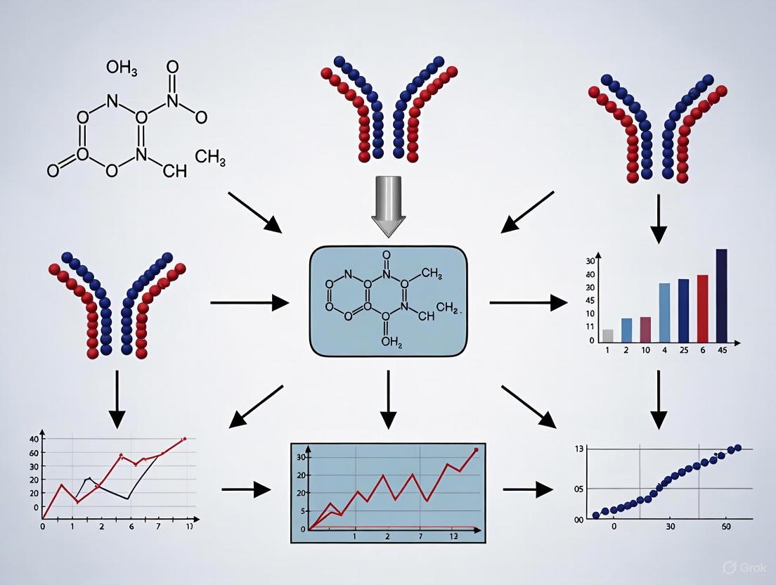
Diagram 1: Biosensor assembly and signal transduction workflows.
Advanced Architectures and Emerging Trends
Biorecognition-Element-Free Sensors
A frontier in biosensing involves developing sensors that forego traditional biorecognition elements. These platforms rely on the intrinsic physical or electrochemical properties of nanomaterials that undergo measurable changes upon interaction with target pesticides [5] [10]. For instance, nanoparticles can be functionalized to undergo aggregation in the presence of a specific pesticide, resulting in a visible color change detectable by colorimetric methods [5]. Similarly, gold interdigitated microelectrodes (IDμE) can directly measure changes in the impedance or capacitance of a sample solution caused by the presence of bacterial cells or other analytes, without any immobilized biorecognition layer [10]. While these approaches can simplify sensor fabrication and enhance stability, a significant challenge remains in achieving high selectivity in complex sample matrices without a specific capture element [5].
Integration with Advanced Transduction Platforms
The performance of a biorecognition element is profoundly influenced by its integration with the transducer. Nanomaterials have been pivotal in this regard, enhancing sensitivity by increasing the surface area for immobilization and facilitating electron transfer in electrochemical sensors [5] [1]. A prominent example is the integration of biorecognition elements with Surface-Enhanced Raman Spectroscopy (SERS). SERS provides exceptional sensitivity through massive signal amplification of Raman scattering from molecules adsorbed on plasmonic nanostructures. By functionalizing SERS substrates with antibodies or aptamers, the platform gains the required specificity, creating a powerful SERS biosensor that can detect trace levels of pesticides with fingerprint identification capability [9].
Table 2: Research Reagent Solutions for Biosensor Development
| Reagent / Material | Function in Biosensor Assembly | Application Example |
|---|---|---|
| Gold Nanoparticles (AuNPs) [5] [9] | Signal amplification; Colorimetric reporter; SERS substrate; Electrode modifier | Colorimetric aggregation assays; SERS biosensor platforms [5] |
| Acetylcholinesterase (AChE) [6] | Biorecognition element for organophosphate/carbamate pesticides | Enzyme inhibition-based electrochemical sensors [6] |
| Molecularly Imprinted Polymers (MIPs) [5] [3] | Synthetic bioreceptor with templated cavities for target capture | Label-free sensors for small molecule pesticides [5] |
| Aptamers (selected via SELEX) [3] | Synthetic oligonucleotide bioreceptor with high affinity and stability | Electrochemical or optical aptasensors for acetamiprid, atrazine [3] |
| Monoclonal Antibodies [4] [8] | High-specificity capture agent for immunoassays | Immunosensors for pyrethroids, atrazine [8] |
| Carbon Nanotubes / Graphene [5] | Electrode nanomaterial for enhanced electron transfer and surface area | Nanocomposite-based electrochemical biosensors [5] |
The Scientist's Toolkit: Experimental Considerations
Immobilization Strategies
The method used to tether the biorecognition element to the transducer surface is critical for biosensor performance. Immobilization must preserve the biological activity of the element while ensuring stability over repeated uses [2]. Common techniques include:
- Adsorption: Physical attachment based on hydrophobic or ionic interactions. Simple but can lead to leaching and unstable coatings [2].
- Covalent Binding: Chemical linkage to activated surface groups (e.g., via amine, carboxyl, or thiol functionalities). Provides stable, oriented immobilization but requires more complex surface chemistry [3] [2].
- Entrapment: Encapsulation within a polymer matrix (e.g., silica sol-gel, conducting polymers). Protects the bioreceptor but can introduce diffusion barriers [6].
- Affinity Binding: Use of high-affinity pairs like biotin-streptavidin. Allows for controlled, oriented immobilization [3].
Troubleshooting and Pitfall Management
Translating biosensor theory into reliable laboratory practice requires awareness of common challenges [2]:
- Biological Stability: Antibodies and enzymes can denature over time. Mitigation strategies include careful storage, use of stabilizing additives, or employing more robust synthetic receptors like aptamers or MIPs [3].
- Matrix Interference: Complex samples like food extracts or soil suspensions can cause non-specific binding and signal fouling. The use of blocking agents, sample dilution, or pre-filtration steps is often necessary [2].
- Sensor Drift: Gradual signal change over time due to bioreceptor degradation or environmental fluctuations. Regular recalibration against reference standards is essential [2].
- Reproducibility: Achieving consistent fabrication across multiple sensors is challenging. Automation of immobilization steps and rigorous characterization of nanomaterials can improve batch-to-batch consistency [3].
Biorecognition elements constitute the foundational intelligence of a biosensor, defining its analytical specificity and enabling the detection of target pesticides amidst complex environmental and food matrices. The architectural integration of these elements—from classical enzymes and antibodies to emerging aptamers and synthetic MIPs—with advanced transduction platforms and nanomaterials, continues to push the boundaries of sensitivity, portability, and multiplexing capabilities. Future developments will likely focus on harnessing artificial intelligence for data analysis, creating more robust and stable synthetic bioreceptors, and engineering fully integrated, miniaturized systems for real-time, on-site monitoring across the entire "farm-to-fork" continuum. The strategic selection and optimization of the biorecognition element remain, therefore, the most critical step in the design of effective biosensor architectures for pesticide detection and beyond.
Biorecognition elements (BREs) are the cornerstone of biosensor technology, conferring specificity and selectivity by interacting with target analytes. In the specific field of pesticide biosensors, the choice of BRE directly dictates the sensor's performance, including its sensitivity, stability, and applicability in complex matrices. The evolution of these elements from natural biological molecules to sophisticated synthetic constructs has significantly advanced the capabilities of modern biosensing platforms. This guide provides a comprehensive technical overview of the major BRE classes, framing their principles, performance, and practical implementation within pesticide detection research. The continuous innovation in this domain, from the refinement of natural molecules to the rational design of fully synthetic systems, is paving the way for a new generation of robust, field-deployable analytical tools for environmental and food safety monitoring [4].
Foundational Classes of Biorecognition Elements
The performance of a biosensor is fundamentally linked to the properties of its biorecognition element. The following sections detail the core classes of BREs, with their operational mechanisms, strengths, and limitations summarized in Table 1 for direct comparison.
Table 1: Core Biorecognition Element Classes in Pesticide Biosensors
| Biorecognition Element | Recognition Principle | Key Advantages | Key Limitations | Example Pesticide Targets |
|---|---|---|---|---|
| Enzymes | Catalytic activity or inhibition | High catalytic turnover; well-established immobilization protocols; reusable | Susceptible to denaturation; limited by inherent enzyme stability; can be inhibited non-specifically | Organophosphates, Carbamates [11] [12] |
| Antibodies | Affinity-based binding (Antigen-Antibody) | Exceptional specificity and high affinity; wide range of available targets | Susceptible to permanent binding; large size can limit density; animal-derived production | Pyrethroids, Herbicides [8] [9] |
| Nucleic Acid Aptamers | 3D structure-complementary binding | Synthetic production; small size for high density; stability across temperatures | Susceptible to nuclease degradation; complex, expensive selection process (SELEX) | Various chemical classes [4] [9] |
| Engineered Whole Cells | Cellular response (e.g., transcription factor activation) | Can detect bioavailable fractions; provide toxicity data; inherently amplified signals | Longer response times; complex maintenance and storage; less specific for single compounds | Heavy metals, Broad-spectrum toxins [13] [14] |
| Molecularly Imprinted Polymers (MIPs) | Shape-complementary cavities in a synthetic polymer | High physical/chemical robustness; reusable; no biological source required | Challenging elution of template molecules; can suffer from heterogeneity | Customizable for various pesticides [4] |
Natural and Bio-Derived Elements
Enzymes: Enzymes are among the most traditional BREs. Their application in pesticide detection often relies on inhibition-based mechanisms. For instance, acetylcholinesterase (AChE) and organophosphorus hydrolase (OPH) are widely used for detecting organophosphorus (OP) and carbamate pesticides. AChE-based sensors operate on the principle that the target pesticide inhibits the enzyme's activity, reducing the catalytic conversion of its substrate and leading to a measurable decrease in signal (e.g., amperometric or colorimetric) [11]. In contrast, OPH-based sensors utilize a direct catalytic mechanism, where OPH hydrolyzes specific OP compounds, generating a detectable product [11]. The main challenges include the inherent instability of enzymes under operational conditions and potential interference from other cholinesterase-inhibiting chemicals in complex samples.
Antibodies: Antibodies, particularly monoclonal and recombinant antibodies, offer exquisite specificity for a single pesticide or a closely related group. This makes them ideal for immunosensors and immunoassays, such as enzyme-linked immunosorbent assays (ELISA) and immunochromatographic test strips (ICTS) [8] [4]. The high affinity of the antibody-antigen interaction allows for very low detection limits. However, their production can be costly, they are susceptible to irreversible binding, and their performance can be compromised in harsh environmental conditions (e.g., extreme pH or temperature) [4].
Synthetic and Engineered Elements
Nucleic Acid Aptamers: Aptamers are single-stranded DNA or RNA oligonucleotides selected in vitro through the Systematic Evolution of Ligands by EXponential enrichment (SELEX) process to bind specific targets with high affinity. They are often called "synthetic antibodies" but offer distinct advantages: they are chemically synthesized, providing excellent batch-to-batch reproducibility, and are generally more stable than proteins [4] [9]. Their small size allows for high-density immobilization on sensor surfaces. Aptamers can undergo conformational changes upon target binding, which can be transduced into a signal, making them versatile for various biosensor platforms [9].
Whole-Cell Biosensors: These systems use living microorganisms (e.g., bacteria, yeast) engineered with synthetic gene circuits to detect target analytes. Upon exposure to a specific pesticide or class of pesticides, a cellular response is triggered, such as the activation of a promoter linked to a reporter gene (e.g., for green fluorescent protein (GFP) or luciferase) [13] [14]. This approach is valuable for assessing the cumulative toxicity or bioavailable fraction of a sample. A key advancement is their integration into Engineered Living Materials (ELMs), where cells are encapsulated in hydrogels or other polymers, enhancing their stability and practicality for field use [14].
Molecularly Imprinted Polymers (MIPs): MIPs are fully synthetic polymeric materials that contain tailor-made recognition sites complementary in shape, size, and functional groups to a target molecule (the "template"). After synthesizing the polymer around the template, the template is removed, leaving behind cavities that can selectively rebind the target analyte [4]. MIPs are highly robust, capable of withstanding extreme pH, temperature, and organic solvents, making them suitable for harsh environmental sampling. Their primary challenge is achieving the same level of specificity and binding affinity as biological receptors.
Experimental Protocols for BRE Development and Integration
To illustrate the practical application of these BREs, detailed protocols for key experimental procedures are provided below.
Protocol: SELEX for Aptamer Selection against a Pesticide Target
This protocol outlines the process for selecting a specific DNA aptamer capable of binding to a target pesticide, such as a common organophosphate [4] [9].
- Library Preparation: Begin with a synthetic single-stranded DNA (ssDNA) library containing a central random sequence region (e.g., 40 nucleotides) flanked by constant primer binding sites.
- Incubation with Immobilized Target: Immobilize the target pesticide molecule on a solid support (e.g., sepharose beads or a microplate). Incubate the ssDNA library with the immobilized target in a binding buffer to allow for the formation of target-aptamer complexes.
- Partitioning and Washing: Remove unbound and weakly bound DNA sequences through extensive washing with the binding buffer.
- Elution of Bound Sequences: Elute the specifically bound DNA sequences from the target. This can be achieved by denaturing the complex, for example, by using a high-temperature incubation or an elution buffer containing a high concentration of the free target molecule to compete for binding.
- Amplification: Amplify the eluted DNA sequences using the polymerase chain reaction (PCR) with primers corresponding to the constant regions.
- Generation of ssDNA Library for Next Round: Convert the double-stranded PCR product back into a single-stranded DNA library, ready for the next selection round.
- Repetition and Counter-SELEX: Repeat steps 2-6 for 8-15 rounds, progressively increasing the selection stringency (e.g., by increasing wash stringency or incorporating counter-SELEX steps with non-target molecules to eliminate cross-reactive binders).
- Cloning and Sequencing: After the final round, clone the enriched DNA pool and sequence individual clones to identify the dominant aptamer sequences.
- Characterization: Chemically synthesize the identified aptamer candidates and characterize their affinity (dissociation constant, Kd) and specificity against structurally similar pesticides.
Protocol: Development of an Acetylcholinesterase (AChE) Inhibition Biosensor
This protocol details the construction of an electrochemical biosensor for detecting organophosphate and carbamate pesticides based on AChE inhibition [11] [15].
- Electrode Modification: Prepare the working electrode (e.g., glassy carbon or screen-printed carbon electrode). To enhance sensitivity, the electrode surface may be modified with nanomaterials such as carbon nanotubes or gold nanoparticles to increase the electroactive surface area and facilitate electron transfer.
- Enzyme Immobilization: Immobilize AChE onto the modified electrode surface. This can be achieved through cross-linking with glutaraldehyde, entrapment within a polymer matrix (e.g., Nafion or chitosan), or physical adsorption.
- Baseline Signal Measurement: Place the modified electrode in an electrochemical cell containing a buffer solution with a known concentration of the enzyme's substrate, acetylthiocholine. Measure the amperometric current generated by the enzymatic reaction. The product, thiocholine, is oxidized at the electrode surface, producing a measurable baseline current (I₀).
- Inhibition (Sample Assay): Incubate the biosensor with a sample solution suspected to contain the pesticide inhibitor for a fixed period (e.g., 10-15 minutes).
- Post-Inhibition Signal Measurement: After incubation, wash the electrode and measure the amperometric current (Iᵢ) again under the same conditions as in step 3.
- Quantification: The degree of enzyme inhibition is calculated as a percentage: Inhibition (%) = [(I₀ - Iᵢ) / I₀] × 100. This percentage is then correlated with the concentration of the inhibiting pesticide in the sample using a pre-established calibration curve.
Protocol: Constructing a Whole-Cell Biosensor in a Hydrogel ELM
This protocol describes the encapsulation of an engineered bacterial biosensor within a hydrogel to create a stable material for pesticide detection [14].
- Genetic Circuit Engineering: Genetically engineer a bacterial strain (e.g., E. coli or B. subtilis) to contain a sensing module. This typically involves a promoter that is activated by a specific stress response (e.g., oxidative stress from herbicides) or directly by a transcription factor that binds the target pesticide. This promoter is fused to a reporter gene, such as gfp (green fluorescent protein).
- Cell Culture and Preparation: Grow the engineered bacteria to the mid-logarithmic phase. Harvest the cells by centrifugation and resuspend them in a sterile buffer or nutrient-poor medium to arrest growth.
- Hydrogel Precursor Preparation: Prepare a sterile solution of the hydrogel precursor. Common materials include alginate, agarose, or synthetic polymers like polyacrylamide.
- Cell-Polymer Mixing: Gently mix the concentrated bacterial suspension with the hydrogel precursor solution to achieve a homogeneous cell-polymer mixture.
- Polymerization/Casting: Induce gelation to form the ELM. For alginate, this is done by extruding the mixture into a solution containing calcium ions (e.g., CaCl₂) to form stable cross-linked beads or films. For thermosensitive polymers like agarose, gelation occurs upon cooling.
- Sensor Validation and Use: Validate the sensor by exposing the ELM to samples with known concentrations of the target pesticide. The cellular response, typically fluorescence, can be quantified using a plate reader, microscope, or a portable fluorometer. The stability of the sensor can be assessed by monitoring its response over days or weeks under storage conditions.
The logical workflow for developing and applying these biosensors, from design to readout, is visualized in the following diagram.
Advanced Sensing Mechanisms and Characterization
Integration with Advanced Transduction Platforms
The performance of a BRE is fully realized through its integration with a sensitive transducer. Surface-Enhanced Raman Spectroscopy (SERS) platforms exemplify this synergy. In a typical SERS biosensor, a BRE like an aptamer or antibody is immobilized on a plasmonic nanostructure (e.g., gold or silver nanoparticles). When the BRE captures the target pesticide, it brings the molecule into the "hot spots" of the nanostructure, resulting in a dramatic enhancement of its characteristic Raman signal, allowing for fingerprint identification and ultra-sensitive detection, potentially down to single-molecule levels [9]. This combination provides the specificity of the BRE with the exceptional sensitivity and rich spectroscopic information of SERS.
Quantitative Performance Metrics
Rigorous characterization of BRE-based biosensors is essential. Key performance metrics are consolidated in Table 2 below, providing a benchmark for comparing sensor efficacy as reported in recent literature.
Table 2: Representative Performance Metrics of Biosensors Using Different Biorecognition Elements
| Biorecognition Element | Transduction Method | Target Pesticide | Limit of Detection (LOD) | Linear Range | Reference Application |
|---|---|---|---|---|---|
| Acetylcholinesterase (AChE) | Electrochemical (Amperometric) | Organophosphates / Carbamates | pM - nM range | Up to 4-5 orders of magnitude | Inhibition-based soil/water screening [11] [15] |
| Antibody | Immunochromatographic Test Strip (ICTS) | Various (e.g., Pyrethroids) | ~ ng/mL range | Visual and semi-quantitative | Rapid on-site tea leaf testing [8] |
| Aptamer | Surface-Enhanced Raman Scattering (SERS) | Specific chemical classes | fM - pM range | Wide dynamic range | Highly sensitive multi-residue detection [9] |
| Organophosphate Hydrolase (OPH) | Electrochemical (Potentiometric) | Organophosphates | ~ 1 × 10⁻¹¹ μM (reported) | Not specified | Direct catalytic detection [11] |
| Engineered Whole Cell | Optical (Fluorescence) | Broad-spectrum stressors | Compound-dependent | Variable, based on promoter | Toxicity assessment in water [14] |
The Scientist's Toolkit: Essential Research Reagents and Materials
The development and deployment of pesticide biosensors require a suite of specialized reagents and materials. The following table lists key components and their functions as derived from the reviewed research.
Table 3: Essential Reagents and Materials for Biosensor Research
| Item Name | Function / Application in Research | Technical Notes |
|---|---|---|
| Acetylcholinesterase (AChE) | Core biorecognition element for inhibition-based detection of OPs and carbamates. | Source (electric eel, human recombinant) and purity significantly impact sensitivity and stability [11]. |
| Gold Nanoparticles (AuNPs) | Signal amplification and labeling; component of SERS substrates and electrochemical transducers. | Functionalized with BREs (antibodies, aptamers); size and shape tune plasmonic properties [13] [9]. |
| Screen-Printed Electrodes (SPEs) | Disposable, miniaturized electrochemical cells for portable biosensor development. | Enable low-cost, mass-produced sensor platforms for field testing [11] [15]. |
| SELEX Kit | In vitro selection of high-affinity DNA/RNA aptamers against target pesticide molecules. | Streamlines the complex and iterative process of aptamer development [4] [9]. |
| Alginate Hydrogel | Biopolymer for encapsulating and protecting whole-cell biosensors in ELMs. | Forms a biocompatible, porous matrix via ionic cross-linking with Ca²⁺ [14]. |
| CRISPR-Cas System (e.g., Cas12a, Cas13a) | Provides nucleic acid detection with single-base specificity; used for signal amplification. | Enables ultrasensitive, amplification-free detection when combined with aptamers or other BREs [16]. |
| Plasmonic Nanostructures | Form the core of SERS biosensors, generating intense electromagnetic fields for signal enhancement. | Typically made of gold or silver; geometry (nanorods, nanostars) is critical for performance [9]. |
| Molecularly Imprinted Polymer (MIP) Pre-polymerization Mix | Synthetic cocktail for creating biomimetic recognition sites for target pesticides. | Contains functional monomers, cross-linkers, and the template (pesticide) molecule [4]. |
The landscape of biorecognition elements for pesticide biosensors is rich and diverse, spanning from highly specific natural molecules to robust, designable synthetic systems. Each class of BRE—enzymes, antibodies, aptamers, whole cells, and MIPs—offers a unique set of advantages and constraints, making them suitable for different application scenarios. The ongoing convergence of synthetic biology, nanotechnology, and materials science is pushing the boundaries of what is possible, leading to the development of intelligent, portable, and highly sensitive biosensing platforms. Future research will likely focus on enhancing the stability and reusability of BREs in real-world environments, developing multi-analyte detection systems, and seamlessly integrating these sensors with data analytics and decision-support systems to create a truly responsive and sustainable agri-food monitoring network.
In the realm of pesticide biosensors, analyte selectivity is the cornerstone of reliable detection. This specificity is governed by the biorecognition element, a biological or biomimetic component that selectively interacts with a target pesticide, and the transduction mechanism that converts this binding event into a measurable signal [5] [17]. The choice and engineering of this biorecognition layer directly determine the sensor's performance, including its sensitivity, robustness, and applicability in complex matrices like food and environmental samples [18]. This guide examines the fundamental binding mechanisms and design principles that underpin selectivity, providing a technical framework for researchers and scientists developing next-generation biosensing platforms. Within the broader thesis on biorecognition elements, understanding these core principles is essential for innovating beyond traditional methods and addressing the limitations of conventional chromatography-based techniques [19] [8].
Fundamental Binding Mechanisms
The selective capture of target analytes by biorecognition elements is driven by a combination of structural compatibility and intermolecular forces. The following diagram illustrates the core binding interactions and signal transduction pathways common across different biosensor platforms.
The high-affinity binding between a biorecognition element and its target pesticide is stabilized by a synergistic combination of several non-covalent interactions [18]:
- Hydrogen Bonding: Directional interactions between hydrogen atoms bound to electronegative atoms (like N or O) and other electronegative atoms. Crucial for orienting the target within the binding pocket.
- Electrostatic Interactions: Attractive forces between oppositely charged ionic groups on the bioreceptor and the pesticide molecule (e.g., between a carboxylate and an ammonium group).
- van der Waals Forces: Weak, non-specific attractive forces that become significant when the shapes of the bioreceptor and target are highly complementary, maximizing surface contact.
- Hydrophobic Effects: The driving force that sequesters non-polar regions of the pesticide away from the aqueous environment and into hydrophobic pockets of the bioreceptor.
- π-π Stacking: Interactions between aromatic rings in the bioreceptor (e.g., nucleobases in an aptamer) and aromatic structures in certain pesticides.
The specific combination and relative contribution of these forces vary with the biorecognition element and the target's chemical structure, ultimately defining the selectivity profile of the biosensor.
Biorecognition Elements and Their Selectivity
The core of a biosensor's selectivity lies in its biorecognition element. The table below provides a comparative overview of the four primary types used in pesticide detection.
Table 1: Key Biorecognition Elements in Pesticide Biosensors
| Biorecognition Element | Origin & Composition | Primary Mechanism for Selectivity | Typical Targets (Pesticides) | Key Advantages | Inherent Limitations |
|---|---|---|---|---|---|
| Aptamers [18] | Synthetic single-stranded DNA/RNA (ssDNA, RNA); 25-90 bases. | Folding into unique 3D structures that form binding pockets; selectivity via SELEX in vitro. | Carbendazim, Thiamethoxam, Acetamiprid [18] [8] | High thermal/chemical stability; small size for high density; reusability; modifiable with functional groups. | In vitro selection (SELEX) can be complex; susceptibility to nuclease degradation (RNA). |
| Antibodies [5] [17] | Immunoglobulins (e.g., IgG); produced in vivo. | Molecular recognition via the paratope (antigen-binding site); high-affinity lock-and-key fit. | Pyrethroids, Organophosphates (e.g., Malathion) [18] [8] | Exceptional specificity and high affinity; well-established conjugation protocols. | Susceptible to denaturation in harsh conditions; batch-to-batch variation; animal use required for production. |
| Enzymes [5] [17] | Proteins (e.g., Acetylcholinesterase, AChE). | Catalytic activity inhibition or activation by the target; specificity for the enzyme's active site. | Organophosphates, Carbamates [5] [19] | Natural catalytic amplification; direct functional readout (inhibition). | Limited to enzyme-inhibiting pesticides; stability issues over time. |
| Molecularly Imprinted Polymers (MIPs) [5] [20] | Synthetic polymers with tailor-made cavities. | Shape complementarity and chemical functionality memory from the polymerization process. | Various, depending on the template molecule used [5] | High robustness in extreme pH/temperature; cost-effective production; long shelf-life. | Challenges with template leakage; sometimes lower affinity compared to biological elements. |
Experimental Protocols for Evaluating Selectivity
Rigorous experimental validation is required to confirm a biosensor's specificity. The following workflow outlines a standard protocol for selectivity assessment, particularly for an electrochemical aptasensor.
Detailed Protocol: Selectivity Testing for an Electrochemical Aptasensor
This protocol details the steps to confirm that an aptamer-based sensor specifically binds its target pesticide (e.g., carbendazim) and minimizes response to interfering substances.
1. Bioreceptor Immobilization:
- Functionalization: Prepare a gold disk working electrode by cleaning via polishing and electrochemical cycling. Incubate with a 2 mM solution of 6-mercapto-1-hexanol (MCH) in ethanol for 1 hour to form a self-assembled monolayer (SAM) [18].
- Immobilization: Inject a solution of thiol-modified DNA aptamer (e.g., 1 µM in Tris-EDTA buffer with Mg²⁺) onto the MCH-modified electrode. Allow 16 hours for covalent Au-S bond formation. Rinse thoroughly to remove physically adsorbed strands [18].
2. Baseline Signal Acquisition:
- Using a potentiostat, perform Electrochemical Impedance Spectroscopy (EIS) in a 5 mM [Fe(CN)₆]³⁻/⁴⁻ redox solution.
- Record the charge transfer resistance (Rₑₜ) as the baseline signal before analyte introduction [18].
3. Target Analyte Exposure:
- Incubate the functionalized electrode with the target pesticide (carbendazim) at a known concentration (e.g., 1 nM in a suitable buffer) for a fixed time (e.g., 30 minutes).
- Rinse the electrode gently to remove unbound molecules.
4. Specificity Test (Interferents):
- Repeat Step 3 independently using a suite of potential interferents. These should include:
- Structurally similar pesticides (e.g., thiabendazole for a carbendazim sensor).
- Pesticides of different classes commonly found in the sample matrix (e.g., organophosphates like chlorpyrifos).
- Common ions (e.g., K⁺, Ca²⁺, NO₃⁻).
- For complex samples like tea, include matrix components like tea polyphenols and catechins [8].
5. Signal Measurement & Analysis:
- After each incubation (target and each interferent), perform EIS again under identical conditions and record the new Rₑₜ value.
- Calculate the signal change (e.g., ΔRₑₜ = Rₑₜ(after) - Rₑₜ(baseline)).
- Quantify selectivity by calculating a selectivity coefficient (k) for each interferent (I) relative to the target (T): k = ΔRₑₜ(I) / ΔRₑₜ(T). A sensor is highly selective for the target when k << 1 for all interferents.
6. Regeneration Test (Optional for Reusability):
- To test aptamer reversibility, expose the sensor to a regeneration buffer (e.g., 1 mM NaOH or a solution with EDTA) for 1-2 minutes to dissociate the bound target.
- Re-measure the EIS signal in the redox solution. A return to the baseline Rₑₜ value indicates successful regeneration and aptamer stability [18].
The Scientist's Toolkit: Essential Research Reagents and Materials
The development and fabrication of selective biosensors rely on a suite of specialized reagents and materials.
Table 2: Essential Reagents and Materials for Biosensor Development
| Item | Function/Application | Technical Notes |
|---|---|---|
| Gold Nanoparticles (Au NPs) [5] [20] | Signal amplification; enhance electron transfer in electrochemical sensors; used in colorimetric and SERS-based sensors. | Functionalized with thiolated aptamers or antibodies. High conductivity and tunable optical properties. |
| Carbon Nanotubes (CNTs) [18] | Increase electrode surface area; improve electron transfer kinetics in electrochemical aptasensors. | Can be single-walled (SWCNT) or multi-walled (MWCNT). |
| Molecularly Imprinted Polymers (MIPs) [5] [20] | Synthetic bioreceptors; provide robust, stable recognition cavities for pesticides in harsh environments. | Created using a template (target pesticide), functional monomers, and a cross-linker. |
| Thiol Linkers (e.g., MCH) [18] | Form self-assembled monolayers (SAMs) on gold surfaces; orient and stabilize immobilized aptamers; reduce non-specific binding. | Creates a well-ordered interface crucial for consistent sensor performance. |
| Methylene Blue[ [18]] | Electroactive label; often tethered to aptamers in "signal-on" or "signal-off" electrochemical sensors. | Redox behavior changes upon aptamer folding/target binding. |
| Metal-Organic Frameworks (MOFs, e.g., MOF-808) [18] | Porous nanomaterials; used to immobilize bioreceptors and pre-concentrate target analytes, boosting sensitivity. | High surface area and modular chemistry. |
| Streptavidin/Biotin System [18] | High-affinity coupling; used to immobilize biotinylated aptamers or antibodies onto sensor surfaces. | Provides a stable and oriented immobilization method. |
| Systematic Evolution of Ligands by EXponential enrichment (SELEX) Kits [18] | In vitro selection of high-affinity aptamers against specific pesticide targets. | The foundational process for generating new DNA/RNA recognition elements. |
The path to achieving high selectivity in pesticide biosensors is multifaceted, relying on a deep understanding of intermolecular binding forces, the strategic selection and engineering of biorecognition elements, and rigorous experimental validation against realistic interferents. The ongoing convergence of nanotechnology, materials science, and synthetic biology promises to yield even more robust and specific sensing platforms. Future advancements will likely involve the rational design of aptamers with pre-defined binding pockets, the creation of more sophisticated MIPs, and the integration of AI to guide material selection and optimize sensor design [20]. These innovations will be critical for meeting the growing demand for rapid, on-site detection of pesticide residues, thereby strengthening global food safety and environmental monitoring protocols.
Biosensors are analytical devices that integrate a biological recognition element with a transducer to produce a signal proportional to the concentration of a target analyte [21]. The performance of these devices, particularly in the critical field of pesticide detection, is governed by a set of core metrics that determine their reliability and applicability in real-world scenarios [8] [22]. Within pesticide biosensors research, the choice of biorecognition element—be it enzymes, antibodies, aptamers, or whole cells—fundamentally influences these performance parameters [17] [23]. This technical guide provides an in-depth examination of the four cornerstone biosensor performance metrics, with specific emphasis on their interplay with biorecognition elements in pesticide detection platforms. The optimization of these metrics is paramount for transitioning laboratory prototypes into robust field-deployable tools for environmental monitoring, food safety, and public health protection [8] [23].
Core Performance Metrics: Definitions and Significance
The analytical performance of biosensors is quantified through standardized figures of merit. These metrics provide objective criteria for evaluating and comparing different biosensing platforms, guiding the development process, and establishing confidence in the generated data [24]. For pesticide biosensors, stringent performance standards are essential due to the low regulatory limits and complex sample matrices involved [8] [23].
Table 1: Core Performance Metrics for Biosensors
| Metric | Technical Definition | Significance in Pesticide Detection |
|---|---|---|
| Sensitivity | Slope of the analytical calibration curve; the minimum amount of analyte that can be reliably detected [21] [24]. | Determines capability to detect trace pesticide residues at regulatory levels (often ng/mL or lower) [21] [8]. |
| Selectivity | Ability of a bioreceptor to detect a specific analyte in samples containing admixtures and contaminants [21] [24]. | Ensures accurate measurement despite interference from structurally similar pesticides or complex sample matrices (e.g., tea, soil) [8]. |
| Reproducibility | Closeness of agreement between measurements under different conditions (operators, apparatus, laboratories) [21] [24]. | Guarantees reliability across different testing scenarios and locations for regulatory compliance monitoring [21]. |
| Reusability | Ability of a biosensor to be regenerated and reused multiple times while maintaining performance. | Reduces cost-per-test and enables continuous monitoring applications; highly dependent on bioreceptor stability [22]. |
The Interplay Between Biorecognition Elements and Performance Metrics
The biological recognition component is the cornerstone of any biosensor, defining its fundamental interaction with target analytes. In pesticide detection, different classes of biorecognition elements offer distinct advantages and challenges that directly impact the core performance metrics [17].
Enzymes as Biorecognition Elements
Enzyme-based biosensors primarily operate on inhibition or catalytic principles. Enzymes such as acetylcholinesterase (AChE) are inhibited by organophosphorus and carbamate pesticides, enabling detection through measurable decreases in enzymatic activity [17]. Alternatively, some enzymes directly metabolize pesticides, with the catalytic transformation providing the measurable signal [17].
Impact on Performance:
- Sensitivity: Enzyme sensors can achieve high sensitivity due to catalytic amplification, but inhibition-based approaches may suffer from limited sensitivity against certain pesticide classes [22].
- Selectivity: A significant challenge as enzymes may be inhibited by multiple compounds, leading to false positives from sample matrices [8].
- Reproducibility: Subject to variability due to enzyme instability under different environmental conditions (pH, temperature) [21].
- Reusability: Generally low as enzyme inhibition is often irreversible, requiring fresh enzyme for each test [22].
Antibodies as Biorecognition Elements
Immunosensors exploit the high specificity of antigen-antibody interactions. Antibodies can be engineered for specific pesticide epitopes or classes, functioning in either label-free or labeled formats [17] [24].
Impact on Performance:
- Sensitivity: Excellent sensitivity with detection limits reaching pg/mL for some targets, suitable for trace pesticide detection [17].
- Selectivity: High specificity for target analytes, though cross-reactivity with structurally similar compounds can occur [8].
- Reproducibility: Generally good with monoclonal antibodies, though storage stability and batch-to-batch variability can affect performance [21].
- Reusability: Moderate; regeneration of antibody binding sites is possible but may gradually reduce binding capacity [22].
Aptamers as Biorecognition Elements
Aptamers are synthetic single-stranded DNA or RNA oligonucleotides selected through SELEX (Systematic Evolution of Ligands by Exponential Enrichment) to bind specific targets with high affinity [17]. They fold into specific three-dimensional structures upon target binding.
Impact on Performance:
- Sensitivity: Can achieve nM to pM detection limits, competitive with antibody-based systems [17].
- Selectivity: High specificity, capable of distinguishing between closely related pesticide analogs [8].
- Reproducibility: Excellent due to synthetic production with minimal batch-to-batch variation [17].
- Reusability: High; aptamers can undergo denaturation-renaturation cycles, allowing multiple regeneration cycles [22].
Whole Cells as Biorecognition Elements
Whole cell biosensors utilize microorganisms (bacteria, fungi, algae) as integrated sensing elements, typically employing metabolic activity, stress responses, or genetic regulation mechanisms [17] [23].
Impact on Performance:
- Sensitivity: Variable; some systems can detect pesticides at ng/mL levels, though generally less sensitive than molecular recognition elements [17].
- Selectivity: Often lower as cells may respond to multiple stressors; useful for class-level detection rather than specific compounds [23].
- Reproducibility: Challenging due to biological variability and maintenance requirements of living systems [17].
- Reusability: Moderate; cells can self-replicate but require careful maintenance of viability and consistent physiological state [17].
Table 2: Comparative Performance of Biorecognition Elements in Pesticide Biosensors
| Biorecognition Element | Sensitivity | Selectivity | Reproducibility | Reusability | Best Applications |
|---|---|---|---|---|---|
| Enzymes | Moderate to High | Moderate | Moderate | Low | Broad-spectrum screening, inhibition-based detection |
| Antibodies | High | High | High | Moderate | Targeted, specific pesticide quantification |
| Aptamers | High | High | High | High | Reusable sensors, harsh environments |
| Whole Cells | Moderate | Low to Moderate | Low | Moderate | Toxicity assessment, class-level detection |
Methodologies for Performance Evaluation
Standardized experimental protocols are essential for reliable evaluation and cross-comparison of biosensor performance. This section details key methodologies for quantifying each core metric.
Sensitivity and Limit of Detection (LOD) Determination
Experimental Protocol:
- Prepare a dilution series of the target pesticide in appropriate buffer across at least 5-6 concentration points.
- For each concentration, measure the biosensor response in triplicate.
- Plot the mean response against pesticide concentration to generate a calibration curve.
- Fit the data using linear regression (y = mx + c, where y is signal, x is concentration).
- Calculate sensitivity as the slope (m) of the linear range.
- Determine LOD using the formula: LOD = 3.3 × σ/S, where σ is the standard deviation of the blank signal and S is the sensitivity [21] [24].
Key Considerations:
- Matrix-matched calibration is essential for real-sample analysis to account for matrix effects.
- The linear range defines the operational concentration window for quantitative analysis.
- For optical biosensors, LOD values for pesticides have been reported in the range of 10 fM to 1 nM, while electrochemical methods can achieve even lower detection limits in some cases [22].
Selectivity and Cross-Reactivity Assessment
Experimental Protocol:
- Test the biosensor response against the target pesticide at a fixed concentration (typically near the middle of the linear range).
- Under identical conditions, test the biosensor against potential interferents individually, including:
- Structurally similar pesticides
- Common environmental contaminants (heavy metals, PAHs)
- Matrix components (for tea sensors: polyphenols, caffeine, pigments) [8]
- Calculate cross-reactivity percentage for each interferent: (Signalinterferent/Signaltarget) × 100%.
- A selectivity coefficient <5% is generally acceptable for most applications [24].
Key Considerations:
- For antibody-based sensors, cross-reactivity profiling is particularly important due to potential recognition of similar epitopes.
- Aptamers generally exhibit lower cross-reactivity than antibodies for closely related compounds [17].
Reproducibility and Repeatability Evaluation
Experimental Protocol:
- Repeatability (intra-assay precision): Perform 10-20 replicate measurements of the same pesticide sample using a single biosensor in one session. Calculate the relative standard deviation (RSD).
- Reproducibility (inter-assay precision): Prepare multiple identical biosensors (n ≥ 5). Measure the same pesticide sample with each sensor on different days or by different operators. Calculate RSD across sensors.
- Intermediate precision: Assess variation between different batches of bioreceptor immobilization or different production lots.
Acceptance Criteria:
- For quantitative analysis, RSD should generally be <10-15% depending on application requirements [21] [24].
- Document all conditions (temperature, pH, sample preparation) that may contribute to variability.
Reusability and Stability Testing
Experimental Protocol:
- Operational stability: Perform repeated measurement-regeneration cycles with the same biosensor. After each measurement, regenerate according to the appropriate protocol:
- Plot the response versus cycle number to determine the maximum number of uses before signal degradation >10%.
- Storage stability: Store biosensors under recommended conditions and test performance at regular intervals over days to months.
- Real-time stability: Continuously operate the biosensor in buffer or complex matrix to assess signal drift over time.
Key Considerations:
- The regeneration method must effectively dissociate the analyte without damaging the bioreceptor.
- Stability is highly dependent on bioreceptor immobilization method and storage conditions [21].
The Scientist's Toolkit: Research Reagent Solutions
Table 3: Essential Research Reagents for Pesticide Biosensor Development
| Reagent Category | Specific Examples | Function in Biosensor Development |
|---|---|---|
| Biorecognition Elements | Acetylcholinesterase (AChE), anti-chlorpyrifos antibodies, organophosphate-binding aptamers, E. coli reporter cells [17] [23] | Target recognition and signal initiation through specific binding or catalytic activity |
| Nanomaterials | Gold nanoparticles, graphene oxide (GO), carbon nanotubes (CNTs), metal-organic frameworks (MOFs) [22] [25] [24] | Signal amplification, increased surface area for bioreceptor immobilization, enhanced electron transfer |
| Immobilization Matrices | Chitosan, Nafion, polyaniline, sol-gels, self-assembled monolayers (SAMs) [22] | Secure bioreceptors to transducer surface while maintaining bioactivity and accessibility |
| Signal Generation Reagents | Horseradish peroxidase (HRP), alkaline phosphatase (ALP), quantum dots, electrochemical mediators (ferrocene derivatives) [17] [24] | Produce measurable signals (colorimetric, fluorescent, electrochemical) upon target recognition |
| Regeneration Buffers | Glycine-HCl (pH 2.0-3.0), NaOH (10-100 mM), EDTA (1-10 mM), SDS (0.1-1%) [22] | Dissociate analyte from bioreceptor between measurements for sensor reusability |
Advanced Optimization Strategies
Nanomaterial-Enhanced Performance
The integration of nanomaterials has revolutionized biosensor performance by addressing multiple metrics simultaneously [25] [24]. Gold nanoparticles provide exceptional signal amplification, with studies demonstrating up to 50-fold improvement in detection limits when incorporated into immunosensors [24]. Carbon nanotubes and graphene oxide enhance electron transfer kinetics in electrochemical biosensors while providing high surface area for bioreceptor immobilization [22] [25]. Metal-organic frameworks (MOFs) offer tunable porosity for both pesticide capture and signal enhancement, with particular utility in both detection and removal applications [22].
Immobilization Techniques for Enhanced Stability
The method of bioreceptor immobilization directly impacts stability, reusability, and overall performance. Covalent immobilization via glutaraldehyde or EDC/NHS chemistry provides stable linkage but may reduce bioactivity. Physical entrapment in polymer matrices (e.g., chitosan, sol-gels) preserves activity but may limit analyte diffusion. Affinity-based immobilization (e.g., streptavidin-biotin) offers oriented attachment that maximizes binding site availability. Recent advances include DNA-directed immobilization for aptasensors and bioorthogonal chemistry for minimal interference with binding sites [22].
Technological Implementation and Workflow
The following diagram illustrates the core operational workflow and logical relationships in a generalized pesticide biosensor system, highlighting how the biorecognition element connects to the measurable signal through the transduction mechanism:
The rigorous characterization of sensitivity, selectivity, reproducibility, and reusability provides the fundamental framework for evaluating and advancing pesticide biosensor technologies. These interdependent metrics collectively determine the practical utility of biosensors for real-world applications in environmental monitoring, food safety, and public health protection. The strategic selection of biorecognition elements—enzymes, antibodies, aptamers, or whole cells—establishes the foundational performance ceiling, while advanced nanomaterial integration and immobilization techniques enable progressive optimization toward this potential. Future developments will likely focus on multi-analyte detection platforms, enhanced field-deployability, and intelligent biosystems incorporating machine learning for data analysis, ultimately creating more robust and accessible monitoring solutions for pesticide residues across the agricultural and environmental sectors.
The accurate detection of pesticide residues is a critical challenge in ensuring food safety, protecting environmental health, and complying with global regulatory standards [26] [27]. Biosensor technology, which integrates a biorecognition element with a transducer, offers a powerful solution for specific, sensitive, and rapid pesticide monitoring [8] [17]. The core of a biosensor's analytical performance lies in the biorecognition element—the biological or biomimetic component that selectively interacts with the target pesticide [5] [28]. The strategic selection of an appropriate biorecognition element, tailored to the chemical class and mode of action of the target pesticide, is therefore paramount to developing a successful detection platform [6] [22]. This guide provides an in-depth technical framework for researchers and scientists to systematically match biorecognition elements to major pesticide classes, detailing the underlying principles, experimental protocols, and advanced material solutions that drive modern pesticide biosensing.
Pesticide Classification and Recognition Principles
Pesticides are categorized based on their chemical structure and biological target, which directly inform the choice of biorecognition element. The four most prevalent classes are organophosphates, carbamates, neonicotinoids, and pyrethroids [5] [27].
Organophosphates (OPs) and carbamates both exert their toxicity through the inhibition of the enzyme acetylcholinesterase (AChE) in the nervous system of target pests. This shared mechanism makes AChE an ideal biorecognition element for their detection [6]. The inhibition is reversible for carbamates and irreversible for most OPs, a difference that can be exploited in sensor design [6].
Neonicotinoids are synthetic insecticides that act as agonists on the nicotinic acetylcholine receptors (nAChRs) in the insect central nervous system [26]. Their detection can leverage these native receptors or engineered versions, as well as antibodies and aptamers selected for high affinity.
Pyrethroids are synthetic derivatives of natural pyrethrins that disrupt voltage-gated sodium channels in nerve membranes [8]. While their detection often employs antibodies in immunoassays, enzyme-based sensors that detect oxidative metabolites are also used.
The principle of "bioactivity-guided" detection is particularly powerful for OPs and carbamates, where the inherent toxicity of the pesticide (enzyme inhibition) is directly translated into a measurable signal [6] [22]. For other classes, "bioaffinity-guided" detection, which relies on selective binding without catalytic transformation, is more common, utilizing elements like antibodies and aptamers [22].
Biorecognition Elements: A Comparative Analysis
The selection of a biorecognition element involves a careful trade-off between specificity, stability, cost, and ease of production. The main types of elements used in pesticide biosensors are enzymes, antibodies, nucleic acid aptamers, and whole cells [17] [28].
- Enzymes: Catalytic proteins that recognize a substrate or inhibitor. AChE is the most prominent example for neurotoxic insecticides [6]. Other enzymes, such as alkaline phosphatase, tyrosinase, and peroxidases, are also used [6].
- Antibodies: Immunoglobulins that bind to a specific antigen (e.g., a pesticide molecule) with high affinity. They are the basis of immunosensors [17] [28].
- Aptamers: Short, single-stranded DNA or RNA oligonucleotides selected in vitro for high-affinity binding to a specific target. They are synthetic alternatives to antibodies [17] [22].
- Whole Cells: Microorganisms (e.g., bacteria, algae) that act as integrated sensing elements, often through engineered genetic circuits that respond to the presence of a pollutant [17] [23].
Table 1: Comparative Profile of Biorecognition Elements for Pesticide Detection
| Biorecognition Element | Key Feature | Primary Detection Mechanism | Advantages | Limitations |
|---|---|---|---|---|
| Enzymes (e.g., AChE) | Catalytic activity | Inhibition of activity (for OPs, carbamates) [6] | High sensitivity, biologically relevant signal [6] | Limited to enzyme-inhibiting pesticides, stability issues [6] |
| Antibodies | High-affinity binding | Binding event (Immunoassay) [17] [28] | Excellent specificity, wide applicability [28] | Animal-derived, batch-to-batch variation, cross-reactivity [5] |
| Aptamers | In vitro selected oligonucleotides | Conformational change upon binding [17] | Chemical synthesis, high stability, modifiable [17] [22] | Susceptibility to nuclease degradation, complex selection process [17] |
| Whole Cells | Self-replicating, integrated metabolism | Stress response, metabolic activity, genetic regulation [17] | Robustness, cost-effective, detects bioavailability [17] [23] | Longer response time, lower specificity, complex signal interpretation [17] |
Matching Biorecognition Elements to Pesticide Classes
The optimal pairing between a biorecognition element and a pesticide is determined by the pesticide's chemical properties and its biochemical mechanism of action. The following section provides detailed matching criteria and experimental workflows.
Table 2: Strategic Matching of Biorecognition Elements to Target Pesticide Classes
| Target Pesticide Class | Exemplary Actives | Recommended Biorecognition Element(s) | Rationale for Matching |
|---|---|---|---|
| Organophosphates | Parathion, Chlorpyrifos, Malathion [5] | Acetylcholinesterase (AChE) [6], Organophosphorus hydrolase (OPH) [22], Aptamers [17] | AChE is the primary biological target; inhibition provides a direct, toxicologically relevant signal [6]. |
| Carbamates | Aldicarb, Carbofuran, Oxamyl [5] | Acetylcholinesterase (AChE) [6] | Shares the AChE inhibition mechanism with OPs, allowing for detection with the same element [6]. |
| Neonicotinoids | Imidacloprid, Dinotefuran [26] [8] | Antibodies [8], Nicotinic acetylcholine receptors (nAChRs) [26], Aptamers [22] | Antibodies and aptamers can be selected for high specificity to the stable neonicotinoid structure [8] [22]. |
| Pyrethroids | Bifenthrin, Fenpropathrin, Fenvalerate [8] | Antibodies [8], E. coli-based whole cells [17] | Immunoassays are highly effective for this structurally diverse class; whole cells can be engineered for a response [8] [17]. |
| Triazines & Herbicides | Atrazine [5] | Antibodies, Aptamers, Photosynthetic System II (PSII) [6] | PSII is the direct target for many herbicides, enabling activity-based detection [6]. |
| Organochlorines | DDT, Lindane [27] | Antibodies, Aptamers [22] | While largely banned, their persistence necessitates monitoring via highly specific affinity elements [27] [22]. |
Detailed Experimental Protocol: AChE-based Electrochemical Sensor for Organophosphates
This protocol details the development of a standard amperometric biosensor for detecting organophosphate (OP) pesticides based on the inhibition of acetylcholinesterase [6].
1. Principle: The enzyme acetylcholinesterase (AChE) catalyzes the hydrolysis of the substrate acetylthiocholine (ATCh) to produce thiocholine and acetate. Thiocholine is then oxidized at the surface of an electrode, generating a measurable amperometric current. The presence of an OP pesticide inhibits AChE activity, leading to a reduction in the enzymatic product and a corresponding decrease in the electrochemical signal. The degree of inhibition is proportional to the pesticide concentration [6].
2. Reagents and Materials:
- Acetylcholinesterase (AChE) from Electric Eel or recombinant source
- Acetylthiocholine (ATCh) chloride or iodide salt
- Phosphate Buffer Saline (PBS, 0.1 M, pH 7.4)
- Target organophosphate pesticide (e.g., chlorpyrifos-oxon, paraoxon)
- Glutaraldehyde (for cross-linking immobilization)
- Bovine Serum Albumin (BSA)
- Screen-printed carbon electrodes (SPCEs) or Gold electrodes
3. Step-by-Step Procedure:
- Electrode Pretreatment: Clean the working electrode of the SPCE according to the manufacturer's instructions (e.g., electrochemical cycling in sulfuric acid or gentle polishing).
- Enzyme Immobilization: Prepare an immobilization mixture containing 5 μL of AChE (2 U/mL) and 2 μL of BSA (1% w/v) in PBS. Add 1 μL of glutaraldehyde (0.25% v/v) as a cross-linker. Spot 5 μL of this mixture onto the working electrode and allow it to dry at 4°C for 1 hour.
- Baseline Measurement: Place the modified electrode in an electrochemical cell containing 10 mL of PBS with 0.1 M ATCh substrate. Apply a constant potential of +0.5 V (vs. Ag/AgCl reference) and record the steady-state amperometric current (I₀). This represents the uninhibited enzyme activity.
- Inhibition/Incubation Step: Incubate the AChE-modified electrode in a sample solution containing the target OP pesticide for a fixed time (e.g., 10-15 minutes). Rinse gently with PBS to remove unbound pesticide.
- Post-Inhibition Measurement: Re-immerse the electrode in the fresh ATCh/PBS solution and record the steady-state current again (Iᵢ).
- Data Analysis: Calculate the percentage of enzyme inhibition using the formula: Inhibition (%) = [(I₀ - Iᵢ) / I₀] × 100. The pesticide concentration in an unknown sample can be determined by interpolating the % inhibition value against a calibration curve constructed with standard solutions.
Detailed Experimental Protocol: Immunosensor for Pyrethroid Detection
This protocol outlines the key steps for developing a competitive immunoassay for pyrethroids using a lateral flow assay (LFA) format [8] [29].
1. Principle: A competitive format is used for detecting small molecules like pesticides. In this setup, pesticide molecules in the sample compete with a labeled pesticide analog (conjugate) for a limited number of binding sites on immobilized antibodies. The signal is inversely proportional to the pesticide concentration in the sample [29].
2. Reagents and Materials:
- Monoclonal antibody specific to the target pyrethroid (e.g., bifenthrin)
- Bifenthrin-protein conjugate (e.g., BSA-Bifenthrin)
- Gold nanoparticles (AuNPs, 20-40 nm) or fluorescent latex microspheres
- Nitrocellulose membrane, sample pad, conjugate pad, and absorbent pad
- Phosphate Buffer Saline (PBS) and blocking buffer (e.g., PBS with 1% BSA)
3. Step-by-Step Procedure:
- Conjugate Pad Preparation: Conjugate the anti-bifenthrin antibody to the AuNPs. Dispense the antibody-AuNP conjugate onto the glass fiber conjugate pad and dry.
- Test Line and Control Line Preparation: Dispense the bifenthrin-BSA conjugate at the test line (T-line) and a species-specific anti-IgG antibody at the control line (C-line) of the nitrocellulose membrane.
- Assembly: Assemble the LFA strip by attaching the sample pad, conjugate pad, nitrocellulose membrane, and absorbent pad in sequential order on a backing card with ~2 mm overlaps.
- Assay Execution: Apply 100 μL of the liquid sample (extract) to the sample pad. The sample migrates via capillary action, rehydrating the conjugate pad.
- Reaction and Signal Development: As the sample flows, the free pesticide in the sample and the pesticide-protein conjugate at the T-line compete for binding to the limited antibody-AuNP conjugates. After 10-15 minutes, the result can be visually read.
- Result Interpretation: A colored T-line indicates a negative result (no pesticide in sample). The absence of a T-line color indicates a positive result. The presence of a C-line confirms the test is valid. For quantitative analysis, a smartphone-based reader can be used to measure the color intensity of the T-line [29].
Visualization of Biosensor Selection and Operation
The following diagrams illustrate the logical workflow for selecting a biorecognition element and the operational mechanisms of the key biosensor types described in the experimental protocols.
Diagram 1: Biorecognition Element Selection Workflow.
Diagram 2: Key Biosensor Signaling Mechanisms.
The Scientist's Toolkit: Essential Research Reagents
The development and implementation of pesticide biosensors require a suite of specialized reagents and materials. The following table details key components and their functions in a typical research setting.
Table 3: Essential Research Reagents for Pesticide Biosensor Development
| Reagent / Material | Function / Application | Technical Notes |
|---|---|---|
| Acetylcholinesterase (AChE) | Biorecognition element for organophosphate and carbamate insecticides [6]. | Source (electric eel, bovine, recombinant) and enzyme variant impact sensitivity and selectivity to different inhibitors [6]. |
| Acetylthiocholine (ATCh) | Enzymatic substrate for AChE; product (thiocholine) is electroactive [6]. | Preferred over acetylcholine for electrochemical sensors due to the electroactivity of its hydrolysis product. |
| Gold Nanoparticles (AuNPs) | Optical label (colorimetric) or electrochemical tag in lateral flow immunoassays and aptasensors [29] [22]. | Provide a vivid red color for visual detection; size and surface functionalization are critical for performance. |
| Screen-Printed Electrodes (SPEs) | Disposable, miniaturized electrochemical transducers for field-deployable sensors [6]. | Enable low-cost, mass-produced sensor platforms. Carbon, gold, and carbon nanotube-based inks are common. |
| Molecularly Imprinted Polymers (MIPs) | Synthetic, biomimetic recognition elements with high stability [22]. | "Plastic antibodies" that offer an alternative to biological elements in harsh environments [22]. |
| Monoclonal Antibodies | High-specificity biorecognition elements for immunoassays targeting a single pesticide [17]. | Provide superior consistency and specificity compared to polyclonal antibodies. |
| Nucleic Acid Aptamers | Synthetic, single-stranded DNA/RNA recognition elements selected via SELEX [17]. | Chemically synthesized, thermally stable, and easily modified, making them versatile bioreceptors [17]. |
The strategic matching of biorecognition elements to target pesticide classes is a fundamental determinant of success in biosensor research. As detailed in this guide, the selection process must be guided by the pesticide's biochemical mode of action, leading to logical pairings such as AChE for neurotoxic organophosphates and carbamates, or high-affinity antibodies and aptamers for structurally distinct pyrethroids and neonicotinoids. Current research is pushing the boundaries of this field through the use of engineered mutant enzymes for enhanced specificity [6], the integration of nanomaterials like graphene oxide and metal-organic frameworks (MOFs) for signal amplification [8] [22], and the application of advanced data analytics like artificial neural networks (ANNs) to resolve signals from complex pesticide mixtures [6] [29]. By adhering to the systematic selection criteria and experimental frameworks outlined herein, researchers can develop next-generation biosensing platforms with the specificity, sensitivity, and robustness required for effective environmental monitoring and food safety assurance.
From Principle to Practice: Operational Mechanisms and Real-World Deployments
Enzyme-based biosensors represent a critical technological advancement in the detection of neurotoxic pesticides, offering a unique combination of biological specificity and analytical precision. Framed within the broader context of biorecognition elements—which include antibodies, nucleic acids, and whole cells—enzyme-based systems are particularly distinguished by their catalytic activity, which enables both substrate transformation and signal amplification [17]. This technical guide focuses on biosensors that leverage enzyme inhibition mechanisms, a highly relevant approach for detecting organophosphorus (OP) and carbamate (CB) pesticides designed to disrupt biological processes in target pests [6] [30].
The fundamental advantage of inhibition-based biosensors lies in their ability to translate a pesticide's inherent toxicity into a quantifiable analytical signal. Unlike conventional chromatographic methods, which are accurate but labor-intensive and ill-suited for rapid screening, these biosensors provide a "biologically relevant" assessment of contamination by measuring the functional inhibition of enzymes such as acetylcholinesterase (AChE) [6]. This guide details the core principles, experimental methodologies, and advanced applications of these systems, providing researchers and drug development professionals with a comprehensive resource for developing and implementing these powerful analytical tools.
Core Principles and Enzyme Mechanisms
Fundamental Biosensor Architecture
An enzyme-based biosensor is an integrated analytical device comprising three essential components: a biological recognition element (the enzyme), a transducer, and an immobilization matrix [31]. The enzyme serves as a highly specific biocatalyst, initiating a reaction with its target substrate. This biochemical reaction produces a measurable change in a physicochemical parameter—such as electron flow, light emission, or mass change—which is then converted by the transducer into a quantifiable electrical or optical signal [31] [17].
The operational principle bifurcates into two primary detection modes for pesticides:
- Substrate Detection: The enzyme metabolizes its specific substrate, and the resulting product concentration is measured.
- Inhibition Detection: The target analyte (e.g., a pesticide) suppresses the enzyme's catalytic activity, leading to a measurable reduction in product formation, which correlates with the inhibitor's concentration [17].
For neurotoxic pesticides, the inhibition mode is most pertinent. The percentage of inhibited enzyme (I%) is quantitatively related to the inhibitor concentration and the incubation time, meaning the residual enzyme activity is inversely proportional to the amount of toxicant present [30].
Key Enzymes for Neurotoxic Pesticide Detection
Several enzymes are exploited in inhibition biosensors, but acetylcholinesterase (AChE) is the most significant for neurotoxic insecticides.
Acetylcholinesterase (AChE): This enzyme catalyzes the hydrolysis of the neurotransmitter acetylcholine into choline and acetate. Organophosphorus and carbamate pesticides exert their toxicity by covalently binding to the serine residue in the enzyme's active site, forming a stable complex that inhibits its function [6] [31]. This inhibition prevents the breakdown of acetylcholine, leading to neurotransmitter accumulation and continuous nerve firing, which is fatal to insects and toxic to humans. AChE-based biosensors measure this inhibition as a drop in enzymatic activity, providing a detection mechanism for these pesticides [31].
Other Relevant Enzymes: While AChE predominates, other enzymes like tyrosinase, laccase, and alkaline phosphatase are also used to detect pesticides from different chemical classes based on their specific inhibition mechanisms [6].
Table 1: Key Enzymes Used in Inhibition Biosensors for Pesticide Detection
| Enzyme | Target Pesticide Classes | Inhibition Mechanism | Typical Transducer |
|---|---|---|---|
| Acetylcholinesterase (AChE) | Organophosphates, Carbamates | Irreversible (OP) or reversible (CB) binding to active site serine | Electrochemical, Optical [6] [31] |
| Tyrosinase | Phenols, Carbamates, Atrazine | Binding to active site, affecting copper centers | Amperometric, Optical [6] [30] |
| Alkaline Phosphatase | Organophosphates | Competitive inhibition | Electrochemical, Fluorescence [6] |
| Photosystem II (PSII) | Triazines, Phenylureas | Inhibition of electron transport chain | Optical, Photoelectrochemical [6] |
Experimental Protocols and Methodologies
Standard Inhibition Assay Protocol
A typical experimental workflow for an AChE-based inhibition biosensor involves a sequence of critical steps to ensure accurate and reproducible results.
Diagram 1: Inhibition assay workflow.
Step 1: Enzyme Immobilization The enzyme must be stabilized and confined near the transducer surface. Common techniques include:
- Physical Adsorption: The enzyme is attached to a solid support (e.g., carbon nanotube, graphene oxide) via weak forces (van der Waals, ionic). It is simple but can lead to enzyme leakage.
- Covalent Binding: Enzyme functional groups (e.g., -NH₂, -COOH) form stable covalent bonds with a functionalized transducer surface. This method offers excellent stability and prevents leaching [31].
- Entrapment: The enzyme is enclosed within a polymeric network (e.g., silica gel, Nafion, or polymer matrices) that allows the substrate and product to diffuse while retaining the enzyme [30].
Step 2: Baseline Activity Measurement The biosensor is exposed to the enzyme's substrate (e.g., acetylcholine for AChE). The resulting reaction product (e.g., thiocholine from acetylthiocholine) is measured to establish the initial, uninhibited signal (S₀). For electrochemical sensors, the anodic oxidation current of thiocholine is a common readout [30].
Step 3: Inhibitor Incubation The biosensor is incubated with the sample containing the pesticide inhibitor for a fixed period (e.g., 10-15 minutes). During this time, the inhibitor binds to the enzyme, reducing its catalytic activity.
Step 4: Residual Activity Measurement After incubation and a brief washing step, the substrate is reintroduced, and the signal from the enzymatic reaction is measured again (Sᵢ). This signal corresponds to the residual activity of the inhibited enzyme.
Step 5: Data Analysis and Quantification The inhibition percentage (I%) is calculated using the formula: I% = [(S₀ - Sᵢ) / S₀] × 100. This value is then correlated with the inhibitor concentration using a calibration curve prepared with standard solutions [30].
The Scientist's Toolkit: Key Research Reagents and Materials
Table 2: Essential Reagents and Materials for AChE-Based Biosensors
| Item | Function/Description | Example & Application Note |
|---|---|---|
| Acetylcholinesterase (AChE) | Biorecognition element; catalyzes acetylcholine hydrolysis. | Source: Electric eel, bovine erythrocytes, or recombinant Drosophila melanogaster mutants for enhanced sensitivity [6]. |
| Acetylthiocholine (ATCh) | Enzyme substrate; preferred over acetylcholine for electrochemical detection. | The product, thiocholine, is easily oxidized on electrode surfaces, generating a measurable current [6]. |
| 5,5'-dithio-bis-(2-nitrobenzoic acid) (DTNB) | Chromogenic agent for optical detection. | Reacts with thiocholine to produce 2-nitro-5-thiobenzoate, a yellow anion with strong absorbance at 412 nm [6]. |
| Immobilization Matrices | Provide a stable support for the enzyme. | Nanomaterials like graphene, carbon nanotubes, or metal-organic frameworks (MOFs) to enhance electron transfer and enzyme stability [31] [22]. |
| Nafion | Cation-exchange polymer used in immobilization. | Acts as a protective membrane, reducing interference from anions in complex samples and preventing enzyme leakage [30]. |
| Screen-Printed Electrodes (SPEs) | Disposable, mass-producible electrochemical transducers. | Ideal for single-use, on-site testing; carbon or gold working electrodes are common [30]. |
Quantitative Data and Performance Metrics
The analytical performance of enzyme inhibition biosensors is characterized by several key parameters, which are crucial for evaluating their suitability for real-world applications.
Table 3: Representative Performance Metrics for Enzyme-Based Biosensors Targeting Neurotoxic Pesticides
| Enzyme / Transducer | Target Pesticide | Detection Limit | Linear Range | Analysis Time | Reference Application |
|---|---|---|---|---|---|
| AChE (Electric eel) / Amperometric | Chlorpyrifos-oxon | ~0.1 μg/L | 0.1 - 10 μg/L | < 15 min | Spiked water samples [6] |
| AChE Mutant (D. melanogaster) / Amperometric | Paraoxon | 0.4 μg/L | 0 - 5 μg/L | < 20 min | Binary mixture analysis [6] |
| AChE-ChOx Bienzyme / Optical | Aldicarb | ~5 μg/L | 5 - 100 μg/L | ~20 min | Food safety screening [30] |
| Tyrosinase / Amperometric | Carbaryl | ~10 μg/L | 10 - 500 μg/L | < 15 min | Environmental water [30] |
Key Performance Parameters:
- Detection Limit (LOD): The lowest concentration of an analyte that can be reliably distinguished from zero. Advanced biosensors using nanomaterials or mutant enzymes can achieve LODs in the sub-ppb (μg/L) range, sufficient to monitor against regulatory limits [6] [22].
- Linear Range: The concentration interval over which the sensor's response is directly proportional to the analyte concentration. This defines the operational window for quantification without sample dilution.
- Analysis Time: The total time from sample introduction to result acquisition, including incubation. Inhibition biosensors typically require 10-30 minutes, far faster than chromatographic methods [17].
- Stability & Reusability: The operational lifetime of the biosensor. Effective immobilization is key, with some systems retaining over 90% activity after a month of storage and allowing for multiple assays [31].
Advanced Strategies and Current Research Trends
Enhancing Selectivity with Enzyme Arrays and Chemometrics
A significant limitation of a single-enzyme biosensor is its inability to discriminate between different inhibitors in a mixture. A powerful solution involves using an array of biosensors, each incorporating a different AChE variant with distinct sensitivity patterns toward various pesticides [6].
Diagram 2: Multi-analyte detection with sensor arrays.
The differential inhibition signals from the array are processed using chemometric methods like Artificial Neural Networks (ANNs) or Partial Least Squares (PLS). For instance, an array of three AChE enzymes from different sources (electric eel, bovine erythrocytes, Drosophila melanogaster) has been used to resolve binary mixtures of paraoxon and carbofuran, with prediction errors below 1.5 μg/L [6]. Genetically engineered mutant enzymes (e.g., Y408F, F368L) further refine this approach by providing a wider range of selectivity patterns [6].
Integration of Nanomaterials and Nanozymes
The incorporation of nanomaterials is a cornerstone of modern biosensor development, significantly boosting performance.
- Signal Enhancement: Nanomaterials like gold nanoparticles, graphene, and carbon nanotubes enhance electrical conductivity and provide a high surface area for enzyme loading, leading to lower detection limits and higher sensitivity [32] [31].
- Nanozymes: These are engineered nanomaterials (e.g., cerium oxide, carbon-based dots) that mimic the catalytic activity of natural enzymes. They offer superior stability, lower cost, and tunable activity, and are resistant to denaturation under harsh conditions, making them ideal for long-term use and deployment in challenging environments [32] [31].
Towards Practical Application: Portability and On-Site Monitoring
Future advancements are focused on transforming laboratory prototypes into field-deployable tools. Key trends include:
- Lab-on-a-Chip and Microfluidics: These technologies enable the miniaturization of entire analytical processes onto a single chip, allowing for compact, portable devices that require minimal sample volumes [31] [17].
- Smartphone Integration: The combination of colorimetric biosensors with smartphone cameras for RGB (Red, Green, Blue) analysis provides a simple, powerful platform for quantitative on-site testing, facilitating data collection and sharing [32].
- Wearable Sensors: Emerging research explores integrating biosensors into wearable formats for continuous monitoring of pesticide exposure in agricultural or industrial settings [33] [31].
Enzyme-based biosensors that operate on inhibition principles are a mature yet rapidly evolving technology for detecting neurotoxic pesticides. Their unique ability to provide a biologically relevant assessment of toxicity, combined with advantages of speed, portability, and cost-effectiveness, positions them as indispensable tools for preliminary environmental and food safety screening. While challenges remain in achieving the same specificity and long-term stability as standard chromatographic methods, ongoing innovations in enzyme engineering, nanomaterial science, data analytics, and device miniaturization are steadily bridging this gap. For researchers and professionals, mastering the core principles and methodologies outlined in this guide is essential for contributing to the next generation of biosensing technologies that will ensure greater safety and sustainability.
Immunosensors represent a critical subclass of biosensors that exploit the high specificity and affinity of antibody-antigen interactions as their core biorecognition principle. Within the broader context of pesticide biosensors research, which utilizes diverse biorecognition elements like enzymes, nucleic acids (aptamers), and whole cells, antibody-based platforms stand out for their exceptional target selectivity [17]. These devices integrate immunological recognition with transducers that convert binding events into measurable electrical, optical, or acoustic signals, enabling the detection of analytes at trace concentrations [34]. The versatility and specificity of immunosensors have established them as powerful tools for monitoring pesticide residues, overcoming limitations of traditional chromatographic methods such as high cost, complex operation, and lack of portability for on-site analysis [8] [22].
This technical guide examines the fundamental principles, operational mechanisms, and experimental protocols of immunosensors, with a specific focus on their application in pesticide detection. It also explores emerging trends and future prospects aimed at enhancing the performance and applicability of these devices in environmental monitoring and food safety.
Core Principles and Working Mechanisms of Immunosensors
The Antibody-Antigen Interaction
The foundation of any immunosensor is the specific molecular recognition between an antibody and its target antigen. Antibodies are Y-shaped glycoproteins produced by the immune system, consisting of two functional regions: the antigen-binding fragments (Fab) that confer specificity to a unique epitope on the target molecule, and the crystallizable fragment (Fc) that mediates immune system responses [34]. Engineered monoclonal antibodies provide uniform binding sites offering high specificity for a single epitope, making them ideal biorecognition elements in immunosensors. The strength of this interaction, characterized by the equilibrium dissociation constant (KD), directly determines the sensor's sensitivity and limit of detection [34].
Signal Transduction Mechanisms
Immunosensors are classified based on their signal transduction methodology. The primary categories include:
- Electrochemical Immunosensors: These devices measure electrical changes (current, potential, impedance) resulting from antibody-antigen binding. They are renowned for their high sensitivity, rapid response, cost-effectiveness, and potential for miniaturization, making them exceptionally suitable for portable, on-site pesticide detection [35] [22].
- Optical Immunosensors: This category encompasses techniques such as colorimetric [36], fluorescence [17], surface plasmon resonance (SPR) [8], and chemiluminescence [22]. They detect changes in light properties (e.g., absorbance, emission, refractive index) upon immunocomplex formation.
- Piezoelectric Immunosensors: These measure changes in the mass or viscoelastic properties on a sensor surface through shifts in the resonance frequency of a quartz crystal.
Table 1: Comparison of Major Immunosensor Transduction Mechanisms
| Transduction Mechanism | Measured Signal | Key Advantages | Typical Applications in Pesticide Detection |
|---|---|---|---|
| Electrochemical | Current, Potential, Impedance | High sensitivity, portability, low cost, rapid analysis | Organophosphorus and pyrethroid pesticide detection [8] [22] |
| Colorimetric | Color intensity / Absorbance | Simplicity, visual readout, suitability for point-of-care | Syphilis antibody detection (demonstrating platform potential) [36] |
| Fluorescence | Light emission intensity | Ultra-high sensitivity, multiplexing capability | Multi-antibiotic residue detection in food [17] |
| Surface Plasmon Resonance (SPR) | Refractive index change | Label-free, real-time kinetics monitoring | Binding affinity characterization [34] |
Experimental Protocols for Immunosensor Development
Oriented Antibody Immobilization using Fc-Binding Ligands
A critical step in constructing a high-performance immunosensor is the controlled immobilization of antibodies onto the transducer surface. Random attachment can block antigen-binding sites, reducing sensitivity. Oriented immobilization, targeting the Fc region, preserves the antigen-binding capacity of the Fab regions.
Protocol: Oriented Immobilization Using a Novel DNA Functional Ligand (A-DNAFL) [34]
- Sensor Surface Functionalization: Use a streptavidin-coated (SAX2.0) biosensor chip. Equilibrate the sensor in PBST-B buffer (0.01 M PBS, 0.02% Tween 20, 0.5% BSA, pH 7.2–7.4) for 120 seconds to establish a baseline.
- Ligand Loading: Immobilize a 0.5 μM solution of biotinylated A-DNAFL onto the streptavidin sensor surface for 600 seconds. This DNA ligand, screened for high affinity to the mouse IgG Fc region (KD = 6.59 × 10⁻⁸), serves as an alternative to Protein A.
- Antibody Capture: Expose the A-DNAFL-functionalized sensor to a solution of the specific monoclonal antibody (e.g., mouse anti-ricin IgG). The optimal immobilization is achieved under physiological pH (7.2–7.4) and ionic strength (∼154 mM Na⁺, 4 mM K⁺). The association phase typically lasts 300 seconds.
- Sensor Regeneration: After detection, bound antibodies can be eluted, and the sensor surface regenerated for reuse by applying a glycine solution (pH 1.70, containing 0.02% Tween 20 and 10 mM glycine). Studies show only an 8.55% signal reduction after two regeneration cycles [34].
Fabrication of a Colorimetric Immunosensor for Antibody Detection
This protocol details the construction of a membrane-based immunosensor for treponemal antibodies, demonstrating a platform adaptable for pesticide detection [36].
- Substrate Preparation: Punch natural silk cocoon membranes (SCM) from Bombyx mori into 8 mm discs. The SCM's inherent 3D porous structure and bioactivity provide a high-surface-area substrate.
- Surface Functionalization: Sequentially couple the SCM with a mouse anti-silk monoclonal antibody (MAS-2H3) and a goat anti-mouse IgG (GAM-IgG). This creates a functionalized surface (F-SCM) for oriented antibody immobilization.
- Capture Element Immobilization: Immobilize the specific capture antibody (e.g., mouse anti-TP mAb 3H12) onto the F-SCM by binding to the GAM-IgG layer via overnight incubation at 4°C.
- Antigen Coating: Incubate the functionalized membrane with the target antigen (e.g., a recombinant chimera antigen TP99 for syphilis, which illustrates the use of a multi-epitope antigen). This forms the complete immunosensor (F-SCM/3H12/TP99).
- Blocking: Treat the membrane with 5% skim milk in PBS at 37°C for 2 hours to block any remaining nonspecific binding sites.
- Detection and Readout: Incubate the sensor with the sample. Antigen-antibody binding is quantified using an enzyme-conjugated secondary antibody and a 3,3',5,5'-Tetramethylbenzidine (TMB) substrate, which produces a color change. The color intensity can be extracted and quantified using image analysis software (e.g., Corrected Ired values).
Immunosensors in Pesticide Monitoring: Performance and Data
Immunosensors have been successfully applied to detect various pesticide classes, including organophosphates, pyrethroids, and carbamates. Their performance often surpasses that of traditional methods in terms of speed and cost while maintaining high sensitivity.
Table 2: Analytical Performance of Selected Immunosensor Platforms for Pesticide Detection
| Target Pesticide / Class | Immunosensor Type | Biorecognition Element | Limit of Detection (LOD) | Linear Range | Key Advantage |
|---|---|---|---|---|---|
| Pyrethroids [8] | Cell-based (Optical) | Engineered E. coli cells | 3 ng/mL | Not Specified | Label-free, utilizes microbial stress response |
| General Pesticide Residues [22] | Electrochemical | Antibodies | Lower LOD vs. Optical | Varies by design | High cost-effectiveness, portability for on-site use |
| Organophosphorus [8] | Fluorescence / Electrochemical | Antibodies | nM to pM range [8] | Varies by design | High sensitivity and specificity in complex tea matrices |
The Scientist's Toolkit: Essential Research Reagents and Materials
The development and implementation of immunosensors rely on a suite of specialized reagents and materials.
Table 3: Essential Research Reagents for Immunosensor Development
| Reagent / Material | Function and Importance in Immunosensor Technology |
|---|---|
| Monoclonal Antibodies [34] | High-specificity biorecognition elements that bind to a single epitope on the target pesticide, ensuring minimal cross-reactivity. |
| Fc-Binding Ligands (Protein A, A-DNAFL) [34] | Enable oriented antibody immobilization on the sensor surface, maximizing antigen-binding site availability and enhancing assay sensitivity. |
| Recombinant Chimera Antigens [36] | Engineered proteins containing multiple epitopes, used in capture-based immunosensors to improve detection sensitivity and breadth. |
| Silk Cocoon Membrane (SCM) [36] | A natural, porous, and bioactive substrate that serves as a low-cost, high-surface-area platform for constructing membrane-based immunosensors. |
| Enzyme Conjugates (e.g., HRP-labeled antibodies) [36] | Used in labeled assay formats (e.g., ELISA-based sensors) to generate an amplified signal (colorimetric, chemiluminescent) upon target binding. |
| Streptavidin-Coated Sensors (SAX2.0) [34] | Provide a universal surface for capturing biotinylated molecules (antibodies, DNA ligands), facilitating stable and flexible sensor design. |
| Blocking Agents (BSA, Skim Milk) [34] [36] | Proteins (e.g., Bovine Serum Albumin) or solutions used to cover nonspecific binding sites on the sensor surface, reducing background noise. |
The field of immunosensing is rapidly evolving, driven by trends toward higher integration, intelligence, and multi-functionality. Key future directions include:
- Multiplexing and Point-of-Care Testing: Development of portable devices capable of simultaneous detection of multiple pesticide residues directly in the field, enabling real-time decision-making [8] [22].
- Advanced Materials Integration: Incorporation of novel nanomaterials like metal-organic frameworks (MOFs) and graphene oxide (GO) to enhance signal amplification, stability, and loading capacity of biorecognition elements [22].
- Alternative Biorecognition Elements: Exploration of nucleic acid aptamers, selected via SELEX, as stable and cost-effective alternatives to traditional antibodies in immunosensor design [17].
- System Integration: Coupling of immunosensors with microfluidic chips for automated sample handling and artificial intelligence for advanced data processing [8].
In conclusion, immunosensors leverage the unparalleled specificity of antibody-antigen interactions to provide robust, sensitive, and increasingly portable platforms for pesticide monitoring. When framed within the broader thesis on biorecognition elements, antibodies offer distinct advantages in specificity for complex molecular targets compared to enzymes, aptamers, or whole cells. Continued research into oriented immobilization strategies, novel transducer materials, and miniaturized systems will further solidify the role of immunosensors in ensuring environmental safety and food security.
In the evolving landscape of biosensors, the quest for optimal biorecognition elements has led to the emergence of nucleic acid aptamers as powerful synthetic alternatives to traditional biological receptors. Aptamers are short, single-stranded DNA or RNA oligonucleotides that bind to specific targets with high affinity and specificity, enabled by their unique three-dimensional structures [37] [38]. The term "aptamer" originates from the Latin word "aptus" (to fit) and the Greek word "meros" (particle), reflecting their lock-and-key relationship with target molecules [18]. These synthetic receptors are increasingly being incorporated into aptasensors—biosensing platforms that leverage aptamers for molecular recognition—particularly in challenging applications such as pesticide residue detection where traditional antibodies face limitations [39].
Within the specific context of pesticide biosensors research, aptamers offer distinct advantages over other biorecognition elements. Their small size (typically 20-80 nucleotides), chemical stability, and ease of modification make them ideally suited for deployment in complex environmental samples and agricultural products [18] [39]. Unlike antibodies, which require biological systems for production, aptamers are synthesized in vitro through a well-defined process called Systematic Evolution of Ligands by Exponential Enrichment (SELEX), resulting in greater batch-to-batch consistency and elimination of animal use [37] [38]. This technical review explores the fundamental properties of nucleic acid aptamers, the SELEX selection methodology, their implementation in aptasensing platforms, and their specific applications within pesticide monitoring, providing researchers with a comprehensive resource on these versatile synthetic receptors.
Nucleic Acid Aptamers: Structure and Molecular Recognition
Fundamental Characteristics and Binding Mechanisms
Aptamers are characterized by their ability to fold into sophisticated three-dimensional architectures that dictate their target recognition capabilities. These structures emerge from self-annealing patterns that create stem-loops, G-quadruplexes, pseudoknots, internal loops, and bulges [37] [38]. The binding between an aptamer and its target is mediated by various molecular forces including van der Waals forces, hydrogen bonding, electrostatic interactions, and aromatic ring stacking [37] [18] [38]. The adaptive nature of aptamers allows them to conform to targets of varying sizes—wrapping around small molecules or fitting into clefts and gaps on larger molecular surfaces [37].
The versatility of aptamers enables them to recognize an impressive range of targets including metal ions, small organic molecules, peptides, proteins, whole cells, and entire pathogens [37] [38]. This diversity makes them particularly valuable in pesticide detection, where targets can vary from small molecule insecticides to larger protein targets. When interacting with small molecule targets such as pesticides, aptamers typically form binding pockets that envelop the molecule, while for larger targets they employ shape complementarity to bind specific epitopes [18].
Advantages Over Alternative Biorecognition Elements
Table 1: Comparison of Aptamers with Antibodies as Biorecognition Elements
| Feature | Aptamers | Antibodies |
|---|---|---|
| Nature | Short ssDNA or RNA oligonucleotides | Large protein molecules (~150 kDa) |
| Production | Fully synthetic via SELEX | Biological (immunization, hybridoma, cell culture) |
| Development Time | Weeks | Months |
| Batch Consistency | High (chemical synthesis) | Variable (biological expression) |
| Size | Small (5–15 kDa) | Large (~150 kDa) |
| Target Range | Proteins, small molecules, toxins, ions, non-immunogenic targets | Mostly proteins and larger antigens |
| Stability | Stable to pH, heat; reversible folding | Sensitive to temperature, pH; irreversible denaturation |
| Modification | Easily and precisely modified (labels, drugs, nanomaterials) | Modifications more limited and complex |
| Tissue Penetration | Better (small size) | Limited (large size) |
| Immunogenicity | Very low | May trigger immune responses |
| Cost | Relatively low (chemical synthesis) | Higher (animal/cell-based production) |
| Regulatory Approval | Fewer approved (e.g., Pegaptanib) | Many approved therapeutics and diagnostics [37] |
For pesticide detection applications, aptamers offer several distinct practical advantages. Their small size enables higher density immobilization on sensor surfaces, potentially increasing sensitivity [18]. Their robust stability allows operation in harsh conditions where proteins would denature, including elevated temperatures and organic solvents sometimes encountered in food sample processing [39]. Perhaps most importantly, aptamers can be generated against small molecule targets that are non-immunogenic or toxic—a significant challenge for antibody production [18] [39]. This combination of properties makes aptamers particularly suitable for environmental monitoring and food safety applications where field deployment, cost-effectiveness, and reliability are essential considerations.
The SELEX Process: Engineering Synthetic Receptors
Fundamental SELEX Workflow
The Systematic Evolution of Ligands by Exponential Enrichment (SELEX) is an iterative in vitro selection process that isolates high-affinity aptamers from vast combinatorial libraries of nucleic acids (typically containing 10^14–10^15 random sequences) [37] [38] [40]. The classical SELEX methodology consists of repeated cycles of selection amplification, gradually enriching the pool for sequences with optimal binding characteristics for the target molecule.
Diagram 1: The classic SELEX process for aptamer selection (5-20 rounds typically required)
The process begins with the synthetic oligonucleotide library where each molecule contains a central randomized region flanked by constant primer binding sites [37] [40]. This library is incubated with the target molecule under controlled buffer conditions that promote binding. The critical partitioning step then separates target-bound sequences from unbound ones, using methods such as membrane filtration, affinity chromatography, or magnetic bead separation [37]. The bound sequences are eluted and amplified via polymerase chain reaction (PCR for DNA aptamers) or reverse transcription-PCR (for RNA aptamers) to create an enriched pool for the subsequent selection round [38] [40]. Through iterative cycles (typically 5-20 rounds), the pool becomes progressively enriched with high-affinity binders, which are finally cloned and sequenced for identification [37].
Advanced SELEX Methodologies
Table 2: Advanced SELEX Methodologies for Improved Aptamer Selection
| SELEX Method | Key Principle | Advantages | Applications in Pesticide Research |
|---|---|---|---|
| Capillary Electrophoresis (CE)-SELEX | Separation based on electrophoretic mobility differences | High efficiency (1-4 rounds required), minimal target immobilization, small sample volumes | Ideal for small molecule pesticides due to high resolution separation [37] [40] |
| Cell-SELEX | Uses whole cells as targets for selection | Identifies aptamers for native cell surface markers, no protein purification needed | Selecting aptamers for pesticide-treated cells or microbial pests [37] |
| Capture-SELEX | Immobilizes the oligonucleotide library rather than target | Suitable for small molecules, minimal target modification | Perfect for pesticide aptamer selection as targets remain unmodified [41] |
| Graphene Oxide (GO)-SELEX | Uses GO to adsorb unbound sequences | No immobilization required, works with targets in natural state | Selection of multi-target aptamers for pesticide mixtures [41] |
| Microfluidic SELEX | Miniaturized selection on chip platforms | Reduced reagent consumption, faster processing, automated workflow | High-throughput pesticide aptamer selection [40] |
| In Silico SELEX | Computational screening and modeling | Reduces experimental rounds, predicts binding structures | Pre-screening aptamers against pesticide targets before wet lab work [38] |
Recent innovations in SELEX technology have significantly improved the efficiency and success rate of aptamer selection. CE-SELEX has emerged as particularly valuable for pesticide applications, as it can yield high-affinity aptamers in just 1-4 rounds by leveraging the differential migration rates of protein-bound and unbound nucleic acids in capillary electrophoresis [37] [40]. For small molecule targets like pesticides, Capture-SELEX and GO-SELEX offer effective approaches where the oligonucleotide library is immobilized or unbound sequences are removed via graphene oxide adsorption, respectively [41]. These methods eliminate the need for pesticide immobilization that might alter its binding characteristics. The integration of next-generation sequencing throughout the SELEX process, rather than only at the endpoint, provides unprecedented insights into sequence evolution and enrichment kinetics, enabling more informed selection of candidate aptamers [40].
Target-Specific Selection Considerations
The SELEX strategy must be tailored to the nature of the target molecule, particularly for pesticide applications:
Small molecule targets (e.g., most pesticides <1000 Da): Present challenges due to limited surface area and epitopes. Capture-SELEX, GO-SELEX, and CE-SELEX are preferred as they address the difficulty of separating small molecule-nucleic acid complexes [41]. Buffer conditions may be optimized to include organic solvents to improve solubility of hydrophobic pesticides.
Protein targets (e.g., pesticide-resistant enzymes): Larger proteins offer more binding surfaces. CE-SELEX enables work with non-immobilized proteins without steric hindrance [41]. Consideration of post-translational modifications and conformational states is essential for relevant binding.
Whole cell targets (e.g., microbial pests): Cell-SELEX identifies aptamers for native cell surface markers in their physiological context [37] [41]. Counter-selection against non-target cells is crucial to eliminate non-specific binders.
Post-SELEX validation includes determining the equilibrium dissociation constant (Kd) to quantify affinity, specificity testing against structural analogs, and truncation to identify minimal functional sequences [41]. For pesticide detection, cross-reactivity testing with common metabolites and structurally related pesticides is particularly important to ensure accurate identification in complex samples.
Aptasensor Construction and Signaling Mechanisms
Immobilization Strategies and Surface Functionalization
The effective integration of aptamers into biosensing platforms requires careful consideration of immobilization techniques that preserve their binding functionality and orientation. Common immobilization methods include:
Covalent bonding: Formation of chemical bonds between aptamer functional groups (e.g., amine, thiol) and electrode surface groups (e.g., carboxyl, amino) [18]. Thiol-gold chemistry is particularly prevalent for electrochemical aptasensors, creating self-assembled monolayers on gold surfaces.
Affinity interactions: Utilization of strong non-covalent pairs such as streptavidin-biotin, where biotinylated aptamers are immobilized on streptavidin-modified surfaces [18]. This approach provides uniform orientation and high stability.
Physical adsorption: Relies on non-covalent interactions between aptamers and surfaces, though this may result in less controlled orientation and potential desorption [18].
For pesticide detection applications, the immobilization strategy must consider the sample matrix effects. The inclusion of spacer molecules (e.g., polyethylene glycol) between the aptamer and surface can reduce steric hindrance and improve accessibility to pesticide targets [18] [39]. Similarly, the use of diluent thiols in mixed self-assembled monolayers can prevent aptamer crowding and maintain flexibility needed for conformational changes upon target binding.
Transduction Mechanisms in Aptasensors
Aptasensors employ various signal transduction mechanisms to convert molecular recognition into measurable signals:
Electrochemical aptasensors: Monitor changes in electrical properties (current, potential, impedance) upon aptamer-pesticide binding [18] [39]. For example, binding-induced conformational changes may alter electron transfer efficiency or surface charge density. These sensors often incorporate nanomaterials such as graphene, carbon nanotubes, or metal nanoparticles to enhance signal amplification [18] [39].
Fluorescent aptasensors: Utilize fluorescence intensity changes through labeled molecular beacons, fluorescence resonance energy transfer (FRET), or anisotropy measurements [39]. The small size of aptamers enables efficient quenching and recovery mechanisms for highly sensitive pesticide detection.
Colorimetric aptasensors: Produce visible color changes detectable by naked eye or spectrophotometry, often leveraging the aggregation of gold nanoparticles or catalytic reactions [39]. These are particularly suitable for field detection of pesticides without sophisticated instrumentation.
Surface-Enhanced Raman Scattering (SERS) aptasensors: Combine the specificity of aptamers with the enhanced Raman signals from plasmonic nanostructures, providing fingerprint identification of pesticides with ultra-high sensitivity [9].
Diagram 2: Classification of aptasensor transduction mechanisms for pesticide detection
Nanomaterial Integration for Enhanced Performance
The integration of nanomaterials has significantly advanced aptasensor capabilities for pesticide detection by improving sensitivity, stability, and response time:
Metal nanoparticles (gold, silver): Provide large surface areas for aptamer immobilization, enhance electrical conductivity in electrochemical sensors, and enable colorimetric detection through aggregation-based color changes [18] [9].
Carbon nanomaterials (graphene, carbon nanotubes): Offer exceptional electrical conductivity, high surface-to-volume ratios, and π-π stacking interactions with nucleic acids for dense aptamer loading [18] [39].
Magnetic nanoparticles: Enable efficient separation and concentration of target-aptamer complexes from complex samples like food extracts, significantly reducing background interference [39].
Metal-organic frameworks (MOFs): Provide ultrahigh porosity for encapsulating signal probes or enzymes, creating amplified detection signals for trace-level pesticide quantification [18].
These nanomaterials address key challenges in pesticide detection by pre-concentrating targets, amplifying signals, and protecting aptamers from degradation in complex matrices, ultimately leading to lower detection limits and improved robustness for real-world applications.
Applications in Pesticide Detection and Research Tools
Specific Aptasensor Platforms for Pesticide Classes
Aptasensors have been successfully developed for numerous pesticide classes, demonstrating remarkable sensitivity and specificity:
Carbendazim (CBZ) fungicide: An electrochemical aptasensor employing gold nanoparticles on boron nitride-modified electrodes achieved detection from 520 pM to 0.52 mM, while a dual-signal platform with MOF-808 and graphene nanoribbons reached ultra-sensitive detection (0.2 fM limit) [18].
Thiamethoxam (TMX) neonicotinoid: Nanomaterial-enhanced aptasensors utilizing complementary strands and redox mediators have been developed for this widely used insecticide [18].
Organophosphorus pesticides: Aptasensors leveraging the intrinsic enzyme inhibition mechanism of these pesticides have been created, often combined with nanomaterials to enhance sensitivity [39].
Chlorpyrifos (CPF): Colorimetric aptasensors using gold nanoparticles provide visual detection suitable for field testing of this organophosphate insecticide [39].
The performance of these aptasensors often surpasses traditional antibody-based assays, particularly for small molecule pesticides, due to the superior stability of aptamers and easier modification for signal amplification strategies.
The Scientist's Toolkit: Essential Research Reagents and Materials
Table 3: Essential Research Reagents and Materials for Aptamer Research
| Reagent/Material | Function | Application Notes |
|---|---|---|
| Oligonucleotide Library | Source of random sequences for selection | Typically 40-80 nt with central randomized region; chemical synthesis with HPLC purification |
| Magnetic Beads | Target immobilization for separation | Streptavidin-coated for biotinylated targets; enable efficient partitioning in SELEX |
| Graphene Oxide | Separation matrix for unbound sequences | Adsorbs single-stranded DNA; useful for small molecule targets like pesticides |
| Modified Nucleotides | Enhanced stability and functionality | 2'-F, 2'-O-methyl RNA for nuclease resistance; biotin, amine, thiol for immobilization |
| Capillary Electrophoresis System | High-resolution separation | Essential for CE-SELEX; enables efficient selection in reduced rounds |
| Gold Electrodes/Surfaces | Aptamer immobilization platform | For electrochemical aptasensors; compatible with thiol chemistry |
| Nanomaterials | Signal enhancement | Gold nanoparticles, carbon nanotubes, graphene for improved sensitivity |
| PCR/RT-PCR Reagents | Amplification of selected sequences | Polymerases, primers, dNTPs for DNA/RNA aptamer amplification during SELEX |
| Surface Plasmon Resonance (SPR) | Binding affinity characterization | Determines kinetic parameters (Kd, Kon, Koff) for aptamer-target interactions [37] [18] [39] |
Addressing Matrix Effects in Complex Samples
A significant challenge in pesticide monitoring is the accurate detection in complex matrices such as food extracts, soil samples, and water with interfering substances. Aptasensors address these challenges through several strategies:
Sample pre-treatment: Simplified extraction procedures compared to chromatographic methods, often requiring only dilution or filtration [39].
Aptamer engineering: Incorporation of specific modifications to enhance resistance to nucleases present in biological samples [37].
Surface passivation: Use of blocking agents (e.g., BSA, polyethylene glycol) to minimize non-specific adsorption in complex matrices [18].
Internal references: Integration of calibration standards within the sensor system to account for matrix effects [39].
The robustness of aptamers in denaturing conditions also enables sensor regeneration and reuse through mild washing steps that remove bound pesticides without damaging the recognition element—a significant advantage over antibody-based sensors [18] [39].
The field of aptasensors continues to evolve with emerging trends focusing on multiplexed detection, field-deployable platforms, and enhanced stability for real-world applications. The integration of microfluidic technologies with aptasensors creates lab-on-a-chip devices capable of simultaneous detection of multiple pesticide residues with minimal sample consumption [8]. Wearable aptasensors incorporated into agricultural monitoring systems represent another frontier for continuous environmental surveillance [42].
The convergence of artificial intelligence with aptamer selection processes is accelerating the discovery of high-affinity aptamers through predictive modeling of structure-function relationships [38]. In silico approaches complement experimental SELEX by pre-screening potential aptamer candidates and optimizing sequences for specific pesticide targets [38]. Additionally, the development of standardized validation protocols for aptamer performance will facilitate regulatory acceptance and commercialization of aptasensors for official pesticide monitoring purposes [39].
In conclusion, aptasensors represent a powerful analytical platform that leverages the unique properties of nucleic acid aptamers for specific molecular recognition. The SELEX process provides a robust methodology for developing these synthetic receptors against diverse pesticide targets, often where traditional antibodies face limitations. As research advances in SELEX methodologies, nanomaterial integration, and sensor engineering, aptasensors are poised to play an increasingly significant role in pesticide monitoring programs, offering sensitive, rapid, and cost-effective solutions for ensuring food safety and environmental health. Their compatibility with point-of-need detection formats further positions them as transformative tools in the evolving landscape of analytical science for sustainable agriculture.
Whole-cell biosensors represent a powerful and versatile technology that leverages the innate metabolic and stress response pathways of living microorganisms for detection purposes. Framed within the broader context of biorecognition elements in pesticide biosensors, these biosystems offer distinct advantages over enzyme-based or antibody-based sensors, particularly for assessing the biological relevance of analyte toxicity and for applications in real-world, complex environments. This whitepaper provides an in-depth technical guide to the core principles, design strategies, and experimental protocols for developing whole-cell biosensors. It highlights how engineered microbes can be used to convert cellular responses to environmental stimuli, such as pesticide exposure, into quantifiable optical or electrochemical signals, enabling precise and cost-effective environmental monitoring and toxicological assessment.
The core of any biosensor is its biorecognition element, the biological component that confers specificity for the target analyte. In pesticide detection research, the most common elements include enzymes (e.g., acetylcholinesterase for neurotoxic insecticides), antibodies, aptamers, and molecularly imprinted polymers (MIPs) [43]. While these elements can provide high sensitivity and specificity, they often face limitations, such as instability in harsh environmental conditions, complex purification requirements, and a lack of information on the overall biological effect or toxicity of the analyte [5].
Whole-cell biosensors address these limitations by using living microorganisms—bacteria, yeast, or microalgae—as the foundational sensing platform. Instead of relying on an isolated biological molecule, these biosensors harness intact cellular systems. The biorecognition event is typically the activation of an intrinsic or engineered cellular pathway, such as a metabolic or stress response pathway, upon exposure to the target pesticide. This activation then triggers the expression of a reporter gene, resulting in a measurable signal [44] [45]. This approach provides a "biologically relevant" readout, as it reflects the bioavailable fraction of a contaminant and its functional impact on a living system [6]. This technical guide explores the fundamental principles and methodologies for constructing and utilizing these sophisticated cellular devices.
Core Principles and Signaling Pathways
Whole-cell biosensors function by genetically linking a specific biological recognition event to a easily detectable phenotypic output. The design hinges on placing a reporter gene under the transcriptional control of a promoter that is selectively induced by the target stimulus.
Key Signaling Pathways for Pesticide Detection
The following pathways are particularly relevant for sensing environmental stressors like pesticides:
- The General Stress Response (RpoS-mediated): This pathway is activated by physiological stresses such as nutrient starvation, osmotic shock, and exposure to certain herbicides. The promoter of the osmY gene (PosmY) is a well-characterized component of this regulon. A biosensor using PosmY can report on a cell's overall physiological stress state, which can be induced by pesticides like glyphosate [45].
- The SOS Response (LexA/RecA-mediated): This pathway is a primary indicator of genotoxicity (DNA damage). It is activated by chemicals that cause DNA lesions, such as certain antibiotics (e.g., nalidixic acid) and environmental genotoxins. The promoter of the sulA gene (PsulA) is tightly regulated by this system and serves as a sensitive reporter for DNA-damaging agents [45].
- The Heat-Shock Response (RpoH-mediated): This pathway indicates cytotoxicity, often triggered by the accumulation of misfolded proteins. The promoter of the grpE gene (PgrpE) is part of this response. It is activated by various stressors, including solvents (e.g., ethanol, 2-propanol) and other compounds that disrupt protein homeostasis [45].
- Catabolic and Metabolic Pathways: For specific compound detection, biosensors can utilize transcription factors that directly bind the target analyte. A prime example is the PobR regulator, which activates transcription in the presence of p-nitrophenol (pNP), a common metabolite of organophosphate pesticides like methyl parathion. When pNP binds to PobR, it induces the expression of a downstream reporter gene, allowing for precise quantification of the analyte [44].
The diagram below illustrates how these pathways are integrated into a single, multi-channel biosensor for multimodal stress response analysis.
Diagram: Multimodal Stress Response in a Whole-Cell Biosensor. This diagram illustrates how a single whole-cell biosensor, engineered with multiple reporter circuits, can process different pesticide-induced stresses through specific cellular pathways and produce distinct, measurable fluorescent outputs.
Experimental Protocols and Methodologies
This section provides a detailed workflow for key experiments in the development and application of whole-cell biosensors.
Protocol: Construction of a Fluorescence-Based Whole-Cell Biosensor
This protocol outlines the creation of a biosensor using a pathogen-free, genetically tractable chassis like E. coli or the halotolerant Halomonas cupida J9U for operation in harsh conditions [44].
1. Genetic Circuit Assembly:
- Cloning: Amplify the chosen inducible promoter (e.g., Ppob for pNP) and the coding sequence of a fluorescent reporter protein (e.g., GFPmut3b for stability and brightness) via PCR.
- Vector Construction: Ligate these elements into a suitable plasmid vector (e.g., pBBR1 origin) with an appropriate antibiotic resistance marker (e.g., kanamycin). The final construct should have the structure: Inducible Promoter - Fluorescent Reporter Gene.
- Transformation: Introduce the constructed plasmid into the chosen bacterial host strain using a method like electroporation or chemical transformation (heat shock). Plate the transformed cells on Luria-Bertani (LB) agar containing the selective antibiotic and incubate overnight at the optimal growth temperature (e.g., 37°C for E. coli).
2. Biosensor Culture and Induction:
- Starter Culture: Inoculate a single transformed colony into liquid LB medium with antibiotic and grow overnight with shaking.
- Experimental Culture: Dilute the overnight culture 1:100 into fresh, pre-warmed medium (with antibiotic) and grow until the mid-exponential phase (OD600 ~0.5).
- Induction: Aliquot the culture into multi-well plates. Add the target analyte (e.g., methyl parathion or pNP) over a range of concentrations to generate a dose-response curve. Include a negative control (no analyte) and a positive control if available. Incubate the plates with shaking for a defined period (e.g., 2-4 hours).
3. Signal Measurement and Data Analysis:
- Fluorescence Reading: Measure the fluorescence intensity (e.g., Excitation: 485 nm, Emission: 515 nm for GFP) and optical density (OD600) of each well using a microplate reader.
- Data Processing: Normalize the fluorescence of each sample to its OD600 to calculate the specific fluorescence. Plot the normalized fluorescence against the analyte concentration to generate a calibration curve and determine the linear range and limit of detection (LOD).
Protocol: A Label-Free, Visual Agglutination Biosensor
This protocol describes an alternative, instrument-free biosensor format for detecting small molecules like the pyrethroid metabolite 3-phenoxybenzoic acid (3-PBA) [46].
1. Engineering the Biosensor Strain:
- Construct a plasmid for the inducible expression of a fusion protein consisting of a nanobody (VHH) specific to the target (e.g., anti-3-PBA VHH) and an outer membrane anchor (e.g., the β-intimin domain).
- To enable visual detection, co-transform this plasmid with a second plasmid constitutively expressing a chromoprotein (e.g., the purple-blue amilCP).
2. Cell Preparation and Lyophilization:
- Grow the engineered E. coli in dual-selective medium, inducing VHH expression at mid-exponential phase.
- Harvest cells by centrifugation, wash, and resuspend in a lyophilization buffer. Lyophilize the cell pellets to create a stable, ready-to-use biosensor powder that can be stored for at least 90 days without performance loss [46].
3. Competitive Agglutination Assay:
- Reconstitution: Rehydrate the lyophilized cells in buffer.
- Agglutination Reaction: Mix the cell suspension with a fixed, low concentration of a protein conjugate of the target molecule (e.g., 3-PBA hapten-BSA). This will cause the cells to crosslink and agglutinate, forming a visible suspension that does not settle.
- Competition: When a sample containing free 3-PBA is added, it competes for binding to the surface-displayed VHH, preventing crosslinking. This results in the formation of a tight cell pellet at the bottom of the tube after a short settling period. The presence of a pellet indicates a positive result for the target analyte.
The workflow for this label-free assay is visualized below.
Diagram: Workflow for a Label-Free Agglutination Biosensor. This diagram outlines the steps of a competitive whole-cell agglutination assay, where the presence of the target analyte (3-PBA) prevents cell crosslinking, leading to pellet formation.
Performance Data and Analytical Characteristics
The performance of whole-cell biosensors is quantified using key analytical parameters. The table below summarizes the performance of selected biosensors for pesticide detection, highlighting their sensitivity and applicability in complex environments.
Table 1: Performance Metrics of Representative Whole-Cell Biosensors
| Target Analytic | Biosensor Chassis & Mechanism | Linearity / Dynamic Range | Limit of Detection (LOD) | Sample Matrix Demonstrated | Citation |
|---|---|---|---|---|---|
| pNP & Methyl Parathion | Halomonas cupida J9U with PobR-GFP circuit | 0.1–60 μM for pNP; 0.1–20 μM for MP | 0.1 μM (in water); 0.019 mg/kg (pNP in soil) | Seawater, high-salinity river water, saline-alkali soil | [44] |
| 3-PBA (Pyrethroid biomarker) | E. coli with surface VHH & competitive agglutination | N/A (Yes/No result near LOD) | 3 ng/mL (improved to 12 ng/mL with chromoprotein) | Synthetic urine and plasma | [46] |
| Genotoxicity (e.g., NA) | E. coli RGB-S reporter (PsulA-GFP) | Dose-dependent response over time | Qualitative (Fold-change measurement) | Laboratory culture medium | [45] |
| General Stress (e.g., Glyphosate) | E. coli RGB-S reporter (PosmY-RFP) | Dose-dependent response over time | Qualitative (Fold-change measurement) | Laboratory culture medium | [45] |
The Scientist's Toolkit: Essential Research Reagents
The development and deployment of whole-cell biosensors rely on a suite of specialized reagents and materials. The following table details key components and their functions in a typical biosensor research workflow.
Table 2: Essential Research Reagents and Materials for Whole-Cell Biosensor Development
| Reagent / Material | Function / Explanation | Example(s) |
|---|---|---|
| Halotolerant Chassis | A host organism that remains viable and functional under high-salt conditions, enabling biosensing in harsh environments like seawater or saline-alkali soil. | Halomonas cupida J9U [44] |
| Reporter Proteins | Genetically encoded proteins that produce a measurable signal (optical, electrochemical) upon expression. | GFP (green fluorescent protein), mRFP1 (red), mTagBFP2 (blue), amilCP (chromoprotein) [44] [45] [46] |
| Inducible Promoters | DNA sequences that initiate transcription of the reporter gene in response to a specific cellular signal or stress. | Ppob (induced by pNP), PsulA (SOS response), PosmY (general stress), PgrpE (heat-shock) [44] [45] |
| Broad-Host-Range Plasmid | A cloning vector capable of replication and maintenance in a wide variety of bacterial species, increasing the flexibility of biosensor design. | pBBR1 origin plasmids [44] |
| Surface Display Anchor | A protein domain fused to a recognition element (e.g., VHH) to anchor it on the outer membrane of the cell, enabling direct interaction with external analytes. | β-intimin domain [46] |
| Lyophilization Buffer | A protective formulation that preserves cell viability and sensor functionality during freeze-drying and long-term storage. | Typically contains cryoprotectants like trehalose or sucrose [46] |
Whole-cell biosensors are powerful tools that effectively utilize the metabolic and stress response pathways of microorganisms to create biologically relevant detection systems for pesticides and other contaminants. Their advantages in cost, portability, and ability to report on functional toxicity make them strong complements to traditional analytical methods and other biosensors relying on purified biorecognition elements. Future advancements will likely focus on integrating nanomaterials to enhance signal transduction, developing robust multiplexed systems for simultaneous detection of multiple analytes, and employing sophisticated computational models to better interpret the complex fluorescence kinetics and dose-response relationships displayed by these living sensors [44] [8]. The continued refinement of these biosensors promises to revolutionize environmental monitoring, food safety testing, and public health protection.
Within the landscape of pesticide biosensors, the selection of the biorecognition element is paramount, as it directly determines the specificity, stability, and practical applicability of the sensor. Molecularly Imprinted Polymers (MIPs) have emerged as robust synthetic receptors capable of mimicking biological recognition. These polymers are artificially engineered with specific cavities complementary to a target pesticide molecule in terms of size, shape, and functional groups, allowing for highly selective capture and binding [47] [48]. Unlike biological receptors such as enzymes or antibodies, MIPs offer exceptional chemical stability, reusability, and are cost-effective to produce, making them particularly suitable for deployment in harsh environmental conditions where pesticide monitoring is critical [47] [49] [5]. This guide details the rational design, synthesis, and application of MIPs as next-generation capture materials for pesticide detection and analysis.
MIPs as Biorecognition Elements: A Comparative Analysis
The performance of any biosensor hinges on the properties of its biorecognition element. The following table compares MIPs against other common elements used in pesticide biosensors, highlighting the distinct advantages of MIPs.
Table 1: Comparison of Biorecognition Elements for Pesticide Biosensors [47] [49] [5]
| Biorecognition Element | Key Features | Advantages | Disadvantages |
|---|---|---|---|
| Molecularly Imprinted Polymers (MIPs) | Synthetic polymers with tailor-made binding sites. | High chemical/thermal stability, low cost, simple preparation, reusable, applicable in harsh environments. | Can suffer from non-specific binding; requires optimization of the polymer recipe. |
| Enzymes | Biological catalysts (e.g., acetylcholinesterase). | High sensitivity and catalytic activity. | Poor stability, strict storage conditions, easily inactivated by environmental factors. |
| Antibodies | Biological immunoglobulins (e.g., in ELISA). | Very high specificity and affinity. | High production cost, limited shelf-life, susceptible to denaturation, animal sacrifice required. |
| Aptamers | Single-stranded DNA or RNA oligonucleotides. | High affinity, good stability, easier synthesis than antibodies. | Time-consuming selection process (SELEX), potential for unpredictable folding and non-specificity. |
Computational Rational Design of MIPs
The traditional combinatorial approach to MIP development is inefficient. Rational design using computational modeling has become a cornerstone for selecting optimal components before synthesis, saving time and resources [50] [51].
Key Objectives of Computational Design
- Virtual Screening of Functional Monomers: To identify monomers that form the most stable pre-polymerization complex with the target pesticide template [50] [51].
- Optimization of Monomer-Template Ratio: To determine the stoichiometry that maximizes complex formation and binding site quality [51].
- Selectivity Analysis: To predict the binding affinity of the MIP for the target versus structurally similar compounds [50].
Established Computational Workflows
Two prominent protocols demonstrate the effective use of computational tools in MIP design. The SYBYL-based protocol leverages a commercial software suite, while MIRATE offers a free, open-source science gateway as an alternative [50] [51].
Table 2: Key Computational Protocols for MIP Design
| Protocol / Tool | Core Methodology | Key Features | Accessibility |
|---|---|---|---|
| SYBYL-based Protocol [51] | Molecular Mechanics/Molecular Dynamics (MM/MD) | Automated screening of monomer libraries; calculation of binding energies (ΔE) to rank monomers. | Commercial software (Tripos Inc.) |
| MIRATE Science Gateway [50] | Integration of multiple tools (HADDOCK, AutoDock, GROMACS) | Web-based platform; offers parametrization, docking, and molecular dynamics workflows; no login required. | Freely accessible online |
The foundational equation for ranking monomer-template interactions in many protocols is the binding energy (ΔE) calculation [51]: ΔE = EComplex - (ETemplate + ΣE_Monomer) A more negative ΔE indicates a more stable complex, guiding the selection of the best functional monomers [51].
Figure 1: Computational Workflow for Rational MIP Design. This diagram outlines the key steps for computationally designing a MIP, from template preparation to the final optimized monomer recipe.
Experimental Synthesis and Methodologies
Following computational design, the proposed MIP is synthesized in the laboratory. The process involves several critical steps to create the specific molecular cavities.
Core Principles and Synthesis Steps
The synthesis of MIPs relies on the copolymerization of functional monomers and a cross-linker around a template molecule (the target pesticide). After polymerization, the template is removed, leaving behind complementary binding sites [47] [48].
Detailed Experimental Protocol:
- Pre-complexation: The template pesticide molecule and selected functional monomer(s) are dissolved in a porogenic solvent. They are allowed to self-assemble via non-covalent interactions (e.g., hydrogen bonding, van der Waals forces) to form a pre-polymerization complex [47].
- Polymerization: A cross-linking agent (e.g., ethylene glycol dimethacrylate - EGDMA) and a radical initiator (e.g., azobisisobutyronitrile - AIBN) are added. Polymerization is initiated thermally or by UV light, freezing the monomer-template complex within a highly cross-linked polymeric network [47] [48].
- Template Removal: The synthesized polymer is ground and sieved to a desired particle size. The template molecules are then extracted using a solvent, often via Soxhlet extraction, leaving vacant imprinted cavities [48].
- Conditioning and Storage: The MIP is dried and stored for subsequent use.
The Scientist's Toolkit: Essential Research Reagents
Table 3: Key Reagents for MIP Synthesis and Their Functions
| Reagent / Material | Function in MIP Synthesis | Common Examples |
|---|---|---|
| Template Molecule | The target molecule that creates the specific cavity; its structure defines the selectivity of the final MIP. | Organophosphorus pesticides (e.g., parathion), Pyrethroids (e.g., deltamethrin). |
| Functional Monomer | Bears chemical groups that interact with the template; forms the specific interactions within the binding site. | Methacrylic acid (MAA), Acrylamide (AM). |
| Cross-linker | Creates the rigid, three-dimensional polymer network; stabilizes the imprinted cavities. | Ethylene glycol dimethacrylate (EGDMA), Trimethylolpropane trimethacrylate (TRIM). |
| Porogen (Solvent) | The solvent in which polymerization occurs; dictates porosity and affects the monomer-template complex formation. | Acetonitrile, Chloroform, Toluene. |
| Initiator | Starts the radical polymerization reaction. | Azobisisobutyronitrile (AIBN). |
MIP-Based Sensors for Pesticide Detection: Mechanisms and Applications
MIPs are integrated into various sensor platforms to transduce the binding event into a measurable signal. The choice of sensor platform depends on the required sensitivity, portability, and application context [47].
Sensor Types and Detection Performance
MIP-based sensors have been successfully developed for a wide range of pesticides, demonstrating high sensitivity and selectivity.
Table 4: Performance of Selected MIP-Based Sensors for Pesticide Detection [47] [49]
| Target Pesticide | Sensor Type | Detection Principle | Linear Range | Limit of Detection (LOD) | Sample Matrix |
|---|---|---|---|---|---|
| Fenvalerate | Optical (SERS) | Surface-enhanced Raman scattering signal change | 1.0–100 nmol/L | 0.2 nmol/L | River Water |
| Cypermethrin | Electrochemical | Current change on a molecularly imprinted electrode | 1.0×10^−13 – 1.0×10^−8 M | 3.0×10^−14 M | Wastewater |
| λ-cyhalothrin | Optical (Fluorescence) | Fluorescence quenching | 0–60 nM | 9.17 nM | Chinese Spirits |
| Profonofos | Electrochemical | Current change on a 3D-CNTs@MIP electrode | Not Specified | High selectivity reported | Food |
Figure 2: General Sensing Mechanism of a MIP-Based Sensor. The binding of the target pesticide to the MIP's cavities induces a physicochemical change that is converted into a quantifiable signal by a transducer.
Emerging Trends and Challenges
The future development of MIPs for pesticide capture is moving towards enhanced performance and practicality. Key trends include [47] [48] [52]:
- Integration with Nanomaterials: Using nanomaterials like graphene and metal-organic frameworks (MOFs) to increase surface area and improve electrical conductivity or optical properties, leading to higher sensitivity.
- Green Synthesis Strategies: Growing emphasis on using sustainable biomass-derived materials and greener solvents to synthesize MIPs, reducing environmental impact [52].
- Portability and On-site Detection: Combining MIPs with microfluidic chips and handheld readers to develop robust devices for field-deployable, rapid analysis of pesticides.
- Multiplexing and AI: Developing sensors capable of detecting multiple pesticides simultaneously and using artificial intelligence to optimize MIP design and analyze complex sensor data [47].
Despite the significant progress, challenges remain in achieving consistent specificity, eliminating non-specific binding, and further simplifying synthesis for widespread adoption.
Molecularly Imprinted Polymers represent a powerful and versatile platform for creating synthetic receptors for pesticide capture. Their design, guided by sophisticated computational tools and realized through controlled synthesis, results in materials that rival natural biorecognition elements in selectivity while surpassing them in durability and cost-effectiveness. As research continues to address existing challenges and incorporate new technologies, MIP-based sensors are poised to become indispensable tools for ensuring food safety and environmental monitoring.
The detection of pesticide residues in environmental and food samples is a critical global challenge, vital for ensuring food safety, protecting ecosystems, and safeguarding public health. While conventional techniques like gas chromatography (GC) and high-performance liquid chromatography (HPLC) offer high precision, they are often ill-suited for field applications due to their requirements for sophisticated laboratories, expensive equipment, complex sample pretreatment, and trained personnel [8] [22] [53]. Biosensor technology has emerged as a powerful alternative, addressing the pressing need for rapid, sensitive, and on-site monitoring. A biosensor typically integrates a biorecognition element, which provides specificity for the target analyte, with a transducer that converts the biological interaction into a quantifiable signal [17] [53].
This technical guide explores the field applications of biosensors for pesticide monitoring, framed within the broader context of biorecognition elements research. It provides an in-depth analysis of real-world case studies in water, food, and environmental monitoring, detailing the experimental protocols, performance metrics, and the pivotal role of various biorecognition elements in achieving selectivity and sensitivity outside laboratory settings.
Core Biorecognition Elements and Transduction Mechanisms
The performance of a biosensor in the field is largely dictated by the synergy between its biorecognition element and its transduction mechanism.
Biorecognition Elements
Biorecognition elements are biological molecules capable of interacting with a specific target analyte with high affinity. The choice of element directly impacts the sensor's specificity, stability, and applicability.
- Enzymes: These are among the most common elements, particularly for organophosphate (OP) and carbamate pesticides. The detection is often based on the inhibition of enzymes like acetylcholinesterase (AChE) or butyrylcholinesterase (BChE) by pesticides. The degree of enzyme inhibition is correlated to the pesticide concentration [17] [53]. Other enzymes, such as organophosphorus hydrolase (OPH), can directly catalyze the degradation of specific pesticides, generating a detectable product [17].
- Antibodies: These proteins form the basis of immunosensors, leveraging the high-specificity antigen-antibody binding. They are highly versatile and can be developed for a wide range of pesticide classes [17] [5].
- Aptamers: These are synthetic single-stranded DNA or RNA oligonucleotides selected through the Systematic Evolution of Ligands by Exponential Enrichment (SELEX) process. Aptamers bind to their targets with high affinity and specificity, comparable to antibodies. They offer advantages of superior stability, easier modification, and lower production costs [17] [9].
- Whole Cells: Using microorganisms (e.g., bacteria, fungi) as sensing elements leverages their inherent metabolic pathways, stress responses, or genetic regulation upon exposure to pesticides. These sensors are robust and can provide information about the bioavailability and toxicity of pollutants [17].
Transduction Mechanisms
The transducer translates the biorecognition event into a measurable signal. The most prevalent mechanisms for field applications are electrochemical and optical.
- Electrochemical Transducers: Measure changes in electrical properties (current, potential, impedance) resulting from the biorecognition event. They are prized for their high sensitivity, portability, low cost, and compatibility with miniaturized systems [22] [54].
- Optical Transducers: Monitor changes in light properties. This category includes:
- Fluorescence: Measures the emission light after excitation, offering high sensitivity [55] [17].
- Colorimetric: Detects changes in color or visible light absorption, enabling simple visual readout [56].
- Surface-Enhanced Raman Spectroscopy (SERS): Enhances the inherently weak Raman scattering signals of molecules adsorbed on nanostructured metal surfaces, providing fingerprint identification and ultra-sensitive detection [9].
Table 1: Summary of Biosensor Types Based on Biorecognition and Transduction Mechanisms
| Biorecognition Element | Transduction Mechanism | Key Advantages | Common Pesticide Targets |
|---|---|---|---|
| Enzyme (e.g., AChE, BChE) | Electrochemical, Colorimetric | Well-established, simple principle, cost-effective | Organophosphates, Carbamates |
| Antibody | Electrochemical, Optical (Fluorescence, SPR) | High specificity and affinity | Broad range (Pyrethroids, Herbicides, etc.) |
| Aptamer | Electrochemical, Optical (SERS, Fluorescence) | High stability, design flexibility, small size | Organophosphates, Carbamates, others |
| Whole Cell | Optical (Bioluminescence, Fluorescence) | Measures functional toxicity, robust | Broad-spectrum toxicity assessment |
Field Application Case Studies
The transition of biosensors from laboratory prototypes to field-deployable tools is evidenced by several compelling case studies.
Case Study 1: On-Glove Detection of Organophosphates on Fruit Peels
This study exemplifies the ultimate in portability and on-site analysis, integrating a biosensor directly onto a wearable glove for direct pesticide detection on food samples [54].
Experimental Protocol:
- Biosensor Fabrication: A screen-printed electrode (SPE) was modified with a bio-hybrid probe comprising Prussian blue, carbon black, and the enzyme butyrylcholinesterase (BChE). This electrode was then integrated onto the index finger of a nitrile glove.
- Sampling and Analysis: The user simply scrubbed the surface of an apple or orange with the finger-mounted sensor. This action simultaneously collected the pesticide residue from the fruit peel.
- Electrochemical Measurement: The glove was connected to a portable potentiostat. The degree of BChE inhibition caused by the pesticide (dichlorvos) was measured electrochemically, with the inhibition level being proportional to the pesticide concentration.
Key Results and Performance: The on-glove biosensor detected dichlorvos at concentrations in the nanomolar range (high ppt), which is lower than the maximum residue limits established by the European Union. The system showed satisfactory repeatability (RSD < 10%) and demonstrated successful application on real fruit peels, highlighting its practicality for non-specialists in field settings [54].
Case Study 2: Monitoring Pesticides in Water Environments
Biosensors offer a viable solution for the routine monitoring of emerging contaminants, including pesticides, in water bodies, addressing the limitations of traditional analytical chemistry methods [17].
Experimental Protocol (Generalized for Water Monitoring):
- Water Sample Collection: Grab samples are collected from the target water body (river, lake, runoff water).
- Sample Pre-treatment: Depending on the biosensor design, minimal pre-treatment such as filtration to remove large particulates may be required.
- Detection Assay: The water sample is introduced to the biosensor. For an aptamer-based SERS biosensor [9], the protocol would be:
- The SERS substrate, functionalized with pesticide-specific aptamers, is incubated with the water sample.
- Pesticide molecules bind to the aptamers, inducing a conformational change or displacing a Raman reporter molecule.
- The SERS spectrum is measured using a portable Raman spectrometer. The signal intensity is directly correlated to the pesticide concentration.
Key Results and Performance: Biosensors for water monitoring have demonstrated detection capabilities for pesticides like organophosphates and atrazine at concentrations ranging from ng/L to μg/L, sufficiently sensitive to monitor against regulatory limits. Their robustness in complex water matrices is a key focus of ongoing research [17].
Case Study 3: Rapid Detection of Tea Contaminants
The complex matrix of tea, rich in polyphenols and other interfering compounds, presents a unique challenge that biosensors are uniquely equipped to handle for supply chain monitoring [8].
Experimental Protocol (e.g., Fluorescent Biosensor):
- Tea Sample Preparation: Tea leaves are lightly crushed and immersed in a suitable buffer. The solution is filtered to obtain a test sample.
- Fluorescence Quenching Assay: As demonstrated with a thermostable esterase (EST2) [55], the enzyme is labeled with a fluorescent probe. The test sample is introduced, and the presence of organophosphates like paraoxon causes fluorescence quenching.
- Signal Measurement: The reduction in fluorescence intensity is measured using a portable fluorometer, with the quenching degree proportional to the pesticide concentration.
Key Results and Performance: Fluorescent and electrochemical biosensors have shown exceptional promise for portable, on-site use in tea quality control. They offer high sensitivity (detection limits in the nM to pM range), rapid response (5–30 minutes), and resilience to interference from the intricate tea matrix, making them suitable for "tea garden-to-cup" monitoring [8].
Table 2: Performance Metrics of Biosensors in Field Application Case Studies
| Case Study | Target Pesticide / Sample | Biorecognition / Transduction | Detection Limit | Analysis Time |
|---|---|---|---|---|
| On-Glove Fruit Analysis [54] | Dichlorvos / Apple & Orange Peel | Enzyme (BChE) / Electrochemical | Nanomolar (high ppt) | Minutes |
| Water Monitoring [17] | Organophosphates, Atrazine / Water | Aptamer / SERS | ng/L to μg/L | < 30 minutes |
| Tea Safety Control [8] | Organophosphates / Tea Leaves | Enzyme (Esterase) / Fluorescence | nM to pM range | 5–30 minutes |
Essential Research Reagent Solutions and Materials
The development and deployment of field-ready biosensors rely on a suite of specialized reagents and materials.
Table 3: Key Research Reagent Solutions for Pesticide Biosensor Development
| Reagent/Material | Function in Biosensor Development | Example Applications |
|---|---|---|
| Acetylcholinesterase (AChE) & Butyrylcholinesterase (BChE) | Inhibition-based biorecognition element for organophosphate and carbamate pesticides. | On-glove sensors [54], enzyme-based electrochemical sensors [53]. |
| Prussian Blue & Carbon Black | Electron mediators and nanomaterials for enhancing electron transfer and signal amplification on electrode surfaces. | Used in the working electrode of the on-glove biosensor [54]. |
| Gold Nanoparticles & SERS Substrates | Plasmonic nanomaterials for enhancing Raman signals; serve as the core for SERS-based detection platforms. | SERS biosensors for high-sensitivity, multi-residue detection [9] [5]. |
| Screen-Printed Electrodes (SPEs) | Disposable, miniaturized electrochemical cells enabling portable, low-cost, and mass-producible sensor design. | Foundation for the on-glove sensor and many other portable electrochemical biosensors [54]. |
| Specific Aptamers | Synthetic biorecognition elements selected for specific pesticides; offer high stability and design flexibility. | Aptasensors integrated with SERS or electrochemical transducers [9] [5]. |
| Molecularly Imprinted Polymers (MIPs) | Synthetic polymer-based receptors with tailor-made cavities for specific pesticide molecules; robust and stable. | Bionic-guided detection strategy as an alternative to biological elements [22]. |
Workflow and Signaling Pathways
The following diagrams illustrate the general workflow for field deployment and the primary signaling mechanisms of enzyme-based biosensors.
Field Deployment Workflow
The diagram below outlines the generalized operational workflow for deploying a biosensor in a field setting, from sample collection to result interpretation.
Enzyme Inhibition Signaling Pathway
This diagram details the two primary signaling mechanisms for enzyme-based biosensors: the inhibition-based method and the catalytic-based method.
Biosensors have unequivocally demonstrated their potential as transformative tools for the on-site monitoring of pesticide residues across diverse field applications. The integration of specific biorecognition elements—enzymes, antibodies, aptamers, and whole cells—with portable transduction technologies like electrochemistry and SERS has enabled rapid, sensitive, and user-friendly detection directly in the field, on food surfaces, and in complex environmental samples. Current research is driving the field toward greater intelligence and integration, exploring trends such as multi-component detection, microfluidic integration, and AI-enhanced data processing. The future of pesticide monitoring lies in the continued refinement of these biosensing platforms, with the goal of creating a comprehensive, real-time monitoring network that ensures safety from the farm to the consumer.
Enhancing Performance: Overcoming Stability, Selectivity, and Real-World Challenges
The performance and real-world applicability of biosensors for pesticide detection are fundamentally governed by three core challenges: stability, reproducibility, and matrix interference. These interconnected limitations determine whether a laboratory proof-of-concept can transition into a reliable analytical tool for researchers and drug development professionals. Stability refers to the maintenance of biosensor performance characteristics over time and under varying storage and operational conditions. Reproducibility encompasses the ability to fabricate multiple sensors with identical performance characteristics and obtain consistent results across repeated measurements. Matrix interference involves the detrimental effects of complex sample components on biosensor accuracy and sensitivity, particularly problematic in environmental and biological samples containing proteins, lipids, salts, and other confounding substances.
These challenges are intrinsically linked to the selection and implementation of biorecognition elements—the biological components that confer specificity to biosensors. Different biorecognition elements, including enzymes, antibodies, aptamers, and molecularly imprinted polymers, present unique advantages and vulnerabilities regarding these three limitations. This technical guide provides a comprehensive examination of these constraints within pesticide biosensor research, offering detailed methodologies and strategic approaches to overcome these barriers for enhanced biosensor reliability and deployment.
Fundamental Biosensor Architecture and Biorecognition Elements
A biosensor is an integrated analytical device comprising three essential components: a biorecognition element that specifically interacts with the target analyte, a transducer that converts the biological event into a measurable signal, and a signal processing system that interprets the output [2]. The biorecognition element defines both the selectivity and substantially influences the sensitivity of the diagnostic device [57].
Table 1: Major Biorecognition Element Classes in Pesticide Biosensors
| Biorecognition Element | Origin/Basis | Key Characteristics | Primary Interaction Mechanism |
|---|---|---|---|
| Enzymes [57] | Biological catalysts (proteins/RNA) | High catalytic activity; can be inhibited by pesticides; specificity toward functional groups | Biocatalytic: Captures and converts target analyte to measurable product |
| Antibodies [3] | Biological (immune system) 3D proteins | High specificity and affinity; "Y"-shaped structure with binding domains on arms | Affinity-based: Binding event forms antibody-antigen immunocomplex |
| Aptamers [57] | Synthetic (SELEX-derived oligonucleotides) | Nanomolar affinity; tunable specificity; stable across wide temperature range | Induced fit binding: Folds into 3D structure complementary to target |
| Molecularly Imprinted Polymers (MIPs) [3] | Synthetic polymer matrices | High chemical/thermal stability; reusable; templated recognition cavities | Size inclusion/exclusion, non-covalent bonding, electrostatic interactions |
| Whole Cells [57] | Microbial cells, yeast, bacteriophage | Provide complex response; low cost; genetically modifiable | Metabolic recognition or phage-host specificity |
The selection of an appropriate biorecognition element represents a critical trade-off among stability, reproducibility, and susceptibility to matrix effects, necessitating careful consideration based on the intended application and operational environment.
Stability Limitations and Mitigation Strategies
Stability encompasses the maintenance of biosensor performance over time, including storage stability, operational stability, and robustness to environmental fluctuations. Biological recognition elements are particularly vulnerable to denaturation, degradation, and inactivation when exposed to non-physiological conditions.
Stability Profiles Across Biorecognition Elements
Different biorecognition elements exhibit markedly different stability profiles. Enzymes can denature under elevated temperatures or extreme pH conditions, losing their catalytic activity and three-dimensional structure [57]. Antibodies, while highly specific, are inherently unstable macromolecules that can aggregate or fragment, ultimately limiting the shelf-life stability of biosensors that incorporate them [57]. Aptamers offer superior stability compared to their protein-based counterparts, maintaining functionality across wide temperature ranges and capable of renaturation after denaturation [57]. Molecularly Imprinted Polymers (MIPs) represent the most stable option, exhibiting exceptional chemical and thermal resistance due to their synthetic polymer composition [3].
Experimental Protocol: Accelerated Stability Testing
Purpose: To predict biosensor shelf-life and identify stability thresholds under stress conditions. Principle: Subjecting biosensors to elevated temperatures to accelerate degradation processes, enabling rapid assessment of long-term stability.
Procedure:
- Fabricate three identical biosensor batches using standardized immobilization protocols.
- Store batches at 4°C (control), 25°C (room temperature), and 37°C (accelerated condition).
- At predetermined intervals (0, 7, 14, 30 days), measure key performance parameters:
- Analytical sensitivity (slope of calibration curve)
- Response time (time to reach 95% of maximum signal)
- Signal magnitude for a standard pesticide concentration
- Calculate remaining activity percentage relative to day 0 measurements.
- Apply the Arrhenius equation to extrapolate degradation rates to normal storage conditions.
Materials:
- Environmental Chamber: For precise temperature and humidity control.
- Reference Pesticide Standards: Certified analytical standards for performance validation.
- Electrochemical Impedance Spectrometer or Spectrofluorometer: Depending on transducer type.
Strategic Approaches to Enhance Stability
- Immobilization Optimization: Covalent attachment to functionalized surfaces (e.g., SAMs on gold, silane layers on glass) enhances stability compared to physical adsorption [2].
- Lyophilization: Freeze-drying biorecognition elements with cryoprotectants (e.g., trehalose) for long-term storage.
- Protein Engineering: Creating enzyme mutants with enhanced thermal stability through rational design or directed evolution.
- Nanomaterial Integration: Encapsulation within protective porous nanomaterials (e.g., mesoporous silica, metal-organic frameworks) to shield from denaturing conditions.
Diagram: Multi-faceted approach to biosensor stability enhancement through immobilization, preservation, engineering, and nanomaterial integration strategies.
Reproducibility Challenges and Standardization Methods
Reproducibility encompasses both the fabrication consistency (producing identical sensors) and measurement reliability (obtaining consistent results). Variations in biorecognition element immobilization, transducer surface properties, and signal processing algorithms contribute significantly to reproducibility challenges.
Biorecognition Element Sourcing: Biological elements, particularly antibodies and enzymes, can exhibit batch-to-batch variation when purified from different sources or production lots [57]. Immobilization Heterogeneity: Inconsistent surface coverage, orientation, or activity retention of biorecognition elements during sensor fabrication. Transducer Surface Variability: Differences in electrode polishing, nanomaterial synthesis, or optical surface quality between production batches.
Experimental Protocol: Reproducibility Assessment
Purpose: To quantify biosensor-to-biosensor and run-to-run variability using statistical measures. Principle: Measuring multiple biosensors from different fabrication batches against standardized samples to calculate coefficients of variation.
Procedure:
- Fabricate 20 biosensors across five separate production batches (4 sensors per batch).
- Prepare standard solutions of target pesticide at five concentrations spanning the dynamic range.
- Measure each biosensor response to all standard concentrations in randomized order.
- Calculate key reproducibility metrics:
- Intra-assay precision: Coefficient of Variation (CV) for replicates within same batch
- Inter-assay precision: CV across different production batches
- Calelinear curve parameters: Slope, intercept, and R² variability across sensors
- Perform statistical analysis (ANOVA) to determine significant differences between batches.
Materials:
- Automated Dispensing System: For consistent biorecognition element deposition.
- Surface Characterization Tools: AFM or SEM for transducer surface uniformity verification.
- Statistical Software Package: For comprehensive data analysis.
Table 2: Reproducibility Metrics Across Biorecognition Element Types
| Biorecognition Element | Typical Intra-assay CV (%) | Typical Inter-assay CV (%) | Major Reproducibility Challenges | Standardization Approaches |
|---|---|---|---|---|
| Enzymes | 5-8% | 8-15% | Activity unit variability, leaching from surface | Activity normalization, cross-linking immobilization |
| Antibodies | 6-10% | 10-20% | Binding affinity variation, orientation control | Fc-specific immobilization, monoclonal antibodies |
| Aptamers | 4-7% | 7-12% | Folding consistency, synthesis quality control | Heat denaturation/renaturation protocol, HPLC purification |
| MIPs | 3-5% | 5-8% | Template removal efficiency, cavity uniformity | Automated polymerization, templating molecule certification |
| Whole Cells | 8-15% | 15-25% | Physiological state variability, culture conditions | Growth phase synchronization, optical density standardization |
Strategic Approaches to Enhance Reproducibility
- Immobilization Standardization: Implementing automated dispensing systems and quality-controlled chemical activation protocols for consistent surface functionalization [2].
- Reference Electrode Integration: Incorporating internal standards or reference channels to normalize signal variations between measurements.
- Quality Control Metrics: Establishing acceptance criteria for raw materials, particularly biological elements, based on binding affinity, purity, and specific activity.
- Robust Signal Processing: Implementing drift correction algorithms and multivariate calibration methods (PLS, PCA) to compensate for minor performance variations [2].
Matrix Interference and Selectivity Enhancement
Matrix interference represents perhaps the most significant challenge for pesticide biosensors deployed for environmental or clinical monitoring, where complex sample compositions can drastically affect biosensor performance through fouling, non-specific binding, or signal suppression/enhancement.
Common Interferents in Pesticide Detection
Environmental Samples: Humic acids, dissolved organic matter, heavy metals, and pH variations in water samples [12]. Biological Samples: Proteins, lipids, carbohydrates, and endogenous enzymes in blood, urine, or tissue extracts. Structural Analogs: Pesticide metabolites and chemically similar compounds that may cross-react with the biorecognition element.
Experimental Protocol: Interference Testing
Purpose: To identify and quantify the effects of potential interferents on biosensor accuracy. Principle: Measuring biosensor response to target analyte in the presence and absence of potential interfering substances.
Procedure:
- Prepare a standard solution of target pesticide at EC₅₀ concentration.
- Spike the standard solution with potential interferents at biologically/environmentally relevant concentrations:
- For water samples: Humic acids (10-100 mg/L), heavy metals (Cd²⁺, Pb²⁺ at 1-10 ppm)
- For biological samples: Albumin (1-10 mg/mL), urea (1-5 mM), ascorbic acid (0.1-0.5 mM)
- Measure biosensor response to:
- Standard alone (Rstandard)
- Standard + interferent (Rmixed)
- Interferent alone (R_interferent)
- Calculate interference percentage: [(Rmixed - Rinterferent)/R_standard - 1] × 100%
- Establish tolerance level: Interferent concentration causing <5% signal alteration.
Materials:
- Potential Interferents: Certified reference materials of common matrix components.
- Dilution System: For preparing precise multi-component mixtures.
- Sample Filtration/Pretreatment Setup: Membranes, solid-phase extraction cartridges.
Diagram: Integrated sample processing workflow for matrix interference reduction through sequential filtration, dilution, extraction, and surface blocking steps.
Strategic Approaches to Mitigate Matrix Interference
- Surface Engineering: Application of anti-fouling coatings (e.g., PEG, zwitterionic polymers) and blocking agents (BSA, casein) to minimize non-specific binding [2].
- Sample Pretreatment: Incorporating filtration, dilution, solid-phase extraction, or dialysis steps to remove or reduce interferent concentrations [12].
- Multisensor Arrays: Employing multiple biorecognition elements with differential selectivity patterns coupled with multivariate calibration to mathematically resolve overlapping responses.
- Separation Integration: Coupling biosensors with preliminary separation techniques (liquid chromatography, capillary electrophoresis) for physical resolution of interferents.
The Scientist's Toolkit: Research Reagent Solutions
Table 3: Essential Research Reagents for Addressing Biosensor Limitations
| Reagent/Material | Function | Specific Application | Considerations |
|---|---|---|---|
| N-Hydroxysuccinimide (NHS)/EDC | Carboxyl group activation | Covalent immobilization of proteins/aptamers | Fresh preparation required; aqueous instability |
| Self-Assembled Monolayer (SAM) Kits | Controlled surface functionalization | Reproducible transducer interface creation | Chain length and terminal group selection critical |
| PEG-Based Blocking Reagents | Anti-fouling surface passivation | Reduction of non-specific binding in complex matrices | Molecular weight affects protection efficiency |
| Cross-linking Reagents (Glutaraldehyde, BS³) | Stabilization of immobilized biomolecules | Enhanced operational stability of enzyme/antibody sensors | Optimization required to avoid activity loss |
| Reference Nanoparticles (Au, SiO₂) | Signal amplification and normalization | Enhanced sensitivity and internal calibration | Size, shape, and surface chemistry standardization |
| Stabilizer/Cryoprotectant Cocktails | Preservation of biological activity | Long-term storage of biorecognition elements | Trehalose superior to sucrose for protein stabilization |
| Certified Pesticide Standards | Method calibration and validation | Quantitative accuracy establishment | Purity certification and proper storage conditions essential |
Addressing the interconnected limitations of stability, reproducibility, and matrix interference requires a systematic, multifaceted approach throughout the biosensor design and development process. The selection of biorecognition elements must balance the inherent advantages of each type against their specific vulnerabilities, while implementation strategies should incorporate appropriate stabilization methods, rigorous standardization protocols, and effective interference mitigation techniques. As biosensor technology continues to evolve toward greater integration with advanced nanomaterials, microfluidics, and artificial intelligence-driven data processing, these fundamental challenges remain central to transforming innovative detection concepts into reliable analytical tools for pesticide monitoring in research and drug development applications [8]. Through deliberate attention to these critical limitations and implementation of the strategic approaches outlined in this technical guide, researchers can significantly enhance the reliability and real-world applicability of biosensors for pesticide detection across diverse application environments.
Advanced Immobilization Techniques for Improved Bioreceptor Stability and Activity
The performance of a biosensor is fundamentally dictated by the stability and activity of its biorecognition element. Within the specific context of pesticide detection, where biosensors offer a rapid and portable alternative to traditional chromatographic methods, maintaining bioreceptor functionality is paramount for reliable field deployment [5] [58]. Biorecognition elements—including enzymes, antibodies, aptamers, and whole cells—are the molecular components that confer specificity to the biosensor by interacting with target analytes such as organophosphorus pesticides and heavy metals [17] [8]. However, their inherent instability in harsh environmental conditions, susceptibility to denaturation, and potential for leaching from the sensor surface pose significant challenges [5].
Advanced immobilization techniques provide a robust solution to these limitations. These methods anchor the bioreceptor to a solid support or matrix, thereby preserving its bioactivity, enhancing its operational stability, and preventing detachment during use [59] [60]. Effective immobilization is not merely a procedural step but a critical engineering strategy that directly influences key biosensor performance metrics: sensitivity, selectivity, shelf-life, and reproducibility [58]. This technical guide delves into the leading-edge immobilization strategies, providing a detailed examination of their methodologies, applications in pesticide biosensing, and protocols for their implementation.
Core Immobilization Techniques: Mechanisms and Applications
The choice of immobilization technique is governed by the nature of the bioreceptor, the transducer surface, and the intended application. The following sections explore the most effective strategies, with a focus on their application in pesticide and environmental monitoring.
Hydrogel Entrapment
This technique involves encapsulating bioreceptors within the porous, hydrophilic network of a cross-linked polymer hydrogel. The hydrogel matrix acts as a protective barrier, shielding the bioreceptor from denaturing agents while allowing for the free diffusion of the analyte and reaction products.
- Mechanism: The bioreceptor, such as an enzyme, is physically entrapped within the interstitial spaces of a hydrogel polymer (e.g., chitosan, polyvinyl alcohol) during its formation. The pore size is designed to be large enough to permit mass transfer of small molecules but small enough to retain the bioreceptor.
- Advantages: This method provides a biocompatible environment with high water content, which helps maintain the bioreceptor's native conformation and activity. It is particularly suitable for immobilizing delicate enzymes and whole cells [59].
- Application in Pesticide Detection: Hydrogel entrapment is widely used in acetylcholinesterase (AChE)-based biosensors for organophosphorus pesticide detection. The hydrogel stabilizes the AChE enzyme, enabling it to withstand the complex matrix of environmental samples [58]. One study optimized a poly(vinyl alcohol) hydrogel for glucose oxidase, demonstrating its superiority in both sensitivity and stability over other methods [59].
Covalent Tethering to Electrospun Nanofibers
Electrospun nanofibers offer an exceptionally high surface-to-volume ratio, providing a vast area for bioreceptor attachment. Covalent tethering creates stable, covalent bonds between functional groups on the bioreceptor and reactive groups on the nanofiber surface.
- Mechanism: The nanofiber mat, often composed of polymers like polycaprolactone, is surface-functionalized to introduce reactive groups (e.g., amino, carboxyl). A cross-linker, such as bifunctional poly(ethylene glycol)-hydydrazide (PEG-hydrazide), is then used to form a stable bridge between the functionalized surface and amino groups on the enzyme [60].
- Advantages: This method produces an exceptionally stable and robust biosensor interface. The covalent linkage prevents enzyme leaching, and the nanofibrous structure facilitates efficient mass transport. Biosensors fabricated with this technique have demonstrated 100% sensitivity retention over extended periods (e.g., at least 8 weeks) [60].
- Application in Pesticide Detection: The stability offered by this method is crucial for wearable or field-deployable devices that monitor pesticides in environmental water or soil samples. The durable interface ensures consistent performance despite fluctuating environmental conditions [5] [60].
Physical Adsorption
This is the simplest immobilization method, relying on non-covalent interactions—such as van der Waals forces, ionic bonding, and hydrogen bonding—between the bioreceptor and the transducer surface.
- Mechanism: The transducer surface is exposed to a solution containing the bioreceptor, which spontaneously adsorbs onto the surface.
- Advantages: The procedure is straightforward and does not require harsh chemical treatments, thereby minimizing the risk of bioreceptor denaturation during the immobilization process.
- Limitations and Applications: The main drawback is the weak binding force, which can lead to gradual desorption and leaching of the bioreceptor during operation, resulting in signal drift and poor long-term stability [59]. While its use in advanced pesticide sensing is limited, it may serve as a quick method for proof-of-concept studies. Quantitative comparisons have shown that physical adsorption results in biosensors with poor sensitivity and unstable performance compared to entrapment or tethering methods [59].
Entrapment in Electrospun Nanofibers
Similar to hydrogel entrapment, this method encapsulates the bioreceptor, but within the solid polymer matrix of a nanofiber.
- Mechanism: The bioreceptor is mixed with a polymer solution, which is then electrospun into a nanofibrous mat. The bioreceptor becomes physically entrapped within the solidifying fibers.
- Advantages: It combines the benefits of entrapment (minimized leaching) with the high surface area of nanofibers.
- Application in Pesticide Detection: This strategy is effective for creating biosensors targeting analytes like glucose in complex media, suggesting its potential for pesticide detection in intricate sample matrices such as tea extracts [59] [8]. It generates biosensors effective over a large linear range, useful for detecting high analyte concentrations.
Table 1: Quantitative Comparison of Enzyme Immobilization Strategies for Biosensing
| Immobilization Technique | Reported Linear Range | Key Stability Findings | Best-Suited Applications |
|---|---|---|---|
| Hydrogel Entrapment | Micromolar to physiological concentrations [59] | Most effective for sensitivity and stability; enables simultaneous analyte monitoring [59] | High-sensitivity, real-time monitoring in complex samples (e.g., food, environmental water) [58] |
| Covalent Tethering to Nanofibers | Effective for micromolar concentrations [60] | Maintained 100% sensitivity for at least 8 weeks [60] | Wearable and field-deployable sensors requiring long-term stability [5] |
| Entrapment in Electrospun Nanofibers | Large linear range, effective >3 mM [59] | More stable than physical adsorption [59] | Detection of high-concentration analytes |
| Physical Adsorption | Not specified | Poor sensitivity and unstable performance [59] | Preliminary proof-of-concept studies |
Experimental Protocols for Key Immobilization Techniques
To ensure reproducibility and facilitate adoption, this section provides detailed, step-by-step protocols for two of the most effective immobilization techniques.
Protocol: Hydrogel Entrapment of Enzymes
This protocol outlines the procedure for immobilizing an enzyme, such as acetylcholinesterase (AChE), within a chitosan hydrogel matrix for electrochemical biosensing of pesticides [59] [58].
- Support Preparation: Begin with a screen-printed electrode or a carbon-fiber microelectrode. The electrode surface may be pre-modified with a mediator, such as Prussian Blue, for enhanced electron transfer [60].
- Hydrogel Solution Preparation: Dissolve 1% (w/v) chitosan in a 1% (v/v) acetic acid solution under constant stirring until a clear solution is obtained.
- Enzyme Incorporation: Add the biorecognition enzyme (e.g., AChE or Glucose Oxidase) to the chitosan solution at a concentration of 1-5 mg/mL. Mix gently to avoid foam formation and prevent enzyme denaturation.
- Cross-linking and Casting: Add a cross-linking agent, such as glutaraldehyde (0.1% v/v), to the enzyme-chitosan mixture. Pipette a precise volume (e.g., 5-10 µL) of this mixture onto the cleaned electrode surface.
- Curing: Allow the casted hydrogel to dry at room temperature for 2-4 hours, forming a stable, cross-linked film on the electrode.
- Post-treatment and Storage: Rinse the modified biosensor gently with a suitable buffer (e.g., phosphate buffer saline, pH 7.4) to remove any unentrapped enzyme molecules. Store the biosensor at 4°C in a dry environment when not in use.
Protocol: Covalent Tethering of Enzymes to Nanofibers
This protocol details the covalent immobilization of an enzyme onto an electrospun nanofiber mat using a bifunctional cross-linker [60].
- Nanofiber Fabrication: Electrospin a solution of polycaprolactone (PCL) (e.g., 10% w/v in chloroform/DMF) to create a nanofibrous mat on a collector substrate.
- Surface Functionalization: Treat the PCL nanofiber mat with a mild hydrolysis step (e.g., using 5M NaOH for 30-60 minutes) to generate surface carboxyl groups.
- Cross-linker Activation: Activate the carboxyl groups on the nanofiber surface by incubating with a solution of EDC (1-ethyl-3-(3-dimethylaminopropyl)carbodiimide) and NHS (N-hydroxysuccinimide) in MES buffer (pH 5.5-6.0) for 30 minutes. This step creates an amine-reactive ester.
- Enzyme Coupling: Incubate the activated nanofiber mat with a solution of the enzyme (e.g., Glucose Oxidase) and the heterobifunctional cross-linker PEG-hydrazide. The cross-linker reacts with the activated ester on the fiber and the amino groups on the enzyme. Perform this step in a neutral phosphate buffer for 2-4 hours at 4°C under gentle agitation.
- Washing and Blocking: Thoroughly rinse the biosensor with buffer to remove any physically adsorbed enzyme. To block any remaining reactive sites, incubate the biosensor with a blocking agent such as ethanolamine or bovine serum albumin (BSA) for 1 hour.
- Storage: Store the final biosensor in a refrigerated buffer solution.
Diagram 1: Hydrogel entrapment experimental workflow.
The Scientist's Toolkit: Essential Research Reagents
Successful implementation of advanced immobilization techniques requires a specific set of high-quality reagents and materials. The following table details key components and their functions in the immobilization process.
Table 2: Essential Research Reagent Solutions for Bioreceptor Immobilization
| Reagent/Material | Function in Immobilization | Specific Example |
|---|---|---|
| Chitosan | Natural polymer for hydrogel entrapment; provides a biocompatible, porous matrix [59] [60]. | Used for entrapping acetylcholinesterase in pesticide biosensors [58]. |
| Poly(vinyl alcohol) (PVA) | Synthetic polymer for forming hydrogels or nanofibers; offers high hydrophilicity and mechanical strength [59]. | Used in nanofiber entrapment and hydrogel formation for enzyme stabilization [59]. |
| Polycaprolactone (PCL) | Biocompatible, synthetic polymer for creating electrospun nanofiber mats [60]. | Serves as a high-surface-area scaffold for covalent tethering of enzymes [60]. |
| Bifunctional PEG-hydrazide | Cross-linker for covalent tethering; links surface functional groups on nanofibers to amino groups on enzymes [60]. | Used to tether glucose oxidase to PCL nanofibers, ensuring long-term stability [60]. |
| EDC / NHS | Carbodiimide cross-linking chemistry; activates carboxyl groups for covalent bonding with amines [60]. | Standard method for functionalizing surfaces and activating polymers for bioconjugation. |
| Glutaraldehyde | Homobifunctional cross-linker; reacts with amine groups to form stable Schiff bases, cross-linking polymer chains [58]. | Commonly used as a cross-linker in chitosan-based hydrogel entrapment methods. |
| Prussian Blue | Electron mediator; often deposited on electrode surfaces before immobilization to enhance electrochemical signal [60]. | Used in amperometric biosensors to improve sensitivity for H₂O₂ detection [60]. |
Impact on Biosensor Performance in Pesticide Detection
The strategic selection of an immobilization technique directly translates to enhanced performance in real-world applications. In the detection of organophosphorus pesticides (OPs) using AChE-based biosensors, advanced immobilization is critical. These biosensors operate on the principle of enzyme inhibition, where the OP compound irreversibly inhibits AChE, reducing its catalytic activity on the substrate acetylcholine [58]. A stably immobilized enzyme ensures that the measured signal decrease is solely due to pesticide inhibition and not enzyme loss, thereby guaranteeing accurate quantification.
The integration of novel nanomaterials like Metal-Organic Frameworks (MOFs), Covalent Organic Frameworks (COFs), and MXenes with these immobilization strategies has further revolutionized the field. These materials can be used as supports or co-matrices, significantly enhancing enzyme stability, amplifying the signal response, and improving the biosensor's anti-interference capability when analyzing complex samples like tea or soil [58] [8]. For instance, an AChE biosensor where the enzyme is covalently tethered to a nanofiber/MOF composite can exhibit not only superior stability but also a lower detection limit, enabling the trace-level detection of pesticides in accordance with stringent maximum residue limits (MRLs) [58].
Diagram 2: Logical relationship from technique to application.
The advancement of biosensing technologies for pesticide monitoring is inextricably linked to progress in bioreceptor immobilization. While simple physical adsorption suffices for preliminary studies, techniques such as hydrogel entrapment and, most notably, covalent tethering to nanostructured scaffolds have proven essential for developing biosensors that are not only highly sensitive and specific but also robust and durable enough for practical field use. The quantitative data and detailed protocols provided in this guide serve as a foundation for researchers to implement these advanced techniques, thereby contributing to the development of more reliable tools for ensuring food safety and environmental protection. Future directions will likely involve the creation of even more sophisticated hybrid materials and the integration of these stabilized biosensors with microfluidic platforms and artificial intelligence for intelligent, real-time monitoring across the entire agricultural supply chain.
The Role of Nanomaterials and Hybrid Composites in Signal Amplification
Electrochemical biosensors have emerged as powerful analytical tools for detecting pesticide residues, merging the high specificity of biological recognition elements with sensitive signal transduction mechanisms [61]. Despite their promise, a significant challenge constraining their widespread application is the ultralow concentration of target analytes, such as pesticide molecules, in complex environmental samples, which creates an urgent need for innovative signal amplification strategies [61] [8]. The integration of nanomaterials and hybrid composites directly addresses this limitation by enhancing electron transfer, increasing immobilization capacity for biorecognition elements, and strengthening signal amplification, thereby pushing detection limits to clinically and environmentally relevant levels [61] [62].
This technical guide examines the pivotal role of advanced materials in amplifying signals within biosensors, with a specific focus on their application in pesticide detection. The content is structured to provide researchers with a thorough understanding of material properties, synthesis strategies, functional mechanisms, and practical experimental protocols, framed within the context of a broader thesis on biorecognition elements in pesticide biosensor research.
Nanomaterials and Hybrid Composites: Properties and Synthesis
Key Material Classes and Their Properties
The strategic selection of nanomaterials is paramount for optimizing biosensor performance. These materials are characterized by properties fundamentally distinct from their bulk counterparts, including quantum confinement effects, ultrahigh surface-to-volume ratios, and macroscopic quantum tunneling, which endow them with exceptional electrocatalytic activity and tunable electronic band structures [61].
- Metal-Organic Frameworks (MOFs) and Covalent Organic Frameworks (COFs): These crystalline porous materials are synthesized through coordination-driven self-assembly (MOFs) or reversible covalent bond formation (COFs), typically under solvothermal or microwave-assisted conditions [61]. They offer ultrahigh surface areas, tunable porosity, and modular functionalization, enabling high-capacity probe loading and selective molecular transport [61].
- Carbon Nanomaterials: This class includes graphene derivatives (e.g., graphene oxide, reduced graphene oxide), carbon nanotubes (CNTs), and graphene nanoplatelets. They are synthesized via chemical vapor deposition (CVD) or chemical exfoliation methods and are prized for their outstanding electrical conductivity, mechanical strength, and abundant surface functional groups that facilitate effective biomolecular conjugation [61] [63] [62].
- Metallic Nanoparticles (NPs): Gold, silver, and other metal nanoparticles, prepared by chemical reduction methods, provide high electrical conductivity, surface plasmon resonance, and large surface area-to-volume ratios, promoting efficient electron transfer and high-density antibody immobilization [61] [64].
- Macrocyclic Complexes (e.g., Phthalocyanines, Pc): Metallophthalocyanines (MPcs) feature a planar N4 macrocyclic structure with delocalized π-conjugated electrons, which ensures effective electron transfer between biological sensor materials and electrode surfaces [64]. Their properties can be finely tuned through the substitution of axial or peripheral groups and the choice of the central metal ion (e.g., Fe, Co, Mn, Ni) [64].
Synthesis of Hybrid Composites
Hybrid composites synergize the properties of individual components to create materials with superior performance. Common integration strategies include:
- In-situ growth, where one material is synthesized in the presence of another.
- Self-assembly, leveraging non-covalent interactions.
- Layer-by-layer deposition, allowing for precise control over film architecture [61]. A prominent example is the development of Phthalocyanine-based polymer–metal–carbon (PMC) hybrids. The integration of Pcs with conductive materials like carbon nanomaterials or metal nanoparticles improves electrochemical response, signal amplification, and biosensor stability [64]. For instance, encapsulating 10% reduced graphene oxide (rGO) into a polymeric network with Pcs can achieve a conductivity of 1.716 × 10⁻³ S/cm [64].
Table 1: Key Properties of Nanomaterials and Hybrid Composites for Signal Amplification
| Material Class | Exemplary Materials | Key Properties | Primary Role in Signal Amplification |
|---|---|---|---|
| Porous Frameworks | MOFs, COFs | Ultrahigh surface area; Tunable porosity; Modular functionality | High-density bioreceptor immobilization; Signal probe loading; Molecular sieving |
| Carbon Nanomaterials | Graphene, CNTs, GNP | High electrical conductivity; Mechanical strength; Functional groups | Enhanced electron transfer; Increased electrode surface area; Biomolecule support |
| Metallic Nanoparticles | AuNPs, AgNPs | High conductivity; Plasmonic effects; Catalytic activity | Electron tunneling; Signal tagging; Electrocatalysis |
| Macrocyclic Complexes | Metallophthalocyanines (MPcs) | Planar π-conjugated structure; Tunable redox activity; Thermal/chemical stability | Catalytic signal amplification; Reduced overpotential; Mediator-free sensing |
| Hybrid Composites | PMCs, Pc-rGO, MOF-CNT | Synergistic properties; Enhanced stability; Tailored interfaces | Multi-mechanism amplification; Improved reproducibility and stability |
Signal Amplification Mechanisms
Nanomaterials and hybrid composites enhance biosensor signals through several interconnected mechanisms, which are crucial for detecting low-abundance pesticide molecules.
Enhanced Electron Transfer and Conductivity
The fundamental operation of electrochemical biosensors relies on the translation of a biological binding event into a measurable electrical signal [61]. Nanomaterials with high electrical conductivity, such as graphene derivatives and CNTs, form conductive networks within the sensor, facilitating rapid electron shuttle between the biorecognition site and the transducer surface [61] [62]. MPcs contribute through their delocalized π-electron system, which ensures efficient electron transfer, thereby increasing the magnitude of the output signal for a given binding event [64].
Increased Immobilization Capacity and Surface Area
The ultrahigh surface-to-volume ratio of nanomaterials like MOFs and graphene provides a vastly enlarged platform for the immobilization of biorecognition elements (enzymes, antibodies, aptamers) [61]. This allows for a higher density of recognition sites per unit area, increasing the probability of capturing target pesticide molecules and thus enhancing the sensor's response [61] [22]. The porous structure of MOFs and COFs can be engineered to accommodate and protect large biomolecules, preserving their bioactivity [61].
Catalytic Signal Amplification
Many nanomaterials possess intrinsic catalytic properties. MPcs, for instance, exhibit electrocatalytic activity toward the reduction of oxygen and hydrogen peroxide, which can be harnessed to amplify signals in enzyme-linked assays [64]. Similarly, metallic nanoparticles like gold and silver can catalyze redox reactions, serving as nanozymes to generate additional signal-producing species [61] [64].
Biomimetic and Self-Assembly Strategies
Innovative approaches beyond conventional materials are also emerging. Peptide self-assembly-engineered signal amplification (PSA-e-SA) uses designed amphiphilic peptides that self-assemble into nanostructures under mild conditions [65]. These nanostructures can be loaded with a high density of electroactive molecules (e.g., methylene blue, ferrocene), leading to a significant amplification of the electrochemical signal upon target binding [65]. This strategy has demonstrated sensitivity enhancements of up to 18-fold compared to unamplified approaches [65].
The following diagram illustrates the core signal amplification mechanisms employed by different classes of nanomaterials.
Integration with Biorecognition Elements
The performance of a biosensor is dictated by the synergistic combination of the biorecognition element and the signal transduction system [61]. Nanomaterials act as a critical interface, enhancing the function and stability of these biological components.
Table 2: Nanomaterial-Enhanced Biorecognition Elements in Pesticide Biosensors
| Biorecognition Element | Mechanism of Action | Synergistic Nanomaterials | Amplification Strategy |
|---|---|---|---|
| Enzymes (e.g., Acetylcholinesterase) | Enzyme inhibition by pesticides; Catalytic transformation of substrate [17] | CNTs, AuNPs, MOFs | Nanomaterial-enhanced electron transfer; Increased enzyme loading & stability [22] |
| Antibodies (Immunosensors) | Specific antigen-antibody binding [61] | Graphene, MOFs, Polymer composites | High-density antibody immobilization; Nanomaterial-based signal tags (e.g., enzyme-loaded MOFs) [61] [22] |
| Aptamers (Aptasensors) | Folding into structure upon binding specific analyte [17] | AuNPs, Graphene oxide, rGO | π-π stacking & electrostatic interactions; AuNP-catalyzed signal amplification [22] [17] |
| Whole Microbial Cells | Metabolic activity, stress response, or gene expression change upon exposure [17] | CNTs, Biocompatible polymers | Enhanced signal transduction from cellular activity; Improved cell immobilization [17] |
Experimental Protocol: Fabrication of a Nanomaterial-Enhanced Aptasensor for Pesticide Detection
The following protocol provides a detailed methodology for constructing an electrochemical aptasensor, representative of current approaches in the field.
Objective: To fabricate a biosensor for the detection of a specific pesticide (e.g., acetamiprid) using a gold nanoparticle/reduced graphene oxide (AuNP/rGO) hybrid composite as the signal-amplifying platform.
Reagents and Materials:
- Screen-printed carbon electrode (SPCE) or Glassy carbon electrode (GCE)
- Graphene oxide (GO) dispersion
- Chloroauric acid (HAuCl₄)
- Sodium citrate or other reducing agents
- Thiol-modified DNA aptamer sequence specific to the target pesticide
- 6-Mercapto-1-hexanol (MCH)
- Target pesticide standard solutions
- Phosphate buffer saline (PBS, 0.1 M, pH 7.4)
- Electrochemical probes (e.g., [Fe(CN)₆]³⁻/⁴⁻)
Apparatus:
- Electrochemical workstation (for Cyclic Voltammetry (CV), Differential Pulse Voltammetry (DPV), Electrochemical Impedance Spectroscopy (EIS))
- Centrifuge
- Ultrasonic bath
- pH meter
- Micropipettes
Procedure:
- Synthesis of AuNP/rGO Nanohybrid:
- Reduce GO to rGO using a chemical (e.g., hydrazine hydrate) or thermal method.
- Synthesize AuNPs via the Turkevich method: Heat 100 mL of 1 mM HAuCl₄ solution to boiling under stirring. Rapidly add 10 mL of 38.8 mM sodium citrate solution. Continue heating and stirring until the solution turns deep red, indicating AuNP formation. Cool to room temperature.
- Mix the as-prepared AuNP colloid with the rGO dispersion under vigorous stirring for several hours. Centrifuge the mixture to obtain the AuNP/rGO nanohybrid and re-disperse in deionized water.
Electrode Modification:
- Polish the bare GCE with alumina slurry (0.3 and 0.05 µm) and clean ultrasonically in ethanol and water. Dry under nitrogen stream.
- Drop-cast 8 µL of the AuNP/rGO suspension onto the clean GCE surface. Allow it to dry at room temperature, forming the AuNP/rGO/GCE.
Aptamer Immobilization:
- Incubate the AuNP/rGO/GCE with 10 µL of the thiolated aptamer solution (e.g., 1 µM in PBS) overnight at 4°C. The thiol groups will form strong Au-S bonds with the immobilized AuNPs.
- Rinse the electrode with PBS to remove loosely bound aptamers.
- To block nonspecific binding sites, treat the electrode with 1 mM MCH solution for 1 hour at room temperature. Rinse thoroughly with PBS. The resulting electrode is the Apt/AuNP/rGO/GCE.
Detection and Electrochemical Measurement:
- Incubate the modified electrode with different concentrations of the target pesticide solution for a fixed time (e.g., 30 minutes) at room temperature.
- Wash the electrode gently with PBS to remove unbound molecules.
- Perform EIS or DPV measurements in a solution containing 5 mM [Fe(CN)₆]³⁻/⁴⁻ in 0.1 M PBS (pH 7.4).
- EIS Analysis: The increase in electron-transfer resistance (Rₑₜ) is proportional to the concentration of pesticide bound, as it hinders electron transfer to the electrode surface.
- DPV Analysis: The decrease in the current peak of the redox probe is measured and correlated to the pesticide concentration.
Validation: Calibrate the sensor with known pesticide concentrations and determine the limit of detection (LOD), dynamic range, and selectivity against interfering substances.
The workflow for this experimental protocol, from sensor fabrication to signal measurement, is summarized below.
The Scientist's Toolkit: Essential Research Reagents and Materials
The following table details key reagents, materials, and instruments essential for research and development in nanomaterial-based biosensing for pesticide detection.
Table 3: Essential Research Reagent Solutions and Materials
| Item Name | Function/Application | Key Characteristics |
|---|---|---|
| Screen-Printed Electrodes (SPEs) | Disposable, portable working electrode platform; Ideal for mass fabrication and field deployment [63]. | Customizable three-electrode system (WE, RE, CE); Carbon, gold, or platinum working surfaces. |
| Gold Nanoparticle (AuNP) Colloid | Signal tag; Immobilization platform for thiolated bioreceptors; Electrocatalyst [61] [64]. | Tunable size (5-100 nm); Functionalized surfaces (e.g., citrate-capped, carboxylated). |
| Graphene Oxide (GO) / Reduced GO (rGO) | High-surface-area electrode modifier; Enhances conductivity and biomolecule loading [61] [63]. | Aqueous dispersibility; Rich in oxygen-containing functional groups for covalent conjugation. |
| Metal-Organic Framework (MOF) Crystals | Nanocarrier for enzymes or signal probes; Selective preconcentration of analytes [61] [22]. | Ultrahigh porosity (e.g., ZIF-8, UiO-66); Tunable pore size and surface chemistry. |
| Thiol-Modified DNA Aptamers | Biorecognition element for aptasensors; Binds targets with high affinity and specificity [17]. | Synthetic oligonucleotides; Selected via SELEX; High stability and reusability. |
| Electrochemical Redox Probes | Generates measurable current in solution-based assays ([Fe(CN)₆]³⁻/⁴⁻) or as labels (Methylene Blue) [65]. | Reversible electrochemistry; Stable and non-toxic. |
| Electrochemical Workstation | Core instrument for signal readout using techniques like DPV, CV, and EIS [63] [64]. | Multi-technique capability; High sensitivity for low-current measurement. |
The integration of nanomaterials and hybrid composites represents a paradigm shift in the design of biosensors for pesticide detection. By leveraging the unique properties of these advanced materials—such as unparalleled surface area, superior conductivity, and catalytic activity—researchers can achieve significant signal amplification, pushing detection limits to the femtomolar or even attomolar range [61]. This capability is critical for meeting the practical demands of monitoring ultralow concentrations of pesticide residues in complex matrices like food and environmental samples [8] [22].
The future of this field lies in the continued innovation of material design and integration strategies. Promising directions include the development of intelligent, multifunctional biosensors that combine detection with remediation [22], the incorporation of microfluidic technologies for automated sample handling [8], and the application of artificial intelligence for data analysis and sensor design optimization [61] [62]. Furthermore, the pursuit of cost-effective, scalable manufacturing processes will be essential for translating these sophisticated laboratory prototypes into robust, field-deployable devices for routine on-site monitoring [8] [17]. Through interdisciplinary collaboration that bridges materials science, electrochemistry, and molecular biology, nanotechnology-enabled biosensors are poised to make a profound impact on ensuring environmental safety and food security.
Employing Chemometric Methods and Mutant Enzymes for Multi-Analyte Discrimination
The accurate detection of multiple pesticides in complex samples remains a significant challenge in environmental and food safety monitoring. While traditional biorecognition elements in biosensors, such as native enzymes, provide excellent sensitivity towards a single analyte or a common class of pesticides, they often lack the selectivity to discriminate between individual compounds in a mixture [6] [66]. This limitation has driven research into advanced strategies that combine the specificity of engineered biological elements with the analytical power of computational methods.
The integration of mutant enzymes and chemometric methods represents a frontier in biosensor development, enabling the resolution of complex pesticide mixtures. By exploiting the differential response patterns of various enzyme mutants towards inhibitors, and processing these multidimensional data with artificial neural networks (ANNs) and other multivariate tools, biosensors can transition from providing a sum parameter of "total toxicity" to offering detailed, multi-analyte quantification [6] [67]. This technical guide explores the fundamental principles, experimental protocols, and recent advancements in this sophisticated approach, framing it within the broader context of biorecognition elements for pesticide analysis.
Mutant Enzymes as Tailored Biorecognition Elements
Rationale for Enzyme Engineering
Wild-type enzymes used in inhibition-based biosensors, such as acetylcholinesterase (AChE) from electric eel or bovine erythrocytes, often exhibit broad and overlapping sensitivity profiles towards different pesticides [6] [67]. This makes discerning the individual components of a mixture difficult. Protein engineering aims to create mutant enzymes with deliberately altered active sites and surface structures, thereby modulating their affinity, sensitivity, and cross-reactivity towards different target analytes.
The primary objective is to develop a suite of biorecognition elements where each variant possesses a unique, "fingerprint-like" response pattern to a panel of pesticides. For instance, a mutant enzyme might be engineered for heightened sensitivity to paraoxon while showing reduced affinity for carbofuran, and another variant may display the opposite behavior [6]. This differential inhibition forms the basis for subsequent multi-analyte discrimination using pattern recognition algorithms.
Mutant enzymes are primarily developed through site-directed mutagenesis or directed evolution, informed by structural biology and computational modeling.
- Source Organisms: AChE from Drosophila melanogaster has been a particularly fruitful template for engineering, yielding well-characterized mutants such as Y408F, F368L, F368H, and F368W, each with distinct inhibition constants (
ki) for various organophosphates (OPs) and carbamates (CBs) [6]. - Mutant Characteristics: These single-point mutations subtly alter the geometry and electronic environment of the enzyme's active site gorge. For example:
- The F368L mutant shows a 4-fold lower
kifor paraoxon and a 2-fold lowerkifor carbofuran compared to the wild-type enzyme. - The Y408F mutant exhibits a 3-fold higher
kifor paraoxon but a similarkifor carbofuran [6]. - The F368W mutant demonstrates an "extremely diminished" sensitivity to paraoxon, making it a highly selective element in an array [6].
- The F368L mutant shows a 4-fold lower
The selection of an appropriate set of mutants is crucial. An effective array should include variants with maximally divergent inhibition profiles to ensure that the resulting data matrix provides sufficient information for the chemometric model to resolve.
Chemometric Methods for Data Deconvolution
Chemometrics involves the application of mathematical and statistical methods to extract meaningful information from chemical data. In multi-analyte biosensing, these methods process the multi-dimensional signal output from the biosensor array.
Artificial Neural Networks (ANNs)
ANNs are a powerful class of machine learning algorithms inspired by the biological brain, capable of modeling complex, non-linear relationships between inputs (inhibition signals) and outputs (analyte concentrations) [6] [67].
- Architecture: A typical feedforward ANN for biosensing comprises an input layer (one node for each mutant enzyme's signal), one or more hidden layers for processing, and an output layer (one node for each analyte concentration to be predicted) [67].
- Training: The network is trained using a set of calibration samples with known analyte concentrations. The back-propagation algorithm is commonly used to iteratively adjust the connection weights between nodes to minimize the prediction error, defined as the difference between the network's output and the known target values [67].
- Application: Once trained, the ANN can predict the concentrations of individual pesticides in an unknown sample based on the inhibition pattern received from the mutant enzyme array. Studies have demonstrated successful resolution of binary mixtures like paraoxon/carbofuran and malaoxon/paraoxon with prediction errors in the low µg/L range [6].
Other Chemometric Techniques
While ANNs are prominent, other methods are also employed:
- Partial Least Squares (PLS) Regression: A linear multivariate method that projects the observed variables into latent factors that maximize the covariance between the input data and the target concentrations [68].
- Principal Component Regression (PCR): Similar to PLS, but the latent factors (Principal Components) are chosen solely to explain the variance in the input data [6].
- Radial Basis Function-ANN (RBF-ANN): A variant of ANN that uses radial basis functions as activation functions, which has shown good performance in resolving pesticide mixtures like carbaryl and phoxim [6].
Table 1: Key Chemometric Methods for Multi-Analyte Discrimination
| Method | Type | Key Principle | Application Example |
|---|---|---|---|
| Artificial Neural Network (ANN) | Non-linear, Machine Learning | Models complex relationships through interconnected nodes organized in layers. | Discrimination of paraoxon and carbofuran using a 4-enzyme array [6]. |
| Partial Least Squares (PLS) | Linear, Multivariate | Projects data to latent variables maximizing covariance with target concentrations. | Commonly used with spectral data for quantitative analysis [68]. |
| Principal Component Regression (PCR) | Linear, Multivariate | Uses principal components (max variance) from input data for regression. | Comparison with PLS and ANN for pesticide mixture resolution [6]. |
Integrated Sensing Platforms: From Concept to Workflow
The practical implementation of this strategy requires the integration of mutant enzymes into a physical sensor platform, often with automation to ensure reproducibility.
Biosensor Array Configurations
The mutant enzymes are typically immobilized on separate transducer elements to form an array. Electrochemical transducers, particularly amperometric systems using screen-printed electrodes, are common due to their portability, low cost, and ease of integration into flow systems [6] [67]. The working electrodes can be modified with different enzyme mutants, and their signals are recorded sequentially or simultaneously.
Automated Flow Analysis
To enhance reproducibility and reduce analysis time, the biosensor array can be integrated into an automated flow analysis manifold (e.g., flow injection analysis). This allows the same sample to be presented to all mutant enzyme sensors simultaneously under identical conditions, minimizing human error and significantly speeding up the measurement process compared to sequential analysis [6].
The following diagram illustrates the complete workflow of an integrated biosensing system combining mutant enzymes, an automated fluidic setup, and chemometric data processing.
Detailed Experimental Protocol
This section provides a step-by-step methodology for developing and employing a mutant enzyme-based biosensor array for the discrimination of two model organophosphorus pesticides, paraoxon and chlorfenvinfos.
Reagents and Apparatus
Table 2: Essential Research Reagent Solutions and Materials
| Item Name | Function / Description | Technical Notes |
|---|---|---|
| Mutant AChEs | Biorecognition element with tailored sensitivity. | Purified AChE from D. melanogaster mutants (e.g., B4, B394) [6]. |
| Screen-Printed Electrodes (SPEs) | Disposable electrochemical transducer. | Often pre-modified with a mediator (e.g., cobalt pthalocyanine) for low-potential detection [67]. |
| Acetylthiocholine (ATCh) | Enzymatic substrate. | Hydrolyzed by AChE to produce electroactive thiocholine [67]. |
| Phosphate Buffered Saline (PBS) | Reaction medium. | Typically 0.1 M, pH 7.4, for optimal enzyme activity. |
| Cross-linking Agent | Enzyme immobilization on transducer. | e.g., Glutaraldehyde, often used with a protein matrix like BSA. |
| Flow Injection Analysis (FIA) System | Automated fluidic delivery. | Includes pump, injection valve, and manifold to deliver sample/reagents to the sensor array [6]. |
| Potentiostat | Measures electrochemical current. | For applying potential and measuring amperometric signal from SPEs. |
Step-by-Step Procedure
Biosensor Fabrication:
- Immobilize different mutant AChEs (e.g., B4 and B394) onto separate working electrodes of screen-printed arrays. A common method is cross-linking: mix 5 µL of purified enzyme solution with 2 µL of 1% BSA and 1 µL of 0.25% glutaraldehyde, then pipette 2 µL of this mixture onto the electrode surface and allow to dry at 4°C for 1 hour [6] [67].
Signal Measurement and Inhibition Assay:
- Place the biosensor array in the flow cell connected to the FIA system.
- Continuously pump PBS buffer (pH 8.0) through the system at a flow rate of 1.0 mL/min.
- Inject a bolus of substrate (e.g., 1 mM acetylthiocholine) and record the steady-state amperometric current for each mutant enzyme sensor at an applied potential of +100 mV vs. Ag/AgCl. This is the initial signal (
I₀). - Switch the flow to the sample solution containing the pesticide mixture and incubate for a fixed period (e.g., 10 minutes) to allow enzyme inhibition.
- Revert to buffer flow to wash the system, then re-inject the substrate and record the residual steady-state current for each sensor (
Iᵢ).
Data Pre-processing:
- For each mutant enzyme sensor
i, calculate the percentage of inhibition (%Inhibitionᵢ) using the formula:%Inhibitionᵢ = [(I₀ - Iᵢ) / I₀] × 100. - Compile the
%Inhibitionvalues from all sensors into a single data vector for each sample.
- For each mutant enzyme sensor
ANN Model Development and Deployment:
- Training Set Preparation: Prepare a calibration set of ~20-30 samples containing known concentrations of paraoxon and chlorfenvinfos in binary mixtures, covering the expected concentration range (e.g., 0-20 µg/L for each) according to a full factorial design [6].
- Network Training: Construct a feedforward ANN with 2 input nodes (for the 2 mutant enzymes), 3-5 nodes in a single hidden layer with sigmoid transfer functions, and 2 output nodes (for the predicted concentrations of the two pesticides). Train the network using the back-propagation algorithm on the calibration set for several hundred epochs until the mean squared error is minimized [67].
- Model Validation: Test the trained ANN on an independent set of validation samples not used in training to evaluate its predictive accuracy (e.g., Root Mean Square Error of Prediction, RMSEP).
- Sample Prediction: Input the
%Inhibitionvector from an unknown sample into the trained ANN to obtain the predicted concentrations of paraoxon and chlorfenvinfos.
Performance and Recent Advancements
The integration of mutant enzymes and chemometrics has demonstrated remarkable performance. One study reported the discrimination of paraoxon and carbofuran in mixtures (0–5 µg/L) with prediction errors of 0.4 µg/L and 0.5 µg/L, respectively [6]. Furthermore, the system was adapted to discriminate between two OPs, malaoxon and paraoxon, underscoring its versatility [6].
Recent trends focus on simplifying the platform without compromising performance. This includes reducing the number of required enzyme variants in the array. For instance, discrimination of chlorpyrifos-oxon and malaoxon was achieved using only two genetically engineered variants (B4 and B394), lowering the complexity and cost of the biosensor [6]. Concurrently, advances in nanotechnology have introduced novel substrates and signal amplification strategies, such as the use of surface-enhanced Raman spectroscopy (SERS) tags, which could be integrated with mutant enzymes to create even more sensitive and multiplexed platforms [5] [9].
The synergy between protein engineering and advanced data processing marks a paradigm shift in biosensor design. The strategy of employing chemometric methods and mutant enzymes for multi-analyte discrimination successfully addresses a critical limitation of traditional inhibition biosensors. By moving beyond a single "sum parameter" to providing detailed compositional data on complex mixtures, this approach significantly enhances the information value of biosensor analysis. Future developments will likely see these systems become more compact, robust, and integrated with other sensing modalities, solidifying their role as powerful tools for comprehensive environmental and food safety monitoring.
Strategies for Mitigating Fouling and Non-Specific Binding in Complex Samples
In the development of biosensors for pesticide detection, non-specific adsorption (NSA) or fouling represents a fundamental barrier to reliability and accuracy. This phenomenon occurs when non-target molecules, such as proteins, lipids, or other matrix components, adhere to the biosensing interface, leading to elevated background signals, reduced sensitivity, false positives, and compromised analytical performance [69] [70]. For researchers working with complex samples—including agricultural products, food extracts, and biological fluids—mitigating NSA is particularly challenging due to the diverse composition of these matrices. The impact of fouling is especially pronounced in affinity-based biosensors, such as immunosensors and aptasensors, where it can directly interfere with the specific biorecognition event, distorting the correlation between signal amplitude and target analyte concentration [71] [69].
The significance of effective antifouling strategies is magnified within the context of pesticide biosensors, where detection occurs at trace levels amidst a background of potentially interfering substances. The performance of the biological recognition elements—be they enzymes, antibodies, or aptamers—is heavily dependent on maintaining a pristine and specific sensing interface. This technical guide provides an in-depth examination of the mechanisms underlying fouling and presents a comprehensive overview of established and emerging strategies to suppress it, with a specific focus on applications in pesticide biosensor research.
Fundamentals of Non-Specific Adsorption
Mechanisms and Contributing Factors
Non-specific adsorption is primarily driven by physicochemical interactions between the sensor surface and components in the sample matrix. Unlike specific binding, which involves lock-and-key recognition, NSA results from the collective effect of several physical forces [69] [70]:
- Hydrophobic Interactions: Non-polar regions on proteins or other molecules adhere to hydrophobic surfaces.
- Electrostatic Interactions: Charged residues on biomolecules are attracted to oppositely charged surfaces.
- van der Waals Forces: Weak, short-range forces contribute to the adhesion of molecules close to the surface.
- Hydrogen Bonding: Polar groups on biomolecules form hydrogen bonds with functional groups on the sensor surface.
The extent of NSA is influenced by the physicochemical properties of both the surface and the interfering proteins, including their charge, hydrophobicity, and stability [71]. In complex samples like serum, milk, or food extracts, the high concentration of proteins and other biomolecules creates a competitive environment where non-target species can rapidly passivate the sensor surface, occluding biorecognition elements and reducing the sensor's dynamic range and limit of detection [69].
Impact on Biosensor Performance
The consequences of NSA are multifaceted and critically detrimental to biosensor function, as illustrated in the diagram below.
The diagram above summarizes the cascading negative effects of NSA on biosensor performance. In electrochemical biosensors, fouling can passivate the electrode surface, impairing electron transfer kinetics and reducing the Faradaic current response. For optical biosensors like those based on surface plasmon resonance (SPR), non-specifically adsorbed molecules produce a refractive index change indistinguishable from that generated by specific binding, leading to overestimation of analyte concentration [69]. In the specific case of electrochemical aptamer-based (E-AB) biosensors, fouling can restrict the conformational freedom of the aptamer, preventing the structure-switching mechanism required for target detection [69].
Passive Mitigation Strategies: Surface Coatings and Blockers
Passive antifouling methods aim to prevent the initial adsorption of non-target molecules by creating a physical or chemical barrier on the sensor surface. These strategies are typically implemented during sensor fabrication or as a blocking step prior to sample introduction.
Physical Blocking Agents
The most straightforward passive approach involves coating the surface with blocker proteins that occupy potential adsorption sites. These agents are particularly useful for traditional assay formats and are often employed in conjunction with other antifouling strategies [70].
Table 1: Common Physical Blocking Agents for NSA Reduction
| Blocking Agent | Mechanism of Action | Common Applications | Key Considerations |
|---|---|---|---|
| Bovine Serum Albumin (BSA) | Adsorbs to hydrophobic surfaces, creating a hydrophilic protein layer that repels further adsorption. | ELISA, Western blotting, electrochemical immunosensors. | Low cost and widely used; can be susceptible to displacement in complex samples. |
| Casein & Milk Proteins | Mixture of proteins that saturate surface binding sites; often provides a more diverse blocking layer. | Immunoassays, blotting techniques. | Effective for a range of surfaces; potential for endogenous interferences in certain assays. |
| Salmon Sperm DNA | Blocks negatively charged surfaces through electrostatic and intercalative interactions. | Microarray platforms, fluorescent-based assays. | Specific to particular surface types and assay formats. |
Chemical Surface Modifications
Chemical modifications provide a more robust and permanent antifouling surface. These methods involve grafting or self-assembling molecules that create a thermodynamically unfavorable, hydrophilic, and neutrally charged interface for protein adsorption [71] [70].
- Polyethylene Glycol (PEG) and Oligo(ethylene glycol) (OEG): These polymers form a hydrated brush-like layer that sterically hinders the approach of proteins. The high mobility and hydration of PEG chains create an energy barrier that favors proteins remaining in solution [71]. PEGylation remains one of the most effective and widely used chemical antifouling strategies.
- Self-Assembled Monolayers (SAMs): SAMs of alkanethiols on gold or silanes on silicon/glass surfaces can be engineered with specific terminal functional groups (e.g., oligo(ethylene glycol)) to impart strong resistance to protein adsorption. The molecular-level control over packing density and orientation makes SAMs highly effective [71].
- Zwitterionic Polymers: Materials such as poly(carboxybetaine) (pCB) and poly(sulfobetaine) (pSB) have gained prominence due to their exceptional antifouling properties. Their zwitterionic moieties create a super-hydrophilic surface that binds water molecules even more strongly than PEG, forming a tight hydration layer that is difficult for proteins to displace [72] [69].
- Hybrid and Advanced Materials: Recent research explores cross-linked protein films [69], peptide-based coatings [69], and hydrogels that combine multiple antifouling mechanisms. These materials can be tuned for specific conductivity and thickness requirements, making them suitable for combined electrochemical and optical detection platforms like EC-SPR [69].
Active Mitigation Strategies: Dynamic Removal of Fouling
Active methods focus on the dynamic removal of adsorbed molecules after they have adhered to the sensor surface. These techniques typically employ external energy to generate forces that overcome the adhesive interactions holding the foulants in place [70].
Table 2: Active NSA Removal Methods in Biosensing
| Method | Principle | Implementation | Advantages & Challenges |
|---|---|---|---|
| Electromechanical Removal | Uses transducer-generated surface waves (e.g., surface acoustic waves) to create high shear forces that displace adsorbed molecules. | Integrated piezoelectric transducers on sensor chip. | High efficiency; can be integrated into microfluidic devices; may require complex fabrication. |
| Acoustic Removal | Applies bulk acoustic waves to agstitute the solution and create microstreaming near the surface. | External or integrated ultrasonic transducers. | Effective for a range of foulants; potential for sensor damage at high power. |
| Hydrodynamic Removal | Relies on controlled fluid flow (e.g., pulsed flow, oscillating flow) to generate shear stress above the adhesion strength of foulants. | Precision pumps and valves in microfluidic systems. | Simple principle; easily integrated into lab-on-a-chip systems; may require optimization for different foulants. |
Active methods are particularly valuable for sensors intended for continuous monitoring or reuse, as they can periodically refresh the sensing surface without requiring chemical reagents or manual intervention. The choice between passive and active methods often depends on the sensor's operational design, with a growing trend toward hybrid approaches that combine a passive antifouling coating with active removal mechanisms for enhanced performance [70].
Experimental Protocols for Antifouling Surface Preparation and Evaluation
Protocol: Preparation of a Zwitterionic Polymer Coating on Gold Electrodes
This protocol details the creation of a robust antifouling surface on electrochemical biosensor electrodes, suitable for detecting pesticides in complex samples like fruit extracts or serum [69].
- Surface Pretreatment: Clean gold screen-printed or disk electrodes by cycling in 0.5 M H₂SO₄ via cyclic voltammetry (e.g., from -0.2 to +1.5 V vs. Ag/AgCl) until a stable voltammogram characteristic of clean gold is obtained. Rinse thoroughly with deionized water and ethanol, then dry under a stream of nitrogen.
- SAM Formation: Immerse the clean gold electrodes in a 1 mM ethanolic solution of a thiolated zwitterionic initiator (e.g., carboxybetaine thiol) for 12-24 hours at room temperature to form a self-assembled monolayer.
- Surface-Initiated Polymerization: Transfer the SAM-modified electrodes to a degassed aqueous solution containing the zwitterionic monomer (e.g., sulfobetaine methacrylate, 0.5 M) and a photo-initiator (e.g., 2-hydroxy-2-methylpropiophenone, 1% v/v). Expose the reaction cell to UV light (λ = 365 nm) for 1-2 hours to initiate polymerization, forming a dense polymer brush.
- Post-Polymerization Processing: Rinse the modified electrodes extensively with deionized water and phosphate-buffered saline (PBS) to remove any unreacted monomer. Characterize the modified surface using electrochemical impedance spectroscopy (EIS) and X-ray photoelectron spectroscopy (XPS) to confirm polymer grafting and film properties.
Protocol: Evaluating Antifouling Efficacy Using Electrochemical Impedance Spectroscopy (EIS)
This quantitative method assesses the extent of NSA on a modified sensor surface by monitoring the change in charge transfer resistance (Rct) upon exposure to a complex sample [69].
- Baseline Measurement: Perform EIS on the modified electrode in a redox probe solution (e.g., 5 mM [Fe(CN)₆]³⁻/⁴⁻ in PBS, pH 7.4). Apply a DC potential equal to the formal potential of the redox couple with a superimposed AC voltage of 10 mV amplitude, scanning frequencies from 100 kHz to 0.1 Hz. Fit the resulting Nyquist plot to a modified Randles equivalent circuit to extract the initial Rct value.
- Fouling Challenge: Incubate the electrode in the complex sample (e.g., 10% blood serum, undiluted milk, or a centrifuged fruit/vegetable extract) for 30-60 minutes at 37°C under gentle agitation.
- Post-Fouling Measurement: Gently rinse the electrode with PBS to remove loosely adsorbed material. Repeat the EIS measurement in the same redox probe solution under identical conditions to obtain the final Rct value.
- Data Analysis: Calculate the percentage change in Rct. A superior antifouling surface will show a minimal change (e.g., < 10%), indicating effective suppression of NSA. Control experiments should be performed on unmodified electrodes to establish the baseline level of fouling.
The workflow for fabricating and testing an antifouling biosensor is summarized below.
The Scientist's Toolkit: Essential Reagents and Materials
Successful implementation of antifouling strategies requires a set of key reagents and materials. The following table catalogs essential components for a research laboratory focused on developing robust pesticide biosensors.
Table 3: Research Reagent Solutions for Antifouling Biosensor Development
| Category | Item | Function & Application |
|---|---|---|
| Surface Chemistry | Thiolated PEG (e.g., HS-C11-EG6) | Forms self-assembled monolayers on gold surfaces for passive antifouling. |
| Silane-PEG (e.g., mPEG-silane) | Grafts PEG layers onto silica, glass, or metal oxide surfaces. | |
| Zwitterionic thiols or silanes (e.g., carboxybetaine thiol) | Creates super-hydrophilic surfaces for superior fouling resistance on gold or silicon. | |
| Blocking Agents | Bovine Serum Albumin (BSA) | Classic protein blocker for occupying non-specific binding sites in assays. |
| Casein or Blotto non-fat dry milk | Complex protein mixture for effective and economical blocking. | |
| Polymerization | Zwitterionic monomers (e.g., SBMA, CBMA) | Building blocks for surface-initiated polymerization of antifouling brushes. |
| Photo-initiators (e.g., Irgacure 2959) | Initiates UV-induced polymerization on sensor surfaces. | |
| Characterization | Ferri/Ferrocyanide redox probe ([Fe(CN)₆]³⁻/⁴⁻) | Electrolyte for EIS characterization of surface fouling and electron transfer. |
| Fibrinogen or Lysozyme | Model foulant proteins for standardized antifouling tests. |
Mitigating fouling and non-specific binding is a cornerstone of developing reliable, sensitive, and specific biosensors for pesticide detection in complex matrices. No single strategy offers a universal solution; the most robust platforms often integrate passive chemical coatings, such as zwitterionic polymers or PEG, with smart surface design and, where feasible, active removal techniques. The choice of strategy must be guided by the nature of the sample matrix, the transducer platform, and the intended use of the biosensor. As the field advances, future research will likely focus on the development of increasingly robust and scalable antifouling materials, the integration of machine learning for fouling prediction and compensation [73], and the creation of standardized protocols for evaluating biosensor performance in real-world conditions. By systematically addressing the challenge of NSA, researchers can unlock the full potential of biorecognition elements and accelerate the translation of pesticide biosensors from the laboratory to the field.
Benchmarking Biosensors: Performance Validation Against Standard Methods
The development of effective biosensors for pesticide detection hinges on the strategic selection of biorecognition elements, which are the biological molecules responsible for the specific identification of target analytes. This technical guide provides an in-depth comparative analysis of the primary classes of biorecognition elements—enzymes, antibodies, nucleic acids (aptamers), and whole cells—within the specific context of pesticide biosensor research. Framed through a Strengths, Weaknesses, Opportunities, and Threats (SWOT) analytical framework, this review synthesizes current data to guide researchers and drug development professionals in selecting appropriate biorecognition strategies. The analysis underscores that while each element type offers distinct advantages in sensitivity, specificity, cost, or stability, the optimal choice is profoundly influenced by the target pesticide, the required detection limits, the complexity of the sample matrix, and the intended application environment, whether in a laboratory or the field. Future advancements are likely to emerge from the intelligent integration of multiple elements and the application of advanced materials and data science.
Biosensors are analytical devices that integrate a biological recognition element with a physicochemical transducer to produce a measurable signal proportional to the concentration of a target analyte [74]. In the critical field of pesticide detection, biosensors present a compelling alternative to conventional techniques like gas chromatography-mass spectrometry (GC-MS) and high-performance liquid chromatography (HPLC), which, despite their high accuracy, are often constrained by high costs, the need for skilled operators, and lengthy analysis times, rendering them unsuitable for rapid, on-site screening [5] [12] [8].
The biorecognition element is the cornerstone of a biosensor, dictating its specificity and affinity. These elements are biological molecules that interact specifically with the target pesticide, and this interaction is subsequently transduced into an optical, electrochemical, thermal, or acoustic signal [22]. The four principal classes of biorecognition elements used in pesticide biosensing are:
- Enzymes: Which catalyze a reaction involving the pesticide or are inhibited by it.
- Antibodies: Which bind to pesticide antigens with high specificity.
- Nucleic Acid Aptamers: Single-stranded DNA or RNA oligonucleotides that fold into unique structures to bind targets.
- Whole Cells: Utilizing intact microorganisms whose metabolic or stress-response pathways are affected by pesticides.
The performance of a biosensor is intrinsically linked to the properties of its biorecognition element. Therefore, a systematic comparison is essential for the rational design of next-generation sensing platforms aimed at ensuring food safety and environmental monitoring [17] [22].
Comprehensive SWOT Analysis of Biorecognition Elements
The following section provides a detailed SWOT analysis for each major class of biorecognition element, with summarized data presented in Table 1 for direct comparison.
Enzyme-Based Biorecognition Elements
Strengths: Enzymes are one of the most established biorecognition elements. Their primary strength lies in the direct functional relevance for detecting neurotoxic insecticides like organophosphates (OPs) and carbamates (CBs), which act by inhibiting the enzyme acetylcholinesterase (AChE) [75]. This makes AChE-based sensors highly "biologically relevant" as they directly report on toxicity. Furthermore, enzymatic reactions often provide inherent signal amplification, leading to high sensitivity, with detection limits capable of reaching sub-nanomolar concentrations for certain pesticides [75] [22]. They are also amenable to a wide range of transduction methods, particularly electrochemical techniques, which are prized for their portability and low cost [75].
Weaknesses: A significant drawback is the limited scope of detection. Enzymes like AChE are only useful for pesticides designed to inhibit them, missing many other classes such as pyrethroids or herbicides [75]. These biosensors can also suffer from lack of specificity, as they may respond to all compounds within a inhibitory class (e.g., all OPs) or be influenced by environmental factors like pH and temperature, leading to instability and a limited operational lifespan [5] [75].
Opportunities: Current research focuses on overcoming these limitations through the use of genetically engineered mutant enzymes. By creating arrays of enzyme variants with differential sensitivity patterns, and analyzing the response with chemometric tools like artificial neural networks (ANNs), researchers can successfully discriminate between specific insecticides in a mixture [75]. The integration of nano-materials (e.g., graphene oxide, metal-organic frameworks) within the sensor assembly is another promising avenue to enhance electron transfer, stabilize the enzyme, and lower detection limits [75] [22].
Threats: The primary threat is the intrinsic vulnerability to deactivation. Harsh environmental conditions, proteolysis, and inhibition by non-target substances present in complex sample matrices like tea or soil can irreversibly degrade performance [8] [75]. Competition from emerging, more robust synthetic recognition elements, such as molecularly imprinted polymers (MIPs), also presents a significant challenge [5] [22].
Antibody-Based Biorecognition Elements (Immunosensors)
Strengths: Antibodies, particularly monoclonal antibodies, are renowned for their exceptional specificity and high affinity, enabling them to distinguish between structurally similar pesticide molecules with minimal cross-reactivity [5] [22]. This high specificity forms the basis for numerous commercial immunoassays, including ELISA. They offer a broad target range, as antibodies can be generated against a wide spectrum of pesticides, not limited to those with specific enzymatic activity [17].
Weaknesses: The development and production of high-quality antibodies is costly and time-consuming, involving animal hosts for initial production [5]. Antibodies are also structurally fragile; they can denature under non-physiological conditions of temperature or pH, which limits their shelf-life and applicability in harsh field environments [5] [17]. Furthermore, they typically allow only single-use application, as the antigen-antibody binding is often irreversible [74].
Opportunities: The integration of antibodies with advanced optical transduction platforms, particularly Surface-Enhanced Raman Spectroscopy (SERS), represents a major opportunity. SERS provides a unique "fingerprint" signal, and when combined with antibody specificity, it allows for highly sensitive and multiplexed detection of pesticides in complex food matrices [9]. Advances in recombinant antibody technology and the development of antibody fragments could lead to more stable and cost-effective recognition elements in the future [22].
Threats: The batch-to-batch variability inherent in biological production can pose challenges for standardization and large-scale manufacturing of reproducible sensors [5]. Furthermore, the inability to readily regenerate the sensing surface for repeated use increases the per-test cost, which can be a barrier for widespread, continuous monitoring applications [17].
Nucleic Acid-Based Biorecognition Elements (Aptasensors)
Strengths: Aptamers, synthetic single-stranded DNA or RNA molecules selected via SELEX (Systematic Evolution of Ligands by Exponential Enrichment), offer several key advantages. They are synthetically produced, which ensures high batch-to-batch consistency and reduces production costs compared to antibodies [17] [22]. They exhibit remarkable structural stability and can refold after exposure to denaturing conditions, making them robust over a wider range of temperatures and pH levels [17]. Their ease of chemical modification facilitates simple labeling and immobilization on sensor surfaces [5] [17].
Weaknesses: A significant weakness is the immaturity of the available repository. While the principle is powerful, well-characterized aptamers for a comprehensive range of pesticide targets are not yet widely available [17]. The in vitro selection process (SELEX) can be lengthy and complex, potentially limiting rapid development for new emerging pesticide threats [17]. In complex samples, non-specific folding or binding to non-target molecules can occasionally occur.
Opportunities: The synthetic nature of aptamers makes them ideal for integration into miniaturized, portable lab-on-a-chip and microfluidic devices for point-of-care testing [17] [76]. Their compatibility with a vast toolkit of nucleic acid amplification techniques (e.g., PCR) and signal amplification strategies allows for the design of ultrasensitive sensors. There is also significant potential for developing multi-analyte sensor arrays on a single platform by deploying different aptamers [8].
Threats: A primary threat is the potential for nuclease degradation in environmental or biological samples, which can compromise the integrity of the aptamer, though this can be mitigated using chemically modified nucleotides [17]. The relative novelty of the technology means that regulatory approval pathways for aptamer-based pesticide detection devices are less established than for antibody-based assays.
Whole Cell-Based Biorecognition Elements
Strengths: Whole cell biosensors utilize bacteria, fungi, or algae as the recognition element. Their most distinctive strength is the ability to report on the functional, integrated toxicity of a sample, providing a biologically relevant overview of the impact on living systems, rather than just the concentration of a single compound [12] [17]. They are notably robust and cost-effective to cultivate and maintain. A key feature is their ability to self-replicate, providing a continuous and low-cost supply of the recognition element [17].
Weaknesses: These sensors almost universally suffer from poor specificity, as they respond to any stressor that affects the cellular pathway being monitored. They have longer response times compared to molecular-based sensors, as the signal depends on the cellular response mechanism (e.g., gene expression) [17]. The complexity of the signal transduction often makes it challenging to quantify specific pesticide concentrations accurately.
Opportunities: Advances in synthetic biology and genetic engineering allow for the design of highly tailored microbial reporters. Genes for specific reporter proteins (e.g., luciferase, GFP) can be placed under the control of promoters induced by specific pesticides, creating highly sensitive and customizable detection systems [17]. Their robustness makes them excellent candidates for deployment in continuous, in-situ environmental monitoring stations [12].
Threats: The use of genetically modified organisms (GMOs) in the environment raises regulatory and public acceptance concerns, which can hinder their field application [17]. The overall lower sensitivity and longer assay times compared to other methods limit their use for rapid, high-throughput screening of samples [12].
Table 1: Comparative SWOT Analysis of Biorecognition Elements in Pesticide Biosensors
| Biorecognition Element | Strengths | Weaknesses | Opportunities | Threats |
|---|---|---|---|---|
| Enzymes | - High sensitivity for specific classes (e.g., OPs, Carbamates) [75]- Direct mechanism relevance (inhibition-based) [75]- Amenable to electrochemical transduction [75] | - Limited to inhibitable pesticides [75]- Lack of specificity within a class [75]- Environmental instability (pH, temperature) [5] | - Genetically engineered mutants for specificity [75]- Integration with nanomaterials [22]- Chemometric analysis for mixture resolution [75] | - Vulnerability to deactivation [8]- Competition from synthetic elements (MIPs) [22] |
| Antibodies | - Exceptional specificity and affinity [5] [22]- Broad target range [17] | - High cost and lengthy production [5]- Sensitive to environmental conditions [17]- Typically single-use [74] | - Integration with SERS for multiplexing [9]- Recombinant antibody fragments [22] | - Batch-to-batch variability [5]- Irreversible binding limits reusability [17] |
| Nucleic Acids (Aptamers) | - Synthetic production (high consistency) [17]- High stability and reusability [17]- Easy chemical modification [5] | - Limited library for pesticides [17]- Complex SELEX selection process [17] | - Ideal for microfluidic lab-on-a-chip devices [76]- Multi-analyte detection arrays [8] | - Nuclease degradation in samples [17]- Less established regulatory pathways |
| Whole Cells | - Reports integrated functional toxicity [12] [17]- Robust and cost-effective [17]- Self-replicating (continuous supply) [17] | - Poor specificity [17]- Long response times [17]- Complex signal transduction [17] | - Engineering with synthetic biology [17]- In-situ environmental monitoring [12] | - Public and regulatory concerns over GMOs [17]- Lower sensitivity and slower speed [12] |
Experimental Protocols and Methodologies
This section outlines detailed experimental methodologies for implementing the primary biorecognition elements discussed, providing a practical guide for researchers.
Protocol for Enzyme Inhibition-Based Detection of Neurotoxic Pesticides
This protocol is widely used for detecting organophosphates and carbamates using acetylcholinesterase (AChE).
Principle: The assay measures the reduction in AChE enzyme activity caused by the presence of inhibiting pesticides. The enzyme normally hydrolyzes the substrate acetylthiocholine, producing thiocholine, which reacts with Ellman's reagent (DTNB) to form a yellow-colored product measurable at 412 nm. Inhibition reduces the rate of color formation [75].
Key Research Reagent Solutions:
- Acetylcholinesterase (AChE): The biorecognition element. Source can be electric eel, bovine erythrocytes, or recombinant mutants for differentiated sensitivity [75].
- Acetylthiocholine Iodide/Chloride (ATCH): Enzyme substrate.
- 5,5'-Dithio-bis-(2-nitrobenzoic acid) (DTNB, Ellman's Reagent): Chromogenic agent that reacts with thiocholine.
- Pesticide Standards: Analytical grade organophosphate (e.g., paraoxon) and carbamate (e.g., carbofuran) for calibration.
- Buffer Solution (e.g., Phosphate Buffered Saline, PBS): To maintain optimal pH (typically 7.4-8.0) for enzymatic activity.
Detailed Workflow:
- Sensor Preparation: Immobilize AChE onto a solid support (e.g., carbon electrode, magnetic beads) via adsorption, cross-linking, or encapsulation within a polymer matrix.
- Baseline Activity Measurement: Incubate the immobilized AChE with a fixed concentration of ATCh and DTNB in buffer. Measure the initial rate of color development (absorbance increase) or electrochemical current change. This is the uninhibited signal (I₀).
- Inhibition Phase: Pre-incubate the immobilized AChE with the sample containing the target pesticide for a fixed time (e.g., 10-15 minutes). This allows the pesticide to bind to and inhibit the enzyme.
- Residual Activity Measurement: Add ATCh and DTNB to the inhibited enzyme and measure the residual rate of signal generation (I).
- Quantification: Calculate the percentage of enzyme inhibition using the formula: % Inhibition = [(I₀ - I) / I₀] × 100. The pesticide concentration is determined by interpolating the % Inhibition value against a calibration curve prepared with known pesticide standards.
Diagram: Enzyme Inhibition Biosensor Workflow
Protocol for Aptamer-Based SERS Detection of Pesticides
This protocol combines the specificity of aptamers with the high sensitivity of Surface-Enhanced Raman Spectroscopy.
Principle: A pesticide-specific aptamer is immobilized on a SERS-active substrate (e.g., gold or silver nanoparticles). The binding of the target pesticide induces a conformational change in the aptamer or displaces a Raman reporter molecule, altering the SERS signal intensity at characteristic peaks, allowing for quantification [9].
Key Research Reagent Solutions:
- SERS-Active Substrate: Colloidal gold or silver nanoparticles, or nanostructured metallic films.
- Pesticide-Specific DNA Aptamer: Synthesized oligonucleotide, often modified with a thiol group for surface attachment.
- Raman Reporter Molecule: A dye (e.g., Cy3, 4-aminothiophenol) with a strong, distinct Raman signature.
- Blocking Agent (e.g., BSA, Mercaptohexanol): To minimize non-specific adsorption on the sensor surface.
Detailed Workflow:
- Functionalization of SERS Substrate: Incubate the SERS substrate (e.g., a gold nanoparticle sol) with the thiolated aptamer, allowing self-assembled monolayers to form. A Raman reporter may be co-immobilized at this stage.
- Blocking: Treat the aptamer-functionalized surface with a blocking agent to passivate any uncovered areas and prevent non-specific binding.
- Assay Execution (Direct or Competitive):
- Direct Assay: Incubate the functionalized substrate with the sample. Pesticide binding induces a conformational change in the aptamer, which is detected as a shift or intensity change in the SERS spectrum.
- Competitive Assay: Pre-bind the Raman reporter to the aptamer. Upon sample introduction, the pesticide competes with the reporter for binding sites, displacing the reporter and causing a decrease in its SERS signal.
- SERS Measurement: Place the substrate under a Raman spectrometer, focus a laser on the sample, and collect the scattered light to generate a SERS spectrum.
- Quantification: Correlate the change in the intensity of a specific Raman peak (either increase or decrease, depending on the assay format) with the pesticide concentration using a pre-established calibration curve.
Diagram: Aptamer-SERS Biosensor Principle
The Scientist's Toolkit: Essential Research Reagents
The following table catalogs key reagents and materials essential for developing and working with different types of pesticide biosensors.
Table 2: Essential Research Reagents for Pesticide Biosensor Development
| Reagent/Material | Function & Application | Biorecognition Element Class |
|---|---|---|
| Acetylcholinesterase (AChE) | Key inhibitory enzyme for detection of organophosphate and carbamate pesticides. | Enzyme |
| Acetylthiocholine (ATCH) | Substrate for AChE; hydrolysis product reacts with DTNB. | Enzyme |
| Ellman's Reagent (DTNB) | Chromogenic agent; produces yellow 2-nitro-5-thiobenzoate upon reaction with thiocholine. | Enzyme |
| Monoclonal Antibodies | High-affinity capture molecules for specific pesticides in immunosensors and ELISA. | Antibody |
| Gold Nanoparticles (AuNPs) | Used for signal amplification, colorimetric labels, and as SERS-active substrates. | All Classes |
| SELEX Library | A library of random single-stranded DNA/RNA sequences for in vitro selection of new aptamers. | Nucleic Acid (Aptamer) |
| Electrochemical Transducer | Converts biological binding event into an electrical signal (e.g., current, impedance). | All Classes |
| Metal-Organic Frameworks (MOFs) | Nanomaterials used to enhance enzyme stability, adsorb pesticides, or preconcentrate analytes. | All Classes |
| Recombinant Microbial Cells | Genetically engineered bacteria (e.g., E. coli) designed to report pesticide presence via bioluminescence or fluorescence. | Whole Cell |
| Microfluidic Chip | Miniaturized device for automating and integrating sample preparation, reaction, and detection. | All Classes |
The strategic selection of a biorecognition element is a fundamental determinant in the performance and applicability of a pesticide biosensor. As elucidated by the SWOT analysis, no single element is universally superior. Enzymes offer functional relevance for neurotoxic agents but lack generality. Antibodies provide unparalleled specificity but at a cost of stability and production complexity. Aptamers present a synthetic, stable alternative with great potential for miniaturization, though their pesticide-specific library needs expansion. Whole cells deliver a holistic view of toxicity but sacrifice specificity and speed.
The future trajectory of pesticide biosensing lies not in the supremacy of a single element, but in their synergistic integration and enhancement with other technologies. The convergence of engineered biological elements (e.g., mutant enzymes, recombinant aptamers) with advanced nanomaterials (e.g., MOFs, graphene), sophisticated transducers (SERS, electrochemical), and intelligent data processing (AI, machine learning) is paving the way for a new generation of biosensors. These systems will be capable of sensitive, specific, multiplexed, and real-time monitoring of pesticide residues, ultimately strengthening the entire "farm-to-fork" safety chain and protecting environmental and public health.
For researchers developing biosensors for pesticide detection, correlating novel biosensor data with established chromatographic standards is a critical step in method validation. This process verifies the accuracy and reliability of biosensor outputs, providing the necessary confidence for their application in food safety, environmental monitoring, and pharmaceutical development. Chromatographic techniques like High-Performance Liquid Chromatography (HPLC) and Gas Chromatography-Mass Spectrometry (GC-MS) provide the reference benchmarks against which biosensor performance must be evaluated [77] [78]. Within the context of a broader thesis on biorecognition elements, understanding these validation protocols is paramount, as the choice of biological recognition component—whether enzyme, antibody, aptamer, or whole cell—directly influences the validation strategy and correlation metrics [8] [22] [17]. This technical guide provides a comprehensive framework for establishing rigorous correlation protocols between emerging biosensor technologies and gold-standard chromatographic methods.
Biosensor Platforms and Their Biorecognition Elements
Biosensors are classified based on their biorecognition elements, each with distinct mechanisms and validation considerations for correlation with chromatographic methods.
- Enzyme-Based Biosensors: These sensors utilize enzymes that metabolize the analyte, are inhibited by it, or undergo characteristic changes upon analyte binding. The catalytic transformation or inhibition rate is measured and correlated with chromatographic quantification [77] [17]. For example, the QualisaFoo kit for acrylamide detection uses an enzymatic biosensor validated against HPLC-MS [77].
- Antibody-Based Immunosensors: These rely on the high specificity of antigen-antibody binding. Signals can be generated via label-free methods (detecting changes in impedance or mass) or labeled approaches (using fluorescent dyes or enzymes) [17]. Surface Plasmon Resonance (SPR) platforms, as used in TNF-α inhibitor studies, fall into this category and can be correlated with UPLC-MS data [79].
- Nucleic Acid-Based Aptasensors: These employ synthetic DNA or RNA aptamers that fold into specific structures to bind targets. Signal transduction occurs via optical, electrochemical, or piezoelectric techniques [17]. Their synthetic nature can offer advantages in batch-to-batch consistency during validation.
- Whole Cell-Based Biosensors: Using microbes like bacteria or algae, these sensors function as integrated machinery based on metabolic activity, stress responses, or genetic regulation [17]. Their self-replicating nature and robustness make them suitable for environmental monitoring, though matrix effects can be significant during correlation studies.
The following diagram illustrates the logical workflow for validating data from these different biosensor classes against chromatographic standards.
Core Validation Parameters and Correlation Methodologies
Successful correlation requires assessing key analytical performance parameters between the biosensor and the reference chromatographic method.
Key Analytical Parameters for Correlation
- Accuracy and Recovery: Assessed through recovery studies using spiked samples. The percentage recovery is calculated and compared between methods. For example, HPLC-MS methods for pesticides in rice achieved recovery rates of 70–119% [78], a range that biosensor methods should target.
- Precision: Evaluated as repeatability (intra-day) and reproducibility (inter-day). Results are expressed as Percent Relative Standard Deviation (%RSD). Methods are considered precise if %RSD is typically < 5–10%, depending on the analyte and matrix [80].
- Sensitivity (LOD and LOQ): The Limit of Detection (LOD) and Limit of Quantification (LOQ) of the biosensor must be compared against those of the chromatographic method. Biosensors for tea pesticide detection, for instance, have achieved detection limits ranging from nano-molar (nM) to pico-molar (pM) concentrations [8].
- Linearity and Dynamic Range: The correlation coefficient (R²) of the biosensor's calibration curve should be >0.99 for quantitative analysis, and its dynamic range should adequately cover the expected analyte concentrations [79] [80].
- Specificity/Selectivity: The biosensor must reliably distinguish the target analyte from interferents in complex matrices (e.g., caffeine in coffee [77] or cannabinoids in cannabis [81]).
Statistical Correlation Techniques
Data correlation typically employs:
- Linear Regression Analysis: Plotting biosensor results (y-axis) against chromatographic data (x-axis) to generate a regression equation (y = mx + c) and correlation coefficient (R²). A study validating an enzymatic biosensor for acrylamide in coffee reported an excellent correlation of R² = 0.999 with HPLC-MS [77].
- Bland-Altman Analysis: Assesses the agreement between two methods by plotting the difference between the measurements against their average, helping identify any systematic bias.
Table 1: Validation Parameters from Representative Studies
| Analysis Target | Biosensor Type | Reference Method | Key Correlation Metric | Matrix |
|---|---|---|---|---|
| Acrylamide [77] | Enzyme-based (QualisaFoo) | HPLC-MS | R² = 0.999 | Coffee |
| Antibody Fragments [82] | Optical Immunosensor | ELISA | Qualitative Agreement | Purification Eluate |
| TNF-α Inhibitors [79] | Surface Plasmon Resonance (SPR) | UPLC-MS | KD = 1.38 × 10⁻⁶ M | Traditional Chinese Medicine |
| Pesticides [78] | N/A (Reference) | HPLC-MS/MS | Recovery: 70-119% | Rice |
Experimental Protocols for Correlation Studies
Sample Preparation for Comparative Analysis
Proper sample preparation is critical for meaningful correlation. The same extracted sample should be split for parallel analysis by both biosensor and chromatographic methods.
- Protocol: QuEChERS Extraction for Pesticide Residues in Plant Matrices [78] [81]
- Homogenization: Reduce particle size of the sample (e.g., dried cannabis flower, tea leaves) using a freezer mill or blender.
- Extraction: Weigh 1-2 g of homogenized sample into a centrifuge tube. Add appropriate internal standards. Extract with acetonitrile (ACN), vortexing vigorously.
- Salting Out: Add salt mixtures (e.g., MgSO₄, NaCl) to induce phase separation between ACN and water. Centrifuge to clarify.
- Clean-up: Transfer an aliquot of the ACN extract to a tube containing dispersive Solid-Phase Extraction (dSPE) sorbents (e.g., PSA, C18) to remove co-extracted interferents like organic acids and pigments. Vortex and centrifuge.
- Analysis Ready: The final extract is diluted in appropriate buffers for biosensor analysis and directly injected into LC-MS/MS or GC-MS/MS systems.
Protocol for Biosensor Calibration and HPLC-MS/MS Analysis
This parallel analysis protocol uses pesticide detection as a model.
Part A: Biosensor Calibration and Measurement [8] [22]
- Calibration: Prepare a series of standard solutions of the target analyte in a clean matrix. Generate a calibration curve by measuring the signal (e.g., electrochemical current, fluorescence intensity, SPR response) for each concentration.
- Sample Analysis: Measure the signal for the prepared sample extracts. Interpolate the concentration from the calibration curve, applying any necessary dilution factors.
- Controls: Always run blank (unspiked) and quality control (spiked) samples within the same batch.
Part B: Confirmatory Analysis by HPLC-MS/MS [78] [81]
- Chromatography:
- Column: Use a reverse-phase C18 column (e.g., 100 mm x 2.1 mm, 1.8 µm).
- Mobile Phase: (A) 0.1% Formic acid in water; (B) Methanol or Acetonitrile.
- Gradient: Employ a gradient elution (e.g., from 5% B to 95% B over 10-16 minutes).
- Flow Rate: 0.3-0.5 mL/min.
- Mass Spectrometry:
- Ionization: Use Electrospray Ionization (ESI) in positive or negative mode.
- Detection: Operate in Multiple Reaction Monitoring (MRM) mode. For each pesticide, optimize two specific precursor-to-product ion transitions.
- Quantification: Use the primary MRM transition for quantification, and the secondary for confirmation. The ratio of the two transitions should match that of the standard.
- Chromatography:
The Scientist's Toolkit: Essential Research Reagents and Materials
Table 2: Key Research Reagents and Equipment for Validation Studies
| Item Name | Function/Application | Example from Literature |
|---|---|---|
| QualisaFoo Kit [77] | Enzymatic, cost-effective biosensor for acrylamide detection. | Validated for coffee analysis against HPLC-MS. |
| SPR Chip (CM5) [79] | Sensor surface for immobilizing biomolecules (e.g., antibodies, TNF-α) in Surface Plasmon Resonance. | Used for affinity measurement of small molecules. |
| QuEChERS Extraction Kits [78] | Standardized kits for quick, efficient extraction of pesticides from complex solid matrices. | Used for pesticide analysis in rice and cannabis. |
| Hypersil BDS-C18 Column [80] | Reversed-phase HPLC column for separation of complex mixtures. | Used for simultaneous determination of multiclass antibiotics. |
| Acclaim Polar Advantage II Column [81] | UHPLC column designed for polar compounds. | Used for pesticide analysis in cannabis. |
| Primary Secondary Amine (PSA) [78] | dSPE sorbent used in clean-up to remove fatty acids and other polar interferents. | Critical for achieving clean extracts in food matrices. |
| Triple Quadrupole Mass Spectrometer [78] [81] | Gold-standard detector for confirmatory analysis, providing high sensitivity and selectivity in MRM mode. | Used for quantifying 96 pesticides in cannabis. |
Case Study: Validation of an Enzymatic Biosensor for Acrylamide in Coffee
A seminal study perfectly illustrates the validation workflow [77]. Researchers aimed to validate the QualisaFoo enzymatic biosensor kit for quantifying acrylamide in Italian coffee against a well-established HPLC-MS method.
- Methods: Four different Italian coffee samples were extracted. The extracts were split and analyzed in parallel using the QualisaFoo kit and the HPLC-MS method.
- Correlation: The average acrylamide values obtained from both methods for the four coffee types were plotted against each other. The resulting data showed an extremely strong linear correlation, with a coefficient of R² = 0.999.
- Conclusion: This high degree of correlation validated the biosensor kit as a reliable, cost-effective, and rapid alternative to HPLC-MS for process control in food production [77]. This case highlights the practical outcome of a successful validation protocol.
Establishing rigorous correlation protocols between biosensor data and chromatographic standards is non-negotiable for the adoption of biosensors in research and regulatory applications. The process demands careful attention to sample preparation, parallel analysis, and statistical comparison of key performance parameters. As the field advances, future validation efforts will need to address challenges in multi-analyte detection, the impact of complex real-world matrices, and the integration of microfluidic and AI-enhanced data processing [8] [22]. Furthermore, the evolution of biorecognition elements—such as the development of more robust aptamers and engineered whole cells—will continuously reshape the validation landscape. By adhering to the structured protocols outlined in this guide, researchers can confidently generate validated data, bridging the gap between innovative biosensing technology and the trusted benchmarks of chromatographic science.
The accurate and timely detection of pesticide residues is a critical challenge in ensuring food safety, protecting environmental health, and safeguarding public health. Within this domain, biosensors incorporating specific biorecognition elements have emerged as powerful analytical tools that offer superior specificity and sensitivity compared to conventional methods. The performance of these biosensors is fundamentally governed by three key performance indicators (KPIs): detection limits, which define the lowest concentration of analyte that can be reliably distinguished; linear range, which specifies the concentration interval over which the sensor response changes linearly; and response time, which measures the time required to obtain a stable analytical signal following sample introduction [17] [23].
These KPIs are not intrinsic properties but are profoundly influenced by the choice and engineering of the biorecognition element—the biological component that confers specificity to the sensing platform. This technical guide provides an in-depth analysis of how different biorecognition elements (enzymes, antibodies, aptamers, and whole cells) impact these core KPIs within the context of pesticide biosensing. By synthesizing current research and presenting structured performance data and standardized experimental methodologies, this review serves as an essential resource for researchers and scientists developing next-generation biosensors for pesticide detection.
Biorecognition Elements in Pesticide Biosensors
Biorecognition elements are biological molecules or systems capable of specifically interacting with a target analyte. The selection of an appropriate biorecognition element is paramount, as it directly influences the analytical performance, operational stability, and practical applicability of the resulting biosensor.
Table 1: Fundamental Characteristics of Biorecognition Elements
| Biorecognition Element | Basis of Recognition | Primary Interaction Mechanism | Key Advantages | Inherent Limitations |
|---|---|---|---|---|
| Enzymes | Catalytic activity | Enzyme inhibition or metabolism of analyte | High catalytic turnover, well-characterized | Susceptibility to environmental conditions (pH, temperature) |
| Antibodies | Structural affinity | Antigen-antibody binding | Exceptional specificity and high affinity | Time-consuming and expensive production; limited stability |
| Aptamers | Three-dimensional structure | Folding into specific 3D structures (via π-π stacking, van der Waals forces, H-bonding) | Chemical stability, synthetic production, reusability | In vitro selection (SELEX) can be complex |
| Whole Cells | Integrated cellular response | Metabolic activity, stress responses, gene expression | Self-replication, robust nature, functional insight | Longer response times, non-specific responses |
The operational principle of a biosensor begins with the specific interaction between the biorecognition element and the target pesticide, which generates a physicochemical change. This change is subsequently converted into a measurable signal by a transducer (electrochemical, optical, piezoelectric, or thermal). The efficiency and fidelity of the initial recognition event are therefore the primary determinants of the resulting biosensor's performance metrics [17] [2].
Performance Analysis Across Biorecognition Elements
The quantitative performance of a biosensor is evaluated against a set of standardized KPIs. The following analysis and data synthesis illustrate how the fundamental properties of each class of biorecognition element, often enhanced by nanomaterials, translate into practical analytical performance.
Comparative KPI Performance Tables
Table 2: Representative Performance Metrics for Different Biorecognition Elements in Pesticide Detection
| Biorecognition Element | Target Pesticide | Transducer | Detection Limit | Linear Range | Response Time |
|---|---|---|---|---|---|
| Enzyme (AChE) | Paraoxon | Electrochemical | 0.1 nM | 1 nM - 100 nM | < 10 s |
| Enzyme (AChE) | Malathion | Colorimetric | 0.5 nM | 1 nM - 1 µM | ~5 min |
| Antibody | Chlorpyrifos | Fluorescent Immunoassay | 0.01 ng/mL | 0.05 - 10 ng/mL | 15-30 min |
| Antibody | Ciprofloxacin | Impedimetric | 10 pg/mL | 0.01 - 100 ng/mL | ~15 min |
| Aptamer | Acetamiprid | Electrochemical | 5 pM | 0.01 - 100 nM | < 5 min |
| Aptamer | Carbendazim | Electrochemical | 0.1 nM | 0.5 nM - 10 µM | ~10 min |
| Whole Cell (E. coli) | Pyrethroid | Optical | 3 ng/mL | 5 - 100 ng/mL | 1-2 hours |
The data in Table 2, synthesized from recent literature [5] [17] [83], demonstrates clear trends. Aptamer-based sensors frequently achieve the lowest detection limits, down to the pico-molar range, benefiting from their small size and the precision of their engineered binding sites. Enzymatic sensors, particularly those based on acetylcholinesterase (AChE) inhibition, offer very fast response times, making them suitable for rapid screening, though their linear range can be constrained by the enzyme inhibition kinetics. Immunosensors exhibit exceptionally low detection limits and high specificity but often involve multi-step assays that result in longer response times. Whole-cell biosensors, while offering robust and functionally relevant data, typically have longer response times due to the requirement for cellular response mechanisms to activate [17].
The Role of Nanomaterials in Enhancing KPIs
The integration of nanomaterials is a cornerstone strategy for augmenting the KPIs of biosensors. Their unique properties directly address the limitations of biorecognition elements.
- Enhanced Sensitivity (Lower Detection Limits): Nanomaterials like gold nanoparticles (AuNPs), graphene oxide, and carbon nanotubes provide a high surface-to-volume ratio, allowing for a greater density of biorecognition element immobilization. This amplifies the signal per unit volume. Furthermore, materials such as AuNPs and silver nanoparticles (AgNPs) are used in signal amplification strategies, particularly in colorimetric and surface-enhanced Raman spectroscopy (SERS) sensors [5] [83].
- Extended Linear Range: Nanohybrids, which combine two or more nanomaterials (e.g., graphene-gold nanocomposites), leverage synergistic effects to create a more robust and responsive sensing interface. This can widen the dynamic range over which the sensor produces a linear response [83].
- Reduced Response Time: The excellent electrical conductivity of nanomaterials like carbon nanotubes and metal nanowires facilitates faster electron transfer in electrochemical biosensors, leading to a more rapid signal generation and shorter response times [83].
Table 3: Impact of Nanomaterials on Biosensor Key Performance Indicators
| Nanomaterial | Primary Function | Key Performance Indicators Enhanced | Example Biorecognition Pairing |
|---|---|---|---|
| Gold Nanoparticles (AuNPs) | Signal amplification, electron transfer, colorimetric agent | Detection Limit, Response Time | Antibodies, Aptamers |
| Carbon Nanotubes (CNTs) | High surface area, enhanced electron transfer | Detection Limit, Linear Range, Response Time | Enzymes, Aptamers |
| Graphene Oxide (GO) | High surface area, excellent conductivity, quenching fluorescence | Detection Limit, Linear Range | Aptamers, Enzymes |
| Metal-Organic Frameworks (MOFs) | Ultra-high porosity, tunable functionality | Detection Limit, Linear Range | Enzymes |
| Nanohybrids (e.g., AuNP-CNT) | Synergistic enhancement of composite properties | All three Key Performance Indicators | All types |
Experimental Protocols for KPI Evaluation
Standardized experimental protocols are essential for the reliable characterization and cross-comparison of biosensor performance. The following sections detail general methodologies for evaluating the core KPIs.
General Biosensor Fabrication Workflow
The process typically begins with the functionalization of the transducer surface, often a screen-printed carbon or gold electrode. This involves a cleaning step (e.g., via electrochemical cycling or oxygen plasma treatment) followed by the attachment of nanomaterials. For instance, a dispersion of carbon nanotubes or graphene oxide can be drop-cast onto the electrode surface and dried. The biorecognition element is then immobilized onto this nanomaterial-modified surface using strategies such as covalent crosslinking (e.g., using EDC/NHS chemistry for enzymes or antibodies), avidin-biotin interaction, or simple physical adsorption. The final and critical step is the application of a blocking agent (e.g., Bovine Serum Albumin - BSA) to passivate any remaining non-specific binding sites on the sensor surface [83] [2].
Diagram Title: General Biosensor Fabrication and Operation Workflow
Protocol for Determining Detection Limit and Linear Range
To establish the sensor's calibration curve, a series of standard solutions with known pesticide concentrations (e.g., from 1 pM to 100 µM) are prepared in an appropriate buffer. The sensor's response (e.g., current for amperometry, impedance change for impedimetry, or fluorescence intensity) is recorded for each concentration. Each measurement should be replicated at least three times (n≥3). The average response is then plotted against the logarithm of the concentration. The linear range is determined from the linear portion of this calibration curve, typically assessed via linear regression with an R² value >0.99. The detection limit (LOD) is calculated using the formula LOD = 3.3 × (SD/S), where SD is the standard deviation of the blank signal (or the y-intercept of the regression line) and S is the slope of the calibration curve within the linear range [83] [23].
Protocol for Measuring Response Time
The response time is typically defined as the time required for the sensor to achieve 90% or 95% of its maximum steady-state signal following the introduction of the analyte. To measure this, the sensor is placed in a stable baseline condition (e.g., in a stirred buffer solution), and a known concentration of the target pesticide is rapidly introduced. The output signal is recorded with high temporal resolution (e.g., using a potentiostat for electrochemical sensors or a spectrophotometer for optical sensors). The time difference between sample introduction and the point where the signal reaches 90% (t90) or 95% (t95) of the maximum plateau is reported as the response time [17] [2].
Diagram Title: Response Time Measurement Protocol
The Scientist's Toolkit: Essential Research Reagents and Materials
The development and evaluation of high-performance pesticide biosensors rely on a suite of specialized reagents and materials.
Table 4: Essential Research Reagents and Materials for Biosensor Development
| Reagent/Material | Function/Purpose | Key Considerations |
|---|---|---|
| Acetylcholinesterase (AChE) | Biorecognition element for organophosphate/carbamate detection via inhibition. | Source (electric eel, human recombinant), specific activity, and stability. |
| Monoclonal Antibodies | Biorecognition element for immunosensors; provide high specificity. | Target analyte specificity, affinity constant (Kd), and cross-reactivity profile. |
| DNA/RNA Aptamers | Synthetic biorecognition element; selected via SELEX. | Purity, modification (biotin, thiol, FAM), and folding buffer conditions. |
| Gold Nanoparticles (AuNPs) | Signal amplification, colorimetric agent, and immobilization platform. | Particle size (e.g., 10nm, 20nm), surface functionalization (citrate, PEG), and concentration. |
| Carbon Nanotubes (CNTs) | Nanomaterial to enhance electron transfer and provide high surface area. | Single vs. multi-walled, carboxylation for covalent immobilization, and dispersion quality. |
| EDC/NHS Crosslinkers | Activate carboxyl groups for covalent immobilization of biomolecules. | Fresh preparation is critical; molar ratio to biomolecule must be optimized. |
| Bovine Serum Albumin (BSA) | Blocking agent to reduce non-specific binding on sensor surfaces. | Grade (e.g., molecular biology grade), concentration (typically 1% w/v). |
| Phosphate Buffered Saline (PBS) | Standard buffer for sample preparation, dilution, and washing steps. | pH (e.g., 7.4), ionic strength, and sterility to prevent microbial growth. |
| Screen-Printed Electrodes | Disposable, miniaturized electrochemical transducer platforms. | Material of working electrode (carbon, gold), and reproducibility between batches. |
The strategic selection and engineering of biorecognition elements are fundamental to optimizing the key performance indicators of pesticide biosensors. As this guide has detailed, each class of element—enzymes, antibodies, aptamers, and whole cells—imparts a distinct profile of sensitivity, dynamic range, and operational speed. The ongoing integration of advanced nanomaterials and sophisticated immobilization techniques continues to push the boundaries of these KPIs, enabling detection limits that rival or surpass those of conventional chromatographic methods, but with the added benefits of portability, speed, and potential for on-site analysis. Future advancements will likely focus on multiplexing for multi-residue detection, enhancing the stability and shelf-life of biorecognition elements for field deployment, and leveraging artificial intelligence for data analysis and sensor calibration, further solidifying the role of biosensors as indispensable tools in modern environmental and food safety monitoring.
The selection of an appropriate biorecognition element is a foundational decision in the development of biosensors for pesticide detection, directly influencing analytical performance, economic viability, and practical applicability. These biological molecules serve as the core sensing component, providing the specificity required to identify and quantify target analytes in complex matrices. Current research identifies several principal categories of biorecognition elements, including enzymes, antibodies, nucleic acids (aptamers), and whole cells, each possessing distinct characteristics that shape their development, production, and operational profiles [22] [17]. This technical guide provides a comprehensive cost-benefit analysis of these elements, focusing on the critical trade-offs between development complexity, production cost, and operational lifespan within the specific context of pesticide biosensing research. The analysis aims to equip researchers and development professionals with structured, quantitative frameworks to inform strategic selection and optimization of biosensor platforms, ultimately bridging the gap between laboratory innovation and field-deployable commercial devices.
Biorecognition Elements: A Comparative Technical Analysis
The functional core of any biosensor is its biorecognition element. The properties of these elements dictate nearly all aspects of sensor performance and economics. The following section provides a detailed comparison of the primary options.
Table 1: Comparative Analysis of Biorecognition Elements for Pesticide Biosensors
| Biorecognition Element | Development Complexity | Production Cost | Operational Lifespan & Stability | Key Advantages | Primary Limitations |
|---|---|---|---|---|---|
| Enzymes (e.g., Acetylcholinesterase) | Moderate: Well-established immobilization protocols; sourcing and purification can be streamlined [31]. | Low to Moderate: Commercial availability; potential for bulk production [31]. | Short to Moderate: Susceptible to denaturation under environmental stress (pH, temperature); inhibition-based detection can affect reusability [17] [31]. | High catalytic activity; well-understood kinetics; reversible inhibition for some pesticides enables reusability [31]. | Limited to pesticides that are enzyme substrates or inhibitors; stability issues limit field deployment [22]. |
| Antibodies (Immunosensors) | High: Requires animal hosts for production; lengthy development and screening for high affinity and specificity [17]. | High: Hybridoma culture or recombinant production is resource-intensive; quality control is critical [17]. | Moderate: Sensitive to storage conditions; can denature over time; susceptible to binding site degradation [2]. | Exceptional specificity and high affinity for a single target; well-suited for complex sample matrices [17] [9]. | Difficult to produce against small molecules (haptens); batch-to-batch variability; high cost [22]. |
| Nucleic Acids (Aptamers) | High in vitro selection (SELEX): Process is technically demanding and time-consuming [17]. | Low once selected: Chemical synthesis is highly reproducible and scalable [17]. | Long: High thermal and chemical stability; can tolerate repeated denaturation/renaturation cycles [17]. | Small size allows for high surface density; can be engineered for specific conformational changes; minimal batch variation [17]. | SELEX process has significant upfront development complexity; off-target binding can be an issue [17]. |
| Whole Cells (Microbial Biosensors) | Moderate to High: Requires genetic engineering to introduce reporter genes (e.g., lux, gfp); transformation and screening can be complex [84] [17]. | Very Low: Cells self-replicate, providing a continuous supply of the recognition element [17]. | Variable: Cell viability must be maintained; sensitive to environmental toxins and conditions other than the target [17]. | Can report on functional responses (e.g., toxicity, bioavailability); inherent signal amplification from metabolism [84] [17]. | Slow response time; non-specific responses to environmental stressors; limited portability [17]. |
Experimental Protocols for Evaluation
To generate comparable data for the cost-benefit analysis, standardized experimental protocols are essential. The following methodologies focus on assessing the key parameters of production cost, stability, and operational lifespan.
Protocol for Assessing Production Cost and Scalability
This protocol provides a framework for quantifying the direct costs associated with generating and immobilizing biorecognition elements.
- Resource Tracking: For each biorecognition element, meticulously document all materials, reagents, and equipment usage throughout the entire production cycle. This includes:
- Enzymes: Purification kits, activity assay reagents, immobilization chemistries (e.g., glutaraldehyde, N-hydroxysuccinimide).
- Antibodies: Animal hosts/cell culture media, antigens for immunization, chromatography resins for purification.
- Aptamers: Nucleotides for SELEX, synthetic oligonucleotides, modification labels (biotin, fluorescent dyes).
- Whole Cells: Culture media, antibiotics, inducers for reporter gene expression.
- Labor Accounting: Record the hands-on time required for each production step by qualified personnel, as labor is a significant cost driver, particularly for complex elements like antibodies.
- Immobilization Yield Assessment: After immobilizing the biorecognition element onto the transducer surface (e.g., electrode, optical fiber), quantify the fraction that remains active. Techniques include measuring retained enzymatic activity, binding capacity via ELISA or surface plasmon resonance (SPR), or reporter signal in whole cells [84].
- Cost Calculation: Normalize the total production and immobilization cost against the final output—for example, cost per sensor with a defined active unit (e.g., unit of activity for enzymes, binding capacity for antibodies). This allows for a direct, quantitative comparison across different biorecognition platforms.
Protocol for Determining Operational Lifespan and Stability
This procedure evaluates the functional longevity of the biosensor under both storage and operational conditions.
- Accelerated Shelf-Life Testing:
- Prepare multiple identical biosensors and store them under controlled conditions (e.g., 4°C in dry argon atmosphere).
- Periodically remove sensors and measure their initial signal response to a standard concentration of the target pesticide.
- Record the time until the signal degrades to 90% and 50% of its original value. Fit the data to a degradation model to predict long-term shelf life.
- Operational Stability Testing:
- Continuously operate the biosensor in a flow-cell system with a buffer matrix or, more stringently, a simulated real sample (e.g., diluted soil extract or fruit juice).
- At fixed intervals (e.g., every 10 measurement cycles), challenge the sensor with the standard pesticide concentration.
- The operational lifespan is defined as the number of cycles performed before the signal output decreases to 80% of its initial value [31].
- Environmental Stress Tolerance:
- Test the sensor's performance across a range of pH (e.g., 5-9) and temperature (e.g., 15-40°C) values relevant to its intended application.
- The stability is reported as the percentage of initial activity retained after a 1-hour incubation at each stress condition.
Workflow Visualization
The following diagram illustrates the critical decision-making pathway and associated technical considerations for selecting a biorecognition element based on project constraints.
Diagram 1: Biorecognition Element Selection Workflow
The Scientist's Toolkit: Essential Research Reagents
The development and evaluation of biorecognition elements rely on a suite of specialized reagents and materials. The following table details key items and their functions in a typical research pipeline.
Table 2: Key Research Reagent Solutions for Biosensor Development
| Reagent/Material | Function in R&D | Specific Application Example |
|---|---|---|
| Acetylcholinesterase (AChE) | Biological recognition element for organophosphate and carbamate pesticides [31]. | Immobilized on electrochemical transducers; pesticide detection is based on inhibition of enzymatic activity [22]. |
| Gold Nanoparticles (AuNPs) | Enhance electron transfer in electrochemical sensors; serve as plasmonic substrate in optical (SERS) sensors [9] [13]. | Used to modify electrode surfaces to increase sensitivity and lower detection limits [13]. |
| N-Hydroxysuccinimide (NHS) / EDC | Crosslinkers for covalent immobilization of biomolecules (e.g., antibodies, enzymes) onto sensor surfaces [2]. | Activates carboxyl groups on a graphene oxide surface for stable antibody attachment in an immunosensor [22]. |
| SELEX Library | A diverse synthetic oligonucleotide library serving as the starting pool for aptamer selection [17]. | Used to identify DNA/RNA sequences with high affinity and specificity for a target pesticide molecule. |
| Circularly Permuted Fluorescent Protein (cpsfGFP) | Engineered reporter for constructing transporter-based biosensors [84]. | Inserted into sugar transporters (e.g., SWEETs) to create biosensors (SweetTrac1) where substrate binding alters fluorescence [84]. |
| Molecularly Imprinted Polymers (MIPs) | Synthetic polymer-based recognition elements with tailored affinity for a target molecule [22]. | Used as robust, stable, and low-cost artificial antibodies for pesticide extraction or direct detection in sensors [22]. |
Advanced Integration and Future Outlook
The integration of advanced materials and data science is pushing the boundaries of what is possible with biosensor technology. Machine learning (ML) algorithms are now being deployed to overcome long-standing challenges in multiplex detection. For instance, ML-enhanced flexible metamaterial biosensors in the terahertz spectrum can analyze complex spectral data to achieve simultaneous qualitative and quantitative detection of multiple pesticide residues with high accuracy, a task difficult for traditional linear models [85]. Furthermore, the emergence of nanozymes—nanomaterials with enzyme-like activity—presents a pathway to circumvent the stability and cost issues associated with natural enzymes. These synthetic enzymes offer greater stability, tunable properties, and resistance to denaturation, making them suitable for harsh environmental conditions or long-term use [31]. As these technologies mature, the cost-benefit calculus for various biorecognition elements will continue to evolve, steering the field toward more robust, multifunctional, and economically viable biosensing platforms for global pesticide monitoring.
The detection and quantification of pesticide residues represent a critical challenge in environmental science, food safety, and public health. Traditional analytical methods, primarily chromatography-based techniques such as high-performance liquid chromatography (HPLC) and gas chromatography-mass spectrometry (GC-MS), provide accurate, sensitive, and reliable results [8] [17]. However, these methods suffer from significant limitations for routine screening: they require expensive instrumentation, complex sample preparation, lengthy analysis times, and skilled personnel [8] [6] [17]. Furthermore, they are ill-suited for on-site, real-time monitoring and cannot easily handle the large sample volumes generated in comprehensive environmental monitoring programs [86].
This landscape creates an analytical gap perfectly addressed by the tiered monitoring paradigm. In this framework, biosensors function as complementary high-throughput screening tools, enabling rapid, cost-effective analysis of numerous samples in the field or laboratory. Suspicious samples flagged by biosensors can then be referred to confirmatory laboratory analysis using traditional chromatographic methods, optimizing resource allocation and analytical efficiency [6]. This paradigm shift is particularly valuable for assessing pesticide toxicity, as many biosensors detect compounds based on their biological activity (e.g., enzyme inhibition), providing information that is more "biologically relevant" than mere concentration data [6]. The core of this approach lies in the sophisticated design and deployment of various biorecognition elements, which form the foundation of biosensor specificity and functionality.
Biosensor Fundamentals: Biorecognition Elements and Signaling Mechanisms
Biosensors are analytical devices that integrate a biological recognition element (bioreceptor) with a physicochemical transducer to produce a measurable signal proportional to the target analyte's concentration [17]. The bioreceptor is the primary source of selectivity, designed to interact specifically with the pesticide molecule or its biological effect.
Classification of Major Biorecognition Elements
Table 1: Key Biorecognition Elements Used in Pesticide Biosensors
| Biorecognition Element | Mechanism of Action | Target Example(s) | Advantages | Limitations |
|---|---|---|---|---|
| Enzymes (e.g., Acetylcholinesterase, Tyrosinase) | Catalytic activity or inhibition by pesticide [6] [17] | Organophosphates, Carbamates [6] | High specificity/sensitivity; measures biological toxicity [6] | Susceptible to environmental conditions; limited lifespan [5] |
| Antibodies (Immunosensors) | High-affinity antigen-antibody binding [17] | Broad range of specific pesticides [5] | Exceptional specificity and sensitivity [5] | Complex/expensive production; susceptible to denaturation [5] |
| Nucleic Acid Aptamers (Aptasensors) | Folding into structure for specific target binding [17] | Metals, organic compounds, cells [17] | Chemical synthesis; stability across wide conditions [17] | In vitro selection (SELEX) can be complex [17] |
| Whole Microbial Cells | Metabolic activity, stress response, genetic regulation [17] | Heavy metals, pesticides, organic contaminants [17] | Robustness; self-replication; sense overall toxicity [17] | Longer response time; less specific [17] |
| Transcription Factors | Natural/engineered protein binding to metabolite [87] [88] | Specific metabolites or pesticides [87] | Can be engineered for new targets; genetic encoding [87] | Requires engineering for non-natural targets [88] |
Signaling and Transduction Mechanisms
Upon biorecognition, the interaction is converted into a quantifiable signal through various transduction mechanisms. Electrochemical transducers are most common due to their rapid response, simplicity, and portability [17]. Optical transducers, including fluorescence [89], colorimetry [5], and surface plasmon resonance (SPR) [5], are also widely used, particularly when paired with nanomaterials that enhance signal output. Piezoelectric and thermal transducers offer alternative detection modalities [6].
High-Throughput Biosensor Screening: Experimental Modalities and Protocols
High-throughput screening (HTS) using biosensors allows for the rapid evaluation of thousands of samples or genetic variants. The choice of screening modality depends on the required throughput, the biosensor's signal type, and the available equipment [87].
Screening Modalities and Workflows
The process of high-throughput screening for pesticide detection or strain improvement follows a logical and structured workflow, encompassing sample preparation, biosensor application, and data analysis.
Diagram 1: High-Throughput Screening Workflow
Table 2: High-Throughput Screening Modalities for Biosensor Applications
| Screening Method | Throughput Capacity | Key Principle | Example Application |
|---|---|---|---|
| Well Plate Assays | Moderate (96, 384, 1536 wells) | Biosensor and samples arrayed in microtiter plates; signal read by plate reader [87] | Screening metagenomic libraries for vanillin/clones [87] |
| Agar Plate Screening | High (Thousands of colonies) | Colonies grown on solid agar; detection via color/fluorescence [87] | Screening RBS libraries for improved mevalonate production [87] |
| FACS/Droplet Screening | Very High ( >10⁷ cells/day) | Cells encapsulated in droplets with biosensor; sorted by fluorescence [87] | Screening mutant libraries for improved L-lysine production [87] |
| Selection-Based Methods | Extreme (Entire library population) | Biosensor links target production to survival/antibiotic resistance [87] | General method for enriching high-producing microbial variants [87] |
Detailed Experimental Protocol: Enzyme Inhibition-Based Biosensor
The following protocol is adapted from methodologies described in the search results for creating an acetylcholinesterase (AChE)-based biosensor to detect neurotoxic insecticides [6].
Objective: To detect organophosphate and carbamate pesticides via inhibition of acetylcholinesterase activity using an electrochemical biosensor.
Materials:
- Biorecognition Element: Acetylcholinesterase (AChE) enzyme, purified from electric eel or recombinant source.
- Transducer Platform: Screen-printed carbon electrode (SPCE) or gold electrode.
- Cross-linker: Glutaraldehyde or BS3 for enzyme immobilization.
- Matrix: Chitosan or Nafion to create a stabilizing polymer matrix.
- Substrate: Acetylthiocholine (ATCH)
- Electrochemical Mediator: Prussian Blue or other suitable mediator to enhance electron transfer.
- Buffer: 0.1 M Phosphate Buffer Saline (PBS), pH 7.4.
- Apparatus: Potentiostat for electrochemical measurements (Cyclic Voltammetry, Amperometry).
Procedure:
- Electrode Pretreatment: Clean the working electrode of the SPCE by cycling the potential in 0.1 M H₂SO₄ or by applying a fixed potential in a stirring PBS solution to establish a stable baseline current.
- Enzyme Immobilization:
- Prepare a mixture containing 5 µL of AChE (2-5 U/mL) with 5 µL of 1% chitosan solution.
- Deposit 5 µL of this mixture onto the working electrode surface.
- Allow the biosensor to dry at room temperature for 1 hour.
- For stronger covalent attachment, expose the enzyme-polymer layer to glutaraldehyde vapor for 30 minutes, then wash thoroughly with PBS to remove any unbound enzyme.
- Baseline Activity Measurement:
- Immerse the biosensor in a electrochemical cell containing 10 mL of 0.1 M PBS (pH 7.4) under continuous stirring.
- Apply a fixed working potential of +0.7 V (vs. Ag/AgCl reference).
- Once a stable baseline is achieved, inject ATCH substrate to a final concentration of 0.5 mM.
- Record the amperometric current increase over time. The steady-state current represents 100% enzyme activity.
- Inhibition (Pesticide Detection):
- Incubate the biosensor for 10 minutes in a solution containing the sample suspected of containing pesticides.
- Wash the biosensor gently with PBS to remove any unbound pesticide molecules.
- Repeat Step 3 to measure the remaining enzyme activity.
- Data Analysis:
- Calculate the percentage of enzyme inhibition using the formula:
Inhibition (%) = [(I_control - I_sample) / I_control] * 100whereI_controlis the steady-state current before inhibition andI_sampleis the current after incubation with the pesticide sample. - Quantify pesticide concentration by comparing the inhibition percentage to a calibration curve constructed with known pesticide standards.
- Calculate the percentage of enzyme inhibition using the formula:
Advanced Biosensor Designs and Data Interpretation
Enhancing Selectivity with Chemometrics and Engineered Elements
A significant challenge for biosensors in complex matrices like tea or soil is distinguishing between multiple similar pesticides. Advanced strategies overcome this limitation:
- Engineered Transcription Factors and Aptamers: Natural biorecognition elements can be engineered for novel or improved specificity. For instance, the transcription factor AsnC was mutated via saturation mutagenesis to create a novel biosensor specific for 5-aminolevulinic acid (5-ALA), a compound for which no natural transcription factor was known [88]. Similarly, aptamers are developed through the Systematic Evolution of Ligands by EXponential enrichment (SELEX) process to bind specific targets with high affinity [17].
- Multi-Sensor Arrays and Artificial Neural Networks (ANNs): Using an array of biosensors, each with a slightly different specificity (e.g., AChE enzymes from different species or genetically engineered mutants), generates a unique fingerprint for each pesticide or mixture. This multivariate data can be deconvoluted using ANNs or other chemometric tools. For example, a system using four different AChE variants successfully discriminated between paraoxon and carbofuran in mixtures with prediction errors below 1.5 µg L⁻¹ [6]. This approach transforms biosensors from mere "alarm" systems into discriminative analytical tools.
The Scientist's Toolkit: Essential Research Reagents
Table 3: Key Research Reagents for Biosensor Development
| Reagent / Material | Function / Application | Specific Examples |
|---|---|---|
| Acetylcholinesterase (AChE) | Biorecognition element for neurotoxic insecticides [6] | Enzyme from electric eel, Drosophila melanogaster mutants (Y408F, F368L) [6] |
| Nucleic Acid Aptamers | Synthetic biorecognition element for broad targets [17] | DNA aptamer for carbendazim; RNA aptamer for acetamiprid [5] |
| Transcription Factors (TFs) | Biorecognition element for metabolite sensing [87] | Engineered AsnC mutant (AC103-3H) for 5-ALA [88] |
| Gold Nanoparticles (AuNPs) | Signal amplification in optical/electrochemical sensors [5] | Colorimetric detection based on aggregation; SERS substrate [5] |
| Screen-Printed Electrodes (SPEs) | Low-cost, disposable electrochemical transducer platform [6] | Carbon, gold, or graphene-based working electrodes |
| Fluorescent Reporter Genes | Visual/quantifiable output for whole-cell biosensors [88] | Red Fluorescent Protein (RFP), Green Fluorescent Protein (GFP) [88] |
| Mercoyanine Dyes | Environmentally-sensitive dye for label-free biosensing [89] | Dyes like mero87 and mero53 attached to protein scaffolds [89] |
The tiered monitoring paradigm, with biosensors as the first-line, high-throughput screening tool, represents a powerful and efficient strategy for modern pesticide monitoring. By leveraging the exquisite specificity of diverse biorecognition elements—from enzymes and antibodies to engineered aptamers and transcription factors—biosensors provide rapid, cost-effective, and biologically relevant data. This complements the confirmatory power of traditional chromatographic methods, creating a robust analytical framework. Future advancements will rely on the continued engineering of more stable and specific biorecognition elements, the deeper integration of nanomaterials and microfluidics, and the application of sophisticated data analysis tools like artificial intelligence. This progression will further solidify the role of biosensors in ensuring environmental safety, food security, and public health.
Conclusion
The strategic selection of a biorecognition element is paramount to the success of a pesticide biosensor, with each type—enzymes, antibodies, aptamers, whole cells, and MIPs—offering a unique set of advantages and trade-offs concerning sensitivity, specificity, stability, and development cost. The future of this field lies in the intelligent integration of these elements with advanced nanomaterials and transduction systems to create next-generation biosensors. Key directions include the development of robust, multiplexed platforms for on-site, real-time monitoring; the creation of highly stable, synthetic bioreceptors; and the refinement of data analysis through artificial intelligence to handle complex environmental samples. For biomedical and clinical research, these advancements promise not only improved environmental surveillance but also the potential for adapting these precise detection paradigms to biomarker discovery and point-of-care diagnostic applications, ultimately contributing to a broader framework of public health protection.
References
- 1. Exploring Biosensors: Distinctive Features and Emerging ... [https://www.sciencedirect.com/science/article/abs/pii/S0003269725002325]
- 2. A Closer Look at Biosensors: A Practical Tutorial for ... [https://www.spectroscopyonline.com/view/a-closer-look-at-biosensors-a-practical-tutorial-for-spectroscopists]
- 3. Guide to Selecting a Biorecognition Element for Biosensors [https://pmc.ncbi.nlm.nih.gov/articles/PMC6416154/]
- 4. Emerging applications of biorecognition elements-based ... [https://link.springer.com/article/10.1007/s44397-025-00003-3]
- 5. Nanotechnology driven biorecognition element and label ... [https://www.sciencedirect.com/science/article/abs/pii/S2213343724023492]
- 6. Advances in Enzyme-Based Biosensors for Pesticide ... [https://pmc.ncbi.nlm.nih.gov/articles/PMC6022933/]
- 7. Bioreceptors as the key components for electrochemical ... [https://www.sciencedirect.com/science/article/pii/S2666386424000213]
- 8. Research Progress of Biosensors in the Detection ... [https://www.mdpi.com/2079-6374/15/12/778]
- 9. Surface-enhanced Raman spectroscopy biosensor ... [https://link.springer.com/article/10.1007/s44462-025-00031-7]
- 10. A New Biorecognition-Element-Free IDμE Sensor for the ... [https://www.mdpi.com/2079-6374/12/8/561]
- 11. Title: “Advances in Nanomaterial-Enhanced Biosensors for... [https://www.linkedin.com/pulse/title-advances-nanomaterial-enhanced-biosensors-pesticide-sreelatha-g-5pewc]
- 12. Harnessing the power of biosensors for environmental ... [https://pmc.ncbi.nlm.nih.gov/articles/PMC11991957/]
- 13. Frontiers | Nano-enabled biosensors in early detection of plant diseases [https://www.frontiersin.org/journals/nanotechnology/articles/10.3389/fnano.2025.1545792/full]
- 14. Synthetic Gene Circuits Enable Sensing in Engineered Living ... [https://pmc.ncbi.nlm.nih.gov/articles/PMC12467368/]
- 15. Electrochemical Biosensors for Smart Agri-Food Monitoring ... [https://www.sciencedirect.com/science/article/abs/pii/S2451910325001292]
- 16. Engineering biosensing platforms for rapid and accurate ... [https://www.sciencedirect.com/science/article/abs/pii/S095656632500822X]
- 17. A Review on the Application of Biosensors for Monitoring ... [https://pmc.ncbi.nlm.nih.gov/articles/PMC12390133/]
- 18. Current Advances in Aptasensors for Pesticide Detection [https://pmc.ncbi.nlm.nih.gov/articles/PMC11930883/]
- 19. What are the Main Sensor Methods for Quantifying Pesticides ... [https://pmc.ncbi.nlm.nih.gov/articles/PMC6680408/]
- 20. Advanced and innovative strategies for pesticide detection [https://www.sciencedirect.com/science/article/abs/pii/S1385894725068597]
- 21. Introduction to biosensors - PMC [https://pmc.ncbi.nlm.nih.gov/articles/PMC4986445/]
- 22. Biomaterials and biosensing technologies in the detection ... [https://www.sciencedirect.com/science/article/abs/pii/S0010854525006800]
- 23. Harnessing the power of biosensors for environmental ... [https://link.springer.com/article/10.1007/s00253-025-13461-x]
- 24. Critical overview on the application of sensors and biosensors ... [https://pmc.ncbi.nlm.nih.gov/articles/PMC7112812/]
- 25. A Review on Biosensors and Recent Development of ... [https://www.mdpi.com/1424-8220/21/4/1109]
- 26. Uncovering Analytical Patterns for Hazardous Components in ... [https://pmc.ncbi.nlm.nih.gov/articles/PMC12469952/]
- 27. Innovative Analytical Approaches for Food Pesticide Residue ... [https://pmc.ncbi.nlm.nih.gov/articles/PMC12452724/]
- 28. Biosensing Strategies to Monitor Contaminants and ... [https://www.mdpi.com/2079-6374/15/7/415]
- 29. Lab-on-paper for point-of-care detection of pesticides [https://www.sciencedirect.com/science/article/pii/S0026265X25018417]
- 30. Enzyme inhibition-based biosensors for food safety and ... [https://www.sciencedirect.com/science/article/abs/pii/S0956566305002198]
- 31. Advances in Enzyme-Based Biosensors: Emerging Trends ... [https://www.mdpi.com/2673-4591/106/1/5]
- 32. Colorimetric Biosensors: Advancements in Nanomaterials ... [https://pmc.ncbi.nlm.nih.gov/articles/PMC12190540/]
- 33. Enzymatic optical biosensors for healthcare applications [https://www.sciencedirect.com/science/article/pii/S2590137022001716]
- 34. Construction of an Immunosensor Based on the Affinity ... [https://pmc.ncbi.nlm.nih.gov/articles/PMC12649849/]
- 35. Next-generation clinically relevant antibody detection [https://www.sciencedirect.com/science/article/pii/S2211383525005672]
- 36. A novel colorimetric immunosensor based on silk cocoon ... [https://pmc.ncbi.nlm.nih.gov/articles/PMC12403623/]
- 37. Recent Advances in the Optimization of Nucleic Acid ... [https://pmc.ncbi.nlm.nih.gov/articles/PMC12564743/]
- 38. Recent Progress and Opportunities for Nucleic Acid ... [https://www.mdpi.com/2075-1729/11/3/193]
- 39. Emerging trends in aptasensor technology for pesticide ... [https://www.sciencedirect.com/science/article/abs/pii/S1387700325013103]
- 40. Advancements in SELEX Technology for Aptamers and ... [https://www.mdpi.com/2218-273X/15/6/818]
- 41. The prospective approach for aptamers applied in ... [https://www.frontiersin.org/journals/pharmacology/articles/10.3389/fphar.2025.1553337/full]
- 42. Aptamer-based biosensors: from SELEX to biomedical ... [https://www.sciencedirect.com/science/article/pii/S0165993625002171]
- 43. Research Advances in Nanosensor for Pesticide Detection in ... [https://pmc.ncbi.nlm.nih.gov/articles/PMC12298915/]
- 44. Creating a novel halotolerant whole-cell biosensor for ... [https://www.sciencedirect.com/science/article/abs/pii/S0956566325011170]
- 45. A three-colour stress biosensor reveals multimodal ... [https://www.nature.com/articles/s41522-023-00424-1]
- 46. A label-free optical whole-cell Escherichia coli biosensor ... [https://www.nature.com/articles/s41598-019-48907-6]
- 47. Research progress on molecularly imprinted polymers (MIPs) [https://www.sciencedirect.com/science/article/abs/pii/S030881462502388X]
- 48. A tutorial on the synthesis and applications of molecularly ... [https://www.sciencedirect.com/science/article/pii/S2772391725000428]
- 49. Recent Advances in the Recognition Elements of Sensors to ... [https://pmc.ncbi.nlm.nih.gov/articles/PMC9220870/]
- 50. MIRATE: MIps RATional dEsign Science Gateway - PMC [https://pmc.ncbi.nlm.nih.gov/articles/PMC6348745/]
- 51. A Protocol for the Computational Design of High Affinity ... [https://www.biolscigroup.us/articles/GJBBS-3-109.php]
- 52. Advances in biomass-based molecularly imprinted polymers [https://pubs.rsc.org/en/content/articlehtml/2025/gc/d5gc01018g]
- 53. Recent advances in rapid detection techniques for pesticide ... [https://pmc.ncbi.nlm.nih.gov/articles/PMC10584040/]
- 54. Organophosphorus pesticide detection directly on fruit peels [https://www.sciencedirect.com/science/article/pii/S0039914024014723]
- 55. Use of biosensors for rapid and sensitive detection of ... - EFSA [https://www.efsa.europa.eu/en/efsajournal/pub/e200922]
- 56. Comparison and Analysis of Several Quantitative ... [https://www.mdpi.com/2227-9717/11/6/1854]
- 57. Biorecognition Element - an overview [https://www.sciencedirect.com/topics/engineering/biorecognition-element]
- 58. Advances in acetylcholinesterase-based biosensing ... [https://www.sciencedirect.com/science/article/abs/pii/S030881462504021X]
- 59. Quantitative Comparison of Enzyme Immobilization ... [https://pubmed.ncbi.nlm.nih.gov/29316144/]
- 60. Influence of enzyme immobilization and skin-sensor ... [https://www.sciencedirect.com/science/article/pii/S0956566322001634]
- 61. Multidimensional signal amplification architectures in ... [https://pmc.ncbi.nlm.nih.gov/articles/PMC12426757/]
- 62. Nanotechnology-Enabled Biosensors: A Review of ... [https://pmc.ncbi.nlm.nih.gov/articles/PMC9856107/]
- 63. Revolutionary advances in nanomaterials-based ... [https://www.sciencedirect.com/science/article/pii/S2666831925001110]
- 64. Recent Advances in Phthalocyanine-Based Hybrid ... [https://www.mdpi.com/2072-666X/15/9/1061]
- 65. Peptide self-assembly-engineered signal amplification ... [https://www.sciopen.com/article/10.26599/NR.2025.94907541]
- 66. Rapid Multi-Residue Detection Methods for Pesticides and ... [https://pmc.ncbi.nlm.nih.gov/articles/PMC7464912/]
- 67. Analysis of Pesticide Mixtures Using Intelligent Biosensors [https://www.intechopen.com/chapters/6806]
- 68. Spectroscopic and Imaging Technologies Combined with ... [https://pmc.ncbi.nlm.nih.gov/articles/PMC12346310/]
- 69. Promising Solutions to Address the Non-Specific ... [https://www.mdpi.com/2227-9040/13/3/92]
- 70. Non-Specific Adsorption Reduction Methods in Biosensing [https://pmc.ncbi.nlm.nih.gov/articles/PMC6603772/]
- 71. Suppressing Non-Specific Binding of Proteins onto ... [https://pmc.ncbi.nlm.nih.gov/articles/PMC6468902/]
- 72. Recent Developments in Microneedle Biosensors for ... [https://pmc.ncbi.nlm.nih.gov/articles/PMC12388307/]
- 73. and Taste-Based Biosensors: Moving Towards Integrated, ... [https://www.mdpi.com/1424-8220/25/22/7000]
- 74. Antimicrobial Peptides: Powerful Biorecognition Elements ... [https://pmc.ncbi.nlm.nih.gov/articles/PMC6100210/]
- 75. Advances in Enzyme-Based Biosensors for Pesticide ... [https://www.mdpi.com/2079-6374/8/2/27]
- 76. Recent advancements in microfluidic-based biosensors for ... [https://www.sciencedirect.com/science/article/pii/S2590137024000530]
- 77. HPLC–MS validation of QualisaFoo® biosensor kit for cost- ... [https://www.sciencedirect.com/science/article/abs/pii/S0956713506002234]
- 78. Validation of an HPLC-MS/MS method for the quantification ... [https://www.sciencedirect.com/science/article/pii/S2590157524008265]
- 79. A Biosensor-Based Quantitative Analysis System of Major ... [https://pmc.ncbi.nlm.nih.gov/articles/PMC6540269/]
- 80. Development and validation of liquid chromatography ... [https://pmc.ncbi.nlm.nih.gov/articles/PMC8862290/]
- 81. Quantitative determination and validation of 96 pesticides ... [https://pmc.ncbi.nlm.nih.gov/articles/PMC12227361/]
- 82. An optical biosensor for real-time chromatography monitoring [https://www.sciencedirect.com/science/article/abs/pii/S0956566398000517]
- 83. Recent Advances in Nanomaterial-Based Biosensors for ... [https://www.mdpi.com/2079-6374/12/8/572]
- 84. Development and quantitative analysis of a biosensor ... [https://pmc.ncbi.nlm.nih.gov/articles/PMC8794804/]
- 85. Machine learning-enhanced flexible metamaterial ... [https://opg.optica.org/oe/fulltext.cfm?uri=oe-33-15-32277]
- 86. A review on real-time plant monitoring sensors for smart ... [https://www.sciencedirect.com/science/article/abs/pii/S1385894725097104]
- 87. Effective use of biosensors for high-throughput library ... [https://pmc.ncbi.nlm.nih.gov/articles/PMC8788864/]
- 88. Utilizing a high-throughput visualization screening ... [https://www.sciencedirect.com/science/article/abs/pii/S0956566324008121]
- 89. A biosensor generated via high throughput screening ... [https://pmc.ncbi.nlm.nih.gov/articles/PMC3135387/]