Biosensor Validation for Clinical Analysis: A Comprehensive Guide from Bench to Bedside
This article provides a definitive guide to the clinical validation of biosensors, tailored for researchers, scientists, and drug development professionals.
Biosensor Validation for Clinical Analysis: A Comprehensive Guide from Bench to Bedside
Abstract
This article provides a definitive guide to the clinical validation of biosensors, tailored for researchers, scientists, and drug development professionals. It systematically addresses the transition of biosensor technology from research prototypes to clinically validated tools. The scope covers foundational principles of biosensing and clinical needs, explores diverse methodological approaches and their real-world applications, details troubleshooting and optimization strategies for robust performance, and establishes rigorous validation frameworks and comparative analyses against gold standards. The content synthesizes current best practices, regulatory expectations, and statistical methodologies to ensure the development of accurate, reliable, and clinically impactful biosensor devices.
The Foundation of Clinical Biosensing: Principles, Purpose, and Market Landscape
Biosensing represents a sophisticated interdisciplinary field centered on the development of analytical devices that harness biological recognition principles for detecting specific analytes. These devices integrate a biological sensing element with a physicochemical detector to translate biochemical interactions into quantifiable signals [1] [2]. In clinical analysis and therapeutic drug monitoring, biosensors have emerged as transformative technologies capable of providing rapid, sensitive, and specific detection of biomarkers, pathogens, and pharmaceuticals, thereby facilitating timely medical intervention and personalized treatment strategies [3] [4].
The evolution from basic biosensing principles to validated clinical constructs requires rigorous methodological standardization and performance validation. This guide provides a systematic comparison of major biosensing platforms, detailing their underlying operational mechanisms, experimental protocols, and performance metrics within the critical context of clinical validation frameworks essential for research and drug development applications.
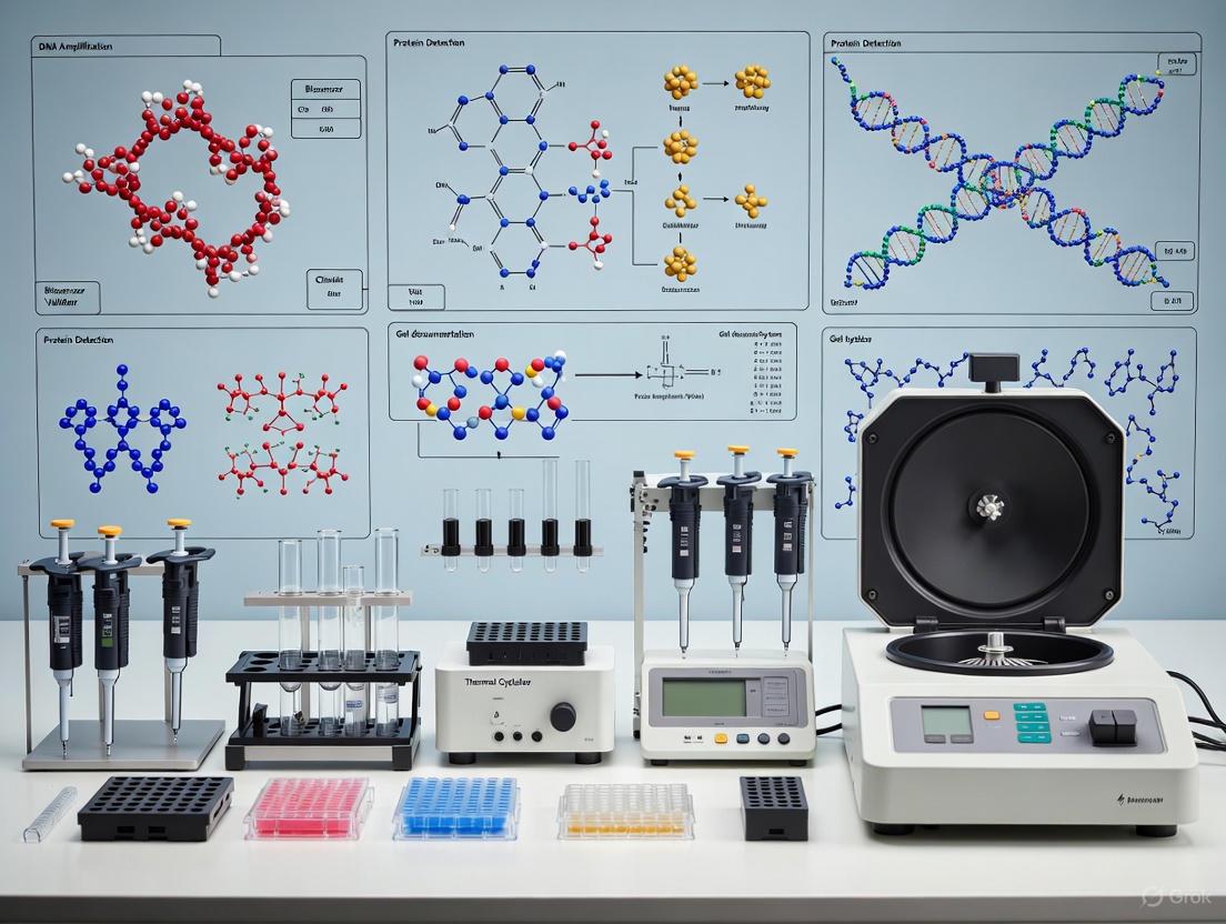
Basic Principles and Core Components of a Biosensor
A biosensor functions through the coordinated operation of three fundamental components: a biorecognition element, a transducer, and a signal processing system [2] [5] [6].
Biorecognition Elements
The bioreceptor is a biological or biomimetic molecule that provides specificity by selectively interacting with the target analyte. Key types include:
- Antibodies: Affinity-based receptors that bind specifically to antigens, widely used in immunosensors [7] [2].
- Enzymes: Biocatalytic receptors that convert a specific substrate into a measurable product, often employed for metabolic analytes like glucose and lactate [7] [2].
- Nucleic Acids (DNA/RNA): Single-stranded sequences that recognize their complementary strand (genosensors) or specific non-nucleic acid targets (aptamers) via base pairing or structural affinity [7] [2].
- Molecularly Imprinted Polymers (MIPs): Synthetic polymers with tailor-made recognition sites mimicking natural molecular interactions, offering enhanced stability [7] [2].
Transducers
The transducer converts the biological interaction into a measurable electronic signal. The primary transduction modalities include:
- Electrochemical: Measures changes in electrical properties (current, potential, impedance) resulting from bio-recognition events [7] [3].
- Optical: Detects variations in light properties (wavelength, intensity, polarization, fluorescence) upon analyte interaction [7] [8].
- Piezoelectric: Sensitive to mass changes on the sensor surface through frequency variations [2].
- Thermometric: Monitors heat changes associated with biochemical reactions [6].
Signal Processing and Readout
The electronic system amplifies, processes, and converts the transducer's analog signal into a user-interpretable digital output displayed numerically, graphically, or via images [2].
The following diagram illustrates the integrated workflow and logical relationships between these core components.
Biosensing Technologies: A Comparative Guide
This section objectively compares the performance, operational principles, and experimental protocols of major biosensor classes relevant to clinical research.
Performance Comparison of Major Biosensor Types
Table 1: Comparative analysis of major biosensor classes based on transduction mechanism.
| Biosensor Type | Principle | Key Clinical Analytes | Limit of Detection | Response Time | Advantages | Disadvantages |
|---|---|---|---|---|---|---|
| Electrochemical [7] [3] | Measures change in current (amperometry), potential (potentiometry), or impedance (impedimetry) from redox reactions. | Glucose, drugs, pathogens, hormones [3] [4]. | Low to sub-nanomolar [3]. | Seconds to minutes [7]. | High sensitivity, portability, low cost, suitable for miniaturization and multiplexing [3]. | Signal can be affected by sample matrix (e.g., pH, conductivity). |
| Optical [7] [8] | Detects change in light properties (e.g., surface plasmon resonance, fluorescence). | Pathogens (SARS-CoV-2), proteins, toxins [7] [8]. | High (e.g., single molecule level possible) [8]. | Minutes [7]. | High sensitivity and specificity, real-time monitoring capability [8]. | Instrumentation can be bulky and expensive; susceptible to ambient light interference. |
| Piezoelectric [2] | Measures frequency change of a crystal due to mass change from analyte binding. | Pathogens, proteins, volatile compounds. | Nanogram mass changes [2]. | Minutes to hours. | Label-free detection, real-time output. | Sensitive to viscosity and temperature changes, not suitable for all sample types. |
| Thermometric [6] | Measures enthalpy change from a biochemical reaction. | Enzymes, metabolites, toxins. | Varies with analyte. | Minutes. | Versatile for any reaction involving heat change. | Requires precise temperature control; non-specific reactions can cause interference. |
Experimental Protocols for Key Biosensor Classes
Standardized experimental protocols are fundamental for obtaining reproducible and reliable data in biosensor research and development.
Experimental Protocol for an Electrochemical Immunosensor
This protocol is typical for detecting pathogens like Salmonella or biomarkers in clinical samples [7] [3].
- Electrode Preparation: Clean the working electrode (e.g., gold, glassy carbon) sequentially with alumina slurry and solvents, then dry.
- Surface Functionalization: Immerse the electrode in a solution containing specific capture antibodies (e.g., anti-Salmonella) for a set period to allow physical adsorption or covalent binding via linkers like cysteamine-glutaraldehyde [7] [5].
- Blocking: Treat the functionalized surface with a blocking agent, such as Bovine Serum Albumin (BSA) or polyethylene glycol (PEG), to minimize non-specific adsorption of other proteins or matrix components from the sample [6].
- Sample Incubation: Apply the clinical sample (e.g., diluted serum, buffer extract) to the sensor surface and incubate to allow target antigen to bind to the immobilized antibody.
- Washing: Rinse the electrode thoroughly with buffer to remove unbound molecules.
- Signal Measurement & Transduction: Perform electrochemical impedance spectroscopy (EIS) or amperometry in a suitable redox solution (e.g., containing ferro/ferricyanide). The binding of the target pathogen increases impedance or decreases current, which is measured and correlated to analyte concentration [7].
- Regeneration (Optional): For reusable sensors, the surface is regenerated using a low-pH glycine buffer to dissociate the antigen-antibody complex without damaging the immobilized antibody.
Experimental Protocol for an Optical DNA Biosensor
This protocol is used for detecting specific nucleic acid sequences, such as viral RNA (e.g., SARS-CoV-2) or bacterial DNA [7] [8].
- Probe Immobilization: Covalently immobilize thiol- or amino-labeled single-stranded DNA (ssDNA) probe sequences onto a functionalized surface (e.g., a gold chip for thiol chemistry or a CMOS-based photodiode sensor silanized with APTES/glutaraldehyde) [5].
- Blocking: Passivate the remaining surface areas with BSA or mercaptohexanol to prevent non-specific DNA adsorption.
- Hybridization: Introduce the sample containing the target DNA/RNA. If the target is complementary, it hybridizes with the immobilized probe.
- Signal Transduction (Fluorescence-based):
- For a label-free detection, an intercalating dye that fluoresces upon binding to double-stranded DNA is added.
- For a labeled detection, the target is pre-labeled with a fluorophore. Upon hybridization and subsequent washing, the retained fluorescence on the sensor surface is quantified using a complementary metal-oxide-semiconductor (CMOS) photodetector or a laser-induced fluorescence reader [5].
- Data Analysis: The fluorescence intensity is measured and is directly proportional to the amount of hybridized target, allowing for quantification.
The workflow for this DNA-based optical detection is summarized below.
The Scientist's Toolkit: Essential Research Reagents and Materials
The development and operation of robust biosensors rely on a suite of specialized reagents and materials. The following table details key components for assembling and experimenting with biosensing platforms.
Table 2: Key research reagent solutions and materials for biosensor development and experimentation.
| Item | Function/Description | Example Use Cases |
|---|---|---|
| Biorecognition Elements | Provides specificity by binding the target analyte. | All biosensor types. |
| Transducer Chips | The solid support where biorecognition occurs and is transduced. | CMOS chips for integrated sensing [5]; Gold electrodes for electrochemical and SPR sensors [5]. |
| Chemical Linkers | Molecules that form a covalent bond between the bioreceptor and transducer surface. | APTES & Glutaraldehyde for oxide surfaces [5]; Thiol compounds (e.g., cysteamine) for gold surfaces [5]. |
| Blocking Agents | Proteins or polymers used to cover unused surface area to prevent non-specific binding. | Bovine Serum Albumin (BSA), casein, or polyethylene glycol (PEG) [6]. |
| Redox Probes | Electroactive molecules used to generate or carry current in electrochemical sensors. | Potassium ferrocyanide/ferricyanide in buffer solution for impedance measurements [7]. |
| Signal Generation Tags | Labels that produce a measurable signal upon analyte binding. | Fluorophores (e.g., fluorescein) for optical detection [7] [8]; Enzyme labels (e.g., Horseradish Peroxidase - HRP) for catalytic signal amplification [7]. |
Validation Frameworks for Clinical Analysis
Translating a biosensor from a research prototype to a clinically applicable tool demands rigorous validation against regulatory standards to ensure reliability, reproducibility, and accuracy [9].
Key Analytical Validation Parameters
Validation requires systematic assessment of key performance parameters as per guidelines from the International Council for Harmonisation (ICH), FDA, and EMA [9]:
- Specificity/SELECTIVITY: The biosensor's ability to measure the analyte accurately in the presence of interfering substances commonly found in the sample matrix (e.g., lipids, bilirubin, unrelated proteins in blood) [9].
- Accuracy: The closeness of agreement between the value found by the biosensor and the accepted true value or reference method result (e.g., mass spectrometry) [9].
- Precision: The degree of agreement among a series of measurements taken from the same homogeneous sample under prescribed conditions. This includes repeatability (within-run) and intermediate precision (between-run, between-days, between-analysts) [9].
- Linearity & Range: The ability of the method to elicit results that are directly proportional to analyte concentration within a given range. The range is the interval between the upper and lower concentration for which linearity, accuracy, and precision have been established [9].
- Limit of Detection (LOD) & Limit of Quantification (LOQ): LOD is the lowest analyte concentration that can be detected but not necessarily quantified. LOQ is the lowest concentration that can be quantified with acceptable accuracy and precision [9] [2].
- Robustness/Ruggedness: A measure of the method's capacity to remain unaffected by small, deliberate variations in procedural parameters (e.g., temperature, pH, incubation time), indicating its reliability during normal usage [9].
Addressing Challenges in Clinical Implementation
Despite their potential, several challenges impede the widespread clinical adoption of biosensors:
- Matrix Effects: Complex biological fluids like blood, saliva, or sweat can contain numerous interferents that affect sensor performance. Strategies to mitigate this include sample purification, dilution, and effective surface blocking protocols [7] [6].
- Reproducibility and Stability: Achieving consistent manufacturing and ensuring long-term stability of the biorecognition element (especially enzymes and antibodies) under storage and operating conditions remains a hurdle [7] [6].
- Standardization and Regulatory Hurdles: The lack of universally accepted standardization protocols makes it difficult to compare data from different biosensors. Navigating the regulatory approval process for in-vitro diagnostics is complex and time-consuming [7] [9].
- Data Security: For connected devices like wearable biosensors, ensuring the security and privacy of transmitted patient data is paramount [6].
Future research is focused on developing synthetic and robust bioreceptors (e.g., MIPs, aptamers), integrating nanotechnology for enhanced sensitivity, and creating fully automated, miniaturized lab-on-a-chip devices for point-of-care testing that meet all validation requirements [1] [5] [6].
The reliance on self-reported data, such as patient surveys and retrospective interviews, has long been a limitation in clinical and research settings. These methods are susceptible to recall bias, subjective interpretation, and cannot capture continuous, real-time physiological data. For researchers and drug development professionals, this introduces significant variability and uncertainty. The emergence of biosensors offers a paradigm shift, enabling passive, ecological monitoring of physiological states in naturalistic environments. This guide objectively compares the performance of traditional self-report methods against modern biosensor-based approaches and their combination, providing experimental data to inform validation protocols for clinical analysis research.
Performance Comparison: Self-Report vs. Biosensor-Based Monitoring
The table below summarizes quantitative performance data from a naturalistic experimental study that directly compared Ecological Momentary Assessment (EMA—a structured self-report methodology) and Ecological Physiological Assessment (EPA—using wearable biosensors) during a high-stakes examination period versus a control week [10] [11].
Table 1: Performance Comparison of Stress Monitoring Methodologies
| Monitoring Methodology | Primary Data Source | Key Measured Parameters | Classification Error Rate (Individualized Models) | Key Findings & Limitations |
|---|---|---|---|---|
| Ecological Momentary Assessment (EMA) | Self-Report | Subjective stress exposure, negative affect, positive affect [11] | 33.45% (1603/4793 beeps) [10] | • High subject burden & intrusive [11]• Prone to careless responses & sparse sampling [11]• Directly captures subjective state |
| Ecological Physiological Assessment (EPA) | Wearable Biosensors | Heart rate (HR), skin conductance (SC) [11] | 36.11% (1648/4565 beeps) [10] | • Physiological arousal is context-neutral (e.g., also linked to positive affect) [10] [11]• Requires psychological context for accurate interpretation [10] |
| Combined EMA & EPA | Self-Report & Biosensors | Subjective mood & autonomic physiology | 29.87% (1363/4565 beeps) [10] | • Provides most comprehensive picture• Optimal classification of stress state [10] |
A critical finding from this study is that individualized models (referenced against an individual's own baseline data) significantly outperformed group-based models across all inputs [10]. This underscores the importance of personalized baselines in biosensor validation and clinical application.
Experimental Protocols for Validating Monitoring Approaches
Protocol: Naturalistic Stress Monitoring with EMA and EPA
This protocol details the methodology used to generate the comparative data in Table 1 [10] [11].
- Objective: To determine the utility of EMA and physiological arousal measured through wearable biosensors in detecting ecologically relevant stress states.
- Population: 83 first-year medical and biomedical science students with no history of psychiatric illness.
- Design: A within-subjects study where each participant completed two monitoring weeks:
- Stress Week: A week culminating in a high-stakes examination.
- Control Week: A week without examinations, occurring on average 16 days from the stress week.
- Data Collection:
- EMA: Repeated questionnaires ("beeps") assessing subjective stress exposure, mood (positive/negative affect), and activities.
- EPA: Continuous recording of autonomic physiological markers (e.g., skin conductance, heart rate) using wearable biosensors worn on the non-dominant hand.
- Data Analysis:
- Generalized linear mixed-effects models were used to investigate the impact of the examination period on subjective and physiological outcomes.
- Machine learning models (individualized, using a leave-one-beep-out approach) were trained to classify data points as belonging to the examination or control week using EMA data, EPA data, or a combination of both.
Protocol: General Biosensor Analytical Validation
For biosensors targeting specific analytes (e.g., glucose), validation follows a rigorous analytical framework [12]. Key parameters include:
- Accuracy: Benchmarking biosensor readings against a reference method (e.g., laboratory-based glucose assay or HPLC) across the clinically relevant range [12].
- Precision: Assessing the consistency of readings via repeated testing of the same concentration under the same conditions [12].
- Sensitivity & Specificity: Determining the lowest detectable concentration and testing for cross-reactivity with interfering substances [12].
- Linearity: Establishing a proportional relationship between the biosensor’s output and the analyte concentration over a specified range [12].
Visualizing Methodological Workflows and Data Interpretation
Experimental Workflow for Naturalistic Monitoring
The following diagram illustrates the workflow of the comparative study on stress monitoring.
Interpreting Biosensor Data in Context
A key challenge in using biosensors for mental health monitoring is the ambiguous nature of physiological arousal. The following diagram outlines the necessary steps for accurate data interpretation.
The Scientist's Toolkit: Essential Research Reagents and Materials
Table 2: Key Materials for Ecological Monitoring and Biosensor Research
| Item | Function in Research |
|---|---|
| Wearable Biosensor | Device for passive, continuous physiological data acquisition (e.g., heart rate, skin conductance) in real-world settings [10] [11]. |
| EMA Platform (Software) | Digital platform for administering repeated subjective questionnaires to capture self-reported mood, stress, and context [10] [11]. |
| Reference Method Assays | Gold-standard laboratory methods against which biosensor accuracy is validated [12]. |
| Biological Recognition Element (BRE) | Key component of a biosensor that provides specificity by binding the target analyte [13]. |
| Signal Amplification Reagents | Reagents used to enhance the signal-to-noise ratio and lower the detection limit [13]. |
In clinical and research settings, psychophysiological measures provide objective, non-invasive insights into the functioning of the autonomic nervous system (ANS). These measures are crucial for understanding an individual's internal state beyond subjective self-reports, which can be biased or limited [14] [15]. The core constructs—arousal, reactivity, and regulation—are fundamental to interpreting these physiological signals. Arousal represents a general state of physiological activation and preparedness. Reactivity refers to the phasic changes in physiological activity in response to specific stimuli or challenges. Regulation describes the capacity to modulate emotional and physiological responses, facilitating adaptation to environmental demands [14] [16]. These constructs are transdiagnostically implicated across a wide spectrum of psychiatric and physical health conditions, making their accurate measurement vital for both clinical assessment and therapeutic development [14] [17].
The following diagram illustrates the relationship between these core constructs and their corresponding physiological measurement systems.
Figure 1: Relationship between core psychophysiological constructs and their primary physiological measures. Solid lines indicate primary associations, while dashed lines represent secondary relationships.
Physiological Measures and Their Neural Correlates
Electrodermal Activity (EDA), Heart Rate (HR), and Heart Rate Variability (HRV) are among the most widely used peripheral indicators of autonomic nervous system activity. Each measure provides unique and complementary information about the sympathetic and parasympathetic branches of the ANS [14] [18].
Electrodermal Activity (EDA): EDA is a pure measure of sympathetic nervous system (SNS) activity, as sweat glands are innervated solely by sympathetic nerves [14]. It reflects changes in the skin's electrical conductance in response to sweat secretion, which increases with emotional arousal, cognitive load, or stress [19] [18]. The amygdala directly mediates skin conductance responses to arousing stimuli, making EDA a sensitive indicator of emotional reactivity [14].
Heart Rate (HR): Defined as the number of heartbeats per minute, HR is influenced by both sympathetic and parasympathetic branches of the ANS [14] [20]. It typically increases with stress, emotional arousal, and cognitive engagement [14] [15]. Tonic cardiovascular function and reactivity are implicated across various forms of psychopathology, including anxiety disorders and posttraumatic stress disorder [14].
Heart Rate Variability (HRV): HRV refers to the variation in time intervals between consecutive heartbeats (interbeat intervals) [19] [18]. It is broadly considered a marker of psychological well-being and cardiovascular fitness [14]. Higher HRV generally reflects greater parasympathetic (vagal) influence and is associated with better emotional regulation capacity, adaptive stress responses, and overall health [14] [17]. Decreased HRV is transdiagnostically associated with various psychiatric conditions and increased cardiovascular risk [14] [19].
The neural circuitry governing these peripheral measures involves key brain structures, including the hypothalamus, amygdala, and ventromedial prefrontal cortex (vmPFC) [14]. These same structures are primary targets of psychological interventions like cognitive behavioral therapy (CBT), suggesting that psychophysiological measures may serve as proxy indicators of therapeutic effectiveness in modifying these neural circuits [14].
Biosensor Validation in Experimental Research
The emergence of wearable biosensors has revolutionized psychophysiological data collection, enabling measurements in naturalistic settings outside traditional laboratory environments. However, the accuracy and reliability of these devices must be rigorously validated against gold-standard laboratory equipment, particularly for clinical research applications [21] [18].
Performance of Wearable Biosensors
Multiple independent studies have evaluated the Empatica E4 wristband, a popular research-grade wearable device. The validation findings consistently show varying levels of agreement with gold-standard equipment across different physiological measures and experimental conditions.
Table 1: Validation Summary of Empatica E4 Performance Against Gold-Standard Devices
| Physiological Measure | Experimental Condition | Correlation/Agreement with Gold Standard | Key Findings | Study Reference |
|---|---|---|---|---|
| HRV (Time Domain, e.g., RMSSD) | Baseline/Rest | High (r > 0.72) | Strong agreement during stationary, low-movement conditions. | [21] |
| Video Clip (Emotional Stimuli) | High (r > 0.71) | Reliable in response to controlled emotional stimuli. | [21] | |
| No-Risk Driving | Good (r > 0.67) | Acceptable performance in low-demand real-world tasks. | [21] | |
| Conversation/Dyadic Interaction | Medium | Reduced but significant correlation; lower than at rest. | [18] | |
| HRV (Frequency Domain) | Baseline/Rest | Sufficient (r > 0.58) | Moderate reliability for spectral power analysis. | [21] |
| Low-Risk Driving | Sufficient (r > 0.52) | Moderate reliability in mildly demanding scenarios. | [21] | |
| Keyboard Typing/Slow Walking | Very Low (r = 0.00-0.07) | Poor agreement during activities involving arm/hand movement. | [18] | |
| Heart Rate (HR) / Interbeat Intervals (IBI) | Rest | High/ Nearly Perfect | Excellent agreement for mean IBI and derived HR. | [18] |
| Social Conversation | High | Maintains good accuracy during conversational states. | [18] | |
| Electrodermal Activity (EDA) | Multiple Conditions (Rest, Stress, Interaction) | No Correlation/ Poor | Consistently failed to produce reliable EDA data across studies. | [21] [18] |
Impact of Experimental Context on Data Quality
The validation data clearly demonstrates that experimental context and participant movement significantly impact data quality. The Empatica E4 shows its highest reliability for cardiac measures (HR and HRV time-domain parameters) during resting states and low-movement conditions [21] [18]. However, performance degrades for HRV during activities involving arm or hand movements, such as typing or gesturing during conversation [18]. Most notably, multiple independent validation studies have found that the E4 fails to produce reliable EDA data compared to laboratory standard systems, which typically use electrodes placed on the fingers or palms [21] [18]. This limitation is critical for researchers aiming to measure sympathetic nervous system activity via EDA in clinical or experimental settings.
Detailed Experimental Protocols
To contextualize the validation data, below are detailed methodologies from key studies that have evaluated biosensor performance or utilized these physiological measures in applied research.
Protocol: Biosensor Validation in a Stressful Driving Environment
This study focused on testing the accuracy of the Empatica E4 for detecting HRV and EDA in stress-inducing and growing-risk driving scenarios, comparing it against a gold-standard configuration [21].
- Participants: 14 healthy subjects (5 male, 9 female; mean age = 33 years).
- Recording Devices: Test device was the Empatica E4 wristband worn on the non-dominant hand. The gold-standard system consisted of:
- eegomylab amplifier with Ag/AgCl electrodes for ECG (Lead II configuration on chest).
- EDA recorded using Ag/AgCl electrodes alternately placed on fingers or shoulder.
- Experimental Conditions: Six conditions were presented in a single session:
- Baseline: 3 minutes sitting quietly with eyes closed.
- Video Clip: Viewing emotionally eliciting video clips from a validated database.
- Scream: Exposure to a sudden, loud auditory stimulus.
- No-Risk Driving: Virtual driving with no hazardous events.
- Low-Risk Driving: Virtual driving with moderate-risk scenarios.
- High-Risk Driving: Virtual driving with high-risk, collision-prone scenarios.
- Data Analysis: Agreement and reliability were assessed using Bland-Altman analysis and Spearman's correlation coefficient. Robust semi-automatic algorithms were applied for PPG-derived HRV signal reconstruction and EDA analysis to enhance signal quality [21].
Protocol: Studying Irritability in Youth via Ambulatory Biosensing
This research protocol leverages ambulatory biosensing to examine physiological mechanisms during exposure-based cognitive behavioral therapy (CBT) for youth with clinically impairing irritability [14].
- Participants: 40 youth (ages 8-17) undergoing six in-person exposure treatment sessions.
- Recording Device: Empatica EmbracePlus worn ambulatorily during therapy sessions.
- Measured Variables:
- Blood Volume Pulse (BVP): Sampled to derive HR and HRV.
- Electrodermal Activity (EDA): Continuously recorded as a measure of sympathetic arousal.
- Clinical Outcomes: Irritability was assessed at baseline, weekly during treatment, and at 3- and 6-month follow-ups using the Clinical Global Impressions Scale (CGI) and the Affective Reactivity Index (ARI).
- Data Analysis: Multilevel modeling is used to assess within- and between-person changes in physiological arousal (EDA, HR) and regulation (HRV) throughout therapy and to determine if these physiological measures predict treatment response [14].
The workflow for a typical psychophysiological validation study is systematized in the following diagram.
Figure 2: Standardized workflow for a biosensor validation study, outlining key stages from participant recruitment to data analysis and conclusion.
The Scientist's Toolkit: Essential Research Reagents and Materials
For researchers designing studies involving psychophysiological measurements, the following table details key equipment and assessment tools essential for rigorous experimental execution.
Table 2: Essential Materials for Psychophysiological Research
| Item Name | Category | Specifications / Model Examples | Primary Function in Research |
|---|---|---|---|
| Research-Grade Wearable | Biosensor | Empatica E4/Empatica EmbracePlus | Wireless, ambulatory recording of BVP (for HR/HRV), EDA, acceleration, and temperature in naturalistic settings. |
| Gold-Standard ECG System | Laboratory Equipment | MindWare Mobile Impedance Cardiograph, BIOPAC Systems with Ag/AgCl electrodes | High-fidelity recording of electrocardiogram (ECG) for validation of HR and HRV metrics derived from wearables. |
| Gold-Standard EDA System | Laboratory Equipment | eegomylab amplifier with auxiliary sensors; Ag/AgCl electrodes (e.g., H124SG) | Clinical-grade measurement of electrodermal activity, typically from palmar or finger sites, for validation of wearable EDA. |
| Generalized Anxiety Disorder Scale (GAD-7) | Clinical Assessment | 7-item self-report questionnaire | Screening and severity measurement of generalized anxiety symptoms; scores ≥10 indicate potential clinical relevance. |
| Affective Reactivity Index (ARI) | Clinical Assessment | Clinician-, parent-, and child-report versions | Multi-informant measure of irritability severity, capturing core dimensions of increased proneness to anger. |
| Visual Analog Scale (VAS) for Anxiety | State Assessment | Single-item self-report (0-10 scale) | Rapid assessment of state anxiety, particularly in relation to specific situations (e.g., patient interaction). Scores ≥7 often indicate high anxiety. |
The objective measurement of arousal, reactivity, and regulation via EDA, HR, and HRV provides a critical window into autonomic nervous system function. Validation studies consistently demonstrate that wearable biosensors like the Empatica E4 offer a viable method for collecting cardiac data (HR and certain HRV parameters) in real-world settings, though with important caveats. Their performance is strongest during rest and low-movement conditions but can be compromised by motion artifacts. A significant and consistent limitation across independent validations is the poor reliability of wrist-based EDA measurements [21] [18].
For clinical researchers and drug development professionals, these findings underscore the necessity of:
- Contextual Device Selection: Choosing measurement technologies appropriate for the experimental context (e.g., laboratory standard for EDA, wearables for ambulatory HR/HRV).
- Rigorous Validation: Conducting pilot studies to validate specific device-parameter combinations for their intended research population and setting.
- Methodological Transparency: Clearly reporting device limitations, data processing pipelines, and potential artifacts in research publications.
The integration of validated psychophysiological biomarkers into clinical trials holds promise for providing objective, quantifiable endpoints for assessing treatment efficacy, particularly for interventions targeting emotional and autonomic regulation.
Market Dynamics and Growth Projections for Wearable and Point-of-Care Biosensors
The biosensor market is experiencing rapid transformation, driven by technological convergence and an increasing shift toward decentralized healthcare. Wearable biosensors and point-of-care (POC) biosensors represent two pivotal segments enabling this shift, each with distinct growth dynamics, technological foundations, and clinical applications. Wearable biosensors are defined as devices worn on the body to monitor physiological parameters in a continuous, non-invasive manner [22] [23]. In contrast, point-of-care biosensors are portable devices used for detecting and measuring biological molecules at the site of patient care, providing rapid diagnostic results [24]. Together, these technologies are moving diagnostic and monitoring capabilities from central laboratories to the patient's home, clinic, or hospital bedside, thereby revolutionizing personalized healthcare management and chronic disease monitoring.
The market growth for these technologies is fueled by several powerful macro-trends. The global aging population is increasing the prevalence of chronic diseases, creating demand for continuous monitoring and convenient testing solutions [24]. Concurrently, rising healthcare expenditure worldwide is pushing cost-effective solutions that can reduce hospital visits and improve patient outcomes [24]. Technological advancements in miniaturization, artificial intelligence (AI), and wireless communication have further accelerated development, making biosensors more accurate, affordable, and user-friendly [22] [23]. Additionally, the COVID-19 pandemic served as a significant catalyst, highlighting the critical need for rapid testing and remote patient monitoring solutions and accelerating both adoption and innovation in the biosensor field [25] [24].
Table 1: Global Market Overview for Wearable and Point-of-Care Biosensors
| Market Metric | Wearable Biosensors | Point-of-Care Biosensors |
|---|---|---|
| Market Size (2024/2025) | USD 2,293.6 million (2025) [26] | USD 83.65 billion (2024) [24] |
| Projected Market Size (2033/2034) | USD 9,177.65 million (2033) [26] | USD 186.71 billion (2034) [24] |
| Compound Annual Growth Rate (CAGR) | 22.4% (2025-2033) [26] | 8.36% (2024-2034) [24] |
| Key Market Drivers | Rising health awareness, chronic disease management, AI integration [26] | Growing demand for personalized healthcare, increasing healthcare expenditure [24] |
| Major Challenges | Privacy concerns, data security, signal accuracy across skin types [26] [27] | High development and production costs, limited accuracy compared to lab tests [24] |
Comparative Performance Analysis: Technological Capabilities and Clinical Validation
Application Landscape and Clinical Performance
Wearable and POC biosensors serve complementary yet distinct roles in the healthcare ecosystem. Wearable biosensors excel in continuous, longitudinal monitoring of physiological parameters, making them ideal for chronic disease management, fitness tracking, and early warning systems. They typically monitor parameters like heart rate, oxygen saturation (SpO₂), respiration rate, skin temperature, and activity levels [22] [25]. Emerging devices are also targeting metabolites in biofluids like sweat, such as lactate and glucose [22] [23]. In contrast, POC biosensors are designed for specific, on-demand diagnostic tests, providing a snapshot of a patient's health status at a given moment. Their dominant applications include glucose monitoring for diabetes, infectious disease testing (e.g., HIV, hepatitis C), and pregnancy testing [24].
A direct comparative study of these platforms for a specific clinical application highlights their performance differences. Research evaluating the detection of Hepatitis B virus (HBV) in a clinical setting compared a microfluidic chip-based POC system against a sweat-based wearable electrochemical biosensor [28]. The POC microfluidic system, which integrated recombinase polymerase amplification with CRISPR-Cas12a for HBV DNA detection, demonstrated superior diagnostic accuracy with a sensitivity of 96.2% and a specificity of 97.5% [28]. The wearable biosensor, while more portable and non-invasive, showed moderate accuracy with a sensitivity of 84.5% and a specificity of 88.0% [28]. This performance gap underscores a common trade-off: POC systems often prioritize clinical-grade diagnostic precision, while wearables prioritize accessibility, comfort, and continuous monitoring.
Table 2: Performance Comparison of Representative Biosensor Types
| Parameter | Wearable Biosensor (e.g., Sweat-based for HBV) | POC Biosensor (e.g., Microfluidic Chip for HBV) | Traditional Lab (qPCR reference) |
|---|---|---|---|
| Sensitivity | 84.5% [28] | 96.2% [28] | 100% (Assumed Gold Standard) |
| Specificity | 88.0% [28] | 97.5% [28] | 100% (Assumed Gold Standard) |
| Limit of Detection (LOD) | 10³ IU/mL [28] | 10¹ IU/mL [28] | Varies by assay, typically very low |
| Time-to-Result | ~30 minutes [28] | ~20 minutes [28] | Several hours to days |
| Key Advantage | Continuous monitoring, non-invasive sampling, portability [28] | High accuracy, minimal instrumentation, small sample volume [28] | High accuracy and throughput |
| Major Limitation | Lower accuracy at low viral loads [28] | Requires sample collection and loading [28] | Time-consuming, requires centralized lab |
Operational Feasibility and Integration with Digital Health
The operational requirements and integration potential of these two biosensor categories differ significantly. Wearable biosensors are designed for autonomy and prolonged use, often featuring wireless communication modules (e.g., Bluetooth) to transmit data continuously to smartphones or cloud-based platforms [22] [25]. This facilitates remote patient monitoring, allowing healthcare providers to track patients' health status in real-time outside clinical settings. A study on monitoring COVID-19 patients demonstrated this effectively: a wearable armband (Everion) transmitted data to a cloud analytics engine (Biovitals Analytics Engine), which generated a machine learning-derived Biovitals Index (BI) that correlated with viral load and predicted clinical deterioration with high sensitivity (94.1%) [25].
POC biosensors, while often used in professional settings like clinics or emergency rooms, are also increasingly found in home-testing formats (e.g., blood glucose meters). The dominant technology platforms in the POC segment are immunoassays, microfluidics, and dipsticks [24]. Among these, immunoassays hold the largest market share due to their high sensitivity and specificity for detecting proteins, hormones, and infectious disease antigens [24]. Microfluidic platforms, which manipulate small fluid volumes in tiny channels, are crucial for creating integrated "sample-in-answer-out" devices that automate complex assay steps, making them user-friendly and suitable for resource-limited settings [28] [29].
Experimental Protocols for Biosensor Validation
Protocol for Validating a Wearable Biosensor in Remote Patient Monitoring
Objective: To evaluate the accuracy and clinical utility of a wearable biosensor for remote monitoring and early detection of health deterioration in patients with infectious diseases [25].
Methodology:
- Participant Recruitment: Recruit patients with a confirmed condition (e.g., COVID-19) managed in isolation wards or at home. Exclude patients requiring intensive care at baseline [25].
- Device and Platform:
- Wearable Biosensor: Use a clinically validated device (e.g., Everion armband) that captures multi-parameter data: heart rate, heart rate variability, respiration rate, oxygen saturation, blood pulse wave, skin temperature, and actigraphy [25].
- Software Platform: Employ a connected platform comprising a patient-facing smartphone app, a secured cloud, and a clinician dashboard (e.g., Biovitals Sentinel Platform) [25].
- Study Procedure:
- Patients wear the biosensor for a target of 23 hours per day, removing it only for charging.
- Patients self-report symptoms daily via the smartphone application.
- Physiology data is continuously transmitted via Bluetooth to the smartphone and then to the cloud.
- A machine learning algorithm (e.g., Biovitals Analytics Engine) analyzes the multivariate data stream in real-time to generate a composite health index (e.g., Biovitals Index) [25].
- Reference Standards:
- Compare sensor-measured physiology parameters (e.g., pulse rate, SpO₂) against periodic manual measurements taken by healthcare workers [25].
- Correlate the machine learning-derived health index with clinical status scores (e.g., National Early Warning Score 2 - NEWS2) and virologic load (e.g., RT-PCR cycle threshold values) [25].
- Outcome Measures:
- Primary: Correlation coefficient between sensor data and manual measurements; correlation between the health index and reference standards.
- Secondary: Sensitivity and specificity of the health index in predicting predefined clinical worsening events (e.g., NEWS2 ≥ 5, need for oxygen therapy) [25].
Protocol for a Comparative Study of POC vs. Wearable Biosensors
Objective: To directly compare the diagnostic accuracy, limit of detection, and operational feasibility of a microfluidic POC biosensor versus a wearable biosensor for detecting a specific pathogen (e.g., Hepatitis B Virus) [28].
Methodology:
- Sample Collection: Collect a set of clinical samples (e.g., 200 serum samples) from patients in a relevant clinical setting [28].
- Biosensor Platforms:
- POC Platform: A microfluidic chip system integrating isothermal amplification (e.g., recombinase polymerase amplification) and a CRISPR-based detection system (e.g., CRISPR-Cas12a) for target pathogen DNA [28].
- Wearable Platform: A sweat-based wearable electrochemical biosensor designed to detect pathogen antigens or DNA (e.g., HBsAg and HBV DNA) [28].
- Testing Procedure:
- Test all samples in parallel using both the POC and wearable biosensor platforms according to manufacturer protocols.
- For the wearable, follow specified wear time and sampling procedures.
- Record the time-to-result for each platform.
- Reference Method: Analyze all samples using a gold standard laboratory method, such as quantitative polymerase chain reaction (qPCR), to determine the true positive and negative status [28].
- Data Analysis:
- Calculate sensitivity, specificity, positive predictive value, and negative predictive value for each platform against the qPCR reference.
- Determine the limit of detection (LOD) for each platform using serial dilutions of the target analyte.
- Compare operational metrics such as cost per test, required instrumentation, and sampling invasiveness [28].
Technical Specifications and Research Toolkit
Key Research Reagent Solutions and Materials
The development and validation of advanced biosensors require a suite of specialized reagents and materials. The table below details essential components for research in this field, derived from the experimental protocols cited.
Table 3: Essential Research Reagents and Materials for Biosensor Development
| Reagent/Material | Function/Description | Example Application |
|---|---|---|
| CRISPR-Cas System (e.g., Cas12a) | A gene-editing derived tool that provides highly specific nucleic acid detection; upon binding to target DNA, it exhibits collateral cleavage activity, which can be linked to a reporter signal [28]. | Pathogen DNA detection in microfluidic POC chips [28]. |
| Isothermal Amplification Reagents (e.g., RPA) | Enzymes and primers for amplifying nucleic acids at a constant temperature (e.g., 37-42°C), eliminating the need for a thermal cycler and making it suitable for POC devices [28]. | Target DNA amplification in POC diagnostics [28]. |
| Flexible/Stretchable Electrodes | Conductive materials (e.g., gold, carbon nanotubes, graphene) patterned on flexible substrates (e.g., PET, silicone) to create robust, skin-conformable sensors for wearables [22] [23]. | Electrochemical detection of biomarkers in sweat for wearable sensors [22]. |
| Enzymes & Antibodies (Biorecognition Elements) | Biological molecules that provide high specificity by binding to a target analyte (e.g., glucose oxidase for glucose, antibodies for viral antigens). The core of most biosensors' selectivity [24]. | Glucose monitoring strips, immunoassay-based infectious disease tests [24]. |
| Microfluidic Chip Substrates (e.g., PDMS, PMMA) | Polymers used to fabricate chips with micron-sized channels and chambers that manipulate fluids and integrate sample preparation, reaction, and detection steps [28] [29]. | Integrated "sample-in-answer-out" diagnostic devices [28] [29]. |
| Enzyme Substrates & Chromogens | Chemicals that produce a measurable signal (colorimetric, fluorescent, electrochemical) when acted upon by an enzyme label (e.g., horseradish peroxidase), enabling detection [29]. | Signal generation in lateral flow immunoassays and other POC tests [29]. |
Technology Roadmap and Future Trends
The biosensor landscape is evolving toward greater integration, intelligence, and clinical relevance. Key future trends shaping both wearable and POC biosensors include:
Multimodal Sensing and Hybrid Platforms: The next generation of devices will combine multiple sensing modalities to improve accuracy and gather more comprehensive health data. For instance, hybrid biosensors that integrate PPG with electrocardiogram (ECG) or bioimpedance are emerging to provide more robust cardiovascular profiling [27]. This fusion of data from different physical and chemical sensors helps cross-validate signals and mitigate the limitations of any single method.
Artificial Intelligence and Machine Learning: AI is transforming biosensors from simple data collectors to intelligent diagnostic partners. Machine learning algorithms are being used for multiple purposes: enhancing signal accuracy by filtering out motion artifacts [27], enabling predictive analytics for early health event detection [25], and creating personalized health baselines for individuals. The use of AI-powered analytics engines, as demonstrated in the remote monitoring of COVID-19 patients, showcases the potential for generating actionable clinical insights from continuous data streams [25].
Material Science and Miniaturization: Advances in materials are leading to more comfortable, durable, and discreet devices. Research is focused on developing flexible electronics, biocompatible materials, and even self-healing substrates for long-term wearables [22] [23]. The form factor is also diversifying beyond wrist-worn devices to include smart patches, smart rings, ear-worn devices, and smart textiles [22] [27]. In microfluidics, the drive is toward more integrated and automated "lab-on-a-chip" systems that consolidate complex laboratory workflows into a single, miniaturized cartridge [29].
Expanded Biomarker Panels: A significant frontier is the move from single-analyte detection to multi-analyte panels. For wearables, this means developing sensors that can simultaneously track a wider range of biomarkers in sweat, interstitial fluid, or tears [23]. For POC devices, it involves creating multiplexed platforms that can test for multiple pathogens or disease markers from a single sample [29]. This provides a more holistic view of the patient's physiological status and improves diagnostic confidence.
The landscape of clinical diagnostics is undergoing a significant transformation, moving from traditional laboratory-based assays toward advanced biosensing technologies that promise to address critical unmet needs in both routine and rare disease diagnosis. Traditional diagnostic methods, while established, often suffer from limitations such as prolonged processing times, requirements for sophisticated laboratory infrastructure, and inadequate sensitivity for early-stage detection, particularly for rare conditions [30]. These limitations create substantial gaps in patient care, leading to delayed interventions and poorer health outcomes. The emergence of sophisticated biosensors represents a paradigm shift in diagnostic capabilities, offering potential solutions to these longstanding challenges. These innovative devices combine biological recognition elements with transducers to convert biological responses into quantifiable electrical, optical, or other signals, enabling rapid, sensitive, and specific detection of disease biomarkers [30] [31]. This evolution is particularly crucial for rare diseases, which often face diagnostic delays due to their uncommon presentation and the lack of readily available testing options. The validation and implementation of these biosensing platforms within clinical analysis research frameworks are essential for bridging the diagnostic gap between routine assays and the precise identification of rare conditions, ultimately facilitating earlier intervention and improved patient prognosis.
Traditional Assays: Established Strengths and Inherent Limitations
Traditional diagnostic assays, including enzyme-linked immunosorbent assays (ELISA), cell cultures, and polymerase chain reaction (PCR)-based methods, have formed the backbone of clinical pathology for decades. These methods provide valuable, proven platforms for detecting pathogens, antibodies, and specific biomarkers associated with various diseases. Their standardized protocols and established performance metrics make them reliable tools in well-equipped laboratory settings [31]. For rare diseases such as mesothelioma, hepatoblastoma, and cystic fibrosis, traditional biomarkers—physical indicators measured through these invasive or minimally invasive sampling methods—often deliver qualitative or semi-quantitative results that offer static, single-time-point readouts [32]. This "snapshot" problem fails to capture the dynamic and complex nature of disease progression, presenting an incomplete physiological picture [32].
The limitations of these conventional systems become particularly apparent when addressing urgent diagnostic needs. They typically require complex sample preparation, often involving labeling or tagging, which limits their utility for at-home point-of-care devices and self-monitoring medical wearables [32]. Furthermore, these assays usually depend on bulky equipment, complex operations, electrical power, and trained personnel, making them poorly suited for resource-challenged environments, point-of-care settings, or rapid screening during public health emergencies [32] [31]. The central unmet clinical need is the development of diagnostic tools that retain the robustness of traditional methods while overcoming these fundamental limitations of timeliness, accessibility, and dynamic monitoring capability.
Biosensor Technology: A New Frontier for Clinical Diagnosis
Biosensors are analytical devices that integrate a biological recognition element (bioreceptor) with a transducer to produce a measurable signal proportional to the concentration of a target analyte. This fundamental architecture enables their application across a wide spectrum of diagnostic challenges.
Core Components and Technological Diversity
The performance and applicability of a biosensor are determined by its constituent parts, primarily the biological recognition element and the transducer mechanism, as illustrated in the following workflow:
Biological recognition elements form the critical target-specific component of biosensors. Several types are employed, each with distinct characteristics:
- Enzyme Biosensors: These leverage the specific binding affinity of enzymes for their substrates. Third-generation enzyme biosensors represent significant advancement, eliminating mediators by utilizing direct electron transfer between the enzyme and electrode, thereby improving selectivity and stability [30].
- Antibody Biosensors: These immunosensors utilize immobilized antibodies that bind target antigens, generating detectable optical, electrochemical, or gravimetric signals. When integrated with microfluidics and wireless systems, they enable portable diagnostic devices [30].
- Aptamer Biosensors (Aptasensors): These employ short, single-stranded DNA or RNA oligonucleotides (aptamers) with high specificity and binding affinity for targets ranging from small molecules to entire cells. Their significant advantage includes chemical synthesis, modification, and exceptional stability, making them ideal for point-of-care diagnostics [30].
- Whole-Cell Biosensors: These utilize living cells with synthetic gene circuits to detect target substances, offering a versatile, self-replicating, and cost-effective platform for environmental, food safety, and health monitoring [30].
Transducer mechanisms convert the biological recognition event into a quantifiable signal:
- Electrochemical Transducers detect electrical changes (current, voltage, impedance) resulting from biorecognition events. They are highly sensitive, selective, and conducive to miniaturization for point-of-care platforms [30] [33].
- Optical Transducers measure changes in light properties (wavelength, intensity, polarization) and are suitable for real-time, non-invasive detection, ideal for in vivo applications [30].
- Other Transducers include thermal (measuring enthalpy changes) and mass-based or gravimetric (e.g., quartz crystal microbalances) systems, each with specific applications in pathogen and biomarker detection [30] [31].
Fabrication Techniques and Material Advancements
The performance of biosensors is heavily influenced by fabrication methodologies and materials. Key techniques include:
- Micropatterning and Lithography: These methods provide precise control over sensor shape and size, enhancing sensitivity and accuracy while offering scalability and reproducibility for mass production [30].
- 3D Printing: This enables rapid, cost-effective production of complex sensor designs, facilitating customization for specific applications [30].
- Nanomaterial Integration: Incorporating nanomaterials like graphene, carbon nanotubes, gold nanoparticles, and magnetic nanoparticles significantly enhances sensor performance by increasing the surface area-to-volume ratio and improving electrical characteristics, leading to greater sensitivity and lower detection limits [30].
Comparative Analysis: Biosensors Versus Traditional Assays
The transition from traditional assays to modern biosensors represents a significant advancement in diagnostic capabilities. The following comparative analysis highlights key performance differences and application suitability:
Table 1: Performance Comparison Between Traditional Assays and Modern Biosensors
| Parameter | Traditional Assays (e.g., ELISA, PCR) | Modern Biosensors | Clinical Implications |
|---|---|---|---|
| Analysis Time | Hours to days [31] | Minutes to hours (<30 min for Optimer-based tests) [34] | Enables rapid diagnosis and point-of-care decision making |
| Sample Preparation | Often complex, requiring labeling [32] | Minimal; some enable direct detection in complex matrices [34] | Reduces procedural errors and operator dependency |
| Sample Volume | Relatively large (mL scale) | Small (µL scale) [31] | Enables testing in pediatric and neonatal populations |
| Sensitivity | Variable; can be high in optimized settings | High; e.g., KD=131 nM for Imatinib detection [34] | Crucial for detecting low-abundance biomarkers in early disease |
| Multiplexing Potential | Limited; typically single-analyte | High potential for simultaneous multi-analyte detection [32] | Provides comprehensive metabolic profiling |
| Portability & POC Use | Generally confined to laboratories | High; wearable, handheld formats possible [32] [31] | Expands access to resource-challenged settings |
| Capacity for Continuous Monitoring | Single time-point ("snapshot") [32] | Continuous, real-time monitoring possible [32] | Captures dynamic physiological changes |
Table 2: Comparison of Biosensor Types for Specific Clinical Applications
| Biosensor Type | Target Analytes / Diseases | Key Performance Metrics | Advantages |
|---|---|---|---|
| Electrochemical Aptasensors | Sepsis biomarkers (CRP, PCT, IL-6) [33] | Low LOD, rapid response, POC potential [33] | High specificity, modifiability, miniaturization potential [33] |
| Optamer-Based Lateral Flow (LFD) | Small molecules, infectious diseases [34] | Results in minutes, high sensitivity, no cold chain needed [34] | Cost-effective, scalable, ideal for remote settings [34] |
| Wearable Metabolite Sensors | Glucose, lactate, electrolytes [32] | Continuous, real-time monitoring, non-invasive sampling [32] | Empowers patient self-management, dynamic data |
| Nanoparticle-Based Biosensors | Cancer biomarkers (e.g., miRNAs, RBP4) [30] | Ultra-sensitive detection; results in ~5 minutes [30] | High surface area-to-volume ratio enhances signal |
Experimental Protocols and Research Reagent Solutions
Detailed Methodology: Electrochemical Aptamer-Based (E-AB) Biosensor
The development and validation of a typical E-AB biosensor for sepsis biomarker detection, such as procalcitonin (PCT) or C-reactive protein (CRP), involves a multi-stage experimental protocol [33]:
- Aptamer Selection and Modification: Target-specific aptamers are selected via Systematic Evolution of Ligands by EXponential enrichment (SELEX). Selected aptamers are then chemically modified at their 5' or 3' end with a thiol group for gold electrode surface attachment and a redox tag (e.g., methylene blue) for signal generation.
- Electrode Functionalization: Gold disk or screen-printed electrodes are cleaned and polished. Thiolated aptamers are incubated on the electrode surface to form self-assembled monolayers via Au-S bonds. The surface is then treated with mercaptohexanol to backfill unoccupied gold sites, minimizing non-specific adsorption and orienting the aptamers upright.
- Sensor Assembly and Integration: The functionalized electrode is integrated into a flow cell or a static electrochemical cell, connected to a potentiostat for measurement. The setup can be miniaturized into a portable device with microfluidics for sample handling.
- Electrochemical Measurement and Detection: Square wave voltammetry (SWV) or electrochemical impedance spectroscopy (EIS) is employed. Upon target binding, the aptamer undergoes a conformational change, altering the electron transfer efficiency between the redox tag and the electrode surface, resulting in a measurable change in current (in SWV) or charge transfer resistance (in EIS).
- Calibration and Validation: The sensor is calibrated using standard solutions of the target biomarker with known concentrations. Performance is validated by testing spiked clinical samples (e.g., serum, plasma) and comparing results with reference methods like ELISA.
The Scientist's Toolkit: Essential Research Reagent Solutions
Table 3: Key Reagents and Materials for Biosensor Development
| Reagent/Material | Function in Biosensor Development | Example Application |
|---|---|---|
| Optimer Binders | Synthetic aptamers serving as highly specific biological recognition elements; offer batch-to-batch consistency and standardised conjugation [34]. | Used in lateral flow devices, ELISA, and biosensors for targets like SARS-CoV-2 S1 protein [34]. |
| Gold Nanoparticles (AuNPs) | Signal amplification labels in colorimetric and electrochemical assays; provide high surface area for biomolecule immobilization [30] [31]. | Rapid colorimetric detection of biomarkers like RBP4 within 5 minutes [30]. |
| Magnetic Nanoparticles | Enable efficient separation and concentration of target analytes from complex biological samples, enhancing sensitivity and reducing matrix effects [30]. | Isolation and detection of specific cancer biomarkers such as microRNAs from blood [30]. |
| Functionalized Electrodes | Transducer platform (e.g., screen-printed, gold disk) that converts biological binding events into measurable electrical signals [30] [33]. | Core component in electrochemical biosensors for sepsis biomarkers [33]. |
| Microfluidic Chips | Miniaturized platforms for manipulating small fluid volumes (µL-nL), enabling automation, integration, and portability of biosensing systems [30] [31]. | Lab-on-a-chip devices for point-of-care viral disease diagnosis [31]. |
Addressing Unmet Needs in Rare and Critical Disease Diagnosis
Biosensor technology demonstrates particular promise in addressing diagnostic challenges in areas where traditional assays fall short, including rare diseases and time-sensitive critical conditions.
The diagnostic pathway for rare diseases is being transformed through the integration of advanced biosensing technologies, as visualized below:
Rare Disease Diagnosis: For conditions like mesothelioma, hepatoblastoma, and cystic fibrosis, diagnosis is often delayed because symptoms are non-specific and prevalence is low, reducing clinical familiarity [30]. Biosensors can target specific protein biomarkers or genetic mutations associated with these rare conditions, enabling rapid and accurate testing even in non-specialized settings. This capability can drastically reduce the diagnostic odyssey for patients.
Sepsis Management: Sepsis is a life-threatening condition where outcomes depend critically on the speed of antibiotic intervention. Electrochemical aptamer-based biosensors are being developed to detect key sepsis biomarkers (C-reactive protein, procalcitonin, interleukin-6) with high sensitivity and rapid response times (minutes rather than hours) directly at the point-of-care [33]. This performance allows for faster diagnosis and therapeutic monitoring, addressing a critical unmet need in emergency and intensive care medicine.
Viral Disease Detection: The COVID-19 pandemic highlighted the urgent need for rapid and accurate viral diagnostics. Miniaturized biosensors, including those using surface plasmon resonance (SPR) and other transducer principles, have been engineered for direct detection of viral antigens or antibodies in complex fluids like saliva, bypassing the need for centralized laboratories [31]. These platforms match or surpass conventional standards regarding time, precision, and cost, proving invaluable for public health response.
Biosensor technology represents a fundamental advancement in clinical diagnostics, effectively bridging the gap between routine laboratory assays and the precise, timely detection of rare diseases. The comparative data and experimental protocols outlined in this guide demonstrate a clear trend: biosensors consistently offer superior performance in terms of speed, sensitivity, and potential for point-of-care use compared to traditional methods. The integration of novel recognition elements like aptamers, combined with advancements in nanotechnology and transducer design, is paving the way for a new generation of diagnostic tools.
Future developments in this field will likely focus on several key areas: enhancing the multiplexing capacity to simultaneously detect panels of biomarkers for improved diagnostic accuracy; advancing continuous monitoring capabilities for chronic disease management and therapeutic drug monitoring; and improving the integration of biosensors with digital health technologies and the Internet of Things (IoT) for real-time data analytics and remote patient management [32]. Furthermore, overcoming translational barriers such as rigorous clinical validation, standardization of manufacturing, and demonstration of cost-effectiveness will be crucial for the widespread adoption of these technologies in routine clinical practice. As these innovations mature, biosensors are poised to fundamentally transform the diagnostic landscape, enabling earlier disease detection, more personalized treatment strategies, and expanded access to advanced diagnostics across diverse healthcare settings.
Biosensor Technologies in Action: Methodologies and Clinical Use Cases
A 5-Step Framework for Selecting the Right Biosensor for Your Context
Biosensors have emerged as indispensable tools in clinical analysis research, enabling the precise and real-time detection of biological markers for disease diagnosis, therapeutic monitoring, and drug development [35] [36]. These devices combine a biological recognition element with a physicochemical detector to generate measurable signals from biological interactions [36]. For researchers and drug development professionals, selecting the appropriate biosensor requires careful consideration of multiple technical and validation parameters to ensure reliable results in specific experimental or clinical contexts. This guide presents a systematic, five-step framework to navigate this selection process, grounded in the critical principles of biosensor validation for clinical applications.
Step 1: Define Your Analytical Requirements
The first step involves precisely defining the needs of your specific application, which will guide all subsequent choices.
Identify the Analyte and Biological Sample
The specific substance (analyte) you need to detect and the sample matrix (e.g., blood, serum, saliva, sweat) are primary determinants [36]. The sample matrix can significantly influence the choice of biosensor due to potential interferences.
Establish Performance Specifications
Determine the required levels for the following key performance metrics [12]:
- Sensitivity: The ability to detect small changes in analyte concentration.
- Specificity: The ability to distinguish the target analyte from other interfering substances.
- Detection Range: The span of analyte concentrations over which the sensor provides a usable signal.
- Accuracy: How closely the sensor's readings match those from a gold-standard reference method.
- Precision: The consistency of readings when the same concentration is measured multiple times.
Define Operational Needs
Consider the context of use:
- Response Time: Is real-time, continuous monitoring required, or are single, discrete measurements sufficient? [35]
- Throughput: How many samples need to be processed?
- User-Friendliness: Will the sensor be used in a controlled lab, at a clinical point-of-care, or by patients at home? [12]
Table 1: Key Questions to Define Analytical Requirements
| Category | Key Questions for Researchers |
|---|---|
| Analyte & Sample | What is the primary biomarker? What is the sample source and matrix? |
| Performance | What is the expected physiological concentration range? What level of precision is critical? What are potential interferents? |
| Operational Context | Is continuous monitoring or single measurement needed? What is the required sample volume? What is the desired sample-to-answer time? |
Step 2: Evaluate Biosensor Transduction Mechanisms
Biosensors are classified by their transduction mechanism—the method of converting the biological recognition event into a measurable signal. The choice of transducer impacts the sensor's sensitivity, cost, and suitability for different applications.
Table 2: Comparison of Common Biosensor Transduction Mechanisms
| Transducer Type | Principle of Operation | Advantages | Limitations | Example Applications |
|---|---|---|---|---|
| Electrochemical [36] [37] | Measures electrical changes (current, potential, impedance) from bio-recognition events. | High sensitivity, portability, low cost, compatibility with miniaturization. | Can be susceptible to electronic noise and interferents. | Glucose monitors, cardiac troponin detection [35]. |
| Optical [36] [37] | Measures changes in light properties (absorbance, fluorescence, luminescence). | High specificity and sensitivity, potential for multiplexing. | Equipment can be bulky and expensive; may require complex optics. | Chemiluminescence immunosensors for troponin [35], fertility hormone tracking [35]. |
| Thermometric [37] | Measures the heat absorbed or released during a biochemical reaction. | Generally label-free and versatile. | Requires excellent thermal insulation; can have limited specificity. | Monitoring enzyme-catalyzed reactions. |
| Piezoelectric [37] | Measures the change in mass on the sensor surface via a change in resonant frequency. | Label-free, real-time monitoring. | Sensitive to non-specific binding; can be difficult to use in liquid samples. | Detection of microbial cells or large proteins. |
Figure 1: Biosensor Signal Transduction Pathways. The core biological event is converted into a measurable signal via different physical mechanisms.
Step 3: Assess Validation and Regulatory Frameworks
A biosensor's value is contingent on its rigorous validation. For clinical applications, a structured framework is essential to ensure analytical and clinical validity [35].
The V3 Validation Model
A robust approach for biosensor validation, particularly for clinical use, is the V3 framework (Verification, Analytical Validation, Clinical Validation) [35].
- Verification: An engineering assessment answering "Was the sensor built right?" This involves bench testing to ensure the device works as specified, without human subject testing [35].
- Analytical Validation: Determines "Can the sensor accurately and reliably measure the analyte?" This step assesses performance characteristics like sensitivity, specificity, precision, and limit of detection against a reference standard [35] [12].
- Clinical Validation: Answers "Does the measurement correlate with the clinical condition or outcome?" This requires testing in the intended patient population to establish the sensor's clinical relevance and utility [35].
Figure 2: The V3 Framework for Biosensor Validation. This three-step model ensures a device is technically sound, analytically accurate, and clinically useful.
Validation in Practice: The Case of Continuous Glucose Monitors (CGMs)
The validation of CGMs provides a real-world example. Their clinical utility is proven through metrics like Time In Range (TIR), which is strongly associated with a reduced risk of microvascular complications [35]. Studies show CGM users experience a 50–60% reduction in hypoglycemia time compared to those using traditional self-monitoring of blood glucose [35]. This demonstrates how robust clinical validation goes beyond simple accuracy to prove impact on patient outcomes.
Step 4: Scrutinize Real-World Performance and Limitations
Even well-designed biosensors can produce false results. A critical evaluation of potential limitations and failure modes is crucial for reliable data interpretation.
- False Positives/Negatives: Can arise from cross-reactivity with similar molecules, non-specific binding, or sensor fouling in complex biological samples [36].
- Biofouling: The accumulation of proteins, cells, or other biological material on the sensor surface can degrade performance, especially in continuous monitoring [36].
- Quantum Decoherence: A specific challenge for emerging quantum biosensors, where environmental interference causes loss of sensitive quantum state information, reducing accuracy and reliability [38].
- Interfering Substances: Endogenous substances (e.g., ascorbic acid, uric acid in blood) or exogenous substances (e.g., common medications) can interfere with the signal [12].
The Role of Artificial Intelligence (AI)
AI and machine learning are being integrated with biosensors to enhance their functionality. AI can process complex data to improve sensitivity, provide predictive insights, and identify patterns [36]. However, AI models are also susceptible to errors if trained on biased or limited datasets, which can lead to misdiagnosis [36]. Therefore, the validation of an "AI biosensor" must include scrutiny of its algorithmic components.
Step 5: Make the Final Selection Based on Integrated Analysis
The final step is to synthesize information from the previous steps to select the optimal biosensor.
Table 3: Final Biosensor Selection Scorecard
| Selection Criterion | Candidate A (e.g., Electrochemical) | Candidate B (e.g., Optical) | Candidate C (e.g., Broad Spectrum) |
|---|---|---|---|
| Meets Analytical Specs (Step 1) | Yes/No - Justification | Yes/No - Justification | Yes/No - Justification |
| Transducer Suitability (Step 2) | High/Medium/Low | High/Medium/Low | High/Medium/Low |
| Validation Status (Step 3) | e.g., Research-use-only | e.g., Clinically validated for X | e.g., CE Mark for Y |
| Limitations & Error Risk (Step 4) | e.g., Known interferent Z | e.g., Requires dedicated operator | e.g., High cost per test |
| Cost & Accessibility | e.g., $ / test, readily available | e.g., $$$ / test, long lead time | e.g., $$ / test, custom order |
Consider Broad-Spectrum vs. Targeted Biosensors
A key strategic decision is choosing between a targeted biosensor and a broad-spectrum biosensor. Broad-spectrum biosensors (e.g., those using 16S ribosomal RNA sequencing) can detect a wide range of organisms using a single, standardized process, which is powerful for identifying unknown pathogens or for biodefense [39]. In contrast, targeted biosensors are optimized for high-performance detection of a specific analyte and are often more suitable for routine monitoring of a known biomarker [39].
The Scientist's Toolkit: Essential Research Reagent Solutions
The following table details key materials and reagents essential for developing or working with biosensors, along with their critical functions in experimental protocols.
Table 4: Key Research Reagent Solutions for Biosensor Development
| Reagent / Material | Function in Biosensor Experiments |
|---|---|
| Bioreceptors (e.g., Enzymes, Antibodies, Aptamers, DNA) [36] | The biological recognition element that specifically binds to the target analyte, providing the sensor's specificity. |
| Nanomaterials (e.g., Carbon Nanotubes, Metal Nanoparticles) [37] | Used to modify working electrodes to enhance signal strength, improve stability, and increase surface area for bioreceptor immobilization. |
| Chemical Mediators [36] | In electrochemical biosensors, these molecules shuttle electrons between the bioreceptor and the electrode, facilitating the measurement. |
| Reference Standards (e.g., purified analyte) [12] | Used for calibration curves and accuracy assessments to ensure the biosensor's readings are traceable to a known quantity. |
| Blocking Agents (e.g., BSA, casein) | Used to passivate the sensor surface and minimize non-specific binding, which reduces false positive signals. |
| Buffer Solutions | Maintain the correct pH and ionic strength to ensure optimal activity of the biological recognition element and stable sensor operation. |
Selecting the right biosensor is a multifaceted process that extends beyond technical specifications. By following this five-step framework—defining requirements, evaluating transduction mechanisms, assessing validation rigor, scrutinizing limitations, and making an integrated final selection—researchers and drug developers can make informed, evidence-based decisions. This systematic approach ensures that the chosen biosensor will be fit-for-purpose, reliable, and capable of generating robust, clinically-relevant data, thereby accelerating the translation of research from the bench to the bedside.
Biosensors are analytical devices that transform a biological interaction into a quantifiable readout signal, comprising a biorecognition element and a transducer [40]. The choice of signal acquisition modality—electrochemical, optical, or mass-sensitive—directly determines a biosensor's performance characteristics, including its sensitivity, specificity, and suitability for clinical applications. The validation of these platforms for clinical analysis hinges on a clear understanding of their respective operating principles, capabilities, and limitations. This guide provides an objective comparison of these three core biosensing modalities, framing them within the context of biosensor validation for clinical research. It synthesizes current experimental data and detailed methodologies to aid researchers, scientists, and drug development professionals in selecting the optimal platform for specific diagnostic challenges.
Comparative Performance Analysis
The performance of electrochemical, optical, and mass-sensitive biosensors varies significantly across key metrics, influencing their suitability for different clinical applications. The table below provides a comparative summary of their characteristics, supported by recent experimental findings.
Table 1: Performance Comparison of Biosensor Signal Acquisition Modalities
| Feature | Electrochemical Biosensors | Optical Biosensors | Mass-Sensitive Biosensors |
|---|---|---|---|
| Core Principle | Measure electrical changes (current, potential, impedance) from redox reactions [41] | Measure changes in light properties (absorbance, fluorescence, refractive index) [42] | Measure mass change on sensor surface via oscillation frequency shift [43] |
| Key Subtypes | Amperometry, Voltammetry (DPV, SWV), Electrochemical Impedance Spectroscopy (EIS) [41] | Photoluminescence, Reflectometric Interference Spectroscopy (RIfS), Surface Plasmon Resonance (SPR), Colorimetry [42] [44] | Surface Acoustic Wave (SAW), Quartz Crystal Microbalance (QCM) [43] |
| Sensitivity | LOD for H2O2: 0.43 µM [45]; LOD for E. coli: 10 CFU/mL [46] | PLS-NAA platforms are more sensitive than RIfS-NAA [44] | Highly sensitive to mass changes at a molecular level [43] |
| Detection Time | ~30 seconds for E. coli [46] | 90-120 minutes for S. aureus [42] | Real-time, label-free monitoring [43] |
| Linearity | Wide linear range (e.g., 0.4-4.0 mM for H22O2) [45] | PLS-NAA shows better linearity than RIfS-NAA [44] | Quantitative analysis of binding affinity [43] |
| Key Advantage | Portability, rapid response, cost-effectiveness, analysis of turbid samples [41] | High sensitivity, multiplexing capability, visual detection possibilities [42] | Label-free operation, real-time kinetic data [43] |
| Key Limitation | Electrode fouling, signal interference in complex media [47] | Photobleaching (fluorescence), matrix interference in opaque samples [41] | Sensitive to environmental interference (viscosity) [43] |
| Exemplary Clinical Application | Cardiac troponin detection [40], Foodborne pathogen detection [46] | Bacterial pathogen detection [42], Biomarker detection [44] | Investigation of antibiotic membrane interactions [43] |
Experimental Protocols and Methodologies
Electrochemical Biosensor for H2O2and Pathogen Detection
Protocol 1: Fabrication of an Enzymatic H2O2 Biosensor The development of a hydrogen peroxide (H2O2) biosensor based on Cholesterol Oxidase (ChOx) exemplifies a biocatalytic electrochemical platform [45].
- Bioplatform Preparation: Multi-walled carbon nanotubes (MWCNTs) are first activated by sequential sonication in 1 M nitric acid and 1 M sulfuric acid. A paste (PMWCNT) is formed by mixing activated MWCNTs with mineral oil in a 70/30 w/w ratio [45].
- Electrode Modification: A glassy carbon electrode is polished with alumina slurry, rinsed, and dried. The PMWCNT paste is then firmly packed onto the electrode surface to create the working electrode [45].
- Enzyme Immobilization: The ChOx enzyme (20 U/mL) is immobilized onto the PMWCNT surface, leveraging the nanomaterial's high surface area for enhanced loading [45].
- Electrochemical Measurement: Detection is performed in a standard three-electrode system with the modified electrode as the working electrode. Using amperometry, the current generated from the enzymatic reduction of H2O2 is measured and correlated to its concentration, showing a linear range from 0.4 to 4.0 mM [45].
- In Silico Validation: Molecular dynamics simulations and docking assays can be employed to characterize and validate the spontaneous binding interaction between ChOx and H2O2, supporting experimental findings [45].
Protocol 2: Non-enzymatic Pathogen Detection using Nanocomposites For pathogen detection, a label-free electrochemical sensor was developed using mesoporous nanocomposites [46].
- Synthesis of Sensing Material: A quaternary nanocomposite, ZrO2-Ag-G-SiO2 (ZAGS), is synthesized. This involves creating a sol-gel solution of ZrO2 and AgNO3, combining it with sonicated graphene oxide (GO), and then incorporating this mixture with SiO2 powder. The final product is obtained after calcination [46].
- Electrode Fabrication: The synthesized ZAGS powder is mixed with ethylcellulose and acetone to form a paste, which is then applied onto a fluorine-doped tin oxide (FTO) glass electrode using the doctor-blade method [46].
- Pathogen Detection: The presence of E. coli O157:H7 cells on the sensor surface modulates the charge carrier concentration in the mesoporous channel. This change is measured directly using cyclic voltammetry (CV), where the signal current decreases linearly with increasing bacterial concentration (101 to 1010 CFU/mL), enabling rapid detection within 30 seconds [46].
Optical Biosensor for Bacterial Detection
Protocol: Real-time Bacterial Detection via Colorimetric Change An optical biosensor for Staphylococcus aureus detection leverages metabolic-induced color changes in a culture medium [42].
- Culture Medium Preparation: Mannitol Salt Agar (ASM), a selective and differential medium for S. aureus, is prepared and used as the detection matrix [42].
- Optical Setup: A simple optical system is configured with light-emitting diodes (LEDs) as light sources at specific wavelengths and a photodetector (e.g., a light-dependent resistor or spectrometer) on the opposite side of the sample chamber to measure transmittance intensity [42].
- Measurement and Analysis: As bacteria grow, they metabolize components in the agar, leading to acidification and subsequent color changes in the pH indicator of the medium. This alters the optical transmittance of the medium. The system tracks the intensity of transmitted light over time, correlating a significant drop in transmittance with bacterial presence, achieving detection in 90-120 minutes [42].
Mass-Sensitive Biosensor for Membrane Interaction Studies
Protocol: Investigating Antibiotic Peptide Interactions using SAW Biosensors Surface-Acoustic Wave (SAW) biosensors are highly effective for studying the molecular interactions of analytes with model membranes [43].
- Sensor Functionalization: A SAW biosensor with a piezoelectric crystal is coated with a model lipid membrane that mimics the bacterial membrane [43].
- Analyte Introduction: A solution containing the antibiotic peptide of interest is injected over the sensor surface [43].
- Data Acquisition: The interaction of the peptide with the model membrane causes a mass increase on the sensor surface. This is detected in real-time as a change in the phase and amplitude of the propagating acoustic wave.
- Data Analysis: The phase change of the oscillation is analyzed to determine binding intensity or affinity. Concurrently, the amplitude of oscillation is monitored to distinguish the mode of interaction—whether the peptide merely binds to the membrane surface or internalizes into it—providing a two-step gain of information on the antibiotic's mechanism of action [43].
Operational Principles and Signaling Pathways
The fundamental working principles of the three biosensor modalities are distinct, as illustrated in the following diagrams.
Figure 1: Fundamental signaling pathways for electrochemical, optical, and mass-sensitive biosensors. Each modality transduces a biological binding event into a measurable signal via a distinct physical mechanism.
Figure 2: A generalized experimental workflow for the development and validation of biosensors, applicable across electrochemical, optical, and mass-sensitive modalities.
The Scientist's Toolkit: Essential Research Reagents and Materials
Successful development and validation of biosensors require specific materials and reagents tailored to the chosen modality.
Table 2: Essential Research Reagents and Materials for Biosensor Development
| Category | Specific Examples | Function in Biosensor Development |
|---|---|---|
| Nanomaterials | Multi-walled Carbon Nanotubes (MWCNTs) [45], Graphene Oxide (GO) [46], Gold Nanoparticles (AuNPs) [41], Mesoporous SiO2 [46] | Enhance surface area for bioreceptor immobilization; improve electron transfer kinetics; amplify signal [45] [46]. |
| Biorecognition Elements | Cholesterol Oxidase (ChOx) [45], Antibodies (e.g., for E. coli O157:H7) [46], Aptamers [41], Model Lipid Membranes [43] | Provide specificity by selectively binding to the target analyte (substrate, pathogen, or other molecule). |
| Electrode & Sensor Materials | Glassy Carbon Electrode [45], Fluorine-doped Tin Oxide (FTO) Glass [46], Piezoelectric Crystals (for SAW) [43] | Serve as the physical platform for the sensing interface and transducer. |
| Chemical Reagents | Mineral Oil (binder) [45], Phosphate Buffer (pH control) [45] [46], Tetrazolium Salts (redox indicators) [42], Triblock Copolymer Pluronic F127 (for mesoporous synthesis) [46] | Facilitate sensor fabrication, maintain optimal biochemical conditions, and act as indicators or templates. |
| Culture Media | Mannitol Salt Agar (ASM) [42] | Supports growth of specific microorganisms (e.g., S. aureus) and produces measurable metabolic changes. |
Electrochemical, optical, and mass-sensitive biosensors each offer a unique set of advantages for clinical analysis. The choice of modality must be guided by the specific analytical problem, weighing factors such as required sensitivity, speed, cost, and sample matrix. Electrochemical sensors stand out for their rapidity and portability, optical sensors for their high sensitivity and versatility, and mass-sensitive sensors for their unique ability to provide label-free, real-time interaction kinetics. Future development is being shaped by the integration of advanced nanomaterials and, increasingly, by machine learning (ML) algorithms that enhance data analysis, compensate for signal interference, and optimize sensor design [47]. The convergence of these technologies with robust experimental protocols promises to deliver a new generation of validated, reliable biosensors that can transform clinical diagnostics and drug development.
The rapid and accurate detection of pathogens and disease-specific biomarkers is a cornerstone of modern clinical analysis and therapeutic development. For decades, polymerase chain reaction (PCR) has served as the gold standard for nucleic acid detection, but its reliance on sophisticated thermal cycling equipment limits its application in resource-limited settings and point-of-care diagnostics [48]. The convergence of isothermal amplification techniques with the CRISPR-Cas system represents a paradigm shift in diagnostic biosensing, offering a powerful alternative that combines simplicity, high sensitivity, and specificity for biosensor validation in clinical research [49] [48].
Isothermal amplification methods, such as Recombinase Polymerase Amplification (RPA) and Loop-Mediated Isothermal Amplification (LAMP), enable rapid nucleic acid amplification at a constant temperature, eliminating the need for complex instrumentation [48] [50]. When integrated with CRISPR-Cas systems, which provide programmable and precise target recognition coupled with signal amplification through collateral cleavage activity, the resulting platforms achieve unparalleled detection performance [51]. This guide provides an objective comparison of RPA/CRISPR and LAMP/CRISPR platforms, detailing their experimental protocols, performance data, and practical applications to inform researcher selection for specific clinical biosensing applications.
Principles of Integration and Key Technologies
Isothermal Amplification Techniques
Isothermal amplification techniques form the first stage of these integrated platforms, responsible for exponentially amplifying the target nucleic acid sequence to detectable levels under constant temperature conditions.
- Recombinase Polymerase Amplification (RPA): This technique operates at a low temperature range of 37–42 °C and can amplify as few as 1–10 target DNA copies within 20 minutes [48] [50]. The process relies on a recombinase enzyme that forms a complex with primers and scans double-stranded DNA for homologous sequences. Upon finding a match, the complex facilitates strand invasion, and single-stranded DNA-binding proteins (SSBs) stabilize the displaced strand. DNA polymerase then initiates synthesis from the primer, leading to exponential amplification [48]. A variant, Reverse Transcriptase RPA (RT-RPA), can also directly target RNA by incorporating a reverse transcription step [48].
- Loop-Mediated Isothermal Amplification (LAMP): LAMP is characterized by its use of four to six primers that recognize distinct regions of the target DNA, leading to highly specific amplification. It is typically performed at 60–65 °C using a Bst DNA polymerase with strand-displacement activity. This reaction produces stem-loop DNA structures that allow for rapid auto-cycling amplification, generating up to 10^9–10^10 copies from just 1-10 initial copies within 15–60 minutes [50].
CRISPR-Cas Systems for Detection
The amplified products are then detected with high specificity by the CRISPR-Cas system, which functions as a programmable nucleic acid sensor. Two Cas effectors are predominantly used in integration with isothermal amplification:
- Cas12a (Cpf1): A Type V effector that, upon recognition and cis-cleavage of a target double-stranded DNA sequence guided by a CRISPR RNA (crRNA), exhibits nonspecific trans-cleavage activity. It indiscriminately degrades surrounding single-stranded DNA (ssDNA) reporters [49] [51]. This collateral cleavage is harnessed to generate a detectable signal by cleaving a reporter molecule labeled with a fluorophore and a quencher.
- Cas13a: A Type VI effector that targets and cleaves single-stranded RNA (ssRNA). Similar to Cas12a, its target binding activates collateral RNase activity, cleaving nearby non-target RNA molecules [52]. This activity is exploited for detecting RNA targets or DNA targets that have been converted into RNA.
The table below summarizes the core components that constitute the essential research reagent solutions for developing these integrated assays.
Table 1: Key Research Reagent Solutions for RPA/LAMP-CRISPR Assays
| Item Category | Specific Examples | Function in the Assay |
|---|---|---|
| Cas Effectors | Cas12a (e.g., LbCas12a), Cas13a | The core detection enzyme; provides programmable recognition and collateral cleavage activity for signal generation. |
| Guide RNA | crRNA (for Cas12a/Cas13a) | A short RNA sequence that guides the Cas effector to the specific target nucleic acid sequence. |
| Isothermal Enzymes | Recombinase (T4 uvsX), SSB, DNA Polymerase (for RPA); Bst DNA Polymerase (for LAMP) | Catalyze the exponential amplification of the target nucleic acid at a constant temperature. |
| Reporter Probes | ssDNA-FQ reporter (e.g., 5′-/6-FAM/-TTATT-/BHQ1/-3′); Lateral Flow Strip reporter (e.g., 5′-/6-FAM/-TTATT-/Biotin/-3′) | A labeled oligonucleotide that is cleaved upon Cas collateral activity, producing a fluorescent or visual readout on a strip. |
| Primers | RPA primers (2), LAMP primers (4-6) | Specifically designed oligonucleotides that initiate the isothermal amplification of the target gene. |
Integrated Workflow
The following diagram illustrates the general workflow for integrating isothermal amplification with CRISPR-Cas detection, which can be adapted for both RPA and LAMP methods.
Integrated workflow for pathogen detection
Comparative Performance Analysis
Direct comparative studies and data from individual assay optimizations provide critical insights into the performance characteristics of RPA-CRISPR and LAMP-CRISPR platforms. The following table summarizes key performance metrics from recent research, which are essential for biosensor validation.
Table 2: Performance Comparison of RPA-CRISPR and LAMP-CRISPR Platforms
| Performance Metric | RPA-CRISPR/Cas12a | LAMP-CRISPR/Cas12a | Traditional Methods |
|---|---|---|---|
| Sensitivity | 0.1 ng/µL (for fungal pathogen D. aspalathi) [53] | 0.01 ng/µL (for fungal pathogen D. aspalathi) [53] | 1.0 ng/µL (Conventional PCR) [53] |
| Detection Limit | 1 copy/µL (for T. vaginalis) [54] | Attomolar-level (10⁻¹⁸ M) sensitivity [52] | Varies; qPCR can achieve 1-10 copies/µL [52] |
| Assay Time | ~30-60 minutes total [53] [54] | ~60 minutes total [53] | Several hours (including thermocycling) |
| Reaction Temperature | 37-42 °C [48] [53] | 60-65 °C [53] [50] | 94-60 °C cycling (PCR) |
| Specificity | High; distinguishes target from closely related species [54] | High; multiple primers enhance specificity [50] | High (qPCR) to Moderate (Conventional PCR) |
| Key Advantage | Lower temperature matches Cas enzyme activity; faster amplification. | Higher sensitivity; robust amplification yield. | Established gold standard; highly quantitative (qPCR). |
| Key Limitation | Risk of primer-dimer artifacts and non-specific amplification. | More complex primer design; higher reaction temperature. | Requires sophisticated equipment; not field-deployable. |
Experimental Protocols and Methodologies
One-Pot RPA-CRISPR/Cas12a Assay forDiaporthe aspalathi
This protocol, adapted from a comparative study on detecting a plant pathogen, demonstrates a single-tube format that minimizes cross-contamination [53].
- crRNA Design and Preparation: Design a ~20 nt guide sequence within the target gene (e.g., LJJS01001645.1 for D. aspalathi). The crRNA is typically transcribed in vitro using a kit like the HiScribe T7 Quick High Yield RNA Synthesis Kit.
- RPA Primer Design: Design forward and reverse RPA primers (typically 30-35 nt) flanking the crRNA binding site.
- Assay Assembly in a Single Tube:
- The main reaction tube contains the RPA reaction mix, including genomic DNA template, RPA primers, and rehydration buffer from a commercial kit (e.g., TwistAmp basic kit).
- The lid of the reaction tube pre-contains the CRISPR detection mix: Cas12a protein (e.g., 133 nM), crRNA (e.g., 133 nM, optimized at a 1:1 ratio with Cas12a), and the ssDNA reporter probe (e.g., 5′-/6-FAM/-TTATT-/BHQ1/-3′ for fluorescence).
- Amplification and Detection:
- Incubate the tube at 37-40 °C for 20 minutes to allow the RPA amplification to proceed.
- Briefly centrifuge the tube to mix the RPA amplicons with the CRISPR detection mix in the lid.
- Continue incubation at 37 °C for an additional period (e.g., 10-15 minutes) to allow for Cas12a recognition and collateral cleavage.
- Signal Readout: Visualize the result under a blue light or UV transilluminator for fluorescence. Alternatively, for lateral flow strips, use a reporter labeled with FAM and biotin; cleavage prevents the test line from forming.
The workflow for this one-pot assay is detailed below.
One-pot RPA-CRISPR assay workflow
LAMP-CRISPR/Cas12a Assay for Plant RNA Viruses
This protocol is tailored for detecting RNA viruses, a common application in agricultural diagnostics, and can be adapted for human RNA viruses [52].
- LAMP Primer Design: Design a set of six LAMP primers (F3, B3, FIP, BIP, LF, LB) targeting conserved regions of the viral RNA genome using specialized software.
- Reverse Transcription and LAMP Amplification:
- Perform a one-step reverse transcription LAMP (RT-LAMP) reaction. The reaction mixture includes the RNA template, Bst DNA polymerase, reverse transcriptase, dNTPs, LAMP primer mix, and MgSO₄ (optimized at ~6 nM).
- Incubate the reaction at 60-65 °C for 30-60 minutes.
- CRISPR Detection:
- After amplification, a portion of the LAMP product is transferred to a separate CRISPR reaction tube. This tube contains Cas12a/crRNA complex (designed to target a specific sequence within the LAMP amplicon) and the ssDNA reporter probe.
- Incubate the CRISPR reaction at 37 °C for 10-20 minutes.
- Signal Readout: Results can be read via fluorescence or lateral flow strip. For lateral flow, apply the reaction mixture to the strip; a positive result is indicated by the absence of a test line due to the cleavage of the reporter.
Discussion and Future Perspectives
The integration of isothermal amplification with CRISPR-Cas systems has undeniably created a new frontier in diagnostic biosensing. The experimental data clearly shows that both RPA-CRISPR and LAMP-CRISPR platforms outperform conventional PCR in terms of speed and portability while matching or even exceeding its sensitivity and specificity [53]. The choice between RPA and LAMP hinges on specific application needs: RPA-CRISPR is advantageous for speed and lower operating temperature, which facilitates one-pot assay design. In contrast, LAMP-CRISPR often achieves higher sensitivity and amplification yield, making it suitable for detecting low-abundance targets [53] [50].
For clinical researchers validating biosensors, these platforms offer a flexible and robust framework. However, challenges remain, including the potential for carryover contamination in two-step assays and the need for stable reagent formulations for long-term storage and distribution [52] [50]. Future developments are focused on engineering thermostable Cas variants (like Cas12b) that can withstand LAMP temperatures, enabling true single-step, single-tube reactions [50]. Furthermore, the integration of these assays with microfluidic chips and smartphone-based readouts is paving the way for fully automated, sample-to-answer diagnostic devices that meet the WHO's ASSURED criteria for point-of-care tests [55] [50]. As these technologies mature, they are poised to become the new standard for molecular diagnosis in both clinical laboratories and field settings.
Impedance biosensors represent a rapidly advancing technological platform for the detection and monitoring of autoimmune disease biomarkers, offering significant advantages in sensitivity, miniaturization, and potential for point-of-care (POC) use. This guide objectively compares the performance of electrochemical impedance spectroscopy (EIS)-based biosensors with other common biosensing transduction methods, such as amperometric and optical techniques, for the specific application of monitoring Systemic Lupus Erythematosus (SLE). SLE, a complex systemic autoimmune disorder, requires chronic monitoring of biomarkers like anti-double-stranded DNA (anti-dsDNA) autoantibodies and vascular cell adhesion molecule-1 (VCAM-1) to track disease activity and prevent irreversible organ damage [56] [57]. While traditional laboratory methods like ELISA remain the clinical standard, they are often time-consuming, require centralized labs, and are not suited for rapid, decentralized testing [56] [58]. Impedance biosensors address these limitations by providing a label-free, rapid, and highly sensitive method for quantifying clinically relevant biomarkers directly in complex biological fluids like serum and urine [57] [59]. The following sections provide a detailed comparison of sensor performance, delineate experimental protocols for sensor development and validation, and contextualize these findings within the broader framework of biosensor validation for clinical analysis research.
Technology Performance Comparison
The analytical performance of a biosensor is paramount for its clinical adoption. The table below provides a quantitative comparison of impedance biosensors against other transducer types reported in the literature for detecting key SLE-related biomarkers.
Table 1: Performance Comparison of Biosensor Types for SLE Biomarker Detection
| Target Analyte | Sensor Type | Detection Principle | Detection Limit | Assay Time | Sample Volume | Reference |
|---|---|---|---|---|---|---|
| VCAM-1 (Urine) | Impedance (EIS) | Label-free electrical immunoassay | 8 fg/mL - 800 pg/mL | 15 min | 50 µL | [57] |
| Anti-dsDNA (Serum) | Electrochemical (Sandwich) | Amperometric | 16 ng (8 µg/mL) | 30 min | Not Specified | [56] |
| MIF (Serum) | Electrochemical (Amperometric) | Nanobiosensor (Au & TiO2 NPs) | 0.02 ng/mL | Not Specified | Not Specified | [56] |
| ACPAs (Serum) | Electrochemical (Amperometric) | MWCNT-PS Composite | Comparable to ELISA | Not Specified | Not Specified | [56] |
| Anti-dsDNA | Optical (SPR) | Label-free refractive index change | Not Specified | Real-time | Not Specified | [59] |
Key Performance Insights:
- Impedance vs. Amperometric Sensors: The impedance biosensor for VCAM-1 demonstrates exceptional sensitivity, detecting concentrations as low as 8 fg/mL, which is superior to many reported amperometric sensors [57]. This extreme sensitivity is crucial for detecting low-abundance biomarkers in early disease stages. Furthermore, impedance-based detection is typically label-free, simplifying the assay protocol by eliminating the need for secondary enzyme-labeled antibodies, a common requirement in amperometric and other sandwich-type assays [56] [58].
- Throughput and Practicality: A significant advantage of the featured impedance biosensor is its rapid analysis time (15 minutes) and minimal sample volume requirement (50 µL of urine) [57]. This non-invasive or minimally invasive approach is highly desirable for frequent patient monitoring and POC applications, reducing patient discomfort and enabling testing outside central laboratories.
- Comparative Context with Optical Sensors: While optical biosensors like Surface Plasmon Resonance (SPR) also offer label-free, real-time monitoring, they often rely on bulky and expensive instrumentation, limiting their portability for POC use [58]. Impedance biosensors, in contrast, are more readily miniaturized and integrated with portable electronic readers, making them a more suitable platform for decentralized clinical testing.
Experimental Protocols & Data
Robust experimental methodology is the foundation of reliable biosensor data. This section details a generalized protocol for developing and validating an impedimetric immunosensor, based on common practices in the field.
Sensor Fabrication and Assay Workflow
The process of creating a functional impedance biosensor involves multiple critical steps, from electrode functionalization to the final electrical measurement. The following diagram and description outline a typical workflow.
Diagram Title: Impedance Biosensor Fabrication and Assay Workflow
Step-by-Step Protocol:
- Surface Functionalization: Clean gold microelectrodes are incubated with a solution of carboxyl-terminated alkanethiols (e.g., 11-mercaptoundecanoic acid) to form a self-assembled monolayer (SAM). This layer provides a stable, functional surface for biomolecule attachment [57] [58].
- Bioreceptor Immobilization: The carboxyl groups on the SAM are activated using a mixture of EDC (1-Ethyl-3-(3-dimethylaminopropyl)carbodiimide) and NHS (N-hydroxysuccinimide). The specific capture probe, such as an anti-VCAM-1 antibody, is then covalently immobilized onto the activated surface [57].
- Surface Blocking: The remaining activated ester groups and non-specific binding sites on the electrode surface are blocked using an inert protein like Bovine Serum Albumin (BSA) or ethanolamine to minimize non-specific adsorption of non-target molecules in the sample.
- Sample Incubation: The functionalized sensor is incubated with the sample (e.g., urine, diluted serum) containing the target analyte (e.g., VCAM-1). The analyte binds specifically to the immobilized antibody.
- Impedance Measurement: The sensor is washed and placed in a buffer solution. A non-faradaic (label-free) impedance measurement is performed using an electrochemical impedance spectrometer (EIS). A small amplitude AC potential (e.g., 10 mV) is applied across a frequency range (e.g., 0.1 Hz to 100 kHz), and the impedance (Z) is measured.
- Data Analysis: The obtained impedance spectra are fitted to an equivalent electrical circuit model (e.g., a modified Randles circuit). The charge transfer resistance (Rct) or the interfacial capacitance, which changes upon biomarker binding, is used as the quantitative signal [57].
Key Experimental Data and Validation
Validation against established clinical standards is critical. The impedance biosensor for urinary VCAM-1 demonstrated a strong correlation with ELISA results when tested on 12 patient samples, confirming its clinical relevance [57]. The sensor's response is quantified by the change in impedance, which correlates directly with the concentration of the target biomarker captured on the electrode surface.
Table 2: Research Reagent Solutions for Impedance Biosensor Development
| Reagent/Material | Function in the Experiment | Key Characteristic |
|---|---|---|
| Gold Microelectrodes | Sensor transducer platform; provides a stable, conductive surface for SAM formation and signal measurement. | High conductivity and well-established chemistry for thiol-based functionalization. |
| Carboxylated Alkanethiols (e.g., 11-MUA) | Forms a self-assembled monolayer (SAM); creates a biocompatible interface and enables covalent antibody immobilization via its terminal carboxyl group. | Provides a dense, ordered molecular layer that minimizes non-specific binding. |
| EDC/NHS Crosslinkers | Activates terminal carboxyl groups on the SAM, converting them into reactive NHS esters for efficient covalent coupling to primary amines in antibodies. | Essential for stable, oriented immobilization of biorecognition elements. |
| Capture Antibody (e.g., anti-VCAM-1) | Biorecognition element; specifically binds to the target biomarker (antigen) with high affinity. | Defines the sensor's specificity; monoclonal antibodies are often preferred for consistency. |
| Blocking Agent (e.g., BSA) | Passivates unreacted sites on the sensor surface after antibody immobilization to prevent non-specific protein adsorption. | Critical for reducing background signal and improving assay specificity in complex samples. |
| Phosphate Buffered Saline (PBS) | Serves as a washing buffer and the electrolyte medium for the final impedance measurement. | Provides a physiologically relevant and chemically stable ionic environment. |
Comparative Analysis & Research Context
Advantages and Limitations in the Research Landscape
When objectively compared to alternatives, impedance biosensors present a distinct profile of advantages and challenges for researchers and clinicians.
Advantages:
- Label-Free Operation: Eliminates the need for costly, time-consuming, and potentially unstable labeling steps (e.g., with enzymes or fluorophores), streamlining the assay and reducing variables [58] [59].
- High Sensitivity and Low Sample Consumption: As evidenced by the fg/mL detection limit for VCAM-1, EIS is exceptionally sensitive to surface binding events, enabling biomarker detection at very low concentrations with minimal sample volume [57].
- Miniaturization and POC Potential: The technology is inherently compatible with microfabrication and integration into compact, portable devices, which is a significant advantage over bulky optical systems like SPR [57] [58].
- Real-Time Monitoring: EIS allows for the real-time observation of biomolecular binding kinetics (kon, koff), providing rich data beyond simple endpoint quantification [60].
Limitations and Challenges:
- Non-Specific Binding (NSB): The electrical signal can be influenced by non-specific adsorption of proteins or other interfering species in complex biofluids like serum, potentially leading to false positives. Rigorous surface blocking and control experiments are mandatory [60] [58].
- Signal Complexity: Interpreting impedance spectra requires fitting to an equivalent circuit model. The electrical double layer, surface roughness, and other interfacial phenomena can complicate data analysis compared to the more direct signal readout of amperometric or fluorescent sensors.
- Standardization and Reproducibility: Reproducible fabrication of functionalized electrodes and consistent assay conditions remain a challenge for widespread commercialization and regulatory approval.
Future Research Directions
The validation of impedance biosensors for clinical analysis is moving towards multiplexed panels and integration with advanced data analytics. The future lies in moving beyond single biomarkers. Research is focusing on developing multiplexed impedance arrays that can simultaneously track a panel of biomarkers, such as the six-marker panel (VSIG4, TNFRSF1B, VCAM1, ALCAM, OPN, anti-dsDNA) for Lupus Nephritis, to improve diagnostic accuracy and enable disease subtyping [61]. Furthermore, the combination of sensor data with machine learning algorithms is being explored to generate predictive flare scores (F-Scores) for conditions like LN, shifting the paradigm from diagnosis to prediction and proactive management [61] [62]. Finally, the full integration of all assay steps—sample preparation, detection, and signal processing—into a single, automated, and handheld "sample-to-answer" device represents the ultimate research and development goal for true POC deployment in clinical and community settings [61].
SERS-Based Platforms for Ultrasensitive Cancer Biomarker Detection
Cancer remains a leading cause of mortality worldwide, with patient outcomes critically dependent on early detection. Conventional diagnostic methods, including tissue biopsy and medical imaging, present limitations in sensitivity, invasiveness, and cost, restricting their utility for widespread screening and dynamic monitoring [63] [64]. Liquid biopsy has emerged as a promising non-invasive strategy that provides a temporal reflection of cancer occurrence and progression by analyzing circulating biomarkers in body fluids such as blood [65]. These biomarkers—including circulating tumor cells (CTCs), extracellular vesicles (exosomes), circulating tumor nucleic acids (ctNAs), and proteins—carry specific biological information from the tumor and can be isolated serially to monitor treatment response and disease evolution [64] [65].
The clinical adoption of liquid biopsy hinges on the development of detection platforms with exceptional sensitivity and specificity, capable of identifying trace amounts of biomarkers in complex biological matrices. Surface-Enhanced Raman Spectroscopy (SERS) has established itself as a powerful analytical technique that meets these demanding requirements. SERS leverages nanostructured metal substrates to amplify the inherently weak Raman scattering signal of molecules, resulting in enhancement factors sufficient for single-molecule detection [66] [64]. This review provides a comparative analysis of current SERS-based platforms, evaluating their operational principles, analytical performance, and potential for integration into clinical workflows for ultrasensitive cancer biomarker detection.
Comparative Analysis of SERS Platform Technologies
SERS biosensors can be broadly categorized based on their assay design and signal generation mechanism. The table below provides a systematic comparison of four prominent SERS platform types used for cancer biomarker detection.
Table 1: Comparison of SERS Platform Technologies for Cancer Biomarker Detection
| Platform Type | Key Mechanism/Feature | Detected Biomarkers | Reported Limit of Detection (LOD) | Key Advantages | Inherent Limitations |
|---|---|---|---|---|---|
| Nanoenzymatic SERS [67] | Au@Pd nanorods catalyze TMB reaction; aptamer competition | Vascular Endothelial Growth Factor (VEGF) | 0.11 pg/mL | Ultra-high sensitivity, rapid (14 min), robust in serum | Requires multiple nanomaterial synthesis steps |
| Dual-Mode PEC-SERS [68] | CRISPR-Cas12a activation; methylene blue as dual-signal reporter | MMP-9, Intestinal Alkaline Phosphatase (IAP) | 0.016 pg/mL (MMP-9, SERS) | Self-validating dual-readout, extreme sensitivity | Complex probe design and assembly |
| Label-Free SERS Immunosensor [69] | Gold Nanourchins (GNUs) for direct, label-free detection | HER-II, CA 15-3 (proteins) | Not Specified (Linear range: 1-100 ng/mL for HER-II) | Avoids labelling steps, provides intrinsic molecular fingerprint | Spectral complexity requires advanced data analysis |
| Microfluidic SERS Assay [70] [64] | Integrated microchannels for automated fluid handling | CTCs, ctDNA, exosomes | Varies by specific biomarker | Automation, high throughput, minimal sample consumption | Fabrication complexity, potential for channel clogging |
The selection of a SERS platform involves critical trade-offs. Nanoenzymatic and dual-mode systems achieve exceptional sensitivity, making them suitable for detecting low-abundance biomarkers in early-stage cancer [67] [68]. Label-free platforms offer a direct reading of the analyte's biochemical composition, which is valuable for biomarker discovery and fundamental studies [69]. Finally, microfluidic SERS assays excel in processing complex liquid biopsy samples like whole blood, automating the workflow from sample preparation to analysis, which is a significant advantage for potential point-of-care applications [70] [64].
Experimental Protocols for Key SERS Platforms
Protocol: Nanoenzymatic SERS Bifunctional Detection Platform
This protocol outlines the procedure for detecting VEGF, a key biomarker in Diabetic Retinopathy, illustrating a competition-based assay design [67].
Synthesis of Au@Pd Nanorods (Nanoenzyme Probes):
- Seed-Mediated Growth: First, synthesize gold nanorod (AuNR) cores using a standard cetyltrimethylammonium bromide (CTAB)-assisted seed growth method.
- Shell Deposition: Deposit a palladium (Pd) shell onto the AuNR cores by reducing Palladium Chloride (PdCl₂) with ascorbic acid in the presence of the AuNR seeds, resulting in Au@Pd core-shell nanorods (Au@Pd NRs).
- Surface Functionalization: Modify the surface of the Au@Pd NRs with single-stranded DNA1 (ssDNA1) via Au-S covalent bonds. These functionalized NRs serve as the nanoenzyme probes.
Preparation of Au Trioctahedra (Au TOHs) Capture Substrate:
- Substrate Fabrication: Form an ordered array of Au TOHs through self-assembly at an oil-water interface. These structures provide numerous electromagnetic "hot spots" due to their sharp tips and edges.
- Aptamer Immobilization: Chemisorb double-stranded DNA structures, comprising the VEGF-specific aptamer and a complementary single-stranded DNA2 (ssDNA2), onto the Au TOHs array.
Assay Procedure and Signal Detection:
- Sample Incubation: Introduce the patient serum sample to the capture substrate. If present, VEGF binds to the aptamer with high affinity, causing the aptamer to detach from the substrate and exposing the previously hidden ssDNA2.
- Probe Binding: Add the ssDNA1-modified Au@Pd nanoenzyme probes. The exposed ssDNA2 on the capture substrate hybridizes with the complementary ssDNA1 on the probes, anchoring the Au@Pd NRs to the substrate.
- Signal Generation and Readout: Introduce the enzyme substrate Tetramethylbenzidine (TMB) and H₂O₂. The Au@Pd NRs on the substrate exhibit peroxidase (POD)-like activity, catalyzing the oxidation of TMB (TMBox). The SERS signal of this TMBox product is measured and is directly proportional to the amount of captured nanoenzyme probes, and thus, to the VEGF concentration.
Protocol: Dual-Mode PEC-SERS Biosensor
This protocol describes a sophisticated dual-mode biosensor for the simultaneous detection of two inflammatory bowel disease biomarkers, MMP-9 and IAP, showcasing the integration of CRISPR-Cas12a [68].
Fabrication of the Bifunctional Substrate:
- Synthesize a composite material of covalent organic frameworks (COFs) and MXene (a 2D material), decorated with Au nanoparticles (COFs/MXene-Au). This substrate serves as the foundation with both enhanced photoelectrochemical (PEC) and SERS properties.
Assay Assembly and Target Recognition:
- MMP-9 Detection (Aptamer-Based): An aptamer specific to Matrix Metalloproteinase-9 (MMP-9) is first employed. The binding of MMP-9 triggers an initial amplification step.
- IAP Detection (CRISPR-Cas12a Activation): A separate strand of the assay uses magnetic beads conjugated to a double-stranded DNA (dsDNA) probe. The presence of Intestinal Alkaline Phosphatase (IAP) catalyzes a reaction that generates an activator DNA strand from this dsDNA probe.
Signal Transduction and Amplification:
- The activator DNA binds to and triggers the trans-cleavage activity of the CRISPR-Cas12a system.
- The activated Cas12a enzyme non-specifically cleaves nearby single-stranded DNA (ssDNA) molecules that are immobilized on the electrode and labeled with a Raman reporter molecule (Methylene Blue, MB).
Dual-Mode Readout:
- The cleavage event causes the release of MB molecules from the electrode surface.
- This release is detected in two synchronized ways: (1) a change in the photoelectrochemical (PEC) current, and (2) a reduction in the SERS signal intensity of MB. The signals are inversely proportional to the concentration of the target biomarkers.
Diagram 1: Workflow of a nanoenzymatic SERS detection platform.
The Scientist's Toolkit: Essential Research Reagents and Materials
Successful development and implementation of SERS biosensors rely on a carefully selected set of nanomaterials, reagents, and recognition elements. The following table details key components referenced in the platforms discussed.
Table 2: Essential Research Reagents for SERS Biosensor Development
| Reagent Category | Specific Examples | Critical Function in SERS Assay |
|---|---|---|
| Plasmonic Nanomaterials | Gold Nanourchins (GNUs) [69], Au@Pd Nanorods [67], Silver Nanoparticles (AgNPs) [71], Au Trioctahedra (Au TOHs) [67] | Serve as the core SERS substrate; their geometry (sharp tips, edges) and composition generate intense electromagnetic 'hot spots' for signal enhancement. |
| Recognition Elements | Antibodies [69], DNA/RNA Aptamers [67] [68], CRISPR-Cas12a system [68] | Provide assay specificity by binding to target biomarkers (proteins, cells, nucleic acids) with high affinity. |
| Signal Reporters | Methylene Blue [68], Rhodamine 6G [69], TMBox (oxidized TMB) [67] | Generate the characteristic Raman signal. Can be an external label or an intrinsic product of an enzymatic reaction. |
| Enzyme Mimics & Catalysts | Au@Pd Nanoenzymes (Peroxidase-like) [67] | Catalyze reactions (e.g., TMB oxidation) to generate or amplify the SERS signal, adding a catalytic amplification layer. |
| Supporting Materials & Substrates | Covalent Organic Frameworks (COFs) [68], MXene [68], Bacterial Cellulose [65], Microfluidic Chips [70] [64] | Provide a high-surface-area, functionalizable scaffold for immobilizing SERS substrates and biomolecules, improving stability and reproducibility. |
The Role of Advanced Data Analysis in SERS Diagnostics
The complex spectral data generated by SERS assays, particularly in label-free modalities, necessitates sophisticated computational tools for effective interpretation and classification. The integration of Artificial Intelligence (AI) and Machine Learning (ML) has become a cornerstone of modern SERS analysis, significantly enhancing diagnostic accuracy [71] [63] [64].
Machine learning algorithms are employed to discern subtle spectral patterns that distinguish diseased from healthy states. Commonly used techniques include Principal Component Analysis (PCA) for dimensionality reduction and visualization, and Linear Discriminant Analysis (LDA) or Support Vector Machines (SVM) for classification [63] [64]. For larger and more complex datasets, deep learning approaches, such as Convolutional Neural Networks (CNNs), have demonstrated remarkable performance. A landmark study analyzing serum from 1,655 early-stage cancer patients and 1,896 healthy controls achieved exceptional accuracy (94.75%) by transforming SERS spectra into 2D heatmaps and analyzing them with a Residual Neural Network (ResNet) [71]. Techniques like Class Activation Mapping (CAM) further provide interpretability, highlighting the specific spectral regions most influential in the model's decision, thereby linking AI output to potential biological significance [71].
Diagram 2: SERS data analysis workflow with AI/ML.
SERS-based platforms represent a rapidly advancing frontier in ultrasensitive cancer biomarker detection. The technologies reviewed—from nanoenzymatic and dual-mode systems to label-free immunosensors and integrated microfluidic devices—demonstrate the field's progression toward achieving exceptional sensitivity, specificity, and operational practicality. The consistent integration of novel nanomaterials, sophisticated assay designs like those employing CRISPR-Cas, and powerful AI-driven data analysis underscores a strong translational potential.
For future research, several key challenges and opportunities exist. Enhancing the scalability and reproducibility of nanomaterial fabrication is critical for clinical adoption [70] [69]. Rigorous validation of these platforms against current clinical standards using large, diverse patient cohorts is the necessary next step to establish diagnostic efficacy and robust clinical performance [67] [71]. Furthermore, the trend toward multi-analyte detection and the deeper integration of AI not only for data analysis but also for optimizing assay design and predicting clinical outcomes, will solidify the role of SERS biosensors in the evolving landscape of precision oncology [70] [72]. The ongoing innovation in this field promises to deliver powerful new tools for early cancer detection, ultimately improving patient survival and quality of life.
Navigating Real-World Hurdles: Troubleshooting and Optimization Strategies
For biosensors to transition from research laboratories to clinical settings, two significant analytical hurdles must be overcome: non-specific adsorption (NSA) and matrix interference. Non-specific adsorption, often termed biofouling, occurs when non-target molecules adhere to the sensor surface, generating background signal that obscures true detection [73]. Matrix interference refers to the effect of complex biological sample components that alter the biosensor's response to the target analyte, leading to inaccurate quantification [74]. Both phenomena critically compromise key performance parameters: sensitivity, specificity, and reproducibility [73] [74]. For point-of-care (POC) clinical diagnostics, where results must be both rapid and reliable, managing these pitfalls is not merely an optimization step but a fundamental requirement for successful validation and deployment [73] [74].
Non-Specific Adsorption: Mechanisms and Impact
Understanding the Root Causes
Non-specific adsorption primarily arises from physisorption—weak intermolecular forces such as hydrophobic interactions, ionic bonds, van der Waals forces, and hydrogen bonding [73]. In immunosensors, a common biosensor format, NSA can be methodologically categorized. It manifests as molecules adsorbing to vacant surface spaces, non-immunological sites, or even immunological sites on capture antibodies, sometimes without blocking antigen access [73]. The outcome is consistently elevated background signals that are frequently indistinguishable from specific binding events, resulting in false positives, altered dynamic range, and a raised limit of detection [73].
Impact on Microfluidic Biosensors
The challenge is particularly acute in microfluidic biosensors, which handle small fluid volumes (10⁻⁶ to 10⁻¹⁸ L) to achieve benefits like low reagent consumption and portability [73]. The high surface-to-volume ratio in these systems amplifies the detrimental effects of any surface fouling. Furthermore, the immobilized bioreceptors (e.g., antibodies, enzymes, DNA) and linker molecules (e.g., Self-Assembled Monolayers) designed for specific capture are themselves prone to NSA, which can drastically decrease the intended assay sensitivity and lead to false diagnostic responses [73].
Matrix Interference: The Hidden Challenge in Complex Samples
Matrix interference represents a major roadblock in translating biosensor performance from controlled buffer solutions to clinical samples like serum, plasma, or whole blood [74]. This effect is caused by various sample components, including heterophilic antibodies, human anti-animal antibodies (HAAA), and other plasma proteins like albumin, lysozyme, and fibrinogen [74]. One study found that substances interfering with analyte-antibody binding were present in 40% of clinical samples, causing interference in 15% of assays [74]. This variability makes matrix management essential for developing reliable bioassays.
The Surface Coverage Hypothesis
A pivotal 2021 study provided a new perspective, systematically analyzing hundreds of microfluidic immunoassay strips to pinpoint the mechanism of serum matrix interference [74]. The research demonstrated that interference is significantly affected by the antibody surface coverage on the sensor capillary walls [74]. The findings suggest that the primary mechanism is not merely passive fouling but an active competition, where low-affinity serum components compete with the high-affinity target antigens for binding sites on the immobilized antibodies [74]. This competition is particularly detrimental when antibody surface coverage is suboptimal, leaving exposed areas or creating inefficient binding conditions.
Comparative Analysis of Mitigation Strategies
Passive vs. Active Methods for NSA Reduction
Strategies to combat these pitfalls are broadly classified into passive and active methods, each with distinct mechanisms and applications.
Table 1: Comparison of NSA Reduction Methods
| Method Type | Sub-category | Mechanism of Action | Common Examples | Key Advantages | Key Limitations |
|---|---|---|---|---|---|
| Passive [73] | Physical Blocking | Coats surface with blocker proteins to prevent NSA. | Bovine Serum Albumin (BSA), Casein, milk proteins [73]. | Simple, well-established, low cost. | Can be incompatible with some sensors; may not be effective for all applications. |
| Passive [73] | Chemical Functionalization | Creates a hydrophilic, non-charged boundary layer. | Self-Assembled Monolayers (SAMs), PEG-based coatings [73]. | Can provide a robust, uniform anti-fouling surface. | Surface chemistry may not be universal for all sensor platforms. |
| Active [73] | Transducer-Based (Electromechanical/Acoustic) | Generates surface forces (e.g., shear) to physically remove adsorbed molecules. | Surface acoustic wave devices, piezoelectric transducers [73]. | Dynamic, on-demand cleaning; can be integrated into sensor operation. | Increased system complexity and cost. |
| Active [73] | Fluid-Based (Hydrodynamic) | Relies on fluid flow to generate shear forces for removal. | Controlled flow regimes in microfluidics [73]. | Simpler than transducer methods; leverages existing microfluidic design. | May require higher flow rates, potentially affecting sensitive surface chemistry. |
Experimental Data on Matrix Interference Management
The investigation into antibody surface coverage revealed its critical role in mitigating matrix effects across different assay types. The following table summarizes quantitative findings from a systematic study using microfluidic immunoassays.
Table 2: Impact of Antibody Surface Coverage on Serum Matrix Interference in Microfluidic Immunoassays [74]
| Assay Type | Key Finding on Surface Coverage | Experimental Observation | Recommended Mitigation Strategy |
|---|---|---|---|
| Direct (mIgG/anti-mIgG) | Serum matrix interference was significantly reduced at optimal surface coverage [74]. | Assay signal in serum became comparable to buffer signal only when antibody concentration was optimized during immobilization [74]. | Optimize capture antibody concentration to achieve full surface coverage. |
| Sandwich (PSA) | Interference was minimized at higher antibody surface coverage [74]. | The discrepancy between signal in serum vs. buffer was minimized, improving assay accuracy in biological samples [74]. | Increase immobilized antibody density to outcompete low-affinity serum interferents. |
| Sandwich (IL-1β) | Similar dependence on antibody surface coverage was confirmed [74]. | The matrix effect was managed by controlling the density of the capture antibody on the capillary surface [74]. | Bioassay optimization should be performed directly in serum, not just buffer. |
The study concluded that optimizing antibody surface coverage, in conjunction with parameters like sample incubation time, provides a novel route to minimize matrix interference without adding extra steps or cost to the assay [74]. This approach emphasizes optimizing the bioassay directly in the complex sample matrix (e.g., serum) rather than solely in a clean buffer system [74].
Experimental Protocols for Key Studies
Protocol: Investigating Antibody Surface Coverage
This methodology is adapted from the systematic study on matrix interference in microfluidic systems [74].
- Materials: Microfluidic fluoropolymer strips (e.g., 10-bore Microcapillary Film), syringe aspirator, capture antibody solutions (0–200 µg/mL), target antigen, biological matrix (e.g., human serum), assay-specific reagents (blocking buffer, wash buffer, detection antibodies/enzymes, substrate) [74].
- Surface Functionalization:
- Load solutions of capture antibody at varying concentrations (e.g., from 0 to 200 µg/mL) into the microcapillaries and incubate to allow immobilization.
- Wash with buffer to remove unbound antibody.
- Block remaining surface with a suitable blocker (e.g., 3% BSA) to minimize subsequent NSA [74].
- Assay Execution:
- Introduce a fixed concentration of antigen prepared in both buffer and non-diluted human serum. Incubate for a fixed time (e.g., 5 minutes).
- Wash to remove unbound antigen.
- Introduce the detection element (e.g., enzyme-conjugated antibody, streptavidin-HRP) and incubate.
- Wash again.
- Add enzymatic substrate (e.g., OPD) and quantify the signal (e.g., via digital imaging) [74].
- Data Analysis: Compare the assay signal for the same antigen concentration across buffer and serum matrices at each level of antibody surface coverage. The optimal coverage minimizes the signal discrepancy between the two matrices [74].
Workflow for Biosensor Optimization in Complex Matrices
The following diagram illustrates the logical workflow for developing a robust biosensor assay, integrating the mitigation strategies for both NSA and matrix interference.
The Scientist's Toolkit: Essential Research Reagents
Successfully navigating NSA and matrix interference requires a specific set of laboratory reagents and materials.
Table 3: Essential Research Reagents for Biosensor Surface Science
| Reagent / Material | Primary Function in Experimentation |
|---|---|
| Bovine Serum Albumin (BSA) [73] [74] | A widely used blocker protein for passive reduction of NSA by occupying vacant binding sites on sensor surfaces. |
| Casein / Milk Proteins [73] | Alternative protein-based blocking agents used to prevent non-specific adsorption in assays like ELISA. |
| Polyethylene Glycol (PEG)-based Coatings [73] | Chemical polymers used to create hydrated, anti-fouling surfaces that resist protein adsorption. |
| Self-Assembled Monolayers (SAMs) [73] | Ordered molecular assemblies that provide a well-defined, functionalizable surface chemistry to control bioreceptor immobilization and minimize NSA. |
| Human Serum Samples [74] | Essential complex biological matrix used during assay optimization to directly study and mitigate matrix interference effects. |
| Fluoropolymer Microcapillary Film (MCF) [74] | A mass-manufactured, low-cost microfluidic substrate ideal for high-replication studies of surface phenomena and matrix effects. |
| Heterophilic Antibody Blocking Reagents [74] | Specialized reagents containing inert immunoglobulins or specific blockers to minimize interference from heterophilic antibodies in serum. |
Non-specific adsorption and matrix interference are inextricably linked challenges that stymie the clinical translation of biosensors. The prevailing approach of optimizing assays solely in buffer is insufficient. Evidence now shows that a paradigm shift is needed: optimization must be conducted directly within complex biological matrices [74]. The strategic control of antibody surface coverage emerges as a powerful, cost-effective lever to suppress matrix interference by outcompeting low-affinity serum components [74]. When combined with established passive blocking and emerging active removal methods for NSA, researchers can develop biosensors that meet the rigorous demands of sensitivity, specificity, and reproducibility required for clinical analysis. Addressing these pitfalls at a fundamental level is the key to unlocking the full potential of biosensors in personalized medicine and point-of-care diagnostics.
The validation of biosensors for clinical analysis research extends beyond analytical performance in controlled laboratories. For data to be truly representative and actionable in real-world clinical research, it must reliably capture physiological states despite the challenges inherent to naturalistic settings. Key among these challenges are the interdependent factors of battery life, sampling methodologies, and connectivity. This guide objectively compares current technologies and approaches for optimizing these factors, providing a framework for researchers and drug development professionals to select and validate biosensor systems that ensure high data quality in decentralized clinical trials and health monitoring studies.
Battery Life & Power Systems: A Comparison of Technologies
Battery life directly influences the duration of continuous monitoring and the consistency of data collection. Extended gaps in data due to frequent recharging can compromise studies on circadian rhythms or drug effects over time. Different power and system architectures offer distinct trade-offs between performance, size, and operational longevity.
Table 1: Comparison of Biosensor Power Systems and Their Impact on Data Quality
| Power System / Architecture | Key Features | Impact on Data Quality | Reported Performance / Experimental Data |
|---|---|---|---|
| Button Cell Battery (Direct Power) [75] | Simple circuit; directly powers a 2-electrode amperometric sensor without a potentiostat. | Enables miniaturization & wearability for continuous sensing; reduced system complexity may lower failure rates. | Detection Range: 0.04762–9.21429 mM lactate.Measurement Time: ~2 minutes. [75] |
| On-Phone Biosensing [76] | Utilizes smartphone's internal sensors (e.g., camera, microphone) and battery. | High portability and user adherence; limited to the phone's sensor types and can drain device battery. | Typical On-Phone Sensor: Camera (16 MP), Ambient Light Sensor (Range: 60,000 lx). [76] |
| Off-Phone Biosensing [76] | External sensor communicates with phone via Bluetooth, NFC, or USB. | Enables a wider range of sensing modalities; form factor and battery life of the external device become critical. | Typical Off-Phone Sensor: Camera (24.5 MP), Ambient Light Sensor (Range: 88,000 lx). [76] |
| Local Data Processing [76] | Data processed on the smartphone or a standalone biosensing accessory. | Reduces latency for real-time feedback; conserves energy by minimizing data transmission. | Mitigates data loss from unstable connectivity; essential for closed-loop alert systems. [22] |
| Server/Cloud Data Processing [76] | Raw data transmitted to cloud/server for intensive computation. | Enables complex AI/ANN analysis and long-term data storage; requires stable, power-intensive connectivity. | Cloud services (e.g., Google Cloud, Azure) used for advanced analytics like personalized health feedback. [22] |
Experimental Protocol: Evaluating Direct-Battery Power for Lactate Sensing
Objective: To validate the performance of a simplified, battery-powered amperometric biosensing system for detecting lactate, mimicking a wearable form factor [75].
Methodology:
- Sensor Fabrication: A two-electrode sensing transducer was fabricated. Carbon graphite ink was printed as the working electrode and Ag/AgCl ink as the reference/counter electrode onto a cardboard substrate. For a wearable format, electrodes were painted inside a 3D-printed ring.
- Bioreceptor Immobilization: Lactate oxidase was immobilized on the working electrode to catalyze the oxidation of lactate, generating hydrogen peroxide (H₂O₂).
- Power Circuit: The sensor was directly connected to a 1.5 V button cell battery, bypassing a traditional potentiostat. The current signal generated from the subsequent oxidation of H₂O₂ at the electrode was measured using a digital multimeter.
- Buffer Optimization: A low-concentration (2 mM) phosphate buffer was used instead of conventional (50-100 mM) buffers to successfully reduce the baseline current.
- Performance Analysis: The system's sensitivity and detection range were established by measuring current response to standard lactate solutions.
Sampling Methodologies: Techniques and Their Analytical Implications
The method of sample collection is a critical pre-analytical variable that can significantly influence the accuracy and reproducibility of biosensor data. Inconsistent sampling can lead to misinterpretation of biomarker levels.
Table 2: Comparison of Sampling Methods in Naturalistic Settings
| Sampling Method | Description | Advantages | Limitations & Impact on Data Quality |
|---|---|---|---|
| Passive Drool [77] | Saliva is collected into a vial by passive drool. | Considered the reference method; provides a more accurate estimation of analyte levels (e.g., sAA). | Less convenient for continuous sampling; requires user compliance. Data Impact: Strong correlation with conventional lab assays. [77] |
| Direct Mouth Sampling [77] | Biosensor is used directly in the mouth for a reading. | High convenience and suitability for real-time, at-home monitoring. | Data Impact: Can yield consistently lower and less precise analyte values (e.g., lower sAA) compared to passive drool. [77] |
| Ultrafiltration / Microdialysis [78] | Continuous on-line sampling techniques for metabolites like glucose, lactate, and cortisol. | Overcomes limitations of discrete sampling; enables continuous, on-line monitoring. | Data Impact: High correlation with discrete blood samples, showing potential for real-time metabolic monitoring. [78] |
| Sweat Sampling [22] | Sweat is collected and analyzed via skin-worn epidermal biosensors. | Non-invasive and allows for easy sampling of biofluids. | Data Impact: Correlation with blood concentrations is complex, influenced by sweat rate and analyte distribution; requires careful calibration. [22] |
Experimental Protocol: Investigating Sampling Method Effects on Salivary Alpha-Amylase (sAA)
Objective: To determine the effect of saliva sampling method on the analytical performance of a salivary alpha-amylase (sAA) biosensor [77].
Methodology:
- Participant Group: 31 healthy volunteers.
- Sample Collection: Concurrent saliva samples were collected using two methods:
- Direct Method: Biosensor readings taken directly from the mouth.
- Passive Drool Method: Saliva collected into a vial via passive drool.
- Sample Processing: The passive drool samples were processed in three ways: unprocessed, thawed after freezing, and thawed followed by centrifugation.
- Analysis: All samples were analyzed using both the sAA biosensor and a conventional reference laboratory assay.
- Data Comparison: Biosensor readings from the direct method were statistically compared (using t-tests and Bland-Altman plots) to those from the processed passive drool samples and the reference assay.
Connectivity and Data Integrity: Architectures for Reliable Data Flow
Connectivity ensures the flow of data from the biosensor to the analysis platform, but it is also a potential point of failure. The system architecture determines how data is handled, processed, and stored, directly impacting its integrity and usability.
Data Flow from Biosensor to Researcher
Mitigating Common Sensor Data Errors
Once data is transmitted, its quality must be assured. Table 3 outlines common sensor data errors and the algorithms used to detect and correct them, which are vital for maintaining data integrity in clinical research [79].
Table 3: Sensor Data Error Types and Computational Mitigation Strategies
| Error Type | Description | Common Detection Methods | Common Correction Methods |
|---|---|---|---|
| Missing Data | Data points are absent due to sensor power-off or transmission failure. | Rule-based checks, statistical analysis of expected data rates. | Association Rule Mining: Infers missing values based on relationships with other recorded data. [79] |
| Outliers | Data points that deviate significantly from normal patterns. | Principal Component Analysis (PCA): Identifies data points that fall outside a defined model. [79]Artificial Neural Networks (ANN): Learns normal patterns and flags anomalies. [79] | PCA/ANN: Can reconstruct or filter out erroneous data points. [79] |
| Bias | A consistent offset from the true value. | Calibration checks against a reference standard. | Re-calibration or algorithmic adjustment of the sensor output. |
| Drift | A gradual change in sensor response over time. | Trend analysis of sensor output in a stable environment. | Bayesian Networks: Can model and correct for drift based on known sensor behavior and other inputs. [79] |
The Scientist's Toolkit: Essential Reagents and Materials
The following table details key materials and reagents essential for developing and deploying biosensors in clinical research, based on the experimental protocols cited.
Table 4: Research Reagent Solutions for Biosensor Development and Validation
| Item | Function / Application | Experimental Example |
|---|---|---|
| Lactate Oxidase | Enzyme bioreceptor that specifically catalyzes the oxidation of lactate. | Used as the biorecognition element in an amperometric lactate biosensor. [75] |
| Carbon Graphite Ink | Conductive material for printing or fabricating working electrodes. | Printed as the working electrode for a disposable, battery-powered lactate sensor. [75] |
| Ag/AgCl Ink | Conductive material with stable potential for reference/counter electrodes. | Used as the combined reference and counter electrode in a two-electrode system. [75] |
| Single-Stranded DNA/RNA Aptamers | Synthetic nucleic acid bioreceptors with high affinity for specific targets (e.g., proteins, ions). | Used in aptasensors for detecting a wide variety of analytes; selected via SELEX. [80] |
| Gold Nanoparticles (AuNPs) | Nanomaterial used for signal amplification and enhancing electron transfer. | Incorporation into electrochemical biosensors significantly improved sensitivity and lowered the limit of detection for DNA and alpha-fetoprotein. [81] |
| Phosphate Buffered Saline (PBS) | A standard buffer solution to maintain a stable pH during biochemical reactions. | Used in various biosensing applications; concentration optimization (e.g., to 2 mM) can be critical for reducing background noise. [75] |
The translation of biosensor technology from research laboratories to clinical and point-of-care settings necessitates rigorous validation protocols that ensure reliable performance across diverse populations and real-world conditions [82]. A critical, yet often underexplored, aspect of this validation is assessing how factors like skin tone, user motion, and environmental variables introduce bias into sensor measurements. Such biases can lead to diagnostic inaccuracies, exacerbate health disparities, and ultimately undermine the clinical utility of these devices [83] [84]. This guide provides a comparative analysis of current methodologies and experimental protocols for identifying and mitigating these biases, framed within the broader context of biosensor validation for clinical analysis.
The performance of biosensors, particularly those relying on optical measurements from the skin or those integrated into wearable platforms, can be significantly influenced by the physiological and anatomical characteristics of the user. For instance, optical properties of human skin vary substantially with melanin concentration and blood volume, which can affect the signal quality of photoplethysmography (PPG)-based heart rate monitors or transcutaneous bilirubin sensors [84]. Similarly, motion artifacts present a pervasive challenge for wearable biosensors, potentially corrupting physiological signals and leading to false readings. Environmental factors such as ambient light and temperature can further modulate sensor performance. A comprehensive and equitable validation framework must, therefore, systematically account for these variables.
Comparative Analysis of Bias in Sensing Technologies
A review of recent literature reveals documented performance disparities across different sensing modalities and population subgroups. The following tables summarize key findings from studies investigating bias related to skin tone and other factors.
Table 1: Documented Performance Disparities in Skin Tone-Related Algorithms
| Technology Category | Documented Bias | Study Findings | Citation |
|---|---|---|---|
| Skin Cancer Classification (ML) | Performance disparity between light and dark skin tones | Model selection rate for malignant images: 27.5% (light) vs. 15.9% (dark); Disparate Impact: 0.577 | [83] |
| Generative AI (Image) | Over-representation of red skin hues | 82% of images in CelebAMask-HQ dataset skewed red; ~80% of AI-generated images skewed red | [84] |
| Face Recognition | Higher accuracy for redder skin hues | AI programs (ArcFace, FaceNet, Dlib) performed better on redder skin in identity verification | [84] |
| "Smile" Detection (Cloud AI) | Higher accuracy for redder skin hues | Microsoft Azure and Amazon AWS services performed better on redder skin hues | [84] |
Table 2: Impact of Dataset Composition on Model Bias
| Dataset / Model | Skin Tone Composition | Intervention | Effect on Bias | Citation |
|---|---|---|---|---|
| ISIC Archive (Subset) | 83.3% light (3019) vs. 16.7% dark (604) | None (Imbalanced dataset) | Significant bias (Disparate Impact: 0.577) | [83] |
| ISIC Archive (Subset) | 83.3% light vs. 16.7% dark | Under-sampling to create balanced dataset | Bias persisted (Disparate Impact: 0.684) | [83] |
| CelebAMask-HQ | 82% skewed toward red skin hues | None | Propagated bias to generative AI models trained on it | [84] |
Experimental Protocols for Bias Assessment
To ensure equitable performance, researchers must adopt robust and standardized experimental protocols for bias assessment during biosensor validation. The following sections detail methodologies for evaluating bias related to skin tone and motion.
Skin Tone Bias Assessment
Objective: To quantitatively evaluate the performance of a biosensor or algorithm across the full spectrum of human skin tone.
Methodology:
- Participant Recruitment & Skin Tone Annotation: Recruit a participant cohort that is diverse in terms of skin tone. For each participant, the skin tone should be annotated using a standardized scale. Current research indicates that a one-dimensional scale (light to dark) may be insufficient. The recommended practice is to use a multi-dimensional system that captures both lightness (L*) and hue (a*, b*) as defined by the CIELAB color space [84]. This allows for the independent analysis of bias related to skin lightness and yellow-red hues.
- Data Acquisition: Collect sensor data (e.g., optical signals, images) from all participants under controlled, consistent conditions to isolate the effect of skin tone.
- Performance Stratification: Stratify the performance metrics of the biosensor (e.g., sensitivity, specificity, signal-to-noise ratio, accuracy) by the annotated skin tone categories.
- Bias Quantification: Calculate fairness metrics. A common metric is Disparate Impact (DI), which is the ratio of the selection rate (e.g., rate of positive classification or successful measurement) for a protected group (darker skin) versus a reference group (lighter skin). A DI below 0.80 is generally indicative of substantial bias [83]. Statistical tests should be applied to confirm the significance of any observed performance differences.
Motion Artifact Assessment
Objective: To characterize the effect of user motion on biosensor signal integrity and measurement accuracy.
Methodology:
- Experimental Setup: Use a controlled motion platform or define a standardized set of activities (e.g., walking, running, arm gestures) that mimic real-world use.
- Data Synchronization: Simultaneously record the biosensor's output and a high-fidelity reference signal (e.g., clinical-grade ECG, validated lab assay) alongside motion data from an inertial measurement unit (IMU).
- Signal Analysis: Corrupt the biosensor signal with recorded motion artifacts. Compare the sensor's output during motion to the gold-standard reference.
- Metric Calculation: Quantify the impact of motion using metrics such as:
- Mean Absolute Error (MAE) or Root Mean Square Error (RMSE) between the sensor reading and the reference truth during motion periods.
- Signal-to-Noise Ratio (SNR) degradation caused by motion artifacts.
- Failure Rate, defined as the percentage of time during which motion renders the signal unusable for accurate measurement.
Visualization of Experimental Workflows
The following diagrams illustrate the logical workflows for the key experimental protocols described in this guide.
Skin Tone Bias Assessment Workflow
Motion Artifact Assessment Workflow
The Scientist's Toolkit: Essential Research Reagents and Materials
The following table details key materials and reagents essential for conducting rigorous biosensor validation experiments, with a focus on assessing skin tone-related bias.
Table 3: Key Research Reagent Solutions for Bias Assessment
| Item | Function/Description | Application in Validation |
|---|---|---|
| Fitzpatrick Skin Type (FST) Scale | A 6-point ordinal scale classifying skin response to UV light. | Initial, low-resolution skin tone classification for participant screening. Lacks nuance for hue [83]. |
| Monk Skin Tone (MST) Scale | A 10-point scale developed with sociologists to better represent global skin tones. | An improved, more inclusive scale for human-annotated skin tone categorization [84]. |
| CIELAB Color Space | A standardized, device-independent color space defining colors by L* (lightness), a* (red-green), b* (yellow-blue). | Provides a quantitative, multi-dimensional measure of skin color from images, critical for detecting hue-based bias [84]. |
| International Skin Imaging Collaboration (ISIC) Archive | A public repository of dermatological images, a subset of which includes skin tone labels. | Serves as a benchmark dataset for training and evaluating the fairness of diagnostic AI models in dermatology [83]. |
| 3-Aminopropyltriethoxysilane (APTES) | A silane compound used to functionalize sensor surfaces with amine groups for bioreceptor immobilization. | Used in biosensor fabrication (e.g., optical resonators) to create a uniform, stable sensing layer, which is foundational for consistent performance [85]. |
| Inertial Measurement Unit (IMU) | An electronic device that measures velocity, orientation, and gravitational forces using accelerometers and gyroscopes. | Essential for quantifying the type and intensity of motion during motion artifact assessment protocols. |
Ensuring equitable performance in biosensors is not an ancillary concern but a fundamental requirement for clinical validity and adoption. As demonstrated by comparative studies, biases related to skin tone and motion are measurable and can be significant. Addressing these challenges requires a multi-faceted approach: moving beyond simplistic, one-dimensional skin tone scales to adopt nuanced, quantitative measures like CIELAB; implementing standardized experimental protocols to systematically quantify bias; and meticulously documenting the performance of biosensors across diverse populations and use-case conditions. By integrating these practices into the core of biosensor validation, researchers and drug development professionals can advance the field towards more reliable, fair, and clinically impactful diagnostic technologies.
The field of clinical diagnostics stands at a technological crossroads, where entrenched laboratory methods increasingly face competition from sophisticated biosensing platforms. This transition is driven by the pressing need for decentralized healthcare, personalized medicine, and cost containment in medical systems worldwide. Biosensors, defined as analytical devices that combine a biological recognition element with a physicochemical transducer to detect analytes, have evolved from single-parameter monitors to complex diagnostic systems [86] [87]. The global biosensor market, valued between USD 30.8 billion and USD 34.5 billion in 2024-2025, is projected to grow at a compound annual growth rate (CAGR) of 9.1% to 11.1% through 2030-2034, potentially reaching USD 54.4 billion to USD 88.2 billion [86] [88] [89]. This growth trajectory underscores the significant investment and innovation occurring within the sector, yet it also raises critical questions regarding the justification for replacing established laboratory methodologies with emerging biosensor technologies.
The validation of biosensors for clinical analysis represents not merely a technical challenge but a fundamental paradigm shift in diagnostic philosophy. Where traditional laboratory methods prioritize high-throughput accuracy under controlled conditions, biosensors emphasize real-time monitoring, point-of-care accessibility, and continuous data streams [35] [87]. This article provides a comprehensive comparison between these technological approaches, examining both quantitative performance metrics and qualitative operational considerations through the lens of cost-benefit analysis for research and drug development applications.
Comparative Performance Analysis: Biosensors Versus Traditional Lab Methods
Analytical Performance Metrics
Direct comparison of analytical performance between biosensors and traditional laboratory methods reveals a complex landscape where each technology demonstrates distinct advantages depending on the clinical application and context of use.
Table 1: Performance Comparison Between Biosensors and Traditional Laboratory Methods
| Performance Parameter | Biosensors | Traditional Laboratory Methods | Comparative Advantage |
|---|---|---|---|
| Analysis Time | Minutes to hours [87] | Hours to days [87] | Biosensors: 46-minute reduction in emergency department stays when used for POCT [90] |
| Sample Volume | Microliters (μL) [87] | Milliliters (mL) | Biosensors enable testing with minimal sample requirements |
| Multiplexing Capability | Emerging multi-analyte detection [89] | Typically single-analyte | Traditional methods: Established single-analyte precision |
| Sensitivity | Detection limits to pg/mL (e.g., PSA detection at 4 pg/mL) [87] | Well-established detection limits | Varies by specific technology and analyte |
| Throughput | Lower individual sample throughput | High-throughput automation | Traditional methods superior for large batch processing |
| Continuous Monitoring | Real-time, continuous data (e.g., CGM) [35] | Single timepoint measurements | Biosensors enable dynamic physiological profiling |
Economic and Operational Considerations
The economic justification for implementing biosensor technologies extends beyond initial acquisition costs to encompass broader operational efficiencies and clinical outcomes.
Table 2: Economic Comparison Between Diagnostic Approaches
| Economic Factor | Biosensors | Traditional Laboratory Methods | Context Notes |
|---|---|---|---|
| Initial Investment | High R&D and manufacturing costs [89] | High equipment costs | Biosensor costs decreasing with scale and technological advances |
| Operational Costs | Lower per-test costs at scale | Significant reagent, labor, and maintenance expenses | Biosensors more cost-effective in decentralized settings |
| Result Turnaround | Immediate (minutes to hours) [87] | Delayed (hours to days) due to batching and transport | Faster turnaround enables more timely clinical decisions |
| Personnel Requirements | Minimal training required [87] | Requires skilled technicians | Biosensors more suitable for resource-limited settings |
| Clinical Impact | 50-60% reduction in hypoglycemia time with CGM vs. SMBG [35] | Established clinical utility | Biosensors demonstrate improved outcomes in chronic disease management |
| Reimbursement Trends | Expanding coverage (e.g., CGM in South Korea reduced diabetes admissions by 18%) [90] | Stable but constrained reimbursement | Biosensors gaining policy support based on outcomes evidence |
Validation Frameworks for Clinical Implementation
The V3 Validation Model for Biosensors
The implementation of biosensors in clinical analysis requires a robust validation framework that addresses both technical performance and clinical utility. The V3 validation model (Verification, Analytical Validation, Clinical Validation) provides a structured approach for establishing biosensor reliability and relevance [35].
Diagram 1: The V3 validation model pathway for biosensors
The V3 framework establishes a structured pathway for biosensor validation that addresses the unique challenges of digitally measured biomarkers. Verification constitutes an engineering assessment conducted at the bench to confirm the device functions as designed, answering "Was the tool made right?" without requiring human subject testing [35]. This phase establishes technical specifications including accuracy, precision, and operational ranges. Analytical validation then assesses whether the tool correctly measures the intended physiological or behavioral characteristic in human subjects, while clinical validation establishes the relationship between the measurement and clinical endpoints [35]. This systematic approach ensures that biosensors meet the rigorous standards required for clinical decision-making while acknowledging their distinct technological characteristics compared to conventional laboratory biomarkers.
High-Content Screening for Biosensor Validation
For research applications, particularly during biosensor development and optimization, high-content screening assays provide a methodological framework for comprehensive validation. These assays enable systematic evaluation of biosensor performance across multiple parameters simultaneously.
Table 3: Key Components of High-Content Biosensor Validation
| Validation Component | Experimental Approach | Output Metrics |
|---|---|---|
| Specificity Assessment | Co-expression with upstream activators/inhibitors [91] | Dose-response curves, activation range |
| Brightness Optimization | Evaluation of different biosensor structures [91] | Signal-to-noise ratios |
| Response Characterization | Automated microscopy in multi-well plates [91] | Fluorescence responses, kinetic profiles |
| Cellular Localization | Visual inspection of subcellular distribution [91] | Compartmentalization accuracy |
| Cell Health Monitoring | Morphological assessment during biosensor expression [91] | Toxicity, adverse effects |
The high-content validation approach employs automated microscopy in multi-well plate formats (e.g., 96-well plates) to systematically examine biosensor performance across numerous experimental conditions simultaneously [91]. This method generates dose-response curves that quantify biosensor dynamics while enabling visual inspection of critical parameters including cell health, biosensor localization, and regulator distribution [91]. Unlike suspension-based assays, this platform preserves spatial and morphological context, providing richer data for biosensor optimization. The protocol is particularly valuable for evaluating single-chain and dual-chain biosensor architectures, such as Rho GTPase biosensors, though it can be adapted to any biosensor expressible in adherent cells [91].
Experimental Protocols for Biosensor Evaluation
High-Content Screening Protocol for Biosensor Validation
A standardized protocol for high-content biosensor validation enables consistent comparison across different biosensor platforms and technologies. The following methodology adapts established approaches for comprehensive biosensor characterization [91]:
Plate Preparation
- Seed adherent cells in 96-well optical-grade plates at appropriate density
- Incubate until 60-70% confluency achieved
Biosensor Expression
- Transfert with biosensor constructs using standardized DNA quantities
- Include controls: biosensor alone, with activators, with inhibitors
- Incubate for 24-48 hours to allow biosensor expression and maturation
Stimulation and Imaging
- Apply titrated concentrations of activating/inhibiting compounds
- Incubate for appropriate stimulus-specific time intervals
- Acquire images using automated microscope with environmental control
- Capture multiple fields per well to ensure statistical robustness
Image Analysis and Data Processing
- Quantify biosensor intensity, localization, and response metrics
- Generate dose-response curves from stimulation experiments
- Calculate dynamic range (maximally activated vs. inactivated states)
- Assess uniformity of response across cell population
This protocol produces quantitative data on biosensor performance while enabling visual assessment of potential artifacts or inconsistent behavior. The multi-well format increases throughput compared to traditional single-experiment approaches, while the dose-response structure provides comprehensive characterization across a range of activity states [91].
Reference Material and Quality Control Framework
Implementing robust quality control measures is essential for both biosensor validation and ongoing performance monitoring in research and clinical settings.
Table 4: Essential Research Reagent Solutions for Biosensor Validation
| Reagent Category | Specific Examples | Function in Validation |
|---|---|---|
| Reference Materials | Certified analyte standards, certified reference materials | Establish measurement traceability and accuracy |
| Control Samples | Positive controls, negative controls, pooled patient samples | Monitor assay performance and reproducibility |
| Binding Elements | Antibodies, aptamers, enzymes, molecularly imprinted polymers | Provide analytical specificity through molecular recognition |
| Signal Transduction Components | Electrode materials, fluorophores, nanoparticles, quantum dots | Convert biological recognition into measurable signals |
| Sample Matrix Components | Artificial saliva, sweat, urine, blood | Simulate real sample conditions for interference testing |
The validation reagents listed in Table 4 represent essential tools for establishing biosensor reliability. Aptamers have emerged as particularly valuable recognition elements due to their superior stability and specificity compared to traditional antibodies in some applications [87]. Similarly, nanomaterial-based transduction elements such as graphene, carbon nanotubes, and quantum dots have significantly enhanced biosensor sensitivity by increasing surface area and providing enhanced signal amplification [89] [92]. These reagents must be carefully characterized and standardized to ensure consistent performance across validation experiments and eventual deployment.
Implementation Challenges and Strategic Considerations
Technical and Regulatory Hurdles
The transition from entrenched laboratory methods to biosensor technologies faces several significant challenges that impact the cost-benefit calculation for research institutions and healthcare organizations:
Regulatory Complexity: Biosensors, particularly those classified as medical devices, face stringent regulatory requirements from agencies including the FDA and EMA, with approval processes often requiring extensive clinical validation and performance verification [86] [88]. The multilayered regulations under frameworks such as the Clinical Laboratory Improvement Amendments (CLIA) create significant barriers to commercialization, with manufacturers required to demonstrate that healthcare professionals and patients can obtain results comparable to clinical laboratory tests [86].
Technical Limitations: Despite advances, biosensors still face challenges related to analytical specificity in complex matrices, reagent stability, and calibration drift over time [86]. Enzyme-based transducers remain expensive due to costs associated with sourcing, extraction, isolation, and purification [86]. Additionally, many biological recognition elements demonstrate limited stability under fluctuating environmental conditions, potentially affecting long-term reliability [88].
Commercialization Barriers: The high costs of research and development, coupled with lengthy certification cycles, create significant market entry barriers particularly for smaller enterprises and academic spin-offs [86] [88]. This is compounded by fragmented intellectual property landscapes around key nanomaterials and detection methods, which inflate production costs through licensing requirements [90].
Strategic Implementation Framework
Successful integration of biosensor technologies requires a strategic approach that acknowledges both the potential benefits and implementation challenges:
Diagram 2: Strategic implementation framework for biosensors
The implementation framework emphasizes systematic progression from needs assessment through ongoing monitoring. Needs assessment should carefully balance analytical requirements with practical operational constraints, recognizing that biosensors excel in applications requiring rapid results, continuous monitoring, or decentralized testing, while traditional methods remain preferable for high-throughput, standardized analyses [87] [90]. Technology evaluation must consider both analytical performance and total cost of ownership, including instrumentation, consumables, personnel requirements, and potential clinical impact [35] [90]. Validation planning should adopt the V3 framework or similar structured approach to ensure comprehensive assessment of both technical and clinical performance [35]. Workflow integration requires particular attention to connectivity with existing laboratory information systems and electronic health records, which has been identified as a significant challenge in many healthcare settings [89]. Finally, performance monitoring establishes systems for ongoing quality assessment to maintain reliability throughout the technology lifecycle.
The cost-benefit analysis of replacing entrenched laboratory methods with biosensor technologies reveals a nuanced landscape where strategic implementation decisions must align with specific application requirements and operational contexts. The strongest justification for biosensor adoption exists in applications requiring rapid results, continuous monitoring, decentralized testing, or personalized therapeutic management [35] [87] [90]. The demonstrated ability of continuous glucose monitoring systems to reduce hypoglycemia time by 50-60% compared to traditional self-monitoring of blood glucose provides a compelling clinical benefit that transcends simple cost-per-test calculations [35].
For research and drug development applications, biosensors offer distinctive advantages through their ability to provide continuous, real-time data on physiological parameters and molecular interactions, enabling more dynamic assessment of biological processes and therapeutic effects [91] [35]. The integration of biosensors with advanced computational approaches including artificial intelligence and machine learning further enhances their value by enabling pattern recognition and predictive analytics from complex, high-dimensional data streams [89].
The future evolution of biosensor technologies will likely strengthen the investment case through multiplexing capabilities, enhanced connectivity, and improved analytical performance driven by nanotechnology and materials science innovations [89] [92]. However, traditional laboratory methods will maintain importance for standardized, high-throughput applications where centralized processing remains efficient. The optimal diagnostic ecosystem will likely feature a complementary integration of both approaches, strategically deployed according to their respective strengths to maximize clinical and research outcomes while containing overall healthcare costs.
In the field of clinical biosensing, technological performance is necessary but insufficient for real-world success. The ultimate translational value of any biosensor depends critically on two interdependent human factors: usability for clinicians and adherence for patients. Usability encompasses how intuitively and efficiently healthcare providers can operate the system, interpret its data, and integrate it into clinical workflows. Adherence refers to how consistently and correctly patients use the device as intended over time. These factors form a critical nexus determining whether a sophisticated biosensor transitions from research prototype to clinically impactful tool. Despite the proliferation of biosensor innovations, failure to address these human-centered design principles remains a primary barrier to widespread clinical adoption [93] [94].
The validation of biosensors for clinical research must therefore extend beyond traditional performance metrics like sensitivity and specificity to include rigorous assessment of human-system interaction. This comprehensive review examines usability and adherence across diverse biosensor platforms, comparing design approaches through structured experimental data and providing methodological guidance for their evaluation within clinical validation frameworks.
Comparative Analysis of Biosensor Platforms: Design Approaches and Outcomes
Table 1: Comparison of Usability and Adherence Across Biosensor Categories
| Biosensor Category | Target Users | Key Usability Features | Quantified Adherence/Usability Outcomes | Primary Clinical Context |
|---|---|---|---|---|
| Wearable Biosensor Patches [95] [96] | Patients, Clinical staff | Chest-worn form factor, wireless data transmission, minimal maintenance | >80% adherence over 4-week study; 2,000 participant-days of sensor data [95] | Respiratory monitoring, overdose prevention |
| Continuous Glucose Monitors (CGMs) [97] [12] | Patients, Clinicians | Mobile app integration, automated glucose trending, alert systems | High accuracy (MARD 9-11%); Reduced manual entry errors [97] | Diabetes management |
| Ingestible Sensors [96] | Patients, Clinicians | Pill-sized form, no required user operation, automated detection | FDA-approved for medication adherence monitoring [96] | Medication compliance |
| Point-of-Care Cancer Biosensors [98] [99] | Laboratory staff, Clinicians | Microfluidic automation, minimal sample prep, digital readouts | Reduced processing time from hours to minutes; 94% sensitivity in detection [98] | Decentralized cancer diagnostics |
| Smartwatch-Based Sensors [96] [94] | Patients, Clinical researchers | Familiar form factor, continuous vital sign tracking, user-friendly interfaces | 60-72% market share in clinical wearables; Higher patient engagement [96] | General medicine trials, cardiology |
Table 2: Experimental Performance Metrics Across Biosensor Types
| Biosensor Type | Experimental Accuracy | Sensitivity/Specificity | User Error Rate | Mean Time Between Failures | Clinical Validation Standard |
|---|---|---|---|---|---|
| Electrochemical Biosensors [93] [12] | MAE: 2-5 mg/dL (glucose) [12] | Sensitivity: 90-95% [93] | 5-8% (user-dependent) [12] | 14-28 days [97] | FDA/EMA guidelines [12] |
| Optical Biosensors [99] | R²: 0.95-0.99 [99] | 94-97% (cancer detection) [99] | <3% (automated systems) [99] | N/R | Laboratory reference methods |
| Quantum Biosensors [38] | Parts-per-trillion detection [38] | Ultra-high (theoretical) [38] | N/R | N/R | In development |
| Machine Learning-Enhanced Sensors [100] | 5-15% improvement over conventional [100] | 92-98% [100] | 2-4% [100] | Varies by hardware | Cross-validation protocols |
Experimental Protocols for Evaluating Usability and Adherence
Protocol for Wearable Biosensor Feasibility Studies
The RESCU-2 study provides a rigorous methodological framework for evaluating wearable biosensor feasibility in real-world settings [95]. This non-randomized, observational feasibility study implemented the following key procedures:
Participant Recruitment and Sampling: The study recruited 50 participants across multiple supported accommodation sites, targeting collection of 2,000 participant-days of sensor data. Each participant was asked to wear the PneumoWave DC biosensor for 4 weeks or longer, with accommodation staff assisting in identification of eligible individuals based on inclusion criteria [95].
Device Implementation: The biosensor system comprised a small chest-worn sensor transmitting real-time chest motion data to a local hub. Participants received training and written guidance, with staff providing support for battery replacement and electrode changes. The unobtrusive design specifically aimed to enable prolonged use without discomfort [95].
Data Collection Metrics: Researchers collected both quantitative and qualitative measures including: (1) duration the biosensor was worn; (2) number of usable hours recorded; (3) signal quality; (4) participant engagement metrics; (5) qualitative feedback through satisfaction surveys; and (6) staff observations regarding implementation challenges [95].
Analysis Methods: Feasibility was assessed through recruitment and retention rates, adherence to study procedures, and completeness of data collection. Acceptability was evaluated through qualitative feedback and satisfaction survey responses. Technical feasibility was determined by the system's ability to capture chest motion data and identify respiratory patterns potentially linked to overdose events [95].
Protocol for Glucose Biosensor Validation
For glucose biosensors, validation requires comprehensive performance assessment coupled with usability evaluation [12]:
Accuracy Assessment: Bench testing using glucose solutions of known concentrations compared against reference methods (e.g., HPLC). Clinical trials with human subjects assess performance in real-world conditions with biological variability [12].
Precision Evaluation: Repeated testing under identical conditions across the device's measuring range to determine consistency. This includes within-run and between-run precision assessments [12].
Interference Testing: Controlled introduction of potentially interfering substances (other sugars, medications) to evaluate biosensor specificity and cross-reactivity [12].
Usability Metrics: Assessment of ergonomic design, clarity of instructions, and minimal maintenance requirements. Particularly important for devices intended for home use by patients [12].
Regulatory Compliance: Documentation aligned with FDA (US) or EMA (Europe) requirements, demonstrating safety and efficacy through compiled validation studies [12].
Visualizing Biosensor Validation Workflows
Biosensor Validation Workflow
The diagram above illustrates the comprehensive validation pathway integrating both technical performance and human factors assessment. This workflow emphasizes that technical validation must precede and inform human factors evaluation, with successful outcomes in both domains required before scale-up implementation.
The Scientist's Toolkit: Essential Research Reagents and Materials
Table 3: Essential Research Reagents for Biosensor Validation
| Reagent/Material | Function in Validation | Application Examples | Key Considerations |
|---|---|---|---|
| Reference Glucose Solutions [12] | Accuracy calibration benchmark | Establishing correlation with reference method | Cover clinically relevant range (hypo- to hyper-glycemic) |
| Interferent Panels [12] | Specificity assessment | Testing cross-reactivity with similar molecules | Include common medications, metabolites |
| Stabilized Biomarker Panels [99] | Sensitivity determination | Cancer biomarker detection assays | Preserve biomarker integrity during storage |
| Nanomaterial-Enhanced Electrodes [93] [98] | Signal amplification | Electrochemical biosensors for low-abundance biomarkers | Balance sensitivity with reproducibility |
| Machine Learning Training Datasets [100] | Algorithm development | Pattern recognition in complex biomarker data | Diverse populations to reduce bias |
| Anti-fouling Coatings [97] | Biocompatibility enhancement | Implantable and wearable sensors | Maintain sensor functionality while reducing immune response |
Discussion: Integrating Usability and Adherence into Biosensor Validation Frameworks
The comparative analysis reveals that successful biosensor implementation requires balancing technical sophistication with human-centered design. Wearable biosensor patches demonstrate that relatively simple physiological monitoring (respiratory rate) can achieve high adherence (>80%) when designed for minimal burden [95]. In contrast, more complex molecular monitoring systems (continuous glucose monitors) achieve adherence through sophisticated algorithms that reduce user intervention [97]. The integration of machine learning approaches shows particular promise, with studies demonstrating 5-15% improvement in diagnostic accuracy compared to conventional biosensors [100].
For clinical researchers, these findings highlight the necessity of including usability and adherence metrics early in validation protocols. As shown in the experimental methodologies, successful approaches combine traditional performance metrics (accuracy, precision) with human factors assessment (wear time, user error rates, satisfaction measures). Furthermore, the emergence of regulatory frameworks specifically addressing digital health technologies underscores the importance of standardized evaluation methodologies [96] [94].
Future biosensor development should prioritize interdisciplinary collaboration between engineers, clinical researchers, and human factors specialists throughout the design process. Only through such integrated approaches can the field realize the full potential of biosensors to transform clinical research and patient care through robust, adoptable, and clinically impactful technologies.
Proving Clinical Utility: Validation Frameworks, Standards, and Comparative Analysis
Biosensors have emerged as transformative tools in clinical analysis, offering the potential for rapid, decentralized diagnostics across a wide range of applications from infectious diseases to chronic condition monitoring [101]. These analytical devices, which convert a biological recognition event into a measurable signal through a transducer, typically consist of three key components: a biological recognition element (antibodies, enzymes, DNA), a substrate (silicon, gold, glass), and an integrated transducer [102]. The promise of biosensors in clinical settings hinges on a critical transition from demonstrating basic analytical function to proving real-world clinical utility—a path formally recognized as the evidence ladder.
This progression from controlled laboratory validation to demonstrated effectiveness in clinical practice represents the most significant challenge in biosensor development. As one systematic review of electrochemical biosensors noted, among 77 studies analyzed, only one conducted validation on naturally contaminated samples rather than artificially spiked samples, highlighting a substantial evidence gap between laboratory development and real-world application [103]. This guide examines the key stages of this validation pathway, comparing biosensor performance against established alternatives and providing the experimental frameworks necessary for rigorous evaluation at each level of the evidence ladder.
The Validation Framework: V3 and Evidence Hierarchies
Conceptual Foundation for Biosensor Validation
A structured approach to biosensor validation has been formalized in the V3 validation model, which outlines three critical stages: verification, analytical validation, and clinical validation [101]. This framework provides a systematic pathway for establishing biosensor credibility from initial development through clinical implementation.
Verification answers the fundamental question "Is the tool made right?" This engineering-focused assessment occurs at the bench level and confirms the biosensor operates according to its technical specifications without requiring human subject testing [101]. Analytical validation examines whether the biosensor correctly measures the intended analyte, establishing key performance characteristics including sensitivity, specificity, limit of detection, repeatability, and reproducibility [81]. Clinical validation addresses whether the biosensor fulfills its intended use in the target population, demonstrating clinical utility and relevance [101].
This validation progression aligns with established evidence hierarchies in healthcare research, which rank study designs based on their freedom from systematic bias [104]. At the foundation lie expert opinions and case reports, followed by cross-sectional studies, case-control designs, cohort studies, randomized controlled trials, and finally systematic reviews of multiple high-quality trials [104]. The Grading of Recommendations, Assessment, Development, and Evaluation (GRADE) approach further classifies evidence quality as high, moderate, low, or very low based on confidence in the estimated effects [104].
Visualization of the V3 Validation Framework
The following diagram illustrates the conceptual progression and key questions addressed at each stage of the V3 validation model for biosensors:
Analytical Validation: Establishing Figures of Merit
Core Performance Metrics
Analytical validation establishes the fundamental performance characteristics of a biosensor through assessment of key figures of merit. These metrics provide the quantitative foundation for evaluating biosensor performance and enable direct comparison with established diagnostic methods [81].
Table 1: Key Analytical Figures of Merit for Biosensor Validation
| Figure of Merit | Definition | Experimental Approach | Target Performance Range |
|---|---|---|---|
| Sensitivity | Slope of the analytical calibration curve; ability to detect small concentration changes | Analysis of serial dilutions of target analyte; comparison of response to concentration gradient | Varies by application; clinical requirements dictate thresholds |
| Selectivity | Ability to differentiate target analyte from interferents | Testing against structurally similar compounds; analysis in complex matrices | >90% discrimination against major interferents |
| Limit of Detection (LOD) | Lowest concentration detectable with acceptable certainty | Signal-to-noise ratio of 3:1; statistical analysis of blank samples | Dependent on clinical need; often ng/mL to pg/mL range |
| Repeatability | Agreement between successive measurements under same conditions | Multiple measurements of same sample same day, same operator, same instrument | CV <10-15% typically acceptable |
| Reproducibility | Agreement between measurements under different conditions | Different days, different operators, different instruments | CV <15-20% typically acceptable |
These figures of merit are typically established through controlled laboratory experiments using standardized buffers and spiked samples. For example, in developing a plasmonic biosensor for SARS-CoV-2 antibodies, researchers generated calibration curves by analyzing different concentrations of specific antibodies ranging from 100 ng mL–1 to 10,000 ng mL–1 in standard buffer or commercial serum diluted to 10% [105]. The sensor reached detection limits in the low ng mL–1 range, comfortably below the estimated antibody levels in patients, which appear to be in the μg mL–1 range [105].
Experimental Protocols for Analytical Validation
Protocol 1: Calibration Curve and Sensitivity Determination
- Prepare serial dilutions of the target analyte in appropriate matrix (buffer, diluted serum, etc.)
- Analyze each concentration in triplicate using the biosensor platform
- Record the response signal for each measurement
- Plot mean response versus analyte concentration
- Perform linear regression analysis to determine the slope (sensitivity) and correlation coefficient
- Calculate the limit of detection (LOD) as 3.3 × σ/S, where σ is the standard deviation of the blank and S is the slope of the calibration curve
This approach was effectively employed in the validation of the COVID-19 serological biosensor, where calibration curves were generated employing the WHO-approved standard for serology assays, consisting of freeze-dried pooled plasma from recovered patients with assigned arbitrary units (BAU mL–1) [105].
Protocol 2: Selectivity Assessment
- Identify potential interferents structurally similar to the target or commonly found in the sample matrix
- Prepare solutions containing the target analyte at its intended working concentration
- Prepare separate solutions containing potential interferents at physiologically relevant concentrations
- Prepare solutions containing both target and interferents
- Measure biosensor response for each solution
- Calculate percentage cross-reactivity as (response to interferent/response to target) × 100%
The importance of selectivity is particularly evident in infectious disease biosensors, where sample matrix effects can significantly impact performance [106]. As biosensors transition to more complex samples, matrix effects become increasingly challenging, potentially requiring additional sample preparation steps or alternative recognition elements.
Clinical Validation: From Controlled Studies to Real-World Evidence
Establishing Diagnostic Performance
Clinical validation advances beyond analytical metrics to evaluate biosensor performance in intended patient populations, directly comparing against established diagnostic reference standards. This stage generates critical data on clinical sensitivity and specificity, which may differ from analytical performance due to biological variability and matrix effects [101].
Table 2: Clinical Validation of Representative Biosensors Versus Reference Methods
| Biosensor Technology | Target Application | Clinical Samples | Reference Method | Performance Outcomes |
|---|---|---|---|---|
| Plasmonic Biosensor [105] | SARS-CoV-2 serology | 120 positive and negative samples | ELISA, CLIA | Sensitivity: 99%, Specificity: 100% |
| Electrochemical Biosensors [103] | Foodborne pathogens | 77 studies (only 1 with natural contamination) | Culture, PCR | Variable performance; limited real-world validation |
| Continuous Glucose Monitoring [101] | Diabetes management | Real-world users | Laboratory glucose analysis | 50-60% reduction in hypoglycemia time vs. self-monitoring |
| Scoping Review of AMR Biosensors [102] | Antimicrobial resistance | Clinical, bacterial, environmental samples | Conventional molecular techniques | Promising but limited comparative validation |
The plasmonic COVID-19 serological biosensor exemplifies rigorous clinical validation, with testing performed on clinical samples collected from hospitals and comparison to standard regulated techniques (ELISA and CLIA) as well as commercial rapid tests based on lateral flow assays [105]. This comprehensive approach provides meaningful performance data in the intended use setting.
Experimental Design for Clinical Validation
Protocol 3: Diagnostic Accuracy Study
- Obtain appropriate clinical samples (e.g., serum, swabs, urine) from well-characterized patient cohorts
- Establish reference classification using gold standard diagnostic methods
- Perform blinded testing of all samples with the biosensor technology
- Compare biosensor results to reference classification
- Calculate sensitivity, specificity, positive predictive value, and negative predictive value
- Construct receiver operating characteristic (ROC) curves if applicable
For the COVID-19 serological biosensor, this involved collecting samples from COVID-19 positive and negative patients attended in different hospitals, with the demonstration of excellent diagnostic sensitivity (99%) and specificity (100%) [105]. The sample size (n = 120) provided reasonable confidence in the performance estimates.
Protocol 4: Method Comparison Study
- Select patient samples covering the measuring interval of interest
- Test all samples with both the biosensor and reference method
- Perform measurements under repeatability conditions
- Plot results using scatter plots and difference plots (Bland-Altman)
- Calculate correlation coefficients and assess systematic differences
This comparative approach is essential for positioning new biosensor technologies against established alternatives. As noted in a scoping review of biosensors for antimicrobial resistance, there is a notable lack of comparative analyses against conventional molecular techniques, limiting their adoption in clinical practice [102].
Real-World Performance: The Ultimate Validation Frontier
Bridging the Laboratory-Clinical Gap
The transition from clinical validation to demonstrated real-world performance represents the most challenging step on the evidence ladder. While analytical and early clinical studies typically use controlled samples and ideal conditions, real-world application introduces numerous variables including operator variability, environmental conditions, sample heterogeneity, and diverse patient populations [103].
The systematic review of electrochemical biosensors for pathogen detection highlights this challenge, noting that among 77 studies, only one conducted validation on naturally contaminated food samples rather than artificially spiked samples [103]. This reliance on spiked samples rather than natural contamination raises significant concerns about real-world applicability and represents a critical evidence gap.
Implementation Challenges and Validation Strategies
Several factors complicate the transition to real-world validation:
Sample Matrix Effects: Complex biological samples (blood, sputum, urine) can interfere with biosensor performance through nonspecific binding, fouling, or component inhibition [106]. One review of infectious disease biosensors noted that matrix effects remain a critical challenge, particularly for label-free biosensors that may be more susceptible to nonspecific binding [106].
Operator Variability: Biosensors intended for point-of-care use must demonstrate consistent performance across users with varying technical expertise. This includes sample preparation, instrument operation, and result interpretation.
Environmental Conditions: Temperature, humidity, and storage conditions may impact biosensor performance in field settings compared to climate-controlled laboratories.
Protocol 5: Real-World Performance Assessment
- Deploy biosensors in intended operational environments (clinics, community settings, homes)
- Train typical end-users (not specialized technicians) in device operation
- Collect samples through routine processes rather than optimized research protocols
- Document all contextual factors (environmental conditions, user experience, sample quality)
- Compare results to reference standards processed through routine clinical laboratories
- Assess practical throughput, ease of use, and failure rates
The validation of continuous glucose monitors (CGMs) provides a successful example of comprehensive real-world assessment. Real-world data analysis of CGM users revealed that increasing the frequency of daily sensor scans was associated with a proportional reduction in hypoglycemia time [101]. This reduction was evident from the second day of CGM use and was attributed to frequent scanning, patient monitoring, and appropriate self-management without professional intervention [101].
The Scientist's Toolkit: Essential Research Reagents and Materials
Successful biosensor development and validation requires specialized reagents and materials optimized for specific detection platforms and applications. The following table summarizes key solutions used across different biosensor technologies.
Table 3: Essential Research Reagent Solutions for Biosensor Development
| Reagent Category | Specific Examples | Function | Considerations for Validation |
|---|---|---|---|
| Biological Recognition Elements | Antibodies, aptamers, enzymes, nucleic acid probes | Target capture and specificity | Batch-to-batch variability, stability, cross-reactivity profiles |
| Signal Transduction Materials | Gold nanoparticles, carbon nanotubes, quantum dots, graphene | Signal amplification and transduction | Reproducibility of synthesis, functionalization efficiency |
| Sensor Substrates | Gold films, silicon chips, screen-printed electrodes, paper | Immobilization platform | Surface chemistry, uniformity, lot consistency |
| Reference Standards | WHO international standards, certified reference materials | Calibration and performance benchmarking | Traceability, stability, commutability with clinical samples |
| Assay Buffers and Reagents | Blocking buffers, washing solutions, regeneration buffers | Optimization of binding conditions and reduction of nonspecific interactions | pH stability, ionic strength, compatibility with biological elements |
The critical importance of appropriate reference standards is illustrated in the COVID-19 serological biosensor, which was validated using the first WHO international standard for anti-SARS-CoV-2 human immunoglobulin, with assigned binding antibody units (BAU) to enable standardization across platforms [105]. This approach facilitates meaningful comparison across different technologies and studies.
For nanomaterials used in signal enhancement, characterization must include not only analytical performance but also stability under storage conditions and operational environments. As noted in a review of clinical biosensors, nanomaterials have been incorporated to improve sensitivity and stability through enhanced electrical properties, larger surface area-to-volume ratio, and additional binding sites [81].
The validation pathway for biosensors represents a progressive accumulation of evidence from basic technical verification through demonstrated real-world utility. At each stage, specific experimental approaches and performance criteria establish the foundation for advancement to the next level. The most significant challenge remains the transition from controlled clinical studies to demonstrated effectiveness in routine practice, with current literature showing limited assessment of biosensors under real-world conditions [103] [102].
Researchers and developers must prioritize this comprehensive validation pathway, addressing not only analytical metrics but also clinical utility and practical implementation. As biosensor technologies continue to evolve, adherence to structured validation frameworks such as the V3 model will be essential for establishing credibility and achieving adoption in clinical practice. Future directions should emphasize real-world validation studies, standardized performance reporting, and direct comparison to established diagnostic methods across diverse clinical settings and patient populations.
In the development and validation of clinical biosensors, the objective demonstration of performance is paramount for regulatory approval and clinical adoption. Researchers, scientists, and drug development professionals must rely on a set of robust, quantitative endpoints to compare new diagnostic technologies against existing standards. This guide provides a structured comparison of four fundamental endpoints—Sensitivity, Specificity, Mean Absolute Error (MAE), and the Area Under the Receiver Operating Characteristic Curve (AUROC)—framed within the context of biosensor validation. We will explore their definitions, methodological protocols for measurement, and illustrative experimental data, offering a clear framework for evaluating biosensor analytical performance.
Understanding the Primary Endpoints
The analytical performance of a biosensor is validated by assessing key figures of merit. These quantifiable terms ensure the quality and reliability of the analytical process and its results [81].
- Sensitivity, in a clinical diagnostics context, is synonymous with the True Positive Rate (TPR) [107] [108]. It measures the proportion of truly diseased subjects that are correctly identified as positive by the test. For a biosensor, the analytical sensitivity refers to the slope of the analytical calibration curve, indicating how much the sensor's response changes per unit change in the analyte concentration [81].
- Specificity, or the True Negative Rate (TNR), measures the proportion of truly healthy subjects correctly identified as negative by the test [107] [108]. A highly specific biosensor minimizes false positives.
- Mean Absolute Error (MAE) is a metric for quantifying the accuracy of quantitative biosensors. It represents the average magnitude of absolute differences between the measured values by the biosensor and the reference values from a gold-standard method. A lower MAE indicates higher accuracy.
- Area Under the Receiver Operating Characteristic Curve (AUROC) is a single, probabilistic measure that summarizes a model's or test's ability to discriminate between two classes (e.g., diseased vs. healthy) across all possible classification thresholds [107]. An AUROC of 1.0 represents perfect classification, while 0.5 represents a test no better than random chance [107] [108].
The following diagram illustrates the logical relationships between these endpoints, the experimental data used for their calculation, and their role in the final clinical interpretation of a biosensor's performance.
Experimental Protocols for Endpoint Measurement
Accurate measurement of these endpoints requires rigorously designed experiments. The following protocols detail the methodologies for obtaining the necessary data for both quantitative and qualitative biosensor validation.
Protocol for Quantitative Analysis (MAE & Calibration)
This protocol is used to validate biosensors that provide continuous, quantitative readings, such as the concentration of an analyte.
Aim: To determine the accuracy (via MAE) and analytical sensitivity of a biosensor for detecting a target analyte (e.g., Methylglyoxal or SARS-CoV-2 antibodies) in a biological matrix.
Methodology:
- Calibration Curve Generation: A series of standard solutions with known concentrations of the target analyte are prepared. These are analyzed using the biosensor.
- Sensor Response Measurement: The biosensor's response (e.g., shift in resonance wavelength for SPR, electrical current for electrochemical sensors) is recorded for each standard [109] [105].
- Data Analysis:
- The sensor responses are plotted against the known concentrations to generate a calibration curve. The slope of this curve represents the analytical sensitivity [81].
- The MAE is calculated by comparing the concentrations predicted by the biosensor's calibration curve against the true, known concentrations of the standards.
Protocol for Qualitative Classification (Sensitivity, Specificity, AUROC)
This protocol is used to validate diagnostic tests that classify subjects into categories, such as "disease positive" or "disease negative."
Aim: To evaluate the diagnostic performance of a biosensor for identifying subjects with a specific condition (e.g., Type-2 Diabetes Mellitus, Sepsis, COVID-19).
Methodology:
- Sample Collection: A set of clinical samples (e.g., blood plasma) is collected from well-characterized subjects, including those with the disease (positive) and without (negative controls). The true disease status is confirmed by a gold-standard reference method (e.g., ELISA, PCR, clinical diagnosis) [109] [110].
- Blinded Testing: Each sample is tested using the biosensor under validation. The biosensor produces a raw output (e.g., a predicted probability or a continuous signal).
- Threshold Determination & Classification: A decision threshold is applied to the biosensor's raw output to classify samples as positive or negative.
- Data Analysis:
- The results are compiled into a confusion matrix (True Positives-TP, False Negatives-FN, False Positives-FP, True Negatives-TN).
- Sensitivity is calculated as TP/(TP+FN). Specificity is calculated as TN/(TN+FP) [107] [108].
- To calculate the AUROC, the decision threshold is varied across its entire range. For each threshold, the Sensitivity and (1 - Specificity) are calculated and plotted to form the ROC curve. The area under this curve is the AUROC [107] [108]. This process can be performed statistically using software like R.
The workflow below summarizes the key steps involved in the clinical validation of a biosensor, from sample processing to the final calculation of performance endpoints.
Performance Comparison of Diagnostic Tools
The following tables consolidate experimental data from published studies to compare the performance of various diagnostic tools, including modern biosensors and traditional clinical scoring systems.
Table 1: Performance Comparison of Clinical Biosensors
| Biosensor Technology | Target Analyte / Condition | Sensitivity | Specificity | AUROC | Key Quantitative Performance |
|---|---|---|---|---|---|
| Electrochemical Biosensor [109] | Methylglyoxal in Type-2 Diabetes | Data Not Provided | Data Not Provided | ~0.90 (vs. ELISA) | LOD: 0.21 µM; LOQ: 0.70 µM; Linear Range: 1.0-7.5 µM |
| Plasmonic (SPR) Biosensor [105] | SARS-CoV-2 Antibodies (COVID-19) | 99% | 100% | Data Not Provided | LOD: Low ng mL–1 range; Assay Time: <15 min |
| ELISA / CLIA (Reference Method) [105] | SARS-CoV-2 Antibodies (COVID-19) | Reference Standard | Reference Standard | N/A | Requires specialized labs, hours to days for results |
Table 2: Performance Comparison of Sepsis Identification Scores in the Emergency Department [110]
| Clinical Scoring System | Sensitivity for SS/SS* | Specificity for SS/SS* | AUROC for SS/SS* | Key Clinical Interpretation |
|---|---|---|---|---|
| NEWS (National Early Warning Score) | 84.2% | 85.0% | 0.91 | Most accurate for sepsis endpoints; recommended for screening. |
| SIRS (Systemic Inflammatory Response Syndrome) | 86.1% | 79.1% | 0.88 | High sensitivity but less specific than NEWS. |
| qSOFA (Quick SOFA) | 28.5% | 98.9% | 0.81 | Poor sensitivity; not recommended as a sole screening tool. |
*SS/SS: Severe Sepsis and Septic Shock
The Scientist's Toolkit: Essential Research Reagents & Materials
The development and validation of high-performance biosensors rely on a suite of specialized reagents and materials. The following table details key components and their functions in a typical biosensor assay.
Table 3: Key Research Reagent Solutions for Biosensor Development
| Item | Function in the Experiment | Example from Search Results |
|---|---|---|
| Biorecognition Elements (Antigens, Antibodies) | Key for specificity; immobilized on the sensor to capture the target analyte. | RBD peptide and N protein used on SPR biochip to capture SARS-CoV-2 antibodies [105]. |
| Nanomaterials (Gold Nanoparticles, CNTs) | Enhance sensitivity and signal; provide high surface area for immobilization and improve electron transfer [81]. | Gold nanoparticles used for signal amplification in a microfluidic immunosensor, improving LOD 50-fold [81]. |
| Clinical Sample Panels | Used for clinical validation; must include confirmed positive and negative samples. | 120 human plasma samples used to validate the COVID-19 SPR biosensor [105]; 130,595 ED visits used to validate sepsis scores [110]. |
| Reference Standards (WHO International Standard) | Provide a universal unit for calibration and comparison of results across different assays and labs. | WHO-approved anti-SARS-CoV-2 immunoglobulin standard used to calibrate the SPR biosensor [105]. |
| Regeneration Solution | Cleaves the antibody-antigen bond, regenerating the sensor surface for multiple uses. | 20 mM NaOH solution used to regenerate the antigen-functionalized plasmonic sensor chip [105]. |
The rigorous validation of clinical biosensors demands a multifaceted approach, leveraging complementary endpoints to paint a complete picture of performance. Sensitivity and Specificity are fundamental for evaluating diagnostic accuracy at a specific threshold, while AUROC provides a robust, threshold-independent measure of overall discriminatory power. For quantitative biosensors, MAE is a critical measure of analytical accuracy. The experimental data and comparisons presented in this guide demonstrate that modern biosensors, such as the electrochemical and plasmonic platforms discussed, can achieve performance on par with or even surpassing traditional laboratory methods, while offering significant advantages in speed and potential for point-of-care use. By adhering to detailed experimental protocols and utilizing the essential research reagents, scientists can objectively compare their biosensor alternatives and robustly validate them for clinical application.
The validation of biosensors for clinical analysis requires a robust statistical plan to ensure that new measurement technologies provide reliable, accurate, and clinically actionable data. As biosensor technologies evolve toward point-of-care applications, proper statistical validation becomes increasingly critical for regulatory approval and clinical adoption. Research indicates that while biosensor research output continues to grow, the transition from research laboratory to clinical laboratory remains slow, often due to insufficient validation approaches [111]. This guide examines two fundamental components of biosensor validation: appropriate sample size calculation and proper application of Bland-Altman analysis for method comparison.
Statistical validation in biosensor development serves to establish both analytical and clinical validity. The analytical validation confirms that the biosensor accurately measures the target analyte, while clinical validation establishes that the measurement provides clinically useful information. For biosensors targeting disease biomarkers, cardiac biomarkers, infectious diseases, or DNA analysis, proper validation is essential for clinical acceptance [111]. This guide provides researchers with practical frameworks for designing validation studies that meet both scientific and regulatory standards, with a focus on appropriate sample size determination and rigorous method-comparison techniques.
Sample Size Calculation Approaches
Key Considerations for Sample Size Determination
Determining an appropriate sample size is a critical step in designing biosensor validation studies. An underpowered study may fail to detect clinically important differences between measurement methods, while an oversized study wastes resources. Three primary approaches to sample size calculation are relevant for biosensor validation: traditional power analysis for bioequivalence testing, value-of-information methodology, and specialized methods for Bland-Altman analysis.
The choice of sample size calculation method depends on the study objectives and the nature of the data. Parametric tests, such as t-tests and ANOVA, require assumptions about data distribution including normality and homogeneity of variances. When these assumptions cannot be met, non-parametric tests (e.g., Wilcoxon-Mann-Whitney test, Wilcoxon Signed-Rank Test, Kruskal-Wallis Test) are necessary but typically require larger sample sizes to achieve the same statistical power [112]. For biosensor data, which often exhibits skewness or outliers, this consideration is particularly important.
Table 1: Sample Size Calculation Methods for Biosensor Validation Studies
| Method | Primary Use Case | Key Input Parameters | Considerations |
|---|---|---|---|
| Two One-Sided Tests (TOST) | Bioequivalence testing for biosensor performance comparison [113] | T/R ratio, coefficient of variation, significance level (α), power (1-β) | Suitable when establishing equivalence to a reference method within predetermined bounds (typically 80-125%) |
| Expected Value of Sample Information (EVSI) | Economic evaluation of validation study investment [114] | Expected gain in net benefit, true-positive/false-positive units, population incidence | Quantifies return on investment; useful when clinical utility outweighs statistical significance |
| Lu et al. Method | Bland-Altman method comparison studies [115] | Expected mean difference, standard deviation of differences, limits of agreement, clinical agreement threshold | Specifically designed for agreement studies; controls Type II error explicitly |
| Non-parametric Methods | When data distribution assumptions are violated [112] | Effect size, variance, desired power | Requires larger sample sizes than parametric equivalents; uses Monte Carlo simulations or asymptotic approximations |
Implementing Sample Size Calculations
TOST for Bioequivalence
The TOST procedure is particularly valuable when comparing a new biosensor method to a reference method, where the goal is to demonstrate equivalence rather than superiority. In this approach, hypotheses are structured to test whether the ratio (θ) of pharmacokinetic parameters between a test biosensor (T) and a reference method (R) falls within predefined equivalence bounds (typically 80-125%) [113]. Sample size calculations must account for the T/R ratio, coefficient of variation, significance level (usually α = 0.05), and desired statistical power (typically 80% or 90%). As the T/R ratio approaches 1, indicating closer similarity between methods, required sample sizes decrease due to reduced variability and narrower confidence intervals needed to demonstrate equivalence [113].
Value-of-Information Methodology
The Expected Value of Sample Information (EVSI) framework offers an alternative perspective by quantifying the expected gain in clinical utility by procuring a validation sample of a given size [114]. This approach is particularly relevant for measures of clinical utility such as net benefit, where traditional inferential statistics may be less relevant. EVSI calculates the expected improvement in true-positive rates or reduction in false-positive rates based on validation study size. In practice, EVSI analysis often reveals that validation studies with more than 4,000 observations show diminishing returns, as the information value approaches its maximum [114].
Sample Size for Bland-Altman Analysis
For method comparison studies using Bland-Altman analysis, a rigorous approach to sample size determination was introduced by Lu et al. (2016) [115]. This method uses a statistical framework based on the distribution of measurement differences and predefined limits of clinical agreement. It explicitly incorporates Type II error control and provides more accurate sample size estimates than earlier approaches that focused only on confidence interval width. Implementation is available through specialized statistical software, including the R package blandPower and commercial tools like MedCalc [115].
Bland-Altman Analysis for Method Comparison
Fundamentals of Bland-Altman Analysis
The Bland-Altman plot, also known as the Tukey mean-difference plot, is a robust statistical method for assessing agreement between two measurement techniques, particularly when comparing a new biosensor to an established reference method [115]. Unlike correlation coefficients that measure association rather than agreement, Bland-Altman analysis directly quantifies the differences between paired measurements, providing clear visualization of any systematic biases and the range of likely differences between methods.
In a Bland-Altman plot, the Cartesian coordinates for each sample are calculated as the mean of the two measurements versus their difference:
- X-axis: (Measurement₁ + Measurement₂)/2
- Y-axis: Measurement₁ - Measurement₂ [115]
The plot includes three horizontal lines: the mean difference (representing systematic bias) and the upper and lower limits of agreement (mean difference ± 1.96 × standard deviation of differences). These limits of agreement represent the range within which 95% of differences between the two measurement methods are expected to fall [115].
Experimental Protocol for Bland-Altman Analysis
Study Design and Data Collection
A properly designed Bland-Altman analysis requires careful planning and execution:
Sample Selection: Include a sufficient number of samples covering the entire measuring range of the biosensor. The sample should represent the intended clinical population and analyte concentrations.
Paired Measurements: Each sample must be measured by both the new biosensor and the reference method under identical conditions. The measurements should be performed independently to avoid introducing bias.
Sample Size: Use appropriate sample size calculation methods (e.g., Lu et al. method) to ensure adequate power. Historically limited guidance was available, but modern approaches explicitly control Type II error [115].
Repeated Measurements: When possible, include repeated measurements to assess within-method variability and its impact on between-method agreement.
Data Analysis Procedure
Calculate Means and Differences: For each sample, compute the mean of the two measurements and their difference.
Assess Normality: Examine the distribution of differences using histograms or normality tests. If differences are not normally distributed, consider data transformation or non-parametric limits of agreement.
Plot and Analyze: Create the Bland-Altman plot with mean difference and limits of agreement. Analyze the plot for patterns suggesting proportional bias or heteroscedasticity.
Calculate Confidence Intervals: Determine confidence intervals for the mean bias and limits of agreement to understand the precision of these estimates.
Diagram 1: Bland-Altman Analysis Workflow for Biosensor Validation
Interpretation and Special Considerations
Interpreting the Bland-Altman Plot
Interpretation of Bland-Altman analysis focuses on several key elements:
Mean Difference: A value close to zero indicates minimal systematic bias between the biosensor and reference method. A positive value suggests the biosensor generally produces higher measurements, while a negative value indicates lower measurements.
Limits of Agreement: The range within which 95% of differences between methods fall. Tighter limits indicate better agreement.
Clinical Agreement Threshold: Predetermined based on clinical requirements. The new biosensor is considered an acceptable substitute if the limits of agreement fall within this threshold [115].
Addressing Proportional Bias and Heteroscedasticity
When differences between methods change proportionally with the magnitude of measurement, proportional bias exists. This appears as a systematic pattern in the Bland-Altman plot where differences increase or decrease with the measurement magnitude. Heteroscedasticity occurs when the variability of differences changes across the measurement range, often visualized as an expanding "v" shape [115].
Solutions for addressing these issues include:
- Data Transformation: Logarithmic transformation is commonly used when variability increases with measurement magnitude.
- Ratio-Based Approach: Plotting ratios instead of differences when proportional bias is present.
- Regression-Based Limits: Calculating variable limits of agreement across the measurement range.
Statistical tests such as the Breusch-Pagan test or White test can formally assess heteroscedasticity [115].
Essential Research Reagents and Materials
Table 2: Essential Research Reagent Solutions for Biosensor Validation
| Reagent/Material | Function in Validation | Specific Application Examples |
|---|---|---|
| Carbon Nanotubes (CNT) | Enhance electrochemical biosensor sensitivity through high surface-to-volume ratio and fast electron transfer kinetics [111] | Used in immunosensors for disease biomarkers like PSA; improves lower detection limit to ~0.25 ng/mL [111] |
| Gold Nanoparticles (AuNP) | Serve as immobilization platforms and labels for signal amplification in electrochemical and optical biosensors [111] | Colorimetric thrombin detection via aptamer-based aggregation (detection limit: 0.83 nmol/L) [111] |
| Magnetic Nanoparticles (MNPs) | Enable magnetoresistive biosensing independent of sample charge or mild temperature variations [116] | Labeling biomarkers for detection with GMR or TMR sensors; reduces interference from sample matrix [116] |
| Field-Effect Transistors (FET) | Transduce biochemical binding events into measurable electrical signals [116] [117] | DNA biosensors for genetic testing; reusable modular designs increase accessibility [117] |
| Complementary DNA Probes | Specifically bind target DNA sequences for detection of genetic biomarkers [117] | Immobilized on sensor surface to capture target DNA; enables detection of mutations and infection biomarkers [117] |
| Succinimidyl Linking Molecules | Covalently immobilize biorecognition elements (antibodies, aptamers) to sensor surfaces [111] | Create stable biosensor interfaces for consistent performance across measurements |
Comparative Experimental Data
Performance Comparison of Biosensor Technologies
Table 3: Comparison of Biosensor Platform Performance Characteristics
| Biosensor Technology | Detection Principle | Key Advantages | Limitations | Reported Performance Examples |
|---|---|---|---|---|
| Magnetoresistive (MR)-Based | Measures resistivity changes from magnetic nanoparticles bound to biomarkers [116] | Insensitive to sample charge and mild temperature gradients; low noise [116] | Requires magnetic labeling; complex instrumentation | Not specifically quantified in results |
| Field-Effect Transistor (FET)-Based | Detects charge accumulation from biomarker binding via threshold voltage shifts [116] [117] | Label-free detection; portable and low-power; highly scalable [116] [117] | Challenging for neutral charge biomarkers; signal interference in complex media [116] | Modular DNA biosensor: high-resolution binding kinetics at low concentrations [117] |
| Quartz Crystal Microbalance (QCM) | Measures mass changes via resonance frequency shifts of piezoelectric materials [116] | Label-free; real-time monitoring; sensitive to mass changes | Sensitive to environmental factors; non-specific binding issues | Not specifically quantified in results |
| Surface Plasmon Resonance (SPR) | Detects binding-induced refractive index changes [116] | High sensitivity; real-time kinetics; label-free | Bulky instrumentation; requires precise temperature control | Not specifically quantified in results |
| Electrochemical with Nanomaterials | Measures electrical signals enhanced with CNT or AuNP [111] | Enhanced sensitivity and lower limits of detection; fast response times | Complex fabrication; potential reproducibility challenges | Cholesterol biosensor: improved response time and sensitivity [111]; Uric acid biosensor: 8s response time [111] |
Case Study: DNA Biosensor Validation
A recent development in FET-based DNA biosensors illustrates the application of these validation principles. Researchers created a modular biosensor with reusable reader components and disposable sensing elements [117]. The validation approach included:
Experimental Design: Testing in liquid samples containing DNA strands associated with ionizing radiation exposure, with varying target DNA concentrations across samples.
Performance Metrics: Focus on binding kinetics sensitivity at low concentrations, demonstrating performance matching integrated, non-modular designs.
Agreement Assessment: While not explicitly using Bland-Altman methodology, the principles of method comparison were applied against established standards.
The biosensor demonstrated accurate measurements even at low concentrations, with the additional advantage of modular design reducing costs while maintaining performance [117]. This case highlights the importance of appropriate validation methodologies in establishing biosensor reliability.
Proper statistical planning incorporating appropriate sample size calculation and Bland-Altman analysis is fundamental to rigorous biosensor validation. The choice of sample size method should align with study objectives, whether establishing bioequivalence via TOST, evaluating economic value through EVSI, or ensuring adequate power for agreement studies using the Lu et al. method. Bland-Altman analysis provides a comprehensive framework for assessing agreement between biosensors and reference methods, with careful attention to assumptions and potential biases.
As biosensor technologies continue to advance toward point-of-care applications, robust statistical validation becomes increasingly crucial for clinical acceptance. By implementing the statistical principles outlined in this guide, researchers can generate compelling evidence for biosensor reliability, ultimately facilitating the translation of innovative biosensing technologies from research laboratories to clinical practice.
The transition of biosensors from research prototypes to validated clinical tools hinges on a critical step: the rigorous demonstration of their analytical and diagnostic performance. This process requires a deliberate and justified choice of a comparator method, or "gold standard," against which the new biosensor is benchmarked. A well-chosen comparator provides the evidence needed to establish credibility with researchers, clinicians, and regulatory bodies. Within the broader thesis of biosensor validation for clinical analysis research, this guide provides a structured framework for selecting an appropriate gold standard. It will illustrate key principles with contemporary case studies, present summarized experimental data, and detail essential methodologies to equip scientists and drug development professionals with the tools for robust biosensor evaluation.
The Critical Role of the Gold Standard Comparator
In biosensor validation, a gold standard refers to an established, widely accepted method whose accuracy for measuring the specific analyte or condition is independently verified and recognized by the scientific and clinical communities [118]. The fundamental purpose of this comparison is to generate performance metrics—such as sensitivity, specificity, and agreement statistics—that objectively quantify how well the new biosensor performs against the best available benchmark [119] [120].
The choice of comparator is not one-size-fits-all; it is dictated by the construct of interest and the intended use context of the biosensor [121]. For instance:
- A biosensor designed for point-of-care (POC) use in a low-resource setting might be validated against a central laboratory-based test to prove its utility outside a controlled lab environment [120].
- A biosensor measuring a novel psychophysiological variable like heart rate variability (HRV) for mental health monitoring might be validated against an established electrocardiography (ECG) system to verify the accuracy of its raw signal acquisition [121].
- A new electrochemical DNA biosensor for pathogen detection would be validated against a gold-standard molecular method like polymerase chain reaction (PCR) [122].
Misalignment between the biosensor's purpose and the chosen comparator can invalidate the study results and hinder clinical adoption.
Case Studies in Comparator Selection
The following case studies from recent literature provide concrete examples of how gold standard comparators are selected and deployed in validation studies.
Case Study 1: Pocket-Sized ECG Biosensor vs. Standard 12-Lead ECG
This study exemplifies the direct comparison of a new portable device against the established clinical standard for the same measurement.
- Biosensor Under Evaluation: SanketLife, a wireless, pocket-sized 12-lead ECG biosensor [119].
- Selected Gold Standard: A standard 12-lead ECG machine (GE-2000) [119].
- Rationale for Selection: The standard 12-lead ECG is the universally accepted tool for diagnosing a wide range of cardiovascular conditions in clinical practice. Comparing the SanketLife directly to this benchmark provides immediate, clinically relevant evidence of its diagnostic accuracy.
- Experimental Protocol:
- Study Design: A prospective diagnostic accuracy trial was conducted in a tertiary cardiac care center.
- Participant Recruitment: 100 patients from a cardiology outpatient department were enrolled.
- Testing Procedure: Consecutive ECGs were taken from each patient using both the standard ECG machine and the SanketLife device.
- Data Analysis: Trained clinicians interpreted the ECG traces for major cardiovascular conditions (e.g., bundle branch blocks, ST-segment elevation/depression, AV block) using standardized Minnesota codes. The findings from the two devices were then compared [119].
Case Study 2: Point-of-Care HPV Test vs. Laboratory-Based PCR Assay
This study demonstrates the comparison of a rapid, field-deployable biosensor against a more complex, lab-based test that offers greater genotyping detail.
- Biosensor Under Evaluation: SD Biosensor Standard M10 HPV, a cartridge-based point-of-care test for high-risk HPV (hrHPV) [120].
- Selected Gold Standard: Seegene Anyplex II HPV HR, a laboratory-based real-time PCR assay [120].
- Rationale for Selection: The Anyplex test provides comprehensive genotyping for 14 hrHPV types, making it a robust benchmark for accuracy. The study aimed to validate whether the simpler, faster M10 test could perform with comparable accuracy in resource-limited settings, thus justifying its use for screening programs where laboratory infrastructure is unavailable.
- Experimental Protocol:
- Sample Collection: 151 archived self-sampled high vaginal swabs were used.
- Blinded Testing: The samples were tested using both the M10 POC test and the Anyplex laboratory assay, with operators blinded to the results of the other test.
- Genotyping and Analysis: Results from both assays were categorized for the presence of hrHPV, with a specific focus on HPV16, HPV18, and other hrHPV types. Statistical analysis, including Cohen’s Kappa (κ) for agreement and McNemar’s test for differences, was performed [120].
The quantitative outcomes of these validation studies are summarized in the table below.
Table 1: Summary of Biosensor Validation Performance Metrics from Case Studies
| Biosensor (Target) | Gold Standard Comparator | Key Performance Metrics | Result | Context of Use |
|---|---|---|---|---|
| SanketLife ECG [119] | Standard 12-Lead ECG (GE-2000) | Sensitivity, Specificity for major cardiovascular conditions | Sensitivity: 98.15%, Specificity: 100% | Tertiary cardiac care center OPD |
| SD Biosensor M10 (hrHPV) [120] | Seegene Anyplex II HPV HR Assay | Overall Agreement, Cohen's Kappa (κ) | Agreement: 92.05%, κ = 0.84 (Almost Perfect) | Analysis of archived self-sampled swabs |
| SD Biosensor M10 (HPV16/18/Other) [120] | Seegene Anyplex II HPV HR Assay | Overall Agreement, Cohen's Kappa (κ) | Agreement: 96.91%, κ = 0.89 (Almost Perfect) | Analysis of archived self-sampled swabs |
Methodological Deep Dive: Experimental Protocols for Comparison
A robust validation protocol requires meticulous planning. Below is a detailed breakdown of common experimental components.
Sample Size and Population Selection
A sufficient sample size is crucial for achieving statistical power.
- Calculation: The HPV study used the OpenEpi sample size calculator, determining a minimum of 139 samples were needed to detect a 90% agreement rate with a 5% margin of error at a 95% confidence level. The study included 151 samples to ensure robustness [120].
- Recruitment: The ECG study prospectively enrolled 100 patients from a cardiology outpatient department, ensuring a population with a relevant prevalence of the conditions of interest [119].
Statistical Analysis for Agreement
Beyond simple percent agreement, statistical tests that account for chance are essential.
- Cohen's Kappa (κ): This metric measures the agreement between two tests beyond what is expected by chance. The HPV study interpreted a κ of 0.84 as "almost perfect" agreement, a strong indicator of the biosensor's reliability [120].
- McNemar's Test: This test determines if there is a statistically significant difference in the proportion of positive results between the two methods. A p-value > 0.05 in the HPV study indicated no significant difference, further supporting the tests' comparable performance [120].
- Sensitivity and Specificity: These are fundamental metrics for diagnostic tests. The ECG study reported exceptionally high values, demonstrating the device's accuracy in both ruling in and ruling out disease [119].
Handling Discordant Results
Inevitably, some results will disagree between the new biosensor and the gold standard. A predefined plan for these is critical.
- Resolution Testing: Discordant samples can be retested using both methods to rule out technical error.
- Tie-Breaker Test: In some cases, a third, more definitive (and often more expensive or complex) method may be used to adjudicate discordant results.
Visualizing the Biosensor Validation Workflow
The following diagram illustrates the logical flow and key decision points in a typical biosensor validation study against a gold standard.
Diagram 1: Biosensor Validation Workflow. This flowchart outlines the key stages in designing and executing a biosensor validation study, from defining its purpose to analyzing the final performance metrics.
The Scientist's Toolkit: Essential Reagents & Materials
The following table lists key reagents and materials commonly required for the experimental validation of biosensors, particularly those based on molecular or electrochemical detection.
Table 2: Key Research Reagent Solutions for Biosensor Validation
| Item | Function / Description | Example Use Case |
|---|---|---|
| Archived or Prospectively Collected Clinical Samples | The biological matrix (e.g., serum, swabs, whole blood) containing the analyte of interest; serves as the test substrate. | Validation of HPV biosensor using archived vaginal swabs [120]. |
| Nucleic Acid Extraction Kits | For molecular biosensors, these kits isolate and purify DNA/RNA from complex clinical samples for analysis. | Used in the HPV study with the Genti Advanced Viral DNA/RNA Extraction Kit [120]. |
| Real-Time PCR Systems & Reagents | The core technology for many gold-standard molecular assays; provides amplification and detection of target sequences. | The Seegene Anyplex assay was run on a CFX96 DX Real-Time PCR Detection System [120]. |
| Signal Transducers (e.g., Electrodes) | The core of electrochemical biosensors; converts the biochemical interaction into a measurable electrical signal. | Glassy carbon or gold electrodes are common platforms for electrochemical nucleic acid detection [122]. |
| Reference Materials & Controls | Certified materials with known analyte concentrations; used for calibrating both the biosensor and the gold standard. | Positive and negative controls are mandatory for both POC cartridges (M10) and lab PCR [120]. |
| Enzymes & Specific Probes | Biological recognition elements (e.g., Taq polymerase, oligonucleotide probes) that enable specific detection in molecular assays. | TaqMan probes with different fluorophores were used in a multiplex qPCR for GMO detection [122]. |
The pathway to clinical acceptance for a novel biosensor is paved with rigorous, evidence-based validation. The careful and justified selection of an appropriate gold standard comparator is the cornerstone of this process. As demonstrated, this choice is contextual, depending entirely on the biosensor's intended use, whether it is to replace, complement, or extend existing diagnostic capabilities. By adhering to robust experimental protocols, employing comprehensive statistical analysis, and transparently reporting outcomes as shown in the featured case studies, researchers can generate the high-quality data necessary to advance the field. This rigorous approach builds the trust required among clinicians and regulators, ultimately accelerating the integration of innovative biosensing technologies into clinical and research practice, a core objective of modern clinical analysis research.
For researchers and scientists developing biosensors for clinical analysis, navigating the complex web of regulatory requirements is a critical part of the validation process. This guide provides a comparative analysis of the major regulatory frameworks—U.S. FDA, EU MDR, and ISO 13485—and integrates the essential principles of data security, offering a structured pathway to compliance for innovative medical devices.
The global regulatory environment for medical devices, including biosensors, is built on a foundation of quality, safety, and efficacy. The U.S. Food and Drug Administration (FDA) and the European Union's Medical Device Regulation (EU MDR) represent the two primary regulatory pillars for market access in the United States and Europe, respectively [123]. The international standard ISO 13485 specifies the requirements for a Quality Management System (QMS) that underpins compliance with both regulatory regimes [124]. Furthermore, as biosensors become increasingly connected and data-driven, data security and cybersecurity have transitioned from ancillary concerns to integral components of device safety and regulatory compliance [125].
For research professionals, understanding the interplay between these frameworks is not merely about legal compliance; it is about building a robust scientific and technical foundation that ensures biosensors are safe for patients and produce reliable, valid data for clinical analysis.
Comparative Analysis of FDA, EU MDR, and ISO 13485
Navigating the regulatory landscape requires a clear understanding of the distinct focuses and overlapping requirements of each framework. The following table provides a high-level comparison.
| Feature | U.S. FDA (QSR/QMSR) | EU MDR | ISO 13485 |
|---|---|---|---|
| Nature & Scope | U.S. federal regulation; mandatory for market access [126]. | European Union law; mandatory for market access in the EEA [123]. | International standard for Quality Management Systems; often a de facto requirement [127]. |
| Primary Focus | Safety and effectiveness of devices marketed in the U.S., enforced via inspection [126]. | Patient safety and device performance within a transparent EU market [124]. | Consistent ability to provide medical devices and related services that meet customer and regulatory requirements [124]. |
| Key Pathway | 510(k) (substantial equivalence) or PMA (premarket approval) [123]. | Conformity Assessment by a Notified Body [123]. | Certification of the QMS by an accredited registrar [126]. |
| QMS Foundation | Aligning with ISO 13485 via the new QMSR (effective Feb 2026) [128] [129] [130]. | Requires a QMS; ISO 13485 certification greatly facilitates compliance [124]. | The foundational QMS standard itself [131]. |
| Clinical Evidence | Required for PMA; for 510(k), often not required if substantial equivalence can be shown via performance testing [123]. | Mandatory clinical evaluation for all devices, with stringent requirements for clinical data and post-market follow-up [123] [124]. | Requires demonstration of effective processes to meet specified requirements, which includes validation [124]. |
Device Classification and Market Pathways
A critical first step in biosensor development is determining how the device will be classified, as this dictates the regulatory pathway and evidence required. The same biosensor may be classified differently under FDA and EU MDR, significantly impacting development strategy [123].
FDA Classification System: The FDA employs a risk-based system with three classes [123]:
- Class I (Low Risk): Subject to general controls (e.g., bandages). Most are exempt from 510(k).
- Class II (Moderate Risk): Requires a 510(k) premarket notification to demonstrate "substantial equivalence" to a legally marketed predicate device.
- Class III (High Risk): Supports or sustains human life (e.g., pacemakers). Requires Premarket Approval (PMA), the most stringent pathway, demanding scientific evidence of safety and effectiveness.
EU MDR Classification System: The EU MDR uses a rule-based system with four classes, ascending with risk [123] [124]:
- Class I (Low Risk): e.g., stethoscopes.
- Class IIa (Low-Medium Risk): e.g., hearing aids.
- Class IIb (Medium-High Risk): e.g., ventilators.
- Class III (High Risk): e.g., heart valves.
Most biosensors for clinical analysis will likely fall into Class II or higher. Under the EU MDR, there is no equivalent to the FDA's 510(k) substantial equivalence pathway; all devices require a clinical evaluation and involvement from a Notified Body (for Class IIa and above) [123].
Quality Management Systems (QMS): The Role of ISO 13485
A compliant Quality Management System is the backbone of medical device development and manufacturing. ISO 13485:2016 is the internationally recognized standard for a QMS in the medical device industry [131]. Its importance is highlighted by two major regulatory shifts:
- EU MDR: While not a legal requirement itself, ISO 13485 certification is a highly effective way to demonstrate the required QMS for EU MDR compliance and is often demanded by Notified Bodies [124].
- FDA QMSR: The FDA is harmonizing its Quality System Regulation (21 CFR Part 820) with ISO 13485. The new Quality Management System Regulation (QMSR) becomes effective on February 2, 2026, making ISO 13485 the foundational QMS requirement for the U.S. market as well [128] [129] [126].
The core requirements of an ISO 13485 QMS include [132] [124]:
- Documentation Control: Comprehensive documentation, including a quality manual and procedures.
- Management Responsibility: Active involvement of top management in the QMS.
- Resource Management: Ensuring competent personnel and adequate infrastructure.
- Product Realization: Controlling all stages from design and development to production and service.
- Measurement, Analysis & Improvement: Processes for internal audit, corrective action (CAPA), and continual improvement.
Comparative Timelines and Costs
Strategic planning requires an understanding of the resource commitment for each regulatory pathway. The table below summarizes key metrics.
| Regulatory Aspect | FDA 510(k) | EU MDR (CE Marking) |
|---|---|---|
| Typical Timeline | 6-12 months [123] | 12-18 months [123] |
| Estimated Cost | $1M - $6M [123] | $500K - $2M [123] |
| Primary Review Body | FDA (Centralized) [123] | Notified Body (Decentralized) [123] |
| Clinical Data | Sometimes required for 510(k); always for PMA [123] | Mandatory clinical evaluation for all devices [123] |
The Critical Role of Data Security in Biosensor Validation
For modern biosensors, which are often connected devices (IoMT - Internet of Medical Things), data security is intrinsically linked to patient safety and device performance. Regulatory bodies now explicitly require cybersecurity to be addressed throughout the device lifecycle [125].
Integrating Cybersecurity into the QMS and Regulatory Submission
Cybersecurity should not be a final-step verification but a integral part of the design and development process, aligned with the QMS [125].
Key Integration Points:
- Risk Management (ISO 14971): Cybersecurity must be incorporated into the device's risk management file. Potential threats like unauthorized access, data tampering, or denial-of-service attacks should be identified, evaluated, and controlled [125].
- Design and Development Controls: Security requirements must be part of design inputs. This includes:
- Threat Modeling: A structured process to identify potential security threats and vulnerabilities.
- Secure Coding Practices: Adherence to standards like IEC 62304 for medical device software.
- Security Verification & Validation: Testing, such as penetration testing and vulnerability scanning, to ensure security controls are effective [125].
- Post-Market Surveillance: A plan for monitoring, identifying, and addressing new cybersecurity vulnerabilities throughout the device's lifespan is required. This includes a process for releasing patches and updates securely [125].
The FDA and EU MDR both provide guidance on cybersecurity. The FDA expects detailed documentation of security features in premarket submissions, while the EU MDR's emphasis on risk management and post-market vigilance explicitly encompasses cybersecurity risks [125].
Experimental Protocols for Regulatory Compliance
The validation of a biosensor for regulatory submission relies on generating objective evidence through structured experiments. The following protocols are central to demonstrating safety and performance.
Protocol: Analytical Performance Validation
This protocol provides the foundational evidence that the biosensor accurately and reliably measures the target analyte.
1. Objective: To establish the analytical performance characteristics of the biosensor, including precision, accuracy, sensitivity, specificity, and measuring range, in accordance with regulatory expectations.
2. Materials & Reagents:
- Biosensor Prototypes: Production-equivalent devices.
- Calibrators and Controls: Traceable to reference standards.
- Clinical Samples: Well-characterized matrix-matched samples (e.g., serum, plasma, whole blood).
- Reference Method: A validated gold-standard method for comparison.
3. Methodology:
- Precision: Assess repeatability (within-run) and intermediate precision (between-run, between-day, between-operator) using at least two levels of controls (normal and pathological). Calculate %CV.
- Accuracy/Method Comparison: Measure a set of clinical samples (n≥40) covering the assay's measuring range with both the biosensor and the reference method. Perform correlation analysis (e.g., Passsing-Bablok regression, Bland-Altman plot).
- Linearity & Measuring Range: Prepare a series of samples with known analyte concentrations across the claimed range. Analyze and plot observed vs. expected values to demonstrate linearity.
- Limit of Blank (LoB), Limit of Detection (LoD), Limit of Quantitation (LoQ): Establish using CLSI guidelines. LoB/LoD are critical for diagnostic sensitivity; LoQ defines the lowest concentration that can be measured with acceptable precision and accuracy.
- Specificity/Interference: Test the potential for interference from common substances (e.g., bilirubin, hemoglobin, lipids, co-medications) and cross-reactivity with structurally similar molecules.
4. Data Analysis & Regulatory Documentation: All experimental data, including raw data, statistical analysis, and summary reports, must be compiled in the Technical Documentation for EU MDR or submitted as part of the 510(k) or PMA application to the FDA [123]. The results must demonstrate that the biosensor meets its pre-defined performance specifications.
Protocol: Clinical Performance Study (for EU MDR)
This protocol is essential for generating the clinical evidence required under the EU MDR.
1. Objective: To demonstrate the biosensor's safety and performance in the intended-use population and clinical setting.
2. Materials & Reagents:
- Validated Biosensors: Devices that have completed analytical validation.
- Clinical Investigation Plan (CIP): The protocol approved by an Ethics Committee.
- Informed Consent Forms: Documents for study participants.
- Data Collection Tools: Electronic Case Report Forms (eCRFs).
3. Methodology:
- Study Design: A prospective, observational, or comparative study design is typical. It may be a paired-sample study where results from the investigational device are compared to the standard of care.
- Subject Enrollment: Recruit a statistically justified number of subjects representative of the target population (including various demographics and disease states).
- Sample Testing: Perform measurements according to the CIP. Blinding of operators to reference method results is often necessary to avoid bias.
- Safety Monitoring: Record and report all adverse events.
4. Data Analysis & Regulatory Documentation: The primary analysis will typically determine clinical sensitivity and specificity. All data, the finalized Clinical Evaluation Report (CER), and the Post-Market Clinical Follow-up (PMCF) plan derived from this study form a critical part of the technical documentation submitted to the Notified Body for EU MDR compliance [123] [124].
Protocol: Software Validation & Cybersecurity Testing
This protocol verifies that the device's software, including its security features, functions as intended.
1. Objective: To provide validation that the embedded software and data security controls are reliable and robust under normal and stress conditions.
2. Materials & Reagents:
- Device Software: The final, production version of the software.
- Test Environment: Hardware and software simulating the production environment.
- Cybersecurity Testing Tools: Vulnerability scanners, static code analysis tools, penetration testing frameworks.
3. Methodology:
- Software Verification: Execute test cases derived from software requirements to verify all functional requirements.
- Vulnerability Analysis: Perform static application security testing (SAST) to analyze source code for vulnerabilities.
- Penetration Testing: Conduct dynamic application security testing (DAST) to simulate attacks on the running software and its interfaces (e.g., data transmission, user authentication).
- Data Integrity Testing: Verify that patient data is stored and transmitted accurately and securely, often requiring encryption validation.
4. Data Analysis & Regulatory Documentation: Test results, including evidence of addressed vulnerabilities, are documented in the Design History File (DHF) and Risk Management File. This documentation is essential for both FDA and EU MDR submissions, demonstrating compliance with cybersecurity guidance [125].
Essential Research Reagent Solutions for Biosensor Development
The materials and reagents used in development and validation are critical to regulatory success. The following table details key solutions.
| Research Reagent / Material | Function in Development & Validation |
|---|---|
| Biological Recognition Element | The core sensing component (e.g., enzyme, antibody, nucleic acid probe, cell) that confers specificity to the target analyte. Must be highly characterized for specificity and stability. |
| Signal Transduction Reagents | Chemicals or materials (e.g., fluorophores, electroactive molecules, nanoparticles) that generate a measurable signal from the biorecognition event. |
| Matrix-Matched Clinical Samples | Patient samples in the appropriate biological matrix (e.g., blood, saliva) used during analytical and clinical validation to ensure assay performance in a real-world context. |
| Certified Reference Materials & Calibrators | Substances with a defined, traceable analyte concentration used to calibrate the biosensor and establish the measuring traceability chain, which is crucial for demonstrating accuracy. |
| Stability Testing Reagents | Materials used in accelerated and real-time stability studies to establish the shelf-life of critical reagents and the finished biosensor device. |
| Interference Testing Substances | Common interferents (e.g., bilirubin, hemoglobin, lipids) used to challenge the assay and demonstrate specificity, a key analytical requirement. |
Visualizing the Regulatory Pathway and QMS Integration
To synthesize the relationship between the QMS, regulatory pathways, and technical activities, the following diagrams provide a conceptual workflow.
Regulatory Workflow Overview - This diagram illustrates how a robust ISO 13485 QMS supports all stages of the device lifecycle, from initial design through post-market surveillance, for both FDA and EU MDR pathways.
Cybersecurity Integration Loop - This diagram shows the continuous process of integrating cybersecurity into the device lifecycle, from initial threat modeling to post-market monitoring and back into improved design.
Conclusion
The successful clinical validation of a biosensor is a multi-staged, rigorous process that extends far beyond technical accuracy in the lab. It requires a holistic approach that integrates a deep understanding of the clinical construct, selection of an appropriate methodological platform, proactive troubleshooting of real-world performance issues, and adherence to a stringent validation framework aligned with regulatory standards. The future of clinical biosensing lies in the development of robust, equitable, and user-centric devices that are not only highly sensitive and specific but also seamlessly integrate into clinical workflows and personalized medicine. By following a structured path from foundational principles through comprehensive validation, researchers and developers can translate promising biosensor technology into reliable tools that truly enhance patient diagnosis, monitoring, and care.
References
- 1. Biosensors in clinical chemistry [https://pubmed.ncbi.nlm.nih.gov/12867275/]
- 2. Basics | IntechOpen Biosensing [https://www.intechopen.com/chapters/89025]
- 3. Electrochemical biosensors in healthcare services: bibliometric analysis and recent developments - PMC [https://pmc.ncbi.nlm.nih.gov/articles/PMC10312160/]
- 4. Biosensing Technologies for Therapeutic Drug Monitoring - PubMed [https://pubmed.ncbi.nlm.nih.gov/28724346/]
- 5. Frontiers | Surface-modified CMOS biosensors [https://www.frontiersin.org/journals/bioengineering-and-biotechnology/articles/10.3389/fbioe.2024.1441430/full]
- 6. Innovations in Biosensor Technologies for Healthcare Diagnostics and Therapeutic Drug Monitoring: Applications, Recent Progress, and Future Research Challenges - PMC [https://pmc.ncbi.nlm.nih.gov/articles/PMC11360123/]
- 7. Biosensing Strategies to Monitor Contaminants and Additives on Fish, Meat, Poultry, and Related Products [https://www.mdpi.com/2079-6374/15/7/415]
- 8. – Knowledge and References – Taylor & Francis Biosensing [https://taylorandfrancis.com/knowledge/Medicine_and_healthcare/Medical_technology_%26_engineering/Biosensing/]
- 9. Requirements in Validation | 17 | Biosensors Biosensors [https://www.taylorfrancis.com/chapters/edit/10.1201/9781003189435-17/validation-requirements-biosensors-leyla-karadurmus-irem-kaya-goksu-ozcelikay-mehmet-gumustas-bengi-uslu-sibel-ozkan]
- 10. Detecting Prolonged Stress in Real Life Using Wearable Biosensors ... [https://pubmed.ncbi.nlm.nih.gov/37856180/]
- 11. Journal of Medical Internet Research - Detecting Prolonged Stress in... [https://www.jmir.org/2023/1/e39995]
- 12. How to Validate Your Glucose Biosensor for Clinical Applications [https://synapse.patsnap.com/article/how-to-validate-your-glucose-biosensor-for-clinical-applications]
- 13. Tips and Methods to Enhance Biosensor Performance [https://www.linkedin.com/advice/0/how-can-you-enhance-biosensor-performance-experimental-xg3sc]
- 14. Can peripheral psychophysiological markers predict response to... [https://pmc.ncbi.nlm.nih.gov/articles/PMC10712194/]
- 15. What happens to your body during learning with computer-based... [https://link.springer.com/article/10.1007/s40692-022-00228-w]
- 16. Physiological Arousal - an overview | ScienceDirect Topics [https://www.sciencedirect.com/topics/social-sciences/physiological-arousal]
- 17. Use of a Wearable Biosensor to Study Heart in... Rate Variability [https://pubmed.ncbi.nlm.nih.gov/35336436/]
- 18. Validating Measures of Electrodermal Activity and Heart Rate Variability Derived From the Empatica E4 Utilized in Research Settings That Involve Interactive Dyadic States - PMC [https://pmc.ncbi.nlm.nih.gov/articles/PMC7461886/]
- 19. Anxiety in a regular day of work: A 24 hour psychophysiological ... [https://pmc.ncbi.nlm.nih.gov/articles/PMC9986460/]
- 20. Arousal - Wikipedia [https://en.wikipedia.org/wiki/Arousal]
- 21. Wrist-Worn Sensor Validation for Heart Rate Variability and Electrodermal Activity Detection in a Stressful Driving Environment [https://www.mdpi.com/1424-8220/23/20/8423]
- 22. Reshaping healthcare with wearable biosensors | Scientific Reports [https://www.nature.com/articles/s41598-022-26951-z]
- 23. Editorial: Current development on wearable biosensors towards biomedical applications - PMC [https://pmc.ncbi.nlm.nih.gov/articles/PMC10442947/]
- 24. - Point - of Size to Hit USD 186.71 Bn by 2034 care Biosensors Market [https://www.precedenceresearch.com/point-of-care-biosensors-market]
- 25. Observational study on wearable biosensors and machine learning-based remote monitoring of COVID-19 patients | Scientific Reports [https://www.nature.com/articles/s41598-021-82771-7]
- 26. Trends and Forecast, Wearable -2033 Biosensors Market 2025 [https://www.marketgrowthreports.com/market-reports/wearable-biosensors-market-100386]
- 27. PPG Biosensors Size & Trends Market to 2035 2025 [https://www.futuremarketinsights.com/reports/ppg-biosensors-market]
- 28. Point - Of - Care Precision - Comparative Study Of Microfluidic Chips Vs. Wearable Biosensors For Hbv Detection | International Journal of Environmental Sciences [https://theaspd.com/index.php/ijes/article/view/11517]
- 29. Editorial: Integrated biosensors towards clinical and point-of-care diagnostics - PMC [https://pmc.ncbi.nlm.nih.gov/articles/PMC9512006/]
- 30. Advances in Biosensors for Early and Rare ... | Preprints.org Disease [https://www.preprints.org/manuscript/202503.0996/v1]
- 31. Recent Trends in Clinical for Viral Disease... | SpringerLink Diagnosis [https://link.springer.com/chapter/10.1007/978-981-16-9897-2_6]
- 32. Next-Generation Continuous Metabolite Sensing toward Emerging... [https://pmc.ncbi.nlm.nih.gov/articles/PMC7948241/]
- 33. Electrochemical Aptamer-Based Biosensors for Sepsis Diagnosis... [https://pubmed.ncbi.nlm.nih.gov/40710052/]
- 34. Assays And Diagnostics | Aptamer Group [https://aptamergroup.com/applications/assays-and-diagnostics/]
- 35. and verification Validation and data integration of... framework [https://www.degruyterbrill.com/document/doi/10.1515/cclm-2023-1455/html]
- 36. , Artificial Intelligence Biosensors , False Results and... Biosensors [https://pmc.ncbi.nlm.nih.gov/articles/PMC12025796/]
- 37. Macro, Micro, and Nano- Biosensors : Potential... | SpringerLink [https://link.springer.com/book/10.1007/978-3-030-55490-3]
- 38. How Accurate are Quantum Biosensors ? 2025 ( Clinical ) Data [https://www.linkedin.com/pulse/how-accurate-quantum-biosensors-2025-clinical-data-wardah-jb-wwg9f]
- 39. The value and validation of broad spectrum biosensors for diagnosis... [https://pmc.ncbi.nlm.nih.gov/articles/PMC3925709/]
- 40. Biosensing Platforms for Cardiac Biomarker Detection - PMC [https://pmc.ncbi.nlm.nih.gov/articles/PMC10918812/]
- 41. Electrochemical Biosensors Driving Model Transformation for Food Testing - PMC [https://pmc.ncbi.nlm.nih.gov/articles/PMC12346509/]
- 42. Biosensor for Bacterial Detection Through Color Change in Culture Medium [https://www.mdpi.com/2079-6374/15/8/551]
- 43. Mass-Sensitive Biosensor Systems to Determine the Membrane Interaction of Analytes - PubMed [https://pubmed.ncbi.nlm.nih.gov/27873251/]
- 44. (PDF) Optically Optimized Photoluminescent and Interferometric... [https://www.academia.edu/23882516/Optically_Optimized_Photoluminescent_and_Interferometric_Biosensors_Based_on_Nanoporous_Anodic_Alumina_A_Comparison]
- 45. Experimental and In Silico Studies on the Development of an Electrochemical Biosensor for the Quantification of H2O2 Based on the ChOx Enzyme [https://www.mdpi.com/2079-6374/15/5/279]
- 46. Study of Comparative Based on Highly... Electrochemical Biosensors [https://pmc.ncbi.nlm.nih.gov/articles/PMC7495462/]
- 47. Machine‐Learning‐Aided Advanced Electrochemical Biosensors - PMC [https://pmc.ncbi.nlm.nih.gov/articles/PMC12369699/]
- 48. Frontiers | Advancements in the synergy of isothermal amplification and CRISPR-cas technologies for pathogen detection [https://www.frontiersin.org/journals/bioengineering-and-biotechnology/articles/10.3389/fbioe.2023.1273988/full]
- 49. -Based CRISPR for Medical Diagnosis: Readout from... Biosensors [https://pmc.ncbi.nlm.nih.gov/articles/PMC11353133/]
- 50. LAMP-CRISPR Technology: A Game-Changer in Point-of-Care Pathogen Detection [https://www.sbsgenetech.com/blog/lamp-crispr-technology-a-game-changer-in-point-of-care-pathogen-detection]
- 51. Next-generation CRISPR / Cas -based ultrasensitive diagnostic tools... [https://pubs.rsc.org/en/content/articlehtml/2024/ra/d4ra04838e]
- 52. - CRISPR 13a as a next-generation tool for rapid and precise plant... Cas [https://plantmethods.biomedcentral.com/articles/10.1186/s13007-025-01401-9]
- 53. Comparative Evaluation of PCR-Based, LAMP and RPA-CRISPR/Cas12a Assays for the Rapid Detection of Diaporthe aspalathi [https://www.mdpi.com/1422-0067/25/11/5773]
- 54. Establishment and application of a CRISPR - Cas 12a-based RPA -LFS... [https://parasitesandvectors.biomedcentral.com/articles/10.1186/s13071-022-05475-5]
- 55. Paper-based loop-mediated isothermal amplification and CRISPR integrated platform for on-site nucleic acid testing of pathogens - ScienceDirect [https://www.sciencedirect.com/science/article/pii/S0956566324002975]
- 56. Biosensing for Autoimmune Chronic Disease—A Review [https://www.mdpi.com/2227-9040/11/7/366]
- 57. Development and validation of an impedance for... biosensor [https://pubmed.ncbi.nlm.nih.gov/28884017/]
- 58. Immunosensors for Autoimmune-Disease-Related Biomarkers: A Literature Review - PMC [https://pmc.ncbi.nlm.nih.gov/articles/PMC10422610/]
- 59. Immunosensors for Biomarker Detection in Autoimmune Diseases [https://link.springer.com/article/10.1007/s00005-016-0419-5]
- 60. An Experimental Framework for Developing Point-of-Need... [https://pmc.ncbi.nlm.nih.gov/articles/PMC9688365/]
- 61. Frontiers | A serum biomarker panel and miniarray detection system for tracking disease activity and flare risk in lupus nephritis [https://www.frontiersin.org/journals/immunology/articles/10.3389/fimmu.2025.1541907/full]
- 62. Advances in precision medicine for lupus nephritis: biomarker- and AI-driven diagnosis and treatment response prediction and targeted therapies - PMC [https://pmc.ncbi.nlm.nih.gov/articles/PMC12167120/]
- 63. Recent application of Raman spectroscopy in tumor diagnosis: from conventional methods to artificial intelligence fusion | PhotoniX | Full Text [https://photonix.springeropen.com/articles/10.1186/s43074-023-00098-0]
- 64. for liquid biopsy towards cancer diagnosis by... SERS biosensors [https://nanoconvergencejournal.springeropen.com/articles/10.1186/s40580-024-00428-3]
- 65. Recent progress in Surface-Enhanced Raman Spectroscopy detection of biomarkers in liquid biopsy for breast cancer - PMC [https://pmc.ncbi.nlm.nih.gov/articles/PMC11260621/]
- 66. A Review of Aptamer-Based Surface-Enhanced Raman Scattering (SERS) Platforms for Cancer Diagnosis Applications | BioNanoScience [https://link.springer.com/article/10.1007/s12668-024-01673-w]
- 67. Nanoenzymatic SERS bifunctional detection platform based on recognition competition strategy for ultrasensitive detection of diabetic retinopathy-related biomarkers - PMC [https://pmc.ncbi.nlm.nih.gov/articles/PMC12313696/]
- 68. Aptamer-Amplified and CRISPR-Cas12a-Assisted Dual-Mode... [https://pubmed.ncbi.nlm.nih.gov/40586733/]
- 69. Dual-Oriented Targeted Nanostructured SERS Label-Free Immunosensor for Detection, Quantification, and Analysis of Breast Cancer Biomarker Concentrations in Blood Serum [https://www.mdpi.com/2079-6374/15/7/447]
- 70. Microfluidic biosensors for biomarker in body fluids: a key... detection [https://biomarkerres.biomedcentral.com/articles/10.1186/s40364-024-00697-4]
- 71. Multi- cancer early detection on serum surface-enhanced... based [https://pmc.ncbi.nlm.nih.gov/articles/PMC11846373/]
- 72. Top Biomarker Technology Platforms For Cancer Diagnoses And... [https://www.linkedin.com/pulse/top-biomarker-technology-platforms-cancer-mbcnc]
- 73. - Non Reduction Methods in Biosensing - PMC Specific Adsorption [https://pmc.ncbi.nlm.nih.gov/articles/PMC6603772/]
- 74. Antibody Surface Coverage Drives Matrix in Microfluidic... Interference [https://pmc.ncbi.nlm.nih.gov/articles/PMC8741144/]
- 75. Amperometric biosensing system directly powered by... | PLOS One [https://journals.plos.org/plosone/article?id=10.1371/journal.pone.0212943]
- 76. Biosensors for Personal Mobile Health: A System Architecture Perspective - PMC [https://pmc.ncbi.nlm.nih.gov/articles/PMC7546526/]
- 77. Saliva sampling affects performance of a salivary α-amylase... method [https://pubmed.ncbi.nlm.nih.gov/24022857/]
- 78. (PDF) Development of sampling and bioselective... - Academia.edu [https://www.academia.edu/76585685/Development_of_sampling_and_bioselective_techniques_for_on_line_clinical_biosensors]
- 79. Sensor data quality: a systematic review | Journal of Big Data | Full Text [https://journalofbigdata.springeropen.com/articles/10.1186/s40537-020-0285-1]
- 80. A Review on the Application of Biosensors for Monitoring Emerging Contaminants in the Water Environment | MDPI [https://www.mdpi.com/1424-8220/25/16/4945]
- 81. Critical overview on the application of sensors and biosensors for clinical analysis - PMC [https://pmc.ncbi.nlm.nih.gov/articles/PMC7112812/]
- 82. A holistic pathway to biosensor translation - Sensors & Diagnostics (RSC Publishing) DOI:10.1039/D4SD00088A [https://pubs.rsc.org/en/content/articlehtml/2024/sd/d4sd00088a]
- 83. Cancer Machine Learning Model Skin Tone Bias [https://arxiv.org/html/2410.06385v1]
- 84. AI Algorithms Are Biased Against Skin With Yellow Hues | WIRED [https://www.wired.com/story/ai-algorithms-are-biased-against-skin-with-yellow-hues/]
- 85. Improvement of the optical cavity-based biosensor ’s limit of detection... [https://www.nature.com/articles/s41598-025-92151-0?error=cookies_not_supported]
- 86. Biosensors Market Size, Share & Trends Report 2030 [https://www.marketsandmarkets.com/Market-Reports/biosensors-market-798.html]
- 87. (PDF) Biosensors for biomarkers in medical diagnostics [https://www.academia.edu/6403238/Biosensors_for_biomarkers_in_medical_diagnostics]
- 88. Biosensors Market Size & Demand Forecast 2025-2035 [https://www.futuremarketinsights.com/reports/biosensors-market]
- 89. Market Size, Trend, Demand Biosensor Till 2034 Analysis [https://www.emergenresearch.com/industry-report/biosensors-market]
- 90. Biosensors Market Size, Growth Drivers, Trends & Share 2030 [https://www.mordorintelligence.com/industry-reports/biosensors-market]
- 91. A High-Content Assay for Biosensor and for Examining... Validation [https://pubmed.ncbi.nlm.nih.gov/25447074/]
- 92. Global Biosensor Devices Market $37.7 Billion by 2033 [https://www.ihealthcareanalyst.com/low-cost-non-invasive-medical-applications-biosensors-market/]
- 93. Biosensor-Based Drug Delivery Systems: Innovations, Applications, and Future Perspectives | SpringerLink [https://link.springer.com/protocol/10.1007/978-1-0716-4554-3_11]
- 94. The Use of Wearables in Clinical Trials [https://www.quanticate.com/blog/wearables-in-clinical-trials]
- 95. A protocol for the evaluation of the PneumoWave biosensor in... [https://journals.plos.org/plosone/article?id=10.1371/journal.pone.0326482]
- 96. Use Of Wearable And Sensor Applications In Clinical Trials Is Booming [https://www.clinicalleader.com/doc/use-of-wearable-and-sensor-applications-in-clinical-trials-is-booming-0001]
- 97. Bioelectronic Devices for Diabetes: Monitoring, Management, and Precision Health | SpringerLink [https://link.springer.com/chapter/10.1007/978-981-95-2117-3_8]
- 98. Decentralising Cancer Diagnostics: Advances in... | Preprints.org [https://www.preprints.org/manuscript/202509.0304/v1]
- 99. Frontiers | Advances in breast cancer diagnosis: a comprehensive... [https://www.frontiersin.org/journals/oncology/articles/10.3389/fonc.2025.1587517/full]
- 100. Role of Machine Learning Assisted Biosensors in... [https://pmc.ncbi.nlm.nih.gov/articles/PMC11443532/]
- 101. Validation and verification framework and data integration of biosensors and in vitro diagnostic devices: a position statement of the IFCC Committee on Mobile Health and Bioengineering in Laboratory Medicine (C-MBHLM) and the IFCC Scientific Division [https://www.degruyter.com/document/doi/10.1515/cclm-2023-1455/html?lang=en]
- 102. Frontiers | Updates on developing and applying biosensors for the... [https://www.frontiersin.org/journals/public-health/articles/10.3389/fpubh.2023.1240584/full]
- 103. Enhancing food safety: A systematic review of electrochemical biosensors for pathogen detection – advancements, limitations, and practical challenges - ScienceDirect [https://www.sciencedirect.com/science/article/abs/pii/S0956713525004724]
- 104. Hierarchy of evidence - Wikipedia [https://en.wikipedia.org/wiki/Hierarchy_of_evidence]
- 105. Label-Free Plasmonic Biosensor for Rapid, Quantitative, and Highly... [https://pmc.ncbi.nlm.nih.gov/articles/PMC8751014/]
- 106. Advances and challenges in biosensor-based diagnosis of infectious diseases - PMC [https://pmc.ncbi.nlm.nih.gov/articles/PMC4104499/]
- 107. Classification Models | AUROC - Scaler Topics [https://www.scaler.com/topics/machine-learning/auroc/]
- 108. Comparing AUCs of Machine Learning Models with DeLong’s Test [https://glassboxmedicine.com/2020/02/04/comparing-aucs-of-machine-learning-models-with-delongs-test/]
- 109. Clinical validation of electrochemical biosensor for the detection of methylglyoxal in subjects with type-2 diabetes mellitus - PubMed [https://pubmed.ncbi.nlm.nih.gov/37951008/]
- 110. Comparison of SIRS, qSOFA, and NEWS for the early identification of... [https://pubmed.ncbi.nlm.nih.gov/30470600/]
- 111. Biosensors in clinical chemistry — 2011 update - PMC [https://pmc.ncbi.nlm.nih.gov/articles/PMC7094392/]
- 112. A Guide to Sample and Power for Non-Parametric Analysis Size [https://www.statsols.com/guides/sample-size-and-power-for-non-parametric-analysis]
- 113. for Bioequivalence Tests... - Biostatistics.ca Sample Size Calculation [https://www.biostatistics.ca/sample-size-calculation-for-bioequivalence-tests-a-powertost-tutorial-with-downloadable-r-code/]
- 114. Expected Value of Sample Information Calculations for Risk... [https://cris.maastrichtuniversity.nl/en/publications/expected-value-of-sample-information-calculations-for-risk-predic]
- 115. – Bland plot - Wikipedia Altman [https://en.wikipedia.org/wiki/Bland%E2%80%93Altman_plot]
- 116. The Challenges of Developing Biosensors for Clinical Assessment: A Review [https://www.mdpi.com/2227-9040/9/11/299]
- 117. New DNA Biosensor Could Unlock Powerful, Low-Cost Clinical Diagnostics | NIST [https://www.nist.gov/news-events/news/2023/01/new-dna-biosensor-could-unlock-powerful-low-cost-clinical-diagnostics]
- 118. Frontiers | Challenges in the application of biosensor technology in the clinical biochemistry laboratory [https://www.frontiersin.org/journals/sensors/articles/10.3389/fsens.2025.1641221/full]
- 119. Assessment of diagnostic accuracy of SanketLife - A wireless... [https://pubmed.ncbi.nlm.nih.gov/31866552/]
- 120. Comparative analysis of SD biosensor standard™ M10 HPV and... [https://bmcinfectdis.biomedcentral.com/articles/10.1186/s12879-025-10714-y]
- 121. Leveraging biosensors in clinical and research settings: a guide to device selection | NPP—Digital Psychiatry and Neuroscience [https://www.nature.com/articles/s44277-025-00037-w]
- 122. Future perspectives of GMO detection in agriculture: strategies for electrochemical nucleic acid detection | Microchimica Acta [https://link.springer.com/article/10.1007/s00604-025-07267-x]
- 123. vs EU : MDR US- FDA Medical Device Regulatory... 2025 EU [https://www.complizen.ai/post/eu-mdr-vs-fda-complete-regulatory-comparison-guide-2025]
- 124. From Standards to Regulations: Understanding EU MDR and ISO 13485 - PECB [https://pecb.com/en/article/from-standards-to-regulations-understanding-eu-mdr-and-iso-13485]
- 125. ISO 13485 and Medical Device Cybersecurity - Blue Goat Cyber [https://bluegoatcyber.com/blog/iso-13485-and-its-relation-to-medical-device-cybersecurity/]
- 126. OpenRegulatory [https://openregulatory.com/articles/the-ultimate-comparison-iso-13485-vs-fda-21-cfr]
- 127. , a global requirement in Medical Device Regulations ISO 13485 [https://info.degrandson.com/blog/iso-13485-a-global-requirement-in-medical-device-regulations]
- 128. Process Validation Requirements for Medical Devices: EU and US [https://medicaldevicehq.com/articles/understanding-process-validation-requirements-for-medical-devices-us-and-eu-perspectives/]
- 129. The 5 Major Regulatory Focus Areas For Medical Device Compliance In 2025 [https://www.meddeviceonline.com/doc/the-major-regulatory-focus-areas-for-medical-device-compliance-in-2025-0001]
- 130. Top 5 Upcoming FDA and EU Regulations – What to Know for 2025 - MEDIcept [https://www.medicept.com/top-5-upcoming-fda-and-eu-regulations-what-to-know-for-2025/]
- 131. Medical Device QMS: FDA, ISO 13485 & EU MDR - Quality Forward [https://www.qualityfwd.com/blog/qms-for-medical-device/]
- 132. Certification in ISO : Requirements, Process & How Scrut... 13485 2025 [https://www.scrut.io/post/iso-13485]