Conquering Biosensor Signal Drift: A Comprehensive Guide to Mechanisms, Calibration, and Stable In Vivo Sensing
This article provides a comprehensive analysis of biosensor signal drift, a critical challenge limiting the reliability and longevity of in vivo molecular monitoring for biomedical research and drug development.
Conquering Biosensor Signal Drift: A Comprehensive Guide to Mechanisms, Calibration, and Stable In Vivo Sensing
Abstract
This article provides a comprehensive analysis of biosensor signal drift, a critical challenge limiting the reliability and longevity of in vivo molecular monitoring for biomedical research and drug development. We systematically explore the fundamental mechanisms of drift, including electrode fouling, monolayer desorption, and ionic interference, drawing on recent research. The scope extends to methodological solutions such as advanced materials, innovative circuit design, and self-calibration strategies. A dedicated troubleshooting section offers optimization techniques for various biosensor platforms, while a validation framework outlines rigorous testing and comparative evaluation of correction methods. This guide aims to equip researchers with the knowledge to enhance biosensor stability, accuracy, and clinical translatability.
Understanding the Enemy: Foundational Mechanisms of Biosensor Signal Drift
What is signal drift and why is it a problem for in vivo biosensing?
Signal drift is the phenomenon where a biosensor's output signal deviates from the true value over time, even when the concentration of the target analyte remains constant [1]. In the context of long-term in vivo monitoring, it manifests as a gradual decrease or change in the sensor's signal, degrading measurement accuracy and precision [2] [3].
This drift is a primary obstacle because it limits the functional lifespan of implanted biosensors. While empirical drift correction methods can achieve good precision over multihour deployments, they eventually fail as the signal-to-noise ratio falls arbitrarily low [2]. For applications requiring continuous, reliable monitoring over days or weeks—such as therapeutic drug monitoring or tracking chronic disease biomarkers—preventing or mitigating signal drift is a critical challenge [4].
What are the primary mechanisms causing signal drift?
Research indicates that signal drift in complex biological environments like blood is not caused by a single mechanism, but by multiple, concurrent factors. The table below summarizes the key mechanisms and their effects [2].
| Mechanism | Description | Impact on Signal |
|---|---|---|
| Fouling | The non-specific adsorption of blood components (proteins, cells) onto the sensor surface [2]. | Causes an initial, rapid (exponential) signal decay by reducing electron transfer rates [2]. |
| Monomer Desorption | Electrochemically-driven desorption of the self-assembled monolayer (SAM) from the gold electrode surface [2]. | Causes a slow, steady (linear) signal loss over time [2]. |
| Enzymatic Degradation | Cleavage of the DNA or RNA recognition element by nucleases present in biological fluids [2]. | Contributes to signal loss; its impact varies with the stability of the oligonucleotide backbone [2]. |
| Component Aging | Long-term degradation of internal sensor materials, such as electrolytes, semiconductors, or adhesives [1]. | Leads to changing electrical characteristics (resistance, capacitance), reducing sensitivity and causing bias [1]. |
| Power Supply Fluctuations | Variations in the voltage supplied to the sensor [1]. | Can alter the operating point of internal circuits, leading to output instability and drift [1]. |
The following diagram illustrates the relationship between these mechanisms and their respective contributions to signal loss over time.
What experimental strategies can diagnose the source of drift?
A systematic approach is required to identify the dominant cause of drift in a specific biosensor configuration. The following experimental protocols, adapted from foundational research, can help isolate different mechanisms [2].
Protocol 1: Differentiating Biological vs. Electrochemical Mechanisms
- Sensor Preparation: Prepare identical, simple EAB-like proxy sensors (e.g., a thiolated, methylene-blue-modified single-stranded DNA immobilized on a gold electrode) [2].
- Experimental Challenge:
- Test Group: Place sensors in undiluted whole blood at 37°C.
- Control Group: Place sensors in phosphate-buffered saline (PBS) at 37°C.
- Interrogation & Data Collection: Interrogate all sensors continuously using square-wave voltammetry over several hours.
- Analysis:
- A biphasic signal loss (exponential followed by linear) in whole blood indicates multiple mechanisms.
- The abolition of the exponential phase in PBS suggests it is driven by blood-specific biological mechanisms (fouling, enzymatic degradation).
- The persistence of a linear phase in PBS of similar magnitude to that in blood points to an electrochemical mechanism (e.g., SAM desorption) [2].
Protocol 2: Confirming Fouling as a Primary Contributor
- Pre-challenge: Record the initial square-wave voltammetry signal of sensors.
- Challenge: Interrogate the sensors in whole blood for 2-3 hours using a narrow potential window (e.g., -0.4 V to -0.2 V) to minimize electrochemical degradation [2].
- Post-challenge Measurement: Record the signal after the challenge.
- Wash & Recovery: Wash the sensors with a solubilizing agent (e.g., concentrated urea) that does not disrupt the sensor's performance but removes non-specifically adsorbed biomolecules.
- Final Measurement: Record the signal after washing.
- Analysis: Recovery of at least 80% of the initial signal after washing strongly implicates reversible fouling as a major contributor to the initial signal loss [2].
Protocol 3: Isolating Electrochemical SAM Desorption
- Sensor Preparation: Prepare sensors as in Protocol 1.
- Variable Testing: Interrogate sensors in PBS at 37°C while systematically varying the positive and negative limits of the applied square-wave potential window.
- Data Collection: Monitor the rate of signal degradation over hundreds of scans for each potential window.
- Analysis: A strong dependence of the degradation rate on the width of the potential window—particularly a significant increase when the positive limit exceeds 0.0 V (vs. Ag/AgCl) or the negative limit falls below -0.4 V—supports the hypothesis of potential-driven SAM desorption. Minimal signal loss occurs within a narrow, stable window (e.g., -0.4 V to -0.2 V) [2].
What quantitative data supports these mechanisms?
The following table summarizes key experimental findings that quantify the impact of different drift mechanisms.
| Experimental Condition | Observed Signal Loss | Inferred Mechanism & Key Insight |
|---|---|---|
| Whole blood, 37°C [2] | Biphasic: ~50% rapid loss (first 1.5 h), then slow linear loss | Two distinct mechanisms: a rapid biological (exponential) phase and a slow electrochemical (linear) phase. |
| PBS, 37°C [2] | Monophasic: Slow linear loss only | The exponential phase is blood-specific (e.g., fouling). The linear phase is electrochemical. |
| Narrow potential window (-0.4 V to -0.2 V) in PBS [2] | Only 5% loss after 1500 scans | SAM desorption is minimized within electrochemically stable windows. |
| SENSBIT biosensor in live rats [4] | Retained >60% signal after 1 week intravenous implantation | Bioinspired design (mucosa-mimetic coating) significantly improves stability, exceeding previous limits (~11 hours). |
What are the key reagent solutions for mitigating drift?
This table lists essential materials and strategies used in recent research to develop drift-resilient biosensors.
| Research Reagent / Solution | Function in Mitigating Drift |
|---|---|
| 2'O-methyl RNA Oligonucleotides [2] | Enzyme-resistant nucleic acid backbone that reduces signal loss from enzymatic degradation. |
| POEGMA (Poly(oligo(ethylene glycol) methyl ether methacrylate)) [5] | A polymer brush interface that extends the Debye length for detection in high-ionic-strength fluids and acts as a non-fouling layer. |
| Nanoporous Gold Electrode + Hyperbranched Polymer Coating (SENSBIT) [4] | A bioinspired design that mimics the intestinal mucosa. The 3D structure protects molecular receptors, while the polymer coating insulates against degradation and fouling. |
| Stable Pseudo-Reference Electrodes (e.g., Palladium) [5] | Replaces bulky Ag/AgCl electrodes in BioFETs, improving portability and stability for point-of-care use. |
| Polymer-Nanoparticle Corona Phases (CoPhMoRe) [3] | Creates "synthetic antibodies" on nanoparticle surfaces (e.g., SWCNTs), offering a stable, non-biological recognition element resistant to degradation. |
What advanced strategies combat signal drift?
Beyond fundamental mechanistic understanding, innovative engineering and design strategies are being developed to overcome drift.
- Bioinspired Sensor Design: The SENSBIT system draws inspiration from the human gut. Its 3D nanoporous gold surface, combined with a protective mucosal-mimetic polymer coating, shields the sensing elements from biofouling and degradation, enabling week-long stability in live rats—an order-of-magnitude improvement over previous technologies [4].
- Rigorous Electrical Testing Methodologies: For transistor-based biosensors (BioFETs), mitigating drift involves maximizing sensitivity through careful passivation, using a stable electrical testing configuration, and employing a rigorous methodology that relies on infrequent DC sweeps rather than static or AC measurements to distinguish drift from true signal [5].
- Data-Driven Signal Processing: Machine learning (ML) techniques, including Random Forests, Gaussian Process Regression (GPR), and Artificial Neural Networks (ANNs), are being applied to model and correct for signal drift, improving the accuracy of analyte concentration predictions [6].
A Sample Experimental Workflow for Drift Investigation
The diagram below outlines a logical workflow for diagnosing and addressing signal drift in a new biosensor platform.
Troubleshooting Guide: Addressing Signal Drift in Electrochemical Biosensors
This guide helps diagnose and resolve common issues related to sensor interface destabilization caused by applied potentials.
Rapid Signal Decay During In Vivo/Whole Blood Measurements
- Problem: Biphasic signal loss—an initial, sharp exponential decrease followed by a slower, linear decline—when sensors are deployed in biological fluids like whole blood at 37°C [2].
- Investigation Steps:
- Check Potential Window: Verify if your applied potentials are triggering reductive or oxidative desorption. The gold-thiol bond in self-assembled monolayers (SAMs) is stable only within a narrow potential window (approximately -0.4 V to 0.0 V vs. a common reference) [2].
- Test in Simpler Media: Run an identical sensor in phosphate-buffered saline (PBS) at 37°C. If the initial exponential drift phase disappears, the issue is likely biofouling and not purely electrochemical [2].
- Inspect for Fouling: Expose the sensor to a concentrated urea wash. A significant signal recovery (e.g., >80%) confirms that fouling by blood components is a primary cause of the initial signal loss [2].
- Solutions:
- Narrow Potential Window: Adjust square-wave voltammetry parameters to stay within the stable window (e.g., -0.4 V to -0.2 V), which can reduce signal loss to <5% over 1500 scans [2].
- Apply Antifouling Coatings: Functionalize surfaces with zwitterionic polymers like phosphorylcholine (PC), which promote the formation of a bound water layer (non-freezing and intermediate water) that resists protein adhesion [7].
- Use Polymer Brushes: Implement a poly(oligo(ethylene glycol) methyl ether methacrylate) (POEGMA) brush layer. This acts as a non-fouling interface and can also extend the Debye length, improving sensitivity in high-ionic-strength environments [5].
Calibration Drift and Irreproducible Results in BioFETs
- Problem: Unstable baseline and drifting signals in transistor-based biosensors, making it difficult to distinguish true biomarker binding from temporal artifacts [5].
- Investigation Steps:
- Monitor Testing Methodology: Avoid continuous or AC measurements that can exacerbate drift. Use a methodology based on infrequent DC sweeps [5].
- Check Reference Electrode: Bulky or unstable reference electrodes (like some Ag/AgCl designs) can be a source of instability. Consider integrated pseudo-reference electrodes (e.g., Palladium) for a more stable point-of-care configuration [5].
- Verify Passivation: Ensure the device is properly encapsulated and passivated to prevent leakage currents and electrolytic ion diffusion into the sensing region [5].
- Solutions:
- Optimize Electrical Configuration: Use a stable testing setup with a dedicated passivation layer alongside the polymer brush coating to maximize sensitivity and stability [5].
- Enforce Rigorous Testing: Implement a protocol that uses control devices (lacking specific bioreceptors) within the same chip environment to confirm that signal changes are due to specific binding and not drift [5].
Signal Loss from DNA or SAM Degradation
- Problem: Gradual signal loss attributed to the degradation of biological recognition elements or the underlying monolayer.
- Investigation Steps:
- Test Nuclease Resistance: Compare the signal stability of a DNA-based sensor with one built from a nuclease-resistant backbone (e.g., 2'O-methyl RNA). If both show similar initial drift, fouling is likely the dominant mechanism over enzymatic degradation [2].
- Vary Redox Reporter Position: For DNA-based sensors, synthesize constructs with the redox reporter (e.g., Methylene Blue) at different internal positions. A strong correlation between drift rate and reporter position indicates that fouling is altering electron transfer dynamics [2].
- Solutions:
- Use Engineered Oligonucleotides: Employ enzyme-resistant nucleic acid analogs (e.g., 2'O-methyl RNA, spiegelmers) to reduce susceptibility to nucleases [2].
Frequently Asked Questions (FAQs)
Q1: What are the primary mechanisms causing signal drift in electrochemical biosensors in vivo? Two primary mechanisms have been identified: electrochemically driven desorption of the self-assembled monolayer (SAM) and biofouling by blood components. The former causes a slow, linear signal loss, while the latter causes a rapid, exponential initial drop [2].
Q2: How does the applied potential window specifically cause SAM desorption? The gold-thiol bond, which anchors the SAM, is vulnerable to applied potentials. Reductive desorption occurs at potentials below approximately -0.5 V, and oxidative desorption occurs at potentials above ~1 V. Even moderate potentials outside the -0.4 V to 0.0 V window can accelerate the breakage of these bonds over time [2].
Q3: Why are Methylene Blue (MB)-based sensors often more stable? MB has a redox potential (E⁰ ≈ -0.25 V) that falls within the narrow electrochemical window where alkane-thiol-on-gold monolayers are most stable. Many other common redox reporters have potentials outside this stable window, which accelerates sensor degradation [2].
Q4: How can I experimentally distinguish between signal drift from fouling and from SAM desorption? A simple protocol is to run the sensor in two different environments [2]:
- In whole blood at 37°C: Observes both fouling and electrochemical drift.
- In PBS at 37°C: Primarily observes electrochemical drift. The difference in the initial signal loss (the exponential phase) can be attributed to fouling. Furthermore, pausing electrochemical interrogation in PBS will halt the linear drift, confirming its electrochemical origin [2].
Q5: What materials can help mitigate fouling-induced drift? Zwitterionic polymers, such as those with phosphorylcholine (PC) groups, are highly effective. They create a hydration layer at the interface rich in "intermediate water" and "non-freezing water," which forms a physical and energetic barrier that prevents proteins and cells from adhering to the sensor surface [7].
Experimental Protocols & Methodologies
Protocol: Isolating Electrochemical Drift from Fouling Drift
Objective: To determine the individual contributions of electrochemical desorption and biofouling to overall signal drift [2].
Materials:
- Electrochemical biosensors (e.g., EAB-like proxy with a thiol-on-gold SAM and Methylene Blue reporter).
- Undiluted, fresh whole blood.
- Phosphate Buffered Saline (PBS), pH 7.4.
- Potentiostat and a three-electrode setup.
- Temperature-controlled water bath or incubator set to 37°C.
Procedure:
- Setup: Place sensors in a three-electrode cell with Ag/AgCl reference and Pt counter electrodes.
- Blood Challenge: Immerse one sensor in whole blood at 37°C and initiate repeated square-wave voltammetry scans.
- PBS Control: Simultaneously, run an identical sensor in PBS at 37°C using the same electrochemical parameters.
- Data Collection: Record the peak faradaic current for both sensors over several hours.
- Pause Test (Optional): For the sensor in PBS, pause the electrochemical scanning for a period (e.g., 1 hour) and observe if the signal decay also pauses.
- Analysis: Plot signal vs. time. The signal in blood will show a biphasic drop. The signal in PBS will show primarily a linear drop. The difference in the initial decay is attributed to fouling.
Protocol: Probing Interfacial Water States with EQCM-D and FTIR
Objective: To characterize the hydration state of a functionalized polymer surface and correlate it with antifouling performance [7].
Materials:
- Functionalized PEDOT films (e.g., PEDOT, PEDOT-OH, PEDOT-PC).
- Electrochemical Quartz Crystal Microbalance with Dissipation (EQCM-D).
- In situ Fourier Transform Infrared (FTIR) spectroscopy setup.
- Electrolytes: Sodium sulfate (Na₂SO₄), sodium perchlorate (NaClO₄).
- Potentiostat.
Procedure:
- Film Preparation: Electropolymerize PEDOT derivative films on appropriate substrates (e.g., gold-coated QCM-D crystals or IR-transparent electrodes) using cyclic voltammetry (e.g., one cycle from -0.6 V to 1.1 V at 100 mV/s) [7].
- EQCM-D Measurement:
- Mount the film in the EQCM-D flow cell with a three-electrode setup.
- Introduce electrolyte and apply a controlled potential sequence (e.g., from open circuit potential (OCP) to -0.5 V, then to +0.5 V, back to 0 V, holding each potential for 3 minutes).
- Monitor the frequency (Δf) and dissipation (ΔD) shifts, which correspond to mass changes and viscoelastic properties of the interface during ion adsorption/desorption [7].
- In situ FTIR Measurement:
- Place the film in the in situ FTIR cell.
- Apply similar potential sequences under different ionic environments.
- Acquire IR spectra, focusing on the O–H stretching region (3000-3600 cm⁻¹).
- Use Gaussian fitting to deconvolute the O–H band into sub-bands representing different water states: Free Water (FW ~3550 cm⁻¹), Intermediate Water (IW ~3410 cm⁻¹), and Non-Freezing Water (NFW ~3220 cm⁻¹) [7].
- Correlation: Correlate the amount of IW and NFW measured by FTIR with the mass of adsorbed foulants or the ion exchange behavior observed via EQCM-D.
This table summarizes how the choice of potential window directly influences the stability of a thiol-on-gold SAM in PBS at 37°C.
| Fixed Positive Potential (V) | Fixed Negative Potential (V) | Scan Window (V) | Observed Signal Loss After 1500 Scans |
|---|---|---|---|
| Not Fixed | Not Fixed | -0.4 to +0.2 | High |
| +0.0 | -0.4 | -0.4 to 0.0 | Low |
| -0.2 | -0.4 | -0.4 to -0.2 | ~5% |
| -0.2 | -0.6 | -0.6 to -0.2 | High |
Table 2: Key Reagent Solutions for Stable Biosensor Interfaces
This table lists essential materials and their functions for developing drift-resistant electrochemical biosensors.
| Research Reagent / Material | Function / Explanation |
|---|---|
| Zwitterionic Polymers (e.g., PEDOT-PC) [7] | Forms a strong hydration layer via "intermediate" and "non-freezing" water, creating a physical barrier against protein adsorption and biofouling. |
| POEGMA Brush [5] | Serves as a non-fouling polymer layer that extends the Debye length in high-ionic-strength solutions and provides a scaffold for bioreceptor immobilization. |
| 2'O-Methyl RNA / Spiegelmers [2] | Provides an enzyme-resistant oligonucleotide backbone for aptamers, reducing signal drift caused by nuclease degradation. |
| Methylene Blue Redox Reporter [2] | Its favorable redox potential (-0.25 V) operates within the stable window of gold-thiol SAMs, minimizing electrochemical desorption. |
| Palladium (Pd) Pseudo-Reference Electrode [5] | Offers a stable, integrated reference electrode alternative to bulky Ag/AgCl, enhancing portability and stability in point-of-care devices. |
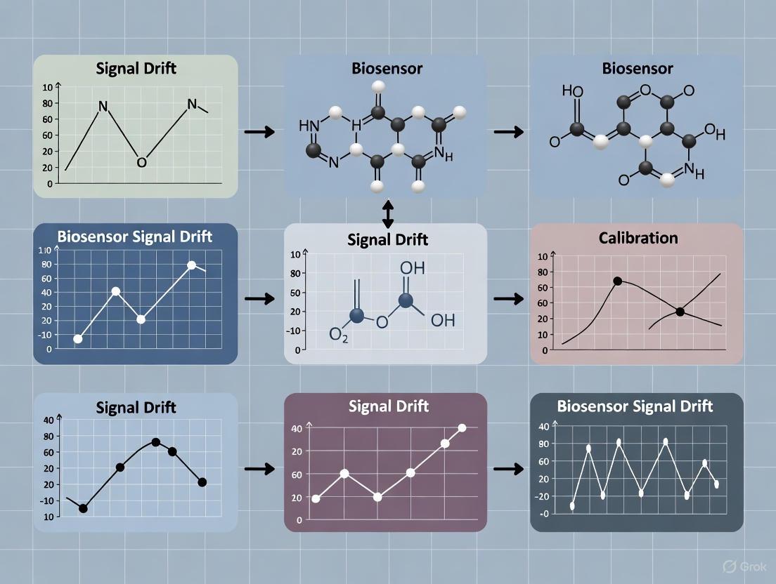
Mechanism and Workflow Visualizations
Diagram: Mechanism of Sensor Signal Drift
Diagram: Experimental Workflow for Drift Analysis
This technical support resource is designed for researchers and scientists working on the front lines of biosensor development. If you are investigating signal drift and calibration stability, particularly within the context of a thesis or a drug development project, you have likely encountered the confounding effects of biofouling. This guide provides targeted, evidence-based answers and methodologies to help you diagnose, understand, and mitigate the non-specific adsorption of proteins and cells that leads to signal loss. The following FAQs and troubleshooting protocols are framed within contemporary research, drawing on the latest advancements in materials science and sensor design to empower your experimental work.
Frequently Asked Questions (FAQs)
FAQ 1: What is the fundamental mechanism by which biofouling causes signal loss in electrochemical biosensors?
Biofouling causes signal loss through two primary, interconnected mechanisms: physical blockage and signal interference.
- Physical Blockage: The spontaneous accumulation of proteins, cells, and other biomolecules on the sensor's sensing area forms a physical diffusion barrier. This layer impedes the transport of the target analyte to the biorecognition element and the transducer surface, slowing the sensor's response and reducing the measured signal amplitude [8] [9].
- Signal Interference: The fouling layer itself can introduce electrical noise, increase background current, and alter the local chemical environment at the electrode-solution interface. In field-effect transistor (FET)-based biosensors, adsorbed biomolecules can act as unintended charge scatterers or create interfacial potentials, directly contributing to signal drift and a loss of sensitivity [5]. Furthermore, in optical sensors like those based on Surface Plasmon Resonance (SPR), the fouling layer changes the refractive index, creating a background signal that obscures the specific binding event of the target analyte [10].
FAQ 2: Beyond simple sensitivity loss, how does biofouling specifically lead to signal drift over time, and how can I distinguish it from other drift sources?
Signal drift from biofouling is typically a time-dependent, often unidirectional process driven by the gradual accumulation of a non-specific layer. This is distinct from abiotic failures (like electrode corrosion) or initial signal stabilization.
- The Drift Mechanism: In a complex fluid, biomolecules continuously adsorb to the sensor surface. The initial rapid formation of a conditioning film (e.g., of proteins) is often followed by slower, more complex biofilm development. As this layer thickens and evolves, it progressively alters the diffusion kinetics and the interfacial properties of the sensor, leading to a continuous change in the baseline signal [5] [8]. This is a key failure mode for implantable biosensors aiming for long-term continuous monitoring.
- Distinguishing Biofouling Drift: To confirm biofouling is the primary drift source, conduct a control experiment.
- Run your biosensor in a complex, protein-rich fluid (e.g., serum, blood plasma) and observe the signal over time.
- Then, transfer the sensor to a clean, simple buffer solution (e.g., PBS) and measure the signal again.
- A partial or full recovery of the signal in the buffer strongly indicates that the drift was caused by reversible or semi-reversible biofouling, rather than permanent damage to the sensor itself [8]. A lack of recovery may point toward irreversible fouling or abiotic failure.
FAQ 3: My biosensor works excellently in buffer but fails in blood serum. Is this solely a charge screening effect, or is biofouling the main issue?
While charge screening (Debye screening) is a major concern in high-ionic-strength fluids like blood serum, biofouling is often the dominant, concurrent problem. The two effects can be conflated but must be addressed with different strategies.
- Charge Screening: In solutions with high ionic strength, the electrical double layer (EDL) at the sensor surface becomes compressed to a length of just a few nanometers. This prevents charged target molecules beyond this distance from influencing the sensor channel, effectively "screening" them out [5].
- Biofouling: This is the physical deposition of proteins and cells, which occurs regardless of charge-based effects. A fouling layer can be electrically neutral but still cause signal loss by blocking analyte access.
- Disentangling the Effects: Research on carbon nanotube-based BioFETs (D4-TFT) demonstrates that it is possible to overcome both challenges simultaneously. By using a polymer brush interface like POEGMA, the effective sensing distance (Debye length) in high ionic strength solution (1X PBS) can be increased via the Donnan potential effect, while the same layer provides non-fouling properties to prevent protein adsorption [5]. Therefore, your sensor failure in serum is likely a combination of both phenomena, requiring a holistic interface design.
FAQ 4: What are the most effective surface coatings to prevent non-specific protein adsorption in biosensing?
The field has moved toward highly hydrophilic, water-retaining coatings that create a physical and energetic barrier to protein adsorption. The following table summarizes the most prominent categories of antifouling materials.
Table 1: Prominent Antifouling Coatings for Biosensors
| Coating Type | Key Examples | Mechanism of Action | Key Characteristics |
|---|---|---|---|
| PEG-based Polymers | Poly(ethylene glycol) (PEG), Poly(OEGMA) | Forms a hydrated, steric barrier; high chain mobility creates an energetically unfavorable surface for protein adhesion [5] [9]. | Biocompatible; widely used; can be grafted as brushes; performance can depend on chain length and density. |
| Zwitterionic Materials | Poly(carboxybetaine), Poly(sulfobetaine) | Contains both positive and negative charges; binds water molecules even more tightly than PEG via electrostatic hydration [8] [11]. | High hydrolytic stability; excellent antifouling performance; resistant to oxidative degradation. |
| Hydrogels | Polyacrylamide, PEG-based hydrogels | A 3D cross-linked network that absorbs large amounts of water, creating a low-fouling, hydrogel layer that blocks access of large biomolecules [9]. | Can be used as a physical barrier; good biocompatibility; may slow analyte diffusion. |
| Self-Assembled Monolayers (SAMs) | Alkane thiolates on gold, Silane-based on oxides | Creates a dense, well-ordered, and often hydrophilic monolayer that presents a uniform, low-fouling surface [10] [11]. | Provides molecular-level control over surface properties; requires specific substrate materials (e.g., Au, SiO₂). |
Troubleshooting Guides
Guide 1: Diagnosing and Mitigating Signal Drift in Complex Fluids
Signal drift undermines calibration and reliability. Follow this systematic guide to identify the root cause and apply corrective measures.
Table 2: Troubleshooting Signal Drift
| Observed Symptom | Potential Root Cause | Diagnostic Experiments | Corrective & Mitigation Strategies |
|---|---|---|---|
| Gradual, monotonic signal decrease over hours/days in biological fluid. | Progressive biofouling forming a diffusion-limiting layer. | Perform a buffer recovery test (see FAQ 2). Image the sensor surface (e.g., with SEM) post-experiment to confirm fouling layer presence [8]. | Implement a robust antifouling coating from Table 1. For implantable sensors, consider active removal methods like mechanical actuation or stimuli-responsive surfaces [8]. |
| Rapid initial signal drop upon exposure to complex fluid, followed by slower drift. | Instantaneous formation of a protein conditioning film. | Use a real-time label-free technique (e.g., SPR or QCM-D) to monitor the initial adsorption kinetics on your sensor surface [10]. | Optimize the density and packing of your antifouling coating to prevent initial protein penetration. Pre-adsorb with a known, benign protein (e.g., BSA) in a controlled manner to passivate vacant sites [9]. |
| Signal drift matches the expected direction of sensor response, leading to false positives. | Biofouling-induced drift that is convoluted with the specific analyte signal [5]. | Use a control device or channel with no biorecognition element. A similar drift in the control channel confirms the signal is non-specific [5]. | Enforce a rigorous testing methodology. Use infrequent DC sweeps instead of continuous static measurements to distinguish binding events from drift [5]. Incorporate reference electrodes for differential measurements. |
Guide 2: Selecting and Validating an Antifouling Coating
Choosing the right coating is critical. This protocol outlines the key steps for selection and functional validation.
Step 1: Coating Selection based on Sensor Platform and Application
- For Optical Biosensors (SPR, LSPR): Thin, uniform coatings are essential. SAMs or polymer brushes like POEGMA are ideal as they minimally perturb the electromagnetic field [10].
- For Electrochemical Sensors: A wider range of coatings can be used, including hydrogels, sol-gels, and porous membranes. Ensure the coating is permeable to your target analyte but blocks larger fouling agents [9].
- For Implantable Sensors: Biocompatibility and long-term stability are paramount. Zwitterionic coatings and certain hydrogels are excellent candidates due to their stability and high fouling resistance [8].
Step 2: Experimental Validation of Coating Performance
- Quantify Non-Specific Adsorption:
- Method: Use a Surface Plasmon Resonance (SPR) instrument or a Quartz Crystal Microbalance with Dissipation (QCM-D). If unavailable, a fluorescently labeled non-target protein (e.g., albumin) can be used, with fluorescence microscopy or a plate reader to quantify adsorption.
- Procedure: First, establish a baseline signal in buffer. Then, flow a solution of a challenging protein mixture (e.g., 10% blood serum, 1 mg/mL fibrinogen) over the coated sensor surface for 30-60 minutes. Monitor the signal change. Finally, switch back to buffer to see if adsorbed proteins desorb.
- Success Metric: A >90% reduction in non-specific adsorption compared to an uncoated sensor is a strong indicator of effective antifouling performance [10] [11].
- Verify Retention of Biosensor Function:
- Method: Perform a standard calibration curve for your target analyte using the antifouling-coated biosensor.
- Procedure: Measure the sensor's response (e.g., current, frequency shift, wavelength shift) to a series of known concentrations of your target analyte in a clean buffer.
- Success Metric: The coated sensor should retain a high sensitivity and a low limit of detection. A significant drop in sensitivity indicates the coating is also blocking the target analyte and needs to be re-optimized (e.g., by adjusting porosity or thickness) [9].
The Scientist's Toolkit: Research Reagent Solutions
This table lists essential materials and their functions for developing biofouling-resistant biosensors, as cited in recent literature.
Table 3: Key Research Reagents for Anti-Biofouling Experiments
| Reagent / Material | Function in Experimentation | Application Context |
|---|---|---|
| POEGMA (Poly(oligo(ethylene glycol) methyl ether methacrylate)) | A polymer brush that extends the Debye length and provides a non-fouling interface via the Donnan potential effect [5]. | BioFETs operating in undiluted physiological fluids (e.g., 1X PBS); ultrasensitive immunoassays. |
| Zwitterionic Solutions (e.g., Poly(sulfobetaine)) | Used to create ultra-low-fouling surfaces via strong electrostatic hydration; often polymerized on surfaces from monomer solutions [8] [11]. | Implantable biosensors, microfluidic chips, and SPR sensors for detection in blood, plasma, and serum. |
| Syringaldazine | A redox mediator that is pre-adsorbed onto electrode surfaces; serves as a model catalyst to rapidly and visually evaluate the protective effect of antifouling layers by monitoring its electrochemical signal degradation in complex media [9]. | High-throughput screening of antifouling coatings for electrochemical sensors in cell culture media or biological fluids. |
| Sol-Gel Silicate | Forms a stable, porous inorganic layer that acts as a physical barrier against large fouling agents while allowing small analyte diffusion [9]. | Long-term protection of electrochemical sensors in cell culture environments (shown to be effective for up to 6 weeks). |
Experimental Workflow and Signaling Pathways
The following diagram illustrates the sequential process of biofouling and the concurrent strategies researchers can employ to mitigate its effects at each stage. This workflow synthesizes the key concepts from the FAQs and troubleshooting guides into a single, logical pathway.
Diagram 1: Biofouling Process and Concurrent Mitigation Workflow. This chart visualizes the temporal sequence of biofouling (red) and the corresponding intervention strategies (green) that can be implemented at each stage to ensure a reliable sensor signal.
Frequently Asked Questions (FAQs) on Degradation and Signal Stability
FAQ 1: What are the primary causes of DNA degradation in biosensing samples, and how can I mitigate them? DNA degradation is a major concern that can compromise biosensor results, especially when working with limited or precious samples. The primary causes are chemical and enzymatic [12].
- Chemical Degradation: This includes:
- Hydrolysis: Water molecules can break the DNA backbone, leading to depurination (loss of purine bases) and strand fragmentation. This is accelerated at non-optimal pH levels [12] [13].
- Oxidation: Exposure to heat, UV radiation, or reactive oxygen species (ROS) modifies nucleotide bases and causes strand breaks [12].
- Enzymatic Breakdown: Nucleases present in biological samples (e.g., blood, tissue) can rapidly degrade DNA if not properly inactivated [12].
Mitigation Strategies:
- Control Temperature: Store samples at -80°C or in cryo conditions to slow all degradation processes. Cold storage is critical for preserving forensic and research samples [12] [13].
- Optimize pH: Use buffered solutions to maintain a stable, neutral pH and avoid acidic or alkaline conditions that accelerate chemical degradation [14].
- Use Chemical Inhibitors: Incorporate chelating agents like EDTA to inactivate nucleases, and consider antioxidants to reduce oxidative damage [12].
- Apply Gentle Processing: For DNA extraction, use optimized mechanical homogenization with controlled parameters (speed, cycle duration) to avoid excessive shearing and fragmentation [12].
FAQ 2: Why does my electrochemical biosensor signal drift over time, and how can I stabilize it? Signal drift is a common challenge in biosensors like BioFETs and electrochemical aptamer-based (EAB) sensors, often caused by slow electrochemical processes at the sensor-solution interface [5] [15].
- Causes of Drift:
- Electrolyte Ion Diffusion: Ions from the solution slowly diffuse into the sensing region, altering gate capacitance and threshold voltage over time [5].
- Biofouling: The non-specific adsorption of proteins or other biomolecules to the sensor surface can foul the interface and degrade performance [5] [15].
- Instability of Reference Elements: The use of pseudo-reference electrodes or changes in the redox reporter's electron transfer kinetics can contribute to drift [5] [16].
Stabilization Strategies:
- Interface Engineering: Coat the sensor with a non-fouling polymer brush, such as poly(oligo(ethylene glycol) methyl ether methacrylate) (POEGMA), to reduce biofouling and create a more stable environment [5].
- Optimized Measurement Protocols: Use a stable electrical testing configuration and rely on infrequent DC sweeps rather than continuous static measurements to minimize drift accumulation [5].
- Advanced Referencing: For EAB sensors, employ techniques like the Kinetic Differential Measurement (KDM), which uses signals from multiple square-wave frequencies to correct for drift and enhance gain [16].
- Proper Passivation: Ensure the sensor is well-passivated to minimize leakage currents and isolate the active sensing area [5].
FAQ 3: My biosensor calibration is inconsistent between devices and days. What factors should I control? Inconsistent calibration often stems from environmental and sample matrix variations that affect the sensor's physicochemical properties [17] [16].
- Key Factors to Control:
- Temperature: This is a critical parameter. The binding affinity (K1/2) of aptamers and the electron transfer rate of redox reporters are temperature-dependent. Always calibrate at the same temperature used for measurements (e.g., 37°C for body temperature) [16].
- Sample Matrix: The composition of the calibration medium (e.g., buffer vs. whole blood) significantly impacts the signal. For the most accurate results, calibrate in a matrix that matches your sample. Note that the age of biological media like blood can also alter the sensor response [16].
- Device-to-Device Variation: Inherent differences between individual sensors (e.g., in transconductance) can lead to varying absolute signals [17].
Calibration Improvement Methods:
- Use a Correlation-Based Calibration: For nanowire FETs, a method that normalizes the absolute current response (ΔI) by the device's gate dependence (dI~ds~/dV~g~) has been shown to suppress device-to-device variation significantly [17].
- Match Media and Temperature: As demonstrated with EAB sensors, using freshly collected whole blood at body temperature for calibration yields high accuracy (better than ±10% for vancomycin measurement) [16].
- Account for Solvent Effects: In optical biosensors, solvents like DMSO with a high refractive index can cause bulk effects. Using a calibration method that corrects for these non-specific changes, rather than simple subtraction, improves accuracy [18].
Research Reagent Solutions
The following table details key reagents and materials used to address degradation and stability issues in biosensing research.
| Item | Function/Benefit | Key Considerations |
|---|---|---|
| POEGMA Polymer Brush [5] | Extends the Debye length in high ionic strength solutions; reduces biofouling. | Enables detection of large antibodies in physiological buffers (e.g., 1X PBS); improves sensor stability. |
| Ultra-Mild Bisulfite (UMBS) Chemistry [19] | Preserves DNA integrity during bisulfite sequencing for methylation analysis. | Minimizes DNA damage; improves library yield and CpG coverage from limited samples (e.g., cell-free DNA). |
| EDTA (Ethylenediaminetetraacetic acid) [12] | Chelating agent that inactivates metal-dependent nucleases. | Prevents enzymatic DNA degradation during extraction and storage; balance concentration to avoid PCR inhibition. |
| Kinetic Differential Measurement (KDM) [16] | A measurement and referencing technique for electrochemical aptamer-based (EAB) sensors. | Corrects for signal drift by using two square-wave frequencies; improves quantification accuracy. |
| Ceramic or Stainless Steel Beads [12] | Used with mechanical homogenizers for effective cell lysis. | Enables efficient disruption of tough samples (e.g., bone, bacteria) while minimizing DNA shearing through parameter control. |
Experimental Protocols & Data
Protocol: Ultra-Mild Bisulfite Sequencing for Preserving DNA Integrity
This protocol, adapted from the work of the He lab, is designed to convert unmethylated cytosines to uracils while minimizing the DNA degradation typical of traditional bisulfite methods [19].
Key Steps:
- Input DNA: Begin with low-input or ultra-low input DNA (e.g., from cell-free DNA, single cells, or early-stage embryos).
- Bisulfite Conversion: Incubate the DNA with the UMBS reaction mixture. The precise control of reaction conditions and stabilizing components is crucial. This step is gentler than conventional methods.
- Desalting and Cleanup: Purify the converted DNA using a standard cleanup kit or column to remove bisulfite salts.
- Elution: Elute the converted DNA in a low-EDTA TE buffer or nuclease-free water.
- Library Preparation and Sequencing: Proceed with standard library preparation protocols for next-generation sequencing.
Expected Outcomes:
- Higher DNA Recovery: UMBS demonstrates dramatically higher DNA recovery rates compared to conventional bisulfite sequencing [19].
- Improved Coverage: Achieves more comprehensive CpG coverage [19].
- Enhanced Accuracy: Improves methylation-call accuracy across diverse sample types [19].
Protocol: Mitigating Signal Drift in Carbon Nanotube BioFETs (D4-TFT)
This protocol outlines the creation of a stable, solution-gated BioFET for attomolar-level detection in biologically relevant ionic strength solutions [5].
Key Steps:
- Device Fabrication: Fabricate a thin-film transistor (TFT) using semiconducting carbon nanotubes (CNTs) as the channel material.
- Surface Functionalization:
- Grow or deposit a POEGMA polymer brush layer above the CNT channel.
- Print capture antibodies (cAb) into the POEGMA layer.
- Print detection antibodies (dAb) tagged with a readily-dissolvable excipient (e.g., trehalose) on a separate pad.
- Stable Encapsulation: Passivate the device to mitigate leakage current and enhance electrical stability [5].
- Assay Operation (D4):
- Dispense: A sample is dispensed onto the device, dissolving the trehalose and releasing the dAb.
- Dissolve & Diffuse: The dAb diffuses and, if the target analyte is present, forms a sandwich complex with the cAb.
- Detect: Measure the drain-source current (I~ds~). The formation of the antibody-analyte sandwich complex alters the current, signaling detection.
Key Parameters for Stability:
- Rigorous Testing Methodology: Use infrequent DC sweeps rather than static or continuous AC measurements to track signal [5].
- Stable Configuration: Employ a palladium (Pd) pseudo-reference electrode and ensure a stable electrical testing setup [5].
Quantitative Data on Factors Affecting Biosensor Calibration
The table below summarizes quantitative findings on how environmental factors impact the calibration of Electrochemical Aptamer-Based (EAB) sensors [16].
| Factor | Condition 1 | Condition 2 | Observed Effect on Calibration |
|---|---|---|---|
| Temperature | Room Temp (~25°C) | Body Temp (37°C) | Up to 10% higher KDM signal at room temp; can lead to substantial concentration underestimates if mismatched [16]. |
| Blood Age | Freshly Collected | 14 Days Old | Older blood produced lower signal gain at high target concentrations, leading to overestimation [16]. |
| Calibration Media | Fresh Whole Blood | Commercial Bovine Blood | Commercially sourced blood yielded lower signal gain, leading to overestimated concentrations [16]. |
| Reference Method | Individual Calibration | Averaged "Out-of-Set" Calibration | No significant change in accuracy was observed, suggesting sensor-to-sensor variation is not a major contributor for this sensor type [16]. |
Diagnostic Workflows and Signaling Pathways
DNA Degradation Pathways and Mitigation
The following diagram illustrates the primary pathways of DNA degradation and the corresponding points for intervention to preserve sample integrity.
Biosensor Signal Drift Mitigation Strategy
This workflow outlines a systematic approach to diagnosing and correcting for signal drift in biosensor systems.
Ionic Interference and Double-Layer Effects in High-Strength Solutions
Troubleshooting Guides
Why is my biosensor signal weak or non-existent in physiological solutions (e.g., 1X PBS, blood)?
A weak signal in high-ionic-strength solutions is primarily caused by the Debye screening effect. The high ion concentration compresses the electrical double layer (EDL), drastically reducing the Debye length (λ_D). This prevents the electric field from sensing charged biomolecules that bind beyond this short screening distance [20] [5] [21].
Solutions:
- Extend the Debye Length: Immobilize a non-fouling polymer brush layer, such as poly(oligo(ethylene glycol) methyl ether methacrylate) (POEGMA), above the sensor. This layer creates a Donnan potential that effectively increases the sensing distance, allowing detection of biomarkers in solutions like 1X PBS [5].
- Shift to Capacitive Sensing: Instead of relying on Faradaic currents, use a non-Faradaic electrochemical impedance spectroscopy (EIS) mode to monitor changes in the double-layer capacitance (
C_dl). This method is less dependent on the charge of the target molecule and more on the overall capacitance change caused by binding events [20] [22]. - Use an Enhanced EDL (EnEDL) FET: Design your FET biosensor to be gated by the enhanced electrical double layer. Applying a sufficiently high gate bias can enhance the EDL capacitance, leading to high sensitivity even in physiological salt concentrations [20].
How can I distinguish true biomarker detection from signal drift?
Signal drift—a gradual, systematic deviation in the sensor's baseline—can falsely mimic or obscure a target signal. Mitigating this requires a combination of robust experimental design and data processing [5] [23].
Solutions:
- Incorporate Rigorous Controls: Always test a control device fabricated on the same chip that is identical in every way except it lacks the specific biorecognition element (e.g., no antibodies printed over the channel). A true positive signal will show a significant change in the experimental device but not in the control [5].
- Employ a Stable Testing Methodology:
- Maximize Device Stability through appropriate passivation and stable polymer brush coatings [5].
- Use a Stable Electrical Configuration, such as a palladium (Pd) pseudo-reference electrode to avoid bulky Ag/AgCl electrodes [5].
- Rely on Infrequent DC Sweeps rather than static or continuous AC measurements to collect data points, as this reduces the influence of temporal drift [5].
- Implement Computational Drift Compensation: For continuous monitoring, use advanced machine learning techniques. The Multi Pseudo-Calibration (MPC) approach can learn a non-linear model of sensor drift by using past measurements for which ground-truth concentrations are later obtained [23].
My DNA-based electrochemical sensor shows slow hybridization kinetics. How can I improve its efficiency?
The kinetics of DNA hybridization at an electrode surface are strongly affected by the electrostatic environment. The negatively charged DNA backbone experiences interference from the similarly charged electrode surface, especially at lower ionic strengths [24].
Solutions:
- Optimize Ionic Strength: Increase the salt concentration of your buffer. A higher ionic strength (e.g., 0.5 M NaClO₄ vs. 0.125 M) screens the repulsive forces between the DNA and the electrode, accelerating hybridization [24].
- Strategically Position the Hybridization Site: Design your DNA probe so that the binding site is strategically placed at a distance from the electrode surface, rather than directly adjacent to it. This moves the hybridization event away from the disruptive electric field of the double layer [24].
Frequently Asked Questions (FAQs)
What is the Debye length, and why is it critical for biosensing?
The Debye length (λ_D) is the characteristic distance over which a charged surface can exert an electrical influence in an electrolyte solution before being screened by counter-ions. It is mathematically defined as:
λ_D = √( (ε₀ε_r k_B T) / (2 N_A q² I) )
where I is the ionic strength of the solution [21]. In high-ionic-strength fluids like blood or PBS, λ_D is compressed to just ~1 nanometer. Since most antibodies are ~10-15 nm in size, their binding events occur far outside this range and are electrically "invisible" to conventional FETs, severely limiting sensitivity [5].
Can I simply dilute my sample to reduce ionic interference?
While sample dilution is a common and straightforward method to increase the Debye length, it has significant drawbacks for real-world applications. It requires additional sample preparation steps, reduces the concentration of the target biomarker, and dilutes other matrix components, which may alter the sample's behavior. For point-of-care diagnostics, dilution is often impractical [20] [5]. Advanced strategies like polymer brushes or capacitive sensing are designed to work in undiluted, physiologically relevant samples.
Are there specific calibration protocols for sensors used in high-ionic-strength environments?
Yes, calibration conditions must match the measurement environment as closely as possible.
- Match Temperature: Calibration curves collected at room temperature can differ significantly from those at body temperature (37°C), leading to concentration underestimates or overestimates. Always calibrate at the temperature you plan to measure at [16].
- Use Fresh Media: The age of calibration media (e.g., blood) can impact sensor response. For the most accurate in-vivo measurements, calibrate using the freshest possible blood [16].
- Account for Drift: For long-term monitoring, implement drift-aware calibration models like Multi Pseudo-Calibration (MPC), which uses periodic ground-truth samples to update and correct the sensor's predictions over time [23].
The following table summarizes key experimental data and parameters related to overcoming ionic interference, as found in the literature.
| Parameter / Strategy | Reported Value / Effect | Experimental Conditions | Citation |
|---|---|---|---|
| Debye Length (λ_D) | ~0.7 nm | Calculated for 1X PBS (~150 mM ionic strength) at room temperature. | [5] |
| POEGMA Polymer Brush | Enabled detection of sub-femtomolar (sub-fM) biomarkers. | CNT-based BioFET (D4-TFT) in 1X PBS. | [5] |
| Enhanced EDL FET (Gate Bias) | Higher gate bias led to enhanced EDL and higher sensitivity. | FET biosensor in physiological salt concentration (e.g., 1X PBS). | [20] |
| Capacitive Sensing (vs. Faradaic) | Direct detection in 1X PBS, sweat, human serum. | EIS-based sensors monitoring double-layer capacitance (C_dl). |
[20] [22] |
| DNA Hybridization Kinetics | Significant interference at low ionic strength (0.125 M); improved efficiency at higher strength (0.5 M). | 10 bp DNA segment hybridization on Au electrode monitored with SWV. | [24] |
| Calibration Temperature | KDM signal difference up to 10% higher at room temp vs. body temp, causing substantial underestimation. | EAB sensor for vancomycin in blood. | [16] |
Detailed Experimental Protocols
Protocol 1: Implementing a Polymer Brush Interface for Debye Length Extension
This protocol is adapted from the D4-TFT (CNT-based BioFET) fabrication process [5].
Objective: To functionalize a sensor surface with a POEGMA brush that extends the effective sensing range beyond the native Debye length in high-ionic-strength solutions.
Materials:
- Substrate: Fabricated sensor chip (e.g., with CNT channel and electrodes).
- Polymer: Poly(oligo(ethylene glycol) methyl ether methacrylate) (POEGMA).
- Initiator: Surface-bound initiator for atom transfer radical polymerization (ATRP).
- Biorecognition Element: Target-specific capture antibodies (cAb).
- Buffer: Appropriate immobilization and washing buffers (e.g., phosphate buffer).
Methodology:
- Surface Preparation: Clean and prepare the sensor surface to ensure uniform coating.
- Initiator Immobilization: Covalently attach the ATRP initiator to the sensor surface.
- Polymer Brush Growth: Submerge the sensor in a solution containing the POEGMA monomer. Under controlled temperature and atmosphere, perform surface-initiated ATRP to grow a dense, brush-like layer of POEGMA.
- Antibody Immobilization: Using a non-contact printer (e.g., inkjet), spot and immobilize the capture antibodies into the POEGMA matrix. The polymer brush acts as a 3D scaffold, embedding the antibodies at a functional distance from the sensor surface.
- Validation: Validate the functionality by testing the sensor's response to the target analyte in 1X PBS, demonstrating attomolar to femtomolar sensitivity.
Protocol 2: Capacitive EIS Detection in Bodily Fluids
This protocol outlines the use of non-Faradaic EIS for label-free detection [20] [22].
Objective: To detect biomarker binding by monitoring changes in the double-layer capacitance (C_dl) at the electrode-electrolyte interface.
Materials:
- Working Electrode: Gold or other noble metal electrode, functionalized with a self-assembled monolayer (SAM) and a specific bioreceptor (antibody, aptamer).
- Reference & Counter Electrodes: Standard Ag/AgCl reference electrode and Pt wire counter electrode.
- Potentiostat: Equipment capable of performing Electrochemical Impedance Spectroscopy (EIS).
- Analyte Solution: Sample in a high-ionic-strength buffer (e.g., PBS, serum).
Methodology:
- Electrode Functionalization: Clean the working electrode and form a SAM. Covalently immobilize the biorecognition element (e.g., antibody) onto the SAM.
- EIS Measurement Setup: Place the three-electrode system in the analyte solution. Apply a low-amplitude AC voltage (e.g., 10 mV) over a frequency range (e.g., 0.1 Hz to 100 kHz) at zero DC bias to focus on non-Faradaic processes.
- Baseline Measurement: Record the EIS spectrum in the pure buffer or blank solution.
- Target Introduction: Introduce the sample containing the target biomarker and incubate to allow binding.
- Post-Binding Measurement: Record the EIS spectrum after binding has occurred.
- Data Analysis: Fit the impedance data to an equivalent circuit model. The key parameter for detection is the change in the double-layer capacitance (
C_dl), which will shift upon biomarker binding to the surface.
Signaling Pathways and Workflows
The Scientist's Toolkit: Research Reagent Solutions
| Material / Reagent | Function in Experiment | Key Consideration |
|---|---|---|
| POEGMA Polymer Brush | Extends the Debye length in high-ionic-strength solutions by establishing a Donnan equilibrium potential, allowing detection of biomarkers in undiluted physiological fluids [5]. | The density and thickness of the brush layer are critical for performance. |
| Hydrogel-based Magneto-resistive Sensors | Used in cross-sensitive sensor arrays for continuous bioprocess monitoring. Their drift can be compensated using machine learning models [23]. | Prone to time-dependent drift, requiring advanced computational correction. |
| Polyethylene Glycol (PEG) / POEGMA | A non-fouling polymer layer used to resist biofouling and, crucially, to modulate the local ionic environment to effectively increase the Debye length [5]. | A well-established and versatile tool for improving biosensor performance in complex media. |
| Pd (Palladium) Pseudo-Reference Electrode | Provides a stable gate voltage for FET-based biosensors in a point-of-care form factor, eliminating the need for bulky, traditional Ag/AgCl reference electrodes [5]. | Enhances portability and stability of the sensing platform. |
| Self-Assembled Monolayer (SAM) | A monolayer of organic molecules that forms on an electrode surface (e.g., gold). It provides a well-defined, functionalizable interface for immobilizing bioreceptors like antibodies or DNA [24] [16]. | SAM quality and packing density are crucial for minimizing non-specific adsorption and ensuring efficient electron transfer. |
Strategic Solutions: Methodologies for Drift Mitigation and Calibration
Stable SAMs, Polymer Brushes, and Non-Fouling Coatings
Frequently Asked Questions (FAQs)
Q1: What are the primary causes of signal drift in biosensors, and how can they be mitigated? Signal drift in biosensors, particularly solution-gated BioFETs, is often caused by the slow diffusion of electrolytic ions from the solution into the sensing region, which alters gate capacitance, drain current, and threshold voltage over time [5]. This can lead to data that falsely implies device success. Mitigation strategies include:
- Chemical Passivation: Maximizing sensitivity through appropriate passivation of the sensor surface alongside polymer brush coatings [5].
- Stable Electrical Configuration: Using a stable electrical testing setup, including stable pseudo-reference electrodes to bypass bulky Ag/AgCl electrodes [5].
- Rigorous Testing Methodology: Employing infrequent DC sweeps rather than static or AC measurements to track signal changes, which helps distinguish drift from true biomarker detection [5].
- Advanced Coatings: Implementing non-fouling polymer brushes like POEGMA or zwitterionic polymers to prevent nonspecific adsorption, which is a key contributor to signal drift and noise [5] [25] [26].
Q2: How can I extend the Debye length to detect large biomolecules in physiologically relevant ionic strength solutions? The Debye screening effect limits detection to charged molecules within a few nanometers of the sensor surface in high ionic strength solutions like blood or PBS. A leading strategy to overcome this is using polymer brush interfaces to establish a Donnan equilibrium potential, effectively extending the sensing distance [5].
- Polymer Brush Interface: Immobilizing a polyethylene glycol-like polymer brush, such as poly(oligo(ethylene glycol) methyl ether methacrylate) (POEGMA), above the device. This brush increases the Debye length in ionic solutions, allowing for antibody-antigen interactions (typically ~10 nm in size) to be detected in 1X PBS [5].
- Antibody Immobilization: Capture antibodies are printed into this polymer brush layer, creating a sensing interface that can detect biomarkers at sub-femtomolar concentrations without diluting the sample [5].
Q3: What are the primary advantages of zwitterionic polymer coatings over traditional PEG coatings? While PEG has been the "gold standard" for non-fouling coatings, zwitterionic polymers have emerged as a powerful alternative with several advantages [26]:
- Stronger Hydration: Zwitterionic polymers interact with water molecules through stronger electrostatic interactions (rather than hydrogen bonding for PEG), forming a denser and more stable hydration layer that significantly improves resistance to non-specific adhesion [26].
- Enhanced Stability: Zwitterionic polymers do not suffer from the oxidative degradation (depolymerization) that PEG does in oxygen-rich environments or in the presence of transition metal ions [26].
- Reduced Immunogenicity: Prolonged use of PEG can lead the body to produce antibodies against it. Zwitterionic polymers, with their biomimetic structure similar to cell membranes, exhibit higher biocompatibility and reduced risk of an immune response [26].
Q4: What key parameters should be tested during biosensor calibration and performance validation? When testing a biosensor's performance, several parameters should be characterized to ensure sensitivity and reliability [27]. An automated microfluidic system can be particularly useful for this validation. Key parameters include:
- Sensitivity
- Selectivity
- Limit of Detection (LOD)
- Reproducibility
- Response Time
- Stability and Signal Drift
- Linear Dynamic Range
Troubleshooting Guides
Issue 1: High Non-Specific Binding (NSB) and Poor Signal-to-Noise Ratio
Problem: High background signal from non-specific adsorption of proteins or other matrix molecules onto the sensor surface. Solutions:
- Implement High-Density Polymer Brushes:
- Recommended Coating: Use surface-initiated atom transfer radical polymerization (SI-ATRP) to grow dense poly(oligo(ethylene glycol) methyl ether methacrylate) (POEGMA) brushes. The comb-like architecture with a high density of oligoethylene glycol moieties provides excellent non-fouling behavior [25].
- Protocol: Immobilize an ATRP initiator on the sensor surface via chemisorption or covalent grafting. Perform SI-ATRP of OEGMA in a deoxygenated solvent under mild conditions to control brush thickness and density. Protein resistance is a function of both thickness and density [25].
- Apply a Zwitterionic Coating:
- Recommended Coating: Apply a coating of poly(carboxybetaine) (pCB) or poly(sulfobetaine) (pSB) using ATRP or RAFT polymerization [26].
- Protocol: Functionalize the sensor surface with initiation sites. For pCB, polymerize carboxybetaine methacrylate. The zwitterionic chains will form a super-hydrophilic surface that binds water molecules into a dense hydration layer, physically blocking protein adsorption [26].
Issue 2: Signal Drift in Solution-Gated Electrochemical or Transistor-Based Biosensors
Problem: The baseline signal shifts over time, obscuring the detection of the target analyte and leading to inaccurate data. Solutions:
- Optimize Measurement Methodology:
- Action: Avoid continuous static measurements. Instead, use infrequent DC voltage sweeps to acquire data points. This reduces the impact of slow ion diffusion processes that cause drift [5].
- Improve Sensor Encapsulation and Passivation:
- Action: Ensure the transducer channel is fully encapsulated and only the functionalized sensing area is exposed to the solution. This prevents leakage currents and enhances stability [5].
- Utilize a Stable Pseudo-Reference Electrode:
- Action: Replace bulky, conventional Ag/AgCl reference electrodes with a stable, miniaturized palladium (Pd) pseudo-reference electrode integrated into the biosensor chip. This contributes to a more stable gate potential in BioFETs [5].
Issue 3: Overcoming the Debye Length Limitation in Physiological Samples
Problem: Inability to detect large biomolecules (e.g., antibodies) in undiluted biological samples (e.g., 1X PBS, serum) due to charge screening by the electrical double layer. Solutions:
- Integrate a POEGMA Polymer Brush as a Debye Length Extender:
- Principle: The POEGMA brush establishes a Donnan potential equilibrium, effectively increasing the charge-sensing distance (Debye length) beyond the few nanometers typical for high ionic strength solutions [5].
- Protocol:
- Grow a POEGMA brush on the sensor surface via SI-ATRP [25].
- Print or immobilize capture antibodies into the polymer brush matrix.
- The assay can now be performed in undiluted 1X PBS, as the brush layer allows for the detection of antibody-antigen binding events that occur beyond the native Debye length [5].
Issue 4: Inconsistent Biosensor Performance and Reproducibility
Problem: Biosensor response varies between fabrication batches or during repeated assays. Solutions:
- Automate Calibration and Testing:
- Action: Use an automated microfluidic system for biosensor calibration and testing [27].
- Protocol: Implement a system with a multi-channel pressure controller and switching valves to sequentially inject different calibrants (e.g., buffers, analyte solutions at varying concentrations) over the sensor. This ensures high precision, reduces manual pipetting errors, and allows for integrated cleaning steps between tests, greatly improving reproducibility [27].
- Employ Advanced Chemometric Data Analysis:
- Action: Process complex biosensor data from samples with complex matrices (e.g., blood) using machine learning algorithms [28].
- Protocol: Collect amperometric or voltammetric data. Model the first-order data with algorithms like Least-Squares Support Vector Machine (LS-SVM), which has been shown to outperform other methods (e.g., PLS, PCR) for selective and sensitive determination of analytes in blood, providing results comparable to ELISA [28].
Protocol 1: Fabrication of a D4-TFT Biosensor with POEGMA Brush
This protocol outlines the creation of an ultrasensitive, drift-resistant carbon nanotube-based BioFET [5].
Workflow:
- Device Fabrication: Fabricate a thin-film transistor (TFT) using semiconducting carbon nanotubes (CNTs) as the channel material.
- Surface Initiation: Functionalize the CNT/dielectric surface with an ATRP initiator.
- Polymer Brush Growth: Perform SI-ATRP of OEGMA to grow a POEGMA brush layer above the CNT channel. Control the reaction time and temperature to achieve the desired brush thickness and density [25].
- Antibody Patterning: Use inkjet printing to spot capture antibodies (cAb) into the POEGMA brush layer.
- Control Spotting: As a critical control, leave a section of the POEGMA brush without antibodies (or with a non-specific antibody) to distinguish specific binding from signal drift or non-specific interference [5].
- Assay Execution: Run the D4 (Dispense, Dissolve, Diffuse, Detect) immunoassay. A sample containing the analyte is dispensed, which dissolves a dried detection antibody (dAb) conjugate. The analyte and dAb diffuse to the surface, forming a sandwich complex with the cAb.
- Electrical Readout: Detect the binding event through a stable DC sweep measurement of the CNT channel's on-current.
Protocol 2: Coating a Surface with Zwitterionic Polymer via SI-ATRP
This general protocol describes creating a non-fouling surface on a gold-coated sensor [26].
Workflow:
- Surface Cleaning: Thoroughly clean the gold substrate.
- Initiator Self-Assembly: Immerse the substrate in an ethanol solution of a thiol-functionalized ATRP initiator (e.g., 11-(2-bromo-2-methylpropionyloxy) undecyl-1-thiol) to form a self-assembled monolayer (SAM).
- Polymerization Mixture: Prepare an aqueous polymerization solution containing the zwitterionic monomer (e.g., carboxybetaine methacrylate), a ligand (e.g., PMDETA), and a copper-based catalyst.
- Deoxygenation: Purge the solution with nitrogen to remove oxygen, which inhibits ATRP.
- "Grafting From" Polymerization: Immerse the initiator-functionalized substrate into the polymerization solution to grow the zwitterionic polymer brushes directly from the surface.
- Rinsing and Characterization: After a controlled time, remove the substrate, rinse it extensively with water, and characterize the brush thickness and non-fouling performance using SPR or QCM-D.
Quantitative Performance Data of Advanced Biosensing Platforms
Table 1: Performance Comparison of Biosensor Technologies and Coatings
| Technology / Coating | Key Feature | Reported Performance / Outcome | Test Medium |
|---|---|---|---|
| CNT-based D4-TFT [5] | POEGMA brush for Debye extension & drift mitigation | Detection of sub-femtomolar to attomolar concentrations | 1X PBS (physiological ionic strength) |
| Graphene-QD Hybrid [29] | Charge transfer-based sensing | Limit of Detection (LOD) down to 0.1 fM | Buffer (for biotin-streptavidin, IgG) |
| POEGMA Brushes [25] | Non-fouling coating | Reduction of non-specific adsorption, improved SNR in immunoassays | Human serum, plasma, whole blood |
| Electrochemical Immunosensor (BRCA-1) [29] | AuNPs-MoS2 nanocomposite | LOD of 0.04 ng/mL, RSD of 3.59% (n=3) | Spiked serum samples |
| Zwitterionic Polymers [26] | Dense hydration layer via electrostatic interactions | Ultra-low fouling, reduced foreign body response in vivo | Complex biological environments |
Key Research Reagent Solutions
Table 2: Essential Materials for Fabricating Stable, Non-Fouling Biosensors
| Reagent / Material | Function / Explanation | Key Reference |
|---|---|---|
| POEGMA (poly(oligo(ethylene glycol) methyl ether methacrylate)) | A comb-like polymer brush that resists non-specific protein adsorption and extends the Debye length via the Donnan effect. | [5] [25] |
| Zwitterionic Monomers (e.g., Carboxybetaine, Sulfobetaine) | Forms ultra-low fouling coatings that bind water tightly via electrostatic interactions, creating a physical and energetic barrier to protein adsorption. | [26] |
| ATRP Initiator (thiol- or silane-functionalized) | Forms a SAM on gold or silicon/silicon oxide surfaces, providing a foundation for growing polymer brushes via surface-initiated ATRP. | [25] [26] |
| Carbon Nanotubes (CNTs) / Graphene | High-sensitivity nanomaterial for the transducer channel in FET-based biosensors due to high mobility and surface-to-volume ratio. | [5] [29] |
| Pseudo-Reference Electrode (e.g., Pd) | Provides a stable gate potential in solution-gated BioFETs without the bulkiness and chloride-dependence of traditional Ag/AgCl electrodes. | [5] |
Experimental Workflow and Signaling Diagrams
Troubleshooting Logic Flow
Polymer Brush Fabrication Workflow
Signal drift is a pervasive challenge in electrochemical biosensors, characterized by a gradual, non-random change in the sensor's output signal over time, even when the target analyte concentration remains constant. This phenomenon results from slow, transient processes at the electrode-electrolyte interface, including electrolytic ion diffusion into the sensing region, which alters gate capacitance, drain current, and threshold voltage [5]. In practical terms, drift manifests as a shifting baseline that can obscure genuine biomarker detection, convolute results, and adversely affect device performance and reliability [5]. Effectively managing drift is particularly critical for applications requiring long-term monitoring or ultra-high sensitivity, such as attomolar-level biomarker detection in point-of-care diagnostics [5].
The socio-economic impact of unresolved drift is significant, contributing to the "valley of death" between laboratory prototypes and clinical/commercial deployment [6]. Key bottlenecks include signal instability, calibration drift, and low reproducibility in large-scale fabrication, which have slowed the translation of promising biosensor technologies into approved diagnostics [6]. This technical brief establishes a technical support framework to help researchers diagnose, mitigate, and calibrate drift in biosensing systems.
Quantitative Analysis of Drift Reduction Techniques
Performance Comparison of Machine Learning Models for Signal Processing
Machine learning techniques have demonstrated significant potential for compensating signal drift through advanced data processing. A systematic evaluation of 26 regression algorithms across six methodological families reveals distinct performance characteristics for modeling the nonlinear relationships between biosensor fabrication parameters and electrochemical signal outputs [6].
Table 1: Performance Metrics of ML Models for Biosensor Signal Processing [6]
| Model Category | Example Algorithms | Best RMSE | Best R² Score | Key Strengths | Drift Compensation Utility |
|---|---|---|---|---|---|
| Tree-Based Ensembles | Random Forest, XGBoost | Very Low | >0.95 | Handles non-linear parameters, robust to outliers | High - Robust against signal noise and environmental variations |
| Kernel-Based Models | Support Vector Regression (SVR) | Low | >0.90 | Effective for high-dimensional spaces | Medium - Compensates for temperature drift in outputs |
| Gaussian Process Regression | GPR | Low | >0.90 | Provides probabilistic uncertainty estimates | High - Calibration-free sensing with uncertainty quantification |
| Artificial Neural Networks | Multilayer Perceptron | Low | >0.92 | Captures complex non-linear relationships | Medium - Effective for analyte concentration prediction and signal denoising |
| Stacked Ensembles | GPR + XGBoost + ANN | Lowest | >0.97 | Combines strengths of multiple approaches | Highest - Novel framework showing superior predictive accuracy for biosensor optimization |
| Linear Models | Linear Regression | High | <0.80 | Simple, interpretable | Low - Unable to model nonlinear drift relationships |
Experimental Reagent Solutions for Drift Mitigation
Specific materials and reagents play crucial roles in implementing effective drift reduction strategies. The following table catalogues key research reagents identified from recent studies.
Table 2: Essential Research Reagents for Drift Reduction Experiments
| Reagent/Material | Function in Drift Reduction | Experimental Application | Key Findings |
|---|---|---|---|
| POEGMA (Poly(oligo(ethylene glycol) methyl ether methacrylate)) | Polymer brush interface extending Debye length; reduces biofouling | Coated above CNT channel in BioFET devices | Enables attomolar detection in 1X PBS by increasing sensing distance and mitigating drift effects [5] |
| PEG-like Polymer Brushes | Establishes Donnan equilibrium potential to extend Debye length | Immobilization on BioFET channel | Effectively increases Debye length in ionic solutions, overcoming charge screening limitations [5] |
| MXenes, Graphene, MOFs | Nanomaterial substrates improving sensitivity and stability | Electrode modification in electrochemical biosensors | Enable femtomolar-level detection limits and improved biocompatibility [6] |
| Glutaraldehyde | Crosslinker for biomolecule immobilization | Enzyme and antibody conjugation on sensor surfaces | Optimization required - minimal amounts recommended to reduce non-specific binding and drift [6] |
| Conductive Polymers | Mediating electron transfer, reducing interfacial resistance | Electrode coatings and transducer interfaces | Improve selectivity and minimize interference [6] |
Troubleshooting Guide: Frequently Asked Questions
FAQ 1: What are the primary root causes of signal drift in electrochemical biosensors?
Signal drift originates from multiple physicochemical processes:
- Electrolytic Ion Diffusion: In solution-gated BioFETs, ions slowly diffuse into the sensing region, altering gate capacitance, drain current, and threshold voltage over time [5].
- Biofouling: Non-specific adsorption of proteins or other biomolecules to the sensor surface gradually modifies interface properties [5].
- Reference Electrode Instability: Potential drift in pseudo-reference electrodes, particularly in miniaturized systems replacing bulky Ag/AgCl electrodes [5].
- Polymer Layer Hydration: Slow hydration processes in polymer brush interfaces (e.g., POEGMA) can cause transient signals [5].
- Environmental Variations: Temperature fluctuations and changing ionic strength directly impact sensor baseline stability [6].
FAQ 2: What experimental methodologies most effectively mitigate drift without sacrificing sensitivity?
Implement a multi-pronged approach:
- Maximize Sensitivity Through Passivation: Appropriate passivation alongside polymer brush coatings enhances both sensitivity and stability [5].
- Stable Electrical Testing Configuration: Use a stable electrical testing configuration with infrequent DC sweeps rather than static or AC measurements [5].
- Rigorous Testing Methodology: Implement a testing methodology that relies on infrequent DC sweeps rather than continuous static measurements or complex AC measurements [5].
- Polymer Brush Interfaces: Utilize POEGMA or similar polymers to create a stable interface that extends the Debye length and reduces biofouling [5].
- Machine Learning Compensation: Apply tree-based ensembles (Random Forests, XGBoost) or Gaussian Process Regression to model and correct drift patterns in sensor output [6].
FAQ 3: How can I validate whether my observed signal change represents true biomarker detection or mere drift?
Employ these verification strategies:
- Control Device Integration: Simultaneously test a control device with no biorecognition elements (e.g., no antibodies printed over the CNT channel) within the same chip environment [5].
- Drift Quantification Protocol: Characterize baseline drift extensively before biosensing experiments to establish inherent drift patterns [5].
- Statistical Cross-Validation: Use 10-fold cross-validation with multiple metrics (RMSE, MAE, MSE, R²) when applying ML models to distinguish drift from signal [6].
- Signal Pattern Analysis: Genuine biomarker detection typically shows specific binding kinetics, while drift often demonstrates more linear temporal patterns [5].
FAQ 4: What calibration circuit designs effectively compensate for drift in point-of-care biosensors?
Implement these circuit design strategies:
- Reference Electrode Optimization: While bulky Ag/AgCl electrodes offer stability, miniaturized systems can use palladium (Pd) pseudo-reference electrodes with appropriate compensation algorithms [5].
- Differential Measurement Architectures: Incorporate reference sensors without biorecognition elements for differential measurement to cancel common-mode drift [5].
- Temperature Compensation Circuits: Integrate temperature sensors with compensation algorithms, as SVR models have successfully reduced RMSE compared to polynomial calibration for temperature drift [6].
- Digital Calibration Interfaces: Implement microcontroller-based systems that apply ML-derived calibration matrices in real-time to raw sensor outputs [6].
Prioritize these computationally efficient approaches:
- Tree-Based Algorithms: Random Forests and XGBoost provide excellent performance for modeling non-linear drift patterns with moderate computational requirements [6].
- Gaussian Process Regression: Offers superior performance for calibration-free sensing with inherent uncertainty quantification, though with higher computational cost [6].
- Support Vector Regression (SVR): Effective for temperature drift compensation with better performance than polynomial methods and efficient implementation [6].
- Model Benchmarking: Always benchmark multiple algorithms (linear, tree-based, kernel-based, ANN) using cross-validation to identify the optimal approach for your specific sensor configuration [6].
Experimental Protocols for Drift Characterization and Reduction
Protocol: DC Sweep Methodology for Stable Biosensor Operation
This protocol outlines the rigorous testing methodology proven to mitigate drift effects in CNT-based BioFETs [5].
Principle: Frequent electrical measurements can accelerate drift through repeated perturbation of the electrochemical interface. Using infrequent DC sweeps minimizes this perturbation while providing sufficient temporal resolution for biosensing.
Materials:
- Potentiostat with voltage sweep capability
- BioFET sensor with integrated pseudo-reference electrode
- Data acquisition system
- Temperature-controlled fluidic cell
Procedure:
- Initial Characterization:
- Perform detailed I-V characterization of the sensor in blank solution (1X PBS)
- Establish baseline drift rate by monitoring output current over 30-60 minutes
Sweep Parameter Optimization:
- Set sweep parameters to minimize perturbation: Typical values include:
- Voltage range: ±0.5V around operating point
- Sweep rate: 0.1-0.5 V/s
- Interval between sweeps: 30-60 seconds
- Set sweep parameters to minimize perturbation: Typical values include:
Experimental Operation:
- For biomarker detection experiments, rely exclusively on these infrequent DC sweeps
- Avoid continuous static measurements or complex AC impedance protocols
- Extract sensing metrics (e.g., on-current shift, threshold voltage) from each sweep
Data Processing:
- Plot sweep-derived metrics versus time
- Compare against pre-established drift baselines
- Apply ML correction algorithms if necessary
Validation: Confirm genuine detection by demonstrating significant signal deviation from the characterized drift profile and using control devices without biorecognition elements [5].
Protocol: Polymer Brush Interface Fabrication for Enhanced Stability
This protocol describes the implementation of POEGMA polymer brushes to extend Debye length and reduce drift in BioFETs [5].
Diagram Title: Polymer Brush Interface Fabrication Workflow
Principle: POEGMA polymer brushes establish a Donnan equilibrium potential that effectively increases the Debye length in high ionic strength solutions, enabling detection beyond the electrical double layer while providing a non-fouling interface that reduces drift.
Materials:
- Carbon nanotube thin-film transistor (CNT TFT)
- POEGMA (poly(oligo(ethylene glycol) methyl ether methacrylate))
- Immobilized antibodies specific to target biomarker
- Oxygen plasma cleaner (for surface activation)
- Chemical vapor deposition system or solution-based polymerization setup
Procedure:
- Substrate Preparation:
- Fabricate CNT TFT using standard microfabrication techniques
- Clean surface thoroughly with appropriate solvents
Surface Activation:
- Treat CNT surface with oxygen plasma to create initiation sites
- Alternatively, use chemical functionalization to introduce polymerization initiation groups
Polymer Brush Growth:
- Grow POEGMA brushes using surface-initiated atom transfer radical polymerization (SI-ATRP)
- Control brush thickness (typically 20-100 nm) by adjusting polymerization time and conditions
Biorecognition Element Integration:
- Print capture antibodies into the POEGMA matrix using non-contact piezoelectric printing
- Optimize antibody density for maximum binding efficiency while maintaining polymer properties
Validation:
- Confirm Debye length extension by measuring sensitivity in 1X PBS
- Verify reduced biofouling using non-specific protein solutions
- Characterize drift performance compared to unmodified devices
Technical Notes: The POEGMA layer serves dual purposes: it extends the sensing distance (Debye length) in solution and mitigates signal drift effects by providing a stable, non-fouling interface [5].
Advanced Framework: Machine Learning-Enhanced Drift Compensation
Implementation Protocol: Stacked Ensemble Framework for Drift Correction
Diagram Title: ML Stacked Ensemble Framework for Drift Compensation
Implementation Workflow:
Feature Engineering:
- Extract fabrication parameters (enzyme amount, crosslinker concentration, scan number of conducting polymer, pH values) as primary features [6]
- Include environmental parameters (temperature, ionic strength, time)
- Incorporate temporal patterns from baseline characterization
Multi-Model Training:
- Implement individual models from three methodological families:
- Gaussian Process Regression (for uncertainty quantification)
- XGBoost (for handling non-linear parameters)
- Artificial Neural Networks (for capturing complex interactions)
- Train each model using 10-fold cross-validation
- Implement individual models from three methodological families:
Stacked Ensemble Development:
- Use predictions from base models as input to meta-learner
- Optimize ensemble weights to minimize RMSE on validation data
- Implement comprehensive interpretability layer (permutation feature importance, SHAP explanations, partial dependence plots)
Integration with Sensor System:
- Deploy trained ensemble for real-time drift correction
- Continuously validate against control sensors
- Update model periodically with new calibration data
This framework has demonstrated superior predictive accuracy for biosensor optimization, transforming the model into a knowledge discovery tool that yields actionable experimental guidelines such as enzyme loading thresholds, pH optimization windows, and crosslinker minimization strategies [6].
Self-calibrating sensors represent a significant advancement in measurement technology, designed to automatically adjust their output to maintain accuracy over time and in the face of changing environmental conditions. These systems are particularly crucial for biosensors and other analytical devices where signal drift, component aging, and environmental interference can compromise data reliability. By integrating internal standards and reference mechanisms, self-calibration architectures enable real-time correction without requiring manual intervention, making them indispensable for long-term monitoring applications in research, healthcare, and environmental monitoring [30].
The core principle behind self-calibration involves the sensor's ability to continuously monitor its own performance and make adjustments automatically. This capability addresses a fundamental limitation of conventional sensors: their tendency to drift over time, leading to progressively less accurate readings. In fields like medical diagnostics and environmental science, where precise, real-time measurements are critical, this autonomous correction functionality ensures data integrity and reliability [30].
Technical FAQs: Addressing Common Research Challenges
Q1: What are the primary causes of signal drift in biosensors that self-calibration architectures address? Signal drift in biosensors stems from multiple sources that self-calibration aims to correct. Enzyme degradation in enzyme-based sensing electrodes inevitably decreases signal stability during long-term subcutaneous monitoring [31]. Environmental fluctuations in temperature and pH can affect sensor physiology and output, as seen in microbial biosensors [32]. Additionally, photobleaching and chemical deterioration of fluorescent components reduce quantum yield and emission efficiency over time [33]. Component aging and fouling from complex biological environments also contribute to progressive signal deviation, necessitating correction mechanisms [31] [34].
Q2: How do differential measurement systems eliminate background signal interference? Differential measurement systems employ two independent signal acquisition channels: a blank reference channel that records background signals, and a test channel that records target-induced responses. By calculating only the signal difference between these channels, these systems effectively eliminate interference from background noise and baseline drift. This approach has been successfully implemented in photoelectrochemical biosensing platforms using dual-channel data acquisition units to provide results with significantly lower error rates [33].
Q3: Can self-calibration enable absolute quantification in single-fluorescent-protein biosensors? Yes, photochromism-enabled absolute quantification (PEAQ) leverages the photochromic properties of biosensors to provide absolute measures of analyte concentration or activity. This method has been demonstrated with photochromic variants of GCaMP calcium biosensors, enabling resolution of dynamic changes in absolute Ca²⁺ concentration in live cells. This approach addresses the traditional limitation of single-FP biosensors, which typically only report relative changes due to their dependence on local probe concentration and illumination intensity [35].
Q4: What computational models are used for self-calibration in resource-constrained sensors? The choice of computational models for self-calibration depends heavily on the available resources. For low-cost wireless sensors with significant computational constraints, simpler approaches like gradient descent or linear regression provide an effective balance between efficiency and performance. More powerful sensor nodes can implement advanced methods including support vector regression or artificial neural networks, which offer superior precision and adaptability but require greater computational capacity [30].
Troubleshooting Guides
Poor Calibration Performance in Electrochemical Biosensors
Problem: Questionable calibration results with error messages during the process.
Solution:
- Verify calibration solution integrity and concentration values. For conductivity calibration, ensure correct unit entry (e.g., 1 mS/cm = 1000 μS/cm) and use fresh, traceable standards with documented expiration dates [36].
- Ensure complete sensor immersion in calibration solution. Conductivity sensors must have their top vent holes fully covered; insufficient coverage can cause the instrument to read approximately half the expected value [36].
- Eliminate air bubbles trapped in the sensor by gently moving the sensor up and down after immersion in calibration solution [36].
- Clean contaminated sensors using appropriate methods. For conductivity sensors, use a specialized cleaning brush with mild detergent, then rinse thoroughly and verify performance by checking that a dry sensor reads less than 1 μS/cm in air [36].
Signal Instability in Long-Term Monitoring Applications
Problem: Decreasing signal stability during extended deployment.
Solution:
- Implement self-calibration modules that correct for component degradation. Microneedle electrode arrays have successfully used delivery-mediated self-calibration to address accuracy loss from enzyme degradation or tissue variation in vivo [31].
- For microbial fuel cell biosensors, optimize external resistance relative to internal resistance to maximize detection range, as lower external resistance can increase the dynamic range from 30 to 240 mg/L BOD₅ [34].
- For vision-based displacement measurement, employ robust planar markers with automatic recognition algorithms to maintain calibration stability during power interruptions or environmental changes [37].
Limited Dynamic Range in FRET Biosensors
Problem: Insufficient signal-to-noise ratio for detecting physiologically relevant analyte concentrations.
Solution:
- Implement engineered FRET pairs with reversible interactions between fluorescent proteins and fluorescently labeled HaloTags. This approach has achieved near-quantitative FRET efficiencies (≥94%), enabling development of calcium, ATP, and NAD+ biosensors with unprecedented dynamic ranges [38].
- Utilize spectral tuning options by changing either the fluorescent protein or the synthetic fluorophore. The ChemoX platform provides multiple options throughout the visible spectrum, enabling multiplexed monitoring of different analytes or compartments [38].
- Consider switching readout modalities to fluorescence intensity, fluorescence lifetime, or bioluminescence through minimal biosensor modifications, providing flexibility for different experimental requirements [38].
Quantitative Performance Data
Table 1: Performance Metrics of Self-Calibrating Biosensor Technologies
| Technology Platform | Analyte Targets | Dynamic Range | Calibration Interval | Reference Method |
|---|---|---|---|---|
| GEM Biosensor [32] | Cd²⁺, Zn²⁺, Pb²⁺ | 1-6 ppb | Continuous via genetic circuit | Fluorescence intensity (R²: 0.9809, 0.9761, 0.9758) |
| PEAQ Biosensing [35] | Ca²⁺ | Absolute concentration | Photochromic characterization | Fluorescence photochromism |
| SC-MMNEA [31] | Glucose, Cholesterol, Uric Acid, Lactate, ROS, Na⁺, K⁺, Ca²⁺, pH | Not specified | MN-delivery-mediated self-calibration | Electrochemical sensing with in vivo correction |
| Microbial Fuel Cell [34] | Biochemical Oxygen Demand | 30-240 mg/L BOD₅ | >800 days stable operation | Current generation correlation |
| Photoelectrochemical Biosensor [33] | Trypsin | Not specified | Continuous via dual-channel differential measurement | Photocurrent response to NIR light |
Table 2: Comparison of Self-Calibration Methodologies and Applications
| Calibration Methodology | Principle | Advantages | Limitations | Implementation Examples |
|---|---|---|---|---|
| Differential Sensing [33] | Dual-channel measurement with blank reference | Eliminates background interference and baseline drift | Requires additional hardware components | Photoelectrochemical biosensors |
| Photochromic Characterization [35] | Leverages photochromic properties for absolute quantification | Enables absolute quantification with single FP biosensors | Requires specialized photochromic proteins | PEAQ biosensing for Ca²⁺ |
| Genetic Circuit Calibration [32] | Internal biological reference via genetic design | Continuous calibration within biological system | Limited to genetically encodable biosensors | GEM biosensors for heavy metals |
| Multiplexed Sensor Arrays [31] | Cross-reference between multiple sensors | Built-in redundancy and validation | Increased complexity and cost | Microneedle electrode arrays |
| Computational Self-Calibration [30] | Algorithmic correction based on sensor models | Adaptable to various sensor types | Dependent on model accuracy and computational resources | Wireless sensor networks |
Experimental Protocols
Development of Genetically Engineered Microbial (GEM) Biosensors with Internal Calibration
This protocol describes the creation of self-calibrating bacterial biosensors for heavy metal detection, based on the CadA/CadR operon system from Pseudomonas aeruginosa [32].
Materials:
- E. coli BL21 bacterial cells
- pJET1.2 plasmid or similar cloning vector
- Chemically synthesized CadA/CadR-eGFP gene circuit
- CdCl₂, Pb(NO₃)₂, Zn(CH₃COO)₂ for stock solutions
- Enhanced Green Fluorescent Protein (eGFP) reporter system
- Luria-Bertani (LB) medium and appropriate antibiotics
- Fluorometer and fluorescence microscopy equipment
Procedure:
- Genetic Circuit Design: Computationally draft the DNA architecture using sequences from the Pseudomonas genome database. Configure DNA motifs to function as a NOT molecular logic gate, interlinking with T7 protein produced by E. coli-BL21 genome to trigger transcription initiation [32].
- Vector Construction: Clone the chemically synthesized CadA/CadR-eGFP gene circuit into the pJET1.2 plasmid using standard molecular biology techniques.
- Transformation: Chemically transform the constructed plasmid into E. coli-BL21 bacterial cells and verify successful transformation using PCR and sequencing.
- Characterization: Culture the modified bacterial cells in LB medium at 37°C and optimum pH 7.0 with heavy metal exposure to verify:
- Normal growth characteristics and sigmoid growth curve maintenance
- Reporter gene expression via qPCR in presence of Cd²⁺, Zn²⁺, and Pb²⁺
- eGFP production quantification via fluorometry
- Visual confirmation of green fluorescence using fluorescence microscopy
- Calibration: Calibrate fluorescent intensity against heavy metal concentration (1-6 ppb range) to generate linear graphs for target metals while verifying specificity against non-specific metals (Fe³⁺, AsO₄³⁻, Ni²⁺) [32].
Implementation of Self-Calibrating Photoelectrochemical Biosensing
This protocol details the establishment of a near-infrared (NIR) driven self-calibration platform for trypsin detection using carbon-rich plasmonic probes [33].
Materials:
- C-Mo₂C carbon-rich plasmonic hybrid (CPH)
- ITO/TiO₂-NPs substrate
- Peptide substrate cleavable by trypsin
- Dual-channel data acquisition unit (N2000)
- NIR light source
- Electrochemical workstation
Procedure:
- Probe Preparation:
- Synthesize carbon nano-onions (CNOs) by vacuum annealing nanodiamonds at 1700°C.
- Prepare Mo₂C nanoparticles by etching MoGaC MXene powder in 20% HF at 55°C for 72 hours.
- Combine to form C-Mo₂C CPH via hydrothermal reaction.
- Assemble C-Mo₂C@peptide probe by conjugating trypsin-cleavable peptides to CPH surface [33].
Sensor Assembly:
- Anchor C-Mo₂C@peptide probe on ITO/TiO₂-NPs substrate via covalent bonding.
- Configure two independent PEC channels as photo-anodes.
Measurement:
- Incubate target trypsin on the test channel electrode, causing peptide cleavage and probe release.
- Irradiate both channels with NIR light to initiate carrier migration and photocurrent generation.
- Record signals from both blank and test channels simultaneously using the dual-channel acquisition unit.
- Calculate the differential signal between channels, which is proportional to target concentration, automatically eliminating background interference [33].
Research Reagent Solutions
Table 3: Essential Research Reagents for Self-Calibrating Biosensor Development
| Reagent/Category | Function | Example Applications | Key Characteristics |
|---|---|---|---|
| HaloTag Technology [38] | Self-labeling protein for covalent fluorophore attachment | FRET biosensors with synthetic fluorophores | Enables spectral tuning with different fluorophore substrates |
| Carbon-Rich Plasmonic Hybrids [33] | Photoactive element for NIR light absorption | Photoelectrochemical biosensors | Combines plasmonic effect (Mo₂C) with photothermal properties (CNOs) |
| Fluorescent Protein Variants [35] [38] | Biological fluorophores for biosensor design | Photochromic biosensors (GCaMP variants), FRET pairs | Engineered for photochromism, brightness, and FRET efficiency |
| Rhodamine Fluorophores [38] | Synthetic dyes with superior photophysical properties | FRET acceptors in chemogenetic biosensors | JF525-JF669 range, high brightness, photostability, fluorogenicity |
| Genetic Circuit Components [32] | Biological sensing and reporting elements | Whole-cell biosensors | Configured as logic gates (e.g., NOT gate) for specific analyte response |
| Dual-Channel Acquisition Systems [33] | Simultaneous reference and test measurement | Differential sensing platforms | Enables real-time background subtraction (e.g., N2000 unit) |
Signaling Pathway and Workflow Diagrams
Diagram 1: Generalized self-calibration architecture showing the dual-channel approach for real-time correction of sensor drift and environmental interference.
Diagram 2: Architecture of chemogenetic FRET biosensors showing the engineered interface between fluorescent proteins and synthetic fluorophores for high dynamic range self-calibrating measurements [38].
Core Concepts: Potential Windows and Frequency
This section addresses the fundamental parameters of electrochemical interrogation and their impact on biosensor signal stability.
What is the purpose of the applied potential window? The potential window defines the voltage range over which the electrochemical measurement is performed. Its primary purpose is to create a sufficient driving force for the redox reaction of your sensor's label (e.g., Methylene Blue) without inducing side-reactions or damaging the sensor surface. An incorrectly set window can lead to poor signal-to-noise ratios, sensor degradation, or failure to observe the faradaic peak, all of which contribute to signal drift and inaccurate readings [39].
How does the square wave frequency affect my E-AB sensor signal? The square wave frequency directly influences the sensor's signaling and its sensitivity to the target analyte. This is because the frequency affects the measured charge transfer rate between the redox tag and the electrode. Higher frequencies can make the signal more sensitive to the conformational change of the aptamer upon target binding. This dependency is so critical that it enables calibration-free sensing by using the ratiometric response from two different frequencies to correct for sensor-to-sensor fabrication variation [39].
Why is my sensor signal unstable between experimental sessions? Signal instability often stems from sensor-to-sensor variations during fabrication, such as differences in the number of immobilized redox species or packing density on the electrode surface. This is a key hurdle in translating biosensor platforms. Utilizing a dual-frequency interrogation approach can internally correct for this variability, eliminating the need for individual calibration before each use and improving data consistency [39].
Troubleshooting Common Experimental Issues
| Problem Description | Potential Root Cause | Troubleshooting Steps & Solutions |
|---|---|---|
| Inconsistent sensor response after fabrication [39] | Variation in probe (aptamer) packing density or number of immobilized redox molecules. | Adopt a calibration-free approach. Use a dual-frequency method (e.g., with cSWV) to obtain a ratiometric response that is generalizable across sensors [39]. |
| Poor temporal resolution when using dual-frequency methods [39] | Need to perform two separate square wave voltammetry (SWV) sweeps at different frequencies. | Implement Continuous Square Wave Voltammetry (cSWV). This technique collects current data at a high rate (e.g., 100 kHz), generating multiple effective frequencies from a single voltammetric sweep [39]. |
| Low signal-to-noise ratio or signal drift [6] | Non-optimized fabrication parameters (e.g., enzyme amount, crosslinker concentration) or environmental interference. | 1. Systematically optimize fabrication parameters using a machine learning (ML) framework to model their effect on signal output [6]. 2. Apply ML-based signal processing for advanced noise reduction [40]. |
| Concentration predictions have high error in complex media [39] | Sensor signal is affected by the sample matrix (e.g., proteins in serum). | Use the multiple frequency approach with cSWV. This has been shown to estimate target concentrations within ±5% error in buffer and complex media like fetal bovine serum [39]. |
Experimental Protocols for Optimization
Protocol 1: Calibration-Free Sensing Using Continuous Square Wave Voltammetry (cSWV)
This protocol leverages cSWV to perform calibration-free measurements, eliminating the need for individual sensor calibration and improving temporal resolution [39].
- Sensor Fabrication: Fabricate E-AB sensors on polished gold electrodes. Incubate a clean 2 mm gold electrode with a thiol- and methylene-blue-modified aptamer solution (e.g., 100-200 nM) for 1 hour. Passivate the surface with 6-mercapto-1-hexanol (30 mM) for 1 hour to prevent non-specific binding [39].
- Electrochemical Setup: Use a standard three-electrode cell (Pt counter electrode, Ag/AgCl reference electrode, fabricated sensor as working electrode). Perform measurements in a suitable buffer (e.g., 20 mM Tris, 100 mM NaCl, 5 mM MgCl₂, pH 7.4) or complex media [39].
- cSWV Interrogation: Interrogate the sensor using cSWV. Apply a potential window from 0 V to -0.5 V with a 50 mV amplitude. The native frequency and step potential can be adjusted based on the specific aptamer (e.g., 50 Hz and 1 mV for tobramycin sensors; 10 Hz and 2 mV for ATP sensors) [39].
- Data Analysis: Convert the acquired .TDMS files to .TXT using a Python script. Process the data with a MATLAB script to format it for analysis. Extract voltammetric current responses at multiple effective frequencies (δt) from the single cSWV sweep.
- Concentration Estimation: Apply a multiple frequency approach. Use the ratiometric current responses from several frequencies to estimate the unknown target concentration, referencing batch calibration curves to achieve high accuracy [39].
Protocol 2: Machine Learning-Guided Fabrication Optimization
This protocol uses a data-driven approach to optimize biosensor fabrication parameters, reducing costly and time-consuming experimental trials [6].
- Dataset Compilation: Assemble a dataset from previous experiments where biosensors were fabricated with varying parameters (features) and their electrochemical current response was recorded. Key features include enzyme amount, crosslinker (e.g., glutaraldehyde) amount, conducting polymer scan number, analyte concentration, and pH [6].
- Model Training and Selection: Employ a 10-fold cross-validation regime to train and evaluate a wide range of regression models. According to recent research, Stacked Ensemble models (combining GPR, XGBoost, and ANN), Gaussian Process Regression (GPR), and XGBoost typically deliver the best predictive performance for this task [6].
- Parameter Optimization and Interpretation: Use the trained model to predict the optimal combination of fabrication parameters that will yield the highest signal response. Employ interpretability tools like SHAP (SHapley Additive exPlanations) to understand the individual and interactive effects of each parameter, transforming the model into a knowledge discovery tool [6].
Quantitative Data for Experimental Design
The following tables summarize key quantitative findings from recent studies to guide your experimental design.
Table 1: Performance of Calibration-Free cSWV Interrogation [39]
| Target Analyte | Solution Media | Interrogation Method | Key Outcome: Concentration Prediction Error |
|---|---|---|---|
| ATP | Buffer & Fetal Bovine Serum | cSWV (Multiple Frequency) | Within ±5% of expected concentration |
| Tobramycin | Buffer & Fetal Bovine Serum | cSWV (Multiple Frequency) | Within ±5% of expected concentration |
Table 2: Key Parameters for E-AB Sensor Fabrication & Interrogation [39]
| Parameter | Typical Range / Value | Function / Impact |
|---|---|---|
| Aptamer Probe Concentration | 100 - 200 nM | Determines surface coverage and density of recognition elements. |
| Potential Window (for cSWV) | 0 V to -0.5 V | Drives the redox reaction of the Methylene Blue tag. |
| Square Wave Amplitude | 50 mV | Optimizes the faradaic current response. |
| Step Potential | 1 - 2 mV | Defines the resolution of the voltammetric sweep. |
The Scientist's Toolkit: Research Reagent Solutions
| Item | Function in Experiment |
|---|---|
| Thiol- and Methylene-Blue-modified Aptamer | The core recognition element; thiol group anchors it to the gold electrode, while Methylene Blue acts as the redox reporter [39]. |
| 6-Mercapto-1-hexanol (MCH) | A passivating agent that forms a self-assembled monolayer to minimize non-specific adsorption to the electrode surface [39]. |
| Tris(2-carboxyethyl)phosphine (TCEP) | A reducing agent used to cleave disulfide bonds in the thiol-modified aptamer before immobilization [39]. |
| Trizma (Tris) Base Buffer | A common buffer system (e.g., 20 mM Tris, 100 mM NaCl, 5 mM MgCl₂, pH 7.4) to maintain a stable physiological pH during experiments [39]. |
| Fetal Bovine Serum (FBS) | A complex biological medium used to validate sensor performance and test for interference in clinically relevant conditions [39]. |
Experimental Workflow & Decision Pathways
The following diagrams outline the key experimental workflows for the protocols described in this guide.
Diagram 1: Troubleshooting and experimental pathway for addressing biosensor signal drift and optimization.
Diagram 2: Data processing workflow for calibration-free sensing using Continuous Square Wave Voltammetry (cSWV).
Frequently Asked Questions (FAQs)
FAQ 1: What is the fundamental challenge that the Donnan potential helps to overcome in biosensing? The primary challenge is the Debye screening effect. In physiological buffers with high ionic strength (e.g., 1X PBS), the electrical double layer (EDL) formed at the sensor surface is extremely thin, with a Debye length (λ_D) of less than 1 nanometer [41]. This physically screens the charge of target biomolecules (like antibodies, which are ~10 nm in size), preventing them from influencing the sensor's signal. The Donnan potential, established by a permeable polyelectrolyte layer on the sensor, creates an ion-impermeable or charge-selective zone that excludes ions, effectively extending the sensing distance beyond the native Debye length and enabling detection in undiluted, physiologically relevant solutions [5] [41] [42].
FAQ 2: Which materials are most effective for creating a Donnan potential to extend the Debye length? Several material classes have proven effective, each functioning as a permeable layer that traps fixed charges:
- Polymer Brushes: Poly(oligo(ethylene glycol) methyl ether methacrylate) (POEGMA) and polyethylene glycol (PEG) are widely used. When grafted onto the sensor surface, they form a hydrated layer that establishes a Donnan equilibrium, pushing the sensing plane further into the solution and mitigating biofouling [5] [42].
- Supported Lipid Bilayers (SLBs): These biomimetic membranes act as ion-impermeable barriers. When formed on the sensor surface, they preserve an ionic imbalance, creating a internal potential that is modulated by target binding, irrespective of the external ionic strength [41].
- Other Polyelectrolyte Coatings: Charged hydrogels and porous polymer matrices with distributed structural charges can also create a stable Donnan potential within their volume, controlling the partitioning of ions [43].
FAQ 3: My biosensor suffers from significant signal drift in solution. How can a Donnan layer help, and what other strategies are needed? While a Donnan layer addresses charge screening, a comprehensive approach is needed for signal drift. Drift originates from the slow diffusion of electrolyte ions into the sensing region, altering gate capacitance and threshold voltage over time [5].
- Stabilizing the Donnan Layer: A well-passivated, stable polymer brush (like POEGMA) provides a consistent interface, reducing transient chemical and physical changes that contribute to drift [5].
- Rigorous Electrical Testing Methodology: Mitigate drift by using a stable electrical configuration (e.g., a palladium pseudo-reference electrode instead of bulky Ag/AgCl), and employing a measurement protocol that relies on infrequent DC sweeps rather than continuous static or AC measurements [5].
- Advanced Data Processing: Employ machine learning (ML) techniques for signal processing and calibration. Algorithms like Gaussian Process Regression (GPR) and Artificial Neural Networks (ANNs) can model and correct for drift and nonlinearities in the sensor response [6].
FAQ 4: How do I validate that my Donnan layer is functioning correctly? Validation requires a combination of electrical, optical, and analytical techniques:
- Electrical Response Monitoring: During the layer formation (e.g., SLB formation from vesicles or polymer polymerization), monitor the sensor's output (e.g., drain current or gate voltage) for a characteristic saturation signature, indicating full surface coverage [41].
- Optical Validation: Use techniques like epifluorescence microscopy and Fluorescence Recovery After Photobleaching (FRAP). FRAP can confirm the fluidity and uniform lateral diffusion of lipids in an SLB, a key indicator of proper formation [41].
- Functional Assay: Perform a control experiment with a known charged analyte (e.g., avidin) and a non-binding control (e.g., Cholera toxin subunit B). A clear signal shift for the target and no shift for the control confirms the layer is both functional and specific [41].
Troubleshooting Guide
| Problem | Potential Cause | Solution |
|---|---|---|
| No signal upon target introduction | Donnan layer is too dense/impermeable, preventing analyte diffusion. | Optimize polymer brush grafting density or SLB composition to ensure permeability to the target biomolecule [5] [41]. |
| High non-specific signal | Incomplete passivation; bare sensor surface is exposed. | Ensure complete and uniform coverage of the Donnan layer. Use non-fouling materials like POEGMA and verify coverage with a negative control [5] [42]. |
| Signal instability and drift | Unstable reference electrode; ion diffusion into sensing area. | Use a stable integrated pseudo-reference electrode (e.g., Pd). Implement a drift-correction measurement protocol (infrequent DC sweeps) and apply ML-based signal processing [6] [5]. |
| Low sensitivity / Poor limit of detection | Debye length extension is insufficient; bioreceptor density is suboptimal. | Increase the thickness of the polymer brush. Optimize the immobilization density of capture probes (e.g., antibodies) within the polymer matrix [5] [42]. |
| Poor reproducibility between sensors | Inconsistent fabrication of the Donnan layer or bioreceptor patterning. | Automate the deposition and functionalization steps (e.g., via inkjet printing). Use a rigorous 10-fold cross-validation protocol during calibration model development [6] [5]. |
Performance Data & Experimental Protocols
Quantitative Performance of Donnan-Based Biosensors
Table 1: Comparison of Biosensor Performance Utilizing Different Donnan Layer Strategies.
| Donnan Layer Material | Target Analyte | Limit of Detection (LOD) | Ionic Strength of Test Solution | Key Advantage |
|---|---|---|---|---|
| POEGMA Brush [5] | Model Biomarkers | Sub-femtomolar (aM) | 1X PBS | Ultrasensitive, integrated drift mitigation, POC-compatible. |
| Supported Lipid Bilayer (SLB) [41] | Avidin | ~100 pM | 1X PBS | Ion-impermeability, excellent reproducibility, biomimetic. |
| PEG-like Polymer [42] | p53 tumour suppressor | 100 pM | Physiological Buffer | Overcomes Debye screening, disposable extended gate. |
| Electrochemical Aptamer (Calibrated) [16] | Vancomycin | ~1-10 µM (Therapeutic Range) | Undiluted Whole Blood | Real-time, in-situ measurement in whole blood. |
Detailed Experimental Protocol: POEGMA Brush-Based BioFET
This protocol outlines the creation of the "D4-TFT" sensor for ultrasensitive detection [5].
1. Sensor Chip Fabrication:
- Start with a substrate (e.g., silicon with a thermal oxide layer).
- Fabricate the carbon nanotube (CNT) thin-film transistor (TFT) channel using photolithography and deposition techniques.
- Pattern and deposit gold extended-gate (EG) electrodes and stable pseudo-reference electrodes (e.g., Pd) on the same chip.
2. Surface Functionalization & Donnan Layer Formation:
- Passivation: Passivate areas around the active sensing region to minimize leakage current and stabilize the device.
- Polymer Grafting: Grow a poly(oligo(ethylene glycol) methyl ether methacrylate) (POEGMA) brush layer from the gold EG surface. This is typically done via surface-initiated atom transfer radical polymerization (SI-ATRP) to control brush density and thickness.
- Antibody Patterning: Immobilize capture antibodies (cAb) into the POEGMA brush matrix using a precision printing technique (e.g., inkjet printing). The POEGMA acts as both a Debye-length extender and a non-fouling background.
3. Measurement & Data Acquisition:
- Setup: Integrate the sensor chip with a printed circuit board (PCB) and place it in a fluid cell. Use a source-meter unit to apply a constant drain-source voltage (V~ds~) and measure the resulting current (I~ds~).
- Testing Methodology: To mitigate drift, operate the sensor using infrequent DC sweeps rather than continuous measurement. The "D4" process involves:
- Dispense: A sample is dispensed onto the sensor.
- Dissolve: A dried reagent (e.g., detection antibody) dissolves.
- Diffuse: The target analyte diffuses and forms a sandwich complex.
- Detect: The electrical readout (I~ds~ shift) is measured.
Workflow Diagram: Implementing a Donnan-Modified Biosensor
The Scientist's Toolkit: Research Reagent Solutions
Table 2: Essential Materials for Developing Donnan-Modified Biosensors.
| Research Reagent | Function in the Experiment |
|---|---|
| POEGMA (Poly(oligo(ethylene glycol) methyl ether methacrylate)) | A polymer brush that forms a hydrated, non-fouling layer on the sensor surface, establishing a Donnan equilibrium to extend the Debye length and mitigate biofouling [5]. |
| DOPC & B-PE Lipids | 1,2-dioleoyl-sn-glycero-3-phosphocholine (DOPC) and Biotinyl-PE (B-PE) are used to form Supported Lipid Bilayers (SLBs), creating an ion-impermeable membrane for Donnan potential generation [41]. |
| PEG-based Crosslinkers | Polyethylene glycol) derivatives are used to create permeable polymer matrices and can serve as spacers for immobilizing bioreceptors, helping to position binding events beyond the native Debye length [42]. |
| Machine Learning Models (e.g., GPR, XGBoost, ANN) | Used for advanced calibration, signal denoising, and interpreting complex data from biosensors. They model nonlinear relationships between fabrication parameters and sensor response, improving accuracy and robustness [6]. |
| Palladium (Pd) Pseudo-Reference Electrode | Provides a stable and miniaturizable reference electrode system, crucial for point-of-care device form factors and reducing signal drift compared to traditional bulky Ag/AgCl electrodes [5]. |
Troubleshooting and Optimization: A Practical Guide for Robust Performance
Signal drift is a pervasive challenge in biosensing, characterized by a gradual, non-specific change in the output signal over time, unrelated to the target analyte's concentration. This phenomenon can obscure true biological signals, reduce measurement accuracy, and compromise the reliability of experimental data and diagnostic results. For researchers and professionals in drug development, effectively diagnosing and mitigating drift is essential for generating robust, reproducible data.
The fundamental issue spans multiple biosensor architectures. In electrochemical biosensors, drift often manifests as a baseline current or voltage shift, complicating the interpretation of amperometric or potentiometric readings [44]. For optical biosensors based on mechanisms like Förster resonance energy transfer (FRET), fluctuations in excitation light intensity or detector sensitivity can cause the FRET ratio (acceptor-to-donor signal) to drift independently of the actual biological event [45]. Transistor-based biosensors (BioFETs), particularly those using nanomaterials like carbon nanotubes (CNTs), are especially prone to drift when operating in biological solutions, where ionic diffusion can alter gate capacitance and threshold voltage over time [5].
This guide provides a structured, practical framework for diagnosing the sources of drift in common biosensor types. It includes visual flowcharts, detailed protocols for systematic investigation, and a catalog of essential reagents for developing stable, drift-resilient systems.
Troubleshooting Guides & FAQs
Frequently Asked Questions on Biosensor Drift
Q1: What is the fundamental difference between signal drift and a true biosensor response? A true biosensor response is a specific, reproducible signal change upon binding or interaction with the target analyte. It is typically reversible (for continuous monitoring) and correlates with analyte concentration. In contrast, signal drift is a non-specific, often unidirectional change in the baseline signal. It can be caused by factors like sensor material instability, biofouling, or environmental fluctuations, and it occurs even in the absence of the target [5] [46].
Q2: Why does using a biological sample (like serum) often cause more drift than a buffer solution? Complex biological matrices like blood or serum introduce numerous challenges. Biofouling, the non-specific adsorption of proteins, lipids, and other biomolecules onto the sensor surface, is a primary cause. This fouling can alter the surface charge, block binding sites, and increase electrical noise [5] [47]. Additionally, the high ionic strength of physiological fluids can lead to Debye screening, limiting the detection range and promoting ionic diffusion into sensitive areas of the sensor, which destabilizes the electrical signal [5].
Q3: My biosensor works perfectly in buffer but fails in a long-term experiment. What are the likely causes? Long-term failure often points to issues of sensor stability. Key factors include:
- Biofouling: Gradual accumulation of non-specifically bound molecules over time [47].
- Enzyme/Receptor Inactivation: The biological recognition element (e.g., an enzyme or antibody) can lose activity due to denaturation or degradation [46].
- Material Degradation: Components of the physical transducer, such as electrode coatings or polymer membranes, can deteriorate, corrode, or delaminate [46].
- Reference Electrode Instability: Drift in the reference potential is a common source of error in electrochemical systems [5].
Q4: Are there specific design strategies that can make a biosensor inherently more resistant to drift? Yes, several design strategies can significantly improve stability:
- Incorporating Anti-Fouling Coatings: Using polymer brushes like poly(oligo(ethylene glycol) methyl ether methacrylate) (POEGMA) creates a physical barrier that minimizes non-specific binding [5] [47].
- Advanced Passivation: Encapsulating sensitive electronic components prevents leakage currents and ionic diffusion from the solution [5].
- Optimized Measurement Schemes: Using infrequent DC sweeps instead of continuous static measurements or complex AC measurements can help mitigate drift contributions to the signal [5].
- Internal Calibration Standards: For optical biosensors like FRET, including "FRET-ON" and "FRET-OFF" calibration standards in the experiment allows for post-hoc normalization of the signal, correcting for fluctuations in imaging conditions [45].
Diagnostic Flowcharts for Common Biosensor Types
Follow these step-by-step flowcharts to systematically identify the root cause of signal drift in your experiment.
Electrochemical Biosensor Drift
Electrochemical biosensors, including amperometric and potentiometric devices, are highly susceptible to drift from electrode fouling and reference electrode instability [46]. The following chart outlines key diagnostic steps.
Optical Biosensor (e.g., FRET) Drift
For FRET-based biosensors, signal drift often stems from instrumental fluctuations or photobleaching, which can be mistaken for a biological response [45]. The diagnostic process focuses on distinguishing these technical artifacts from true signals.
Transistor-Based Biosensor (BioFET) Drift
BioFETs, particularly those using nanomaterials like carbon nanotubes (CNTs), graphene, or semiconductors, are highly sensitive but suffer from pronounced drift in ionic solutions due to charge screening and ion diffusion [5].
Experimental Protocols for Drift Investigation and Mitigation
Protocol: Quantifying Drift in Electrochemical Biosensors
This protocol provides a standardized method to measure and characterize baseline drift, which is critical for assessing sensor performance and validating mitigation strategies [5] [46].
1. Objective: To quantitatively determine the baseline drift rate of an electrochemical biosensor in a relevant buffer and complex matrix.
2. Materials:
- Potentiostat or dedicated electrochemical reader.
- Biosensor working electrode (functionalized and bare control).
- Appropriate reference and counter electrodes.
- Test solutions: Blank buffer (e.g., 1X PBS, pH 7.4) and a relevant complex matrix (e.g., 10% fetal bovine serum in buffer).
- Data acquisition software.
3. Procedure: 1. Electrode Preparation: Clean and prepare the working electrode according to standard protocols. If testing a functionalized sensor, ensure the biorecognition layer (e.g., antibodies, enzymes) is immobilized. 2. Baseline Acquisition in Buffer: * Immerse the sensor in a stirred blank buffer solution. * Apply the operating potential (for amperometry) or begin the measurement protocol (for EIS). * Record the signal continuously for a period that matches or exceeds your intended experimental duration (e.g., 30-60 minutes). * Note the initial signal value (I₀ or Z₀). 3. Drift Rate Calculation: After the recording period, note the final signal value (If or Zf). Calculate the drift rate using the formula: * Drift Rate = (If - I₀) / (Time × I₀) × 100% (for current) * Express the result as % change per hour. 4. Matrix Challenge Test: * Replace the buffer with the complex matrix solution. * Repeat steps 2 and 3 to calculate the drift rate in the matrix. 5. Data Analysis: Compare the drift rates between the buffer and the matrix. A significant increase in the matrix indicates substantial biofouling or matrix interference.
Protocol: Implementing Internal Calibration for FRET Biosensors
This protocol uses internal calibration standards to normalize the FRET ratio, correcting for instrumental drift and enabling reliable cross-experimental comparisons [45].
1. Objective: To normalize FRET biosensor signals against calibration standards to correct for fluctuations in laser intensity, detector sensitivity, and path length.
2. Materials:
- Confocal or widefield fluorescence microscope with stable environmental control (temperature/CO₂).
- Cells expressing:
- Your FRET biosensor of interest.
- A "FRET-ON" standard (e.g., CFP-YFP linked with a high-efficiency linker).
- A "FRET-OFF" standard (e.g., non-interacting CFP and YFP).
- Donor-only and acceptor-only controls.
- Standard cell culture materials.
3. Procedure: 1. Sample Preparation: Seed a mixture of cells expressing the different constructs (biosensor, FRET-ON, FRET-OFF, controls) on the same imaging dish. This can be achieved by co-culturing or using a barcoding method to identify different cell types [45]. 2. Image Acquisition: Image all cells simultaneously under your standard FRET imaging conditions (e.g., CFP excitation, CFP & YFP emission collection) over the desired time course. 3. Background Subtraction: For each cell and each time point, subtract the background signal from both the donor and acceptor channels. 4. Calibration Curve Construction: For each time point, calculate the apparent FRET ratio (R = IAcceptor / IDonor) for the FRET-ON and FRET-OFF cells. These values define the high and low ends of your calibration scale for that specific time point. 5. Signal Normalization: For each cell expressing your biosensor of interest, normalize the measured apparent FRET ratio (Rraw) to the calibration scale using the formula: * Rnormalized = (Rraw - RFRET-OFF) / (RFRET-ON - RFRET-OFF) 6. Data Interpretation: The normalized ratio (Rnormalized) is now corrected for system-wide instrumental drift, allowing for accurate comparison of FRET efficiency across different cells and time points.
The Scientist's Toolkit: Key Research Reagent Solutions
The following table details essential reagents and materials cited in recent literature for addressing biosensor drift and performance issues.
Table 1: Key Reagents for Drift Mitigation and Biosensor Enhancement
| Reagent/Material | Function/Benefit | Application Example | Key Reference |
|---|---|---|---|
| POEGMA (Poly(oligo(ethylene glycol) methyl ether methacrylate)) | A polymer brush coating that provides excellent anti-fouling properties. Extends the Debye length in high ionic strength solutions via the Donnan potential, overcoming charge screening. | Coating on CNT-based BioFETs for detection in 1X PBS; used in magnetic bead assays to eliminate blocking steps. | [5] [47] |
| Palladium (Pd) Pseudo-Reference Electrode | A stable, miniaturized alternative to bulky Ag/AgCl reference electrodes. Enables more compact and point-of-care compatible device designs. | Used as an integrated pseudo-reference electrode in a handheld D4-TFT biosensor platform. | [5] |
| FRET Calibration Standards ("FRET-ON", "FRET-OFF") | Genetically encoded constructs that provide fixed high and low FRET efficiency signals. Used for normalizing and calibrating imaging data against instrumental drift. | Co-imaged with biosensors to generate a calibration curve, making FRET ratios independent of laser power or detector gain. | [45] |
| Passivation Layers (e.g., SiO₂, Al₂O₃, SU-8) | Thin-film materials used to encapsulate and protect sensitive electronic components (e.g., transistor channels, electrodes) from the solution, preventing leakage currents. | Applied alongside polymer brushes in CNT BioFETs to enhance electrical stability and maximize sensitivity. | [5] |
| Stable Biorecognition Elements (e.g., GluOx) | Enzymes or other receptors known for high stability, rapid turnover, and activity at physiological conditions. Fundamental for long-term sensor operation. | Glucose oxidase (GluOx) is a key factor in the commercial success of glucose biosensors due to its inherent stability. | [46] |
Biosensor performance in real-world samples is critically limited by fouling—the accumulation of organic, inorganic, colloidal, or biological material on the sensing surface. This fouling directly causes signal drift, reduced sensitivity, and inaccurate readings by nonspecifically interacting with the sensor surface and biorecognition elements [48]. In complex biological matrices like blood or serum, matrix molecules can bind to the sensor surface, limiting access to active sites and altering the sensor's response over time [48]. Effective surface regeneration protocols are therefore essential not merely for cleaning but for maintaining the calibration integrity and measurement accuracy essential for reliable research and diagnostic applications.
Troubleshooting Guide: Fouling Identification and Resolution
Frequently Asked Questions (FAQs)
Q1: My biosensor signal steadily drifts upward during measurements in serum samples. What type of fouling is most likely responsible, and how can I address it? A: This pattern strongly suggests organic fouling or biofouling [48] [49]. Proteins and other organic molecules from the serum are likely adsorbing nonspecifically to the sensor surface, altering its properties and causing a gradual signal change. To address this, implement a post-measurement regeneration protocol using an alkaline solution or a surfactant, which are effective at dissolving organic matter and biological films [50] [49].
Q2: Chemical cleaning restored my sensor's signal baseline but significantly reduced its sensitivity to the target analyte. What went wrong? A: Over-cleaning or chemical degradation of the bioreceptor layer is the probable cause [50]. Harsh chemicals or excessively long cleaning times can damage the immobilized antibodies, aptamers, or enzymes. To prevent this, ensure you are using the mildest effective cleaning agent and the shortest possible contact time. Validate the concentration and duration against a performance standard to find the optimal balance between fouling removal and bioreceptor preservation.
Q3: How can I determine whether signal drift originates from electrode fouling or from a failure of the biorecognition element? A: A systematic diagnostic approach is needed. First, run a calibration standard in a clean buffer. If the drift persists, the issue is likely with the transducer or electrode surface. If the drift is absent in the clean buffer but returns when testing complex samples, the problem is specific to sample matrix fouling. Furthermore, using a control device with a passivated surface (no bioreceptor) can help isolate the source of the drift [5].
Fouling-Type Diagnostic and Resolution Table
The table below summarizes common fouling types, their characteristics, and recommended chemical regeneration protocols.
Table 1: Diagnostic Guide to Biosensor Fouling and Regeneration Methods
| Fouling Type | Common Sources | Impact on Signal | Recommended Chemical Treatments | Protocol Considerations |
|---|---|---|---|---|
| Organic Fouling [48] [49] | Proteins, humic acids, oils, polysaccharides | Signal drift, reduced sensitivity, baseline instability | Alkaline cleaners (e.g., 0.1-1% NaOH, KOH) [50] [49]Surfactants (e.g., SDS, biosurfactants like rhamnolipids) [50]Oxidants (e.g., Hydrogen Peroxide) [49] | Effective for mobilizing oils and proteins. Monitor exposure time to avoid damaging delicate bioreceptors. |
| Inorganic Fouling / Scaling [50] [49] | Precipitated salts (CaCO₃, CaSO₄), metal oxides | Reduced permeate flux, altered surface charge, sensor passivation | Acid cleaners (e.g., 0.1-0.5% Citric acid, HCl) [50] [49]Chelating agents (e.g., EDTA, Citric acid) [50] | Excellent for dissolving mineral scale and complexing metal ions. Rinse thoroughly to remove residual acid. |
| Biofouling [50] [49] | Bacteria, algae, fungi, biofilm formation | Severe signal drift, increased hydraulic resistance, nonspecific binding | Oxidizing agents (e.g., Sodium Hypochlorite, Hydrogen Peroxide) [49]Alkaline solutions (e.g., NaOH + NaClO combination) [49]Enzymatic cleaners (e.g., Proteases, Lipases) [49] | Most effective against biological films. Sodium hypochlorite is highly effective but can be corrosive to some sensor components. |
| Colloidal Fouling [49] | Silica, suspended particles | Physical blockage, concentration polarization | Alkaline solutions with surfactants [50]Combined acid/alkali cleaning cycles [49] | Surfactants help mobilize and emulsify colloidal particles for removal. |
Detailed Experimental Regeneration Protocols
The following protocols are adapted from documented procedures for regenerating fouled surfaces, including reverse osmosis membranes and biosensing interfaces [49]. Always validate and adapt these protocols for your specific biosensor platform.
Protocol A: Combined Alkaline-Oxidizing Clean for Organic and Biofouling
This protocol is highly effective for removing tough organic films and biofouling, a common challenge in biosensors used with biological fluids [49].
- Objective: To remove complex organic and biological foulants that cause significant signal drift.
- Principle: Sodium hydroxide hydrolyzes and solubilizes organic matter, while sodium hypochlorite oxidizes and disrupts biological components [49].
- Materials:
- Sodium hydroxide (NaOH) solution (0.1% w/v)
- Sodium hypochlorite (NaClO) solution (200 ppm)
- Ultrapure water (e.g., Milli-Q)
- pH meter
- Precision pipettes and containers
- Procedure:
- Pre-rinse: Gently rinse the fouled biosensor surface with ultrapure water to remove loose debris.
- Alkaline Stage: Immerse the sensor in the 0.1% NaOH solution for 30-60 minutes at room temperature with gentle agitation.
- Intermediate Rinse: Thoroughly rinse with ultrapure water to remove all alkali.
- Oxidizing Stage: Immerse the sensor in the 200 ppm NaClO solution for 30 minutes at room temperature.
- Final Rinse: Rinse copiously with ultrapure water until the effluent is neutral pH and no residual oxidant is detected.
- Calibration Check: Perform a functional test by measuring a calibration standard to verify signal recovery and sensitivity.
Protocol B: Mild Acid Clean for Inorganic Scaling
This protocol targets inorganic scale, which can be a problem in biosensors if buffer solutions precipitate or when used in environmental monitoring [49].
- Objective: To dissolve inorganic salt precipitates and metal oxides from the sensing surface.
- Principle: Acid protons (H⁺ ions) react with carbonate and sulfate salts, converting them into soluble ions, while chelating agents bind metal cations [50].
- Materials:
- Citric acid solution (0.5% w/v) OR Hydrochloric acid (HCl) solution (0.1% v/v)
- Ultrapure water
- pH meter
- Procedure:
- Pre-rinse: Rinse the sensor with ultrapure water.
- Acid Immersion: Immerse the sensor in the citric or hydrochloric acid solution for 60 minutes at room temperature.
- Final Rinse: Rinse thoroughly with ultrapure water until neutral pH is achieved.
- Performance Validation: Check the sensor's baseline and response to a standard to ensure functionality has been restored.
Workflow Visualization: A Systematic Path to Surface Regeneration
The diagram below outlines a logical decision workflow for diagnosing fouling and selecting an appropriate regeneration strategy.
Biosensor Regeneration Workflow
The Scientist's Toolkit: Essential Reagents for Surface Regeneration
Table 2: Key Research Reagent Solutions for Fouling Removal
| Reagent / Solution | Primary Function | Mechanism of Action | Compatible Sensor Types |
|---|---|---|---|
| Sodium Hydroxide (NaOH) [49] | Alkaline Cleaning | Hydrolyzes and solubilizes proteins, fats, and biological polymers. | Robust surfaces (e.g., gold, carbon, metal oxides); avoid with pH-sensitive layers. |
| Sodium Hypochlorite (NaClO) [49] | Oxidizing / Disinfecting | Oxidizes cellular components and organic molecules, disrupting biofilms. | Use with caution; can corrode or degrade certain metals and organic materials. |
| Citric Acid [50] [49] | Acid Cleaning & Chelating | Dissolves carbonate scales and chelates metal ions like calcium and iron. | Generally safe for most surfaces; mild and biodegradable. |
| Hydrochloric Acid (HCl) [50] [49] | Acid Cleaning | Dissolves inorganic scales through protonation and solubilization. | Compatible with many materials but can be corrosive. Use at low concentrations. |
| Ethylenediaminetetraacetic acid (EDTA) [50] | Chelating Agent | Sequesters di- and trivalent metal ions (Ca²⁺, Mg²⁺), preventing scale formation. | Very high compatibility; often used as an additive in buffers to prevent scaling. |
| Sodium Dodecyl Sulfate (SDS) [50] | Surfactant | Reduces surface tension, emulsifies oils, and disrupts lipid membranes. | Excellent for removing hydrophobic foulants; may require thorough rinsing. |
Integrating Regeneration into a Comprehensive Drift Mitigation Strategy
Surface regeneration is one critical component of a holistic approach to combating signal drift. Research demonstrates that drift originates from multiple sources, including ionic diffusion at the solution-gate interface and slow electrochemical processes [5]. Therefore, a robust strategy should integrate:
- Preventive Surface Engineering: Using non-fouling coatings like poly(ethylene glycol) (PEG) or POEGMA polymers can create a bio-inert barrier, significantly reducing the initial rate of fouling and the subsequent need for aggressive cleaning [5].
- Stable Measurement Techniques: Employing infrequent DC sweeps instead of continuous static measurements can help deconvolute the sensor's true response from time-based drift artifacts [5].
- Rigorous Calibration Schedules: Establishing a regular calibration routine that accounts for baseline shifts is essential. Automated microfluidic systems can facilitate this by providing reproducible fluid handling and cleaning cycles between tests [27].
By combining advanced surface regeneration protocols with these complementary strategies, researchers can significantly enhance the reliability, longevity, and data integrity of their biosensing platforms.
Frequently Asked Questions (FAQs)
Q1: What is signal drift in biosensors, and how is it related to my measurement protocol? Signal drift is a gradual, time-dependent change in the biosensor's output signal that is not caused by the specific binding of the target analyte. It can be due to factors like the slow diffusion of ions from the electrolyte into the sensor's sensing region, which alters gate capacitance and threshold voltage over time [5]. If your measurement protocol involves long, continuous readings or static measurements, it can be particularly susceptible to this effect, potentially obscuring genuine detection events and leading to false positives or inaccurate data [5].
Q2: How can adjusting measurement frequency help mitigate signal drift? A rigorous testing methodology that relies on infrequent DC sweeps rather than static or continuous measurements can effectively mitigate the effects of signal drift [5]. Continuously applying a voltage (static DC measurement) or using very high-frequency AC measurements can accelerate ion diffusion and other processes that cause drift. By taking measurements less frequently, you reduce the electrical stress on the system and obtain more stable, reliable data points that better represent the actual biomarker binding event.
Q3: Why is the choice of measurement protocol critical for defining a biosensor's characteristic curve? The characteristic curve, which shows the sensor's output against target concentration, is fundamental to analyzing performance. A common but flawed protocol is the Consecutive measurement using a Single Sensor (CS protocol), where a single sensor is exposed to progressively higher concentrations. This can lead to cumulative errors because the sensor surface may not fully regenerate between doses [51]. A more accurate method is the Separate measurement using Individual sensors (SI protocol), where a different sensor is used for each concentration. The SI protocol provides a more reliable characteristic curve, as it avoids the inaccuracies introduced by cumulative binding [51]. The table below summarizes the differences.
Table 1: Comparison of Measurement Protocols for Biosensor Characterization
| Protocol | Description | Advantages | Disadvantages |
|---|---|---|---|
| CS Protocol [51] | A single sensor is used to measure all concentrations consecutively. | Efficient use of sensor hardware; faster data collection for a single device. | Prone to cumulative error; may inaccurately represent the true sensor characteristics. |
| SI Protocol [51] | A different, individual sensor is used for each concentration measured. | Provides a more accurate and reliable characteristic curve; avoids carry-over effects. | Requires multiple identical sensors; more resource-intensive. |
Q4: How does signal processing software enhance the reliability of LSPR biosensors? Advanced software for Localized Surface Plasmon Resonance (LSPR) biosensors can process real-time spectra and track multiple sensing parameters simultaneously, such as resonance wavelength shift, full width at half maximum (FWHM), and amplitude [52]. This multi-parameter approach provides a more complete picture of the sensor's performance and the binding event. By using software with adaptive data filtering and real-time monitoring, users can dynamically adjust processing parameters to obtain more precise and stable readings, thereby improving the signal-to-noise ratio and the overall reliability of the detection system [52].
Troubleshooting Guides
Issue: High Signal Drift Obscuring Detection Signal
Potential Cause: The measurement strategy and electrical configuration may be promoting instability in the liquid environment [5].
Solution:
- Optimize Measurement Type: Shift from static DC or high-frequency AC measurements to infrequent DC voltage sweeps [5]. This reduces the constant electrical stress that contributes to ionic diffusion and drift.
- Stabilize Electrical Configuration: Ensure a stable and well-passivated electrical setup. This includes using a stable pseudo-reference electrode (e.g., Palladium) instead of bulky Ag/AgCl electrodes where possible, and proper encapsulation of the device to mitigate leakage currents [5].
- Implement a Calibration Method: For devices like nanowire FET biosensors, a strong correlation often exists between the absolute current response (ΔI) and the gate dependence (dI~ds~/dV~g~). You can suppress device-to-device variation and drift effects by calibrating the absolute response. The following table outlines a method based on this principle [17].
Table 2: Experimental Protocol for Calibrating Nanowire/Nanotube Biosensor Response
| Step | Action | Purpose & Notes |
|---|---|---|
| 1 | Measure the gate-dependent characteristic (dI~ds~/dV~g~) for each device. | This parameter reflects the intrinsic transconductance of the sensor. |
| 2 | Perform the biosensing experiment and record the absolute change in current (ΔI). | This is the raw signal from the biomarker binding event. |
| 3 | Calculate the calibrated response for each device using the formula: Calibrated Response = ΔI / (dI~ds~/dV~g~). | This step normalizes the signal, accounting for device-to-device differences and improving the uniformity and reliability of the data [17]. |
Issue: Inaccurate Characteristic Curve and Limit of Detection (LOD)
Potential Cause: Using a consecutive measurement (CS) protocol on a single sensor without a proper regeneration step [51].
Solution:
- Adopt an SI Protocol: For definitive characterization of your biosensor's performance, use the Separate measurement using Individual sensors (SI) protocol. This means using a new, freshly functionalized sensor for each data point on your concentration curve [51].
- Ensure Sensor Regeneration: If you must use a single sensor, implement and validate a robust regeneration step between concentration exposures. This step must completely remove the bound analyte and return the sensor baseline to its original state to be considered equivalent to the SI protocol [51].
- Validate with a Blind Test: Compare the accuracy of concentration predictions using characteristic curves generated by both CS and SI protocols. The SI protocol will typically show superior accuracy, confirming its reliability [51].
The following workflow diagram illustrates the decision process for establishing a reliable sensor characteristic curve.
Issue: Low Signal-to-Noise Ratio in Optical Biosensors (e.g., LSPR)
Potential Cause: Suboptimal signal processing and parameter selection for determining the resonance peak [52].
Solution:
- Track Multiple Parameters: Instead of relying solely on the resonance wavelength (λ~max~), configure your software to also track other parameters like the centroid wavelength (λ~c~) and the Full Width at Half Maximum (FWHM). The centroid is less sensitive to peak shape irregularities and noise, providing a more stable reading [52].
- Employ Advanced Filtering: Use integrated software features for adaptive data filtering, such as Savitzky-Golay filters or wavelet transforms, to smooth spectral data and reduce high-frequency noise without distorting the signal [52].
- Leverage Real-Time Feedback: Utilize software that provides real-time feedback on signal quality. This allows for immediate adjustments to excitation power or alignment during the experiment to maximize the signal-to-noise ratio [52].
The Scientist's Toolkit: Key Research Reagent Solutions
Table 3: Essential Materials for Biosensor Development and Drift Mitigation
| Item | Function in Experiment |
|---|---|
| POEGMA (Poly(oligo(ethylene glycol) methyl ether methacrylate)) | A polymer brush coating that acts as a non-fouling interface and extends the Debye length, enabling antibody-antigen detection in high ionic strength solutions (e.g., 1X PBS) by mitigating charge screening effects [5]. |
| Pseudo-Reference Electrode (e.g., Palladium) | Provides a stable gate potential in solution-gated biosensors without the need for a bulky Ag/AgCl electrode, contributing to a more stable signal and a point-of-care compatible form factor [5]. |
| Polyethylene Glycol (PEG)-like Polymer Layers | Used to modulate the interfacial potential and extend the effective sensing distance (Debye length) in biological solutions, allowing for the detection of larger biomolecules beyond the native electrical double layer [5]. |
| Structure-Switching Aptamers | Optamers that undergo a conformational change upon binding their target. This property is highly advantageous for creating biosensors with direct and sensitive signal transduction, as the physical change can be directly linked to the output signal [53]. |
| Fresh Standard Buffer Solutions | Critical for calibrating pH biosensors and ensuring measurement accuracy. Old or contaminated buffers can lead to significant calibration errors and signal drift [54]. |
FAQs on Signal Drift Mechanisms and Stabilization
What are the primary causes of signal drift in electrochemical biosensors?
Signal drift in electrochemical biosensors, such as Electrochemical Aptamer-Based (EAB) sensors, primarily arises from two key mechanisms working over different timescales [2]:
- Initial Exponential Drift (Biology-Driven): A rapid, exponential signal loss occurring over approximately 1.5 hours. This phase is dominated by biofouling, where blood components like proteins and cells adsorb to the sensor surface, physically obstructing electron transfer.
- Long-Term Linear Drift (Electrochemistry-Driven): A slower, linear signal decrease that continues over many hours. This is primarily caused by electrochemically driven desorption of the self-assembled monolayer (SAM) from the gold electrode surface due to the applied electrical potentials.
Other potential contributors, found to be less significant in these studies, include irreversible redox reporter degradation and enzymatic cleavage of the DNA oligonucleotide [2].
How does the choice of redox reporter influence sensor stability?
The redox reporter's stability is critically dependent on its formal potential (E⁰). This is because the stability of the gold-thiol SAM is highly sensitive to the applied potential window [2].
- Optimal Choice: Methylene Blue (MB) is a highly stable reporter because its redox potential (E⁰ = -0.25 V vs. Ag/AgCl at pH 7.5) falls within a narrow "safe" window where the SAM is stable. When the potential window was limited to -0.4 V to -0.2 V, sensors experienced only a 5% signal loss after 1500 scans [2].
- Suboptimal Choices: Nearly all other tested redox reporters with potentials outside this window led to significantly faster signal degradation due to SAM desorption [2].
What strategies can be used to stabilize oligonucleotides against nuclease degradation?
The native phosphate-sugar backbone of oligonucleotides is prone to rapid cleavage by nucleases in biological fluids. Several backbone modifications can dramatically enhance stability [55] [56].
- Phosphorothioate (PS) Linkages: A early and well-established modification where a sulfur atom replaces one of the non-bridging oxygen atoms in the phosphate group. This modification prevents nuclease cleavage while preserving the overall architecture and charge of the backbone [55] [56].
- 2'-Sugar Modifications: Incorporating 2'-Fluoro or 2'-O-Methyl nucleotides into the oligonucleotide backbone. These modifications can increase the half-life of an aptamer from minutes (for unmodified RNA) to days in plasma [56].
- Cationic Linkages: The Nucleosyl Amino Acid (NAA) modification is an artificial cationic backbone linkage that replaces a standard phosphate diester. This partial zwitterionic structure confers significant stability against 3'- and 5'-exonuclease degradation as well as in complex media like human plasma and cell lysate [55].
- Terminal Modifications: Adding inverted nucleotide caps or dTdT "tails" to the 3' or 5' end can block exonuclease binding and degradation. These are often used in combination with backbone modifications for a synergistic stabilizing effect [56].
Troubleshooting Guide: Diagnosing and Correcting Signal Drift
Problem: Rapid Signal Drop Within the First Few Hours
- Most Likely Cause: Biofouling of the sensor surface.
- Supporting Evidence: The signal drop is exponential and occurs primarily in complex biological media like blood, not in buffer solutions like PBS [2].
- Solutions:
- Surface Coating: Implement a non-fouling polymer brush interface, such as poly(oligo(ethylene glycol) methyl ether methacrylate) (POEGMA), above the sensing element [5].
- Post-Hoc Regeneration: In some cases, the fouling is reversible. Washing the sensor with concentrated urea can recover up to 80% of the initial signal [2].
- Reporter Positioning: For EAB sensors, placing the redox reporter closer to the electrode surface can reduce its susceptibility to fouling-induced signal loss [2].
Problem: Slow, Continuous Signal Decrease Over Many Hours
- Most Likely Cause: Electrochemically driven desorption of the self-assembled monolayer (SAM).
- Supporting Evidence: The signal loss is linear and persists in simple buffer solutions like PBS. The rate of loss is highly dependent on the applied potential window [2].
- Solutions:
- Optimize Potential Window: Use the narrowest possible potential window for measurement. Limit the positive potential to ≤ 0.0 V and the negative potential to ≥ -0.4 V (vs. Ag/AgCl) to avoid SAM desorption [2].
- Stable Redox Reporters: Select redox reporters, like Methylene Blue, with formal potentials that lie within the stable window of the SAM [2].
Problem: Loss of Oligonucleotide Functionality
- Most Likely Cause: Nuclease-mediated degradation of the DNA or RNA strand.
- Supporting Evidence: The oligonucleotide itself is degraded when analyzed after exposure to biological media. Using nuclease-resistant analogs (e.g., 2'O-methyl RNA) improves performance [2] [56].
- Solutions:
Data Tables for Component Selection
Table 1: Stability of Oligonucleotide Backbone Modifications in Biological Media
| Modification Type | Example | Key Feature | Stability Against 3'-Exonuclease | Stability in Human Plasma | Relative Duplex Stability (vs. Native DNA) |
|---|---|---|---|---|---|
| Native DNA | - | Anionic phosphate backbone | Fully degraded in <2 hrs [55] | Low | Reference |
| Phosphorothioate (PS) | - | Sulfur-substituted backbone | Completely stable over 24+ hrs [55] | Stable for 24h in 90% blood [56] | Comparable [55] |
| 2'-Sugar Modifications | 2'-F, 2'-O-Me | Modified ribose sugar | Highly stable [56] | Half-life extended to days [56] | Varies, generally good |
| Cationic Linkage | NAA-modification | Zwitterionic backbone | Highly stable (similar to PS) [55] | Significantly enhanced [55] | Moderately destabilized [55] |
| Terminal Caps | Inverted dT | Blocks exonuclease entry | Highly stable [56] | Enhanced [56] | Minimal impact [56] |
Table 2: Impact of Experimental Parameters on Signal Drift
| Experimental Parameter | Condition Favoring High Drift | Condition Favoring Low Drift | Observed Effect on Drift Rate |
|---|---|---|---|
| Potential Window (in PBS) | Wide window (e.g., -0.5 V to +0.2 V) | Narrow window (e.g., -0.4 V to -0.2 V) | Strongly dependent; >10x reduction with narrow window [2] |
| Biological Matrix | Whole blood, 37°C | PBS buffer, 37°C | Exponential drift phase abolished in PBS [2] |
| Redox Reporter | Reporter with E⁰ outside SAM-stable window | Methylene Blue (E⁰ = -0.25 V) | MB far more stable than other reporters tested [2] |
| Oligo Backbone | Unmodified DNA | 2'O-methyl RNA / NAA-modified | Resistant backbones reduce biology-driven drift component [2] [55] |
| Measurement Frequency | Continuous AC/DC signals | Infrequent DC sweeps | Reduced measurement frequency mitigates drift [5] |
Detailed Experimental Protocols
Protocol 1: Assessing Drift Mechanisms in Complex Media
This protocol is adapted from systematic studies designed to elucidate the contributions of different drift mechanisms [2].
Objective: To dissect the contributions of electrochemical and biological mechanisms to overall signal drift.
Materials:
- EAB sensor or EAB-like proxy (e.g., MB-modified single-stranded DNA on a gold electrode).
- Potentiostat.
- Undiluted whole blood, pre-warmed to 37°C.
- Phosphate Buffered Saline (PBS), pre-warmed to 37°C.
- Incubator or water bath maintained at 37°C.
Method:
- Baseline Measurement: In PBS, perform repeated square-wave voltammetry (SWV) scans (e.g., 100 scans) to establish a stable baseline signal.
- Blood Challenge: Transfer the sensor to undiluted whole blood at 37°C. Continuously perform SWV scans for a period of 6-10 hours.
- Data Analysis: Plot the peak SWV current versus time. Fit the data to identify an initial exponential decay phase (attributable to biofouling) followed by a linear decay phase (attributable to SAM desorption).
- Control in PBS: In a parallel experiment, challenge a second sensor in PBS at 37°C with identical electrochemical interrogation. The absence of a significant exponential phase confirms its biological origin.
Protocol 2: Testing Oligonucleotide Stability Against Nucleases
This protocol outlines a method to evaluate the stability of modified oligonucleotides, based on gel electrophoresis analysis [55].
Objective: To determine the half-life of modified oligonucleotides in the presence of exonucleases.
Materials:
- Test oligonucleotides (native and modified).
- Exonucleases: Snake Venom Phosphodiesterase (SVP, 3'→5' exonuclease) and Bovine Spleen Phosphodiesterase (BSP, 5'→3' exonuclease).
- Appropriate reaction buffers.
- Thermostat set to 37°C.
- Polyacrylamide Gel Electrophoresis (PAGE) equipment with urea.
Method:
- Reaction Setup: Incubate the oligonucleotide (at a concentration suitable for UV detection) with a defined amount of enzyme (e.g., 0.4 mU SVP or 30 mU BSP) in buffer at 37°C.
- Time-Course Sampling: Withdraw aliquots from the reaction mixture at specific time intervals (e.g., 0, 15, 30, 60, 120 minutes).
- Reaction Termination: Heat-inactivate the enzyme in the aliquots.
- Analysis: Analyze the aliquots by denaturing PAGE. Visualize the intact oligonucleotide band using UV shadowing or staining.
- Quantification: Plot the intensity of the intact band versus time. The half-life is determined as the time at which 50% of the material remains intact. A phosphorothioate-modified control can serve as a stable reference.
Diagrams of Signaling Pathways and Workflows
Biosensor Drift Mechanisms
Oligonucleotide Stabilization Strategy
The Scientist's Toolkit: Research Reagent Solutions
Essential Materials for Developing Drift-Resistant Biosensors
| Reagent / Material | Function in Drift Mitigation | Key Consideration |
|---|---|---|
| Methylene Blue (MB) | A stable redox reporter whose formal potential lies within the SAM-stable window, minimizing electrochemically driven desorption [2]. | Its E⁰ of -0.25 V is ideal. Avoid reporters with potentials requiring scans beyond -0.4 V to +0.0 V. |
| 2'O-Methyl RNA Oligos | Enzyme-resistant oligonucleotide backbone that reduces signal loss from nuclease degradation, addressing a potential biological drift mechanism [2]. | Effective against a broad range of nucleases. Can be used in combination with DNA bases in chimeric sequences. |
| Phosphorothioate (PS) Linkages | A backbone modification that replaces a non-bridging oxygen with sulfur, conferring high nuclease resistance [55] [56]. | A well-established, "gold-standard" modification. Can be incorporated synthetically throughout the sequence or at terminal. |
| POEGMA Polymer Brush | A non-fouling polymer coating that extends the Debye length and creates a bio-inert barrier, dramatically reducing biofouling-induced drift [5]. | Effective in undiluted physiological buffers (e.g., 1X PBS). Can be functionalized with capture antibodies or aptamers. |
| Nucleosyl Amino Acid (NAA) | A cationic internucleotide linkage used to create partially zwitterionic oligonucleotides, providing enhanced stability in plasma and cell lysates [55]. | A newer modification that retains base-pairing fidelity while offering an alternative stabilization mechanism. |
Passivation and Encapsulation Strategies to Shield the Sensing Element
Troubleshooting Guides and FAQs
Frequently Asked Questions
Q1: What is the most effective passivation strategy for a carbon nanotube (CNT) Field-Effect Transistor (BioFET) to minimize signal drift in ionic solutions?
The most effective strategy identified is a combination of photoresist and a dielectric layer. Research demonstrates that passivating contacts with SU-8 photoresist followed by encapsulating the entire device with a hafnium dioxide (HfO₂) dielectric layer yields superior results. This combined approach results in the lowest average leakage current (approximately 2 nA), the highest long-term stability (less than 0.01% change in on-current over 400 testing cycles), and the greatest wafer-scale device yield (approximately 90%) [57] [58]. This schema effectively shields the electronic components from the ionic environment, paving the path for robust biosensing.
Q2: How can I extend the Debye length in my biosensor to overcome charge screening in biologically relevant ionic strength solutions?
A promising method is to immobilize a non-fouling polymer layer, such as poly(oligo(ethylene glycol) methyl ether methacrylate) (POEGMA), above the sensor channel. This polymer brush interface establishes a Donnan equilibrium potential, which effectively increases the sensing distance (Debye length) in solution [5]. This allows for the detection of biomarkers, such as antibodies which are typically ~10 nm in size, in undiluted ionic solutions like 1X PBS, thereby overcoming a major limitation for point-of-care BioFETs.
Q3: Beyond material choices, what methodological practices can help mitigate the effects of signal drift during electrical biosensing?
A rigorous testing methodology is crucial. Signal drift can be mitigated by a combination of:
- Maximizing Sensitivity through Passivation: Using appropriate passivation layers to minimize leakage currents [57] [58].
- Stable Electrical Configuration: Employing a stable electrical testing setup.
- Infrequent DC Sweeps: Relying on infrequent DC sweeps for measurement rather than continuous static or AC measurements, which are more susceptible to temporal drift artifacts [5]. This approach helps to distinguish true biomarker detection signals from time-based signal drift.
Q4: My enzyme-based biosensor is losing sensitivity. What are some advanced immobilization techniques to improve enzyme stability?
Encapsulation techniques offer a significant advantage for stabilizing enzymes. Methods such as liposomal vesicle encapsulation, polymer entrapment, and hydrogel encapsulation create a protective 3D matrix around the enzyme [59]. For instance, liposome encapsulation preserves enzyme activity by providing a biocompatible environment similar to a cell membrane, protecting the enzyme from deformation and external environmental stresses like extreme pH or temperature fluctuations [59]. Single-enzyme nanoparticles (SENs), which surround an enzyme with a porous composite organic/inorganic network, also demonstrate successful stabilization against chemical and environmental perturbations [59].
Troubleshooting Common Experimental Issues
Problem: High leakage current and significant signal drift in solution-gated BioFETs.
- Potential Cause: Inadequate or incomplete passivation of conductive electrodes and the semiconductor channel, allowing parasitic currents to flow through the ionic solution [58].
- Solution: Implement a multi-layer passivation strategy. Begin with SU-8 photoresist to cover the metal contacts, followed by a conformal dielectric layer (e.g., HfO₂ deposited via Atomic Layer Deposition) over the entire device. This combination has been proven to offer the best performance in reducing leakage and enhancing stability [57] [58].
Problem: Inability to detect large biomolecules (e.g., antibodies) in physiological-strength buffers.
- Potential Cause: The electrical double layer (Debye length) in high ionic strength solutions is very short (angstroms to a few nanometers) and screens the charge of biomarkers that bind beyond this distance [5].
- Solution: Integrate a polymer brush layer like POEGMA on the sensor surface. This layer increases the effective Debye length via the Donnan potential effect, allowing for sensitive detection of large biomolecules in solutions like 1X PBS without requiring sample dilution [5].
Problem: Rapid deactivation of the biological recognition element (e.g., enzyme) leading to short sensor lifespan.
- Potential Cause: The biomolecule is exposed to denaturing environmental conditions or loses its native conformation upon immobilization.
- Solution: Utilize encapsulation techniques instead of simple surface adsorption. Entrapping enzymes in liposomes, polymers, or hydrogels provides a protective "cage" that maintains enzyme activity and selectivity for a longer duration by shielding it from environmental stresses [59].
Detailed Protocol: Combined Photoresist and Dielectric Passivation for CNT-BioFETs [58]
- Device Fabrication: Fabricate Cr/Pd source/drain electrodes on a SiO₂/Si wafer using standard photolithography, e-beam evaporation, and lift-off processes.
- CNT Channel Printing: Functionalize the substrate with Poly-L-Lysine. Aerosol-jet print semiconducting single-walled carbon nanotube ink to form the transistor channel between the electrodes. Anneal the printed CNTs at 500°C for 8 minutes under vacuum.
- SU-8 Photoresist Passivation:
- Spin-coat SU-8 TF 6000.5 photoresist onto the substrate (500 rpm for 5s, then 3000 rpm for 30s).
- Soft bake, expose to UV light (120 mJ/cm² for 9s), and perform a post-exposure bake.
- Develop in SU-8 developer, followed by rinsing with isopropyl alcohol and DI water.
- HfO₂ Dielectric Deposition:
- Deposit a hafnium dioxide (HfO₂) layer over the entire device using Atomic Layer Deposition (ALD) to ensure conformal, pinhole-free coverage.
- Validation: Characterize device performance in phosphate-buffered saline (PBS) by measuring leakage current (IG), on/off-current ratio, hysteresis, and subthreshold swing to confirm passivation efficacy.
Quantitative Comparison of Passivation Strategies [57] [58]
The table below summarizes key performance metrics for different passivation methods applied to CNT-BioFETs, highlighting the superiority of the combined approach.
| Passivation Strategy | Average Leakage Current (in PBS) | Device Yield (Meeting Criteria) | Long-Term On-Current Stability | On/Off-Current Ratio | Hysteresis | Subthreshold Swing |
|---|---|---|---|---|---|---|
| Non-passivated | High (Not specified) | Low | Poor | Low | Large | Large |
| SU-8 Photoresist | Improved | Improved | Good | ~10⁴ | ~75 mV | <300 mV/decade |
| HfO₂ Dielectric | Improved | Improved | Good | ~10⁴ | ~75 mV | <300 mV/decade |
| SU-8 + HfO₂ | ~2 nA | ~90% | <0.01% change | ~10⁴ | ~32 mV | ~192 mV/decade |
The Scientist's Toolkit: Research Reagent Solutions
| Item | Function / Explanation |
|---|---|
| SU-8 Photoresist | A common, high-resolution, epoxy-based negative photoresist. It is used for patterning and insulating metal contacts and interconnects on biosensors, preventing unwanted electrochemical reactions and current leakage [57] [58]. |
| HfO₂ (Hafnium Dioxide) | A high-κ dielectric material. When deposited via ALD, it forms a uniform, pinhole-free insulating layer over the entire sensor device, providing excellent protection against ionic penetration from the solution [57] [58]. |
| POEGMA Polymer Brush | Poly(oligo(ethylene glycol) methyl ether methacrylate). This non-fouling polymer grafted onto the sensor surface serves a dual purpose: it reduces non-specific protein adsorption (biofouling) and extends the Debye length, enabling detection in physiological buffers [5]. |
| Liposomes (Vesicles) | Spherical lipid bilayers. Used to encapsulate enzymes and other biomolecules, providing a biocompatible microenvironment that stabilizes their conformation and protects them from denaturation [59]. |
| Single-Enzyme Nanoparticles (SENs) | A porous composite organic/inorganic network that encapsulates individual enzyme molecules. This nano-scaffold stabilizes the enzyme against environmental perturbations like extreme pH or temperature [59]. |
Workflow and Strategy Diagrams
BioFET Passivation Strategy Selection
Overcoming Debye Screening with Polymers
Validation and Comparative Analysis: Benchmarking Drift Correction Efficacy
This technical support center provides troubleshooting guides and FAQs to help researchers address the critical challenge of signal drift in biosensor development, directly supporting advanced research in drug development and diagnostics.
▎Frequently Asked Questions (FAQs)
What is signal drift and how can I quickly identify it in my experiment? Signal drift is a temporal change in the biosensor's baseline signal, such as a gradual shift in drain current or threshold voltage, which is not caused by the specific binding of the target analyte. It can falsely imply successful detection and convolute results [5]. You can identify it by monitoring the output signal (e.g., current, voltage) of your biosensor in a control solution that does not contain the target analyte. A consistent, unidirectional change in the signal over time indicates drift [5].
Which experimental factors most commonly cause signal drift? The primary causes include:
- Electrolytic Ion Diffusion: In solution-gated biosensors, ions from the electrolyte can slowly diffuse into the sensing region, altering gate capacitance and threshold voltage over time [5].
- Unstable Measurement Configurations: Using static (DC) measurements or inadequate passivation layers can exacerbate temporal instability [5].
- Suboptimal Sensor Fabrication: The immobilization strategy, surface chemistry, and materials used can significantly influence long-term stability [60] [6].
What are the most effective strategies to mitigate signal drift? A multi-pronged approach is recommended:
- Maximize Sensitivity through Passivation: Use appropriate passivation and polymer brush coatings (e.g., POEGMA) to stabilize the sensor interface [5].
- Employ a Stable Electrical Testing Configuration: Use a stable pseudo-reference electrode (e.g., Palladium) and avoid bulky Ag/AgCl electrodes where possible. Rely on infrequent DC sweeps rather than continuous static or AC measurements [5].
- Apply Advanced Data Processing: Utilize machine learning algorithms for signal denoising and drift correction. Methods like Least-Squares Support Vector Machine (LS-SVM), Gaussian Process Regression (GPR), and Artificial Neural Networks (ANNs) have shown success in compensating for drift and improving signal interpretation [6] [28].
- Systematic Optimization: Use chemometric tools like Design of Experiments (DoE) to systematically optimize fabrication and measurement parameters, accounting for interacting variables that can affect stability [60].
How can I quantify the drift rate and stability for my biosensor? Quantifying drift requires a standardized benchmarking protocol. The summary below outlines key metrics and methodologies based on recent research.
Table 1: Quantitative Metrics for Signal Drift and Stability
| Metric | Description | Exemplary Methodology | Reported Outcome/Goal |
|---|---|---|---|
| Drift Rate | The rate of change of a key signal (e.g., current, voltage) over time in a non-target solution. | Monitor drain current in 1X PBS over 60+ minutes using infrequent DC sweeps [5]. | Stable baseline with no change in measured signal for control devices [5]. |
| Threshold Voltage Shift ($V_{th}$ drift) | The temporal shift in the threshold voltage of a transistor-based biosensor. | Track $V{th}$ from transfer characteristic ($ID$-$V_G$) sweeps over time [5]. | Minimized shift, demonstrating electrical stability in liquid environments [5]. |
| Long-term Reproducibility | Consistency of the sensor response across multiple measurements or sensor batches. | Use a Central Composite Design (CCD) to optimize parameters and validate with statistical metrics [60] [28]. | High repeatability and reproducibility, often confirmed via cross-validation [6] [28]. |
| Limit of Detection (LOD) | The lowest analyte concentration that can be reliably distinguished from noise and drift. | Calculate using LOD = 3σ/S, where σ is the standard deviation of the blank and S is sensitivity [44]. | Achieved sub-femtomolar (aM) detection in high ionic strength solutions [5]. |
▎Detailed Experimental Protocols
Protocol 1: Mitigating Drift in CNT-Based BioFETs via Interface Engineering and Measurement
This protocol is adapted from research demonstrating attomolar-level detection in physiologically relevant ionic strength solutions [5].
Sensor Fabrication & Functionalization:
- Fabricate a carbon nanotube (CNT) thin-film transistor (TFT) on a suitable substrate.
- Grow a poly(oligo(ethylene glycol) methyl ether methacrylate) (POEGMA) polymer brush interface above the CNT channel. This layer acts as a Debye length extender and non-fouling surface [5].
- Print capture antibodies (cAb) into the POEGMA matrix. A control device with no antibodies must be fabricated on the same chip for validation [5].
Stable Measurement Configuration:
Rigorous Electrical Testing:
- Perform measurements in undiluted, high ionic strength solution (e.g., 1X PBS).
- To mitigate drift, use infrequent DC sweeps to capture the sensor's transfer characteristics. Avoid continuous static (DC) measurements or complex AC measurements that can induce temporal instability [5].
- For the D4-TFT immunoassay, follow the Dispense, Dissolve, Diffuse, and Detect steps. The detection of the target biomarker is confirmed by a specific on-current shift caused by antibody sandwich formation, which should not occur in the control device [5].
Protocol 2: Employing Design of Experiments (DoE) for Systematic Optimization
This chemometric approach efficiently identifies optimal conditions and interactions between variables, reducing experimental time and cost [60].
Define the System:
- Identify Factors (Input Variables): Select key fabrication or operational parameters (e.g., enzyme amount, crosslinker concentration (glutaraldehyde), pH, scan number of conducting polymer) [6] [28].
- Define the Response (Output): Choose a quantitative measure of performance (e.g., peak current, signal-to-noise ratio, drift rate) [60].
Select and Execute a DoE:
- Start with a 2k factorial design to screen for significant factors. This involves running 2k experiments, where each of the k factors is set at two levels (coded -1 and +1). This design efficiently estimates the main effects of each factor and their two-way interactions [60].
- If curvature in the response is suspected, augment the design to a Central Composite Design (CCD) to fit a more accurate quadratic model [60] [28].
- Run all experiments in a randomized order to avoid introducing systematic bias [60].
Analyze Data and Validate Model:
- Use linear regression to build a mathematical model linking the factors to the response.
- Analyze the model to identify significant factors and interactions. The insights can reveal optimal parameter windows, such as enzyme loading thresholds or pH optimization ranges [6].
- Validate the model's predictive accuracy with a new set of experiments [60].
The following diagram visualizes the DoE workflow for systematic biosensor optimization.
▎The Scientist's Toolkit: Key Research Reagent Solutions
Table 2: Essential Materials for Drift-Robust Biosensor Development
| Reagent/Material | Function in Addressing Signal Drift & Enhancing Stability |
|---|---|
| POEGMA Polymer Brush | Serves as a non-fouling interface that extends the Debye length via the Donnan potential, enabling detection in physiological fluids and reducing non-specific binding that contributes to drift [5]. |
| Palladium (Pd) Pseudo-Reference Electrode | Provides a stable reference potential in a point-of-care-compatible form factor, eliminating the need for bulky Ag/AgCl electrodes [5]. |
| Multiwalled Carbon Nanotubes (MWCNTs) | Used as a high-surface-area nanomaterial in electrode modification to enhance electrical conductivity and sensitivity, which can improve the signal-to-noise ratio [28]. |
| Design of Experiments (DoE) Software | A chemometric tool for systematically optimizing fabrication and measurement parameters, accounting for variable interactions to find the most stable and robust operational conditions [60]. |
| Machine Learning Libraries (e.g., for LS-SVM, GPR) | Enable advanced calibration and interpretation of sensor data, correcting for baseline drift and improving the accuracy of analyte quantification in complex samples [6] [28]. |
This technical support center is designed to assist researchers in the development and troubleshooting of amperometric biosensors for alanine aminotransferase (ALT) detection. ALT is a key biomarker for liver health, with elevated levels indicating potential liver damage from conditions such as hepatitis, cirrhosis, or fatty liver disease [61]. This resource focuses specifically on the comparative analysis of two primary enzymatic biorecognition elements: pyruvate oxidase (POx) and glutamate oxidase (GlOx). You will find detailed experimental protocols, performance data, and solutions to common challenges such as signal drift and calibration, framed within the context of advanced biosensor research.
The core challenge in ALT biosensing is that ALT itself is not electrochemically active. Therefore, its activity must be measured indirectly by detecting the reaction products—pyruvate or glutamate—using secondary enzymes like POx or GlOx. These oxidases produce hydrogen peroxide, which can be amperometrically detected at a platinum electrode [61]. Understanding the trade-offs between these two systems is fundamental to designing robust and accurate biosensors.
Comparative Performance Data & System Selection
The choice between a POx-based and a GlOx-based biosensor involves balancing sensitivity, stability, and specificity. The table below summarizes the key analytical parameters of both systems, providing a basis for informed experimental design.
Table 1: Direct Comparison of POx-based and GlOx-based Amperometric Biosensors for ALT Detection [61]
| Performance Parameter | POx-Based Biosensor | GlOx-Based Biosensor |
|---|---|---|
| Biorecognition Element | Pyruvate Oxidase | Glutamate Oxidase |
| Detection Target | Pyruvate (from ALT reaction) | Glutamate (from ALT reaction) |
| Immobilization Method | Entrapment in PVA-SbQ polymer | Covalent crosslinking with Glutaraldehyde (GA) |
| Optimal Immobilization pH | pH 7.4 | pH 6.5 |
| Linear Range | 1–500 U/L | 5–500 U/L |
| Limit of Detection (LOD) | 1 U/L | 1 U/L |
| Sensitivity (at 100 U/L ALT) | 0.75 nA/min | 0.49 nA/min |
| Key Advantage | Higher sensitivity, lower detection limit | Greater stability in complex solutions, lower cost |
| Key Disadvantage | More complex working solution | Can be affected by Aspartate Aminotransferase (AST) activity |
Guidance for System Selection
- Choose the POx-based system when your application demands the highest possible sensitivity and a very low limit of detection. It is uniquely suited for ALT determination [61].
- Choose the GlOx-based system when your priority is robustness for measurements in complex biological fluids like blood serum, and where cost-effectiveness is a concern. Be aware that its signal can be influenced by AST activity in the sample, though this configuration can also be adapted for targeted AST detection [61].
Detailed Experimental Protocols
Electrode Preparation and Modification
A critical first step for any amperometric biosensor is the preparation of a selective electrode surface to minimize interference from other electroactive compounds present in biological samples.
Protocol: Fabrication of a Semi-Permeable Poly(m-phenylenediamine) Membrane [61]
- Electrode Cleaning: Begin by polishing the platinum disc working electrode and thoroughly cleaning it with ethanol.
- Polymerization Solution: Prepare a 5 mM solution of meta-phenylenediamine (m-PPD) in 10 mM phosphate buffer (pH 6.5).
- Electrochemical Deposition: Immerse the cleaned electrode into the m-PPD solution. Use a standard three-electrode system (Pt working electrode, Pt counter electrode, Ag/AgCl reference electrode) and perform cyclic voltammetry with the following parameters:
- Potential window: 0 V to +0.9 V
- Step potential: 0.005 V
- Scan rate: 0.02 V/s
- Process Completion: Continue the cycling for 10-20 cycles. Stable voltammograms indicate complete surface coverage. The resulting membrane allows H₂O₂ to diffuse to the electrode surface while blocking larger interfering molecules like ascorbic acid.
Enzyme Immobilization Procedures
The method of enzyme immobilization is crucial for sensor stability and performance. The two optimized protocols for POx and GlOx are detailed below.
Protocol A: POx Immobilization via Entrapment [61]
- Prepare Enzyme Gel: Mix the following components in 25 mM HEPES buffer (pH 7.4):
- Glycerol: 10% (v/v)
- Bovine Serum Albumin (BSA): 5% (w/v)
- Pyruvate Oxidase (POx): 4.86 U/µL
- Mix with Photopolymer: Combine the enzyme gel with a 19.8% (w/v) solution of polyvinyl alcohol with steryl pyridinium groups (PVA-SbQ) in a 1:2 ratio (gel:polymer). The final mixture will contain:
- 1.62 U/µL POx
- 13.2% PVA-SbQ
- 3.3% Glycerol
- 1.67% BSA
- Apply and Polymerize: Deposit 0.15 µL of the final mixture onto the pre-coated working electrode surface. Expose the electrode to UV light (365 nm) for approximately 8 minutes (until a total energy of 2.4 J is delivered) to photopolymerize the film.
- Rinse: Before measurements, rinse the electrode 2-3 times for 3 minutes each in the working buffer.
Protocol B: GlOx Immobilization via Covalent Crosslinking [61]
- Prepare Enzyme Gel: Mix the following components in 100 mM phosphate buffer (pH 6.5):
- Glycerol: 10% (v/v)
- BSA: 4% (w/v)
- Glutamate Oxidase (GlOx): 8% (w/v)
- Add Crosslinker: Mix this gel with a 0.5% (v/v) glutaraldehyde (GA) solution in a 1:2 ratio (gel:GA). The final mixture will contain:
- 2.67% GlOx
- 0.3% Glutaraldehyde
- 3.3% Glycerol
- 1.3% BSA
- Apply and Cure: Deposit 0.05 µL of the mixture onto the electrode surface. Allow it to air-dry at room temperature for 35 minutes to complete the crosslinking process.
- Rinse: Rinse the finished biosensor with working buffer to remove any unbound molecules.
Measurement of ALT Activity
Protocol: Standard Amperometric Measurement [61]
- Setup: Use a three-electrode system in a 2 mL stirred measurement cell at room temperature.
- Applied Potential: Apply a constant potential of +0.6 V vs. Ag/AgCl to the working electrode.
- Reaction: The ALT enzyme in the sample catalyzes the conversion of L-alanine and α-ketoglutarate to pyruvate and L-glutamate. Subsequently, the immobilized POx or GlOx converts its respective substrate (pyruvate or glutamate), producing hydrogen peroxide (H₂O₂).
- Detection: The H₂O₂ is oxidized at the platinum electrode surface (H₂O₂ → O₂ + 2H⁺ + 2e⁻), generating an electrical current that is directly proportional to the ALT activity in the sample.
Diagram 1: Biosensor Fabrication Workflow.
The Scientist's Toolkit: Research Reagent Solutions
Table 2: Essential Materials and Reagents for Biosensor Development [61]
| Reagent / Material | Function / Role | Example from Study |
|---|---|---|
| Pyruvate Oxidase (POx) | Biorecognition element; catalyzes oxidation of pyruvate from ALT reaction, producing H₂O₂. | From Aerococcus viridans; 1.62 U/µL in final immobilization matrix. |
| Glutamate Oxidase (GlOx) | Biorecognition element; catalyzes oxidation of glutamate from ALT reaction, producing H₂O₂. | Recombinant from Streptomyces sp.; 2.67% in final immobilization matrix. |
| Polyvinyl Alcohol w/ SbQ (PVA-SbQ) | Photocrosslinkable polymer for enzyme entrapment; forms a stable hydrogel matrix upon UV exposure. | Used at 13.2% for POx entrapment. |
| Glutaraldehyde (GA) | Crosslinking agent; forms covalent bonds between enzyme molecules (e.g., GlOx) and carrier proteins (e.g., BSA). | Used at 0.3% for GlOx immobilization. |
| Bovine Serum Albumin (BSA) | Carrier protein; used in immobilization gels to enhance enzyme stability and reduce leaching. | Used at 1.67% (POx) and 1.3% (GlOx) in final mixtures. |
| meta-Phenylenediamine (m-PPD) | Monomer for electropolymerization; forms a selective membrane to block interferents (e.g., ascorbic acid). | Polymerized from a 5 mM solution onto Pt electrode. |
| Thiamine Pyrophosphate (TPP) | Cofactor for POx enzyme; essential for its catalytic activity. | Included in the working solution for the POx-based assay [61]. |
| Pyridoxal Phosphate (PLP) | Cofactor for the ALT enzyme; essential for the transamination reaction. | Included in the reaction mixture for ALT activity measurement [61]. |
Troubleshooting Guides & FAQs
Addressing Signal Instability and Drift
Q1: My biosensor signal decreases rapidly during measurements or between use. What could be the cause?
Signal drift is a common challenge with multiple potential sources. The underlying mechanisms can be categorized as electrochemical or biological [2].
- A1: Check for Electrode Fouling. The adsorption of proteins, cells, or other biomolecules from complex samples (like blood serum) onto the sensor surface can block electron transfer and reduce the signal. This often causes an initial, rapid (exponential) signal decay [2].
- Solution: Consider incorporating anti-fouling layers, such as the m-PPD membrane described in the protocols. In research settings, rinsing fouled electrodes with a solubilizing agent like concentrated urea can recover up to 80% of the initial signal [2].
- A2: Investigate Monolayer Desorption. For sensors using self-assembled monolayers (SAMs, e.g., thiol-on-gold chemistry), the electrochemical interrogation itself can cause the slow, linear desorption of the monolayer over time [2].
- Solution: Optimize the electrochemical parameters. Using a narrower potential window that avoids the thresholds for reductive (below -0.5 V) and oxidative (above ~1 V) desorption can significantly improve stability. One study showed only 5% signal loss after 1500 scans when using a narrow window of -0.4 V to -0.2 V [2].
- A3: Verify Enzyme Immobilization Integrity. Enzyme leaching from an unstable matrix can cause signal loss.
- Solution: Ensure your immobilization protocol (entrapment for POx, crosslinking for GlOx) was followed precisely, including adequate rinsing post-fabrication to remove loosely bound enzyme. The use of BSA and crosslinkers like glutaraldehyde helps create a robust, stable enzyme layer [61].
Q2: Why is my biosensor's background current too high or noisy?
- A1: Inadequate Interferent Rejection. The selective membrane might be incomplete or damaged, allowing electroactive interferents (e.g., ascorbic acid, acetaminophen) to reach the electrode surface [61] [62].
- Solution: Confirm the quality of the m-PPD membrane by inspecting the voltammograms during deposition. Ensure the membrane is fully continuous. Re-prepare the electrode if necessary.
- A2: Non-Specific Binding. Biomolecules in the sample may be binding non-specifically to the sensor surface, contributing to a non-functional background signal [2] [63].
- Solution: Include non-ionic detergents (e.g., Tween 20) in your washing and/or dilution buffers to reduce non-specific adsorption.
Calibration and Specificity Issues
Q3: My calibration curve is non-linear or has poor sensitivity. How can I fix this?
- A1: Optimize Cofactors and Cations. The activities of both ALT and the oxidases (especially POx) depend on specific cofactors.
- Solution: For the POx system, ensure your working solution contains sufficient Thiamine Pyrophosphate (TPP) and Mg²⁺ ions. For the ALT reaction itself, Pyridoxal Phosphate (PLP) is essential. Their omission will drastically reduce signal and sensitivity [61].
- A2: Check Enzyme Loading and Activity. The immobilized enzymes may have lost activity due to improper storage or harsh immobilization conditions.
- Solution: Verify the activity of your enzyme stocks. Titrate the enzyme loading during immobilization to find the optimal balance between sensitivity and the linear range. Store fabricated biosensors dry at 8°C between experiments [61].
Q4: My GlOx-based biosensor shows a high signal even in samples with no ALT. What is the source of this interference?
- A1: Check for Cross-Reactivity with AST. The GlOx-based biosensor detects glutamate. Aspartate aminotransferase (AST), another common liver enzyme, also produces glutamate in its reaction. If your sample contains both ALT and AST, the GlOx sensor will report a combined signal, leading to overestimation of ALT [61] [64].
- Solution: This is a fundamental limitation of the GlOx system for specific ALT detection. If high specificity for ALT is required, the POx-based biosensor is the superior choice, as it is uniquely suited for ALT determination [61]. Alternatively, you can develop a parallel AST sensor to correct for the interference.
Diagram 2: High Background Signal Troubleshooting.
Frequently Asked Questions: Troubleshooting Biosensor Performance
What are the most common causes of signal drift in biosensors? Signal drift, where the sensor signal decreases over time, is a major challenge for biosensors, especially in complex biological environments. The primary mechanisms identified are electrochemically driven desorption of the self-assembled monolayer on the electrode and fouling by blood components (e.g., proteins and cells) that adsorb to the sensor surface [2]. In transistor-based biosensors (BioFETs), drift can also be caused by the slow diffusion of electrolytic ions from the solution into the sensing region, which alters gate capacitance and threshold voltage over time [5].
Why is my biosensor signal too high or too low across the entire plate? Uniformly high or low signals often point to issues with the assay procedure rather than the sensor itself.
- High/Uniform Background: This is frequently caused by insufficient washing, which leaves unbound reagents behind [65] [66]. Other causes include a substrate solution that was prepared too early and turned blue, or contamination of buffers or plastics with HRP (for enzymatic detection) [65] [66].
- Very Low Readings: This can result from insufficient development time (for colorimetric assays), using old or degraded coated plates, or an incorrect detection wavelength on your reader [65].
How can I improve poor reproducibility between experiments? Poor assay-to-assay reproducibility is often due to uncontrolled variables.
- Variations in Protocol or Temperature: Adhere strictly to the same protocol, incubation times, and recommended temperatures from run to run. Avoid incubating plates in areas with fluctuating environmental conditions [65] [66].
- Use of Old or Contaminated Reagents: Always prepare fresh buffers and solutions for each experiment [65].
- Improper Standard Curve Calculations: Check your calculations and make a new standard curve. Using internal controls can also help normalize results [65].
What does it mean if I have a good standard curve but poor sample discrimination? A flat standard curve with poor discrimination between data points suggests an issue with the assay's dynamic range. This can be caused by:
- Not enough detection antibody or streptavidin-HRP: Check the dilution and titrate if necessary [65].
- Capture antibody did not bind well to the plate: Ensure you are using a plate validated for ELISAs and not a tissue culture plate [65].
- Insufficient plate development: Increase the substrate solution incubation time [65] [66].
Troubleshooting Guide: Common Issues and Solutions
The table below summarizes specific problems, their potential sources, and recommended actions.
| Problem | Potential Source | Recommended Test or Action |
|---|---|---|
| High Background | Insufficient washing [65]. | Increase number of washes; add a 30-second soak step between washes [65]. |
| Inadequate blocking [66]. | Increase blocking time and/or concentration of the blocker (e.g., BSA, Casein) [66]. | |
| No Signal or Weak Signal | Reagents added in incorrect order or prepared incorrectly [65] [66]. | Repeat assay, closely following protocol for preparation and order of addition [65] [66]. |
| Antibody concentration is too low [66]. | Increase concentration of primary or secondary antibody; titrate for optimal results [65] [66]. | |
| Standard has gone bad [65]. | Check handling instructions; use a new vial [65]. | |
| Poor Duplicates/High Variability | Insufficient or uneven washing [65] [66]. | Ensure no residual solution remains in wells; increase wash number/duration; check automatic plate washer ports [65] [66]. |
| Uneven coating or solution mixing [66]. | Ensure all solutions are thoroughly mixed before adding to the plate; verify pipette calibration [66]. | |
| Plate sealers reused or not used [65]. | Use a fresh plate sealer for each incubation step to prevent evaporation and contamination [65]. | |
| Signal Drift & Edge Effects | Uneven temperature across plate [65] [66]. | Avoid incubating plates in areas with varying environmental conditions; use plate sealers [65] [66]. |
| Reagents not at room temperature [65] [66]. | Ensure all reagents are at room temperature before pipetting, unless protocol specifies otherwise [65] [66]. | |
| Interrupted or slow assay setup [65]. | Assay set-up should be continuous; have all standards and samples prepared before starting [65]. |
Experimental Protocols for Validation and Calibration
Protocol 1: Implementing Antibody-Free Controls
Purpose: To distinguish a specific biosensor response from non-specific signal drift and fouling.
Methodology:
- Fabricate Control Sensors: Create sensors that are identical to your functional biosensors in every way, except they lack the capture antibody (or other biological receptor) [5].
- Simultaneous Testing: Challenge both the functional biosensors and the antibody-free control sensors in the same experimental run and under identical conditions (e.g., in whole blood at 37°C) [2] [5].
- Signal Analysis: Any signal change observed in the antibody-free control must be attributed to non-specific effects like fouling or monolayer desorption. This baseline drift can be subtracted from the signal of the functional biosensor to isolate the specific response to the target analyte [2].
Diagram 1: Workflow for antibody-free control validation.
Protocol 2: Calibrating with FRET Standards
Purpose: To normalize FRET biosensor signals against imaging parameter fluctuations, enabling accurate cross-experiment comparison and long-term studies.
Methodology:
- Generate FRET Standards: Engineer cells to express standard FRET plasmids with known FRET efficiencies. You will need both "FRET-ON" (high FRET efficiency) and "FRET-OFF" (low FRET efficiency) standards [45].
- Multiplexed Imaging: Use a barcoding method to mix and image cells expressing your biosensor of interest with cells expressing the calibration standards within the same session [45].
- Signal Normalization: Use the fluorescence signals from the FRET-ON and FRET-OFF standards to normalize the FRET ratio (acceptor-to-donor signal ratio) of your biosensor. This corrects for variability in laser intensity and detector sensitivity [45]. Advanced methods like Im-SCC-FRET can simultaneously determine critical system calibration parameters (G, k) and the acceptor-to-donor extinction coefficient ratio from a single cell [67].
Diagram 2: FRET calibration with high/low standards.
Protocol 3: Mitigating Signal Drift in BioFETs
Purpose: To achieve stable, drift-free performance in BioFETs deployed in biologically relevant ionic strength solutions.
Methodology:
- Interface Engineering: Grow a non-fouling polymer brush layer, such as poly(oligo(ethylene glycol) methyl ether methacrylate) (POEGMA), on the sensor surface. This layer serves to extend the Debye length and reduce biofouling [5].
- Stable Testing Configuration: Use a stable electrical testing configuration with a palladium (Pd) pseudo-reference electrode to avoid bulky Ag/AgCl electrodes. Ensure proper passivation of the device to minimize leakage currents [5].
- Rigorous Testing Methodology: Mitigate drift by using infrequent DC sweeps rather than static or continuous measurements. This reduces the electrochemical stress on the sensor that contributes to drift [5].
The Scientist's Toolkit: Key Research Reagent Solutions
| Item | Function |
|---|---|
| POEGMA Polymer Brush | A non-fouling polymer coating grafted onto biosensor surfaces to extend the Debye length in high ionic strength solutions and reduce non-specific adsorption of proteins and cells [5]. |
| FRET-ON/FRET-OFF Standards | Genetically encoded calibration standards with known high and low FRET efficiencies, used to normalize biosensor signals against fluctuations in imaging conditions [45]. |
| Antibody-Free Control Sensor | A sensor identical to the functional biosensor but lacking the capture antibody, essential for quantifying non-specific signal drift and fouling [5]. |
| Palladium (Pd) Pseudo-Reference Electrode | A stable, miniaturized electrode for BioFET measurements that enables point-of-care form factors by replacing bulky traditional Ag/AgCl reference electrodes [5]. |
| Enzyme-Resistant Oligonucleotides | Non-natural oligonucleotide backbones (e.g., 2'O-methyl RNA) used in electrochemical sensors to confer resistance to enzymatic degradation in biological fluids [2]. |
Troubleshooting Guides
Signal Drift and Instability in Whole Blood
Problem: Biosensor signals are unstable and drift over time when tested in whole blood, leading to inaccurate measurements. Causes and Solutions:
| Cause | Diagnostic Steps | Solution |
|---|---|---|
| Biofouling | Inspect for signal decay over time in blood vs. buffer. | Implement non-fouling polymer coatings like poly(oligo(ethylene glycol) methyl ether methacrylate) (POEGMA) to create a bio-inert surface [5]. |
| Electrolyte Diffusion | Monitor gate capacitance and threshold voltage drift in ionic solution. | Use a stable electrical testing configuration with infrequent DC sweeps instead of static or AC measurements to mitigate drift [5]. |
| Inappropriate Calibration Media | Compare sensor response in fresh blood vs. commercial/old blood. | Calibrate in freshly collected whole blood at body temperature (37°C). Blood age impacts sensor response [16]. |
Detailed Protocol for Drift Mitigation in BioFETs:
- Sensor Fabrication: Fabricate a Carbon Nanotube (CNT)-based BioFET.
- Surface Coating: Grow a POEGMA polymer brush interface above the device channel to act as a Debye length extender and anti-fouling layer [5].
- Antibody Immobilization: Print capture antibodies into the POEGMA layer [5].
- Stable Measurement:
Poor Correlation Between Buffer and Whole-Blood Results
Problem: Sensor demonstrates excellent performance in buffer solutions but fails to correlate when deployed in whole blood. Causes and Solutions:
| Cause | Diagnostic Steps | Solution |
|---|---|---|
| Debye Length Screening | Test sensor in buffers of different ionic strengths; observe signal loss in high ionic strength. | Extend the sensing distance using a polymer brush interface (e.g., POEGMA) to establish a Donnan equilibrium potential, overcoming charge screening in physiological solutions [5]. |
| Matrix Interference | Compare calibration curves in buffer, plasma, and whole blood. | Adopt a multiparametric sensing approach. Use machine learning models to distinguish target signal from background interference [68]. |
| Temperature Discrepancy | Collect calibration curves at both room temperature and 37°C. | Perform all calibration and testing at physiological temperature (37°C). Temperature affects binding equilibrium and electron transfer rates [16]. |
Detailed Protocol for Accurate Whole-Blood Calibration (for EAB sensors):
- Sample Collection: Use freshly collected, undiluted whole blood. Commercially sourced blood that is days old can alter sensor gain [16].
- Temperature Control: Maintain blood samples and sensor at 37°C using a temperature-controlled flow cell or water bath [16].
- Sensor Interrogation: Interrogate the sensor using Square Wave Voltammetry (SWV) at carefully selected "signal-on" and "signal-off" frequencies.
- Data Processing: Convert voltammogram peak currents into Kinetic Differential Measurement (KDM) values to correct for drift and enhance gain.
- Curve Fitting: Fit the averaged KDM values to a Hill-Langmuir isotherm to generate the calibration curve.
- Quantification: Use the fitted parameters (KDMmin, KDMmax, K1/2, nH) to convert sensor output into target concentration [16].
Inconsistent Biosensor Response Due to Blood Viscosity and Hematocrit
Problem: Sensor response varies with changes in blood viscosity or hematocrit levels between samples. Causes and Solutions:
| Cause | Diagnostic Steps | Solution |
|---|---|---|
| Variable Hematocrit | Measure sensor response in blood samples with controlled Hct levels (e.g., 30%, 40%, 50%). | Integrate a viscosity correction system. Use a microfluidic-based biosensor that simultaneously measures blood viscosity and the target analyte [69]. |
| RBC Sedimentation | Monitor signal drift over time in a stationary blood sample. | For flow-through systems, ensure rapid analysis. For stopped-flow measurements, account for sedimentation in the calibration model [69]. |
Detailed Protocol for Simultaneous Viscosity and Analytic Measurement:
- Device Setup: Use a T-shaped microfluidic channel with two inlets for blood sample and reference fluid.
- Fluid Delivery: Employ two air-compressed syringes (ACSs) to deliver blood and reference fluid (e.g., 40% glycerin solution with RBC tracers) into the channel.
- Velocity Measurement: Obtain velocity fields of both fluids using a time-resolved micro-particle image velocimetry (µPIV) system.
- Interface Detection: Use digital image processing to determine the interfacial location (
αB) between the blood and reference fluid in the co-flowing stream. - Viscosity Calculation: Calculate blood viscosity using a parallel co-flowing method with a correction factor, based on the averaged velocities and the interfacial location [69].
Frequently Asked Questions (FAQs)
Q1: Why is calibrating in whole blood crucial, even if my sensor is intended for a different biofluid like sweat or ISF? Calibrating in whole blood provides a benchmark for performance in a physiologically relevant matrix with high ionic strength and complex composition. Studies on non-invasive lactate sensing show that weak correlation between biofluid (e.g., sweat, ISF) lactate and blood lactate is a major challenge. Blood calibration helps validate your sensor's fundamental mechanism and establishes a ground truth for physiological relevance [68].
Q2: Can I use a proxy solution instead of fresh whole blood for calibration to simplify the process? While possible, it can introduce error. Research on Electrochemical Aptamer-Based (EAB) sensors shows that calibration in proxy media like diluted buffer or commercial bovine blood can lead to significant quantification errors (over- or under-estimation) compared to fresh whole blood due to differences in ionic strength, pH, and the presence of interferents. For the highest accuracy, calibrate in the most relevant matrix, which is freshly collected whole blood at 37°C [16].
Q3: How does temperature specifically affect my biosensor's performance in blood? Temperature significantly impacts both the biochemical and electronic aspects of your sensor. For example, in EAB sensors:
- Binding Affinity: The binding curve midpoint (K1/2) can shift.
- Electron Transfer Rate: This rate increases with temperature, changing the optimal "signal-on" and "signal-off" frequencies for SWV.
- Signal Gain: The overall signal gain (KDM_max) can differ between room and body temperature. Mismatched temperatures between calibration and use can lead to concentration underestimates exceeding 10% [16].
Q3: My sensor works perfectly in PBS. What is the primary cause of failure when switching to blood? The most common culprit is the Debye screening effect. In high ionic strength solutions like blood (~150 mM), the electrical double layer (EDL) is compressed to a few nanometers, screening any charge changes from larger biomolecules (e.g., antibodies, ~10 nm) beyond this distance. In diluted PBS, the EDL is extended, making sensing seem easier. Solutions involve engineering the sensor surface to increase the effective sensing distance, for example, by using PEG-like polymer brushes [5].
Research Reagent Solutions
Key materials and reagents used in developing and testing biosensors for complex matrices like whole blood.
| Reagent/Material | Function in Research | Application Example |
|---|---|---|
| Poly(OEGMA) brush | Extends Debye length via Donnan potential; reduces biofouling. | Interface for antibody immobilization in CNT-Based BioFETs for sensing in undiluted PBS [5]. |
| Carbon Nanotubes (CNTs) | High-sensitivity nanomaterial for field-effect transistors (BioFETs). | Channel material in D4-TFT platform for attomolar-level detection [5]. |
| Palladium (Pd) Pseudo-Reference Electrode | Stable, miniaturized reference electrode for point-of-care devices. | Replaces bulky Ag/AgCl electrodes in handheld biosensor platforms [5]. |
| Air-Compressed Syringes (ACSs) | Disposable, pump-free fluid delivery system for microfluidics. | Delivering blood and reference fluid in a microfluidic viscometer/ESR sensor [69]. |
| Silicon Impedance Chip | Reagent-free monitoring of buffer concentration and properties. | Detecting post-dilution concentration of biological buffers [70]. |
Experimental Workflows and Signaling Pathways
EAB Sensor Calibration and Quantification Workflow
Overcoming Debye Screening in BioFETs
This technical support center provides troubleshooting guides and FAQs for researchers addressing the critical challenge of signal drift in long-term biosensor studies. The content is framed within the broader thesis of advancing biosensor calibration and stability research.
Frequently Asked Questions
Q1: What are the primary causes of signal drift in electrochemical biosensors deployed in biological fluids?
Signal drift in complex biological environments like whole blood is typically biphasic [2]. An initial, rapid exponential drift phase (over ~1.5 hours) is primarily caused by biofouling, where blood components like proteins and cells adsorb to the sensor surface, hindering electron transfer. A subsequent, slower linear drift phase is largely driven by electrochemically-driven desorption of the self-assembled monolayer (SAM) from the electrode surface [2]. The relative contribution of each mechanism depends on your sensor's design and the operational environment.
Q2: How can I determine if signal loss is due to biofouling or electrochemical degradation?
You can perform a medium-comparison test [2].
- Step 1: Run your sensor in the complex biological fluid (e.g., whole blood, serum) at 37°C and observe the signal loss over time. You will likely see a biphasic drift.
- Step 2: Run the same sensor under identical electrochemical parameters in a controlled buffer solution like PBS at 37°C.
- Interpretation: If the rapid exponential phase disappears in PBS, it confirms that the initial signal loss in biological fluid is due to biofouling. The persistent linear phase in both media is likely due to electrochemical SAM desorption [2]. Furthermore, fouling can often be partially reversed by washing with a solubilizing agent like concentrated urea, while SAM desorption is irreversible [2].
Q3: My biosensor shows significant temporal drift in human serum during control experiments with no analyte present. What could be causing this?
This is a common observation, for example, in organic electrochemical transistor (OECT) biosensors. This drift is likely caused by the non-specific penetration and accumulation of ions (e.g., Na⁺, Cl⁻) from the serum into the gate material of the sensor [71]. This process can be modeled with first-order kinetics and is influenced by the material properties and thickness of the bioreceptor layer. To mitigate this, consider using a dual-gate OECT architecture, which has been shown to significantly cancel out this source of drift, even in human serum [71].
Q4: How does the electrochemical potential window affect the stability of my biosensor?
The applied potential window is critical for stability. Alkane-thiol-on-gold monolayers are unstable at extreme potentials. Reductive desorption occurs at potentials below -0.5 V, and oxidative desorption occurs above ~1.0 V [2].
- Recommendation: To minimize drift from SAM desorption, use the narrowest possible potential window that still captures your redox reporter's signal. For example, limiting the window to -0.4 V to -0.2 V can result in only 5% signal loss after 1500 scans [2]. The choice of redox reporter with a potential within this stable window (like methylene blue) is therefore crucial for long-term deployments.
Troubleshooting Guides
Guide 1: Diagnosing and Mitigating Biofouling
Biofouling is a major cause of signal loss in the first few hours of sensor operation within biological fluids [2].
- Symptoms: A rapid, exponential drop in signal amplitude; a decrease in the electron transfer rate constant [2].
- Confirmation Test: After signal loss has occurred, wash the sensor with a solubilizing agent (e.g., 6-8 M urea). Recovery of 80% or more of the initial signal strongly indicates that fouling was the primary cause [2].
- Mitigation Strategies:
- Sensor Design: Use enzyme-resistant oligonucleotide backbones (e.g., 2'O-methyl RNA). Note that this may not eliminate fouling entirely but will rule out enzymatic DNA degradation as a cofounding factor [2].
- Physical Barriers: Investigate the use of fouling-resistant membranes or hydrogels as a physical barrier over the sensing electrode.
Guide 2: Minimizing Electrochemically-Induced Drift
This guide addresses the slow, linear signal drift often observed in both simple and complex media [2].
- Symptoms: A slow, near-linear signal decrease over time that continues even in buffer solutions like PBS [2].
- Confirmation Test: Pause the repeated electrochemical interrogation of the sensor. If the signal loss stops, it confirms the drift is electrochemically driven [2].
- Mitigation Strategies:
- Optimize Potentials: As outlined in FAQ #4, use a narrow potential window that avoids the thresholds for SAM desorption [2].
- Stable Reporters: Select redox reporters (e.g., methylene blue) with formal potentials that fall within the stable window of your electrode monolayer [2].
- Advanced Circuitry: For transistor-based sensors, a dual-gate architecture can actively compensate for current drift caused by ion accumulation [71].
Guide 3: Calibrating Against Environmental and Imaging Drift
For optical biosensors, such as FRET-based sensors, drift from photobleaching and fluctuating imaging conditions is a major challenge [72] [45].
- Symptoms: The FRET ratio (acceptor/donor signal) drifts over long imaging sessions; reciprocal changes in donor and acceptor signals are obscured; results are not reproducible across different imaging sessions [45].
- Solution - Internal Calibration: Incorporate calibration standards into your experiments.
- Use engineered "FRET-ON" and "FRET-OFF" standard cells imaged alongside your biosensor cells.
- Use the signals from these standards to normalize your biosensor's FRET ratio, making it independent of laser intensity, detector sensitivity, and photobleaching [72] [45].
- This method also allows for the determination of actual FRET efficiency and facilitates highly multiplexed biosensor imaging [45].
Experimental Data & Protocols
Table 1: Quantitative Drift Analysis of EAB Sensor in Whole Blood
Data derived from a model 37-base EAB-like sensor challenged in undiluted whole blood at 37°C [2].
| Drift Phase | Time Scale | Primary Mechanism | Effect on Electron Transfer Rate | Potential Mitigation |
|---|---|---|---|---|
| Exponential Phase | ~1.5 hours | Biofouling (proteins, cells) | Decreased by a factor of 3 [2] | Urea wash, fouling-resistant membranes [2] |
| Linear Phase | Hours to >10 hours | SAM Desorption | Minimal change over 10 hours [2] | Narrow potential window, stable redox reporters [2] |
Table 2: Key Reagents for Investigating Biosensor Drift
Essential materials for conducting stability studies and troubleshooting signal drift.
| Reagent / Material | Function in Stability Studies |
|---|---|
| 2'O-methyl RNA Oligonucleotide | Enzyme-resistant backbone to isolate fouling contribution from enzymatic degradation [2]. |
| Urea (6-8 M Solution) | Solubilizing agent used to wash sensors to confirm and partially reverse biofouling [2]. |
| FRET-ON & FRET-OFF Standards | Genetically encoded calibration cells for normalizing FRET ratios and correcting for imaging drift [72] [45]. |
| Human IgG-Depleted Serum | Biological fluid for testing biosensor performance without interference from abundant native biomarkers [71]. |
| Dual-Gate OECT Architecture | Circuit design that mitigates temporal current drift caused by non-specific ion absorption in single-gate sensors [71]. |
Protocol 1: Differentiating Drift Mechanisms In Vitro
Objective: To systematically determine the contributions of biofouling and electrochemical desorption to overall signal drift [2].
- Sensor Preparation: Fabricate your electrochemical biosensor (e.g., a thiol-on-gold EAB sensor with a methylene blue reporter).
- Blood Challenge: Immerse the sensor in undiluted, fresh whole blood maintained at 37°C. Begin continuous electrochemical interrogation (e.g., square-wave voltammetry every 1-2 minutes).
- Buffer Control: In parallel, run an identical sensor in phosphate buffered saline (PBS) at 37°C using the same electrochemical parameters.
- Data Analysis: Plot signal amplitude vs. time for both conditions.
- The signal in blood will show biphasic decay.
- The signal in PBS will show primarily linear decay. The difference in the initial decay rates is attributable to biofouling.
- Fouling Reversibility Test (Optional): After 2-3 hours in blood, wash the sensor with a concentrated urea solution (e.g., 6-8 M) and observe any signal recovery.
Protocol 2: Quantifying Ion-Driven Drift in Gate-Functionalized Biosensors
Objective: To model and mitigate drift caused by ion diffusion in the gate material of OECTs in human serum [71].
- Device Fabrication: Prepare both single-gate (S-OECT) and dual-gate (D-OECT) biosensors functionalized with your bioreceptor layer (e.g., PT-COOH polymer).
- Serum Exposure: Place the devices in human serum (ideally IgG-depleted to control analyte concentration) and apply a constant gate voltage.
- Current Monitoring: Record the drain current (I_DS) over time for both device configurations in the absence of the target analyte (control experiment).
- Data Fitting: Fit the temporal drift data from the S-OECT to a first-order kinetic model for ion adsorption:
∂cₐ/∂t = c₀k₊ - cₐk₋wherecₐis ion concentration in the gate material andc₀is in solution [71]. - Performance Comparison: Compare the magnitude of the drift and the signal-to-noise ratio between the S-OECT and D-OECT platforms to validate the drift-canceling effect of the dual-gate design.
Conceptual Diagrams
Biphasic Signal Drift
Dual-Gate Drift Compensation
Conclusion
Addressing biosensor signal drift is not a singular challenge but requires a multi-faceted strategy rooted in a deep understanding of its underlying mechanisms. The convergence of materials science, electronic engineering, and rigorous bioanalytical validation is paving the way for highly stable and reliable sensors. Key takeaways include the critical importance of interface engineering to prevent fouling and desorption, the power of innovative calibration—both electronic and biochemical—to correct residual drift, and the necessity of standardized validation in physiologically relevant conditions. Future progress hinges on the development of universally stable biorecognition elements, the seamless integration of calibration into point-of-care form factors, and the execution of long-term in vivo studies. Success in this endeavor will finally unlock the full potential of biosensors for transformative applications in continuous health monitoring, closed-loop drug delivery, and personalized medicine.
References
- 1. Sensor Drift: Causes, Mechanisms, and Effective ... [https://zeroinstrument.com/sensor-drift-causes-mechanisms-and-effective-compensation-methods/]
- 2. Elucidating the Mechanisms Underlying the Signal Drift of ... [https://pmc.ncbi.nlm.nih.gov/articles/PMC12038169/]
- 3. In vivo biosensing: progress and perspectives - PMC [https://pmc.ncbi.nlm.nih.gov/articles/PMC5520645/]
- 4. Synthetic Biosensor Monitors Drugs In Vivo Over Time [https://www.genengnews.com/topics/translational-medicine/synthetic-biosensor-monitors-drug-molecules-in-vivo-over-extended-time-period/]
- 5. Addressing Signal Drift and Screening for Detection of ... [https://pmc.ncbi.nlm.nih.gov/articles/PMC12327215/]
- 6. Machine learning-based prediction and interpretation of ... [https://www.sciencedirect.com/science/article/abs/pii/S0026265X25030048]
- 7. Real-Time In Situ Spectroscopic and Electrochemical Analysis ... [https://pmc.ncbi.nlm.nih.gov/articles/PMC12163894/]
- 8. Anti-Biofouling Strategies for Long-Term Continuous Use ... [https://www.mdpi.com/2227-9040/8/3/66]
- 9. Methods of Protection of Electrochemical Sensors against ... [https://pmc.ncbi.nlm.nih.gov/articles/PMC10831843/]
- 10. Low-Fouling Substrates for Plasmonic Sensing of ... [https://pmc.ncbi.nlm.nih.gov/articles/PMC7345924/]
- 11. Non-Specific Adsorption Reduction Methods in Biosensing [https://www.mdpi.com/1424-8220/19/11/2488]
- 12. How to Work with Challenging or Limited Genomic Samples (Updated for 2025) [https://blog.omni-inc.com/blog/how-to-work-with-challenging-or-limited-genomic-samples-updated-for-2025]
- 13. DNA Degradation: Stages & Prevention | Vaia [https://www.vaia.com/en-us/explanations/law/forensic-science/dna-degradation/]
- 14. pH-dependent DNA degradation pathways for adeno ... [https://pubmed.ncbi.nlm.nih.gov/41020264/]
- 15. The challenge of long-term stability for nucleic acid-based ... [https://www.sciencedirect.com/science/article/pii/S2451910321002167]
- 16. Improved calibration of electrochemical aptamer-based ... [https://www.nature.com/articles/s41598-022-09070-7]
- 17. A Calibration Method for Nanowire Biosensors to Suppress ... [https://pmc.ncbi.nlm.nih.gov/articles/PMC2805439/]
- 18. Analysis of calibration methodologies for solvent effects in ... [https://www.sciencedirect.com/science/article/pii/S095656630400394X]
- 19. University of Chicago scientists develop ultra-mild bisulfite ... [https://physicalsciences.uchicago.edu/news/article/university-of-chicago-scientists-develop-ultra-mild-bisulfite-sequencing-to-preserve-dna-integrity-in-methylation-analysis/]
- 20. Demonstration of the enhancement of gate bias and ionic ... [https://www.sciencedirect.com/science/article/abs/pii/S0925400521001350]
- 21. Field effect transistor based wearable biosensors for ... [https://jnanobiotechnology.biomedcentral.com/articles/10.1186/s12951-023-02153-1]
- 22. Capacitive Sensors for Label-Free Detection in High-Ionic ... [https://www.mdpi.com/2079-6374/15/8/491]
- 23. On-line drift compensation for continuous monitoring with ... [https://www.sciencedirect.com/science/article/abs/pii/S0925400522007225]
- 24. Ionic Strength and Hybridization Position Near Gold ... [https://pmc.ncbi.nlm.nih.gov/articles/PMC10370289/]
- 25. In Pursuit of Zero 2.0: Recent Developments in Non - fouling ... Polymer [https://pmc.ncbi.nlm.nih.gov/articles/PMC6986790/]
- 26. Recent advances in zwitterionic polymers-based non- ... [https://www.sciencedirect.com/science/article/abs/pii/S2468519424003380]
- 27. Biosensor Testing Pack [https://elveflow.com/microfluidics-application-packs/biosensor-testing/]
- 28. Smart chemometrics-assisted electrochemical biosensor ... [https://www.sciencedirect.com/science/article/pii/S1452398125002494]
- 29. Biosensors, Volume 15, Issue 5 (May 2025) – 69 articles [https://www.mdpi.com/2079-6374/15/5]
- 30. Self-Calibrating Sensors: An Overview [https://www.azosensors.com/article.aspx?ArticleID=3146]
- 31. Self-calibrating multiplexed microneedle electrode array for ... [https://pmc.ncbi.nlm.nih.gov/articles/PMC11846035/]
- 32. Validation and calibration of a novel GEM biosensor for ... [https://pmc.ncbi.nlm.nih.gov/articles/PMC10709830/]
- 33. A photoelectrochemical biosensor based on self ... [https://www.sciencedirect.com/science/article/abs/pii/S0956566324002793]
- 34. No re-calibration required? Stability of a ... [https://www.sciencedirect.com/science/article/pii/S0956566321004292]
- 35. Absolute measurement of cellular activities using ... [https://pmc.ncbi.nlm.nih.gov/articles/PMC8986857/]
- 36. YSI ProDSS Water Quality Meter Calibration Tips [https://www.ysi.com/ysi-blog/water-blogged-blog/2018/06/ysi-prodss-water-quality-meter-calibration-tips-the-ultimate-guide?srsltid=AfmBOopbSETx2FgXSBb5kq1O0f8trR-z8QFJXPEeS1tGDCGwEhf3nUUt]
- 37. Self-calibrating technique for 3D displacement ... [https://www.sciencedirect.com/science/article/abs/pii/S092658052300523X]
- 38. A general method for the development of multicolor ... [https://www.nature.com/articles/s41589-023-01350-1]
- 39. Single voltammetric sweep calibration-free interrogation of ... [https://pmc.ncbi.nlm.nih.gov/articles/PMC12014223/]
- 40. A co-simulation framework for biosensor modeling and real ... [https://peerj.com/articles/cs-3372/]
- 41. Ionic contrast across a lipid membrane for Debye length ... [https://www.nature.com/articles/s41467-021-24122-8]
- 42. A Reliable BioFET Immunosensor for Detection of p53 ... [https://www.mdpi.com/1424-8220/20/21/6364]
- 43. Conditional existence of Donnan potential in soft particles ... [https://www.sciencedirect.com/science/article/abs/pii/S0167732223014472]
- 44. Point-of-care biosensors for infectious disease diagnosis [https://pubs.rsc.org/en/content/articlehtml/2025/ra/d5ra03897a?page=search]
- 45. Robust calibration and quantification of FRET signals using ... [https://pmc.ncbi.nlm.nih.gov/articles/PMC12595091/]
- 46. The Example of Blood Glucose Meters [https://www.mdpi.com/2227-9040/13/8/300]
- 47. Insights in biosensors and biomolecular electronics 2024: ... [https://pmc.ncbi.nlm.nih.gov/articles/PMC12423070/]
- 48. Transformative biomedical devices to overcome biomatrix ... [https://www.sciencedirect.com/science/article/abs/pii/S0956566325002477]
- 49. Chemical Cleaning Techniques for Fouled RO Membranes [https://www.mdpi.com/2077-0375/14/10/204]
- 50. Smart and Sustainable Regeneration of Fouled ... [https://pmc.ncbi.nlm.nih.gov/articles/PMC12371207/]
- 51. Comparison of measurement protocol for biosensors using ... [https://www.sciencedirect.com/science/article/abs/pii/S106852002030314X]
- 52. Integrated LSPR Biosensing Signal Processing Strategy ... [https://www.mdpi.com/2072-666X/15/5/631]
- 53. Designing the Future of Biosensing: Advances in Aptamer ... [https://www.mdpi.com/2079-6374/15/10/637]
- 54. How to Troubleshoot pH Biosensor Problems in 6 Steps [https://www.linkedin.com/advice/1/what-do-you-your-ph-biosensors-working-properly-zkpkf]
- 55. Enhanced Stability of DNA Oligonucleotides with Partially ... [https://pmc.ncbi.nlm.nih.gov/articles/PMC6278555/]
- 56. The Stability Of Aptamers [https://aptamergroup.com/the-stability-of-aptamers/]
- 57. for Enhancing Solution-Gated Carbon... | CoLab Passivation Strategies [https://colab.ws/articles/10.1021%2Facsanm.2c04098]
- 58. Passivation Strategies for Enhancing Solution-Gated ... [https://pmc.ncbi.nlm.nih.gov/articles/PMC9943062/]
- 59. Recent progress in bio-sensing techniques with ... [https://www.sciencedirect.com/science/article/abs/pii/S0956566310002034]
- 60. Perspectives on systematic optimization of ultrasensitive ... [https://pmc.ncbi.nlm.nih.gov/articles/PMC11403992/]
- 61. Development and Comparative Evaluation of Two Enzyme ... [https://pmc.ncbi.nlm.nih.gov/articles/PMC12566202/]
- 62. Amperometric Glucose Sensors: Sources of Error and ... [https://pmc.ncbi.nlm.nih.gov/articles/PMC2825644/]
- 63. Photonic and electrochemical biosensors for near-patient ... [https://pmc.ncbi.nlm.nih.gov/articles/PMC11601118/]
- 64. Development and optimisation of the biosensor for aspartate ... [https://www.semanticscholar.org/paper/Development-and-optimisation-of-the-biosensor-for-Mruga-Dzyadevych/3762a2b1678d5bee30ac5fbadf070123a26b78a9]
- 65. Troubleshooting Guide: ELISA [https://www.rndsystems.com/resources/troubleshooting-guide-elisa-development]
- 66. ELISA Troubleshooting Guide [https://www.bio-techne.com/resources/protocols-troubleshooting/elisa-troubleshooting]
- 67. Im-SCC-FRET: improved single-cell-based calibration of a ... [https://opg.optica.org/oe/fulltext.cfm?uri=oe-31-26-43764]
- 68. Critical perspectives on electrochemical biosensing strategies ... [https://pubs.rsc.org/en/content/articlehtml/2025/ay/d5ay00941c]
- 69. Microfluidic-Based Biosensor for Blood Viscosity and ... [https://www.mdpi.com/2072-666X/11/2/215]
- 70. Monitoring the Dilution of Buffer Solutions with Different pH ... [https://www.mdpi.com/1424-8220/24/17/5751]
- 71. A Study of the Drift Phenomena of Gate-Functionalized ... [https://www.mdpi.com/1420-3049/29/7/1459]
- 72. Robust calibration and quantification of FRET signals using ... [https://www.sciencedirect.com/science/article/pii/S2589004225020048]