Electrochemical Biosensors: Principles, Methods, and AI-Driven Advances for Biomedical Research
This article provides a comprehensive analysis of the working principles of electrochemical biosensors, tailored for researchers and drug development professionals.
Electrochemical Biosensors: Principles, Methods, and AI-Driven Advances for Biomedical Research
Abstract
This article provides a comprehensive analysis of the working principles of electrochemical biosensors, tailored for researchers and drug development professionals. It explores the foundational concepts of biorecognition and signal transduction, detailing core electrochemical techniques like amperometry, voltammetry, and impedance spectroscopy. The scope extends to advanced applications in disease diagnosis and food safety, the integration of machine learning for signal optimization and interpretation, and a critical comparison with optical biosensors. By synthesizing foundational knowledge with recent innovations in nanotechnology and artificial intelligence, this review serves as a strategic resource for the rational design and implementation of next-generation biosensing platforms in clinical and research settings.
Core Principles and Biorecognition Elements: The Foundation of Electrochemical Sensing
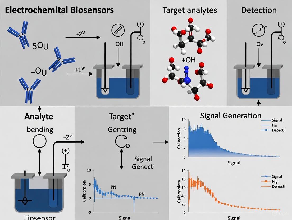
An electrochemical biosensor is an integrated analytical device that combines a biological recognition element with an electrochemical transducer, converting a biological event into a quantifiable electronic signal [1] [2]. These self-contained devices are designed to provide specific, quantitative, or semi-quantitative analytical information using a biological recognition system connected to a physicochemical transducer [1]. The core function of any biosensor is to offer an alternative to complex laboratory techniques by providing rapid, sensitive, and cost-effective analysis suitable for point-of-care testing, environmental monitoring, and food safety applications [3].
Since the invention of the first enzyme-based electrochemical biosensor by Leland C. Clark in 1962, the field has experienced explosive growth, driven by advancements in biotechnology, nanotechnology, and microelectronics [4] [5]. The most successful example of this technology remains the glucose sensor, which has revolutionized diabetes management worldwide [5]. Recent bibliometric analysis reveals consistently high research output, with over 2,000 publications on electrochemical biosensors in 2022 alone, demonstrating the field's continued expansion and relevance [6].
Core Components and Working Principle
The operational principle of an electrochemical biosensor hinges on the precise interaction and synergy between three fundamental components: the bioreceptor, transducer, and signal processor. This integrated system transforms biological information into an easily processed electronic signal [5] [1].
The Bioreceptor: Molecular Recognition Element
The bioreceptor is the biological recognition element responsible for the selective interaction with the target analyte. High specificity for the analyte among a matrix of other chemical or biological components is the critical requirement for the bioreceptor [2]. The bioreceptor can be composed of various biological or bio-mimetic materials, each with distinct mechanisms of action:
- Enzymes: Utilize specific binding capabilities and catalytic activity, often converting the analyte into an electrochemically detectable product. They are not consumed in reactions, allowing for continuous operation [2].
- Antibodies/Antigens: Employ the highly specific binding affinity of antibodies for a specific compound or antigen. The interaction is analogous to a lock and key fit, providing exceptional specificity [2].
- Nucleic Acids: Function based on complementary base pairing (in genosensors) or utilize specific nucleic acid-based antibody mimics known as aptamers (in aptasensors) [2].
- Whole Cells and Tissues: Exploit the response of bacteria, organelles, or tissue cultures to given substances or environmental conditions, often used for detecting global parameters like toxicity [2].
- Artificial Binding Proteins: Engineered protein scaffolds with favorable biophysical properties that offer advantages over traditional antibodies, including smaller size, enhanced stability, and easier production [2].
The bioreceptor is typically immobilized on the transducer surface through various physical or chemical methods to ensure stability and reproducibility [4].
The Transducer: Signal Conversion Interface
The transducer serves as the interface where the biological recognition event is converted into a measurable electrochemical signal. In electrochemical biosensors, the transducer is an electrode system that detects changes in electrical properties resulting from the interaction between the bioreceptor and analyte [5] [1]. The most common transducer configurations utilize a three-electrode system:
- Working Electrode: The primary transduction element where the specific biochemical reaction occurs.
- Reference Electrode: Maintains a known and stable potential against which the working electrode is measured.
- Counter Electrode: Completes the electrical circuit, allowing current to flow through the system.
The transduction mechanism can monitor various electrical properties, including current (amperometry), potential difference (potentiometry), impedance (impedimetry), or conductance (conductometry) [5]. Recent advances focus on nanomaterial-enhanced transducers that increase the active surface area and improve electron transfer kinetics, thereby boosting sensitivity [4] [7].
The Signal Processor: Data Interpretation System
The signal processor comprises the electronic components that convert the transducer signal into a meaningful physical parameter. This system typically includes a signal amplifier, processor, and user interface display [1] [2]. The signal conditioning circuit amplifies the often weak electrical signal from the transducer while filtering out noise. The processor then converts this conditioned signal into a digital format, applies necessary algorithms, and outputs the results in a user-friendly format, such as concentration values, through a display interface [8]. Modern biosensors increasingly incorporate machine learning algorithms to enhance data interpretation and improve analytical accuracy [4].
Table 1: Core Components of an Electrochemical Biosensor
| Component | Function | Key Characteristics | Examples |
|---|---|---|---|
| Bioreceptor | Selective recognition of target analyte | High specificity, stability, compatibility with transducer | Enzymes, antibodies, nucleic acids, cells [2] |
| Transducer | Converts biological event to electrical signal | High sensitivity, fast response, miniaturization capability | Electrodes (working, reference, counter) [5] [1] |
| Signal Processor | Amplifies, processes, and displays results | Low noise, appropriate algorithms, user-friendly interface | Amplifiers, microprocessors, displays [1] [8] |
Electrochemical Transduction Mechanisms
Electrochemical biosensors are classified based on their specific transduction method, each with distinct principles, advantages, and applications. The strategic selection of transduction mechanism depends on the target analyte, required sensitivity, and operational environment.
Amperometric Biosensors
Amperometric biosensors measure the current resulting from the electrochemical oxidation or reduction of an electroactive species. A constant potential is applied between the working and reference electrodes, and the current generated is directly proportional to the concentration of the analyte [9]. These sensors benefit from high sensitivity, wide linear ranges, and relatively simple instrumentation. The classic example is the glucose oxidase-based sensor, where enzyme catalysis generates electrons that produce a measurable current [5]. Recent developments incorporate microtip immunoassays and redox mediators to enhance sensitivity and detection limits [9].
Potentiometric Biosensors
Potentiometric biosensors measure the accumulation of charge at the electrode-electrolyte interface when negligible current flows through the system. They typically employ ion-selective electrodes (ISEs) or field-effect transistors (FETs) to detect potential differences resulting from biological recognition events [9] [1]. These sensors provide a logarithmic response with a high dynamic range and are particularly useful for detecting small ions and metabolites. Recent innovations include extended-gate FET configurations for serological diagnosis and solid-state devices for improved stability [9] [8].
Impedimetric Biosensors
Impedimetric biosensors utilize Electrochemical Impedance Spectroscopy (EIS) to monitor changes in the impedance (both resistance and reactance) at the electrode-electrolyte interface. This label-free technique measures how biological binding events alter the electrical properties of the sensing surface [9]. EIS can operate in Faradaic mode (using a redox probe) or non-Faradaic mode (relying on intrinsic capacitive behavior). These sensors are particularly valuable for monitoring binding events in real-time without requiring labeled components, making them ideal for affinity-based detection and kinetic studies [9] [10].
Voltammetric Biosensors
Voltammetric biosensors apply a varying potential to the working electrode and measure the resulting current. Different techniques include cyclic voltammetry, differential pulse voltammetry, and square wave voltammetry, each offering distinct advantages for specific applications [9]. These methods provide information about electrochemical reactions, including redox potentials and reaction kinetics. Recent developments incorporate advanced nanomaterials like molybdenum disulfide nanosheets to enhance signal response and lower detection limits for targets such as viral DNA [9].
Table 2: Comparison of Electrochemical Transduction Mechanisms
| Transduction Type | Measured Parameter | Detection Limit | Advantages | Common Applications |
|---|---|---|---|---|
| Amperometric | Current from redox reaction | ~nM range [9] | High sensitivity, fast response | Metabolic markers, disease detection [9] [5] |
| Potentiometric | Potential difference | ~nM range [9] | Wide dynamic range, simple instrumentation | Ion concentration, pH, serological diagnosis [9] [1] |
| Impedimetric | Impedance/charge transfer resistance | ~0.1-10 ng/mL [9] [10] | Label-free, real-time monitoring | Affinity binding, cell analysis, DNA detection [9] [10] |
| Voltammetric | Current vs. applied potential | ~nM range [9] | Rich electrochemical information, multiple techniques | Drug discovery, environmental toxins [9] |
Experimental Protocol: Impedimetric miRNA Detection for Oral Cancer
To illustrate the practical implementation of an electrochemical biosensor, the following detailed protocol for detecting microRNA-31 (a potential oral cancer biomarker) using Electrochemical Impedance Spectroscopy (EIS) is provided based on recent research [10].
Materials and Reagents
- Glassy Carbon Electrode (GCE): Serves as the base transducer (diameter: 3 mm)
- Graphene nanosheets: Conductive nanomaterial for signal enhancement (X and Y dimensions: >2 μm, thickness: 8-15 nm)
- 1-Pyrenebutanoic acid Succinimidyl Ester (PBSE): Molecular tethering agent for biomolecule immobilization
- ssDNA Probe: Bioreceptor sequence (5'-NH₂-C₆-AGCTATGCCAGCATCTTGCCT-3') with C6 amine modification
- Target miRNA-31: Analyte of interest (5'-AGGCAAGAUGCUGGCAUAGCU-3')
- Non-complementary miRNA-25: Specificity control (5'-AGGCGGAGACUUGGGCAUUG-3')
- Electrochemical Cell: Three-electrode setup with platinum counter electrode and Ag/AgCl reference electrode
- Buffer Solutions:
- Phosphate Buffer (PB): 10 mM Na₂HPO₄/NaH₂PO₄, pH 7.4 for electrode washing
- Immobilization Buffer (IB): 10 mM PB with 0.15 M NaCl for ssDNA immobilization
- Hybridization Buffer (HB): 10 mM PB with 1 M NaCl and 20 mM MgCl₂ for miRNA hybridization
- Redox Probe: 5 mM K₃[Fe(CN)₆]/K₄[Fe(CN)₆] in 0.1 M KCl for EIS measurements
Step-by-Step Methodology
Electrode Pretreatment:
- Polish the GCE surface with 0.05 μm alumina slurry on a microcloth
- Rinse thoroughly with deionized water (18 MΩ·cm resistivity)
- Clean via sonication in ethanol and deionized water (5 minutes each)
- Dry under nitrogen stream
Graphene Nanomaterial Modification:
- Prepare graphene dispersion (1 mg/mL) in dimethylformamide (DMF)
- Deposit 8 μL suspension onto GCE surface
- Allow to dry overnight in a desiccator at room temperature
PBSE Interlayer Formation:
- Prepare fresh PBSE solution (2 mM in DMF)
- Incubate graphene-modified GCE with 8 μL PBSE for 1 hour
- Wash extensively with DMF and PB to remove unbound PBSE
ssDNA Probe Immobilization:
- Dilute amino-modified ssDNA probe to 1 μM concentration in Immobilization Buffer
- Apply 8 μL to PBSE-functionalized electrode
- Incubate for 1.5 hours at room temperature in a humidified chamber
- Rinse with PB to remove physically adsorbed DNA
Surface Blocking:
- Treat electrode with 8 μL ethanolamine (1 M) for 10 minutes
- Wash with PB to deactivate and remove excess NHS esters
Target miRNA Hybridization and Detection:
- Apply 8 μL of target miRNA-31 (various concentrations in Hybridization Buffer)
- Incubate for 1.5 hours at 37°C
- Rinse with PB to remove unhybridized miRNA
- Perform EIS measurements in redox probe solution
EIS Measurement Parameters:
- Frequency range: 0.1 Hz to 100 kHz
- AC amplitude: 10 mV
- DC potential: 0.23 V (formal potential of redox probe)
- Record Nyquist plots and extract charge transfer resistance (Rct)
Data Analysis:
- Calculate ΔRct = Rct(post-hybridization) - Rct(pre-hybridization)
- Plot ΔRct vs. log[miRNA-31] for calibration curve
- Determine limit of detection using Six Sigma method (10 pM in buffer, 100 pM in diluted serum)
Diagram 1: miRNA Detection Workflow (Width: 760px)
The Scientist's Toolkit: Essential Research Reagents
Table 3: Key Research Reagents for Electrochemical Biosensing
| Reagent/Material | Function | Specific Example | Considerations |
|---|---|---|---|
| Electrode Materials | Signal transduction platform | Glassy carbon, screen-printed electrodes, gold electrodes | Surface area, conductivity, functionalization compatibility [5] [10] |
| Nanomaterials | Signal amplification | Graphene, metal nanoparticles, metal-organic frameworks (e.g., ZIF-67) | Enhanced surface area, improved electron transfer, catalytic properties [7] [10] |
| Molecular Tethers | Bioreceptor immobilization | PBSE, SAMs (self-assembled monolayers), glutaraldehyde | Stable attachment, proper orientation, maintained bioreceptor activity [10] |
| Bioreceptors | Target recognition | ssDNA, antibodies, enzymes, aptamers | Specificity, stability, binding affinity, immobilization requirements [2] [10] |
| Redox Probes | Electrochemical signal generation | Ferricyanide/ferrocyanide, ruthenium hexamine | Reversible electrochemistry, stability, compatibility with system [9] [10] |
| Blocking Agents | Minimize non-specific binding | Ethanolamine, BSA, casein, synthetic polymers | Effective surface coverage, non-interference with bioreceptor [10] |
Recent Advances and Future Perspectives
The field of electrochemical biosensors continues to evolve through integration with emerging technologies. Recent highlights include the development of bimetallic metal-organic frameworks (MOFs) such as Mn-doped ZIF-67, which demonstrate exceptional sensitivity for pathogen detection with limits as low as 1 CFU mL⁻¹ for E. coli [7]. The incorporation of machine learning algorithms for data analysis enhances signal interpretation and enables multiplexed detection capabilities [4] [3].
Wearable formats and implantable sensors represent another frontier, enabling continuous monitoring of biomarkers in interstitial fluid for personalized healthcare applications [4]. The convergence of electrochemical sensing with optical techniques such as electrochemiluminescence (ECL) and surface-enhanced Raman spectroscopy (SERS) creates powerful hybrid systems that combine the sensitivity of electrochemical methods with the spatial resolution of optical techniques [3].
Future development will likely focus on increasing integration and miniaturization while improving the analytical reliability needed for clinical diagnostics. The successful translation of research prototypes to commercial devices will require enhanced reproducibility, stability in complex matrices, and validation against standard laboratory methods [4] [3].
The electrochemical biosensor represents a perfect synergy between biological recognition and electronic signal transduction. Its core architecture—comprising the bioreceptor, transducer, and signal processor—enables the specific, sensitive, and rapid detection of target analytes across diverse fields including clinical diagnostics, environmental monitoring, and food safety. Continued advancements in nanotechnology, biotechnology, and microelectronics will further enhance the capabilities of these devices, driving their evolution toward more sophisticated, integrated, and user-friendly analytical systems. As research progresses, electrochemical biosensors are poised to play an increasingly vital role in addressing global challenges in healthcare, environmental protection, and beyond.
Electrochemical biosensors have emerged as a highly promising platform for advanced bioanalytical applications, synergistically integrating the high molecular recognition specificity of biorecognition elements with the rapid and sensitive signal transduction capabilities of electrochemical interfaces [11]. The core of any biosensor is its biorecognition element, which dictates the sensor's specificity, sensitivity, and overall analytical performance. These elements are responsible for the selective interaction with target analytes, ranging from small molecules and metal ions to proteins, nucleic acids, and entire cells [4] [12]. The choice of biorecognition element—whether enzymatic, immunologic, nucleic acid-based, or cellular—fundamentally influences the design strategy, operational principle, and eventual application scope of the resulting biosensing platform.
Within the context of electrochemical biosensors research, understanding the comparative advantages and limitations of different biorecognition elements is crucial for developing next-generation diagnostic systems. The ongoing evolution in this field is driven by innovations at the intersection of chemistry, materials science, biomedical engineering, and environmental monitoring [11]. Recent advances have highlighted the trend toward point-of-care (POC) and on-site diagnostic formats characterized by low cost, facile miniaturization, and compatibility with complex sample matrices [4] [13]. The global biosensors market, where electrochemical biosensors dominate with over 70% market share, reflects the significance of these developments [14].
This technical guide provides a comprehensive comparative analysis of four primary classes of biorecognition elements—enzymes, antibodies, aptamers, and whole cells—framed within the working principles of electrochemical biosensors research. By examining their fundamental characteristics, operational mechanisms, and performance metrics, this review aims to equip researchers and drug development professionals with the critical knowledge needed to select appropriate biorecognition elements for specific biosensing applications.
Fundamental Principles of Electrochemical Biosensing
Electrochemical biosensors transduce biochemical interactions at the sensor interface into measurable electrical signals through various electrochemical techniques. The basic components include the analyte (target molecule), bioreceptor (recognition element), transducer (electrochemical interface), and readout system [4]. The working principle involves the specific binding of the target analyte to the biorecognition element, which generates a biochemical signal converted by the transducer into an quantifiable electrical output such as current, potential, or impedance [15].
Electrochemical biosensors can be classified based on their transduction mechanism:
- Amperometric sensors measure current resulting from redox reactions at a constant applied potential [16] [12].
- Potentiometric sensors detect potential differences between working and reference electrodes under zero-current conditions [4].
- Impedimetric sensors (EIS) monitor changes in charge transfer resistance at the electrode-electrolyte interface upon target binding [16] [15].
- Voltammetric sensors (CV, DPV, SWV) record current while scanning the applied potential, providing information about electroactive species [17] [12].
The following diagram illustrates the generalized working principle of an electrochemical biosensor, highlighting the key components and signal transduction pathway:
Comparative Analysis of Biorecognition Elements
Enzymes
Enzyme-based electrochemical biosensors represent one of the most established categories, with glucose monitoring as the quintessential application [14]. Enzymes function as biorecognition elements through their specific catalytic activity toward target substrates, generating electroactive products that can be detected at the electrode surface.
Key Characteristics:
- Mechanism: Catalytic conversion of specific substrates to measurable products
- Signal Generation: Typically through detection of enzymatic reaction products (e.g., H₂O₂ from oxidase enzymes)
- Immobilization Methods: Adsorption, covalent binding, cross-linking, entrapment in polymers or gels [15]
The primary advantage of enzymatic recognition lies in the signal amplification inherent to catalytic turnover, where a single enzyme molecule can generate multiple product molecules, enhancing sensitivity. However, enzymes suffer from limited environmental stability, sensitivity to pH and temperature variations, and potential inhibition by interfering substances.
Antibodies
Antibodies, particularly immunoglobulin G (IgG), form the basis of electrochemical immunosensors, leveraging the specific antigen-antibody binding principle in immunology [15]. These biorecognition elements provide exceptional specificity toward a wide range of antigens, including proteins, hormones, and pathogens.
Key Characteristics:
- Mechanism: Lock-and-key binding to specific epitopes on target antigens
- Signal Generation: Label-free (direct binding-induced impedance changes) or labeled (enzyme-linked, nanoparticle-tagged) approaches [12]
- Immobilization Methods: Protein A/G binding, covalent attachment via amine/carboxyl groups, physisorption on nanostructured surfaces [4]
Antibodies offer high affinity and maturity of commercial production, but they present challenges including batch-to-batch variability, sensitivity to denaturation, and high production costs, particularly for monoclonal antibodies [12]. Traditional immunoassays like ELISA have been successfully adapted to electrochemical platforms, enhancing sensitivity and reducing analysis time [12] [15].
Aptamers
Aptamers are short, single-stranded DNA or RNA oligonucleotides selected through Systematic Evolution of Ligands by Exponential Enrichment (SELEX) to bind specific targets with high affinity [17] [12]. These nucleic acid-based recognition elements have gained significant attention as promising alternatives to antibodies in biosensing applications.
Key Characteristics:
- Mechanism: Target-induced conformational change or direct binding
- Signal Generation: Conformational change altering electron transfer efficiency or distance of redox tags [17]
- Immobilization Methods: Au-Thiol chemistry, biotin-streptavidin interaction, covalent attachment to functionalized surfaces [16]
Aptamers offer numerous advantages including thermal stability, ease of chemical synthesis and modification, reusability, and cost-effectiveness [17] [16]. Their relatively small size enables higher surface density on electrodes compared to antibodies. Electrochemical biosensors utilizing aptamers (aptasensors) have demonstrated excellent performance for detecting diverse targets from small molecules and metal ions to proteins and whole cells [11] [16].
Whole Cells
Whole-cell biosensors utilize microorganisms (bacteria, yeast) or mammalian cells as recognition elements, typically employing their innate metabolic or stress response pathways to detect target analytes.
Key Characteristics:
- Mechanism: Cellular response to analytes (metabolic, stress, or transcriptional activation)
- Signal Generation: Production of electroactive metabolites, viability markers, or reporter enzymes
- Immobilization Methods: Entrapment in polymer matrices, membrane confinement, biofilm formation
Whole-cell biosensors are particularly valuable for assessing functional parameters like toxicity, bioavailability, and metabolic effects that cannot be determined using molecular recognition elements. However, they present challenges including longer response times, maintenance of cell viability, and relatively lower specificity compared to molecular recognition elements.
Table 1: Comparative Performance Metrics of Biorecognition Elements
| Parameter | Enzymes | Antibodies | Aptamers | Whole Cells |
|---|---|---|---|---|
| Specificity | High (catalytic) | Very High (immunological) | Very High (structural) | Moderate to High (functional) |
| Sensitivity | nM-pM | pM-fM | pM-fM [16] | Variable |
| Stability | Moderate (temperature/pH sensitive) | Moderate (sensitive to denaturation) | High (thermostable, reusable) [17] | Low (requires viability) |
| Development Time | Weeks-Months | Months | Weeks (SELEX process) [12] | Weeks-Months |
| Production Cost | Moderate | High | Low (chemical synthesis) [17] [12] | Low-Moderate |
| Modification Ease | Moderate | Difficult | Easy (synthetic modification) [17] | Difficult |
Table 2: Preferred Applications of Different Biorecognition Elements
| Biorecognition Element | Ideal Applications | Limitations |
|---|---|---|
| Enzymes | Metabolic markers, small molecules, toxicants | Limited target scope, instability |
| Antibodies | Protein biomarkers, pathogens, hormones [15] | Batch variability, cost, temperature sensitivity |
| Aptamers | Diverse targets (ions, small molecules, proteins, cells) [11] [16] | Susceptibility to nuclease degradation |
| Whole Cells | Toxicity screening, bioavailability, functional assessment | Limited specificity, slow response |
Experimental Protocols and Methodologies
Aptamer-Based Sensor Development Protocol
The development of aptamer-based electrochemical biosensors involves aptamer selection, sensor fabrication, and analytical validation. The following workflow details a standard protocol for constructing an electrochemical aptasensor:
Detailed Experimental Steps:
Aptamer Selection and Preparation
- SELEX Process: Identify specific aptamers through Systematic Evolution of Ligands by Exponential Enrichment against the target molecule [17] [12]. This typically involves 8-15 rounds of selection with increasing stringency.
- Aptamer Modification: Synthesize aptamers with terminal modifications (thiol, amino, or biotin groups) to facilitate immobilization. For thiol-modified DNA, reduce disulfide bonds using TCEP (tris(2-carboxyethyl)phosphine) and purify via HPLC or gel filtration [17].
Electrode Preparation and Functionalization
- Clean electrode surfaces (typically gold, glassy carbon, or screen-printed carbon) through physical (polishing) and chemical (piranha solution for Au, acid treatment for carbon) methods [4].
- For gold electrodes, characterize surface cleanliness using cyclic voltammetry in sulfuric acid solution, observing well-defined gold oxidation and reduction peaks.
- Functionalize electrode surface with capture elements: self-assembled monolayers (SAMs) for thiolated aptamers, streptavidin for biotinylated aptamers, or carboxylic acid groups for amine-modified aptamers using EDC/NHS chemistry [16].
Aptamer Immobilization
- Incubate modified electrodes with aptamer solution (0.1-5 µM concentration) in appropriate immobilization buffer (e.g., PBS with Mg²⁺ for structure formation) for 2-16 hours at room temperature [17].
- Optimize immobilization time and concentration to achieve optimal surface density without steric hindrance.
Surface Blocking
- Treat aptamer-functionalized surfaces with blocking agents (e.g., 6-mercapto-1-hexanol for thiol-based systems, BSA for nonspecific protein blocking) to minimize nonspecific binding [17].
- Incubate for 30-120 minutes, then rinse thoroughly with buffer to remove unbound blocking agents.
Target Binding and Electrochemical Measurement
- Incubate functionalized electrodes with target solutions of varying concentrations for optimal binding (typically 15-60 minutes).
- Perform electrochemical measurements using appropriate techniques:
- Electrochemical Impedance Spectroscopy (EIS): Monitor charge transfer resistance changes in [Fe(CN)₆]³⁻/⁴⁻ redox probe (frequency range: 0.1 Hz-100 kHz, amplitude: 5-10 mV) [16].
- Differential Pulse Voltammetry (DPV): Measure current changes of redox tags (e.g., methylene blue, ferrocene) with pulse parameters (amplitude: 25-50 mV, pulse width: 50-100 ms) [17] [12].
- Square Wave Voltammetry (SWV): Utilize parameters (frequency: 10-25 Hz, amplitude: 25 mV) for sensitive detection of conformational change-induced signal variations [12].
Sensor Regeneration and Reusability
- Regenerate sensor surface using mild conditions such as low pH buffer (e.g., 10 mM glycine-HCl, pH 2.0-3.0), denaturing agents (e.g., 4-8 M urea), or elevated temperature to dissociate aptamer-target complexes without damaging immobilized aptamers [17].
- Validate sensor stability over multiple regeneration cycles (typically 5-10 cycles).
Antibody-Based Sensor Development Protocol
The development of electrochemical immunosensors follows a similar workflow but with distinct considerations for antibody immobilization and preservation of biological activity:
Antibody Selection and Preparation
- Select appropriate antibody type (monoclonal for specificity, polyclonal for signal amplification) based on application requirements [12].
- For sandwich-type assays, optimize pairings of capture and detection antibodies to recognize non-overlapping epitopes on the target antigen.
Electrode Modification for Antibody Immobilization
- Modify electrode surfaces with nanomaterials (e.g., gold nanoparticles, graphene, carbon nanotubes) to increase surface area and enhance electron transfer [16] [15].
- Functionalize surfaces with oriented immobilization strategies: Protein A/G for Fc region binding, hydrazide chemistry for oxidized glycan groups, or maleimide-thiol coupling for thiolated antibodies [15].
Immunoassay Protocol
- Apply capture antibody solution (10-100 µg/mL) to functionalized electrodes and incubate (1-2 hours at 25°C or overnight at 4°C).
- Block nonspecific sites with blocking buffers (BSA, casein, or commercial blocking agents) for 1 hour.
- Incubate with antigen samples (30-60 minutes) at optimized concentration range.
- For sandwich formats, incubate with labeled detection antibody (30-60 minutes).
- Perform electrochemical measurement appropriate to the label system (amperometric for enzyme labels, voltammetric for nanoparticle labels).
The Scientist's Toolkit: Essential Research Reagents and Materials
Table 3: Essential Research Reagents for Electrochemical Biosensor Development
| Reagent/Material | Function | Examples & Applications |
|---|---|---|
| Biorecognition Elements | Molecular recognition of target analytes | Enzymes (glucose oxidase, horseradish peroxidase), Antibodies (IgG, monoclonal, polyclonal), Aptamers (DNA/RNA, thiol-modified) [17] |
| Electrode Materials | Signal transduction platform | Gold electrodes, Glassy carbon, Screen-printed electrodes (disposable), Indium tin oxide (transparent) [4] |
| Nanomaterials | Signal amplification, enhanced immobilization | Gold nanoparticles, Graphene/GO, Carbon nanotubes, MOFs, MXenes [16] [18] |
| Crosslinking Chemicals | Covalent immobilization of biorecognition elements | EDC/NHS chemistry, Glutaraldehyde, Sulfo-SMCC, Maleimide-thiol coupling reagents [15] |
| Redox Probes | Electrochemical signal generation | [Fe(CN)₆]³⁻/⁴⁻, Methylene blue, Ferrocene derivatives, Ru(NH₃)₆³⁺ [12] |
| Blocking Agents | Minimize nonspecific binding | BSA, Casein, MCH (6-mercapto-1-hexanol), Polyethylene glycol, Zwitterionic peptides [17] |
| Buffer Systems | Maintain optimal biorecognition conditions | PBS, HEPES, Tris, Acetate with specific ion requirements (e.g., Mg²⁺ for aptamers) [17] |
The selection of appropriate biorecognition elements represents a critical design consideration in electrochemical biosensor development, with significant implications for analytical performance, practical implementation, and commercial viability. Each class of biorecognition element—enzymes, antibodies, aptamers, and whole cells—offers distinct advantages and limitations that must be carefully evaluated against specific application requirements.
Enzymes provide excellent catalytic amplification but limited target scope. Antibodies offer exceptional specificity and established protocols but suffer from production costs and stability issues. Aptamers present a versatile alternative with superior stability, modification ease, and cost-effectiveness, though they may require stabilization strategies for complex biological samples [17] [16] [12]. Whole cells enable functional assessment but offer lower specificity and slower response times.
Future directions in biorecognition element development include engineering hybrid systems combining multiple recognition elements, creating artificial biomimetic receptors (MIPs), and integrating advanced nanomaterials to enhance sensitivity and stability. The convergence of artificial intelligence with biosensor technology is expected to accelerate biorecognition element design through computational prediction of binding affinities and optimal sequences [11] [14]. As electrochemical biosensors continue to evolve toward point-of-care applications, wearable formats, and multiplexed detection platforms, the strategic selection and engineering of biorecognition elements will remain fundamental to advancing biosensing capabilities across healthcare, environmental monitoring, and food safety domains.
Electrochemical biosensors are analytical devices that synergistically integrate a biological recognition element with an electrochemical transducer, enabling the quantitative detection of specific analytes by converting a biological interaction into a measurable electrical signal [11] [19]. This transduction process is fundamental to the function of these biosensors, which have attracted intense interest as promising tools for point-of-care (POC) applications due to their rapid response times, high sensitivity and selectivity, portability, and ease of operation [19]. The core principle involves the selective binding of a target analyte (e.g., a disease biomarker, viral particle, or bacterial pathogen) by an immobilized biorecognition element (such as an antibody, enzyme, or nucleic acid aptamer), which subsequently triggers a change in electrical properties at the electrode-solution interface [11] [20]. This review provides an in-depth technical examination of the signal transduction mechanisms in electrochemical biosensors, framed within the broader context of advancing their working principles for research and clinical applications.
Fundamental Principles of Electrochemical Transduction
At its core, an electrochemical biosensor consists of a biorecognition layer immobilized on the surface of an electrochemical transducer. The transducer translates the biological event into an electrical signal that can be processed and measured [19]. The specificity of the sensor is conferred by the biorecognition element, which may be an antibody, enzyme, nucleic acid aptamer, or receptor. When the target analyte binds to this element, it induces a physicochemical change that is converted by the transducer into an electrical output such as current, potential, or impedance [20].
The analytical performance of these biosensors—including sensitivity, detection limit, and dynamic range—is heavily influenced by the efficiency of this signal transduction process. Recent advances in nanomaterials, micro/nanofabrication technologies, and signal amplification strategies have markedly improved this performance, enabling ultra-sensitive and highly selective detection of a diverse array of analytes [11]. The integration of functional nanomaterials such as gold nanoparticles (AuNPs), graphene oxide (GO), carbon nanotubes (CNTs), and metal–organic frameworks (MOFs) has been particularly impactful, enhancing sensor performance by improving electron transfer, enabling signal amplification, and increasing biocompatibility [20].
Major Signal Transduction Mechanisms
Electrochemical biosensing architectures translate detection events into measurable electrical signals through several distinct mechanisms, primarily based on changes in conductance, resistance, capacitance, or the generation of electrochemical reactions upon analyte binding [19]. The following sections detail the primary transduction mechanisms employed in modern electrochemical biosensors.
Voltammetric and Amperometric Transduction
Voltammetric and amperometric transducers measure current resulting from electrochemical oxidation or reduction reactions at the electrode surface. The biological recognition event often modulates this faradaic current, either by generating an electroactive species or by altering the electron transfer efficiency.
A prominent example involves aptamer-based sensors where conformational changes in the aptamer structure upon target binding either facilitate or hinder electron transfer to the electrode surface. This can be monitored using techniques such as differential pulse voltammetry (DPV) or cyclic voltammetry (CV) [19]. In one documented immunosensor for interleukin-6 (IL-6), the DPV peak current was used to quantify the target analyte in buffer, cerebrospinal fluid (CSF), and serum, achieving detection limits as low as 1.6 pg mL⁻¹ [19].
Potentiometric Transduction
Potentiometric biosensors measure the accumulation of charge or change in potential at the electrode-electrolyte interface under conditions of zero current. The measured potential is proportional to the logarithm of the concentration of the target ion or molecule. While less commonly the primary mechanism in complex bioassays, it can play a role in systems where binding events alter the local ion distribution or membrane potential.
Impedimetric Transduction
Impedimetric biosensors, often utilizing Electrochemical Impedance Spectroscopy (EIS), monitor changes in the impedance (resistance to electron transfer) at the electrode surface following a biorecognition event. This label-free method is highly sensitive to surface modifications, such as the binding of a target protein to its immobilized receptor.
For instance, in the aforementioned IL-6 immunosensor, EIS measurements showed that the charge transfer resistance (Rₜ꜀) decreases upon IL-6 binding. This observation was attributed to a structural change upon antibody-antigen binding that opens up the architecture, allowing a redox probe to access the electrode surface more easily [19]. This decrease in Rₜ꜀ provides a direct electrical signal correlating with target concentration.
Conductometric and Capacitive Transduction
Conductometric sensors measure the change in electrical conductivity of a solution between two electrodes, while capacitive sensors detect changes in the dielectric properties or capacitance of an electrode interface. These mechanisms are particularly useful for detecting binding events that do not involve direct electron transfer but still alter the electrical properties of the sensing layer.
Table 1: Comparison of Major Electrochemical Transduction Mechanisms
| Transduction Mechanism | Measured Signal | Key Advantages | Common Techniques | Typical Applications |
|---|---|---|---|---|
| Voltammetric/Amperometric | Current from redox reactions | High sensitivity, wide dynamic range | DPV, CV, Amperometry | Detection of cytokines, metabolites, pathogens |
| Impedimetric | Impedance at electrode interface | Label-free, real-time monitoring | EIS | Protein detection, cell analysis, affinity-based sensors |
| Potentiometric | Potential difference | Simple instrumentation, ion sensitivity | Ion-selective electrodes | pH, ion concentrations, enzyme substrates |
| Conductometric/Capacitive | Conductivity/Capacitance change | Label-free, suitable for miniaturization | Conductometry, Capacitance measurement | DNA hybridization, gas sensing |
Advanced Transduction Architectures and Signal Amplification
To achieve the high sensitivity required for detecting low-abundance biomarkers in complex samples, researchers have developed sophisticated transduction architectures that incorporate signal amplification strategies.
Redox Cycling and Bipolar Electrodes
Advanced electrode geometries, such as interdigitated electrode arrays (IDEAs) and closed bipolar electrodes (CBEs), enable signal amplification via redox cycling [19]. In this process, a redox species is repeatedly oxidized and reduced between closely spaced electrodes, significantly amplifying the faradaic current and enhancing the detection signal.
A CBE-based metabolite sensing platform was constructed for detecting diagnostic metabolites like cholesterol, glucose, and lactate in undiluted human blood [19]. This system employed a blood-compatible electrode chemistry based on phosphorylcholine (PPC) and phenylbutyric acid (PBA) mixed layers, electroactive ferrocene (Fc) moieties, redox-active enzymes, and a diffusing mediating species. Multiple electron mediation routes were identified as contributing to the overall sensing scheme, with both surface-bound and diffusible mediating species complementing each other to produce an enhanced electrochemical response [19].
Nanomaterial-Enhanced Transduction
The integration of functional nanomaterials has revolutionized signal transduction in electrochemical biosensors. These materials enhance sensitivity by providing high surface areas for bioreceptor immobilization, facilitating electron transfer, and enabling novel signal amplification pathways.
DNA aptamers integrated with nanomaterials such as gold nanoparticles (AuNPs), graphene oxide (GO), carbon nanotubes (CNTs), and metal-organic frameworks (MOFs) significantly enhance sensor performance [20]. For example, AuNPs can serve as excellent conduits for electron transfer, while GO provides a large surface area with unique electronic properties. CNTs facilitate electron tunneling, and MOFs offer exceptionally high porosity for encapsulating signal probes or enzymes.
Experimental Protocols for Transduction Mechanism Analysis
This section provides detailed methodologies for key experiments cited in this review, enabling researchers to replicate and build upon established transduction principles.
Protocol 1: Impedimetric Detection of Protein Biomarkers
This protocol outlines the development of a label-free electrochemical immunosensor for the detection of interleukin-6 (IL-6), as described in the literature [19].
Materials and Reagents:
- Gold interdigitated electrode arrays (IDEAs)
- IL-6 antibodies (monoclonal)
- Interleukin-6 (IL-6) antigen standard
- Blocking buffer (e.g., 1% BSA in PBS)
- Human cerebrospinal fluid (CSF) and serum samples
- Redox probe solution ([Fe(CN)₆]³⁻/⁴⁻)
- Phosphate buffer saline (PBS, 0.1 M, pH 7.4)
Methodology:
- Electrode Pretreatment: Clean gold IDEA electrodes via electrochemical cycling in 0.5 M H₂SO₄ or by oxygen plasma treatment.
- Antibody Immobilization: Incubate electrodes with IL-6 antibody solution (10-50 μg/mL in PBS) for 2 hours at room temperature or overnight at 4°C.
- Surface Blocking: Treat electrode with blocking buffer for 1 hour to prevent nonspecific adsorption.
- Sample Incubation: Expose functionalized electrode to standards or samples containing IL-6 for 30-60 minutes.
- Electrochemical Impedance Spectroscopy (EIS): Perform EIS measurements in redox probe solution over a frequency range of 0.1 Hz to 100 kHz with an amplitude of 10 mV at formal potential of the redox probe.
- Data Analysis: Extract charge transfer resistance (Rₜ꜀) values from Nyquist plots and plot against IL-6 concentration to generate a calibration curve.
Expected Results: Rₜ꜀ values decrease with increasing IL-6 concentration due to structural changes upon antibody-antigen binding that facilitate redox probe access to the electrode surface. The sensor should demonstrate a linear range for IL-6 in buffer, CSF, and serum with detection limits potentially as low as 1.6 pg mL⁻¹ in buffer [19].
Protocol 2: Redox-Mediated Metabolite Sensing in Whole Blood
This protocol details the construction of a closed bipolar electrode (CBE)-based metabolite sensing platform for detecting metabolites directly in undiluted human blood [19].
Materials and Reagents:
- Closed bipolar electrode system
- Blood-compatible surface chemistry components (phosphorylcholine-PPC and phenylbutyric acid-PBA)
- Ferrocene (Fc) derivatives for electron mediation
- Redox-active enzymes (cholesterol oxidase, glucose oxidase, or lactate oxidase)
- Diffusing mediating species
- Human whole blood samples (unprocessed)
- Smartphone with camera for readout
Methodology:
- Electrode Modification: Prepare mixed self-assembled monolayers on electrode surfaces using PPC and PBA to create blood-compatible layers that resist nonspecific protein adsorption.
- Enzyme and Mediator Integration: Immobilize appropriate oxidase enzymes and incorporate both surface-bound ferrocene moieties and diffusing mediating species in the sensing scheme.
- Sensor Assembly: Incorporate the sensing scheme into the CBE platform, where electrochemical reactions at one pole induce complementary reactions at the other pole.
- Sample Application: Apply unprocessed human whole blood samples directly to the sensor.
- Signal Detection: Monitor the coupled redox-mediated color change using a smartphone camera and perform subsequent RGB analysis for quantification.
- Validation: Compare results with commercial, FDA-approved point-of-care devices for validation.
Expected Results: The CBE sensor should enable determination of cholesterol, glucose, and lactate levels in real blood samples, with results in excellent agreement with commercial devices. The system leverages multiple electron mediation routes, with both surface-bound and diffusible mediating species complementing each other to produce an enhanced electrochemical response [19].
Visualization of Transduction Mechanisms
The following diagrams, created using Graphviz DOT language, illustrate key signaling pathways and experimental workflows in electrochemical biosensor transduction.
Aptamer-Based Electrochemical Sensing Mechanism
Impedimetric Biosensor Experimental Workflow
The Scientist's Toolkit: Essential Research Reagents and Materials
The following table details key research reagent solutions and essential materials used in the development and implementation of electrochemical biosensors based on the transduction mechanisms discussed.
Table 2: Essential Research Reagents and Materials for Electrochemical Biosensor Development
| Item | Function/Purpose | Specific Examples & Applications |
|---|---|---|
| Biorecognition Elements | Provides molecular specificity for target analyte | Nucleic acid aptamers, antibodies, enzymes; Used for specific capture of disease biomarkers, pathogens [11] [20] |
| Electrode Materials | Serves as transducer platform; influences electron transfer efficiency | Gold interdigitated electrode arrays (IDEAs), carbon electrodes; Used for signal transduction, redox cycling amplification [19] |
| Nanomaterials | Enhances signal, improves immobilization, increases surface area | Gold nanoparticles (AuNPs), graphene oxide (GO), carbon nanotubes (CNTs); Improve electron transfer, enable signal amplification [20] |
| Surface Chemistry Reagents | Enables robust bioreceptor immobilization, prevents non-specific binding | Phosphorylcholine (PPC), phenylbutyric acid (PBA) mixed layers; Creates blood-compatible surfaces for whole fluid sensing [19] |
| Redox Probes/Mediators | Facilitates electron transfer, enables signal generation | Ferrocene (Fc) derivatives, [Fe(CN)₆]³⁻/⁴⁻; Used as electron mediators in voltammetric and impedimetric measurements [19] |
| Signal Amplification Components | Enhances detection sensitivity for low-abundance analytes | Enzymatic labels (horseradish peroxidase, alkaline phosphatase), diffusing mediating species; Amplifies signals in CBE systems [19] |
The transduction mechanism—the process of converting biological interactions into quantifiable electrical signals—represents the fundamental operating principle of electrochemical biosensors. Through various mechanisms including voltammetric, impedimetric, and potentiometric transduction, these devices effectively translate molecular recognition events into analytical readouts. Current research continues to refine these mechanisms through advanced electrode architectures, nanomaterial integration, and sophisticated signal amplification strategies. The convergence of electrochemistry, materials science, and molecular biology in this field has yielded practical biosensors capable of determining health-critical biomarkers in complex biological samples, bridging the gap between laboratory research and clinical diagnostics. Future perspectives point toward the development of increasingly portable, multiplexed, and intelligent biosensing systems incorporating artificial intelligence-assisted data interpretation, wearable formats, and Internet of Things (IoT) integration for next-generation diagnostic applications [11].
Electrochemical biosensors represent a cornerstone of modern analytical science, merging the specificity of biological recognition with the sensitivity and versatility of electrochemical transducers. These devices are defined as "analytical devices incorporating a biological material, a biologically derived material or a biomimic intimately associated with or integrated within a physicochemical transducer or transducing microsystem" [21]. The fundamental working principle involves converting a biological recognition event into a quantifiable electrical signal such as current, potential, or impedance [22]. The significance of these biosensors extends across numerous fields including healthcare diagnostics, environmental monitoring, food safety, and drug development [23] [24]. Their benefits are substantial, offering cost-efficiency, short response times, ease of use, excellent limits of detection, sensitivity, and straightforward miniaturization [23]. This technical guide provides an in-depth examination of the four essential electrochemical techniques—amperometry, potentiometry, voltammetry, and electrochemical impedance spectroscopy (EIS)—that form the foundation of biosensing research and development, with particular emphasis on their working principles, applications, and experimental implementation within the context of biosensor technology.
Fundamental Principles of Electrochemical Biosensors
Electrochemical biosensors typically employ a three-electrode system consisting of working, reference, and counter electrodes [22]. The biological recognition element, which can be an enzyme, antibody, nucleic acid, or whole cell, is immobilized on the working electrode surface. When the target analyte interacts with this biorecognition layer, it triggers a biochemical reaction that is transduced into an electrical signal by the underlying electrochemical transducer. Based on the measured electrical parameter, these transducers are classified into different categories, primarily amperometric, potentiometric, voltammetric, and impedimetric systems [25] [22].
The performance of electrochemical biosensors is critically dependent on the immobilization strategy employed for the biorecognition element. Traditional methods include physical adsorption, entrapment in polymer matrices, and cross-linking with agents like glutaraldehyde [21]. Recent advances have introduced more sophisticated approaches such as electrophoretic protein deposition assisted in situ co-crosslinking enzyme immobilization, which provides superior spatial control and reproducibility [21]. Additionally, the integration of nanomaterials like graphene, carbon nanotubes, and nanoparticles has dramatically enhanced sensor performance by increasing surface area, improving electron transfer kinetics, and enhancing catalytic activity [26].
Table 1: Core Components of an Electrochemical Biosensing System
| Component | Function | Common Materials |
|---|---|---|
| Working Electrode | Site of biorecognition event and electrochemical transduction | Platinum, Gold, Glassy Carbon, Graphene-modified electrodes |
| Reference Electrode | Provides stable, known potential for accurate measurement | Ag/AgCl, Saturated Calomel Electrode (SCE) |
| Counter Electrode | Completes the electrical circuit, carrying current | Platinum wire, Carbon materials |
| Biorecognition Layer | Selectively interacts with target analyte | Enzymes, Antibodies, Aptamers, Nucleic Acids |
| Permselective Membrane | Reduces fouling and rejects interferents | Nafion, Poly(o-phenylenediamine), Overoxidized polypyrrole |
Core Electrochemical Techniques
Amperometry
Principles and Applications: Amperometry is an electroanalytical technique that measures the current generated by the oxidation or reduction of an electroactive species at a constant applied potential relative to a reference electrode [27]. The resulting current is directly proportional to the concentration of the analyte in solution [27]. This technique forms the operational basis for many commercially successful biosensors, most notably the blood glucose monitor [27]. In a typical enzymatic amperometric biosensor, such as one using glucose oxidase, the enzyme catalyzes the oxidation of glucose, producing hydrogen peroxide, which is then oxidized at the working electrode, generating a current proportional to glucose concentration [27]. The Clark oxygen electrode, historically significant for dissolved oxygen measurement, also operates on amperometric principles, where oxygen diffusing through a gas-permeable membrane is reduced at the cathode, producing a measurable current [27].
Key Advantages and Limitations: Amperometry offers simplicity, high sensitivity, and excellent temporal resolution, making it suitable for real-time monitoring [28]. However, its main limitation is susceptibility to electrochemical interferents present in complex samples, such as ascorbate, urate, and paracetamol, which can be oxidized at similar potentials and contribute to false positive signals [21]. Strategies to mitigate this include the use of permselective membranes, mediated electron transfer, and tailored operating potentials [21].
Figure 1: Amperometry Fundamental Principle
Potentiometry
Principles and Applications: Potentiometry measures the potential (voltage) difference between a working electrode and a reference electrode at zero current flow [29]. This potential difference is related to the concentration of the target analyte through the Nernst equation: Ecell = E⁰cell − (RT/nF) ln Q [29]. Unlike amperometry, which monitors current, potentiometric sensors are governed by equilibrium thermodynamics and do not involve net current flow, resulting in minimal consumption of the analyte and reduced invasiveness, which is particularly advantageous for in vivo applications such as neurochemical monitoring [28]. Potentiometric biosensors are often based on ion-selective membranes or field-effect transistors (FETs) that respond specifically to target ions or molecules.
Key Advantages and Limitations: The primary advantages of potentiometry include high compatibility with miniaturized systems, low power consumption, and the ability to detect a wide range of ions and molecules [28] [29]. A significant limitation is their generally lower sensitivity compared to amperometric or voltammetric methods, along with potential drift over time requiring frequent calibration [29].
Voltammetry
Principles and Applications: Voltammetry encompasses a group of techniques where the current is measured as a function of an applied potential that is systematically varied over time [29]. This approach provides rich information about the thermodynamics and kinetics of redox reactions and the electrochemical reactivity of analytes [29]. Common voltammetric techniques include cyclic voltammetry (CV), differential pulse voltammetry (DPV), and square-wave voltammetry (SWV). Voltammetry is a universal technique for investigating electrochemical processes in complex matrices and is widely used in biomedical diagnosis and environmental analysis [29]. Its versatility allows for the miniaturization of electrodes and is extensively applied in neuroscience for in vivo monitoring of neurotransmitters like dopamine and serotonin [28].
Key Advantages and Limitations: Voltammetry's main strength is its ability to provide qualitative and quantitative information about analytes, including their redox potentials and concentration [28] [29]. It can achieve excellent sensitivity and low detection limits, especially with advanced pulse techniques. Limitations include slower temporal resolution compared to amperometry and more complex data interpretation requiring sophisticated modeling.
Figure 2: Voltammetry Core Workflow
Electrochemical Impedance Spectroscopy (EIS)
Principles and Applications: EIS is a powerful, non-destructive technique that measures the impedance (complex resistance) of an electrochemical system over a wide range of frequencies [30] [29]. A small amplitude sinusoidal AC voltage is applied, and the system's current response is measured to determine the impedance, which is characterized by its magnitude (|Z|) and phase shift (Φ) [29]. The results are typically presented in two formats: Nyquist plots (imaginary vs. real impedance) and Bode plots (log |Z| and phase vs. log frequency) [29]. EIS is exceptionally sensitive to interfacial properties and changes at the electrode surface, making it ideal for label-free biosensing [30] [29]. In biosensing, the binding of a target (e.g., pathogen, DNA, protein) to an immobilized bioreceptor alters the interfacial electrical properties, increasing the charge transfer resistance (Rct) or modifying the capacitance, which can be precisely measured by EIS [30] [29].
Key Advantages and Limitations: EIS offers label-free, real-time monitoring of biorecognition events with high sensitivity and minimal sample preparation [30]. It can probe a wide range of frequencies to disentangle different interfacial processes (charge transfer, diffusion, capacitance) [29]. The main drawbacks are the complexity of data interpretation, often requiring equivalent circuit modeling, and potential issues with non-specific binding in complex samples [30].
Table 2: Comparative Analysis of Core Electrochemical Techniques
| Technique | Measured Signal | Key Operational Parameter | Detection Limit | Primary Applications in Biosensing |
|---|---|---|---|---|
| Amperometry | Current | Constant applied potential | Nanomolar to micromolar [21] | Continuous monitoring, enzyme-based biosensors (e.g., glucose) [27] |
| Potentiometry | Potential | Zero current condition | Micromolar [29] | Ion detection, in vivo sensing, wearable sensors [28] |
| Voltammetry | Current | Linearly or pulsed scanning potential | Picomolar to nanomolar [28] | Neurotransmitter detection, mechanism studies, multi-analyte detection [28] [29] |
| EIS | Impedance (Z), Phase (Φ) | Sinusoidal AC voltage over frequency | Varies (e.g., 10²-10⁶ CFU/mL for pathogens) [30] | Label-free affinity biosensors, pathogen detection, protein sensing [30] [29] |
Advanced Methodologies and Protocols
Fabrication of an Interferent-Free Amperometric Glucose Biosensor
A cutting-edge protocol for creating a highly selective amperometric biosensor involves all-electrochemically assisted procedures for both enzyme immobilization and the formation of a permselective membrane [21].
Step 1: Electrophoretic Protein Deposition (EPD) for Enzyme Immobilization
- Prepare a solution containing glucose oxidase (GOD), bovine serum albumin (BSA) as an inert protein, and glutaraldehyde (GLU) as a crosslinker at low concentrations to slow the crosslinking kinetics [21].
- Apply an electrical field between the target Pt working electrode (anode) and a counter electrode, causing the electrophoretic migration of negatively charged GOD and BSA toward the anode [21].
- The increased protein concentration at the electrode/solution interface triggers in situ co-crosslinking exclusively onto the electrode surface, forming a robust, spatially controlled enzyme layer. Key parameters controlling deposition thickness are applied voltage, current, and deposition time [21].
Step 2: Electrosynthesis of a Permselective Polymer Membrane
- Select a monomer solution (e.g., o-phenylenediamine, pyrrole, o-aminophenol) capable of forming a non-conducting polymer with built-in permselectivity [21].
- Using cyclic voltammetry, electrosynthesize the polymer directly onto the enzyme-coated electrode. This creates a thin, uniform film that acts as a molecular sieve, rejecting common interferents like ascorbic acid, uric acid, and acetaminophen while allowing the target analyte (e.g., H₂O₂ from the GOD reaction) to reach the electrode surface [21].
Step 3: Analytical Performance Validation
- Characterize the biosensor by amperometry at a constant potential (e.g., +0.7 V vs. Ag/AgCl for H₂O₂ oxidation).
- Establish a calibration curve by measuring the steady-state current response to successive glucose additions.
- Evaluate selectivity by challenging the biosensor with potential interferents and measuring the bias (%) in glucose measurement.
Label-Free Pathogen Detection Using EIS
EIS is exceptionally well-suited for label-free detection of pathogens (bacteria, viruses) in clinical and environmental samples [30].
Step 1: Electrode Functionalization and Bioreceptor Immobilization
- Use a gold or screen-printed carbon electrode. Clean and modify the surface with nanomaterials (e.g., graphene oxide, carbon nanotubes) to increase surface area and enhance electron transfer [30] [26].
- Immobilize a specific biorecognition element (e.g., antibody, aptamer) against the target pathogen onto the nanostructured surface. Common immobilization chemistries include EDC-NHS coupling for antibodies or thiol-gold bonds for aptamers [30].
Step 2: EIS Measurement and Data Acquisition
- Perform EIS measurements in a solution containing a redox probe, typically [Fe(CN)₆]³⁻/⁴⁻, using a three-electrode system [30] [29].
- Apply a small sinusoidal AC voltage (amplitude 5-10 mV) superimposed on a DC potential (often the formal potential of the redox couple) over a wide frequency range (e.g., 0.1 Hz to 100 kHz) [29].
- Record the impedance spectrum (Nyquist plot) of the functionalized electrode before and after exposure to the sample containing the pathogen.
Step 3: Data Analysis and Quantification
- Fit the obtained Nyquist plots to an appropriate equivalent circuit model, typically a modified Randles circuit comprising solution resistance (Rₛ), constant phase element (CPE), charge transfer resistance (Rct), and Warburg impedance (W) [29].
- The primary sensing parameter is the charge transfer resistance (Rct), which increases upon the binding of the target pathogen to the electrode surface, as the bound biomolecules hinder electron transfer of the redox probe [30] [29].
- Quantify the pathogen concentration by plotting the normalized ΔRct against the logarithm of the pathogen concentration [30].
Figure 3: EIS-based Pathogen Detection
Essential Research Reagents and Materials
The development and implementation of advanced electrochemical biosensors rely on a carefully selected toolkit of reagents and materials. The following table details key components and their functions in biosensor fabrication and operation.
Table 3: Research Reagent Solutions for Electrochemical Biosensing
| Reagent/Material | Function/Application | Technical Notes |
|---|---|---|
| Glucose Oxidase (GOD) | Model enzyme for amperometric biosensors; catalyzes glucose oxidation [21]. | Source: Aspergillus niger. Immobilization via EPD or cross-linking with BSA/GLU is critical for stability [21]. |
| Permselective Polymer Monomers (o-phenylenediamine, pyrrole) | Electrosynthesized into membranes to reject interferents [21]. | Polymerization conditions (CV parameters, monomer concentration) must be optimized to prevent enzyme denaturation [21]. |
| Redox Probe ([Fe(CN)₆]³⁻/⁴⁻) | Essential for Faradaic EIS measurements; enables electron transfer for Rct monitoring [29]. | Concentration typically 1-5 mM in supporting electrolyte. Stability is crucial for reproducible results [29]. |
| Nanomaterials (Graphene, CNTs) | Enhance electrode conductivity, surface area, and biomolecule loading [26]. | Graphene offers high conductivity & large surface area [26]. Functionalization (e.g., to form GO or rGO) aids bioreceptor immobilization [26]. |
| Cross-linking Agents (Glutaraldehyde - GLU) | Cross-links enzymes with inert proteins (BSA) for stable immobilization [21]. | Used in EPD solutions at low concentrations (~0.1%). Vapor-phase cross-linking is an alternative for pre-deposited enzymes [21]. |
The four electrochemical techniques detailed in this guide—amperometry, potentiometry, voltammetry, and EIS—constitute the fundamental toolbox for modern biosensing research and development. Each technique offers unique advantages and is suited to particular applications, from the continuous real-time monitoring enabled by amperometry to the label-free, information-rich analysis provided by EIS. The ongoing convergence of these techniques with advancements in nanotechnology, novel immobilization strategies, microfluidics, and data science is pushing the boundaries of biosensor performance [23] [22] [30]. Future directions point toward the development of increasingly automated, high-throughput, and multiplexed systems capable of operating reliably in complex, real-world matrices like blood, food, and environmental samples [23] [24]. As these technologies mature, they hold immense promise for revolutionizing point-of-care diagnostics, personalized medicine, and rapid on-site monitoring, ultimately fulfilling the critical need for robust, rapid, and low-cost analytical tools across the healthcare, food safety, and drug development sectors.
Detection Methodologies and Transformative Applications in Health and Industry
Amperometric and Voltammetric Sensors for Metabolites and Neurotransmitter Detection
Electrochemical biosensors have emerged as powerful analytical tools that combine the high sensitivity of electrochemical transducers with the exceptional selectivity of biological recognition elements. Within this domain, amperometric and voltammetric sensors are particularly prominent for the detection of metabolites and neurotransmitters, as they enable rapid, sensitive, and often continuous monitoring of these critical biomarkers. The accurate measurement of neurotransmitters like dopamine, serotonin, and epinephrine, as well as metabolites such as lactate and galactose, is crucial for understanding neurological health, diagnosing diseases, and facilitating drug development [4] [31] [32]. This technical guide details the working principles, sensor architectures, advanced methodologies, and practical experimental protocols that form the basis of modern research in this field, providing a framework for the advancement of electrochemical biosensing within a broader thesis on their fundamental operating principles.
Working Principles and Sensor Architectures
Fundamental Principles of Amperometric Sensing
Amperometric sensors operate by measuring the current generated from an enzymatic or bioaffinity redox reaction occurring at the surface of a working electrode, which is maintained at a constant potential relative to a reference electrode [33]. The magnitude of the resulting current is directly proportional to the concentration of the electroactive species produced or consumed in the reaction, in accordance with Faraday's law [33]. This principle is widely employed in oxidase-based enzyme sensors, where the target analyte is oxidized by its specific enzyme, producing hydrogen peroxide (H₂O₂) as a byproduct. The H₂O₂ is then oxidized at the electrode surface, generating a measurable current. A significant challenge is the high operating potential required for H₂O₂ oxidation, which can invite interference from other electroactive species in complex samples. A key advancement to mitigate this is the use of redox mediators, such as Prussian blue (PB), which acts as an "artificial peroxidase" [34]. PB catalyzes the reduction of H₂O₂ at a much lower potential, thereby enhancing sensor selectivity by minimizing interfering signals [34].
Fundamental Principles of Voltammetric Sensing
Voltammetric sensors, in contrast, apply a variable potential to the working electrode and measure the resulting current, which provides a rich fingerprint of the redox reactions taking place [35] [36]. The current-potential relationship, or voltammogram, allows for the identification and quantification of analytes based on their characteristic redox potentials. Common techniques include:
- Cyclic Voltammetry (CV): Applies a linear potential sweep in forward and reverse directions, providing information on reaction reversibility and electron transfer kinetics [36].
- Differential Pulse Voltammetry (DPV): Uses small potential pulses superimposed on a linear sweep, measuring the current difference to minimize background capacitive current, resulting in higher sensitivity and lower detection limits for trace analysis [31] [36].
- Square Wave Voltammetry (SWV): Utilizes a square-shaped waveform to efficiently reject background current, making it one of the most sensitive voltammetric techniques [36].
The following diagram illustrates the core working principles and signal transduction pathways for these two sensor types.
Key Sensor Components and Materials
The performance of both amperometric and voltammetric sensors is heavily dependent on the materials used for electrode fabrication and modification. The integration of nanomaterials has been a revolutionary advancement, significantly boosting sensitivity, selectivity, and stability.
- Electrode Platforms: Screen-printed carbon electrodes (SPCEs) are widely used for their disposability, low cost, and suitability for point-of-care devices [33] [34]. Glassy carbon electrodes (GCEs) are another common choice for laboratory research due to their well-defined surface and wide potential window [31].
- Nanomaterials for Signal Enhancement:
- Carbon-based Nanomaterials: Graphene, reduced graphene oxide (RGO), and carbon nanotubes (CNTs) provide a large surface area, excellent electrical conductivity, and promote fast electron transfer, which lowers detection limits [31] [35] [32]. For instance, an RGO-modified GCE was successfully used for the simultaneous detection of four neurotransmitters [31].
- Metal Nanoparticles: Gold nanoparticles (AuNPs) and silver nanoparticles (AgNPs) offer high electrocatalytic activity, good biocompatibility, and can be used as platforms for immobilizing bioreceptors, thereby enhancing signal amplification [33] [34] [32].
- Conductive Polymers: Polymers like Nafion and chitosan are frequently used. Chitosan, a polycationic polymer, is biocompatible and facilitates the formation of stable thin films via the layer-by-layer (LBL) technique. Nafion, a polyanion, helps repel interfering anions and improves the stability of the immobilization layer [34].
- Biological Recognition Elements: The selectivity of biosensors is conferred by elements such as enzymes (e.g., galactose oxidase, lactate oxidase), antibodies, and aptamers, which are specifically chosen for their target analyte [34] [32].
Table 1: Key Nanomaterials and Their Functions in Sensor Design
| Material Category | Example | Key Properties | Impact on Sensor Performance |
|---|---|---|---|
| Carbon Nanomaterials | Reduced Graphene Oxide (RGO) | High surface area, excellent conductivity, fast electron transfer | Lowers detection limits; enables simultaneous detection of multiple analytes [31] [35] |
| Metal Nanoparticles | Gold Nanoparticles (AuNPs) | High electrocatalytic activity, biocompatibility, facile bioconjugation | Amplifies electrochemical signal; improves bioreceptor immobilization [33] [32] |
| Redox Mediators | Prussian Blue (PB) | "Artificial peroxidase," high electrocatalytic activity for H₂O₂ reduction | Reduces operating potential, thereby minimizing interferences [34] |
| Conductive Polymers | Chitosan & Nafion | Biocompatibility, film-forming ability, charged properties | Enables stable layer-by-layer assembly; repels interfering species [34] |
Advanced Methodologies for Complex Analysis
Chemometrics for Multiplexed Detection
A significant challenge in neurotransmitter sensing is the overlapping voltammetric signals of chemically similar compounds like dopamine (DA), epinephrine (EP), norepinephrine (NE), and serotonin (5-HT) in complex matrices such as blood serum [31]. To deconvolute these signals, researchers are increasingly turning to chemometric tools. These mathematical methods extract meaningful quantitative information from complex, high-dimensional voltammetric data.
One advanced approach is the Tchebichef Curve Moment (TcM) method, which was developed to handle the first-order data from techniques like DPV. The TcMs calculated from voltammograms serve as robust features that are less susceptible to noise, potential drift, and unknown interferences. These features are then used to build quantitative models via stepwise regression, allowing for the simultaneous determination of multiple neurotransmitters without requiring complete electrochemical separation [31]. Furthermore, the field is rapidly evolving with the integration of machine learning (ML) and deep learning. ML algorithms can optimize biosensor design, handle large and noisy datasets from continuous monitoring, and enhance data processing efficiency, leading to more accurate and reliable predictive models for diagnostics [37].
Sensor Integration and Novel Device Architectures
Beyond material and algorithmic advances, novel device configurations are expanding the capabilities of electrochemical sensors.
- Flow-Based Systems: Amperometric biosensors are easily integrated with flow injection analysis (FIA) and capillary electrophoresis (CE), enabling automated and high-throughput in-situ analysis of target analytes [33].
- Organic Electrochemical Transistors (OECTs): These devices, which use conductive polymers as the channel material, offer inherent signal amplification. For example, an n-type polymer OECT coupled with lactate oxidase demonstrated direct electron transfer from the enzymatic reaction into the channel, enabling the detection of lactate across a wide concentration range (10 μM to 10 mM) [38]. This architecture is particularly promising for wearable and implantable sensing applications.
Experimental Protocols and Performance Metrics
Representative Experimental Workflow
The following diagram and protocol outline a typical process for fabricating a modified electrode for biosensing applications, synthesizing elements from several cited studies [31] [34].
Protocol: Fabrication of a Nanomaterial-Enhanced Galactose Biosensor [34]
- Electrode Pretreatment: Immerse a screen-printed carbon electrode (SPCE) in a saturated Na₂CO₃ solution. Apply an amperometric potential of 1.20 V for 300 seconds to clean the electrode surface of contaminants.
- Prussian Blue (PB) Modification: Modify the SPCE with Prussian blue to create an SPCE/PB electrode. This serves as the foundational mediator for low-potential H₂O₂ detection.
- Layer-by-Layer (LBL) Assembly for Enzyme Immobilization:
- Apply a layer of gold nanoparticle (GNP) solution onto the SPCE/PB surface.
- Deposit a layer of chitosan (CHIT), a polycationic polymer.
- Immobilize the biorecognition element, galactose oxidase (GaOX), on the CHIT layer.
- Add a layer of a Nafion (NAF)-GNP composite. The electrostatic attraction between the positively charged CHIT and negatively charged Nafion creates a stable, multi-layered immobilization matrix.
- Electrochemical Characterization: Use Cyclic Voltammetry (CV) in a standard redox probe solution like potassium ferricyanide to confirm the successful modification of the electrode surface and assess the improvement in electron transfer kinetics.
- Amperometric Measurement: Place the fabricated biosensor in a buffer solution under stirred conditions. Apply the optimized constant potential (e.g., -0.05 V for the galactose sensor). Inject standard or sample solutions and record the steady-state current response generated by the enzymatic oxidation of galactose.
The Scientist's Toolkit: Essential Research Reagent Solutions
Table 2: Key Reagents and Materials for Sensor Development
| Reagent/Material | Function | Example Application |
|---|---|---|
| Screen-Printed Carbon Electrode (SPCE) | Disposable, cost-effective electrode platform; serves as a substrate for modifications. | Base transducer for amperometric free galactose biosensor [34]. |
| Prussian Blue (PB) | Redox mediator; catalyzes H₂O₂ reduction at low potentials. | "Artificial peroxidase" to minimize interferences in oxidase-based biosensors [34]. |
| Gold Nanoparticles (AuNPs/GNP) | Nanostructured signal amplifier; enhances conductivity and surface area for bioreceptor immobilization. | Used in immunosensors and enzyme sensors for signal amplification [33] [34]. |
| Chitosan (CHIT) | Biocompatible polycationic polymer; facilitates layer-by-layer assembly. | Forms a stable immobilization matrix with Nafion for enzymes [34]. |
| Nafion (NAF) | Perfluorosulfonated polyanion; ion-exchange polymer that repels interferents. | Used in composite films to improve selectivity and stability [34]. |
| Reduced Graphene Oxide (RGO) | Carbon nanomaterial; provides high surface area and excellent electron transfer capability. | Modifies GCE for simultaneous detection of neurotransmitters [31]. |
| Galactose Oxidase (GaOX) | Biological recognition element; specifically catalyzes the oxidation of D-galactose. | Bioreceptor for selective galactose detection in plasma [34]. |
Analytical Performance of Representative Sensors
The performance of advanced sensors is quantified by key metrics such as detection limit (LOD), linear range, and precision. The following table summarizes reported data for various sensor designs targeting metabolites and neurotransmitters.
Table 3: Performance Metrics of Amperometric and Voltammetric Sensors
| Target Analyte | Sensor Type / Architecture | Linear Detection Range | Limit of Detection (LOD) | Key Materials | Ref. |
|---|---|---|---|---|---|
| Galactose | Amperometric / Enzymatic Biosensor | 0.025 – 10 mM | Not specified | GaOX, Prussian Blue, Chitosan, Nafion, AuNPs | [34] |
| MC-LR (Algal Toxin) | Amperometric / Immunosensor | Not specified | 1.68 ng L⁻¹ | Carbon Nanofibers (CNFs), AuNPs | [33] |
| Dopamine (DA) | Voltammetric / Chemometric Model | 50 pM – 15 nM | 27 pM | Fe/N-doped Graphene | [32] |
| Dopamine (DA) | Voltammetric / Chemometric Assay | Not specified | 74 nM | Reduced Graphene Oxide (RGO) | [31] |
| Serotonin (5-HT) | Voltammetric / Chemometric Assay | Not specified | 97 nM | Reduced Graphene Oxide (RGO) | [31] |
| Lactate | Organic Electrochemical Transistor (OECT) | 10 μM – 10 mM | Not specified | n-type polymer, Lactate Oxidase | [38] |
Amperometric and voltammetric sensors represent a sophisticated and rapidly advancing frontier in bioanalytical chemistry. The ongoing development of novel nanomaterials, the strategic integration of chemometrics and machine learning for data deconvolution, and the innovation in sensor designs like OECTs are collectively pushing the boundaries of what is possible in metabolite and neurotransmitter detection. These advancements are paving the way for highly sensitive, selective, and robust analytical tools that are capable of functioning in complex biological environments. This progress not only deepens our fundamental understanding of electrochemical biosensor principles but also accelerates their translation into real-world applications in clinical diagnostics, neuroscience research, and pharmaceutical development. The future of the field lies in the continued convergence of materials science, biotechnology, and data science to create next-generation intelligent sensing systems.
Label-Free Sensing with Electrochemical Impedance Spectroscopy (EIS) for Biomarker Binding Studies
Electrochemical Impedance Spectroscopy (EIS) has emerged as a powerful, label-free analytical technique for quantifying biomarker binding events with exceptional sensitivity and minimal sample processing. This technical guide explores the core principles, experimental protocols, and applications of EIS-based biosensors within the broader context of electrochemical biosensor research. By translating complex biochemical interactions into measurable electrical signals, EIS enables real-time, non-destructive monitoring of biomolecular interactions, offering significant advantages for diagnostic and drug development applications. This whitepaper provides researchers and scientists with a comprehensive framework for implementing EIS in biomarker studies, including detailed methodologies, data interpretation strategies, and performance comparisons across recent applications.
Electrochemical impedance spectroscopy is a steady-state technique that measures the impedance of an electrochemical system by applying a small sinusoidal alternating current (AC) or voltage perturbation across a frequency spectrum and analyzing the resultant response [30] [39]. Unlike traditional label-dependent detection methods such as enzyme-linked immunosorbent assay (ELISA), which require secondary binding events with reporter tags, EIS-based biosensors operate on a label-free principle, directly transducing the binding of target biomarkers at a functionalized electrode surface into quantifiable electrical signals [40]. This direct detection approach simplifies assay protocols, reduces analysis time from hours to minutes, and preserves the native conformation and binding affinity of biomolecules [41] [30].
The fundamental working principle of all electrochemical biosensors involves the integration of a biological recognition element (such as an antibody, aptamer, or synthetic protein) immobilized on a transducer surface [40]. When this recognition element captures its target biomarker from a sample solution, the resulting biorecognition event alters the physical and chemical properties at the electrode-electrolyte interface. EIS is exceptionally sensitive to these interfacial changes, including modifications to capacitance, charge-transfer resistance, and dielectric characteristics, enabling the detection of target analytes at remarkably low concentrations, often in the femtogram per milliliter range [41] [42]. The technique's non-destructive nature further allows for real-time monitoring of binding kinetics, providing valuable insights into biomolecular interactions for both diagnostic and fundamental research applications [30].
Principles and Mechanisms of EIS
Fundamental Theory and Data Representation
At its core, EIS measures the opposition to current flow (impedance, Z) in an electrochemical cell when an AC potential is applied. The impedance is a complex quantity that extends the concept of resistance to AC circuits and depends on the frequency of the applied signal [43]. In a typical potentiostatic EIS experiment, a sinusoidal potential E(t) = E₀·sin(ωt) is applied, where E₀ is the amplitude and ω is the radial frequency. The system responds with a current I(t) = I₀·sin(ωt + Φ), which has the same frequency but is phase-shifted by an angle Φ [39] [43]. The impedance is then calculated as Z = E/I = Z₀(cosΦ + i sinΦ), consisting of a real component (Zreal = Z₀ cosΦ) and an imaginary component (Zimag = Z₀ sinΦ) [39].
Two primary formats are used to represent EIS data:
- Nyquist Plot: This representation plots the negative imaginary impedance (-Zimag) against the real impedance (Zreal) across the frequency spectrum. Each point on the plot corresponds to the impedance at a specific frequency, with higher frequencies typically appearing on the left and lower frequencies on the right. The Nyquist plot often features a semicircular region at high frequencies, corresponding to electron transfer kinetics, followed by a linear portion at low frequencies representing diffusion-controlled processes [39].
- Bode Plot: This format uses two separate graphs: one displaying the logarithm of impedance magnitude (log |Z|) versus log frequency, and another showing phase angle (Φ) versus log frequency. Bode plots are particularly useful for identifying capacitive behavior and visualizing the frequency dependence of different interfacial processes [39].
EIS Sensing Mechanism and Equivalent Circuit Modeling
The mechanism of EIS-based biosensing relies on detecting changes in electrical properties at the electrode surface upon biomarker binding. Before analyte introduction, the electrode with immobilized capture probes exhibits a baseline impedance. When target biomarkers bind to these recognition elements, the formation of immunocomplexes creates a barrier to electron transfer, increasing the charge-transfer resistance (Rct) and/or altering the interfacial capacitance [41] [39]. This change in impedance parameters directly correlates with the concentration of the captured analyte.
To quantitatively interpret these changes, experimental EIS data is fitted to an equivalent circuit model comprising electrical components that represent physical processes at the electrode interface. The Randles circuit is a commonly used model, which includes:
- Solution Resistance (Rs): The resistance between working and reference electrodes through the electrolyte.
- Charge-Transfer Resistance (Rct): The resistance to electron transfer across the electrode interface, which typically increases upon target binding.
- Constant Phase Element (CPE): Often replaces an ideal capacitor to account for the non-ideal capacitive behavior of the electrical double layer at the electrode surface.
- Warburg Impedance (W): Represents diffusion-controlled mass transport of redox species to the electrode surface [39].
In biosensing applications, the change in Rct (ΔRct) before and after analyte binding serves as the primary quantitative signal for determining target concentration [41] [42].
Figure 1: EIS Biomarker Detection Workflow. This diagram illustrates the sequential process of label-free biomarker detection using electrochemical impedance spectroscopy.
Experimental Protocols and Methodologies
Electrode Functionalization and Bioreceptor Immobilization
The sensitivity and specificity of an EIS biosensor fundamentally depend on the proper functionalization of the electrode surface and the efficient immobilization of biorecognition elements. A typical protocol for gold electrode functionalization involves the following key steps [41]:
Electrode Cleaning: Gold electrodes are sonicated for 10-20 minutes in detergent solutions (e.g., 1% Triton X-100 in 100 mM NaOH) followed by organic solvents (e.g., ethanol or acetone) to remove organic contaminants and ensure a clean, reproducible surface.
Self-Assembled Monolayer (SAM) Formation: Cleaned electrodes are immersed in 1 mM ethanolic solution of carboxylic-acid-terminated monothiol-alkane-polyethylene glycol (HS-C11-(EG)6-OCH2-COOH) containing 5% acetic acid for 12-24 hours to form a well-ordered SAM. The PEG spacer reduces non-specific binding, while the terminal carboxylic acid groups provide sites for subsequent bioreceptor immobilization.
Bioreceptor Attachment: The SAM-functionalized electrode is activated with a mixture of 1-ethyl-3-(3-dimethylaminopropyl)carbodiimide hydrochloride (EDC) and N-hydroxysuccinimide (NHS) (typically 0.4 M EDC/0.1 M NHS) for 7-15 minutes to convert terminal carboxylic groups to amine-reactive NHS esters. Subsequently, the specific biorecognition element (antibody, aptamer, or synthetic protein) at an optimized concentration (typically 5-20 µg/mL) is immobilized onto the activated surface via amide bond formation during 60-90 minutes of incubation. Finally, unreacted sites are blocked with 1 M ethanolamine-HCl solution for 10-20 minutes to prevent non-specific binding.
Alternative immobilization strategies include direct thiol-gold bonding for thiol-modified aptamers [44] or the use of nanomaterial-enhanced surfaces for increased bioreceptor loading [45] [42].
EIS Measurement Parameters and Data Collection
For consistent and reproducible EIS measurements, the following standardized parameters and procedures are recommended [41] [44] [42]:
Measurement Setup: EIS measurements are performed using a three-electrode system comprising the functionalized working electrode, a platinum or carbon counter electrode, and a stable reference electrode (typically Ag/AgCl) in an electrochemical cell containing a redox probe solution, commonly 5 mM K₃[Fe(CN)₆]/K₄[Fe(CN)₆] in 0.1 M PBS (pH 7.0) with 1.0 M KCl.
Impedance Acquisition: The impedance spectrum is recorded by applying a small sinusoidal AC voltage amplitude of 5-10 mV (to maintain system linearity) over a frequency range from 0.1 Hz to 100 kHz, measuring 10-20 frequency points per decade. The experiment is conducted at the formal potential of the redox couple (typically 0.2-0.3 V vs. Ag/AgCl for ferri/ferrocyanide) or at the open circuit potential.
Data Analysis: The obtained Nyquist plot is fitted to an appropriate equivalent circuit model (typically a modified Randles circuit) using specialized software to extract numerical values for Rct, Rs, CPE, and other parameters. The change in Rct (ΔRct) before and after sample incubation serves as the analytical signal for quantification.
Optimization of Electrode Geometry for Enhanced Sensitivity
Recent studies have demonstrated that electrode geometry, particularly in interdigitated electrode (IDE) configurations, significantly impacts EIS biosensor sensitivity. Finite element analysis and experimental validation have revealed a direct correlation between reduced inter-electrode gap and improved detection limits [46]. For instance, IDE chips with 3 μm gaps demonstrated detection capabilities for anti-SARS-CoV-2 monoclonal antibodies at 50 ng/mL, a concentration undetectable by designs with 4 μm or 5 μm gaps [46]. This enhancement occurs because smaller electrode gaps increase the sensitivity of the electrical field to surface binding events, making this a critical consideration in biosensor design for point-of-care applications.
Performance Comparison of EIS Biosensors
The analytical performance of EIS biosensors has been demonstrated for a wide range of biomarkers across different disease areas. The following table summarizes key performance metrics from recent research applications:
Table 1: Performance Metrics of EIS Biosensors for Various Biomarkers
| Target Biomarker | Disease Context | Biorecognition Element | Linear Detection Range | Limit of Detection | Time-to-Result | Reference |
|---|---|---|---|---|---|---|
| Interleukin-8 (IL-8) | Inflammatory diseases | Synthetic cystatin scaffold protein | 900 fg/mL - 900 ng/mL | 90 fg/mL | 15 minutes | [41] |
| Phosphorylated-tau (p-tau181) | Alzheimer's disease | Anti-p-tau181 antibody | 1 fg/mL - 100 pg/mL | 0.92 fg/mL | ~30 minutes (incubation) | [42] |
| Carcinoembryonic Antigen (CEA) | Lung cancer | DNA aptamer | 0.2 - 15.0 ng/mL | 0.085 ng/mL | Not specified | [44] |
| HER2 breast cancer cells | Breast cancer | Herceptin antibody | 10² - 10⁶ cells/mL | 5 cells/mL | Not specified | [45] |
| SARS-CoV-2 spike protein | COVID-19 | Spike protein antibody | 50 - 500 ng/mL | 50 ng/mL | Not specified | [46] |
These examples highlight the exceptional sensitivity, wide dynamic range, and rapid analysis times achievable with EIS-based detection platforms. The sub-picogram per milliliter detection limits for protein biomarkers like IL-8 and p-tau181 are particularly noteworthy, as they enable measurement at clinically relevant concentrations for early disease diagnosis [41] [42].
The Scientist's Toolkit: Essential Research Reagents and Materials
Successful implementation of EIS-based biomarker detection requires careful selection of materials and reagents. The following table outlines key components and their functions in a typical EIS biosensor development workflow:
Table 2: Essential Research Reagents and Materials for EIS Biosensor Development
| Component Category | Specific Examples | Function and Purpose | Application Notes |
|---|---|---|---|
| Electrode Materials | Gold, indium tin oxide (ITO), carbon, screen-printed electrodes | Serve as transduction platform for electrical signal measurement | ITO microelectrodes (500 μm) enable detection in minimized sample volumes [42] |
| Surface Chemistry Reagents | Monothiol-alkane-PEG-acid, EDC, NHS, ethanolamine | Enable covalent immobilization of biorecognition elements | PEG spacer reduces non-specific binding; EDC/NHS activates carboxyl groups [41] |
| Biorecognition Elements | Antibodies, DNA aptamers, synthetic scaffold proteins | Provide specific binding to target biomarkers | Synthetic proteins offer enhanced stability over antibodies [41]; aptamers allow easier modification [44] |
| Nanomaterials | Gold nanoparticles, reduced graphene oxide (rGO), Fe₃O₄ nanoparticles | Enhance electrode conductivity and surface area for improved sensitivity | rGO/β-cyclodextrin nanocomposites enhance detection capabilities [42] |
| Redox Probes | Potassium ferricyanide/ferrocyanide ([Fe(CN)₆]³⁻/⁴⁻) | Provide measurable electron transfer signal | Concentration typically 5 mM in PBS buffer with 1M KCl [44] |
| Blocking Agents | Bovine serum albumin (BSA), casein, ethanolamine | Minimize non-specific binding on electrode surface | Critical for maintaining specificity in complex samples like serum [41] |
Figure 2: EIS Biosensor Fabrication Process. This diagram outlines the sequential layers and components involved in constructing a functional EIS biosensor for biomarker detection.
Electrochemical Impedance Spectroscopy represents a powerful and versatile analytical technique that aligns perfectly with the evolving needs of modern biosensor research and development. As demonstrated by the exceptional sensitivity, wide dynamic range, and rapid analysis times achieved in recent applications, EIS-based biosensors offer significant advantages for biomarker detection across diverse fields including oncology, neurodegenerative diseases, and infectious disease diagnostics. The label-free nature of EIS not only simplifies assay procedures but also enables real-time monitoring of biomolecular interactions, providing valuable insights for both diagnostic applications and fundamental biological research.
Future advancements in EIS biosensing will likely focus on several key areas: further optimization of electrode nanostructuring and materials to enhance sensitivity, development of robust multiplexed platforms for simultaneous detection of multiple biomarkers, and improved integration with microfluidic systems for fully automated sample-to-answer diagnostic devices. Additionally, the continued exploration of alternative biorecognition elements beyond traditional antibodies, such as the synthetic proteins and optimized aptamers highlighted in this review, will expand the application scope and commercial viability of EIS-based biosensors. As these technologies mature, EIS is poised to play an increasingly important role in point-of-care diagnostics, personalized medicine, and biomarker discovery programs, ultimately contributing to earlier disease detection and improved patient outcomes.
Point-of-Care and Wearable Biosensors for Continuous Health Monitoring
Electrochemical biosensors have emerged as transformative tools in modern healthcare, enabling a paradigm shift from centralized laboratory testing to decentralized, continuous health monitoring. These devices combine the high specificity of biological recognition elements with the excellent sensitivity of electrochemical transducers, offering rapid response times, portability, and cost-effectiveness [32]. The integration of these biosensors into point-of-care (POC) platforms and wearable systems represents a significant advancement in personalized medicine, allowing for real-time physiological monitoring and early disease detection.
The fundamental appeal of electrochemical biosensors lies in their ability to convert biological recognition events into quantifiable electrical signals such as current, potential, or impedance changes [47] [4]. This capability, coupled with advancements in nanomaterials and manufacturing technologies, has facilitated the development of sophisticated biosensing platforms that rival traditional analytical methods in performance while offering unprecedented convenience and accessibility [32] [48]. The ongoing research in this field continues to push the boundaries of sensitivity, specificity, and form factor, paving the way for increasingly sophisticated health monitoring solutions.
Fundamental Working Principles of Electrochemical Biosensors
Core Components and Architecture
Electrochemical biosensors are sophisticated analytical devices comprising three fundamental components that work in concert to detect and quantify target analytes. The biorecognition element serves as the molecular specificity provider, utilizing biological entities such as enzymes, antibodies, nucleic acids, or aptamers to selectively bind to target molecules in complex biological samples [47] [49]. This biological recognition event is subsequently translated into a measurable electrical signal by the transducer component, typically consisting of a working electrode, reference electrode, and counter electrode configured in an electrochemical cell [47] [4]. The resulting electrical signal is finally processed and interpreted by the signal processor, which converts raw electrochemical data into meaningful analytical information about the target analyte's presence and concentration [47].
The operational principle hinges on the transducer's ability to detect changes in the electrochemical properties at the electrode-solution interface resulting from biorecognition events. When target analytes bind to their complementary biorecognition elements immobilized on the electrode surface, subsequent changes in current, potential, or impedance occur, which can be precisely measured using various electrochemical techniques [4]. The specificity of this process is governed by the biorecognition element, while the overall sensitivity is determined by the efficiency of signal transduction and the electrode's electrochemical properties [32].
Table 1: Core Components of Electrochemical Biosensors
| Component | Function | Common Examples |
|---|---|---|
| Biorecognition Element | Selective binding to target analyte | Enzymes, antibodies, aptamers, nucleic acids |
| Transducer | Converts biological recognition event to electrical signal | Working, reference, and counter electrode system |
| Signal Processor | Interprets and quantifies electrical signals | Potentiostat, data acquisition system, algorithm |
Electrochemical Transduction Mechanisms
Electrochemical biosensors employ distinct transduction mechanisms based on the specific electrical property being measured. Amperometric sensors operate by applying a constant potential and measuring the resulting current generated from the redox reaction of an electroactive species, with the current magnitude being proportional to the analyte concentration [4]. This approach offers high sensitivity and is widely used in continuous monitoring applications such as glucose sensors. Potentiometric sensors measure the potential difference between working and reference electrodes under conditions of zero current flow, with this potential being proportional to the logarithm of the analyte concentration according to the Nernst equation [4]. Impedimetric sensors utilize electrochemical impedance spectroscopy (EIS) to monitor changes in the resistance and capacitive properties at the electrode-electrolyte interface, which are influenced by biorecognition events such as antigen-antibody binding or DNA hybridization [47] [4]. This label-free approach is particularly valuable for studying binding kinetics and detecting biomolecular interactions in real-time.
The selection of an appropriate transduction mechanism depends on various factors including the target analyte, required sensitivity, sample matrix, and intended application. Recent advances have also seen the development of coupled transduction methods such as electrochemiluminescence (ECL), which combines electrochemical stimulation with light emission detection, offering extremely high sensitivity with reduced background interference [4] [37].
Point-of-Care Biosensor Systems
Design Principles and Requirements
The development of effective POC biosensor systems is guided by the REASSURED criteria, an acronym representing the key attributes for ideal diagnostic tools: Real-time connectivity, Ease of sample collection, Affordability, Sensitivity, Specificity, User-friendliness, Rapid and robust operation, Equipment-free operation, and Deliverability to end-users [47]. These principles ensure that POC biosensors can function effectively in resource-limited settings while providing laboratory-comparable diagnostic capabilities. The miniaturization of electrochemical cells and integration of all necessary components into compact, portable devices has been a major focus of recent research, enabling sophisticated analytical capabilities in handheld formats [4].
A critical advancement in POC biosensor technology has been the development of fully integrated systems that incorporate sample preparation, reaction chemistry, and detection modules onto miniaturized platforms such as lab-on-chip (LoC) or lab-on-printed-circuit-board (LoPCB) configurations [48]. These integrated systems reduce assay times from hours to minutes while dramatically decreasing reagent consumption, making them ideal for rapid diagnostics in both clinical and field settings [48]. The strategic design of these systems from scratch enables platform technologies that can be adapted across multiple related applications, accelerating the development of diagnostic solutions for emerging health threats.
Applications in Infectious Disease Diagnosis
POC electrochemical biosensors have demonstrated remarkable utility in the diagnosis of infectious diseases, addressing critical needs for rapid, accurate, and accessible testing. During the COVID-19 pandemic, the urgency for such diagnostic tools became particularly evident, driving accelerated development of biosensors for pathogen detection [47]. These platforms have been designed to detect various infectious disease biomarkers, including viral RNA sequences, surface antigens, and host antibodies, with performance metrics rivaling traditional methods like PCR and ELISA while offering significantly reduced time-to-results [47].
The application of POC biosensors extends beyond COVID-19 to other infectious diseases such as HIV, tuberculosis, and malaria, where early detection is crucial for effective treatment and outbreak control [47]. In resource-limited settings, where access to advanced laboratory facilities is constrained, these biosensors offer a viable alternative for timely diagnosis and clinical management. The integration of multiplexing capabilities further enhances their utility by enabling simultaneous detection of multiple pathogens or biomarkers from a single sample, providing comprehensive diagnostic information that guides appropriate treatment decisions [47] [32].
Table 2: Performance Metrics of Selected POC Electrochemical Biosensors
| Target Analyte | Detection Mechanism | Linear Range | Limit of Detection | Biological Sample |
|---|---|---|---|---|
| PSA (Prostate Cancer) [32] | Au Nanofiber-modified SPCE | 0-100 ng/mL | 0.28 ng/mL | Blood/Serum |
| Endotoxin [32] | MOF/Ag-P-N-CNT Aptasensor | 1 fg/mL - 100 ng/mL | 0.55 fg/mL | Various |
| SARS-CoV-2 [4] | ZnO Nanoflower-based | Not Specified | High Accuracy | Nasal Swab |
| Dopamine [32] | Fe/N-doped Graphene | 50 pM - 15 nM | 27 pM | Brain Fluid/Blood |
Wearable Biosensing Platforms
Implementation Modalities and Biofluid Sampling
Wearable biosensors have evolved from simple physical activity monitors to sophisticated biochemical sensing platforms capable of continuous health assessment. These devices can be categorized based on their implementation modalities: skin-worn devices including patches, tattoos, and wristbands that primarily analyze sweat or interstitial fluid; textile-integrated sensors woven into clothing for extended area monitoring; mouth-mounted sensors embedded in dental fixtures or mouthguards for saliva analysis; and ocular sensors incorporated into contact lenses for tear-based monitoring [50] [49] [51]. Each modality offers unique advantages for accessing different biofluids and monitoring various physiological parameters.
The choice of biofluid is a critical consideration in wearable biosensor design, with each fluid offering distinct advantages and challenges. Sweat represents the most accessible biofluid for skin-worn devices, containing a diverse array of electrolytes, metabolites, hormones, and small molecules that reflect physiological status [52]. Interstitial fluid (ISF) provides a rich source of biomarkers with concentrations that often closely correlate with blood levels, making it valuable for monitoring glucose, lactate, and other metabolites [49]. Tears offer non-invasive access to various biomarkers including glucose, electrolytes, and proteins, though sample collection presents technical challenges [49]. Saliva contains hormones, electrolytes, antibodies, and other biomarkers that can be monitored for both oral and systemic health assessment [49].
Advanced Materials and Fabrication Technologies
The performance and wearability of biosensing platforms are heavily dependent on the materials used in their construction. Graphene has emerged as a particularly transformative material due to its exceptional electrical conductivity, mechanical flexibility, large surface area, and biocompatibility [53]. These properties make it ideal for creating sensitive, durable, and conformable biosensors that maintain performance under mechanical deformation. Various graphene synthesis strategies, including chemical vapor deposition and solution-based processing, have been developed to produce high-quality graphene suitable for biosensing applications [53].
Noble metal nanomaterials, particularly gold and silver nanoparticles, are widely employed to enhance biosensor performance through their excellent electrical conductivity, large specific surface area, and facile functionalization chemistry [32]. Gold nanoparticles (AuNPs) provide outstanding chemical stability and are frequently used as signal amplification elements or immobilization platforms, while silver nanoparticles (AgNPs) offer superior oxidative activity and electrochemical reactivity for signal enhancement [32]. Carbon-based nanomaterials beyond graphene, including carbon nanotubes and carbon nanofibers, contribute high electrical conductivity and mechanical strength while enabling the creation of three-dimensional porous structures that enhance biomolecule loading and analyte accessibility [47] [32].
Table 3: Advanced Nanomaterials in Electrochemical Biosensors
| Material Class | Key Properties | Biosensing Applications | Performance Enhancements |
|---|---|---|---|
| Graphene [53] | High carrier mobility, Mechanical flexibility, Large surface area, Biocompatibility | Wearable sweat sensors, Continuous monitoring patches | Enhanced electron transfer, Conformability, Stable biomolecule immobilization |
| Gold Nanoparticles [32] | Excellent conductivity, Chemical stability, Facile functionalization | Cancer biomarker detection, Infectious disease diagnostics | Signal amplification, Improved bioreceptor immobilization |
| Carbon Nanotubes [47] | High aspect ratio, Excellent conductivity, Mechanical strength | Label-free detection of small molecules, Wearable textiles | Increased electrode surface area, Enhanced sensitivity |
| Conductive Polymers [32] | Flexibility, Biocompatibility, Tunable conductivity | Skin-worn sensors, Implantable devices | Improved biocompatibility, Flexible electrode fabrication |
Experimental Methodologies and Protocols
Electrode Functionalization and Bioreceptor Immobilization
The functionalization of electrode surfaces and subsequent immobilization of biorecognition elements represent critical steps in biosensor fabrication that directly impact analytical performance. Common immobilization strategies include: Physical adsorption, relying on non-specific interactions between bioreceptors and electrode surfaces; Covalent bonding, utilizing chemical linkers such as thiol-gold interactions for stable attachment; Entrapment within polymers, embedding bioreceptors within conductive polymer matrices during electropolymerization; and Avidin-biotin interactions, exploiting this high-affinity biological pairing for oriented immobilization [47]. The selection of an appropriate immobilization method depends on the nature of the bioreceptor, electrode material, and intended application, with each approach offering distinct advantages in terms of stability, orientation control, and binding capacity.
For graphene-based electrodes, surface modification is often necessary to facilitate efficient biomolecule immobilization. Oxygen-containing functional groups introduced through chemical oxidation provide anchoring sites for covalent attachment of biomolecules, while π-π stacking interactions can be leveraged for non-covalent functionalization that preserves graphene's electronic properties [53]. The density and orientation of immobilized bioreceptors must be carefully optimized to ensure accessibility to target analytes while minimizing non-specific binding. Characterization techniques such as chronocoulometry are employed to calculate probe surface density and assess immobilization efficiency [47].
Electrochemical Measurement Techniques
Various electrochemical techniques are employed in biosensing applications, each with specific advantages for different types of targets and sensing scenarios. Cyclic Voltammetry (CV) applies a linearly varying potential to the working electrode while measuring the resulting current, providing information about redox processes and electrode surface characteristics [47] [4]. This technique is particularly valuable during biosensor development for characterizing electrode modification and assessing electron transfer efficiency. Differential Pulse Voltammetry (DPV) and Square Wave Voltammetry (SWV) offer enhanced sensitivity compared to CV by minimizing capacitive background currents, making them ideal for detecting low analyte concentrations in clinical samples [47] [4].
Electrochemical Impedance Spectroscopy (EIS) measures the complex resistance of an electrochemical system to alternating current as a function of frequency, providing label-free detection of binding events through monitoring changes in charge transfer resistance [47] [4]. This technique is especially valuable for studying biomolecular interactions in real-time without requiring redox labels. Amperometry measures current response at a fixed potential over time, offering continuous monitoring capabilities essential for wearable sensing applications [4]. The selection of appropriate measurement technique is guided by factors including required sensitivity, measurement speed, target analyte, and sample matrix.
The Scientist's Toolkit: Essential Research Reagents and Materials
Table 4: Essential Research Reagents and Materials for Biosensor Development
| Item | Function/Application | Specific Examples |
|---|---|---|
| Biorecognition Elements | Molecular recognition of target analytes | Enzymes (Glucose oxidase, Lactate oxidase), Antibodies, DNA probes, Aptamers [47] [32] |
| Electrode Materials | Signal transduction platform | Screen-printed carbon electrodes, Gold electrodes, Glassy carbon, Graphene-modified electrodes [4] [53] |
| Nanomaterials | Signal amplification, Enhanced surface area | Gold nanoparticles, Graphene, Carbon nanotubes, Metal-organic frameworks [47] [32] [53] |
| Crosslinking Reagents | Bioreceptor immobilization | Glutaraldehyde, EDC/NHS chemistry, Thiol-based linkers [47] |
| Redox Probes | Electrochemical signal generation | Ferricyanide, Methylene blue, Ruthenium hexamine [47] [4] |
| Flexible Substrates | Wearable sensor fabrication | Polyimide, PDMS, Polyurethane, Textile materials [50] [53] |
Emerging Trends and Future Perspectives
Integration of Artificial Intelligence and Machine Learning
The incorporation of artificial intelligence (AI) and machine learning (ML) algorithms represents a paradigm shift in biosensor technology, enabling sophisticated data analysis, pattern recognition, and predictive capabilities that significantly enhance diagnostic utility. ML approaches are particularly valuable for addressing challenges such as electrode fouling, interference from non-target analytes, and variability in testing conditions [37]. Supervised learning algorithms, including support vector machines and random forests, can be trained on labeled datasets to classify complex biosensor responses, improving diagnostic accuracy beyond traditional threshold-based approaches [48]. Unsupervised learning methods facilitate the discovery of hidden patterns in large datasets, potentially identifying novel biomarker correlations or patient stratification patterns [48].
Deep learning architectures, particularly convolutional neural networks (CNNs) and recurrent neural networks (RNNs), offer advanced capabilities for processing complex biosensor data including time-series signals from continuous monitoring platforms and multidimensional outputs from multiplexed sensors [48]. These algorithms can detect subtle patterns often overlooked by conventional analysis, enabling earlier disease detection and more reliable diagnosis. Beyond data analysis, ML approaches are increasingly being applied to optimize biosensor design parameters and experimental conditions, accelerating development cycles and enhancing overall system performance [37] [48].
Commercial Translation and Clinical Implementation
The translation of electrochemical biosensors from research laboratories to commercial products and clinical practice faces several challenges that must be addressed through multidisciplinary approaches. Regulatory approval processes require extensive validation studies demonstrating analytical performance, clinical utility, and reliability under real-world conditions [49]. For wearable biosensors, long-term stability represents a particular hurdle, as continuous operation in biological environments presents challenges related to biofouling, bioreceptor stability, and signal drift [49] [52]. Establishing clear correlations between biomarker concentrations in easily accessible biofluids like sweat or tears and clinically relevant blood levels remains an ongoing research focus necessary for widespread clinical adoption [49] [52].
The future trajectory of POC and wearable biosensors points toward increasingly integrated, multiplexed, and intelligent systems. Convergence with digital health technologies, including smartphone connectivity and cloud-based data analytics, will enable comprehensive health monitoring ecosystems that facilitate remote patient management and personalized therapeutic interventions [50] [48]. Advances in energy harvesting technologies and low-power electronics will support the development of self-sustaining wearable sensors capable of extended continuous operation [53]. As these technologies mature, they hold the potential to transform healthcare from reactive treatment to proactive wellness management, fundamentally changing how we monitor and maintain human health.
Electrochemical biosensors have emerged as powerful analytical tools that combine the high specificity of biological recognition with the high sensitivity of electrochemical transducers. These devices are defined as analytical instruments that incorporate a biological material (such as an enzyme, antibody, or nucleic acid) intimately associated with or integrated within a physicochemical transducer [5]. The primary advantage of electrochemical biosensors lies in their direct conversion of a biological event into an quantifiable electronic signal, enabling rapid, accurate, and economical detection of disease biomarkers [54] [55]. In the context of a broader thesis on the working principles of electrochemical biosensors, this review focuses on their transformative application in detecting clinically relevant biomarkers for cancer, infectious diseases including SARS-CoV-2, inflammatory conditions, and neurodegenerative disorders.
The global biosensor industry is increasingly dominated by electrochemical platforms due to their robustness, easy miniaturization, excellent detection limits, and ability to function in complex biological fluids [5] [6]. Since the pioneering work of Leland Clark Jr. who developed the first enzyme-based glucose sensor in 1962, the field has experienced exponential growth, particularly over the past decade [54] [5]. Bibliometric analyses reveal a significant increase in publications since 2013, with annual publications exceeding 2,000 by 2022, reflecting the intense research interest and technological advancement in this field [6]. The escalating demand for point-of-care testing (POCT), accelerated by the COVID-19 pandemic, has further highlighted the practical significance of electrochemical biosensors for real-time disease detection and health monitoring [56].
Fundamental Working Principles and Sensing Mechanisms
Core Components and Transduction Principles
All electrochemical biosensors share three fundamental components: (1) a biorecognition element that selectively interacts with the target analyte, (2) a transducer that converts the biological response into a measurable electrical signal, and (3) a signal processor that amplifies and displays the result [6]. The specificity of these biosensors originates from biological molecular recognition, including antibody-antigen interactions, enzyme-substrate relationships, aptamer-target binding, and nucleic acid hybridization [5] [16].
The transducer component operates based on electrochemical principles, measuring changes in electrical properties—including voltage, current, conductivity, or resistance—that occur when the target analyte binds to the biorecognition element [55]. The sensing mechanism fundamentally relies on the fact that biorecognition events affect the electrochemical behavior at the electrode-solution interface [55]. These interactions can be measured through various electrochemical techniques, each with distinct advantages for specific diagnostic applications.
Classification of Electrochemical Detection Techniques
Table 1: Fundamental Electrochemical Detection Techniques in Biosensing
| Technique | Measured Parameter | Working Principle | Key Advantages | Typical Applications |
|---|---|---|---|---|
| Amperometric | Current | Measures current generated from redox reactions at constant potential | High sensitivity, simple instrumentation | Enzyme-based sensors, continuous monitoring |
| Voltammetric | Current | Measures current while applying varying potential | Detailed electrochemical profiling, multiple analytes | Detection of electroactive species, DNA sensors |
| Potentiometric | Potential/Charge | Measures potential or charge accumulation at zero current | Wide detection range, simple instrumentation | pH sensing, ion detection |
| Impedimetric | Impedance | Measures opposition to current flow (resistance & reactance) | Label-free detection, minimal sample preparation | Affinity-based sensors, cell detection |
| Conductometric | Conductivity | Measures electrical conductivity of solution | Simple setup, low cost | Enzyme substrates, environmental monitoring |
Amperometric biosensors measure the current generated by electrochemical oxidation or reduction of an electroactive species at a constant applied potential. The measured current is directly proportional to the concentration of the target analyte [16]. This technique forms the basis for most enzyme-based biosensors, including the widely used glucose monitors, where the enzymatic reaction produces or consumes an electroactive species [5].
Voltammetric techniques, including cyclic voltammetry (CV), differential pulse voltammetry (DPV), and square wave voltammetry (SWV), involve applying a potential sweep to the working electrode and measuring the resulting current [16]. These methods provide more detailed information about the electrochemical properties of the system, including redox potentials and reaction kinetics. DPV and SWV are particularly valued for their high sensitivity and low detection limits due to their effective background current suppression [16].
Electrochemical Impedance Spectroscopy (EIS) is a powerful label-free technique that measures the impedance of the electrode-electrolyte interface across a range of frequencies [16]. When target molecules bind to recognition elements on the electrode surface, they alter the interfacial properties, leading to measurable changes in charge transfer resistance (Rct) and double-layer capacitance (Cdl) [16]. EIS is particularly valuable for studying biomolecular interactions without requiring labeling.
Potentiometric sensors measure the accumulation of charge at the working electrode at zero current, with the potential being proportional to the logarithm of the analyte concentration [5]. These sensors typically utilize ion-selective membranes or electrodes to achieve specificity.
Conductometric biosensors monitor changes in the electrical conductivity of the solution resulting from biological reactions [5]. While less commonly used in clinical applications, they offer simplicity and low cost for specific applications.
Advanced Materials and Nanotechnology in Sensor Design
Nanomaterial-Enhanced Biosensing Platforms
The integration of functional nanomaterials has revolutionized electrochemical biosensing by significantly enhancing sensitivity, selectivity, and stability. These nanomaterials provide high surface-to-volume ratios, excellent electrical conductivity, and unique catalytic properties that amplify detection signals [55] [15].
Noble metal nanomaterials, particularly gold nanoparticles (AuNPs), are extensively utilized due to their large specific surface area, outstanding electrical conductivity, electrocatalytic activity, and good biocompatibility [55]. AuNPs serve as excellent platforms for immobilizing biomolecules while facilitating electron transfer between the recognition element and electrode surface [55]. Silver nanoparticles (AgNPs) exhibit high oxidative activity and strong interactions with target molecules, making them valuable for signal amplification in sandwich-type assay formats [55].
Carbon-based nanomaterials represent another important class of sensing materials. Graphene and carbon nanotubes, composed primarily of sp² carbon atoms forming a conjugated π-electron network, possess exceptional electrical conductivity and mechanical properties [55]. These materials can be further enhanced through doping with foreign atoms, which modulates their electrochemical sensing characteristics and significantly expands their functionality [55].
Conductive polymers offer unique advantages for biosensing applications, particularly in wearable devices. Materials such as poly(3,4-ethylenedioxythiophene) (PEDOT) combine excellent conductivity with the flexibility and stretchability of polymers, making them indispensable for flexible biosensors that can conform to biological tissues [55].
Porous materials, including metal-organic frameworks (MOFs) and covalent organic frameworks (COFs), have emerged as promising platforms due to their extremely high surface areas, tunable pore sizes, and versatile functionalization options [55]. These materials can be engineered to selectively preconcentrate target analytes, thereby significantly enhancing detection sensitivity.
The Scientist's Toolkit: Essential Research Reagents and Materials
Table 2: Key Research Reagents and Materials for Electrochemical Biosensor Development
| Material/Reagent | Function | Specific Examples | Application Notes |
|---|---|---|---|
| Gold Nanoparticles (AuNPs) | Signal amplification, biomolecule immobilization | Spherical AuNPs, gold nanofibers, Au@Pd core-shell | Enhanced electron transfer, biocompatible surface for thiol binding |
| Graphene & Derivatives | Electrode modification, charge transfer enhancement | Graphene oxide (GO), reduced GO, Fe/N-doped graphene | Large surface area, excellent conductivity, tunable functional groups |
| Carbon Nanotubes | Nanowire supports, electron transfer mediation | Single-walled CNTs, multi-walled CNTs | High aspect ratio, rapid electron transfer, enzyme immobilization |
| Metal-Organic Frameworks | Signal amplification, selective adsorption | MOF/Ag-P-N-CNT nanohybrids, ZIF-8 | Ultrahigh porosity, molecular sieving, catalytic activity |
| Screen-Printed Electrodes | Disposable sensor platforms | Carbon, gold, or platinum SPEs | Mass producible, miniaturized, ideal for point-of-care devices |
| Biological Recognition Elements | Target specificity | Antibodies, aptamers, enzymes, molecularly imprinted polymers | Determines sensor specificity; aptamers offer stability over antibodies |
| Redox Probes | Electrochemical signal generation | Ferricyanide, methylene blue, Prussian blue | Reversible redox behavior, stable electrochemical signals |
Detection Methodologies and Experimental Protocols
Immunosensing Approaches
Immunosensors utilize the specific binding between antibodies and antigens for target detection. Two primary formats are employed: label-free and labeled systems [15].
Label-free electrochemical immunosensors directly detect the binding event through changes in electrochemical parameters at the electrode interface. A typical protocol involves: (1) electrode modification with nanomaterials to enhance surface area and conductivity; (2) immobilization of capture antibodies on the modified surface; (3) blocking of non-specific binding sites with bovine serum albumin (BSA) or similar reagents; (4) incubation with sample containing target antigen; (5) electrochemical measurement of binding-induced changes (e.g., increased charge transfer resistance in EIS) [15].
Sandwich-type immunosensors employ a detection antibody (secondary antibody) labeled with an electroactive tag or enzyme to enhance sensitivity. The standard protocol includes: (1) electrode modification and capture antibody immobilization; (2) antigen binding; (3) incubation with labeled detection antibody; (4) electrochemical measurement of the label signal [15]. This approach typically offers lower detection limits due to signal amplification.
Competitive immunosensors are used for small molecules with limited epitopes. In this format, sample antigens compete with labeled antigens for limited antibody binding sites, with signal intensity inversely proportional to analyte concentration [15].
Aptamer-Based Sensing Platforms
Aptamer-based electrochemical biosensors (AEBs) have gained prominence as promising alternatives to antibody-based systems [16]. Aptamers—single-stranded DNA or RNA oligonucleotides selected for high-affinity binding to specific targets—offer advantages including greater stability, ease of synthesis, and lower batch-to-batch variability compared to traditional antibodies [16].
A typical aptasensing protocol involves: (1) immobilization of thiol- or amino-modified aptamers on appropriately functionalized electrode surfaces; (2) blocking with mercaptohexanol or BSA to minimize non-specific adsorption; (3) incubation with target analyte; (4) electrochemical detection of binding-induced conformational changes or using external redox reporters [16]. Signal-off, signal-on, and sandwich-type configurations can be employed depending on the specific application.
Applications in Disease Diagnosis
Cancer Biomarker Detection
Electrochemical biosensors have demonstrated remarkable capabilities in detecting protein biomarkers for various cancers at clinically relevant concentrations. Specific applications include:
Prostate Cancer: Detection of Prostate-Specific Antigen (PSA) using AuNF-modified screen-printed carbon electrodes (SPCE) has achieved a detection limit of 0.28 ng/mL (8.78 fM) with a linear range of 0-100 ng/mL [55]. This ultrasensitive detection enables early diagnosis and monitoring of disease progression.
Liver Cancer: Alpha-fetoprotein (AFP) detection using Cu-Ag nanoparticles on polydopamine-modified cellulose nanofibers has reached a limit of detection of 4.27 pg mL⁻¹, demonstrating high specificity in clinical samples [15].
Various Cancers: Carcinoembryonic antigen (CEA), a biomarker for colorectal, lung, pancreatic and other cancers, has been detected using various nanomaterial-enhanced platforms with femtomolar sensitivity, significantly surpassing conventional ELISA methods [16].
Infectious Disease Diagnosis
The COVID-19 pandemic has accelerated the development of electrochemical biosensors for rapid pathogen detection:
SARS-CoV-2 Detection: Aptamer-based electrochemical sensors have been developed for detecting SARS-CoV-2 RNA and spike proteins, highlighting the feasibility of point-of-care diagnostics for real-time pathogen detection [16]. These platforms offer rapid results (within minutes) without requiring complex instrumentation or sample processing.
Hepatitis B: Electrochemical immunosensors utilizing porous graphene oxide functionalized with AuNPs (p-GO@Au) as substrate materials and MoS₂@MWCNTs modified with Au@Pd NPs as signal-amplifying molecules have achieved ultrahigh sensitivity for quantitative measurement of hepatitis B e antigen [15].
Other Disease Applications
Neurodegenerative Disorders: Impedimetric aptasensors with graphene-modified electrodes have successfully detected amyloid-beta peptides, key biomarkers for Alzheimer's disease, with high selectivity in cerebrospinal fluid samples [16].
Cardiovascular Diseases: AEBs capable of detecting cardiac troponin I, myoglobin, and N-terminal pro b-type natriuretic peptide (NT-proBNP) have been developed for early diagnosis and monitoring of acute myocardial infarction and heart failure [16].
Inflammatory Conditions: Various electrochemical biosensors have been designed to detect inflammatory biomarkers like C-reactive protein (CRP) and cytokines at clinically relevant levels, enabling rapid assessment of inflammatory status.
Table 3: Performance Comparison of Electrochemical Biosensors for Disease Biomarkers
| Target Disease | Biomarker | Sensor Type | Detection Limit | Linear Range | Detection Method |
|---|---|---|---|---|---|
| Prostate Cancer | PSA | AuNF-modified SPCE | 0.28 ng/mL | 0-100 ng/mL | Amperometry |
| Liver Cancer | AFP | Cu-Ag/PDA/cellulose | 4.27 pg/mL | 0.01-50 ng/mL | DPV |
| Hepatitis B | HBeAg | p-GO@Au/MoS₂@MWCNTs | Ultrahigh sensitivity | Not specified | EIS |
| Alzheimer's | Amyloid-beta | Graphene impedimetric | High selectivity | Not specified | EIS |
| Heart Failure | BNP | Aptamer-based | Not specified | Not specified | Multiple |
| COVID-19 | Spike Protein | Aptasensor | Femtomolar range | Not specified | EIS/SWV |
Emerging Trends and Future Perspectives
The field of electrochemical biosensors is rapidly evolving with several emerging trends shaping its future development:
Integration of Artificial Intelligence: AI and machine learning algorithms are being integrated with electrochemical biosensors to enhance analytical precision and automate data interpretation [57] [58]. These technologies address limitations of traditional methods by learning RGB values and color change patterns in colorimetric systems, and by improving signal interpretation in impedance-based sensors [57] [16].
Wearable and Implantable Sensors: The development of flexible biosensors employing polymer substrates, paper, and textiles represents a significant advancement toward wearable healthcare monitoring devices [56]. These platforms enable real-time, continuous physiological monitoring through integration with clothing, skin patches, or smart contact lenses [56].
Multiplexed Detection Systems: There is growing emphasis on developing multiplexed biosensing platforms capable of simultaneously detecting multiple disease biomarkers in a single assay [16]. These systems provide comprehensive diagnostic information and are particularly valuable for disease stratification and monitoring treatment efficacy.
Point-of-Care and Home-Use Devices: Miniaturization and simplification of electrochemical biosensors are driving their translation from laboratory tools to practical point-of-care and home-use devices [6] [56]. The convergence of microfluidics, smartphone technology, and electrochemical sensing is creating robust platforms for decentralized healthcare.
Despite these promising advancements, challenges remain in the widespread clinical adoption of electrochemical biosensors, including stability in complex biological matrices, reproducibility in mass production, and rigorous clinical validation [57] [16]. Future research will likely focus on addressing these limitations while further enhancing sensitivity, specificity, and practicality for diverse diagnostic applications.
Electrochemical biosensors have established themselves as powerful tools for disease diagnosis, offering high sensitivity, specificity, and potential for point-of-care testing. Their applications span cancer biomarker detection, infectious disease diagnosis including SARS-CoV-2, and monitoring of inflammatory and neurodegenerative conditions. Continued advancements in nanomaterials, biorecognition elements, and detection methodologies are further enhancing their capabilities. As integration with AI, wearable technology, and multiplexing platforms progresses, electrochemical biosensors are poised to play an increasingly transformative role in personalized medicine, early disease detection, and global healthcare management.
Electrochemical biosensors have emerged as powerful analytical tools that combine the high specificity of biological recognition with the high sensitivity of electrochemical transducers. While their impact in healthcare, particularly in glucose monitoring, is well-documented, their application extends far beyond medical diagnostics into the critical domains of food safety and environmental monitoring [5] [4]. These sensors transform chemical information into an analytically useful signal by incorporating biological recognition elements such as enzymes, antibodies, aptamers, or nucleic acids integrated with electrochemical transducers [59] [24].
The operational principle hinges on the specific binding of a target analyte to the biorecognition element, which generates an electrochemical signal measurable through various techniques including voltammetry, amperometry, potentiometry, or impedance spectroscopy [5]. This fundamental mechanism allows for the detection of an enormous range of chemical substances, from pathogens and toxins in food to heavy metals and pesticides in environmental samples [59]. The last decade has witnessed phenomenal growth in the field, driven by advancements in nanotechnology, material science, and micro fabrication, enabling the development of highly sensitive, selective, portable, and cost-effective sensing platforms that defy the limitations of conventional analytical techniques [59] [4].
Fundamental Principles and Components
Core Architecture of Electrochemical Biosensors
Electrochemical biosensors consist of four essential components: the analyte, bioreceptor, transducer, and readout system [4]. The bioreceptor, a biological molecular species with specific binding affinity, selectively interacts with the target analyte. This interaction is converted into a measurable electrical signal by the transducer, which is then processed and displayed by the readout system [5].
Table 1: Core Components of Electrochemical Biosensors
| Component | Description | Examples |
|---|---|---|
| Bioreceptor | Biological recognition element that provides specificity | Enzymes, antibodies, aptamers, nucleic acids, whole cells [5] |
| Transducer | Converts biological interaction to electrical signal | Working electrode with modified surface [59] |
| Electrode System | Provides electrochemical interface | Reference, counter, and working electrodes (classic three-electrode system) [5] |
| Readout | Processes and displays the signal | Potentiostat, connected software, display unit [4] |
Electrochemical Transduction Mechanisms
The biological recognition event is transduced into an electrical signal through various mechanisms, primarily classified based on the measured electrical parameter:
- Amperometric Sensors: Measure current resulting from the electrochemical oxidation or reduction of an electroactive species at a constant applied potential [5]. The measured current is directly proportional to the concentration of the analyte.
- Potentiometric Sensors: Measure the potential difference between working and reference electrodes under conditions of zero current, often using ion-selective membranes [5].
- Conductometric Sensors: Monitor changes in the electrical conductivity of the medium between electrodes resulting from biochemical reactions [5].
- Impedimetric Sensors: Utilize electrochemical impedance spectroscopy (EIS) to measure the impedance of the electrode interface, which changes upon binding of the target analyte [59] [5].
Advanced Materials and Nanotechnology Enhancements
Nanomaterial-Based Signal Amplification
The integration of nanomaterials has dramatically enhanced the performance of electrochemical biosensors by improving electron transfer kinetics, increasing surface area for bioreceptor immobilization, and providing catalytic activity.
- Noble Metal Nanomaterials: Gold nanoparticles (AuNPs) and silver nanoparticles (AgNPs) are widely utilized due to their large specific surface area, outstanding electrical conductivity, and excellent biocompatibility [32]. AuNPs are frequently used as signal amplification molecules or carriers, while AgNPs exhibit high oxidative activity and strong interactions with target molecules [32].
- Carbon-Based Nanomaterials: Graphene and carbon nanotubes, composed primarily of sp² carbon atoms, form a conjugated π-electron network that grants exceptional electrical conductivity and mechanical properties [32]. Doping these materials with foreign atoms further modulates their electrochemical sensing characteristics.
- Conductive Polymers: Organic macromolecular materials with conjugated chemical double bonds on polymer chains offer excellent conductivity alongside unique bending resistance and stretchability, making them indispensable for flexible and wearable biosensors [32].
Research Reagent Solutions for Biosensor Development
Table 2: Essential Research Reagents and Materials
| Reagent/Material | Function | Application Example |
|---|---|---|
| Gold Nanoparticles (AuNPs) | Signal amplification; Bioreceptor immobilization | PSA detection using gold nanofiber-modified SPCE [32] |
| Screen-Printed Electrodes (SPE) | Disposable, low-cost sensor substrates | Point-of-care testing; On-site monitoring [59] |
| Horseradish Peroxidase (HRP) | Enzyme label for signal generation | Catalyzes substrate reaction for amplified detection [60] |
| Aptamers | Synthetic bioreceptors with high specificity | Sandwich-type electrochemical aptasensor for endotoxin detection [32] |
| Metal-Organic Frameworks (MOFs) | Porous materials for enhanced loading | MOF/Ag-P-N-CNT nanohybrids for signal amplification [32] |
| Redox Mediators | Electron transfer facilitators | Ferrocene derivatives, methylene blue for current response [60] |
Applications in Food Safety Monitoring
Detection of Food Contaminants
Food safety monitoring represents a significant application area for electrochemical biosensors, focusing on the detection of unwanted contaminants including biological toxins, antibiotic residues, food allergens, pathogenic microbes, and pesticides [59]. Conventional techniques like chromatography and mass spectrometry, while sensitive, are limited by delayed analysis times, expensive and laborious sample preparation, and the requirement for highly trained personnel [59]. Electrochemical biosensors overcome these limitations by allowing screening of food samples at all production phases, from raw materials to final products [59].
A critical advancement is the ability to analyze complex food matrices with minimal sample preparation. For instance, a two-electrode EIS setup was successfully employed for detection of aflatoxin M1 in milk samples, where the change in impedance caused by antigen-antibody interactions was measured at the electrode surface [59]. Similarly, electrochemical sensors have been developed for monitoring Hg²⁺ in dairy products using square-wave voltammetry, with measurements taken in a potential window from -0.50 to 0 V (vs. SCE) under a step potential of 4 mV [60].
Food Quality and Authenticity Assessment
Beyond safety monitoring, electrochemical biosensors contribute to establishing nutritional value and verifying food authenticity. Authenticity analysis confirms the origin or production process of food commodities while providing information about adulteration and imitation [59]. For example, chronoamperometric sensors have been developed for user-friendly and low-cost tyramine tracking during fish spoilage [60], while EIS platforms have been applied to classify the freshness of fish samples from different origins and assess the quality of Atlantic salmon and rainbow trout through charge-transfer resistance (Rct) shifts [60].
Applications in Environmental Monitoring
Detection of Environmental Pollutants
Electrochemical biosensors provide robust platforms for monitoring various environmental pollutants including heavy metals, pesticides, herbicides, and toxic organic compounds in water, soil, and air [4]. Their portability, sensitivity, and capacity for real-time analysis make them particularly valuable for on-site environmental assessment, overcoming the limitations of laboratory-bound techniques.
Heavy metal detection exemplifies the capabilities of electrochemical biosensors in environmental monitoring. Sensors utilizing DNAzymes, aptamers, or whole cells as recognition elements have been developed for ultrasensitive detection of lead, mercury, cadmium, and arsenic at concentrations relevant to regulatory limits [4]. For instance, an WO₃-based Pb(II) aptasensor demonstrated excellent performance in lake water analysis, showcasing the potential for real-world environmental application [4].
Continuous Monitoring and Field Deployment
A significant advantage of electrochemical biosensors in environmental monitoring is their adaptability to continuous monitoring systems. Beyond traditional sensing methodologies that rely on collecting samples for off-site analysis, on-site sensing and continuous dynamics monitoring have gained substantial interest [4]. Electrochemical sensing methodologies offer a versatile, repeatable, reproducible platform for tracking pollutant levels over time, providing valuable data for environmental management and regulatory compliance.
Recent advances include the development of fully integrated biosensing systems for detecting and continuously monitoring environmental quality [4]. Portable and implantable electrochemical sensors represent an emerging technology for such applications, with innovations in system design, machine learning integration, nanomaterial engineering, and electrochemiluminescence-based sensing driving the field forward [4].
Experimental Protocols and Methodologies
Development of Nanomaterial-Modified Electrodes
Protocol: Preparation of Gold Nanoparticle-Modified Screen-Printed Electrodes
- Electrode Pretreatment: Clean screen-printed carbon electrodes (SPCEs) by cycling the potential between 0 and +1.0 V in 0.1 M H₂SO₄ at a scan rate of 100 mV/s for 10 cycles.
- Nanoparticle Synthesis: Prepare gold nanoparticles (AuNPs) by the citrate reduction method: Heat 100 mL of 1 mM HAuCl₄ to boiling while stirring. Rapidly add 2.5 mL of 1% trisodium citrate solution. Continue heating and stirring until the solution turns deep red (approximately 10 minutes). Cool to room temperature.
- Electrode Modification: Deposit 5 μL of the AuNP solution onto the working electrode surface. Allow to dry at room temperature for 2 hours.
- Bioreceptor Immobilization: Apply 5 μL of bioreceptor solution (e.g., 1 μM aptamer or 1 mg/mL antibody) in appropriate buffer to the AuNP-modified electrode. Incubate overnight at 4°C.
- Blocking: Treat the electrode with 5 μL of 1% bovine serum albumin (BSA) for 1 hour to block non-specific binding sites.
- Storage: Rinse with buffer and store at 4°C until use.
Detection of Pathogenic Bacteria in Food Samples
Protocol: Impedimetric Detection of E. coli in Milk
- Sample Preparation: Centrifuge 10 mL of milk at 10,000 × g for 10 minutes. Resuspend the pellet in 1 mL of phosphate buffer saline (PBS, 0.1 M, pH 7.4).
- Sensor Preparation: Use antibody-functionalized screen-printed gold electrodes. Incubate the electrodes with different concentrations of E. coli standards (10¹ to 10⁶ CFU/mL) for 30 minutes at room temperature.
- Measurement Setup: Perform electrochemical impedance spectroscopy (EIS) measurements in 5 mM K₃[Fe(CN)₆]/K₄[Fe(CN)₆] (1:1) in PBS. Apply a frequency range from 0.1 Hz to 100 kHz with a 10 mV amplitude at the formal potential of the redox couple.
- Data Analysis: Record the charge-transfer resistance (Rct) values from the Nyquist plots. Plot the ΔRct (Rct after binding - Rct before binding) versus the logarithm of E. coli concentration.
- Calibration: Establish a calibration curve using the standard concentrations. Use this curve to determine E. coli concentration in unknown samples.
Detection of Pesticides in Water Samples
Protocol: Amperometric Detection of Organophosphorus Pesticides
- Enzyme Immobilization: Prepare acetylcholinesterase (AChE) solution (5 U/mL in phosphate buffer, pH 7.4). Deposit 5 μL of the enzyme solution onto the working electrode of an SPCE. Allow to dry at 4°C for 2 hours.
- Measurement Setup: Use a standard three-electrode configuration with the AChE-modified working electrode, Ag/AgCl reference electrode, and platinum counter electrode.
- Substrate Addition: Add acetylthiocholine chloride (ATCl) to the measurement cell at a final concentration of 1.0 mM in 0.1 M phosphate buffer (pH 7.4).
- Amperometric Measurement: Apply a constant potential of +0.5 V (vs. Ag/AgCl) and record the steady-state current generated by the enzymatic hydrolysis of ATCl to thiocholine and acetate.
- Inhibition Assay: Incubate the AChE-modified electrode with pesticide samples for 10 minutes. Measure the residual enzyme activity following steps 3-4.
- Quantification: Calculate the percentage inhibition: % Inhibition = [(I₀ - I)/I₀] × 100, where I₀ is the current before inhibition and I is the current after inhibition. Determine pesticide concentration from a calibration curve of % inhibition versus pesticide concentration.
Performance Comparison and Analytical Validation
Quantitative Performance of Representative Biosensors
Table 3: Performance Metrics of Electrochemical Biosensors for Food and Environmental Monitoring
| Target Analyte | Sample Matrix | Bioreceptor | Detection Technique | Linear Range | Limit of Detection |
|---|---|---|---|---|---|
| Aflatoxin M1 [59] | Milk | Antibody | EIS (Two-electrode) | Not specified | Not specified |
| Endotoxin [32] | Standard solution | Aptamer | Sandwich-type amperometry | 1 fg/mL - 100 ng/mL | 0.55 fg/mL |
| Hg²⁺ [60] | Dairy products | Not specified | Square-Wave Voltammetry | Not specified | Not specified |
| Dopamine [32] | Standard solution | Fe/N-doped graphene | Voltammetry | 50 pM - 15 nM | 27 pM |
| Prostate Specific Antigen (PSA) [32] | Standard solution | Antibody | Amperometry | 0 - 100 ng/mL | 0.28 ng/mL (8.78 fM) |
| Zearalenone [60] | Cereal crops | Aptamer | Photoelectrochemistry | Not specified | Not specified |
Advantages Over Conventional Analytical Techniques
Electrochemical biosensors offer distinct advantages that position them as attractive alternatives to conventional analytical methods for food and environmental monitoring:
- Rapid Analysis: Biosensors significantly reduce detection times from several days to hours or even minutes, which is particularly valuable for time-sensitive applications such as detection of fungal toxins in food products [59].
- Minimal Sample Preparation: Unlike conventional techniques that often require extensive sample homogenization and extraction, biosensors can frequently analyze samples with minimal or no pretreatment, avoiding problems associated with the release of antimicrobial compounds during sample preparation [59].
- Portability and Field-Deployment: The compatibility with miniaturized electronics enables the development of hand-held or mobile devices that can be operated with minimal guidance, providing real-time analysis in field settings [59].
- Cost-Effectiveness: Electrochemical biosensors do not require expensive instrumentation or highly trained personnel, making them economically viable for widespread monitoring applications [32].
Future Perspectives and Challenges
Despite significant advances, several challenges must be addressed to fully realize the potential of electrochemical biosensors in food safety and environmental monitoring. These include the need for improved stability and reproducibility, standardization of fabrication and measurement protocols, and demonstration of long-term performance in real-world conditions [4]. Future developments will likely focus on several key areas:
- Multiplexed Detection: Developing sensors capable of simultaneously detecting multiple analytes will enhance screening efficiency and provide more comprehensive safety assessment [60].
- Advanced Materials: Continued innovation in nanomaterials, including engineered nanostructures with enhanced catalytic properties and stability, will further improve sensor performance [32] [60].
- Artificial Intelligence Integration: Incorporating machine learning for data analysis can improve interpretation of complex signals and enhance detection accuracy [60].
- Sustainability Focus: Development of biodegradable sensors and environmentally friendly fabrication methods will align biosensor technology with global sustainability goals [60].
- System Integration: Creating fully integrated systems incorporating sampling, fluid handling, detection, and data transmission will enable autonomous monitoring capabilities [4].
The transformation of electrochemical biosensors from laboratory tools to widely deployed monitoring platforms represents an ongoing research frontier with significant potential impact on global food safety and environmental protection. As these technologies mature, they are poised to become indispensable tools in our collective efforts to ensure food security and environmental sustainability.
Enhancing Performance with Nanomaterials and Machine Learning
Electrochemical biosensors have emerged as powerful tools in clinical diagnostics and drug development, transforming biological recognition events into quantifiable electrical signals. The core working principle involves the binding of a target analyte (e.g., a protein, hormone, or DNA sequence) to a biorecognition element (e.g., an antibody or aptamer) immobilized on an electrode surface. This binding event modulates the electrochemical properties at the electrode-solution interface, resulting in a measurable change in current, potential, or impedance that is proportional to the analyte concentration [61]. However, a significant challenge in this field is the detection of low-abundance biomarkers, which necessitates signal amplification to achieve clinically relevant sensitivity and limits of detection (LOD) [62].
The integration of advanced nanomaterials such as graphene, gold nanoparticles (AuNPs), and metal-organic frameworks (MOFs) addresses this challenge by engineering the sensor's interface to enhance electron transfer, increase the surface area for biorecognition immobilization, and introduce catalytic activity [63]. This technical guide delves into the synergistic roles of these nanomaterials, providing a detailed framework for their application in next-generation electrochemical biosensors, framed within the broader context of enhancing the fundamental working principles of this technology.
Individual Nanomaterial Roles and Synergistic Effects
Graphene: The Conductive Backbone
Graphene, a two-dimensional honeycomb lattice of sp²-bonded carbon atoms, serves as an excellent conductive backbone in biosensors. Its exceptional properties include high electrical conductivity, enormous specific surface area (theoretically ~2630 m²/g), and good biocompatibility [64] [65]. These features are crucial for the working principle of electrochemical biosensors, as they:
- Enhance Electron Transfer: Provides a fast pathway for electrons between biorecognition elements and the electrode surface, sharpening the sensor's response [64].
- Increase Immobilization Density: Its large surface area allows for a high loading of biorecognition elements (e.g., antibodies, aptamers), increasing the probability of capturing target analytes [61].
Gold Nanoparticles (AuNPs): The Electron Transfer Facilitator
AuNPs are versatile nanomaterials widely used for signal amplification. Their utility stems from:
- High Conductivity and Biocompatibility: They facilitate efficient electron tunneling, directly amplifying the Faradaic current [66] [67].
- Easy Functionalization: Their surface can be readily modified with thiol groups, allowing for the stable immobilization of biomolecules like aptamers and antibodies [61] [68].
- Catalytic Properties: AuNPs can catalyze electrochemical reactions, further boosting the signal output [67]. Their large surface-to-volume ratio makes them excellent carriers for secondary detection probes (e.g., enzyme-linked antibodies) in sandwich-type assays, leading to significant signal amplification [61].
Metal-Organic Frameworks (MOFs): The Porous Nanoreactor
MOFs are crystalline porous materials formed by the self-assembly of metal ions and organic linkers. They bring unique advantages:
- Extremely High Porosity and Surface Area: This allows for the encapsulation of an immense number of signal probes (e.g., redox molecules) or enzymes, creating a high-density reaction zone that dramatically amplifies signals [69] [70].
- Tunable Pore Structures: Pore sizes can be engineered to selectively accommodate specific analytes or biomolecules, enhancing selectivity [68] [69].
- Enzyme Stabilization: MOFs can immobilize and protect enzymes, maintaining their catalytic activity and stability for repeated use, which is vital for enzyme-based biosensors [69].
Synergistic Amplification Mechanisms
The true power of these nanomaterials is unlocked through their integration, creating synergistic effects that surpass the capabilities of any single component.
- Graphene-AuNPs Composites: Graphene provides a highly conductive, large-area support that prevents AuNPs from aggregating. In return, AuNPs spacer the graphene sheets, preventing their restacking and thus maintaining the high surface area. This combination results in a nanocomposite with superior conductivity and a high density of sites for biomolecule attachment [64] [61].
- MOF-AuNPs Composites: AuNPs can be embedded within or on the surface of MOFs. The MOF acts as a porous reservoir, while the AuNPs enhance the overall conductivity of the often-insulating MOF and provide direct electron transfer pathways. This structure was pivotal in a testosterone sensor, where a semi-amorphous Fe-MOF combined with AuNPs on a graphene electrode achieved a remarkably low detection limit of 0.5 nM [68].
- Graphene-MOF Composites: Graphene can serve as a substrate for the in-situ growth of MOFs, forming a 3D hierarchical structure. This architecture benefits from the high conductivity of graphene and the immense surface area/loading capacity of the MOF, leading to exceptional sensitivity [70].
The diagram below illustrates the synergistic signal amplification mechanism achieved by integrating these three nanomaterials.
Quantitative Performance of Nanomaterial-Enhanced Biosensors
The integration of graphene, AuNPs, and MOFs has consistently demonstrated superior analytical performance across various sensing applications. The table below summarizes representative examples from recent literature, highlighting key metrics such as detection limit and linear range.
Table 1: Performance Comparison of Biosensors Using Graphene, AuNPs, and MOFs
| Target Analyte | Nanomaterial Platform | Detection Principle | Linear Range | Limit of Detection (LOD) | Reference / Application |
|---|---|---|---|---|---|
| Testosterone | AuNPs/semi-amorphous MIL-100(Fe) MOF on Graphene SPE | Voltammetry | 1 – 50 nM | 0.5 nM | Clinical diagnosis & doping control [68] |
| Cardiac Troponin I (cTnI) | CDs-3D-Porous Graphene-Pd@Au Nanoclusters | Amperometry (H₂O₂ reduction) | Not Specified | 33.3 fg mL⁻¹ | Acute Myocardial Infarction diagnosis [64] |
| E. coli O157:H7 | AuNPs / reduced Graphene Oxide-Polyvinyl Alcohol | Electrochemical Impedance | Not Specified | 9.34 CFU mL⁻¹ | Foodborne pathogen detection [61] |
| Oxytetracycline | MWCNTs-AuNPs / Chitosan-AuNPs / rGO-AuNPs | Differential Pulse Voltammetry | Not Specified | 30.0 pM | Antibiotic residue in milk [61] |
| Salmonella | reduced Graphene Oxide / Titanium Dioxide ( Aptamer) | Differential Pulse Voltammetry | Not Specified | 10 CFU mL⁻¹ | Bacterial detection [61] |
Experimental Protocol: A Case Study in Testosterone Sensing
The following detailed protocol for constructing an eco-friendly AuNPs/MOF-based sensor for testosterone monitoring exemplifies the practical application of these nanomaterials [68]. This workflow can be adapted for detecting other analytes by selecting appropriate recognition elements.
Synthesis of Semi-Amorphous MIL-100(Fe) MOF
- Reagents: Iron (II) chloride tetrahydrate (FeCl₂·4H₂O), trimesic acid (H₃BTC), ethanol.
- Procedure:
- Dissolve FeCl₂·4H₂O and H₃BTC in a solvent mixture (e.g., ethanol/water).
- Transfer the solution to a sealed reactor and heat at a defined temperature (e.g., 85°C).
- Critical Parameter: For optimal electrochemical performance, use a short crystallization time of 2 hours. This yields a semi-amorphous MOF (MOF2h) that demonstrates enhanced response compared to MOFs synthesized over longer periods (e.g., 24 hours) [68].
- Recover the product by centrifugation, wash thoroughly with ethanol and water, and activate by drying.
Electrode Modification and Sensor Fabrication
- Platform: Use a Graphene-based Screen-Printed Electrode (GPH/SPE) as the foundational conductive substrate.
- MOF Immobilization: Prepare a dispersion of the synthesized MOF2h in a suitable solvent (e.g., ethanol). Drop-cast a defined volume (e.g., 5-10 µL) onto the GPH/SPE surface and allow it to dry under ambient conditions.
- AuNPs Decoration: Drop-cast a colloidal suspension of AuNPs (diameter ~18-20 nm) directly onto the MOF2h/GPH/SPE surface. The AuNPs will adsorb onto the MOF and graphene, forming the final sensing platform: AuNPs/MOF2h/GPH/SPE.
Biorecognition Element Immobilization
- While the cited study relied on the intrinsic electrochemical activity of testosterone [68], a typical biosensor requires a recognition layer.
- Option 1 (Aptamer): Incubate the electrode with a thiolated aptamer specific to testosterone. The thiol group will covalently bind to the AuNPs, forming a self-assembled monolayer.
- Option 2 (Antibody): Use a similar strategy by adsorbing or covalently linking anti-testosterone antibodies to the AuNPs or MOF surface.
Electrochemical Measurement and Detection
- Technique: Use Cyclic Voltammetry (CV) and Electrochemical Impedance Spectroscopy (EIS) in a solution containing a redox probe like [Fe(CN)₆]³⁻/⁴⁻ to characterize the electrode's modification and the binding of testosterone.
- Detection: For quantitative analysis, Differential Pulse Voltammetry (DPV) is highly sensitive. Monitor the change in current of a specific redox peak upon the addition of testosterone samples.
- Portability: The sensor's performance can be interfaced with a miniaturized potentiostat (e.g., Sensit-Smart) connected to a smartphone, demonstrating its potential for in-situ, point-of-care testing [68].
The experimental workflow from material synthesis to final signal readout is visualized below.
The Scientist's Toolkit: Essential Research Reagents and Materials
Successful implementation of these advanced biosensors requires a carefully selected set of reagents and materials. The following table outlines key components and their functions in the experimental workflow.
Table 2: Essential Research Reagents and Materials for Nanomaterial-Enhanced Biosensing
| Category | Item | Primary Function in Experiment |
|---|---|---|
| Electrode & Base Materials | Graphene Screen-Printed Electrode (SPE) | Disposable, conductive platform; provides a high-surface-area foundation for nanomaterial assembly. |
| Glassy Carbon Electrode (GCE) | Reusable polished electrode often used in benchtop development and characterization. | |
| MOF Synthesis | Iron (II) Chloride Tetrahydrate (FeCl₂·4H₂O) | Metal ion source for the synthesis of Fe-based MOFs (e.g., MIL-100(Fe)). |
| Trimesic Acid (H₃BTC) | Organic linker molecule for constructing the MOF framework. | |
| Nanoparticles & Modifiers | Gold Nanoparticles (AuNPs, ~20 nm) | Enhance conductivity, facilitate electron transfer, and provide a surface for biomolecule immobilization via thiol chemistry. |
| Chitosan | A biopolymer used to form stable nanocomposite films on electrodes and improve biocompatibility. | |
| Reduced Graphene Oxide (rGO) | Highly conductive carbon nanomaterial used to increase electrode surface area and enhance signals. | |
| Biorecognition Elements | Thiolated Aptamers | Single-stranded DNA/RNA ligands that bind specific targets; thiol group allows for covalent attachment to AuNPs. |
| Antibodies | Immunorecognition elements that provide high specificity to the target analyte. | |
| Electrochemical Reagents | Potassium Ferricyanide/Ferrocyanide | Redox probe ([Fe(CN)₆]³⁻/⁴⁻) used in CV and EIS to characterize electrode surface modifications. |
| Phosphate Buffered Saline (PBS) | Standard electrolyte solution for maintaining pH and ionic strength during electrochemical measurements. | |
| Instrumentation | Miniaturized Potentiostat (e.g., Sensit-Smart) | Portable instrument for on-site electrochemical measurements, connectable to a smartphone for data acquisition. |
| Benchtop Potentiostat/Galvanostat | Laboratory instrument for comprehensive electrochemical characterization (CV, DPV, EIS). |
The strategic integration of graphene, AuNPs, and MOFs represents a paradigm shift in the design of electrochemical biosensors, directly augmenting their core working principles. This synergy creates a sensing interface that excels in conductivity, biorecognition capacity, and catalytic activity, leading to unprecedented levels of sensitivity and selectivity. As the field progresses, future research will likely focus on enhancing the reproducibility and scalability of these nanomaterial composites, improving their stability in complex biological matrices, and further integrating them with portable devices and artificial intelligence for automated, real-time diagnostic outcomes [63] [70]. This approach holds the promise of revolutionizing point-of-care testing and drug development by providing robust, sensitive, and rapid analytical tools.
Addressing Signal Noise and Matrix Effects in Complex Biological Samples
Electrochemical biosensors have established themselves as powerful analytical tools in clinical diagnostics, environmental monitoring, and drug development due to their exceptional sensitivity, portability, and capacity for real-time analysis [5] [71]. The fundamental working principle of these devices involves the specific recognition of a target analyte by a biological element (such as an enzyme, antibody, or nucleic acid), which is then transduced into a quantifiable electrical signal [72]. However, when deployed in complex biological matrices—such as blood, serum, or urine—their performance is significantly compromised by signal noise and matrix effects. These challenges manifest as electrode fouling by proteins and lipids, poor signal-to-noise ratio due to electroactive interferents, chemical interference, and variable sample pH and ionic strength, all of which can lead to inaccurate readings and reduced sensor stability [73] [72]. Overcoming these obstacles is therefore a critical research frontier for transforming electrochemical biosensors from laboratory curiosities into reliable, point-of-care diagnostic tools. This guide details the core principles and advanced methodologies for mitigating these issues, framed within the broader context of refining the working principles of electrochemical biosensors for real-world application.
Understanding the Core Challenges
The journey from a controlled buffer solution to a complex biological sample introduces two primary categories of analytical interference: signal noise and matrix effects. A precise understanding of their origins is the first step toward developing effective countermeasures.
Signal noise in electrochemical biosensors refers to unwanted fluctuations that obscure the true analytical signal. It can originate from multiple sources:
- Environmental Noise: This includes external electromagnetic interference picked up by the measurement setup or cables.
- Instrumental Noise: Intrinsic to the electronics, this encompasses thermal (Johnson) noise and flicker (1/f) noise [73].
- Electrochemical Interface Noise: This is particularly problematic in biosensing and arises from non-specific adsorption of biomolecules (e.g., proteins, cells) onto the electrode surface, a process known as fouling. Fouling insulates the electrode, increasing impedance and reducing the Faradaic current signal [72]. Furthermore, the presence of endogenous electroactive species (such as ascorbic acid, uric acid, and acetaminophen in blood) can generate a non-specific background current that is indistinguishable from the target analyte's signal, thereby lowering the signal-to-noise ratio [73].
Deconstructing Matrix Effects
Matrix effects are alterations of the analytical signal caused by the sample's chemical and physical environment, distinct from the target analyte itself. In biological samples, these effects are profound [73]:
- Chemical Interference: The sample's pH and ionic strength can dramatically alter the activity of immobilized biological recognition elements (e.g., enzymes) and influence the electrochemical reaction kinetics, leading to signal drift or suppression [5] [72].
- Non-Specific Binding (NSB): The co-presence of a multitude of proteins, lipids, and other biomolecules can bind to the sensor surface through hydrophobic or electrostatic interactions, even if they are not the target. This NSB can block access to recognition sites and contribute to a false positive or negative signal [73] [72].
- Biocomplexity: The sheer diversity of components in a sample like blood or plasma means that these interference mechanisms often act in concert, creating a compounded effect that is difficult to predict and mitigate.
Table 1: Common Interferents in Biological Samples and Their Effects on Electrochemical Biosensors.
| Interferent Type | Example Compounds | Primary Effect on Sensor |
|---|---|---|
| Electroactive Species | Ascorbic Acid, Uric Acid, Acetaminophen | Direct oxidation/reduction, generating competing background current [73] |
| Proteins | Albumin, Immunoglobulins, Fibrinogen | Surface fouling, increased impedance, blocked active sites [72] |
| Lipids & Cells | Lipoproteins, Red Blood Cells | Physical fouling and insulation of the electrode surface [72] |
| Variable Ionic Strength | Na⁺, K⁺, Cl⁻ | Alters double-layer structure and electron transfer rates [5] |
Strategic Approaches for Signal Enhancement and Interference Suppression
A multi-pronged strategy is required to preserve the integrity of the electrochemical signal within complex environments. The following section outlines key methodological approaches, complete with experimental protocols.
Physical and Chemical Shielding: Surface Engineering
The most direct approach is to create a physical barrier on the electrode surface that selectively allows the target analyte to pass while excluding interferents.
- Nanomaterial-Based Coatings: The use of nanostructured materials like graphene, carbon nanotubes, metal-organic frameworks (MOFs), and gold nanoparticles is a cornerstone of modern biosensor design [74]. These materials provide a high surface area, which enhances the loading of biorecognition elements and can catalyze the electrochemical reaction, thereby amplifying the specific signal relative to the noise. Furthermore, coatings like Nafion (a perfluorosulfonated ionomer) create a negatively charged film that repels ubiquitous anionic interferents like ascorbic acid and uric acid, while allowing neutral targets like H₂O₂ (a common enzymatic reaction product) to pass through [72].
- Self-Assembled Monolayers (SAMs) and Hydrogels: SAMs of alkanethiols on gold electrodes can be engineered with specific terminal functional groups (-OH, -EG, -COOH) to create a dense, ordered layer that resists protein adsorption [72]. Similarly, hydrogels like poly(ethylene glycol) (PEG) create a hydrated, bio-inert barrier that minimizes non-specific binding.
Protocol 1: Fabrication of a Nafion/Graphene Oxide (GO) Modified Electrode for Selective H₂O₂ Sensing.
- Objective: To create an electrode with enhanced sensitivity and anti-fouling properties for the detection of hydrogen peroxide in serum.
- Materials: Glassy Carbon Electrode (GCE), GO dispersion (1 mg/mL in DI water), Nafion solution (5 wt% in lower aliphatic alcohols), phosphate buffer saline (PBS, pH 7.4).
- Methodology:
- Electrode Polishing: Polish the GCE sequentially with 1.0, 0.3, and 0.05 µm alumina slurry on a microcloth. Ruminate thoroughly with DI water between each step.
- Electrochemical Cleaning: Perform cyclic voltammetry (CV) in 0.5 M H₂SO₄ from -0.2 V to +1.0 V (vs. Ag/AgCl) until a stable CV profile is obtained.
- GO Modification: Drop-cast 5 µL of the GO dispersion onto the clean GCE surface and allow it to dry under an infrared lamp.
- Nafion Coating: Drop-cast 3 µL of the diluted Nafion solution (0.5% in ethanol) over the GO-modified GCE and let it dry at room temperature.
- Validation: Characterize the modified electrode using CV and electrochemical impedance spectroscopy (EIS) in a redox probe like [Fe(CN)₆]³⁻/⁴⁻. The charge transfer resistance (Rct) should decrease after GO modification (due to enhanced conductivity) and may increase slightly after Nafion coating, confirming successful modification.
Advanced Signal Transduction and Processing
Moving beyond passive shielding, active signal processing and the choice of transduction mode can inherently improve robustness.
- Impedimetric vs. Amperometric Sensing: While amperometry measures current at a fixed potential, it is highly susceptible to electroactive interferents. Impedimetric biosensors, which measure the frequency-dependent resistance of the electrode interface, are often preferred in complex matrices [72]. They can be operated in a label-free mode and are less affected by soluble electroactive species, as they primarily probe the interfacial properties.
- Artificial Intelligence (AI) for Signal Deconvolution: This is a cutting-edge approach where machine learning algorithms are trained to recognize and filter out noise and matrix-specific interference patterns from the raw electrochemical data [73]. AI can perform feature extraction and noise reduction, effectively distinguishing the target signal from the complex background, thus addressing the fundamental issue of poor signal-to-noise ratio in physiological microenvironments.
Protocol 2: Label-Free Impedimetric Detection of a DNA Hybridization Event in 50% Fetal Bovine Serum (FBS).
- Objective: To detect a specific DNA sequence without labels while mitigating fouling from a complex serum matrix.
- Materials: Gold disk electrode, thiolated DNA probe, 6-mercapto-1-hexanol (MCH), FBS, EIS instrument.
- Methodology:
- Electrode Functionalization: Immerse the clean gold electrode in a 1 µM solution of thiolated probe DNA in PBS for 1 hour. This forms a covalent Au-S bond.
- Backfilling: Rinse the electrode and immerse it in a 1 mM solution of MCH for 30 minutes. This step creates a SAM that displaces non-specifically adsorbed DNA and passivates the surface against NSB.
- Baseline Measurement: Record a Nyquist plot via EIS (e.g., from 0.1 Hz to 100 kHz, at a DC potential of +0.2 V vs. OCP) in a buffer containing 50% FBS.
- Target Incubation: Incubate the functionalized electrode with the sample containing the complementary DNA target in 50% FBS for 20 minutes.
- Post-Hybridization Measurement: Rinse the electrode and record the EIS spectrum again in the clean measurement buffer.
- Data Analysis: The specific binding of the target DNA will increase the charge transfer resistance (Rct), visible as a larger diameter of the semicircle in the Nyquist plot. The MCH layer ensures this change is due to hybridization and not NSB from serum proteins.
Table 2: Comparison of Signal Transduction Methods for Complex Samples.
| Transduction Method | Key Advantage for Complex Samples | Primary Challenge |
|---|---|---|
| Amperometry | High sensitivity, well-established protocols | Direct interference from electroactive species [73] |
| Potentiometry | Simple instrumentation, wide concentration range | Sensitivity to sample pH and ionic strength [25] |
| Impedimetry (EIS) | Label-free, less sensitive to soluble interferents, probes interfacial changes [72] | Can be sensitive to non-specific adsorption if surface is not well-passivated |
| Voltammetry (e.g., DPV) | Ability to discriminate signals by redox potential | Peaks can be broadened or shifted by matrix |
The Scientist's Toolkit: Essential Research Reagent Solutions
Table 3: Key Research Reagent Solutions for Mitigating Noise and Matrix Effects.
| Reagent / Material | Function | Example Application |
|---|---|---|
| Nafion | Cation-exchange polymer; repels anionic interferents and provides anti-fouling properties. | Coating on glucose sensor electrodes to block ascorbic/uric acid [72]. |
| 6-Mercapto-1-hexanol (MCH) | Backfilling molecule in SAMs; displaces non-specific adsorption and orientates probe DNA. | Essential for creating robust, specific DNA and aptamer sensors on gold [72]. |
| Poly(ethylene glycol) (PEG) | Bio-inert polymer; forms a hydration layer that minimizes non-specific protein adsorption. | Used as a blocking agent on various sensor surfaces to reduce fouling in serum/plasma. |
| Bovine Serum Albumin (BSA) | Blocking protein; saturates uncovered binding sites on the sensor surface. | Standard step in immunosensor development to prevent non-specific antibody binding [72]. |
| Metal-Organic Frameworks (MOFs) | Nanostructured porous material; provides high surface area for receptor loading and can pre-concentrate the analyte. | Used to enhance signal and selectivity in sensors for small molecules and gases [74]. |
| Enzyme Labels (e.g., HRP, ALP) | Signal amplification; generates many electroactive product molecules per binding event. | Used in sandwich-type immunosensors and nucleic acid assays to lower detection limits [75]. |
Visualizing the Integrated Strategy
The following diagrams, created using Graphviz, illustrate the logical workflow and key signaling pathways involved in a robust biosensor design.
Diagram 1: Integrated Noise Mitigation Workflow
This diagram outlines the systematic approach to addressing interference in electrochemical biosensors, from problem identification to solution implementation.
Diagram 2: Surface Modification Signaling Pathway
This diagram details the molecular-level events at a modified electrode surface, showing how specific signaling is achieved while non-specific interference is blocked.
The path to achieving reliable electrochemical biosensing in complex biological samples is paved with a deep understanding of interfacial electrochemistry and the composition of the sample matrix. Success hinges on a synergistic strategy that combines thoughtful surface engineering to create a privileged interface for the target analyte, the selection of a robust electrochemical transduction technique, and the application of advanced data processing tools like artificial intelligence. As research continues to push the boundaries of nanotechnology and machine learning, the next generation of electrochemical biosensors will become increasingly immune to the chaotic environment of biological fluids. This will unlock their full potential for precise, point-of-care diagnostics, real-time therapeutic drug monitoring, and accelerated drug development, solidifying their role as indispensable tools in modern biomedical science.
The integration of machine learning (ML) with electrochemical biosensors represents a paradigm shift in sensor development, enabling accurate prediction of sensor responses and optimization of fabrication parameters. This whitepaper provides an in-depth technical examination of ML frameworks applied to biosensor optimization, with particular emphasis on signal interpretation and performance enhancement. Drawing on recent research, we demonstrate how ensemble learning models and interpretable AI can identify critical optimization parameters, significantly reducing development time and cost while improving analytical accuracy. Within the broader context of electrochemical biosensor research, these data-driven approaches address long-standing challenges in signal stability, reproducibility, and translation from laboratory prototypes to commercial applications.
Electrochemical biosensors are analytical devices that combine a biological recognition element with an electrochemical transducer to convert a biological event into a quantifiable electronic signal [5] [71]. A typical biosensor comprises several key components: (a) bioreceptors that specifically bind to the analyte; (b) an interface architecture where the biological event occurs; (c) a transducer element that converts the biological response to an electrical signal; (d) electronic signal processing circuits; and (e) a user interface for data presentation [5]. The successful biosensor must meet stringent requirements including high specificity, accuracy, precision, reproducibility, minimal pre-treatment of samples, and cost-effectiveness for semi-skilled operators [5].
The glucose oxidase (GOx) biosensor, first introduced in 1962, remains the most successful commercial example of electrochemical biosensing technology [5]. Despite this success, the broader field has faced challenges in transitioning from laboratory prototypes to widespread commercial deployment, particularly due to issues with signal noise, calibration drift, and environmental variability [76]. Traditional optimization approaches require extensive experimental setups with numerous fabrication variables, making the process time-consuming and costly [76].
Machine learning offers powerful solutions to these challenges by modeling complex, nonlinear relationships between fabrication parameters and sensor performance. ML algorithms can process multidimensional datasets to predict sensor behavior, identify optimal fabrication conditions, and interpret signals with enhanced accuracy [76] [77]. The integration of ML is particularly valuable for biosensors based on emerging nanomaterials and complex transducer principles, where multiple interdependent parameters influence the final sensor performance.
Fundamentals of Electrochemical Biosensing
Working Principles and Transduction Mechanisms
Electrochemical biosensors function through the direct conversion of a biological recognition event into an electronic signal [71]. The biological recognition element, which may include enzymes, antibodies, nucleic acids, or whole cells, provides specificity for the target analyte [5]. The transducer then quantifies this interaction through various electrochemical techniques:
- Amperometric methods measure current generated from redox reactions at a specific applied potential.
- Potentiometric techniques detect potential or charge accumulation at zero current flow.
- Conductometric approaches monitor changes in the conductive properties of the medium between electrodes.
- Impedimetric systems measure impedance (both resistance and reactance) to characterize biomolecular interactions.
- Field-effect transistors use semiconductor technology to measure current changes resulting from potentiometric effects at a gate electrode [5].
The performance of electrochemical biosensors is critically dependent on surface architectures that connect the sensing element to the biological sample at the nanometer scale [5]. Precise control over the interplay between surface nano-architectures, functionalization, and the chosen transducer principle determines the ultimate sensitivity, selectivity, and stability of the biosensor.
Key Performance Challenges
Electrochemical biosensors must overcome several inherent limitations for practical deployment. Signal instability stems from variations in pH and ionic strength in biofluids, which particularly affects immunosensors [5]. Calibration drift occurs over time due to enzyme deactivation or bioreceptor degradation. Non-specific binding interferes with signal specificity, while fabrication variability leads to inconsistent performance between production batches [76]. Additionally, the complex matrix effects in real biological samples (e.g., blood, urine, saliva) can mask or mimic the target analyte response. These challenges collectively contribute to the "valley of death" between academic proof-of-concept devices and clinically approved diagnostics [76].
Machine Learning Framework for Biosensor Optimization
Data Collection and Feature Engineering
The foundation of effective ML models for biosensor optimization is a systematically generated dataset encompassing variations in critical fabrication and operational parameters. Key features typically include:
- Enzyme amount: Concentration of biological recognition element
- Glutaraldehyde concentration: Crosslinking agent for enzyme immobilization
- pH: Measurement condition affecting enzyme activity and stability
- Scan number of conducting polymer: Related to polymer thickness and conductivity
- Analyte concentration: Target molecule concentration for response characterization [76]
Additional parameters may include temperature, nanomaterial properties, immobilization methods, and electrode surface characteristics. The target variable is typically the electronic signal intensity or other relevant sensor response metrics.
Algorithm Selection and Model Training
A comprehensive ML framework for biosensor optimization should evaluate multiple algorithmic families to identify the most suitable approach for a specific biosensor system:
Table 1: Machine Learning Algorithms for Biosensor Optimization
| Algorithm Family | Representative Algorithms | Key Characteristics | Performance Metrics |
|---|---|---|---|
| Tree-based | Decision Tree, Random Forest, XGBoost | Handles nonlinear relationships, feature importance ranking | RMSE ≈ 0.1465, R² = 1.00 [76] |
| Kernel-based | Support Vector Regression (SVR) | Effective in high-dimensional spaces | Variable performance [76] |
| Gaussian Process | Gaussian Process Regression (GPR) | Provides uncertainty estimates | RMSE ≈ 0.1465, R² = 1.00 [76] |
| Neural Networks | Artificial Neural Networks (ANN) | Captures complex nonlinear patterns | RMSE ≈ 0.1465, R² = 1.00 [76] |
| Ensemble Methods | Stacked Ensembles | Combines multiple models for improved generalization | RMSE = 0.143 [76] |
| Regularized Linear | LASSO, Elastic-Net, Ridge Regression | Prevents overfitting, performs feature selection | Generally inferior for nonlinear biosensor data [76] [77] |
Model training should employ rigorous validation techniques such as k-fold cross-validation (typically 10-fold) to avoid overfitting and ensure generalizability. Performance evaluation should include multiple metrics: Root Mean Square Error (RMSE), Mean Absolute Error (MAE), Mean Squared Error (MSE), and Coefficient of Determination (R²) [76].
Model Interpretation and Feature Importance
Interpretable ML approaches are crucial for translating model predictions into actionable insights for biosensor optimization. Permutation feature importance and SHAP (SHapley Additive exPlanations) analysis identify the most influential parameters affecting biosensor performance [76].
Table 2: Feature Importance in Biosensor Optimization
| Parameter | Relative Influence | Impact on Biosensor Performance |
|---|---|---|
| Enzyme Amount | High (~25%) | Determines number of recognition sites and catalytic activity [76] |
| pH | High (~20%) | Affects enzyme activity, stability, and binding kinetics [76] |
| Analyte Concentration | High (~15%) | Directly correlates with signal generation in proper dynamic range [76] |
| Glutaraldehyde Concentration | Medium | Influences cross-linking density and enzyme immobilization efficiency [76] |
| Conducting Polymer Properties | Medium | Affects electron transfer kinetics and signal transduction [76] |
SHAP analysis reveals not only the magnitude but also the direction of each parameter's effect, enabling researchers to identify optimal value ranges for each factor. For instance, it might show that enzyme amount has a positive effect on signal response up to a saturation point, beyond which additional enzyme provides diminishing returns [76].
Case Study: ML-Driven Optimization of Enzymatic Glucose Biosensors
Experimental Protocol and Workflow
The following diagram illustrates the integrated experimental and ML workflow for biosensor optimization:
Biosensor Fabrication Methodology
Materials and Reagents:
- Glucose oxidase enzyme: Biological recognition element for glucose
- Glutaraldehyde: Crosslinking agent for enzyme immobilization
- Conducting polymer (e.g., polyaniline, polypyrrole): Signal transduction medium
- Electrode system (working, reference, and counter electrodes): Signal measurement platform
- Buffer solutions: pH control during measurement
- Glucose solutions: Analytic for sensor calibration and testing
Fabrication Procedure:
- Electrode Preparation: Clean and polish electrode surfaces to ensure reproducible starting conditions.
- Polymer Deposition: Electrodeposit conducting polymer onto working electrode through cyclic voltammetry with controlled scan numbers.
- Enzyme Immobilization: Apply optimized concentration of glucose oxidase with glutaraldehyde crosslinker.
- Curing and Stabilization: Allow crosslinking to complete under controlled humidity and temperature conditions.
- Quality Control: Verify surface morphology and enzyme loading through appropriate characterization techniques.
Measurement Protocol:
- Activate biosensor in appropriate buffer at optimal pH.
- Record baseline signal in analyte-free solution.
- Introduce glucose standards across the concentration range of interest.
- Measure electrochemical response (e.g., amperometric current) for each concentration.
- Repeat measurements for statistical significance under varying conditions.
Research Reagent Solutions
Table 3: Essential Research Reagents for Biosensor Development
| Reagent/Material | Function | Optimization Considerations |
|---|---|---|
| Glucose Oxidase | Biological recognition element catalyzing glucose oxidation | Amount critically affects sensitivity; optimal loading balances activity with stability [76] |
| Glutaraldehyde | Crosslinking agent for enzyme immobilization | Concentration optimization minimizes unnecessary consumption while maintaining stable immobilization [76] |
| Conducting Polymers | Electron transfer mediation between enzyme and electrode | Thickness (scan number) affects conductivity and diffusion properties [76] |
| Buffer Solutions | pH maintenance during operation | pH significantly affects enzyme activity and must be strictly controlled [76] |
| Nanomaterials | Signal amplification (e.g., MXenes, graphene, MOFs) | Enhance surface area and electron transfer kinetics [76] |
Results and Discussion
Predictive Performance of ML Models
The application of 26 regression models to biosensor optimization reveals distinct performance patterns. Stacked ensemble models combining GPR, XGBoost, and ANN achieve superior prediction stability and generalization across validation folds with RMSE = 0.143 [76]. Decision tree regressors, Gaussian Process Regression, and wide artificial neural networks consistently achieve near-perfect performance (RMSE ≈ 0.1465, R² = 1.00), outperforming classical linear and kernel-based methods [76]. This performance advantage stems from the ability of these algorithms to capture the complex, nonlinear relationships between fabrication parameters and sensor response.
Notably, tree-based models provide an excellent balance between prediction accuracy, computational efficiency, and interpretability, making them particularly suitable for guiding experimental optimization. Their inherent feature importance calculations directly inform researchers which parameters most significantly affect biosensor performance.
Interpretation of Optimized Parameters
SHAP analysis demonstrates that enzyme amount, pH, and analyte concentration collectively account for more than 60% of the predictive variance in biosensor response [76]. This interpretation provides actionable guidance for experimental optimization:
- Enzyme amount: The most influential parameter, with optimal loading balancing between sufficient catalytic sites and diffusion limitations. Excessive enzyme can create steric hindrance and increase response time without improving sensitivity.
- pH: Strongly influences enzyme conformation and charge states, directly affecting catalytic efficiency. The identified optimal pH range maximizes enzyme activity while maintaining stability.
- Analyte concentration: Directly correlates with signal generation within the dynamic range, with the model accurately predicting response saturation at higher concentrations.
- Glutaraldehyde concentration: Crosslinker optimization enables material cost reduction through minimized consumption without compromising immobilization efficiency [76].
Advantages Over Traditional Optimization Approaches
ML-driven optimization significantly reduces development time and cost compared to traditional one-factor-at-a-time experimental approaches. By accurately predicting biosensor responses across multidimensional parameter spaces, ML models minimize the number of experimental iterations required to identify optimal fabrication conditions [76]. Furthermore, ML models can extrapolate beyond the experimentally tested parameter ranges, suggesting previously unexplored optimization pathways.
The integration of explainable AI techniques like SHAP analysis transforms ML models from black-box predictors to knowledge discovery tools, providing insights into the underlying relationships between fabrication parameters and biosensor performance that might not be apparent through conventional analysis.
Advanced Applications and Future Directions
Extension to Other Biosensor Platforms
The ML framework described for enzymatic biosensors can be adapted to other biosensor platforms with appropriate modifications:
- Immunosensors: Feature selection must incorporate antibody-antigen binding kinetics and surface coverage parameters.
- Genosensors: DNA probe density, hybridization conditions, and redox reporter characteristics become critical features.
- Whole-cell biosensors: Cellular viability, membrane permeability, and reporter gene expression parameters must be included.
- Nanomaterial-enhanced biosensors: Nanomaterial properties (size, shape, functionalization) introduce additional optimization dimensions.
Integration with Complementary Technologies
Future biosensor platforms will increasingly combine electrochemical sensing with complementary technologies:
- Microfluidics: For automated sample handling and precise reagent delivery
- Wireless connectivity: Enabling real-time monitoring and data transmission
- Multi-analyte detection: Simultaneous measurement of multiple biomarkers
- Self-calibration systems: Automatic correction of signal drift over time [76]
ML approaches will be essential for managing the increased complexity of these integrated systems, optimizing multiple performance parameters simultaneously, and interpreting complex multivariate signals.
Emerging Trends in Intelligent Biosensing
Fifth and sixth-generation intelligent biosensors are characterized by self-powered operation, self-calibration, and IoT integration for real-time monitoring [76]. These systems will leverage ML not only during development but also during operation, with embedded algorithms continuously adapting sensor performance to changing environmental conditions and sample matrices.
The expanding availability of omics data has also enabled the development of novel biosensing approaches that use entire molecular profiles as "biosensors" for physiological states or environmental conditions. Methods like OmicSense use ensemble learning-like frameworks to generate predictions from multidimensional omics data, achieving high prediction performance (r > 0.8) across diverse datasets [78].
Machine learning represents a transformative approach to electrochemical biosensor development, enabling data-driven optimization that significantly accelerates the design process while enhancing sensor performance. The case study presented demonstrates that ensemble ML models, particularly stacked approaches combining GPR, XGBoost, and ANN, achieve exceptional accuracy in predicting biosensor responses based on fabrication parameters. Through interpretable AI techniques like SHAP analysis, these models provide actionable insights into parameter optimization, identifying enzyme amount, pH, and analyte concentration as the most influential factors.
The integration of ML frameworks with fundamental biosensor principles addresses critical challenges in signal fidelity, calibration stability, and reproducibility that have hindered the transition from laboratory prototypes to commercial applications. As biosensing technologies evolve toward greater complexity and intelligence, ML-driven design and optimization will play an increasingly central role in realizing the full potential of electrochemical biosensors for medical diagnostics, environmental monitoring, and biotechnology applications.
Strategies for Improving Sensor Stability, Selectivity, and Shelf-Life
Electrochemical biosensors, which transduce biological recognition events into quantifiable electrical signals, have become indispensable tools in clinical diagnostics, environmental monitoring, and food safety [24] [71]. The core components of these sensors include a biological recognition element (such as enzymes, antibodies, or aptamers) and a transducer that converts the biorecognition event into an electrical signal [5] [22]. Despite their widespread application, the reliability and commercial viability of electrochemical biosensors are often hampered by challenges related to long-term stability, selectivity in complex matrices, and shelf-life [24] [79]. These parameters are crucial for point-of-care testing, where devices must deliver consistent performance outside controlled laboratory environments [80].
Framed within broader research on the working principles of electrochemical biosensors, this technical guide synthesizes current strategies to enhance these critical performance attributes. The effectiveness of a biosensor is fundamentally determined by the precise interplay between its surface nano-architecture, the method of bioreceptor immobilization, and the chosen transduction principle [5]. This review provides researchers and drug development professionals with detailed, actionable methodologies to develop robust, reproducible, and reliable sensing platforms.
Stability Enhancement Strategies
Sensor stability refers to the ability of a biosensor to maintain its analytical performance over time and throughout repeated use. Degradation of the biological recognition element and fouling of the electrode surface are primary causes of signal drift.
Biomimetic Receptors and Robust Immobilization
The use of molecularly imprinted polymers (MIPs) as synthetic, biomimetic receptors has emerged as a powerful strategy to overcome the stability limitations of biological receptors. MIPs offer exceptional chemical and thermal stability, a long shelf life, and resistance to enzymatic degradation, making them ideal for applications in harsh environments [79]. A key advancement is the electro-fabrication of MIPs, which allows for precise control over polymer film thickness and morphology, directly influencing stability and reproducibility [79].
The integration of redox probes, such as Prussian blue nanoparticles (PB NPs), during fabrication enables real-time, non-destructive quality control. PB NPs exhibit a stable and reversible redox reaction, and their current intensity can be monitored to ensure consistent electropolymerization and template extraction, leading to highly reproducible MIP biosensors with a significantly reduced relative standard deviation (RSD) [79].
Advanced Nanomaterials and Protective Coatings
The incorporation of nanomaterials is a cornerstone for enhancing sensor stability. Their high surface-to-volume ratio facilitates a greater loading of biorecognition elements and can improve electron transfer kinetics [22]. For instance, polydopamine-based coatings, inspired by mussel adhesion proteins, provide a versatile, biocompatible, and stable surface for functionalization, protecting the electrode from fouling and non-specific binding [81].
Similarly, the development of a highly stable, enzyme-free glucose sensor using a nanostructured composite of porous gold, polyaniline, and platinum nanoparticles demonstrates how careful material selection can yield sensors that maintain performance in challenging biological fluids like interstitial fluid [81].
Table 1: Nanomaterials for Enhanced Sensor Stability
| Nanomaterial | Function | Impact on Stability | Example Application |
|---|---|---|---|
| Prussian Blue Nanoparticles (PB NPs) | Embedded redox probe, electron mediator | Enables real-time QC during fabrication; stable redox cycles [79]. | QC strategy for MIP biosensors [79]. |
| Polydopamine | Surface coating, adhesion layer | Biocompatible, versatile coating that resists fouling [81]. | Detection of metal ions, drugs, pesticides [81]. |
| Porous Gold/Polyaniline/Platinum NPs | Nanostructured composite electrode | High stability in interstitial fluid; enzyme-free operation [81]. | Wearable glucose monitoring [81]. |
Selectivity Enhancement Strategies
Selectivity is the sensor's ability to distinguish the target analyte from interfering substances in a complex sample matrix. This is primarily governed by the specificity of the biorecognition element and the suppression of non-specific interactions.
High-Specificity Biorecognition Elements
The choice of bioreceptor is paramount. While enzymes and antibodies are highly specific, aptamers (single-stranded DNA or RNA oligonucleotides) are gaining prominence due to their high affinity, excellent stability, and ease of modification [81] [22]. For glycosylation analysis, lectins and glycan-specific antibodies are immobilized on electrodes to selectively capture target glycoproteins [82]. The strategic design of these receptors ensures that the binding event is specific to the target structure, minimizing cross-reactivity.
Surface Engineering and Anti-Fouling Layers
A critical aspect of selectivity is engineering the sensor interface to repel non-specific adsorption. Creating a well-defined nano-architecture that presents the bioreceptor while suppressing other interactions is essential [5]. This can be achieved through the use of passivating layers and specific chemical linkers.
For example, forming a self-assembled monolayer (SAM) of alkanethiols on a gold electrode creates a controlled interface that can be further functionalized with bioreceptors. The remaining spaces can be backfilled with molecules like poly(ethylene glycol) (PEG) or bovine serum albumin (BSA) to create a hydrophilic, protein-resistant layer that effectively minimizes non-specific binding [22].
Figure 1: Surface Engineering for Enhanced Selectivity. A specific bioreceptor (e.g., antibody, aptamer) is immobilized on an anti-fouling layer, which allows selective target binding while repelling interferents.
Shelf-Life Extension Strategies
Shelf-life is the duration for which a biosensor can be stored without significant deterioration in performance. It is critically dependent on the stability of the immobilized biological component and the integrity of the electrode surface.
Quality Control During Manufacturing and Storage
Implementing rigorous Quality Control (QC) and Quality Management System (QMS) strategies during manufacturing and storage is essential for producing sensors with long and predictable shelf-lives [79]. This involves systematic monitoring and evaluation at every fabrication stage to identify and correct deviations. As demonstrated with MIP biosensors, using the current intensity of embedded PB NPs as a QC metric allows for the real-time assessment of the electropolymerization and template extraction processes, ensuring that only sensors with consistent and optimal surface properties are selected [79]. Adherence to standardized protocols for storage conditions (e.g., controlled temperature and humidity) is equally crucial for maintaining shelf-life [79].
Biomimetic Receptors and Stabilizing Formulations
The inherent instability of biological receptors like enzymes and antibodies is a major limitation. As previously discussed, replacing them with MIPs directly addresses this issue, as these synthetic polymers are not prone to denaturation [79]. For sensors that must use biological elements, immobilization into stabilizing matrices is key. Entrapment in gels like polyvinyl alcohol (PVA) or polymers such as polyaniline can protect the bioreceptor from degradation, thereby extending the functional life of the sensor [22] [79].
Table 2: Strategies for Extending Sensor Shelf-Life
| Strategy | Methodology | Key Advantage | Reference |
|---|---|---|---|
| In-process QC with PB NPs | Real-time monitoring of current intensity during MIP fabrication. | Ensures batch-to-batch reproducibility; identifies defective sensors pre-storage. [79] | |
| Molecularly Imprinted Polymers (MIPs) | Use synthetic, cross-linked polymers as recognition elements. | Superior chemical/thermal stability over antibodies/enzymes; long shelf life. [79] | |
| Stabilizing Matrices | Entrapment of enzymes in polymers (e.g., PVA, polyaniline). | Protects biological element from denaturation and environmental stress. [22] |
Detailed Experimental Protocol: A Case Study in QC
The following protocol, adapted from a study producing highly reproducible MIP biosensors, details a comprehensive QC strategy that directly impacts stability and shelf-life [79].
QC Strategy for Reproducible MIP Biosensor Fabrication
Objective: To fabricate MIP biosensors with minimal batch-to-batch variation through real-time, non-destructive quality control. Primary Materials: Screen-printed electrodes (SPEs), Prussian blue (PB) solution, Pyrrole monomer, Template molecule (e.g., Agmatine, GFAP), Phosphate buffered saline (PBS), Solvent for extraction (e.g., methanol/acetic acid). Instrumentation: Potentiostat for Cyclic Voltammetry (CV), Square Wave Voltammetry (SWV), and Electrochemical Impedance Spectroscopy (EIS).
Figure 2: Workflow for Quality-Controlled MIP Biosensor Fabrication. Four non-destructive QC checks (blue) are performed at critical stages to ensure reproducibility and enhance the final product's stability and shelf-life.
Procedure:
- QC1: Bare Electrode Inspection. Visually inspect SPEs for defects. Verify storage conditions and expiration dates. Discard non-conforming electrodes.
- Step 1: Electrodeposition of PB NPs. Deposit PB NPs onto the working electrode via CV (e.g., cycling between -0.05 V and 0.35 V vs. Ag/AgCl at 50 mV/s in a solution of FeCl₃, K₃Fe(CN)₆, and KCl).
- QC2: PB NP Redox Activity Check. Characterize the modified electrode using CV and SWV in a blank buffer (e.g., KCl). Electrodes must exhibit stable and pronounced oxidation/reduction peaks for PB. Electrodes with peak currents outside a pre-defined threshold (e.g., ± 5% of the batch mean) are discarded.
- Step 2: Electropolymerization of MIP Film. Perform CV on the PB-modified electrode in a solution containing the pyrrole monomer and the template molecule. The polymerization is monitored by a drop in the PB NP current intensity as the non-conductive polymer film grows.
- QC3: Polymer Growth Verification. The current drop from QC2 to QC3 must meet a pre-defined threshold, indicating successful and consistent film formation. Electrodes failing this check are discarded.
- Step 3: Template Extraction. Remove the template molecules to create binding cavities. This can be done via electro-cleaning (applying potentials in buffer) or solvent extraction (e.g., washing with methanol/acetic acid).
- QC4: Extraction Efficiency Check. Perform SWV after extraction. A significant recovery of the PB NP current signal indicates successful template removal. Electrodes that do not show sufficient signal recovery are discarded.
Expected Outcomes: This protocol resulted in a drastic reduction of RSD for agmatine detection (from 9.68% to 2.05%) and for GFAP detection (from 11.67% to 1.44%), proving its effectiveness in creating highly reproducible and stable biosensors [79].
The Scientist's Toolkit: Essential Research Reagents
Table 3: Key Reagent Solutions for Biosensor Development and QC
| Reagent/Material | Function | Example Use Case |
|---|---|---|
| Prussian Blue (PB) Nanoparticles | Embedded redox probe for real-time QC and signal amplification. | Monitoring MIP fabrication steps (electropolymerization, extraction) [79]. |
| Polypyrrole | Conducting polymer for MIP formation; enables controllable electrofabrication. | Building the molecularly imprinted polymer matrix on electrode surfaces [79]. |
| Screen-Printed Electrodes (SPEs) | Disposable, miniaturized electrochemical cell (WE, RE, CE). | Low-cost, portable biosensor platform for POC testing [79] [80]. |
| Aptamers | Single-stranded DNA/RNA oligonucleotides with high-affinity binding. | As stable recognition elements in affinity biosensors for toxins or biomarkers [81] [22]. |
| Polydopamine | Versatile, biocompatible coating for surface modification. | Creating anti-fouling layers on sensors to minimize non-specific binding [81]. |
AI-Driven Analytics for Noise Reduction and Complex Data Pattern Recognition
Electrochemical biosensors function by converting a biological recognition event, such as an antibody-antigen binding or an enzymatic reaction, into a quantifiable electrical signal (current, potential, impedance, or charge) [83]. While these sensors are renowned for their cost-efficiency, rapid response, and ease of miniaturization, their raw output is often plagued by low signal-to-noise ratios and complex, non-specific interference from the sample matrix [23] [73]. This sensor-derived data in its raw form requires systematic pre-processing steps to attenuate signal noise and optimize signal quality, facilitating robust analytical interpretation [84]. AI-driven analytics, particularly machine learning (ML) and deep learning (DL), have emerged as transformative tools to overcome these limitations. These technologies enhance the accuracy, sensitivity, and repeatability of electrochemical sensors through advanced feature extraction and noise reduction suppression of sensing signals, effectively addressing common issues such as electrode fouling, poor signal-to-noise ratio, chemical interference, and matrix effects [73] [83]. This technical guide explores the core algorithms, methodologies, and experimental protocols that underpin AI's role in unlocking the full potential of electrochemical biosensors.
Core AI Algorithms for Data Processing and Pattern Recognition
The integration of AI into electrochemical biosensing creates an intelligent pipeline for data analysis. The following table summarizes the primary algorithms and their functions in noise reduction and pattern recognition.
Table 1: AI Algorithms for Electrochemical Sensor Data Analysis
| Algorithm Category | Specific Examples | Primary Function in Data Processing | Typical Application in Biosensing |
|---|---|---|---|
| Supervised Learning | Support Vector Machines (SVM), Random Forests (RF), k-Nearest Neighbors (k-NN) | Classification of signal patterns, regression for concentration estimation | Differentiating disease states from biofluid spectra; quantifying biomarker levels [84] [83] |
| Unsupervised Learning | Principal Component Analysis (PCA), Hierarchical Cluster Analysis (HCA) | Dimensionality reduction; identifying hidden structures or clusters in unlabeled data | Label-free identification of bacterial species [83] |
| Deep Learning (DL) | Convolutional Neural Networks (CNN), Recurrent Neural Networks (RNN), Artificial Neural Networks (ANN) | Automated feature extraction from raw or pre-processed signals; processing sequential data | Analyzing complex SERS spectra for pathogen identification with >97% accuracy [84] [83] |
| Reinforcement Learning (RL) | -- | Learning optimal sensing policies through trial-and-error interaction with a dynamic environment | Adaptive monitoring in dynamically changing physiological conditions [84] |
Machine Learning for Signal Classification and Regression
Machine learning algorithms excel at identifying, categorizing, and characterizing intricate data patterns from biosensors [58].
- Support Vector Machines (SVM): This powerful supervised learning technique operates by mapping input data into a higher-dimensional space using a kernel function to identify the optimal hyperplane that separates data points from different classes [84]. SVMs are particularly effective for datasets where the classes are not linearly separable.
- Random Forests (RF): An ensemble learning method, RF constructs multiple decision trees during training and aggregates their outputs to improve prediction accuracy and robustness. This approach, known as bootstrap aggregating or bagging, helps reduce the risk of overfitting, ensuring better performance on unseen data [84] [83].
- k-Nearest Neighbors (k-NN): A simple yet effective algorithm, k-NN assigns a class to a data point based on the majority label of its 'k' nearest neighbors in the feature space. It performs well in scenarios with irregular or complex decision boundaries [84].
Deep Learning for Automated Feature Extraction
Deep learning represents a paradigm shift by automatically learning hierarchical representations directly from raw or minimally processed data, eliminating the need for manual feature engineering [84].
- Convolutional Neural Networks (CNN): While widely used in image processing, CNNs have demonstrated remarkable success in analyzing biosensor data, such as Serum Raman Spectroscopy (SERS) spectra. They can automatically extract relevant features from spectral data, achieving high accuracy in tasks like rapid identification of methicillin-resistant Staphylococcus aureus [83].
- Artificial Neural Networks (ANN) and Deep Neural Networks (DNN): As the basic structural units of neural networks, ANNs are mathematical algorithms that transform inputs into outputs in a specific manner. DNNs, with their multiple hidden layers, can control model size and complexity to achieve predictive accuracy at any desired level, making them suitable for predicting biomarker concentrations from complex electrochemical signals [83].
Experimental Protocols for AI-Enhanced Sensor Development
The following section provides detailed methodologies for implementing AI-driven analytics in electrochemical biosensor research, from material selection to data processing.
Protocol 1: Sensor Fabrication and Signal Amplification for Low-Abundance Biomarkers
Objective: To develop a highly sensitive biosensor platform for detecting trace biomarkers in complex biological fluids (e.g., sweat, serum) [85].
Materials and Reagents:
- Organic Electrochemical Transistors (OECTs): Serve as the signal amplification component. These are thin-film transistors that operate in aqueous environments and offer high sensitivity and low-voltage operation [85].
- Biofuel Cells (Enzymatic or Microbial): Act as the biorecognition element. Enzymatic fuel cells use enzymes like glucose dehydrogenase, while microbial fuel cells use electroactive bacteria [85].
- Functionalization Probes: Antibodies, aptamers, or engineered enzymes for specific target capture.
- Channel Material: Specific polymers (e.g., PEDOT:PSS) for the OECT channel to optimize amplification [85].
- Target Analyte: e.g., Lactate in sweat or arsenite in water.
Methodology:
- Sensor Assembly: Couple the biofuel cell (BFC) with the OECT electronically. Use either a cathode-gate or anode-gate configuration. The cathode-gate configuration has been shown to provide superior amplification, potentially by a factor of 1,000 to 7,000 [85].
- System Isolation: Keep the OECT and fuel cell in separate but connected electrolyte environments to ensure optimal chemical conditions for both components.
- Signal Measurement: Apply a constant voltage to the OECT. Monitor the drain current modulation as the target analyte (e.g., lactate) is metabolized by the BFC, generating a small current that gates the OECT.
- Data Recording: The OECT amplifies the weak bioelectronic signal from the BFC while simultaneously reducing background noise. Record the amplified drain current over time.
AI Integration:
- The amplified, noise-reduced signal from the OECT is used as the input for machine learning models.
- For a task like arsenite detection, the engineered microbial fuel cell's OECT-amplified response to different concentrations (e.g., from 0.1 µM and above) is recorded to create a labeled dataset.
- A regression algorithm (e.g., SVM Regression or a simple DNN) is then trained on this dataset to accurately predict unknown arsenite concentrations from new OECT signal data [85].
Protocol 2: ML-Assisted Pattern Recognition for Multi-Target Analysis
Objective: To differentiate between multiple similar analytes (e.g., bacterial species, viral strains) using a sensor array and ML-based pattern recognition [83].
Materials and Reagents:
- Sensor Array: A platform comprising multiple sensing elements (e.g., gold nanoparticles functionalized with different polyclonal antibodies, a suite of nanozymes, or a set of electrochemical electrodes with varied surface modifications) [83].
- Sample Set: Known, purified analytes for training (e.g., specific bacteria, viruses, or amyloid proteins).
- Data Acquisition System: A potentiostat for electrochemical sensors or a spectrometer for optical systems.
Methodology:
- Array Exposure: Expose the sensor array to each known analyte individually and collect the raw signal response from each element. For example, functionalized gold nanoparticles will aggregate in the presence of their target antigen, causing a color change [83].
- Data Matrix Creation: For each sample, extract features from the raw signals. These could be simple (e.g., peak current, charge transfer resistance) or complex (e.g., full voltammogram, entire spectrum). This creates a multi-dimensional feature vector for each sample.
- Model Training:
- Dimensionality Reduction: First, apply an unsupervised algorithm like Principal Component Analysis (PCA) to visualize the data and see if natural clusters form.
- Classifier Training: Use a supervised algorithm like Random Forest (RF) or Support Vector Machine (SVM). Train the model using the feature vectors from the known analytes (the training set). The model learns the unique "fingerprint" that each analyte produces on the sensor array.
Validation:
- Validate the trained model using a separate set of samples (the test set) that were not used in training.
- The model's performance is evaluated based on its accuracy, precision, and recall in correctly identifying the analytes in the test set. Accuracies as high as 96.7% for clinical samples and 100% for bacterial identification have been demonstrated [83].
Visualization of AI-Empowered Biosensing Workflows
The following diagrams, generated with Graphviz DOT language, illustrate the logical relationships and experimental workflows described in this guide.
Diagram 1: AI-Enhanced Data Processing Pipeline
AI-Enhanced Data Processing Pipeline
Diagram 2: OECT-Biofuel Cell Sensor System
OECT-Biofuel Cell Sensor System
The Scientist's Toolkit: Essential Research Reagents and Materials
The development and operation of AI-enhanced electrochemical biosensors rely on a suite of specialized materials and reagents. The following table catalogs key components and their functions.
Table 2: Essential Research Reagents and Materials for AI-Enhanced Electrochemical Biosensing
| Category/Item | Function | Example Application |
|---|---|---|
| Biorecognition Elements | ||
| Enzymes (e.g., Glucose Dehydrogenase) | Catalyze oxidation/reduction of target analytes; generate current in enzymatic fuel cells [85] | Metabolite monitoring (e.g., glucose, lactate) |
| Engineered Electroactive Bacteria | Metabolize organic substrates; produce measurable current in microbial fuel cells [85] | Detection of specific contaminants (e.g., arsenite) |
| Antibodies & Aptamers | Provide high specificity for target binding (antigens, proteins) [83] | Pathogen detection (e.g., SARS-CoV-2) |
| Transducer & Amplification Materials | ||
| Organic Electrochemical Transistors (OECTs) | Amplify weak electrical signals by 3-4 orders of magnitude; reduce noise [85] | Signal enhancement in low-power biosensors |
| Functionalized Gold Nanoparticles | Serve as plasmonic substrates; colorimetric reporters upon target-induced aggregation [83] | Visual detection of viruses in saliva |
| Advanced Nanomaterials | ||
| Carbon Nanotubes (CNTs) & Graphene | Enhance electron transfer, increase electrode surface area, improve sensitivity [84] | Platform for various electrochemical sensors |
| Metal-Organic Frameworks (MOFs) | Provide high surface area and tunable porosity for selective analyte capture [84] | Selective gas sensing |
| Data Processing Tools | ||
| Machine Learning Libraries (e.g., Scikit-learn, TensorFlow) | Provide algorithms for classification, regression, and data pre-processing [84] [83] | Training models for analyte identification |
| Dimensionality Reduction Algorithms (e.g., PCA) | Simplify complex, multi-dimensional data for visualization and analysis [83] | Identifying clusters in sensor array data |
The confluence of electrochemical biosensing and AI-driven analytics marks a transformative step forward in healthcare technology, environmental monitoring, and precision medicine [58]. By systematically overcoming the traditional limitations of noise, interference, and complex data interpretation, this synergy creates powerful, intelligent sensing systems. The experimental protocols and toolkits outlined provide a framework for researchers to develop next-generation biosensors capable of real-time, accurate decision-making in complex environments. Future developments will likely focus on the tighter integration of AI at the hardware level through in-sensor and near-sensor computing to reduce latency and power consumption, and the convergence of these intelligent sensors with the Internet of Things (IoT) and edge computing for decentralized, ubiquitous health monitoring [86]. Overcoming ongoing challenges related to data privacy, model interpretability, and algorithmic bias will be crucial for the widespread clinical adoption of these transformative technologies.
Benchmarking Performance: Electrochemical vs. Optical and Other Diagnostic Platforms
Biosensors are analytical devices that combine a biological recognition element with a physicochemical transducer to detect specific analytes. The transducer converts the biological response into a quantifiable and processable signal [5]. Among the various transduction methods, electrochemical and optical techniques represent two of the most prominent categories in biosensor research and development. The selection between these transduction principles significantly influences the sensor's performance characteristics, application suitability, and integration potential into point-of-care (POC) devices. This review provides a comprehensive technical comparison between electrochemical and optical biosensors, focusing on their fundamental working principles, performance parameters, and practical implementation considerations. The analysis is framed within the context of advancing electrochemical biosensor research, particularly through the integration of innovative materials and structural designs that enhance their capabilities for drug development and clinical diagnostics.
Fundamental Working Principles
Electrochemical Biosensors
Electrochemical biosensors function by converting a biological recognition event into a measurable electrical signal. These devices incorporate a biological recognition element (such as enzymes, antibodies, nucleic acids, or cells) immobilized on an electrode transducer surface [87] [5]. When the target analyte interacts with this recognition element, it triggers a biochemical reaction that results in electron transfer, which the transducer detects as a change in electrical properties.
The working mechanism involves several sequential steps: (1) preparation of the sensing surface with immobilized biological recognition elements; (2) introduction of the sample containing the target analyte; (3) specific binding or reaction between the analyte and recognition element; (4) electron transfer resulting from this interaction; and (5) measurement of the resulting electrical signal using techniques such as amperometry, potentiometry, or impedance spectroscopy [88]. The measured electrical signal—which can be a current (amperometric), potential or charge accumulation (potentiometric), or change in conductive properties (conductometric)—is then processed and analyzed to determine the concentration or presence of the target analyte [5].
Recent advancements in electrochemical biosensing have focused on enhancing sensitivity through three-dimensional (3D) immobilization of capture probes. The integration of 3D structured materials such as metal nanoparticles, carbon-based materials, and metal-organic frameworks expands the binding surface area for biorecognition probes and optimizes signal transduction mechanisms [89]. This architectural approach significantly increases the density of recognition elements and improves electron transfer efficiency, thereby enhancing the overall sensor performance.
Optical Biosensors
Optical biosensors detect and quantify specific analytes by utilizing the interaction of light with biological molecules. These sensors employ optical principles and techniques to convert biochemical interactions into measurable optical signals [88]. The fundamental operation involves monitoring changes in optical properties—such as absorbance, reflectance, fluorescence, refractive index, or light polarization—that occur when the target analyte binds to the recognition element immobilized on the sensing surface.
The operational workflow of optical biosensors comprises: (1) immobilization of biological recognition elements (enzymes, antibodies, nucleic acids) on a specialized sensing surface; (2) introduction of the sample containing the target analyte; (3) binding or reaction between the analyte and the recognition element; (4) alteration of optical properties at the sensing surface due to this interaction; and (5) detection and analysis of the optical signal using appropriate detectors such as photodiodes, photomultiplier tubes, or spectrophotometers [88].
Various optical sensing mechanisms have been developed, including surface plasmon resonance (SPR), optical fiber sensing, fluorescence sensing, chemiluminescence, and surface-enhanced Raman spectroscopy (SERS) [90]. Recent innovations in optical biosensing have focused on integrating flexible materials such as polymer substrates, nanostructured materials, MXenes, hydrogels, and textile-based platforms to enhance functionality, sensitivity, and adaptability in wearable applications [90]. The development of wearable optical biosensors represents a significant advancement for non-invasive detection, real-time monitoring, and personalized healthcare technologies.
Performance Comparison and Analysis
Comparative Performance Metrics
The selection between electrochemical and optical biosensing platforms depends heavily on the specific application requirements and performance priorities. The table below summarizes the key differences in performance characteristics and operational parameters between these two biosensor categories.
Table 1: Performance comparison between electrochemical and optical biosensors
| Parameter | Electrochemical Biosensors | Optical Biosensors |
|---|---|---|
| Detection Mechanism | Measurement of electrical signals [88] | Interaction of light with target molecule [88] |
| Working Principle | Utilizes electrochemical reactions (e.g., redox reactions) [88] | Relies on optical properties (e.g., absorbance, fluorescence) [88] |
| Transducer Element | Electrodes [88] | Light [88] |
| Detection Dynamic Range | Limited [88] | Wide [88] |
| Accuracy and Resolution | Low [88] | High [88] |
| Response Time | Fast (seconds) [88] | Slow (minutes) [88] |
| Sample Requirement | Can work with complex or crude samples [88] | Often requires purified samples [88] |
| Multiplexing Capability | Limited [88] | Allows multiplexing (can detect multiple analytes) [88] |
| Lifetime | Up to several minutes [88] | Up to several years [88] |
| Electromagnetic Interference | Yes [88] | No [88] |
| Contactless Measurement | Not available [88] | Available [88] |
| Size/Portability | Compact [88] | Bulky [88] |
| Cost | Relatively lower (simple setup) [88] | Generally higher (specialized optics required) [88] |
Material and Structural Considerations
Electrochemical Biosensor Materials
The performance of electrochemical biosensors is heavily dependent on electrode materials and their modification. Commonly used electrode materials include:
- Gold (Au): Valued for its biocompatibility, stability, and conductivity. Gold surfaces can be modified with various molecules and nanomaterials to enhance sensitivity [87].
- Carbon-based materials: Include glassy carbon (GC) and carbon paste, offering conductivity, mechanical strength, renewability, and a wide potential window [87].
- Screen-printed electrodes (SPEs): Cost-effective sensing platforms available in diverse materials and geometries, enabling portable analytical tools for miniaturized biosensing devices [87].
- Nanomaterials: Metal nanoparticles (MeNPs), carbon nanotubes, and graphene provide enhanced surface area and improved electron transfer kinetics [87].
Recent research has demonstrated that three-dimensional (3D) structured materials can significantly enhance biosensor performance by expanding the binding surface area for biorecognition probes and optimizing signal transduction mechanisms [89]. Surface modification techniques such as spin coating, dip coating, electrodeposition, and layer-by-layer assembly have been developed to produce advanced 3D coatings that improve sensor sensitivity and specificity [89].
Optical Biosensor Materials
Optical biosensors benefit from specialized materials that enhance their light-matter interaction capabilities:
- Polymer substrates: Including polydimethylsiloxane (PDMS), polyimide (PI), and polyethylene terephthalate (PET), valued for flexibility, optical transparency, and biocompatibility [90].
- Nanostructured materials: Classified as zero-dimensional (0D, e.g., nanoparticles), one-dimensional (1D, e.g., nanotubes), and two-dimensional (2D, e.g., nanosheets) structures that enhance sensitivity through unique size effects and surface characteristics [90].
- MXenes: Two-dimensional inorganic compounds that offer high electrical conductivity and tunable surface chemistry [90].
- Hydrogels: Cross-linked hydrophilic polymer networks that provide an ideal biocompatible matrix for biomolecule capture and retention [90].
The integration of flexible materials with optical sensing technologies has advanced wearable optical biosensors, offering significant potential in personalized medicine and health monitoring [90]. These materials enhance functionality, sensitivity, and adaptability, particularly in wearable applications.
Experimental Protocols and Methodologies
Protocol for Electrochemical Biosensor with 3D Immobilization
Objective: To fabricate a highly sensitive electrochemical biosensor for influenza virus detection using 3D immobilization of capture probes.
Materials:
- Screen-printed carbon or gold electrodes
- Capture probes (antibodies, aptamers, or oligonucleotides)
- 3D scaffolding materials (e.g., 3D graphene oxide, hydrogel, porous silica)
- Cross-linking agents (e.g., glutaraldehyde, EDC/NHS)
- Blocking agents (e.g., BSA, casein)
- Electrochemical measurement solution (e.g., containing redox mediators like [Fe(CN)₆]³⁻/⁴⁻)
Procedure:
Electrode Pretreatment:
- Clean electrode surface via electrochemical cycling in 0.5 M H₂SO₄ (for gold) or mechanical polishing (for glassy carbon)
- Rinse thoroughly with deionized water and dry under nitrogen stream
3D Nanomaterial Synthesis:
- Prepare 3D graphene oxide hydrogel via hydrothermal reduction of graphene oxide dispersion at 180°C for 6 hours
- Alternatively, synthesize metal-organic frameworks (MOFs) via solvothermal method
Probe Immobilization:
- Apply 3D material suspension to electrode surface using spin coating (3000 rpm, 30 seconds) or drop-casting method
- Activate surface with cross-linking agents (e.g., 2.5% glutaraldehyde in PBS for 1 hour or EDC/NHS mixture for 30 minutes)
- Immobilize capture probes (antibodies at 10-100 μg/mL or aptamers at 1-10 μM) by incubating for 2 hours at room temperature
- Wash with PBS to remove unbound probes
Surface Blocking:
- Treat with blocking solution (1% BSA in PBS) for 1 hour to minimize non-specific binding
- Rinse with PBS to remove excess blocking agent
Target Detection:
- Incubate modified electrode with sample containing target analyte for 15-20 minutes
- Wash to remove unbound material
- Perform electrochemical measurements using techniques such as:
- Electrochemical impedance spectroscopy (EIS): Frequency range 0.1-100,000 Hz, amplitude 10 mV
- Differential pulse voltammetry (DPV): Potential range -0.2 to +0.6 V, pulse amplitude 50 mV
- Cyclic voltammetry (CV): Scan rate 50 mV/s, potential range -0.2 to +0.6 V
Data Analysis:
- Quantify target concentration based on changes in charge transfer resistance (EIS) or current response (DPV/CV)
- Compare to calibration curve generated with standards of known concentration
Protocol for Optical Biosensor Based on Fluorescence
Objective: To develop a flexible wearable optical biosensor for continuous monitoring of biomarkers.
Materials:
- Flexible polymer substrate (PDMS, PI, or PET)
- Fluorescent probes (organic dyes, quantum dots, or fluorescent proteins)
- Optical components (LEDs, photodetectors, optical fibers)
- Biomolecular recognition elements (enzymes, antibodies, or aptamers)
- Encapsulation materials for environmental protection
Procedure:
Substrate Preparation:
- Fabricate flexible substrate via spin-coating (PDMS at 2000 rpm for 60 seconds) or laser cutting (for PET/PI)
- Cure PDMS at 70°C for 2 hours
Waveguide Fabrication:
- Create optical waveguides via soft lithography or imprinting techniques
- Apply core layer (higher refractive index polymer) and cladding layer (lower refractive index polymer)
Recognition Element Immobilization:
- Functionalize sensing region with appropriate chemical groups (e.g., amine, carboxyl, thiol)
- Covalently attach recognition elements using cross-linkers (e.g., sulfo-SMCC for thiol-maleimide conjugation)
- Block non-specific sites with 1% BSA for 1 hour
Fluorescent Probe Integration:
- Conjugate recognition elements with fluorescent tags (e.g., FITC, Cy3, or quantum dots)
- Alternatively, use label-free detection through intrinsic fluorescence or refractive index changes
Opto-electronic Integration:
- Integrate micro-LEDs (excitation source) and photodetectors onto flexible substrate
- Align optical components with waveguides using micromanipulation equipment
- Connect to power source and signal processing unit
Calibration and Testing:
- Expose sensor to standard solutions with known analyte concentrations
- Measure fluorescence intensity, lifetime, or wavelength shift
- Generate calibration curve correlating optical signal to analyte concentration
- Validate with real samples (e.g., sweat, saliva, or interstitial fluid)
Performance Evaluation:
- Determine limit of detection (LOD) and dynamic range
- Assess specificity against interfering compounds
- Evaluate long-term stability under operational conditions
Research Reagent Solutions and Essential Materials
Table 2: Essential research reagents and materials for biosensor development
| Category | Specific Materials | Function/Purpose | Example Applications |
|---|---|---|---|
| Electrode Materials | Gold, glassy carbon, carbon paste, screen-printed electrodes | Serve as transduction platform for electron transfer | Working electrode in electrochemical cells [87] |
| 3D Scaffolding Materials | 3D graphene oxide, metal-organic frameworks (MOFs), hydrogels, porous silica | Provide enhanced surface area for probe immobilization | 3D immobilization of capture probes [89] |
| Recognition Elements | Antibodies, aptamers, oligonucleotides, enzymes, molecularly imprinted polymers (MIPs) | Specifically bind to target analytes | Target capture in both electrochemical and optical sensors [87] |
| Flexible Substrates | Polydimethylsiloxane (PDMS), polyimide (PI), polyethylene terephthalate (PET) | Provide flexible, biocompatible platform for wearable sensors | Flexible optical biosensors [90] |
| Nanomaterials | Metal nanoparticles, carbon nanotubes, quantum dots, MXenes | Enhance signal transduction, provide catalytic activity | Signal amplification in both sensor types [90] [87] |
| Cross-linking Agents | Glutaraldehyde, EDC/NHS, sulfo-SMCC | Covalently attach recognition elements to transducer surface | Probe immobilization in both sensor types [89] [87] |
| Blocking Agents | Bovine serum albumin (BSA), casein, ethanolamine | Minimize non-specific binding | Surface passivation after probe immobilization [89] |
| Optical Components | LEDs, photodiodes, optical fibers, diffraction gratings | Generate and detect optical signals | Signal generation and detection in optical sensors [90] [88] |
| Redox Mediators | [Fe(CN)₆]³⁻/⁴⁻, methylene blue, ferrocene derivatives | Facilitate electron transfer in electrochemical systems | Signal generation in electrochemical sensors [5] |
Electrochemical and optical biosensors represent complementary technologies with distinct advantages and limitations that make them suitable for different applications. Electrochemical biosensors offer significant benefits in terms of portability, cost-effectiveness, rapid response, and compatibility with complex sample matrices, making them ideal for point-of-care testing and field-deployable devices [88]. The ongoing development of advanced materials, particularly 3D nanostructured scaffolds for probe immobilization, continues to address their traditional limitations in sensitivity and multiplexing capabilities [89].
Optical biosensors provide superior sensitivity, accuracy, multiplexing capacity, and contactless measurement capabilities, making them well-suited for laboratory-based diagnostics and applications requiring high precision [88]. Recent innovations in flexible materials and wearable platforms have significantly expanded their potential for continuous health monitoring and personalized medicine [90].
Future research directions in electrochemical biosensing include further development of 3D immobilization strategies, integration of artificial intelligence for data analysis, creation of multiplexed platforms for simultaneous contaminant detection, and development of eco-friendly nanocomposites [89] [91] [92]. The convergence of these technologies with wireless communication and IoT systems will likely lead to the next generation of intelligent biosensing platforms that transform diagnostic capabilities across healthcare, environmental monitoring, and drug development applications.
Within the rapidly advancing field of electrochemical biosensors, the demonstration of analytical validity is a critical step in the transition from innovative concept to trusted application. These sensors, which transduce biological events into quantifiable electrical signals, offer the compelling advantages of portability, rapid analysis, and potential for point-of-care testing [5] [93]. However, their acceptance by the scientific, clinical, and regulatory communities hinges on rigorous benchmarking against established analytical techniques. High-Performance Liquid Chromatography (HPLC), Gas Chromatography-Mass Spectrometry (GC-MS), and Enzyme-Linked Immunosorbent Assay (ELISA) represent the "gold standard" methods against which new biosensor performance must be validated [94] [95]. This guide details the principles and protocols for this essential validation process, providing researchers and drug development professionals with a framework for establishing the credibility of novel electrochemical biosensing platforms.
The Imperative of Validation in Biosensor Research
Electrochemical biosensors function by integrating a biorecognition element (e.g., enzyme, antibody, nucleic acid) with an electrode transducer. The specific biological interaction at the heart of the sensor, such as an enzyme-substrate reaction or antigen-antibody binding, generates an electrical signal (current, potential, impedance) that is measured and quantified [5] [71] [96]. While this design enables direct, label-free detection and easy miniaturization, the complex matrix of real-world samples like blood, urine, or food extracts can introduce interference, affecting the delicate interplay between surface nano-architecture, bioreceptor function, and signal transduction [5] [93].
Validation against a gold standard method provides objective evidence of a biosensor's accuracy, specificity, and robustness. It answers a critical question: does the biosensor measure the intended analyte correctly and reliably? For instance, an electrochemical immunosensor designed to detect a specific protein biomarker must be validated against a quantitative ELISA to confirm that the measured current is indeed proportional to the target protein concentration and is not influenced by non-specific binding or matrix effects [97]. This process is not merely a regulatory hurdle; it is a fundamental scientific practice that identifies sources of bias, establishes the limits of detection and quantification, and ultimately builds confidence in the new technology [94] [95].
Gold Standard Methodologies: Principles and Workflows
A thorough understanding of the reference methods is a prerequisite for designing a meaningful validation study.
High-Performance Liquid Chromatography (HPLC) and HPLC-MS/MS
HPLC separates the components of a complex mixture based on their differential partitioning between a mobile liquid phase and a stationary phase. When coupled with mass spectrometry (MS) or tandem mass spectrometry (MS/MS), it provides unparalleled specificity and sensitivity by separating analytes and identifying them based on their mass-to-charge ratio [98] [94]. HPLC-MS/MS is often considered the ultimate reference method due to its ability to provide structural confirmation of the analyte identity [94].
Typical Experimental Workflow for HPLC-FLD (e.g., for Aflatoxins or Melamine):
- Sample Extraction: The solid or complex sample (e.g., food, biological tissue) is homogenized and extracted with an organic solvent (e.g., methanol, acetonitrile) to isolate the target analytes [98] [95].
- Clean-up and Purification: The crude extract is purified to remove interfering compounds. This can involve liquid-liquid extraction, solid-phase extraction (SPE), or immunoaffinity columns (IAC) which use antibody-coated substrates to bind the specific analyte with high selectivity [98] [95].
- Chromatographic Separation: The purified extract is injected into the HPLC system. It is pumped by a high-pressure system through a column packed with the stationary phase. Analytes are separated based on their chemical properties as they interact with the column packing material.
- Detection: The eluting analytes pass through a detector. Common detectors include:
- Fluorescence Detector (FLD): Used for naturally fluorescent compounds (e.g., melamine) or those that can be derivatized to become fluorescent [98].
- Ultraviolet/Diode Array Detector (UV/DAD): Measures absorbance at specific wavelengths [95].
- Mass Spectrometer (MS): Fragments the analyte and detects the resulting ions, providing a unique fingerprint for the molecule [97] [94].
- Data Analysis: The analyte is identified by its retention time and confirmed by its spectral properties (UV, fluorescence, or mass spectrum). Quantification is achieved by comparing the peak area or height to a calibration curve prepared from standard solutions [98] [95].
Gas Chromatography-Mass Spectrometry (GC-MS)
GC-MS is a powerful technique ideal for volatile, thermally stable compounds. It separates analytes in a heated gaseous mobile phase and identifies them via mass spectrometry.
Typical Experimental Workflow for GC-MS (e.g., for Metabolites or Environmental Contaminants):
- Derivatization: Many biological molecules are not volatile and require chemical derivatization to make them suitable for GC analysis.
- Separation: The derivatized sample is injected into the GC inlet, vaporized, and carried by an inert gas (e.g., helium) through a capillary column. Separation occurs based on the analyte's volatility and interaction with the column's stationary phase.
- Ionization and Detection: As analytes elute from the column, they are ionized (e.g., by electron impact) and the resulting ions are separated by the mass spectrometer according to their mass-to-charge ratio.
- Data Analysis: The resulting mass spectrum is compared against reference libraries for definitive identification. Quantification uses internal standards, often isotope-labeled versions of the analyte, to correct for variability [94].
Enzyme-Linked Immunosorbent Assay (ELISA)
ELISA is an immunoassay that uses antibodies for detection and provides high specificity. It is widely used for proteins, hormones, and other large molecules.
Typical Experimental Workflow for a Sandwich ELISA:
- Coating: A capture antibody is immobilized onto the surface of a microplate well.
- Blocking: The well is "blocked" with an inert protein (e.g., bovine serum albumin) to prevent non-specific binding of other proteins later in the assay.
- Sample Incubation: The sample containing the analyte is added to the well. If the target analyte is present, it binds to the capture antibody.
- Washing: Unbound components are washed away.
- Detection Antibody Incubation: A second antibody, conjugated to an enzyme (e.g., horseradish peroxidase, HRP), is added. This "detection antibody" binds to a different epitope on the captured analyte, forming a "sandwich."
- Washing: Unbound detection antibody is washed away.
- Signal Development: A substrate for the enzyme is added. The enzyme catalyzes a reaction that produces a colored, fluorescent, or chemiluminescent product.
- Signal Measurement: The signal is measured using a microplate reader. The intensity is proportional to the amount of analyte in the sample [97] [95].
Experimental Design for Comparative Validation
A robust validation study requires careful planning to ensure the comparison is fair and statistically sound.
- Sample Set: Use a statistically relevant number of samples that cover the entire dynamic range of the assay (from below the expected detection limit to the upper limit of quantification). Include samples of varying complexity (e.g., spiked buffers, contrived samples, and real-world specimens) to assess matrix effects [95].
- Sample Splitting: The same aliquot of a sample should be analyzed by the novel biosensor and the gold standard method to eliminate sample-to-sample variability.
- Blinding: The operator should be blinded to the results of the comparator method to prevent conscious or unconscious bias.
- Replication: Perform multiple independent measurements (n≥3) to assess precision and repeatability.
- Statistical Analysis: Employ correlation analyses (e.g., Pearson's r), linear regression (slope, intercept, R²), and Bland-Altman plots to assess agreement between the two methods. Key performance parameters to compare are detailed in Table 1.
Table 1: Key Analytical Parameters for Method Validation
| Parameter | Definition | How it is Compared |
|---|---|---|
| Limit of Detection (LOD) | The lowest concentration that can be detected but not necessarily quantified. | Biosensor LOD should be comparable to or lower than the gold standard for the intended application. |
| Limit of Quantification (LOQ) | The lowest concentration that can be quantified with acceptable precision and accuracy. | Determines the operational range of the assay. |
| Linear Dynamic Range | The concentration range over which the response is linearly proportional to the analyte concentration. | The biosensor's linear range should cover clinically or analytically relevant concentrations. |
| Accuracy | The closeness of agreement between the test result and the true value. | Measured as percent recovery of known standards; compared via regression against gold standard results. |
| Precision | The closeness of agreement between independent test results obtained under stipulated conditions. | Expressed as % Relative Standard Deviation (RSD); repeatability (within-run) and reproducibility (between-run) are assessed. |
| Specificity/Selectivity | The ability to measure the analyte accurately in the presence of interferences. | Tested by spiking samples with structurally similar compounds or using samples with a complex matrix. |
Case Studies in Validation
Case Study 1: Urinary 8-oxodG Analysis (HPLC-MS/MS vs. ELISA)
The oxidative stress biomarker 8-oxo-7,8-dihydro-2'-deoxyguanosine (8-oxodG) is frequently measured in urine. A direct comparison study analyzed nine human urine samples using HPLC-MS/MS and three commercial ELISA kits [94].
Results: The study found that urinary 8-oxodG levels detected by ELISA were 7.6 to 23.5 times higher than those measured by HPLC-MS/MS. The correlation between the methods was poor, though it improved after solid-phase extraction (SPE) purification of the samples for some kits. The overestimation by ELISA is attributed to cross-reactivity of the antibodies with other urinary components or structurally similar molecules that are separated and correctly identified by HPLC-MS/MS [94].
Conclusion: HPLC-MS/MS was confirmed as the more accurate and reliable method for this specific analyte, highlighting a key limitation of immunoassays that researchers must consider when validating an electrochemical immunosensor for similar targets.
Case Study 2: Vitamin D-Binding Protein (DBP) Analysis (LC-MS/MS vs. ELISA)
The accurate measurement of DBP is crucial for estimating bioavailable vitamin D. A study compared a monoclonal ELISA, a polyclonal ELISA, and LC-MS/MS for measuring serum DBP [97].
Results: The monoclonal ELISA showed a significant bias, yielding disproportionately lower DBP concentrations for individuals with a specific genotype (Gc1f), 95% of whom were Black. In contrast, the polyclonal ELISA and LC-MS/MS showed no such race-based discrepancy. The study demonstrated that 85% of the variability in the monoclonal ELISA results was explained by DBP genotype, whereas genotype explained ≤9% of the variability in the LC-MS/MS and polyclonal ELISA results [97].
Conclusion: This case underscores that the choice of gold standard and the specific reagents used (e.g., monoclonal vs. polyclonal antibodies) can profoundly impact validation outcomes and that MS-based methods can uncover hidden biases in immunoassays.
Case Study 3: Aflatoxin Analysis in Food (HPLC vs. ELISA)
A study validated HPLC and ELISA techniques for detecting aflatoxins in various food samples (wheat, corn, dried fig, coffee) [95].
Results: Both methods were found suitable for screening, with the analytical figures of merit (LOD, LOQ) for both techniques being significantly lower than the maximum limits established by the European Union. The study highlighted that while ELISA is excellent for high-throughput screening, HPLC provides higher accuracy and confirmation and is necessary for regulatory compliance in disputed cases [95].
Conclusion: This illustrates a common real-world scenario where a biosensor may be validated against a slightly less accurate but highly practical method like ELISA for screening purposes, while HPLC/MS remains the definitive arbitrer.
The Scientist's Toolkit: Essential Research Reagents
The following table details key reagents and materials essential for conducting the validation experiments described in this guide.
Table 2: Key Research Reagent Solutions for Validation Studies
| Reagent/Material | Function | Example Use Cases |
|---|---|---|
| Immunoaffinity Columns (IAC) | Selective clean-up of samples using immobilized antibodies to bind specific analytes, reducing matrix interference. | Purification of aflatoxins from food extracts [95] or melamine from milk [98] prior to HPLC analysis. |
| Certified Reference Materials (CRMs) | Provides a known, certified concentration of an analyte to establish accuracy (recovery) and calibrate instruments. | Essential for validating the accuracy of both the biosensor and the gold standard method [95]. |
| Stable Isotope-Labeled Internal Standards | Added to samples at the start of analysis to correct for analyte loss during sample preparation and ionization variability in MS. | Critical for achieving high accuracy in LC-MS/MS and GC-MS methods, e.g., [15N5]8-oxodG for oxidative stress biomarker analysis [94]. |
| Specific Bioreceptors | The core recognition element for sensors and ELISAs. Includes enzymes, antibodies (monoclonal/polyclonal), and aptamers. | Immobilization on electrodes (biosensors) or microplates (ELISA) to confer specificity [5] [96]. The choice can affect specificity and bias [97]. |
| Signal-Generating Enzymes/Substrates | Enzymes (e.g., HRP) conjugated to detection antibodies catalyze a reaction with a substrate to produce a measurable signal in ELISA. | HRP/TMB (colorimetric) is a common system for endpoint detection in ELISA [95]. |
Workflow Visualization
The following diagram illustrates the logical workflow for validating a novel electrochemical biosensor against a gold standard analytical method, integrating the key stages and decision points discussed in this guide.
Diagram 1: Biosensor Validation Workflow. This flowchart outlines the systematic process of validating a novel electrochemical biosensor against a gold standard method, from experimental design to final assessment.
Electrochemical biosensors have emerged as a preeminent technology in analytical chemistry, offering rapid, sensitive, and cost-effective detection of analytes across diverse fields including clinical diagnostics, environmental monitoring, and food safety [4] [5]. The operational principle of these devices hinges on the specific biorecognition of a target analyte, followed by transduction of this biological event into a quantifiable electrical signal [71] [72]. While the fundamental working principles are well-established, the true value and reliability of an electrochemical biosensor are determined by rigorously evaluating its key performance metrics. Sensitivity, specificity, and the Limit of Detection (LOD) form the foundational triad of parameters that dictate a sensor's analytical capability, while real-world applicability ensures its utility beyond controlled laboratory environments [99] [93]. This guide provides an in-depth technical examination of these critical metrics, framed within the broader context of electrochemical biosensor research and development. It aims to equip researchers and drug development professionals with the methodologies and considerations necessary to critically assess and enhance biosensor performance for practical application.
Core Performance Metrics: Definitions and Significance
The performance of an electrochemical biosensor is quantified through a set of standardized metrics that collectively describe its detection capability and reliability. A thorough understanding of these parameters is essential for both the development and critical evaluation of any biosensing platform.
Sensitivity refers to the ability of a biosensor to respond to minute changes in analyte concentration. In electrochemical systems, this is often expressed as the slope of the calibration curve (e.g., current output per unit concentration change, such as µA/µM or nA/ng/mL) [4] [100]. A highly sensitive sensor produces a significant signal shift for a small change in analyte level, which is crucial for detecting low-abundance biomarkers or pathogens. Sensitivity is profoundly influenced by the transducer design, the electro-catalytic properties of the electrode materials, and the efficiency of electron transfer between the biorecognition element and the electrode surface [93] [22]. The integration of nanomaterials like carbon nanotubes and graphene is a common strategy to enhance sensitivity due to their large surface area and excellent electrical conductivity [93].
Specificity defines the sensor's capacity to selectively detect the target analyte without responding to other interfering substances present in the sample matrix. This characteristic is primarily conferred by the biorecognition element (e.g., enzyme, antibody, aptamer), which is immobilized on the transducer surface [5] [72]. Non-specific binding (NSB) of matrix constituents, such as proteins in serum, is a major challenge that can compromise specificity, leading to false-positive signals and inaccurate quantification [99]. A high degree of specificity is non-negotiable for analyses in complex biological fluids like blood, urine, or saliva.
Limit of Detection (LOD) is the lowest concentration of an analyte that can be reliably distinguished from a blank sample (no analyte). It is typically calculated statistically, often as three times the standard deviation of the blank signal divided by the sensitivity of the calibration curve [101] [22]. The LOD represents the ultimate detection capability of the biosensor and is a critical parameter for applications requiring the identification of trace-level analytes, such as early-stage disease biomarkers or low-level environmental contaminants.
Table 1: Key Performance Metrics for Electrochemical Biosensors
| Metric | Technical Definition | Influence on Performance | Primary Determining Factors |
|---|---|---|---|
| Sensitivity | Change in output signal per unit change in analyte concentration (e.g., ΔI/Δc) | Determates the smallest measurable change in concentration | Transducer design, electrode material, nanomaterial integration, electron transfer rate |
| Specificity | Ability to detect target analyte exclusively in the presence of interferents | Prevents false positives; ensures analytical accuracy | Biorecognition element selectivity, surface chemistry, effectiveness of reference controls |
| Limit of Detection (LOD) | Lowest analyte concentration distinguishable from background noise | Defines the ultimate detection threshold; crucial for trace analysis | Signal-to-noise ratio, sensitivity, system stability, and reproducibility |
The interplay between these metrics dictates the overall analytical performance. For instance, a sensor might exhibit excellent sensitivity and a low LOD in buffer solution, but its practical value is nullified by poor specificity in a complex sample matrix. Therefore, a holistic evaluation strategy that assesses all metrics under realistic conditions is imperative [99] [93].
Experimental Protocols for Metric Evaluation
Robust experimental design is paramount for the accurate determination of biosensor performance metrics. The following protocols outline standardized methodologies for characterizing sensitivity, specificity, and LOD.
Calibration Curve Generation and LOD Determination
Objective: To establish the quantitative relationship between analyte concentration and sensor response, and to calculate the Limit of Detection.
Materials:
- Functionalized electrochemical biosensor
- Standard solutions of the target analyte at known concentrations (prepared in a relevant buffer, e.g., PBS)
- Electrochemical analyzer (potentiostat)
- Three-electrode system: Working Electrode (functionalized), Reference Electrode (e.g., Ag/AgCl), Counter Electrode (e.g., Pt)
Methodology:
- Preparation: Activate or condition the biosensor according to its specific protocol.
- Baseline Measurement: Immerse the sensor in a blank solution (buffer without analyte) and record the baseline signal (e.g., via Cyclic Voltammetry (CV) or Electrochemical Impedance Spectroscopy (EIS)).
- Standard Addition: Sequentially expose the sensor to standard solutions of increasing analyte concentration. For each concentration:
- Allow sufficient time for the biorecognition reaction to reach equilibrium.
- Apply the appropriate electrochemical technique (e.g., Amperometry for steady-state current, EIS for charge-transfer resistance) and record the signal.
- Rinse the sensor gently between measurements if it is a reusable platform.
- Data Analysis:
- Plot the measured signal (e.g., current, Rct) against the analyte concentration to generate the calibration curve.
- Perform linear regression analysis on the linear portion of the curve. The sensitivity is the slope of this fitted line.
- Calculate the LOD using the formula: LOD = 3.3 * σ / S, where σ is the standard deviation of the blank signal (y-intercept residuals) and S is the sensitivity (slope) of the calibration curve [101] [22].
Specificity and Cross-Reactivity Assessment
Objective: To verify that the sensor responds exclusively to the target analyte and not to structurally similar interferents.
Materials:
- Functionalized biosensor
- Standard solution of the target analyte
- Solutions of potential interferents (e.g., other proteins, metabolites, ions expected in the sample matrix)
- Complex matrix samples (e.g., diluted serum, urine)
Methodology:
- Control Signal: Measure the sensor response for the target analyte at a defined concentration.
- Interference Test: Independently measure the sensor response for each potential interferent at a concentration higher than physiologically or environmentally relevant levels.
- Mixed Solution Test: Measure the sensor response for a solution containing the target analyte spiked with a mixture of all potential interferents.
- Real Matrix Test: Measure the response in a real sample matrix (e.g., 1% fetal bovine serum in buffer) with and without spiking the target analyte.
- Data Analysis:
- The signal from interferents alone should be negligible compared to the target signal.
- The recovery of the target analyte signal in the mixed solution and real matrix should be close to 100%. A significant signal in the interferent-only solutions indicates non-specific binding and poor specificity [99].
- The use of a reference (negative control) probe on a separate sensor channel is a powerful strategy for quantifying and subtracting nonspecific binding contributions. The optimal reference (e.g., Bovine Serum Albumin, an isotype control antibody) must be empirically determined for each assay [99].
Figure 1: Experimental workflow for assessing biosensor specificity and cross-reactivity.
Enhancing Performance through Material Science and Engineering
The pursuit of superior sensitivity, lower LODs, and robust specificity has driven extensive research into advanced materials and engineering strategies.
Nanomaterials play a pivotal role in performance enhancement. Carbon-based nanomaterials like single-walled carbon nanotubes (SWCNTs) and graphene offer a large active surface area that increases the loading capacity of biorecognition elements and facilitates efficient electron transfer, thereby boosting sensitivity [93]. Metal nanoparticles (e.g., gold and silver) and metal nanoclusters (MNCs) provide excellent electrocatalytic properties and can be used as labels for signal amplification, leading to lower LODs [101] [93].
Three-dimensional (3D) immobilization strategies represent a significant advancement over traditional two-dimensional (2D) surfaces. By immobilizing capture probes on 3D scaffolds such as hydrogels, porous silica, or metal-organic frameworks, the binding surface area is dramatically expanded. This not only enhances the probability of capturing target analytes (improving sensitivity and LOD) but can also improve specificity by allowing for optimized probe orientation and density [89].
Surface Chemistry and Anti-Fouling Strategies: The functionalization protocol is critical for maintaining biological activity and minimizing non-specific binding. Covalent immobilization techniques using EDC/NHS chemistry provide stable bonding of bioreceptors [93]. Furthermore, incorporating anti-fouling layers such as polyethylene glycol (PEG) or biomembrane-mimicking structures on the sensor surface is essential to mitigate NSB in complex media like serum, thereby preserving specificity [99] [100].
Table 2: Performance Enhancement Strategies and Their Mechanisms
| Strategy | Key Materials/Approaches | Mechanism of Action | Primary Metric Improved |
|---|---|---|---|
| Nanomaterial Integration | Carbon nanotubes, Graphene, Metal nanoparticles/nanoclusters | Increases electroactive surface area; enhances electron transfer kinetics; provides electrocatalysis | Sensitivity, LOD |
| 3D Probe Immobilization | Hydrogels, Porous silica, Metal-Organic Frameworks | Maximizes probe density and accessibility; improves mass transport of analyte | Sensitivity, LOD |
| Advanced Surface Chemistry | PEGylation, Silane chemistry, Biomembrane coatings | Suppresses non-specific binding of interferents; maintains bioreceptor orientation and activity | Specificity, Real-world applicability |
| Reference Control Probes | Isotype antibodies, BSA, Anti-FITC | Enables signal subtraction for nonspecific binding and matrix effects | Specificity, Accuracy |
The Scientist's Toolkit: Essential Research Reagents and Materials
The development and evaluation of high-performance electrochemical biosensors rely on a suite of specialized reagents and materials.
Table 3: Essential Research Reagents and Materials for Biosensor Development
| Item | Function/Explanation | Application Context |
|---|---|---|
| Biorecognition Elements | Provides molecular specificity for the target analyte. | Enzymes (e.g., Glucose Oxidase), Antibodies, Aptamers, Nucleic Acid Probes. |
| Carbon Nanotubes (SWCNT/MWCNT) | Nanomaterial used to modify electrodes; enhances electron transfer and provides a large surface area for bioreceptor immobilization. | Used as a scaffold on glassy carbon or screen-printed electrodes to boost sensitivity [93]. |
| Gold Nanoparticles (AuNPs) | Conductive nanomaterial with excellent biocompatibility and facile surface functionalization (e.g., via thiol chemistry). | Used for signal amplification and as a platform for immobilizing biomolecules [89] [93]. |
| EDC/NHS Crosslinker | Activates carboxyl groups, enabling covalent immobilization of biomolecules (with primary amines) onto electrode surfaces. | Standard chemistry for stable antibody or enzyme attachment to functionalized electrodes [93]. |
| Reference Control Probes | A negative control (e.g., BSA, isotype antibody) immobilized on a reference channel to quantify and correct for nonspecific binding. | Critical for ensuring specificity in label-free sensors analyzing complex samples like serum [99]. |
| Electrochemical Analyzer (Potentiostat) | Instrument used to apply potential and measure the resulting current, impedance, or other electrical parameters. | Essential for all electrochemical measurements (CV, DPV, EIS, Amperometry). |
| Screen-Printed Electrodes (SPEs) | Disposable, miniaturized electrodes that integrate working, reference, and counter electrodes on a single chip. | Enables portable, low-cost, and single-use biosensor design for point-of-care testing [4] [22]. |
Figure 2: Logical relationship between core biosensor components showing the layered architecture from the electrode to the capture of the target analyte.
The rigorous evaluation of sensitivity, specificity, and the limit of detection is the cornerstone of credible electrochemical biosensor research. As this guide has detailed, these metrics are interdependent and must be assessed using standardized experimental protocols that increasingly incorporate real-world sample matrices to validate performance claims. The field is being propelled forward by innovations in nanotechnology, 3D immobilization strategies, and sophisticated surface chemistry, all aimed at pushing the boundaries of these key metrics. For researchers and drug development professionals, a deep understanding of these principles is not merely academic; it is critical for translating promising biosensing technologies from the laboratory bench into reliable tools for clinical diagnostics, environmental surveillance, and therapeutic monitoring. The future of electrochemical biosensing lies in the continued refinement of these performance parameters to achieve unprecedented levels of reliability and utility in real-world applications.
Chronic wounds (CW) represent a significant healthcare challenge globally, characterized by their failure to proceed through an orderly and timely healing process. These wounds affect millions of people worldwide and pose a substantial public health concern due to their prolonged healing time and associated complications [102]. Traditional wound assessment methods rely primarily on visual inspection and subjective evaluation, approaches that are prone to inter-observer variability and lack the quantitative precision necessary for optimal treatment guidance [102] [103]. The healing of chronic wounds involves four sequential stages: hemostasis, inflammation, proliferation, and remodeling. However, chronic wounds often remain stuck in the proinflammatory phase, failing to progress to subsequent healing stages [104].
Biomarkers play a critical role in objectively evaluating wound status and predicting healing outcomes, providing quantitative measures of wound healing progress, inflammation, infection, and tissue regeneration [102]. Key protein biomarkers, including growth factors, cytokines, proteases, and extracellular matrix components, have garnered significant research attention for their ability to reflect the complex molecular and cellular processes within chronic wounds [102] [104]. Monitoring these biomarkers, particularly inflammation-related proteins, is essential for assessing wound progression and guiding treatment decisions. Stage-specific variations in the concentration of low-abundance protein biomarkers can accurately indicate the healing stage and condition of wounds, providing integrated information on cellular activities, tissue regeneration, and immune responses [104].
Despite the recognized importance of biomarker monitoring, current clinical wound assessment methods often rely on expensive instruments, cumbersome procedures, and experienced operators, which hinders real-time diagnosis, patient rehabilitation, and efficient medical resource allocation [104]. This case study provides a comprehensive assessment of detection platforms for chronic wound biomarkers, with particular emphasis on electrochemical biosensing technologies and their working principles within the broader context of biosensor research.
Electrochemical Biosensing Principles
Fundamental Components and Mechanisms
Electrochemical biosensors are analytical devices that combine biological recognition elements with electrochemical transducers to convert biological events into quantifiable electronic signals [5] [71]. These biosensors achieve the direct conversion of a biological response into an easily processed electronic signal, making them particularly attractive for analyzing biological samples [5]. The fundamental components of a typical biosensor include: (a) bioreceptors that specifically bind to the analyte; (b) an interface architecture where a specific biological event occurs and generates a signal; (c) a transducer element that picks up the signal; (d) detection electronics that convert and amplify the signal; and (e) a user interface that presents the processed information [5].
Bio-electrochemical sensing represents an interdisciplinary field that merges biological detection capabilities with electrochemical transduction principles. These sensors utilize biological materials such as enzymes, antibodies, DNA, or cells as receptors to detect target analytes in various samples [71]. Electrodes serve as the crucial interface that converts biological interactions into electrical signals, which can be analyzed using various electrochemical techniques [71]. The inherent advantages of electrochemical biosensors include their robustness, easy miniaturization, excellent detection limits even with small analyte volumes, and ability to function in turbid biofluids containing optically absorbing and fluorescing compounds [5].
The working principle of electrochemical protein biosensors typically involves antibody-antigen interactions based on immunological principles [105]. When a target protein biomarker comes into contact with the sensor electrode interface, specific binding between the antigen and antibody produces an immune complex. This immune response is then converted into a detectable electrical signal by the transducer, enabling qualitative and quantitative protein detection [105]. The strong specificity of antigen-antibody binding provides the high specificity and sensitivity that make these biosensors particularly valuable for clinical applications.
Classification and Detection Techniques
Electrochemical biosensors can be categorized into several types based on their detection mechanisms and design configurations. The primary classification includes label-free and labeled biosensors, with the latter further divided into sandwich and competitive types [105]. Label-free electrochemical biosensors detect analyte proteins through a one-step antibody-antigen immunoreaction, where the binding event directly hinders charge transfer at the electrode interface, resulting in measurable changes in current or impedance [105].
Sandwich-type electrochemical biosensors employ a more complex architecture utilizing a labeled detection antibody (secondary antibody) and an electrode surface-immobilized capture antibody (primary antibody) to capture the protein antigen, forming a "sandwich" structure that enhances both selectivity and sensitivity [105]. In contrast, competitive electrochemical biosensors involve sample antigens competing with labeled antigens for limited antibody binding sites, generating signals inversely related to the amount of sample antigen present [105].
Various electrochemical detection techniques are employed in these biosensors, including:
- Cyclic Voltammetry (CV): Measures current while varying potential in a cyclic manner
- Differential Pulse Voltammetry (DPV): Applies small amplitude pulses to enhance sensitivity
- Linear Sweep Voltammetry (LSV): Measures current during a linear potential sweep
- Electrochemical Impedance Spectroscopy (EIS): Analyzes impedance changes across a frequency range [105]
These analytical methods enable the determination and analysis of target substances by measuring changes in conductivity, resistance, or capacitance on the electrode surface of the sensor [105].
Comparative Analysis of Detection Platforms
Electrochemical vs. Optical Sensing Platforms
The detection of chronic wound biomarkers employs two primary sensing modalities: electrochemical and optical approaches. Recent comparative analyses have highlighted the distinct advantages and limitations of each platform for wound monitoring applications [102] [103]. Both sensing strategies have demonstrated promising results for chronic wound biomarker detection, yet each exhibits unique characteristics that make them suitable for different application scenarios.
Electrochemical sensors offer significant benefits including high sensitivity, portability, cost-effectiveness, compatibility with point-of-care applications, and capability for real-time monitoring of wound biomarkers [102]. These sensors directly convert biological events into electronic signals, enabling robust detection even in turbid biofluids with optically absorbing and fluorescing compounds – a particular advantage in wound environments [5]. Additionally, electrochemical platforms facilitate easy miniaturization and demonstrate excellent detection limits with small analyte volumes, making them ideal for miniaturized, wearable wound monitoring devices [5] [102].
Optical sensors, while also valuable for biomarker detection, typically rely on changes in optical properties such as absorbance, fluorescence, or surface plasmon resonance [102]. These platforms can offer high sensitivity and the ability for multiplexed detection but often face limitations in wearable applications due to requirements for complex optical components, limited penetration depth in turbid media, and potential interference from wound exudate components [102].
Table 1: Comparative Analysis of Sensor Platforms for Chronic Wound Biomarker Detection
| Characteristic | Electrochemical Sensors | Optical Sensors |
|---|---|---|
| Sensitivity | High (can detect low-abundance proteins with amplification strategies) [104] | High |
| Specificity | Excellent (due to specific antibody-antigen binding) [105] | Excellent |
| Portability | Excellent (easily miniaturized) [5] [102] | Moderate to Low (often requires bulky components) |
| Cost | Low to Moderate [102] | Moderate to High |
| Real-time Monitoring | Excellent [102] | Good |
| Compatibility with Turbid Media | Excellent (minimal interference) [5] | Limited (signal attenuation) |
| Multiplexing Capability | Moderate to High (with array designs) [104] | High |
| Point-of-Care Compatibility | Excellent [102] | Moderate |
Performance Metrics of Advanced Electrochemical Platforms
Recent advancements in electrochemical sensing technology have yielded significant improvements in detection capabilities for chronic wound biomarkers. Innovative approaches incorporating nanotechnology and advanced amplification strategies have dramatically enhanced sensitivity while addressing challenges specific to wound monitoring environments.
Table 2: Performance Metrics of Advanced Electrochemical Biosensing Platforms
| Sensor Technology | Target Biomarkers | Detection Limit | Linear Range | Key Advantages |
|---|---|---|---|---|
| Soft bioelectronics with Self-Confined Tetrahedral DNA (SCTD) circuit [104] | TNF-α, IL-6, TGF-β1, VEGF | Decreased by an order of magnitude vs. conventional methods | Not specified | Mechanical stability (<3% variation after 1000 bends), long-term stability (4 weeks), reduced biofouling (>50% BSA adhesion reduction) |
| Electrochemical immunosensor with porous graphene oxide functionalized with Au NPs [105] | Hepatitis B e antigen | Ultrahigh sensitivity | Not specified | Satisfactory detection performance in real samples |
| Immunosensor with Cu-Ag nanoparticles on polydopamine-modified cellulose nanofibers [105] | Alpha-fetoprotein (AFP) | 4.27 pg mL−1 | Not specified | High specificity, electrocatalytic reduction of H2O2 for signal amplification |
The SCTD-embedded platform represents a particularly significant advancement, as it addresses multiple challenges simultaneously: detecting low-abundance proteins, maintaining mechanical stability under deformation, resisting biofouling, and preventing performance degradation in complex wound environments [104]. This comprehensive approach to solving practical implementation barriers demonstrates the evolving sophistication of electrochemical biosensing platforms for chronic wound applications.
Experimental Protocols for Electrochemical Detection
Sensor Fabrication and Functionalization
The development of high-performance electrochemical biosensors for chronic wound biomarker detection involves sophisticated fabrication and functionalization protocols. A notable example is the soft bioelectronics platform embedded with Self-Confined Tetrahedral DNA circuit (SCTD) for wound monitoring [104]. The fabrication process begins with the creation of a porous nanofiber substrate through electrospinning of biocompatible thermoplastic polyurethane (TPU) and polyacrylonitrile (PAN) at an optimal mass ratio of 1:1, producing uniform fibers approximately 110 nm in diameter without bead formation [104]. This substrate provides essential breathability, heat dissipation, and softness required for wound interfaces without impeding the healing process.
Conductive biosensing regions are subsequently created by depositing gold electrode arrays onto the nanofiber substrate through thermal evaporation using shadow masks [104]. The resulting Au/nanofiber electrodes demonstrate exceptional electrical stability, with negligible resistance changes (within 4% variation) during 2000 bending cycles, confirming their durability under mechanical deformation [104]. Electrochemical stability is validated through cyclic voltammetry (CV) and electrochemical impedance spectroscopy (EIS) measurements conducted before and after 1000 bending cycles at a 3 cm radius, showing less than 1% variation in redox peak currents and peak potential separation, and less than 8% change in charge transfer resistance [104].
The functionalization process involves modifying hydrophilic biochemical biosensing areas with tetrahedral DNA (TDNA) via Au-S bonding, then pre-coating with hairpin DNA (H1) dry powder containing target-specific recognition aptamer sequences [104]. This creates a confined reaction pool where amplification reactions are autonomously spatially restricted within designated hydrophilic regions through surface tension effects. The hydrophilic areas serve as reservoirs for auxiliary hairpin DNA while surrounding hydrophobic areas prevent diffusion, establishing the self-confined DNA cascade amplification pool essential for sensitive detection of low-abundance proteins [104].
Detection Methodology and Signal Amplification
The detection process initiates when wound exudate gathers in the hydrophilic area, dissolving the H1 dry powder and enabling it to capture target proteins in the exudate [104]. The unfolded H1, modified with methylene blue (MB) signal molecules, exposes sticky ends that open the hairpin located on the top of TDNA (H2), decorating MB on the biosensing electrode according to the target concentration [104]. Due to the higher affinity between H1-H2 compared to H1-target, the protein is released from the intermediate and circularly participates in a cascade reaction with H1 and H2, enabling recyclable utilization of the target [104]. This confined DNA-based signal amplification pool enables sensitive monitoring of low-abundance wound proteins, decreasing detection limits by an order of magnitude compared to conventional methods.
For immunosensors based on electrode-modified nanomaterials, the protocol typically involves incubating the sensor with sample solutions containing target biomarkers, followed by washing steps to remove non-specifically bound materials [105]. Electrochemical measurements are then performed using techniques such as differential pulse voltammetry (DPV) or electrochemical impedance spectroscopy (EIS) to quantify the binding events. In the case of the immunosensor utilizing Cu-Ag nanoparticles on polydopamine-modified cellulose nanofibers, the detection protocol leverages the electrocatalytic reduction of H2O2 by the nanoparticles, which effectively amplifies the electrical signal of the immunocomplex formed between the target biomarker and its specific antibody [105].
Signaling Pathways and Experimental Workflows
Chronic Wound Biomarker Signaling Pathways
Diagram 1: Chronic Wound Biomarker Signaling Pathways. This diagram illustrates the sequential stages of wound healing and the key protein biomarkers associated with each phase.
The signaling pathways involved in chronic wound healing follow a coordinated sequence of biological events. During the initial inflammatory phase, cells release inflammatory cytokines including tumor necrosis factor-α (TNF-α) and interleukin-6 (IL-6) to initiate the inflammatory response [104]. As healing progresses, the concentration of anti-inflammatory factors such as transforming growth factor-β1 (TGF-β1) gradually increases, marking the transition to the anti-inflammatory and repair phase [104]. Simultaneously, growth factors including vascular endothelial growth factor (VEGF) essential for angiogenesis and tissue repair rise during the wound healing process [104]. Chronic wounds typically remain arrested in the proinflammatory phase, characterized by persistently elevated levels of inflammatory cytokines without progression to subsequent healing stages.
Electrochemical Biosensing Workflow
Diagram 2: Electrochemical Biosensing Workflow for Wound Biomarkers. This diagram outlines the sequential steps involved in detecting chronic wound biomarkers using electrochemical biosensing platforms.
The electrochemical biosensing workflow for chronic wound biomarkers involves multiple coordinated steps beginning with sample collection from wound exudate [104]. Sensor preparation includes substrate fabrication (often using electrospun nanofibers for wearable applications), electrode modification with functional nanomaterials, and bioreceptor immobilization (antibodies, aptamers, or DNA circuits) [105] [104]. Biomarker binding occurs when target proteins in the wound exudate interact specifically with immobilized bioreceptors, leading to signal transduction through various electrochemical mechanisms [105]. Signal amplification, a critical step for detecting low-abundance biomarkers, may employ innovative strategies such as self-confined tetrahedral DNA circuits that enable recyclable target utilization and significant signal enhancement [104]. Finally, electrochemical detection using techniques such as DPV or EIS, followed by data processing and result interpretation, completes the workflow, providing quantitative information about biomarker concentrations that reflect wound status [105] [104].
Research Reagent Solutions and Essential Materials
Table 3: Essential Research Reagents and Materials for Electrochemical Wound Biosensing
| Category | Specific Examples | Function and Application |
|---|---|---|
| Nanomaterials | Au NPs, Ag NPs, Pt NPs, Cu NPs, porous graphene oxide (p-GO), MoS2-functionalized MWCNTs, Cu-Ag nanoparticles [105] | Electrode modification to enhance charge transfer, increase specific surface area, improve biomolecule immobilization, and provide catalytic activity for signal amplification |
| Biorecognition Elements | Natural antibodies, aptamers, "plastic antibodies" (molecularly imprinted polymers), tetrahedral DNA (TDNA) structures [105] [104] | Specific recognition and binding of target biomarkers through immunological principles, DNA-based recognition, or synthetic recognition sites |
| Substrate Materials | Electrospun polyacrylonitrile/thermoplastic polyurethane (PAN/TPU) nanofibers [104] | Provide breathable, flexible, and biocompatible substrate for wearable sensors; enable hydrophilic patterning for confined reactions |
| Signal Elements | Methylene blue (MB), metal nanoparticles with electrocatalytic properties (e.g., Cu-Ag NPs for H2O2 reduction) [105] [104] | Generate measurable electrochemical signals through redox reactions; amplify signals through catalytic processes |
| Amplification Components | Hairpin DNA (H1), self-confined tetrahedral DNA (SCTD) circuits, auxiliary nucleic acids [104] | Enable signal amplification through target-triggered DNA cascade reactions; provide spatial confinement for enhanced efficiency |
| Electrode Systems | Gold electrodes, three-electrode systems (working, counter, reference electrodes), field-effect transistors (FETs) [5] [105] | Serve as transduction elements for converting biological binding events into electrical signals; provide stable reference potentials |
The selection of appropriate research reagents and materials is critical for developing effective electrochemical biosensing platforms for chronic wound biomarker detection. Nanomaterials play a particularly crucial role in enhancing sensor performance by acting as charge transfer mediators between electrodes and biomolecules, increasing charge transfer rates, providing high specific surface areas for biomolecule immobilization, and offering catalytic properties for signal amplification [105]. Gold nanoparticles (Au NPs) have demonstrated exceptional utility due to their unique electrical properties, stability, and biocompatibility when functionalized with suitable ligands [105].
Biorecognition elements determine the specificity of the biosensing platform. While natural antibodies provide high specificity through immunological antigen-antibody binding, alternative recognition elements such as aptamers and molecularly imprinted polymers offer advantages including enhanced stability and potentially lower production costs [105]. Recent innovations in tetrahedral DNA structures have further expanded the toolbox for biosensor development, providing excellent mechanical stability, anti-degradation properties, and reduced biofouling in complex wound environments [104].
Substrate materials have evolved significantly to meet the demanding requirements of wearable wound monitoring applications. Electrospun nanofibers represent a particularly promising substrate technology, offering high porosity for breathability, mechanical flexibility to conform to wound contours, and compatibility with hydrophilic-hydrophobic patterning for creating self-confined reaction pools [104]. These advanced material systems enable the development of biosensors that can operate effectively in the challenging wound environment without impeding the natural healing process.
The assessment of chronic wound biomarker detection platforms reveals significant advancements in electrochemical biosensing technologies that offer promising solutions for wound monitoring challenges. Electrochemical biosensors demonstrate particular utility for this application due to their high sensitivity and specificity, portability, cost-effectiveness, compatibility with point-of-care applications, and ability to perform real-time monitoring of wound biomarkers [102]. Recent innovations in nanotechnology, electrode modification strategies, and signal amplification techniques have substantially improved the detection capabilities for low-abundance protein biomarkers relevant to wound healing progression [105] [104].
The integration of electrochemical biosensing platforms with wearable technologies represents a particularly promising direction for chronic wound management. Soft, flexible bioelectronics embedded with sophisticated detection mechanisms such as self-confined tetrahedral DNA circuits address multiple challenges simultaneously: detecting low-abundance biomarkers, maintaining mechanical stability under deformation, resisting biofouling, and providing long-term stability in complex wound environments [104]. These advancements create opportunities for continuous, real-time monitoring of wound status, enabling more personalized and effective treatment approaches.
Future developments in electrochemical biosensing for chronic wound applications will likely focus on enhancing multiplexing capabilities to monitor comprehensive biomarker panels simultaneously, further improving sensitivity and detection limits, increasing automation and integration with wireless technologies, and validating performance in diverse clinical settings. As these technologies continue to mature, they hold significant potential to transform chronic wound management by providing quantitative, objective data that enables timely intervention and personalized treatment strategies, ultimately improving patient outcomes and reducing healthcare burdens.
Market Trends and the Path from Laboratory Prototype to Commercial Diagnostic
Electrochemical biosensors have revolutionized diagnostic testing by converting biological interactions into quantifiable electrical signals, enabling rapid, sensitive, and cost-effective analysis for healthcare, environmental monitoring, and food safety [5] [71]. The global biosensors market, valued at USD 28.50 billion in 2025, is projected to surpass USD 42.20 billion by 2030, with electrochemical technologies dominating this space by capturing over 72% of the revenue share [106]. This growth is largely driven by the increasing demand for point-of-care testing (POCT), home-based health monitoring, and the continuous digital transformation of diagnostics [107] [106].
Despite the compelling advantages of electrochemical biosensors and vibrant research activity producing numerous laboratory prototypes, the transition to commercially successful diagnostic products remains challenging. While the glucose sensor is a notable exception, widespread commercialization of other biosensor types has been limited by issues of reproducibility, stability in complex biological samples, and the ability to cost-effectively miniaturize and mass-produce these devices [5] [4]. This whitepaper examines the current market landscape, core technological principles, and the critical development pathway that researchers must navigate to transform innovative electrochemical sensing concepts into robust, market-ready diagnostic solutions.
Market Landscape and Key Drivers
The biosensors market is experiencing dynamic growth and diversification, fueled by technological convergence and evolving healthcare needs. The following table summarizes key market segments and growth drivers:
Table 1: Global Biosensors Market Segmentation and Key Drivers (2025-2030)
| Segment | 2025 Market Value (USD Billion) | Projected CAGR (%) | Key Growth Drivers |
|---|---|---|---|
| Overall Market | 28.50 [106] | 8.17 [106] | Digital transformation in diagnostics, rise in chronic diseases, demand for preventive healthcare [106]. |
| By Technology | |||
| Electrochemical | 20.52 (72% share) [106] | - | Robustness, easy miniaturization, excellent detection limits, and low-cost production [5] [106]. |
| Optical | - | - | - |
| By Application | |||
| Medical Diagnostics | - | - | Point-of-care testing expansion, wearable health devices, and increasing home testing adoption [107] [106]. |
| Food Toxicity Testing | - | - | Increasing regulatory requirements and consumer safety awareness [106]. |
| Environmental Monitoring | - | - | Growing need for on-site pollutant detection and compliance monitoring [106]. |
| By End User | |||
| Point-of-Care/Home Healthcare | - | - | Demand for rapid results, patient self-management, and decentralized testing models [107] [106]. |
Several overarching trends are shaping the strategic direction of the market. Automation and the Internet of Medical Things (IoMT) are becoming critical for enhancing lab efficiency and patient care. In 2025, automation is expanding into pre-analytical steps like manual aliquoting, improving reproducibility and freeing skilled personnel for higher-value tasks [107] [108]. Enhanced machine-to-machine communication enables seamless connectivity between instruments, robots, and smart consumables, creating more efficient and collaborative diagnostic ecosystems [107] [108].
The rise of Artificial Intelligence (AI) and advanced data analytics is transforming laboratory operations. AI is reducing time-consuming repetitive tasks, suggesting reflex testing based on initial results, and improving diagnostic accuracy and throughput [108]. Furthermore, there is a growing emphasis on sustainability, with labs prioritizing energy-efficient equipment, waste reduction, and greener processes, often enabled by the very technologies that improve diagnostic efficiency [107].
Core Principles of Electrochemical Biosensors
Fundamental Components and Operating Mechanisms
An electrochemical biosensor is an analytical device that converts a biological response into a quantifiable and processable electronic signal [5]. Its architecture consists of four key components:
- Bioreceptor: A biological recognition element (e.g., enzyme, antibody, nucleic acid, aptamer, or whole cell) that specifically binds to the target analyte [5] [4].
- Transducer: An electrode-based platform that transforms the biological interaction (e.g., binding or catalysis) into an electrical signal [5] [93].
- Interface Architecture: The nano-scale surface connecting the bioreceptor to the transducer, often modified with functional materials to enhance performance [5] [93].
- Readout System: The electronic circuitry and software that process, amplify, and display the transducer signal in a user-interpretable format [5] [4].
The operational principle involves the specific binding or catalytic conversion of the target analyte by the bioreceptor immobilized on the electrode surface. This biorecognition event alters the electrochemical properties at the electrode-electrolyte interface, which is then measured as a change in current (amperometry), potential (potentiometry), or impedance (impedimetry) [5] [71].
Diagram 1: Electrochemical biosensor core architecture.
Electrochemical Transduction Techniques
Different electrochemical techniques are employed based on the target analyte and required sensitivity:
- Amperometry: Measures the current resulting from the electrochemical oxidation or reduction of a species at a constant working electrode potential. The current is directly proportional to the concentration of the electroactive species [5] [109].
- Potentiometry: Measures the potential difference between a working electrode and a reference electrode at zero current, which relates to the concentration of the ionic analyte [5].
- Voltammetry: Applies a varying potential and measures the resulting current, providing information about the redox properties of analytes. Cyclic voltammetry is a widely used variant [5] [4].
- Electrochemical Impedance Spectroscopy (EIS): Measures the impedance (resistance to current flow) of the electrode interface over a range of frequencies. Binding events that insulate the electrode surface or block charge transfer cause measurable changes in impedance [5] [109].
Performance Requirements and Technical Specifications
For a biosensor to transition from a laboratory prototype to a successful commercial diagnostic, it must meet a stringent set of performance criteria that address real-world operational demands.
Table 2: Key Performance Criteria for Commercial Diagnostic Biosensors
| Performance Criterion | Technical Specification | Rationale & Commercial Impact |
|---|---|---|
| Analytical Sensitivity | Low Limit of Detection (LOD), e.g., µM to pM range [4] [109]. | Enables early disease detection and measurement of low-abundance biomarkers in complex samples. |
| Selectivity/Specificity | Minimal interference from complex sample matrices (e.g., blood, urine) and structurally similar compounds [4] [93]. | Ensures diagnostic accuracy and reliability, which is critical for clinical decision-making and regulatory approval. |
| Reproducibility | Low coefficient of variation (<5-10%) between production batches and individual sensor units [4]. | Essential for manufacturing quality control, consistent product performance, and user trust. |
| Stability | Long shelf life under normal storage conditions; stability during operation in biological fluids [5]. | Reduces product waste, ensures performance at time of use, and enables distribution and storage. |
| Linearity | Wide linear dynamic range over the concentration of clinical interest [5]. | Allows for accurate quantification across physiologically relevant levels without sample dilution. |
| Robustness | Performance independent of physical parameters like stirring, pH, and temperature within a specified range [5]. | Makes the device suitable for use by non-specialists in various environments, including home use. |
Beyond these core analytical metrics, commercial viability demands attributes that facilitate user adoption and integration into healthcare workflows. Portability and miniaturization are crucial for point-of-care testing, requiring the sensor to be cheap, small, portable, and operable by semi-skilled users [5] [4]. Rapid response time for real-time or near-real-time analysis is essential for clinical decision-making and patient monitoring [5]. Finally, achieving a low cost-per-test through cost-effective production methods and materials is fundamental for large-scale deployment and market penetration [5] [106].
The Development Workflow: From Concept to Commercial Prototype
The journey from a novel sensing concept to a validated commercial prototype is a multi-stage, iterative process that demands cross-disciplinary expertise.
Stage 1: Sensor Design and Material Selection
The initial phase focuses on designing the biosensor architecture, selecting appropriate materials, and establishing the fabrication protocol.
Bioreceptor Selection: The choice depends on the target analyte and required specificity. Enzymes (e.g., Glucose Oxidase, Cholesterol Oxidase) offer catalytic amplification but can have limited stability. Antibodies provide high specificity for immunosensors, while aptamers and nucleic acids are more stable and can be engineered for specific targets [5] [71] [109].
Electrode and Nanomaterial Integration: The electrode is a key component, serving as both a solid support for biomolecule immobilization and a transducer for electron movement [93]. Integrating nanomaterials is a primary strategy for enhancing performance. Carbon-based nanomaterials like Multi-Walled Carbon Nanotubes (MWCNTs) and graphene provide a large surface area for increased bioreceptor loading, excellent electrical conductivity for faster electron transfer, and often electrocatalytic properties [4] [93] [109]. For instance, a study developing a hydrogen peroxide biosensor used a MWCNT paste electrode, which, when combined with the enzyme Cholesterol Oxidase, enhanced sensitivity by 21 times compared to the system without the enzyme [109]. Non-carbon nanomaterials such as metallic (e.g., gold, platinum) and metal oxide nanoparticles are also widely used to enhance electrochemical properties and facilitate bioreceptor immobilization [93].
Immobilization Techniques: Stable and oriented immobilization of the bioreceptor is critical to maintain its biological activity. Common methods include covalent bonding (e.g., via EDC-NHS chemistry between carboxyl and amine groups), physical adsorption, affinity-based interactions (e.g., avidin-biotin), and entrapment within polymer matrices [71] [93].
Stage 2: Experimental Optimization and Characterization
This stage involves rigorous testing and refinement of the biosensor prototype to optimize its analytical performance.
A Representative Experimental Protocol: Development of an H₂O₂ Biosensor
The following methodology, adapted from a 2025 study, illustrates a typical biosensor development and optimization process [109].
- Objective: To develop a sensitive biosensor for hydrogen peroxide (H₂O₂) based on the electrocatalytic reduction of H₂O₂ by the enzyme Cholesterol Oxidase (ChOx) immobilized on a Multi-Walled Carbon Nanotube Paste (PMWCNT) electrode.
- Reagents & Materials: MWCNTs, Mineral oil, Cholesterol oxidase (ChOx), Hydrogen peroxide, Phosphate buffer (0.050 M, pH 7.4), Nitric acid, Sulfuric acid [109].
- Instrumentation: Potentiostat (for voltammetry/amperometry), Electrochemical Impedance Spectrometer (EIS), Ag/AgCl reference electrode, Graphite counter electrode [109].
Step-by-Step Procedure:
- Electrode Preparation (MWCNT Activation):
- MWCNTs are placed in 1 M nitric acid and sonicated for 30 minutes.
- The process is repeated with 1 M sulfuric acid.
- Activated MWCNTs are filtered and extensively washed with ethanol and acetone until the washings reach neutral pH [109].
- Paste Formation:
- The activated MWCNTs are thoroughly mixed with mineral oil in a 70/30 (w/w) ratio to form a homogeneous paste (PMWCNT) [109].
- Bioplatform Assembly (ChOx Immobilization):
- The PMWCNT is packed into an electrode body.
- The enzyme ChOx is immobilized onto the PMWCNT surface, likely through physical adsorption or covalent linkage, to form the final PMWCNT/ChOx biosensing platform [109].
- Electrochemical Characterization:
- Cyclic Voltammetry (CV): Used to study the redox behavior and electrocatalytic activity of the platform towards H₂O₂ reduction.
- Electrochemical Impedance Spectroscopy (EIS): Used to characterize the interfacial properties and electron transfer resistance of the electrode after each modification step.
- Amperometry: Performed under constant stirring by applying a fixed reduction potential and successive additions of H₂O₂. The steady-state current is measured and plotted against H₂O₂ concentration to generate a calibration curve [109].
- Analytical Performance Assessment:
- The sensitivity (26.15 µA/mM in the cited study), linear range (0.4 to 4.0 mM), limit of detection (LOD) (0.43 µM), and limit of quantification (LOQ) (1.31 µM) are determined from the amperometric calibration data [109].
- In Silico Validation (Optional but Emerging):
- Molecular dynamics simulations and docking studies can be performed to characterize and validate the molecular interaction between the bioreceptor (e.g., ChOx) and the analyte (H₂O₂), providing a structural basis for the observed biosensor performance [109].
Stage 3: Prototype Engineering and System Integration
Once the core sensing element is optimized, the focus shifts to creating a functional, integrated device.
Miniaturization and Circuit Integration: The traditional three-electrode system (working, reference, counter) is miniaturized, often using techniques like screen-printing to create disposable, low-cost strips or chips [4]. This is coupled with the development of portable, low-power electronic readout circuits that can amplify and process the small electrochemical signals [4].
Fluid Handling and Assay Automation: For ease of use, the prototype must integrate simple and reliable sample introduction, such as microfluidic channels or capillary flow cells, to deliver a consistent sample volume to the sensor [4].
Data Processing and User Interface: Software and algorithms are developed to convert raw sensor data (e.g., current in µA) into a meaningful result (e.g., analyte concentration in mM). This involves signal smoothing, calibration curve fitting, and potentially integrating AI for data interpretation or quality control [4] [108]. The result is then displayed on a simple screen or transmitted via wireless protocols like IoMT to other devices [107].
Diagram 2: Biosensor development workflow.
Stage 4: Clinical Validation and Commercial Scale-Up
The final, and often most demanding, stage involves proving the device's worth in real-world conditions and establishing a viable manufacturing pipeline.
Real Sample Analysis and Interference Testing: The biosensor must be tested with clinically relevant samples (e.g., blood, serum, urine, saliva) to assess its performance against the complex, interfering background of a real sample matrix [4] [93]. This is a critical hurdle where many laboratory prototypes fail.
Clinical Cross-Validation: The results from the biosensor must be rigorously compared with those from established gold-standard laboratory instruments to demonstrate diagnostic equivalence and gain clinical and regulatory confidence [4].
Manufacturing Scale-Up and Reproducibility: The laboratory fabrication process must be translated into a scalable, high-volume, and cost-effective manufacturing workflow. This requires moving from manual, precise techniques like drop-casting to automated processes like screen-printing and reel-to-reel fabrication, all while maintaining stringent reproducibility and quality control [4].
The Scientist's Toolkit: Key Research Reagents and Materials
The table below details essential materials and reagents commonly used in the development of electrochemical biosensors, as exemplified in the featured experiment and broader literature.
Table 3: Essential Research Reagents and Materials for Biosensor Development
| Material/Reagent | Function in Biosensor Development | Exemplar Use Case |
|---|---|---|
| Multi-Walled Carbon Nanotubes (MWCNTs) | Nanomaterial scaffold for electrode modification; provides high surface area for bioreceptor immobilization and enhances electron transfer kinetics [93] [109]. | Used as a paste electrode (PMWCNT) to create a sensitive platform for H₂O₂ detection [109]. |
| Cholesterol Oxidase (ChOx) | Bioreceptor (enzyme); in a novel application, it catalyzes the electrochemical reduction of H₂O₂, acting as both recognition element and electrocatalyst [109]. | Key biological component in the featured H₂O₂ biosensor study [109]. |
| Hydrogen Peroxide (H₂O₂) | Target analyte; a common by-product of many oxidase enzyme reactions and a clinically relevant reactive oxygen species [109]. | The primary analyte quantified in the featured experimental protocol [109]. |
| Phosphate Buffer (PB) | Electrolyte solution; maintains a stable pH during electrochemical measurements, which is crucial for maintaining enzyme activity and stable signals [109]. | Used as the supporting electrolyte and solvent for preparing analyte and enzyme stock solutions [109]. |
| Mineral Oil | Binder; an inert, non-conductive material used to bind powdered carbon nanomaterials into a cohesive, packable paste for electrode construction [109]. | Combined with activated MWCNTs to form the paste electrode matrix [109]. |
| Nitric & Sulfuric Acid | Activating agents; used in the purification and functionalization of carbon nanotubes, creating oxygen-containing groups (e.g., -COOH) on their surface [109]. | Treatment of "as-received" MWCNTs to create "activated MWCNTs" with improved properties [109]. |
| Gold & Screen-Printed Electrodes | Transducer substrate; provide a robust and versatile solid support for constructing the biosensor. Gold allows for thiol-based self-assembled monolayers, while screen-printed electrodes offer low-cost, disposable platforms [5] [93]. | Common base electrodes used in many biosensor configurations beyond the featured example [5]. |
The path from a promising laboratory prototype for an electrochemical biosensor to a successful commercial diagnostic is a complex but navigable journey. It requires a deep understanding of core electrochemical principles, a meticulous approach to optimizing sensor performance and interface architecture, and a clear strategy for overcoming the formidable challenges of miniaturization, system integration, and clinical validation. The convergence of nanotechnology, biotechnology, and data science, coupled with strong market drivers toward decentralized and personalized healthcare, presents an unprecedented opportunity. By adhering to a disciplined, iterative development workflow that prioritizes not only sensitivity and selectivity but also reproducibility, robustness, and scalability, researchers can significantly enhance the likelihood of their innovations making a tangible impact on the future of diagnostics.
Conclusion
The evolution of electrochemical biosensors is fundamentally reshaping biomedical research and clinical diagnostics. The integration of sophisticated biorecognition elements with advanced electrochemical techniques provides a powerful foundation for sensitive and specific detection. The ongoing convergence with nanotechnology and artificial intelligence is decisively addressing traditional challenges of signal fidelity and calibration, paving the way for intelligent, self-optimizing systems. As these sensors become more miniaturized, robust, and integrated into wearable and point-of-care platforms, they promise to enable a new era of decentralized, personalized medicine. Future progress will hinge on overcoming hurdles in standardized validation and large-scale manufacturing, ultimately bridging the critical gap between innovative laboratory proof-of-concept and clinically deployed, real-world diagnostic solutions that improve patient outcomes.
References
- 1. What are Electrochemical Biosensors? [https://www.news-medical.net/life-sciences/What-are-Electrochemical-Biosensors.aspx]
- 2. Biosensor [https://en.wikipedia.org/wiki/Biosensor]
- 3. Advanced Electrochemical and Opto- Electrochemical ... Biosensors [https://pmc.ncbi.nlm.nih.gov/articles/PMC9138235/]
- 4. Electrochemical Biosensors: A Prospective Insight to ... [https://www.sciencedirect.com/science/article/abs/pii/S2451910325001358]
- 5. Electrochemical Biosensors - Sensor Principles and ... [https://pmc.ncbi.nlm.nih.gov/articles/PMC3663003/]
- 6. Hotspots and trends of electrochemical technology... biosensor [https://pubs.rsc.org/en/content/articlehtml/2023/ra/d3ra05889a]
- 7. High-performance electrochemical biosensor comprising ... [https://www.nature.com/articles/s42004-025-01703-y]
- 8. What is a Biosensor : Types & Its Applications [https://www.elprocus.com/what-is-a-biosensor-types-of-biosensors-and-applications/]
- 9. sciencedirect.com/topics/engineering/ electrochemical - biosensors [https://www.sciencedirect.com/topics/engineering/electrochemical-biosensors]
- 10. An electrochemical biosensor for the detection of microRNA ... [https://jbioleng.biomedcentral.com/articles/10.1186/s13036-025-00492-1]
- 11. Aptamer-based electrochemical biosensors: Signal ... [https://www.sciencedirect.com/science/article/pii/S2666053925000840]
- 12. A review of antibody, aptamer, and nanomaterials ... [https://www.frontiersin.org/journals/bioengineering-and-biotechnology/articles/10.3389/fbioe.2024.1361469/full]
- 13. MIT engineers develop electrochemical sensors for cheap ... [https://news.mit.edu/2025/mit-engineers-develop-electrochemical-sensors-cheap-disposable-diagnostics-0701]
- 14. Biosensors Global Market Report 2025: Electrochemical [https://www.globenewswire.com/news-release/2025/04/21/3064484/28124/en/Biosensors-Global-Market-Report-2025-Electrochemical-Biosensors-Dominate-with-Over-70-Market-Share-in-2024.html]
- 15. Electrochemical protein biosensors for disease marker ... [https://www.nature.com/articles/s41378-024-00700-w]
- 16. Advances in aptamer-based electrochemical biosensors for ... [https://pubs.rsc.org/en/content/articlehtml/2025/nh/d5nh00368g?page=search]
- 17. A Brief Review of Aptamer-Based Biosensors in Recent Years [https://pmc.ncbi.nlm.nih.gov/articles/PMC11852377/]
- 18. Recent advances in biosensors based on ... [https://pubs.rsc.org/en/content/articlelanding/2025/an/d5an00235d]
- 19. Electrochemical Biosensors for Point-of-Care Diagnostics in ... [https://curate.nd.edu/articles/thesis/Electrochemical_Biosensors_for_Point-of-Care_Diagnostics_in_Whole_Human_Fluids/24851289]
- 20. Advances in aptamer-based electrochemical biosensors ... [https://pubs.rsc.org/en/content/articlelanding/2025/nh/d5nh00368g]
- 21. A New Approach for Interferent-Free Amperometric ... [https://pmc.ncbi.nlm.nih.gov/articles/PMC12384068/]
- 22. Recent Advances in Electrochemical Biosensors [https://www.mdpi.com/2079-6374/11/9/336]
- 23. Electrochemical sensors: Types, applications, and the ... [https://www.sciencedirect.com/science/article/pii/S0956566324011060]
- 24. Electrochemical biosensors: The beacon for food safety ... [https://www.sciencedirect.com/science/article/pii/S0308814625005357]
- 25. Recent advances in electrochemical biosensors – A brief ... [https://www.sciencedirect.com/science/article/pii/S2773207X23000064]
- 26. Innovations in graphene-based electrochemical biosensors in ... [https://pmc.ncbi.nlm.nih.gov/articles/PMC11982133/]
- 27. Video: Amperometry: Overview [https://www.jove.com/science-education/v/14570/amperometry-overview]
- 28. In Vivo Electrochemical Biosensing Technologies for ... [https://pubmed.ncbi.nlm.nih.gov/39748564/]
- 29. Electrochemical Impedance Spectroscopy (EIS): Principles, ... [https://www.mdpi.com/1424-8220/21/19/6578]
- 30. Electrochemical Impedance Spectroscopy-Based Biosensors ... [https://pmc.ncbi.nlm.nih.gov/articles/PMC12293632/]
- 31. Chemometrics-assisted simultaneous voltammetric ... [https://www.sciencedirect.com/science/article/abs/pii/S1567539421000025]
- 32. Research Progress of Electrochemical Biosensors for ... [https://www.mdpi.com/2079-6374/15/4/231]
- 33. Amperometric Sensor - an overview [https://www.sciencedirect.com/topics/engineering/amperometric-sensor]
- 34. Development of a New Amperometric Biosensor for ... [https://pmc.ncbi.nlm.nih.gov/articles/PMC10882682/]
- 35. Voltammetric sensors modified with nanomaterials [https://link.springer.com/article/10.1007/s44373-025-00027-9]
- 36. Application and Progress of Chemometrics in Voltammetric ... [https://pmc.ncbi.nlm.nih.gov/articles/PMC9313226/]
- 37. Machine-Learning-Aided Advanced Electrochemical ... [https://pubmed.ncbi.nlm.nih.gov/40489203/]
- 38. Plastic sensors detect metabolites | Nature Electronics [https://www.nature.com/articles/s41928-018-0125-x]
- 39. Electrochemical Impedance Spectroscopy (EIS): Principles, ... [https://pmc.ncbi.nlm.nih.gov/articles/PMC8512860/]
- 40. Label-free and label-based electrochemical detection of ... [https://pmc.ncbi.nlm.nih.gov/articles/PMC11289512/]
- 41. Label-free electrochemical impedance biosensor to detect ... [https://pmc.ncbi.nlm.nih.gov/articles/PMC4785862/]
- 42. Electrochemical impedance-based biosensor for label-free ... [https://www.sciencedirect.com/science/article/abs/pii/S0003267023007560]
- 43. Electrochemical Impedance Spectroscopy (EIS) Basics [https://pineresearch.com/support-article/eis-basics/]
- 44. Label-free Electrochemical Impedance Spectroscopy ... [https://www.frontiersin.org/journals/chemistry/articles/10.3389/fchem.2021.721008/full]
- 45. Label-free electrochemical biosensor based on green ... [https://www.nature.com/articles/s41598-024-62231-8]
- 46. Sensitivity studies and optimization of an impedance- ... [https://www.sciencedirect.com/science/article/pii/S2590137024000438]
- 47. Point-of-care biosensors for infectious disease diagnosis [https://pmc.ncbi.nlm.nih.gov/articles/PMC12377050/]
- 48. POC Sensor Systems and Artificial Intelligence—Where ... [https://www.mdpi.com/2079-6374/15/9/589]
- 49. Wearable biosensors for healthcare monitoring - PMC [https://pmc.ncbi.nlm.nih.gov/articles/PMC8183422/]
- 50. Reshaping healthcare with wearable biosensors [https://www.nature.com/articles/s41598-022-26951-z]
- 51. Wearable biosensors in modern healthcare: Emerging ... [https://www.sciencedirect.com/science/article/pii/S2666831925000888]
- 52. Sweat-based wearable biosensors: a new era of ... [https://www.sciencedirect.com/science/article/pii/S2950235725000253]
- 53. Wearable biosensors for health monitoring: advances in ... [https://pubs.rsc.org/en/content/articlehtml/2025/nh/d5nh00141b]
- 54. A bibliometric and visualization analysis of electrochemical ... [https://pmc.ncbi.nlm.nih.gov/articles/PMC11757256/]
- 55. Research Progress of Electrochemical Biosensors for ... [https://pmc.ncbi.nlm.nih.gov/articles/PMC12024860/]
- 56. Flexible Biosensors Based on Colorimetry, Fluorescence ... [https://pmc.ncbi.nlm.nih.gov/articles/PMC8505690/]
- 57. Colorimetric Biosensors: Advancements in Nanomaterials ... [https://www.mdpi.com/2079-6374/15/6/362]
- 58. Artificial intelligence−powered electrochemical sensor [https://www.sciencedirect.com/science/article/pii/S2405844024139953]
- 59. Food Safety Analysis Using Electrochemical Biosensors [https://pmc.ncbi.nlm.nih.gov/articles/PMC6164425/]
- 60. Electrochemical Biosensors Driving Model Transformation ... [https://www.mdpi.com/2304-8158/14/15/2669]
- 61. Research Progress on Aptamer Electrochemical ... [https://pmc.ncbi.nlm.nih.gov/articles/PMC12300297/]
- 62. Revolutionary advances in nanomaterials-based ... [https://www.sciencedirect.com/science/article/pii/S2666831925001110]
- 63. Engineering signal interfaces for enhanced ... [https://www.sciencedirect.com/science/article/abs/pii/S0165993625001785]
- 64. Research progress on nanomaterial-empowered ... [https://pubs.rsc.org/en/content/articlehtml/2025/ra/d5ra04555j?page=search]
- 65. Enhancement and optimization of a graphene-based ... [https://www.nature.com/articles/s41598-025-20570-0]
- 66. Gold nanoparticles (AuNPs): A versatile material for ... [https://www.sciencedirect.com/science/article/pii/S2666831924000419]
- 67. Gold Nanomaterials-Based Electrochemical Sensors and ... [https://pmc.ncbi.nlm.nih.gov/articles/PMC8950254/]
- 68. An ecofriendly AuNPs/semi-amorphous MOF-based sensor ... [https://www.frontiersin.org/journals/sensors/articles/10.3389/fsens.2025.1527587/full]
- 69. Metal-organic frameworks for enzymes immobilization [https://www.sciencedirect.com/science/article/abs/pii/S0010854525007271]
- 70. 2D MOF structure, electrochemical biosensing in ... [https://www.nature.com/articles/s41699-025-00603-y]
- 71. Fundamentals of bio-electrochemical sensing [https://www.sciencedirect.com/science/article/pii/S266682112300073X]
- 72. A Closer Look at Biosensors: A Practical Tutorial for ... [https://www.spectroscopyonline.com/view/a-closer-look-at-biosensors-a-practical-tutorial-for-spectroscopists]
- 73. AI-Empowered Electrochemical Sensors for Biomedical ... [https://pubmed.ncbi.nlm.nih.gov/40862948/]
- 74. Hotspots and trends of electrochemical biosensor technology [https://pmc.ncbi.nlm.nih.gov/articles/PMC10585615/]
- 75. State-of-the-art signal amplification strategies for nucleic ... [https://www.sciencedirect.com/science/article/pii/S2666053924000845]
- 76. Machine learning-based prediction and interpretation of ... [https://www.sciencedirect.com/science/article/abs/pii/S0026265X25030048]
- 77. Machine Learning Algorithms for Prediction of Optical ... [https://pmc.ncbi.nlm.nih.gov/articles/PMC10302917/]
- 78. A quantitative prediction method utilizing whole omics data ... [https://www.nature.com/articles/s41598-024-84323-1]
- 79. A novel strategy for controllable electrofabrication of ... [https://www.nature.com/articles/s41598-025-93025-1]
- 80. Device integration of electrochemical biosensors - PMC [https://pmc.ncbi.nlm.nih.gov/articles/PMC9951169/]
- 81. Biosensors, Volume 15, Issue 9 (September 2025) [https://www.mdpi.com/2079-6374/15/9]
- 82. Advancements in Electrochemical Biosensors for ... [https://pmc.ncbi.nlm.nih.gov/articles/PMC11991509/]
- 83. AI-Empowered Electrochemical Sensors for Biomedical ... [https://pmc.ncbi.nlm.nih.gov/articles/PMC12384469/]
- 84. The Role of Artificial Intelligence in Advancing Biosensor ... [https://pmc.ncbi.nlm.nih.gov/articles/PMC12392873/]
- 85. New method developed to dramatically enhance bioelectronic ... [https://news.rice.edu/news/2025/new-method-developed-dramatically-enhance-bioelectronic-sensors]
- 86. AI empowers intelligent chemical sensing systems [https://www.the-innovation.org/article/doi/10.59717/j.xinn-inform.2025.100014]
- 87. Electrochemical (Bio)Sensors for Toxins, Foodborne ... [https://pmc.ncbi.nlm.nih.gov/articles/PMC12293878/]
- 88. Optical vs. Electrochemical Biosensors: Key Differences [https://www.rfwireless-world.com/terminology/optical-vs-electrochemical-biosensors]
- 89. Electrochemical biosensors based on the 3D immobilization of ... [https://pubs.rsc.org/en/content/articlehtml/2025/ra/d5ra03744a]
- 90. Recent Advances in Flexible Materials for Wearable ... [https://pmc.ncbi.nlm.nih.gov/articles/PMC12467128/]
- 91. Electrochemical Sensors and Biosensors for the Detection ... [https://www.mdpi.com/2227-9040/13/2/65]
- 92. Emerging trends in AI-integrated optical biosensors for point ... [https://pubs.rsc.org/en/content/articlehtml/2025/cc/d5cc04899k]
- 93. Electrochemical biosensors: perspective on functional ... [https://biomaterialsres.biomedcentral.com/articles/10.1186/s40824-019-0181-y]
- 94. Comparison of an HPLC-MS/MS Method with Multiple ... [https://openaccesspub.org/new-developments-in-chemistry/article/933]
- 95. Validation of HPLC and Enzyme-Linked Immunosorbent ... [https://www.mdpi.com/2304-8158/9/5/661]
- 96. Electrochemical biosensors for point-of-care testing [https://link.springer.com/article/10.1007/s42242-024-00301-6]
- 97. Comparison of Two ELISA Methods and Mass ... [https://pmc.ncbi.nlm.nih.gov/articles/PMC4945118/]
- 98. Validation of a method of high performance liquid ... [https://www.sciencedirect.com/science/article/abs/pii/S0956713514006914]
- 99. Everything's under Control: Maximizing Biosensor ... [https://pmc.ncbi.nlm.nih.gov/articles/PMC11840803/]
- 100. Performance metrics and enabling technologies for ... [https://www.nature.com/articles/s41467-018-06419-3]
- 101. Optical and Electrochemical Biosensors for Detection of ... [https://pmc.ncbi.nlm.nih.gov/articles/PMC12293134/]
- 102. Comparative analysis of electrochemical and optical ... [https://www.sciencedirect.com/science/article/pii/S095656632400099X]
- 103. Comparative analysis of electrochemical and optical ... [https://pubmed.ncbi.nlm.nih.gov/38382268/]
- 104. Soft bioelectronics embedded with self-confined ... [https://www.nature.com/articles/s41467-025-63927-9]
- 105. protein Electrochemical for disease marker detection... biosensors [https://www.nature.com/articles/s41378-024-00700-w?error=cookies_not_supported&code=dc658bdf-b4cd-4769-ad99-fa9355de89bd]
- 106. Biosensors Market Dominated by Electrochemical Sensors ... [https://www.prnewswire.com/news-releases/biosensors-market-dominated-by-electrochemical-sensors-with-72-revenue-share-as-global-biosensors-market-size-is-set-to-surpass-usd-42b-by-2030-says-mordor-intelligence-302618373.html]
- 107. Top lab trends for 2025 | LabLeaders [https://diagnostics.roche.com/global/en/article-listing/lab-leaders/article/top-lab-trends-2025.html]
- 108. Experts See 17 Laboratory Trends Dominating 2025 [https://clpmag.com/diagnostic-technologies/experts-see-17-laboratory-trends-dominating-2025/]
- 109. Experimental and In Silico Studies on the Development of ... [https://www.mdpi.com/2079-6374/15/5/279]