Electrochemical vs Optical Biosensors: A Comprehensive Comparison for Biomedical Research and Diagnostics
This article provides a critical analysis of electrochemical and optical biosensors, two leading technologies transforming biomedical research and diagnostic development.
Electrochemical vs Optical Biosensors: A Comprehensive Comparison for Biomedical Research and Diagnostics
Abstract
This article provides a critical analysis of electrochemical and optical biosensors, two leading technologies transforming biomedical research and diagnostic development. Tailored for researchers, scientists, and drug development professionals, it explores the fundamental principles, operational mechanisms, and diverse applications of each platform. The scope extends from foundational concepts and material innovations to methodological applications across clinical and bioanalytical fields. It further addresses crucial troubleshooting, optimization strategies, and the rigorous validation protocols required for clinical and commercial translation. By synthesizing performance metrics, limitations, and future trajectories, this review serves as a strategic guide for selecting and advancing biosensor technologies to meet evolving demands in precision medicine and point-of-care diagnostics.
Core Principles and Transduction Mechanisms: Understanding the Biosensor Foundation
Defining Electrochemical and Optical Biosensor Architectures
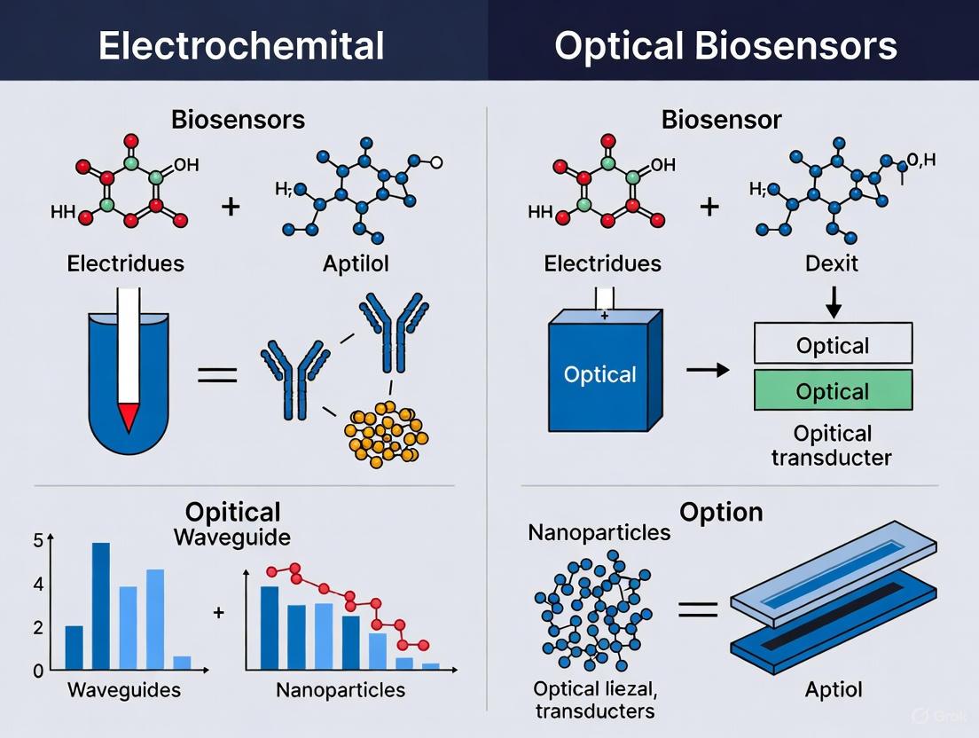
Biosensors are analytical devices that combine a biological recognition element with a physicochemical transducer to detect specific analytes. The core architecture of any biosensor consists of two fundamental components: a bioreceptor (e.g., enzyme, antibody, aptamer, or nucleic acid) that selectively interacts with the target molecule, and a transducer that converts this biological interaction into a measurable signal [1]. Among the various transduction mechanisms, electrochemical and optical methods have emerged as the most prominent technologies, each with distinct operational principles, advantages, and limitations. Electrochemical biosensors detect electrical changes resulting from biological recognition events, measuring parameters such as current (amperometric/voltammetric), potential (potentiometric), impedance (impedimetric), or conductance (conductometric) [2] [3]. Optical biosensors, conversely, utilize light as the detection mechanism, transducing bio-recognition events into measurable optical signals through various phenomena including surface plasmon resonance (SPR), fluorescence, chemiluminescence, colorimetry, and surface-enhanced Raman spectroscopy (SERS) [1] [2] [4]. The selection between electrochemical and optical architectures involves careful consideration of factors including sensitivity requirements, detection environment, cost constraints, portability needs, and the specific application, from clinical diagnostics and environmental monitoring to food safety and bioprocess control [5].
Comparative Analysis of Biosensor Architectures
Performance Characteristics and Applications
The table below summarizes the core characteristics, advantages, and limitations of electrochemical and optical biosensor architectures, providing a foundational comparison for researchers and developers.
Table 1: Fundamental comparison of electrochemical and optical biosensor architectures
| Feature | Electrochemical Biosensors | Optical Biosensors |
|---|---|---|
| Transduction Principle | Measures electrical changes (current, potential, impedance) from bio-recognition events [2] [3] | Measures changes in light properties (wavelength, intensity, polarization) from bio-recognition events [1] [4] |
| Key Advantages | High sensitivity, rapid response, low fabrication cost, ease of miniaturization and analysis, portability for point-of-care testing (POCT) [2] [5] [3] | High specificity and analytical accuracy, capability for real-time and multiplexed detection, generally non-invasive features, immunity to electromagnetic interference [2] [5] [4] |
| Primary Limitations | Relatively weaker long-term stability, susceptible to electrical noise, performance can be influenced by environmental factors [5] [3] | Light quenching can affect results, may require complex instrumentation, limited portability for some modalities (e.g., SPR), potential for background interference from sample matrices [2] [5] |
| Common Detection Techniques | Cyclic Voltammetry (CV), Differential Pulse Voltammetry (DPV), Electrochemical Impedance Spectroscopy (EIS), Amperometry [1] [5] | Surface Plasmon Resonance (SPR), Fluorescence, Chemiluminescence (CL), Colorimetry, Surface-Enhanced Raman Spectroscopy (SERS) [1] [2] [4] |
| Typical Applications | Clinical diagnostics (glucose, cardiac markers), pathogen detection (E. coli), environmental monitoring (pesticides, heavy metals) [6] [3] | Drug discovery, biomarker validation, cancer diagnostics (AFP, PSA), kinetic binding studies, food safety analysis [7] [5] |
Quantitative Performance Comparison
The selection of an appropriate biosensor architecture often depends on specific performance metrics required for the application. The following table compiles experimental data from recent research, highlighting the achievable performance in detecting various analytes.
Table 2: Experimental performance data of recent electrochemical and optical biosensors
| Analyte | Biosensor Type & Architecture | Linear Range | Detection Limit | Reference/Application Context |
|---|---|---|---|---|
| E. coli | Electrochemical (Mn-ZIF-67/Anti-O antibody) | 10 to 1010 CFU mL–1 | 1 CFU mL–1 | Pathogen detection in food and water [6] |
| Alpha-fetoprotein (AFP) | Optical (SERS-based, Au-Ag Nanostars) | 0 - 500 ng/mL | 16.73 ng/mL | Liver cancer biomarker detection [7] |
| Prostate-Specific Antigen (PSA) | Electrochemical (Gold Nanofiber-modified SPCE) | 0 to 100 ng/mL | 0.28 ng/mL (8.78 fM) | Prostate cancer screening [3] |
| Let-7a miRNA | Electrochemical (MnO2 Nanozyme) | Not Specified | Signal stable via DPV | miRNA detection for disease diagnostics [5] |
| C-Reactive Protein (CRP) | Optical (SPR-based on flexible PDMS) | Not Specified | High sensitivity claimed | Inflammatory marker monitoring on wearable platforms [4] |
| Dopamine (DA) | Electrochemical (Fe/N-doped graphene) | 50 pM – 15 nM | 27 pM | Neurotransmitter detection for neurological disorders [3] |
Experimental Protocols for Biosensor Development
Protocol for a High-Performance Electrochemical Biosensor
Objective: To develop a sensitive and selective electrochemical biosensor for E. coli detection using a Mn-doped Zeolitic Imidazolate Framework-67 (Mn-ZIF-67) conjugated with anti-O antibody [6].
Materials:
- Mn-ZIF-67 Synthesis: Cobalt nitrate hexahydrate, Manganese chloride tetrahydrate, 2-Methylimidazole, Methanol.
- Bioconjugation: Anti-E. coli O-specific antibody, Phosphate Buffered Saline (PBS), 1-Ethyl-3-(3-dimethylaminopropyl) carbodiimide (EDC), N-Hydroxysuccinimide (NHS).
- Electrochemical Cell: Three-electrode system (Glassy Carbon Working Electrode, Pt wire Counter Electrode, Ag/AgCl Reference Electrode).
- Instrumentation: Potentiostat for Cyclic Voltammetry (CV) and Electrochemical Impedance Spectroscopy (EIS) measurements.
Methodology:
- Synthesis of Mn-ZIF-67:
- Dissolve cobalt nitrate and manganese chloride in methanol at specific molar ratios (e.g., Co:Mn = 5:1).
- Prepare a separate methanolic solution of 2-methylimidazole.
- Rapidly mix the two solutions under vigorous stirring and maintain the reaction at room temperature for 24 hours.
- Centrifuge the resulting purple precipitate, wash thoroughly with methanol, and dry under vacuum [6].
Electrode Modification:
- Polish the Glassy Carbon Electrode (GCE) sequentially with alumina slurry (1.0, 0.3, and 0.05 µm) and rinse with deionized water.
- Prepare a homogeneous ink by dispersing 2 mg of the synthesized Mn-ZIF-67 in 1 mL of a water/ethanol mixture (1:1 v/v) with brief sonication.
- Drop-cast a precise volume (e.g., 5 µL) of the ink onto the clean GCE surface and allow it to dry at room temperature [6].
Antibody Conjugation:
- Activate the carboxylic groups on the Mn-ZIF-67 modified electrode by incubating with a fresh solution of EDC and NHS in PBS (pH 7.4) for 1 hour.
- Rinse the electrode gently with PBS to remove excess EDC/NHS.
- Incubate the activated electrode with a solution of anti-O antibody (e.g., 10 µg/mL in PBS) for 2 hours at 4°C, allowing the formation of stable amide bonds.
- Rinse again with PBS to remove physically adsorbed antibodies. The biosensor is now ready for use [6].
Electrochemical Detection and Measurement:
- Immerse the modified electrode in a standard electrochemical cell containing a redox probe such as [Fe(CN)₆]³⁻/⁴⁻ in PBS.
- Record the EIS spectra or CV curves before and after exposure to E. coli samples.
- The binding of E. coli cells to the antibody on the electrode surface increases the electron transfer resistance (Rₑₜ), which is quantified and correlated to the bacterial concentration. A standard calibration curve is constructed by plotting ΔRₑₜ against the logarithm of E. coli concentration [6].
Protocol for a SERS-Based Optical Biosensor
Objective: To fabricate a liquid-phase Surface-Enhanced Raman Scattering (SERS) immunoassay platform using Au-Ag nanostars for the sensitive detection of the cancer biomarker Alpha-fetoprotein (AFP) [7].
Materials:
- Nanostar Synthesis: Gold(III) chloride trihydrate, Silver nitrate, Ascorbic acid (as a reducing agent), Cetyltrimethylammonium bromide (CTAB, as a surfactant).
- Bioconjugation: Monoclonal anti-AFP antibodies, Mercaptopropionic acid (MPA), EDC, NHS, PBS.
- Instrumentation: Raman spectrometer with a laser excitation source (e.g., 785 nm).
Methodology:
- Synthesis of Au-Ag Nanostars:
- Prepare a seed solution of gold nanoparticles.
- In a growth solution containing CTAB, gold chloride, and silver nitrate, add a controlled amount of ascorbic acid.
- Introduce the gold seed solution to the growth mixture under gentle stirring. The morphology and "sharpness" of the nanostars, critical for SERS enhancement, can be tuned by varying the centrifugation time (10, 30, or 60 minutes) [7].
Functionalization of Nanostars with Antibodies:
- Centrifuge the synthesized nanostars and re-disperse them in an aqueous solution.
- Incubate the nanostars with MPA to form a self-assembled monolayer via Au-S bonds.
- Activate the terminal carboxylic acid groups of MPA using a mixture of EDC and NHS for 30 minutes.
- Add the anti-AFP antibody to the activated nanostars and incubate for 2 hours, enabling covalent amide bond formation between the antibody and MPA. Remove excess reagents by centrifugation [7].
SERS Immunoassay and Detection:
- Incubate the antibody-conjugated nanostars with samples containing different concentrations of AFP antigen.
- The immunocomplex formation on the nanostar surface alters the local dielectric environment.
- Place a droplet of the immunoassay mixture on a substrate and acquire SERS spectra using the Raman spectrometer.
- The intensity of the characteristic Raman peak of AFP (or a reporter molecule, if used) is measured. This intensity is directly proportional to the AFP concentration captured on the nanostars, allowing for quantitative analysis with a calibration curve [7].
The Scientist's Toolkit: Key Research Reagent Solutions
The performance and reliability of biosensors are heavily dependent on the careful selection of materials and reagents. The following table details essential components used in the development of advanced electrochemical and optical biosensors.
Table 3: Essential reagents and materials for biosensor development
| Reagent/Material | Function in Biosensor Development | Typical Application Context |
|---|---|---|
| Gold Nanoparticles (AuNPs) | Signal amplification; carrier for biomolecule immobilization; enhances electron transfer in electrochemical sensors; plasmonic core in optical SERS/SPR sensors [3]. | Electrochemical immunosensors (e.g., for PSA); SERS-based optical platforms (e.g., Au-Ag nanostars for AFP) [7] [3]. |
| Graphene & Carbon Nanotubes | Electrode modifier; provides large specific surface area and excellent electrical conductivity; enhances sensitivity via π-π interactions and electron transfer efficiency [5] [3]. | Dopamine sensors (Fe/N-doped graphene) [3]; various electrochemical platforms for signal enhancement [5]. |
| Metal-Organic Frameworks (MOFs) | Nanostructured porous material; provides immense surface area for analyte loading/binding; can be doped with metals to enhance conductivity and catalytic activity (e.g., Mn-ZIF-67) [6] [5]. | High-sensitivity electrochemical sensing of pathogens (e.g., E. coli) and other biomarkers [6]. |
| EDC/NHS Crosslinkers | Carboxyl group activators; facilitate covalent conjugation between biorecognition elements (antibodies, aptamers) and sensor surfaces or nanocarriers [6] [7]. | Standard procedure for immobilizing antibodies on MOFs (electrochemical) and noble metal nanoparticles (optical) [6] [7]. |
| Polydimethylsiloxane (PDMS) | Flexible, optically transparent polymer substrate; excellent for fabricating wearable and conformable optical biosensors due to its biocompatibility and mechanical properties [4]. | Flexible SPR sensors (e.g., for CRP detection); wearable fluorescent sensors (e.g., ZnO-PDMS dental protector) [4]. |
| Nanozymes (e.g., MnO₂ Nanosheets) | Nanomaterials with enzyme-like catalytic activity; stable alternatives to natural enzymes for catalyzing reactions that generate optical or electrochemical signals [5]. | Electrochemical miRNA detection; used as signal probes to amplify detection signals [5]. |
Visualizing Biosensor Architectures and Workflows
The following diagrams, generated using Graphviz DOT language, illustrate the core operational principles and experimental workflows for the two primary biosensor architectures.
Electrochemical Biosensor Signaling Pathway
Optical Biosensor Signaling Pathway
Experimental Workflow for Biosensor Development
Electrochemical and optical biosensors represent two foundational pillars of modern detection technology, each with distinct transduction mechanisms, performance profiles, and application suitability. Electrochemical biosensors convert biological recognition events into measurable electrical signals—current, potential, or impedance—offering advantages in portability, cost, and miniaturization for point-of-care and continuous monitoring. Optical biosensors transduce binding events into optical signals—such as changes in light absorption, fluorescence, or refractive index—providing superior sensitivity, multiplexing capability, and label-free detection for advanced laboratory diagnostics and pharmaceutical research. The selection between these mechanisms is application-dependent, hinging on specific requirements for sensitivity, operational environment, and required throughput.
Fundamental Transduction Principles
Electrochemical Transduction
Electrochemical biosensors operate on the principle of converting a biological recognition event (e.g., enzyme-substrate binding, antigen-antibody interaction) into an electrical signal. The core structure involves a biological recognition element (bioreceptor) immobilized on the surface of an electronic transducer, typically a three-electrode system (working, reference, and counter electrodes) [8] [9]. When the target analyte interacts with the bioreceptor, it triggers a biochemical reaction that produces or consumes electrons, thereby altering the electrical properties at the electrode-electrolyte interface. This change is measured and quantified. These sensors are broadly categorized based on the measured electrical parameter [2] [9]:
- Amperometric: Measures current generated by the electrochemical oxidation or reduction of an electroactive species at a constant applied potential.
- Potentiometric: Measures the potential difference between the working and reference electrodes under conditions of zero current.
- Impedimetric: Measures changes in the impedance (resistance to alternating current) of the electrode interface due to binding events.
- Voltammetric: Measures current while systematically varying the applied potential.
Optical Transduction
Optical biosensors detect analytes by measuring changes in the properties of light, leveraging various optical phenomena [10] [11] [12]. The biological interaction on the sensor surface modulates an optical signal, which is detected and analyzed. Key optical transduction mechanisms include:
- Fluorescence: Detects the emission of light from a fluorophore after excitation by a specific wavelength of light. The intensity, wavelength, or polarization of the emitted light can be modulated by the binding event [13] [12].
- Surface Plasmon Resonance (SPR) and Localized SPR (LSPR): Measures changes in the refractive index near a thin metal (typically gold) surface. The binding of a target analyte shifts the resonance condition, which is detected as a change in the angle or wavelength of reflected light [11] [12].
- Chemiluminescence/Bioluminescence: Measures the emission of light generated as a by-product of a chemical or biochemical reaction, without the need for an excitation light source [11] [2].
- Surface-Enhanced Raman Spectroscopy (SERS): Greatly enhances the weak Raman scattering signal from molecules adsorbed on rough metal nanostructures, providing a unique vibrational "fingerprint" for detection [2] [12].
- Colorimetric: Detects a change in color or absorbance of light, often visible to the naked eye, due to the accumulation of a chromophore [2].
The following diagram illustrates the core operational workflows for these two fundamental biosensor classes.
Performance Comparison: Quantitative Data
The following tables summarize key performance characteristics and application parameters for electrochemical and optical biosensors, synthesized from comparative studies and recent reviews.
Table 1: Comparison of Key Performance Parameters
| Parameter | Electrochemical Biosensors | Optical Biosensors |
|---|---|---|
| Typical Sensitivity | High (pM-nM) [8] | Very High (fM-pM) [11] [14] |
| Selectivity | High (depends on bioreceptor) [9] | Very High (depends on bioreceptor) [10] |
| Multiplexing Capability | Moderate (limited by electrode design) [15] | High (e.g., multi-wavelength detection) [15] [11] |
| Response Time | Seconds to minutes [8] | Seconds to minutes (real-time for SPR) [10] |
| Sample Volume | Low (µL) [8] [16] | Low to Moderate (µL to mL) [10] |
| Portability & Miniaturization | Excellent (wearable, implantable) [8] [2] | Moderate (challenged by optics, but improving) [15] [11] |
| Robustness/Environmental Stability | Good to Excellent [15] | Moderate (can be sensitive to ambient light/temperature) [15] |
| Cost per Test | Low [8] [2] | Moderate to High [15] |
Table 2: Application Suitability and Operational Characteristics
| Characteristic | Electrochemical Biosensors | Optical Biosensors |
|---|---|---|
| Preferred Application Domains | Point-of-care testing, continuous monitoring (e.g., glucose, lactate), environmental field testing [8] [16] [2] | Laboratory diagnostics, drug discovery, biomolecular interaction analysis, high-throughput screening [10] [11] [14] |
| Common Bioreceptors | Enzymes, antibodies, aptamers, whole cells [8] [9] | Antibodies, aptamers, nucleic acids [10] [13] |
| Label Requirement | Often label-free; sometimes uses enzymatic labels [9] | Label-free (SPR) or labeled (fluorescence, SERS) [10] [2] |
| Ease of Integration | Easy integration with electronics and microfluidics [8] | Integration can be complex (light sources, detectors) but advancing (e.g., smartphone-based) [14] [12] |
| Key Limitation | Biofouling, signal reproducibility [8] | Signal interference in complex media, portability [15] |
Experimental Protocols in Practice
To illustrate these principles, here are detailed methodologies for representative sensor types from recent literature.
Protocol: Fabrication of a Wearable Lactate Electrochemical Biosensor
This protocol is adapted from research on enzymatic lactate sensors for sweat monitoring [16].
1. Objective: To construct a flexible, enzymatic electrochemical biosensor for continuous lactate monitoring in sweat. 2. Materials:
- Transducer: Screen-printed carbon electrode (SPCE) on a flexible polymer substrate.
- Bioreceptor: Lactate oxidase (LOx) enzyme.
- Immobilization Matrix: Chitosan hydrogel or a cross-linked polymer like poly(m-phenylenediamine).
- Chemical Reagents: Lactate standard solutions, phosphate buffer saline (PBS, 0.1 M, pH 7.4), glutaraldehyde (for cross-linking).
- Equipment: Potentiostat, Ag/AgCl reference electrode.
3. Step-by-Step Methodology:
- Step 1: Electrode Pretreatment. Clean the working electrode surface of the SPCE by performing cyclic voltammetry (CV) in 0.1 M H₂SO₄ between -0.5 V and +1.0 V for 10-15 cycles.
- Step 2: Enzyme Immobilization. Prepare an immobilization mixture containing 5-10 U/µL of LOx and 0.5% (w/v) chitosan in a dilute acetic acid solution. Deposit 5-10 µL of this mixture onto the working electrode area. Alternatively, mix LOx with a monomer solution and electropolymerize it onto the electrode to form a stable polymer-enzyme film.
- Step 3: Curing and Storage. Allow the sensor to cure at 4°C for 12 hours to ensure proper enzyme immobilization and film formation. Store the finished biosensors dry at 4°C until use.
- Step 4: Calibration and Measurement. Perform amperometric measurements at a constant applied potential of +0.4 to +0.7 V (vs. Ag/AgCl). Record the steady-state current upon successive additions of lactate standard into a stirred PBS buffer. The generated current is proportional to the lactate concentration.
4. Data Analysis: Plot the calibration curve of current (µA) vs. lactate concentration (mM). Calculate the sensor's sensitivity from the slope of the linear region, and the limit of detection (LOD) using 3×standard deviation of the blank signal divided by the sensitivity.
Protocol: FB1 Mycotoxin Detection via a Fluorescent Aptasensor
This protocol is based on a "signal-on" fluorescent aptasensor utilizing graphene oxide (GO) [13].
1. Objective: To detect Fumonisin B1 (FB1) mycotoxin with high sensitivity and specificity using a fluorescent aptasensor. 2. Materials:
- Bioreceptor: FB1-specific DNA aptamer, labeled with a carboxy-X-rhodamine (ROX) fluorophore.
- Nanomaterial: Graphene oxide (GO) nanosheets.
- Chemical Reagents: FB1 standard, nuclease enzyme, buffer solution (e.g., Tris-EDTA).
- Equipment: Fluorescence spectrophotometer, microcentrifuge, incubation shaker.
3. Step-by-Step Methodology:
- Step 1: Aptamer Pre-incubation. Dilute the ROX-labeled aptamer in the appropriate buffer and incubate it with a optimized concentration of GO suspension for 15-20 minutes. The aptamer adsorbs onto the GO surface via π-π stacking, leading to fluorescence quenching ("signal-off" state).
- Step 2: Target Introduction and Incubation. Introduce the sample (or FB1 standard) to the aptamer-GO mixture and incubate for 30-60 minutes. In the presence of FB1, the aptamer undergoes a conformational change, binds to the target, and desorbs from the GO surface. This distance increase between ROX and GO restores fluorescence ("signal-on").
- Step 3: Signal Amplification (Optional). Add a nuclease enzyme that specifically digests the aptamer-FB1 complex. This releases FB1 and the fluorophore, allowing the cycle to repeat and providing signal amplification for lower LOD.
- Step 4: Fluorescence Measurement. Measure the fluorescence intensity of ROX at its characteristic excitation/emission wavelengths (e.g., ~580/605 nm).
4. Data Analysis: The increase in fluorescence intensity (F - F₀) is directly correlated with the FB1 concentration. Generate a calibration curve to quantify FB1 in unknown samples.
The Scientist's Toolkit: Essential Research Reagents and Materials
The development and implementation of high-performance biosensors rely on a suite of specialized materials and reagents.
Table 3: Key Research Reagent Solutions for Biosensor Development
| Category / Item | Function in Biosensors | Specific Examples |
|---|---|---|
| Transducer Materials | Forms the core physical platform for signal transduction. | Screen-printed carbon electrodes (SPCEs) [16], Gold thin films (for SPR) [12], Optical fibers [14] [12], Indium tin oxide (ITO) glass. |
| Biorecognition Elements | Provides high specificity and selectivity for the target analyte. | Lactate oxidase (LOx) [16], Antibodies (for immunosensors) [2] [9], DNA/RNA aptamers (e.g., for FB1) [13], Whole cells [9]. |
| Nanomaterials | Enhances signal, improves bioreceptor loading, and increases sensitivity. | Graphene Oxide (GO) - fluorescence quenching & aptamer protection [13], Gold nanoparticles (AuNPs) - LSPR & SERS enhancement [11] [2], Prussian Blue nanoparticles - electrocatalysis [9], Carbon nanotubes - electron transfer facilitation [8]. |
| Immobilization Reagents | Stabilizes and attaches bioreceptors to the transducer surface. | Chitosan (hydrogel matrix) [16], Glutaraldehyde (cross-linker) [16], N-hydroxysuccinimide (NHS)/EDC chemistry (for covalent bonding) [16], Self-assembled monolayers (SAMs). |
| Labels & Dyes | Generates a measurable optical or electrochemical signal. | Carboxy-X-rhodamine (ROX), Fluorescein (FAM) - fluorescent tags [13] [12], Horse Radish Peroxidase (HRP) - enzymatic label for chemiluminescence/electrochemistry [2], Raman reporter molecules (e.g., malachite green) for SERS [2]. |
The choice between electrochemical and optical transduction is a strategic decision in biosensor design. Electrochemical biosensors are the undisputed champions for miniaturization, cost-effectiveness, and field-deployment, making them ideal for point-of-care diagnostics and wearable health monitors. Conversely, optical biosensors offer peerless sensitivity, robust multiplexing, and label-free kinetic analysis, securing their place as indispensable tools in fundamental life science research and advanced laboratory diagnostics. The future trajectory points not to outright competition, but to a complementary evolution. Emerging trends like the integration of artificial intelligence for data analysis [15] [9], the development of novel low-dimensional nanomaterials [11], and the creation of modular, multi-modal sensor designs [15] will further blur the lines, enabling a new generation of intelligent, highly sensitive, and connected biosensing systems tailored for specific challenges in research and clinical practice.
Biosensors represent a cornerstone of modern analytical science, combining a biological recognition element with a physicochemical detector to measure specific analytes. For researchers, scientists, and drug development professionals, the selection between electrochemical and optical biosensing platforms hinges on a critical evaluation of key performance metrics: sensitivity, specificity, and limit of detection (LOD). These parameters directly determine a sensor's utility in clinical diagnostics, environmental monitoring, and drug discovery.
Sensitivity refers to the magnitude of the output signal change per unit change in analyte concentration. Specificity defines the sensor's ability to distinguish the target analyte from interfering substances in a complex matrix. The LOD is the lowest analyte concentration that can be reliably distinguished from background noise, typically expressed as the mean blank signal plus three standard deviations. The ongoing research and development in both electrochemical and optical biosensors aim to optimize these metrics through material innovations, novel biorecognition elements, and advanced signal processing techniques.
This guide provides a structured comparison of electrochemical and optical biosensors, supported by experimental data and detailed methodologies, to inform platform selection for specific research and development applications.
Performance Metrics Comparison
The following table summarizes the typical performance ranges and characteristics of electrochemical and optical biosensors for key metrics, based on recent research.
Table 1: Comparative Performance Metrics of Electrochemical and Optical Biosensors
| Performance Metric | Electrochemical Biosensors | Optical Biosensors |
|---|---|---|
| Typical Limit of Detection (LOD) | fM to pM range (e.g., 4 fM for HCV RNA) [17] | Low ng/mL to pM range (e.g., 27 ng/mL for streptavidin; 377 pM for CRP) [18] |
| Sensitivity (General) | High; enhanced by nanomaterials and catalytic signal amplification [19] [20] | Very High; benefits from high-resolution spectral shifts and label-free detection [21] [15] |
| Specificity | High; dependent on biorecognition element (antibody, aptamer, DNA) [19] [17] | High; dependent on biorecognition element; can be affected by non-specific binding [15] [18] |
| Multiplexing Capability | Moderate; evolving with array designs [15] | High; facilitated by multiple wavelengths and spatial resolution [21] [15] |
| Real-time Monitoring | Good for some modalities (e.g., amperometric) [22] | Excellent; allows for real-time, label-free tracking of biomolecular interactions [21] [20] |
| Susceptibility to Sample Matrix | Moderate; can be mitigated with membrane coatings [2] | Moderate to High; optical properties can be affected by turbidity or autofluorescence [2] |
Experimental Protocols for Performance Validation
Electrochemical Biosensor Protocol: Direct HCV RNA Detection
A highly sensitive and specific electrochemical biosensor for the direct detection of Hepatitis C Virus (HCV) RNA demonstrates the capability of this platform for clinical application without the need for target amplification [17].
1. Principle: The sensor employs a strand displacement method. A magnetic nanoparticle (MNP)-labeled capture probe is pre-hybridized with a gold nanoparticle-labeled reporter probe (AuRP). Upon introduction of the target RNA, it displaces the AuRP, which is then quantified electrochemically. The measured signal is directly proportional to the target RNA concentration [17].
2. Key Materials and Reagents:
- Bioprobes: Biotinylated capture probe and thiolated reporter probe.
- Nanoparticles: 40 nm Gold Nanoparticles (AuNPs) and Streptavidin-coated Magnetic Beads (e.g., Dynabeads).
- Electrochemical Cell: Disposable screen-printed carbon electrodes (SPCE).
- Chemical Reagents: Tris(2-carboxyethyl)phosphine (TCEP, for probe reduction), Tris-acetate buffer, and anodic stripping solution (1 M HBr/0.1 mM Br₂).
3. Experimental Procedure:
- Step 1: Probe Functionalization. The thiolated reporter probe is conjugated to AuNPs via a salt-aging method. The biotinylated capture probe is immobilized onto streptavidin-coated magnetic beads [17].
- Step 2: Duplex Formation. The functionalized capture probe and AuNP-reporter probe (AuRP) are hybridized to form a pre-displacement duplex on the magnetic beads.
- Step 3: Target Hybridization and Strand Displacement. The sample containing target RNA is introduced. The target RNA hybridizes with the capture probe, displacing the AuRP into the solution.
- Step 4: Magnetic Separation. A magnetic field is applied to separate the bead-bound complex from the supernatant containing the displaced AuRP.
- Step 5: Electrochemical Detection. An aliquot of the supernatant is deposited on the SPCE. Gold from the AuRP is dissolved in HBr/Br₂ solution and electrochemically re-deposited onto the working electrode. The deposited gold is then quantified using Differential Pulse Anodic Stripping Voltammetry (DPASV), where the oxidation current of re-dissolved gold is measured [17].
4. Performance Data: This sensor achieved an exceptional LOD of 4 fM for synthetic HCV RNA targets and successfully detected HCV RNA directly in clinical plasma samples without RNA extraction, showing high concordance with RT-PCR results [17].
Optical Biosensor Protocol: Streptavidin Detection Using an Optical Cavity
This protocol details the enhancement of an optical cavity-based biosensor's (OCB) LOD through optimized surface functionalization, a critical step for optical platforms [18].
1. Principle: The biosensor is based on a Fabry-Perot interferometer (FPI) structure. Light undergoes multiple reflections within a cavity formed by two partially reflective mirrors. The binding of target analytes (e.g., streptavidin) to receptors (e.g., biotin) immobilized on the cavity surface alters the local refractive index, shifting the resonance characteristics of the transmitted light. This shift is detected as a change in intensity at a specific wavelength [18].
2. Key Materials and Reagents:
- Substrate: Soda lime glass.
- Cavity Material: SU-8 photoresist (forms the microfluidic channel and cavity structure).
- Reflective Layers: Sputter-deposited silver.
- Functionalization Reagents: 3-aminopropyltriethoxysilane (APTES), Biotin (e.g., sulfo-NHS-biotin).
- Target Analyte: Streptavidin.
- Detection Components: Laser diodes (808 nm and 880 nm) and a CCD/CMOS camera.
3. Experimental Procedure:
- Step 1: Sensor Fabrication. A microfluidic OCS is fabricated by patterning SU-8 on a glass substrate, followed by deposition of silver layers to create the reflective surfaces of the FPI [18].
- Step 2: Surface Functionalization (Critical Step). The internal sensor surface is silanized with APTES to create an amine-terminated layer for subsequent bioconjugation. The study systematically compared three APTES deposition methods:
- Ethanol-based: APTES diluted in ethanol.
- Methanol-based: APTES diluted in methanol (0.095% v/v).
- Vapor-phase: Exposure to APTES vapor. The methanol-based protocol yielded the most uniform monolayer and best performance [18].
- Step 3: Biotin Immobilization. Sulfo-NHS-biotin is covalently linked to the amine groups on the APTES-functionalized surface.
- Step 4: Differential Detection. Streptavidin samples are introduced into the microfluidic cavity. The binding event is detected in real-time by measuring the intensity changes of two laser wavelengths (808 nm and 880 nm) using a CCD camera. The differential signal between the two wavelengths enhances sensitivity and reduces noise [18].
4. Performance Data: Optimizing the APTES process with the methanol-based protocol resulted in a threefold improvement in LOD, achieving 27 ng/mL for streptavidin, underscoring the impact of surface chemistry on optical biosensor performance [18].
Research Reagent Solutions Toolkit
Table 2: Essential Research Reagents and Materials for Biosensor Development
| Item | Function | Example Applications |
|---|---|---|
| Gold Nanoparticles (AuNPs) | Signal amplification tag; facilitates electron transfer in electrochemical sensors; used as plasmonic enhancers in optical sensors [17]. | Electrochemical HCV RNA sensor [17]; SERS-based immunoassays [7]. |
| Magnetic Nanoparticles | Solid support for probe immobilization; enables efficient magnetic separation of bound and unbound components [17]. | Sample preparation and concentration; electrochemical biosensors with strand displacement [17]. |
| Screen-Printed Carbon Electrodes (SPCE) | Low-cost, disposable electrochemical platform; integrates working, reference, and counter electrodes [17]. | Point-of-care electrochemical detection; portable diagnostic devices [2] [17]. |
| APTES (3-Aminopropyltriethoxysilane) | Silane coupling agent; creates an amine-functionalized surface on glass/silica for biomolecule immobilization [18]. | Surface functionalization for optical biosensors [18]; general surface chemistry. |
| SU-8 Photoresist | A polymer used to fabricate microfluidic channels and optical cavity structures in lab-on-a-chip devices [18]. | Construction of the optical cavity in FPI-based biosensors [18]. |
| Biotin/Streptavidin | High-affinity binding pair; used as a model system for sensor validation and for immobilizing biotinylated bioreceptors [18]. | Benchmarking sensor performance; universal immobilization strategy [18]. |
Biosensor Workflow Diagrams
Electrochemical Biosensor Workflow (Strand Displacement)
Optical Biosensor Workflow (Cavity-Based)
The choice between electrochemical and optical biosensors is application-dependent, requiring careful consideration of the required detection limits, sample matrix, and resource constraints. Electrochemical biosensors, as demonstrated by the HCV RNA sensor, offer exceptionally low LODs (reaching fM levels), portability, and cost-effectiveness, making them strong candidates for point-of-care diagnostics [17]. Optical biosensors provide superior capabilities for real-time, label-free monitoring and high multiplexing, which are invaluable for kinetic studies and drug discovery applications [21] [20].
Future advancements are poised to blur the lines between these platforms through the integration of artificial intelligence for data analysis, the development of novel nanomaterials for enhanced signal transduction, and the creation of hybrid systems that leverage the strengths of both electrochemical and optical methods [21] [15]. The ongoing optimization of surface chemistry and biorecognition elements will continue to push the boundaries of sensitivity, specificity, and LOD for both sensor classes.
Biosensors are analytical devices that combine a biological recognition element with a physicochemical detector to measure a specific analyte. The core of any biosensor is its biorecognition element, which is responsible for the selective and specific interaction with the target molecule. The choice of this element fundamentally influences the biosensor's performance characteristics, including its sensitivity, selectivity, reproducibility, and reusability [23]. This guide provides an objective comparison of three of the most versatile biorecognition elements—enzymes, antibodies, and aptamers—framed within the ongoing research and development of electrochemical and optical biosensing platforms [2].
The broader context of biosensor research often pits two main transduction methods against each other: electrochemical and optical. Electrochemical biosensors measure electrical signals (current, potential, impedance) resulting from biochemical interactions and are prized for their portability, low cost, and suitability for point-of-care testing [15] [24] [8]. In contrast, optical biosensors exploit light-matter interactions (e.g., surface plasmon resonance, fluorescence) to detect molecular changes, offering high sensitivity, label-free detection, and real-time monitoring capabilities [25] [26] [2]. The selection of a biorecognition element intersects critically with this choice of transducer, as each element possesses unique properties that can optimize the performance of either platform.
Characteristics and Comparison of Biorecognition Elements
- Enzymes: Enzymes are biocatalytic biorecognition elements that achieve specificity through binding cavities within their three-dimensional structure. They capture and catalytically convert the target analyte into a measurable product, a process often monitored via amperometric or electrochemical methods. The catalytic reaction typically involves the formation of an intermediate complex before the release of the product [23].
- Antibodies: Antibodies are affinity-based, three-dimensional protein structures (~150 kDa) that naturally bind to specific antigens with high specificity. Their binding event can be monitored using various transduction methods, including colorimetric and piezometric techniques. Antibody fragments, such as single-chain variable fragments (scFv) and antigen-binding fragments (Fab'), offer advantages like smaller size and easier immobilization, potentially leading to higher sensitivity and lower limits of detection [23] [27].
- Aptamers: Aptamers are single-stranded DNA or RNA oligonucleotides synthesized artificially through a process called Systematic Evolution of Ligands by Exponential Enrichment (SELEX). They fold into distinct three-dimensional shapes that bind to a wide range of targets, from small ions to whole cells, with high affinity and specificity. Their small size (~1–2 nm) allows for high-density immobilization on sensor surfaces [23] [27] [26].
Comparative Analysis of Key Parameters
The table below summarizes the critical characteristics of enzymes, antibodies, and aptamers, providing a direct comparison to guide selection.
Table 1: Comprehensive Comparison of Biorecognition Elements
| Parameter | Enzymes | Antibodies | Aptamers |
|---|---|---|---|
| Type of Interaction | Catalytic | Affinity-based | Affinity-based |
| Origin | Biological (naturally occurring) | Biological (naturally occurring) | Synthetic (SELEX process) |
| Production Process | Purification from biological sources or recombinant expression | Production in animals; costly and time-consuming purification [23] | Chemical synthesis; requires SELEX for discovery [23] |
| Molecular Size | Large (varies by enzyme) | Large (~150 kDa for full antibody) [27] | Small (~1-2 nm) [27] |
| Stability | Moderate; can be sensitive to temperature and pH | Moderate; sensitive to denaturation at high temperatures and non-physiological conditions [26] | High; thermally stable and can be regenerated after denaturation [27] [26] |
| Modifiability | Low to moderate | Moderate (e.g., fragmentation to Fab'/scFv) [27] | High; easily chemically modified for immobilization or signaling [27] [26] |
| Target Range | Primarily substrates and inhibitors | Primarily immunogenic molecules (proteins, etc.) | Broad (ions, small molecules, proteins, cells) [23] [26] |
| Binding Affinity | Varies (catalyzes transformation) | High (nanomolar to picomolar) [27] | High (nanomolar to picomolar), comparable to antibodies [27] |
| Development Cost & Time | Moderate | High cost and time [23] | SELEX is costly, but chemical synthesis is inexpensive [23] |
Performance in Biosensing Applications
The fundamental properties of biorecognition elements directly translate into performance differences in functional biosensors. The following table compares these elements based on key biosensor metrics.
Table 2: Biosensor Performance Metrics
| Performance Metric | Enzymes | Antibodies | Aptamers |
|---|---|---|---|
| Sensitivity | High for catalytic targets | High | Very High (due to high surface density and strong binding) [27] [26] |
| Selectivity/Specificity | High for specific substrates | Very High | Very High, but cross-reactivity can be an issue [26] |
| Reproducibility | Moderate (batch-to-batch variation possible) | Can be variable due to production methods [23] | High (synthetic production ensures consistency) [23] [26] |
| Reusability/Regeneration | Low to Moderate | Low (due to irreversible binding and sensitivity) [23] | High (stable under harsh regeneration conditions) [27] [26] |
| Suitability for Point-of-Care | High (e.g., glucose meters) | High in established tests (e.g., LFIA) [2] | High potential due to stability and modifiability [26] |
| Immobilization Control | Can be random, affecting activity | Often random, potentially obscuring binding sites [26] | Can be precisely oriented via chemical modifications [27] [26] |
Experimental Protocols and Methodologies
Immobilization Techniques
The method used to attach a biorecognition element to the transducer surface is critical, as it affects orientation, stability, and accessibility.
- Antibody Immobilization: Full antibodies are often immobilized via random covalent linkage to a sensor surface, forming a brush-like array. A superior approach for biosensing involves using Fab' fragments, which contain free thiol groups (-SH) from the antibody hinge region. These thiols allow for directed, site-specific covalent immobilization onto gold surfaces or maleimide-functionalized substrates, optimizing the orientation of the binding paratope towards the solution and enhancing sensitivity [27].
- Aptamer Immobilization: The synthetic nature of aptamers makes them highly amenable to controlled immobilization. They can be synthesized with specific functional groups (e.g., thiol, amine, biotin) at their terminus, enabling oriented coupling to complementary surfaces (e.g., gold for thiols) or functional groups (e.g., streptavidin for biotin). This ensures the binding-competent structure is presented correctly [27].
- Enzyme Immobilization: Enzymes are commonly embedded within surface structures or matrices to allow short diffusion pathways for the analyte and product. Immobilization can be achieved through adsorption, entrapment within a polymer gel, or covalent binding to functionalized surfaces [23].
Typical Workflow for Biosensor Development and Testing
The following diagram illustrates a generalized experimental workflow for developing and validating a biosensor, applicable to all three biorecognition elements.
Signaling Pathways and Transducer Integration
The mechanism of signal generation differs fundamentally between electrochemical and optical biosensors, and the choice of biorecognition element must be compatible with the chosen transducer.
- Electrochemical Biosensing: In enzymatic biosensors, the signal typically originates from the electroactive products of the catalytic reaction. For example, glucose oxidase produces hydrogen peroxide, which can be oxidized at an electrode, generating a measurable current [23]. For affinity-based elements (antibodies, aptamers), the binding event itself must be transduced. This is often achieved using electrochemical labels (e.g., enzymes that generate an electroactive product) or by monitoring changes in interfacial properties, such as impedance [24] [2].
- Optical Biosensing: Optical platforms often rely on label-free detection. Techniques like Surface Plasmon Resonance (SPR) detect changes in the refractive index on the sensor surface when a binding event occurs, providing real-time kinetics data [25] [26]. Alternatively, fluorescence-based assays use labeled biorecognition elements or intercalating dyes, where binding causes a change in fluorescence intensity, polarization, or resonance energy transfer (FRET) [25] [2].
The diagram below visualizes the logical relationship between biorecognition elements and their primary associated transduction methods.
The Scientist's Toolkit: Essential Research Reagents and Materials
Successful biosensor development relies on a suite of specialized reagents and materials. The following table details key components used in experiments featuring the discussed biorecognition elements.
Table 3: Key Research Reagent Solutions for Biosensor Development
| Item | Function/Description | Example Use Case |
|---|---|---|
| SELEX Kit | A commercial kit containing libraries and reagents for the Systematic Evolution of Ligands by Exponential Enrichment. | Used for the in vitro selection of novel aptamers against a specific target analyte [23]. |
| Fab' Preparation Kits | Kits containing enzymes (e.g., pepsin, ficin) and reagents for the proteolytic cleavage of whole antibodies to generate Fab' fragments. | Production of antibody fragments with free thiols for oriented immobilization on biosensor surfaces [27]. |
| Functionalized Monomers | Chemical monomers (e.g., acrylic, vinyl) with specific functional groups used in polymer synthesis. | Synthesis of Molecularly Imprinted Polymers (MIPs) as synthetic biorecognition elements [23]. |
| Gold Sensor Chips | Glass or silicon substrates with a thin, pristine layer of gold coating. | The substrate of choice for Surface Plasmon Resonance (SPR) and for thiol-based immobilization of aptamers or Fab' fragments [27] [26]. |
| Electrochemical Labels | Molecules that produce an electrochemical signal upon redox reaction, such as enzymes like Horseradish Peroxidase (HRP) or small molecules like methylene blue. | Conjugated to biorecognition elements (e.g., secondary antibodies) to generate a measurable current in affinity-based electrochemical biosensors [2]. |
| Self-Assembled Monolayer (SAM) Thiols | Alkanethiol molecules (e.g., 6-mercapto-1-hexanol) that form ordered monolayers on gold surfaces. | Used to create a well-defined, non-fouling interface on gold transducers, minimizing non-specific binding and spacing immobilized biorecognition elements [27]. |
Enzymes, antibodies, and aptamers each offer a unique profile of advantages and limitations as biorecognition elements. The optimal choice is not universal but is dictated by the specific requirements of the application.
- Enzymes remain the gold standard for targets where catalytic amplification is possible and desired, as demonstrated by the unparalleled success of the glucose biosensor.
- Antibodies provide high specificity and affinity and are the backbone of many established clinical immunoassays. However, challenges related to production, stability, and immobilization control are driving the adoption of smaller fragments and exploration of alternatives.
- Aptamers, as synthetic molecules, offer significant benefits in terms of stability, modifiability, and reproducible production. While their commercial footprint is currently smaller, their versatility makes them exceptionally promising for future biosensing applications, particularly in point-of-care and environmental monitoring [26].
The ongoing research in electrochemical and optical biosensors is not about one platform or one biorecognition element winning out, but about strategic integration. Electrochemical systems benefit from the portability and low cost offered by robust elements like aptamers, while optical platforms leverage the high sensitivity and real-time kinetics provided by well-oriented antibodies and aptamers. Future progress will hinge on cross-disciplinary collaboration, further refinement of immobilization strategies, and the continued development of robust, synthetic biorecognition elements to meet the growing demands of diagnostic and analytical science.
Impact of Advanced Materials and Nanotechnology on Signal Enhancement
The performance of biosensors is fundamentally governed by their ability to convert a biological recognition event into a measurable signal with high fidelity. Signal enhancement refers to the strategies and material innovations that amplify this transduction process, leading to improved analytical sensitivity, lower limits of detection (LOD), and more robust assay performance. The integration of advanced materials and nanotechnology has been pivotal in overcoming the inherent limitations of conventional biosensing platforms, enabling the detection of target analytes at ultra-low concentrations that are crucial for early disease diagnosis, environmental monitoring, and food safety [15] [28].
The convergence of nanotechnology with biosensor design has created a new generation of sensing platforms. By manipulating matter at the nanoscale (typically 1-100 nanometers), scientists can engineer materials with unique optical, electrical, and catalytic properties that are not present in their bulk counterparts. These properties are harnessed to enhance signal transduction in both electrochemical and optical biosensors. For instance, metal nanoclusters (MNCs) exhibit strong photoluminescence and catalytic activity, while graphene and other two-dimensional materials facilitate exceptional electron transfer [28] [29]. The application of these materials is contextualized within the broader comparison of electrochemical and optical biosensors, which represent two dominant transduction principles in modern analytical science.
Comparative Analysis of Biosensor Platforms
Performance Metrics of Electrochemical vs. Optical Biosensors
The selection between electrochemical and optical biosensing platforms depends heavily on the specific application requirements. The table below provides a structured comparison of their performance, highlighting the impact of advanced materials.
Table 1: Comparative performance of electrochemical and optical biosensors incorporating nanomaterials.
| Performance Metric | Electrochemical Biosensors | Optical Biosensors |
|---|---|---|
| Typical Limit of Detection (LOD) | Ultra-high sensitivity; e.g., Femtomolar (fM) for biomarkers [29]; 16.73 ng/mL for α-fetoprotein [7] | Exceptional sensitivity; e.g., 3.5×10⁻³ mg/L for dyes via SERS [29]; Phase sensitivity up to 3.1×10⁵ deg RIU-1 [7] |
| Key Nanomaterials Used | Gold nanoparticles (AuNPs), Molybdenum disulfide (MoS2), Graphene, Polydopamine, Metal Nanoclusters (MNCs) [24] [19] [29] | Gold-Silver Nanostars (Au-Ag), Quantum Dots, Metal Nanoclusters (MNCs), Graphene in SPR sensors [7] [28] [29] |
| Signal Transduction Mechanism | Measurement of electrical changes (current, potential, impedance) from redox reactions or binding events [15] [24] | Measurement of light-based changes (absorption, fluorescence, reflectance, Raman scattering) [15] [28] |
| Multiplexing Capability | Moderate, can be challenging for simultaneous multi-analyte detection [15] | High, inherent capability for multiplexed analysis using different optical probes [15] |
| Portability & Cost | High portability, low cost, suitable for point-of-care testing (POCT) [15] [24] | Can be limited by component size and cost; however, simpler designs (e.g., colorimetric) are portable [15] [28] |
| Environmental Robustness | Generally good stability and resilience [15] | Can be susceptible to environmental interference in complex matrices [15] |
Enhancement Mechanisms Enabled by Nanomaterials
Nanomaterials enhance biosensor signals through several distinct physical and chemical mechanisms, which are exploited differently across sensor platforms.
Surface Area and Probe Density: A fundamental strategy involves using three-dimensional (3D) nanostructures to increase the surface area available for immobilizing biological recognition elements (e.g., antibodies, aptamers). Materials such as metal-organic frameworks (MOFs), 3D graphene oxide, and nanoporous hydrogels provide a vast number of binding sites, increasing the probability of capturing target analytes and thus amplifying the final signal [19]. This is particularly impactful in electrochemical sensors, where a higher density of capture probes can lead to a more significant electrical change.
Electrochemical Catalysis and Conductivity: In electrochemical biosensors, nanomaterials like gold nanoparticles (AuNPs), molybdenum disulfide (MoS2), and graphene are prized for their excellent electrical conductivity and catalytic properties. They facilitate faster electron transfer between the recognition element and the electrode surface, which directly amplifies the electrochemical signal (e.g., current). For example, a nanocomposite of AuNPs and MoS2 was used to create an immunosensor for the BRCA-1 cancer biomarker, achieving a remarkably low LOD of 0.04 ng/mL due to enhanced electron transfer efficiency [29].
Optical Field Enhancement: Optical biosensors leverage the unique interaction of light with nanostructures. Plasmonic nanoparticles like gold-silver nanostars (Au-Ag) concentrate optical fields at their sharp tips, leading to immense enhancement of Raman scattering signals in Surface-Enhanced Raman Spectroscopy (SERS). This allows for the detection of minute quantities of analyte, even without a Raman reporter molecule, by exploiting the intrinsic vibrational signature of the target [7]. Similarly, quantum dots and metal nanoclusters (MNCs) provide bright, stable fluorescence signals that are highly sensitive to the local environment, making them excellent transducers for fluorescence-based biosensors [28].
Signal Labeling and Amplification: Nanomaterials can act as carriers for multiple signal tags or enzymes, enabling significant signal amplification per binding event. This is the principle behind many sophisticated immunosensors and aptasensors. For instance, rolling circle amplification (RCA), an isothermal DNA amplification method, allows for localized signal amplification that is spatially resolved, eliminating the need for physical compartmentalization and enhancing capabilities for single-molecule counting assays [7].
Detailed Experimental Protocols
To illustrate the practical application of these principles, below are detailed methodologies for key experiments cited in the comparison.
Protocol 1: Electrochemical Immunosensor for BRCA-1 Detection
This protocol details the construction of an ultrasensitive electrochemical immunosensor for the BRCA-1 protein, a key cancer biomarker [29].
Electrode Modification: Begin with a disposable pencil graphite electrode (PGE). Prepare a homogeneous suspension of the nanocomposite by dispersing molybdenum disulfide (MoS2), gold nanoparticles (AuNPs), and chitosan (CS) in a suitable solvent (e.g., deionized water). Chitosan acts as a biocompatible glue. Drop-cast a measured volume of this suspension onto the cleaned PGE surface and allow it to dry, forming a stable, conductive film.
Antibody Immobilization: Prepare a solution containing the specific monoclonal anti-BRCA-1 antibodies. Incubate the modified electrode with this antibody solution at a controlled temperature (e.g., 4°C) for several hours. During this step, the antibodies covalently attach or physically adsorb to the nanocomposite surface. Subsequently, wash the electrode thoroughly with a buffer solution to remove any unbound antibodies.
Blocking Step: To prevent non-specific binding of other proteins to the remaining active sites on the electrode surface, incubate the sensor with a blocking agent, such as Bovine Serum Albumin (BSA) or casein. This step is critical for ensuring the specificity of the biosensor.
Target Analyte Incubation and Detection: Expose the prepared immunosensor to samples containing varying concentrations of the BRCA-1 antigen. After a defined incubation period, wash the electrode. Electrochemical measurement is then performed using a technique such as electrochemical impedance spectroscopy (EIS) or differential pulse voltammetry (DPV) in a solution containing a redox probe like [Fe(CN)6]3−/4−. The binding of BRCA-1 increases the electron transfer resistance, which is quantified and correlated to the analyte concentration.
Protocol 2: SERS-Based Immunoassay for α-Fetoprotein (AFP)
This protocol describes a liquid-phase SERS platform for the detection of α-fetoprotein (AFP), a liver cancer biomarker, using Au-Ag nanostars [7].
Synthesis and Optimization of Au-Ag Nanostars: Synthesize gold-silver core-shell nanostars via a seed-mediated growth method. The sharp, spiky morphology of these nanostars is crucial for generating "hot spots" for SERS enhancement. Tune the concentration and aggregation state of the nanostars through simple centrifugation at different durations (e.g., 10, 30, and 60 minutes) to maximize the SERS signal intensity.
Surface Functionalization: Activate the surface of the optimized nanostars by incubating them with a bifunctional linker like mercaptopropionic acid (MPA), which binds to the metal surface via its thiol group. Then, treat the MPA-coated nanostars with a cross-linking mixture of 1-Ethyl-3-(3-dimethylaminopropyl) carbodiimide (EDC) and N-Hydroxysuccinimide (NHS) to activate the terminal carboxylic acid groups of MPA.
Antibody Conjugation: Incubate the activated nanostars with a solution of monoclonal anti-α-fetoprotein antibodies (AFP-Ab). The activated NHS esters on the nanostar surface covalently bind to primary amine groups on the antibodies, creating a stable immunoprobe. Purify the conjugate via centrifugation to remove excess antibodies.
Antigen Detection and SERS Measurement: Mix the functionalized immunoprobe with the sample containing the AFP antigen. The binding of the antigen to the antibody on the nanostar surface alters the local dielectric environment or induces aggregation, which can be monitored by changes in the SERS spectrum. The intensity of the intrinsic Raman signal of AFP or a reporter molecule is measured and used for quantification, achieving a LOD of 16.73 ng/mL.
The Scientist's Toolkit: Essential Research Reagents and Materials
The development and operation of high-performance biosensors rely on a suite of specialized materials and reagents. The table below details key components used in the featured experiments and the broader field.
Table 2: Key research reagents and materials for nanomaterial-enhanced biosensors.
| Item Name | Function in Biosensing | Example Application |
|---|---|---|
| Gold Nanoparticles (AuNPs) | High conductivity, catalytic activity, and biocompatibility; used for electrode modification and signal amplification. | Electrode modification in electrochemical immunosensors (e.g., BRCA-1 detection) [29]. |
| Molybdenum Disulfide (MoS₂) | A 2D material with a high surface area that enhances electron transfer kinetics in electrochemical platforms. | Component of nanocomposite for electrode modification [29]. |
| Metal Nanoclusters (MNCs) | Ultra-small nanomaterials (e.g., Au, Ag, Cu NCs) with strong, tunable photoluminescence and catalytic properties. | Signal transducers in fluorescence and electrochemical biosensors for pathogen detection [28]. |
| Gold-Silver Nanostars | Plasmonic nanoparticles with sharp tips that create intense electromagnetic "hot spots" for SERS. | Core platform in SERS-based immunoassays (e.g., for α-fetoprotein) [7]. |
| Graphene & Derivatives | Excellent electrical and thermal conductivity; enhances electron transfer in electrodes and can be used in SPR sensors. | Used in field-effect transistors (FETs) and hybrid composites for ultrasensitive detection [7] [29]. |
| Chitosan (CS) | A natural biopolymer used as a dispersing agent and biocompatible matrix for immobilizing biomolecules on electrodes. | Forming stable nanocomposite films on electrode surfaces [29]. |
| EDC/NHS Crosslinker | A common carbodiimide crosslinking chemistry for covalently conjugating antibodies or other probes to functionalized surfaces. | Covalent immobilization of antibodies onto nanomaterial surfaces [7]. |
| Specific Antibodies | Biological recognition elements that provide high specificity by binding to a unique epitope on the target analyte. | Capture probe for proteins (e.g., anti-BRCA-1, anti-AFP) [7] [29]. |
Signaling Pathways and Experimental Workflows
The following diagrams visualize the core signaling pathways and experimental workflows discussed, highlighting the role of nanomaterials.
Diagram 1: Workflow for constructing a nanomaterial-enhanced electrochemical immunosensor. The green boxes highlight key steps where nanomaterials are integrated to enhance performance.
Diagram 2: Signal enhancement mechanism in a SERS-based optical biosensor. The plasmonic nanoparticle concentrates the incident light to create a highly localized enhanced electromagnetic (EM) field, which dramatically boosts the optical signal from the target analyte.
Platform Applications: From Laboratory Research to Point-of-Care Diagnostics
Electrochemical Biosensors in Disease Biomarker Detection and Continuous Monitoring
The field of medical diagnostics has been transformed by the development of biosensors, devices that combine a biological recognition element with a physicochemical transducer to detect specific analytes. Among these, electrochemical and optical biosensors represent two predominant transducing approaches for disease biomarker detection and continuous monitoring. Electrochemical biosensors operate by measuring electrical signals (current, potential, impedance) generated from biochemical reactions, while optical biosensors detect changes in light properties (absorbance, fluorescence, refractive index) resulting from biomarker interactions [2] [30]. The selection between these platforms involves critical trade-offs in sensitivity, cost, complexity, and applicability to continuous monitoring scenarios that are essential for researchers and drug development professionals to understand.
This guide provides a structured comparison of these technologies, focusing on their operational principles, performance characteristics in detecting disease biomarkers, and suitability for continuous monitoring applications. By synthesizing recent experimental data and development trends, we aim to deliver an objective resource for selecting appropriate biosensing platforms based on specific research or clinical requirements.
Fundamental Operational Principles
Electrochemical Biosensing Mechanisms
Electrochemical biosensors function by converting biological recognition events into quantifiable electrical signals. The basic configuration typically involves a three-electrode system (working, reference, and counter electrodes) where the working electrode surface is modified with biological recognition elements (enzymes, antibodies, aptamers) [31] [8]. When target biomarkers interact with these recognition elements, they trigger biochemical reactions that alter the electrical properties at the electrode-solution interface.
Several measurement techniques are employed in electrochemical biosensing:
- Amperometry measures current generated by redox reactions at a constant potential.
- Potentiometry detects potential differences at zero current.
- Impedimetry monitors changes in electrical impedance due to binding events.
- Voltammetry (including cyclic and differential pulse) applies potential sweeps and measures resulting currents [2] [31].
These sensors are further classified into label-free and labeled formats. Label-free sensors directly detect impedance changes from binding events, while labeled sensors use enzyme-catalyzed redox reactions for signal amplification in sandwich or competitive assays [31].
Optical Biosensing Mechanisms
Optical biosensors detect biomarkers through changes in optical properties resulting from bio-recognition events. Various optical phenomena are exploited, with the most common being:
- Surface Plasmon Resonance (SPR): Measures refractive index changes near a metal surface.
- Fluorescence: Detects light emission from excited fluorophores.
- Chemiluminescence: Measures light emission from chemical reactions.
- Colorimetry: Monovers color changes, often enhanced by metal nanoparticles [2] [30].
These sensors typically consist of a light source, optical components, and a photon detector. The recognition elements are immobilized on a surface where interaction with target analytes modulates the optical signal, which is then transduced and quantified [30].
Figure 1: Classification of major biosensor types based on transduction mechanisms, highlighting the diversity of approaches within electrochemical and optical categories.
Performance Comparison: Experimental Data
Quantitative Performance Metrics
Direct comparison of recently reported biosensor performance for various disease biomarkers reveals distinct patterns in detection capabilities between electrochemical and optical platforms.
Table 1: Performance comparison of electrochemical and optical biosensors for disease biomarker detection
| Target Biomarker | Sensor Type | Detection Mechanism | Linear Range | Limit of Detection (LOD) | Response Time | Reference |
|---|---|---|---|---|---|---|
| E. coli | Electrochemical | Mn-ZIF-67/Ab | 10–1010 CFU mL–1 | 1 CFU mL–1 | <30 min | [6] |
| E. coli | Optical (Fluorescence) | PCR + CE with fluorescence | 2–3 cells | 2–3 cells | 10 min | [32] |
| BRCA-1 | Electrochemical | AuNPs/MoS2 | 0.05–20 ng/mL | 0.04 ng/mL | ~15 min | [29] |
| Glucose | Electrochemical | FAD-GDH/Quinone | 1–30 mM | ~0.1 mM | <30 sec | [33] |
| Glucose | Optical | GOx/Ru(bpy)32+ ECL | 0.01–7.0 mM | 1 μM | ~2 min | [29] |
| Influenza (H1N1) | Optical | SPR/AuNP-QD | 0.03–0.4 pg/mL | 0.03 pg/mL (water) | 5 min | [32] |
| Lactate | Electrochemical | Lactate Oxidase | 0.1–8 mM | ~50 μM | <30 sec | [34] |
| Malachite Green | Optical | MIP-SERS | - | 3.5×10⁻³ mg/L | ~5 min | [29] |
Comparative Analysis of Key Performance Parameters
Table 2: Overall performance characteristics comparing electrochemical and optical biosensing platforms
| Parameter | Electrochemical Biosensors | Optical Biosensors |
|---|---|---|
| Sensitivity | Very high (fM–pM for proteins) | Extremely high (fM–aM for pathogens) |
| Detection Limit | CFU mL–1 for bacteria, nM for biomarkers | Single cells for bacteria, fg mL–1 for biomarkers |
| Dynamic Range | 4–6 orders of magnitude | 3–8 orders of magnitude |
| Response Time | Seconds to minutes | Minutes to hours |
| Multiplexing Capability | Limited | Excellent |
| Sample Preparation | Minimal for complex samples | Often requires purification |
| Equipment Cost | Low to moderate | High |
| Portability | Excellent | Moderate to poor |
| Susceptibility to Interference | Moderate (matrix effects) | Low to moderate (background signals) |
| Lifetime | Minutes to weeks | Months to years |
Electrochemical biosensors generally offer faster response times and superior portability, making them well-suited for point-of-care applications. Optical platforms typically provide higher sensitivity and better multiplexing capabilities but often require more complex instrumentation [30] [32]. The selection between these technologies involves careful consideration of the specific application requirements, with electrochemical methods favoring rapid, field-deployable detection and optical methods excelling in laboratory settings where ultimate sensitivity is paramount.
Experimental Protocols for Key Applications
Protocol: Electrochemical Detection of E. coli Using Mn-ZIF-67
Objective: Sensitive detection of E. coli in contaminated water samples using a Mn-doped Zeolitic Imidazolate Framework-67 (Mn-ZIF-67) modified electrochemical biosensor [6].
Materials and Reagents:
- Co(NO3)2·6H2O and Mn(NO3)2·4H2O
- 2-Methylimidazole
- Anti-E. coli O-specific antibody
- Phosphate Buffered Saline (PBS), pH 7.4
- N-hydroxysuccinimide (NHS) and 1-ethyl-3-(3-dimethylaminopropyl)carbodiimide (EDC)
- Screen-printed carbon electrodes (SPCEs)
Procedure:
- Synthesis of Mn-ZIF-67:
- Dissolve Co(NO3)2·6H2O (1.16 g) and Mn(NO3)2·4H2O (0.124 g) in 20 mL methanol (solution A).
- Dissolve 2-methylimidazole (1.31 g) in 20 mL methanol (solution B).
- Mix solutions A and B under vigorous stirring for 24 hours at room temperature.
- Centrifuge the resulting purple precipitate, wash with methanol, and dry at 60°C overnight.
Electrode Modification:
- Prepare Mn-ZIF-67 dispersion (2 mg mL−1) in ethanol and deposit 5 μL on SPCE surface.
- Dry at room temperature to form a uniform film.
- Activate with EDC/NHS mixture (40 mM/10 mM) for 30 minutes.
- Immobilize anti-E. coli antibody (10 μg mL−1) by incubating for 2 hours at 25°C.
- Block nonspecific sites with 1% BSA for 1 hour.
Electrochemical Measurement:
- Incubate modified electrode with sample for 15 minutes.
- Perform electrochemical impedance spectroscopy (EIS) in 5 mM [Fe(CN)6]3−/4− solution.
- Apply frequency range: 0.1–105 Hz at formal potential.
- Monitor increase in charge transfer resistance (Rct) proportional to E. coli concentration.
Data Analysis:
- Plot Rct vs. log [E. coli] to establish calibration curve.
- Calculate detection limit using 3σ/slope method.
Protocol: Solid-Phase Electrochemiluminescence Glucose Detection
Objective: Quantitative detection of glucose using a solid-phase electrochemiluminescence (ECL) enzyme sensor with immobilized Ru(bpy)32+ [29].
Materials and Reagents:
- Indium tin oxide (ITO) electrodes
- (3-Aminopropyl)triethoxysilane (APTES) and (3-trimethoxysilyl propyl)diethylenetriamine (TMPDA)
- Tris(2,2'-bipyridyl)ruthenium(II) chloride (Ru(bpy)3Cl2)
- Glucose oxidase (GOx, ≥100 U mg−1)
- Triethanolamine (TEA) and glutaraldehyde
- Phosphate buffer (0.1 M, pH 7.4)
Procedure:
- Fabrication of Bipolar Silica Nanochannel Array (bp-SNA):
- Clean ITO electrodes sequentially with acetone, ethanol, and deionized water.
- Prepare electrochemical cell with ITO as working electrode in plating solution.
- Apply constant current (0.7 mA cm−2) for 5 s to deposit n-SNA layer.
- Similarly, deposit p-SNA layer using TMPDA precursor.
- Characterize film thickness and morphology by SEM.
Immobilization of Ru(bpy)32+ and GOx:
- Incubate bp-SNA/ITO in Ru(bpy)32+ solution (1 mM) for 30 minutes.
- Wash thoroughly to remove physically adsorbed Ru(bpy)32+.
- Activate surface aldehyde groups with 2.5% glutaraldehyde for 1 hour.
- Immobilize GOx (10 mg mL−1) for 2 hours at 4°C.
ECL Measurement:
- Place modified electrode in electrochemical cell with TEA co-reactant (50 mM).
- Apply linear potential sweep from 0 to 1.2 V at 100 mV s−1.
- Measure ECL intensity simultaneously using photomultiplier tube.
- Record decrease in ECL signal with increasing glucose concentration.
Data Analysis:
- Plot (I0 - I)/I0 vs. glucose concentration, where I0 and I are ECL intensities without and with glucose.
- Determine glucose concentration using Stern-Volmer relationship.
Figure 2: Experimental workflow for electrochemical detection of E. coli using Mn-ZIF-67 modified electrode, highlighting key steps in sensor preparation and measurement [6].
Continuous Monitoring Applications
Wearable Electrochemical Sensors for Metabolic Monitoring
Continuous monitoring of disease biomarkers represents a significant advancement in personalized healthcare, particularly for chronic disease management. Recent developments in fully integrated wearable systems have demonstrated the practical implementation of electrochemical biosensors for real-time metabolite tracking [34].
A notable example is a 3D-structured nickel foam biosensor integrated into a wearable platform for simultaneous glucose and lactate monitoring in interstitial fluid. This system addresses the critical need for diabetic patients to monitor both glucose levels and potential lactate accumulation from certain antidiabetic treatments [34].
Key Design Features:
- 3D-Structured Nickel Foam Electrodes: Provide high specific surface area (96 μm pore size, 77.8% porosity) enabling high sensitivity of 460.5 μA/(mM·cm2) for lactate and 283.09 μA/(mM·cm2) for glucose.
- Dual-Channel Configuration: Enables simultaneous detection of multiple metabolites while sharing counter and reference electrodes, minimizing device footprint.
- Enzyme Functionalization: Glucose oxidase and lactate oxidase immobilized via crosslinking on nickel foam scaffold.
- Fully Integrated System: Incorporates microneedle-based sampling, signal processing, and wireless communication modules.
Performance Characteristics:
- Wide linear ranges: 400–1400 μM for lactate, 0.1–8 mM for glucose
- Low detection limits: ~50 μM for both analytes
- Excellent correlation with blood measurements (gold standard)
- Stable continuous operation during in vivo animal testing
This wearable platform demonstrates the distinct advantage of electrochemical biosensors for continuous monitoring applications, combining miniaturization, multi-analyte capability, and wireless connectivity for practical health monitoring [34].
Comparative Suitability for Continuous Monitoring
When evaluating biosensing platforms for continuous monitoring applications, several factors favor electrochemical approaches:
Electrochemical Advantages:
- Lower power requirements compatible with wearable electronics
- Minimal sensitivity to ambient light or motion artifacts
- Direct electrical output simplifies integration with readout circuitry
- Smaller form factors enabling discrete wearable devices
- Capacity for recalibration in continuous operation environments
Optical Challenges:
- Light source power requirements limit battery operation
- Sensitivity to environmental interference (ambient light, temperature)
- Bulky optical components hinder miniaturization
- Potential photobleaching of labels in long-term monitoring
- Complex optical alignment maintenance during movement
These practical considerations explain why the majority of successful commercial continuous monitoring systems (e.g., continuous glucose monitors) have predominantly utilized electrochemical rather than optical transduction mechanisms [8] [34].
Essential Research Reagent Solutions
Successful development and implementation of biosensing platforms require specific material solutions that optimize sensor performance. The following table summarizes key reagents and their functions in biosensor fabrication.
Table 3: Essential research reagents for biosensor development and their functions
| Reagent Category | Specific Examples | Function in Biosensor | Considerations for Use |
|---|---|---|---|
| Electrode Materials | Screen-printed carbon, Gold nanoparticles, ITO | Signal transduction, Bioreceptor immobilization | Surface area, Conductivity, Functionalization ease |
| Nanomaterial Modifiers | Mn-ZIF-67, MoS2, Graphene-QD hybrids | Signal amplification, Increased surface area | Biocompatibility, Dispersion stability, Cost |
| Recognition Elements | Anti-E. coli O antibody, Glucose oxidase, DNA aptamers | Target-specific binding | Specificity, Stability, Immobilization requirements |
| Crosslinkers | EDC/NHS, Glutaraldehyde | Bioreceptor immobilization | Reaction efficiency, Nonspecific binding |
| Redox Mediators | Quinone derivatives, Ferricyanide, Ru(bpy)32+ | Electron shuttle in electrochemical detection | Reversibility, Potential window, Interference |
| Blocking Agents | BSA, Casein, Ethanolamine | Minimize nonspecific binding | Compatibility with recognition elements |
| Optical Labels | Quantum dots, Au nanoparticles, Fluorescent dyes | Signal generation in optical detection | Photostability, Background interference |
The selection of appropriate reagent solutions significantly impacts biosensor performance parameters including sensitivity, stability, and specificity. Recent advances in nanomaterial science have particularly enhanced both electrochemical and optical platforms through improved charge transfer characteristics and signaling capabilities [31] [6] [29].
Electrochemical and optical biosensors present complementary capabilities for disease biomarker detection and continuous monitoring applications. Electrochemical platforms offer advantages in miniaturization, cost, and power requirements, making them particularly suitable for point-of-care testing and wearable monitoring devices. Optical systems typically provide superior sensitivity and multiplexing capabilities, benefiting laboratory-based diagnostics and applications requiring ultimate detection limits.
Future development trajectories suggest continued refinement of both technologies, with electrochemical biosensors addressing sensitivity gaps through advanced nanomaterials like bimetallic MOFs [6], while optical platforms pursue miniaturization and field-deployment capabilities [2] [35]. The convergence of these technologies with artificial intelligence, improved data analytics, and enhanced manufacturing methods will further expand their applications in research and clinical practice.
For researchers selecting between these platforms, the decision framework should prioritize application-specific requirements including needed sensitivity, sample matrix, required operational lifetime, portability needs, and available budget. Both technologies continue to evolve rapidly, offering increasingly sophisticated solutions for the complex challenges of disease biomarker detection and health monitoring.
Optical Biosensors in Multiplexed Assays and High-Throughput Screening
The field of diagnostic biosensing has undergone a significant paradigm shift from single-analyte detection toward multiplexed analysis, driven by the clinical understanding that complex diseases like cancer cannot be accurately characterized by relying on a single biomarker [36]. Multiplex biosensors represent a powerful technological advancement that enables the simultaneous detection of multiple analytes within a single assay, providing a comprehensive biomarker profile that offers superior diagnostic and prognostic value [36] [37]. This capability is particularly advantageous for high-throughput screening, detailed biomarker profiling, and understanding complex biological processes where the interplay between multiple proteins rather than a single entity determines disease state and therapeutic response [36] [37].
Optical biosensing platforms have emerged as particularly suitable for multiplexed applications due to their diverse signaling mechanisms and compatibility with various detection strategies including spectral separation, spatial resolution, and label-free detection [36]. The integration of optical biosensors with advanced nanomaterials and automated instrumentation has further enhanced their sensitivity and throughput, making them indispensable tools in modern clinical diagnostics and drug development pipelines [36] [38]. This review provides a comprehensive comparison of optical biosensing technologies for multiplexed applications, examining their performance characteristics, experimental methodologies, and positioning within the broader context of biosensor research, particularly in comparison to electrochemical alternatives.
Comparative Performance of Optical Biosensing Platforms
Optical biosensors for multiplexed detection employ various physical phenomena including light absorption, reflectance, fluorescence, surface plasmon resonance (SPR), and luminescence [36]. Each technology platform offers distinct advantages and limitations for high-throughput screening applications, with selection depending on specific assay requirements including sensitivity, multiplexing capacity, and instrumentation complexity.
Table 1: Performance Comparison of Major Optical Biosensing Platforms for Multiplexed Detection
| Technology | Multiplexing Approach | Detection Limit | Throughput Capacity | Key Advantages | Major Limitations |
|---|---|---|---|---|---|
| Fluorescence | Spectral separation using different fluorophores | High (single molecule possible) | Very High | High sensitivity, well-established protocols, compatible with standard plates | Background interference, photobleaching issues |
| Surface Plasmon Resonance (SPR) | Spatial separation on sensor surface or wavelength modulation | Moderate (pg/mL range) | High | Label-free, real-time kinetics monitoring | Bulk refractive index sensitivity, lower multiplexing capacity |
| Localized SPR (LSPR) | Spatial arrays of nanostructures | Moderate to High | High | Enhanced sensitivity, label-free detection | Complex nanofabrication requirements |
| Chemiluminescence (CL) | Spatial separation or sequential detection | High (fg/mL range possible) | High | Low background, wide dynamic range | Requires reaction initiation, limited multiplexing |
| Surface-Enhanced Raman Spectroscopy (SERS) | Spectral fingerprinting with nanotags | Very High (single molecule) | Moderate to High | Excellent multiplexing with narrow bands, high specificity | Complex probe design, substrate reproducibility issues |
| Photonic Crystal (PC) | Spatial patterning or wavelength shift encoding | Moderate | High | Highly sensitive to binding events, visual detection | Fabrication complexity, limited commercial availability |
Throughput and Sensitivity Analysis
When evaluating optical biosensors for high-throughput screening, both analysis speed and detection sensitivity must be considered simultaneously. Fluorescence-based platforms currently dominate high-throughput applications due to their compatibility with standard microplate formats and existing instrumentation [36]. Recent advancements in optical modulation biosensing (OMB) have demonstrated particularly impressive performance, enabling reading of a 96-well plate within 10 minutes while maintaining high sensitivity for protein detection at low concentrations [38].
For applications requiring ultimate sensitivity, digital ELISA platforms like Quanterix SIMOA HD-1 Analyzer provide single-molecule detection by isolating individual immunocomplexes in femtoliter-sized wells [38]. This approach offers detection limits in the fg/mL range, significantly surpassing conventional ELISA [38]. Similarly, SERS-based platforms utilizing Au-Ag nanostars have demonstrated powerful surface-enhanced Raman scattering capable of detecting cancer biomarkers like α-fetoprotein with limits of detection of 16.73 ng/mL without requiring Raman reporters [7].
Experimental Protocols for Multiplexed Optical Biosensing
Fluorescence-Based Multiplexed Immunoassay Protocol
Fluorescence-based multiplexing remains one of the most widely implemented approaches due to its established methodology and instrumentation compatibility. The following protocol outlines a representative procedure for simultaneous detection of multiple protein biomarkers using spectral separation:
Day 1: Sample Preparation and Coating (4-6 hours)
- Capture Antibody Immobilization: Prepare distinct regions on the biosensor surface with different capture antibodies using spatial patterning techniques. For spectral multiplexing, use antibody-conjugated fluorescent magnetic beads with different color codes (e.g., Luminex system) [38].
- Blocking: Incubate the sensor surface with 1% BSA in PBS for 1 hour at room temperature to prevent non-specific binding.
- Sample Incubation: Apply samples containing target analytes to the sensor surface and incubate for 2 hours at room temperature or overnight at 4°C for maximum sensitivity.
Day 2: Detection and Signal Development (3-4 hours)
- Washing: Remove unbound analytes by washing three times with PBS containing 0.05% Tween-20.
- Detection Antibody Incubation: Apply fluorophore-conjugated detection antibodies specific to each target analyte. For spectral multiplexing, use different fluorophores with non-overlapping emission spectra (e.g., FITC, Cy3, Cy5).
- Final Wash: Remove unbound detection antibodies with three additional washing steps.
- Signal Reading: Acquire fluorescence signals using appropriate excitation/emission filter sets on a plate reader or specialized imaging system. For magnetic bead-based systems, analyze individual beads using flow cytometry principles [38].
SPR-Based Multiplexed Detection Protocol
Surface plasmon resonance enables label-free multiplexing through spatial separation on the sensor surface. The following protocol outlines the key steps:
Sensor Chip Preparation (1 day)
- Surface Functionalization: Clean the gold sensor surface with oxygen plasma treatment, then immobilize different capture molecules in distinct flow cells or spotting areas.
- Reference Channel Preparation: Designate one flow cell as a reference surface with no capture molecules or with non-specific IgG for background subtraction.
Sample Analysis (2-3 hours per cycle)
- System Priming: Prime the SPR instrument with running buffer until a stable baseline is achieved.
- Sample Injection: Inject samples simultaneously or sequentially through different flow cells containing specific capture molecules.
- Real-Time Monitoring: Monitor binding events in real-time through changes in resonance angle or wavelength.
- Regeneration: Remove bound analytes using regeneration buffer (typically low pH or high salt) to prepare the surface for the next sample cycle.
For LSPR-based platforms, the protocol is similar but utilizes nanostructured surfaces that are more sensitive to local refractive index changes, offering enhanced sensitivity but requiring more specialized fabrication [36].
Optical Modulation Biosensing (OMB) Protocol
The emerging OMB technology combines magnetic separation with modulated optical detection for high-sensitivity multiplexing:
Assay Preparation (2 hours)
- Magnetic Bead Conjugation: Incubate antibody-conjugated magnetic beads with sample containing target analytes for 30 minutes with continuous mixing.
- Detection Antibody Binding: Add fluorescently-labeled detection antibodies and incubate for an additional 30 minutes to form sandwich immunocomplexes.
Detection Phase (10 minutes per plate)
- Bead Immobilization: Transfer the solution to a standard 96-well plate and position a cylindrical permanent magnet beneath each well to concentrate magnetic beads at the illumination spot [38].
- Optical Modulation: Illuminate the bead cluster with a laser beam that is modulated between the bead cluster and background solution.
- Signal Demodulation: Detect the modulated fluorescent signal and digitally subtract background noise, effectively performing a "digital wash" without physical washing steps [38].
This approach significantly reduces assay time while maintaining high sensitivity, making it particularly suitable for high-throughput screening applications.
Signaling Pathways and Experimental Workflows
The detection mechanisms in optical biosensors rely on well-defined physical principles that can be visualized through standardized workflows. The following diagrams illustrate key signaling pathways and experimental processes in multiplexed optical biosensing.
Diagram 1: Multiplexed Detection Signaling Pathways. This workflow illustrates the parallel detection pathways in optical biosensing, highlighting both spectral and spatial multiplexing approaches. The process begins with sample application containing multiple biomarkers, followed by specific recognition events, optical transduction, signal modulation, and finally detection and data processing for multiple analytes.
Diagram 2: Generalized Experimental Workflow for Multiplexed Optical Biosensing. This diagram outlines the standard experimental procedure for multiplexed optical biosensing assays, highlighting technology-specific variations for SPR, fluorescence, and OMB platforms. The workflow progresses from assay planning through surface preparation, sample incubation, washing, detection, signal measurement, and final data analysis.
Essential Research Reagent Solutions
Successful implementation of multiplexed optical biosensing requires carefully selected reagents and materials optimized for specific detection platforms. The following table details essential research reagent solutions and their functions in experimental workflows.
Table 2: Essential Research Reagent Solutions for Multiplexed Optical Biosensing
| Reagent Category | Specific Examples | Function in Assay | Compatibility Notes |
|---|---|---|---|
| Capture Elements | Monoclonal antibodies, Aptamers, Molecularly Imprinted Polymers (MIPs) | Specific recognition and immobilization of target analytes | Antibodies preferred for protein targets; aptamers offer better stability |
| Signal Transduction Elements | Fluorophores (FITC, Cy dyes, Quantum dots), Enzyme labels (HRP, ALP), SERS nanotags | Generate measurable optical signals upon target binding | Quantum dots offer better photostability; SERS tags enable extreme multiplexing |
| Solid Supports | Functionalized glass slides, Gold films (SPR), Magnetic beads, 96-well plates | Platform for immobilizing capture elements | Magnetic beads enable separation; gold films essential for SPR/LSPR |
| Signal Amplification Systems | Tyramide signal amplification (TSA), Rolling circle amplification (RCA), Enzyme-precipitation systems | Enhance detection sensitivity for low-abundance targets | TSA provides >100x signal enhancement; RCA enables localized amplification |
| Blocking Agents | BSA, Casein, Fish skin gelatin, Synthetic blocking polymers | Reduce non-specific binding to improve signal-to-noise | Synthetic blockers often outperform protein-based for consistency |
| Wash Buffers | PBS with Tween-20, Tris-buffered saline with surfactant | Remove unbound reagents while maintaining assay integrity | Optimal surfactant concentration critical for stringent washing |
Comparative Analysis: Optical vs. Electrochemical Biosensors
The selection between optical and electrochemical biosensing platforms involves careful consideration of performance requirements and application constraints. The following table provides a direct comparison based on key parameters relevant to multiplexed detection and high-throughput screening.
Table 3: Optical vs. Electrochemical Biosensors for Multiplexed Applications
| Parameter | Optical Biosensors | Electrochemical Biosensors |
|---|---|---|
| Multiplexing Capability | High (spectral, spatial, imaging) | Moderate (limited simultaneous detection) |
| Sensitivity | Very High (single molecule possible) | High (pM-fM range typical) |
| Throughput | Very High (96-384 well plates) | Moderate to High (array formats) |
| Sample Volume | Moderate (μL range) | Low (nL-μL range) |
| Instrumentation Cost | High (complex optics required) | Low to Moderate (miniaturizable) |
| Ease of Miniaturization | Moderate (challenging optics) | High (compatible with microelectronics) |
| Real-time Monitoring | Excellent (SPR, reflectometry) | Good (amperometric, potentiometric) |
| Label-free Operation | Excellent (SPR, LSPR, interferometry) | Limited (typically require labels) |
| Multiplex Detection Limits | fg-pg/mL for proteins | pg-ng/mL for proteins |
Optical biosensors generally offer superior multiplexing capabilities and sensitivity but often require more complex instrumentation compared to electrochemical platforms [2]. Electrochemical biosensors excel in miniaturization potential and cost-effectiveness, making them particularly suitable for point-of-care applications [2] [24]. The integration of both technologies with advanced nanomaterials has significantly enhanced their performance characteristics, blurring traditional distinctions while expanding their application ranges [36] [24].
For high-throughput screening applications in drug development and clinical diagnostics, optical biosensors currently maintain dominance due to their compatibility with existing laboratory infrastructure and well-established multiplexing methodologies [36] [37]. However, electrochemical platforms are rapidly advancing, particularly through the development of multielectrode arrays and addressing systems that enable parallel detection [2].
Optical biosensors represent a mature yet rapidly evolving technology platform for multiplexed assays and high-throughput screening applications. Their superior multiplexing capabilities, high sensitivity, and compatibility with automated screening systems make them indispensable tools for modern biomarker research, drug discovery, and clinical diagnostics. While optical platforms generally require more complex instrumentation compared to electrochemical alternatives, they offer unparalleled flexibility in multiplexing approaches through spectral separation, spatial resolution, and imaging capabilities.
Recent advancements in optical modulation biosensing, digital detection, and nanomaterial-enhanced signal transduction continue to push the boundaries of sensitivity and throughput [38]. The ongoing integration of artificial intelligence with optical biosensing platforms further enhances their analytical performance through improved signal processing, pattern recognition, and automated decision-making [21]. As these technologies continue to converge, optical biosensors are poised to maintain their critical role in multiplexed analysis while expanding into new applications in personalized medicine and point-of-care diagnostics.
Point-of-Care and Wearable Device Integration for Real-Time Health Monitoring
The healthcare paradigm is shifting from a reactive to a proactive and personalized model, driven by technological advancements in point-of-care (POC) and wearable biosensors. These devices enable real-time health monitoring, moving diagnostics from centralized laboratories directly to the patient's bedside, home, or even onto their body [39] [40]. This transformation is critically supported by the development of two dominant sensing methodologies: electrochemical and optical biosensors. The integration of these biosensors into wearable formats—such as smartwatches, patches, and electronic textiles—is creating unprecedented opportunities for continuous physiological monitoring, early disease detection, and personalized medicine [39] [41]. This guide provides a comparative analysis of electrochemical and optical biosensing platforms, examining their underlying mechanisms, performance characteristics, and suitability for specific POC and wearable applications within digital health frameworks.
Fundamental Principles: A Tale of Two Transduction Mechanisms
Biosensors are analytical devices that combine a biological recognition element (e.g., enzyme, antibody, DNA) with a physicochemical transducer to produce a measurable signal proportional to the concentration of a target analyte [24]. The core difference between electrochemical and optical biosensors lies in their fundamental transduction principles.
Electrochemical Biosensors
Electrochemical biosensors transduce a biological recognition event into an electrical signal [2] [24]. They operate by measuring electrical changes occurring at the surface of an electrode in response to a biochemical interaction. These sensors are typically classified based on the measured electrical parameter:
- Amperometric: Measures current generated by the oxidation or reduction of an electroactive species at a constant applied potential.
- Potentiometric: Measures the potential difference between a working electrode and a reference electrode at zero current.
- Impedimetric: Measures changes in the impedance (resistance and capacitance) of the electrode-solution interface.
A significant driver for their use in wearables is their compatibility with miniaturization, low power requirements, and ability to function in complex biological fluids like sweat, interstitial fluid, or saliva [24] [42].
Optical Biosensors
Optical biosensors detect analytes by measuring changes in light properties resulting from the interaction of the target molecule with the biological recognition element [2] [11]. Common optical transduction mechanisms include:
- Colorimetric: Detects visible color changes, often quantified with smartphones or simple readers.
- Photoluminescence: Measures light emission from photo-excited molecules (fluorescence or phosphorescence).
- Surface Plasmon Resonance (SPR) and Localized SPR: Detects changes in the refractive index near a metal surface.
- Surface-Enhanced Raman Scattering (SERS): Greatly enhances Raman scattering signals from molecules adsorbed on rough metal surfaces.
- Chemiluminescence: Measures light emission as a result of a chemical reaction.
These sensors are valued for their high sensitivity, potential for multiplexing, and the fact that many do not require direct physical contact with the sample [11].
Table 1: Core Principles of Electrochemical and Optical Biosensors
| Feature | Electrochemical Biosensors | Optical Biosensors |
|---|---|---|
| Transduced Signal | Electrical Current, Potential, or Impedance | Light (Intensity, Wavelength, Polarization) |
| Key Mechanisms | Amperometry, Potentiometry, Impedimetry | Colorimetry, Fluorescence, SPR, SERS, Chemiluminescence |
| Measurable Output | Current (Amperes), Potential (Volts), Impedance (Ohms) | Absorbance, Luminescence Intensity, Wavelength Shift (nm) |
| Typical Recognition Elements | Enzymes, Antibodies, Aptamers, DNA | Enzymes, Antibodies, Aptamers, DNA |
The fundamental working principles of these two sensor classes are illustrated below.
Diagram 1: Fundamental signaling principles of electrochemical and optical biosensors.
Performance Comparison: Quantitative Data and Experimental Evidence
Direct comparison of sensor performance is essential for selecting the appropriate technology for a specific application. The following tables summarize key performance metrics and application-specific data.
Table 2: Overall Performance Characteristics Comparison
| Parameter | Electrochemical Biosensors | Optical Biosensors |
|---|---|---|
| Sensitivity | Very High (can achieve attomolar levels) [40] | Extremely High (e.g., single-molecule detection with SERS) [11] |
| Selectivity | High (dependent on biorecognition element) | Very High (dependent on biorecognition element) |
| Detection Limit | Attomolar (aM) range reported [40] | Sub-picomolar range reported [40] |
| Dynamic Range | Wide (can be tailored) | Wide (can be tailored) |
| Multiplexing Capability | Moderate (requires multiple electrodes) | High (e.g., different fluorescent labels) [11] |
| Sample Volume | Low (microliters, suitable for microfluidics) [42] | Low to Moderate |
| Measurement Time | Seconds to Minutes | Seconds to Minutes |
Table 3: Application-Specific Performance in Wearable/POC Formats
| Application & Analyte | Sensor Type | Device Format | Reported Performance | Experimental Context |
|---|---|---|---|---|
| Glucose Monitoring | Electrochemical (Amperometric) | Skin Patch / Implantable | High correlation with blood glucose; FDA approved (e.g., GlucoWatch) [39] | Iontophoretic extraction of interstitial fluid; continuous monitoring over hours [39]. |
| Lactate Monitoring | Electrochemical (Amperometric) | Eyeglasses (nose pad) | Real-time monitoring from sweat during exercise [39] | Enzyme-based biosensor (Lactate Oxidase); tested on human subjects [39]. |
| Cardiac Monitoring (Atrial Fibrillation) | Optical (Photoplethysmography - PPG) | Smartwatch / Wristband | Detection of irregular pulse [39] | PPG sensor with accelerometer; algorithm development for AFib detection from heart rate data [39]. |
| Infectious Disease (SARS-CoV-2) | Optical (Colorimetric LFIA) | Lateral Flow Immunoassay | Rapid (15-30 min), qualitative/quantitative result | Antigen-antibody binding with gold nanoparticle labels; visual or smartphone readout [2]. |
| Sweat Analysis (Multi-analyte) | Electrochemical (Potentiometric) | Wristband / Patch | Simultaneous detection of Na+, K+, glucose [39] [41] | Ion-selective electrodes and enzyme-based sensors; validated against standard clinical assays [39]. |
Detailed Experimental Protocols
To illustrate how the data in Table 3 is generated, here are detailed methodologies for two key experiments:
Protocol 1: Evaluation of a Wearable Electrochemical Lactate Sensor [39]
- Sensor Fabrication: A working electrode is fabricated on a flexible substrate (e.g., polyethylene terephthalate or PET). The electrode is modified with a nanocomposite material (e.g., carbon nanotubes or MXenes [42]) to enhance conductivity. The enzyme Lactate Oxidase (LOx) is immobilized on the electrode surface via cross-linking with a matrix like glutaraldehyde/Bovine Serum Albumin (BSA).
- Device Integration: The sensor is integrated into the nose pad of a pair of eyeglasses, ensuring good contact with the skin for sweat sampling.
- In-Vivo Testing & Data Acquisition: Human subjects wear the glasses during controlled exercise (e.g., stationary cycling). A small potentiostat circuit applies a constant potential, and the current generated from the enzymatic oxidation of lactate is measured continuously.
- Data Analysis & Validation: The measured current is calibrated to lactate concentration. Simultaneously, sweat samples are collected from adjacent skin and analyzed using a standard laboratory method (e.g., spectrophotometry) to validate the sensor's accuracy.
Protocol 2: Development of an Optical LFIA for Pathogen Detection [2]
- Conjugate Pad Preparation: Gold nanoparticles (AuNPs) are functionalized with antibodies specific to the target pathogen (e.g., SARS-CoV-2 nucleocapsid protein).
- Test Strip Assembly: The AuNP-antibody conjugates are deposited on a conjugate pad. A nitrocellulose membrane contains a Test line (coated with capture antibodies) and a Control line (coated with secondary antibodies). A sample pad and absorbent pad complete the lateral flow strip.
- Assay Procedure: A sample (e.g., nasal swab in buffer) is applied to the sample pad. The liquid migrates via capillary action, rehydrating the AuNP conjugates. If the target antigen is present, it binds to the AuNP-antibody conjugate.
- Detection and Readout: The complex flows along the membrane and is captured at the Test line, forming a visible red line due to the accumulation of AuNPs. The Control line must always appear to confirm valid test. The intensity of the Test line can be quantified using a smartphone camera and dedicated app for semi-quantitative analysis.
The workflow for developing and testing these integrated biosensors is summarized below.
Diagram 2: Generalized workflow for developing and validating integrated biosensors.
The Scientist's Toolkit: Essential Research Reagents and Materials
The advancement of POC and wearable biosensors relies on a suite of specialized materials and reagents. The following table details key components used in the development of these platforms.
Table 4: Essential Research Reagents and Materials for Biosensor Development
| Category | Item | Primary Function in Biosensor Development |
|---|---|---|
| Nanomaterials | Graphene, Carbon Nanotubes (CNTs) | Enhance electrode conductivity and surface area in electrochemical sensors [41] [42]. |
| Gold Nanoparticles (AuNPs), Quantum Dots | Serve as optical labels (colorimetric, fluorescent) or signal amplifiers in optical sensors [2] [11]. | |
| MXenes, Transition Metal Dichalcogenides (TMDs) | Novel 2D materials providing high surface area and tunable properties for both electrochemical and optical transduction [42]. | |
| Biorecognition Elements | Enzymes (e.g., Glucose Oxidase, Lactate Oxidase) | Catalyze a specific reaction with the target analyte, generating a measurable product [39] [24]. |
| Antibodies (Monoclonal, Polyclonal) | Bind specifically to protein antigens (e.g., cardiac troponin, viral proteins) for immunoassays [2] [24]. | |
| Aptamers (ssDNA/RNA oligonucleotides) | Synthetic recognition elements with high specificity and stability; used in aptasensors [2] [24]. | |
| Substrates & Fabrics | Poly(dimethylsiloxane) (PDMS), Ecoflex | Flexible, stretchable, and biocompatible polymers for wearable patches and microfluidics [41]. |
| Polyethylene Terephthalate (PET) | Transparent and flexible plastic substrate for electrodes and optical components [41]. | |
| Electronic Textiles (E-textiles) | Fabrics with integrated conductive yarns (e.g., graphene, silver) for clothing-based sensors [41]. | |
| Signal Generation | Redox Mediators (e.g., [Fe(CN)₆]³⁻/⁴⁻) | Shuttle electrons in enzymatic electrochemical sensors, improving sensitivity and stability [24]. |
| Chemiluminescent Substrates (e.g., Luminol) | Produce light upon chemical reaction, used as a detectable signal in optical sensors [2]. |
Integration and Future Outlook: The Role of AI and Advanced Materials
The true potential of POC and wearable biosensors is unlocked through their integration with other digital health technologies. Machine Learning (ML) and Artificial Intelligence (AI) are transformative, moving devices from simple data loggers to intelligent diagnostic systems [40]. ML algorithms, particularly convolutional neural networks (CNNs) and recurrent neural networks (RNNs), are adept at processing complex data streams from sensors. They improve diagnostic accuracy by recognizing subtle patterns in physiological signals (e.g., ECG, PPG) that may be indicative of conditions like atrial fibrillation or early-stage infection [40]. Furthermore, AI enables predictive analytics, forecasting potential health events based on continuous data trends, thus facilitating proactive interventions [40].
The future of this field is also tightly linked to the development of novel materials. Two-dimensional (2D) materials beyond graphene, such as MXenes and black phosphorus, are being integrated into lab-on-chip (LOC) platforms. These materials offer exceptional electronic, optical, and catalytic properties that significantly enhance sensor performance, pushing detection limits to single molecules in some cases and enabling new form factors [42]. The convergence of sophisticated biorecognition elements, advanced nanomaterials, and powerful AI analytics is poised to create a new generation of highly sensitive, specific, and intelligent wearable and POC diagnostic systems that seamlessly integrate into personalized healthcare regimens.
Applications in Pharmaceutical Development and Bioprocess Monitoring
Biosensors are analytical devices that combine a biological recognition element with a physicochemical transducer to detect specific analytes. In the demanding fields of pharmaceutical development and bioprocess monitoring, the selection of an appropriate biosensing technology is critical for ensuring product quality, process efficiency, and patient safety. Electrochemical and optical biosensors represent two of the most prominent categories, each with distinct operating principles, advantages, and limitations. Electrochemical biosensors transduce biological recognition events into an measurable electrical signal, such as current or impedance [43]. Optical biosensors, conversely, rely on the detection of changes in light properties, including absorbance, fluorescence, or refractive index, resulting from the interaction between the analyte and the biorecognition element [43]. This guide provides an objective comparison of these technologies, focusing on their performance characteristics, supported by experimental data and detailed protocols, to inform researchers and scientists in their selection process.
Core Principles and Comparative Advantages
Understanding the fundamental mechanisms of electrochemical and optical biosensors is a prerequisite for evaluating their application-specific performance. The following diagram illustrates the core signaling pathways and logical relationships for both sensor types.
Electrochemical Biosensors function by immobilizing a biological recognition element (e.g., enzyme, antibody) onto a conductive electrode surface [20]. Upon binding the target analyte, a biochemical reaction occurs, which produces or consumes electrons, thereby altering the electrical properties of the solution at the electrode interface. This change is measured as a current (amperometry), potential (potentiometry), or impedance (impedimetry) [2] [43]. A key advantage is their suitability for miniaturization and portability, making them dominant in point-of-care (POC) markets, particularly for glucose monitoring [44]. They are generally cost-effective and offer high sensitivity, often enhanced by nanomaterials like graphene and carbon nanotubes [20].
Optical Biosensors rely on the modulation of light waves by the biological recognition event. This can include changes in light absorption, fluorescence intensity, chemiluminescence, or the refractive index near a sensor surface, as in Surface Plasmon Resonance (SPR) [2] [45]. These sensors are renowned for their exceptional sensitivity, capability for multiplexing (detecting multiple analytes simultaneously), and ability to enable label-free, real-time monitoring of biomolecular interactions [2] [45]. They are particularly valuable in bioprocess development for tracking cell density and metabolite concentrations without invasive sampling [46] [45].
Experimental Comparison: Methodology and Performance Data
To objectively compare performance, we examine representative experimental setups for monitoring key bioprocess parameters: a critical biomarker using an electrochemical immunosensor and cell density using an optical sensor.
Experimental Protocol 1: Electrochemical Immunosensor for Oxidative Stress Biomarker
This protocol details the development of a highly sensitive electrochemical sensor for 8-hydroxy-2'-deoxyguanosine (8-OHdG), a biomarker for oxidative stress, relevant in drug toxicity studies [47].
1. Sensor Fabrication and Electrode Modification:
- Bare Sensor Board: A three-electrode system (Working, Reference, Counter electrode) was fabricated using Printed Circuit Board (PCB) technology. A 3 μm thick gold layer was electrodeposited on the working and counter electrodes to ensure stability and low sheet resistance, while a silver conductive epoxy reference electrode was used [47].
- Nanomaterial Functionalization: The gold working electrode was modified with a seeding layer of Graphene Oxide (GO) and Zinc Acetate (ZnAc) via spray coating (12 layers of each) to promote homogeneous growth. Zinc Oxide Nanorods (ZnO NRs) were subsequently hydrothermally grown on this seeding layer to provide a high-surface-area scaffold for antibody immobilization [47].
- Bioreceptor Immobilization: A specific anti-8-OHdG antibody was immobilized onto the ZnO NRs surface. The amount of antibody and incubation time were optimized to avoid site saturation, ensuring cost-effectiveness and assay reproducibility [47].
2. Measurement and Detection:
- The immunosensor was incubated with samples containing varying concentrations of 8-OHdG.
- Electrochemical measurements were performed using Cyclic Voltammetry (CV) and Electrochemical Impedance Spectroscopy (EIS) in a solution containing potassium ferrocyanide/ferricyanide as a redox probe.
- The binding of 8-OHdG to the antibody increased the electron transfer resistance, which was quantified and correlated to the analyte concentration [47].
Experimental Protocol 2: Optical Sensor for Biomass Monitoring
This protocol outlines the use of in-situ optical density probes for real-time monitoring of cell density, a critical process parameter in fermenters and bioreactors [46].
1. Sensor Setup and Installation:
- A commercially available in-situ optical probe is sterilized and installed directly into a port on the bioreactor, allowing for direct contact with the culture broth.
- The probe is connected to a control unit that houses the light source and detector.
2. Measurement and Detection:
- The operating principle is based on either light absorption (turbidity) or light scattering (nephelometry) in the visible or near-infrared (NIR) range [46].
- Light is emitted from the probe into the broth. As microbial cell density increases, more light is scattered or absorbed, reducing the amount of light detected.
- The detector measures the transmitted or scattered light intensity, which is automatically correlated to optical density (OD) or biomass concentration based on pre-established calibration models. This allows for accurate, continuous on-line determination without the need for manual sampling [46].
Performance Data Comparison
The quantitative data from the described experiments and literature are summarized in the table below for direct comparison.
Table 1: Comparative Performance of Electrochemical and Optical Biosensors
| Performance Characteristic | Electrochemical (ZnO NRs Immunosensor) [47] | Optical (In-Situ Biomass Probe) [46] | Generalized Comparative Summary |
|---|---|---|---|
| Detection Limit | 0.001 ng mL⁻¹ (8-OHdG in urine) | Not specified, but enables accurate on-line cell density determination. | Optical sensors often excel in sensitivity; electrochemical can achieve ultra-low detection with nanomaterials [2]. |
| Dynamic Range | 0.001 – 5.00 ng mL⁻¹ | Linearity dependent on cell type and calibration. | Both offer wide dynamic ranges, suitable for most process applications. |
| Reproducibility | Coefficient of variation: 5.1% (for ZnO NRs-based sensor) | High correlation with off-line methods (e.g., hemocytometer). | Reproducibility is highly dependent on fabrication and immobilization stability [47] [8]. |
| Assay Time | Minutes (after incubation) | Real-time, continuous | Optical sensors are superior for real-time kinetics; electrochemical offers rapid endpoint detection [2]. |
| Sample Volume | Microliters (µL) | In-situ, no sample withdrawal | Optical in-situ probes are non-invasive. Electrochemical requires small sample aliquots. |
| Key Advantage | High sensitivity and selectivity for specific biomarkers; portability. | Non-invasive, real-time monitoring of physical culture parameters. | |
| Key Limitation | Requires electrode modification and sample incubation. | Can be susceptible to interference from air bubbles or background particles. |
The Scientist's Toolkit: Essential Research Reagents and Materials
The performance of biosensors is heavily reliant on the materials used in their construction. The following table details key components and their functions.
Table 2: Key Research Reagent Solutions for Biosensor Development
| Material/Reagent | Function in Biosensor | Example Use Case |
|---|---|---|
| Gold Electrodes | Provides a stable, conductive, and easily functionalized surface for biomolecule immobilization. | Used as the working electrode in the 8-OHdG immunosensor; a 3 μm thickness was optimal for stability [47]. |
| Zinc Oxide Nanorods (ZnO NRs) | Nanostructures that provide a high surface-to-volume ratio, improving bioreceptor loading and enhancing electron transfer kinetics. | Immobilization platform for anti-8-OHdG antibodies, significantly boosting sensor sensitivity [47]. |
| Graphene & Carbon Nanotubes | Nanomaterials with excellent electrical conductivity and large surface area, used to enhance signal amplification. | Incorporated into electrochemical sensors to improve limit of detection and electron transfer rates [43] [20]. |
| Reduced Graphene Oxide (RGO) | A form of graphene with superior conductivity and a topography that increases electrochemical active sites. | Used in composites with ZnO NRs to allow detection of weak signals, though with trade-offs in reproducibility [47]. |
| Specific Antibodies | Biological recognition elements that provide high specificity and selectivity for the target analyte. | Immobilized on ZnO NRs for specific capture of the 8-OHdG biomarker in a complex urine matrix [47]. |
| Oxygen-Sensitive Dyes | Fluorescent indicators whose emission is quenched in the presence of oxygen. | Immobilized at the tip of optical fibers to create fluorescence-based dissolved oxygen sensors for bioreactors [46]. |
Both electrochemical and optical biosensors are powerful tools that address distinct needs within pharmaceutical development and bioprocess monitoring. The choice between them is dictated by the specific application requirements.
Electrochemical biosensors are the leading choice for decentralized, cost-effective, and highly sensitive quantification of specific molecular biomarkers, such as metabolites, therapeutic proteins, or host cell impurities. Their dominance in the POC market, holding over 70% share, is a testament to their practicality and integrability into compact devices [44]. The experimental data on the ZnO NRs-based immunosensor underscores their capability for precise, low-cost detection of clinically and pharmaceutically relevant analytes.
Optical biosensors, conversely, offer unparalleled capabilities for non-invasive, real-time monitoring of critical process variables (CPVs) like biomass, dissolved oxygen, and pH directly within the bioreactor [46] [45]. Their high sensitivity and multiplexing potential make them ideal for understanding and controlling the complex dynamics of bioprocesses, thereby ensuring consistent product quality and yield.
Future advancements in both fields are leaning towards greater integration of artificial intelligence for data analysis, the development of sustainable materials, and the creation of multi-analyte sensing platforms [15] [44] [20]. Researchers are advised to select electrochemical biosensors for targeted, quantitative molecular analysis and optical biosensors for non-invasive, real-time process parameter monitoring and advanced biomolecular interaction studies.
The evolution of biosensor technology has profoundly impacted modern biomedical diagnostics, enabling rapid, sensitive, and specific detection of disease biomarkers. Electrochemical and optical biosensors represent two dominant transduction principles in biosensor research, each with distinct advantages and limitations. This guide provides an objective comparison of these technologies through detailed case studies across infectious disease, cancer biomarker, and neurodegenerative disorder applications. We synthesize recent experimental data and methodologies to offer researchers, scientists, and drug development professionals a comprehensive resource for selecting appropriate biosensing platforms for specific diagnostic challenges.
Fundamental Principles and Comparative Advantages
Electrochemical biosensors convert biological recognition events into measurable electrical signals (current, potential, impedance), leveraging reactions that generate or consume electrons [9] [48]. These sensors typically employ a three-electrode system (working, reference, and counter electrodes) and detection techniques including amperometry, voltammetry, potentiometry, and electrochemical impedance spectroscopy [49].
Optical biosensors transduce biorecognition events into measurable optical signals through various mechanisms including surface plasmon resonance (SPR), resonant nanopillars (RNPs), fluorescence, chemiluminescence, and fiber-optic based systems [2] [14] [50]. These sensors typically monitor changes in refractive index, light absorption, or photon emission resulting from target analyte binding.
Table 1: Fundamental Comparison of Biosensor Transduction Principles
| Characteristic | Electrochemical Biosensors | Optical Biosensors |
|---|---|---|
| Signal Type | Electrical (current, voltage, impedance) | Optical (wavelength shift, intensity, reflectance) |
| Detection Mechanism | Electron transfer during redox reactions | Changes in refractive index, light absorption/emission |
| Key Metrics | Sensitivity, selectivity, detection limit | Sensitivity, specificity, quality factor (Q-factor) |
| Common Formats | Amperometric, voltammetric, impedimetric, potentiometric | SPR, resonant nanopillars, fluorescence, chemiluminescence |
| Equipment Needs | Potentiostat, electrodes | Light source, spectrometer, optical detectors |
| Sample Volume | Typically small (μL) | Small to moderate (μL to mL) |
| Multiplexing Capability | Moderate | High (with array designs) |
Case Study 1: Infectious Disease Detection
Infectious disease diagnostics demand rapid, sensitive, and specific detection of pathogens or their biomarkers. Electrochemical biosensors have demonstrated particular promise for point-of-care infectious disease testing due to their portability, cost-effectiveness, and compatibility with miniaturized systems [48]. Optical platforms offer alternative pathways for sensitive detection, often with enhanced multiplexing capabilities.
Table 2: Performance Comparison for Infectious Disease Detection
| Target/Pathogen | Biosensor Type | Biorecognition Element | Detection Method | Linear Range | Limit of Detection | Reference |
|---|---|---|---|---|---|---|
| Tuberculosis (LAM antigen) | Optical Fiber | Anti-LAM antibody CS-35 | Phase shift-cavity ring-down spectroscopy (PS-CRDS) | Not specified | 10 pg/mL | [14] |
| Sepsis (IL-6) | Paper-based Electrochemical | IL-6 antibody | Amperometric | Not specified | 1.3 pg/mL (in blood) | [51] |
| Endotoxin | Electrochemical Aptasensor | Aptamer | Sandwich-type electrochemical | 1 fg/mL - 100 ng/mL | 0.55 fg/mL | [49] |
Detailed Experimental Protocol: Electrochemical Aptasensor for Endotoxin Detection
Objective: Ultrasensitive detection of bacterial endotoxin using a sandwich-type electrochemical aptasensor [49].
Materials and Reagents:
- Thiol-modified signal probes (TSPs): For immobilization on electrode surface
- Metal-Organic Frameworks (MOF)/Ag-P-N-CNT nanohybrids: Signal amplification platform
- Silver nanoparticles (AgNPs): Provide electrochemical activity and immobilization sites
- Screen-printed carbon electrodes (SPCEs): Transduction platform
- Endotoxin standards: For calibration curve generation
Methodology:
- Electrode Modification: SPCEs are modified with MOF/Ag-P-N-CNT nanohybrids decorated with AgNPs
- Aptamer Immobilization: Thiol-modified signal probes are immobilized on AgNPs via gold-thiol interactions
- Sample Incubation: Endotoxin samples are applied and allowed to bind with aptamer recognition elements
- Electrochemical Measurement: Differential pulse voltammetry or electrochemical impedance spectroscopy is performed
- Signal Measurement: Changes in current or impedance are measured and correlated with endotoxin concentration
Data Analysis: The sensor demonstrated a wide detection range from 1 fg/mL to 100 ng/mL with an exceptionally low detection limit of 0.55 fg/mL, significantly surpassing traditional endotoxin detection methods [49].
Case Study 2: Cancer Biomarker Detection
Cancer biomarker detection requires high sensitivity and specificity for early diagnosis and treatment monitoring. Both electrochemical and optical platforms have been extensively developed for this application, with each offering distinct advantages for different clinical scenarios.
Table 3: Performance Comparison for Cancer Biomarker Detection
| Biomarker | Biosensor Type | Biorecognition Element | Detection Method | Linear Range | Limit of Detection | Reference |
|---|---|---|---|---|---|---|
| IL-8 | Optical RNP | Anti-IL-8 antibody | Label-free resonant wavelength shift | Not specified | 22.7 ng/mL | [50] |
| IL-8 | Optical Fiber | Anti-IL-8 antibody | Label-free fiber optic | 273 aM - 59 fM | 0.91 fM | [14] |
| PSA | Electrochemical | PSA antibody | Amperometric | 0 - 100 ng/mL | 0.28 ng/mL (8.78 fM) | [49] |
Detailed Experimental Protocol: Optical Biosensing of IL-8
Objective: Continuous, label-free detection of human interleukin-8 (IL-8) using resonant nanopillar (RNP) transducers [50].
Materials and Reagents:
- Resonant nanopillar (RNP) chips: 10 pairs of Bragg reflectors of Si₃N₄/SiO₂ with central SiO₂ cavity
- Polydimethyl siloxane (PDMS) microfluidic chip: For reagent delivery
- Anti-IL-8 monoclonal antibody: Biorecognition element
- (3-ethoxydimethylsilyl)propylamine (APDMS): For surface silanization
- Carbonyl diimidazole (CDI): Linker for antibody immobilization
- BSA: For blocking non-specific binding sites
Methodology:
- Surface Functionalization: RNP surface is chemically modified with APDMS silane followed by CDI activation
- Antibody Immobilization: Anti-IL-8 antibodies are covalently attached to activated amine groups
- Blocking: Remaining surfaces are blocked with BSA to prevent non-specific binding
- Fluidic Integration: Functionalized RNP chip is integrated with PDMS microfluidic system
- Optical Interrogation: RNP backside is vertically interrogated via optical fiber connected to visible spectrometer
- Real-time Monitoring: Resonance wavelength shifts are monitored continuously as IL-8 samples flow through the system
Data Analysis: The platform successfully generated sensograms showing anti-IL-8 immobilization and specific IL-8 recognition, achieving a detection limit of 22.7 ng/mL, suitable for monitoring cell-secreted IL-8 concentrations in cancer-relevant models [50].
Workflow Visualization: Optical RNP Biosensing
Case Study 3: Neurodegenerative Disorder Biomarkers
Detection of neurodegenerative disease biomarkers presents unique challenges due to the low concentrations of target analytes and the need to analyze complex matrices such as cerebrospinal fluid or blood. Recent advances in both electrochemical and optical biosensors have enabled increasingly sensitive detection of key biomarkers including β-amyloid, tau proteins, and neuroinflammatory factors [52] [53].
Table 4: Performance Comparison for Neurodegenerative Disorder Biomarker Detection
| Biomarker | Biosensor Type | Biorecognition Element | Detection Method | Linear Range | Limit of Detection | Reference |
|---|---|---|---|---|---|---|
| Dopamine | Electrochemical (Fe/N-GR) | Not specified | Amperometric | 50 pM - 15 nM | 27 pM | [49] |
| Alzheimer's Biomarkers | Nanomaterial-based Sensors | Antibodies, aptamers | Electrochemical, Optical, SERS | Varies by target | Not specified | [53] |
Detailed Experimental Protocol: Electrochemical Dopamine Sensor
Objective: Sensitive detection of dopamine using Fe/N-doped graphene (Fe/N-GR) electrochemical biosensor [49].
Materials and Reagents:
- Fe/N-doped graphene (Fe/N-GR): Working electrode material
- Screen-printed carbon electrodes (SPCEs): Transduction platform
- Dopamine standards: For calibration
- Phosphate buffered saline (PBS): Electrolyte solution
Methodology:
- Electrode Preparation: SPCEs are modified with Fe/N-doped graphene nanocomposite
- Electrochemical System Setup: Three-electrode system assembled with Fe/N-GR/SPCE as working electrode
- Buffer Condition: Measurements performed in PBS, pH 7.4
- Dopamine Detection: Amperometric measurements conducted with applied potential optimized for dopamine oxidation
- Calibration: Current response measured against dopamine concentration
Data Analysis: The Fe/N-GR modified electrode exhibited exceptional electrocatalytic activity toward dopamine, achieving a wide linear response range from 50 pM to 15 nM and a detection limit of 27 pM. The N-doped graphene facilitated electron transfer while providing abundant functional groups to enhance Fe active site exposure and interactions with dopamine [49].
The Scientist's Toolkit: Essential Research Reagents and Materials
Table 5: Key Research Reagent Solutions for Biosensor Development
| Reagent/Material | Function | Example Applications |
|---|---|---|
| Gold Nanoparticles (AuNPs) | Signal amplification, electrode modification, biocompatible interface | Prostate specific antigen (PSA) detection [49] |
| Screen-Printed Carbon Electrodes (SPCEs) | Disposable, cost-effective electrode platforms | Portable electrochemical biosensors [49] |
| Resonant Nanopillars (RNPs) | Optical transducers, label-free detection | Continuous IL-8 monitoring [50] |
| Carbon Nanotubes (CNTs) | Enhanced electron transfer, large surface area | Dopamine detection [49] |
| Metal-Organic Frameworks (MOFs) | High surface area, tunable porosity | Endotoxin aptasensing [49] |
| Silane Coupling Agents (e.g., APDMS) | Surface functionalization for biomolecule immobilization | Antibody attachment to RNP surfaces [50] |
| Conductive Polymers (e.g., PEDOT) | Flexible, conductive substrates | Wearable sweat lactate sensors [49] |
Comparative Analysis and Technology Selection Guidelines
Performance Metrics Comparison
Electrochemical biosensors generally offer advantages in portability, cost-effectiveness, and simplicity, making them particularly suitable for point-of-care testing and resource-limited settings [2] [48]. Optical biosensors typically provide higher sensitivity and superior multiplexing capabilities, beneficial for research applications and complex biomarker panels [14] [50].
Technology Selection Framework
Electrochemical and optical biosensors present complementary technologies for detection of infectious diseases, cancer biomarkers, and neurodegenerative disorders. Electrochemical systems excel in point-of-care applications requiring portability and cost-effectiveness, while optical platforms offer superior sensitivity and multiplexing capabilities for research and complex diagnostic panels. Future directions include increased integration of nanomaterials, development of multimodal sensing platforms, enhanced multiplexing capabilities, and improved connectivity with digital health technologies. The optimal biosensor selection depends on specific application requirements including sensitivity needs, sample matrix, required throughput, cost constraints, and intended use setting.
Overcoming Technical Hurdles: Strategies for Enhanced Performance and Robustness
Addressing Sensor Fouling, Interference, and Signal Drift
Thesis Context: This guide provides an objective performance comparison between electrochemical and optical biosensors, focusing on their susceptibility and resilience to fouling, interference, and signal drift. The analysis is framed within ongoing research aimed at selecting optimal biosensor platforms for reliable diagnostics and drug development.
For researchers and scientists developing biosensor technologies, fouling, interference, and signal drift represent three critical challenges that can compromise data reliability and clinical validity. Fouling refers to the non-specific adsorption of proteins, cells, or other biological components onto the sensor surface, which can occlude recognition elements and hinder analyte access [54]. Interference occurs when substances other than the target analyte produce a false signal, reducing measurement specificity [55]. Signal drift describes a gradual change in the sensor's baseline signal over time, independent of analyte concentration, which can lead to inaccurate quantitative readings, particularly in long-term monitoring applications [54].
Understanding the distinct mechanisms and mitigation strategies for these phenomena is essential for advancing robust biosensing platforms, especially for point-of-care diagnostics and continuous monitoring in complex biological matrices.
The fundamental differences in how electrochemical and optical biosensors transduce signals lead to variations in their vulnerability to these performance-limiting factors.
Electrochemical Biosensors
In electrochemical platforms, signal generation depends on electron transfer between the biorecognition element and the electrode surface. This makes them particularly susceptible to certain failure modes:
- Fouling Mechanisms: Biofouling from blood components (proteins, cells) forms an insulating layer on the electrode surface. This layer physically blocks electron transfer and alters the dynamics of the redox reporter, significantly dampening the signal [54]. Studies on electrochemical aptamer-based (EAB) sensors show that fouling in whole blood can cause an initial exponential signal loss, with the electron transfer rate decreasing by a factor of three [54].
- Interference Sources: Electroactive species present in biological samples (e.g., ascorbic acid, uric acid) can undergo oxidation or reduction at the working electrode potential, generating a non-specific current that obscures the signal from the target analyte [55] [2].
- Drift Origins: A primary source of signal drift in electrochemical sensors is the electrochemically driven desorption of the self-assembled monolayer (SAM) that anchors biorecognition elements to the gold electrode. This desorption is strongly dependent on the applied potential window, with degradation accelerating when potentials exceed the stable range of the gold-thiol bond [54].
Optical Biosensors
Optical biosensors, which rely on measuring changes in light properties, face a different set of challenges:
- Fouling Mechanisms: Non-specific adsorption on the sensing surface (e.g., a prism, waveguide, or nanoparticle) can alter the local refractive index or quench fluorescence, leading to background shifts rather than a complete loss of signal [2]. For surface plasmon resonance (SPR) sensors, fouling can cause a baseline drift as the fouling layer builds up.
- Interference Sources: In fluorescence-based sensors, interfering compounds can cause auto-fluorescence or absorb excitation/emission light (inner filter effect). In colorimetric assays, sample turbidity or inherent color can produce a false positive reading [55] [2]. The presence of molecules like hemoglobin can significantly limit fluorescence-based detection [2].
- Drift Origins: Drift in optical systems is often related to instrumental instabilities, such as intensity fluctuations in the light source, detector drift, or environmental changes like temperature variations that affect the optical path [21].
Table 1: Primary Sources of Performance Degradation in Biosensors
| Factor | Electrochemical Biosensors | Optical Biosensors |
|---|---|---|
| Fouling | Insulating layer blocking electron transfer [54] | Refractive index change or fluorescence quenching [2] |
| Interference | Direct redox reaction of electroactive species [55] | Auto-fluorescence, sample turbidity, absorbance [55] |
| Signal Drift | SAM desorption driven by applied potential [54] | Light source instability, detector drift, temperature [21] |
Experimental Performance Data and Comparison
Controlled studies reveal how these mechanisms translate into quantifiable performance differences. The following table summarizes key experimental findings from recent literature.
Table 2: Experimental Performance Comparison Under Challenging Conditions
| Sensor Type / Model | Test Medium | Key Performance Metric | Result & Impact on Signal | Reference |
|---|---|---|---|---|
| Electrochemical (EAB proxy) | Whole blood, 37°C | Signal Loss Over Time | Biphasic loss: ~Exponential drop over 1.5h, then linear drift [54] | [54] |
| Electrochemical (EAB proxy) | Whole blood, 37°C | Signal Recovery Post-Urea Wash | ~80% signal recovery, confirming fouling is a primary mechanism [54] | [54] |
| Electrochemical (EAB proxy) | PBS, 37°C (Narrow Potential Window) | Signal Stability | Only 5% signal loss after 1500 scans, vs. major loss in wide window [54] | [54] |
| Optical (SERS-based) | Aqueous buffer, AFP detection | Limit of Detection (LOD) | LOD of 16.73 ng/mL for antigen in a surfactant-free platform [7] | [7] |
| Optical (Fluorescence) | Complex biological fluid | Susceptibility to Interference | High interference from molecules like hemoglobin [2] | [2] |
Detailed Experimental Protocols for Mitigation Studies
To facilitate replication and further research, here are detailed methodologies for key experiments cited in this guide.
Protocol: Investigating Drift in Electrochemical Aptamer-Based Sensors
This protocol is adapted from studies elucidating the mechanisms of signal drift in EAB sensors [54].
- Sensor Fabrication:
- Substrate: Use gold disk working electrodes (e.g., 2 mm diameter).
- Cleaning: Polish electrodes with alumina slurry and sonicate in ethanol and deionized water.
- SAM Formation: Immerse electrodes in a solution containing a ternary SAM of alkane-thiolated DNA probes, mercaptohexanol, and carboxylic acid-terminated thiols for 12-24 hours.
- Experimental Setup:
- Control: Phosphate Buffered Saline (PBS), 37°C.
- Challenge Medium: Undiluted, fresh whole blood, maintained at 37°C.
- Instrumentation: Potentiostat connected to a three-electrode cell (fabricated sensor as working electrode, Pt counter electrode, Ag/AgCl reference electrode).
- Data Acquisition:
- Interrogation: Perform continuous square-wave voltammetry (SWV).
- Parameters: Apply different potential windows to probe stability, for example:
- Wide window: -0.4 V to +0.2 V (vs. Ag/AgCl).
- Stable/narrow window: -0.4 V to -0.2 V (vs. Ag/AgCl).
- Duration: Run experiments over several hours (e.g., 8-10 h).
- Fouling Remediation Test:
- After 2.5 h of interrogation in whole blood, wash the electrode with a concentrated urea solution (e.g., 6-8 M).
- Re-measure the signal in PBS to quantify the recovered signal percentage.
Protocol: Assessing a SERS-Based Optical Immunosensor
This protocol outlines the development and testing of a surface-enhanced Raman scattering (SERS) platform for biomarker detection, relevant for interference studies [7].
- Nanostar Synthesis and Optimization:
- Synthesize spiky Au-Ag nanostars via a seed-mediated growth method.
- Concentration Tuning: Concentrate the nanostar solution by centrifugation for different durations (e.g., 10, 30, and 60 minutes). Evaluate SERS performance using probe molecules like methylene blue.
- Sensor Functionalization:
- Immerse the optimized nanostars with mercaptopropionic acid (MPA) to form a self-assembled layer.
- Activate carboxyl groups using EDC (1-Ethyl-3-(3-dimethylaminopropyl) carbodiimide) and NHS (N-Hydroxy succinimide) chemistry.
- Conjugate monoclonal antibodies (e.g., anti-α-fetoprotein) to the activated surface via amide bond formation.
- Detection and Interference Assessment:
- Sample Preparation: Spike the target antigen (e.g., AFP) into a relevant buffer across a concentration range (e.g., 0-500 ng/mL). To test interference, prepare samples with common interferents like bovine serum albumin or hemoglobin.
- Measurement: Incubate the functionalized nanostars with the sample. Perform SERS measurements using a portable or benchtop Raman spectrometer.
- Data Analysis: Plot the intensity of the intrinsic Raman band of the antigen (or a reporter) against concentration to generate a calibration curve and determine the Limit of Detection (LOD).
Signaling Pathways and Experimental Workflows
The following diagrams illustrate the core mechanisms of signal degradation and the general workflow for evaluating sensor stability.
Electrochemical Sensor Degradation Pathways
Biosensor Stability Assessment Workflow
The Scientist's Toolkit: Key Research Reagent Solutions
This table details essential materials and reagents used in the featured experiments for studying and mitigating fouling, interference, and drift.
Table 3: Essential Reagents for Biosensor Stability Research
| Item | Function / Application | Example in Context |
|---|---|---|
| Gold Electrodes | The most common transducer substrate for thiol-based SAM formation in electrochemical sensors. | Used in EAB sensor studies for investigating SAM desorption [54]. |
| Alkane-Thiols (e.g., Mercaptohexanol) | Form the self-assembled monolayer (SAM) that passivates the electrode and provides a matrix for bioreceptor attachment. | Critical for preventing non-specific adsorption and organizing DNA probes on the surface [54]. |
| EDC / NHS Crosslinkers | Carbodiimide chemistry agents used to activate carboxyl groups for covalent conjugation of antibodies or other bioreceptors. | Used for functionalizing SERS-active nanostars with antibodies [7]. |
| Urea Solution (6-8 M) | A denaturant used to solubilize and remove adsorbed proteins without damaging the underlying SAM, testing the fouling hypothesis. | Washing sensors with urea recovered ~80% signal, confirming fouling's role [54]. |
| Redox Reporters (e.g., Methylene Blue) | Molecules that undergo reversible redox reactions, providing the measurable current in many electrochemical biosensors. | Methylene blue's stability within a narrow potential window makes it a preferred reporter [54]. |
| SERS-Active Nanostructures (Au-Ag Nanostars) | Plasmonic nanoparticles that provide intense electromagnetic field enhancement for highly sensitive optical detection. | Their sharp tips enable powerful SERS for detecting biomarkers like AFP [7]. |
| 2'O-Methyl RNA / Spiegelmers | Nuclease-resistant, non-natural oligonucleotides used to differentiate signal loss from enzymatic degradation vs. fouling. | Used in control experiments to demonstrate that drift persists despite nuclease resistance [54]. |
The performance and commercial viability of biosensors are fundamentally limited by their stability, a critical parameter encompassing shelf life, reusability, and continuous operational reliability [56]. For researchers, scientists, and drug development professionals, the choice between electrochemical and optical biosensing platforms is often dictated by the specific application's requirements for sensitivity, selectivity, and stability. Material innovations serve as a cross-cutting enabler for both transduction principles, directly addressing inherent stability challenges such as biofouling in complex biological media, signal drift under dynamic physiological conditions, and the degradation of biological recognition elements [56]. This guide provides a comparative analysis of how three key material classes—flexible substrates, nanocomposites, and hydrogels—are being engineered to enhance the stability of both electrochemical and optical biosensors, supported by experimental data and protocols.
Material Classes and Their Stabilizing Mechanisms
Flexible Substrates
Flexible substrates form the foundational layer of modern wearable biosensors, providing the mechanical compliance needed for intimate and stable contact with the textured, dynamic surfaces of the human body [57] [4].
- Polydimethylsiloxane (PDMS): Favored for its exceptional flexibility, optical transparency, and biocompatibility, PDMS is widely used in optical biosensors like SPR imaging platforms and fluorescent dental protectors [4]. Its low surface energy helps minimize nonspecific adsorption, thereby improving signal stability.
- Polyimide (PI) and Polyethylene Terephthalate (PET): These polymers offer excellent thermal stability and mechanical strength. PET, in particular, is widely integrated into wearable fabrics and skin-attached sensors due to its durability [4].
The primary stabilizing mechanism of these substrates is their ability to mitigate mechanical stress and motion artifacts, which is a common source of signal noise and performance degradation in both electrochemical and optical wearable devices [57].
Nanocomposites
Nanocomposites leverage the unique properties of nanomaterials dispersed within a matrix to significantly boost the analytical performance and stability of biosensors.
- Carbon-Based Nanomaterials: Materials like reduced graphene oxide and carbon nanotubes are extensively used to modify electrodes in electrochemical biosensors. They enhance electron transfer rates, increase the effective surface area for biomolecule immobilization, and improve the mechanical robustness of the sensing interface [56] [58]. This directly translates to higher sensitivity and longer operational stability.
- Metal Nanoparticles and Localized Surface Plasmon Resonance (LSPR): Gold and silver nanoparticles are cornerstones of optical biosensors. Their LSPR properties, which are highly sensitive to the local dielectric environment, are exploited for label-free detection [59]. The stability of these nanoparticles is crucial, as aggregation or oxidation can lead to signal drift. Coating strategies (e.g., with polymers or silica) are often employed to enhance their colloidal and operational stability [58].
Hydrogels
Hydrogels are three-dimensional, hydrophilic polymer networks that resemble hydrated biological tissues, making them exceptionally biocompatible [60].
- Biocompatibility and In Vivo Stability: Their high water content minimizes biofouling and unwanted inflammatory responses, which is paramount for the long-term stability of implantable biosensors [60].
- Function as Immobilization Matrices: In electrochemical biosensors, hydrogels serve as a porous scaffold for immobilizing enzymes, antibodies, or DNA probes. This 3D structure enhances the loading capacity and can help maintain the biological activity of the recognition elements, directly improving sensor lifetime and reusability [60].
- Stimuli-Responsive Sensing: Hydrogels can be engineered to swell or shrink in response to specific stimuli (e.g., pH, glucose concentration). This volume change can be transduced into an optical signal (e.g., color shift in photonic crystals) or an electrochemical signal, providing a robust and direct sensing mechanism [60].
The table below summarizes the stability enhancements offered by these material classes.
Table 1: Stability Enhancements from Key Material Innovations
| Material Class | Key Components | Primary Stabilizing Mechanisms | Impact on Biosensor Stability |
|---|---|---|---|
| Flexible Substrates | PDMS, PI, PET [4] [57] | Mechanical compliance, conformal contact, strain dissipation [57] | Reduces motion artifacts; enables durable, skin-conformal wearables |
| Nanocomposites | Reduced Graphene Oxide, Carbon Nanotubes, Gold Nanoparticles [56] [58] | Enhanced electron transfer, increased surface area, robust immobilization, LSPR effects [56] [58] | Improves signal-to-noise ratio, shelf life, and reusability |
| Hydrogels | PEGDA, DNA-based, peptide hydrogels [60] [61] | High biocompatibility, 3D porous matrix for biomolecule encapsulation, biomimetic hydration [60] | Minimizes biofouling; enhances long-term stability for in vivo applications |
Experimental Comparison and Performance Data
The integration of advanced materials leads to measurable improvements in biosensor performance. The following experimental data highlights these advancements in both electrochemical and optical platforms.
Table 2: Quantitative Performance Comparison of Material-Enhanced Biosensors
| Target Analyte | Biosensor Type & Material Innovation | Key Experimental Performance Metrics | Stability & Reusability Data |
|---|---|---|---|
| Vascular Endothelial Growth Factor (VEGF) | Optical (Microarray); PEGDA Hydrogel Nanoparticles [60] | Detection Limit: 5.2 ± 1.0 pg/mL; Analysis time: <1 hour [60] | PEGDA resists protein adsorption, reducing nonspecific binding and improving signal reliability [60] |
| HER2 (Cancer Biomarker) | Electrochemical; Peptide Hydrogel & Conducting Polymer [60] | Detection Limit: 45 pg/mL; Linear Range: 0.1 ng/mL to 1.0 μg/mL [60] | Effective alleviation of nonspecific adsorption/biofouling in human serum, demonstrating clinical accuracy [60] |
| Creatinine | Optical (LSPR); GO, AuNPs, MoS₂ Nanocomposites [58] | Sensitivity: 0.0025 nm/M; Limit of Detection: 128.4 μM [58] | Functionalized nanocomposites increase enzyme binding sites and enhance probe's sensing capabilities [58] |
| Glucose | Optical (LSPR); GO & MWCNT Nanocomposites [58] | Sensitivity: 0.93 nm/mM (without GO); Improved with GO functionalization [58] | Nanocomposite layer improves enzyme loading and stability on the fiber optic probe surface [58] |
Detailed Experimental Protocol: DNA-Based Hydrogel Microarray
The following workflow is adapted from studies on creating highly specific, stable DNA-sensing platforms [61].
- Hydrogel Precursor Preparation: A photoactive polymer mixture (e.g., containing acrylate groups) is prepared. Acrydite-modified DNA probe molecules are added directly to this mixture.
- Probe Immobilization & Hydrogel Synthesis: The solution is dispensed onto a solid support (e.g., a functionalized glass slide) or into a mold. A one-step synthesis and immobilization is achieved by exposing the setup to UV light. This triggers free radical co-polymerization, simultaneously forming the hydrogel matrix and covalently attaching the DNA probes within it.
- Target Incubation & Signal Detection: The functionalized hydrogel microarray is incubated with a sample containing the target DNA sequence.
- For fluorescence detection, the target is labeled, and hybridization is quantified using a microarray scanner.
- For label-free detection, the hydrogel structures can be fabricated into holographic surface relief gratings (biogratings), where target binding induces a measurable change in the diffraction pattern [61].
Diagram: Material Enhancement Pathways for Biosensor Stability
The following diagram illustrates the logical relationship between material classes, their intrinsic properties, and the resulting stability benefits for biosensor platforms.
The Scientist's Toolkit: Essential Research Reagents and Materials
This table details key materials used in the featured experiments for developing stable biosensors.
Table 3: Essential Research Reagent Solutions for Biosensor Development
| Item Name | Function in Experiment | Key characteristic for Stability |
|---|---|---|
| Poly(ethylene glycol) diacrylate (PEGDA) | Monomer for forming robust, bio-inert hydrogel networks [60] | Resists protein adsorption, reducing nonspecific binding and background noise [60] |
| Acrydite-modified DNA | Probe for functionalizing hydrogels in a single UV polymerization step [61] | Covalent attachment during hydrogel synthesis ensures a stable and dense probe layer [61] |
| Reduced Graphene Oxide (rGO) | Nanomaterial for electrode modification or LSPR enhancement [56] [58] | High conductivity and surface area enhance electron transfer and biomolecule loading [56] |
| Gold Nanoparticles (AuNPs) | Plasmonic material for optical (LSPR, SERS) transduction [59] [58] | Strong, tunable plasmon resonance; surface can be functionalized with thiolated ligands for stability [59] |
| Polydimethylsiloxane (PDMS) | Flexible substrate for wearable and conformal sensors [4] [57] | Excellent optical transparency, flexibility, and biocompatibility for stable skin contact [4] |
The strategic selection and engineering of materials are pivotal for advancing the stability of both electrochemical and optical biosensors. As evidenced by the experimental data, flexible substrates like PDMS enable robust mechanical integration, nanocomposites based on graphene and gold nanoparticles significantly enhance signal transduction, and hydrogels provide a biocompatible microenvironment that preserves the activity of biological recognition elements. For researchers in drug development and diagnostics, these material innovations offer a toolkit to design next-generation biosensors with the reliability, longevity, and accuracy required for demanding applications from continuous health monitoring to precise in vitro diagnostics. Future progress will likely focus on the development of multifunctional composite materials that synergistically combine the advantages of each class to overcome remaining stability challenges.
Biosensors are analytical devices that combine a biological recognition element with a physicochemical transducer to detect specific analytes. The global biosensors market, valued between USD 27.4 billion and USD 32.3 billion in 2024, is projected to grow at a compound annual growth rate (CAGR) of 7.9% to 9.3%, reaching up to USD 68.5 billion by 2032-2034 [62] [63] [64]. This growth is largely driven by the increasing prevalence of chronic diseases like diabetes and the rising demand for point-of-care testing. Among the various transduction principles, electrochemical and optical biosensors represent the two most prominent categories, each with distinct operational mechanisms, advantages, and suitability for different applications [15] [2].
Electrochemical biosensors transduce a biological recognition event into an electrical signal, such as current (amperometric), potential (potentiometric), or impedance (impedimetric) [8] [65]. They are renowned for their high sensitivity, portability, and cost-effectiveness. In contrast, optical biosensors detect changes in light properties—including wavelength, intensity, or polarity—resulting from the interaction between the analyte and the bioreceptor. Common optical techniques include surface plasmon resonance (SPR), fluorescence, and bio-layer interferometry (BLI) [25] [65]. These sensors are prized for their exceptional sensitivity and capacity for multiplexing, or detecting multiple analytes simultaneously [15].
The integration of machine learning (ML) and artificial intelligence (AI) is emerging as a transformative force in biosensing. These computational solutions enhance biosensor performance by enabling the analysis of complex, multi-dimensional data, improving detection accuracy, mitigating the effects of signal drift, and facilitating real-time analysis [15] [64]. This review provides a critical comparison of electrochemical and optical biosensing platforms, focusing on their integration with algorithmic and computational solutions for advanced data analysis in research and drug development.
Comparative Performance Analysis
The following tables summarize the key performance characteristics and market dynamics of electrochemical and optical biosensors, based on recent literature and market analyses.
Table 1: Technical Performance Comparison of Electrochemical and Optical Biosensors
| Performance Parameter | Electrochemical Biosensors | Optical Biosensors |
|---|---|---|
| Fundamental Principle | Measures electrical changes (current, potential, impedance) [65] | Measures changes in light properties (wavelength, intensity, polarity) [65] |
| Typical Sensitivity | Very High (e.g., LOD for HER2 protein: 0.29 pg mL⁻¹) [65] | Extremely High (e.g., LOD for lysozyme: 1.24 fM) [65] |
| Multiplexing Capability | Limited | Excellent [15] |
| Portability | High, suitable for miniaturization and field use [15] [24] | Lower, often constrained by optical component size [15] |
| Sample Matrix Tolerance | Moderate to High, but can suffer from fouling [64] | Can be affected by interfering substances (e.g., hemoglobin) [2] |
| Key Strengths | Low cost, portability, ease of use, low power requirements [15] [2] | High sensitivity, real-time, label-free detection, rich data output [15] [25] |
| Primary Limitations | Limited multiplexing, signal stability over time [15] [8] | Cost, size, sensitivity to environmental conditions [15] |
Table 2: Market Share, Growth, and Application Focus (2024 Data)
| Characteristic | Electrochemical Biosensors | Optical Biosensors |
|---|---|---|
| 2024 Market Share | Dominant (41.6% - 72.4% of revenue) [63] [64] | Smaller share, but growing rapidly [62] |
| Projected CAGR | Lower than optical, but from a larger base | Highest CAGR (~10.5%) among biosensor technologies [64] |
| Primary Application | Glucose monitoring (e.g., CGM systems) [62] [63] | Drug discovery, biomedical research, sensitive diagnostics [62] [25] |
| End-User Focus | Point-of-care testing, home diagnostics [24] [62] | Research laboratories, pharmaceutical companies [25] |
Experimental Protocols for Core Biosensing Methodologies
Protocol for Electrochemical Aptasensing
This protocol details the development of an ultrasensitive electrochemical aptasensor for heavy metal detection, representative of common practices in the field [8].
1. Electrode Functionalization: - Materials: Glassy Carbon Electrode (GCE), tungsten oxide (WO₃) nanomaterial dispersion, aptamer sequence with terminal amine modification, standard phosphate buffer saline (PBS) solution. - Procedure: Clean the GCE surface thoroughly. Drop-cast 5 µL of WO₃ dispersion onto the electrode and dry under nitrogen. Activate the modified surface with a carbodiimide crosslinker. Immobilize the amine-modified aptamer by incubating the electrode in a 1 µM aptamer solution for 12 hours at 4°C. Wash with PBS to remove unbound strands.
2. Electrochemical Measurement: - Materials: Electrochemical workstation, three-electrode system (functionalized GCE as working electrode, Pt wire as counter electrode, Ag/AgCl as reference electrode), ferri/ferrocyanide redox probe. - Procedure: Incubate the functionalized electrode with the sample solution for 15 minutes. Perform Electrochemical Impedance Spectroscopy (EIS) in a solution containing the redox probe, scanning frequencies from 100 kHz to 0.1 Hz at a fixed potential. The increase in electron-transfer resistance (Rₑₜ) is correlated to the target concentration.
3. Data Acquisition and Analysis: - Record the Nyquist plot from the EIS measurement. - Fit the EIS data to an equivalent Randles circuit to extract the Rₑₜ value. - Construct a calibration curve by plotting ΔRₑₜ (Rₑₜ after incubation - Rₑₜ before incubation) against the logarithm of analyte concentration.
Protocol for Optical SPR-Based Immunosensing
This protocol outlines the steps for a Surface Plasmon Resonance (SPR) immunosensor for protein biomarker detection, such as cardiac troponin [25].
1. Sensor Chip Functionalization: - Materials: SPR instrument with a gold sensor chip, carboxymethylated dextran matrix, specific monoclonal antibody, N-hydroxysuccinimide (NHS) and N-ethyl-N'-(3-dimethylaminopropyl)carbodiimide (EDC) solutions, ethanolamine-HCl. - Procedure: Dock the gold sensor chip in the SPR instrument. Prime the system with running buffer (e.g., HBS-EP). Inject an NHS/EDC mixture to activate the dextran carboxyl groups. Inject the antibody solution to allow covalent immobilization via amine coupling. Deactivate any remaining active esters with an ethanolamine solution.
2. Sample Interaction and Detection: - Materials: Purified analyte standard or clinical sample (e.g., serum), running buffer. - Procedure: Establish a stable baseline with running buffer. Inject the sample solution over the functionalized sensor surface for 2-5 minutes at a constant flow rate. Monitor the change in the SPR angle (expressed in Resonance Units, RU) in real-time. The binding event causes a quantifiable shift in the SPR angle.
3. Regeneration and Data Processing: - Inject a regeneration solution (e.g., glycine-HCl, pH 2.0) to dissociate the antibody-analyte complex and prepare the surface for the next cycle. - The sensorgram (RU vs. time) is analyzed to determine the association and dissociation rate constants, from which the equilibrium dissociation constant (K_D) and analyte concentration are derived.
Diagram 1: Comparative experimental workflows for optical (SPR) and electrochemical (EIS) biosensors, highlighting the integration point for machine learning in analyzing complex signal outputs.
Integration of Machine Learning for Data Analysis
Machine learning algorithms are increasingly critical for unlocking the full potential of both electrochemical and optical biosensors, particularly for complex analyses and real-world applications.
ML for Enhanced Specificity and Drift Correction
In electrochemical sensors, a key challenge is signal drift and interference from complex sample matrices. Machine learning models, such as support vector machines (SVMs) and artificial neural networks (ANNs), can be trained on large datasets of EIS or voltammetry measurements to distinguish the specific signal of the target analyte from non-specific binding or background noise [8] [64]. This significantly improves diagnostic accuracy. Furthermore, ML algorithms can learn and correct for long-term signal drift, a common issue that compromises the stability and reliability of these sensors [8].
ML for Multiplexing and Complex Pattern Recognition
Optical biosensors, especially those based on SPR or SERS, generate rich, high-dimensional data. ML is indispensable for deconvoluting signals in multiplexed assays where multiple biomarkers are detected simultaneously on a single platform [15] [28]. Techniques like principal component analysis (PCA) and linear discriminant analysis (LDA) can reduce data dimensionality and cluster patterns, enabling the identification of specific disease signatures from a complex mixture of biomarkers [64]. For instance, AI-assisted spectral unmixing in optical-fiber neuro-biosensors has achieved a 100-fold improvement in sensitivity for neurotransmitters, allowing for longitudinal tracking in research models [64].
Diagram 2: Machine learning algorithms act as a central processing unit for raw data from both electrochemical and optical biosensors, enabling a range of enhanced analytical applications.
Case Study: AI-Enhanced SERS Biosensor
A specific example of ML integration is found in a SERS-based biosensor for pathogen detection [28]. The experimental workflow and ML integration are as follows:
1. Experimental Setup: - Biosensor Platform: A paper-based substrate impregnated with silver nanoparticles (AgNPs) functionalized with specific antibodies. - Measurement: The sample is applied, and after a brief incubation, the substrate is analyzed using a portable Raman spectrometer. The binding of the pathogen to the antibodies alters the SERS signal.
2. Machine Learning Integration: - Data Acquisition: Hundreds of SERS spectra are collected from positive and negative samples. - Model Training: A convolutional neural network (CNN) is trained on this spectral dataset. The model learns the subtle spectral features that are characteristic of the target pathogen. - Outcome: The trained CNN model can automatically and accurately identify the presence of the pathogen from new, unknown SERS spectra with high sensitivity and specificity, outperforming traditional quantitative analysis methods [28].
The Scientist's Toolkit: Essential Research Reagent Solutions
The performance and reliability of biosensors are fundamentally linked to the quality and properties of the materials and reagents used in their construction. Below is a table of key research-grade solutions essential for developing and optimizing advanced biosensors.
Table 3: Essential Research Reagents and Materials for Biosensor Development
| Material/Reagent | Function in Biosensor | Key Characteristics & Considerations |
|---|---|---|
| Gold Nanoparticles (AuNPs) | Bioreceptor immobilization; enhances electron transfer in electrochemical sensors; plasmonic signal generation in optical sensors [65]. | High surface-area-to-volume ratio, tunable optical properties, excellent biocompatibility. Functionalization with thiolated DNA or antibodies is common. |
| Indium Tin Oxide (ITO) | Electrode material for electrochemical and optical (e.g., SPR) transducers [65]. | Optically transparent and electrically conductive, allowing for simultaneous optical and electrical measurements. |
| Metal-Organic Frameworks (MOFs) | Porous matrix for immobilizing bioreceptors; enhances sensor stability and loading capacity [65]. | Extremely high surface area, tunable pore size, and versatile functionality. |
| Specific Aptamers | Synthetic biological recognition elements (bioreceptors). | High stability, target versatility (ions, small molecules, proteins), and ease of chemical modification for surface attachment [28]. |
| Monoclonal Antibodies | High-specificity biological recognition elements for immunoassays. | High affinity and specificity. Require careful immobilization strategies to preserve activity; stability can be a limitation [2]. |
| Graphene & CNTs | Nanomaterial for electrode modification in electrochemical sensors. | Excellent electrical conductivity, large surface area, and strong biomolecule adsorption properties, boosting sensitivity [8] [64]. |
| Fluorescent Dyes / Quantum Dots | Labels for fluorescence-based optical biosensors. | High quantum yield, photostability. Quantum dots offer size-tunable emission and are superior to traditional dyes for multiplexing [25]. |
Electrochemical and optical biosensors offer complementary strengths for the scientific and drug development communities. Electrochemical platforms provide an unrivaled combination of portability, low cost, and ease of use, making them ideal for point-of-care diagnostics and decentralized monitoring [24] [62]. Conversely, optical biosensors excel in applications demanding ultra-high sensitivity, real-time kinetic analysis, and multiplexed detection, which are critical for fundamental research and biomarker discovery [15] [25].
The integration of machine learning and algorithmic data analysis is a powerful unifying trend that is pushing the boundaries of both technologies. ML mitigates inherent weaknesses, such as the drift in electrochemical sensors and the complex data interpretation in optical systems, while simultaneously amplifying their core strengths. Future progress will be fueled by the continued co-development of novel bioreceptors (like engineered aptamers), advanced nanomaterials (such as MOFs and graphene), and sophisticated, user-friendly AI tools. This synergy will accelerate the transition of biosensors from research laboratories into robust platforms for personalized medicine, advanced diagnostics, and accelerated drug discovery.
Mitigating Environmental and Motion Artifacts in Real-World Deployments
The evolution of biosensing technologies has revolutionized fields from medical diagnostics to environmental monitoring. Electrochemical and optical biosensors represent two dominant transduction principles, each with distinct advantages and vulnerability profiles. A critical challenge impeding their transition from controlled laboratory settings to real-world deployments is their susceptibility to environmental and motion artifacts. These interfering signals can significantly degrade data quality, leading to inaccurate readings and potential misdiagnosis. Environmental artifacts encompass fluctuations in temperature, humidity, and the presence of non-target chemical interferents in complex biological or environmental samples [15] [8]. Motion artifacts, particularly prevalent in wearable and point-of-care applications, arise from physical movement that disrupts the sensor-skin interface or the optical path, generating noise that can obscure the true physiological or analytical signal [66] [4].
Understanding and mitigating these artifacts is not merely an engineering refinement but a fundamental requirement for developing reliable, field-deployable biosensing systems. This guide provides a structured comparison of how electrochemical and optical biosensing platforms are affected by these artifacts and surveys the leading experimental strategies employed to enhance their robustness. The performance of these platforms is analyzed in terms of key parameters such as sensitivity, specificity, and stability under dynamic conditions, with a focus on the experimental data and protocols that underpin recent advancements.
Performance Comparison: Electrochemical vs. Optical Biosensors
The core performance characteristics of electrochemical and optical biosensors are differently influenced by environmental and motion-based interference. The following table summarizes their comparative resilience based on recent experimental findings.
Table 1: Comparative Analysis of Artifact Resilience in Electrochemical and Optical Biosensors
| Performance Parameter | Electrochemical Biosensors | Optical Biosensors |
|---|---|---|
| Primary Motion Artifact Source | Fluctuations in skin-electrode impedance; changing electrode contact pressure [66] [8]. | Physical displacement of optical components; variation in light path length or coupling efficiency; changes in skin contact for wearable devices [66] [4]. |
| Key Environmental Interferents | pH variation, ionic strength, temperature, electroactive compounds (e.g., ascorbic acid, acetaminophen) [8] [67]. | Ambient light, sample turbidity, autofluorescence from complex matrices, temperature-sensitive optical components [15] [21]. |
| Typical Signal-to-Noise Ratio (SNR) in Dynamic Conditions | Moderate; can be significantly compromised by motion-induced impedance changes [66]. | High in controlled settings; can be severely degraded by motion and ambient light without proper shielding and processing [66] [4]. |
| Strategies for Motion Artifact Mitigation | - Adaptive filtering algorithms (e.g., LMS, NLMS) [66]- Stable electrode design (e.g., flexible, hydrogel-based interfaces) [8] [4]- Nanomaterial-based coatings for consistent contact [68] | - 2D Convolutional De-noising Auto-Encoders (2D-CEDNet) [66]- Mechanical stabilization via flexible, conformal substrates [4]- Reference channel subtraction [21] |
| Strategies for Environmental Artifact Mitigation | - Electrode surface shielding/permselective membranes [8]- Temperature compensation circuits [8]- Use of selective biorecognition elements (e.g., aptamers) [67] | - Integrated temperature control [15]- Optical filters and time-gated fluorescence detection [21]- AI-enabled signal pattern recognition [21] [69] |
| Multiplexing Capability in Noisy Environments | Good; multiple electrodes can be patterned, but crosstalk and complex electronics are challenges [68]. | Excellent; multiple wavelengths can be detected simultaneously, though signal separation can be complicated by artifacts [15] [21]. |
| Portability & Field-Deployment Suitability | High; inherently suited for miniaturization, low power consumption, and portable use [15] [8]. | Moderate; while miniaturization is advancing, components like lasers and detectors can challenge portability and power budgets [15] [4]. |
Experimental Protocols for Artifact Mitigation
To evaluate and enhance the robustness of biosensors, standardized experimental protocols are essential. The following section details key methodologies cited in recent literature for quantifying and mitigating both motion and environmental artifacts.
Protocol for Evaluating Motion Artifact Reduction in Wearable Biosensors
This protocol, adapted from studies on flexible ECG biosensors, outlines a method for validating motion artifact reduction algorithms using deep learning models [66].
- Objective: To quantitatively assess the performance of a 2D Convolutional De-noising Auto-Encoder (2D-CEDNet/2D-SCEDNet) in removing motion artifacts from electrophysiological signals acquired by flexible wearable biosensors.
- Materials:
- Flexible Biosensor Platform: A wearable biosensor (e.g., ECG patch) with known electrode motion (EM) susceptibility [66].
- Data Acquisition System: A system capable of recording raw, unfiltered signals from the biosensor.
- Reference Signal: A simultaneously recorded high-fidelity "clean" signal from a clinical-grade instrument, or a clean signal artificially corrupted with known motion noise for validation.
- Computing Environment: A workstation with deep learning frameworks (e.g., TensorFlow, PyTorch) for implementing the 2D-CEDNet model.
- Procedure:
- Data Collection & Pre-processing: Collect long-term ECG signals from the flexible biosensor under resting and dynamic movement conditions (e.g., walking, running). Manually annotate periods of significant motion artifacts.
- Data Preparation for 2D Input: Convert 1D ECG signal segments into 2D grayscale image representations. This transformation allows the convolutional network to leverage spatial relationships in the signal pattern.
- Model Training: Train the 2D-CEDNet model using pairs of input images (corrupted signal) and target images (clean signal). The model learns an end-to-end mapping from a noisy input to a clean output.
- Model Validation & Testing: Evaluate the trained model on a separate test dataset not used during training. Common movement scenarios (e.g., arm swings, torso twists) should be included.
- Performance Quantification: Calculate performance metrics by comparing the model's output to the clean reference signal.
- Key Metrics for Evaluation:
- Signal-to-Noise Ratio (SNR) Improvement: Measured in decibels (dB) before and after processing.
- Percent Root Mean Square Difference (PRD): Quantifies the signal distortion introduced by the processing.
- Correlation Coefficient (CC): Measures the morphological similarity between the processed signal and the clean gold standard.
- Supporting Data: Experimental results have demonstrated that such 2D-CNN DAEs can successfully dispel motion artifacts like electrode motion (EM) and baseline wanders (BWs), leading to a reliable reconstruction of clean signals that preserve critical clinical features such as the QRS complex [66].
Protocol for Assessing Environmental Interference in Electrochemical Biosensors
This protocol focuses on testing the resilience of electrochemical sensors to common environmental interferents, a critical step for applications in non-invasive health monitoring and environmental sampling [8] [67].
- Objective: To determine the selectivity and stability of an electrochemical biosensor in the presence of chemical interferents and variable physiological temperatures.
- Materials:
- Fabricated Electrochemical Biosensor: A three-electrode system (working, reference, counter) with a functionalized surface (e.g., with enzymes, antibodies, or aptamers) [8].
- Potentiostat: Instrument for applying potentials and measuring current.
- Analyte Solutions: Prepared solutions of the target analyte at physiological relevant concentrations (e.g., glucose in mM range).
- Interferent Solutions: Solutions of common interferents (e.g., 0.1 mM ascorbic acid, 0.1 mM uric acid, 0.1 mM acetaminophen).
- Buffer Solutions: A range of buffers to simulate pH variations (e.g., pH 5.0 - 8.0) and ionic strength.
- Temperature-Controlled Electrochemical Cell.
- Procedure:
- Baseline Measurement: Using amperometry or cyclic voltammetry, record the signal response of the sensor to the target analyte in a controlled buffer at a standard temperature (e.g., 37°C).
- Interference Test: Add a known concentration of a single interferent to the solution and measure the subsequent signal change. Repeat for all interferents of interest. The sensor's design (e.g., use of permselective coatings like Nafion) can be evaluated by comparing signals with and without the coating [8].
- Selectivity Coefficient Calculation: For each interferent, calculate the selectivity coefficient as K = (Signal from Interferent) / (Signal from Analyte at same concentration). A lower K value indicates superior selectivity.
- pH & Ionic Strength Stability Test: Measure the sensor's response to a fixed concentration of the target analyte across a range of pH and ionic strength values.
- Temperature Stability Test: Repeat the baseline measurement at different temperatures (e.g., 25°C, 37°C, 40°C) to assess the temperature dependency of the sensor's response.
- Key Metrics for Evaluation:
- Selectivity Coefficients for each major interferent.
- Recovery Rate: The accuracy of detection when the target is spiked into a complex matrix like serum or urine.
- Limit of Detection (LOD) and Sensitivity across different environmental conditions.
- Supporting Data: Reproducibility and selectivity are key challenges for electrochemical biosensors. Studies highlight that proper electrode functionalization protocols and the use of nanostructured materials can significantly enhance performance by improving loading efficacy and charge transfer characteristics, thereby reducing interference [8].
Visualization of Artifact Mitigation Strategies
The following diagrams illustrate the core workflows and logical relationships involved in the key artifact mitigation strategies discussed in this guide.
Motion Artifact Reduction in Biosignals
Diagram Title: Motion Artifact Reduction via Deep Learning
Integrated Approach to Environmental Resilience
Diagram Title: Multi-Level Strategy for Environmental Resilience
The Scientist's Toolkit: Key Research Reagents and Materials
The development of robust biosensors requires a specific set of materials and reagents tailored to mitigate artifacts. The following table details essential components for research in this field.
Table 2: Essential Research Toolkit for Developing Artifact-Resilient Biosensors
| Tool/Reagent | Function in Artifact Mitigation | Exemplar Use Case |
|---|---|---|
| Permselective Membranes (e.g., Nafion) | Blocks access of large, negatively charged interferents (e.g., uric acid, ascorbic acid) to the electrode surface, improving selectivity in complex samples [8]. | Coated on glucose sensor electrodes to reduce false positives from ascorbic acid in blood serum. |
| Molecularly Imprinted Polymers (MIPs) | Synthetic polymers with cavities complementary to a target molecule, providing high selectivity and reducing cross-reactivity with environmental interferents [67] [29]. | Used in SERS plasmonic sensors for selective detection of malachite green in seawater despite other organic dyes [29]. |
| Flexible Polymer Substrates (e.g., PDMS, PI) | Provides conformal contact with dynamic, curved skin surfaces, minimizing motion-induced signal loss in wearable sensors [66] [4]. | Serves as the base for flexible optical and electrochemical patches, reducing motion artifacts from poor skin contact. |
| Nanostructured Materials (e.g., AuNPs, MoS₂, Graphene) | Enhance signal-to-noise ratio through increased surface area and superior charge transfer, allowing for lower detection limits and better discrimination against noise [68] [8] [29]. | AuNPs and MoS₂ used in electrochemical immunosensors for ultrasensitive BRCA-1 detection, improving SNR [29]. |
| Stable Biorecognition Elements (e.g., Aptamers) | Single-stranded DNA/RNA molecules with high affinity and stability against temperature and pH variations, offering robust performance in non-ideal environments [67]. | Employed in biosensors for heavy metal detection (e.g., As, Pb) in environmental samples with variable pH. |
| 2D Convolutional De-noising Auto-Encoders (2D-CEDNet) | A deep learning algorithm that treats noisy signals as 2D images to learn and remove motion artifacts, preserving the original signal morphology [66]. | Processing ECG signals from wearable flexible biosensors to remove electrode motion artifacts and baseline wander. |
| Hydrogels | Biocompatible, water-swollen networks that maintain a stable interface between the skin and sensor, hydrating the skin and improving electrical contact for electrochemical sensors [4]. | Integrated into wearable electrodes for continuous monitoring of biomarkers in sweat, stabilizing the signal. |
The paradigm in many fields, from clinical diagnostics to environmental monitoring, is shifting from centralized laboratories to point-of-need testing. This transition is powered by the development of biosensors that prioritize usability, portability, and a seamless user experience. Electrochemical and optical biosensors are at the forefront of this revolution, each offering distinct pathways to achieve these goals. Electrochemical biosensors are celebrated for their inherent portability and low cost, making them strong candidates for disposable, on-the-go devices [15] [70]. In contrast, optical biosensors are known for their exceptional sensitivity and multiplexing capabilities, enabling sophisticated diagnostics in a single test [15] [21]. This guide objectively compares the performance of these two biosensor types, framing the analysis within the critical context of design for usability. We will dissect their performance through experimental data, detail the methodologies for obtaining it, and provide a toolkit for researchers to navigate this dynamic field.
Performance Comparison: Electrochemical vs. Optical Biosensors
The choice between electrochemical and optical biosensing platforms involves balancing performance metrics with the practical demands of the intended application. The following tables summarize key quantitative comparisons based on recent research.
Table 1: Core Performance and Usability Metrics
| Performance Metric | Electrochemical Biosensors | Optical Biosensors |
|---|---|---|
| Typical Sensitivity | High (e.g., Femtomolar LODs with nanomaterials) [70] | Very High (e.g., Single-molecule detection possible) [71] |
| Specificity | High, dependent on bioreceptor and electrode functionalization [68] | Very High, excellent for complex biological mixtures [71] |
| Multiplexing Capability | Moderate, challenging for simultaneous multi-analyte detection [15] | High, inherent capability for parallel multi-analyte detection [21] [72] |
| Response Time | Seconds to Minutes [73] | Seconds to Minutes (Real-time, label-free possible) [71] |
| Portability & Size | Excellent; easily miniaturized for compact, handheld devices [15] [70] | Good; advancements in silicon photonics enabling miniaturization [21] [72] |
| Sample Volume | Low (Microliters) [73] | Low (Microliters, especially with microfluidics) [73] [71] |
| Key Usability Strength | Low cost, adaptability to wearables, simple instrumentation [15] [72] | Non-invasive detection, high-information content, compatibility with wearables [72] |
Table 2: Design for Manufacturing and Operational Considerations
| Consideration | Electrochemical Biosensors | Optical Biosensors |
|---|---|---|
| Robustness & Environmental Resilience | Generally robust; signal can be affected by ionic interference [15] [72] | Can be sensitive to ambient light and temperature fluctuations; requires precise optical alignment [15] [71] |
| Power Consumption | Low | Moderate to High (lasers, detectors) |
| Ease of Miniaturization | High, leveraging established electronic manufacturing [68] | Moderate, simplified by silicon photonics and semiconductor processes [21] [72] |
| Instrument Complexity | Low (potentiostat) | High (light sources, spectrometers, detectors) |
| Cost per Test | Low (disposable electrodes) [15] | Moderate to High (reusable chips but costly readers) |
| Primary Design Challenge | Signal drift, calibration requirements, fouling [70] | Complexity of integration, cost of portable readers, data privacy in IoT systems [21] [71] |
Experimental Protocols for Performance Validation
To generate the comparative data cited in this guide, standardized experimental protocols are essential. The following sections detail key methodologies for evaluating biosensor performance.
Protocol for Sensitivity and Limit of Detection (LOD)
Objective: To determine the lowest concentration of an analyte that can be reliably detected by the biosensor.
Materials: Biosensor platform (fabricated electrode or optical chip), solutions of the target analyte at known serial dilutions, buffer solution, signal transducer (potentiostat for electrochemical; spectrophotometer/CCD for optical).
Workflow:
- Calibration Curve Generation: The biosensor is exposed to a series of standard analyte solutions with increasing concentration.
- Signal Measurement: For each concentration, the response signal is recorded (e.g., current for amperometric sensors, wavelength shift for SPR sensors, fluorescence intensity for fluorescent aptasensors) [13].
- Data Analysis: The average signal for the blank solution (without analyte) is calculated, and the standard deviation (SD) of the blank is determined. The LOD is typically calculated as 3 × SDblank/S, where S is the slope of the calibration curve [13] [7].
Figure 1: Experimental workflow for determining biosensor sensitivity and LOD.
Protocol for Multiplexing Capability
Objective: To evaluate the biosensor's ability to detect multiple different analytes simultaneously from a single sample.
Materials: Multiplexed biosensor platform (e.g., multi-electrode array for electrochemical; multi-well chip or coded microbeads for optical), sample solution containing multiple target analytes, specific bioreceptors for each target.
Workflow:
- Sensor Functionalization: Different bioreceptors (e.g., aptamers for FB1, AFB1, OTA) are immobilized at distinct spatial locations on the transducer [13].
- Sample Introduction: A single sample containing a mixture of the target analytes is applied to the sensor.
- Signal Acquisition and Deconvolution: The signal from each spatial region is recorded independently. In optical sensors, different fluorescent labels or Raman tags can be used for spectral deconvolution [21] [71]. Electrochemical sensors can use different redox labels or potential settings [15].
- Cross-talk Assessment: The degree of interference between adjacent sensing spots is quantified to validate assay robustness.
Essential Research Reagent Solutions
The performance of modern biosensors is heavily dependent on the materials and reagents used in their fabrication. The table below details key components for developing high-performance electrochemical and optical biosensors.
Table 3: Research Reagent Solutions for Biosensor Development
| Reagent/Material | Function | Example in Use |
|---|---|---|
| Nucleic Acid Aptamers | Biorecognition Element; binds target with high specificity and affinity. | SSDNA aptamer for Fumonisin B1 (FB1) detection in food safety [13]. |
| MXenes / Graphene Oxide | Nanomaterial Transducer; provides high surface area, excellent conductivity, and fluorescence quenching ability. | Enhances electron transfer in electrochemical sensors and serves as a quencher in FRET-based optical aptasensors [70] [13]. |
| Gold and Silver Nanoparticles | Plasmonic Nanostructure; enhances optical signals (e.g., SPR, SERS) and facilitates electron transfer. | Au-Ag nanostars used as a SERS platform for ultrasensitive detection of cancer biomarkers like α-fetoprotein [7]. |
| Quantum Dots | Fluorescent Nanomaterial; acts as a highly bright and stable fluorescent label. | CdTe QDs used as a donor in a FRET-based sensor for mycotoxin detection [13]. |
| Microfluidic Chip | Sample Handling; enables precise manipulation of minute fluid volumes (nL-μL). | Integrated with biosensors in Lab-on-a-Chip devices for automated Point-of-Care Testing (POCT) [73] [71]. |
| CRISPR-Cas12a | Signal Amplification System; provides ultra-sensitive, specific nucleic acid detection. | Used in fluorescent aptasensors for FB1 detection, where the aptamer binding event activates non-specific cleavage of a reporter [13]. |
Figure 2: Core logical relationship of a biosensor's components.
The comparative analysis underscores that there is no universal "best" biosensor platform. The decision between electrochemical and optical technologies is a trade-off dictated by the application's specific requirements. Electrochemical biosensors are the pragmatic choice for applications demanding low cost, high portability, and simplicity, such as in resource-limited settings or for disposable single-use devices. Conversely, optical biosensors excel in scenarios requiring the highest sensitivity, multi-analyte profiling, and rich, label-free data from complex samples, as often needed in advanced clinical diagnostics and research.
The future of biosensor usability lies in convergence and intelligence. The integration of artificial intelligence and machine learning is poised to overcome current limitations, enabling intelligent signal processing, drift correction, and calibration that will enhance robustness and user-friendliness [70] [21]. Furthermore, the rise of modular designs and silicon photonics will streamline the manufacturing of compact, cost-effective optical systems [72], while innovations in self-powered and wearable designs will blur the lines between biosensors and everyday consumer health products [70] [72]. By focusing on these interdisciplinary advances, researchers can continue to break down barriers, creating biosensors that are not only powerful in performance but also a seamless part of the user's world.
Benchmarking and Commercial Translation: A Data-Driven Comparative Analysis
Standardized Validation Protocols and Statistical Analysis for Clinical Acceptance
The translation of biosensor technology from promising laboratory research to clinically accepted diagnostic tools hinges on the establishment and adherence to standardized validation protocols. Electrochemical and optical biosensors have emerged as the two predominant sensing platforms, each with distinct operational principles and performance characteristics. While conventional analytical techniques like enzyme-linked immunosorbent assay (ELISA) and polymerase chain reaction (PCR) remain the gold standard for analytical accuracy, they are often hindered by lengthy processing times, complex instrumentation, and limited field applicability [15] [74]. Biosensors offer a compelling alternative, providing advantages in real-time analysis, portability, and cost-effectiveness [15]. However, their clinical adoption is hampered by a critical challenge: the lack of universally accepted validation frameworks, leading to variability in performance reporting and insufficient field testing [15] [75]. This guide provides a critical comparison of electrochemical and optical biosensors, focusing on the experimental protocols and statistical analyses necessary to demonstrate clinical validity, thereby bridging the gap between innovative research and practical, trusted medical application.
Performance Comparison: Electrochemical vs. Optical Biosensors
A critical evaluation of biosensor performance is the first step in understanding their clinical potential. The table below summarizes the core characteristics of electrochemical and optical biosensors based on current literature.
Table 1: Comparative Analysis of Electrochemical and Optical Biosensor Platforms
| Performance Parameter | Electrochemical Biosensors | Optical Biosensors |
|---|---|---|
| Fundamental Principle | Measures changes in electrical properties (current, potential, impedance) due to biorecognition events [75] [2]. | Measures changes in optical properties (absorbance, fluorescence, luminescence, refractive index) [21] [2]. |
| Typical Sensitivity | Femtomolar (fM) to attomolar (aM) range reported for nanomaterial-enhanced systems [75] [76]. | Exceptionally high; can detect single molecules under optimal conditions; enhanced by plasmonic effects [21]. |
| Multiplexing Capability | Moderate; requires multiple electrode arrays or distinct redox labels [15]. | High; easily multiplexed using multiple wavelengths or spatial encoding [15] [21]. |
| Portability & Miniaturization | Excellent; highly suited for compact, low-cost, and wearable point-of-care devices [8] [2]. | Moderate; can be limited by the size of optical components (e.g., light sources, detectors) [15] [21]. |
| Sample Matrix Tolerance | Can be susceptible to fouling and interference in complex fluids; requires careful interface design [75] [77]. | Also susceptible to interference (e.g., autofluorescence, light scattering) in complex samples [2]. |
| Key Advantage | Low cost, portability, adaptability to on-site applications, and simple instrumentation [15] [8]. | Exceptional sensitivity, capacity for real-time, label-free detection, and high multiplexing [15] [21]. |
| Primary Limitation | Variability in electrode functionalization can impact reproducibility [8]. | Limitations in portability and environmental resilience; potentially higher cost [15]. |
Core Validation Protocols for Clinical Acceptance
For a biosensor to be considered for clinical use, it must undergo a rigorous and standardized validation process. The following protocols are essential to assess analytical performance, mirroring the expectations for in vitro diagnostic devices.
Analytical Sensitivity and Limit of Detection (LOD)
Objective: To determine the lowest concentration of the analyte that can be reliably distinguished from zero. Experimental Protocol:
- Prepare a series of calibration standards with known analyte concentrations in a relevant biological matrix (e.g., diluted serum, artificial saliva) to account for matrix effects.
- Measure each concentration with a minimum of n=3 replicates.
- Plot the measured signal (e.g., current, fluorescence intensity) against the analyte concentration.
- Perform a regression analysis to establish the calibration curve.
- The LOD is typically calculated as LOD = 3.3 × (Standard Error of the Regression / Slope of the Calibration Curve) [74] [49].
Analytical Specificity and Selectivity
Objective: To confirm that the biosensor responds only to the target analyte and not to interferents. Experimental Protocol:
- Test the biosensor response against the target analyte at a clinically relevant concentration (e.g., the medical decision level).
- In parallel, test the biosensor against a panel of potentially cross-reacting substances or structurally similar molecules that are likely to be present in the sample matrix.
- The signal from the interferent solutions should be less than <10% of the signal from the target analyte at its decision level. Cross-reactivity is calculated as (Signal from Interferent / Signal from Target) × 100% [75] [77].
Precision and Reproducibility
Objective: To measure the closeness of agreement between repeated measurements under specified conditions. Experimental Protocol:
- Within-day Precision (Repeatability): Analyze three different analyte concentrations (low, medium, high) with n=5 replicates each within a single assay run. Calculate the mean, standard deviation (SD), and coefficient of variation (CV = (SD/Mean)×100%). A CV of <15% is generally acceptable for bioanalytical methods [49].
- Between-day Precision (Intermediate Precision): Analyze the same three concentrations over n=5 different days, preferably by different analysts. Calculate the overall CV. This assesses the robustness of the biosensor fabrication and measurement protocol [8].
Accuracy and Recovery
Objective: To evaluate the agreement between the measured value and the true value of the analyte. Experimental Protocol:
- Use the standard addition method: Spike a known amount of the target analyte into a real clinical sample with a known (or unknown but consistent) baseline level.
- Measure the concentration before and after spiking.
- Calculate the %Recovery as (Measured Concentration after Spiking – Baseline Concentration) / Spiked Concentration × 100%. Recovery values between 85-115% are typically considered acceptable [42] [49].
Essential Statistical Analysis Framework
The data generated from the above protocols must be analyzed using robust statistical methods to provide evidence of clinical validity.
Table 2: Key Statistical Measures for Biosensor Validation
| Statistical Measure | Formula / Method | Interpretation in Clinical Context |
|---|---|---|
| Linearity & Calibration | Linear Regression: y = mx + cCoefficient of Determination (R²) | R² > 0.99 indicates a strong linear relationship across the assay's dynamic range, which is crucial for reliable quantification [74]. |
| Limit of Detection (LOD) | LOD = 3.3σ/S | Where σ is the standard deviation of the blank signal and S is the slope of the calibration curve. Must be lower than the lowest clinically relevant concentration [49]. |
| Limit of Quantification (LOQ) | LOQ = 10σ/S | The lowest analyte concentration that can be quantitatively determined with acceptable precision and accuracy [75]. |
| Coefficient of Variation (CV) | CV = (Standard Deviation / Mean) × 100% | A measure of precision. CV < 15% is a common benchmark for analytical methods, ensuring consistent results [49]. |
| Bland-Altman Analysis | Plots the difference between two methods against their average. Calculates "limits of agreement" (Mean difference ± 1.96 SD). | Used for method comparison (e.g., new biosensor vs. gold standard). Determines if the bias between methods is acceptable for clinical use [2]. |
| Receiver Operating Characteristic (ROC) Curve | Plots the True Positive Rate (Sensitivity) against the False Positive Rate (1-Specificity) at various threshold settings. | Used for diagnostic tests to find the optimal cutoff value that maximizes both sensitivity and specificity. The Area Under the Curve (AUC) evaluates overall performance (AUC=1 is perfect, 0.5 is random) [76]. |
Experimental Workflow for Biosensor Validation
The following diagram illustrates the comprehensive workflow from biosensor development to clinical acceptance, integrating the core protocols and analyses described above.
Biosensor Development to Clinical Acceptance Workflow
The Scientist's Toolkit: Essential Research Reagent Solutions
The performance of biosensors is critically dependent on the quality and functionality of the materials used in their construction. Below is a table of key reagents and their roles in developing robust sensing platforms.
Table 3: Essential Research Reagents for Biosensor Development
| Reagent / Material | Function / Role in Biosensing | Application Examples |
|---|---|---|
| Gold Nanoparticles (AuNPs) | Enhance electron transfer in electrochemical sensors; provide plasmonic signal for colorimetric/SPR optical sensors [75] [49]. | Signal amplification labels; electrode surface modifiers; colorimetric reporters in lateral flow assays [49] [2]. |
| Graphene & Carbon Nanotubes (CNTs) | Provide a large surface area for bioreceptor immobilization; excellent electrical conductivity for electrochemical sensors [42] [49]. | Base modified layer on electrodes to enhance sensitivity for detecting proteins like PSA [8] [75]. |
| Aptamers (ssDNA/RNA) | Serve as synthetic biorecognition elements with high specificity and stability; easily modifiable for surface attachment [74] [75]. | Used for detecting sepsis biomarkers (PCT, IL-6), cancer markers (CEA), and pathogens [74] [75] [76]. |
| Metal-Organic Frameworks (MOFs) | Porous materials that increase the loading capacity of signal probes or enzymes on the sensor surface [42] [49]. | Used in sandwich-type aptasensors to immobilize numerous signal tags for ultra-sensitive endotoxin detection [49]. |
| Locked Nucleic Acids (LNAs) | Chemically modified nucleotides that increase the stability and binding affinity of nucleic acid probes [75]. | Used to enhance nuclease resistance of aptamers in complex biological fluids for in vivo applications [75]. |
The path to clinical acceptance for electrochemical and optical biosensors is unequivocally dependent on the implementation of standardized validation protocols and rigorous statistical analysis. While electrochemical biosensors show superior potential for decentralized, portable point-of-care testing due to their miniaturization and cost advantages, optical biosensors offer exceptional sensitivity and multiplexing capabilities for laboratory-based diagnostics [15] [2]. A critical barrier remains the lack of uniform testing and reporting, which limits comparative assessments and consensus within the field [15]. Future progress hinges on interdisciplinary collaboration to establish community-wide standards, integrate artificial intelligence for improved data interpretation [21] [77], and conduct extensive clinical validation studies with large sample sizes. By adhering to the detailed protocols and statistical frameworks outlined in this guide, researchers can systematically advance biosensor technologies from promising prototypes to reliable, clinically accepted diagnostic tools.
Biosensors have revolutionized diagnostic testing, environmental monitoring, and food safety by providing rapid, sensitive, and often portable analytical capabilities. Among the various transduction principles, electrochemical and optical biosensors represent the two most prominent technologies, each with distinct advantages and limitations [2]. This guide provides a systematic, evidence-based comparison of their analytical performance, focusing on key figures of merit such as sensitivity, detection limit, and operational characteristics. The evaluation is contextualized within the growing biosensors market, which is projected to surge from US$30.6 billion in 2024 to US$49.6 billion by 2030, where electrochemical biosensors currently dominate with over 70% market share [44]. Understanding the relative strengths and limitations of these platforms is essential for researchers, scientists, and drug development professionals to select the optimal technology for specific applications.
Performance Metrics Comparison
Analytical Figures of Merit
Table 1: Direct comparison of key analytical performance metrics between electrochemical and optical biosensors.
| Performance Parameter | Electrochemical Biosensors | Optical Biosensors |
|---|---|---|
| Typical Detection Limit | Femtomolar (fM) to attomolar levels achievable [29] [70] | Generally high sensitivity, suitable for early disease detection [44] |
| Demonstrated Example | 0.1 fM for IgG–anti-IgG [29]; 1 CFU/mL for E. coli [6] | Varies by specific optical technique (e.g., SPR, SERS) |
| Sensitivity | High; enhanced by nanomaterials (e.g., MXenes, graphene, QDs) [70] | Ultrasensitive properties [44]; Enhanced by plasmonic nanostructures (e.g., Au-Ag nanostars) [7] |
| Selectivity | High, can discriminate non-target bacteria (e.g., >80% over 5 weeks) [6] | High; dependent on bioreceptor and surface functionalization [7] |
| Linearity | Wide dynamic range (e.g., 10 to 1010 CFU mL–1 for E. coli) [6] | Defined linear range (e.g., 500–0 ng/mL for AFP antigen) [7] |
| Multiplexing Capability | Limited | Excellent; inherent capability for multi-analyte detection [15] |
| Portability | Excellent; compatible with compact, low-cost POC devices [2] [15] | Varies; can be limited in portability and environmental resilience [15] |
| Cost-Effectiveness | Low-cost production; economical for disposable electrodes [70] | Can be higher cost depending on light source and detector requirements |
Operational and Application-Based Characteristics
Table 2: Comparison of operational characteristics and application suitability.
| Characteristic | Electrochemical Biosensors | Optical Biosensors |
|---|---|---|
| Primary Transduction Modes | Amperometric, Potentiometric, Impedimetric [2] | Surface Plasmon Resonance (SPR), Colorimetry, Fluorescence, SERS [2] [78] |
| Real-Time Monitoring | Good | Excellent; strong for real-time, label-free detection [79] [15] |
| Sample Volume | Low | Low to moderate |
| Compatibility with Complex Matrices | Good; often designed for direct analysis in blood, serum, urine [6] | Can be susceptible to interference (e.g., from absorbing or fluorescent compounds) |
| Key Applications | Glucose monitoring, wearable sensors, infectious disease detection, environmental monitoring [44] [6] | Cancer biomarker detection, drug discovery, fundamental biomolecular interaction studies [7] |
| Robustness & Field-Applicability | High; more resilient to environmental factors like turbidity [15] | Can be less robust in unpredictable or harsh environments [15] |
Experimental Protocols for Performance Validation
Protocol for Electrochemical Biosensor (e.g., Pathogen Detection)
The following protocol outlines the development and testing of a high-performance electrochemical biosensor for E. coli detection, based on a Mn-doped ZIF-67 metal-organic framework (MOF) [6].
Sensor Fabrication:
- Synthesis of Mn-ZIF-67: Combine cobalt and manganese salts with 2-methylimidazole ligand in a methanolic solution. Hydrothermally process the mixture at a controlled temperature to form crystalline Co/Mn ZIF with varying molar ratios (e.g., 10:1, 5:1, 2:1, 1:1).
- Electrode Modification: Deposit the synthesized Co/Mn ZIF nanocomposite onto a clean glassy carbon electrode surface and allow to dry.
- Bioreceptor Immobilization: Functionalize the modified electrode with anti-E. coli O-specific antibody using a crosslinking agent (e.g., glutaraldehyde or EDC/NHS) to create the sensing interface.
Measurement and Data Acquisition:
- Apparatus: Use a standard three-electrode electrochemical setup (potentiostat) with the modified electrode as the working electrode.
- Procedure: Incubate the biosensor with samples containing varying concentrations of E. coli (e.g., from 10 to 1010 CFU mL–1) for a fixed duration.
- Transduction Mechanism: Perform electrochemical impedance spectroscopy (EIS) or amperometry. The binding of bacterial cells to the antibody selectively blocks electron transfer, increasing the charge-transfer resistance (Rct) in EIS or altering the current in amperometry.
Data Analysis:
- Calibrate the sensor by plotting the change in Rct (or current) against the logarithm of E. coli concentration.
- The detection limit is calculated as the concentration corresponding to the signal from the blank plus three times the standard deviation of the blank.
Protocol for Optical Biosensor (e.g., Cancer Biomarker Detection)
This protocol details the steps for a Surface-Enhanced Raman Scattering (SERS)-based immunoassay for detecting α-Fetoprotein (AFP), a common cancer biomarker [7].
Sensor Fabrication:
- SERS Substrate Preparation: Synthesize Au-Ag nanostars via a seed-mediated growth method. The sharp-tipped morphology is critical for generating intense electromagnetic "hot spots" for signal enhancement.
- Substrate Functionalization: Concentrate the nanostars by centrifugation and functionalize them with a Raman reporter molecule (e.g., mercaptopropionic acid, MPA).
- Bioreceptor Immobilization: Activate the carboxyl groups of MPA using EDC/NHS chemistry to covalently conjugate monoclonal anti-AFP antibodies onto the nanostar surface.
Measurement and Data Acquisition:
- Apparatus: Use a Raman spectrometer equipped with a specific laser wavelength (e.g., 785 nm).
- Procedure: Incubate the functionalized SERS nanoprobes with serum samples containing AFP antigen. The immunocomplex formation on the nanoprobe surface is the recognition event.
- Transduction Mechanism: Measure the SERS signal of the Raman reporter. The presence of the target analyte can either quench or enhance the intrinsic SERS signal, or a label-free approach can be used to detect the intrinsic vibrational modes of the captured AFP itself.
Data Analysis:
- Record the SERS intensity at a characteristic peak of the reporter or the analyte.
- Construct a calibration curve by plotting the SERS intensity against the AFP concentration (e.g., across 500–0 ng/mL).
- Calculate the limit of detection (LOD) based on the lowest concentration that provides a signal statistically significant above the background.
The Scientist's Toolkit: Key Research Reagent Solutions
The performance of modern biosensors is heavily reliant on advanced materials and biological reagents. The table below details key components used in the featured experiments and their critical functions.
Table 3: Essential research reagents and materials for biosensor development.
| Reagent/Material | Function in Biosensor | Example Use Case |
|---|---|---|
| Metal-Organic Frameworks (MOFs) | Signal amplification; provides high surface area for bioreceptor immobilization; enhances electron transfer [6]. | Mn-ZIF-67 matrix in electrochemical E. coli sensor [6]. |
| Gold-Silver (Au-Ag) Nanostars | SERS substrate; generates intense localized surface plasmons for dramatic signal enhancement at sharp tips [7]. | Core platform in optical AFP immunosensor [7]. |
| Graphene & Quantum Dots (QDs) | Transducer element; improves electrical conductivity and surface-to-volume ratio; enables femtomolar sensitivity [29] [70]. | Graphene-QD hybrid for dual-mode detection [29]. |
| Specific Bioreceptors (Antibodies, Aptamers) | Molecular recognition; provides high specificity and selectivity for the target analyte [1]. | Anti-O antibody for E. coli [6]; Anti-AFP antibody for cancer biomarker [7]. |
| Crosslinking Agents (EDC, NHS, Glutaraldehyde) | Surface chemistry; facilitates covalent immobilization of bioreceptors onto the sensor surface [70] [6]. | Conjugation of antibodies to MOFs or SERS nanoprobes. |
Electrochemical and optical biosensors are powerful analytical tools with complementary strengths. Electrochemical biosensors are the champions of portability, cost-effectiveness, and field-deployable quantitative analysis, making them ideal for point-of-care diagnostics like glucose monitoring and rapid pathogen detection [44] [6]. In contrast, optical biosensors excel in applications requiring ultra-high sensitivity, real-time and label-free monitoring of biomolecular interactions, and multiplexed detection, which is valuable in drug discovery and advanced clinical diagnostics [15] [7].
The choice between these two technologies is not a matter of superiority but of application-specific suitability. Key differentiators include the required detection limit, the need for portability, sample matrix complexity, and available budget. The future of biosensing lies in the continued integration of novel nanomaterials, the application of artificial intelligence for data analysis and sensor calibration [70], and the development of intelligent, self-powered devices that can combine the best attributes of both electrochemical and optical principles to meet the growing demands of global health and environmental monitoring.
The selection between electrochemical and optical biosensing platforms has profound implications for the feasibility, cost, and ultimate success of both research projects and commercial diagnostic products. This guide provides an objective comparison of these two dominant biosensor classes, focusing on the critical economic and operational factors of manufacturing, scalability, and ongoing expenses. Performance metrics are analyzed alongside practical implementation barriers to support strategic decision-making by researchers, scientists, and drug development professionals. The analysis is framed within a broader thesis on biosensor comparison, utilizing current market data and experimental findings to ground the assessment in measurable realities.
Performance and Market Positioning
A direct comparison of core performance metrics and commercial standing reveals fundamental differences in the profiles of electrochemical and optical biosensors, which dictate their suitability for various applications.
Table 1: Key Performance and Market Metrics Comparison
| Parameter | Electrochemical Biosensors | Optical Biosensors |
|---|---|---|
| Global Market Revenue Share (2024-2025) | Dominant (71.1% of biosensors market) [80] | Smaller, but growing segment [80] |
| Projected CAGR (2025-2035) | ~9.1% (Overall Biosensors Market) [80] | Higher (~10.8%) [81] |
| Typical Sensitivity | High (enhanced by nanomaterials) [82] | Exceptionally High (e.g., picomolar detection) [81] |
| Multiplexing Capability | Limited | High (a key advantage) [15] |
| Response Time | Rapid [82] | Rapid / Real-time [11] |
| Portability & Miniaturization | Excellent (highly adaptable) [15] | Good, but can be limited by component complexity [15] |
Electrochemical biosensors represent the established incumbent technology, holding a commanding 71.1% share of the biosensors market revenue [80]. Their dominance is attributed to proven accuracy, scalability, and cost-effectiveness, making them the go-to choice for high-volume applications like glucose monitoring [80]. In contrast, optical biosensors, while currently holding a smaller market share, are projected to grow at a faster rate of 10.8% CAGR from 2025 to 2032 [81]. This growth is fueled by their exceptional sensitivity—capable of detecting biomarkers at picomolar concentrations for early-stage cancer diagnostics—and superior multiplexing capabilities, allowing for the simultaneous detection of multiple analytes in a single assay [15] [81].
Manufacturing and Scalability Analysis
The path from laboratory prototype to mass-produced device presents distinct challenges for each biosensor type, heavily influenced by material choices, fabrication complexity, and supply chain considerations.
Table 2: Manufacturing and Scalability Profile
| Aspect | Electrochemical Biosensors | Optical Biosensors |
|---|---|---|
| Primary Materials | Nanostructured electrodes (e.g., graphene, gold nanoparticles), enzymes, polymers [82] | Precious metals (gold for SPR), specialized glasses, quantum dots, fluorophores [11] [81] |
| Fabrication Process | Highly compatible with microfabrication and scalable production methods; lower per-unit cost [80] | Often involves more complex optical alignment and precise nanofabrication; can be costlier [15] |
| Key Scalability Advantage | Cost advantages relative to optical alternatives in high-volume settings [80] | Miniaturization potential with integrated optics and lab-on-a-chip designs [83] [11] |
| Key Scalability Challenge | long-term stability and reproducibility under fluctuating conditions [80] | High development costs (R&D can exceed $2 million per platform) and technical complexity [81] |
The manufacturing landscape for electrochemical biosensors is characterized by their adaptability to miniaturized, portable devices and cost advantages in high-volume production, such as for point-of-care testing [15] [80]. Advances in nanostructured electrodes and enzyme immobilization techniques have further strengthened their performance and ease of manufacturing [80]. However, challenges related to electrode fouling, interference from non-target analytes, and signal variability across samples and conditions can impact production yields and consistency [82].
Optical biosensor manufacturing is often propelled by performance-driven innovation, though at a higher cost. The development of a new optical biosensor platform can exceed $2 million in R&D expenditure [81]. Fabrication can involve complex processes to create components like micro-ring resonators (MRRs) and precise optical pathways on a chip [69]. The reliance on precious metals like gold for Surface Plasmon Resonance (SPR) and the need for specialized materials also contribute to the higher Bill of Materials (BOM). Nevertheless, trends in integrated optics and lab-on-a-chip technologies are paving the way for more scalable and cost-effective production in the future [83] [69].
Operational Expenses and Lifetime Costs
The total cost of ownership extends beyond initial manufacturing to encompass implementation, maintenance, and usage over the device's operational life.
Table 3: Operational Cost and Implementation Analysis
| Cost Factor | Electrochemical Biosensors | Optical Biosensors |
|---|---|---|
| Initial Capital Outlay | Generally lower for readers/instruments [2] | Can be high for systems with sophisticated light sources and detectors [2] |
| Per-Test Cost | Low (a key driver for glucose strips) [80] | Can be higher due to reagents and specialized consumables |
| Maintenance & Calibration | Required, but often simpler [2] | Can be demanding; SPR technologies require frequent calibration [81] |
| Environmental Robustness | Generally good for point-of-care use [15] | Reduced performance in extreme temperature/humidity; limits field use [81] |
| Regulatory Pathway | Stringent, but well-established for dominant applications (e.g., glucose) [80] | Lengthy and complex; can delay product launches by 12-18 months [81] |
Operationally, electrochemical platforms often excel in environments where low per-test cost, portability, and ease of use are critical. Their simpler instrumentation contributes to a lower initial capital outlay for end-users [2]. While they require calibration and maintenance, these processes are typically less complex than those for optical systems.
Optical biosensors, however, can face higher operational hurdles. Sophisticated systems may require frequent calibration and specialized expertise to operate effectively, increasing training and service costs [81]. Furthermore, some optical platforms demonstrate reduced performance in extreme environmental conditions, which can limit their deployment in field settings or resource-limited environments and increase costs related to environmental control [81]. A significant operational and strategic consideration for both technologies is the stringent regulatory environment for medical devices. Navigating diverse global compliance landscapes can delay product launches by 12-18 months, adding substantially to development and operational budgets [80] [81].
Experimental Protocols for Performance Validation
To objectively compare the performance claims for each biosensor type, standardized experimental protocols are essential. The following methodologies are representative of those used in foundational research.
Protocol for Electrochemical Glucose Sensor Characterization
This protocol outlines the testing of a durable, enzyme-free glucose sensor based on a nanostructured composite electrode, a common design in advanced research [7].
- Objective: To determine the sensitivity, linear range, and stability of a nanostructured composite electrode for glucose detection in interstitial fluid.
- Materials:
- Working Electrode: A printed circuit board (PCB) integrated with a nanostructured composite of highly porous gold, polyaniline, and platinum nanoparticles [7].
- Counter and Reference Electrodes: Standard Ag/AgCl reference electrode and Pt wire counter electrode.
- Analyte: A series of glucose standard solutions prepared in a buffer mimicking interstitial fluid.
- Instrumentation: A potentiostat for applying potential and measuring current.
- Method:
- Electrode Preparation: The nanostructured working electrode is cleaned and stabilized in the buffer solution via cyclic voltammetry.
- Calibration Curve: Amperometric measurements (e.g., at +0.5V vs. Ag/AgCl) are recorded with successive additions of glucose standards. The steady-state current is plotted against glucose concentration.
- Sensitivity Calculation: The slope of the linear portion of the calibration curve is reported as sensitivity (e.g., in µA mM⁻¹ cm⁻²). A reported sensitivity of
95.12 ± 2.54 µA mM−1 cm−2indicates high performance [7]. - Stability Test: The electrode is exposed to a constant glucose concentration in interstitial fluid over several hours/days, with the signal drift measured to assess operational stability.
Protocol for SERS-Based Immunoassay
This protocol details a quantitative optical detection method for a cancer biomarker, showcasing the high sensitivity of optical platforms [7].
- Objective: To detect and quantify α-Fetoprotein (AFP) biomarker using a Surface-Enhanced Raman Scattering (SERS) immunoassay with an Au-Ag Nanostars platform.
- Materials:
- SERS Substrate: Au-Ag nanostars, which provide intense plasmonic enhancement due to their sharp-tipped morphology [7].
- Biorecognition Elements: Anti-AFP antibodies conjugated to a Raman reporter molecule (e.g., Malachite Green isothiocyanate).
- Analyte: A series of AFP antigen standard solutions.
- Instrumentation: A portable or benchtop Raman spectrometer.
- Method:
- Assay Assembly: The antibody-Raman reporter conjugates are immobilized on the SERS substrate. The sample containing AFP is applied, followed by a detection step.
- Signal Acquisition: The SERS platform is irradiated with a laser, and the Raman spectrum is collected. The intensity of the characteristic peak of the Raman reporter is measured.
- Quantification: A calibration curve is constructed by plotting the SERS intensity of the characteristic peak against the concentration of AFP. The low detection limit achieved demonstrates the high sensitivity of the platform.
Technology Workflow and Signaling Pathways
The fundamental operational principles of electrochemical and optical biosensors can be visualized as distinct signaling pathways, from analyte binding to readable output. The diagram below illustrates these core mechanisms.
The workflow highlights the shared initial step of biorecognition (e.g., an enzyme reacting with its substrate or an antibody binding to its target). The divergence occurs in the signal generation step. Electrochemical biosensors produce an electrochemical signal like a change in current or impedance, which is transduced by a potentiostat into a digital readout [2] [82]. Optical biosensors generate an optical signal, such as a change in refractive index (for SPR), fluorescence intensity, or Raman scattering, which is captured and converted by a photodetector or camera (CCD/CMOS) [11] [2].
The Scientist's Toolkit: Key Research Reagent Solutions
The development and operation of both biosensor types rely on a suite of specialized materials and reagents. The following table details essential components and their functions, forming a foundational toolkit for researchers in the field.
Table 4: Essential Research Reagents and Materials
| Item | Core Function | Representative Examples / Compositions |
|---|---|---|
| Nanostructured Electrodes | Enhances electron transfer and surface area for improved sensitivity in electrochemical sensors [82]. | Highly porous gold, polyaniline, platinum nanoparticles, graphene [7] [82]. |
| Biorecognition Elements | Provides high specificity for the target analyte. | Enzymes (e.g., glucose oxidase), antibodies, aptamers, whole cells [2]. |
| Plasmonic Nanomaterials | Serves as the core for signal enhancement in optical sensors like SPR and SERS [11]. | Gold nanoparticles, Au-Ag nanostars, silver nanocubes [7] [11]. |
| Optical Labels & Reporters | Generates a measurable optical signal upon biorecognition. | Fluorophores, Raman reporter molecules (e.g., Malachite Green isothiocyanate), quantum dots [7] [2]. |
| Enzyme Substrates | Triggers a chemiluminescent or electrochemical reaction when catalyzed by an enzyme label. | Luminol, hydrogen peroxide, p-aminophenol [2]. |
| Microfluidic Cartridges | Manages precise fluid handling and miniaturizes the analytical process for PoC devices. | Paper-based strips, polymer-based lab-on-a-chip devices [2]. |
The cost-benefit analysis between electrochemical and optical biosensors reveals a clear, application-dependent trade-off. Electrochemical biosensors are the pragmatic choice for high-volume, cost-sensitive applications requiring robust field deployment, as evidenced by their market dominance and superior scalability. Their strengths lie in low per-unit cost, portability, and operational simplicity. Conversely, optical biosensors are the technology of choice where ultimate performance is non-negotiable. They justify their higher manufacturing and operational complexity through exceptional sensitivity, real-time label-free detection, and powerful multiplexing capabilities, making them indispensable for advanced research and complex diagnostic panels.
The future trajectory for both platforms is being shaped by the integration of Artificial Intelligence (AI) and Internet of Things (IoT) technologies [21] [82]. AI enhances data analysis, improves accuracy in noisy environments, and even assists in sensor design [69] [82]. IoT enables remote monitoring and data integration, expanding the utility of biosensors in decentralized healthcare and environmental sensing [21] [81]. Furthermore, the convergence of both technologies with trends in miniaturization, wearable devices, and sustainable materials will continue to push the boundaries of what is possible, driving down costs and opening new avenues for application in personalized medicine and global health security.
Navigating the Regulatory Landscape and Pathways to Market
The development and commercialization of biosensors represent a critical intersection of scientific innovation and clinical application. Within this domain, electrochemical and optical biosensors have emerged as the two predominant sensing modalities, both in the research landscape and the commercial marketplace [2] [84]. A search of the scientific literature over the past five years reveals approximately 9000 returns for "electrochemical AND biosensor" compared to approximately 4000 for "(optical OR photonic) AND biosensor" on Web of Science, reflecting a significant difference in research activity [84]. This disparity is mirrored commercially, where electrochemical biosensors hold a commanding 72% market share worldwide, followed by optical biosensors at approximately 14% [84] [80]. This market dominance is further evidenced by the segment valuation of electrochemical biosensors at USD 13.4 billion in 2024, representing 41.6% of the global biosensors market [85].
The global biosensors market was valued at USD 32.3 billion in 2024 and is projected to reach USD 68.5 billion by 2034, growing at a compound annual growth rate (CAGR) of 7.9% [85]. This growth trajectory underscores the expanding role of biosensing technologies across healthcare, environmental monitoring, and food safety applications. A separate analysis projects the market to grow from USD 31.8 billion in 2025 to USD 76.2 billion by 2035, at a CAGR of 9.1% [80]. This robust growth is fueled by several key factors: the rising prevalence of chronic diseases such as diabetes and cardiovascular conditions, increasing demand for point-of-care testing (POCT), and continual technological advancements in miniaturization, nanotechnology, and connectivity [85] [80].
Understanding the regulatory pathways for these technologies requires a fundamental grasp of their operating principles, performance characteristics, and the very different scientific and commercial ecosystems in which they have evolved. This guide provides an objective comparison of electrochemical and optical biosensors, focusing on their technological capabilities within the context of market navigation and regulatory approval.
Fundamental Operating Principles and Detection Mechanisms
The operational fundamentals of electrochemical and optical biosensors diverge significantly, which in turn influences their application scope, performance, and the regulatory evidence required for approval.
Electrochemical Biosensors
Electrochemical biosensors transduce a biological recognition event into an electrical signal—such as current, potential, or impedance—that is quantifiable [2] [8]. These sensors are classified based on the measured electrical parameter:
- Amperometric: Measures current generated by the oxidation or reduction of an electroactive species at a constant applied potential [2].
- Potentiometric: Measures the potential difference between a working electrode and a reference electrode at zero current [8].
- Impedimetric: Monitors changes in the impedance (resistance to alternating current) at the electrode-electrolyte interface, often tracking the formation of an antibody-antigen layer [2] [20].
A key challenge in electrochemical biosensor development is ensuring reproducibility, which largely stems from the functionalization protocol used to immobilize bioreceptors (e.g., antibodies, enzymes, aptamers) onto the electrode surface [8]. The integration of nanostructured materials has been a critical advancement, enhancing loading efficacy and influencing charge transfer characteristics at this critical interface [8].
Optical Biosensors
Optical biosensors detect analytes by measuring changes in the properties of light, such as its intensity, wavelength, or phase [59]. These can be broadly divided into label-free and label-based methods [59]. The most common optical biosensing approaches include:
- Surface Plasmon Resonance (SPR): A label-free technique that detects changes in the refractive index on a sensor surface, allowing for real-time monitoring of biomolecular interactions [59]. SPR provides direct kinetic data (association rate,
k_on, and dissociation rate,k_off) for interacting molecules, which is highly valuable for regulatory submissions characterizing binding affinity [59]. - Localized Surface Plasmon Resonance (LSPR): An emerging, miniaturized version of SPR that utilizes metallic nanostructures and is considered a next-generation plasmonic label-free method [59].
- Fluorescence and Chemiluminescence: Label-based techniques that offer high sensitivity. Fluorescence measures light emitted by a label after excitation by a specific wavelength of light, while chemiluminescence detects light emission triggered by a chemical reaction [2]. These methods are increasingly being integrated with smartphone-based detectors for point-of-care applications [2] [20].
The fundamental difference in detection physics leads to a practical distinction in their interaction with the sample: the electrochemical potential overlaps more closely with the molecular layer than the optical field, making it inherently more sensitive to surface changes but also more susceptible to surface variability and non-specific binding from the sample matrix [84].
Performance Comparison and Experimental Data
The following tables provide a structured, quantitative comparison of the two biosensor types based on key performance metrics and commercial characteristics, drawing from recent research and market analyses.
Table 1: Technical Performance and Operational Characteristics
| Parameter | Electrochemical Biosensors | Optical Biosensors |
|---|---|---|
| Fundamental Mechanism | Measurement of current, potential, or impedance changes from redox reactions or interfacial properties [2] [8]. | Measurement of changes in light properties (wavelength, intensity, phase) [59]. |
| Key Modalities | Amperometric, Potentiometric, Impedimetric [2] [80]. | SPR, LSPR, Fluorescent, Colorimetric, Chemiluminescent [2] [59] [80]. |
| Sensitivity (Typical LOD) | Very high (fM-nM range); Enhanced by nanomaterials [8]. | High (pM-nM range); Extremely high for fluorescence and SPR [28] [59]. |
| Sample Volume | Low (microliters), suitable for compact electrochemical cells [8]. | Varies; can be low in integrated microfluidic systems [42]. |
| Multiplexing Capability | Moderate; requires multiple electrode arrays [8]. | High; easily achieved with imaging (e.g., SPRi) and multicolor detection [84] [59]. |
| Real-time Monitoring | Yes (e.g., continuous glucose monitoring) [8]. | Yes (a key feature of label-free techniques like SPR) [59]. |
| Susceptibility to Sample Matrix Effects | Higher susceptibility to surface fouling and non-specific binding due to close proximity of the electrical double layer [84]. | Lower susceptibility for label-free modes like SPR, but fluorescence can be affected by autofluorescence [2] [84]. |
Table 2: Commercial, Regulatory, and Application Landscape
| Parameter | Electrochemical Biosensors | Optical Biosensors |
|---|---|---|
| Global Market Share (2024) | ~72% (USD 13.4 Billion in 2024) [85] [84]. | ~14% [84]. |
| Dominant Application | Medical diagnostics, notably glucose monitoring (>55% of medical segment) [85] [80]. | Research (biomolecular interaction analysis), infectious disease testing, cancer biomarker detection [2] [59]. |
| Ease of Miniaturization & Portability | Excellent; strong compatibility with wearable and implantable formats [8] [80]. | Good; advancements in smartphone-based and lab-on-chip platforms [2] [42]. |
| Regulatory Precedents | Extensive, well-established pathway for Class II/III medical devices (e.g., glucose meters) [85]. | Less extensive but growing, particularly for SPR-based systems in quality control and some diagnostics [59]. |
| Cost & Scalability | Generally lower cost per unit; highly scalable manufacturing [8] [80]. | Often higher cost per unit for label-free systems; colorimetric LFIAs are very cost-effective [2] [84]. |
Experimental Protocols for Key Assays
To generate the comparative data required for regulatory submissions, standardized experimental protocols are essential. Below are detailed methodologies for representative assays from each modality.
Protocol for Electrochemical Impedimetric Aptasensor
This protocol is adapted from studies detecting proteins like cardiac biomarkers or pathogens, detailing the steps for fabricating and testing a typical impedimetric biosensor [8] [28].
1. Electrode Preparation and Modification:
- Materials: Gold disk working electrode (2 mm diameter), Phosphate Buffered Saline (PBS, 0.1 M, pH 7.4), [Fe(CN)6]3−/4− redox probe, thiolated DNA aptamer specific to the target.
- Method: Polish the gold electrode with alumina slurry (0.05 µm) and rinse thoroughly with deionized water. Clean electrochemically via cyclic voltammetry (CV) in 0.5 M H2SO4. Incubate the cleaned electrode with a 1 µM solution of the thiolated aptamer in PBS for 16 hours at 4°C to form a self-assembled monolayer (SAM). Subsequently, backfill with 1 mM 6-mercapto-1-hexanol (MCH) for 1 hour to block non-specific binding sites.
2. Electrochemical Impedance Spectroscopy (EIS) Measurement:
- Materials: Potentiostat, three-electrode system (modified Au working electrode, Pt counter electrode, Ag/AgCl reference electrode).
- Method: Perform EIS in a solution containing 5 mM [Fe(CN)6]3−/4− in 0.1 M PBS. Apply a DC potential equal to the formal potential of the redox couple and a small AC voltage amplitude (e.g., 5 mV) over a frequency range from 100 kHz to 0.1 Hz. Record the impedance spectrum (Nyquist plot) after each modification step: (a) bare electrode, (b) after aptamer/MCH immobilization, and (c) after exposure to the target analyte.
3. Data Analysis:
- Fit the obtained EIS data to a Randles equivalent circuit to extract the charge transfer resistance (R_ct).
- The increase in Rct upon target binding is proportional to the analyte concentration. A calibration curve is constructed by plotting ΔRct against the logarithm of the target concentration.
Protocol for Surface Plasmon Resonance (SPR) Immunosensor
This protocol outlines the steps for a label-free kinetic analysis of an antibody-antigen interaction, a common application for SPR biosensors [59].
1. Sensor Surface Functionalization:
- Materials: SPR instrument (e.g., Biacore series), carboxymethylated dextran (CM5) sensor chip, N-hydroxysuccinimide (NHS), N-ethyl-N'-(3-dimethylaminopropyl)carbodiimide (EDC), ethanolamine-HCl, running buffer (e.g., HBS-EP: 10 mM HEPES, 150 mM NaCl, 3 mM EDTA, 0.005% v/v Surfactant P20, pH 7.4), ligand (e.g., monoclonal antibody).
- Method: Activate the carboxymethyl groups on the sensor chip surface by injecting a 1:1 mixture of NHS and EDC for 7 minutes. Dilute the ligand antibody in sodium acetate buffer (pH 5.0) and inject it over the activated surface until the desired immobilization level (Response Units, RU) is achieved. Deactivate any remaining activated esters by injecting ethanolamine-HCl for 7 minutes. A reference flow cell should be activated and deactivated without ligand immobilization.
2. Kinetic Binding Analysis:
- Materials: Analyte (antigen) at a series of concentrations (e.g., 0.5, 1, 2, 5, 10 nM) in running buffer.
- Method: Dilute the analyte in running buffer. Inject each concentration over both the ligand and reference surfaces at a constant flow rate (e.g., 30 µL/min) for an association phase (e.g., 180 seconds), followed by a dissociation phase with running buffer (e.g., 300 seconds). Regenerate the surface between cycles with a brief pulse of glycine-HCl (pH 2.0) to remove bound analyte without damaging the immobilized ligand.
3. Data Processing and Kinetic Fitting:
- Subtract the sensorgram from the reference flow cell to account for bulk refractive index changes and non-specific binding.
- Fit the corrected, concentration-series sensorgrams globally to a 1:1 Langmuir binding model using the instrument's software. This analysis yields the kinetic rate constants: the association rate constant (
k_on, M−1s−1) and the dissociation rate constant (k_off, s−1). The equilibrium dissociation constant (K_D, M) is calculated ask_off/k_on.
Visualization of Biosensing Mechanisms and Workflows
The following diagrams, generated using Graphviz DOT language, illustrate the core signaling mechanisms and experimental workflows for both biosensor types.
Electrochemical Biosensor Signaling Mechanism
Optical Biosensor (SPR) Signaling Mechanism
The Scientist's Toolkit: Essential Research Reagent Solutions
The development and validation of biosensors rely on a suite of specialized reagents and materials. The following table details key components and their functions in a typical research setting.
Table 3: Key Research Reagent Solutions for Biosensor Development
| Reagent/Material | Function | Application in Electrochemical Sensors | Application in Optical Sensors |
|---|---|---|---|
| Bioreceptors (Antibodies, Aptamers, Enzymes) | Biological recognition element that provides specificity to the target analyte [2] [20]. | Immobilized on electrode surface to capture target; enzymatic generation of electroactive species [8]. | Immobilized on sensor chip (SPR) or conjugated to labels (fluorescence) for detection [59]. |
| Nanomaterials (Graphene, MWCNTs, Au/Ag NPs, MXenes) | Enhance signal transduction by providing high surface area, improving electron transfer, or acting as catalytic labels [8] [42]. | Electrode modification to increase sensitivity and loading of bioreceptors [8] [42]. | Used as LSPR transducers, fluorescence quenchers/enhancers, or SERS substrates [42] [59]. |
| Redox Probes ([Fe(CN)₆]³⁻/⁴⁻, Ru(NH₃)₆³⁺) | Mediate electron transfer in solution for impedimetric and amperometric detection [8]. | Essential for EIS measurements; change in electron transfer efficiency indicates binding. | Not typically used. |
| Fluorescent Dyes & Quantum Dots | Labels that emit light at a specific wavelength upon excitation [2]. | Limited use, primarily in research contexts with electrocatalytic labels. | Primary signal source in fluorescence-based biosensors; can be conjugated to antibodies or aptamers [2] [28]. |
| Self-Assembled Monolayer (SAM) Kits (e.g., thiolated alkanes) | Create a well-defined, organized layer on metal surfaces (e.g., gold) for controlled bioreceptor immobilization [8] [59]. | Used to functionalize gold electrodes, providing a platform for attaching bioreceptors and reducing non-specific binding [8]. | Used to functionalize gold films in SPR sensors for ligand immobilization [59]. |
| Sensor Chips (e.g., SPR gold chips with carboxymethyl dextran) | The physical substrate where the biorecognition event occurs, integrated with the transducer [59]. | Various electrode materials (e.g., glassy carbon, screen-printed carbon, gold). | Specialized chips with precious metal coatings (SPR) or functionalized polymers. |
Navigating the pathway from research to a commercially available biosensor requires a strategic choice of technology aligned with the target application and its regulatory context. Electrochemical biosensors offer a compelling profile for high-volume, portable, and continuous monitoring applications, particularly in the medical diagnostics space. Their well-established manufacturing processes, lower cost, and dominance in markets like glucose monitoring create a clearer, though still challenging, regulatory pathway. The primary hurdles remain achieving exceptional reproducibility and stability under real-world conditions [8].
Conversely, optical biosensors, particularly SPR, provide unparalleled capabilities for detailed biomolecular interaction analysis, which is invaluable in drug discovery and development. While the regulatory path for optical diagnostics is less trodden than for electrochemical ones, their strengths in multiplexing and providing rich kinetic data position them for growth in complex diagnostics and quality control applications. The key challenges for optical platforms are often related to cost reduction and system miniaturization for widespread POC use beyond simple colorimetric tests [84].
For researchers and developers, the decision matrix should be guided by the end goal. If the objective is a decentralized, patient-centric diagnostic device, the electrochemical route, with its extensive precedent, is often more straightforward. If the need is for high-information-content analysis in a controlled setting, such as for validating a biotherapeutic's binding properties, an optical platform like SPR may be indispensable. Ultimately, successful market entry for either modality will depend on a development strategy that integrates robust performance data, a clear understanding of the target clinical or research need, and early engagement with the stringent regulatory requirements that govern medical devices and diagnostic tools [85] [80].
Biosensors are analytical devices that combine a biological recognition element with a transducer to detect specific analytes, converting biological responses into quantifiable signals [86]. The core of a biosensor's functionality lies in its two main components: the biorecognition element (such as enzymes, antibodies, or aptamers) that provides specificity, and the transducer that converts the binding event into a measurable output [9] [86]. The choice of transducer technology fundamentally directs a biosensor's performance characteristics, operational requirements, and ultimate application suitability. Among the diverse transduction methods available, electrochemical and optical approaches have emerged as the most extensively developed and commercially implemented platforms, particularly for point-of-care (POC) diagnostics and biomedical applications [2].
Electrochemical biosensors measure electrical signals—current, potential, or impedance—arising from biochemical interactions on the electrode surface [9] [2]. Optical biosensors, in contrast, detect changes in light properties—intensity, wavelength, or polarization—resulting from the binding event [87] [14]. While both platforms aim to provide sensitive and specific detection, their underlying operational principles impose distinct advantages and limitations that render them uniquely suited for particular application scenarios. This analysis provides a structured comparison of these technologies, supported by experimental data and methodological protocols, to guide researchers and developers in selecting the optimal platform for specific diagnostic and research requirements.
Comparative Performance Analysis: Electrochemical vs. Optical Biosensors
The selection between electrochemical and optical biosensing platforms requires careful consideration of performance parameters relative to application needs. The following comparative analysis synthesizes data from recent research to highlight characteristic differences in sensitivity, detection capabilities, and operational requirements.
Table 1: Performance Comparison of Electrochemical and Optical Biosensors
| Performance Parameter | Electrochemical Biosensors | Optical Biosensors |
|---|---|---|
| Typical Sensitivity | High (pM–fM) [88] | Very High (fM–aM) [14] |
| Multiplexing Capability | Limited | Excellent [15] [14] |
| Portability & Miniaturization | Excellent [15] [2] | Moderate [15] |
| Sample Matrix Tolerance | Moderate to High [9] | Lower (can require sample preprocessing) [15] |
| Cost per Analysis | Low [15] | Moderate to High |
| Quantitative Readiness | Inherently quantitative | Often requires instrumentation for quantification |
| Real-time Monitoring | Yes | Yes (excellent for kinetics) [87] |
| Key Strengths | Cost-effective, portable, suitable for complex matrices [15] [88] | Superior sensitivity, multiplexing, rich data output [15] [14] |
Table 2: Application Suitability Matrix
| Application Domain | Recommended Technology | Rationale |
|---|---|---|
| Point-of-Care Testing (PoCT) | Electrochemical [88] [2] | Portability, cost-effectiveness, and rapid results with minimal user steps. |
| Therapeutic Drug Monitoring | Electrochemical/Wearable Optical [86] | Capacity for continuous, real-time monitoring in wearable formats. |
| Multiplexed Biomarker Panels | Optical (e.g., SPR, Interferometry) [15] [87] | Superior multiplexing capability for simultaneous multi-analyte detection. |
| Detection of Low-Abundance Biomarkers | Optical (e.g., Fluorescence, SERS) [14] [13] | Exceptional sensitivity (down to aM) for trace-level analytes. |
| Food Safety & Environmental Monitoring | Electrochemical [9] [13] | Robustness in complex matrices and potential for field deployment. |
| Biomolecular Interaction Analysis | Optical (e.g., SPR, Resonant Mirror) [87] | Label-free, real-time monitoring of binding kinetics and affinity. |
The experimental data underlying this comparison reveals a consistent trade-off. Electrochemical platforms excel in applications demanding portability, cost-effectiveness, and operational simplicity, making them ideal for point-of-care testing and field deployment [15] [2]. Their inherent compatibility with miniaturized electronics and ability to function in complex matrices like blood or food samples are significant advantages [9]. Optical platforms, however, generally provide superior sensitivity and a higher capacity for multiplexing, which is crucial for advanced diagnostics requiring simultaneous detection of multiple biomarkers or the analysis of trace-level analytes [15] [14]. Furthermore, label-free optical techniques like Surface Plasmon Resonance (SPR) are unparalleled for obtaining detailed kinetic parameters of biomolecular interactions [87].
Experimental Protocols for Biosensor Evaluation
To ensure reliable and comparable results when developing or evaluating biosensors, adherence to standardized experimental protocols is essential. The following sections detail common methodologies for assessing the performance of both electrochemical and optical biosensing platforms.
Generalized Protocol for Electrochemical Aptasensor Development
This protocol, adapted for the detection of targets like Fumonisin B1 (FB1), outlines key steps in fabricating and characterizing an electrochemical biosensor [13].
- Electrode Pretreatment: Clean the working electrode (e.g., glassy carbon or gold) by polishing with alumina slurry followed by sequential sonication in ethanol and deionized water. Electrochemically clean via cyclic voltammetry (CV) in a suitable electrolyte (e.g., 0.5 M H₂SO₄) until a stable CV profile is obtained.
- Electrode Modification: Functionalize the electrode surface with nanomaterials (e.g., graphene oxide, gold nanoparticles, or carbon nanotubes) to enhance the electroactive surface area and facilitate electron transfer. This is typically done via drop-casting and drying.
- Aptamer Immobilization: Covalently immobilize or adsorb the thiol- or amino-modified aptamer specific to the target analyte onto the modified electrode surface. Incubate for a predetermined time (e.g., 12-16 hours), then rinse thoroughly to remove unbound strands.
- Blocking Step: Treat the electrode with a blocking agent (e.g., bovine serum albumin or 6-mercapto-1-hexanol) to passivate any remaining active sites and minimize non-specific adsorption.
- Electrochemical Measurement: Perform measurements using techniques such as Electrochemical Impedance Spectroscopy (EIS) or Differential Pulse Voltammetry (DPV) in the presence of a redox probe like [Fe(CN)₆]³⁻/⁴⁻.
- Target Detection & Quantification: Incubate the functionalized electrode with samples containing the target analyte. The binding event causes a measurable change in electrical properties (e.g., increased impedance or changed current). The signal change is correlated with analyte concentration to generate a calibration curve.
Generalized Protocol for Fluorescent Optical Aptasensor Development
This protocol describes the setup for a "signal-on" fluorescent aptasensor, incorporating nanomaterials like Graphene Oxide (GO) for detection of targets such as FB1 [13].
- Aptamer Labeling: Design an aptamer sequence with a fluorophore (e.g., ROX, FAM) attached at a specific terminus.
- Sensor Assembly: Mix the fluorophore-labeled aptamer with GO in buffer. The π-π stacking interaction between the single-stranded DNA and GO will quench the fluorophore's signal, creating a "signal-off" state.
- Target Incubation: Introduce the sample containing the target analyte. The aptamer undergoes a conformational change, preferentially binding to the target and forming a complex that detaches from the GO surface.
- Signal Measurement: The displacement from GO results in the recovery of fluorescence ("signal-on"). Measure the fluorescence intensity using a plate reader or portable fluorometer at the appropriate excitation/emission wavelengths.
- Signal Amplification (Optional): For enhanced sensitivity, incorporate an enzyme-assisted amplification step. Nucleases can be added to digest the aptamer-target complex, releasing the target and fluorophore. The target can then bind another aptamer, initiating a cycling amplification process [13].
Essential Research Reagent Solutions
The performance and reliability of biosensors are fundamentally linked to the quality and appropriate selection of research reagents. The following table details key materials and their critical functions in biosensor development and operation.
Table 3: Key Research Reagents and Materials for Biosensor Development
| Reagent/Material | Function | Example Applications |
|---|---|---|
| Nucleic Acid Aptamers | Synthetic biorecognition elements; bind targets with high specificity and affinity. | Detection of mycotoxins (e.g., FB1), proteins, cells [13]. |
| Graphene Oxide (GO) | Fluorescence quencher and aptamer carrier; provides large surface area for immobilization. | "Signal-on" fluorescent sensors for small molecules [13]. |
| Gold Nanoparticles (AuNPs) | Colorimetric labels, electrode modifiers, SERS substrates; enhance conductivity and signal. | Lateral flow assays, electrode modification for electrochemical sensors [89] [2]. |
| Prussian Blue Nanoparticles | Artificial peroxidase enzyme; electrocatalyst for H₂O₂ reduction. | Signal generation in electrochemical enzyme biosensors [9]. |
| Screen-Printed Electrodes (SPEs) | Disposable, miniaturized electrochemical cells; enable portability and mass production. | Point-of-care electrochemical testing for glucose, cholesterol [9] [2]. |
| Blocking Agents (e.g., BSA) | Passivate unused surface sites on the transducer; reduce non-specific binding. | Essential step in most immunosensors and aptasensors to improve specificity [89]. |
| Membranes (Nitrocellulose) | Porous substrate for capillary flow; immobilize capture reagents in test zones. | Critical component in Lateral Flow Immunoassays (LFIAs) [89]. |
The suitability analysis between electrochemical and optical biosensors reveals a clear technological dichotomy without a universal "best" choice. The optimal selection is decisively application-dependent. Electrochemical biosensors present a compelling solution for applications where cost, portability, and ease of use are paramount, such as in point-of-care testing, routine field monitoring, and resource-limited settings [15] [2]. Conversely, optical biosensors are the preferred technology for applications demanding ultra-high sensitivity, the ability to monitor binding kinetics in real-time without labels, and the capacity to detect multiple analytes simultaneously in a single run [15] [87] [14].
The future trajectory of biosensing points toward convergence and enhancement rather than the displacement of one technology by another. Key trends include the integration of both transduction methods into hybrid systems to leverage their combined advantages, the widespread incorporation of machine learning for advanced data interpretation and noise reduction [9], and the continuous development of novel nanomaterials to push the boundaries of sensitivity and stability [86]. Furthermore, the drive toward miniaturization and the development of robust, implantable, and wearable form factors will continue to expand the practical applications of both electrochemical and optical biosensors in personalized medicine and continuous health monitoring [86]. By carefully matching the inherent strengths of each platform to the specific requirements of the analytical problem, researchers and developers can effectively harness these powerful technologies to advance diagnostic capabilities and scientific discovery.
Conclusion
Electrochemical and optical biosensors are not competing technologies but complementary pillars of modern bioanalysis, each with distinct advantages. Electrochemical platforms excel in portability, cost-effectiveness, and adaptability for point-of-care and continuous monitoring, while optical biosensors offer superior sensitivity, multiplexing capabilities, and versatility for complex laboratory assays. The future trajectory of both platforms is converging on the integration of intelligent systems, including AI and machine learning for advanced data processing and optimization, and IoT for real-time data connectivity. Success in clinical translation will depend on a concerted interdisciplinary effort to overcome persistent challenges in real-world stability, multi-analyte detection, and standardized validation. For researchers and drug development professionals, the strategic selection between electrochemical and optical systems should be guided by a clear understanding of the intended application, required performance parameters, and the pathway to end-user deployment, ultimately accelerating the development of next-generation diagnostic and research tools.
References
- 1. Recent advances of optical and electrochemical (bio) ... [https://www.sciencedirect.com/science/article/abs/pii/S1226086X25001698]
- 2. Electrochemical vs. Optical Biosensors for Point-of-Care ... [https://www.mdpi.com/2227-9040/11/10/546]
- 3. Research Progress of Electrochemical Biosensors for ... [https://www.mdpi.com/2079-6374/15/4/231]
- 4. Recent Advances in Flexible Materials for Wearable ... [https://pmc.ncbi.nlm.nih.gov/articles/PMC12467128/]
- 5. Nanomaterial-Mediated Electrochemical and Optical ... [https://pmc.ncbi.nlm.nih.gov/articles/PMC12473853/]
- 6. High-performance electrochemical biosensor comprising ... [https://www.nature.com/articles/s42004-025-01703-y]
- 7. Biosensors, Volume 15, Issue 9 (September 2025) [https://www.mdpi.com/2079-6374/15/9]
- 8. Electrochemical Biosensors: A Prospective Insight to ... [https://www.sciencedirect.com/science/article/abs/pii/S2451910325001358]
- 9. Recent Advances in Electrochemical Biosensors [https://pmc.ncbi.nlm.nih.gov/articles/PMC8472208/]
- 10. Optical biosensors: a decade in review [https://www.sciencedirect.com/science/article/pii/S1110016822008249]
- 11. Recent advances in nanomaterial-based optical ... [https://www.sciencedirect.com/science/article/pii/S2095177925001662]
- 12. Editorial to the Special Issue “Advances in Optical ... [https://www.mdpi.com/2079-6374/14/9/447]
- 13. A comprehensive review on optical and electrochemical ... [https://www.frontiersin.org/journals/nutrition/articles/10.3389/fnut.2025.1596673/full]
- 14. Advances in Optical Biosensors for Biomedical Applications [https://pmc.ncbi.nlm.nih.gov/articles/PMC11161368/]
- 15. The role of electrochemical and optical biosensors in ... [https://pubmed.ncbi.nlm.nih.gov/41128785/]
- 16. Exploring Lactate Electrochemical Biosensors [https://www.mdpi.com/2227-9040/13/11/402]
- 17. Highly sensitive and specific electrochemical biosensor for ... [https://www.nature.com/articles/s41598-024-74454-w]
- 18. Improvement of the optical cavity-based biosensor's limit ... [https://www.nature.com/articles/s41598-025-92151-0]
- 19. Electrochemical biosensors based on the 3D immobilization of ... [https://pubs.rsc.org/en/content/articlehtml/2025/ra/d5ra03744a]
- 20. Advancing protein biosensors: redefining detection through ... [https://pubs.rsc.org/en/content/articlehtml/2025/ra/d4ra06791f]
- 21. Emerging trends in AI-integrated optical biosensors for point ... [https://pubs.rsc.org/en/content/articlehtml/2025/cc/d5cc04899k]
- 22. Polymer-Based Electrochemical Sensors for the Diagnosis of ... [https://pmc.ncbi.nlm.nih.gov/articles/PMC12106273/]
- 23. Guide to Selecting a Biorecognition Element for Biosensors [https://pmc.ncbi.nlm.nih.gov/articles/PMC6416154/]
- 24. Biomedical field applications of electrochemical biosensors ... [https://www.sciencedirect.com/science/article/abs/pii/S2451910325000973]
- 25. Optical Biosensor Market Report | Global Forecast From ... [https://dataintelo.com/report/optical-biosensor-market]
- 26. Aptamers and antibodies in optical biosensing [https://link.springer.com/article/10.1007/s44371-025-00094-2]
- 27. Aptamers, antibody scFv, and antibody Fab' fragments [https://www.sciencedirect.com/science/article/abs/pii/S0956566316303694]
- 28. Optical and Electrochemical Biosensors for Detection of ... [https://www.mdpi.com/2079-6374/15/7/460]
- 29. Biosensors, Volume 15, Issue 5 (May 2025) – 69 articles [https://www.mdpi.com/2079-6374/15/5]
- 30. Optical vs. Electrochemical Biosensors: Key Differences | RF Wireless World [https://www.rfwireless-world.com/terminology/other-wireless/optical-vs-electrochemical-biosensors]
- 31. Electrochemical protein biosensors for disease marker ... [https://www.nature.com/articles/s41378-024-00700-w]
- 32. A comparison of optical, electrochemical, magnetic, and colorimetric point-of-care biosensors for infectious disease diagnosis - PMC [https://pmc.ncbi.nlm.nih.gov/articles/PMC6736529/]
- 33. Insights into the performance-determining aspects of ... [https://pubs.rsc.org/en/content/articlehtml/2025/sd/d5sd00095e]
- 34. Continuous monitoring of multiple biomarkers with an ... [https://pmc.ncbi.nlm.nih.gov/articles/PMC10545935/]
- 35. Maximising the translation potential of electrochemical ... [https://pubs.rsc.org/en/content/articlelanding/2025/cc/d5cc02322j]
- 36. Recent advances in receptor-based optical biosensors for ... [https://www.sciencedirect.com/science/article/abs/pii/S0039914024012311]
- 37. Evaluation of Optical Detection Platforms for Multiplexed ... [https://pmc.ncbi.nlm.nih.gov/articles/PMC4299016/]
- 38. High-Throughput Optical Modulation Biosensing ... [https://www.intechopen.com/chapters/1205653]
- 39. Evolution of Wearable Devices with Real-Time Disease ... [https://pmc.ncbi.nlm.nih.gov/articles/PMC6631918/]
- 40. POC Sensor Systems and Artificial Intelligence—Where We ... [https://pmc.ncbi.nlm.nih.gov/articles/PMC12467669/]
- 41. Advances in healthcare wearable devices [https://www.nature.com/articles/s41528-021-00107-x]
- 42. A review of electrochemical and optical biosensor [https://www.sciencedirect.com/science/article/abs/pii/S0026265X23008664]
- 43. Critical overview on the application of sensors and biosensors ... [https://pmc.ncbi.nlm.nih.gov/articles/PMC7112812/]
- 44. Biosensors Global Market Report 2025: Electrochemical [https://www.globenewswire.com/news-release/2025/04/21/3064484/28124/en/Biosensors-Global-Market-Report-2025-Electrochemical-Biosensors-Dominate-with-Over-70-Market-Share-in-2024.html]
- 45. Optical sensor systems for bioprocess monitoring [https://pubmed.ncbi.nlm.nih.gov/12728296/]
- 46. Optical sensor systems for bioprocess monitoring [https://www.sciencedirect.com/science/article/abs/pii/S0167779998012475]
- 47. Controlling parameters and characteristics of ... [https://www.nature.com/articles/s41598-019-43680-y]
- 48. Point-of-care biosensors for infectious disease diagnosis [https://pubs.rsc.org/en/content/articlehtml/2025/ra/d5ra03897a?page=search]
- 49. Research Progress of Electrochemical Biosensors for ... [https://pmc.ncbi.nlm.nih.gov/articles/PMC12024860/]
- 50. Continuous Optical Biosensing of IL-8 Cancer Biomarker ... [https://pmc.ncbi.nlm.nih.gov/articles/PMC12562091/]
- 51. Next-generation biosensors for infectious disease ... [https://www.sciencedirect.com/science/article/abs/pii/S1877117325000766]
- 52. Advances in biosensors for diagnosis of Alzheimer's and ... [https://pubmed.ncbi.nlm.nih.gov/40349564/]
- 53. Recent advances in nanomaterial-based biosensors for ... [https://pubs.rsc.org/en/content/articlelanding/2025/cc/d5cc02469b]
- 54. Elucidating the Mechanisms Underlying the Signal Drift of ... [https://pmc.ncbi.nlm.nih.gov/articles/PMC12038169/]
- 55. Biosensors, Artificial Intelligence Biosensors, False Results ... [https://pmc.ncbi.nlm.nih.gov/articles/PMC12025796/]
- 56. Stability in biosensors derived from domain map analysis of ... [https://pmc.ncbi.nlm.nih.gov/articles/PMC11077358/]
- 57. Recent Developments of Flexible and Stretchable ... [https://pmc.ncbi.nlm.nih.gov/articles/PMC7143805/]
- 58. Optically Active Nanomaterials and Its Biosensing ... [https://pmc.ncbi.nlm.nih.gov/articles/PMC9855722/]
- 59. Optical biosensors - PMC [https://pmc.ncbi.nlm.nih.gov/articles/PMC4986466/]
- 60. Hydrogel-Based Biosensors - PMC - PubMed Central - NIH [https://pmc.ncbi.nlm.nih.gov/articles/PMC9777866/]
- 61. DNA -based hydrogels for high-performance optical ... [https://www.sciencedirect.com/science/article/pii/S0039914022002235]
- 62. Biosensors Market Size, Share & Analysis | Growth Report ... [https://www.fortunebusinessinsights.com/industry-reports/bio-sensors-market-100146]
- 63. Biosensors Market Size & Share Report, 2025 – 2034 [https://www.gminsights.com/industry-analysis/biosensors-market]
- 64. Biosensors Market Size, Trends & Growth Drivers 2030 [https://www.mordorintelligence.com/industry-reports/biosensors-market]
- 65. Electrochemical and Optical Biosensors: Innovation in ... [https://www.preprints.org/manuscript/202506.0107]
- 66. An end-to-end motion artifacts reduction method with 2D ... [https://www.sciencedirect.com/science/article/abs/pii/S1051200425000752]
- 67. A review of biosensor for environmental monitoring [https://pmc.ncbi.nlm.nih.gov/articles/PMC10308875/]
- 68. Modern designs of electrochemical sensor platforms for ... [https://www.sciencedirect.com/science/article/pii/S2214158823000053]
- 69. Optical Biosensors enabled by integrated optics and AI [https://www.lightcounting.com/research-note/may-2025-optical-biosensors-enabled-by-integrated-optics-and-ai-400]
- 70. Machine learning-based prediction and interpretation of ... [https://www.sciencedirect.com/science/article/abs/pii/S0026265X25030048]
- 71. Advances and challenges in portable optical biosensors for ... [https://www.sciencedirect.com/science/article/abs/pii/S0165993624001225]
- 72. Optical Biosensors: Innovations in Medical Technology [https://www.lumitex.com/blog/optical-biosensors-innovations-in-medical-technology]
- 73. A Short Review on Miniaturized Biosensors for the Detection ... [https://pmc.ncbi.nlm.nih.gov/articles/PMC10046286/]
- 74. Aptamer-Based Electrochemical for Sepsis Diagnosis... Biosensors [https://pubmed.ncbi.nlm.nih.gov/40710052/]
- 75. Advances in aptamer-based electrochemical for disease... biosensors [https://pubs.rsc.org/en/content/articlehtml/2025/nh/d5nh00368g]
- 76. From detection to drug delivery: innovations in cancer biosensors from... [https://biotechsustainablematerials.biomedcentral.com/articles/10.1186/s44316-025-00040-3]
- 77. Integration of Artificial Intelligence in Biosensors for Enhanced ... [https://pmc.ncbi.nlm.nih.gov/articles/PMC12564411/]
- 78. Global Optical Bio Sensors Market Competitive Landscape ... [https://www.statsndata.org/report/optical-bio-sensors-market-172078]
- 79. Comparative analysis of electrochemical and optical ... [https://www.sciencedirect.com/science/article/pii/S095656632400099X]
- 80. Biosensors Market Size & Demand Forecast 2025-2035 [https://www.futuremarketinsights.com/reports/biosensors-market]
- 81. Optical Detection Biosensors Market 2025 [https://semiconductorinsight.com/report/optical-detection-biosensors-market/]
- 82. Machine-Learning-Aided Advanced Electrochemical ... [https://pubmed.ncbi.nlm.nih.gov/40489203/]
- 83. Biosensors Market Size to Worth USD 61.29 Bn by 2034 [https://www.precedenceresearch.com/biosensors-market]
- 84. Photonic and electrochemical biosensors for near-patient ... [https://pmc.ncbi.nlm.nih.gov/articles/PMC11601118/]
- 85. Biosensors Industry Forecast Report 2025-2034 - The U.S. [https://www.globenewswire.com/news-release/2025/06/17/3100408/28124/en/Biosensors-Industry-Forecast-Report-2025-2034-The-U-S-Biosensors-Market-Thrives-Amid-Regulatory-Challenges-with-Innovation-and-Partnerships-Fuelling-Expansion.html]
- 86. Innovations in Biosensor Technologies for Healthcare ... [https://www.mdpi.com/1424-8220/24/16/5143]
- 87. Critical assessment of relevant methods in the field ... [https://pmc.ncbi.nlm.nih.gov/articles/PMC7214504/]
- 88. Electrochemical biosensors represent promising detection ... [https://www.sciencedirect.com/science/article/pii/S2773045X23000365]
- 89. Review Biosensors and lateral flow immunoassays [https://www.sciencedirect.com/science/article/abs/pii/S0009898125001512]