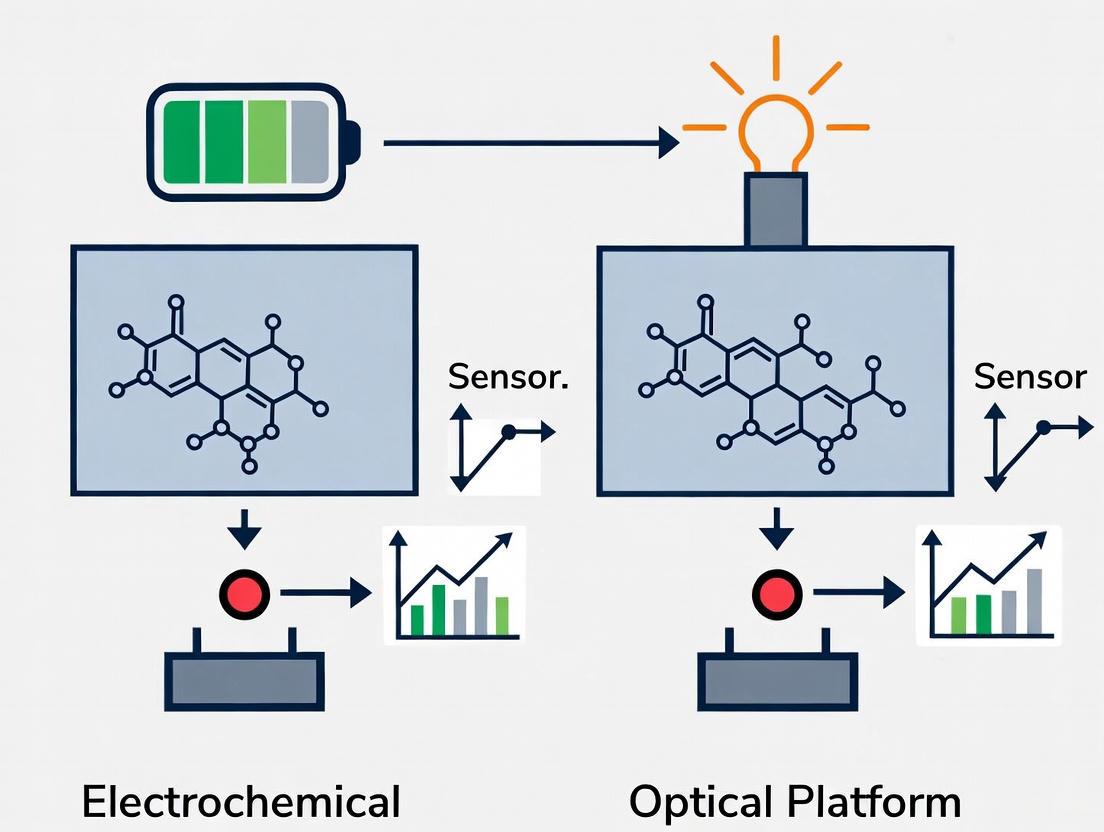
Electrochemical vs. Optical Biosensors: A Comprehensive Comparison for Biomedical Research and Precision Diagnostics
This article provides a detailed comparative analysis of electrochemical and optical biosensor platforms, tailored for researchers and drug development professionals.
Electrochemical vs. Optical Biosensors: A Comprehensive Comparison for Biomedical Research and Precision Diagnostics
Abstract
This article provides a detailed comparative analysis of electrochemical and optical biosensor platforms, tailored for researchers and drug development professionals. It explores the fundamental principles, operational mechanisms, and distinct advantages of each technology. The scope extends to their methodological applications in disease diagnosis, therapeutic drug monitoring, and wearable sensors, alongside practical troubleshooting for sensitivity and real-world performance. A critical evaluation of clinical validation protocols and a direct performance comparison offer essential insights for selecting the optimal biosensing strategy to advance precision medicine and point-of-care diagnostics.
Core Principles and Working Mechanisms: Building a Foundation in Biosensing Technology
Biosensors are analytical devices that integrate a biological recognition element with a physicochemical transducer to produce a measurable signal proportional to the concentration of a target analyte. The fundamental operation of all biosensors relies on two essential components: a bioreceptor that selectively interacts with the target molecule and a transducer that converts this biological recognition event into a quantifiable signal. The specificity of biosensors stems from the selective binding of recognition elements to target molecules, minimizing interference from impurities and enabling ultra-low detection limits, typically in the nanomolar or picomolar range [1].
The transduction principle forms the core of biosensor functionality, determining key performance parameters including sensitivity, detection limit, dynamic range, and applicability to different experimental settings. This guide provides a systematic comparison of the two predominant biosensor transduction platforms: electrochemical and optical. Within the broader context of biosensor research, understanding these fundamental transduction principles enables researchers to select the optimal platform for specific applications in drug development, clinical diagnostics, and biomedical research.
Electrochemical Transduction Principles
Electrochemical biosensors transform biological recognition events into measurable electrical signals through changes in electrical properties at the electrode-solution interface. The high specificity of these systems stems from the selective binding of recognition elements to target molecules, which subsequently affects the electrochemical behavior of the electrode surface [1]. These sensors are categorized based on their specific signal transduction mechanism.
Amperometric/Voltammetric Sensors monitor current resulting from the electrochemical oxidation or reduction of an electroactive species at a constant or varying potential. The generated current has a linear relationship with the target analyte concentration. A notable application is a disposable thread-based electrochemical biosensor for lung cancer diagnosis, which demonstrated excellent sensitivity for biomarker detection [2].
Potentiometric Sensors measure the potential difference between working and reference electrodes when the net current is zero. This potential change occurs in response to the accumulation of charge or changes in ion concentration resulting from a biological recognition event.
Impedimetric Sensors analyze the resistance and reactance of a system to an applied alternating current. The binding of biomolecules to the electrode surface alters the interfacial electron transfer resistance, which can be quantified via electrochemical impedance spectroscopy.
Conductometric Sensors track changes in the electrical conductivity of a solution resulting from enzymatic reactions that generate or consume ions.
Table 1: Electrochemical Transduction Mechanisms and Applications
| Transduction Type | Measured Parameter | Detection Principle | Example Application |
|---|---|---|---|
| Amperometric/Voltammetric | Current | Redox current from electroactive species | Lung cancer biomarker detection [2] |
| Potentiometric | Potential | Ion concentration change | Ion-selective electrode sensors |
| Impedimetric | Impedance | Electron transfer resistance at electrode interface | Label-free protein detection |
| Conductometric | Conductivity | Ionic strength change from reaction | Enzyme-based metabolite sensors |
A key advantage of electrochemical biosensors is their performance enhancement through nanomaterial integration. Noble metal nanomaterials like gold nanoparticles provide large specific surface area and outstanding electrical conductivity, while carbon-based nanomaterials such as graphene and carbon nanotubes form conjugated π-electron networks granting exceptional electrical properties [1]. For instance, one study developed a biosensing system using gold nanofiber-modified screen-printed carbon electrodes that significantly enhanced electron transfer efficiency, achieving a detection limit of 0.28 ng/mL for prostate-specific antigen [1].
Optical Transduction Principles
Optical biosensors convert biological recognition events into measurable optical signals through changes in light properties including intensity, wavelength, polarization, or phase. These platforms have gained prominence due to their high sensitivity, capability for multiplexed detection, and non-invasive nature, though they may face limitations in portability and environmental resilience compared to electrochemical alternatives [3].
Fluorescence and FRET-based Biosensors utilize energy transfer between donor and acceptor fluorophores. When the donor fluorophore is excited, it transfers energy to the acceptor if they are in close proximity, resulting in acceptor emission. Biological events that alter the distance or orientation between fluorophores change the FRET efficiency. Genetically encoded FRET biosensors have revolutionized the monitoring of cellular processes, exemplified by the AuxSen auxin biosensor that employs mNeonGreen and Aquamarine fluorescent proteins coupled to a modified bacterial tryptophan repressor [4]. This sensor enables real-time monitoring of auxin concentrations at subcellular resolution with reversibility and high temporal resolution.
Surface-Enhanced Raman Scattering (SERS) utilizes nanostructured metallic surfaces to enhance Raman scattering signals by several orders of magnitude. Sharp-tipped Au-Ag nanostars provide intense plasmonic enhancement due to their sharp-tipped morphology, enabling powerful SERS detection without dependence on Raman reporters [5]. One platform demonstrated sensitive detection of α-fetoprotein, a cancer biomarker, with a limit of detection of 16.73 ng/mL by exploiting the intrinsic vibrational modes of the target [5].
Surface Plasmon Resonance (SPR) measures changes in the refractive index near a metal surface, typically gold or silver, resulting from biomolecular binding events. This label-free technique enables real-time monitoring of molecular interactions without requiring fluorescent labeling. SPR biosensors have been successfully employed for fragment-based drug discovery, with one study screening 930 fragment compounds against multiple drug targets including HIV-1 protease, thrombin, and carbonic anhydrase [6].
Photoelectrochemical Biosensors represent a hybrid category combining optical excitation with electrochemical detection. These systems use light to excite a photosensitive material, generating an electrical signal measured electrochemically. For example, an organic photoelectrochemical transistor biosensor based on BiVO₄-ZnIn₂S₄ material was developed for efficient and sensitive detection of MCF-7 cancer cells [7].
Table 2: Optical Transduction Mechanisms and Applications
| Transduction Type | Measured Parameter | Detection Principle | Example Application |
|---|---|---|---|
| FRET | Fluorescence intensity ratio | Energy transfer between fluorophores | Real-time auxin monitoring in plants [4] |
| SERS | Raman scattering intensity | Plasmonic enhancement on nanostructures | α-fetoprotein cancer biomarker detection [5] |
| SPR | Refractive index change | Biomolecular binding on metal surface | Fragment screening for drug discovery [6] |
| Photoelectrochemical | Photocurrent | Light-induced electron transfer | Cancer cell detection [7] |
Figure 1: Optical Biosensor Transduction Mechanisms. FRET biosensors (top) detect conformational changes through energy transfer between fluorophores. SPR biosensors (bottom) measure biomolecular binding via refractive index changes at a metal surface.
Comparative Performance Analysis
Direct comparison of electrochemical and optical biosensor platforms reveals distinct advantages and limitations for each technology. Performance evaluation encompasses sensitivity, specificity, detection limits, operational stability, cost-effectiveness, and suitability for point-of-care applications.
Electrochemical biosensors demonstrate exceptional performance for miniaturized, portable applications due to their low cost, adaptability to point-of-care formats, and compatibility with complex biological matrices. Recent advancements include a high-performance electrochemical biosensor comprising Mn-ZIF-67 conjugated with anti-O antibody for Escherichia coli detection, which achieved an impressive linear range of 10 to 10¹⁰ CFU mL⁻¹ with a detection limit of 1 CFU mL⁻¹, outperforming many optical sensors for the same analyte [8]. The sensor maintained >80% sensitivity over 5 weeks and successfully recovered 93.10–107.52% of E. coli spiked in tap water samples, demonstrating remarkable environmental robustness [8].
Optical biosensors, particularly SERS and SPR platforms, offer superior sensitivity and multiplexing capabilities but may face limitations in portability and environmental resilience [3]. The SERS-based immunoassay for α-fetoprotein detection achieved a limit of detection of 16.73 ng/mL using an Au-Ag nanostars platform, addressing current limitations in cancer biomarker detection such as low sensitivity and dependence on Raman reporters [5]. SPR biosensor technology has proven valuable for fragment-based lead discovery, enabling efficient screening of 930 compounds against multiple drug targets with minimal promiscuous binders [6].
Table 3: Direct Performance Comparison of Electrochemical vs. Optical Biosensors
| Performance Parameter | Electrochemical Biosensors | Optical Biosensors |
|---|---|---|
| Typical Detection Limit | 0.28 ng/mL (PSA) [1], 1 CFU/mL (E. coli) [8] | 16.73 ng/mL (α-fetoprotein) [5] |
| Sensitivity | High, enhanced by nanomaterials | Very high, particularly for SERS/SPR |
| Multiplexing Capability | Moderate | High (multiple wavelengths/channels) |
| Portability | Excellent for point-of-care | Limited for some platforms |
| Cost-Effectiveness | High (low-cost electrodes, simple instrumentation) | Moderate to high (may require expensive optics) |
| Environmental Robustness | Good to excellent | Moderate (may require controlled conditions) |
| Measurement Speed | Fast (seconds to minutes) | Fast (real-time for SPR) |
| Sample Throughput | Moderate | High for plate-based readers |
The selection between electrochemical and optical biosensor platforms ultimately depends on the specific application requirements. Electrochemical systems excel in field-deployable, cost-sensitive applications requiring robust performance, while optical platforms offer superior capabilities for laboratory-based applications demanding ultra-high sensitivity and multiplexed detection.
Experimental Protocols and Methodologies
Electrochemical Biosensor Fabrication and Measurement Protocol
The development of a high-performance electrochemical biosensor for pathogen detection follows a systematic fabrication and characterization process, as demonstrated for the Mn-ZIF-67 based E. coli sensor [8]:
Electrode Modification Protocol:
- Material Synthesis: Prepare Mn-doped ZIF-67 by combining cobalt and manganese precursors with 2-methylimidazole ligand in solution. Manganese incorporation induces phase reconstruction, enhances surface area, and improves electron transfer.
- Physicochemical Characterization: Analyze crystallinity using XRD spectroscopy, functional groups via FTIR spectroscopy, and surface area/pore volume through N₂ adsorption-desorption measurements with Brunauer-Emmett-Teller (BET) analysis.
- Bioreceptor Immobilization: Conjugate anti-O-specific antibodies to the Mn-ZIF-67 material. Antibody conjugation modulates wettability, introduces amide I and II vibrational modes, and selectively blocks electron transfer upon bacterial binding.
- Electrode Preparation: Deposit the functionalized material onto the working electrode surface and characterize using electrochemical cyclic voltammetry measurements to confirm enhanced electron transfer.
Measurement Procedure:
- Sample Incubation: Expose the modified electrode to sample solutions containing target bacteria for a specified period (typically 15-30 minutes).
- Electrochemical Analysis: Perform electrochemical measurements using techniques such as electrochemical impedance spectroscopy (EIS) or differential pulse voltammetry (DPV).
- Signal Measurement: Quantify the electron transfer resistance change or current response resulting from bacterial binding to the antibody-functionalized surface.
- Data Analysis: Generate calibration curves by plotting signal response against analyte concentration, achieving a wide linear detection range from 10 to 10¹⁰ CFU mL⁻¹.
Optical Biosensor Validation and Screening Protocol
Validation of optical biosensors, particularly for cellular applications, requires standardized methodologies to ensure reliability and reproducibility. A high-content assay for biosensor validation in a 96-well plate format using automated microscopy provides a robust framework [9]:
Biosensor Validation Protocol:
- Cell Preparation: Plate adherent cells in 96-well microplates and transfer with biosensor DNA (e.g., Rho GTPase FRET biosensors) along with upstream regulatory proteins.
- Titration Analysis: Co-express biosensors with increasing amounts of regulator DNA to determine saturation points and dynamic range. Include donor-only and acceptor-only controls for bleedthrough correction.
- Automated Imaging: Acquire images using an automated microscope with appropriate filter sets for FRET measurements. Maintain consistent environmental control (temperature, CO₂) throughout imaging.
- Image Analysis: Calculate FRET ratios and generate dose-response curves. Visually inspect images for cell health, biosensor localization, and potential artifacts.
SPR Biosensor Screening Protocol for Fragment-Based Drug Discovery [6]:
- Surface Preparation: Immobilize target proteins (HIV-1 protease, thrombin, carbonic anhydrase) on sensor chips via standard amine-coupling chemistry.
- Fragment Library Design: Select 930 compounds from commercially available sources using physicochemical and medicinal chemistry filters.
- Screening Conditions: Inject fragments under standardized conditions with appropriate buffer systems. Include reference surfaces and solvent correction cycles.
- Binding Assessment: Evaluate responses for specific binding, excluding promiscuous binders (interacting with stoichiometry ≥5:1 with all proteins).
- Hit Validation: Confirm specific binding through competition assays and dose-response measurements.
Figure 2: Experimental Workflows for Biosensor Development and Application. Electrochemical biosensors (top) rely on electrode modification and electrical signal measurement. Optical biosensors (bottom) utilize light-based detection of molecular recognition events.
Research Reagent Solutions and Essential Materials
The performance and reliability of biosensor platforms depend critically on the quality and appropriateness of research reagents and materials. The following table details essential components for biosensor development and implementation.
Table 4: Essential Research Reagents and Materials for Biosensor Development
| Reagent/Material | Function/Purpose | Example Applications |
|---|---|---|
| Gold Nanoparticles (AuNPs) | Signal amplification, electron transfer enhancement, biocompatible substrate | Prostate-specific antigen detection [1] |
| Metal-Organic Frameworks (ZIF-67) | Porous scaffold with large surface area, enhanced electrical conductivity | E. coli detection sensor [8] |
| Fluorescent Proteins (mNeonGreen, Aquamarine) | FRET donor-acceptor pairs for conformational biosensors | Auxin biosensor (AuxSen) [4] |
| Specific Antibodies (anti-O antibody) | Biorecognition element for selective target capture | E. coli O-polysaccharide detection [8] |
| Screen-Printed Carbon Electrodes (SPCEs) | Disposable, cost-effective electrode platform for point-of-care sensors | Lactate detection in sweat [1] |
| Aptamers | Nucleic acid-based recognition elements with high specificity and stability | NAD(H) detection [7] |
| Au-Ag Nanostars | Plasmonic nanoparticles for SERS enhancement | α-fetoprotein cancer biomarker detection [5] |
| Conductive Polymers (PEDOT) | Flexible, conductive substrates for wearable sensors | Sweat lactate sensor [1] |
Electrochemical and optical biosensing platforms offer complementary strengths for converting biological events into readable signals. Electrochemical systems provide robust, cost-effective solutions for field-deployable diagnostics with recent demonstrations achieving impressive detection limits for pathogens and disease biomarkers. Optical platforms deliver exceptional sensitivity and multiplexing capabilities for laboratory-based applications, with advanced FRET and SERS biosensors enabling real-time monitoring of cellular processes and ultrasensitive biomarker detection.
The continuing evolution of both platforms is being driven by nanomaterials innovation, improved bioreceptor engineering, and integration with automated systems. Future directions include developing multimodal sensing platforms that combine electrochemical and optical detection mechanisms, creating increasingly miniature and implantable form factors, and incorporating artificial intelligence for enhanced signal processing and data analysis. These advancements will further establish biosensors as indispensable tools across biomedical research, clinical diagnostics, and drug development.
Biosensors represent a critical convergence of biological specificity and analytical detection, serving pivotal roles in medical diagnostics, environmental monitoring, and food safety. The performance of these devices fundamentally depends on two key components: the biological recognition element that provides analyte specificity and the transducer interface that converts biological events into measurable signals. This comprehensive guide examines the operational principles, experimental methodologies, and performance characteristics of two dominant biosensor platforms: electrochemical and optical systems. By objectively comparing their respective advantages, limitations, and implementation requirements through structured data analysis and experimental protocols, this review provides researchers and development professionals with the foundational knowledge necessary to select appropriate biosensing architectures for specific applications.
Biosensors are analytically defined as self-contained integrated devices capable of providing specific quantitative or semi-quantitative analytical information using a biological recognition element retained in direct spatial contact with a transduction element [10]. This fundamental architecture enables the detection and quantification of target analytes across diverse fields including clinical diagnostics, environmental surveillance, food quality control, and bioprocess engineering [10] [11].
The core functionality of any biosensor relies on the synergistic operation of its two primary components: (1) the biological recognition element, which confers specificity through selective binding or catalytic interaction with the target analyte, and (2) the transducer interface, which transforms the biological response into a quantifiable signal [10]. Biological recognition elements encompass enzymes, antibodies, nucleic acids, aptamers, cellular receptors, or whole cells, each providing distinct binding affinities and operational mechanisms [10] [5]. The transducer component, typically classified as electrochemical, optical, piezoelectric, or thermal, defines the fundamental detection methodology and determines critical performance parameters including sensitivity, detection limits, and dynamic range [10] [12].
The escalating demand for decentralized diagnostics and real-time monitoring has accelerated refinement of both components, driven by advances in microfabrication, nanomaterials, and spectroscopic interrogation methods [10] [13]. This review systematically examines the implementation of these key components across electrochemical and optical biosensing platforms, providing comparative performance analysis and methodological protocols to guide platform selection for specific research and development applications.
Biological Recognition Elements: Specificity Foundations
Biological recognition elements form the molecular interface for selective analyte interaction, determining the fundamental specificity of biosensing systems. These elements operate through various mechanisms including catalytic transformation, affinity binding, or whole-cell responses, with selection dictated by target analyte properties and required assay conditions.
Element Classification and Operational Mechanisms
Enzyme-Based Recognition: Enzymes provide recognition through catalytic conversion of specific substrates, generating products detectable by transducers. Glucose oxidase exemplifies this approach, catalyzing glucose oxidation to gluconolactone while producing electrons measurable electrochemically [10] [14]. Enzyme sensors typically exhibit high turnover rates, amplifying detection signals, but may lack absolute specificity when facing structurally similar substrates.
Immunological Recognition Elements: Antibodies and antibody fragments enable detection through high-affinity binding to specific antigenic epitopes [10] [8]. Immunosensors provide exceptional specificity for proteins, pathogens, and high-molecular-weight compounds, though their non-catalytic binding mechanism typically requires labeling or secondary detection systems for signal generation. Recent advances incorporate monoclonal and recombinant antibodies to enhance reproducibility and stability [8].
Nucleic Acid-Based Recognition: DNA and RNA probes facilitate detection through complementary hybridization to target nucleic acid sequences, enabling genetic mutation identification, pathogen detection, and gene expression monitoring [10] [5]. Aptamers—engineered single-stranded oligonucleotides—fold into specific three-dimensional structures that bind molecular targets with antibody-like affinity, offering advantages of thermal stability and synthetic production [5].
Cellular and Tissue-Based Recognition: Whole cells, cellular receptors, or tissue sections provide recognition capability for complex analytes or functional responses [10]. These systems enable detection of biologically active compounds through metabolic pathway activation or receptor binding, though they typically exhibit slower response times and reduced operational stability compared to molecular recognition elements.
Immobilization Methodologies
Effective biosensor performance requires stable integration of biological recognition elements with transducer surfaces while maintaining biological activity. Common immobilization approaches include:
Adsorption: Physical attachment through van der Waals forces, ionic interaction, or hydrophobic binding provides simple implementation but may yield unstable bonding under varying pH or ionic strength conditions [10].
Covalent Attachment: Chemical bonding between functional groups on biological elements (amine, carboxyl, thiol) and activated transducer surfaces creates stable, irreversible immobilization [10] [8]. This approach often employs cross-linking agents like glutaraldehyde or EDC/NHS chemistry [5].
Entrapment: Physical confinement within polymeric matrices (e.g., hydrogels, sol-gels, conducting polymers) or behind semi-permeable membranes preserves biological activity while providing protective microenvironment [10] [13].
Affinity Binding: Specific biological interactions (e.g., avidin-biotin, protein A/G-antibody) enable oriented immobilization that optimizes binding site accessibility [10].
Transducer Interfaces: Signal Generation Mechanisms
Transducers constitute the signal conversion core of biosensors, transforming molecular recognition events into quantifiable electrical or optical outputs. The transduction mechanism fundamentally determines operational characteristics including sensitivity, response time, and instrumentation requirements.
Electrochemical Transduction
Electrochemical biosensors measure electrical signals generated from biochemical interactions, typically employing a three-electrode configuration (working, reference, and counter electrodes) to control potential and measure current [11] [14]. These systems are classified according to their measured electrical parameter:
Amperometric Sensors: Detect current generated from redox reactions at constant applied potential, with current magnitude proportional to analyte concentration [10] [15]. The extensively commercialized glucose biosensor exemplifies this approach, monitoring electron flow generated from enzymatic glucose oxidation [14].
Potentiometric Sensors: Measure potential difference accumulation at electrode surfaces under conditions of negligible current flow, typically using ion-selective membranes or field-effect transistors [10] [15].
Impedimetric Sensors: Monitor changes in system resistance and capacitance resulting from biomolecular binding events at modified electrode surfaces [10] [11]. Electrochemical impedance spectroscopy (EIS) provides label-free detection capability suitable for tracking binding kinetics [10].
Voltammetric Sensors: Apply potential sweeps or pulses while measuring resultant current, enabling discrimination of multiple electroactive species through their characteristic oxidation/reduction potentials [11] [14].
The following diagram illustrates the operational workflow of a typical electrochemical biosensing system:
Optical Transduction
Optical biosensors exploit light-matter interactions to detect binding events or concentration changes, utilizing parameters including absorbance, fluorescence, luminescence, refractive index, or reflectance [10] [13]. Major optical transduction platforms include:
Surface Plasmon Resonance (SPR): Measures refractive index changes at metal-dielectric interfaces during biomolecular binding, enabling label-free, real-time kinetic monitoring [10] [13]. Recent advances include SPR imaging for multiplexed analysis and smartphone-compatible miniaturization [13] [15].
Fluorescence-Based Detection: Utilizes light absorption and emission characteristics of fluorophores, providing exceptional sensitivity down to single-molecule detection under optimal conditions [10] [16]. Fluorescent biosensors employ intensity, lifetime, anisotropy, energy transfer (FRET), or quenching measurements to monitor molecular interactions [10].
Chemiluminescence and Bioluminescence: Detect light emission from chemical or enzymatic reactions without requiring excitation illumination, minimizing background signal [15]. These approaches offer exceptional sensitivity but typically require careful reagent integration and preservation.
Colorimetric Sensing: Monitors visible color changes from nanoparticle aggregation, enzyme-linked reactions, or pH indicators, enabling simple visual detection or smartphone quantification [15]. Lateral flow immunoassays represent widely deployed commercial applications of this principle [15].
Surface-Enhanced Raman Spectroscopy (SERS): Explores plasmonic nanoparticle enhancement of normally weak Raman scattering, providing vibrational "fingerprint" identification with single-molecule sensitivity in optimized configurations [5] [15].
The operational sequence for optical biosensing systems follows this generalized pathway:
Comparative Performance Analysis: Electrochemical vs. Optical Biosensors
The selection between electrochemical and optical transduction platforms involves balancing multiple performance parameters against application requirements. The following tables provide systematic comparison across critical operational characteristics.
Table 1: Fundamental operational characteristics comparison
| Parameter | Electrochemical Biosensors | Optical Biosensors |
|---|---|---|
| Detection Mechanism | Measurement of electrical signals (current, potential, impedance) from redox reactions [12] | Interaction of light with target molecules measuring optical property changes [12] |
| Transducer Element | Electrodes (Au, carbon, Pt) [11] [12] | Light sources, waveguides, photodetectors [12] [13] |
| Detection Dynamic Range | Limited [12] | Wide [12] |
| Multiplexing Capability | Limited support [12] | Excellent, enables simultaneous multi-analyte detection [12] [13] |
| Response Time | Fast (seconds) [12] | Slow (minutes) [12] |
| Contactless Measurement | Not available [12] | Available [12] |
| Electromagnetic Interference | Susceptible [12] | Immune [12] |
Table 2: Implementation and application considerations
| Parameter | Electrochemical Biosensors | Optical Biosensors |
|---|---|---|
| Sample Requirement | Works with complex, crude samples [12] | Often requires purified samples [12] |
| Instrumentation Size | Compact, easily miniaturized [11] [12] | Bulky, though miniaturization advancing [12] [13] |
| Cost | Relatively low, simple setup [12] | Generally higher, specialized optics required [12] |
| Lifetime | Minutes to days (biological component-dependent) [12] | Up to several years [12] |
| Primary Applications | Clinical diagnostics, food safety, environmental monitoring [11] [12] | Research, medical diagnostics, environmental monitoring [12] [13] |
Experimental Protocols and Performance Data
Electrochemical Biosensor Experimental Protocol: E. coli Detection
A recently developed high-performance electrochemical biosensor for Escherichia coli detection exemplifies contemporary methodology employing bimetallic metal-organic frameworks (MOFs) for signal enhancement [8].
Experimental Workflow:
- Electrode Modification: Glassy carbon electrodes are polished sequentially with 0.3 and 0.05 μm alumina slurry, followed by sonication in ethanol and deionized water [8].
- Mn-ZIF-67 Nanocomposite Synthesis: Mn-doped Co zeolitic imidazolate framework (ZIF-67) prepared through solvothermal reaction with varying Co:Mn molar ratios (10:1, 5:1, 2:1, 1:1) to optimize electron transfer properties [8].
- Bioreceptor Immobilization: Anti-E. coli O-specific antibody conjugated to Mn-ZIF-67 modified electrode surface via EDC/NHS chemistry, targeting O-polysaccharide antigen for selective recognition [8].
- Electrochemical Measurement: Amperometric detection performed in 0.1M PBS (pH 7.4) containing 5mM Fe(CN)₆³⁻/⁴⁻ as redox probe, monitoring current decrease following antigen-antibody binding [8].
Performance Metrics:
- Linear Detection Range: 10 to 10¹⁰ CFU mL⁻¹
- Limit of Detection: 1 CFU mL⁻¹
- Selectivity: Effectively discriminates non-target bacteria (Salmonella, Pseudomonas aeruginosa, Staphylococcus aureus)
- Stability: Maintains >80% sensitivity over 5 weeks
- Real Sample Recovery: 93.10–107.52% in spiked tap water samples [8]
Optical Biosensor Experimental Protocol: SERS-Based Immunoassay
A surface-enhanced Raman scattering (SERS) platform utilizing Au-Ag nanostars demonstrates advanced optical biosensing methodology for cancer biomarker detection [5].
Experimental Workflow:
- Nanostar Synthesis: Au-Ag nanostars prepared via seed-mediated growth, with sharp-tipped morphology optimized for plasmonic enhancement [5].
- Platform Optimization: Nanostar concentration tuned by centrifugation (10, 30, 60 minutes) with SERS performance evaluated using methylene blue and mercaptopropionic acid as probe molecules [5].
- Bioreceptor Functionalization: Optimized nanostars functionalized with mercaptopropionic acid (MPA), followed by EDC/NHS activation for covalent attachment of monoclonal anti-α-fetoprotein antibodies [5].
- SERS Detection: α-fetoprotein antigen detection across 500–0 ng/mL range using intrinsic vibrational modes without Raman reporters [5].
Performance Metrics:
- Linear Detection Range: 167–38 ng/mL (antibody), 500–0 ng/mL (antigen)
- Limit of Detection: 16.73 ng/mL for antigens
- Key Innovation: Aqueous, surfactant-free platform exploiting intrinsic AFP vibrational modes
- Application Potential: Sensitive and rapid cancer biomarker detection for early diagnostics [5]
Research Reagent Solutions: Essential Materials
Table 3: Key research reagents and materials for biosensor development
| Reagent/Material | Function | Example Applications |
|---|---|---|
| Flavin-adenine dinucleotide-dependent glucose dehydrogenase (FAD-GDH) | Oxygen-insensitive enzyme for glucose detection | Glucose biosensors, avoids oxygen interference [14] |
| Quinoline-5,8-dione (QD) mediator | Electron shuttle in enzymatic reactions | High-sensitivity glucose sensor strips [14] |
| Mn-doped ZIF-67 (Co/Mn ZIF) | Bimetallic MOF for enhanced electron transfer | Electrochemical E. coli biosensor [8] |
| Au-Ag nanostars | Plasmonic nanoparticles for signal enhancement | SERS-based immunoassays [5] |
| Polydimethylsiloxane (PDMS) | Flexible, transparent polymer substrate | Wearable optical biosensors [13] |
| Mercaptopropionic acid (MPA) | Linker molecule for surface functionalization | Antibody immobilization on SERS platforms [5] |
| EDC/NHS chemistry | Carboxyl-to-amine crosslinking | Covalent antibody immobilization [5] [8] |
| Anti-O-specific antibody | Bioreceptor for bacterial surface antigen | Selective E. coli detection [8] |
Electrochemical and optical biosensing platforms present complementary advantages that recommend them for distinct application environments. Electrochemical systems offer superior portability, rapid response, and compatibility with complex samples, making them ideal for point-of-care diagnostics and field deployment [11] [12] [15]. Optical platforms provide exceptional sensitivity, multiplexing capability, and resistance to electromagnetic interference, advantages that support research applications and laboratory-based diagnostic systems [16] [12] [13].
Future development trajectories indicate increasing convergence of these platforms with advanced materials (particularly nanomaterials), artificial intelligence for signal processing, and wireless connectivity for data transmission [11] [16] [13]. The integration of machine learning algorithms significantly enhances analytical performance in both platforms, enabling improved signal discrimination, drift correction, and multivariate analysis [16]. Selection between electrochemical and optical biosensing platforms ultimately depends on application-specific requirements including sensitivity thresholds, sample matrix complexity, operational environment, and resource constraints.
Electrochemical biosensors have emerged as powerful analytical tools that combine the specificity of biological recognition with the sensitivity of electrochemical transducers. These devices convert a biological response into a quantifiable electrical signal, enabling the detection of a wide range of analytes from clinical diagnostics to environmental monitoring [17]. The growing demand for point-of-care testing (POCT) systems has further accelerated development in this field, with electrochemical biosensors offering significant advantages including portability, low cost, rapid analysis, and compatibility with miniaturization [18] [15]. The fundamental operation of these sensors relies on the precise interplay between a biological recognition element (such as enzymes, antibodies, or nucleic acids) and an electrochemical transducer that detects the binding event or catalytic reaction [19].
Among the various transduction mechanisms, amperometric, potentiometric, and impedimetric techniques represent three foundational approaches that dominate both research and commercial applications. Each mechanism offers distinct operating principles, performance characteristics, and implementation considerations. Amperometric sensors measure current resulting from electrochemical oxidation or reduction, potentiometric sensors detect potential differences at zero current, and impedimetric sensors monitor changes in the electrical impedance of the electrode interface [20] [17]. The selection of an appropriate sensing mechanism depends on the specific application requirements, including target analyte, required detection limit, sample matrix, and necessary measurement throughput. This review provides a comprehensive comparison of these three electrochemical biosensing mechanisms, highlighting their fundamental principles, experimental implementations, and relative performance characteristics to guide researchers in selecting optimal sensing strategies for their specific applications.
The three primary electrochemical biosensing mechanisms operate on distinct physical principles, each with unique advantages and limitations. Understanding these fundamental differences is crucial for selecting the appropriate transduction method for specific applications.
Amperometric biosensors operate by applying a constant potential to an electrochemical cell and measuring the resulting current from the reduction or oxidation of an electroactive species involved in the biological recognition process [20]. The measured current is directly proportional to the concentration of the analyte, following the Cottrell equation: i = nFAc_j0√(D_j/πt), where i represents current, n is the number of electrons transferred, F is Faraday's constant, A is the electrode area, c_j0 is the initial concentration of the electroactive species, and D_j is its diffusion coefficient [20]. This technique is particularly sensitive to catalytic reactions, making it ideal for enzyme-based biosensors where the enzymatic reaction produces or consumes an electroactive species.
Potentiometric biosensors function differently by measuring the potential difference between working and reference electrodes under conditions of zero current flow [18] [20]. This potential develops as a result of specific sensor-analyte interactions that establish a local Nernstian equilibrium at the sensor interface. The measured potential relates to analyte concentration through the Nernst equation, with a theoretical sensitivity of 59 mV per decade of concentration change at 25°C [18]. These sensors can be implemented through various configurations including ion-selective electrodes (ISEs), coated-wire electrodes (CWEs), and field-effect transistors (FETs) [20]. Recent advances have demonstrated potentiometric sensors with exceptional performance, including pH sensors with sensitivities approaching the theoretical Nernstian limit and DNA detection systems capable of distinguishing single-nucleotide polymorphisms [18].
Impedimetric biosensors, also referred to as conductometric sensors, monitor changes in the impedance (both resistance and reactance) of the electrode-electrolyte interface resulting from specific biological recognition events [17]. This technique typically applies a small-magnitude alternating potential across a range of frequencies and measures the system's response in a steady state. The significant advantages of this approach include its ability to perform sensitive measurements that can be averaged over extended periods, theoretical treatment using generalized linear current-potential characteristics, and measurement capability across a broad frequency range [20]. Impedimetric biosensors are particularly valuable for label-free detection of binding events, such as antibody-antigen interactions or DNA hybridization, without requiring electroactive species.
Table 1: Comparative Overview of Electrochemical Biosensing Mechanisms
| Parameter | Amperometric | Potentiometric | Impedimetric |
|---|---|---|---|
| Measured Quantity | Current | Potential | Impedance (Resistance & Reactance) |
| Applied Signal | Constant potential | Zero current | Small AC potential with frequency sweep |
| Theoretical Basis | Cottrell equation | Nernst equation | Complex nonlinear least squares fitting |
| Detection Limit | Picomolar range [20] | Millimolar to micromolar [20] | Picomolar to femtomolar [17] |
| Dynamic Range | 3-6 orders of magnitude | 2-4 orders of magnitude | 3-5 orders of magnitude |
| Key Applications | Enzyme substrates, metabolic monitoring | Ions, pH, DNA hybridization, redox potential | Binding kinetics, cell growth, antibody-antigen interactions |
| Label Requirement | Often requires enzyme labels | Generally label-free | Typically label-free |
Figure 1: Fundamental operational principles of the three primary electrochemical biosensing mechanisms, showing the relationship between applied signals and measured outputs.
Experimental Protocols and Methodologies
Amperometric Biosensor Implementation
Amperometric biosensors typically employ a three-electrode system consisting of working, reference, and counter electrodes immersed in an electrolyte solution containing the analyte of interest [20]. The fundamental instrumentation requires a controlled-potential system, often called a potentiostat, which applies a constant potential between the working and reference electrodes while measuring the current flowing between the working and counter electrodes [20] [17]. A critical requirement for controlled-potential measurements is the inclusion of a supporting electrolyte to prevent electromigration effects, lower solution resistance, and maintain constant ionic strength [20].
For enzyme-based amperometric biosensors, the experimental protocol typically involves immobilizing the enzyme on the working electrode surface, applying the predetermined operating potential, and monitoring the steady-state current or transient current response following analyte introduction. The working potential is carefully selected to maximize the signal-to-noise ratio for the specific electrochemical reaction of interest while minimizing interfering reactions from other electroactive species in the sample matrix. Microelectrodes have become particularly valuable in amperometric sensing due to their high mass transport density, small double-layer capacitance, and small ohmic drop, though careful design is required to prevent cross-talk between adjacent electrodes in array configurations [18].
Recent advances in amperometric sensing include the development of sophisticated switching circuits that enable high-speed multipoint measurement across microelectrode arrays. This approach addresses the inherent limitation of traditional systems that require seconds to minutes to reach steady-state conditions at each electrode, thereby enabling rapid spatial and temporal mapping of analyte distributions [18].
Potentiometric Biosensor Implementation
Potentiometric biosensor measurements are performed under conditions of zero current flow, requiring a high-impedance voltmeter to measure the potential difference between the working and reference electrodes without drawing significant current [20]. The experimental setup typically involves an ion-selective membrane or sensitive surface that generates a potential response specific to the target analyte. The reference electrode must maintain a stable and well-defined potential throughout the measurement, typically achieved through stable internal fill solutions and consistent junction potentials [20].
For integrated sensor arrays compatible with complementary metal-oxide semiconductor (CMOS) technology, specialized circuit designs such as the CMOS source-drain follower have been developed [18]. This configuration maintains both gate-source and gate-drain voltages of the sensor transistor at constant values, providing infinite input impedance for both DC and AC signals—a critical characteristic for potentiometric measurements that prevents loading of the sensing electrode [18]. This approach has been successfully implemented in large-scale arrays, with demonstrations including a million-sensor array on a single chip for genome sequencing applications [18].
A significant advancement in potentiometric sensing involves the transition from direct charge detection to redox potential detection methods. Direct charge detection suffers from limitations including charge screening by ions in solution, influence of molecular shape on charge distribution, and unstable floating electrode potentials [18]. In contrast, redox potential detection using modified electrodes (e.g., ferrocenyl-alkanethiol modified gold electrodes) detects the ratio of oxidizer to reducer concentration, providing improved stability and reduced drift compared to direct charge detection methods [18].
Impedimetric Biosensor Implementation
Impedimetric biosensor measurements utilize a small-amplitude alternating potential (typically 5-50 mV) applied across a range of frequencies (often 0.1 Hz to 100 kHz) to characterize the electrochemical impedance of the electrode-electrolyte interface [20] [17]. The measured impedance data is commonly presented as Nyquist plots (imaginary vs. real components) or Bode plots (magnitude and phase vs. frequency), with equivalent circuit modeling used to extract meaningful parameters related to specific biological recognition events.
The experimental protocol for impedimetric biosensing involves establishing a stable baseline measurement in the appropriate buffer system, followed by monitoring changes in specific impedance parameters (such as charge transfer resistance or double-layer capacitance) upon introduction of the target analyte or during the binding process. Non-Faradaic approaches monitor changes in the electrode double layer without redox mediators, while Faradaic approaches utilize reversible redox couples (such as ferro/ferricyanide) to enhance sensitivity to surface binding events [17].
Recent implementations have leveraged the label-free nature of impedimetric biosensing for continuous monitoring applications, including cell growth and proliferation studies, where increasing cell coverage on electrode surfaces produces quantifiable changes in the measured impedance [17]. The technique has proven particularly valuable for monitoring antibody-antigen interactions, DNA hybridization, and protein binding events without requiring enzymatic or fluorescent labels.
Table 2: Characteristic Performance Parameters of Electrochemical Biosensing Mechanisms
| Performance Metric | Amperometric | Potentiometric | Impedimetric |
|---|---|---|---|
| Glucose Detection Limit | ~2 mg/dL (0.1 mM) [18] | N/A | N/A |
| pH Sensitivity | N/A | 41-59 mV/pH [18] | N/A |
| DNA Detection Signal | N/A | 12-100 mV for hybridization [18] | N/A |
| Redox Potential Sensitivity | N/A | 57.9 mV/decade [18] | N/A |
| Typical Response Time | Seconds to minutes | Seconds to minutes | Minutes |
| Regeneration Potential | Limited | Good | Excellent |
| Stability | Moderate (enzyme degradation) | Good (membrane aging) | Excellent |
Research Reagent Solutions and Materials
The performance of electrochemical biosensors depends critically on the careful selection of materials and reagents that facilitate both biological recognition and electrochemical transduction. The following table summarizes key research reagents and their functions in developing effective biosensing platforms.
Table 3: Essential Research Reagents and Materials for Electrochemical Biosensors
| Reagent/Material | Function | Application Examples |
|---|---|---|
| Glucose Oxidase | Enzyme bioreceptor for glucose detection | Amperometric glucose biosensors [17] |
| 11-Ferrocenyl-1-undecanethiol (11-FUT) | Redox mediator for potential stabilization | Potentiometric redox sensors [18] |
| Cat-CVD Silicon Nitride | pH-sensitive membrane material | Potentiometric pH sensors and DNA detection [18] |
| Zeolitic Imidazolate Frameworks (ZIF-67) | Nanostructured porous sensing layer | Enhanced surface area for improved sensitivity [8] |
| Mn-doped ZIF-67 | Electron transfer enhancement | Improved conductivity in bimetallic MOF biosensors [8] |
| Anti-O Antibody | Biorecognition element for bacterial detection | E. coli immunosensors [8] |
| Thiol-modified Oligonucleotides | DNA probe immobilization | DNA hybridization sensors [18] |
| Polypyrrole | Conducting polymer for NH₃ sensing | Impedimetric gas sensors [20] |
| Hexacyanoferrate Mixture | Reversible redox couple | Electron transfer mediator in impedimetric sensors [18] |
| Au/Ag Nanostars | Plasmonic enhancement substrate | SERS-based biosensing platforms [5] |
Figure 2: Biosensor architecture showing the relationship between sample analytes, recognition elements, transducer materials, and resulting signal types.
Comparative Performance Analysis
When selecting an appropriate biosensing mechanism for specific applications, researchers must consider the relative advantages and limitations of each approach across multiple performance parameters. The following analysis provides a framework for this decision-making process based on characteristic performance data and implementation requirements.
Sensitivity and Detection Limits: Amperometric biosensors typically offer excellent sensitivity with theoretical detection limits extending to picomolar concentrations, attributable to the direct proportionality between analyte concentration and Faradaic current [20]. Practical implementations have demonstrated clinically relevant detection limits, such as glucose detection at 2 mg/dL in continuous flow systems [18]. Potentiometric biosensors generally provide slightly higher detection limits in the millimolar to micromolar range, though specialized implementations can achieve significantly better performance [20]. For example, DNA hybridization has been detected with signal changes of 12-100 mV, enabling discrimination of complementary and reverse-complementary sequences [18]. Impedimetric biosensors can achieve remarkably low detection limits, sometimes extending to femtomolar concentrations, due to their exceptional sensitivity to nanoscale surface modifications and binding events [17].
Dynamic Range and Linearity: Amperometric sensors typically provide the widest dynamic range, often spanning 3-6 orders of magnitude, making them suitable for applications requiring quantification across diverse concentration ranges [20]. The linearity of amperometric response is generally excellent within this range, following predictable diffusion-limited current relationships. Potentiometric sensors exhibit more limited dynamic range (typically 2-4 orders of magnitude) due to the logarithmic relationship between potential and concentration described by the Nernst equation [18] [20]. Impedimetric sensors demonstrate intermediate dynamic range capabilities, typically spanning 3-5 orders of magnitude, though this is highly dependent on the specific sensing interface and measurement frequency [17].
Response Time and Measurement Duration: Amperometric measurements can provide rapid response times ranging from seconds to minutes, depending on the diffusion path to the electrode surface and the kinetics of the electrochemical reaction [20]. Microelectrode designs significantly enhance response time by establishing rapid steady-state conditions [18]. Potentiometric sensors similarly achieve response times in the seconds to minutes range, though settling time can be significantly influenced by the sample matrix—with rapid settling in redox buffer solutions but potentially longer times in simple buffer systems [18]. Impedimetric measurements typically require longer acquisition times, particularly when full spectral analysis is performed across multiple frequencies, though single-frequency measurements can provide rapid monitoring capabilities for dynamic processes [20] [17].
Stability and Reproducibility: Long-term stability varies considerably between sensing mechanisms. Amperometric biosensors employing enzymatic recognition elements often suffer from limited stability due to enzyme degradation or inactivation, though this can be mitigated through sophisticated immobilization approaches [17]. Potentiometric sensors demonstrate good stability, with primary limitations arising from membrane aging or reference electrode drift [20]. Impedimetric sensors typically offer excellent long-term stability, as they often rely on robust recognition elements such as antibodies or DNA probes and do not consume reagents during measurement [17]. Reproducibility remains a challenge across all electrochemical biosensing platforms, with significant efforts focused on improving electrode-to-electrode and batch-to-batch consistency through standardized fabrication and functionalization protocols [11].
The comparative analysis of amperometric, potentiometric, and impedimetric biosensing mechanisms reveals a complex landscape of performance trade-offs that must be carefully considered for specific application requirements. Amperometric systems excel in sensitivity and wide dynamic range, making them ideal for detecting low concentrations of electroactive species, particularly in enzymatic sensing applications. Potentiometric approaches offer simplicity and direct relationship to thermodynamic parameters, providing robust platforms for ion detection and redox potential monitoring. Impedimetric techniques provide exceptional label-free capabilities and sensitivity to surface binding events, enabling detailed characterization of biomolecular interactions.
The ongoing convergence of electrochemical biosensing with advancements in nanomaterials science, micro fabrication technologies, and complementary detection modalities continues to expand the capabilities of all three sensing mechanisms [11]. Emerging trends include the development of multi-modal sensing platforms that combine complementary techniques, the integration of machine learning for enhanced data analysis, and the creation of increasingly miniaturized systems for point-of-care and wearable applications [11]. These advancements promise to further blur the traditional boundaries between sensing mechanisms while expanding the application space for electrochemical biosensors across clinical diagnostics, environmental monitoring, food safety, and biomedical research.
As the field continues to evolve, the optimal selection of biosensing mechanism will increasingly depend on the specific requirements of each application rather than inherent superiority of any single approach. Researchers are encouraged to consider the comprehensive performance characteristics outlined in this review when designing sensing strategies for their particular analytical challenges.
Optical biosensors represent a cornerstone of modern analytical science, combining the exquisite specificity of biological recognition elements with the high sensitivity of optical transduction mechanisms. These devices function by detecting changes in light properties—such as intensity, wavelength, polarization, or phase—resulting from interactions between a target analyte and an immobilized biological recognition element (e.g., antibody, aptamer, enzyme) immobilized on a transducer surface [21]. The evolution of optical biosensing has been remarkable, transitioning from laboratory-bound instruments to platforms amenable to point-of-care (POC) testing through integration with microfluidics, simplified manufacturing technologies, and portable detectors [21]. This progression aligns with the growing demand for diagnostic tools that meet the ASSURED criteria (Affordable, Sensitive, Specific, User-friendly, Rapid and robust, Equipment-free, and Deliverable) as defined by the World Health Organization, particularly in resource-limited settings [22].
Among the diverse optical transduction schemes available, four principal techniques have emerged as particularly impactful for biomedical and diagnostic applications: Surface Plasmon Resonance (SPR), fluorescence, colorimetric, and Surface-Enhanced Raman Spectroscopy (SERS). Each technique operates on distinct physical principles and offers unique advantages and limitations in terms of sensitivity, multiplexing capability, equipment requirements, and suitability for quantitative analysis. SPR biosensors exploit the generation of charge-density oscillations at a metal-dielectric interface to monitor biomolecular interactions in real-time without labeling [23]. Fluorescence-based biosensors detect the light emitted by fluorophores following excitation, offering exceptional sensitivity and the potential for multiplexing [24]. Colorimetric biosensors translate molecular recognition events into visible color changes that can be interpreted by the naked eye or simple digital imaging [25]. SERS biosensors amplify the inherently weak Raman scattering signals of molecules adsorbed on nanostructured metallic surfaces, combining molecular fingerprint specificity with trace-level sensitivity [26]. This guide provides a comprehensive technical comparison of these four fundamental optical biosensing mechanisms, with particular emphasis on their operational principles, performance characteristics, experimental implementation, and relevance to drug development and clinical diagnostics.
Comparative Performance Analysis of Optical Biosensing Techniques
The selection of an appropriate biosensing technique for a specific application requires careful consideration of multiple performance parameters. The following table provides a systematic comparison of the key attributes of SPR, fluorescence, colorimetric, and SERS biosensing platforms, summarizing their relative advantages and limitations across critical performance metrics relevant to research and diagnostic applications.
Table 1: Performance Comparison of Major Optical Biosensing Techniques
| Parameter | SPR | Fluorescence | Colorimetric | SERS |
|---|---|---|---|---|
| Typical Limit of Detection (LOD) | pg/mL-fg/mL [23] | fg/mL [24] | ng/mL-µg/mL [25] | Single molecule [26] |
| Quantitative Capability | Excellent (real-time, label-free) | Excellent | Semi-quantitative to Quantitative (with instrumentation) | Excellent |
| Multiplexing Potential | Moderate | High | Moderate | Very High (sharp spectral peaks) |
| Real-time Monitoring | Yes | Yes | No | No (typically endpoint) |
| Throughput | Moderate | High (with microplates) | High (with lateral flow) | Moderate |
| Equipment Complexity | High | Moderate to High | Low | Moderate to High |
| Cost per Analysis | High | Moderate | Low | Moderate |
| Key Strength | Label-free kinetic analysis | Ultra-high sensitivity | Simplicity and portability | Molecular fingerprinting |
As evidenced in Table 1, no single technique is superior across all metrics; rather, each excels in specific domains. SPR stands out for its ability to monitor biomolecular interactions—such as antibody-antigen binding or protein-DNA interactions—in real-time and without labels, providing valuable kinetic parameters (association/dissociation rates) and affinity constants, which are crucial for basic research and drug development [23]. Fluorescence-based biosensors offer exceptional sensitivity, often achieving detection limits in the fg/mL range, and are highly amenable to multiplexing when combined with different fluorophores, making them ideal for high-throughput screening applications [24]. Colorimetric biosensors, while generally less sensitive, provide unmatched simplicity and are the foundation of many point-of-care tests (e.g., lateral flow immunoassays); their utility is enhanced when coupled with smartphone-based color analysis for semi-quantitative readouts [25]. SERS offers a unique combination of single-molecule sensitivity and molecular specificity due to its vibrational spectroscopy foundation, allowing for the identification of specific analytes in complex mixtures, which is valuable in both diagnostic and research settings [26].
Beyond the core parameters listed, practical considerations such as robustness in complex biological matrices, reagent stability, and development time also significantly influence technique selection. For instance, while colorimetric assays are highly robust and equipment-free, they can suffer from lower sensitivity and susceptibility to environmental interference compared to other methods [22] [25]. Conversely, the high sensitivity of fluorescence can be compromised by background autofluorescence from biological samples, and SERS requires careful design and fabrication of plasmonic substrates to ensure signal reproducibility [26] [15].
Principle and Protocol: Surface Plasmon Resonance (SPR)
Mechanism and Signaling Pathway
SPR biosensors function by exploiting the electromagnetic surface plasmon waves generated at a thin metal film (typically gold)-dielectric interface when illuminated under total internal reflection conditions. The resonance phenomenon is highly sensitive to changes in the refractive index within the evanescent field extending a few hundred nanometers from the metal surface. When biomolecules bind to the functionalized sensor surface, the local refractive index increases, causing a measurable shift in the resonance angle or wavelength, which can be correlated to the mass concentration of the bound analyte in real-time [23]. This label-free nature is a significant advantage, as it avoids potential alterations to biomolecule activity that can occur with fluorescent or other tags.
Figure 1: SPR Biosensor Signaling Pathway
Detailed Experimental Protocol
Sensor Chip Functionalization: The foundational step involves preparing the gold sensor surface. This typically begins with a thorough cleaning protocol using oxygen plasma or piranha solution to remove organic contaminants. A self-assembled monolayer (SAM) is then formed on the gold surface using alkanethiols, creating a stable, ordered surface for subsequent immobilization. Carboxylated dextran polymers (e.g., in CM5 chips) are commonly used to provide a hydrophilic matrix that minimizes non-specific binding and offers high ligand loading capacity. The biorecognition element (e.g., antibody, receptor protein, DNA probe) is immobilized onto this matrix via standard amine-coupling chemistry: the dextran surface is activated with a mixture of N-hydroxysuccinimide (NHS) and N-ethyl-N'-(3-dimethylaminopropyl)carbodiimide (EDC), followed by injection of the ligand and subsequent deactivation of remaining active esters with ethanolamine [23].
Sample Analysis and Regeneration: Once the sensor surface is prepared, the analytical cycle begins. A continuous flow of running buffer is maintained over the sensor surface to establish a stable baseline. The sample containing the analyte is injected for a defined period (typically 1-5 minutes), during which the analyte binds to the immobilized ligand, resulting in an increasing signal (association phase). After sample injection, running buffer is flowed again, and the subsequent decrease in signal is monitored as the analyte dissociates from the ligand (dissociation phase). To reuse the sensor surface for multiple analyses, a regeneration solution (e.g., low pH glycine buffer, high salt, or mild detergent) is injected to disrupt the analyte-ligand interaction without denaturing the immobilized ligand. The sensor surface is then re-equilibrated with running buffer, ready for the next cycle. The entire process generates a sensorgram—a plot of response units (RU) versus time—from which kinetic rate constants (ka, kd) and the equilibrium dissociation constant (KD) can be extracted using appropriate fitting models [23].
Principle and Protocol: Fluorescence-Based Biosensing
Mechanism and Signaling Pathway
Fluorescence biosensors operate on the principle of detecting light emitted by a fluorophore when it returns from an excited electronic state to its ground state. The analyte concentration is quantified by measuring changes in fluorescence intensity, lifetime, polarization, or energy transfer. A key advantage is the potential for extremely high sensitivity, often down to the single-molecule level under optimal conditions. Recent advancements have focused on improving performance through strategies such as ratiometric measurements (using two inverse dynamic emissions for self-calibration), plasmon-enhanced fluorescence (PEF) to increase emission intensity, and the integration with smartphones for portable detection [24]. Ratiometric fluorescence is particularly reliable as it mitigates variations caused by environmental factors, probe concentration, and instrumental efficiency by measuring the ratio of fluorescence at two wavelengths [24].
Figure 2: Fluorescence Biosensor Signaling Pathway
Detailed Experimental Protocol
Ratiometric Fluorescence Assay: This protocol describes a general ratiometric detection strategy for enhanced accuracy. First, a fluorescence probe system with two emissive states is designed or selected. A common approach involves using two fluorescent dyes or a dye and a reference nanoparticle whose emissions change inversely in response to the target analyte. For example, a probe may exhibit an increase in emission at one wavelength (e.g., green) while simultaneously showing a decrease at another wavelength (e.g., red) upon binding the target. The assay is performed by adding the sample to a solution containing the ratiometric probe in a suitable buffer within a microplate or cuvette. After an appropriate incubation period for the reaction to reach equilibrium, the fluorescence intensities at the two emission wavelengths (I1 and I2) are measured sequentially or simultaneously using a spectrofluorometer or a custom-built fluorescence detector. The ratio R = I1 / I2 is calculated and plotted against the analyte concentration to generate a calibration curve. This ratiometric approach corrects for fluctuations in light source intensity, variations in probe concentration, and changes in environmental conditions, leading to more reliable and reproducible quantification [24].
Smartphone-Based Fluorescence Detection: For point-of-care applications, a portable fluorescence biosensor can be constructed using a smartphone. In this setup, the assay is typically performed on a lateral flow strip, in a microfluidic chip, or a multi-well plate. The smartphone, housed in a darkbox to exclude ambient light, is used as both the excitation source (via its LED flash) and the detector (via its CMOS camera). An additional lens might be used to focus the emission light, and inexpensive optical filters are placed in front of the camera to block the excitation light and only transmit the fluorescence emission. A dedicated mobile application controls the LED, captures the image, and analyzes the intensity of the fluorescence signal by quantifying the RGB (Red, Green, Blue) values of the image. The green channel intensity is often most sensitive for quantification. The app correlates this intensity with analyte concentration using a pre-loaded calibration curve, displaying the result on the screen [24]. This approach significantly reduces the cost and complexity of fluorescence detection, making it suitable for field use.
Principle and Protocol: Colorimetric Biosensing
Mechanism and Signaling Pathway
Colorimetric biosensors translate a biochemical reaction into a visible color change, which can be assessed qualitatively by the naked eye or quantitatively using simple spectrometers or smartphone cameras. The most common mechanisms include (1) the aggregation of plasmonic nanoparticles (e.g., gold nanoparticles), which causes a redshift in their Localized Surface Plasmon Resonance (LSPR) absorption band and a consequent color change from red to blue; (2) enzymatic reactions that produce colored products (e.g., horseradish peroxidase with TMB substrate); and (3) chemical reactions that alter the absorption properties of a chromophore [25]. The simplicity and minimal equipment requirements make colorimetric biosensors ideal for rapid, low-cost screening tests. The integration of nanomaterials has dramatically improved their sensitivity and versatility.
Figure 3: Colorimetric Biosensor Signaling Pathway
Detailed Experimental Protocol
Au-Nanoparticle (AuNP) Aggregation Assay: This protocol leverages the LSPR properties of gold nanoparticles for the detection of specific DNA sequences or proteins. Citrate-stabilized AuNPs (∼20 nm diameter, exhibiting a red color due to LSPR at ~520 nm) are synthesized or commercially obtained. The surface of the AuNPs is functionalized with probe molecules, such as thiol-modified DNA oligonucleotides or antibodies. In the absence of the target analyte, the functionalized AuNPs remain dispersed in a high-ionic-strength buffer due to electrostatic repulsion, and the solution remains red. Upon introduction of the target analyte (e.g., a complementary DNA strand or a multivalent protein), cross-linking occurs between the AuNPs, inducing their aggregation. This aggregation increases the inter-particle plasmonic coupling, shifting the LSPR peak to longer wavelengths (~650 nm) and causing a visible color change from red to purple or blue. The result can be visually interpreted or quantified by measuring the absorbance ratio (A650/A520) with a UV-Vis spectrophotometer, which increases with the degree of aggregation and target concentration [25].
Dipstick-Based Colorimetric Biosensor (e.g., for Acetylcholinesterase Inhibitors): This protocol details the construction of a simple cellulose-based dipstick biosensor. A cellulose membrane (e.g., Whatman filter paper) is cut into strips. The biorecognition element, Acetylcholinesterase (AChE) enzyme, is immobilized onto one end of the strip by applying a solution containing AChE and gelatin (as a stabilizing agent) and allowing it to dry. On the opposite end of the same strip, a chromogenic substrate, indoxylacetate, is deposited and dried. For the assay, the enzyme-containing end of the strip is immersed in the sample solution (e.g., water suspected to contain pesticides) and incubated. If an AChE inhibitor (e.g., paraoxon, carbofuran) is present in the sample, the enzyme is inhibited. The strip is then folded so that the substrate-containing end contacts the enzyme zone. In the absence of inhibitor, AChE hydrolyzes indoxylacetate to indoxyl, which spontaneously dimerizes to form indigo blue, producing an intense blue color. If the enzyme is inhibited, the color development is weak or absent. The intensity of the blue color, which is inversely proportional to the inhibitor concentration, can be scored visually using arbitrary units or quantified with a smartphone scanner [27].
Principle and Protocol: Surface-Enhanced Raman Spectroscopy (SERS)
Mechanism and Signaling Pathway
SERS biosensors combine the molecular fingerprinting specificity of Raman spectroscopy with the extreme sensitivity afforded by plasmonic enhancement. The mechanism involves two primary components: (1) an electromagnetic enhancement, where the local electric field is dramatically amplified by several orders of magnitude due to the excitation of LSPR on a nanostructured noble metal surface (e.g., gold or silver nanoparticles); and (2) a weaker chemical enhancement due to charge transfer between the analyte molecule and the metal surface. When a molecule is adsorbed on or in close proximity to such a nanostructured surface, its inherently weak Raman scattering signal can be enhanced by factors up to 10^10–10^11, enabling single-molecule detection [26]. This allows for the identification and quantification of specific analytes based on their unique vibrational spectra, even in complex biological matrices.
Figure 4: SERS Biosensor Signaling Pathway
Detailed Experimental Protocol
SERS-Based Lateral Flow Immunoassay (LFIA): This protocol describes a quantitative SERS-LFIA for detecting a protein biomarker, such as a viral antigen. First, SERS nanotags are synthesized by incubating gold nanoparticles (e.g., 60 nm) with a Raman reporter molecule (e.g., 4-mercaptobenzoic acid, 4-MBA) that has a strong, characteristic Raman signature and can bind to gold via a thiol group. The reporter-labeled nanoparticles are then conjugated with detection antibodies specific to the target analyte. A conventional lateral flow strip is prepared with a test line (coated with capture antibodies) and a control line. When the sample is applied, the target analyte binds to the SERS nanotags, and the complex migrates along the strip via capillary action. If the target is present, it is captured at the test line, immobilizing the SERS nanotags. The strip is then air-dried. For readout, the test line is illuminated with a portable or benchtop Raman spectrometer equipped with a laser (e.g., 785 nm to minimize fluorescence background). The intensity of the characteristic Raman peak of the reporter molecule (e.g., ~1585 cm⁻¹ for 4-MBA) is measured at the test line. This intensity is directly proportional to the amount of captured analyte, allowing for highly sensitive and quantitative analysis, a significant improvement over the qualitative or semi-quantitative readout of traditional colorimetric LFIAs [26] [15].
Label-Free SERS Detection in a Microfluidic Chip: For direct detection of small molecules or pathogens, a label-free SERS protocol can be employed. A SERS-active substrate, such as a silicon wafer coated with silver nanoparticles or an array of gold nanopyramids, is integrated into a microfluidic channel. The sample (e.g., bacterial lysate, serum) is injected into the microfluidic chip and allowed to interact with the SERS substrate, where analyte molecules are adsorbed. The chip is then placed under the Raman microscope. A laser is focused onto the substrate through an objective lens, and the Raman scattered light is collected and directed to the spectrometer. Multiple spectra are collected from different spots on the substrate to account for heterogeneity. The resulting spectra are processed (background subtraction, smoothing) and analyzed. Identification is achieved by matching the peak positions (Raman shifts) to reference spectra of the pure analyte. Quantification can be performed by measuring the intensity of a characteristic peak and comparing it to a calibration curve generated with known concentrations of the standard [26]. This approach is powerful for discovering biomarkers and studying complex biological mixtures without the need for extensive labeling.
The Scientist's Toolkit: Essential Research Reagents and Materials
The successful development and implementation of optical biosensors rely on a suite of specialized reagents and materials. The table below catalogs key solutions and their critical functions in the construction and operation of the biosensing platforms discussed in this guide.
Table 2: Key Research Reagent Solutions for Optical Biosensors
| Reagent/Material | Function | Primary Application(s) |
|---|---|---|
| Gold Nanoparticles (AuNPs) | LSPR chromophores; signal labels in aggregation assays and SERS substrates. | Colorimetric (LSPR), SERS |
| Raman Reporter Molecules (e.g., 4-MBA, DTNB) | Provide unique, intense Raman signature for SERS nanotags. | SERS |
| Fluorescent Dyes & Quantum Dots | High-intensity emission labels for signal generation. | Fluorescence |
| Carboxymethylated Dextran Matrix | Hydrophilic matrix for ligand immobilization with low non-specific binding. | SPR |
| NHS/EDC Coupling Kit | Standard chemistry for covalent immobilization of biomolecules on carboxylated surfaces. | SPR, Fluorescence |
| Chromogenic Enzyme Substrates (e.g., TMB, Indoxylacetate) | Enzymatic conversion produces a visible color change. | Colorimetric (Enzymatic) |
| Plasmonic Nanostructures (e.g., Ag nanoparticles, nanorods) | Enhance electromagnetic field for signal amplification. | SERS, Plasmonic Fluorescence |
| Functionalized Microplates/Microfluidic Chips | Miniaturized platforms for high-throughput or automated analysis. | Fluorescence, SERS, Colorimetric |
The selection of appropriate reagents is paramount to the performance of the biosensor. For instance, the size and shape of AuNPs directly determine their LSPR wavelength and the resultant color in colorimetric assays [25]. Similarly, the choice of Raman reporter molecule in SERS impacts the magnitude of the enhancement and the specificity of the spectral fingerprint [26]. Furthermore, the immobilization strategy, such as the widely used NHS/EDC chemistry in SPR, must provide a stable and oriented presentation of the biorecognition element to ensure high activity and accessibility for the target analyte [23]. The ongoing development of novel nanomaterials and sophisticated surface chemistry protocols continues to push the boundaries of sensitivity, specificity, and robustness for all optical biosensing modalities.
Biosensors, which combine a biological recognition element with a physicochemical detector, are powerful tools in clinical diagnostics, environmental monitoring, and food safety [13]. Among the various transduction mechanisms, electrochemical and optical biosensors have emerged as two of the most prominent and widely researched platforms. Electrochemical biosensors transduce biological interactions into measurable electrical signals such as current or potential [28] [11]. In contrast, optical biosensors detect changes in the properties of light, including intensity, phase, or polarization, resulting from the interaction between the analyte and the biorecognition element [13] [29]. The choice between these platforms involves careful consideration of their inherent strengths and limitations, which are dictated by their underlying physical principles and technological maturity. This guide provides an objective, high-level comparison of these two platforms, focusing on performance parameters critical for researchers and drug development professionals.
The following tables summarize the core characteristics, performance metrics, and application suitability of electrochemical and optical biosensors, synthesizing data from recent literature.
Table 1: Fundamental Characteristics and Core Performance Comparison
| Parameter | Electrochemical Biosensors | Optical Biosensors |
|---|---|---|
| Transduction Principle | Measures electrical signals (current, potential, impedance) from redox reactions [28] | Measures changes in light properties (wavelength, intensity, phase) [13] |
| Key Techniques | Amperometry, Voltammetry, Potentiometry, Impedimetry [28] [11] | Surface Plasmon Resonance (SPR), Fluorescence, Chemiluminescence, SERS [13] [29] |
| Typical Sensitivity | High (e.g., fM to pM for viruses) [28] [30] | Very High (e.g., single-molecule detection possible) [13] [5] |
| Selectivity | High (dependent on bioreceptor specificity) [31] | Very High (dependent on bioreceptor and optical label) [29] |
| Label Requirement | Often label-free; can use enzymatic labels for amplification [28] | Both label-free (e.g., SPR) and labeled (e.g., fluorescence) formats [13] |
| Multiplexing Capability | Moderate (requires multiple electrode arrays) [11] | High (e.g., spectral/spatial encoding) [29] |
Table 2: Suitability for Application Environments and Operational Factors
| Parameter | Electrochemical Biosensors | Optical Biosensors |
|---|---|---|
| Sample Volume | Low (µL range) [11] | Varies (can be low with microfluidics) [29] |
| Sample Matrix Effect | Can be significant; may require shielding or sample prep [31] | Can be significant; turbidity/color may interfere [29] |
| Portability & Miniaturization | Excellent; well-suited for compact, battery-operated POC devices [28] [11] | Good; advancing rapidly with smartphone-based and fiber-optic systems [13] [29] |
| Cost per Test | Generally low (simple instrumentation, disposable strips) [11] [32] | Moderate to High (cost of optics, lasers, some substrates) [29] |
| Throughput | Moderate | High (especially with microplate readers) [29] |
| Integration with Wearables | Excellent (flexible electrodes, low-power needs) [11] | Promising (flexible waveguides, smart textiles) [13] |
Experimental Protocols for Platform Evaluation
To generate the comparative data summarized above, standardized experimental protocols are essential. Below are detailed methodologies for a representative assay using each platform, focusing on the detection of a specific analyte.
Protocol for an Impedimetric Electrochemical Biosensor
This protocol details the development of a label-free immunosensor for the detection of the dengue virus NS1 protein, as exemplified by Cecchetto et al. [28].
1. Sensor Fabrication and Functionalization:
- Working Electrode Preparation: A gold disk electrode or screen-printed gold electrode is cleaned and polished to a mirror finish.
- Surface Modification: The electrode is immersed in a solution of a thiolated self-assembled monolayer (SAM) to form a stable, organized surface for biomolecule attachment.
- Bioreceptor Immobilization: Anti-NS1 antibodies are covalently immobilized onto the SAM-modified electrode surface using EDC/NHS chemistry. The surface is then blocked with a non-reactive protein to minimize non-specific binding.
2. Measurement and Data Acquisition:
- Setup: The functionalized electrode is placed in an electrochemical cell with a standard three-electrode configuration, immersed in a solution containing a redox probe.
- Impedance Measurement: Electrochemical Impedance Spectroscopy is performed. A small amplitude sinusoidal AC potential is applied across a range of frequencies, and the resulting current is measured.
- Data Analysis: The charge-transfer resistance is extracted by fitting the impedance data to an equivalent circuit model. An increase in resistance is correlated with the concentration of NS1 antigen bound to the surface.
Protocol for a Surface-Enhanced Raman Scattering (SERS) Optical Biosensor
This protocol outlines the steps for a SERS-based immunoassay for Alpha-Fetoprotein detection, as reported by García-Ramírez et al. [5].
1. Substrate and Probe Preparation:
- SERS Substrate Synthesis: Au-Ag nanostars are synthesized via a wet-chemical method. Their sharp tips provide intense electromagnetic field enhancement for SERS.
- Substrate Functionalization: The nanostars are concentrated and functionalized with a Raman reporter molecule. They are then conjugated with monoclonal anti-AFP antibodies using EDC/NHS crosslinking.
2. Assay Execution and Signal Readout:
- Antigen Capture: The functionalized nanostars are incubated with the sample containing the target AFP antigen, forming an immunocomplex.
- Washing: Unbound molecules are removed through centrifugation and washing to reduce background signal.
- SERS Measurement: The complex is resuspended, and a laser is focused onto the sample. The Raman spectrum is collected, showing the intrinsic fingerprint of the target or the reporter molecule. The intensity of characteristic Raman peaks is quantified and plotted against analyte concentration to generate a calibration curve.
Visualizing Biosensor Operational Principles
The core working principles of representative electrochemical and optical biosensors can be visualized through the following diagrams.
Electrochemical Impedimetric Biosensor Workflow
SERS-Based Optical Biosensor Workflow
The Scientist's Toolkit: Essential Research Reagents and Materials
The performance of both biosensing platforms is heavily dependent on the quality and properties of the materials and reagents used. The following table details key components for developing high-performance sensors.
Table 3: Key Research Reagent Solutions for Biosensor Development
| Item | Function in Biosensors | Platform Applicability |
|---|---|---|
| Gold Electrodes | Provide a stable, conductive, and easily functionalizable surface for bioreceptor immobilization [28]. | Electrochemical |
| Redox Probes | Molecules that shuttle electrons in solution; essential for generating a measurable current or impedance change [28]. | Electrochemical |
| Enzymes | Biocatalysts used for signal amplification; common examples include Horseradish Peroxidase and Glucose Oxidase [28] [14]. | Primarily Electrochemical |
| Noble Metal Nanoparticles | Enhance signals via high conductivity or by acting as labels. Au-Ag nanostars are premier SERS substrates [5]. | Both |
| 2D Materials | Provide a high surface area for probe immobilization and enhance electron transfer or optical properties [13] [30]. | Both |
| EDC/NHS | Crosslinking agents for the covalent immobilization of biomolecules onto sensor surfaces [5]. | Both |
| Specific Bioreceptors | Provide selectivity; includes antibodies, aptamers, and enzymes that bind the target analyte [28] [30]. | Both |
| Flexible Polymer Substrates | Enable the development of wearable and conformable biosensors [13]. | Both |
Electrochemical and optical biosensing platforms offer distinct advantages that make them suitable for different application niches. Electrochemical biosensors excel in portability, cost-effectiveness, and miniaturization, making them the dominant technology for decentralized point-of-care testing, such as glucose monitoring [11] [32]. Their primary strengths lie in their high sensitivity, instrumental simplicity, and low power requirements.
Conversely, optical biosensors generally offer superior sensitivity, higher multiplexing capabilities, and the advantage of label-free, real-time kinetic monitoring of biomolecular interactions [13] [29]. While traditionally associated with larger, benchtop instruments, the field is rapidly advancing in miniaturization and portability through innovations in smartphone integration and flexible optics [29].
The choice between these platforms is not a matter of superiority but of strategic alignment with the application's requirements. For rapid, low-cost, in-field diagnostics, electrochemical systems are often optimal. For high-sensitivity, multiplexed analysis in a centralized or research setting, optical platforms may be preferable. The ongoing convergence of these technologies, through the shared use of nanomaterials and the integration with AI for data analysis [16], promises a new generation of hybrid and intelligent biosensors that will further empower researchers and clinicians.
Application Landscapes: From Disease Diagnosis to Wearable Health Monitoring
The rapid and accurate detection of disease-specific biomarkers is a cornerstone of modern precision medicine, directly influencing early intervention and treatment strategies. Electrochemical and optical biosensors have emerged as two dominant transducer technologies in clinical diagnostics, offering viable alternatives to conventional methods like enzyme-linked immunosorbent assay (ELISA) or polymerase chain reaction (PCR), which can be time-consuming, costly, and require centralized laboratory facilities [33] [34]. These biosensor platforms function by integrating a biological recognition element (such as an antibody, aptamer, or enzyme) with a physicochemical transducer that converts a specific biorecognition event into a quantifiable signal [33] [35].
Electrochemical biosensors detect biomarkers by measuring electrical changes—such as in current, potential, or impedance—resulting from biochemical reactions on the sensor surface [33] [35]. They are prized for their high sensitivity, portability, and cost-effectiveness, making them strong candidates for point-of-care (POC) testing [2] [33]. In contrast, optical biosensors transduce biological binding events into measurable optical signals, utilizing techniques such as surface plasmon resonance (SPR), fluorescence, or colorimetric shifts [36] [37]. These platforms are recognized for their exceptional sensitivity, capacity for multiplexing, and ability to enable label-free, real-time monitoring [36] [37]. This guide provides a objective, data-driven comparison of these two platforms, focusing on their application in detecting biomarkers for cancer, infectious diseases, and neurodegenerative disorders. It is structured to assist researchers and drug development professionals in selecting the appropriate technology based on performance characteristics, experimental requirements, and practical application contexts.
Comparative Performance Analysis of Biosensor Platforms
The following tables provide a consolidated overview of the quantitative performance metrics and key operational characteristics of electrochemical and optical biosensors across major disease categories, based on recent experimental studies.
Table 1: Performance Metrics for Biomarker Detection Across Disease Types
| Disease Category | Target Biomarker | Biosensor Type | Detection Limit | Detection Mechanism | Clinical Sample |
|---|---|---|---|---|---|
| Neurodegenerative | α-Synuclein | Electrochemical | Not Specified | AuNPs/PGA-modified ITO electrode [34] | Cerebrospinal Fluid (CSF) [34] |
| Neurodegenerative | Amyloid-β (Aβ) | Electrochemical | Not Specified | Not Specified [34] | Not Specified [34] |
| Neurodegenerative | Tau proteins | Electrochemical | Not Specified | Not Specified [34] | Not Specified [34] |
| Neurodegenerative | Amyloid-β (Aβ) | Optical (SIMOA) | Femtomolar (fM) range [36] | Digital immunoassay with paramagnetic beads in microwells [36] | Not Specified |
| Neurodegenerative | Various protein biomarkers | Optical (FRET) | Picomolar (pM) range [36] | Energy transfer between donor and acceptor fluorophores [36] | Not Specified |
| Cancer | Various DNA/RNA/protein biomarkers | Electrochemical | Ultra-low concentrations [38] [39] | Immunosensors, Aptasensors, Nanomaterial-enhanced platforms [39] | Blood, saliva, urine [39] |
| Cancer | Circulating tumor cells (CTCs), proteins | Optical (SPR/LSPR) | High sensitivity [37] | Refractive index shift [37] | Blood, saliva, urine [37] |
| Infectious Diseases | Pathogens (e.g., COVID-19, HIV) | Electrochemical | Not Specified | Current/voltage modulation, DPV, EIS [33] [35] | Blood, saliva [33] |
| Infectious Diseases | Pathogens (e.g., COVID-19, HIV) | Optical | Not Specified | Refractive index shifts, fluorescence [33] | Blood, saliva [33] |
Table 2: Key Characteristics and Comparative Analysis of Biosensor Platforms
| Feature | Electrochemical Biosensors | Optical Biosensors |
|---|---|---|
| Primary Transduction Signal | Electrical (current, potential, impedance) [33] | Optical (light intensity, wavelength, refractive index) [36] [37] |
| Typical Sensitivity | High [2] [33] | Very High (down to fM/pM range) [36] [37] |
| Multiplexing Capability | Moderate [3] | High [3] |
| Portability & Suitability for POC | High (miniaturized, portable) [2] [33] | Moderate (can be complex; though portable systems exist) [3] |
| Label Requirement | Often label-free [34] | Both label-free (e.g., SPR) and label-based (e.g., fluorescence) [36] [37] |
| Cost & Ease of Fabrication | Low cost, relatively simple fabrication [33] | Generally higher cost, complex instrumentation [3] |
| Robustness to Environmental Interference | Moderate [3] | High (resistant to electromagnetic interference) [33] |
| Key Advantage(s) | Cost-effective, portable, high sensitivity, suitable for decentralized settings [2] [33] | Exceptional sensitivity, real-time and label-free detection, high multiplexing potential [36] [3] [37] |
| Primary Limitation(s) | Susceptible to interference from electroactive species in complex samples [34] | Limited portability for some platforms, higher cost, potential for photo-bleaching (fluorescence-based) [3] |
Experimental Protocols for Biosensor Development and Validation
General Fabrication Workflow for Electrochemical Biosensors
The development of a typical nanomaterial-enhanced electrochemical biosensor involves a multi-step process focused on electrode modification and characterization [33] [34] [35].
- Electrode Pretreatment: The working electrode (e.g., glassy carbon, gold, or screen-printed carbon) is mechanically polished with alumina slurry and sequentially sonicated in ethanol and deionized water to create a clean, reproducible surface [34].
- Nanomaterial Modification: To enhance the electroactive surface area and sensitivity, a nanomaterial suspension (e.g., graphene oxide, carbon nanotubes, or gold nanoparticles) is drop-cast onto the electrode surface and allowed to dry [34] [39]. This layer facilitates electron transfer and provides a scaffold for bioreceptor immobilization.
- Bioreceptor Immobilization: The biological recognition element (e.g., antibody, aptamer, or DNA probe) is immobilized onto the modified electrode. Common techniques include:
- Covalent Bonding: Using cross-linkers like EDC/NHS to form amide bonds between carboxylic groups on the sensor surface and amine groups on the bioreceptor [34].
- Affinity-Based Immobilization: Utilizing gold-thiol self-assembled monolayers (SAMs) for thiol-modified aptamers or antibodies [33] [35].
- Physical Adsorption: Direct physisorption of bioreceptors onto the nanomaterial surface [33].
- Blocking: The remaining active sites on the electrode are blocked with inert proteins (e.g., Bovine Serum Albumin - BSA) to prevent non-specific binding of non-target molecules in the sample.
- Electrochemical Characterization: Each modification step is verified using techniques like Cyclic Voltammetry (CV) and Electrochemical Impedance Spectroscopy (EIS) in a standard redox probe solution (e.g., [Fe(CN)₆]³⁻/⁴⁻). An increase in electron transfer resistance (Rₑₜ) typically confirms successful layer-by-layer assembly [33] [34].
Diagram 1: Electrochemical biosensor fabrication workflow.
Representative Experimental Protocol for Optical Biosensors (SPR-based)
Surface Plasmon Resonance (SPR) biosensors are widely used for label-free, real-time monitoring of biomolecular interactions [36] [37].
- Sensor Chip Functionalization: A gold-coated sensor chip is first cleaned in a piranha solution. A self-assembled monolayer (SAM) of alkanethiols is formed on the gold surface to provide a well-defined chemical interface [36].
- Surface Activation: The SAM surface is activated for bioreceptor coupling. For carboxyl-terminated SAMs, this involves injecting a solution of EDC (1-ethyl-3-(3-dimethylaminopropyl)carbodiimide) and NHS (N-hydroxysuccinimide) to create reactive NHS esters.
- Ligand Immobilization: The bioreceptor (e.g., an antibody or DNA probe) in a sodium acetate buffer (pH ~5.0) is injected over the activated surface, resulting in its covalent immobilization via amine coupling.
- Blocking: Remaining NHS esters are deactivated by injecting a solution of ethanolamine hydrochloride.
- Baseline Stabilization: A running buffer (e.g., HBS-EP) is flowed through the system to establish a stable baseline signal.
- Analyte Binding Analysis: The sample containing the target analyte is injected over the sensor surface. The binding event causes a shift in the refractive index at the surface, which is detected in real-time as a resonance angle shift and displayed on a sensorgram (Response Units vs. Time).
- Regeneration: The sensor surface is regenerated for subsequent analyses by injecting a glycine-HCl buffer (pH ~2.5) to dissociate the bound analyte without denaturing the immobilized ligand.
- Data Analysis: The association and dissociation rate constants (kₐ and kd) and the equilibrium dissociation constant (KD) are determined by fitting the sensorgram data with an appropriate binding model (e.g., 1:1 Langmuir binding) using the instrument's software.
Diagram 2: SPR-based optical biosensor experimental workflow.
The Scientist's Toolkit: Essential Research Reagent Solutions
The performance and reliability of biosensors are heavily dependent on the quality and specificity of the reagents and materials used in their construction. The following table details key components essential for developing state-of-the-art biosensing platforms.
Table 3: Essential Research Reagents and Materials for Biosensor Development
| Reagent/Material | Function | Example Applications |
|---|---|---|
| Gold Nanoparticles (AuNPs) | Signal amplification; enhance electron transfer in electrochemical sensors; cause colorimetric shifts in optical sensors [34] [36]. | α-Synuclein detection in CSF [34]; Colorimetric aggregation assays [36]. |
| Graphene & Carbon Nanotubes | Nanomaterial transducer; provide high surface area and excellent conductivity for electrode modification [33] [34]. | Platform for various immunosensors and aptasensors [39]. |
| Specific Antibodies | Biological recognition element; provide high specificity and affinity for target protein biomarkers [33] [36]. | Immobilized on sensor surfaces to capture antigens in ELISA-style and SPR assays [36]. |
| Aptamers (ssDNA/RNA) | Synthetic biological recognition element; offer high stability and specificity for targets ranging from ions to whole cells [33] [39]. | Used in aptasensors for cancer biomarkers and infectious agents [39]. |
| Molecularly Imprinted Polymers (MIPs) | Artificial receptors; provide synthetic, stable recognition sites for specific analytes [33]. | Used as robust alternatives to natural receptors in POC sensors [33]. |
| Fluorescent Dyes/Labels | Signal generation in fluorescence-based optical biosensors (e.g., FRET, SIMOA) [36]. | Labeling antibodies or nucleotides for ultra-sensitive digital immunoassays [36]. |
| EDC/NHS Cross-linker Kit | Activate carboxyl groups on sensor surfaces for covalent immobilization of bioreceptors [34]. | Standard protocol for coupling antibodies to sensor chips in SPR and electrochemical platforms [34]. |
Electrochemical and optical biosensor platforms represent powerful, yet distinct, technological paths for advancing disease diagnostics. Electrochemical biosensors excel in settings where cost, portability, and rapid deployment are paramount, offering a practical solution for point-of-care screening and resource-limited environments [2] [33]. Optical biosensors, meanwhile, offer superior sensitivity and the unique capability for real-time, label-free interaction analysis, making them exceptionally valuable for fundamental research, detailed biomarker validation, and multiplexed clinical assays [36] [3] [37].
The choice between these platforms is not a matter of superiority but of strategic alignment with the research or clinical objective. Factors such as the required detection limit, sample matrix, need for multiplexing, available budget, and intended operational environment must be carefully weighed. The ongoing integration of both platforms with nanotechnology and artificial intelligence is poised to further blur their performance boundaries, enhancing the sensitivity, specificity, and data-processing capabilities of next-generation diagnostic tools [38] [40]. This evolution will continue to empower researchers and clinicians in the critical mission of early and accurate disease diagnosis.
Therapeutic Drug Monitoring (TDM) is a critical clinical practice that involves measuring specific drugs in a patient's bloodstream to optimize dosage regimens, ensuring therapeutic efficacy while minimizing toxicity. This approach is particularly vital for Narrow Therapeutic Index (NTI) drugs, which the U.S. Food and Drug Administration defines as "those drugs where small differences in dose or blood concentration may lead to serious therapeutic failures and/or adverse drug reactions that are life-threatening or result in persistent or significant disability or incapacity" [41]. The narrow range between effective and toxic concentrations makes precise dosing paramount for these pharmaceuticals. The global TDM market, valued at an estimated USD 1.3 billion in 2025 and projected to reach USD 2.7 billion by 2035, reflects the growing clinical importance of this field, driven by an increasing emphasis on personalized medicine [42].
Conventional methods for TDM, including immunoassays and techniques like liquid chromatography-mass spectrometry (LC-MS/MS), often serve as gold standards but present limitations such as prolonged turnaround times, operational complexity, high costs, and the inability to perform rapid on-site detection [1]. These constraints have stimulated significant research into advanced biosensor technologies that can offer rapid, sensitive, accurate, and simple diagnostic methods suitable for point-of-care testing (POCT) and continuous monitoring [1] [11]. Among these, electrochemical and optical biosensors have emerged as two leading platforms, each with distinct operational principles, advantages, and performance characteristics for the detection and quantification of NTI drugs.
This guide provides a objective, data-driven comparison of electrochemical and optical biosensor platforms for TDM applications. It is structured within the broader thesis of advancing biosensor research to support personalized pharmacotherapy. The content is designed for researchers, scientists, and drug development professionals, summarizing quantitative data in structured tables, detailing experimental protocols, and providing visualizations of key concepts and workflows.
Comparison of Biosensor Platforms for TDM
Fundamental Principles and Technical Characteristics
Electrochemical and optical biosensors differ fundamentally in their signal transduction mechanisms. Electrochemical biosensors transduce a biological recognition event into an electrical signal such as current, potential, or impedance. They can be further classified into amperometric/voltammetric, potentiometric, conductometric, and impedimetric sensors based on the specific electrochemical property measured [1]. Their high specificity stems from the selective binding of recognition elements (e.g., enzymes, antibodies, aptamers) to target molecules, which affects the electrochemical behavior at the electrode-solution interface. The signal generated typically has a linear relationship with the target analyte's concentration, enabling identification and quantification [1]. Recent trends focus on miniaturized, portable, and low-cost systems, with research exploring fully integrated biosensing systems for continuous monitoring of drug therapy [11].
In contrast, optical biosensors convert the biorecognition process into an optical signal, such as changes in light absorption, fluorescence, luminescence, or refractive index. A prominent example is surface-enhanced Raman scattering (SERS), where platforms like Au-Ag nanostars provide intense plasmonic enhancement, enabling powerful and sensitive detection of biomarkers [5]. These sensors are known for their simple detection methods; some results can even be observed with the naked eye without expensive and complex equipment [1].
Table 1: Fundamental Comparison of Electrochemical and Optical Biosensors.
| Characteristic | Electrochemical Biosensors | Optical Biosensors (e.g., SERS) |
|---|---|---|
| Transduction Principle | Measures changes in electrical properties (current, potential, impedance). | Measures changes in optical properties (light absorption, scattering, fluorescence). |
| Key Measurable Output | Current (Amperometry), Charge (Coulometry), Potential (Potentiometry), Impedance (EIS). | Raman Intensity, Fluorescence Intensity, Wavelength Shift, Reflectivity. |
| Typical Bioreceptors | Enzymes, Antibodies, DNA, Aptamers [1] [2]. | Antibodies, Aptamers, Molecularly Imprinted Polymers (MIPs) [5]. |
| Readout Equipment | Potentiostat, often miniaturized for portability [11]. | Spectrometer, Photodetector, which can limit portability [3]. |
| Primary Advantage for TDM | High sensitivity, portability, low cost, and suitability for miniaturization and point-of-care testing [2] [1] [11]. | Exceptional sensitivity and multiplexing capabilities (potential to detect multiple analytes simultaneously) [5] [3]. |
| Primary Limitation for TDM | Susceptibility to environmental interference (e.g., pH, temperature) and fouling of the electrode surface [1] [11]. | Limitations in portability and environmental resilience; can depend on external labels or reporters [5] [3]. |
Performance Metrics and Experimental Data
When deployed for real-world applications, both sensor platforms demonstrate robust performance, though their strengths manifest in different areas. Electrochemical biosensors are celebrated for their high sensitivity and low detection limits, often achieving detection in the nanomolar or picomolar range, which is crucial for measuring low concentrations of NTI drugs [1]. A state-of-the-art electrochemical biosensor for detecting Escherichia coli, which exemplifies advanced biosensor design, reported a wide linear range of 10 to 1010 CFU mL–1 and an exceptionally low detection limit of 1 CFU mL–1, outperforming many optical and other sensor types [8]. Furthermore, these sensors maintain excellent stability, with some reports indicating retained sensitivity of over 80% after five weeks, and demonstrate high accuracy in complex samples like tap water, with recovery rates of 93.10–107.52% [8].
Optical biosensors, particularly SERS-based platforms, excel in sensitivity and specificity. A SERS-based immunoassay for the alpha-fetoprotein (AFP) biomarker achieved a detection limit of 16.73 ng/mL for the antigen, leveraging the intense plasmonic enhancement of sharp-tipped Au-Ag nanostars to detect intrinsic vibrational modes of the target without relying on external Raman reporters [5]. A critical review notes that while optical biosensors exhibit exceptional sensitivity and multiplexing capabilities, they can be limited by portability and environmental resilience compared to their electrochemical counterparts [3].
Table 2: Comparative Experimental Performance of Representative Biosensors.
| Sensor Platform / Target | Linear Range | Limit of Detection (LOD) | Key Performance Highlights | Reference |
|---|---|---|---|---|
| Electrochemical: E. coli Biosensor | 10 to 1010 CFU mL–1 | 1 CFU mL–1 | >80% sensitivity over 5 weeks; 93.10–107.52% recovery in real samples. | [8] |
| Electrochemical: PSA (Prostate Cancer) | 0 to 100 ng/mL | 0.28 ng/mL (8.78 fM) | Used gold nanofiber-modified electrode to enhance electron transfer. | [1] |
| Electrochemical: Dopamine | 50 pM – 15 nM | 27 pM | Fe/N-doped graphene improved electron transfer and active site exposure. | [1] |
| Optical (SERS): α-Fetoprotein (AFP) | 500–0 ng/mL (Antigen) | 16.73 ng/mL | Surfactant-free, liquid-phase platform using intrinsic vibrational modes of AFP. | [5] |
| Optical (SERS): Methylene Blue (Model Analyte) | N/A (Signal Scaling) | N/A | Signal intensity scaled with nanostar content, enabling sensitive detection. | [5] |
Experimental Protocols for Biosensor Development and Evaluation
The development of a reliable biosensor for TDM involves a multi-step process, from material synthesis and electrode modification to analytical validation. The following protocols detail the key methodologies cited in the recent literature for both electrochemical and optical platforms.
Protocol 1: Fabrication of a High-Performance Electrochemical Biosensor
This protocol outlines the development of a Mn-doped ZIF-67 (Metal-Organic Framework) based electrochemical biosensor for bacterial detection, a methodology that can be adapted for NTI drug monitoring by changing the bioreceptor [8].
1. Synthesis of Mn-Doped ZIF-67 (Co/Mn ZIF): - Materials: Cobalt nitrate (Co(NO3₂•6H₂O), Manganese chloride (MnCl2), 2-Methylimidazole, Methanol. - Procedure: Dissolve Co(NO3)₂•6H₂O and MnCl2 in methanol at specific molar ratios (e.g., Co:Mn = 10:1, 5:1, 2:1, 1:1). In a separate container, dissolve 2-Methylimidazole in methanol. Rapidly mix the two solutions and stir vigorously at room temperature for 1 hour. The resulting precipitate is collected by centrifugation, washed repeatedly with methanol, and dried under vacuum.
2. Electrode Modification and Bioreceptor Immobilization: - Materials: Glassy Carbon Electrode (GCE) or screen-printed carbon electrode (SPCE), Phosphate Buffered Saline (PBS), anti-target antibody (e.g., anti-E. coli O antibody), mercaptopropionic acid (MPA), 1-Ethyl-3-(3-dimethylaminopropyl) carbodiimide (EDC), N-Hydroxysuccinimide (NHS). - Procedure: - a. Electrode Coating: Disperse the synthesized Co/Mn ZIF material in a solvent like ethanol. Deposit the suspension onto the cleaned electrode surface and allow it to dry, forming a uniform film. - b. Surface Functionalization: Incubate the modified electrode with MPA to form a self-assembled monolayer. Subsequently, activate the carboxyl groups of MPA using a mixture of EDC and NHS in PBS. - c. Antibody Conjugation: Immerse the activated electrode in a solution containing the monoclonal antibody (e.g., anti-O antibody). The NHS-esters on the surface will covalently bind to amine groups on the antibody, immobilizing the bioreceptor. The modified electrode is then rinsed and stored in PBS at 4°C when not in use.
3. Electrochemical Measurement and Detection: - Materials: Potentiostat, Redox probe (e.g., [Fe(CN)₆]³⁻/⁴⁻), Sample containing the target analyte. - Procedure: Use a standard three-electrode system with the functionalized electrode as the working electrode. Electrochemical techniques such as Cyclic Voltammetry (CV) or Electrochemical Impedance Spectroscopy (EIS) are performed in a solution containing the redox probe. The binding of the target analyte (e.g., bacteria or drug molecule) to the immobilized antibody modulates the electron transfer at the electrode interface, leading to a measurable change in current or impedance. The signal change is correlated with the target concentration [8].
Protocol 2: Development of a SERS-Based Optical Immunosensor
This protocol describes the creation of a liquid-phase SERS platform for biomarker detection, a method directly applicable for monitoring drugs or their metabolites [5].
1. Synthesis and Optimization of Plasmonic Nanoparticles: - Materials: Hydrogen tetrachloroaurate (HAuCl4), Silver nitrate (AgNO3), Ascorbic acid, Cetyltrimethylammonium bromide (CTAB). - Procedure: Synthesize Au-Ag nanostars using a seed-mediated growth method. Tune the concentration and morphology of the nanostars, which are critical for plasmonic enhancement, via simple centrifugation at different durations (e.g., 10, 30, and 60 minutes). The sharp, branched morphology of the nanostars is key to generating intense electromagnetic "hot spots" for SERS signal amplification.
2. Bioconjugation of SERS Platform: - Materials: Au-Ag nanostars, monoclonal antibodies (e.g., anti-α-fetoprotein antibodies), MPA, EDC, NHS. - Procedure: Functionalize the optimized Au-Ag nanostars with MPA. Activate the carboxyl-terminated surface with EDC/NHS chemistry. Incubate the activated nanostars with the specific monoclonal antibody to form a covalent amide bond, creating the immunocomplex. This step ensures the selectivity of the platform for the target antigen.
3. SERS Measurement and Detection: - Materials: Raman spectrometer, Laser source. - Procedure: Incubate the bioconjugated SERS platform with the sample containing the target antigen. Upon binding, the intrinsic vibrational modes of the target molecule (or a labeled Raman reporter) are dramatically enhanced by the plasmonic nanostars. The SERS spectrum is acquired using a Raman spectrometer. The intensity of the characteristic Raman peaks is quantified and used to determine the concentration of the target analyte in the sample. This platform's key advantage is its ability to operate in an aqueous, surfactant-free environment and exploit intrinsic molecular vibrations, eliminating the need for external Raman reporters in some designs [5].
Visualization of Biosensor Workflows and Functional Principles
Logical Workflow for Biosensor-Based TDM
The following diagram illustrates the generalized logical workflow for conducting Therapeutic Drug Monitoring using a biosensor platform, from sample introduction to clinical decision-making.
Biosensor-Based TDM Workflow - This flowchart outlines the sequential process from sample collection to clinical decision-making, highlighting the critical role of the biosensor's signal transduction and readout.
Functional Principles of Electrochemical vs. Optical Biosensors
This diagram contrasts the core functional components and signal generation mechanisms of electrochemical and optical biosensors.
Biosensor Functional Principles - This diagram compares the key components and signal pathways of electrochemical and optical biosensors, highlighting their distinct transduction mechanisms.
The Scientist's Toolkit: Essential Research Reagents and Materials
The development and fabrication of advanced biosensors rely on a suite of specialized materials and reagents. The table below details key components used in the featured experiments and the broader field, along with their specific functions.
Table 3: Essential Research Reagent Solutions for Biosensor Development.
| Material/Reagent | Function in Biosensor Development |
|---|---|
| Gold Nanoparticles (AuNPs) | Provide a large surface area, excellent electrical conductivity, and biocompatibility. Used for signal amplification, as electrode modifiers, and as platforms for bioreceptor immobilization [1]. |
| Metal-Organic Frameworks (MOFs) | Nanomaterials like ZIF-67 offer ultra-high surface area and porosity. Enhances loading of bioreceptors and can be doped with metals (e.g., Mn) to improve electron transfer and catalytic performance [8] [1]. |
| Screen-Printed Electrodes (SPEs) | Low-cost, disposable, and mass-producible electrodes. Enable miniaturization and portability of electrochemical biosensors, making them ideal for point-of-care TDM devices [1] [11]. |
| Specific Bioreceptors (Antibodies, Aptamers) | Provide the selective recognition element for the target drug molecule. Antibodies offer high affinity, while aptamers (synthetic oligonucleotides) are more stable and easier to modify [2] [1]. |
| Electrochemical Redox Probes | Molecules like [Fe(CN)6]3−/4− are used in solution to probe electron transfer efficiency at the electrode surface. Changes in their redox signal upon target binding are the basis for quantification in many electrochemical sensors [8]. |
| Coupling Agents (EDC, NHS) | Chemicals used to activate carboxyl groups on sensor surfaces, facilitating the covalent immobilization of bioreceptors (like antibodies) containing amine groups, thus creating a stable sensing interface [5] [8]. |
The continuous evolution of biosensor technology is poised to revolutionize Therapeutic Drug Monitoring, moving it from centralized laboratories to the point-of-care and enabling real-time, personalized dosage adjustments for NTI drugs. Both electrochemical and optical biosensor platforms offer compelling advantages to achieve this goal. Electrochemical biosensors currently lead in portability, cost-effectiveness, and potential for miniaturization and system integration, making them strong candidates for decentralized TDM. Optical biosensors, particularly SERS-based platforms, offer exceptional sensitivity and multiplexing capabilities, which are vital for complex assays and detecting multiple analytes simultaneously.
Future development will focus on overcoming existing limitations. Key areas include improving the reproducibility and stability of functional layers, standardizing validation protocols, enhancing multi-analyte detection capabilities, and conducting more extensive field testing [3] [11]. The integration of artificial intelligence for data analysis, the use of sustainable materials, and the design of modular, regenerative sensor systems are identified as critical trends that will enhance the real-world applicability and reliability of these devices in diverse clinical scenarios [3] [11]. As these technologies mature, they will undoubtedly become indispensable tools in the paradigm of personalized medicine, ensuring that patients receiving NTI drugs achieve optimal therapeutic outcomes with minimal risk of toxicity.
The convergence of advanced materials science with biosensing technology has catalyzed the development of wearable and implantable biosensors, revolutionizing approaches to personalized medicine and continuous health monitoring. These devices leverage flexible materials to enable intimate, long-term integration with biological tissues, facilitating real-time analysis of biomarkers in biofluids such as sweat, interstitial fluid, and saliva [13] [43]. This continuous monitoring capability provides a dynamic picture of an individual's physiological status, offering significant advantages over traditional single-timepoint measurements for the early detection of diseases and management of chronic conditions [13]. For researchers and drug development professionals, these platforms present new opportunities for gathering rich, longitudinal pharmacological data and developing closed-loop therapeutic systems.
The performance and integration of these biosensors are fundamentally dictated by their underlying sensing modalities, primarily categorized as electrochemical and optical systems. Electrochemical biosensors transduce biological recognition events into an electrical signal, while optical biosensors convert these interactions into a measurable optical output [13]. Each platform presents a unique set of advantages and limitations concerning sensitivity, selectivity, miniaturization potential, and compatibility with flexible substrates. This guide provides a comparative analysis of these two platforms, focusing on their operational principles, performance metrics when integrated with flexible materials, and their respective suitability for specific biomedical applications.
Platform Comparison: Electrochemical vs. Optical Biosensors
The core of biosensor design lies in selecting the appropriate transduction mechanism. The following table provides a structured comparison of the two dominant platforms for wearable and implantable applications.
Table 1: Performance Comparison of Electrochemical and Optical Biosensing Platforms with Flexible Materials
| Feature | Electrochemical Biosensors | Optical Biosensors |
|---|---|---|
| Fundamental Principle | Measure electrical changes (current, potential, impedance) from biochemical reactions at a transducer interface [14] [44]. | Measure changes in optical properties (wavelength, intensity, polarization) due to bio-recognition events [13] [36]. |
| Key Sensing Mechanisms | Amperometry, Potentiometry, Voltammetry, Impedimetry [14] [8]. | Surface Plasmon Resonance (SPR), Fluorescence, Chemiluminescence, Colorimetric assays, Interferometry [13] [36]. |
| Typical Sensitivity | High (e.g., detection limits demonstrated for pathogens like E. coli at 1 CFU mL–1 [8] and miRNAs in the nanomolar range [44]). | Ultra-high to high (e.g., Single Molecule Array (SIMOA) down to femtomolar range; FRET-based sensors in picomolar range [36]). |
| Key Material Requirements | Conductive, flexible substrates & electroactive materials. Examples: Polyurethane membranes, carbon nanomaterials, metal-organic frameworks (MOFs) like Mn-doped ZIF-67 [8] [43]. | Flexible, optically transparent substrates & photonic materials. Examples: Polydimethylsiloxane (PDMS), polymer nanogratings, gold-silver nanostars for SERS, fluorescent nanocomposites [5] [13]. |
| Advantages | High sensitivity, easy miniaturization, low power consumption, cost-effective fabrication, well-suited for miniaturized, battery-operated wearable devices [8] [44]. | High sensitivity, immunity to electromagnetic interference, capability for multiplexed detection via spectral separation, potential for non-invasive sensing [13]. |
| Challenges | Susceptibility to biofouling and electrode passivation in complex biofluids, potential electrical interference, requires a counter and reference electrode [44]. | Signal interference from ambient light, potential for photobleaching (in fluorescence), more complex instrumentation for some modalities, challenges in miniaturizing certain optical components [13]. |
| Example Application | Conformational change-based sensor for direct detection of miRNA-29c in whole human serum [44]. | Fluorescent dental protector based on ZnO-PDMS nanocomposites for detecting volatile sulfur compounds in the mouth [13]. |
Experimental Protocols for Key Studies
Electrochemical Biosensor for Pathogen Detection
A high-performance electrochemical biosensor was developed for detecting Escherichia coli (E. coli), demonstrating the potential of advanced metal-organic frameworks (MOFs) in electrochemical sensing [8].
- Sensor Fabrication: The working electrode was modified with a bimetallic Mn-doped ZIF-67 (Zeolitic Imidazolate Framework-67) MOF. The doping process involved varying the Co/Mn ratios (10:1, 5:1, 2:1, 1:1) during synthesis to optimize the material's electrocatalytic properties and surface area, which reached a maximum of 2025 m² g⁻¹ for the 1:1 ratio [8].
- Bioreceptor Immobilization: The optimized Mn-ZIF-67 composite was functionalized with anti-E. coli O-specific antibodies. Fourier-transform infrared (FTIR) spectroscopy confirmed the introduction of new amide I and II vibrational modes post-conjugation, indicating successful antibody attachment [8].
- Electrochemical Measurement & Detection: The biosensor performance was evaluated using electrochemical impedance spectroscopy (EIS) or similar techniques in a standard three-electrode cell. Bacterial binding to the antibody-functionalized surface selectively blocked electron transfer, increasing the measured impedance. This change was quantitatively correlated to E. coli concentration [8].
- Performance Metrics: The sensor exhibited a wide linear detection range from 10 to 10¹⁰ CFU mL⁻¹ with an exceptionally low detection limit of 1 CFU mL⁻¹. It showed high selectivity against non-target bacteria, maintained over 80% sensitivity for 5 weeks, and achieved recovery rates of 93.10–107.52% in spiked tap water samples [8].
Optical Biosensor for Metabolic Monitoring
A wearable optical biosensor in the form of a fluorescent dental protector was created for monitoring oral health, showcasing the integration of nanomaterials with flexible polymers [13].
- Material Synthesis & Sensor Fabrication: Zinc oxide (ZnO) quantum dots were synthesized and incorporated into a flexible PDMS matrix to form a ZnO-PDMS nanocomposite. PDMS was selected for its high optical transparency, flexibility, and biocompatibility [13].
- Sensing Mechanism: The device operates on fluorescence sensing. The ZnO quantum dots exhibit intrinsic fluorescence, which is selectively quenched upon interaction with target volatile sulfur compounds (VSCs) associated with oral pathologies [13].
- Measurement & Readout: The dental protector is worn intraorally. During monitoring, a miniaturized optical reader, likely based on a light-emitting diode (LED) to excite the fluorescence and a photodetector to measure the emission intensity, is used to quantify the fluorescent signal. A reduction in fluorescence intensity indicates the presence and concentration of VSCs [13].
- Performance Validation: The sensor demonstrated the ability to accurately locate dental lesions over 7 hours of continuous monitoring. The signal was visualized through 3D imaging analysis. Biocompatibility was confirmed with cell viability exceeding 95% after 72 hours of culture [13].
Visualizing Biosensor Operating Principles
The following diagrams illustrate the fundamental signaling pathways and workflows for the two primary biosensor types.
Electrochemical Biosensor Signaling Pathway
Diagram 1: Electrochemical Sensor Signal Pathway. This workflow shows how a bioreceptor immobilized on a flexible electrode captures the target analyte. This binding event alters electron transfer kinetics at the electrode-solution interface, generating a measurable electrical signal.
Optical Biosensor Signaling Pathway
Diagram 2: Optical Sensor Signal Pathway. This workflow illustrates how a biorecognition event on a flexible optical transducer causes a change in the local optical properties, such as fluorescence intensity or refractive index, which is detected and quantified as the output signal.
The Scientist's Toolkit: Essential Research Reagents and Materials
The development and fabrication of advanced biosensors rely on a suite of specialized materials and reagents. The following table details key components used in the featured experiments and the broader field.
Table 2: Key Reagent Solutions for Flexible Biosensor Development
| Material/Reagent | Function in Biosensors | Application Example |
|---|---|---|
| Polydimethylsiloxane (PDMS) | A flexible, optically transparent, and biocompatible polymer used extensively as a substrate or encapsulation layer [13]. | Serves as the flexible matrix in the ZnO-PDMS nanocomposite for the fluorescent dental protector [13]. |
| Metal-Organic Frameworks (MOFs) | Microporous materials with极高表面积, used to enhance sensor sensitivity and for immobilizing bioreceptors. | Mn-doped ZIF-67 MOF increases surface area and electron transfer for ultrasensitive E. coli detection [8]. |
| Gold Nanoparticles (AuNPs) & Nanostars | Provide intense plasmonic enhancement for optical signals like Surface-Enhanced Raman Scattering (SERS) [5]. | Au-Ag nanostars form a SERS platform for sensitive detection of cancer biomarkers like α-fetoprotein [5]. |
| Specific Bioreceptors | Molecules that provide selective binding to the target analyte (e.g., antibodies, aptamers, enzymes). | Anti-E. coli O-specific antibody enables selective pathogen detection [8]. DNA aptamer allows for conformational-change based miRNA-29c detection [44]. |
| Redox Probes | Molecules that facilitate electron transfer in electrochemical sensors, often used as labels. | Methylene blue tag acts as the redox reporter in the E-DNA sensor for miRNA-29c [44]. |
| Zinc Oxide (ZnO) Nanostructures | Semiconductor nanomaterials with useful electrical and optical (e.g., fluorescent) properties [45]. | ZnO quantum dots act as the fluorescent element in the wearable dental patch for VSC sensing [13]. |
The objective comparison of electrochemical and optical biosensing platforms reveals a clear trade-off: electrochemical sensors often lead in miniaturization, cost, and power efficiency, making them strong candidates for consumer-grade wearables. In contrast, optical sensors frequently offer superior sensitivity and multiplexing capabilities, which are valuable for clinical diagnostics and rigorous scientific research [13] [8] [36].
Future advancements in this field will likely focus on overcoming the existing challenges of both platforms. For electrochemical sensors, research is directed toward developing advanced antifouling coatings and more stable reference electrodes [44]. For optical sensors, the push is for the miniaturization of components and the creation of more robust, power-efficient light sources and detectors [13]. A significant trend is the move toward multimodal sensing, where a single device incorporates both electrochemical and optical elements to provide complementary data, thereby enhancing overall reliability and information depth [46]. Furthermore, the integration of artificial intelligence for data analysis is poised to transform raw sensor data into clinically actionable insights, paving the way for intelligent, closed-loop diagnostic and therapeutic systems [13] [43]. For researchers and drug developers, these evolving platforms offer powerful new tools for biomarker discovery, pharmacokinetic/pharmacodynamic studies, and the development of personalized medicine.
The rising global demand for accessible, rapid, and reliable medical diagnostics has catalyzed the development of innovative point-of-care (POC) testing platforms. Among these, lab-on-a-chip (LoC) and paper-based microfluidic analytical devices (μPADs) represent transformative technologies that miniaturize and integrate complex laboratory functions onto single, portable substrates [47] [48]. These platforms are engineered to perform complete analytical processes—from sample introduction to final detection—dramatically reducing analysis time, reagent consumption, and operational complexity compared to conventional laboratory methods [49]. The core technological principle underpinning these devices is microfluidics, the science of manipulating fluids at sub-millimeter scales, which enables precise control over minute fluid volumes (typically nanoliters to microliters) through microfabricated channels and chambers [47] [50]. This miniaturization facilitates the creation of diagnostic tools that meet many of the World Health Organization's ASSURED criteria (Affordable, Sensitive, Specific, User-friendly, Rapid and robust, Equipment-free, and Deliverable) for ideal POC tests [22] [51].
The evolution of these platforms is driven by critical healthcare needs, particularly in resource-limited settings where access to centralized laboratory facilities is constrained [52] [48]. Traditional diagnostic methods like enzyme-linked immunosorbent assay (ELISA), polymerase chain reaction (PCR), and culture-based techniques, while sensitive and specific, are often time-consuming, require sophisticated instrumentation, and depend on skilled technicians [22] [51]. LoC and paper-based platforms address these limitations by automating and streamlining analytical workflows, making advanced diagnostic capabilities available at the point of need, whether in a remote clinic, a physician's office, or even a patient's home [47] [52]. This review provides a comprehensive comparison of these platforms, with a specific focus on their integration with electrochemical and optical biosensing modalities, to elucidate their relative advantages, performance parameters, and suitability for various diagnostic applications.
Core Platform Technologies: LoC vs. Paper-Based Systems
Lab-on-a-Chip (LoC) Platforms
Lab-on-a-Chip technology integrates one or several laboratory functions onto a single chip ranging from millimeters to a few square centimeters in size [47] [49]. These devices employ microfluidics to manipulate and control fluids in channels with dimensions of tens to hundreds of micrometers, effectively replicating and miniaturizing processes typically performed in a full-scale laboratory [47]. The development of LoC systems has evolved significantly from early microelectromechanical systems (MEMS) in the 1960s to the highly integrated, multifunctional platforms available today [47] [50].
The materials used in LoC fabrication are chosen based on the specific application and required functionality. Common materials include:
- Silicon: Offers high precision and thermal conductivity but is optically opaque and requires cleanroom fabrication [50] [49].
- Glass: Provides excellent optical transparency and chemical resistance but involves complex, high-temperature processing [50] [49].
- Polymers (e.g., PDMS, PMMA): Popular for prototyping and specific applications; PDMS is flexible, gas-permeable, and optically transparent, but can absorb hydrophobic molecules and is not ideal for mass production [50] [49].
- Thermoplastics (e.g., PS, PMMA): Suitable for industrial-scale production via injection molding, offering good chemical and mechanical properties [50].
- Printed Circuit Boards (Lab-on-PCB): An emerging approach that leverages established, low-cost PCB manufacturing to seamlessly integrate microfluidics with electronic sensing and control elements, showing great promise for scalable, multifunctional devices [53].
The operational principle of LoC devices involves a coordinated sequence of microfluidic functions: sample preparation (filtration, concentration), reaction (amplification, incubation), separation (electrophoresis, dielectrophoresis), and detection (optical, electrochemical) [47]. This integration reduces the need for bulky equipment and extensive manual handling, leading to increased automation and efficiency [47].
Paper-Based Microfluidic Analytical Devices (μPADs)
Paper-based microfluidic devices represent a distinct subset of POC platforms that utilize the inherent capillary action of porous paper to transport fluids without external pumping mechanisms [48] [49]. First pioneered by the Whitesides group, μPADs create hydrophilic channels bounded by hydrophobic barriers on cellulose paper, guiding liquid samples from application zones to detection areas [49]. This simple, pump-free operation makes them exceptionally well-suited for resource-limited environments.
The primary advantages of μPADs include their extremely low cost, disposability, and equipment-free operation [48]. The porous, white background of paper also provides an excellent substrate for colorimetric assays, allowing for visual readout without instrumentation, though portable readers can be used for quantification [48] [15]. However, paper-based systems typically offer less precise fluid control compared to chip-based microfluidics and may have limitations in implementing complex, multi-step assay protocols [48]. They are particularly effective for qualitative or semi-quantitative detection of analytes in clinical diagnostics, environmental monitoring, and food safety applications [48].
Detection Modalities: Electrochemical vs. Optical Biosensing
The analytical performance of POC diagnostic platforms is largely determined by their integrated detection methods. The two predominant modalities—electrochemical and optical sensing—offer distinct advantages and limitations, which are compared in detail below and summarized in Table 1.
Table 1: Comparison of Electrochemical and Optical Biosensing Modalities for POC Diagnostics
| Parameter | Electrochemical Biosensors | Optical Biosensors |
|---|---|---|
| Principle | Measures electrical signals (current, potential, impedance) from redox reactions [15] | Measures light-based signals (color, fluorescence, luminescence) [22] [15] |
| Sensitivity | High (can detect down to femtomolar concentrations) [15] | High (e.g., fluorescence can detect single molecules) [22] |
| Specificity | Depends on biorecognition element (antibody, aptamer, enzyme) [15] | Depends on biorecognition element; can be enhanced with labels [15] |
| Equipment Needs | Relatively simple; portable potentiostats [15] | Varies from simple (UV lamp, camera) to complex (microscopes, spectrophotometers) [22] [15] |
| Cost | Generally low; minimal instrumentation [15] | Variable; from low (naked-eye readout) to high (specialized detectors) [15] |
| Multiplexing Capability | Moderate (via arrayed electrodes) [15] | High (multicolor fluorescence, spatial encoding) [22] [15] |
| Sample Volume | Very low (microliters or less) [15] | Typically low, but can vary [22] |
| Integration with Microfluidics | Excellent; easy electrode patterning [53] [15] | Good; requires optical access/transparency [15] |
| Representative Techniques | Amperometry, potentiometry, impedimetry [15] | Colorimetry, fluorescence, chemiluminescence, SPR [22] [15] |
| Key Applications | Glucose monitoring, pathogen detection, cardiac biomarkers [15] [51] | Lateral flow assays (LFIA), pathogen detection, protein biomarkers [22] [15] |
Electrochemical Biosensors
Electrochemical biosensors transduce biological recognition events into measurable electrical signals such as current (amperometry), potential (potentiometry), or impedance (impedimetry) [15]. These sensors typically employ a biorecognition element (e.g., antibody, DNA probe, enzyme) immobilized on an electrode surface, which specifically binds to the target analyte. This binding event subsequently alters the electrical properties of the electrode-solution interface, producing a quantifiable signal [15].
The advantages of electrochemical sensing for POC applications are substantial. They generally feature simple instrumentation, low power requirements, high sensitivity, and excellent compatibility with miniaturization and mass production [15]. The easy patterning of electrodes onto various substrates, including silicon, glass, and plastics, makes them particularly suitable for integration with LoC platforms [53] [15]. For instance, Lab-on-PCB technology naturally accommodates electrochemical detection by incorporating electrode structures directly during standard circuit board fabrication [53]. A notable application is the glucose meter, which represents the most successful commercial example of an electrochemical POC device [51].
Optical Biosensors
Optical biosensors detect analytes by measuring changes in light properties resulting from bio-recognition events [22] [15]. Various optical phenomena are exploited in POC diagnostics, including:
- Colorimetry: Detection of color changes visible to the naked eye or via simple imaging systems; widely used in lateral flow immunoassays (e.g., pregnancy tests, COVID-19 rapid tests) [15].
- Fluorescence: Measurement of light emission from excited fluorophores; offers high sensitivity and multiplexing capabilities with different fluorescent labels [22] [15].
- Chemiluminescence: Detection of light emitted as a byproduct of chemical reactions; provides high sensitivity with low background noise [15].
- Surface Plasmon Resonance (SPR): Monitoring changes in refractive index near a metal surface; enables label-free, real-time detection of biomolecular interactions [15].
Optical biosensors provide high sensitivity, the potential for multiplexed detection through different wavelengths or spatial encoding, and, in the case of colorimetric assays, the possibility of instrument-free visual readout [22] [15]. However, they often require more complex instrumentation than electrochemical sensors, such as light sources, optical filters, and detectors, which can increase the cost and size of the overall system [15]. Recent advancements aim to address these challenges by leveraging smartphone cameras as portable detectors and integrating miniaturized optical components into compact devices [15].
Experimental Protocols and Performance Data
Representative Experimental Workflows
The development and validation of LoC and paper-based biosensors follow systematic experimental protocols. Figure 1 illustrates a generalized workflow for the development and operation of a microfluidic biosensor, integrating common steps from various research studies [47] [22] [15].
Figure 1: Generalized workflow for the development and operation of a microfluidic biosensor, showing the common pathway from device fabrication to result output, with branching at the signal transduction stage for electrochemical versus optical detection.
A typical fabrication protocol for a paper-based device involves patterning hydrophobic barriers onto chromatography paper using techniques like wax printing or photolithography to create defined hydrophilic channels and test zones [48] [49]. For LoC devices, soft lithography with PDMS is common for prototyping, while injection molding or hot embossing is used for thermoplastics in mass production [50] [49]. Lab-on-PCB devices utilize standard printed circuit board manufacturing processes, with additional steps to incorporate microfluidic features [53].
Biofunctionalization is a critical step where specific capture probes (antibodies, DNA probes, aptamers) are immobilized on the sensor surface. This often involves chemical modification of the substrate to create functional groups (e.g., amine, carboxyl) for covalent attachment of probes, ensuring optimal orientation and activity [15]. Assay protocols then involve applying the clinical sample (blood, saliva, urine) to the device, followed by buffer washes and, if necessary, addition of detection reagents (enzyme conjugates, fluorescent labels, redox reporters) [22] [15] [51]. The entire process from sample to answer is designed to be automated and completed within minutes to a few hours [47] [51].
Quantitative Performance Comparison
Reported experimental data from the literature demonstrate the capabilities of both detection modalities across various diagnostic targets. Table 2 summarizes representative performance metrics for electrochemical and optical biosensors in detecting different classes of analytes.
Table 2: Experimental Performance Data for Representative POC Biosensors
| Target Analyte | Platform Type | Detection Method | Limit of Detection (LOD) | Assay Time | Sample Matrix | Reference |
|---|---|---|---|---|---|---|
| Mycobacterium tuberculosis DNA | LoC | Electrochemical (CNT/Ferrocene) | 0.1 fM | NR | Clinical samples | [51] |
| Influenza A (H1N1) Virus | LoC | Optical (Localized SPR) | 0.03 pg/mL (in water) | 5 min | Water, Human serum | [22] |
| Avian Influenza Virus (H9N2) | LoC | Optical (Quantum dots) | 8.94 ng/mL | NR | NR | [22] |
| E. coli and S. aureus | LoC | Optical (Fluorescence with PCR) | 2-3 bacterial cells | 10 min | Water | [22] |
| Inflammatory Protein Biomarkers (VEGF, IP-10, etc.) | LoC | Optical (Fluorescence immunoassay) | Comparable to ELISA | 70 min | Human saliva | [51] |
| HIV or Hepatitis B | LoC | Optical (Quantum dot barcodes + smartphone) | 1000 viral genetic copies/mL | 1 h | NR | [22] |
NR = Not Reported
The data illustrate that both electrochemical and optical approaches can achieve remarkably high sensitivity, often down to the femtomolar level for DNA detection or single-cell level for pathogens [22] [51]. Assay times are significantly reduced compared to conventional methods, with some devices providing results in as little as 5-10 minutes [22]. This combination of high sensitivity and rapid analysis underscores the potential of these technologies to transform diagnostic medicine.
Research Toolkit: Essential Materials and Reagents
The development and implementation of LoC and paper-based biosensors rely on a standardized set of materials, reagents, and instruments. Table 3 details key components of the research toolkit for these platforms.
Table 3: Research Toolkit for LoC and Paper-Based Biosensor Development
| Category | Item | Function/Description | Examples/Alternatives |
|---|---|---|---|
| Substrates & Materials | Silicon, Glass | Rigid substrates for high-precision LoCs; glass offers optical clarity [50] [49] | Borosilicate glass, Fused silica |
| Polymers (PDMS, PMMA, PS) | Flexible, moldable substrates for prototyping and production [50] [49] | Polydimethylsiloxane (PDMS), Polymethyl methacrylate (PMMA), Polystyrene (PS) | |
| Paper | Low-cost substrate for capillary-driven flow [48] [49] | Chromatography paper, Filter paper | |
| Printed Circuit Boards (PCB) | Substrate with integrated electronics for Lab-on-PCB [53] | FR4, Flexible PCBs | |
| Biorecognition Elements | Antibodies | Provide specific binding to protein antigens [15] [51] | Monoclonal, Polyclonal antibodies |
| Nucleic Acid Probes | Hybridize to complementary DNA/RNA sequences [22] [15] | DNA oligonucleotides, Molecular beacons | |
| Aptamers | Synthetic nucleic acid or peptide binders with high specificity [15] | DNA aptamers, RNA aptamers, Peptide aptamers | |
| Enzymes | Catalyze reactions producing detectable signals [15] [51] | Horseradish peroxidase (HRP), Alkaline phosphatase (ALP) | |
| Labels & Reporters | Enzymatic Substrates | Generate colored, fluorescent, or luminescent products [15] | TMB (colorimetric), AMPPD (chemiluminescent) |
| Fluorophores | Emit light upon excitation for optical detection [22] [15] | Fluorescein, Quantum dots, Rhodamine | |
| Electroactive Tags | Participate in redox reactions for electrochemical detection [15] | Ferrocene, Methylene blue, Hexaammineruthenium(III) chloride | |
| Metal Nanoparticles | Provide colorimetric signal (e.g., AuNPs) or enhance electrochemical signal [15] | Gold nanoparticles (AuNPs), Silver nanoparticles | |
| Fabrication Equipment | Photolithography Setup | Creates high-resolution patterns for master molds [50] [49] | Mask aligner, Spin coater, UV light source |
| Soft Lithography Tools | Replicates microstructures in PDMS from masters [50] [49] | Plasma cleaner, Oven for curing | |
| Wax Printer | Patterns hydrophobic barriers on paper substrates [48] | Solid ink printers | |
| 3D Printer | Rapid prototyping of microfluidic molds and devices [53] | Stereolithography (SLA), Fused deposition modeling (FDM) printers | |
| Detection Instruments | Potentiostat/Galvanostat | Measures electrochemical signals (current, potential, impedance) [15] | Portable potentiostats, PalmSens, EmStat |
| Microplate Reader | Detects optical signals (absorbance, fluorescence, luminescence) [15] | Spectrophotometers, Fluorometers, Luminometers | |
| Smartphone with Camera | Captures images for colorimetric or fluorescent quantification [15] | Various models with high-resolution cameras | |
| Portable Microscope | Imaging for fluorescence or cell-based assays [22] | Miniature fluorescence microscopes |
This toolkit enables researchers to select appropriate components based on their specific application requirements, target analytes, and intended use settings. The trend is toward increasingly integrated systems that combine multiple elements into self-contained, user-friendly devices.
Lab-on-a-Chip and paper-based platforms represent complementary technological approaches to point-of-care diagnostics, each with distinct advantages for specific applications. LoC devices offer greater fluidic control and capability for complex, multi-step assays, while paper-based platforms provide exceptional simplicity and low cost. The choice between electrochemical and optical detection modalities involves trade-offs between sensitivity, instrumentation complexity, cost, and operational requirements.
Future developments in this field will likely focus on several key areas. Integration and miniaturization will continue to advance, with Lab-on-PCB technology offering a promising path toward commercially viable, fully integrated analytical systems [53]. Multiplexing capabilities will expand, enabling simultaneous detection of multiple pathogens or biomarkers from a single sample [22] [51]. Artificial intelligence and machine learning are being incorporated to enhance data analysis, interpret complex signals, and improve diagnostic accuracy [47] [49]. Novel materials with enhanced properties will improve device performance, while connectivity solutions such as smartphone integration and Internet of Things (IoT) capabilities will facilitate data sharing and remote monitoring [48] [15].
The convergence of these technologies holds the promise of creating increasingly sophisticated yet accessible diagnostic tools that can democratize healthcare quality across diverse global settings. As research addresses current challenges in standardization, validation, and mass production, LoC and paper-based platforms are poised to play an increasingly central role in the evolving landscape of medical diagnostics.
The ability to simultaneously detect multiple analytes from a single sample—a capability known as multiplexing—represents a paradigm shift in diagnostic testing and biomedical research. Multiplexed detection strategies have emerged as powerful tools that address critical limitations of traditional single-analyte tests, which often require larger sample volumes, longer processing times, and higher costs when multiple targets need identification. These advanced platforms are revolutionizing diverse fields including clinical diagnostics, infectious disease surveillance, drug discovery, and personalized medicine by providing comprehensive biomarker profiles from minimal sample input. The growing demand for high-throughput analysis in proteomics, genomics, and pathogen detection has accelerated innovation in multiplexing technologies, particularly in the realms of electrochemical and optical biosensors [2] [54].
The fundamental advantage of multiplexed systems lies in their ability to analyze complex biological interactions in parallel rather than sequentially. This capability is particularly valuable when sample material is limited or when rapid diagnosis is critical for treatment decisions, such as in sepsis, respiratory infections, or cancer biomarker profiling. Modern multiplexing platforms can simultaneously detect dozens to thousands of analytes, generating rich datasets that capture the complexity of biological systems. For researchers and drug development professionals, these technologies offer unprecedented insights into disease mechanisms, drug responses, and protein interaction networks, enabling more informed decisions throughout the research and development pipeline [55] [56].
This guide provides a comprehensive comparison of two dominant technological approaches in multiplexed detection: electrochemical and optical biosensors. By objectively examining their operating principles, performance characteristics, experimental methodologies, and implementation considerations, we aim to equip scientists with the knowledge needed to select the appropriate platform for specific research applications.
Comparative Analysis of Biosensor Platforms
Electrochemical Biosensors
Electrochemical biosensors function by detecting changes in electrical properties—including current, potential, or impedance—that occur when target analytes interact with biological recognition elements immobilized on a transducer surface. These platforms incorporate biological sensing elements such as enzymes, antibodies, aptamers, or DNA probes that specifically bind to target molecules, coupled with an electrochemical transducer that converts this biological recognition into a quantifiable electrical signal [2].
The inherent advantages of electrochemical platforms make them particularly suitable for point-of-care testing and field-deployable diagnostic applications. They offer exceptional sensitivity, often achieving detection limits down to single molecules or colony-forming units, as demonstrated by a recently developed Mn-ZIF-67-based sensor that detected Escherichia coli at 1 CFU mL⁻¹ [8]. Their minimal power requirements, compatibility with miniaturization technologies, and cost-effectiveness for mass production further enhance their utility in resource-limited settings. Additionally, electrochemical systems perform robustly in complex biological samples like blood, serum, or urine, where optical methods might suffer from interference due to turbidity or intrinsic chromophores [2] [3].
Recent innovations have substantially advanced the capabilities of electrochemical multiplexing. The integration of nanomaterials, particularly metal-organic frameworks (MOFs) like zeolitic imidazolate framework-67 (ZIF-67), has dramatically improved sensor performance by increasing surface area for bioreceptor immobilization and enhancing electron transfer efficiency. Doping with transition metals such as manganese has further optimized these properties, creating bimetallic centers with synergistic effects that boost catalytic performance and conductivity. These material advancements have enabled the development of highly stable biosensors that maintain over 80% sensitivity after five weeks of storage [8]. Additionally, the emergence of flexible substrates and screen-printed electrodes has facilitated the design of multiplexed arrays capable of simultaneously detecting multiple pathogens or biomarkers in a compact format.
Optical Biosensors
Optical biosensors detect analyte interactions through changes in optical properties, including light absorption, emission, reflection, or scattering. These platforms employ various transduction mechanisms, with colorimetric, fluorescence-based, surface plasmon resonance (SPR), and surface-enhanced Raman scattering (SERS) representing the most prevalent approaches for multiplexed detection [54].
The key advantage of optical biosensors lies in their superior multiplexing capacity, which enables parallel monitoring of thousands of interactions. For instance, the SPOC (Sensor-Integrated Proteome On Chip) platform can simultaneously express and screen up to 2,400 unique proteins on a single biosensor chip, generating real-time kinetic data for each interaction [55]. Optical systems also provide exceptional sensitivity, with certain configurations capable of detecting single molecules, and enable real-time, label-free monitoring of binding events—a critical feature for kinetic studies in drug discovery. Furthermore, the spatial resolution of optical detection facilitates high-density array formats, maximizing information content per unit area [54] [55].
Recent developments in optical multiplexing have focused on enhancing sensitivity, expanding multiplexing capabilities, and improving portability. Fluorescence-based biosensors have benefited from advanced FRET (Förster Resonance Energy Transfer) pairs with near-quantitative efficiency (≥94%), significantly expanding their dynamic range [57]. Colorimetric sensors have incorporated nanoparticle-based signal amplification, enabling visual detection of multiple pathogens without sophisticated instrumentation [54]. Additionally, innovations like the SPOC platform have integrated cell-free protein expression with SPR detection, creating a high-throughput system for kinetic profiling of protein interactions that bypasses the need for costly protein purification and storage [55].
Table 1: Performance Comparison of Electrochemical and Optical Biosensors
| Parameter | Electrochemical Biosensors | Optical Biosensors |
|---|---|---|
| Sensitivity | Excellent (e.g., 1 CFU mL⁻¹ for E. coli) [8] | High to exceptional (single-molecule detection possible) [54] |
| Multiplexing Capacity | Moderate (typically <10 targets) [3] | High (up to thousands of targets) [55] |
| Detection Limit | 10-10¹⁰ CFU mL⁻¹ for pathogen detection [8] | ~10 CFU mL⁻¹ for bacterial detection [54] |
| Assay Time | Minutes to <1 hour [2] | <10 minutes to several hours [54] [56] |
| Portability | Excellent for point-of-care use [2] | Limited for some technologies (e.g., SPR) [3] |
| Cost | Low to moderate [2] [3] | Moderate to high [3] |
| Real-time Monitoring | Limited | Excellent (for label-free formats like SPR) [55] |
| Sample Compatibility | Performs well with turbid samples [2] | Can be affected by sample turbidity [54] |
| Dynamic Range | Wide (e.g., 10 to 10¹⁰ CFU mL⁻¹) [8] | Varies by technology [57] |
Experimental Protocols and Methodologies
Electrochemical Biosensor Protocol for Pathogen Detection
The development and implementation of electrochemical biosensors for pathogen detection involve a multi-step process that integrates material synthesis, bioreceptor immobilization, and electrochemical measurement. The following protocol details the specific methodology used in the Mn-ZIF-67/E. coli detection study, which exemplifies current best practices in the field [8].
Sensor Fabrication:
- Synthesis of Mn-doped ZIF-67 (Co/Mn ZIF): Prepare solutions of cobalt nitrate (Co(NO₃)₂) and 2-methylimidazole in methanol. For doping, add manganese chloride (MnCl₂) at varying molar ratios (Co:Mn = 10:1, 5:1, 2:1, 1:1) to the cobalt solution before combining with the ligand solution. Incubate the mixture at room temperature for 24 hours, then centrifuge and wash the resulting purple precipitate repeatedly with methanol. Dry the final product under vacuum overnight [8].
- Electrode Modification: Prepare an ink by dispersing the Co/Mn ZIF material in a mixture of ethanol and Nafion. Deposit a precise volume (e.g., 5-10 µL) of this ink onto the surface of a glassy carbon electrode and allow it to dry at room temperature, forming a uniform film [8].
- Bioreceptor Immobilization: Functionalize the modified electrode with a specific biorecognition element. In the referenced study, anti-E. coli O antibody was conjugated to the Co/Mn ZIF surface. This is typically achieved through carbodiimide crosslinking chemistry, where the antibody is incubated on the electrode surface in a humid environment, followed by washing to remove unbound antibodies. Blocking with bovine serum albumin (BSA) or similar proteins is essential to minimize non-specific binding [8].
Measurement and Detection:
- Apparatus Setup: Perform electrochemical measurements using a standard three-electrode system with the modified electrode as working electrode, platinum wire as counter electrode, and Ag/AgCl as reference electrode [8].
- Sample Incubation: Incubate the biosensor with samples containing varying concentrations of target E. coli (10-10¹⁰ CFU mL⁻¹) and control bacteria (Salmonella, Pseudomonas aeruginosa, Staphylococcus aureus) for selectivity assessment. Use appropriate buffer conditions (e.g., phosphate-buffered saline) [8].
- Electrochemical Analysis: Employ electrochemical techniques such as electrochemical impedance spectroscopy (EIS) or cyclic voltammetry (CV) to measure the electron transfer resistance changes upon bacterial binding. In EIS, the increase in electron transfer resistance (Rₑₜ) correlates with bacterial concentration, enabling quantification [8].
- Data Processing: Calculate the detection limit based on the standard deviation of the blank response and the slope of the calibration curve. Determine recovery rates (93.10–107.52%) by spiking known bacterial concentrations into real samples like tap water [8].
Optical Biosensor Protocol for Multiplexed Protein Interaction Analysis
The SPOC (Sensor-Integrated Proteome On Chip) platform represents a cutting-edge methodology for high-throughput, multiplexed protein interaction screening. This protocol integrates cell-free protein synthesis with real-time label-free detection, enabling kinetic analysis of thousands of interactions simultaneously [55].
Platform Preparation:
- DNA Array Printing: Design and print a customized array of plasmid DNAs encoding proteins of interest as HaloTag fusion constructs onto a silicon nanowell slide containing thousands of discrete wells (2.0 nL volume each). The center-to-center spacing typically ranges from 225-375 µm, accommodating 10,000-30,000 wells per slide [55].
- Biosensor Surface Functionalization: Prepare a gold biosensor capture slide by functionalizing its surface with HaloTag chloroalkane linker, which enables covalent capture of the expressed HaloTag fusion proteins [55].
On-Chip Protein Expression and Screening:
- Automated Protein Array Formation: Using the proprietary AutoCap instrument, align the DNA-printed nanowell slide with the functionalized biosensor capture slide. Inject human HeLa cell-based in vitro transcription and translation (IVTT) lysate between the slides and press-seal them together, creating isolated nanoliter-volume reaction chambers. Incubate the assembly at 30°C for 2-4 hours to allow simultaneous cell-free protein expression and capture-purification onto the biosensor surface [55].
- Real-time Label-free Analysis: After protein array formation, analyze the biosensor chip using a high-throughput surface plasmon resonance (SPR) instrument such as the Carterra LSA. Introduce analytes (e.g., antibodies, drug candidates) in solution phase over the protein array surface [55].
- Kinetic Data Acquisition: Monitor binding events in real-time by measuring changes in the local refractive index at the biosensor surface. Record association and dissociation phases for each interaction spot across the array. Generate sensorgrams depicting response units (RU) versus time for each protein-analyte combination [55].
- Data Processing and Analysis: Extract kinetic parameters (association rate kₐ, dissociation rate kd, equilibrium dissociation constant KD) by fitting sensorgram data to appropriate binding models. Validate functionality through specific binding assays, such as using monoclonal antibodies against their targets or profiling binding kinetics to numerous SARS-CoV-2 RBD variants [55].
Diagram 1: Experimental workflows for electrochemical and optical multiplexed biosensor platforms demonstrate distinct methodological approaches tailored to their respective detection paradigms.
Performance Data and Comparative Analysis
Table 2: Experimental Performance Data for Representative Multiplexed Detection Platforms
| Platform | Specific Technology | Target Analytes | Multiplexing Capacity | Sensitivity/Detection Limit | Assay Time | Reference |
|---|---|---|---|---|---|---|
| Electrochemical | Mn-ZIF-67/Anti-O antibody | Escherichia coli | Single-plex | 1 CFU mL⁻¹ | Not specified | [8] |
| Electrochemical | General platform | Cancer, diabetes, Alzheimer's biomarkers | Moderate | High sensitivity for biomarkers | Rapid analysis | [2] |
| Optical | SPOC platform | SARS-CoV-2 RBD variants | Up to 2,400 proteins/chip | Sufficient for kinetic profiling | 2-4h (expression) + detection | [55] |
| Optical | Colorimetric with LAMP amplification | Salmonella, S. aureus, E. coli O157:H7 | 3 pathogens | Visual detection limit | ~1 hour | [54] |
| Optical | Fluorescent ratiometric sensor | 8 bacterial species | 8 targets | Differentiation by Gram-stain | Rapid visualization | [54] |
| Optical | BioFire FilmArray Global Fever Panel | Multiple HCID pathogens | 19 pathogens | 85.71% overall sensitivity | <1 hour | [56] |
The comparative performance data reveals a consistent pattern where electrochemical biosensors excel in applications requiring extreme sensitivity and portability, while optical platforms offer superior multiplexing capabilities and rich kinetic information. The electrochemical Mn-ZIF-67 sensor demonstrated exceptional sensitivity for E. coli detection (1 CFU mL⁻¹), outperforming many optical sensors for this specific application [8]. This advantage stems from the efficient electron transfer properties of the bimetallic MOF structure and the specific antibody-antigen recognition, which collectively enable detection at single-bacterium levels. Additionally, electrochemical platforms maintained strong performance in complex matrices like tap water, achieving recovery rates of 93.10-107.52%, which confirms their robustness for real-world sample analysis [8].
Optical biosensors show their strengths in comprehensive screening applications. The SPOC platform's capacity to profile thousands of protein interactions in parallel provides unprecedented throughput for proteomic studies and drug discovery [55]. Similarly, the BioFire FilmArray system demonstrated 85.71% overall sensitivity for detecting high-consequence infectious diseases, with perfect detection (1/1) for critical pathogens like Ebola virus, Marburg virus, and Crimean-Congo hemorrhagic fever virus, though it showed limitations for Salmonella and Leptospira detection [56]. The key advantage of these optical systems is their ability to generate rich datasets—either through real-time kinetic monitoring in SPR or simultaneous multi-pathogen identification in multiplex PCR—that support more informed research conclusions and diagnostic decisions.
Implementation Considerations for Research Applications
Research Reagent Solutions and Essential Materials
Table 3: Essential Research Reagents and Materials for Multiplexed Biosensor Development
| Category | Specific Examples | Function in Experimental Workflow |
|---|---|---|
| Nanomaterials | Mn-doped ZIF-67, Gold nanoparticles, Silver nanoparticles | Enhance electron transfer (electrochemical), signal amplification (optical) [54] [8] |
| Biorecognition Elements | Anti-O antibody (E. coli), HaloTag fusion proteins, Specific primers (Salmonella, S. aureus, E. coli) | Target capture and specificity [54] [8] |
| Signal Transduction Components | Silicon rhodamine (SiR), Fluorophore-labeled HaloTag substrates, Enzymes (alkaline phosphatase) | Signal generation and detection [54] [57] |
| Platform-Specific Components | Plasmid DNA arrays, HaloTag chloroalkane linker, IVTT lysate | Enable in situ protein expression and capture (SPOC platform) [55] |
| Amplification Reagents | LAMP mix, PCR reagents, Cell-free expression systems | Target amplification for enhanced sensitivity [54] [55] |
Selection Guidelines for Research Applications
Choosing between electrochemical and optical biosensor platforms requires careful consideration of research objectives, resource constraints, and intended applications. The following guidelines support informed technology selection:
Select Electrochemical Biosensors When:
- Research requires ultra-sensitive detection of limited targets (1-10 analytes), particularly in point-of-care or field-deployment scenarios [2] [8].
- Budget constraints exist, as electrochemical systems typically have lower instrumentation costs and consumable expenses compared to advanced optical platforms [3].
- Working with complex sample matrices that may interfere with optical measurements, such as turbid fluids or colored solutions [2].
- Portability and miniaturization are priorities, as electrochemical platforms readily adapt to compact, handheld formats [2].
Select Optical Biosensors When:
- Research demands high-throughput multiplexing of dozens to thousands of targets, such as in proteomic profiling or comprehensive pathogen panels [54] [55].
- Real-time kinetic data is essential for understanding binding mechanisms, as with drug candidate screening or protein interaction studies [55].
- Spatial resolution is important, such as in tissue analysis or high-density array applications [54].
- Infrastructure supports higher instrumentation costs and technical expertise for operation and data interpretation [3].
For research programs requiring both high sensitivity and substantial multiplexing, emerging hybrid approaches that combine electrochemical and optical detection principles may offer optimal solutions. Additionally, the integration of artificial intelligence with biosensor data analysis is enhancing classification accuracy and detection reliability, particularly for complex multiplexing applications [58].
Electrochemical and optical biosensor platforms each offer distinct advantages for multiplexed detection applications, with the optimal choice being highly dependent on specific research requirements. Electrochemical systems provide exceptional sensitivity, portability, and cost-effectiveness for focused assays targeting limited analytes, while optical platforms deliver superior multiplexing capacity and rich kinetic data for comprehensive screening applications. The continuing evolution of both technologies—driven by innovations in nanomaterials, bioreceptor engineering, and detection methodologies—promises to further expand their capabilities and research applications. As these platforms mature, researchers can expect increasingly sophisticated tools that push the boundaries of multiplexed analysis, enabling deeper insights into complex biological systems and accelerating the development of novel diagnostic and therapeutic interventions.
Overcoming Technical Hurdles: Strategies for Enhanced Sensitivity, Stability, and Integration
The transition of biosensors from controlled laboratory settings to real-world clinical and point-of-care applications is significantly hampered by three interconnected challenges: electrode fouling, signal interference, and matrix effects. These phenomena collectively degrade sensor performance, reducing sensitivity, specificity, and operational stability [59]. Electrode fouling, the non-specific adsorption of proteins, cells, or other biomolecules onto sensor surfaces, physically blocks access to the sensing interface and increases background noise [60]. Signal interference arises when electroactive species in complex samples (e.g., ascorbic acid, urea, acetaminophen) contribute to the measured current, obscuring the target analyte's signal [61]. Matrix effects encompass the broader influence of the sample's physical and chemical environment (viscosity, pH, ionic strength) on the binding efficiency between the biorecognition element and the target analyte, potentially leading to inaccurate quantification [59] [62].
These challenges are particularly pronounced when detecting diagnostic biomarkers in complex biological matrices such as blood, serum, saliva, or sweat, often necessitating laborious sample preparation that compromises the ideal of rapid, point-of-care testing [59] [62]. This review objectively compares the strategies employed by electrochemical and optical biosensor platforms to overcome these barriers, providing a structured analysis of experimental data and methodologies to guide researchers and developers in the field.
Comparative Analysis of Mitigation Strategies Across Biosensing Platforms
Table 1: Comparative Performance of Electrochemical and Optical Biosensors in Complex Matrices
| Challenge | Sensor Platform | Mitigation Strategy | Key Material/Technique | Reported Performance | Experimental Conditions |
|---|---|---|---|---|---|
| Electrode Fouling | Electrochemical | Antifouling Hydrogel Coating | PANi hydrogel + hydrophilic polypeptides [63] | Stable cortisol detection in sweat; LOD: 33 pg/mL [63] | Artificial and human sweat; 10⁻¹⁰ to 10⁻⁶ g/mL range [63] |
| Electrochemical | 3D Porous Protein Composite | Cross-linked BSA matrix with g-C₃N₄ & Bi₂WO₆ [60] | Maintained 90% signal after 1 month in plasma, serum, wastewater [60] | Untreated human plasma, serum, wastewater [60] | |
| Optical (Wearable) | Flexible Polymer Substrates | PDMS, PI, and textile-based platforms [13] | Improved biocompatibility and reduced nonspecific adsorption on skin [13] | Continuous monitoring; skin-contact conditions [13] | |
| Signal Interference | Electrochemical | Machine Learning-Enhanced Signal Processing | ML algorithms for data analysis and interference isolation [61] | Resolved analyte signal from interferents; handled signal non-linearities [61] | Complex biological samples with low signal-to-noise ratio [61] |
| Optical (SERS) | Nanostructured Substrates | Au-Ag nanostars for intrinsic biomarker detection [5] | LOD of 16.73 ng/mL for α-fetoprotein; avoided Raman reporters [5] | Aqueous, surfactant-free platform; biomarker detection in liquid phase [5] | |
| Electrochemical | Nanomaterial-Enhanced Selectivity | Aptamers with AuNPs, graphene, CNTs [64] | Achieved fM to aM detection limits for disease biomarkers [64] | Serum, whole blood samples for cancer/cardiovascular biomarkers [64] | |
| Matrix Effects | Optical (SPR/THz) | Label-Free Affinity Sensing | Graphene-integrated Otto configuration [5] | Phase sensitivity up to 3.1×10⁵ deg/RIU in liquid sensing [5] | Theoretical simulation for liquid and gas sensing [5] |
| Electrochemical | Sample Processing & Surface Chemistry | Microfluidic integration & aptamer stabilization (LNA, PEG) [64] | Enhanced robustness in physiological conditions [64] | Raw serum, saliva; in vivo-like conditions [64] | |
| Both | Assay Simplification | Lateral Flow Assays (LFAs) [59] | Rapid, onsite detection despite matrix complexity (e.g., COVID-19 tests) [59] | Unprocessed clinical samples (nasal swabs, saliva) [59] |
Detailed Experimental Protocols for Key Studies
Protocol: Antifouling Wearable Cortisol Sensor
This protocol is adapted from the work on a wearable electrochemical biosensor based on conducting polyaniline (PANi) hydrogel for cortisol detection in sweat [63].
- 1. Sensor Fabrication: A novel electrode platform is created by combining conducting PANi hydrogel with hydrophilic antifouling peptides. The PANi hydrogel provides a three-dimensional conductive network and water retention capability, while the peptides form a hydrophilic layer that resists non-specific protein adsorption.
- 2. Bioreceptor Immobilization: Cortisol-specific antibodies or aptamers are immobilized onto the modified electrode surface to serve as the biorecognition element.
- 3. Electrochemical Measurement: The sensor is integrated into a wearable patch for sweat collection. Detection is performed using chronoamperometry or electrochemical impedance spectroscopy (EIS) in a standard three-electrode cell configuration. The change in current or impedance is correlated to cortisol concentration.
- 4. Validation: Sensor performance is validated in both buffer solution and artificial sweat across a concentration range from 10⁻¹⁰ to 10⁻⁶ g/mL. Results are cross-verified with a commercially available enzyme-linked immunosorbent assay (ELISA) kit using sweat samples from human volunteers collected at different times of the day [63].
Protocol: Robust Bismuth Composite for Heavy Metal Detection
This protocol summarizes the methodology for creating a robust antifouling bismuth composite for heavy metal detection in complex matrices [60].
- 1. Composite Synthesis: A pre-polymerization solution is prepared containing Bovine Serum Albumin (BSA), 2D g-C₃N₄, and flower-like bismuth tungstate (Bi₂WO₆). Glutaraldehyde (GA) is added as a cross-linker.
- 2. Electrode Modification: The pre-polymerized solution is uniformly dispersed via mixing and ultrasonic treatment, then drop-cast onto a bare electrode surface to form a stable, porous cross-linked coating.
- 3. Antifouling Assessment: The antifouling performance is quantified by incubating the modified electrode in a 10 mg/mL Human Serum Albumin (HSA) solution for 1 day. The electrode's performance is evaluated before and after incubation using Cyclic Voltammetry (CV) in a standard potassium ferrocyanide/ferricyanide redox system. Key parameters like current density retention and peak potential difference (ΔEp) are measured.
- 4. Analytical Application: The sensor is deployed for the square-wave anodic stripping voltammetry (SWASV) detection of heavy metals (e.g., Pb²⁺, Cd²⁺) in untreated human plasma, serum, and wastewater. Long-term stability is assessed by monitoring the signal retention over one month [60].
Protocol: SERS-Based Immunoassay for Biomarker Detection
This protocol describes a surface-enhanced Raman scattering (SERS) based immunoassay for the detection of the cancer biomarker α-fetoprotein (AFP) [5].
- 1. Nanoplatform Preparation: Au-Ag nanostars are synthesized and concentrated via centrifugation (10-60 min) to tune their concentration. Their sharp-tipped morphology provides intense electromagnetic field enhancement for SERS.
- 2. Functionalization: The nanostars are functionalized with mercaptopropionic acid (MPA) to form a self-assembled monolayer. The carboxyl groups are then activated with EDC/NHS chemistry for covalent conjugation with monoclonal anti-α-fetoprotein antibodies (AFP-Ab).
- 3. Assay Procedure: The functionalized nanostars are incubated with the sample containing the target AFP antigen. The immunocomplex formation on the nanostar surface alters the local refractive index and enables direct detection of the biomarker's intrinsic Raman fingerprint, eliminating the need for a separate Raman reporter molecule.
- 4. Signal Detection and Quantification: SERS spectra are collected from the liquid-phase platform. The intensity of the intrinsic Raman bands of AFP is used to quantify the antigen concentration across a range of 500–0 ng/mL, with a calculated limit of detection (LOD) of 16.73 ng/mL [5].
The Scientist's Toolkit: Essential Research Reagents and Materials
Table 2: Key Reagents and Materials for Fouling and Interference Mitigation
| Item Name | Function/Benefit | Exemplary Application |
|---|---|---|
| Polyaniline (PANi) Hydrogel | Conductive polymer matrix with high water retention; provides antifouling properties and a 3D structure for biomolecule immobilization [63]. | Wearable sweat sensors [63] |
| Aptamers | Single-stranded DNA/RNA oligonucleotides as biorecognition elements; offer high stability, ease of synthesis, and lower batch variability than antibodies [64]. | Electrochemical biosensors for disease biomarkers [64] |
| Gold Nanoparticles (AuNPs) | Nanomaterials that enhance electron transfer, facilitate signal amplification, and provide a scaffold for bioreceptor immobilization [5] [64]. | SERS platforms & electrochemical aptasensors [5] [64] |
| Bovine Serum Albumin (BSA) | A blocking agent used to passivate unused binding sites on sensor surfaces, reducing non-specific adsorption [60]. | Standard component in surface chemistry protocols [60] |
| Polydimethylsiloxane (PDMS) | A flexible, optically transparent, and biocompatible polymer substrate ideal for wearable optical and electrochemical sensors [13]. | Flexible sensor substrates [13] |
| Machine Learning (ML) Algorithms | Software tools for advanced data analysis; deconvolute signals from interferents, compensate for fouling-induced drift, and handle non-linear data [61]. | Data processing for biosensors in complex samples [61] |
| Glutaraldehyde (GA) | A homobifunctional crosslinker used to create stable 3D polymer networks (e.g., with BSA) for robust antifouling coatings [60]. | Fabrication of cross-linked composite films [60] |
Logical Workflow for Biosensor Development and Validation
The following diagram illustrates a generalized, high-level workflow for developing and validating a biosensor intended for use in complex sample matrices, integrating strategies to overcome fouling, interference, and matrix effects.
Biosensor Development Workflow
The relentless challenge of electrode fouling, signal interference, and matrix effects necessitates a multi-faceted engineering approach. As evidenced by the experimental data, both electrochemical and optical biosensor platforms have seen significant advancements through the use of novel antifouling materials, smart nanomaterial integrations, and sophisticated data processing techniques like machine learning. The choice between platforms depends heavily on the specific application, target analyte, and sample matrix. Electrochemical sensors excel in portability and miniaturization for point-of-care testing, while optical sensors offer high sensitivity and multiplexing capabilities. Future progress will likely hinge on the continued convergence of these strategies—developing increasingly robust and "smart" sensors that can self-correct for fouling and interference, thereby reliably bridging the gap between laboratory performance and real-world diagnostic utility.
Biosensors are analytical devices that combine a biological recognition element with a physicochemical transducer to detect target analytes. The global biosensors market, valued at USD 27.40 billion in 2024, is projected to grow at a compound annual growth rate (CAGR) of 9.3% to reach USD 55.78 billion by 2032 [65]. Within this expanding field, electrochemical and optical biosensors represent the two dominant platforms, each with distinct operating principles, performance characteristics, and application landscapes. Electrochemical biosensors measure electrical signals (current, potential, or impedance) resulting from biochemical interactions, while optical biosensors detect changes in light properties (wavelength, intensity, or polarization) [11] [66]. The strategic integration of advanced materials—including nanostructures, polymers, and flexible substrates—is critically enhancing the performance parameters of both platforms, enabling gains in sensitivity, specificity, and operational functionality that are redefining their capabilities for research, clinical diagnostics, and drug development [11] [67] [68].
This guide provides a systematic comparison of these two biosensor classes, focusing on how material innovations are addressing their inherent limitations and expanding their application boundaries. We present objective performance data, detailed experimental methodologies, and analytical frameworks to assist researchers, scientists, and drug development professionals in selecting and optimizing the appropriate biosensor technology for their specific needs.
Comparative Performance Analysis of Biosensor Platforms
The performance of electrochemical and optical biosensors is quantified through several key metrics. The following tables provide a comparative analysis based on recent technological implementations incorporating material innovations.
Table 1: Overall Performance Comparison of Electrochemical vs. Optical Biosensors
| Performance Parameter | Electrochemical Biosensors | Optical Biosensors (SPR, Fluorescence) |
|---|---|---|
| Sensitivity | Ultra-low detection limits (zeptomole-femtomole) [11] | High sensitivity; capable of single-molecule detection under optimized conditions [5] |
| Selectivity | High (dependent on bioreceptor: enzyme, aptamer, antibody) [2] | Very High (excellent for biomolecular interaction studies) [69] [70] |
| Dynamic Range | Typically 3-4 orders of magnitude [11] | Typically 4-5 orders of magnitude [69] |
| Response Time | Seconds to minutes [11] [2] | Real-time to minutes (label-free); faster with labels [69] |
| Multiplexing Capability | Moderate (requires multiple electrode arrays) [11] | High (e.g., fluorescence-based multiplexing) [69] [70] |
| Portability & Miniaturization | Excellent (suited for compact, battery-operated POC devices) [11] [65] | Moderate (miniaturization possible, but reader systems can be complex) [69] |
| Cost per Test | Low (inexpensive electrodes and electronics) [11] [2] | Moderate to High (cost of optics/lasers, specialized chips) [69] [65] |
| Key Material Innovations | Nanostructured carbon, graphene, polyaniline, metal NPs [11] [2] [66] | Gold NPs, quantum dots, fluorophores, 2D materials (e.g., graphene for SPR) [69] [5] |
Table 2: Application-Oriented Market and Technical Positioning
| Aspect | Electrochemical Biosensors | Optical Biosensors |
|---|---|---|
| Dominant Application Segment | Glucose monitoring (largest market share) [65] | Drug discovery and biomolecular interaction analysis [69] [70] |
| Projected Market CAGR (2025-2032) | High (driven by glucose monitoring and POC expansion) [65] | 8.1% (Optical Biosensor Market CAGR to 2032) [69] |
| Impact of Nanomaterials | Enhanced electron transfer, increased bioreceptor loading [11] [2] | Enhanced plasmonic effects, fluorescence signal amplification [69] [5] |
| Compatibility with Flexible Substrates | High (enables wearable, implantable sensors) [11] [67] | Moderate (challenges with optical alignment on flexible surfaces) [71] |
Experimental Protocols for Performance Validation
To objectively compare the performance gains enabled by material innovations, standardized experimental protocols are essential. The following sections detail methodologies for fabricating and testing advanced biosensors of each type.
Protocol for Nanostructured Electrochemical Biosensor
This protocol outlines the development of an ultrasensitive electrochemical aptasensor for biomarker detection, leveraging graphene and gold nanoparticles (AuNPs) [11] [2].
1. Electrode Modification and Nanostructuring:
- Materials: Glassy carbon electrode (GCE), graphene oxide (GO) dispersion, hydrogen tetrachloroaurate (HAuCl₄), specific aptamer sequence, phosphate buffer saline (PBS), EDC/NHS coupling reagents.
- Procedure:
- GCE Pretreatment: Polish the GCE sequentially with 0.3 and 0.05 µm alumina slurry. Rinse with deionized water and dry.
- Graphene Modification: Drop-cast 5 µL of GO dispersion (2 mg/mL) onto the GCE surface and dry. Electrochemically reduce GO to reduced graphene oxide (rGO) by performing cyclic voltammetry (CV) in PBS (pH 7.4) from -1.5 to 0.6 V for 10 cycles.
- AuNPs Electrodeposition: Immerse the rGO/GCE in a 0.5 mM HAuCl₄ solution (in 0.1 M KCl). Apply a constant potential of -0.4 V for 30 s to electrodeposit AuNPs.
- Function: The rGO provides a high-surface-area, conductive base, while AuNPs facilitate electron transfer and offer a platform for biomolecule immobilization.
2. Bioreceptor Immobilization:
- Procedure: Incubate the AuNPs/rGO/GCE with a thiol-modified aptamer solution (1 µM) for 12 hours at 4°C. The thiol group forms a stable Au-S bond with the nanoparticles. Rinse to remove unbound aptamers.
- Function: The aptamer serves as the high-affinity biological recognition element for the target analyte.
3. Electrochemical Measurement and Detection:
- Technique: Use Differential Pulse Voltammetry (DPV) in a solution containing a redox probe (e.g., 5 mM [Fe(CN)₆]³⁻/⁴⁻).
- Procedure:
- Record the DPV signal of the aptamer-functionalized electrode in pure buffer.
- Incubate the electrode with the sample containing the target analyte for 15 minutes.
- Record the DPV signal again. The binding of the target causes a measurable decrease in the peak current due to hindered electron transfer.
- The change in current (∆I) is proportional to the analyte concentration.
- Validation: Calibrate the sensor with standard solutions of the analyte to determine the limit of detection (LOD), dynamic range, and selectivity against interfering substances.
Protocol for Plasmon-Enhanced Optical Biosensor
This protocol describes the development of a Surface Plasmon Resonance (SPR)-based immunosensor using a gold-Ag nanostars platform for label-free cancer biomarker detection [69] [5].
1. Synthesis and Functionalization of Plasmonic Nanostructures:
- Materials: Chloroauric acid (HAuCl₄), silver nitrate (AgNO₃), ascorbic acid, citrate, monoclonal antibodies, mercaptopropionic acid (MPA), EDC/NHS.
- Procedure:
- Au-Ag Nanostars Synthesis: Seed-mediated growth. First, prepare gold nanoparticle seeds (~15 nm) via citrate reduction of HAuCl₄. Then, add seeds to a growth solution containing HAuCl₄, AgNO₃, and ascorbic acid. The Ag⁺ ions guide anisotropic growth, forming sharp-tipped nanostars.
- Nanostar Concentration: Concentrate the nanostars via centrifugation at different durations (10, 30, 60 min) and re-disperse in water.
- Surface Functionalization: Incubate nanostars with MPA to form a self-assembled monolayer. Activate the terminal carboxyl groups with EDC/NHS. Subsequently, incubate with the monoclonal antibody for covalent conjugation.
- Function: The sharp tips of the nanostars generate intense localized surface plasmon resonance (LSPR), acting as signal amplifiers.
2. Sensor Assay and Signal Acquisition:
- Procedure:
- The antibody-conjugated nanostars are mixed with the sample solution containing the target antigen.
- Antigen-antibody binding occurs on the nanostar surface.
- The mixture is then analyzed using a Raman spectrometer. The binding event induces a shift in the LSPR properties and enhances the intrinsic Raman scattering signal of the antigen itself.
- The intensity of the intrinsic Raman signal (or a shift in LSPR wavelength) is directly correlated to the antigen concentration.
- Validation: The platform's performance is evaluated by its LOD, which has been reported to be as low as 16.73 ng/mL for α-fetoprotein, a liver cancer biomarker [5]. Selectivity is tested against non-target proteins.
Signaling Pathways and Workflows
The fundamental operational principles of electrochemical and optical biosensors can be visualized as distinct signaling pathways, from analyte binding to readable output.
Electrochemical Biosensor Signaling Pathway
Optical Biosensor Signaling Pathway
The Scientist's Toolkit: Essential Research Reagent Solutions
The development and implementation of high-performance biosensors rely on a suite of specialized materials and reagents. The following table details key components and their functions in modern biosensor design.
Table 3: Key Research Reagent Solutions for Advanced Biosensors
| Reagent/Material | Function in Biosensors | Example Use Cases |
|---|---|---|
| Graphene & Carbon Nanotubes (CNTs) | Conductive nanomaterial; enhances electron transfer rate and provides high surface area for bioreceptor immobilization [11] [66]. | Electrochemical sensor base electrodes; component in nanocomposites [2]. |
| Gold Nanoparticles (AuNPs) & Nanostars | Plasmonic nanomaterial; amplifies optical signals (e.g., SPR, SERS) and facilitates electron transfer in electrochemical sensors [5] [66]. | Signal amplification in SPR/ECL biosensors; platform for antibody/aptamer conjugation [5] [2]. |
| Polyaniline (PANI) | Conductive polymer; improves biocompatibility and stability, and can act as an electron mediator [2] [66]. | Enzyme immobilization matrix in electrochemical glucose sensors [2]. |
| Specific Bioreceptors | Provides high selectivity and specificity for the target analyte. | Antibodies (immunosensors), aptamers (aptasensors), enzymes (enzyme sensors) [11] [2]. |
| Flexible Polymer Substrates | Serves as a lightweight, bendable, and conformable base for electronic components [67] [71]. | Polyimide (PI), Polyethylene Terephthalate (PET) for wearable and implantable sensors [67] [71]. |
| EDC/NHS Crosslinkers | Activates carboxyl groups for covalent conjugation of biomolecules (e.g., antibodies) to sensor surfaces or other molecules [5]. | Immobilizing antibodies on COOH-functionalized AuNPs or sensor chips [5]. |
| Quantum Dots | Semiconductor nanocrystals; highly bright and photostable fluorescent labels for multiplexed detection [69]. | Optical biosensors for simultaneous detection of multiple pathogens or biomarkers [69]. |
The ongoing competition between electrochemical and optical biosensor platforms is a key driver of innovation in diagnostic and research technologies. Electrochemical biosensors, empowered by conductive nanostructures and polymers, currently dominate applications demanding miniaturization, portability, and low-cost continuous monitoring, such as glucose sensing for diabetes management [65] [2]. In parallel, optical biosensors, enhanced by advanced plasmonic materials and fluorophores, maintain a strong position in settings requiring ultra-high sensitivity, real-time, label-free biomolecular interaction analysis, and multiplexing, such as in drug discovery and fundamental life science research [69] [70].
The divergence in their development is increasingly shaped by material science. Innovations in flexible substrates (e.g., plastic films, metal foils) are particularly synergistic with electrochemical systems, paving the way for conformal wearables and implantables [67] [71]. Meanwhile, advancements in nanophotonics and synthetic chemistry are expanding the capabilities of optical platforms. The future trajectory points toward hybrid systems that leverage the strengths of both transduction principles, guided by a deepening understanding of how nanostructures, polymers, and interfaces can be engineered to achieve unprecedented performance gains for precision medicine and global health.
The evolution of biosensor technology has been significantly accelerated by the integration of artificial intelligence (AI) and machine learning (ML), transforming how data from electrochemical and optical platforms is processed and interpreted. These intelligent systems enhance the analytical performance of biosensors by enabling sophisticated signal processing, pattern recognition, and multi-analyte detection capabilities that surpass traditional analytical methods [3] [72]. For researchers and drug development professionals, the synergy between advanced biosensing platforms and AI/ML algorithms represents a paradigm shift toward more precise, reliable, and information-rich diagnostic tools capable of operating in complex biological environments.
This article examines the current applications of AI and ML in processing signals from both electrochemical and optical biosensors, providing a comparative analysis of their performance enhancements. By exploring specific experimental implementations and their outcomes, we aim to delineate how intelligent data analytics are addressing long-standing challenges in biosensor technology, including signal variability, environmental interference, and multi-analyte discrimination, thereby expanding their utility in both clinical diagnostics and pharmaceutical development.
AI/ML-Enhanced Electrochemical Biosensing
Electrochemical biosensors have benefited substantially from AI and ML integration, particularly in improving sensitivity, specificity, and reliability in complex sample matrices. These sensors generate multidimensional data from techniques such as electrochemical impedance spectroscopy (EIS) and differential pulse voltammetry (DPV) that are ideally suited for machine learning analysis.
Signal Processing and Noise Reduction
A primary application of ML in electrochemical biosensing involves distinguishing faradaic signals from non-faradaic background noise and interference. Traditional signal processing techniques often struggle with the complex, non-linear nature of electrochemical signals in biological samples, where components like proteins, lipids, and salts create variable background effects. Machine learning algorithms, particularly convolutional neural networks (CNNs) and recurrent neural networks (RNNs), can be trained to recognize characteristic signal patterns associated with target analytes while filtering out non-specific interactions [3] [72].
For example, in pathogen detection, ML models have been deployed to process impedimetric signals from electrodes functionalized with specific antibodies or aptamers. These systems achieve superior discrimination between target binding and non-specific adsorption events, significantly reducing false-positive rates in complex media like blood, saliva, or sputum [3]. The integration of ML has enabled some electrochemical platforms to achieve detection limits previously only attainable with laboratory-based techniques, with one study reporting detection of E. coli at concentrations as low as 1 CFU mL⁻¹ through intelligent signal processing of data from a Mn-doped zeolitic imidazolate framework (ZIF-67) biosensor [8].
Multi-analyte Detection and Cross-reactivity Compensation
Electrochemical systems employing multiple working electrodes or frequency multiplexing generate complex datasets that challenge conventional analysis methods. ML algorithms excel at deconvoluting these signals to enable simultaneous quantification of multiple biomarkers. This capability is particularly valuable in applications like therapeutic antibody monitoring and infectious disease panels, where measuring several analytes in a single sample provides crucial clinical information [73].
AI approaches have also demonstrated effectiveness in addressing cross-reactivity challenges in electrochemical immunosensors. For pathogens with high genetic similarity, such as Zika and Dengue viruses, ML classifiers can differentiate between related antibodies based on subtle differences in their binding kinetics and charge transfer characteristics. These systems analyze the entire shape of voltammetric peaks or impedance spectra rather than relying on single-point measurements, substantially improving diagnostic specificity [3] [73].
Table 1: Performance Comparison of ML-Enhanced Electrochemical Biosensors
| Target Analyte | Sensor Platform | ML Algorithm | Performance Improvement | Reference |
|---|---|---|---|---|
| E. coli | Mn-ZIF-67/Anti-O antibody | Not specified | LOD: 1 CFU mL⁻¹; 93.10–107.52% recovery in tap water | [8] |
| Zika vs. Dengue antibodies | Carbon electrode with EDIII/NS1 proteins | Pattern recognition | Selective recognition in human sera and saliva | [73] |
| HER2 biomarker | Nanodiamond/AuNP platform | Classification algorithm | LOD: 0.29 pg mL⁻¹; minimal cross-reactivity | [74] |
| SARS-CoV-2 | Paper-based AuNP sensor | Signal processing | LOD: 6.9 copies/μL without amplification | [72] [74] |
AI/ML-Enhanced Optical Biosensing
Optical biosensors, including those based on surface plasmon resonance (SPR), fluorescence, and surface-enhanced Raman spectroscopy (SERS), generate rich spectral datasets that benefit significantly from AI and ML implementation. These technologies enable extraction of subtle signal patterns imperceptible to human analysts or conventional algorithms.
Spectral Analysis and Feature Extraction
The application of ML to optical biosensing has revolutionized interpretation of complex spectral data. For SERS-based biosensors, which produce intricate vibrational spectra with numerous peaks, ML algorithms can identify characteristic spectral fingerprints of target analytes even in the presence of substantial background interference. This capability has enabled development of liquid-phase SERS platforms for cancer biomarker detection, such as α-fetoprotein, without dependence on external Raman reporters [5].
In fluorescence-based biosensing, ML models enhance resolution of overlapping emission spectra, enabling simultaneous detection of multiple targets with similar fluorescent signatures. This multiplexing capability is particularly valuable for monitoring complex biological processes or disease biomarkers panels. Unsupervised learning algorithms have also been employed to identify novel spectral patterns associated with specific disease states, potentially discovering new diagnostic biomarkers in the process [22] [72].
Image Processing and Pattern Recognition
For optical biosensors that generate image-based data, such as lateral flow assays or cell-based detection systems, computer vision algorithms dramatically improve analytical performance. Deep learning architectures like CNNs can reliably interpret faint test lines, irregular spot patterns, or morphological changes in cells that might be misclassified by human operators or traditional image analysis software [72].
This application has proven particularly valuable in resource-limited settings, where smartphone-based imaging combined with ML analysis enables laboratory-quality diagnostic interpretation without specialized equipment. During the COVID-19 pandemic, such approaches were successfully implemented for both antigen and antibody detection, achieving sensitivity and specificity comparable to standard laboratory methods while providing quantitative results from essentially qualitative tests [72].
Table 2: Performance Comparison of ML-Enhanced Optical Biosensors
| Target Analyte | Sensor Platform | ML Algorithm | Performance Improvement | Reference |
|---|---|---|---|---|
| α-fetoprotein | Au-Ag nanostars SERS | Spectral analysis | LOD: 16.73 ng/mL; intrinsic vibrational mode detection | [5] |
| SARS-CoV-2 | Lateral flow immunoassay | CNN image processing | Quantitative results from colorimetric tests; high concordance with PCR | [72] |
| Influenza strains | Localized SPR/AuNP | Pattern recognition | Differentiation between H3N2, H7N7, H9N2 in human serum | [22] |
| Cardiac troponin I | Membrane protein biosensor | Classification algorithm | Detection of critical cardiac biomarker with high specificity | [66] |
Comparative Experimental Analysis
To objectively evaluate the performance enhancements afforded by AI/ML integration in both biosensor platforms, we examine key experimental parameters across multiple studies.
Sensitivity and Limit of Detection
Both electrochemical and optical biosensors demonstrate substantially improved sensitivity through AI/ML implementation. Electrochemical platforms benefit primarily through advanced noise reduction algorithms, with some studies reporting up to 100-fold improvement in signal-to-noise ratios enabling detection of previously unmeasurable analyte concentrations [8] [72]. Optical platforms achieve sensitivity enhancements mainly through sophisticated spectral deconvolution, allowing detection of weak signals against complex backgrounds.
Notably, ML-enhanced electrochemical sensors frequently achieve exceptional sensitivity for microbial detection, as evidenced by the 1 CFU mL⁻¹ limit of detection for E. coli [8]. Optical platforms excel in protein biomarker detection, with SERS-based systems reaching ng/mL sensitivity without amplification steps [5].
Specificity and Cross-reactivity Management
The capability to distinguish between highly similar analytes represents one of the most significant advantages of AI/ML-enhanced biosensing. Electrochemical systems leverage subtle differences in charge transfer kinetics and binding-induced impedance changes that may be imperceptible through conventional analysis. Optical systems exploit minimal variations in spectral characteristics or binding affinities that become statistically significant through ML pattern recognition.
In direct comparative studies, ML-enhanced platforms consistently outperform traditional analytical methods in specificity metrics, particularly when discriminating between related pathogens or structurally similar biomarkers. This capability reduces the need for extensive sample purification and minimizes false positives in complex biological samples [3] [22].
Robustness and Environmental Stability
A critical metric for biosensor performance is consistency across varying environmental conditions and sample matrices. ML algorithms trained on diverse datasets can compensate for variations in temperature, pH, and ionic strength that traditionally compromise biosensor accuracy. This capability is particularly valuable for point-of-care applications where environmental control is limited.
Experimental data indicates that ML-enhanced biosensors maintain significantly higher accuracy across variable conditions compared to conventional systems. Adaptive algorithms can recognize and correct for environmental drift, while classification models remain robust against matrix effects that typically interfere with analyte quantification [72] [74].
Table 3: Experimental Comparison of AI/ML Performance Enhancements
| Performance Parameter | Electrochemical Platform | Optical Platform | Comparative Advantage |
|---|---|---|---|
| Sensitivity Enhancement | 10-100× improvement in LOD | 5-50× improvement in LOD | Electrochemical superior for microbial detection |
| Specificity Improvement | 15-30% reduction in false positives | 10-25% reduction in false positives | Electrochemical better for charge-based discrimination |
| Multiplexing Capability | 3-5 analytes simultaneously | 5-10 analytes simultaneously | Optical superior for spectral multiplexing |
| Sample Matrix Tolerance | Effective in blood, saliva | Effective in serum, buffers | Electrochemical more tolerant to complex matrices |
| Measurement Speed | Seconds to minutes | Milliseconds to seconds | Optical faster for real-time kinetics |
Experimental Protocols for AI/ML-Enhanced Biosensing
To illustrate the implementation of intelligent signal processing in biosensor research, we present detailed methodologies for key experiments cited in this review.
Protocol: ML-Enhanced Electrochemical Detection of Pathogens
This protocol outlines the procedure for developing an AI-enhanced electrochemical biosensor for bacterial detection, based on the Mn-ZIF-67 platform for E. coli detection [8].
Materials and Reagents:
- Mn-doped ZIF-67 synthesis: Cobalt nitrate hexahydrate, manganese chloride, 2-methylimidazole
- Electrode functionalization: Anti-O antibody specific to E. coli
- Electrochemical measurement: Phosphate buffered saline (PBS) with ferro/ferricyanide redox couple
- Sample preparation: E. coli cultures, non-target bacteria (Salmonella, Pseudomonas aeruginosa, Staphylococcus aureus) for specificity testing
Experimental Workflow:
Sensor Fabrication:
- Synthesize Mn-ZIF-67 with varying Co/Mn ratios (10:1, 5:1, 2:1, 1:1) via hydrothermal method
- Characterize materials using XRD, FTIR, and BET surface area analysis
- Deposit optimized material on carbon electrode surface
- Functionalize with anti-O antibody via EDC/NHS chemistry
Data Collection:
- Acquire electrochemical impedance spectra (0.1 Hz to 100 kHz) for E. coli concentrations from 10 to 10¹⁰ CFU mL⁻¹
- Perform control measurements with non-target bacteria
- Collect data in different matrices (buffer, tap water) to assess robustness
ML Model Development:
- Preprocess impedance data using normalization and feature scaling
- Extract features from Nyquist plots including charge transfer resistance, solution resistance, and constant phase element parameters
- Train random forest classifier for pathogen identification
- Develop regression model for concentration quantification
Validation:
- Test model performance with blind samples not used in training
- Evaluate cross-reactivity with non-target bacteria
- Assess recovery in spiked real-world samples
Diagram 1: Workflow for ML-enhanced electrochemical pathogen detection
Protocol: ML-Enhanced Optical Biosensing for Cancer Biomarkers
This protocol describes the development of an AI-enhanced SERS biosensor for α-fetoprotein detection, based on the Au-Ag nanostars platform [5].
Materials and Reagents:
- SERS substrate: Au-Ag nanostars with controlled sharp-tipped morphology
- Functionalization: Monoclonal anti-α-fetoprotein antibodies, MPA, EDC, NHS
- Sample preparation: α-fetoprotein antigen in concentration range 0-500 ng/mL
- Raman instrumentation: Portable Raman spectrometer with consistent laser power and integration times
Experimental Workflow:
Substrate Optimization:
- Synthesize Au-Ag nanostars with varying concentrations via centrifugation (10, 30, 60 min)
- Evaluate SERS performance using methylene blue and mercaptopropionic acid as probe molecules
- Select optimal nanostar concentration based on signal intensity
Biosensor Assembly:
- Functionalize optimized nanostars with MPA
- Activate with EDC/NHS for covalent antibody attachment
- Immobilize monoclonal anti-α-fetoprotein antibodies
- Validate binding capability with target antigens
Spectral Data Acquisition:
- Collect SERS spectra across antibody concentration range (167-38 ng/mL)
- Acquire spectra across antigen concentration range (500-0 ng/mL)
- Record multiple spectra per sample to assess reproducibility
- Include control spectra from non-specific proteins
Machine Learning Implementation:
- Preprocess spectra: baseline correction, vector normalization, noise filtering
- Employ principal component analysis for dimensionality reduction
- Train support vector machine classifier for biomarker identification
- Develop partial least squares regression model for concentration quantification
- Implement neural network for intrinsic vibrational mode recognition
Validation and Performance Assessment:
- Determine limit of detection (16.73 ng/mL) through receiver operating characteristic analysis
- Evaluate specificity against related biomarkers
- Assess clinical utility with spiked human serum samples
Diagram 2: Workflow for ML-enhanced optical cancer biomarker detection
Research Reagent Solutions for AI/ML-Enhanced Biosensing
The successful implementation of intelligent biosensing platforms requires specific materials and reagents optimized for compatibility with machine learning data processing. The following table details essential research reagents and their functions in developing these advanced biosensors.
Table 4: Essential Research Reagents for AI/ML-Enhanced Biosensor Development
| Reagent/Material | Function | Application Examples | AI/ML Consideration |
|---|---|---|---|
| Mn-doped ZIF-67 | Enhanced electron transfer, large surface area for bioreceptor immobilization | Electrochemical pathogen detection [8] | Provides consistent signal patterns for ML feature extraction |
| Au-Ag nanostars | Plasmonic enhancement for SERS, sharp tips for signal intensification | Optical cancer biomarker detection [5] | Generates reproducible enhancement for quantitative ML models |
| Specific antibodies | Biorecognition elements (anti-O antibody for E. coli, anti-α-fetoprotein) | Target-specific detection in both platforms [8] [5] | High specificity reduces training data complexity for ML |
| EDC/NHS chemistry | Covalent immobilization of bioreceptors on sensor surfaces | Antibody conjugation to various substrates [5] [74] | Ensures uniform surface coverage for consistent ML training |
| Nanodiamond/AuNP composites | High conductivity, large surface area, biocompatibility | HER2 detection electrochemical platform [74] | Stable signal baseline improves ML classification accuracy |
| MPA with EDC/NHS | Linker for SERS substrate functionalization | Antibody attachment to Au-Ag nanostars [5] | Creates uniform molecular layers for reproducible ML features |
| Ferro/ferricyanide redox couple | Electrochemical probe for impedance and voltammetry measurements | Charge transfer characterization in EIS [8] | Provides standardized signal metrics for cross-platform ML |
| SARS-CoV-2 N-gene targeting ssDNA | Specific recognition of viral genetic material | COVID-19 detection on paper-based platforms [72] [74] | Enables sequence-specific detection for targeted ML models |
The integration of AI and machine learning with electrochemical and optical biosensing platforms represents a transformative advancement in analytical technology, enabling unprecedented capabilities in signal processing, pattern recognition, and multi-analyte detection. While both platforms benefit substantially from intelligent data analytics, their complementary strengths suggest context-dependent advantages: electrochemical biosensors excel in environments requiring robust operation in complex matrices, while optical platforms offer superior multiplexing capabilities and faster measurement times.
For researchers and drug development professionals, these AI-enhanced biosensors provide powerful tools for therapeutic monitoring, diagnostic development, and fundamental biological research. The continued evolution of these technologies will likely focus on developing more efficient algorithms requiring smaller training datasets, improving real-time processing capabilities, and creating standardized validation frameworks for clinical implementation. As these intelligent biosensing platforms mature, they hold significant promise for advancing personalized medicine, point-of-care diagnostics, and pharmaceutical development through more precise, reliable, and information-rich biological measurement.
The convergence of electrochemical and optical biosensing platforms with digital health infrastructures represents a paradigm shift in diagnostic medicine. Modern healthcare demands technologies that are not only highly accurate at the molecular level but also seamlessly integrated into connected health ecosystems through Internet of Things (IoT) connectivity, cloud computing, and smartphone-based detection. This integration enables real-time health monitoring, remote patient management, and data-driven clinical decision-making, moving diagnostic capabilities from centralized laboratories directly to point-of-care settings and patients' homes. For researchers and drug development professionals, understanding the comparative performance characteristics and integration pathways of these two dominant biosensing platforms is essential for developing next-generation diagnostic solutions.
Electrochemical and optical biosensors have emerged as the leading technologies in this digital health transformation, each with distinct operational principles, performance parameters, and integration capabilities. Electrochemical biosensors transduce biological recognition events into measurable electrical signals (current, potential, or impedance), while optical biosensors convert these events into optical signals (absorption, fluorescence, luminescence, or refractive index changes). The selection between these platforms involves multifaceted considerations including sensitivity requirements, target analytes, miniaturization potential, power consumption, and compatibility with digital health infrastructures. This comparison guide provides a systematic evaluation of both platforms within the context of modern digital health frameworks, supported by experimental data and implementation protocols to inform research direction and technology development.
Performance Comparison: Electrochemical vs. Optical Biosensors
The selection between electrochemical and optical biosensing platforms requires careful consideration of performance characteristics relative to specific application requirements. The following comparison synthesizes data from multiple experimental studies to provide a quantitative foundation for this decision-making process.
Table 1: Comprehensive Performance Comparison of Biosensor Platforms
| Performance Parameter | Electrochemical Biosensors | Optical Biosensors |
|---|---|---|
| Detection Limit | nM to pM rangeGlucose: 0.1-10 μM [75] | pM to fM rangeInfluenza H1N1: 0.03 pg/mL [22] |
| Response Time | Seconds to minutesContinuous glucose monitoring: <30 sec [75] | Minutes to hoursSurface Plasmon Resonance: 2-5 minutes [21] |
| Multiplexing Capability | Limited (typically 1-4 analytes) | High (10+ analytes simultaneously)Photonic crystals: multi-analyte detection [21] |
| Sample Matrix Tolerance | High (minimal sample preparation)Direct blood analysis [3] | Moderate to Low (often requires sample purification)Serum/plasma preferred [21] |
| Power Consumption | Low (μW to mW range)Ideal for wearable devices [75] [76] | Moderate to High (mW to W range)Lasers/detectors require significant power [21] |
| Miniaturization Potential | Excellent (microelectrode arrays,wearable form factors) [76] | Moderate (challenges with opticalcomponent integration) [21] |
| Cost per Test | $1-10 [22] | $10-100+ (specialized opticsand detectors) [3] |
| IoT Integration Compatibility | High (simple data acquisition,low power) [75] [77] | Moderate (complex data processing,higher power requirements) [16] |
Experimental data from direct comparative studies reveals that electrochemical biosensors generally excel in applications requiring rapid detection, operational simplicity, and minimal power consumption – characteristics highly compatible with wearable health monitoring devices. For instance, in continuous glucose monitoring systems, electrochemical platforms demonstrate detection limits in the 0.1-10 μM range with response times under 30 seconds, enabling real-time metabolic management for diabetic patients [75]. Their inherent compatibility with miniaturized electronics and low-power operation makes them particularly suitable for integration into IoT-enabled wearable devices that require extended battery life.
Conversely, optical biosensing platforms offer superior sensitivity and multiplexing capabilities, detecting target analytes at fM concentrations and simultaneously monitoring multiple biomarkers in a single assay [21] [22]. Surface Plasmon Resonance (SPR) and fluorescence-based systems can achieve detection limits as low as 0.03 pg/mL for viral pathogens like Influenza H1N1, providing exceptional diagnostic precision for infectious disease monitoring [22]. However, these platforms typically require more complex instrumentation, higher power consumption, and extensive sample preparation, creating integration challenges for fully miniaturized point-of-care devices. The emerging integration of artificial intelligence with optical biosensors is beginning to address these limitations by enhancing signal processing efficiency and reducing false-positive rates [16].
Experimental Protocols for Performance Validation
Standardized Testing Protocol for Biosensor Performance Evaluation
To ensure consistent and comparable evaluation of biosensor platforms, researchers should implement standardized testing protocols that simulate real-world operating conditions. The following methodology provides a framework for generating comparable performance data across different biosensor platforms and configurations.
Table 2: Key Research Reagent Solutions for Biosensor Development
| Reagent/Material | Function | Example Applications |
|---|---|---|
| Gold Nanoparticles | Signal amplification,enhanced electron transfer,plasmonic enhancement | Electrode modification (electrochemical),SPR enhancement (optical) [22] |
| Specific Antibodies/Aptamers | Biorecognition elements,target capture | Molecular recognition for proteins,viruses, bacteria [21] [22] |
| Redox Mediators(e.g., Ferrocene, Methylene Blue) | Electron transfer facilitation,signal generation | Amplifying electrochemical response,enabling detection in complex media [3] |
| Quantum Dots | Fluorescent labels,electrochemical tags | Multiplexed detection (optical),enhanced sensitivity [22] |
| Polymer Membranes(e.g., Nafion, PDEAAM) | Anti-fouling protection,selective permeability,hydrogel matrix | Improving specificity in complex samples,controlled release systems [76] |
Sample Preparation Protocol:
- Spike-and-Recovery Experiments: Prepare analyte stocks in purified buffer and serially dilute in the target biological matrix (serum, whole blood, saliva) to create calibration standards spanning the expected physiological range (e.g., 0.1-1000 nM for protein biomarkers).
- Matrix Effects Assessment: Test each biosensor platform with at least 3 different biological matrices relevant to the intended application (e.g., serum, urine, saliva) to quantify interference effects.
- Control Measurements: Include negative controls (matrix without analyte) and positive controls (matrix with known analyte concentration) in each experiment set.
Data Acquisition Parameters:
- Electrochemical Biosensors: Perform measurements using a standard three-electrode configuration (working, reference, counter electrode). Apply appropriate techniques: amperometry (fixed potential: +0.2 to +0.5V vs. Ag/AgCl), electrochemical impedance spectroscopy (frequency: 0.1-100,000 Hz, amplitude: 10 mV), or cyclic voltammetry (scan rate: 50 mV/s, range: -0.2 to +0.6V) [3].
- Optical Biosensors: For SPR platforms, monitor refractive index changes in real-time with flow rate of 5-20 μL/min. For fluorescence-based systems, use appropriate excitation/emission filters matched to fluorophore characteristics. Acquire data points at 1-5 second intervals to capture binding kinetics [21].
Validation Methodology:
- Reference Method Correlation: Compare biosensor results with established reference methods (e.g., ELISA, LC-MS/MS) using Passing-Bablok regression and Bland-Altman analysis.
- Repeatability Assessment: Perform 10 consecutive measurements of a medium-concentration sample to calculate intra-assay coefficient of variation (CV).
- Reproducibility Assessment: Test the same sample across 3 different devices or days to determine inter-assay CV.
- Stability Testing: Monitor signal response for the same analyte concentration over 7-30 days under appropriate storage conditions.
This standardized protocol enables direct comparison between electrochemical and optical platforms, providing researchers with validated performance metrics for specific application scenarios.
IoT Integration and Data Transmission Workflow
The value of biosensors in modern healthcare is significantly enhanced through seamless integration with digital health ecosystems. The following experimental workflow details the implementation of a complete IoT-enabled biosensing system for remote health monitoring.
Diagram 1: IoT-Enabled Biosensing System Architecture
Implementation Protocol:
Sensor Layer Integration:
- Interface electrochemical or optical biosensors with appropriate signal conditioning circuits (potentiostats for electrochemical, photodetectors for optical).
- For electrochemical systems: Implement a three-electrode configuration with low-noise analog front-end (e.g., AD8233 or LMP91000).
- For optical systems: Integrate appropriate light sources (LEDs, lasers) and photodetectors (photodiodes, CMOS sensors) with stable power supplies.
- Convert analog signals to digital with 16-bit resolution at sampling rates appropriate for the application (typically 1-100 Hz).
Edge Processing Implementation:
- Program microcontrollers (e.g., ARM Cortex-M4, ESP32) to perform initial data processing: signal filtering (Butterworth filter, 0.1-5 Hz cutoff), baseline correction, and feature extraction.
- Implement lightweight machine learning models (TinyML) for anomaly detection and data classification directly on the device:
- Apply data compression algorithms (delta encoding, run-length encoding) to reduce transmission payload size by 40-70%.
Communication Protocol Configuration:
- Select appropriate connectivity modules based on power and range requirements:
- Bluetooth Low Energy (BLE) for short-range personal area networks (wearables).
- Wi-Fi for home-based monitoring systems.
- LTE-M/NB-IoT for wide-area coverage with low power consumption.
- Implement MQTT or HTTP protocols for data transmission with JSON-formatted payloads:
- Incorporate security measures: TLS encryption for data in transit, device authentication certificates.
- Select appropriate connectivity modules based on power and range requirements:
Cloud Analytics Deployment:
- Configure cloud platforms (AWS IoT, Azure IoT Hub, Google Cloud IoT) to ingest sensor data streams.
- Implement real-time analytics pipelines for trend analysis, anomaly detection, and alert generation.
- Store processed data in structured databases (SQL) for long-term trend analysis and raw data in data lakes for potential reprocessing.
- Develop REST APIs for integration with electronic health record (EHR) systems and healthcare provider portals.
This comprehensive integration workflow enables researchers to validate complete digital health solutions that combine the molecular detection capabilities of biosensors with the connectivity and computational power of modern IoT ecosystems.
IoT and Digital Health Integration Pathways
The integration of biosensors with digital health infrastructures significantly amplifies their diagnostic value by enabling continuous monitoring, real-time analytics, and remote healthcare delivery. Both electrochemical and optical biosensors can be incorporated into these ecosystems, though they present different integration considerations and opportunities.
Connectivity Technologies for Biosensor Integration
The selection of appropriate connectivity technologies is crucial for optimizing the performance and utility of integrated biosensing systems. The leading connectivity options each offer distinct advantages for different healthcare scenarios.
Table 3: Connectivity Technologies for Biosensor Integration
| Connectivity Technology | Data Rate | Range | Power Consumption | Ideal Biosensor Applications |
|---|---|---|---|---|
| Bluetooth Low Energy (BLE) | 1-2 Mbps | Short-range (10-100m) | Very Low | Wearable biosensors,continuous physiological monitoring [78] [76] |
| Wi-Fi (802.11ax/n) | 10 Mbps - 1 Gbps | Medium-range (50-100m) | Medium to High | Home monitoring systems,clinic-based biosensor networks [78] |
| LTE-M/NB-IoT | 0.2-1 Mbps | Long-range (km) | Low | Remote patient monitoring,distributed environmental sensing [78] [79] |
| 5G RedCap | 10-100 Mbps | Long-range (km) | Medium | Bandwidth-intensive applications,real-time video-based optical sensing [80] [79] |
Implementation Considerations:
- Electrochemical biosensors typically generate low-data-rate signals (a few kbps) well-suited to BLE or NB-IoT connectivity, enabling compact form factors with extended battery life (days to months) [78] [76]. This makes them ideal for continuous wearable monitoring applications such as glucose tracking, electrolyte balance, and cardiac biomarker detection.
- Optical biosensors often produce higher-data-rate signals (image data, spectral information) that may require Wi-Fi or 5G RedCap connectivity, particularly for multiplexed assays or high-resolution imaging applications [21] [16]. While this enables more sophisticated analytical capabilities, it presents greater challenges for power management and miniaturization.
The emergence of edge computing architectures addresses bandwidth and latency challenges by processing data closer to the source. For biosensor networks, this enables real-time signal processing and preliminary analysis at the device or gateway level, reducing cloud transmission requirements and enabling faster response times for critical alerts [80] [79]. Implementing TinyML models on microcontrollers allows sophisticated algorithms (such as anomaly detection or signal classification) to run directly on the biosensor hardware, further enhancing system autonomy and responsiveness [77].
Smartphone-Based Detection Platforms
Smartphones have emerged as powerful platforms for biosensor integration, combining processing capabilities, multiple connectivity options, and user-friendly interfaces in a ubiquitous form factor. The implementation approaches differ significantly between electrochemical and optical biosensing platforms.
Smartphone Integration Architectures:
Direct Smartphone Integration:
- Electrochemical Approach: Connect biosensors to the smartphone via USB-C or Lightning ports using compact, low-power potentiostats (e.g., iStat, EmStat4). Leverage the smartphone for signal processing, data display, and connectivity.
- Optical Approach: Utilize built-in cameras and flash LEDs for optical detection. Implement sample holders with precision alignment for consistent imaging. Use microfluidic cartridges for sample handling.
Wireless Peripheral Model:
- Biosensors operate as independent devices with BLE connectivity to smartphone applications.
- Enables greater design flexibility and placement options (wearable form factors).
- Smartphone application provides data visualization, sharing capabilities, and user interface.
Implementation Protocol for Smartphone-Based Optical Detection:
- Hardware Configuration:
- Design a 3D-printed attachment to precisely position the biosensor relative to the smartphone camera.
- Incorporate additional optical components if needed (lenses, filters, controlled light sources).
- Ensure uniform illumination using the smartphone flash or external LEDs.
Software Development:
- Develop mobile applications (iOS/Android) using platform-specific frameworks or cross-platform solutions (React Native, Flutter).
- Implement image processing algorithms for signal extraction:
- Incorporate cloud synchronization for data backup and healthcare provider access.
Validation Methodology:
- Compare smartphone-based results with reference laboratory instruments across the analytical measurement range.
- Assess performance under different environmental conditions (lighting, temperature).
- Evaluate usability with target user populations (patients, healthcare workers).
The integration of AI algorithms directly on smartphones is enhancing the analytical capabilities of these platforms, enabling sophisticated image analysis, pattern recognition, and quality control without continuous cloud connectivity [77] [16]. This approach is particularly valuable for optical biosensors in resource-limited settings where internet connectivity may be unreliable.
Emerging Trends and Future Research Directions
The convergence of biosensor technologies with advanced computing and connectivity infrastructures is accelerating the evolution of both electrochemical and optical platforms. Several emerging trends are particularly noteworthy for researchers and drug development professionals.
AI-Enhanced Biosensing: The integration of artificial intelligence and machine learning is transforming both electrochemical and optical biosensing platforms. For electrochemical systems, AI algorithms enhance signal processing in noisy environments, enabling accurate operation in complex biological matrices [77]. For optical platforms, AI enables sophisticated image analysis, multi-analyte pattern recognition, and signal classification with minimal human intervention [16]. Deep learning models, particularly convolutional neural networks (CNNs) and transformer architectures, are achieving prediction accuracies of 85-95% in classifying complex biosensor data, significantly outperforming traditional analytical methods [77].
Advanced Material Integration: Novel nanomaterials are substantially enhancing the performance of both biosensing platforms. For electrochemical biosensors, graphene and MXenes are increasing sensitivity through enhanced electron transfer kinetics and larger surface areas [76]. For optical biosensors, plasmonic nanoparticles and metamaterials are enabling extraordinary field enhancement effects, dramatically improving detection limits for low-abundance biomarkers [21] [16]. These material advances are particularly impactful for therapeutic drug monitoring and early cancer detection applications where extreme sensitivity is required.
Next-Generation Connectivity: The ongoing deployment of 5G and development of 6G technologies promises to address current limitations in data transmission for bandwidth-intensive biosensing applications [80] [79]. Features such as network slicing will enable quality-of-service guarantees for critical healthcare applications, while reduced capability (RedCap) 5G devices will optimize connectivity for resource-constrained biosensors. These advancements will support real-time high-resolution data streaming from optical biosensors and enable more sophisticated multiplexed electrochemical arrays.
Federated Learning Architectures: Emerging privacy-preserving analytics approaches enable model training across distributed biosensor networks without centralizing sensitive patient data [77] [79]. This architecture is particularly valuable for healthcare applications where data privacy is paramount, allowing continuous improvement of detection algorithms while maintaining compliance with evolving regulatory requirements.
These emerging directions highlight the dynamic nature of biosensor integration with digital health platforms and present numerous opportunities for research innovation. The successful translation of these technologies into clinical practice will require interdisciplinary collaboration across materials science, electrical engineering, data science, and clinical medicine.
The comparative analysis of electrochemical and optical biosensing platforms reveals distinct advantage profiles that recommend their application in different digital health scenarios. Electrochemical biosensors offer superior characteristics for wearable, continuous monitoring applications where miniaturization, power efficiency, and operational simplicity are paramount. Their compatibility with IoT connectivity platforms and straightforward signal processing makes them ideal for decentralized health monitoring outside clinical settings. Conversely, optical biosensors provide exceptional sensitivity and multiplexing capabilities better suited for clinical diagnostics and laboratory applications where maximum analytical performance is required and power/complexity constraints can be accommodated.
The integration of both platforms with cloud computing, IoT architectures, and smartphone-based detection significantly enhances their utility in modern healthcare ecosystems. This digital integration enables real-time analytics, remote monitoring, and data-driven clinical decision support – transforming discrete measurements into continuous health insights. For researchers and drug development professionals, the strategic selection between these platforms should be guided by specific application requirements including sensitivity needs, sample matrix characteristics, form factor constraints, and connectivity considerations.
Future advancements in AI-enhanced signal processing, novel nanomaterials, and 5G/6G connectivity will further blur the distinctions between these platforms, enabling new hybrid approaches and application scenarios. The ongoing convergence of biological detection with digital technologies promises to accelerate the transition from reactive healthcare to proactive wellness management, with biosensors serving as the critical interface between the biological and digital domains.
The evolution of biosensing technology has positioned electrochemical and optical biosensors as two dominant platforms for applications ranging from clinical diagnostics to environmental monitoring. The performance of these biosensors—encompassing specificity, reproducibility, and shelf-life—is not inherent to the platform alone but is critically determined by the optimization of their assay conditions. Specificity ensures accurate target recognition amidst complex matrices, reproducibility guarantees consistent performance across manufacturing batches and repeated uses, and shelf-life determines practical deployment and commercial viability [15]. For researchers and drug development professionals, selecting and optimizing a platform requires a deep understanding of the distinct advantages and inherent challenges each technology presents. This guide provides a structured, data-driven comparison of electrochemical and optical biosensors, focusing on the experimental strategies employed to enhance these three critical performance parameters, thereby supporting informed decision-making in research and development.
Performance Comparison: Electrochemical vs. Optical Biosensors
The choice between electrochemical and optical biosensing platforms involves trade-offs dictated by the application's requirements. The following table summarizes their key characteristics, which directly influence optimization strategies for specificity, reproducibility, and shelf-life.
Table 1: Comparative Analysis of Electrochemical and Optical Biosensor Platforms
| Performance Parameter | Electrochemical Biosensors | Optical Biosensors |
|---|---|---|
| Typical Sensitivity | Very high (detection limits often in nM to fM range) [1] | Very high (e.g., SPR can detect biomarker interactions in real-time) [81] [21] |
| Inherent Specificity | Primarily determined by the biorecognition element (e.g., enzyme, antibody, aptamer) [82]. | High; label-free methods like SPR directly monitor specific binding events [81]. |
| Key Reproducibility Challenges | Electrode surface fouling; inconsistency in bioreceptor immobilization [11]. | Stability of optical components; surface regeneration for reusable sensors [81]. |
| Shelf-Life Influencing Factors | Stability of the biological element and the electrode/electrolyte interface [11]. | Long-term stability of light sources, detectors, and surface chemistry [15]. |
| Multiplexing Capability | Moderate; can be achieved with multi-electrode arrays [11]. | High; techniques like SPR imaging (SPRi) enable high-throughput analysis of hundreds of interactions [81]. |
| Portability & Miniaturization | Excellent; compatible with compact, low-power electronics for point-of-care use [11] [15]. | Varies; colorimetric LFIA strips are highly portable, while SPR instruments are often benchtop [15]. |
| Cost & Complexity | Generally low-cost for disposable strips; portable readers are affordable [15]. | Higher cost for sophisticated systems (e.g., SPR); simple colorimetric strips are very low-cost [15]. |
Experimental Protocols for Assay Optimization
Optimizing assay conditions requires a methodical, experimentally-driven approach. The following protocols detail established methodologies for enhancing the performance of both sensor types.
Protocol for Optimizing Specificity in Electrochemical Aptasensors
This protocol focuses on using a multivariate optimization approach to enhance the specificity of an electrochemical biosensor for food safety analysis, a method that can be adapted for other targets [82].
- Sensor Fabrication: Begin by preparing a glassy carbon electrode (GCE). Polish the GCE surface with alumina slurry (e.g., 1.0, 0.3, and 0.05 µm) to create a uniform surface. Rinse thoroughly with deionized water and dry.
- Nanomaterial Modification: To enhance sensitivity and provide a stable immobilization matrix, modify the electrode surface with nanomaterials. A common method is drop-casting a suspension of multi-walled carbon nanotubes (MWCNTs) and allowing it to dry [82].
- Bioreceptor Immobilization: Immobilize the selected aptamer onto the modified electrode surface. This can be achieved via covalent bonding using EDC/NHS chemistry or physical adsorption, depending on the functional groups present.
- Multivariate Optimization (Design of Experiments - DoE): Instead of the traditional "one-factor-at-a-time" (OFAT) approach, employ a DoE to efficiently find optimal conditions for specificity.
- Identify Critical Factors: Select factors that significantly influence specificity, such as aptamer concentration, incubation time with the target, pH of the buffer, and ionic strength.
- Design the Experiment: Use a statistical design (e.g., a Central Composite Design) to define the levels for each factor.
- Run Experiments and Analyze Response: Measure the sensor's response (e.g., peak current) to the target analyte and to potential interferents at each condition defined by the DoE. The signal-to-noise ratio or the ratio of target response to interferent response can be the optimization response.
- Model and Validate: Use statistical software to build a model predicting the optimal factor levels that maximize specificity. Validate the model experimentally.
Protocol for Enhancing Reproducibility via Surface Chemistry
This protocol outlines the steps for creating a highly reproducible Surface Plasmon Resonance (SPR) biosensor surface, which is crucial for reliable kinetic and concentration analyses [81].
- Surface Cleaning and Activation: Clean a bare gold SPR sensor chip with a piranha solution (a mixture of concentrated sulfuric acid and hydrogen peroxide) or via oxygen plasma treatment. This removes organic contaminants and creates a pristine surface for chemistry.
- Formation of a Self-Assembled Monolayer (SAM): Immerse the gold chip in an ethanolic solution of a thiolated alkane. A common choice is a mixture of 11-mercaptoundecanoic acid and 6-mercapto-1-hexanol. This forms a well-ordered, covalently attached SAM that presents carboxyl groups for further functionalization.
- Activation of Carboxyl Groups: Rinse the SAM-modified chip and activate the carboxylic acid groups by injecting a solution of N-ethyl-N'-(3-dimethylaminopropyl)carbodiimide (EDC) and N-hydroxysuccinimide (NHS). This forms a reactive NHS ester.
- Ligand Immobilization: Inject the solution containing the ligand (e.g., an antibody, protein, or DNA) over the activated surface. The ligand's primary amines will react with the NHS esters, forming stable amide bonds. The SPR instrument will monitor the immobilization in real-time.
- Surface Blocking and Conditioning: Deactivate any remaining NHS esters by injecting a solution of ethanolamine. To ensure reproducibility between sensor chips, establish a standard protocol for the number of "conditioning cycles" (injections of a mild acid or base to remove non-covalently bound material) performed before the first analytical run.
Protocol for Assessing and Extending Shelf-Life
Evaluating shelf-life is critical for both platform types and involves accelerated stability studies.
- Sample Preparation: Prepare multiple identical biosensors (electrochemical strips or functionalized optical chips). Store them under different, controlled conditions: (A) dry, inert atmosphere at 4°C; (B) dry, inert atmosphere at 25°C; and (C) in buffer solution at 4°C.
- Accelerated Aging: To simulate long-term storage, include a set of samples stored at an elevated temperature (e.g., 40°C) under dry conditions.
- Periodic Performance Testing: At predetermined time points (e.g., 1 day, 1 week, 1 month, 3 months), retrieve sensors from each storage condition.
- Data Analysis: Plot the sensor's response (e.g., signal intensity, sensitivity) versus storage time for each condition. The shelf-life is typically defined as the duration over which the sensor retains >90% of its initial performance. The accelerated aging data can be used to model and predict long-term stability under normal storage conditions.
Signaling Pathways and Experimental Workflows
The fundamental operational principles of electrochemical and optical biosensors can be visualized as distinct workflows. The diagrams below illustrate the core signaling mechanisms and the logical flow of an optimization experiment.
Diagram 1: Biosensor Signaling Pathways
Diagram 2: Assay Optimization Workflow
The Scientist's Toolkit: Key Research Reagent Solutions
The successful development and optimization of biosensors rely on a suite of specialized materials and reagents. The following table details essential components and their functions in assay development.
Table 2: Essential Research Reagents and Materials for Biosensor Optimization
| Reagent/Material | Function in Assay Development | Application in Sensor Type |
|---|---|---|
| Gold Nanoparticles (AuNPs) | Signal labels for colorimetric detection; enhance electron transfer in electrochemical sensors; platforms for LSPR biosensors [81] [15] [1]. | Optical & Electrochemical |
| Carboxymethylated Dextran Matrix | A hydrogel polymer used to create a 3D matrix on SPR sensor chips, increasing ligand immobilization capacity and reducing non-specific binding [81]. | Optical (SPR) |
| Screen-Printed Electrodes (SPEs) | Disposable, mass-producible electrodes that form the core of portable electrochemical sensors. Enable customizable design and material integration [11] [1]. | Electrochemical |
| EDC/NHS Crosslinkers | Bifunctional crosslinkers used to activate carboxyl groups, enabling stable, covalent immobilization of biomolecules (e.g., antibodies, aptamers) onto sensor surfaces [81]. | Optical & Electrochemical |
| Aptamers | Single-stranded DNA or RNA molecules selected for high-affinity binding to specific targets. Offer advantages over antibodies in stability and synthetic production [1]. | Optical & Electrochemical |
| Multi-Walled Carbon Nanotubes (MWCNTs) | Nanomaterials used to modify electrode surfaces, providing a large surface area, enhancing electrical conductivity, and improving biosensor sensitivity [82]. | Electrochemical |
| Specific Bioreceptors | Molecules like antibodies, enzymes, or engineered proteins that provide the fundamental specificity for the target analyte. The choice is critical for assay performance [82]. | Optical & Electrochemical |
The journey to a robust, high-performance biosensor is one of meticulous optimization. As this guide has detailed, the paths for optimizing electrochemical and optical platforms, while distinct, share a common reliance on rigorous experimental design, sophisticated surface chemistry, and systematic stability testing. Electrochemical biosensors often excel in portability, cost-effectiveness, and sensitivity, making them strong candidates for decentralized point-of-care testing. Optical biosensors, conversely, offer powerful label-free capabilities, real-time monitoring, and superior multiplexing potential, which are invaluable for advanced laboratory analysis and drug discovery. The optimal choice is not a matter of which platform is universally superior, but which is most appropriate for a specific application, considering the required balance between specificity, reproducibility, shelf-life, and overall operational context. By applying the structured experimental protocols and leveraging the key reagents outlined herein, researchers can effectively advance both platforms toward more reliable and commercially viable diagnostic solutions.
Benchmarks and Real-World Performance: Clinical Validation and Direct Technology Comparison
The translation of biosensor technology from a promising laboratory prototype to a commercially viable diagnostic tool hinges on a rigorously defined clinical validation process. For researchers, scientists, and drug development professionals, navigating the pathway to investor funding and regulatory approval demands a clear, evidence-based demonstration that the device is not only analytically sound but also clinically effective and user-centric. This process is governed by a structured "evidence ladder" that begins with analytical validation and progresses through controlled clinical studies to real-world performance assessment [83]. The overarching goal is to de-risk the technology for potential investors and manufacturers by providing exhaustive data on its accuracy, robustness, and utility in the intended clinical setting. This guide objectively compares the clinical validation frameworks for the two dominant biosensing platforms—electrochemical and optical—synthesizing current analytical benchmarks, statistical protocols, and experimental methodologies critical for a successful commercial launch.
Analytical Performance Benchmarks: Electrochemical vs. Optical Biosensors
The foundational stage of validation involves establishing the core analytical figures of merit. These benchmarks provide the first objective comparison between technology platforms and are essential for positioning the device within the competitive landscape.
Table 1: Core Analytical Benchmarks for Biosensor Validation
| Figure of Merit | Definition | Electrochemical Biosensor Typical Advantages | Optical Biosensor Typical Advantages |
|---|---|---|---|
| Sensitivity | Slope of the analytical calibration curve; ability to produce a large signal change for a small concentration change [84]. | High sensitivity from enzymatic catalysis and nanomaterial signal amplification (e.g., using carbon nanotubes, metal nanoparticles) [84] [28]. | Exceptional sensitivity for trace contaminants; can be enhanced via surface plasmon resonance (SPR) or fluorescent labels [15] [85]. |
| Limit of Detection (LOD) | The lowest concentration of analyte that can be reliably distinguished from a blank sample [84]. | Can achieve ultra-low LODs (e.g., fM for DNA, pg mL⁻¹ for proteins) using aptamers, CRISPR systems, or impedimetric label-free detection [86] [28] [87]. | Suitable for detecting low levels of toxins and pathogens; SERS and fluorescence can provide ultra-sensitive detection [85] [87]. |
| Selectivity | The ability to differentiate the target analyte from other interfering substances in the sample matrix [84]. | High specificity from bio-affinity agents (antibodies, aptamers); selectivity can be engineered into DNA or enzyme-based sensors [86] [10]. | High specificity from biorecognition (e.g., antibody-antigen); potential for non-specific binding in complex matrices requires mitigation [85]. |
| Repeatability & Reproducibility | Closeness of agreement between successive measurements under same (repeatability) or different (reproducibility) conditions [84]. | Strong reproducibility facilitated by mass-manufacturable test strip formats and stable electronic readers [86] [28]. | Can be susceptible to environmental factors (e.g., light source drift); requires robust calibration [85] [10]. |
A critical factor influencing these benchmarks is the integration of nanomaterials. For both electrochemical and optical platforms, nanomaterials such as gold nanoparticles, quantum dots, and carbon nanotubes are incorporated to enhance sensitivity, lower the LOD, and improve stability by providing a larger surface area for biorecognition events and enhancing signal transduction [84] [87]. For instance, the use of gold nanoparticles in an electrochemical DNA sensor was shown to lower the LOD from 0.5 nM to 10 fM [84].
Experimental Protocols for Core Biosensor Validation
A standardized experimental protocol is mandatory to generate comparable and reliable data. The following sections detail the methodologies for assessing key analytical and clinical performance metrics.
Protocol for Analytical Characterization
This protocol establishes the basic performance parameters of the biosensor.
- 1. Calibration Curve Generation: Prepare a minimum of five standard solutions of the analyte across the expected dynamic range. For electrochemical sensors, measure the current (amperometric) or charge-transfer resistance (impedimetric) [28]. For optical sensors, measure absorbance, fluorescence intensity, or shift in resonance angle (SPR) [15] [85]. Plot the signal versus concentration to determine linearity and sensitivity.
- 2. Limit of Detection (LOD) Determination: Perform replicate measurements (n≥10) of a blank sample (matrix without analyte). Calculate the LOD as the mean blank signal plus three times the standard deviation of the blank, applied to the calibration curve.
- 3. Selectivity and Interference Testing: Test the sensor against structurally similar analogs and common interferents found in the sample matrix (e.g., ascorbic acid, uric acid, hemoglobin for blood sensors). The signal change from interferents should be less than 10-15% of the signal from the target analyte at its clinical decision level [10].
- 4. Intra- and Inter-assay Precision: Analyze three quality control samples (low, medium, high concentration) in replicates (n≥5) within a single run (intra-assay) and over multiple different runs, days, and operators (inter-assay). Report results as coefficient of variation (CV) [84].
Protocol for Clinical Sample Analysis
This protocol validates the biosensor's performance against the accepted gold standard in clinically relevant samples.
- 1. Sample Collection and Ethics: Obtain human samples (e.g., serum, whole blood, saliva) under an approved ethical protocol. The sample size must be justified by a statistical power calculation [83].
- 2. Reference Method Comparison: Analyze all samples with the novel biosensor and the gold standard method (e.g., clinical-grade ELISA, PCR, mass spectrometry, or 12-lead ECG for cardiac rhythm) [83]. Ensure sample processing and analysis are performed in a blinded manner.
- 3. Statistical Agreement Analysis:
- For continuous data (e.g., concentration, heart rate), perform a Bland-Altman analysis to plot the difference between the two methods against their mean, establishing the mean bias and 95% limits of agreement [83].
- Calculate the Mean Absolute Error (MAE) and Intra-class Correlation Coefficient (ICC) [83].
- For binary data (e.g., disease positive/negative), construct a 2x2 contingency table to calculate sensitivity, specificity, and positive/negative predictive values against the gold standard [83].
The following workflow diagram illustrates the key stages of this clinical validation process.
Figure 1. The Clinical Validation Evidence Ladder. This workflow outlines the staged progression of studies required to build a comprehensive evidence dossier for investors and regulators, from basic analytical performance to real-world clinical utility [83].
Statistical Analysis Plan for Regulatory Submissions and Investor Scrutiny
A pre-specified statistical analysis plan (SAP) is a non-negotiable component of a credible validation report. Investors and regulators will scrutinize this plan to ensure the results are robust and statistically powered.
Table 2: Key Statistical Metrics for Clinical Biosensor Validation
| Metric | Definition and Formula | Application Example | Investor Expectation | ||
|---|---|---|---|---|---|
| Sensitivity & Specificity | Sensitivity = True Positives / (True Positives + False Negatives). Specificity = True Negatives / (True Negatives + False Positives). | For an atrial fibrillation (AF) detection wearable, sensitivity ≥0.95 vs. a cardiologist-adjudicated 12-lead ECG [83]. | High sensitivity is critical for rule-out tests; high specificity for rule-in tests. Exact 95% confidence intervals must be reported [83]. | ||
| Bland-Altman Analysis | Plots the difference between two methods against their mean. Key outputs: mean bias and 95% Limits of Agreement (LoA = bias ± 1.96*SD) [83]. | For a continuous glucose monitor, the LoA should be within clinically acceptable bounds (e.g., ±15 mg/dL) [83]. | The pre-specified clinically acceptable bounds must be justified. The plot visually reveals bias and precision across the measurement range. | ||
| Mean Absolute Error (MAE) | ( \text{MAE} = \frac{1}{n}\sum_{i=1}^{n} | yi - xi | ) where (y) is biosensor reading and (x) is reference value. | For heart rate monitoring, investors may expect MAE ≤5 bpm across resting and active states [83]. | A simple, intuitive measure of average error magnitude. Often requested as a primary endpoint for wearables. |
| Area Under the ROC Curve (AUROC) | Plots the true positive rate (sensitivity) against the false positive rate (1-specificity) at various threshold settings. | Assessing the diagnostic power of a new cardiac troponin immunosensor to rule out myocardial infarction [86]. | A value of 1 indicates perfect discrimination; >0.9 is considered excellent. Demonstrates the trade-off between sensitivity and specificity. | ||
| Intra-class Correlation Coefficient (ICC) | Measures reliability or agreement for continuous data, accounting for within-subject and between-subject variability. | Assessing the test-retest reliability of a biosensor measuring a biomarker over time [83]. | Values >0.9 indicate excellent reliability, while <0.5 indicate poor reliability. |
Sample Size Calculation
A powered sample size is critical. Investors expect a formal calculation. For a binary outcome (e.g., disease detection), the required number of positive cases ((n_{pos})) can be calculated using the normal approximation for a proportion:
[ n_{pos} = \frac{Z^2 \times Se \times (1-Se)}{d^2} ]
Where:
- (Z) = Z-score (1.96 for 95% CI)
- (Se) = Expected sensitivity
- (d) = Desired margin of error (half-width of CI)
For example, to validate an AF-detection wearable with an expected sensitivity of 0.95 and a confidence interval half-width of 0.03, you would need approximately 203 positive AF cases. If the prevalence of AF in the study population is 5%, the total sample size required would be 4,060 participants [83]. This math must be explicitly shown in the study protocol.
Essential Research Reagent Solutions and Materials
The performance of a biosensor is fundamentally linked to the quality and specificity of its biological and material components.
Table 3: Key Research Reagent Solutions for Biosensor Development
| Reagent / Material | Function | Example in Electrochemical Sensors | Example in Optical Sensors |
|---|---|---|---|
| Biorecognition Elements | Provides specificity by binding the target analyte. | Antibodies, DNA aptamers, enzymes (e.g., glucose oxidase), CRISPR/Cas systems [86] [10]. | Antibodies, nucleic acid probes, molecularly imprinted polymers (MIPs) [85] [87]. |
| Signal Transduction Materials | Converts the biological event into a measurable signal. | Screen-printed electrodes (SPEs), redox mediators (e.g., ferrocene), graphene FETs [86] [28]. | Gold films for SPR, quantum dots, fluorescent dyes, metal nanoclusters (Au, Ag, Cu) [85] [87]. |
| Nanomaterials for Signal Amplification | Enhances sensitivity and LOD by increasing surface area and improving electron transfer or optical properties. | Gold nanoparticles, carbon nanotubes (CNTs), molybdenum disulfide (MoS₂) nanosheets [84] [28]. | Gold and silver nanoparticles, quantum dots, carbon-based nanomaterials for SERS and fluorescence enhancement [84] [85]. |
| Surface Chemistry & Immobilization Reagents | Attaches the biorecognition element to the transducer while maintaining its activity. | Self-assembled monolayers (SAMs), Nafion, chitosan, avidin-biotin chemistry [28] [10]. | SAMs, silane coupling agents, polyethylene glycol (PEG) to reduce non-specific binding [85] [10]. |
Navigating the Regulatory and Commercialization Pathway
Ultimately, validation data must be structured to meet regulatory and investor expectations. The REASSURED criteria provide a useful framework: Real-time connectivity, Ease of specimen collection, Affordable, Sensitive, Specific, User-friendly, Rapid and robust, Equipment-free, and Deliverable to end-users [86]. Aligning a biosensor's development with these criteria from the outset maximizes its translational potential.
Investors expect a staged validation strategy that systematically de-risks the technology [83]. This includes not only analytical and clinical data but also evidence of usability across diverse populations (including performance across different skin tones using Fitzpatrick scales), cybersecurity for digital components, and a clear quality management system (e.g., ISO 13485) [83]. Early engagement with regulatory bodies (e.g., FDA, PPB/Kenya, SAHPRA/South Africa) to discuss the validation protocol is considered "investor-gold" as it de-risks the regulatory pathway [83].
A rigorous, transparent, and thoroughly documented clinical validation protocol is the bridge between innovative biosensor research and a successful market product. For electrochemical and optical biosensors, while the specific transduction mechanisms and thus some analytical benchmarks differ, the overarching validation philosophy remains consistent: a staged, statistically powered journey from the laboratory bench to the intended clinical setting. By adhering to the structured frameworks for analytical benchmarking, experimental protocol design, and statistical analysis outlined in this guide, researchers can generate the compelling evidence required to secure investor confidence and achieve regulatory approval, ultimately accelerating the delivery of transformative diagnostic technologies to the market.
The selection between electrochemical and optical biosensing platforms is a critical decision in diagnostic development, pharmaceutical research, and clinical diagnostics. This guide provides a structured, data-driven comparison of these two predominant biosensor technologies, focusing on the core performance parameters of sensitivity, specificity, cost, and portability. Framed within the broader context of biosensor research, this analysis synthesizes recent experimental data and technological trends to offer researchers, scientists, and drug development professionals an objective foundation for selecting the optimal platform for specific applications, from point-of-care (POC) diagnostics to laboratory-based biomarker analysis [88] [3].
The following table provides a high-level overview of the defining strengths and limitations of electrochemical and optical biosensors, offering an at-a-glance comparison to guide initial technology selection.
Table 1: High-level comparison of electrochemical and optical biosensors
| Performance Parameter | Electrochemical Biosensors | Optical Biosensors |
|---|---|---|
| Primary Strength | Excellent portability and low cost [88] [89] | Superior sensitivity and multiplexing capabilities [16] [3] |
| Typical Sensitivity | High (nanomolar to picomolar) [30] | Very High (picomolar to femtomolar) [3] |
| Specificity | High, dependent on biorecognition element [88] | Very High, dependent on biorecognition element [16] |
| Cost Profile | Low-cost manufacturing; simple instrumentation [88] [90] | Higher cost; complex optics and instrumentation [69] |
| Portability | Excellent; easily miniaturized for POC use [88] [89] | Moderate to Low; often benchtop, though newer portable systems exist [3] |
| Best-Suited Applications | Resource-limited settings, home-use devices, wearable sensors [89] [90] | Laboratory-based diagnostics, multiplexed assays, kinetic studies [16] [69] |
Detailed Performance Metrics and Experimental Data
To move beyond general characteristics, this section delves into quantitative performance data derived from recent research, providing a concrete basis for comparison.
Sensitivity and Specificity in Practice
Sensitivity defines the lowest concentration of an analyte a sensor can reliably detect, while specificity is the sensor's ability to distinguish the target from similar molecules. Both are critically influenced by the choice of biorecognition element (e.g., antibody, aptamer) and the signal transduction mechanism [88].
Table 2: Experimental sensitivity data for various biosensor configurations
| Analyte | Biosensor Type | Biorecognition Element | Signal Transduction Method | Reported Limit of Detection (LOD) | Citation |
|---|---|---|---|---|---|
| Fumonisin B1 (Mycotoxin) | Optical Fluorescent | Aptamer | Fluorescence quenching with Graphene Oxide | 0.15 ng/mL | [91] |
| Influenza Virus | Electrochemical | Antibody/Oligonucleotide | 3D-nanomaterial enhanced amperometry | Not specified, but reported as "highly sensitive" | [30] |
| α-Fetoprotein (Cancer Biomarker) | Optical SERS | Antibody | Surface-Enhanced Raman Scattering (SERS) | 16.73 ng/mL | [5] |
| Carbapenem Resistance Genes | Optical Plasmonic | DNA Probe | Gold-nanoparticle plasmon shift | High sensitivity for visual detection | [92] |
Electrochemical biosensors achieve high sensitivity through the strategic use of nanoscale materials that increase the electroactive surface area and enhance electron transfer. For instance, the use of three-dimensional (3D) structures like metal-organic frameworks (MOFs) or graphene oxide provides a larger area for immobilizing capture probes (e.g., antibodies, aptamers), leading to a higher capture efficiency of target analytes and a stronger output signal [30]. Specificity is conferred almost entirely by the biorecognition element, which is engineered to bind only to the intended target.
Optical platforms often achieve higher ultimate sensitivity by exploiting phenomena like surface plasmon resonance (SPR), which can detect minute changes in refractive index, or fluorescence, which can detect single molecules [16] [3]. The SERS-based sensor for α-fetoprotein, which uses spiky Au-Ag nanostars, demonstrates how nanostructure engineering can generate intense electromagnetic "hot spots" for dramatically enhanced signal output [5]. The integration of artificial intelligence (AI) and machine learning for intelligent signal processing further refines the sensitivity and specificity of optical biosensors by reducing noise and improving pattern recognition [16].
Cost and Portability Analysis
Cost and portability are often interlinked, determining a technology's suitability for use outside central laboratories.
Cost Analysis: The cost structure differs significantly between the two platforms. Electrochemical biosensors benefit from simple electronic components (e.g., potentiostats) that are inexpensive to produce and can be fabricated using low-cost methods like screen printing or wax printing on paper substrates [89] [90]. This makes them exceptionally suited for mass production and single-use, disposable applications. Optical biosensors, in contrast, require more sophisticated and costly components such as lasers, high-resolution detectors, and precision optics. While the cost per test can be low, the initial instrument investment is substantially higher, making them more appropriate for centralized laboratory settings [69] [3].
Portability and POC Suitability: Electrochemical biosensors are the undisputed leader in portability. Their simple electronics allow for easy miniaturization into handheld or wearable formats. The emergence of paper-based electrochemical sensors underscores this advantage, as they are lightweight, biodegradable, and can operate via capillary action without the need for pumps [89]. This makes them ideal for POC testing in resource-limited settings [88]. While optical biosensors are traditionally benchtop systems, recent innovations are improving their portability. For example, smartphone-based readers and compact RGB (red, green, blue) detectors are being developed to read colorimetric or fluorescent assays in the field, narrowing the portability gap [92]. However, they generally remain less portable than their electrochemical counterparts [3].
Table 3: Direct comparison of cost and portability factors
| Factor | Electrochemical Biosensors | Optical Biosensors |
|---|---|---|
| Instrument Cost | Low | High |
| Fabrication Cost | Very Low (e.g., paper-based, screen-printed electrodes) | Moderate to High (e.g., precision optics, gold sensor chips) |
| Ease of Miniaturization | Excellent | Moderate |
| Suitability for Disposable Use | Excellent | Poor |
| Field-Deployability | Excellent | Limited (though improving with smartphone integration) |
Experimental Protocols for Key Methodologies
To contextualize the performance data, this section outlines standard experimental methodologies employed in the development and validation of both sensor types.
Fabrication of a Paper-Based Electrochemical Biosensor
This protocol details the creation of a low-cost, portable biosensor, ideal for POC applications [89].
- Substrate Patterning: A paper or cellulose-based substrate is patterned with hydrophobic barriers to create defined microfluidic channels. This is typically achieved through wax printing, where a wax printer deposits a pattern that is then heated to penetrate the paper and create hydrophobic walls.
- Electrode Fabrication: Working, counter, and reference electrodes are fabricated on the paper substrate. This can be done by screen-printing conductive inks (e.g., carbon, silver/silver chloride) or even by manually drawing with a conductive ink pen or a specific graphite pencil.
- Surface Modification: The working electrode is modified to enhance its performance. This involves: a. Nanomaterial Deposition: A suspension of nanomaterials (e.g., reduced graphene oxide, gold nanoparticles) is drop-cast onto the electrode surface and dried to increase conductivity and surface area. b. Biorecognition Immobilization: A solution containing the biorecognition element (e.g., antibody, aptamer) is deposited on the modified electrode. This often involves chemical cross-linking (e.g., using EDC/NHS chemistry) to covalently bind the bioreceptor to the nanomaterial surface.
- Assay Procedure: The liquid sample is applied to the sample pad. Capillary action moves the sample through the channel to the electrodes. The interaction between the target analyte and the biorecognition element alters the electrochemical properties at the electrode interface. A simple potentiostat or custom reader measures this change, typically via amperometry (current measurement) or voltammetry.
Setup for a Surface Plasmon Resonance (SPR) Optical Biosensor
This protocol describes a label-free method for real-time biomolecular interaction analysis, commonly used in drug discovery [16] [69].
- Sensor Chip Functionalization: A gold-coated glass sensor chip is placed in the SPR instrument. The surface is cleaned and activated, often using a plasma cleaner. A self-assembled monolayer (SAM) is then formed on the gold surface to create a bio-inert background.
- Ligand Immobilization: The biorecognition element (ligand, e.g., an antibody or protein receptor) is immobilized onto the sensor chip surface. This can be achieved via amine-coupling, where the SAM is activated with EDC/NHS to form reactive esters that covalently link to primary amines in the ligand.
- Baseline Stabilization: A continuous flow of running buffer is established over the sensor chip to achieve a stable refractive index baseline, which is monitored as the SPR angle.
- Binding Kinetics Measurement: The analyte solution is injected over the sensor surface in a precise pulse.
- Association Phase: The binding of analyte to the immobilized ligand causes a local increase in mass, shifting the SPR angle in real-time.
- Dissociation Phase: The flow is switched back to buffer, and the dissociation of the bound complex is monitored.
- Data Analysis: The resulting sensorgram (a plot of response units vs. time) is analyzed using specialized software to calculate kinetic parameters such as the association rate constant (kon), dissociation rate constant (koff), and equilibrium binding affinity (KD).
Technology Selection Workflow
The following diagram maps the logical decision process for selecting between electrochemical and optical biosensor platforms based on primary application requirements.
The Scientist's Toolkit: Essential Research Reagents and Materials
Successful biosensor development relies on a suite of specialized materials and reagents. The following table details key components used in the featured experimental protocols.
Table 4: Essential materials and reagents for biosensor development
| Item | Function in Experiment | Example Use Case |
|---|---|---|
| Biorecognition Elements | Provides high specificity by binding the target analyte. | Antibodies for influenza detection [30]; DNA aptamers for Fumonisin B1 detection [91]. |
| Nanomaterials | Enhances signal transduction by increasing surface area and facilitating electron transfer. | Gold nanoparticles (AuNPs) and graphene oxide in electrochemical sensors [30] [89]; Au-Ag nanostars for SERS enhancement [5]. |
| Conductive Inks | Forms the electrodes for electrical contact in electrochemical sensors. | Carbon or silver/silver chloride pastes for screen-printing electrodes on paper or plastic substrates [89]. |
| Cross-linking Reagents | Covalently immobilizes biorecognition elements onto the sensor surface to ensure stability. | EDC (1-Ethyl-3-(3-dimethylaminopropyl)carbodiimide) and NHS (N-Hydroxysuccinimide) for coupling antibodies to surfaces [5] [91]. |
| Signal Probes | Generates a measurable signal upon target binding. | Methylene blue (redox probe) in electrochemical sensors; fluorophore/quencher pairs (e.g., FAM/Dabcyl) in fluorescent assays [91]. |
| Microfluidic Substrates | Serves as a low-cost, disposable platform that facilitates fluid transport via capillary action. | Chromatography or filter paper for constructing paper-based analytical devices (µPADs) [89]. |
The transition of biosensing technologies from controlled laboratory settings to real-world, decentralized applications demands rigorous evaluation of their robustness. For wearable biosensors, performance must remain consistent across diverse skin tones, during user motion, and under varying environmental conditions. These factors can significantly influence signal accuracy, sensor stability, and overall reliability, presenting critical challenges for clinical and consumer adoption. This guide provides a systematic comparison of two dominant sensing platforms—electrochemical and optical biosensors—evaluating their resilience to these real-world variables based on current research and experimental data. Understanding these performance characteristics is essential for researchers and developers selecting appropriate technologies for specific applications, from point-of-care diagnostics to continuous health monitoring.
Performance Comparison: Electrochemical vs. Optical Biosensors
The tables below summarize the comparative performance of electrochemical and optical biosensing platforms when deployed under challenging real-world conditions.
Table 1: Quantitative Performance Comparison Across Real-World Conditions
| Performance Metric | Electrochemical Biosensors | Optical Biosensors |
|---|---|---|
| Signal Stability on Different Skin Tones | Generally unaffected; relies on electrochemical reactions in biofluid [93]. | Potentially affected by optical properties of skin; requires melanin-compensated algorithms [13]. |
| Motion Artifact Resistance | Moderate to Low; fluid flow fluctuations and contact changes affect signal [93]. | Moderate to High; immune to electromagnetic interference, but physical displacement can misalign optics [13]. |
| Environmental Interference Resistance | Susceptible to biofouling, pH, and temperature changes affecting electrode reactivity [94] [95]. | Susceptible to ambient light fluctuations and temperature-dependent optical material properties [13]. |
| Limit of Detection (Example: Glucose) | Varies with platform; highly sensitive with nanomaterials (e.g., fM for Tau protein) [96] [95]. | e.g., 0.31 mM for Glucose in sweat using photonic sensors [93]. |
| Key Strengths | High sensitivity & selectivity; portability; well-suited for miniaturized POC devices [95] [89]. | High sensitivity; immunity to electromagnetic noise; capability for multiplexed & label-free detection [13]. |
| Primary Limitations | Sensitivity to complex biofluid composition requiring recalibration; long-term stability issues [94] [93]. | Complexity of integration into flexible, wearable formats; potential sensitivity to motion & ambient light [13]. |
Table 2: Comparison of Material and Design Strategies for Enhanced Robustness
| Strategy | Electrochemical Approach | Optical Approach |
|---|---|---|
| Flexibility/Stretchability | Liquid metal-polymer composites [96], graphene/elastomer hybrids [95]. | Polymeric substrates (PDMS, PI), hydrogels, textile-integrated platforms [13]. |
| Self-Healing Capability | Intrinsic and extrinsic self-healing polymers to recover conductivity after damage [97]. | Self-healing hydrogels to maintain optical path and structural integrity [97]. |
| Biocompatibility & Fouling Resistance | Biocompatible graphene coatings [95]; antifouling membranes [94]. | Biocompatible polymers (PDMS); hydrogels with controlled permeability [13] [93]. |
| Microfluidic Integration | Paper-based microfluidics for controlled, pump-free fluid transport [89]. | PDMS microfluidic patches for efficient, contamination-free sweat sampling [93]. |
Experimental Protocols for Robustness Evaluation
To obtain the comparative data presented, standardized experimental protocols are required to evaluate sensor performance under various stressors.
Protocol for Evaluating Skin Tone Independence
Objective: To determine the correlation between sensor signal accuracy and the user's skin melanin index. Methodology:
- Participant Recruitment: Recruit a cohort of participants representing the full range of the Fitzpatrick skin phototype scale (I-VI). Measure each participant's melanin index quantitatively using a validated device like a spectrophotometer.
- Sensor Deployment: Deploy the biosensor (e.g., a wearable sweat sensor) on all participants under standardized resting conditions.
- Reference Measurement: Simultaneously, collect reference analyte measurements (e.g., blood glucose for a glucose sensor) via gold-standard methods (e.g., venous blood draw and laboratory analysis).
- Data Analysis: Calculate the sensor error (sensor reading vs. reference value) for each measurement. Perform a statistical correlation analysis (e.g., Pearson correlation) between the absolute sensor error and the participant's melanin index. A robust sensor will show no statistically significant correlation.
Protocol for Evaluating Motion Artifact Resistance
Objective: To quantify signal degradation induced by physical activity and user movement. Methodology:
- Baseline Recording: Record sensor signals while the participant is at complete rest to establish a stable baseline.
- Controlled Motion Tasks: Have the participant perform a series of standardized movements. These should include:
- Ambulation: Walking and running on a treadmill at varying speeds.
- Arm Movements: Specific gestures mimicking activities of daily living (e.g., eating, drinking).
- Impact Exercises: Jumping jacks or running in place to introduce high-frequency artifacts.
- Signal Analysis: Compare the sensor signal during motion to the resting baseline. The signal-to-noise ratio (SNR) and the rate of data dropouts or invalid readings are key metrics. Sensor data is often synchronized with motion tracking from an accelerometer/gyroscope to directly correlate artifacts with specific movements.
Protocol for Evaluating Environmental Resilience
Objective: To test sensor performance against variables like temperature, humidity, and ambient light. Methodology:
- Controlled Environmental Chamber: Place sensors in an environmental chamber that allows precise control of temperature and relative humidity.
- Stress Testing: Cycle the sensors through a defined profile of temperatures (e.g., 15°C to 40°C) and humidity levels (e.g., 20% to 90% RH) relevant to their use case.
- Optical Sensor-Specific Test: For optical sensors, additional testing under different lighting conditions (darkness, fluorescent light, direct sunlight) is necessary to assess susceptibility to ambient light interference.
- Performance Metrics: Monitor key performance parameters throughout the test, including baseline signal drift, sensitivity change, response time, and limit of detection. Long-term stability tests are conducted over days or weeks to assess biofouling and material degradation.
Signaling Pathways and Experimental Workflows
The following diagrams illustrate the fundamental working principles of the two biosensor types and a generalized workflow for assessing their robustness.
Biosensor Operational Mechanisms
Robustness Testing Workflow
The Scientist's Toolkit: Key Research Reagent Solutions
This table details essential materials and reagents used in the development and testing of robust wearable biosensors, as featured in the cited research.
Table 3: Essential Research Reagents and Materials for Robust Biosensor Development
| Reagent/Material | Function in Research | Application Example |
|---|---|---|
| Polydimethylsiloxane (PDMS) | Flexible, biocompatible substrate for sensors and microfluidics; optically transparent [13] [93]. | Used as the primary material for soft wearable microfluidic patches [93]. |
| Graphene & Derivatives (GO, rGO) | Conductive nanomaterial for electrodes; high surface area enhances sensitivity in electrochemical sensors [96] [95]. | Electrode modifier in electrochemical immunosensors for ultrasensitive detection [96] [95]. |
| Liquid Metal (e.g., EGaIn) | Stretchable, conductive filler for composite electrodes; enables conductivity under strain [96]. | Creating stretchable conductive composites for motion-resistant wearable sensors [96]. |
| Self-Healing Polymers | Materials that autonomously repair mechanical damage; extend sensor operational lifespan [97]. | Developing durable electronic skins (E-skins) and textiles that recover from cuts or scratches [97]. |
| Hydrogels (e.g., IPN Hydrogels) | Hydrophilic, stimuli-responsive networks that swell/shrink; act as transduction element in optical sensors [93]. | Forming the core of CLCN-IPN photonic sensors; volume change induces color shift for detection [93]. |
| Enzymes (e.g., Glucose Oxidase) | Biorecognition element that catalyzes a specific reaction with the target analyte [94] [93]. | Immobilized on sensor surface to selectively detect metabolites like glucose in sweat [93]. |
| Cholesteric Liquid Crystal Networks (CLCNs) | One-dimensional photonic materials that reflect specific wavelengths of light; responsive to external stimuli [93]. | Core component of power-free, optical wearable sensors that change color in response to biomarkers [93]. |
The commercialization of biosensors, particularly electrochemical and optical platforms, requires rigorous navigation of both international standards and national regulatory guidance. In the United States, the Food and Drug Administration (FDA) provides specialized guidances for medical devices through its Centers for Devices and Radiological Health (CDRH) and Biologics Evaluation and Research (CBER) [98]. These documents outline critical requirements for premarket submissions, quality management, and performance validation. Concurrently, international consensus standards, particularly those developed by the International Organization for Standardization (ISO), provide globally recognized frameworks for quality and safety [99]. Manufacturers often conform to these standards and submit a Declaration of Conformity to the FDA as part of their regulatory strategy [99].
A significant development in this landscape is the FDA's updated Quality Management System Regulation (QMSR), which aligns the US medical device current good manufacturing practice (CGMP) requirements with the international standard ISO 13485:2016 [100]. This harmonization, once the final rule becomes effective, is intended to streamline quality system requirements for device manufacturers operating in multiple global markets. The draft guidance "Quality Management System Information for Certain Premarket Submission Reviews" outlines expectations for QMS information in premarket submissions under this new framework [100].
However, harmonization is not always seamless. The recent approval of ISO 10993-1:2025, which governs the biological evaluation of medical devices, was advanced despite the U.S. delegation voting "No" at each stage of the process [101]. This indicates specific concerns from FDA and U.S. industry experts regarding the revised standard's implementation. Key changes in this updated standard include enhanced risk estimation aligned with ISO 14971, assessment of reasonably foreseeable misuse, a shift away from in vivo testing through improved material characterization, revised exposure duration categories, increased genotoxicity evaluations, and greater emphasis on end-of-life considerations [101]. This situation creates uncertainty for manufacturers distributing globally, who may face divergent expectations from different regulatory bodies.
Comparative Analysis: Electrochemical vs. Optical Biosensors
For researchers and developers selecting an appropriate biosensing platform, understanding the performance characteristics and regulatory considerations of electrochemical and optical technologies is fundamental. The following analysis compares these platforms across key parameters.
Table 1: Performance Comparison of Electrochemical and Optical Biosensors
| Parameter | Electrochemical Biosensors | Optical Biosensors |
|---|---|---|
| Primary Transduction Mechanism | Measures electrical changes (current, potential, impedance) from biochemical reactions [15] [102]. | Measures changes in light properties (absorbance, fluorescence, luminescence) [15]. |
| Sensitivity | High sensitivity; can achieve detection limits in the pM range (e.g., 250 pM for thrombin) [103]. | High sensitivity; can achieve detection limits in the low nM range (e.g., 5 nM for thrombin) [103]. |
| Key Advantages | Cost-effective, compact form factor, suitability for mass production and miniaturization, high compatibility with point-of-care and wearable devices [15] [102]. | Capable of high-throughput analysis, non-invasive detection, label-free capabilities, and multiplexing [104] [15]. |
| Common Applications | Continuous glucose monitoring (CGM), cardiac telemetry, drug testing [105] [102]. | Blood oxygen monitoring, cancer diagnostics, pathogen detection [105] [15]. |
| Market Share & Growth | Dominated 72.4% of biosensor revenue in 2024 [102]. | Smaller market share but growing at a significant CAGR [102]. |
The selection between electrochemical and optical sensing often involves trade-offs between cost, complexity, and application requirements. Electrochemical platforms excel in decentralized settings due to their direct transduction of biochemical reactions into electrical signals, which facilitates miniaturization and integration into wearable formats like adhesive patches and smartwatches [105] [102]. Optical platforms, while sometimes facing higher cost and integration hurdles, provide powerful capabilities for label-free, multiplexed detection, which is particularly valuable in complex diagnostic panels and research settings [102].
Experimental Protocols for Performance Validation
Robust experimental validation is essential for regulatory submissions. The following protocols, adapted from recent research, provide methodologies for directly comparing biosensor platforms.
Protocol 1: Aptamer-Based Affinity Measurement Using Graphene Oxide
This protocol efficiently evaluates the affinity between an aptamer and its target, such as thrombin, using both electrochemical and optical methods on a graphene oxide (GO) substrate, allowing for a controlled comparison [103].
Materials and Reagents:
- Graphene Oxide (GO) dispersion: Serves as a universal substrate with high adsorption properties for nucleic acids and high mechanical strength [103].
- Thrombin-Binding Aptamer (TBA): DNA aptamer (sequence: 5′-GGTTGGTGTGGTTGG-3′); used as the bioreceptor. Variants include FAM-fluorophore-labeled for optical detection and amine-modified for surface functionalization [103].
- Target Analyte: Thrombin from bovine plasma [103].
- Phosphate-Buffered Saline (PBS): Reaction buffer at pH 7.4 [103].
- Chemical Reagents: N-(3-dimethylaminopropyl)-N′-ethylcarbodiimide and N-hydroxysuccinimide for covalent immobilization chemistry [103].
Experimental Workflow:
The following diagram illustrates the parallel experimental pathways for electrochemical and optical analysis detailed in this protocol.
Diagram 1: Workflow for comparative aptamer-based biosensing.
Electrochemical Measurement Procedure:
- Sensor Fabrication: Create a GO-based chip integrated with a field-effect transistor (FET) to monitor electrical resistance [103].
- Aptamer Immobilization: Adsorb the TBA onto the GO surface. The GO acts as a conductive substrate, and the aptamer serves as the biorecognition element [103].
- Baseline Measurement: Record the initial electrical resistance of the functionalized chip [103].
- Target Introduction & Measurement: Introduce thrombin samples at varying concentrations (as low as 250 pM). The binding event between the aptamer and thrombin induces a measurable change in electrical resistance at the FET. Record the signal change, which can be correlated to thrombin concentration. The entire detection process can be completed within five minutes [103].
Optical Measurement Procedure:
- Aptamer Labeling: Use a TBA conjugated with a fluorescein phosphoramidite (FAM) fluorophore at the 5′-end [103].
- Fluorescence Quenching: Mix the FAM-labeled TBA with GO, which quenches the fluorescence of the dye via the fluorescence resonance energy transfer (FRET) mechanism [103].
- Baseline Measurement: Measure the initial quenched fluorescence signal [103].
- Target Introduction & Measurement: Introduce thrombin. The aptamer binds to its target, causing a conformational change or detachment from the GO surface, leading to the recovery of fluorescence. Measure the increase in fluorescence intensity, which is proportional to the thrombin concentration (with detection limits as low as 5 nM) [103].
Key Research Reagent Solutions
The critical reagents and their functions for the aforementioned experimental protocol are summarized below.
Table 2: Essential Research Reagents for Biosensor Comparative Analysis
| Reagent / Material | Function in Experiment |
|---|---|
| Graphene Oxide (GO) | Universal 2D substrate with high adsorption for nucleic acids; provides a platform for both electrical conductivity (when reduced) and fluorescence quenching [103]. |
| Nucleic Acid Aptamer | Single-stranded DNA or RNA bioreceptor that binds a specific target (e.g., thrombin); offers advantages of stability, cost-effectiveness, and design flexibility over antibodies [103]. |
| Fluorescent Dye (e.g., FAM) | Label for optical detection; its fluorescence is quenched by GO and recovered upon target-induced displacement, providing the optical signal [103]. |
| Electrochemical Transducer (e.g., FET) | Converts the biorecognition event (aptamer-thrombin binding) into a quantifiable electrical signal (change in resistance) [103]. |
Navigating the FDA Regulatory Pathway
Pre-Submission and Submission Strategies
The FDA offers several mechanisms to facilitate device development and review. The Q-Submission (Q-Sub) Program is a critical tool, allowing sponsors to obtain FDA feedback on proposed test methods, data requirements, and premarket submission strategies before formal submission [98]. The agency has issued specific guidances for this program, the latest in May 2025, underscoring its importance in the regulatory process [98].
For the premarket submission itself, the type depends on the device's risk and novelty. A Premarket Notification (510(k)) requires demonstrating substantial equivalence to a legally marketed predicate device [98]. Recent draft guidances, such as "Best Practices for Selecting a Predicate Device" (September 2023), provide updated recommendations for this pathway [98]. For higher-risk devices without a predicate, a Premarket Approval (PMA) application is necessary. The "Premarket Approval Application and Humanitarian Device Exemption Modular Review" guidance (January 2025) outlines a structured review process for these complex submissions [98].
The following diagram summarizes the key stages and decision points in the U.S. regulatory pathway for biosensors.
Diagram 2: Key stages of the FDA regulatory pathway for biosensors.
Special and Emerging Topics
The FDA has released targeted guidances for advanced biosensor technologies. For software functions, including those in biosensors, the "Content of Premarket Submissions for Device Software Functions" (June 2023) is highly relevant [98]. Furthermore, the rise of artificial intelligence and machine learning (AI/ML) has prompted the FDA to issue guidances on "Predetermined Change Control Plans" (August 2025 and December 2024), which outline a structure for managing future, FDA-authorized modifications to AI/ML-enabled device software functions [98]. Cybersecurity is another critical area, with a dedicated guidance ("Cybersecurity in Medical Devices: Quality System Considerations and Content of Premarket Submissions"; June 2025) emphasizing the need to address cybersecurity risks throughout the device lifecycle [98].
Successfully commercializing electrochemical and optical biosensors demands a strategic and integrated approach that balances technical performance with regulatory compliance. Electrochemical sensors currently lead in market share and are well-suited for point-of-care and wearable applications due to their miniaturization and cost advantages. Optical sensors, while facing higher integration costs, offer powerful, multiplexed detection capabilities for complex diagnostics. Navigating the regulatory landscape requires careful planning, from selecting the appropriate predicate device or de novo/PMA pathway to engaging with the FDA via the Q-Sub process. Manufacturers must also stay abreast of evolving standards like ISO 10993-1:2025 and the FDA's QMSR, which, despite moving toward greater international harmonization, can still present divergent requirements. A deep understanding of both the technological comparisons and the regulatory roadmap is therefore indispensable for researchers and developers aiming to bring innovative biosensor platforms from the laboratory to the global market.
The selection of an appropriate biosensing platform is a critical first step in the development of new diagnostic tools, therapeutic monitoring systems, and environmental detection assays. Electrochemical and optical biosensors represent two dominant transduction modalities that have matured significantly in recent years, yet researchers face considerable challenges in selecting the optimal platform for specific applications. This guide provides a structured, evidence-based framework for choosing between electrochemical and optical biosensors by examining their fundamental operating principles, performance characteristics, and practical implementation requirements. By synthesizing recent advances and comparative data, we aim to equip researchers and drug development professionals with a systematic approach for matching platform capabilities to application-specific needs, particularly within point-of-care and resource-limited settings.
The distinction between these platforms extends beyond mere transduction mechanisms to encompass differences in sensitivity limits, multiplexing capabilities, susceptibility to matrix effects, and potential for miniaturization and commercialization. Electrochemical biosensors dominate the current market with approximately 72% share worldwide, while optical biosensors hold about 14% of the market [106]. This market distribution reflects both historical development paths and inherent characteristics that influence commercial implementation. Understanding the technical foundations of each platform enables more informed decision-making for specific research and development objectives.
Fundamental Principles and Classification
Biosensors can be fundamentally classified based on their energy transfer mechanisms, which dictate their operational characteristics, advantages, and limitations. This classification provides a foundational framework for understanding how different biosensors interact with target analytes and generate measurable signals.
Electrochemical Biosensing Modalities
Electrochemical biosensors measure electrical signals resulting from biochemical interactions at an electrode-electrolyte interface. These can be broadly categorized into two distinct operational modes:
Faradaic Processes: Involve charge transfer across the electrode-electrolyte interface through reduction or oxidation reactions. These include techniques such as amperometry, voltammetry (cyclic, differential pulse, square wave), and potentiometry. Faradaic processes enable highly sensitive detection of electroactive species, with performance dependent on the electrochemical kinetics and mass transport of reactants [106] [11].
Non-Faradaic Processes: Measure changes in electrical properties without net charge transfer, typically through impedance spectroscopy (EIS) or conductometry. These methods probe the electrical double layer that forms at the electrode-electrolyte interface, which extends only nanometers from the surface, making them exquisitely sensitive to surface modifications but vulnerable to non-specific binding and environmental variables [106] [15].
The electrochemical double layer, with a thickness typically under 10 nm for ionic electrolytes, creates an extremely short-range sensing zone that provides high sensitivity to surface-bound biomarkers but also increases susceptibility to fouling and matrix effects [106].
Optical Biosensing Modalities
Optical biosensors utilize light-matter interactions to detect and quantify biological events, with two primary operational categories:
Label-Free Techniques: Rely on inherent optical properties changes, such as refractive index variations measured through surface plasmon resonance (SPR), localized SPR (LSPR), photonic crystals, or interferometers. These methods monitor direct binding events without additional reagents but may suffer from limited specificity in complex matrices [107] [106].
Label-Dependent Techniques: Employ specialized markers that generate optical signals upon biological recognition. These include fluorescence, chemiluminescence, colorimetry, and surface-enhanced Raman scattering (SERS). These approaches often provide enhanced sensitivity and specificity through signal amplification but require additional labeling steps that can increase complexity and cost [15] [108].
The optical decay length (typically hundreds of nanometers) significantly exceeds the electrochemical Debye length, making optical sensors less sensitive to very near-surface events but potentially more robust against non-specific binding and surface variability [106].
Figure 1: Biosensor Classification by Transduction Mechanism. This diagram illustrates the fundamental categorization of electrochemical and optical biosensors based on their signal generation principles, highlighting key techniques within each category.
Comparative Performance Analysis
Technical Specifications and Performance Metrics
Direct comparison of technical parameters reveals distinct performance profiles for electrochemical and optical biosensors, enabling researchers to match platform capabilities to application requirements.
Table 1: Comprehensive Performance Comparison Between Electrochemical and Optical Biosensors
| Performance Parameter | Electrochemical Biosensors | Optical Biosensors |
|---|---|---|
| Market Share | 72% [106] | 14% [106] |
| Detection Limit | ~1 CFU/mL for E. coli [8] | Varies by technique; typically pM-nM for fluorescence [108] |
| Detection Range | 10-1010 CFU/mL demonstrated for pathogen detection [8] | Dependent on technique and label; generally wide dynamic range |
| Multiplexing Capability | Limited by electrode design and available redox probes | Excellent; multiple wavelengths and spatial resolution [108] |
| Sample Volume | Microliter range possible [11] | Typically larger volumes required |
| Measurement Time | Seconds to minutes [107] | Minutes, including incubation for labeled approaches |
| Susceptibility to Matrix Effects | High (short Debye length) [106] | Moderate (longer penetration depth) [106] |
| Miniaturization Potential | Excellent (portable, wearable formats) [11] [15] | Moderate (bulky components for some techniques) |
| Instrumentation Cost | Low to moderate | Moderate to high |
| Commercial Penetration | High (e.g., glucose sensors) [106] | Moderate (e.g., SPR systems, LFIA) [15] |
Application-Specific Performance Data
Recent experimental studies demonstrate how each platform performs in real-world applications, providing valuable data for platform selection.
Table 2: Experimental Performance in Application-Specific Contexts
| Application Domain | Electrochemical Performance | Optical Performance | Reference |
|---|---|---|---|
| Pathogen Detection | 1 CFU/mL for E. coli using Mn-ZIF-67/anti-O antibody; linear range 10-1010 CFU/mL [8] | 50 fM for hepatitis B virus DNA using AgNP-enhanced fluorescence [108] | [8] [108] |
| Pharmaceutical Contaminants | LOD 0.0005 nM for ibuprofen using differential pulse voltammetry [109] | LOD 60 ng/L for ibuprofen using HPLC with UV-fluorescence [109] | [109] |
| Influenza Virus Detection | Enhanced sensitivity using 3D immobilization platforms [30] | Rapid antigen tests available but with potentially lower sensitivity [30] | [30] |
| Multiplexed Biomarker Detection | Limited by electrode design and available redox probes | Excellent for simultaneous detection of multiple disease biomarkers [108] | [108] |
Experimental Protocols and Methodologies
Representative Electrochemical Biosensor Protocol: Pathogen Detection
The following protocol details the development of a high-performance electrochemical biosensor for E. coli detection, demonstrating the implementation of advanced materials for sensitivity enhancement [8]:
1. Electrode Modification with Mn-doped ZIF-67:
- Synthesize Mn-doped zeolitic imidazolate framework (ZIF-67) via solvothermal method with varying Co:Mn ratios (10:1, 5:1, 2:1, 1:1)
- Characterize materials using XRD, FTIR, and BET analysis to confirm crystallinity, functional groups, and surface area (2025 m² g⁻¹ for Co/Mn ZIF 1:1)
- Prepare ink suspension of optimized Co/Mn ZIF (1 mg/mL in ethanol:water 1:1)
- Drop-cast 5 μL suspension onto polished glassy carbon electrode surface
- Dry under nitrogen atmosphere at room temperature
2. Bioreceptor Immobilization:
- Activate Co/Mn ZIF modified surface with EDC/NHS chemistry (10 mM each in MES buffer, pH 6.0, 30 min)
- Incubate with anti-O antibody specific to E. coli O-polysaccharide (10 μg/mL in PBS, 2 hours, 4°C)
- Block non-specific sites with BSA (1% in PBS, 1 hour)
3. Electrochemical Measurement:
- Employ three-electrode system in Faraday cage
- Use electrochemical impedance spectroscopy (EIS) in 5 mM [Fe(CN)₆]³⁻/⁴⁻ redox probe
- Apply frequency range 0.1 Hz to 100 kHz with 10 mV amplitude
- Measure charge transfer resistance (Rₑₜ) increase upon bacterial binding
- Validate with standard addition method in spiked tap water samples
This protocol achieves exceptional detection limits (1 CFU/mL) through the synergistic effect of Mn doping, which enhances electron transfer, and antibody functionalization for specific recognition [8].
Representative Optical Biosensor Protocol: Multiplex Biomarker Detection
This protocol outlines the development of a multiplexed optical biosensor utilizing metal-enhanced fluorescence (MEF) for simultaneous detection of disease biomarkers [108]:
1. Nanostructured Substrate Fabrication:
- Synthesize gold nanostars (~80 nm core with multiple sharp tips) via seed-mediated growth
- Functionalize with silica spacer layer (7-8 nm thickness) using TEOS hydrolysis
- Characterize LSPR properties using UV-Vis spectroscopy and TEM
- Pattern substrate into microarray format using photolithography
2. Probe Immobilization:
- Functionalize distinct array regions with different capture probes (antibodies, aptamers)
- Employ carbodiimide chemistry for antibody immobilization
- Use thiol-gold chemistry for DNA-based aptamer immobilization
- Verify surface density using fluorescence labeling and quantification
3. Assay Procedure:
- Incubate sample (serum, plasma) with fluorescence-labeled detection probes (30 min, 37°C)
- Wash thoroughly to remove unbound detection probes
- Scan using customized fluorescence reader with multiple excitation/emission filters
- Apply spectral unmixing algorithms to deconvolve overlapping signals
4. Signal Enhancement Mechanism:
- Utilize MEF effect where plasmonic nanostructures enhance fluorescence intensity
- Maintain optimal 7-8 nm distance between fluorophore and metal surface
- Achieve 1500-fold signal amplification compared to unenhanced fluorescence [108]
This approach capitalizes on the unique advantages of optical biosensors for multiplexed detection while overcoming inherent sensitivity limitations through nanomaterial engineering [108].
Decision Framework Implementation
The selection between electrochemical and optical platforms should follow a systematic approach based on application requirements, analytical performance needs, and practical constraints.
Figure 2: Biosensor Platform Selection Algorithm. This decision flowchart provides a systematic approach for selecting between electrochemical and optical biosensor platforms based on key application requirements and constraints.
Application-Specific Recommendations
Based on comparative performance data and technical characteristics, specific recommendations emerge for common application scenarios:
Point-of-Care Diagnostics: Electrochemical platforms are preferable due to superior miniaturization potential, lower cost, and compatibility with portable/wearable formats [11] [15]. Their dominance in commercial markets (e.g., glucose sensors) demonstrates practical implementation advantages.
Multiplexed Biomarker Panels: Optical platforms offer superior capabilities when simultaneous detection of multiple biomarkers is required, leveraging multiple wavelengths, spatial resolution, and diverse labeling strategies [108]. This is particularly valuable for complex disease diagnosis where single biomarkers lack specificity.
Resource-Limited Settings: Electrochemical systems provide advantages due to lower instrumentation costs, reduced power requirements, and robustness. Their compatibility with paper-based microfluidics further enhances deployment potential in challenging environments [15].
Complex Matrices: Optical platforms, particularly label-free approaches like SPR, may demonstrate better resilience against fouling and non-specific binding in complex samples like blood, urine, or environmental samples due to their longer penetration depth compared to the short-range electrochemical double layer [106].
Ultra-Sensitive Detection: Both platforms can achieve exceptional sensitivity through nanomaterial enhancement. The choice depends on secondary factors like multiplexing needs, sample volume, and cost constraints [8] [108].
Essential Research Reagent Solutions
Successful implementation of either biosensing platform requires careful selection of specialized reagents and materials that significantly impact performance characteristics.
Table 3: Key Research Reagents and Materials for Biosensor Development
| Reagent Category | Specific Examples | Function | Platform Compatibility |
|---|---|---|---|
| Nanomaterials | Mn-doped ZIF-67 [8], gold nanostars [108], graphene oxide [30] | Signal amplification, surface area enhancement | Both (material-dependent) |
| Biorecognition Elements | Anti-O antibody [8], DNA aptamers [30], enzymes [107] | Target capture and specificity | Both |
| Signal Transduction Elements | [Fe(CN)₆]³⁻/⁴⁻ redox probe [8], fluorophores (Cy3, Cy5) [108] | Signal generation and measurement | Platform-specific |
| Surface Chemistry Reagents | EDC/NHS [8], thiol linkers [108], silanes | Bioreceptor immobilization | Both |
| Blocking Agents | BSA [8], casein, PEG-based blockers | Reduce non-specific binding | Both |
| Signal Amplification Strategies | Enzymatic labels (HRP, AP) [15], metal nanoparticles [108] | Enhance detection sensitivity | Both (implementation varies) |
Emerging Trends and Future Directions
The biosensing landscape continues to evolve with several emerging trends that may influence future platform selection decisions:
Artificial Intelligence Integration: AI and machine learning are being increasingly applied to both electrochemical and optical biosensors, particularly for enhancing signal processing, pattern recognition, and data analysis. Optical biosensors currently lead in AI integration for multiplexed detection scenarios [16].
Hybrid Approaches: Combinations of electrochemical and optical detection modalities are emerging to leverage the advantages of both platforms, such as electrochemiluminescence (ECL) that combines electrochemical stimulation with optical readout [11] [15].
Advanced Nanomaterials: Both platforms benefit from ongoing developments in nanomaterials, including metal-organic frameworks (MOFs), 2D materials, and bespoke nanostructures designed to enhance sensitivity and specificity [8] [30] [108].
Point-of-Care Integration: The drive toward decentralized testing continues to fuel innovations in miniaturization, with both platforms evolving toward more compact, user-friendly formats, though electrochemical sensors currently lead in commercial portable devices [11] [15].
Multiplexing Capabilities: While optical platforms naturally excel at multiplexing, electrochemical systems are advancing through innovative electrode designs and multiple redox probes, narrowing the performance gap for simultaneous detection applications [108].
The selection between electrochemical and optical biosensor platforms represents a critical decision point in biosensor development that significantly influences analytical performance, implementation practicality, and commercial viability. Electrochemical platforms offer compelling advantages in cost, miniaturization potential, and field deployment, while optical systems provide superior multiplexing capabilities and resilience in complex matrices. The decision framework presented herein enables researchers to systematically evaluate their specific application requirements against the demonstrated strengths and limitations of each platform, facilitating evidence-based selection aligned with project objectives and constraints. As both technologies continue to advance through nanomaterial integration, artificial intelligence implementation, and design innovation, the optimal choice will increasingly depend on subtle trade-offs between performance parameters rather than fundamental limitations of either approach.
Conclusion
Electrochemical and optical biosensor platforms are powerful, complementary technologies driving innovation in biomedical research and clinical diagnostics. Electrochemical sensors excel in portability, cost-effectiveness, and quantitative analysis, making them ideal for miniaturized point-of-care devices. Optical sensors offer superior sensitivity, multiplexing capabilities, and label-free detection for complex laboratory analyses. The integration of AI, advanced materials, and IoT is blurring the lines between these platforms, leading to a new generation of intelligent, connected biosensors. Future progress hinges on tackling challenges in long-term stability, widespread clinical adoption, and equitable performance across diverse populations. For researchers and developers, a clear understanding of the comparative advantages outlined in this review is crucial for selecting the right technology to usher in the next wave of precision medicine and decentralized diagnostics.
References
- 1. Research Progress of Electrochemical Biosensors for ... [https://pmc.ncbi.nlm.nih.gov/articles/PMC12024860/]
- 2. Biomedical field applications of electrochemical biosensors ... [https://www.sciencedirect.com/science/article/abs/pii/S2451910325000973]
- 3. The role of electrochemical and optical biosensors in ... [https://pubmed.ncbi.nlm.nih.gov/41128785/]
- 4. A biosensor for the direct visualization of auxin [https://www.nature.com/articles/s41586-021-03425-2]
- 5. Biosensors, Volume 15, Issue 9 (September 2025) [https://www.mdpi.com/2079-6374/15/9]
- 6. Experimental Validation of a Fragment Library for Lead ... [https://www.sciencedirect.com/science/article/pii/S247255522207736X]
- 7. Biosensors and Bioelectronics | Vol 271, 1 March 2025 [https://www.sciencedirect.com/journal/biosensors-and-bioelectronics/vol/271/suppl/C]
- 8. High-performance electrochemical biosensor comprising ... [https://www.nature.com/articles/s42004-025-01703-y]
- 9. A high content assay for biosensor validation and ... [https://pmc.ncbi.nlm.nih.gov/articles/PMC4324324/]
- 10. A Closer Look at Biosensors: A Practical Tutorial for ... [https://www.spectroscopyonline.com/view/a-closer-look-at-biosensors-a-practical-tutorial-for-spectroscopists]
- 11. Electrochemical Biosensors: A Prospective Insight to ... [https://www.sciencedirect.com/science/article/abs/pii/S2451910325001358]
- 12. Optical vs. Electrochemical Biosensors: Key Differences [https://www.rfwireless-world.com/terminology/optical-vs-electrochemical-biosensors]
- 13. Recent Advances in Flexible Materials for Wearable ... [https://pmc.ncbi.nlm.nih.gov/articles/PMC12467128/]
- 14. Insights into the performance-determining aspects of ... [https://pubs.rsc.org/en/content/articlehtml/2025/sd/d5sd00095e]
- 15. Electrochemical vs. Optical Biosensors for Point-of-Care ... [https://www.mdpi.com/2227-9040/11/10/546]
- 16. Emerging trends in AI-integrated optical biosensors for point ... [https://pubs.rsc.org/en/content/articlehtml/2025/cc/d5cc04899k]
- 17. Electrochemical Biosensors - Sensor Principles and ... [https://pmc.ncbi.nlm.nih.gov/articles/PMC3663003/]
- 18. Potentiometric, Amperometric, and Impedimetric CMOS ... [https://www.intechopen.com/chapters/41005]
- 19. Fundamentals of bio-electrochemical sensing [https://www.sciencedirect.com/science/article/pii/S266682112300073X]
- 20. Electrochemical Sensors and Their Applications: A Review [https://www.mdpi.com/2227-9040/10/9/363]
- 21. Recent advances in optical biosensing approaches for ... [https://www.sciencedirect.com/science/article/pii/S2590137022001625]
- 22. A comparison of optical, electrochemical, magnetic, and ... [https://pmc.ncbi.nlm.nih.gov/articles/PMC6736529/]
- 23. Surface plasmon resonance biosensors and their medical ... [https://www.sciencedirect.com/science/article/pii/S0956566325001824]
- 24. Sensitivity, high throughput, and smart detection [https://www.sciencedirect.com/science/article/abs/pii/S0924424724001134]
- 25. Colorimetric Biosensors: Advancements in Nanomaterials ... [https://www.mdpi.com/2079-6374/15/6/362]
- 26. Recent advances of surface enhanced Raman ... [https://www.sciencedirect.com/science/article/pii/S0165993624004734]
- 27. Preparation and performance of a colorimetric biosensor ... [https://pmc.ncbi.nlm.nih.gov/articles/PMC4436211/]
- 28. sciencedirect.com/topics/engineering/ electrochemical - biosensors [https://www.sciencedirect.com/topics/engineering/electrochemical-biosensors]
- 29. Advances and challenges in portable optical biosensors for ... [https://www.sciencedirect.com/science/article/abs/pii/S0165993624001225]
- 30. Electrochemical biosensors based on the 3D immobilization of ... [https://pubs.rsc.org/en/content/articlehtml/2025/ra/d5ra03744a]
- 31. Electrochemical biosensors: The beacon for food safety ... [https://www.sciencedirect.com/science/article/pii/S0308814625005357]
- 32. Market Size, Share, Growth & Forecast 2035 Biosensors [https://www.researchnester.com/reports/biosensors-market/5832]
- 33. Point-of-care biosensors for infectious disease diagnosis [https://pmc.ncbi.nlm.nih.gov/articles/PMC12377050/]
- 34. Recent Advances in Electrochemical Biosensors for ... [https://www.mdpi.com/2079-6374/15/3/151]
- 35. Point-of-care biosensors for infectious disease diagnosis [https://pubs.rsc.org/en/content/articlehtml/2025/ra/d5ra03897a?page=search]
- 36. Optical biosensors for diagnosing neurodegenerative ... [https://www.nature.com/articles/s44328-025-00040-3]
- 37. Comprehensive Analysis of Advancement in Optical ... [https://www.mdpi.com/2079-6374/15/5/292]
- 38. a next-gen technology in cancer biomarker detection [https://pubs.rsc.org/en/content/articlelanding/2025/nr/d5nr01675d]
- 39. Advances in cancer detection: A review on electrochemical ... [https://www.sciencedirect.com/science/article/pii/S2214180425000923]
- 40. Artificial intelligence and biosensors: Transforming cancer ... [https://www.sciencedirect.com/science/article/pii/S2590093525000293]
- 41. Narrow Therapeutic Index Drugs : FDA Experience, Views, and... [https://pubmed.ncbi.nlm.nih.gov/39529254/]
- 42. Market Growth Therapeutic -2035 Drug Monitoring 2025 [https://www.futuremarketinsights.com/reports/therapeutic-drug-monitoring-market]
- 43. Advances in materials for wearable biosensors [https://www.sciencedirect.com/science/article/abs/pii/S1877117325000754]
- 44. Conformational change-based electrochemical biosensor ... [https://pmc.ncbi.nlm.nih.gov/articles/PMC12548363/]
- 45. Biosensors, Volume 15, Issue 1 (January 2025) – 64 articles [https://www.mdpi.com/2079-6374/15/1]
- 46. Biosensors, Volume 15, Issue 11 (November 2025) [https://www.mdpi.com/2079-6374/15/11]
- 47. Advancing healthcare through laboratory on a chip technology [https://pmc.ncbi.nlm.nih.gov/articles/PMC11577522/]
- 48. Review of paper-based microfluidic analytical devices for ... [https://www.sciencedirect.com/science/article/pii/S0003267023008358]
- 49. Lab on chip for medical and clinical applications [https://pubs.rsc.org/en/content/articlehtml/2025/sd/d5sd00096c]
- 50. Introduction to lab-on-a-chip 2024: review, history and future [https://elveflow.com/microfluidic-reviews/introduction-to-lab-on-a-chip-review-history-and-future/]
- 51. Lab-on-chip technology for chronic disease diagnosis [https://www.nature.com/articles/s41746-017-0014-0]
- 52. Microfluidic Point-of-Care (POC) Devices in Early Diagnosis [https://pmc.ncbi.nlm.nih.gov/articles/PMC8875995/]
- 53. Recent advances in bio-microsystem integration and Lab- ... [https://www.nature.com/articles/s41378-025-00940-4]
- 54. Research Progress on Multiplexed Pathogen Detection ... [https://pmc.ncbi.nlm.nih.gov/articles/PMC12191064/]
- 55. Multiplexed proteomic biosensor platform for label-free real ... [https://www.nature.com/articles/s42003-025-07844-z]
- 56. Comparative Evaluation of a multiplex PCR panel for high ... [https://www.sciencedirect.com/science/article/pii/S1201971225003698]
- 57. A general method for the development of multicolor ... [https://pmc.ncbi.nlm.nih.gov/articles/PMC10449634/]
- 58. Multiplex PCR Assay Market CAGR, Segments and ... [https://www.towardshealthcare.com/insights/multiplex-pcr-assay-market-sizing]
- 59. Transformative biomedical devices to overcome biomatrix ... [https://www.sciencedirect.com/science/article/abs/pii/S0956566325002477]
- 60. Antifouling bismuth composite for robust electrochemical ... [https://www.nature.com/articles/s43246-025-00990-1]
- 61. Machine‐Learning‐Aided Advanced Electrochemical ... [https://pmc.ncbi.nlm.nih.gov/articles/PMC12369699/]
- 62. Transformative biomedical devices to overcome biomatrix effects [https://pubmed.ncbi.nlm.nih.gov/40120290/]
- 63. A wearable electrochemical biosensor based on ... [https://www.sciencedirect.com/science/article/pii/S1001841725000713]
- 64. Advances in aptamer-based electrochemical biosensors for ... [https://pubs.rsc.org/en/content/articlehtml/2025/nh/d5nh00368g?page=search]
- 65. Biosensors Market Size, Share & Analysis | Growth Report ... [https://www.fortunebusinessinsights.com/industry-reports/bio-sensors-market-100146]
- 66. Advancing protein biosensors: redefining detection through ... [https://pubs.rsc.org/en/content/articlehtml/2025/ra/d4ra06791f]
- 67. Towards robust flexible electronics: Fabrication ... [https://www.sciencedirect.com/science/article/pii/S0924424725008593]
- 68. European Leaders in Polymer Nanotechnology: A 2025 ... [https://blog.polymernanocentrum.cz/european-leaders-in-polymer-nanotechnology-a-2025-outlook/]
- 69. Optical Biosensor Market Report | Global Forecast From ... [https://dataintelo.com/report/optical-biosensor-market]
- 70. Global Biosensors Market Size [https://www.rootsanalysis.com/reports/global-biosensors-market.html]
- 71. Flexible Substrates Market Size to Reach USD ... [https://dimensionmarketresearch.com/report/flexible-substrates-market/]
- 72. Low-Cost Biosensor Technologies for Rapid Detection of ... [https://pmc.ncbi.nlm.nih.gov/articles/PMC11537281/]
- 73. Recent Developments of Electrochemical and Optical ... [https://pmc.ncbi.nlm.nih.gov/articles/PMC6981776/]
- 74. Electrochemical and Optical Biosensors: Innovation in ... [https://www.preprints.org/manuscript/202506.0107/v1]
- 75. IoT-enabled biosensors for real-time monitoring and early ... [https://pmc.ncbi.nlm.nih.gov/articles/PMC11811615/]
- 76. Wearable biosensors in modern healthcare: Emerging ... [https://www.sciencedirect.com/science/article/pii/S2666831925000888]
- 77. Machine learning and IoT in healthcare [https://www.sciencedirect.com/science/article/pii/S2543106425000201]
- 78. Number of connected IoT devices growing 14% to 21.1 billion [https://iot-analytics.com/number-connected-iot-devices/]
- 79. Top 7 IoT Trends in 2025: In-Depth Analysis & Predictions [https://www.minew.com/iot-trends/]
- 80. Top 5 IoT Hardware Trends to Watch in 2025 [https://www.jaycon.com/top-5-iot-hardware-trends-to-watch-in-2025/]
- 81. Optical biosensors - PMC [https://pmc.ncbi.nlm.nih.gov/articles/PMC4986466/]
- 82. Multivariate Optimization of Electrochemical Biosensors for the ... [https://pmc.ncbi.nlm.nih.gov/articles/PMC10377565/]
- 83. Clinical Validation of Biosensors: Protocols That Investors ... [https://medstartups.org/clinical-validation-biosensors-protocols-investors-expect]
- 84. Critical overview on the application of sensors and biosensors ... [https://pmc.ncbi.nlm.nih.gov/articles/PMC7112812/]
- 85. Advancements in Optical Biosensor Technology for Food ... [https://www.mdpi.com/2673-4591/106/1/6]
- 86. Maximising the translation potential of electrochemical ... [https://pmc.ncbi.nlm.nih.gov/articles/PMC12355650/]
- 87. and Optical for Detection of Pathogens... Electrochemical Biosensors [https://pmc.ncbi.nlm.nih.gov/articles/PMC12293134/]
- 88. Electrochemical Nanobiosensors for Low-Cost Clinical ... [https://www.sciencedirect.com/science/article/pii/S1452398125002810]
- 89. Emerging Trends in Paper-Based Electrochemical ... [https://www.mdpi.com/2673-4591/106/1/8]
- 90. Electrochemical Sensor for Biological Samples Monitoring [https://link.springer.com/article/10.1007/s11244-025-02080-5]
- 91. A comprehensive review on optical and electrochemical ... [https://www.frontiersin.org/journals/nutrition/articles/10.3389/fnut.2025.1596673/full]
- 92. Biosensors, Volume 15, Issue 2 (February 2025) – 62 articles [https://www.mdpi.com/2079-6374/15/2]
- 93. Smart wearable photonic array biosensor for human sweat ... [https://www.oaepublish.com/articles/ss.2025.02]
- 94. A Review on the Application of Biosensors for Monitoring ... [https://pmc.ncbi.nlm.nih.gov/articles/PMC12390133/]
- 95. Innovations in graphene-based electrochemical ... [https://link.springer.com/article/10.1007/s00604-025-07141-w]
- 96. Biosensors, Volume 15, Issue 7 (July 2025) – 74 articles [https://www.mdpi.com/2079-6374/15/7]
- 97. Smart biosensors with self-healing materials - RSC Publishing [https://pubs.rsc.org/en/content/articlehtml/2025/tb/d5tb01139f]
- 98. Devices Guidances [https://www.fda.gov/vaccines-blood-biologics/general-biologics-guidances/devices-guidances]
- 99. Recognized Consensus Standards: Medical Devices [https://www.accessdata.fda.gov/scripts/cdrh/cfdocs/cfstandards/search.cfm]
- 100. QMS Information for Certain PMA Reviews Draft Guidance [https://www.fda.gov/regulatory-information/search-fda-guidance-documents/quality-management-system-information-certain-premarket-submission-reviews]
- 101. ISO 10993-1:2025 approved, but US FDA has concerns [https://www.linkedin.com/posts/rebecca-bader-5365667_iso-10993-12025-approved-but-us-says-activity-7353707417571844097-s70V]
- 102. Biosensors Market Size, Trends & Growth Drivers 2030 [https://www.mordorintelligence.com/industry-reports/biosensors-market]
- 103. Comparison of Optical and Electrical Sensor ... [https://pmc.ncbi.nlm.nih.gov/articles/PMC9688426/]
- 104. Comparative analysis of electrochemical and optical ... [https://www.sciencedirect.com/science/article/pii/S095656632400099X]
- 105. Future of Biosensors Market 2025 to 2030: Innovation ... [https://www.marketsandmarketsblog.com/future-of-biosensors-market-2025-to-2030-innovation-integration-and-intelligence.html]
- 106. Photonic and electrochemical biosensors for near-patient ... [https://pmc.ncbi.nlm.nih.gov/articles/PMC11601118/]
- 107. Recent advances of optical and electrochemical (bio) ... [https://www.sciencedirect.com/science/article/abs/pii/S1226086X25001698]
- 108. Multiplexed Optical Nanobiosensing Technologies for ... [https://pmc.ncbi.nlm.nih.gov/articles/PMC12562344/]
- 109. Electrochemical Sensors and Biosensors for the Detection ... [https://www.mdpi.com/2227-9040/13/2/65]