ELISA Troubleshooting Guide: Solving High Background Signal for Robust Results
This comprehensive guide addresses the pervasive challenge of high background signal in ELISA, a critical issue that compromises data accuracy for researchers and drug development professionals.
ELISA Troubleshooting Guide: Solving High Background Signal for Robust Results
Abstract
This comprehensive guide addresses the pervasive challenge of high background signal in ELISA, a critical issue that compromises data accuracy for researchers and drug development professionals. It systematically explores the fundamental causes of background noise, provides actionable methodological protocols for various ELISA formats, details a step-by-step troubleshooting framework for immediate problem-solving, and outlines rigorous validation techniques to ensure assay reliability and reproducibility in biomedical research and clinical applications.
Understanding ELISA Background Signal: Core Principles and Common Pitfalls
What Constitutes High Background in an ELISA?
ELISA High Background FAQs
What are the common symptoms of high background?
High background in an ELISA typically manifests as excessive color development or high optical density (OD) readings across the entire plate, including negative controls and blank wells. In some cases, you may observe high OD readings even when visual color development does not appear dark [1]. This elevated signal reduces the signal-to-noise ratio, compromising the assay's accuracy and reliability by making it difficult to distinguish true positive signals from background noise [2].
How do I differentiate high background from true positive signals?
To differentiate, compare the optical density of your sample wells against your negative control and blank wells. A true positive should have a significantly higher signal than the negative control. High background is indicated when the negative control wells and sample wells show unexpectedly high, and often similar, OD values [3]. Running appropriate controls is essential for this assessment, including a control without the primary antibody to check for non-specific binding of your secondary antibody [4].
What threshold defines a "high" background?
While specific thresholds can vary between assays, a common practical guideline is that the background signal (measured in negative control wells) should be low enough to maintain a strong signal-to-noise ratio. The background is generally considered unacceptably high if it significantly compresses the dynamic range of your standard curve or if the negative control OD values approach or exceed the lower end of your standard curve, impairing accurate quantification [2].
High Background Troubleshooting Guide
Table: Primary Causes and Solutions for High Background in ELISA
| Cause Category | Specific Cause | Recommended Solution |
|---|---|---|
| Washing | Insufficient washing between steps [4] [5] | Increase number of washes; add a 30-second soak step; ensure complete aspiration between washes [5] [6]. |
| Poor-quality wash water or contaminated washer [1] | Use distilled or deionized water; clean washer system with 10% bleach solution [1]. | |
| Antibodies | Secondary antibody binding non-specifically [4] | Run control without primary antibody; use secondary antibody from different species than sample; use pre-adsorbed antibodies [4]. |
| Primary or secondary antibody concentration too high [4] [7] | Titrate antibodies to optimal concentration; reduce incubation time [4] [7]. | |
| Blocking & Samples | Insufficient blocking of non-specific binding [4] [7] | Increase blocking incubation time; change blocking agent (e.g., 5-10% normal serum) [4] [7]. |
| Sample issues (e.g., hemolyzed, too concentrated) [8] [6] | Use non-hemolytic samples; dilute samples and re-test [8] [6]. | |
| Reagents & Protocol | Substrate over-incubation or deterioration [4] [5] | Reduce substrate incubation time; ensure substrate is colorless before use [4] [1]. |
| Delay in reading plate after stop solution [4] [2] | Read plate immediately after adding stop solution [4] [2]. | |
| Incorrect incubation temperature [8] | Maintain room temperature (e.g., 18-25°C); avoid heat sources and direct sunlight [1] [8]. | |
| Equipment & Contamination | Contaminated reagents or consumables [1] [3] | Use fresh buffers and plasticware; avoid reusing plate sealers [5] [6]. |
| Plate reader malfunction [1] | Verify reader is functioning correctly and blanked properly [1]. |
Experimental Protocols for Diagnosis and Resolution
Protocol 1: Systematic Troubleshooting of Washing and Blocking
Purpose: To methodically identify and resolve issues related to inadequate washing and blocking.
Materials:
- Fresh, high-quality wash buffer (e.g., PBS with 0.05-0.1% Tween-20)
- Appropriate blocking agent (e.g., BSA, normal serum, or commercial blockers like StabilGuard [2])
- Multichannel pipette and calibrated plate washer
Methodology:
- Washing Optimization:
- Increase standard wash cycles from 3 to 5-6 times [4].
- Implement a 30-second soak step with wash buffer in each cycle before aspiration to dislodge weakly bound proteins [5] [6].
- Visually inspect wells for residues after washing. Ensure at least 400 µL of wash buffer is dispensed per well per wash and that the plate washer ports are not clogged [1].
- Blocking Optimization:
- If background remains high, increase the concentration of your blocking agent by 1.5x or extend the blocking incubation time by 30-60 minutes [7].
- If ineffective, switch blocking buffers. For example, if using protein-based blockers (e.g., BSA), try a commercial formulation with alternative blocking mechanisms like StabilBlock, which is designed to reduce non-specific binding [2].
Protocol 2: Antibody Titration and Specificity Controls
Purpose: To determine the optimal antibody concentrations and identify non-specific binding.
Materials:
- Primary and secondary antibodies
- Assay diluent (e.g., MatrixGuard or a protein-free alternative [2])
- Coated ELISA plate
Methodology:
- Antibody Titration:
- Prepare a series of dilutions for both primary and secondary antibodies as recommended on the datasheet or through a checkerboard titration [4].
- Run the assay with these dilutions. The optimal concentration is the one that provides the strongest specific signal with the lowest background in negative controls.
- Specificity Controls:
- Include a control well that contains all reagents except the primary antibody. This identifies non-specific binding from the secondary antibody [4].
- Use a secondary antibody that is pre-adsorbed against the immunoglobulins of the species from which your samples are derived to minimize cross-reactivity [4] [2].
Troubleshooting Logic and Workflow
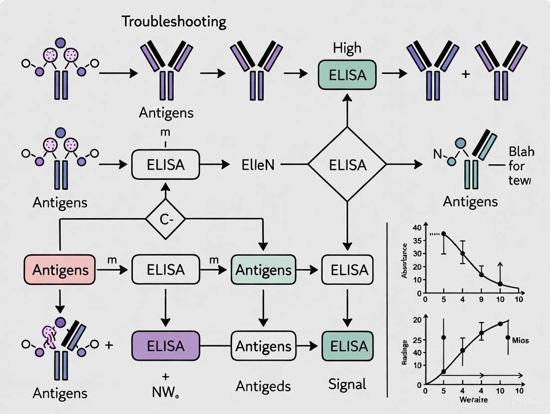
The following diagram outlines a systematic approach to diagnosing and resolving high background issues in ELISA.
The Scientist's Toolkit: Essential Reagents for Background Reduction
Table: Key Reagents for Minimizing ELISA Background
| Reagent Category | Example Products | Function & Application |
|---|---|---|
| Blocking Buffers | StabilGuard, StabilBlock [2] | Coats the well surface to prevent non-specific binding of proteins and antibodies, crucial for reducing background. |
| Assay/Sample Diluents | MatrixGuard (protein-containing), Surmodics Assay Diluent (protein-free) [2] | Dilutes samples and reagents while blocking matrix interferences and heterophilic antibodies (e.g., HAMA) that cause false positives. |
| Wash Buffers | PBS or Tris-based buffers with Tween-20 [7] | Removes unbound reagents during washing steps; detergent helps reduce hydrophobic interactions. |
| Secondary Antibodies | Pre-adsorbed/Secondaries [4] | Antibodies that have been adsorbed against immunoglobulins from multiple species to minimize cross-reactivity and non-specific binding. |
| Validated Components | DIARECT Antigens and Antibodies [2] | Using well-characterized and matched antibody pairs can inherently reduce the risk of cross-reactivity and high background. |
Key Takeaways for Researchers
Successfully troubleshooting high background in ELISA requires a systematic approach that prioritizes the most common culprits: inadequate washing, suboptimal antibody concentrations, and insufficient blocking. Begin by rigorously validating your washing technique and including the correct controls. Methodically titrate all antibodies and consider the sample matrix's role. Utilizing high-quality, specialized reagents designed for blocking and diluting can often provide a straightforward solution to persistent background challenges, ensuring your ELISA data remains robust and reliable [4] [2] [7].
The Critical Impact of Background Noise on Assay Sensitivity and Specificity
In enzyme-linked immunosorbent assay (ELISA) development, background noise is a pivotal factor that directly compromises the sensitivity and specificity of your results. High background signal can obscure true positive results, reduce the dynamic range of your assay, and lead to inaccurate data interpretation. This technical support center provides targeted troubleshooting guides and frequently asked questions to help researchers, scientists, and drug development professionals identify, resolve, and prevent the common causes of high background in ELISA, thereby enhancing the reliability of your experimental outcomes.
Troubleshooting Guide: Common Causes and Solutions for High Background
The following table summarizes the primary causes of high background in ELISA and their respective solutions.
| Cause of High Background | Underlying Reason | Recommended Solution |
|---|---|---|
| Inadequate Blocking [4] [9] | Unsaturated binding sites allow non-specific attachment of detection reagents. | Increase blocking incubation time; try different blocking agents (e.g., BSA, casein, or normal serum from the detection antibody species) [10] [4]. |
| Non-Specific Binding (NSB) [2] | Antibodies (especially secondary) bind to unintended targets like Fc receptors or sample proteins. | Use species-specific pre-adsorbed secondary antibodies; include serum or specific protein blockers in diluents to reduce NSB [2] [4]. |
| Insufficient Washing [4] [9] | Unbound antibodies, proteins, or detection reagents remain in wells, causing false-positive signals. | Increase wash steps and soak duration (e.g., +30 sec); ensure complete buffer removal by tapping plate on absorbent paper; check automated washer for clogs [9]. |
| Antibody Concentration Too High [4] [11] | Excess antibody leads to non-specific binding and increased background noise. | Titrate primary and secondary antibodies to determine optimal, diluted concentration for strong signal with low background [4]. |
| Sample-Related Issues [2] [11] | Contaminants, endotoxins, or interfering substances (e.g., heterophilic antibodies) in complex matrices. | Use clean, purified samples; avoid repeated freeze-thaw cycles; dilute samples in appropriate buffer; use specific diluents to block matrix interferences [2] [11]. |
| Cross-Reactivity [2] [11] | Detection antibody binds to non-target antigens with similar epitopes. | Use highly specific monoclonal antibodies; validate antibodies for minimal cross-reactivity; optimize assay conditions [2] [11]. |
| Substrate or Detection Issues [2] [4] | Innate substrate color; waiting too long to read after stop solution; precipitate formation in wells. | Choose high-quality substrates (e.g., chemiluminescent for high S/N); read plate immediately after stop solution; decrease substrate concentration [2] [10] [4]. |
| Edge Effect [9] | Temperature variation across the plate causes uneven reactions and absorbance in edge wells. | Incubate plates in a temperature-stable environment; use plate sealers during incubation; avoid stacking plates [9]. |
Essential Experimental Protocols for Optimization
Checkerboard Titration for Antibody Optimization
A checkerboard titration is fundamental for optimizing matched antibody pairs in a sandwich ELISA, helping to determine the ideal concentration for both capture and detection antibodies while minimizing background.
- Coat the Plate: Prepare a two-fold dilution series of the capture antibody in coating buffer across the rows of a microplate (e.g., from 10 μg/mL to 0.1 μg/mL). Incubate overnight at 4°C or for 2 hours at 37°C [12].
- Block the Plate: Aspirate the coating solution, wash the plate three times with wash buffer (e.g., PBS with 0.05% Tween-20), and add a blocking buffer (e.g., 1% BSA or 5% non-fat dry milk in PBS). Incubate for 1-2 hours at room temperature [10] [12].
- Add Antigen: Wash the plate as before. Add a fixed, known concentration of the target antigen to all wells. Incubate for 1-2 hours at room temperature [13].
- Detect with Titrated Antibody: Wash the plate. Prepare a two-fold dilution series of the detection antibody down the columns of the same plate. Incubate and wash again [13].
- Add Substrate and Read: Add the enzyme substrate, stop the reaction at the optimal time, and read the plate. The ideal combination is the lowest concentration of capture and detection antibodies that yields the highest signal-to-noise ratio [13].
Blocking Buffer Optimization
An insufficiently blocked plate is a major contributor to high background. This protocol helps identify the most effective blocking agent for your specific assay.
- Coat and Wash: Coat multiple plate wells with your optimized capture antibody concentration and wash.
- Apply Different Blockers: Apply different blocking buffers to separate sets of wells. Common blockers include:
- Continue Standard ELISA: After blocking for 1-2 hours, wash the plates and continue with your standard ELISA procedure, including the addition of sample (or a negative control), detection antibodies, and substrate.
- Analyze Results: Measure the optical density. The optimal blocking buffer is the one that yields the lowest signal in negative control wells (low background) while maintaining a strong positive signal.
Research Reagent Solutions
The following table lists key reagents essential for minimizing background noise and optimizing ELISA performance.
| Reagent Category | Specific Examples | Function & Importance |
|---|---|---|
| Blocking Buffers [2] [10] | BSA, Casein, Normal Serum, Commercial StabilGuard, StabilBlock | Coats all remaining protein-binding sites on the plate after coating, preventing non-specific binding of detection antibodies and sample proteins. |
| Wash Buffers [10] [9] | PBS or Tris-based buffer with 0.05% Tween-20 | Removes unbound reagents and proteins during washing steps. The mild detergent (Tween-20) helps disrupt weak, non-specific interactions. |
| Sample/Assay Diluents [2] [11] | Protein-based (e.g., MatrixGuard) or protein-free commercial diluents | Dilutes samples and reagents in a matrix that reduces non-specific interactions and matrix effects, particularly crucial for complex samples like serum. |
| High-Specificity Antibodies [10] [11] | Monoclonal Antibodies, Pre-adsorbed Secondary Antibodies | Ensures binding is specific to the target epitope only. Pre-adsorbed secondary antibodies are purified to eliminate cross-reactivity with immunoglobulins from other species. |
| Optimized Substrates [2] [10] | TMB (for HRP), PNPP (for AP), Chemiluminescent substrates | Generates the detectable signal. Chemiluminescent substrates often provide a higher signal-to-noise ratio than colorimetric ones. |
FAQs on ELISA Background Noise
Q1: My negative controls are showing a high signal. What is the most likely cause and how can I address it?
Run a control well that omits the primary antibody. If the background remains high, the issue is likely non-specific binding of your secondary antibody. To solve this, ensure your secondary antibody is raised against the correct species and is pre-adsorbed to minimize cross-reactivity. Also, re-optimize your blocking step and secondary antibody dilution [4].
Q2: I've followed the protocol exactly, but I'm still getting high background across the entire plate. What should I check?
First, verify the quality of your water and freshly prepared buffers, as contaminants can cause high background [11]. Second, ensure you are reading the plate immediately after adding the stop solution, as a delay can increase the background signal [2] [4]. Finally, check that your plate reader is properly calibrated.
Q3: What is the "edge effect" and how can I prevent it?
The "edge effect" occurs when the outer wells of a microplate show different absorbance values compared to the inner wells, often due to temperature variation across the plate during incubation. To prevent this, use a temperature-stable incubator for all incubation steps and avoid stacking plates. Using a plate sealer can also help minimize evaporation and temperature differences [9].
Q4: How can sample quality affect my background?
Samples with contaminants like endotoxins, detergents, or lipids can interfere with antibody binding and increase background. Complex sample matrices (e.g., serum, plasma) can also cause non-specific binding. To reduce these effects, ensure samples are clean and properly prepared. Diluting samples in an appropriate assay diluent or pre-treating them can significantly reduce matrix effects [11] [14].
Experimental Workflow for Troubleshooting High Background
The following diagram illustrates a logical, step-by-step workflow for diagnosing and resolving high background issues in your ELISA experiments.
FAQ: What are the primary causes of high background in ELISA?
High background signal in ELISA is most frequently caused by non-specific binding, cross-reactivity, and matrix effects. These issues lead to excessive color development or high optical density readings, which can compromise assay sensitivity and accuracy. The table below summarizes these core problems and their immediate effects.
| Fundamental Cause | Description | Impact on ELISA |
|---|---|---|
| Non-Specific Binding (NSB) | The unintended attachment of antibodies or sample proteins to the solid phase or other assay components, not mediated by the target analyte [2] [15]. | Increases general background noise, leading to false positive signals and a reduced signal-to-noise ratio [2] [16]. |
| Cross-Reactivity | occurs when an antibody binds to non-target proteins or epitopes that share structural similarities with the intended target [2] [15]. | Causes inaccurate quantification and false positives, particularly in complex sample matrices [2]. |
| Matrix Effects | Interference from components within the sample itself (e.g., serum, plasma, or tissue extracts) that can alter antibody binding or signal generation [2] [17]. | Can mask or enhance the true signal, leading to over- or under-estimation of the analyte concentration [17]. |
FAQ: How can I troubleshoot and resolve non-specific binding?
Non-specific binding is often the leading contributor to high background. A systematic approach to troubleshooting and optimization is required to resolve it. The following workflow outlines a sequence of key investigative and corrective actions.
Detailed Corrective Methodologies
Enhance Blocking Efficiency
- Increase blocking concentration or time: If using Bovine Serum Albumin (BSA), try increasing the concentration from 1% to 2-5% or extend the blocking incubation time, potentially with agitation [4] [16].
- Change blocking agents: Switch to an alternative blocking protein such as normal serum (5-10% recommended), casein, or non-fat dry milk. Different blockers may be more effective for specific sample types [4] [15]. Commercial specialized blocking buffers (e.g., StabilGuard, StabilBlock) are formulated for superior performance [2].
- Add a surfactant: Incorporate a non-ionic detergent like Tween 20 at a concentration of 0.05% (v/v) to the blocking buffer to minimize hydrophobic interactions [18].
Optimize Antibody Usage
- Titrate antibodies: The concentration of both capture and detection antibodies may be too high. Perform a checkerboard titration to determine the optimal dilution that provides a strong specific signal with low background. Recommended starting ranges are provided in the table below [19] [17] [18].
- Use affinity-purified antibodies: Affinity-purified antibodies significantly reduce background compared to crude antisera or ascites fluid [19] [18].
- Run a no-primary-antibody control: This control helps identify if the secondary antibody is binding non-specifically [4].
Improve Washing Stringency
- Increase wash volume and frequency: Ensure at least 400 µL of wash buffer is used per well per cycle. Perform a minimum of 3-5 washes between steps, increasing to 6 washes after the enzyme conjugate incubation [5] [18].
- Incorporate soak steps: Add a 30-second soak with wash buffer between aspiration steps to improve dislodging of unbound materials [5] [16].
- Ensure complete drainage: After washing, invert the plate and tap it forcefully onto absorbent tissue to remove any residual fluid [5] [17].
FAQ: How do I minimize cross-reactivity and matrix effects?
Mitigating Cross-Reactivity
Cross-reactivity is an issue of antibody specificity that must be addressed during assay development and validation.
- Antibody Selection and Validation: For sandwich ELISA, use a well-characterized matched antibody pair that recognizes distinct, non-overlapping epitopes on the target antigen [17] [18]. Always validate antibodies for your specific application and sample type.
- Secondary Antibody Specificity: Use secondary antibodies that are pre-adsorbed against the immunoglobulin of the species from which your samples are derived. This minimizes cross-reaction with immunoglobulins that may be present in the sample [4] [15].
- Use Specialized Diluents: Commercial assay diluents (e.g., MatrixGuard) are specifically designed to neutralize interfering substances like heterophilic antibodies (e.g., HAMA), rheumatoid factor, and other serum proteins that cause cross-reactivity [2].
Controlling Matrix Effects
Matrix effects arise from the sample itself and require strategies to isolate the true analyte signal.
- Use Appropriate Standard Diluents: The standard curve must be prepared in a diluent that closely mimics the sample matrix. For serum samples, use a diluent containing a similar concentration of inert serum or a commercial matrix-mimicking diluent [19] [17].
- Perform Spike-and-Recovery and Linearity-of-Dilution Experiments:
- Spike-and-Recovery: Add a known amount of pure analyte to the sample matrix and measure the recovery. Optimal recovery (typically 80-120%) confirms the matrix is not interfering with detection [19] [18].
- Linearity-of-Dilution: Dilute the sample serially and measure the analyte. The measured concentrations should decrease linearly with dilution. Non-linear results indicate matrix interference [19] [17].
- Sample Dilution: Diluting the sample can reduce the concentration of interfering substances below their threshold for causing an effect [2] [17].
Quantitative Data for ELISA Optimization
The following tables provide recommended concentration ranges for key reagents to guide your optimization experiments.
Table 1: Recommended Antibody Concentrations for ELISA Optimization [19] [18]
| Antibody Source | Coating Antibody Concentration (µg/mL) | Detection Antibody Concentration (µg/mL) |
|---|---|---|
| Polyclonal Serum | 5 – 15 | 1 – 10 |
| Crude Ascites | 5 – 15 | 1 – 10 |
| Affinity-Purified Polyclonal | 1 – 12 | 0.5 – 5 |
| Affinity-Purified Monoclonal | 1 – 12 | 0.5 – 5 |
Table 2: Recommended Enzyme Conjugate Concentrations by Detection System [19] [18]
| Enzyme | Detection System | Recommended Concentration |
|---|---|---|
| Horseradish Peroxidase (HRP) | Colorimetric | 20 – 200 ng/mL |
| HRP | Chemiluminescent | 10 – 100 ng/mL |
| Alkaline Phosphatase (AP) | Colorimetric | 100 – 200 ng/mL |
| Alkaline Phosphatase (AP) | Chemiluminescent | 40 – 200 ng/mL |
The Scientist's Toolkit: Key Research Reagent Solutions
This table lists essential reagents and their specific functions in mitigating high background in ELISA.
| Reagent Category | Example Products / Types | Primary Function in Reducing Background |
|---|---|---|
| Blocking Buffers | BSA, Casein, Non-Fat Dry Milk, Normal Serum, Fish Gelatin, Commercial StabilGuard/StabilBlock | Saturate unused binding sites on the plate surface and assay components to prevent non-specific protein adsorption [2] [15] [18]. |
| Sample/Assay Diluents | MatrixGuard (protein-containing), Surmodics Assay Diluent (protein-free) | Neutralize matrix interferences (e.g., heterophilic antibodies) in patient samples, reducing false positives without sacrificing assay sensitivity [2]. |
| Wash Buffers | PBS or TBS with 0.05% Tween-20 | Remove unbound reagents and loosely attached proteins during the washing steps. The detergent helps disrupt hydrophobic interactions [5] [18]. |
| Secondary Antibodies | Pre-adsorbed/Absorbed Secondary Antibodies | Secondary antibodies that have been treated to remove antibodies that cross-react with immunoglobulins from other species, minimizing non-specific signal [4] [2]. |
| Matched Antibody Pairs | Validated capture and detection antibody pairs | Ensure two antibodies bind to distinct epitopes on the same target antigen, providing high specificity and minimizing cross-reactivity in sandwich ELISA [17] [18]. |
ELISA Format Variations and Their Susceptibility to Background Issues
Within the broader research on troubleshooting high background signals in ELISA, it is a well-established thesis that the fundamental architecture of the assay format is a primary determinant of its susceptibility to non-specific binding and high background. Different ELISA formats—direct, indirect, sandwich, and competitive—leverage the antibody-antigen interaction in distinct ways, which inherently introduces unique pathways for background signal generation. Understanding these format-specific vulnerabilities is critical for researchers, scientists, and drug development professionals to effectively diagnose issues and implement targeted corrective protocols, thereby ensuring the accuracy and reliability of their experimental data.
FAQ: ELISA Formats and Background Signal
1. How does the choice of ELISA format influence the source of high background?
The mechanism of detection and the number of immunological reagents required vary by format, which directly correlates to the potential pathways for non-specific binding. Each format has a unique profile of vulnerability [20] [21]. For instance, indirect ELISA requires more reagents and steps, which increases the chance of non-specific binding.
2. Why is background often higher in indirect ELISA compared to direct ELISA?
Indirect ELISA employs a labeled secondary antibody that is reactive against the host species of the primary antibody. This secondary antibody is a common source of high background for two main reasons: it can non-specifically adsorb to the solid phase, or it can cross-react with other proteins in the sample, such as Fc receptors or non-target immunoglobulins [20] [21]. Direct ELISA uses a conjugated primary antibody, eliminating the secondary antibody and thus this major source of non-specificity.
3. What unique background challenges are present in sandwich ELISA?
Sandwich ELISA requires a matched pair of antibodies. A key challenge is avoiding cross-reactivity between the capture and detection antibodies, where they might bind directly to each other instead of to the target antigen, creating a false-positive signal [20]. Furthermore, if the sample contains heterophilic antibodies or other proteins that can bridge the capture and detection antibodies, this can also mimic the target antigen and lead to high background.
4. Are some formats inherently less prone to background?
Yes. While all formats require optimization, the sandwich ELISA is generally recognized for its high specificity because the requirement for two distinct antibodies to bind the antigen simultaneously significantly reduces non-specific detection [21]. However, this is contingent on a well-optimized and validated antibody pair.
5. Can the same troubleshooting strategy be applied to all formats?
While core principles like adequate washing and blocking are universal, the most effective troubleshooting is format-specific. For example, optimizing the blocking buffer for a sandwich ELISA might involve using BSA to avoid cross-reactivity, whereas for an indirect ELISA, it might involve using a serum from a species unrelated to the secondary antibody [20].
Troubleshooting Guide: Format-Specific Background Issues
The table below summarizes the primary sources of background signal for each major ELISA format and provides targeted solutions.
Table 1: Format-Specific Troubleshooting for High Background in ELISA
| ELISA Format | Primary Background Sources | Recommended Optimization Strategies |
|---|---|---|
| Direct ELISA | Non-specific adsorption of the enzyme-labeled primary antibody to the solid phase [20]. | Use 5% non-fat dry milk or 1-3% BSA as a blocking agent. Add 0.05-0.1% Tween-20 to reduce hydrophobic interactions [20]. |
| Indirect ELISA | Non-specific binding of the enzyme-labeled secondary antibody to the solid phase or sample proteins [20] [4]. | Block with 5% non-fat dry milk + 0.05% Tween-20 or 10% normal serum from a species unrelated to the secondary antibody. Avoid serum from the same species as the primary antibody [20]. |
| Sandwich ELISA | Cross-reactivity between capture and detection antibodies; "bridging" by sample proteins mimicking the antigen [20]. | Use high-purity 3-5% BSA as a blocking buffer. Pre-saturate potential cross-reactive sites with an excess of unlabeled detection or irrelevant isotype control antibody [20]. |
| Competitive ELISA | Similar to the base format used (direct, indirect, or sandwich), with additional complexity from the competing antigen or antibody. | Follow troubleshooting for the underlying format. Ensure the competing reagent is pure and at the optimal concentration to effectively compete with the sample analyte. |
Experimental Protocols for Background Troubleshooting
Protocol 1: Systematic Optimization of Washing Conditions
Inadequate washing is a universal contributor to high background, but the optimal stringency can depend on the ELISA format [22] [23].
Methodology:
- Wash Buffer: Use a standard wash buffer such as PBS or Tris-buffered saline (TBS) containing a non-ionic detergent like Tween-20 at concentrations ranging from 0.01% to 0.1% [20] [16].
- Wash Volume and Cycles: A typical wash involves dispensing 300 µL of buffer per well. The number of cycles should be tested systematically.
- Direct ELISA: Start with 3-5 cycles, each lasting 30 seconds to 1 minute [20].
- Indirect ELISA: Due to the higher propensity for secondary antibody binding, increase to 5 cycles of 1-2 minutes each [20].
- Sandwich ELISA: Implement a graded approach: 3 cycles after coating, 4 cycles after antigen incubation, and 5 cycles after detection antibody incubation [20].
- Soak Step: For persistent background, introduce a 30-second soak step where wash buffer is left in the wells before aspiration. This can help dissociate weakly bound materials [23] [6].
- Draining: After the final wash, invert the plate and tap it firmly onto clean absorbent paper to remove any residual liquid [22] [23].
Protocol 2: Format-Specific Blocking Buffer Optimization
The goal of blocking is to cover all unoccupied binding sites on the plate and reagents with non-interfering proteins or molecules [20] [16].
Methodology:
- Prepare Candidate Blocking Buffers: Common agents include:
- Protein-based: 1-5% Bovine Serum Albumin (BSA), 1-10% non-fat dry milk, or 1-10% normal animal serum (e.g., goat, donkey).
- Protein-Detergent Combination: 1-5% BSA or milk with 0.05% Tween-20.
- Blocking Procedure:
- After coating the plate, add 200-300 µL of each candidate blocking buffer to separate wells.
- Incubate for 1 hour at 37°C or overnight at 4°C [20].
- Validation: Proceed with the standard ELISA protocol. The optimal blocking buffer is the one that yields the highest signal-to-noise ratio (specific signal vs. background) for your specific format and antibody set. For example, BSA is often preferred in sandwich ELISAs to avoid potential interference from biotin present in milk [20].
Visualization of Background Pathways and Solutions
The following diagram illustrates the logical relationship between different ELISA formats, their specific background sources, and the primary optimization strategies.
The Scientist's Toolkit: Key Research Reagent Solutions
The table below details essential materials and reagents critical for diagnosing and resolving high background issues in ELISA development.
Table 2: Essential Reagents for ELISA Background Troubleshooting
| Reagent / Material | Function & Rationale | Format-Specific Considerations |
|---|---|---|
| BSA (Bovine Serum Albumin) | A high-purity blocking protein that saturates non-specific binding sites on the plate and reagents. | Preferred in sandwich ELISA to avoid potential cross-reactivity from contaminants in milk. Ideal for phosphorylated-specific antibodies [20]. |
| Non-Fat Dry Milk | A cost-effective blocking agent containing casein and other proteins to prevent non-specific adsorption. | Excellent for direct and indirect ELISAs. Avoid if milk components (e.g., biotin) could interfere with biotin-streptavidin detection systems [20]. |
| Normal Serum | Serum from an unrelated animal species used for blocking. It provides immunoglobulins that occupy Fc receptor binding sites. | Critical for indirect ELISA to prevent secondary antibody cross-reaction. Must be from a species different from the primary and secondary antibodies [20] [4]. |
| Tween-20 | A non-ionic detergent that reduces hydrophobic interactions between proteins and the plastic plate. | Added to wash buffers (0.01-0.1%) and sometimes to blocking buffers. Concentration can be adjusted based on format; higher concentrations (0.1%) may be tested for direct ELISA [20] [16]. |
| Pre-adsorbed Secondary Antibodies | Secondary antibodies that have been purified over immobilized serum proteins from multiple species to remove cross-reactive antibodies. | Highly recommended for indirect and indirect sandwich ELISAs, especially when working with complex sample matrices like serum or plasma, to minimize non-specific binding [4]. |
Key Reagents and Equipment Contributing to Background Signal
High background signal is a common challenge in Enzyme-Linked Immunosorbent Assay (ELISA) that can compromise assay sensitivity and accuracy by reducing the signal-to-noise ratio. This issue stems from various technical factors affecting the specific interaction between target analytes and detection systems. Understanding the key reagents and equipment contributing to background is essential for researchers, scientists, and drug development professionals to optimize ELISA performance and generate reliable data. This guide systematically addresses the primary sources of high background and provides targeted troubleshooting methodologies to resolve these issues effectively.
Key Contributors to High Background and Solutions
The following table summarizes the primary factors contributing to high background signal in ELISA, along with their specific causes and recommended solutions:
| Contributing Factor | Specific Cause of High Background | Recommended Solution |
|---|---|---|
| Antibodies | Non-specific binding; Cross-reactivity; Concentration too high [4] [11] [2] | Optimize antibody concentrations; Use affinity-purified or pre-adsorbed antibodies; Ensure host species compatibility [18] [4]. |
| Blocking Buffer | Inadequate blocking of non-specific sites [4] [11] [16] | Increase blocking buffer concentration or incubation time; Change blocking agent (e.g., BSA, casein, normal serum); Add non-ionic detergent (e.g., 0.05% Tween 20) [18] [16]. |
| Washing Process | Insufficient removal of unbound reagents [4] [5] [6] | Increase wash cycles (e.g., 3-6 washes); Include a soak step (30 seconds); Ensure adequate wash volume (≥300μL/well); Verify plate washer function [18] [5] [6]. |
| Sample & Matrix | Contaminants; Endotoxins; Matrix effects (e.g., serum, plasma) [11] [16] [2] | Dilute samples; Use fresh, properly stored samples; Perform spike-and-recovery experiments [18] [11]. |
| Microplate | Using the wrong plate type for detection method [18] [24] | Use clear plates for colorimetric detection; white plates for chemiluminescence; black plates for fluorescence [18] [24]. |
| Detection System | Enzyme conjugate concentration too high; Substrate over-incubation; Innate substrate color [18] [4] [2] | Titrate enzyme conjugate; Monitor substrate incubation time; Read plate immediately after adding stop solution [18] [4] [2]. |
| Contamination | Contaminated reagents or water; Reused plate sealers [11] [5] [6] | Prepare fresh buffers; Use high-quality water; Use fresh plate sealers for each step [11] [5]. |
Experimental Troubleshooting Workflow
The following diagram outlines a systematic, step-by-step experimental approach to diagnose and resolve high background signal issues in your ELISA assays:
The Researcher's Toolkit: Essential Reagents & Equipment
Successful troubleshooting of high background requires an understanding of the key reagents and equipment involved in ELISA. The following table outlines these critical components and their optimal specifications:
| Toolkit Component | Function & Role in Background | Optimal Specifications & Notes |
|---|---|---|
| Coating Antibody | Binds target antigen to solid phase; high concentrations can cause nonspecific binding [18] [24]. | Affinity-purified; 1-12 μg/mL in PBS or carbonate-bicarbonate buffer (pH 9.4) [18]. |
| Detection Antibody | Binds to captured antigen; must be specific and not cross-react [18] [24]. | Use a "matched pair" with coating antibody; 0.5-5 μg/mL for affinity-purified [18]. |
| Blocking Buffer | Covers unsaturated binding sites on the plate to prevent nonspecific binding [18] [24]. | 5-10% protein (BSA, casein, or serum); often with 0.05% Tween 20 [18] [16]. |
| Wash Buffer | Removes unbound reagents; insufficient washing is a primary cause of high background [4] [5]. | PBS or TBS with 0.05% Tween 20; fresh and uncontaminated [18] [11]. |
| Enzyme Conjugate | Generates detectable signal; over-concentration drastically increases background [18] [4]. | HRP or Alkaline Phosphatase; requires careful titration [18]. |
| Microplate | Solid phase for assay; material and color affect background and signal detection [18] [24]. | Colorimetric: Clear flat-bottom [18] [24]. Chemiluminescent: Opaque white [18] [24]. Fluorescent: Opaque black [18] [24]. |
| Plate Washer | Automated washing; malfunction or improper calibration leads to residual unbound components [16] [5]. | Ensure all ports are clean and dispense/aspirate correctly; calibrate regularly [16] [6]. |
Frequently Asked Questions (FAQs)
1. My entire plate shows a uniformly high signal. What is the most likely cause? The most probable cause is insufficient washing, which leaves unbound enzyme-conjugated detection antibody in the wells. This residual conjugate reacts with the substrate across the entire plate, generating a high, uniform background [5] [6]. Increase the number and volume of wash cycles, ensure complete aspiration between washes, and consider adding a brief soak step (e.g., 30 seconds) to improve removal of unbound reagents [16] [5].
2. I am sure my washing is thorough, but I still have high background. What should I check next? If washing is adequate, the issue likely lies with ineffective blocking or excessive antibody/enzyme-conjugate concentrations [4] [16]. First, try increasing the concentration of your blocking agent (e.g., from 1% to 2% BSA) or extending the blocking incubation time. If the problem persists, perform a chessboard titration to systematically optimize the concentrations of your capture and detection antibodies, as well as the enzyme conjugate [18].
3. How does the sample type contribute to high background? Complex biological matrices like serum, plasma, or cell culture supernatants contain numerous proteins, lipids, and other factors (including heterophilic antibodies) that can bind nonspecifically to assay components [11] [16] [2]. This is known as the "matrix effect." To mitigate this, always dilute samples in an appropriate diluent and run a spike-and-recovery experiment to validate your assay conditions for your specific sample type [18] [11].
4. Why is it critical to use the correct microplate? Using a tissue culture-treated plate instead of an ELISA plate can result in poor and uneven protein binding. Furthermore, using a clear plate for chemiluminescent detection can cause signal "crosstalk" between wells due to light leakage, increasing perceived background. Similarly, using a black plate for a colorimetric readout will result in no signal [18] [24]. Always use a plate designed for ELISA and select the plate color based on your detection method.
Proactive Assay Design and Optimization to Minimize Background
The Enzyme-Linked Immunosorbent Assay (ELISA) is a powerful plate-based technique widely used for detecting and quantifying soluble substances such as peptides, proteins, antibodies, and hormones [25]. Since first described in 1971, ELISA has become a fundamental tool in research and clinical laboratories due to its sensitivity, specificity, and capacity to handle large sample numbers [26].
The success of any ELISA depends on selecting the appropriate format for your experimental goals and understanding the specific requirements of each system. The four main ELISA formats—direct, indirect, sandwich, and competitive—each offer distinct advantages and limitations tailored to different applications, from basic research to clinical diagnostics and environmental monitoring [26].
Figure 1: This flowchart provides a systematic approach for selecting the appropriate ELISA format based on experimental requirements, including antigen size, antibody availability, and sensitivity needs.
Comparative Analysis of ELISA Formats
Technical Characteristics and Applications
Table 1: Comprehensive comparison of the four primary ELISA formats, their methodologies, advantages, and typical applications.
| Format | Basic Principle | Advantages | Disadvantages | Best Applications |
|---|---|---|---|---|
| Direct ELISA | Antigen coated directly on plate; detected with enzyme-conjugated primary antibody [25] [26] | - Quick and easy: fewer steps and reagents- No cross-reactivity from secondary antibody [25] | - Limited signal amplification- Potential immunoreactivity impact from labeling- Fewer commercially available conjugated primaries [25] | - Quick screening tests- Detecting specific proteins in patient serum [26] |
| Indirect ELISA | Antigen coated on plate; detected with unlabeled primary antibody followed by enzyme-conjugated secondary antibody [25] [26] | - Enhanced sensitivity: multiple secondaries bind to each primary- Wide variety of labeled secondary antibodies available- Maximum immunoreactivity of primary antibody [25] | - Potential for cross-reactivity- Additional incubation step required [25] | - Quantifying antibody concentrations- Allergy testing- Immune response monitoring [26] |
| Sandwich ELISA | Antigen captured between two specific antibodies (capture and detection) [25] [26] | - Superior specificity: two antibodies recognize different epitopes- High sensitivity for low-abundance targets- Suitable for complex samples [25] [26] | - Requires matched antibody pairs- More protocol steps- Extensive optimization needed [25] | - Clinical diagnostics- Cytokine detection- Biomarker quantification [26] |
| Competitive ELISA | Sample antigen competes with labeled reference for limited antibody binding sites [25] [26] | - Effective for small antigens with single epitopes- Robust for complex sample matrices | - Challenging to set up- Inverse signal relationship- May require in-house labeling [26] | - Detecting small molecules- Drug testing- Environmental monitoring [26] |
Visual Guide to ELISA Methodologies
Figure 2: Workflow comparison of the four main ELISA formats, highlighting the increasing complexity from direct to sandwich ELISA, and the unique competition step in competitive ELISA.
The Scientist's Toolkit: Essential Research Reagent Solutions
Table 2: Key reagents and materials required for successful ELISA experiments, with specifications and functional roles.
| Reagent/Material | Specifications | Function & Importance |
|---|---|---|
| Microplates | 96-well or 384-well polystyrene plates; clear for colorimetric, black/white for fluorescent/chemiluminescent detection [25] | Solid phase for immobilization; minimum binding capacity of 400 ng/cm² with low CV (<5%) for reproducibility [25] |
| Coating Buffers | Carbonate-bicarbonate buffer (pH 9.6) or PBS (pH 7.4) [27] | Facilitates passive adsorption of proteins to plate surface via hydrophobic interactions [25] |
| Blocking Agents | BSA (1-5%), non-fat dry milk, casein, or normal serum (5-10%) from same species as detection antibody [4] [27] | Saturates unsaturated binding sites to prevent non-specific antibody binding and reduce background [27] |
| Wash Buffers | PBS or Tris-buffered saline with 0.05-0.1% Tween-20 [7] [27] | Removes unbound reagents while maintaining protein stability; detergent reduces hydrophobic interactions |
| Detection Enzymes | Horseradish peroxidase (HRP) or Alkaline phosphatase (AP) [25] | Reporter enzymes conjugated to antibodies for signal generation; choice affects substrate options and sensitivity |
| Substrates | TMB (colorimetric/HRP), PNPP (colorimetric/AP), enhanced chemiluminescent [27] | Enzyme substrates that generate measurable signal (color, light); selection depends on sensitivity needs and instrumentation |
| Stop Solutions | 1N sulfuric acid (for TMB), other acid or base solutions specific to substrate [27] | Terminates enzyme-substrate reaction at defined timepoint for accurate measurement |
Troubleshooting High Background Signal in ELISA
Systematic Approach to Background Reduction
High background signal is one of the most common challenges in ELISA, potentially leading to inaccurate results and reduced assay sensitivity [2]. The following troubleshooting guide addresses the primary causes and evidence-based solutions.
Figure 3: Systematic troubleshooting approach for identifying and resolving high background signals in ELISA, focusing on the four main problem areas and their specific solutions.
Detailed Troubleshooting Guide: Causes and Solutions
Table 3: Comprehensive troubleshooting guide for high background signal in ELISA with specific causes and recommended solutions.
| Problem Category | Specific Cause | Recommended Solution | Preventive Measures |
|---|---|---|---|
| Washing Issues [4] [5] | Insufficient washing between steps | Increase number of washes; add 30-second soak steps; ensure complete buffer removal [5] [6] | Follow standardized washing protocol; calibrate automated plate washers regularly |
| Residual unbound antibodies | Extend washing time; use adequate wash buffer volumes [4] | Implement consistent tapping on absorbent tissue after washing [5] | |
| Blocking Problems [4] [7] | Inadequate blocking concentration or time | Increase blocking agent concentration (5-10% BSA or serum); extend blocking incubation [4] [7] | Use blocking agents matched to detection system (e.g., serum from same species as secondary antibody) [4] |
| Ineffective blocking agent | Switch blocking buffer (e.g., BSA, casein, non-fat dry milk, or commercial blockers) [7] | Test different blocking agents during assay optimization | |
| Antibody-Related Issues [4] [7] | Secondary antibody binding non-specifically | Run control without primary antibody; use secondary antibodies pre-adsorbed against sample immunoglobulins [4] | Use secondary antibodies raised against different species than samples [4] |
| Antibody concentration too high | Further dilute primary and/or secondary antibodies to optimal concentration [4] [7] | Perform antibody titration experiments to determine optimal dilution [4] | |
| Cross-reactivity between antibodies | Ensure capture and detection antibodies are from different host species [25] | Use cross-adsorbed secondary antibodies to remove antibodies with affinity for capture antibody [25] | |
| Reagent & Technical Issues [4] [5] | Substrate preparation or handling | Prepare substrate immediately before use; protect from light [5] [7] | Use fresh substrate solutions; limit light exposure during assay |
| Delay in reading after stop solution | Read plate immediately after adding stop solution [4] [5] | Measure at consistent time points after stopping reaction [4] | |
| Contaminated reagents or plates | Use fresh buffers and consumables for each assay [4] [7] | Use distilled/deionized water for buffers; inspect plates before use |
Frequently Asked Questions (FAQs)
Q1: What are the most effective strategies for reducing non-specific binding in sandwich ELISA? Non-specific binding can be minimized through multiple approaches: (1) Ensure thorough blocking using optimized blocking agents such as 5-10% normal serum from the same species as your detection antibody [4]; (2) Use secondary antibodies that have been pre-adsorbed against the immunoglobulins of your sample species to reduce cross-reactivity [4]; (3) Implement stringent washing with buffers containing detergents like Tween-20 [7]; and (4) Verify that your capture and detection antibody pair recognize distinct epitopes to prevent interference [26].
Q2: How does the choice between direct and indirect detection impact assay sensitivity and background? Indirect detection typically provides greater sensitivity because multiple secondary antibodies can bind to each primary antibody, resulting in signal amplification [25]. However, this format may increase the risk of higher background due to potential cross-reactivity of the secondary antibody [25]. Direct detection eliminates secondary antibody cross-reactivity but offers limited signal amplification and may reduce immunoreactivity of the primary antibody due to the conjugation process [25]. The choice depends on your priority: maximum sensitivity (indirect) versus minimal potential cross-reactivity (direct).
Q3: What specific steps can I take to optimize washing procedures to reduce background? Effective washing is crucial for background reduction. Implement these evidence-based practices: (1) Increase both the number and duration of wash cycles [5] [6]; (2) Incorporate 30-second soak steps between washes to improve removal of unbound reagents [5]; (3) Use wash buffers containing 0.01-0.1% Tween-20 to reduce hydrophobic interactions [7]; (4) Ensure complete removal of wash buffer by inverting plates forcefully onto absorbent tissue between steps [5]; and (5) Regularly maintain and calibrate automated plate washers to ensure consistent performance [6].
Q4: When should I consider using a competitive ELISA format instead of sandwich ELISA? Competitive ELISA is particularly advantageous when: (1) Targeting small molecules or haptens with only single epitopes that cannot accommodate two simultaneous antibodies [26]; (2) Working with antigens that have limited available epitopes [25]; (3) Analyzing complex samples where specific capture is challenging [26]; (4) Only one specific antibody is available for the target analyte [26]. Common applications include drug testing, environmental monitoring, and small molecule detection [26].
Q5: How can I troubleshoot high background that appears specifically in my sample wells but not in controls? Sample-specific background suggests matrix effects or interfering substances. Address this by: (1) Diluting samples to reduce concentration of interfering components [7]; (2) Using specialized sample diluents designed to block matrix interferences while maintaining assay signal [2]; (3) Pre-treating samples to remove contaminants (e.g., centrifugation, filtration, or extraction) [3]; (4) Incorporating heterophilic antibody blocking reagents if human anti-animal antibodies are suspected [2]; and (5) Testing sample recovery by spiking with known analyte concentrations [7].
Optimal Coating and Blocking Strategies to Prevent Non-Specific Binding
Why Do Coating and Blocking Matter?
In ELISA development, high background signal is a frequent challenge that directly compromises assay sensitivity and reliability. This interference is predominantly caused by non-specific binding (NSB), where proteins, antibodies, or other matrix components adhere to surfaces other than the intended binding sites [2]. Effective coating and blocking strategies form the primary defense against NSB, ensuring that the signal generated is specific to the target analyte.
Core Principles and Troubleshooting FAQs
FAQ 1: What are the primary causes of high background due to inadequate blocking?
High background often stems from incomplete blocking, where unsaturated binding sites on the microplate remain available for proteins in the sample or detection reagents to bind non-specifically [2].
- Solution: Ensure the blocking step is performed after plate coating and before adding the sample [28]. Use a sufficient concentration and volume of blocking agent to cover all potential binding sites. If background remains high, consider increasing the blocking agent's concentration, extending the incubation time, or agitating the plate during blocking [16].
FAQ 2: How does the choice of blocking buffer influence non-specific binding?
The composition of the blocking buffer is critical. Different agents are suited to different experimental conditions, and an inappropriate choice can lead to high background.
- Solution: The table below summarizes common blocking agents and their applications.
| Blocking Agent | Typical Working Concentration | Key Advantages | Potential Pitfalls |
|---|---|---|---|
| Bovine Serum Albumin (BSA) [28] | 1-5% (w/v) | Widely available, inexpensive. | Commercial preparations may contain contaminating IgGs or proteases that cause background [28]. |
| Normal Serum [28] | 1-10% (v/v) | Excellent for preventing binding to Fc receptors; use serum from the same species as the detection antibody. | Risk of the primary antibody binding to serum proteins if there is significant epitope identity [28]. |
| Casein | 1-5% (w/v) | Effective, low cost, and can be used in protein-free applications. | Can vary by source and preparation. |
| Non-fat Dry Milk | 1-5% (w/v) | Very effective and inexpensive. | Not suitable for systems involving biotin-streptavidin due to endogenous biotin. May contain IgGs. |
| Commercial Specialty Blockers [2] | As per manufacturer | Often formulated with multiple mechanisms to maximize signal-to-noise; highly consistent. | Can be more expensive than generic options. |
FAQ 3: My plate is well-blocked, but I still get high background. Could the coating process be at fault?
Yes, an suboptimal coating procedure is a common source of non-specific binding.
- Solution:
- Use the Correct Plate: Always use plates designed for ELISA, which have a high protein-binding capacity and low well-to-well variation [28] [12].
- Optimize Coating Conditions: The coating buffer should be protein-free to prevent competition for binding sites [28]. A 0.2 M carbonate/bicarbonate buffer (pH 9.4-9.6) is commonly used, but the optimal pH depends on the isoelectric point of the protein being coated [28].
- Optimize Coating Concentration: Avoid "over-coating" the plate, as this can lead to poorly bound proteins washing away or creating a multi-layered, unstable surface that contributes to background [12]. Use a checkerboard titration to find the optimal concentration for your capture antibody or antigen [29] [30].
FAQ 4: Can my detection antibodies contribute to high background?
Absolutely. Antibody-related cross-reactivity and non-specific binding are major contributors to background signal.
- Solution:
- Use Cross-Adsorbed Secondary Antibodies: When using a sandwich ELISA, ensure your detection antibody is raised in a different host species than your capture antibody. Use secondary antibodies that have been "cross-adsorbed" against the serum proteins of other species to minimize cross-reactivity [28].
- Optimize Antibody Concentrations: Antibody concentrations that are too high can saturate specific sites and force antibodies to bind non-specifically. Titrate all antibodies to find the dilution that provides the best signal-to-noise ratio [4] [29].
- Validate Antibodies: Use antibodies that have been validated for the specific ELISA application to ensure high specificity and affinity for the target [28].
FAQ 5: How critical are washing steps in managing background noise?
Inadequate washing is one of the most frequent causes of high background. Residual unbound antibodies or sample components remaining in the wells will produce a false positive signal [4] [5].
- Solution:
- Follow a Rigorous Washing Protocol: Wash the plate extensively with a buffer (e.g., PBS or TBS containing 0.05 - 0.1% Tween-20) between each step [5] [16].
- Incorporate Soak Steps: For stubborn background, add short soak periods (e.g., 30 seconds) after dispensing the wash buffer to allow for more effective dislodging of unbound material [5] [16].
- Ensure Complete Removal: After washing, invert the plate and tap it forcefully on absorbent tissue to remove any residual fluid [5] [30].
Experimental Optimization Protocol
A systematic, step-wise approach is essential for developing a robust and sensitive ELISA.
Checkerboard Titration for Assay Optimization
This experiment simultaneously optimizes the concentrations of your capture and detection antibodies. The workflow for setting up this experiment is outlined below.
Procedure:
- Prepare a series of dilutions of your capture antibody in a suitable coating buffer. Dispense different concentrations across the plate (e.g., by column).
- After coating and blocking, add a fixed, medium concentration of your target antigen.
- Prepare a series of dilutions of your detection antibody. Add these different concentrations down the plate (e.g., by row).
- Complete the assay with your chosen detection system.
- Analyze the results to identify the combination of capture and detection antibody concentrations that yields the strongest specific signal with the lowest background [29] [30].
Validating the Assay: Spike and Recovery & Linearity of Dilution
Once optimal reagent concentrations are found, validate the assay against matrix effects.
- Spike and Recovery: Add a known quantity of the purified analyte into your sample matrix (e.g., serum, cell culture media) and into a standard diluent. Run the ELISA and calculate the percentage of the spiked analyte recovered from the sample matrix. A recovery of 80-120% generally indicates minimal matrix interference [28] [29].
- Linearity of Dilution (Parallelism): Serially dilute a sample with a known high concentration of the endogenous analyte. The measured concentrations of these dilutions should be proportional to the dilution factor. A loss of linearity indicates interference from the sample matrix [29].
The Scientist's Toolkit: Essential Reagents
| Reagent / Material | Critical Function in Preventing NSB |
|---|---|
| ELISA Microplates [28] [12] | Polystyrene plates with high protein-binding capacity and low well-to-well variation are essential for consistent coating. |
| Coating Buffers [28] | Protein-free buffers (e.g., carbonate-bicarbonate, PBS) stabilize the coating biomolecule and facilitate its passive adsorption to the plate. |
| Blocking Agents (e.g., BSA, Serum) [28] | These proteins or specialty formulations occupy any remaining hydrophobic binding sites on the plastic surface after coating. |
| Wash Buffers [5] [16] | Buffers containing a mild detergent (e.g., PBS with 0.05-0.1% Tween-20) help remove unbound reagents and reduce hydrophobic interactions. |
| Cross-Adsorbed Secondary Antibodies [28] | These antibodies have been purified to remove reactivity against immunoglobulins from other species, drastically reducing cross-reactive background. |
| Specialty Blockers/Diluents [2] | Commercial formulations can provide superior blocking for difficult sample matrices (e.g., serum, plasma) and reduce heterophilic antibody interference. |
Antibody Titration and Validation for Maximum Signal-to-Noise Ratio
Troubleshooting Guides
FAQ: What are the primary causes of high background in ELISA?
High background in ELISA is most frequently caused by non-specific antibody binding, insufficient blocking or washing, and suboptimal reagent concentrations [4] [2].
- Non-specific Binding: This occurs when antibodies bind to unintended targets, such as Fc receptors on cells, or when proteins from the sample bind to the solid phase or coated antibody [2]. Using a secondary antibody raised against a different species than your sample and employing Fc receptor blocking agents can mitigate this [4] [31].
- Insufficient Blocking: If potential binding sites on the plate are not adequately blocked, detection antibodies can bind non-specifically. Increasing the blocking incubation time or changing the blocking agent (e.g., to 5-10% normal serum) is recommended [4] [10].
- Inadequate Washing: Residual unbound antibodies or proteins between steps can produce a false positive signal. Ensure extensive washing with buffer between all steps, and verify that your plate washer is dispensing and aspirating properly [4] [1].
- Excessive Antibody Concentration: Using too high a concentration of primary or secondary antibody is a common cause of high background. Performing antibody titration to find the optimal dilution is crucial [4] [31].
- Substrate-Related Issues: Using a substrate with innate color, waiting too long to read the plate after adding the stop solution, or substrate contamination can all increase background. Use a clear, colorless substrate and read the plate immediately after stopping the reaction [4] [1] [2].
FAQ: Why is antibody titration critical for assay performance?
Antibody titration is the process of determining the reagent concentration that provides the best resolution between a positive signal and the background. It is the first step in any assay optimization to ensure reliable and reproducible results [31].
Using the optimal antibody concentration ensures that all specific binding sites are saturated with minimal antibody excess. If the antibody concentration is too low, the signal will be weak, leading to poor resolution and an underestimation of target expression. If the concentration is too high, it can lead to non-specific binding, reagent waste, and detector overloading, all of which contribute to a poor signal-to-noise ratio [31]. This validation is required for each sample type, reagent clone and lot, and specific staining protocol [31].
FAQ: How can I reduce non-specific binding in my assay?
Non-specific binding (NSB) is a leading cause of high background and false positives.
- Use Blocking Reagents: Incorporate high-quality protein-based blockers like bovine serum albumin (BSA), casein, or commercial formulations (e.g., StabilGuard) to cover unused binding sites on the plate [2] [10].
- Employ Sample/Assay Diluents: Use specialized diluents (e.g., MatrixGuard) designed to block matrix interferences and heterophilic antibodies (like HAMA) in patient samples without sacrificing assay sensitivity [2].
- Optimize Washing: Add a mild detergent like Tween-20 to your wash buffer to disrupt weak, non-specific interactions [10].
- Validate Antibodies: Ensure your antibodies are specific to the intended target. Run controls without the primary antibody to check for secondary antibody non-specificity [4] [31].
Experimental Protocols
Detailed Antibody Titration Protocol
This protocol, adapted from flow cytometry best practices which are directly applicable to ELISA development, provides a method for determining the optimal antibody concentration [31].
Materials:
- Flow Staining Buffer (1x PBS can be substituted for ELISA)
- V-bottom 96-well plate
- Multichannel pipette (15-300 µL)
- Centrifuge with plate adapters
- Paper towels
Methodology:
- Antibody Dilution Preparation:
- Determine the antibody stock concentration from the product sheet.
- Prepare the first dilution in a final volume of 200-300 µL. For antibodies with mg/mL concentrations, a starting point of 1000 ng/test is recommended.
- Perform 2-fold serial dilutions across the plate. Add 150 µL of buffer to all but the first well. In the first well, prepare the initial antibody dilution, then transfer 150 µL to the next well, mix thoroughly, and continue this process across the plate. Discard 150 µL from the final well.
- Store the dilution plate in the dark until ready for use.
Cell (or Antigen) Preparation:
- Resuspend your cells or coat the plate with antigen at a consistent concentration (e.g., 2 × 10^6 cells/mL for cell-based assays).
- Add 100 µL of sample to each titration well, bringing the final volume to 250 µL. Pipette to mix and avoid bubbles.
- Incubate for 20 minutes at room temperature in the dark (or according to your specific protocol).
- Centrifuge the plate at 400 × g for 5 minutes, decant the supernatant, and blot on paper towels.
- Resuspend in 200 µL of staining buffer.
- Repeat the washing steps (centrifugation, decanting, blotting) two more times.
- Store the plate at 4°C in the dark until analysis.
Data Analysis:
- The optimal titer is the concentration that provides the highest signal-to-noise ratio, not merely the strongest positive signal. This is calculated by plotting the fluorescence intensity (or OD for ELISA) of stained samples against negative events on a concentration-response curve [31].
Quantitative Data for Antibody Titration
The table below summarizes the effects of antibody concentration on assay performance, guiding the interpretation of titration experiments [31].
Table 1: Effects of Antibody Concentration on Assay Parameters
| Assay Parameter | Antibody Concentration Too Low | Antibody Concentration Optimal | Antibody Concentration Too High |
|---|---|---|---|
| Signal Strength | Weak, suboptimal | Strong, specific signal | Saturated, potentially off-scale |
| Background Noise | Low | Minimal | High due to non-specific binding |
| Signal-to-Noise Ratio | Low | Maximum | Low |
| Data Resolution | Poor, high variability | High, reliable | Poor, spillover spreading |
| Reproducibility | Low | High | Low |
| Reagent Usage | Inefficient | Efficient | Wasteful |
Signaling Pathways and Workflows
Antibody Titration Optimization Workflow
This diagram outlines the logical workflow for optimizing antibody concentration to achieve the maximum signal-to-noise ratio.
ELISA Non-Specific Binding Pathways
This diagram illustrates the primary causes of and solutions for non-specific binding in ELISA, which directly impacts background noise.
The Scientist's Toolkit
Table 2: Key Research Reagent Solutions for Reducing Background
| Reagent Category | Example Products | Function & Application |
|---|---|---|
| Dried Protein Stabilizers & Blockers | StabilCoat, StabilGuard, StabilBlock [2] | Single-step reagents for stabilizing dried proteins on plates and blocking unused binding sites to maximize signal-to-noise. |
| Sample/Assay Diluents | MatrixGuard (protein-containing), Surmodics Assay Diluent (protein-free) [2] | Used to dilute samples and reagents; effectively block heterophilic antibodies (e.g., HAMA) and other matrix interferences to reduce false positives. |
| Wash Buffer Additives | Tween-20 [10] | A mild detergent added to wash buffers to minimize non-specific binding by disrupting weak hydrophobic interactions. |
| Fc Receptor Blocking Agents | Normal Serum, Monoblock [31] [2] | Prevents secondary antibodies from binding non-specifically to Fc receptors on cells, a major contributor to background. |
| High-Quality Substrates | BioFX Substrates [2] | Colorimetric and chemiluminescent substrates designed for low background and high sensitivity. Ensure the substrate is clear and colorless before use [1]. |
Best Practices in Plate Washing and Buffer Preparation
In ELISA-based research, high background signal is a frequent challenge that directly compromises data integrity and assay sensitivity. This technical support guide addresses this issue by detailing optimized protocols for the two most critical procedural steps: plate washing and buffer preparation. Proper execution of these steps is fundamental to reducing non-specific binding and variability, ensuring the accuracy and reproducibility of your results in drug development and scientific research.
FAQs: Core Principles of Plate Washing and Buffer Preparation
Q1: What is the primary goal of plate washing in ELISA?
The primary goal is to remove unbound reagents, such as excess antibodies or sample proteins, from the microplate wells. Effective washing is crucial for reducing background noise and non-specific binding, thereby improving the signal-to-noise ratio and the overall sensitivity and specificity of the assay [32] [33]. Inadequate washing leaves residual unbound antibodies that can produce a false positive signal, while overly aggressive washing can dissociate the antibody-analyte complex, leading to higher variability and lower sensitivity [32] [4].
Q2: Why is the composition of the wash buffer so important?
Wash buffer composition is critical because it maintains the stability of bound complexes while facilitating the removal of unbound materials. A correctly formulated buffer prevents non-specific electrostatic interactions and avoids osmotic stress. The inclusion of a surfactant like TWEEN 20 is particularly important, as it reduces surface tension and helps displace weakly bound, non-specific proteins from the microplate surface [33]. Using the wash concentrate provided in your specific ELISA kit is essential, as substitutes may negatively impact assay performance [32].
Q3: What is residual volume, and why is it a key parameter?
Residual volume is the small amount of liquid remaining in each well after the final aspiration step of washing. It is a critical parameter because high residual volume dilutes the subsequent substrate or detection reagent, leading to lower signal intensity and increased measurement variability across the plate. For robust ELISA results, the industry standard is to aim for a residual volume of less than 5 µL per well [33]. This is achieved through precise calibration of the aspiration probe's depth and speed.
Troubleshooting Guide: High Background Signal
The following table outlines the most common causes related to washing and buffer preparation that contribute to high background, along with recommended solutions.
Table: Troubleshooting High Background in ELISA
| Problem | Possible Cause | Recommended Solution |
|---|---|---|
| High Background | Inadequate washing [4] [5] | Increase wash cycles and soak time; ensure complete aspiration after each wash [5]. |
| Incorrect wash buffer composition [8] | Use the correct, freshly prepared wash buffer with appropriate surfactant (e.g., TWEEN 20); prepare with clean, deionized water [33] [2]. | |
| High residual volume after washing [33] | Calibrate automated washer aspiration depth to minimize residual volume to <5 µL/well [33]. | |
| Non-specific binding of antibodies [4] | Optimize antibody concentrations; use specific blockers or a secondary antibody that has been pre-adsorbed against the sample species [4] [2]. | |
| Substrate issues [2] | Ensure substrate is not exposed to light prior to use; read the plate immediately after adding the stop solution [2] [5]. |
Experimental Protocols and Methodologies
Protocol for Manual Plate Washing
Manual washing is often recommended to minimize variability and prevent the dissociation of bound complexes, which can occur with aggressive automated washing [32].
Materials:
- Squirt bottle with beveled tip trimmed off
- Low-lint absorbent paper
- Prepared wash buffer
Methodology:
- Discard Liquid: Invert the plate over a sink and rapidly accelerate your arm downward in a smooth motion. Stop abruptly to force the liquid out. Repeat the dumping motion a second time [32].
- Blot and Tap: Immediately blot the inverted plate onto absorbent paper. Move the plate to an unused section and firmly tap it 3 times. Avoid banging too hard, as excessive force can cause variable dissociation of complexes [32].
- Wash: Using the squirt bottle, fill all wells until they are overflowing with wash buffer. As soon as the last well is filled, immediately discard the solution and tap the plate as in steps 1 and 2 [32].
- Repeat and Drain: Repeat the washing procedure 3 more times for a total of 4 washes. For the second and fourth washes, add the solution from the bottom to the top of the plate to equalize dwell time for all wells. After the last wash, let the plate rest upside down for 20 seconds, then tap firmly 4 more times, rotating the plate 180° between each tap [32].
- Final Wipe: Wipe the bottom outside of all wells with clean absorbent paper to remove any remaining liquid. Add substrate immediately to prevent wells from drying out [32].
Protocol for Automated Plate Washer Optimization
Automated washers offer reproducibility and efficiency for high-throughput settings but require careful calibration [33] [34].
Materials:
- Calibrated automated microplate washer
- Filtered wash buffer
Methodology:
- System Preparation: Prime the system to remove air bubbles from the fluidic path. Use clean, filtered wash solutions to prevent clogging and contamination [33] [34].
- Parameter Setting: Program the washer with the following optimized parameters for a standard ELISA [33]:
- Dispense Volume: 300-350 µL per well to ensure complete liquid exchange.
- Soak Time: Incorporate a soak time of 5-30 seconds to help dislodge non-specifically bound material.
- Wash Cycles: Typically 3-6 cycles, sufficient for background reduction.
- Aspiration Depth & Speed: Calibrate the aspiration probe to be as close as possible to the well bottom without touching it, using a slow aspiration speed to minimize bubble formation and achieve a residual volume of <5 µL [33].
- Validation: Regularly validate performance by checking dispensing volume accuracy and aspiration uniformity. Use a dye-dilution method or gravimetric analysis to confirm residual volume [33].
Protocol for Wash Buffer Preparation
A correctly prepared wash buffer is foundational for effective washing.
Materials:
- Wash concentrate (from ELISA kit) or PBS/TBS base
- TWEEN 20 surfactant
- High-purity, deionized or distilled water
- pH meter
- 0.2 µm filter
Methodology:
- Preparation: If using a kit, empty the contents of the wash concentrate into a 1 L bottle and fill to volume with distilled water [32]. If preparing from scratch, prepare a base of PBS or TBS.
- Add Surfactant: Add TWEEN 20 to a final concentration of 0.05% - 0.1% (v/v). This is the critical component for reducing non-specific binding [33].
- Adjust pH: Adjust the buffer to a physiological pH, typically 7.2 - 7.4, and verify with a pH meter [33].
- Filter: Filter the buffer through a 0.2 µm filter to remove particulates that could cause contamination or clog automated washer lines [33]. The use of a slightly warmed buffer (e.g., 25-37°C) can improve the removal of non-specifically bound reagents [33].
Signaling Pathways and Workflows
The following diagram illustrates the logical workflow for diagnosing and resolving high background signals related to the plate washing process.
Diagram: High Background Troubleshooting Workflow
The Scientist's Toolkit: Research Reagent Solutions
Table: Essential Reagents for Optimal ELISA Washing
| Item | Function | Key Considerations |
|---|---|---|
| Wash Buffer Concentrate | Provides the correct salts and buffering agents for maintaining physiological pH and ionic strength. | Always use the solution provided with the kit for guaranteed performance [32]. |
| TWEEN 20 (Polysorbate 20) | Non-ionic detergent that reduces surface tension to displace non-specifically bound proteins [33]. | Final concentration is critical; typically 0.05-0.1% (v/v) [33]. |
| High-Purity Water | Diluent for wash buffers and other reagents. | Prevents contamination from ions or organics; use distilled or deionized water [8] [2]. |
| Blocking Agents (e.g., BSA, Normal Serum) | Proteins used to coat unused binding sites on the well surface, preventing non-specific attachment of antibodies [4]. | Selection depends on the assay; normal serum from the detection antibody species can be effective [4]. |
| Pre-adsorbed Secondary Antibodies | Secondary antibodies that have been adsorbed against immunoglobulins from multiple species to minimize cross-reactivity [4] [2]. | Essential for reducing background when working with complex biological samples like serum or plasma [2]. |
Substrate Selection and Signal Development Control
Frequently Asked Questions (FAQs)
1. How does substrate selection directly influence high background in ELISA?
The chemical properties of your substrate are a primary factor in controlling background. Substrates with innate color or those that have deteriorated will increase background absorbance readings [2]. Furthermore, allowing the enzymatic reaction to proceed for too long by extending the substrate incubation time can lead to excessive signal development, even in wells without the target analyte, resulting in high background [4]. For colorimetric assays, it is critical to read the plate immediately after adding the stop solution, as a delay can cause a change in the developed color and elevate background readings [4] [2].
2. What are the key differences between colorimetric and chemiluminescent substrates when troubleshooting background?
The key difference lies in the mechanism of signal generation and its associated challenges. Colorimetric substrates, such as TMB, produce a soluble colored product. Background can be affected by the substrate's innate color prior to the reaction and turbidity in the well, which scatters light and increases apparent absorbance [2]. Chemiluminescent substrates produce light upon reaction with the enzyme. Background here is more dependent on the specificity of the enzyme-substrate reaction and the presence of contaminants that might cause non-enzymatic luminescence (glow) or rapid flash kinetics. Both types require optimized concentration and incubation time, but the parameters for optimization differ [2].
3. My substrate was prepared correctly, but I still have high background. What other signal-related factors should I investigate?
High background is often multi-factorial. You should investigate these key areas beyond substrate preparation:
- Signal Amplification Systems: If you are using a biotin-streptavidin amplification system, the concentration of your detection conjugate (e.g., streptavidin-HRP) may be too high, leading to excessive signal amplification and high background. Titrate these reagents to find the optimal concentration [4].
- Antibody Concentration: The concentration of your primary or secondary antibody may be too high, leading to non-specific binding and increased background signal. Perform a titration experiment to determine the optimal dilution for your antibodies [4] [7].
- Insufficient Blocking: Inadequate blocking fails to prevent non-specific binding of antibodies to the plate well, which is then detected by your substrate. Consider increasing your blocking incubation time or switching to a more effective blocking agent [4] [7].
Troubleshooting Guide: High Background from Substrate and Signal
The table below summarizes common issues related to substrate and signal development that cause high background, along with specific investigative actions and protocols to resolve them.
| Problem | Possible Cause | Investigation & Action | Experimental Protocol |
|---|---|---|---|
| Excessive Substrate Signal | Substrate concentration too high or incubation too long [4] | Action: Dilute substrate according to manufacturer's instructions; reduce incubation time. Investigation: Perform a substrate incubation time course (e.g., 5, 10, 15 minutes) to find the optimal duration. | Prepare a fresh dilution of substrate. Add to wells and incubate at room temperature, protected from light. Monitor color development and stop the reaction at the determined optimal time point. |
| Non-Specific Signal Amplification | Over-amplification from a biotin-streptavidin system [4] | Action: Titrate the biotinylated secondary antibody and streptavidin-enzyme conjugate. Investigation: Test a series of dilutions for each component to find the combination that yields the best signal-to-noise ratio. | Using a checkerboard titration, test different concentrations of biotinylated antibody against different concentrations of streptavidin-HRP on a plate coated with a known antigen. |
| Innate Substrate Color or Contamination | Substrate is contaminated or has a natural color, increasing baseline OD [1] [2] | Action: Visually inspect substrate before use; it should be clear and colorless (for TMB). Prepare fresh immediately before use. Investigation: Test a new vial of substrate. | Before adding to the plate, inspect the substrate solution in a clean vial. If colored, discard and prepare a fresh solution from a new stock. |
| Delayed Signal Measurement | Color continues to develop after the reaction has been stopped [4] [2] | Action: Read the plate immediately (within 5-30 minutes) after adding the stop solution. Investigation: Read the plate at multiple time points after stopping to observe signal stability. | After adding stop solution, tap the plate gently to mix. Place the plate in the reader and initiate reading immediately, ensuring no delays between wells. |
| Turbidity | Precipitation or turbidity in wells, which increases light scattering and absorbance [2] | Action: Ensure all reagents are fully dissolved and solutions are clear. Centrifuge samples if necessary. Investigation: Visually inspect wells for precipitates or cloudiness before reading. | Before adding the substrate, check the plate for any visible signs of precipitation. If present, decrease substrate concentration or ensure proper washing. |
Substrate Characteristics and Selection Criteria
Selecting the right substrate is a balance between sensitivity requirements and background tolerance. The table below compares general characteristics of common ELISA substrate types.
| Substrate Type | Typical Detection Limit | Dynamic Range | Key Consideration for Background Control |
|---|---|---|---|
| Colorimetric (e.g., TMB) | Moderate | ~2-3 logs | Innate color and turbidity can increase background; requires precise timing [2] [35]. |
| Chemiluminescent | High | ~3-4 logs | Very low background possible; can be susceptible to contamination causing "glow" or "flash" kinetics [2]. |
| Fluorescent | High | ~3-4 logs | Background can be caused by plate autofluorescence or buffer components; requires filter optimization [7]. |
Experimental Protocol: Substrate Optimization
This protocol outlines a systematic approach to optimizing substrate incubation time and concentration to minimize background.
Objective: To determine the optimal substrate incubation time that provides a strong specific signal with minimal background.
Materials:
- Coated, blocked, and antigen-loaded ELISA plate (include positive and negative control wells)
- Prepared detection antibodies (primary and secondary, if applicable)
- Wash buffer
- Substrate solution (e.g., TMB)
- Stop solution (e.g., 1M H₂SO₄ for TMB)
- Plate reader
Method:
- Plate Preparation: Complete all steps of your ELISA protocol up to the substrate addition step.
- Substrate Addition: Add the substrate solution to all wells uniformly and note the start time.
- Time Course Incubation: Do not add stop solution to the entire plate at once. Instead, add stop solution to a set of positive and negative control wells at different time intervals (e.g., 5, 10, 15, 20 minutes). Ensure the reaction is stopped for each set of wells at exactly the predetermined time.
- Measurement: Read the plate absorbance for each set of wells after their respective reactions have been stopped.
- Analysis: Calculate the signal-to-noise ratio (Positive Control OD / Negative Control OD) for each time point. The optimal time is typically at or just before the plateau of the highest signal-to-noise ratio, not necessarily the point of highest raw signal.
Logical Workflow for Substrate-Related Background Troubleshooting
The following diagram illustrates a step-by-step decision process to identify and resolve the root cause of high background related to substrate and signal development.
Diagram Title: Substrate and Signal Background Troubleshooting Pathway
The Scientist's Toolkit: Research Reagent Solutions
The following table lists key reagents essential for controlling background through effective substrate use and signal management.
| Reagent / Material | Function in Background Control | Application Note |
|---|---|---|
| TMB Substrate | Colorimetric HRP substrate for signal generation. | Must be clear and colorless before use; prepare fresh to prevent non-specific color development [1] [2]. |
| Stop Solution | Acidic solution to halt enzyme-substrate reaction. | Allows for signal stabilization; plate must be read immediately after addition to prevent signal drift [4] [5]. |
| Stabilized Signal Amplification Conjugates | Pre-optimized reagents (e.g., Streptavidin-HRP) for sensitive detection. | Using a stabilized, pre-titrated conjugate reduces variability and over-amplification that causes background [2]. |
| High-Quality Water | Base for preparing wash buffers and reagents. | Prevents microbial or ionic contamination that can cause turbidity or non-specific reactions; use distilled or deionized water [1] [2]. |
| Protein-Free Blocking Buffer | Blocks remaining protein-binding sites on the plate. | Reduces non-specific binding of detection antibodies, a common source of background signal, without introducing interfering proteins [2]. |
Systematic Troubleshooting of High Background: A Step-by-Step Diagnostic Guide
Troubleshooting Guide: Resolving High Background in ELISA
A high background signal is a common issue in ELISA that can compromise data accuracy by reducing the assay's signal-to-noise ratio. This guide addresses the primary causes related to washing efficiency and reagent quality, providing targeted solutions for researchers.
| Primary Cause Category | Specific Cause | Recommended Solution |
|---|---|---|
| Washing Efficiency | Incomplete or insufficient washing [4] [2] | Increase wash cycles and volume; ensure thorough aspiration [4] [33]. |
| Inadequate wash buffer composition [33] | Use buffer with surfactant (e.g., 0.05% Tween 20 in PBS) [33]. | |
| High residual volume after washing [33] | Calibrate automated washers to minimize residual volume (<5 µL) [33]. | |
| Reagent Quality | Poor quality water or contaminated reagents [8] [2] [11] | Use clean, sterile ultrapure water; prepare fresh buffers [8] [11]. |
| Antibody concentration too high [4] [11] | Titrate antibodies to optimal concentration; dilute further [4]. | |
| Inadequate blocking [4] [11] | Increase blocking incubation time; optimize blocking agent concentration (e.g., 5-10% normal serum) [4]. | |
| Non-specific antibody binding [4] [2] | Use pre-adsorbed secondary antibodies; include appropriate controls [4]. | |
| Sample & Assay Conditions | Sample contamination or hemolysis [8] [11] | Use non-hemolytic samples; avoid repeated freeze-thaw cycles [8] [11]. |
| Incorrect substrate or prolonged development [4] [2] | Dilute substrate; reduce incubation time; read plate immediately after stop solution [4]. | |
| Improper incubation temperature [8] | Follow kit instructions precisely (often room temperature or 37°C) [8]. |
Detailed Experimental Protocols
Protocol 1: Optimization of Microplate Washing
This protocol is critical for removing unbound reagents and minimizing background caused by non-specific binding [33].
Methodology:
- Buffer Formulation: Prepare a wash buffer consisting of phosphate-buffered saline (PBS) or Tris-buffered saline (TBS) at a physiological pH of 7.2–7.4. Supplement with the non-ionic surfactant Tween 20 at a concentration of 0.05% - 0.1% (v/v) to reduce surface tension and displace weakly bound proteins [33].
- Washing Parameters:
- Volume: Dispense a volume sufficient to completely exchange the liquid in the wells, typically 300-350 µL for a 96-well plate [33].
- Soak Time: Allow a soak time of 5-30 seconds between dispense cycles to help dislodge non-specifically bound materials [33].
- Cycles: Perform 3-5 wash cycles between each assay step. For automated systems, a medium-to-high dispense rate is generally effective for ELISA [33].
- Aspiration and Residual Volume:
- Calibrate the aspiration probe to be as close to the well bottom as possible without touching it to minimize residual volume [33].
- The target residual volume after the final aspiration should be less than 5 µL. High residual volume dilutes detection reagents and increases variability [33].
- Validation: Perform gravimetric analysis (weighing the plate before and after washing) periodically to validate residual volume consistency [33].
Protocol 2: Systematic Assessment of Reagent Quality and Performance
This protocol uses controls and checkerboard titrations to identify reagent-related issues.
Methodology:
- Implement Essential Controls: Include the following controls on every plate to diagnose background and specificity issues [36] [29]:
- Blank Control: Well containing only buffer and substrate. Identifies signal from the substrate or plate itself [36].
- Negative Control: Well containing all reagents except the target analyte (e.g., sample diluent). Measures non-specific binding and background noise [4] [29].
- Secondary Antibody Control: Well containing all reagents except the primary antibody. Confirms the detection antibody is not binding non-specifically [29].
- Checkerboard Titration:
- Coat the plate with a range of capture antibody concentrations (e.g., across columns) [29].
- Use a range of detection antibody concentrations (e.g., down rows) [29].
- After running the assay, analyze the results to identify the combination of antibody concentrations that yields the strongest specific signal with the lowest background [29].
- Validate Water and Buffer Quality:
Logical Workflow Diagram
The Scientist's Toolkit: Essential Research Reagent Solutions
| Reagent / Material | Function | Key Consideration |
|---|---|---|
| Wash Buffer (PBS/TBS + Tween 20) | Removes unbound reagents; surfactant reduces non-specific binding [33]. | Optimal Tween 20 concentration is 0.05-0.1%; prepare fresh with high-purity water [33]. |
| Blocking Agents (BSA, Skim Milk, Normal Serum) | Coats unused plastic surface to prevent non-specific protein adsorption [4] [37]. | Select based on assay compatibility; 5-10% concentration is common; increase time/concentration if background is high [4]. |
| Primary Antibody | Binds specifically to the target analyte. | Concentration is critical; titrate to find optimal dilution that maximizes signal-to-noise [4] [29]. |
| Secondary Antibody (Pre-adsorbed) | Enzyme-conjugated antibody that binds primary antibody for detection. | Use pre-adsorbed against the Ig species of your samples to minimize cross-reactivity [4]. |
| High-Purity Water | Solvent for buffers and reagents. | Contaminated water is a common cause of high background; use distilled, deionized, or ultrapure water [8] [2]. |
| Microplate Washer | Automates and standardizes the washing process. | Calibrate regularly to ensure consistent aspiration depth and low residual volume [33]. |
Frequently Asked Questions (FAQs)
Q1: I have followed the kit protocol exactly, but my background is still high. What should I investigate first? First, verify the performance of your wash buffer and technique. Ensure your wash buffer contains a surfactant like Tween 20 and is prepared fresh with high-quality water. Second, run a full set of controls, including a secondary antibody-only control. If this control shows high signal, it indicates non-specific binding of your detection antibody, and you may need to switch to a pre-adsorbed secondary antibody or optimize your blocking step [4] [2].
Q2: What is the single most critical parameter for effective microplate washing? While multiple factors are important, minimizing residual volume is paramount. A high residual volume after the final wash dilutes the subsequent detection reagents and is a major source of background noise and inter-well variability. For robust ELISA results, the residual volume should be less than 5 µL, achieved by precise calibration of the aspiration probe depth [33].
Q3: How can I determine if my antibodies are the source of high background? Perform a checkerboard titration to find the optimal concentration for both your capture and detection antibodies. Using antibodies at too high a concentration is a frequent cause of non-specific binding and high background [29]. Additionally, include a control well without primary antibody. If this well has a high signal, it confirms the secondary antibody is binding non-specifically, and you should consider a different blocking agent or a more specific secondary antibody [4].
Q4: Can the sample itself cause high background? Yes. Complex sample matrices like serum or plasma can contribute to background through non-specific binding or matrix effects [11]. Hemolyzed samples should be avoided [8]. To mitigate this, pre-treat samples or dilute them in an appropriate diluent to reduce interfering substances. Running a spike-and-recovery experiment can help validate that your sample matrix is not interfering with the assay [29].
Troubleshooting Guides
Guide 1: High Background Signal
A high background signal is a common issue that reduces the assay's signal-to-noise ratio and can mask true positive results [38].
| Possible Cause | Solution | Experimental Verification |
|---|---|---|
| Insufficient Washing | Perform 3-5 wash cycles with a soak step of 30 seconds to 2 minutes between incubations. Ensure complete removal of buffer by firmly inverting the plate onto absorbent paper after each wash [5] [4] [38]. | Compare background levels before and after implementing extended soak steps and thorough plate tapping. |
| Incomplete Blocking | Use an appropriate blocking agent (e.g., 5-10% normal serum, BSA, or non-fat dry milk). Ensure adequate blocking time (at least 1-2 hours; consider overnight at 4°C for stubborn issues) [4] [38]. | Run a control well without antigen but with all subsequent antibodies and substrates. High signal indicates insufficient blocking. |
| Excessive Antibody Concentration | Titrate both primary and secondary antibodies to find the optimal concentration that delivers the best signal-to-noise ratio [4] [39] [38]. | Set up an antibody chessboard titration experiment to systematically test different capture and detection antibody concentrations. |
| Secondary Antibody Cross-Reactivity | Use a secondary antibody that is pre-adsorbed against immunoglobulins from the species of your sample to minimize non-specific binding [4] [40]. | Run a control without the primary antibody. A high signal indicates non-specific binding of the secondary antibody. |
Guide 2: Weak or No Signal
Weak signal can lead to false negatives and is often linked to reagent integrity or assay conditions [39].
| Possible Cause | Solution | Experimental Verification |
|---|---|---|
| Low Antibody Affinity or Activity | Use high-affinity, affinity-purified antibodies. Avoid repeated freeze-thaw cycles by aliquoting antibodies. Confirm reagents are not expired [41] [39] [38]. | Test antibody performance in a known positive control system, such as a Western blot for the target antigen. |
| Insufficient Antibody Concentration | Increase the concentration of the primary or detection antibody. Refer to the datasheet for the recommended range and perform titration if necessary [5] [6] [39]. | Perform an antibody titration curve to find the concentration that yields a strong signal without excessive background. |
| Suboptimal Incubation Conditions | Ensure all reagents are at room temperature before starting the assay. Adhere strictly to recommended incubation times and temperatures; consider extending incubation times for low-abundance targets [5] [39] [38]. | Use a thermometer to verify the temperature of the bench surface, incubator, or reagents. |
| Target Present Below Detection Limit | Concentrate your sample or decrease the dilution factor. Verify the expected concentration of your target and ensure it falls within the dynamic range of the assay [39] [42]. | Spike a known amount of recombinant standard protein into your sample matrix to confirm recovery and detectability. |
Guide 3: Cross-Reactivity and Poor Specificity
Cross-reactivity leads to false positives and compromises data reliability [40].
| Possible Cause | Solution | Experimental Verification |
|---|---|---|
| Use of Non-Specific Antibodies | Use monoclonal antibodies or cross-adsorbed polyclonal antibodies with validated specificity for your target to minimize recognition of off-target proteins [41] [40]. | Check the manufacturer's datasheet for validation data on cross-reactivity. Perform a BLAST search to check for homologous proteins. |
| Inadequate Buffer Conditions | Ensure wash buffers contain a detergent like Tween-20 (e.g., 0.05% in PBS). Optimize salt concentration in washing buffers to reduce non-specific ionic interactions [39] [38]. | Test different wash buffer formulations and compare the signal in negative control wells. |
| Hook Effect (Prozone Effect) | For samples with potentially very high analyte concentrations, test multiple sample dilutions. A signal that decreases with higher dilution indicates the hook effect [6] [40]. | Run a series of sample dilutions (e.g., 1:10, 1:100, 1:1000). The measured concentration should be proportional to the dilution factor. |
Experimental Protocols
Protocol 1: Antibody Titration for Optimal Concentration
This protocol is essential for optimizing any new antibody pair to maximize sensitivity while minimizing background [38] [40].
Workflow:
Materials:
- ELISA microplate [12]
- Capture and detection antibodies
- Coating Buffer (e.g., PBS or carbonate-bicarbonate buffer, pH 9.4) [12]
- Blocking Buffer (e.g., 1-5% BSA or 5-10% serum in PBS) [4] [38]
- Wash Buffer (PBS or TBS with 0.05% Tween-20) [39]
- Enzyme substrate (e.g., TMB for HRP) [43]
Method:
- Coat the plate with a fixed, optimal concentration of the capture antibody overnight at 4°C [12].
- Block the plate with an appropriate blocking buffer for at least 1-2 hours at room temperature [38].
- Prepare a series of dilutions (e.g., from 1:500 to 1:5000) of the detection antibody in the recommended diluent.
- Add the diluted detection antibodies to the wells in duplicate or triplicate.
- Incubate as per standard protocol, wash thoroughly, and add the substrate.
- Measure the signal. The optimal antibody concentration is the one that gives the strongest specific signal with the lowest background (typically the point just before the signal plateaus).
Protocol 2: Verifying Antibody Specificity and Cross-Reactivity
This protocol helps confirm that the signal generated is specific to the target antigen [41] [40].
Workflow:
Materials:
- Coated and blocked ELISA plate
- Primary antibody
- Secondary antibody (cross-adsorbed if necessary)
- Excess purified target antigen (for pre-absorption control)
- Protein with known homology to your target
Method:
- No Primary Antibody Control: Omit the primary antibody to check for non-specific binding of the secondary antibody [4].
- Pre-absorption Control: Pre-incubate the primary antibody with a 5-10 fold molar excess of the purified target antigen for 1 hour before adding it to the well. A significant signal reduction confirms specificity [40].
- Cross-Reactivity Check: If a homologous protein is available, coat a separate well with it. A low signal indicates minimal cross-reactivity.
Frequently Asked Questions (FAQs)
Q1: What is the single most critical step to reduce high background in an ELISA? Thorough and consistent washing is paramount [5] [6] [40]. Incomplete washing leaves unbound antibodies and enzymes in the wells, which continue to react with the substrate and generate a high, non-specific signal. Always include a soak step and ensure the plate is drained completely [38].
Q2: How can I tell if my weak signal is due to a bad antibody versus a low-abundance target? Include a known positive control, such as a recombinant standard of your target protein, in your assay [39]. If the positive control yields a strong signal, the antibodies are likely functional, and the issue lies with your sample (low abundance, degradation, or interference). If the positive control signal is also weak, the problem likely lies with the assay reagents or protocol.
Q3: Why should I use a cross-adsorbed secondary antibody, and when is it necessary? A cross-adsorbed secondary antibody has been purified to remove antibodies that could bind to immunoglobulins from other species [40]. This is crucial when your sample (e.g., serum, plasma, or cell lysate) originates from a species similar to the host of the primary antibody. It prevents the secondary antibody from binding directly to proteins in the sample, which would cause high background [4].
Q4: What is the "Hook Effect," and how can I troubleshoot it? The Hook Effect occurs in sandwich ELISA when the concentration of the target analyte is extremely high. The excess analyte saturates both the capture and detection antibodies, preventing the formation of the "sandwich" complex and leading to a paradoxically low signal [40]. To troubleshoot, simply test a series of sample dilutions. If the measured concentration increases with higher dilution, you have identified the Hook Effect.
The Scientist's Toolkit: Research Reagent Solutions
| Item | Function | Key Consideration |
|---|---|---|
| High-Affinity Antibodies | Strongly bind to the target antigen, improving assay sensitivity and reducing the amount of antibody needed [41]. | Look for antibodies with validated affinity constants. Consistent use enhances reproducibility [41]. |
| Cross-Adsorbed Secondary Antibodies | Minimize non-specific binding to proteins in the sample matrix, which is critical for reducing background in indirect ELISA formats [4] [40]. | Ensure the secondary antibody is adsorbed against the species of your sample. |
| Affinity-Purified Antibodies | Purified to isolate only the antibodies specific to the target antigen, significantly reducing cross-reactivity and background [39] [40]. | Preferred over crude antisera for both capture and detection. |
| Optimized Blocking Buffers | Cover all unsaturated binding sites on the microplate well to prevent non-specific adsorption of assay reagents [4] [38]. | Common agents include BSA, non-fat dry milk, or normal serum. The optimal type may depend on the antibody pair. |
| Buffers with Detergent | Used in wash buffers to help solubilize and remove unbound proteins and reduce non-specific hydrophobic interactions [39]. | PBS or TBS with 0.05% Tween-20 is standard. |
Why Do Sample Issues Cause High Background?
In ELISA, the accuracy of your results is fundamentally dependent on sample quality. Specific sample-related problems—namely hemolysis, contamination, and the presence of endogenous interfering substances—are frequent contributors to high background signals. This elevated background can obscure true positive signals, reduce the signal-to-noise ratio, and compromise the reliability of your data [44] [45]. Understanding and addressing these sample-specific issues is therefore a critical step in robust assay development and troubleshooting.
The logical pathway below outlines a systematic approach to diagnosing and resolving these common sample-related problems.
Troubleshooting Guides & FAQs
Hemolyzed Samples
- Problem: The serum or plasma sample has a red or pink hue, indicating the rupture of red blood cells and the release of hemoglobin.
- Effect on Assay: Hemoglobin possesses peroxidase-like activity. In assays using horseradish peroxidase (HRP) as the enzyme, this can catalyze the substrate independently of the target antigen-antibody reaction, leading to non-specific color development and high background [45].
- Solution:
- Prevention is best: Ensure clean venipuncture and avoid frothing during sample transfer.
- Centrifugation: Re-centrifuge the sample at a high speed (e.g., 10,000 × g for 10 minutes) to pellet cellular debris. Carefully use the clear supernatant for the assay [45].
- Re-draw: If hemolysis is severe, the most reliable solution is to obtain a new sample.
Bacterial Contamination
- Problem: The sample appears turbid or cloudy, indicating microbial growth.
- Effect on Assay: Bacteria can produce endogenous enzymes, including peroxidases and phosphatases, that directly react with the ELISA substrate, causing false-positive signals and high background [45].
- Solution:
- Use sterile tubes and practice aseptic technique during sample collection and handling.
- Store samples at recommended temperatures (e.g., -20°C or -80°C for long-term storage) and avoid repeated freeze-thaw cycles [45] [3].
- If contamination is suspected, the sample should be discarded and a new one collected.
Endogenous Interfering Substances
These are substances naturally present in the sample that can interfere with the immunoassay reaction. The table below summarizes common interferents and their mechanisms.
- Solution Strategies:
- Sample Dilution: Diluting the sample can reduce the concentration of interferents below a critical threshold. This is the simplest first step [6].
- Use of Specialized Diluents: Commercial assay diluents (e.g., those containing proprietary protein mixtures or heterophilic blocking agents) are designed to bind and neutralize interfering substances like HAMA and RF without affecting the specific signal [2].
- Pre-adsorption: For persistent HAI, using an antibody pre-adsorbed against the species of the sample can be effective [4].
- Immunoglobulin Removal: For RF interference, using specific reagent kits to remove rheumatoid factors from the sample prior to testing can be beneficial.
| Interfering Substance | Description | Mechanism of Interference |
|---|---|---|
| Heterophilic Antibodies (HAI) [2] [45] | Human antibodies that can bind animal-derived antibodies (e.g., anti-mouse antibodies). | Bridge capture and detection antibodies even in the absence of the target analyte, causing false positives. |
| Rheumatoid Factor (RF) [45] | An IgM autoantibody that binds to the Fc region of IgG. | Binds to the Fc portion of the capture/detection antibodies, creating a false signal. |
| Complement Factors [45] | Proteins part of the innate immune system. | Can bind to antibody complexes, interfering with the antibody-antigen interaction. |
| Autoantibodies [45] | Antibodies that target self-antigens. | If they bind the target analyte, they can block detection (false negative) or form complexes that are detected (false positive). |
Experimental Protocols for Identification and Resolution
Protocol 1: Sample Pre-treatment and Dilution Linearity Test
This protocol helps determine if interference can be diluted out and establishes the valid dilution range for your samples.
- Prepare Sample Dilutions: Prepare a series of dilutions (e.g., neat, 1:2, 1:5, 1:10) of the problematic sample using the recommended assay diluent [2] [6].
- Run ELISA: Analyze all dilutions in your standard ELISA alongside the appropriate calibrators and controls.
- Data Analysis: Plot the measured concentration against the dilution factor.
- Interpretation:
- Linear Recovery: If the measured concentration decreases proportionally with dilution, the sample is likely free of significant interference at these dilutions. The results from the linear range are valid.
- Non-linear Recovery: If the measured concentration does not decrease as expected (e.g., remains high or drops erratically), non-specific interference is likely still present. Further pre-treatment is needed.
Protocol 2: Spike-and-Recovery Experiment
This test evaluates whether the sample matrix itself is inhibiting or enhancing the assay signal.
- Divide Sample: Split the test sample into two aliquots.
- Spike: Add a known quantity of the purified target analyte (the "spike") to one aliquot. The other aliquot serves as the unspiked baseline.
- Analyze: Measure the concentration in both the spiked and unspiked samples.
- Calculate Recovery:
- % Recovery = (Measured Concentration in Spiked Sample – Measured Concentration in Unspiked Sample) / Known Concentration of Spike × 100
- Interpretation: A recovery value of 80-120% is generally acceptable, indicating minimal matrix interference. Recovery outside this range suggests interference that requires mitigation, such as using a different diluent or further sample purification [6].
The Scientist's Toolkit: Research Reagent Solutions
This table lists key reagents used to mitigate sample-specific high background in ELISA.
| Reagent | Function & Application |
|---|---|
| Specialized Assay Diluents [2] | Formulations containing blockers (proteins, polymers) that neutralize heterophilic antibodies, rheumatoid factor, and other interferents in the sample matrix. |
| Heterophilic Blocking Reagent (HBR) [2] | A specific formulation of non-specific immunoglobulins used to pre-treat samples or included in diluents to prevent HAI. |
| Blocking Buffers (e.g., BSA, Casein, Normal Serum) [4] [46] | Used to coat any remaining protein-binding sites on the plate well after coating, preventing non-specific binding of sample proteins. |
| Pre-adsorbed Secondary Antibodies [4] | Antibodies that have been passed over columns containing serum proteins from other species (e.g., human) to remove any cross-reactive components, reducing background. |
A technical support guide for resolving high background signals in ELISA
This guide addresses how incubation conditions—specifically time, temperature, and agitation—can be optimized to resolve the common and frustrating issue of high background in ELISA, thereby improving the accuracy and reliability of your results for research and diagnostic purposes.
Frequently Asked Questions
How do incubation time and temperature specifically cause high background?
Excessive incubation time or incorrect temperature can lead to high background by promoting non-specific binding. Over-incubation allows antibodies to bind to sites they are not specifically targeted for, while elevated temperatures can increase the rate of these non-specific interactions and may also lead to excessive evaporation of reagents, which concentrates them and exacerbates background issues [47] [11] [38].
What is the recommended starting point for incubation conditions?
For most commercial ELISA kits, the manufacturer's protocol is the definitive source. As a general rule, incubations are often performed at room temperature (around 25°C) or 37°C [8]. Adhering to the prescribed times and temperatures is the first and most crucial step in avoiding high background [5] [11].
Can agitation during incubation reduce high background?
Yes, agitation can promote more uniform binding and prevent the settling of reagents, which contributes to consistent results and can help reduce background. Ensuring consistent conditions across all wells is key to minimizing variability and non-specific signals [38] [46].
If my background is high, should I simply reduce all incubation times?
Not necessarily. While excessively long incubations can increase background, arbitrarily shortening steps can lead to other problems, such as weak or incomplete signal from the target analyte [47]. The optimal approach is a controlled optimization where one parameter is changed at a time to find the best signal-to-noise ratio [38].
Besides incubation, what other factors should I check if I have high background?
Incubation is just one part of the puzzle. A systematic check of other common culprits is essential [4]:
- Washing: Ensure thorough and consistent washing to remove unbound reagents [5] [2].
- Blocking: Verify that your blocking buffer is effective and that incubation was sufficient [38].
- Antibody Concentration: Over-concentrated antibodies are a frequent cause of non-specific binding [4] [11].
- Reagent Quality: Use fresh, properly prepared substrates and buffers made with high-quality water [2] [47].
Troubleshooting Guide: Incubation-Related High Background
The table below outlines common incubation-related problems, their effects on your assay, and the recommended solutions.
| Problem | Effect on Assay | Recommended Solution |
|---|---|---|
| Temperature too high [47] [8] | Increased non-specific binding; potential reagent evaporation [47]. | Use a calibrated incubator; adhere to protocol-specified temperature (commonly RT or 37°C) [47] [46]. |
| Incubation time too long [5] [47] | Increased opportunity for non-specific antibody binding [38]. | Precisely follow protocol incubation times; use a timer [5]. |
| Inconsistent temperature (Edge effects) [5] [38] | Uneven results across the plate; some wells with high background. | Use a well-calibrated, forced-air incubator; avoid stacking plates; use a plate sealer [5] [38]. |
| Evaporation during incubation [47] [38] | Unintended concentration of reagents, leading to high background. | Always cover the plate with a seal during incubations [5] [47]. |
| Insufficient reagent mixing [38] [46] | Uneven concentration, leading to inconsistent binding and variable background. | Gently vortex or invert all reagents before use; consider agitation during incubation [38]. |
Experimental Optimization Protocol
To systematically optimize incubation conditions for your specific assay, follow this detailed methodology. The core strategy is to vary one parameter at a time while keeping others constant to find the optimal signal-to-noise ratio.
1. Define the Test Setup
- Plate Layout: Use a plate layout that includes a high-positive standard, a low-positive sample, and a negative control (blank or sample known to be negative) in replicates (n=3 is recommended) [38].
- Baseline: Start with the manufacturer's recommended protocol or your current standard protocol as the baseline for comparison.
2. Optimize Incubation Time Hold temperature and agitation constant. Test a range of incubation times for the critical steps (e.g., primary and secondary antibody incubation).
- Typical Range: Test times both shorter and longer than the baseline (e.g., -25%, -10%, baseline, +10%, +25%).
- Procedure: Perform the assay with the different incubation times on the same plate to minimize inter-plate variability.
- Measurement: After development, calculate the signal-to-noise ratio for each condition:
(Mean OD of Low-Positive Sample) / (Mean OD of Negative Control). - Goal: Identify the incubation time that yields the highest signal-to-noise ratio, indicating strong specific signal with minimal background [38].
3. Optimize Incubation Temperature Using the optimal time identified in the previous step, now test different incubation temperatures.
- Common Temperatures: Test room temperature (18-25°C), 37°C, and possibly 4°C for overnight incubations [47] [8].
- Procedure: Ensure all reagents are fully equilibrated to the target temperature before starting the incubation. Use a calibrated incubator or water bath for accurate temperature control [46].
- Measurement: Again, calculate and compare the signal-to-noise ratios across the different temperatures [47].
4. Evaluate Agitation Using the optimal time and temperature, test the effect of agitation.
- Options: Compare static incubation against gentle, continuous agitation on an orbital shaker.
- Goal: Agitation should improve the consistency between replicates and may enhance specific binding, leading to a more robust assay [38] [46].
The following diagram illustrates the logical workflow for this sequential optimization experiment.
The Scientist's Toolkit: Research Reagent Solutions
The following table lists key reagents mentioned in this guide that are critical for troubleshooting high background issues related to incubation and beyond.
| Item | Function in Troubleshooting |
|---|---|
| Blocking Buffers (e.g., BSA, Casein, Normal Serum) | Coats the well surface to prevent non-specific binding of antibodies, a primary defense against high background [4] [47] [38]. |
| Wash Buffer with Tween-20 | A non-ionic detergent added to wash buffers helps remove unbound and loosely-bound reagents, reducing background. Fresh preparation is key [11] [46]. |
| High-Affinity, Pre-adsorbed Antibodies | Antibodies purified to remove specific cross-reactivities minimize non-specific binding to proteins in sample matrices [4] [2]. |
| Sample/Assay Diluents | Specialized diluents can block matrix interferences (e.g., heterophilic antibodies) in complex samples like serum, reducing false positives [2]. |
| Fresh Substrate & Stop Solutions | Degraded or contaminated substrate can cause high innate color or precipitate formation. Fresh solutions ensure clean reaction initiation and termination [47] [46]. |
| Calibrated Pipettes & Tips | Ensure accurate and consistent dispensing of all reagents, which is fundamental to achieving reproducible results and a clean background [38] [46]. |
Frequently Asked Questions (FAQs)
1. What is the primary advantage of using a checkerboard titration in ELISA development? The primary advantage is the ability to efficiently optimize two critical assay parameters simultaneously, such as antibody concentration and sample concentration, to find the optimal ratio that delivers a strong signal with low background. This method saves significant time and reagents compared to optimizing one variable at a time [19] [48].
2. My ELISA has a high background even after optimizing antibody concentrations. What else should I investigate? High background is often linked to non-specific binding or inadequate washing [4] [5]. You should review your blocking step, ensure you are using an appropriate blocking agent, and verify your washing procedure is thorough [4] [49]. Also, confirm that your secondary antibody is not binding non-specifically by running a control without the primary antibody [4].
3. What critical controls should be included in a checkerboard titration experiment? Always include a background control (e.g., a well with no detection antibody) to assess non-specific signal [48]. It is also good practice to include controls for non-specific binding of the detection antibody and to ensure your substrate is not contributing to background [29].
4. After checkerboard titration, how do I validate that my new ELISA conditions are reliable? Perform spike-and-recovery and linearity-of-dilution experiments [19] [29]. These tests confirm that the sample matrix does not interfere with detection and that the assay accurately measures the analyte across its intended range [19] [29].
Troubleshooting Guide: High Background Signal
A high background signal is a common challenge in ELISA that can obscure true results. The following table outlines the primary causes and their solutions.
| Possible Cause | Detailed Explanation | Recommended Solution |
|---|---|---|
| Insufficient Washing [4] [5] | Residual unbound antibodies or enzymes remain in the wells, leading to non-specific substrate conversion. | Increase wash cycles and duration; include a soak step; ensure complete drainage after each wash [5] [6]. |
| Non-Specific Antibody Binding [4] [2] | Antibodies, particularly secondary antibodies, bind to sites other than the target antigen, such as Fc receptors or the plate surface. | Use affinity-purified, cross-adsorbed secondary antibodies; optimize antibody concentrations; include a well-blocking step with agents like BSA, casein, or normal serum [4] [2] [49]. |
| Substrate-Related Issues [5] [2] | The substrate may have an innate color, be exposed to light, or be left too long after the stop solution is added. | Protect substrate from light; read the plate immediately after adding the stop solution; ensure the substrate is appropriate for your detection system [5] [2]. |
| Sample Matrix Effects [2] [3] | Components in the sample (e.g., serum, lipids) can interfere, causing non-specific binding or masking detection. | Use a sample diluent that closely matches the standard matrix; perform spike-and-recovery experiments to validate; pre-treat samples to remove interferents if necessary [19] [3]. |
Experimental Protocol: Checkerboard Titration
Checkerboard titration is a powerful method for optimizing key reagents like capture and detection antibodies in a sandwich ELISA. The following diagram illustrates the core workflow.
Detailed Methodology
1. Plate Setup and Coating
- Prepare a series of dilutions for your capture antibody in a suitable coating buffer (e.g., PBS, pH 7.4, or carbonate-bicarbonate, pH 9.5) [49]. A recommended starting range is 1–12 µg/mL for affinity-purified antibodies [19].
- Dispense the different capture antibody concentrations across the columns of a 96-well microplate (e.g., from column 1 to 12) [48].
- Incubate overnight or as required, then wash and block the plate using an optimized blocking buffer [19].
2. Sample and Detection Antibody Titration
- Prepare a series of dilutions for your antigen (sample) [48].
- Prepare a series of dilutions for your detection antibody in standard diluent. A recommended starting range is 0.5–5 µg/mL for affinity-purified antibodies [19].
- Dispense the different antigen concentrations down the rows of the plate (e.g., from row A to H). Then, add the different detection antibody concentrations down the rows as well [19] [48]. All other reagents should be kept constant.
3. Assay Completion and Analysis
- Continue with the remaining ELISA steps, including the addition of enzyme conjugate and substrate, according to your standard protocol [19].
- After stopping the reaction, measure the absorbance.
- Identify the combination of capture and detection antibody concentrations that yields the strongest specific signal with the lowest background (best signal-to-noise ratio) [29] [48]. This optimal condition should be used for subsequent assays.
The Scientist's Toolkit: Key Research Reagent Solutions
The following table details essential reagents and their critical functions in ELISA optimization and troubleshooting.
| Item | Function & Importance in Optimization |
|---|---|
| Coating & Capture Antibody | Immobilizes the target antigen onto the solid phase. Its concentration and binding efficiency are foundational to assay sensitivity and must be optimized [19] [49]. |
| Detection Antibody | Binds to the captured antigen, enabling quantification. Must recognize a different epitope than the capture antibody. Its concentration is critical for signal strength and minimizing background [19]. |
| Blocking Buffers (e.g., BSA, Casein) | Critical for reducing high background. They coat any remaining protein-binding sites on the plate to prevent non-specific attachment of assay components [4] [49]. |
| Sample/Assay Diluents | The matrix used to dilute standards and samples. It must be optimized to match the sample matrix, preventing interference (matrix effects) that can skew results [19] [2]. |
| Wash Buffers | Remove unbound reagents at every step. The composition, volume, number of washes, and inclusion of soak steps are vital for reducing background caused by residual material [5] [49]. |
| Enzyme Conjugates (e.g., HRP, AP) | Conjugated to the detection system (e.g., secondary antibody or streptavidin) to catalyze signal generation. Concentration must be titrated to avoid excessive signal or high background [19]. |
| Substrates (Colorimetric/Chemiluminescent) | Converted by the enzyme to a measurable product. Selection depends on the required sensitivity; innate color or instability can contribute to background [19] [2]. |
Assay Validation and Performance Assessment for Reliable Data Interpretation
FAQ: Understanding and Implementing ELISA Controls
What are the fundamental types of controls needed for a reliable ELISA?
Controls are indispensable in ELISA to validate assay performance, distinguish true signals from background noise, and ensure results are accurate and trustworthy [50] [51]. They can be broadly classified into two groups:
- Positive Controls are samples or reagents that you know will produce a detectable signal. They confirm that all assay components are functioning correctly. Examples include the standard curve, a known positive sample, or a spiked sample [52] [51].
- Negative Controls are characterized by the absence of reagents essential for specific analyte detection. They help estimate the level of non-specific binding and identify its source. Common negative controls include assay buffer blanks and sample matrices guaranteed to be free of the analyte [52] [51].
The table below summarizes the core types of controls, their functions, and their typical composition.
| Control Type | Primary Function | Typical Composition |
|---|---|---|
| Blank | Measures background absorbance from plasticware and buffer [51]. | Empty well or well with assay buffer only [51]. |
| S0 (Zero Standard) | Determines assay background in sandwich/direct ELISA; the signal here defines the lower limit of the standard curve [51]. | All assay reagents added, but zero concentration of the standard analyte [51]. |
| B0 (Maximal Binding) | Determines maximum signal in competitive ELISA; used to calculate the %B/B0 for the standard curve [51]. | All assay reagents added, but zero concentration of the standard analyte [51]. |
| Negative Matrix Control | Identifies false positives from matrix interference in complex samples (e.g., serum, plasma) [51]. | Sample matrix (e.g., naive serum) confirmed to lack the analyte [51]. |
| Spiked Matrix Sample (Positive Matrix Control) | Controls for false negatives and verifies assay accuracy and analyte recovery within a specific sample matrix [52] [51]. | Sample matrix with a known quantity of analyte added ("spiked in") [52] [51]. |
| Secondary Antibody Control | Detects non-specific binding of the detection antibody in sandwich or direct ELISAs [51]. | Well incubated with detection antibody but without the primary or capture antibody [51]. |
How do I use controls to troubleshoot high background signals?
High background signal is a common issue that can lead to false positives or inaccurate quantification [53]. Your controls serve as diagnostic tools to pinpoint the source of the problem. The following workflow outlines a logical troubleshooting path using your control data.
Why is my standard curve poor even when my controls look acceptable?
A poor standard curve (e.g., low signal, poor linearity, high CV) can occur even with acceptable background controls. This often points to issues with the standard itself or the capture phase of the assay [5].
- Cause: Incorrect standard reconstitution or serial dilution. Inaccurate pipetting is a common source of error [5].
- Solution: Double-check calculations and use proper pipetting technique. Ensure the standard is reconstituted in the correct diluent as specified in the kit protocol [53].
- Cause: The capture antibody did not bind properly to the plate. This can happen when developing your own ELISA [5].
- Solution: Confirm you are using an ELISA plate (not a tissue culture plate), dilute the antibody in the recommended coating buffer (e.g., PBS), and ensure correct incubation time and temperature for the coating step [12] [5].
How can I validate that my sample matrix does not interfere with the assay?
Complex sample matrices like serum, plasma, or cell culture supernatants can contain interfering components that cause inaccurate results [53] [51]. The following protocol uses spiked samples and serial dilutions to validate your matrix.
Experiment: Validation of Sample Matrix and Linearity of Dilution [53] [51]
- Preparation: Pool several of your test samples to create a representative pooled sample. Prepare a "spiked" sample by adding a known, moderate concentration of the pure analyte (standard) to a portion of the pooled sample. Another portion serves as the unspiked ("native") pool.
- Dilution Series: Create a series of dilutions (e.g., neat, 1:2, 1:4, 1:8) for both the spiked and unspiked pooled samples using the appropriate assay diluent.
- Analysis: Run the diluted samples in the ELISA alongside your standard curve.
- Interpretation:
- Recovery: In the spiked sample, the measured concentration should be close to the expected concentration (spiked amount + native amount) across dilutions.
- Parallelism: The dilution curve of the native pooled sample should be parallel to the standard curve. A lack of parallelism indicates significant matrix interference [53].
- Linearity: The measured concentrations for both spiked and native samples should decrease linearly with the dilution factor.
The Scientist's Toolkit: Essential Reagents for Control and Validation
| Item | Function in Control Experiments |
|---|---|
| Analyte-Free Matrix | Serves as the negative matrix control and the diluent for preparing spiked positive controls. Crucial for identifying matrix-derived interference [52] [51]. |
| Purified Standard | The known quantity of analyte used to generate the standard curve. It is the primary positive control for assay functionality and quantification [51]. |
| Blocking Buffer (e.g., BSA, Normal Serum) | Used to cover unsaturated binding sites on the microplate well. It is critical for minimizing non-specific binding, a common cause of high background [4] [12]. |
| Pre-adsorbed Secondary Antibody | A secondary antibody that has been adsorbed against immunoglobulins from other species. It reduces cross-reactivity and non-specific binding, especially in sandwich ELISAs [4] [12]. |
| Wash Buffer | Used to remove unbound reagents between each assay step. Thorough and consistent washing is essential for reducing background and improving precision [4] [5]. |
Spike and Recovery Experiments to Evaluate Matrix Interference
FAQ: Understanding Matrix Interference
What is matrix interference, and why is it a problem in ELISA? Matrix interference occurs when substances present in the sample (such as plasma, serum, or cell culture media) interfere with the antibody-antigen binding in an ELISA, leading to inaccurate results [2]. This can cause false positives, false negatives, or generally unreliable data, compromising the assay's validity [2] [16]. The complexity of the sample milieu can lead to greater non-specific binding, which is a common cause of high background signal [16].
How can I tell if my ELISA results are affected by matrix interference? A key indicator is obtaining unexpectedly high or inconsistent recovery rates in a spike and recovery experiment [6]. Furthermore, if you notice high background signal even in your blank or negative control wells, or if the signal-to-noise ratio is low, matrix effects could be a contributing factor [2] [38]. High background severely compresses the dynamic range of detection and reduces assay sensitivity [16].
What are the most common sources of matrix interference? Common sources include heterophilic antibodies (e.g., Human Anti-Mouse Antibodies or HAMA), rheumatoid factors, proteins from hemolyzed or lipemic (fatty) samples, and various other bioactive molecules present in complex biological fluids [2] [38]. The use of animal-derived products for patient treatment has increased the prevalence of some of these interfering antibodies [2].
Can I use the same ELISA protocol for different sample types (e.g., plasma and cell culture media)? Not without validation. Switching sample types can significantly affect your assay because the assay milieu changes, potentially leading to greater non-specific binding and high background [16]. If using a commercial kit, consult the manufacturer on validated sample types. For in-house assays, you may need to re-optimize blocking and washing steps or use specialized diluents for the new matrix [16].
Troubleshooting Guide: High Background from Matrix Interference
Problem: Consistently high background across the plate, including in blanks and negative controls, after switching to a new sample matrix.
Possible Causes and Solutions
| Possible Cause | Recommended Action | Underlying Principle |
|---|---|---|
| Insufficient Blocking | Increase blocking agent concentration (e.g., from 1% to 2% BSA) or extend blocking incubation time [16]. Consider blocking overnight at 4°C [38]. | Blocking agents occupy non-specific binding sites on the plate, preventing sample proteins from binding indiscriminately [4] [7]. |
| Non-Specific Binding | Use a specialized sample/assay diluent designed to reduce matrix interferences [2]. Incorporate non-ionic detergents like Tween-20 (0.01-0.1%) in wash buffers [16] [7]. | Diluents can contain proteins or polymers that neutralize interfering substances, while detergents help wash away unbound proteins [2]. |
| Inadequate Washing | Increase the number of wash cycles. Introduce a 30-second soak step with wash buffer between aspirating cycles to improve the removal of unbound materials [5] [6]. | Thorough washing is critical to remove residual sample matrix components and unbound reagents that contribute to background signal [4] [38]. |
| Sample-Related Interferences | Dilute the sample and re-run the assay [7]. For hemolyzed or lipemic samples, use clean samples or pre-treat samples to remove interferents [38]. | Dilution reduces the concentration of interfering substances. Some interferents, like hemoglobin, can be visually identified and avoided [8] [38]. |
Experimental Protocol: Spike and Recovery
Objective: To quantitatively assess the extent of matrix interference in your sample and confirm the validity of your ELISA results within that specific matrix.
Principle: A known quantity of the pure analyte (the "spike") is added to the sample matrix. The measured concentration is then compared to the expected concentration. The ratio, expressed as a percentage, indicates how much the matrix is enhancing (recovery >100%) or suppressing (recovery <100%) the signal.
Materials:
- Test sample matrix (e.g., pooled normal human serum).
- Standard solution of the analyte at a known, high concentration.
- Assay buffer (the matrix used to reconstitute the standard).
- ELISA kit or reagents for your target.
Procedure:
- Prepare Samples:
- Background Sample: Dilute the test sample matrix in assay buffer as you would for a normal assay. This measures the endogenous level of the analyte.
- Spiked Sample: Dilute the test sample matrix in a solution of the standard, such that the final concentration of the spike is within the assay's dynamic range. The volume of the standard solution added should be small to avoid diluting the matrix.
- Standard in Buffer: Prepare the same concentration of the standard diluted only in assay buffer. This represents the expected value (100% recovery).
- Run ELISA: Analyze all prepared samples in duplicate according to your established ELISA protocol.
- Calculate Recovery:
- First, subtract the average concentration of the Background Sample from the average concentration of the Spiked Sample. This gives the measured concentration of the added spike.
- Then, divide this value by the average concentration of the Standard in Buffer and multiply by 100.
Interpretation of Results: Acceptable recovery typically falls between 80% and 120% [6]. Recovery outside this range suggests significant matrix interference.
The Scientist's Toolkit: Key Research Reagent Solutions
The following reagents are essential for diagnosing and mitigating matrix interference.
| Reagent / Material | Function in Addressing Matrix Interference |
|---|---|
| Specialized Sample/Assay Diluents (e.g., protein-containing or protein-free formulations) | These are specifically designed to block matrix interferences like heterophilic antibodies and rheumatoid factors, dramatically reducing false positives without sacrificing assay sensitivity [2]. |
| Dried Protein Stabilizers & Blockers (e.g., StabilGuard, StabilBlock) | Used after plate coating, these reagents preserve antibody activity and provide superior blocking to reduce non-specific binding, thereby maximizing the signal-to-noise ratio [2]. |
| Wash Buffer with Detergent (PBS with 0.01-0.1% Tween-20) | Critical for removing unbound proteins and matrix components during washing steps. The detergent helps reduce non-specific binding that leads to high background [16] [7]. |
| Blocking Buffers (e.g., BSA, Non-Fat Dry Milk, Casein) | Proteins like BSA occupy any remaining protein-binding sites on the microplate after coating, preventing non-specific attachment of sample components [4] [38]. |
| Pre-adsorbed Secondary Antibodies | These antibodies have been pre-treated to remove reactivity against immunoglobulins from other species, minimizing cross-reactivity and non-specific binding in complex samples [4]. |
Assessing Dilutional Linearity and Parallelism for Sample Compatibility
FAQ: Understanding Sample Compatibility
What are dilutional linearity and parallelism, and why are they important? Dilutional linearity and parallelism are critical validation experiments that determine if your sample matrix (e.g., serum, plasma) is compatible with your ELISA. They ensure that the analyte you are measuring behaves similarly to the standard provided in the kit, guaranteeing that your quantitative results are accurate and reliable. When these parameters are not confirmed, matrix effects can cause inaccurate measurements, which is a common contributor to high background or erroneous signals [54] [55].
How do I know if my sample fails these tests? A failure in dilutional linearity is typically indicated when the calculated concentration of a serially diluted sample does not fall within 80-120% of the expected value after accounting for the dilution factor [54]. For parallelism, a failure is signified by a high percent coefficient of variation (%CV)—often above 20-30%—across the dilutions of a sample with high endogenous analyte, indicating that the dilution curve is not parallel to the standard curve [54].
My sample failed parallelism. What is the next step? A failure in parallelism suggests a significant difference in immunoreactivity between your sample's endogenous analyte and the standard/calibration analyte used in the kit [54]. This can be due to post-translational modifications in your sample or other unspecified matrix effects. The next step is to optimize your sample diluent to better match the standard matrix or to use a different diluent that more closely aligns with your sample's properties [54] [56].
Troubleshooting Guides
Problem: Poor Dilutional Linearity
Potential Causes:
- Matrix Interference: Components in your sample matrix (e.g., salts, detergents, other proteins) are interfering with antibody-analyte binding [54].
- Suboptimal Diluent: The diluent used for your samples does not match the matrix of the kit's standards, causing a mismatch in assay compatibility [55] [56].
- "Hook Effect": In rare cases for samples with extremely high analyte concentrations, a prozone effect can occur, leading to underestimation of concentration [55].
Recommended Solutions:
- Use Kit-Recommended Diluent: Always use the assay-specific diluent provided with the kit or recommended by the manufacturer, as its formulation matches the standard matrix [55].
- Validate New Diluents: If you must use an alternative diluent, validate it by ensuring it produces an OD value similar to the kit's zero standard and demonstrates 95-105% spike-and-recovery [55].
- Optimize Sample Dilution: Find the "optimal dilution" where your sample demonstrates linearity (80-120% recovery). Using a sample concentration that is either too high or too low can lead to inaccuracies [54] [56].
Problem: Failed Parallelism
Potential Causes:
- Immunoreactivity Differences: The antibodies in the ELISA kit have different binding affinities for the endogenous analyte in your sample compared to the purified standard [54].
- Presence of Modifications: The analyte in your sample may have post-translational modifications not present on the standard, altering antibody recognition [54].
- Matrix Effects: Similar to dilutional linearity failures, components in the sample matrix are affecting the immunoreactivity in a dilution-dependent manner.
Recommended Solutions:
- Confirm High-Concentration Samples: Ensure you are starting with a sample that has a high endogenous concentration of the analyte, as parallelism cannot be accurately assessed with samples near the lower limit of detection [54].
- Optimize Assay Conditions: Re-optimize components such as the blocking buffer or sample diluent to reduce non-specific binding and matrix interference [56].
- Consult Manufacturer: Contact the kit manufacturer's technical support, as the issue may be related to a known incompatibility, and they may suggest an alternative kit or protocol.
Experimental Protocols & Data
Protocol 1: Dilutional Linearity Experiment
This test determines if a sample matrix spiked with a known analyte can provide reliable quantification after dilution.
- Spike: Introduce a known quantity of the standard analyte into your sample matrix at a concentration above the upper detection limit of the standard curve [54].
- Serially Dilute: Perform a 1:2 serial dilution of the spiked sample matrix using an appropriate diluent until the predicted concentration reaches the lower limit of the standard curve quantification [54].
- Run ELISA: Analyze the neat and diluted samples alongside the standard curve.
- Calculate and Assess: Calculate the observed concentration and the percent recovery for each dilution.
- % Recovery = (Observed Concentration / Expected Concentration) × 100
- Acceptance Criterion: Recoveries should ideally be within 80-120% of the expected value [54].
Table 1: Example Data for a Dilutional Linearity Experiment
| Dilution | Expected Concentration (pg/ml) | Observed Concentration (pg/ml) | Recovery (%) |
|---|---|---|---|
| Neat | — | 390.8 | — |
| 1:2 | 195.4 | 194.6 | 100% |
| 1:4 | 97.7 | 105.1 | 108% |
| 1:8 | 48.8 | 67.0 | 137% |
| 1:16 | 24.4 | 27.9 | 114% |
| 1:32 | 12.2 | 12.1 | 99% |
In this example, the 1:8 dilution falls outside the acceptable recovery range, indicating poor linearity at that dilution. The optimal dilution for this sample would be 1:16 or higher [54].
Protocol 2: Parallelism Experiment
This test determines if a sample with a high endogenous level of the analyte behaves similarly to the standard curve upon dilution.
- Identify Samples: Select at least 3 samples with high endogenous analyte concentrations that are within the standard curve's quantifiable range [54].
- Serially Dilute: Perform 1:2 serial dilutions of each sample using the sample diluent until the concentration falls below the lower limit of quantification [54].
- Run ELISA: Analyze the neat and diluted samples.
- Calculate and Assess: Determine the mean concentration for each sample with the dilution factor applied. Calculate the %CV between the dilutions.
- Acceptance Criterion: The %CV between dilutions is generally acceptable within 20-30%, though the exact threshold should be determined by the user [54].
Table 2: Example Data for a Parallelism Experiment (Intra-assay Variation)
| Endogenous Corticosterone (pg/mL) | Intra-assay %CV |
|---|---|
| Low (171) | 8.0 |
| Medium (403) | 8.4 |
| High (780) | 6.6 |
This example data shows excellent parallelism, as the %CV across different sample concentrations is low [54].
Workflow Visualization
The Scientist's Toolkit: Key Research Reagent Solutions
Table 3: Essential Reagents for ELISA Sample Compatibility and Troubleshooting
| Item | Function in Troubleshooting | Key Consideration |
|---|---|---|
| Assay-Specific Diluent [55] | Dilutes samples to match the standard matrix, minimizing dilutional artifacts. | Using the manufacturer's recommended diluent is crucial for accurate recovery. |
| Protein Stabilizers & Blockers (e.g., StabilGuard, BSA, Casein) [2] | Blocks unoccupied sites on the ELISA plate to reduce non-specific binding (NSB) and high background. | The choice of blocker can affect both signal and background; optimization may be needed. |
| Sample/Assay Diluents (e.g., MatrixGuard, Protein-Free Diluent) [2] | Formulated to block matrix interferences (e.g., heterophilic antibodies) that cause false positives or high background. | Protein-containing and protein-free options offer different blocking mechanisms for various sample types. |
| Wash Buffer | Removes unbound reagents and sample matrix components between assay steps. | Inadequate washing is a primary cause of high background and poor reproducibility [4] [5] [2]. |
| Colorimetric Substrate (e.g., TMB) | Enzyme substrate that produces a measurable color change. | Contamination or using a substrate with innate color can increase background. Always use a fresh, clear solution [2] [47]. |
Inter-Assay and Intra-Assay Reproducibility Analysis
Core Concepts and Quantitative Standards
Inter-assay and intra-assay reproducibility describe the precision of an ELISA when measurements are made across multiple independent runs or within a single run, respectively. They are crucial for confirming that experimental results are consistent and reliable over time and across laboratory conditions [57].
These precision measurements are typically expressed as the Coefficient of Variation (%CV), which is calculated as (Standard Deviation / Mean) x 100 [58] [57]. The generally accepted performance thresholds for these metrics are summarized in the table below.
Table 1: Acceptable CV Ranges for ELISA Reproducibility
| Precision Type | Description | Acceptable %CV Range | Measurement Context |
|---|---|---|---|
| Intra-Assay | Precision within a single assay run (plate-to-plate) | < 10% to 15% [58] [57] | Measurement of multiple replicates (N≥10) of the same sample on one plate [57]. |
| Inter-Assay | Precision between multiple independent runs (day-to-day) | < 15% to 20% [58] [57] | Measurement of the same sample (in triplicate) over at least 3 different days [57]. |
Experimental Protocols for Determining Reproducibility
Protocol 1: Determining Intra-Assay Precision
This protocol evaluates how consistent results are across the wells of a single microtiter plate.
- Sample Preparation: Select a sample with a known analyte concentration within the quantitative range of the assay, ideally in the middle of the standard curve [57].
- Plate Setup: Add this sample to a minimum of 10 replicate wells on the ELISA plate [57].
- Assay Execution: Run the complete ELISA procedure according to the kit or established protocol.
- Data Analysis: Calculate the mean concentration, standard deviation, and %CV for the replicate wells. A %CV below 10-15% indicates good intra-assay precision [57].
Protocol 2: Determining Inter-Assay Precision
This protocol evaluates how consistent results are when the assay is performed on different days.
- Sample Preparation: Prepare multiple aliquots of several samples (e.g., two low-concentration, one mid-concentration, and two high-concentration samples) that span the range of the standard curve [57].
- Assay Execution: On three separate days, run a complete ELISA for each of the samples in triplicate [57]. Ensure all reagents are at room temperature before use and adhere strictly to the same incubation times and temperatures for each run [5].
- Data Analysis: For each sample, calculate the mean concentration, standard deviation, and %CV from the results gathered across the different days. A %CV below 15-20% for each sample indicates acceptable inter-assay precision [57].
Troubleshooting High Background and Poor Reproducibility
High background signal is a frequent issue that can severely compromise the precision and reproducibility of your ELISA data. The following guide addresses common causes and solutions.
Diagram 1: High background causes and solutions.
Frequently Asked Questions (FAQs)
Q1: My inter-assay %CV is consistently high, but my intra-assay CV is fine. What does this indicate? This typically points to variability introduced between assay runs [57]. Common causes include:
- Inconsistent reagent preparation: Check pipette calibration and dilution calculations [5] [58].
- Variations in incubation temperature or time: Use a calibrated incubator and strictly adhere to timed steps [5].
- Using reagents from different lots or that have degraded over time: Ensure consistent reagent lots and proper storage [5].
Q2: How does high background specifically affect reproducibility metrics like %CV? High background signal reduces the signal-to-noise ratio, making it difficult to distinguish true positive signals from background. This compression of the dynamic range leads to greater variability and imprecision in sample measurements, ultimately increasing the calculated %CV [2].
Q3: My washing technique is careful, but I still get high background and poor duplicates. What else should I check?
- Operator technique and instrumentation: Poorly calibrated pipets or low-quality pipet tips can adversely affect precision. Have a second analyst perform the assay with different equipment to isolate the variable [58].
- Plate reader issues: A failing light source or other component can cause intermittent variability. Check your instrument's performance and use dual-wavelength readings to correct for imperfections [58].
- Contamination: Trace contamination of reagents with concentrated sources of the analyte (e.g., from upstream samples) can cause high background and inconsistent results. Perform the ELISA in a dedicated, clean area [58].
Q4: Are there specific reagents that can help reduce background? Yes, using specialized immunoassay reagents can be highly effective:
- Blocking buffers: Formulations like StabilGuard and StabilBlock are designed to reduce non-specific binding more effectively than standard buffers [2].
- Sample/assay diluents: Products like MatrixGuard are specifically designed to block matrix interferences and heterophilic antibodies (e.g., HAMA), which are common causes of false positives and high background [2].
The Scientist's Toolkit: Key Research Reagent Solutions
Table 2: Essential Reagents for Optimizing ELISA Reproducibility
| Reagent Type | Function | Example Products |
|---|---|---|
| Dried Protein Stabilizers & Blockers | Preserve antibody activity on coated plates and block remaining binding sites to minimize non-specific binding in a single step. | StabilCoat, StabilGuard, StabilBlock [2] |
| Sample/Assay Diluents | Dilute samples while simultaneously blocking interfering substances (e.g., heterophilic antibodies) present in sample matrices to reduce false positives. | MatrixGuard Diluent, Surmodics Assay Diluent (Protein-Free) [2] |
| Specialized Substrates | Provide sensitive detection with low innate color and stable signal generation, which is crucial for achieving a high signal-to-noise ratio. | BioFX TMB Substrates [2] |
| Quality Wash Buffers | Effectively remove unbound reagents during washing steps without disrupting antibody-antigen bonds. Critical for minimizing background. | Various manufacturer-specific wash buffers [2] |
Diagram 2: ELISA reproducibility workflow.
Comparative Analysis of Troubleshooting Solutions Across Different Sample Types
FAQ: What are the universal, sample-agnostic causes of high background in ELISA?
High background in ELISA can be caused by several fundamental issues that apply regardless of your sample type. The most common universal causes and their solutions are summarized in the table below.
Table 1: Universal Troubleshooting Solutions for High Background in ELISA
| Cause of High Background | Recommended Solution |
|---|---|
| Insufficient Washing [4] [6] [5] | Increase the number of wash cycles. Incorporate a 30-second soak step during washing. Ensure wells are thoroughly drained after each wash [6] [5]. |
| Inadequate Blocking [4] [7] [38] | Increase the concentration of your blocking agent or extend the blocking incubation time. Consider switching to a more effective blocking buffer (e.g., BSA, casein, or serum-based blockers) [7] [38]. |
| Excessive Antibody Concentration [4] [7] [11] | Titrate both your primary and secondary antibodies to determine the optimal concentration that provides the best signal-to-noise ratio [4] [7]. |
| Contaminated Reagents [3] [8] [11] | Prepare fresh buffers and washing solutions using high-quality, deionized, or distilled water. Avoid reagent contamination [3] [8]. |
| Substrate Issues [4] [2] [38] | Protect substrate from light and prepare it immediately before use. Read the plate immediately after adding the stop solution to prevent post-reaction color development [4] [2]. |
| Non-specific Antibody Binding [4] [2] [59] | Use a secondary antibody that is pre-adsorbed against the immunoglobulins of the species in your sample. Ensure it is raised in a different species than your sample [4]. |
| Improper Plate Sealing [6] [5] [59] | Use a fresh plate sealer during every incubation step to prevent evaporation and contamination, which can cause edge effects and high background [6] [5]. |
FAQ: How do troubleshooting strategies differ when dealing with complex biological samples like serum or plasma?
Complex biological samples such as serum and plasma present unique challenges due to their intricate composition. The matrix effects—interference caused by other components in the sample—are a primary contributor to high background in these sample types.
Table 2: Sample-Specific Troubleshooting for Complex Biological Matrices
| Sample Type | Specific Challenge | Tailored Troubleshooting Strategy |
|---|---|---|
| Serum & Plasma | Matrix Effects: Presence of heterophilic antibodies (e.g., HAMA), rheumatoid factors (RF), lipids, and other interfering proteins that cause non-specific binding [2] [59] [11]. | - Sample Dilution: Dilute samples at least 1:2 in an appropriate diluent, or perform a series of dilutions to check for recovery [6] [11].- Specialized Blockers: Use commercial diluents or blocking agents specifically formulated to neutralize heterophilic antibody interference [2] [59].- Sample Pre-treatment: For lipemic or hemolyzed samples, consider centrifugation or filtration to remove interfering substances [38]. |
| Cell Culture Supernatant | High Protein Content & Media Components: Culture media often contain phenol red, high levels of bovine immunoglobulins, or other proteins that can cross-react or increase non-specific binding. | - Increased Blocking: Use a robust blocking buffer containing protein (e.g., 5-10% normal serum) and a small amount of non-ionic detergent like Tween-20 [7] [11].- Sample Clarification: Centrifuge samples to remove any cells or debris before analysis [38]. |
| Complex Tissue Lysates | Cellular Debris & Abundant Non-Target Proteins: Lysates can be viscous and contain proteases, lipids, and a high concentration of non-target proteins. | - Optimal Dilution: Determine the optimal protein concentration for the assay through a dilution series [38].- Enhanced Washing: Add Tween-20 (e.g., 0.01-0.1%) to the wash buffer to reduce non-specific binding more effectively [7].- Protease Inhibition: Ensure lysis buffers contain protease inhibitors to prevent antibody degradation [38]. |
The following workflow provides a systematic approach to diagnosing and resolving high background, integrating both universal and sample-specific strategies.
FAQ: What experimental protocols are critical for systematically diagnosing high background?
A methodical approach is required to isolate the specific factor causing high background. The following diagnostic protocols are essential.
Protocol 1: The Antibody Titration and Control Experiment
Purpose: To determine if high background is caused by non-specific antibody binding or excessive antibody concentration [4] [7].
Methodology:
- Prepare Control Wells: Include control wells without the primary antibody (secondary antibody only) and wells without any sample (assay background). For sandwich ELISAs, also include wells without the detection antibody [4].
- Titrate Antibodies: Prepare a series of dilutions for both the primary and secondary antibodies. A standard starting range is 1:500 to 1:5000, but this should be guided by the manufacturer's datasheet [4] [38].
- Run the Assay: Process the plate with these different conditions.
- Analyze Results: The optimal antibody concentration is the one that yields the highest signal for the positive control while maintaining a low signal (OD close to zero) in the no-primary-antibody control [7].
Protocol 2: The Blocking Buffer Optimization Experiment
Purpose: To evaluate and identify the most effective blocking agent for your specific assay and sample type [7] [38].
Methodology:
- Select Blocking Agents: Choose 2-3 common blocking buffers for testing (e.g., 1-5% BSA, 5-10% non-fat dry milk, 1-5% normal serum from the same species as the secondary antibody, or commercial specialty blockers) [4] [38].
- Prepare Plates: Coat plates with your capture protein/antibody as usual.
- Apply Blockers: Block different sets of wells with the different blocking agents. Include a no-blocking control. Incubate for 1-2 hours at room temperature or overnight at 4°C for maximum effectiveness [38].
- Run the Assay: Continue with the standard assay protocol.
- Analyze Results: The best blocking buffer will show the lowest background in negative control and sample wells while preserving a strong positive control signal.
Protocol 3: The Sample Matrix Interference Test
Purpose: To determine if components within the sample itself are causing non-specific binding and high background [6] [11].
Methodology:
- Prepare Spiked Samples: Spike a known concentration of the target analyte into the standard diluent buffer. Also, spike the same concentration into a pool of your sample matrix (e.g., serum from a naive subject).
- Prepare Dilutions: Create a dilution series of both the buffer-spiked and matrix-spiked samples.
- Run the Assay: Analyze all samples in duplicate.
- Analyze Results (Parallelism): Plot the observed concentration against the dilution factor for both the buffer and matrix spikes. If the lines are parallel, matrix interference is minimal. Non-parallel lines or significant differences in recovery indicate strong matrix interference, necessitating sample dilution or pre-treatment [6].
FAQ: What key reagent solutions are essential for mitigating high background?
The right selection of reagents is a powerful tool for preventing high background. The following table details essential reagents and their functions.
Table 3: Research Reagent Solutions for High Background Mitigation
| Reagent Category | Specific Examples | Function in Reducing Background |
|---|---|---|
| Specialized Blocking Buffers [2] [59] [38] | StabilGuard, StabilBlock, Casein, BSA, Non-fat Dry Milk | Coats the well surface to occupy any remaining protein-binding sites after coating, thereby preventing non-specific attachment of antibodies or sample proteins [2] [38]. |
| Matrix-Blocking Diluents [2] [59] [11] | MatrixGuard, Surmodics Assay Diluent (Protein-Free) | Specifically formulated to be used for diluting samples. They contain components that neutralize interfering substances like heterophilic antibodies (HAMA), rheumatoid factor, and other matrix effects found in complex samples [2] [59]. |
| Pre-adsorbed Secondary Antibodies [4] [7] | Anti-Rabbit IgG (Pre-adsorbed vs. Human, Mouse, Bovine) | These antibodies have been cross-adsorbed against immunoglobulins from other species to minimize cross-reactivity, which is a major source of non-specific signal, especially in species-rich samples [4]. |
| High-Quality Wash Buffers [7] [11] | PBS or Tris Buffer with 0.01-0.1% Tween-20 | The buffer salt maintains pH and ionic strength, while the detergent (Tween-20) helps to solubilize and wash away unbound proteins and reagents that contribute to background [7]. |
| Stable Substrate Systems [2] [59] | BioFX TMB Substrates | Provide a clear, colorless solution before use (low innate color) and consistent performance to prevent precipitate formation or non-enzymatic development that increases background [2] [59]. |
FAQ: How can I ensure consistent, low-background results across multiple assays?
Achieving reproducibility requires strict adherence to standardized protocols and environmental control. Key strategies include:
- Standardized Washing: Use an automated plate washer where possible. If washing manually, ensure consistency in filling, soaking, draining, and tapping force across all wells and all assays [6] [38].
- Reagent Consistency: Use fresh reagents and buffers for each assay. Do not reuse plate sealers or reagent reservoirs, as they can harbor contaminating enzymes (e.g., residual HRP) that cause high background [6] [5].
- Environmental Control: Perform all incubations at a consistent temperature, using a calibrated incubator to avoid edge effects caused by temperature gradients across the plate [5] [7] [38].
- Proper Technique: Allow all reagents to equilibrate to room temperature before starting the assay to prevent condensation and ensure uniform binding kinetics [5] [38]. Use calibrated pipettes and ensure all operators are trained on a single, detailed Standard Operating Procedure (SOP) [38].
Conclusion
Effectively managing high background in ELISA requires a holistic approach that integrates foundational knowledge, proactive assay design, systematic troubleshooting, and rigorous validation. By understanding the core principles behind background signal and implementing the methodological and optimization strategies outlined, researchers can significantly enhance assay performance, data reliability, and reproducibility. The consistent application of these practices is paramount for advancing drug development, ensuring accurate diagnostic test results, and generating high-quality research data. Future directions will involve the development of more specific blocking reagents, advanced substrate systems with lower innate background, and integrated digital tools for real-time troubleshooting guidance, further solidifying ELISA's role as a cornerstone technique in biomedical science.
References
- 1. ELISA Troubleshooting: High Background [https://www.sinobiological.com/category/elisa-troubleshooting-high-background]
- 2. What Causes High Background in ELISA Tests? [https://shop.surmodics.com/elisa-test-results-high-background-causes]
- 3. How to troubleshoot if the Elisa Kit has high background? [https://www.jg-biotech.com/blog/how-to-troubleshoot-if-the-elisa-kit-has-high-background-191623.html]
- 4. How to deal with high background in ELISA [https://www.abcam.com/en-us/technical-resources/troubleshooting/high-background-elisa]
- 5. ELISA Troubleshooting Guide [https://www.thermofisher.com/us/en/home/life-science/protein-biology/protein-biology-learning-center/protein-biology-resource-library/pierce-protein-methods/overview-elisa/elisa-troubleshooting-guide.html]
- 6. Troubleshooting Guide: ELISA [https://www.rndsystems.com/resources/troubleshooting-guide-elisa-development]
- 7. ELISA Guide; Part 4: Troubleshooting [https://www.jacksonimmuno.com/secondary-antibody-resource/immuno-techniques/elisa-guide-part-4-troubleshooting/]
- 8. 5 - High Background in ELISA? Causes and Solutions for ... [https://www.multisciences.net/high-background-in-elisa-causes-and-solutions-for-optimal-results-7487.html]
- 9. ELISA Troubleshooting Guide [https://blog.abclonal.com/blog/elisa-troubleshooting-tips]
- 10. How to Reduce Background Noise in ELISA Assays [https://synapse.patsnap.com/article/how-to-reduce-background-noise-in-elisa-assays]
- 11. Causes Of High Background In ELISA Tests and How to ... [https://www.caltagmedsystems.co.uk/information/causes-of-high-background-in-elisa-tests-and-how-to-solve-them/]
- 12. ELISA Assay Technique | Thermo Fisher Scientific - US [https://www.thermofisher.com/us/en/home/life-science/protein-biology/protein-biology-learning-center/protein-biology-resource-library/pierce-protein-methods/overview-elisa.html]
- 13. ELISA Assay Challenges | MyBioSource Learning Center [https://www.mybiosource.com/learn/troubleshooting-elisa-assay-challenges]
- 14. Troubleshooting common problems during ELISA ... [https://www.mybiosource.com/learn/troubleshooting-common-problems-during-elisa-experiments/]
- 15. Non-Specific Binding and Cross-Reaction of ELISA [https://pmc.ncbi.nlm.nih.gov/articles/PMC8394222/]
- 16. Elisa troubleshooting tips – High background - Blog [https://www.arp1.com/blog/post/elisa-troubleshooting-tips-high-background.html]
- 17. How To Optimize Your ELISA Experiments [https://www.ptglab.com/news/blog/how-to-optimize-your-elisa-experiments/?srsltid=AfmBOopuPuJOHwnLwbsMenDZYF9RR7Z5klg9vh4Bc8FOcf38sxb7n6LP]
- 18. Factors Affecting Signal Generation in ELISA [https://www.thermofisher.com/us/en/home/life-science/protein-biology/protein-biology-learning-center/protein-biology-resource-library/pierce-protein-methods/overview-elisa/factors-affecting-ELISA-signal.html]
- 19. ELISA Development and Optimization [https://www.thermofisher.com/us/en/home/life-science/protein-biology/protein-biology-learning-center/protein-biology-resource-library/pierce-protein-methods/overview-elisa/elisa-development-optimization.html]
- 20. 如何针对性优化封闭液成分、洗涤条件以降低非特异性结合? [https://www.cusabio.cn/hotspots-test-kit/14663.html]
- 21. antibodies.com/applications/ elisa [https://www.antibodies.com/applications/elisa]
- 22. ELISA 常见问题指南 [https://www.thermofisher.cn/cn/zh/home/life-science/protein-biology/protein-biology-learning-center/protein-biology-resource-library/pierce-protein-methods/overview-elisa/elisa-troubleshooting-guide.html]
- 23. ELISA Troubleshooting Guide [https://www.thermofisher.com/es/en/home/life-science/protein-biology/protein-biology-learning-center/protein-biology-resource-library/pierce-protein-methods/overview-elisa/elisa-troubleshooting-guide.html]
- 24. Overview of ELISA - Components [https://www.cellsignal.com/applications/elisa/elisa-components?srsltid=AfmBOorVeDZB6zr3SvZlwrXddqQi_jDmNj8Uwq62oK4GKwHXDreBKtJH]
- 25. ELISA Assay Technique | Thermo Fisher Scientific - ID [https://www.thermofisher.com/id/en/home/life-science/protein-biology/protein-biology-learning-center/protein-biology-resource-library/pierce-protein-methods/overview-elisa.html]
- 26. Introduction to ELISA, Formats and Signal Amplification [https://www.jacksonimmuno.com/secondary-antibody-resource/immuno-techniques/elisa-guide-part-1/]
- 27. A Guide to Building a Direct Sandwich ELISA [https://www.ptglab.com/news/blog/a-guide-to-building-a-direct-sandwich-elisa/?srsltid=AfmBOoq8EAHxv_GtrqxTD-yueMYMZx0OdJtJdfCUN0m2-UAhsJbXi3dP]
- 28. ELISA Guide; Part 2: The ELISA Protocol [https://www.jacksonimmuno.com/secondary-antibody-resource/immuno-techniques/elisa-guide-part-2/]
- 29. ELISA Guide; Part 3: ELISA Optimization [https://www.jacksonimmuno.com/secondary-antibody-resource/immuno-techniques/elisa-guide-part-3-elisa-optimization/]
- 30. How To Optimize Your ELISA Experiments [https://www.ptglab.com/news/blog/how-to-optimize-your-elisa-experiments/?srsltid=AfmBOooRoSCZRHQCz9cnxa0KNWyaXah6WHHzQSKBsJ-ldw2Aijz8-UkG]
- 31. The Power of Reagent Titration in Flow Cytometry - PMC [https://pmc.ncbi.nlm.nih.gov/articles/PMC11506663/]
- 32. ELISA Plate Washing Guide: Best Practices & Techniques [https://www.cygnustechnologies.com/guide-to-elisa-plate-washing?srsltid=AfmBOorZG379bEPikjk6BM3IKjQISAawa2H09hpD2kpVH10H-D-dg5xE]
- 33. How to Optimize Microplate Washing for ELISA and Cell- ... [https://www.labx.com/resources/how-to-optimize-microplate-washing-for-elisa-and-cell-based-assays/5573]
- 34. Introduction to ELISA Plate Washers: Features, Tips & Tricks [https://www.portotheme.com/introduction-to-elisa-plate-washers-features-tips-tricks/]
- 35. How to Analyze ELISA Data and Calculate Results [https://www.bosterbio.com/blog/post/how-to-analyze-elisa-data-and-calculate-results-step-by-step-guide-with-troubleshooting-tips?srsltid=AfmBOorx5d2FMdl8ne4JwNc5VuqAUC0wZdLAiTL2tvB0V_652osX9QP1]
- 36. ELISA Controls: Importance, Types, and Best Practices [https://www.astorscientific.us/blogs/news/elisa-controls?srsltid=AfmBOor7h9IJmSHi7eqJJ-7s9CMBM_j_NQKPTzta5-dV_4QVlB-yVIpM]
- 37. Enhancing ELISA Sensitivity: From Surface Engineering to ... [https://pmc.ncbi.nlm.nih.gov/articles/PMC12293621/]
- 38. ELISA Troubleshooting Guide is Here!_AntibodySystem [https://www.antibodysystem.com/archive/469.html]
- 39. Kit ELISA | Rockland Troubleshooting [https://www.rockland.com/resources/ELISA-Kit-Troubleshooting/]
- 40. Common ELISA Mistakes: Addressing High , Weak... Background [https://www.aaabiotech.com/blogs/common-elisa-mistakes-and-how-to-avoid-them]
- 41. ELISA Sensitivity and Specificity [https://www.mybiosource.com/learn/elisa-sensitivity-and-specificity/]
- 42. How to select the right ELISA kit - Biomedica [https://www.bmgrp.com/how-to-select-the-right-elisa/]
- 43. An overview of ELISA: a review and update on best laboratory ... [https://pmc.ncbi.nlm.nih.gov/articles/PMC11808753/]
- 44. Factors affecting the accuracy and reliability of ELISA results [https://www.mybiosource.com/learn/factors-affecting-the-accuracy-and-reliability-of-elisa-results/]
- 45. What Factors Could Affect ELISA Results? [https://www.cusabio.com/c-15109.html?srsltid=AfmBOopDW0dNDMlYD2VCyTo84ZjFqV8mx8GNrtCYDGjCipSeLnjH_A5H]
- 46. Elisa Troubleshooting | MyBioSource Learning Center [https://www.mybiosource.com/learn/elisa-troubleshooting/]
- 47. Troubleshooting ELISA [https://www.hycultbiotech.com/elisa-test/troubleshooting/]
- 48. Optimize ELISA Assays with Checkerboard Titration ... [https://www.bosterbio.com/blog/post/how-to-use-checkerboard-titration-to-optimize-your-elisa-immunoassays?srsltid=AfmBOop28qhRS8FZ4mYKP_a-8ojkVEoHHNTu-lyQwaXCZbgarMgf1rLz]
- 49. Tips for ELISA Optimization [https://www.southernbiotech.com/tips-for-elisa-optimization/]
- 50. Guide to ELISA Controls: Types, Importance & Best Practices [https://www.betalifesci.com/blogs/news/elisa-controls?srsltid=AfmBOoowrk3Xh3iIguoGyooe9UEo-tvi8HW0miiDE-sCk-xI1KniI2WL]
- 51. Which Controls to Use in ELISA Assays? [https://www.enzo.com/note/which-controls-to-use-in-elisa-assays/]
- 52. ELISA Controls: The Importance of Positive, Negative, and ... [https://www.rndsystems.com/resources/importance-of-elisa-controls]
- 53. ELISA Assay Validation: Best Practices, Methods, and Key ... [https://www.nebiolab.com/elisa-assay-validation-guide/]
- 54. Dilutional Linearity, Parallelism, Spike-and-Recovery in ... [https://www.enzo.com/note/dilutional-linearity-parallelism-spike-and-recovery-in-elisa-how-to-qc-your-results/]
- 55. Troubleshooting & FAQs [https://www.cygnustechnologies.com/resources/guidelines?srsltid=AfmBOoqvo7eK11e4md-O25IfxSoC9x2NRirsqcxRczEk_L0Y0K7Z9-uQ]
- 56. How To Optimize Your ELISA Experiments [https://www.ptglab.com/news/blog/how-to-optimize-your-elisa-experiments/?srsltid=AfmBOoq7RK51hH9bv46fETuls7JfYqlQ9EDe-mXcaDdHDXy7zo55IYVF]
- 57. Why is ELISA lot-to-lot Reproducibility Critical for your ... [https://www.enzo.com/note/why-is-elisa-lot-to-lot-reproducibility-critical-for-your-research/]
- 58. ELISA Assay Qualification: Precision & Reproducibility Tips [https://www.cygnustechnologies.com/evaluating-precision-and-reproducibility-in-your-elisa?srsltid=AfmBOoqxHL8B8qH02mZyw9yUDSlfHuHqzneVe_vQZTc0TOJZIrSsc2fe]
- 59. What are Some Potential Errors that can Occur in an ELISA? [https://shop.surmodics.com/elisa-test-results-troubleshooting]