Ensuring Specificity: A Comprehensive Guide to Clinical Biosensor Selectivity Validation
This article provides a systematic framework for validating the selectivity of clinical biosensors, a critical parameter for their translation from research to clinical laboratories.
Ensuring Specificity: A Comprehensive Guide to Clinical Biosensor Selectivity Validation
Abstract
This article provides a systematic framework for validating the selectivity of clinical biosensors, a critical parameter for their translation from research to clinical laboratories. Aimed at researchers, scientists, and drug development professionals, it covers the foundational principles of biosensor selectivity, explores advanced methodological approaches for its assessment, and details troubleshooting strategies to mitigate non-specific adsorption and interference. By synthesizing current literature and emerging trends, this guide offers a roadmap for rigorous, comparative validation that meets the stringent requirements of clinical biochemistry, ultimately aiming to bridge the gap between innovative biosensor technology and reliable diagnostic application.
The Critical Role of Selectivity in Clinical Biosensor Efficacy
Defining Selectivity and Specificity in a Clinical Context
In clinical biosensing, the accurate detection of target analytes amidst the complex milieu of biological samples is paramount. The terms selectivity and specificity are fundamental to describing this capability, yet they are often used interchangeably despite representing distinct concepts. Specificity is the ability of a biosensor to detect a single, exact analyte within a mixture, recognizing no other substances. This is the ideal scenario, often approached by highly specific biorecognition elements like antibodies, aptamers, or enzymatic lock-and-key pairs [1]. In contrast, selectivity is the ability to differentiate and measure multiple different analytes from one another within a complex mixture [2]. This distinction forms the bedrock of robust biosensor validation protocols, guiding the choice of sensing strategy—from highly specific single-analyte tests to cross-reactive sensor arrays—for different clinical applications, from single biomarker diagnosis to complex disease state profiling [1].
Comparative Analysis: Selectivity vs. Specificity
The following table delineates the key characteristics of specificity and selectivity, providing a clear framework for their comparison in clinical biosensor design and validation.
Table 1: Defining Characteristics of Specificity and Selectivity
| Characteristic | Specificity | Selectivity |
|---|---|---|
| Core Definition | Ability to identify a single, exact target analyte [2]. | Ability to distinguish between multiple different analytes in a mixture [2]. |
| Analogy | A single key (bioreceptor) for a single lock (analyte) [1]. | A master key that can open several, distinct locks. |
| Typical Sensing Strategy | Single, highly specific bioreceptor (e.g., antibody, aptamer) [1]. | Array of cross-reactive sensors creating a unique fingerprint for each analyte [1]. |
| Data Output | Direct, quantitative measurement of a single analyte's concentration. | Multidimensional data pattern requiring interpretation via chemometrics [1]. |
| Primary Clinical Use | Confirming the presence/absence of a known, specific biomarker (e.g., hCG in pregnancy tests) [1]. | Hypothesis-free sensing; differentiating complex disease states via multiple biomarkers; sample classification [1]. |
Quantitative Performance Metrics in Reported Biosensors
The performance of biosensors regarding selectivity and specificity is quantified through concrete experimental data. The table below summarizes the demonstrated capabilities of various biosensor platforms as reported in recent literature, highlighting key metrics such as Limit of Detection (LOD) and the assessed interferents.
Table 2: Experimental Performance of Select Biosensor Platforms
| Target Analyte | Biosensor Platform / Mechanism | Reported Limit of Detection (LOD) | Demonstrated Selectivity/Specificity Against | Sample Matrix |
|---|---|---|---|---|
| Mercury (Hg²⁺) | Cell-free, merR gene with luciferase/eGFP reporter [3] | 1 ppb | Selective for Hg²⁺; specificity enhanced via pH optimization & chelating agents [3] | Water |
| Mercury (Hg²⁺) & Lead (Pb²⁺) | Cell-free paper-based biosensor using Allosteric Transcription Factors (aTFs) [3] | Hg²⁺: 0.5 nMPb²⁺: 0.1 nM | High selectivity for target metals; validated in real water samples (91-123% recovery rates) [3] | Water |
| Tetracycline Antibiotics | Riboswitch-based cell-free biosensor with RNA aptamers [3] | 0.4 µM (Tetracycline) | Broad-spectrum selectivity across tetracycline family (oxytetracycline, chlortetracycline, doxycycline) [3] | Milk |
| α-Fetoprotein (AFP) | SERS-based immunoassay on Au-Ag Nanostars [4] | 16.73 ng/mL | Specificity provided by monoclonal anti-AFP antibodies; intrinsic AFP vibrational modes used [4] | Aqueous / Clinical |
| Pathogens (e.g., B. anthracis) | Cell-free biosensor targeting 16S rRNA with retroreflective Janus particles [3] | Femtomolar (16S rRNA) | High specificity for multiple dangerous pathogens; multiplexing capability demonstrated [3] | - |
Experimental Protocols for Assessing Selectivity and Specificity
Validating a biosensor's selectivity and specificity requires rigorous, standardized experimental methodologies. The protocols below are foundational for clinical biosensor research.
Protocol for Specificity Assessment: Single Analyte Recovery
This protocol tests a biosensor's specificity by challenging it with its intended target in a complex sample matrix to calculate the percentage recovery.
- Sample Spiking: Divide a known, analyte-free sample of the clinical matrix (e.g., blood serum, urine) into aliquots. Spike these aliquots with known, varying concentrations of the pure target analyte.
- Measurement: Analyze each spiked sample using the biosensor platform. Each concentration should be tested with multiple replicates (n ≥ 3).
- Data Analysis: Calculate the percentage recovery for each concentration using the formula:
- % Recovery = (Measured Concentration / Spiked Concentration) × 100
- Interpretation: A highly specific biosensor will demonstrate consistent recoveries close to 100% across the tested range, indicating minimal matrix interference [3].
Protocol for Selectivity Assessment: Cross-Reactivity and Interference Testing
This protocol evaluates a biosensor's selectivity by measuring its response to structurally similar compounds or common interferents present in the sample.
- Interferent Selection: Identify a panel of potential interferents. These may include:
- Solution Preparation: Prepare solutions containing the target analyte at a fixed, clinically relevant concentration (e.g., near the LoD). Then, prepare separate solutions containing the same concentration of the target analyte, each with an addition of a single potential interferent at a high, physiologically relevant concentration.
- Measurement and Calculation: Measure the biosensor's response for each solution. The cross-reactivity (CR) for each interferent is calculated as:
- % Cross-Reactivity = (Signal from Interferent / Signal from Target Analyte) × 100
- A solution containing only the interferent should also be tested to check for false positive signals.
- Interpretation: A biosensor with high selectivity will show a high signal for the target and low % cross-reactivity (<1-5% is typically desirable) for all interferents [5].
Protocol for Array-Based Selective Sensors: Pattern Recognition and Classification
For sensor arrays designed for selective, hypothesis-free sensing, validation requires a different, pattern-based approach.
- Training Set Creation: Analyze a large set of known samples (e.g., from diseased and healthy patients) using the sensor array. This generates a multidimensional response pattern or "fingerprint" for each sample type [1].
- Chemometric Analysis: Use statistical and machine learning techniques (e.g., Linear Discriminant Analysis, Principal Component Analysis) to build a classification model that correlates the response patterns with the known sample identities [1].
- Validation with Test Set: Challenge the trained model with a separate, blinded set of known samples (the test set). The model's ability to correctly classify these samples validates the array's selectivity [1].
- Interpretation: Successful classification of the test set samples demonstrates that the array can selectively differentiate complex samples based on their overall composition, without necessarily quantifying individual components [1].
Visualizing Biosensor Selectivity and Specificity
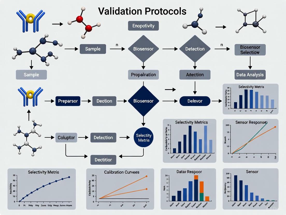
The following diagrams illustrate the core conceptual and operational differences between specific and selective biosensing strategies.
Diagram 1: Specific vs. Selective Sensing Mechanisms. A specific biosensor uses a single bioreceptor for a single analyte. A selective array uses cross-reactive sensors to generate a unique fingerprint.
Diagram 2: Experimental Workflow for Validation. A decision flow for validating specificity through recovery studies and selectivity through cross-reactivity testing.
The Scientist's Toolkit: Essential Reagents and Materials
The development and validation of selective and specific clinical biosensors rely on a core set of research reagents and materials.
Table 3: Essential Research Reagent Solutions for Biosensor Validation
| Reagent / Material | Function in Validation | Example Use Case |
|---|---|---|
| High-Affinity Bioreceptors | Acts as the primary recognition element for the target analyte, dictating inherent specificity [1]. | Monoclonal antibodies for specific immunoassays; aptamers for small molecule detection [1] [4]. |
| Allosteric Transcription Factors (aTFs) | Engineered proteins that change conformation upon binding a target, triggering a signal in cell-free systems [3]. | Detection of metal ions (e.g., Hg²⁺, Pb²⁺) in environmental water samples [3]. |
| RNA Aptamers / Riboswitches | Nucleic acid-based receptors that bind specific ligands, offering a synthetic path to selectivity [3]. | Creating riboswitch-based biosensors for broad-spectrum antibiotic detection [3]. |
| Lyophilized Cell-Free Systems | Pre-packaged, stable biochemical machinery for protein synthesis without living cells, enabling field-deployable biosensing [3]. | Point-of-care paper-based biosensors for toxins or pathogens [3]. |
| Permselective Membranes (e.g., Nafion) | Polymer membranes used to block electrochemical interferents (e.g., ascorbic acid, uric acid) from reaching the transducer [5]. | Improving selectivity of implantable glucose sensors by rejecting common electroactive interferents [5]. |
| Sentinel Sensors | A control sensor lacking the bioreceptor, used to measure and subtract signals from non-specific interactions and matrix effects [5]. | Differentiating specific biosensor response from background signal drift or interferents in complex samples like blood [5]. |
In clinical diagnostics, selectivity refers to the ability of an analytical method to detect a specific target analyte without being influenced by other substances present in a complex sample matrix [5]. For biosensors, which combine a biological recognition element with a physicochemical detector, poor selectivity directly undermines diagnostic accuracy, leading to false positives and false negatives [6]. These errors propagate through the clinical decision-making process, resulting in patient harm through misdiagnosis, inappropriate treatment, and delayed care [7] [8]. Furthermore, repeated diagnostic inaccuracies erode trust in healthcare systems, fostering clinical reluctance among both providers and patients [9]. This guide objectively compares the performance of various biosensor technologies and experimental protocols, framing the analysis within the broader thesis that robust validation protocols are paramount for reliable clinical biosensor selectivity.
The Direct Link Between Poor Selectivity and Misdiagnosis
Mechanisms of Selectivity Failure
Poor selectivity in biosensors primarily manifests through two mechanisms: nonspecific binding and electrochemical interferences.
- Nonspecific Binding: This occurs when molecules other than the target analyte interact with the biorecognition element (e.g., an antibody or aptamer) or the sensor surface. In complex samples like blood, serum, or urine, thousands of proteins, lipids, and other biomolecules can compete for binding sites [10]. For example, in implantable glucose biosensors, compounds like ascorbic acid, uric acid, and acetaminophen are well-documented interferents that can produce falsely elevated readings [5].
- Electrochemical Interferences: In electrochemical biosensors, electroactive compounds that oxidize or reduce at a similar potential to the target analyte can generate a signal indistinguishable from the true signal. This is a particular challenge for first-generation biosensors, which operate at high applied potentials [5].
Clinical Consequences of False Results
The clinical impact of these selectivity failures is severe and multifaceted. False positives can lead to unnecessary and potentially harmful treatments, anxiety, and additional invasive testing. False negatives can result in delayed diagnosis and treatment, allowing a disease to progress [8]. Studies based on case record reviews indicate that diagnostic errors contribute to approximately 10% of patient deaths and are a significant factor in adverse hospital events [7] [8]. The consequences are not only physical but also psychological, with misdiagnosis often leading to patient anxiety, depression, and a lasting erosion of trust in the medical system [9].
Comparative Performance of Biosensor Technologies
The selectivity of a biosensor is intrinsically linked to its underlying technology and design. Different transducing mechanisms offer varying levels of inherent resistance to interference.
Table 1: Comparison of Biosensor Technologies and Their Selectivity Profiles
| Biosensor Technology | Transduction Principle | Common Interferences | Inherent Selectivity Features | Reported Strategies to Enhance Selectivity |
|---|---|---|---|---|
| Electrochemical (1st Gen) [5] | Measures current from reaction products at high potential. | Ascorbic acid, uric acid, acetaminophen. | Low; prone to electrochemical interferents. | Use of permselective membranes (e.g., Nafion/cellulose acetate). |
| Electrochemical (2nd/3rd Gen) [5] | Uses mediators or direct electron transfer to lower operating potential. | Endogenous electroactive species. | Moderate to High; reduced interference via lower potential. | Employing redox polymers and "wired" enzymes for direct electron transfer. |
| Magnetoresistive (MR-Based) [11] | Detects magnetic nanoparticles bound to targets. | Sample charge, mild temperature gradients. | High; signal (magnetic field) is immune to common sample noise. | Not required for charge/temperature, but specific binding must be ensured. |
| Optical (e.g., SPR, SERS) [4] | Measures changes in light properties (refractive index, Raman scattering). | Compounds affecting refractive index; sample turbidity. | Moderate; can be affected by nonspecific adsorption. | Functionalization with specific bioreceptors (e.g., aptamers); spectral filtering. |
The data reveals a clear technological evolution aimed at mitigating selectivity issues. While first-generation electrochemical biosensors require additional components like membranes to achieve acceptable selectivity, newer platforms like magnetoresistive and third-generation electrochemical sensors build interference resistance directly into their core operating principle [5] [11].
Experimental Protocols for Validating Selectivity
Robust validation is essential to ensure a biosensor's selectivity claims are valid. The following protocols are standard in the field.
Protocol 1: Assessing the Impact of Common Interferents
This experiment tests the biosensor's response to structurally similar compounds and molecules commonly found in the target sample matrix.
- Step 1: Solution Preparation. Prepare separate solutions of the target analyte and potential interferents (e.g., ascorbic acid, uric acid, acetaminophen for a serum sensor) at physiologically relevant concentrations, and a mixture of all compounds.
- Step 2: Signal Measurement. Measure the biosensor's signal for each individual solution and the mixture.
- Step 3: Data Analysis. Calculate the signal deviation caused by the interferents. The response from the mixture should not significantly differ (e.g., <±5-10%) from the signal of the target analyte alone. A significant deviation indicates poor selectivity [5].
Protocol 2: Using a Sentinel Sensor for Signal Subtraction
This method involves a control sensor to quantify and correct for signals from nonspecific binding and matrix effects.
- Step 1: Sentinel Sensor Fabrication. Create a sensor identical to the biosensor but lacking the specific biorecognition element. This is often done by immobilizing an "inert" protein like Bovine Serum Albumin (BSA) [5].
- Step 2: Parallel Measurement. Expose both the active biosensor and the sentinel sensor to the sample.
- Step 3: Signal Correction. The sentinel sensor records signals from nonspecific binding and electrochemical interferences. Subtract the sentinel signal from the active biosensor's signal to obtain a corrected, analyte-specific reading [5].
Protocol 3: Evaluating Real Sample Matrix Effects
This protocol validates performance in the actual clinical sample to uncover matrix-specific interferences not seen in buffer.
- Step 1: Spiked Sample Preparation. Take a sample of the target matrix (e.g., blood, saliva) confirmed to be free of the analyte ("blank"). Spike it with a known concentration of the target analyte.
- Step 2: Calibration and Recovery. Analyze the spiked sample and calculate the recovery of the analyte. High recovery (e.g., 85-115%) indicates good selectivity despite the complex sample matrix [10].
- Step 3: Comparison to Gold Standard. Results should be correlated with a standard reference method (e.g., ELISA for proteins, PCR for nucleic acids) using a sufficient number of clinical samples [5] [10].
The Scientist's Toolkit: Essential Reagents and Materials
Successful development and validation of selective biosensors rely on a suite of specialized reagents and materials.
Table 2: Key Research Reagent Solutions for Selectivity Enhancement
| Reagent / Material | Function in Selectivity Control | Specific Example |
|---|---|---|
| Permselective Membranes [5] | Blocks access of interfering compounds to the transducer surface based on size, charge, or hydrophobicity. | Nafion (charge-based exclusion of anions like ascorbate), Cellulose Acetate (size-exclusion). |
| Artificial Mediators & Redox Polymers [5] | Shuttles electrons in 2nd-gen biosensors, lowering operational potential to a window with fewer interferences. | Ferrocene derivatives, Osmium-based redox polymers. |
| Enzymes for Interference Scavenging [5] | Converts an electroactive interferent into an inactive compound before it reaches the transducer. | Ascorbate Oxidase (converts ascorbic acid to dehydroascorbic acid). |
| Sentinel Sensor Components [5] | Provides a baseline signal for nonspecific binding and matrix effects, enabling signal correction. | Bovine Serum Albumin (BSA) used to create a non-specific binding surface. |
| High-Affinity Bioreceptors [10] [12] | The primary element for molecular recognition; engineered for high specificity to the target over analogs. | Engineered Aptamers (from SELEX), Monoclonal Antibodies, Molecularly Imprinted Polymers (MIPs). |
| Magnetic Nanoparticles (MNPs) [11] | Used as labels in MR-based biosensors; their magnetic signal is inherently selective against sample background. | Streptavidin-coated MNPs for binding to biotinylated detection antibodies. |
The consequences of poor biosensor selectivity—misdiagnosis and the ensuing clinical reluctance—are too significant to overlook. As shown, the performance of biosensor platforms varies considerably, with technological choices directly impacting inherent interference resistance. The path to trustworthy clinical biosensors lies in the systematic implementation and reporting of rigorous, multi-faceted validation protocols. By adopting the experimental workflows and reagent solutions detailed in this guide, researchers can provide the robust data needed to confidently translate biosensor technologies from the laboratory to the clinic, ultimately restoring and strengthening trust in clinical diagnostics.
Selectivity is a cornerstone of clinical biosensor performance, determining the device's ability to accurately identify a specific target analyte within complex biological matrices such as blood, saliva, or urine [13]. Achieving high selectivity is paramount for reliable diagnostic outcomes, treatment monitoring, and drug development. This critical attribute emerges from the sophisticated interplay of three core components: the bioreceptor, which provides molecular recognition; the transducer, which converts the biological event into a quantifiable signal; and the surface chemistry, which orchestrates the stable and functional interface between them [14] [15]. A profound understanding of how these components collectively influence validation protocols is essential for advancing clinical biosensor research from laboratory prototypes to trusted diagnostic tools. This guide objectively compares the performance of these core components and the experimental methodologies used to evaluate their selectivity, providing a framework for robust clinical validation.
Core Component 1: Bioreceptors and Their Selectivity Mechanisms
Bioreceptors are biological or biomimetic molecules immobilized on the biosensor surface that are responsible for the specific recognition of the target analyte [13]. The choice of bioreceptor fundamentally defines the intrinsic selectivity of the biosensing platform.
Table 1: Comparison of Key Bioreceptor Types and Their Selectivity Performance
| Bioreceptor Type | Mechanism of Selectivity | Key Performance Characteristics | Common Clinical Targets | Experimental Evidence of Selectivity |
|---|---|---|---|---|
| Antibodies [14] | High-affinity, lock-and-key binding to a specific antigen epitope [13]. | Very high specificity and sensitivity; can be susceptible to denaturation [14]. | Pathogens (e.g., E. coli [16]), protein biomarkers (e.g., α-Fetoprotein [4]), hormones. | A biosensor for E. coli using anti-O antibody demonstrated a low detection limit of 1 CFU mL⁻¹ and could discriminate non-target bacteria like Salmonella and S. aureus [16]. |
| Enzymes [14] | Catalytic transformation of a specific substrate into a product. | High specificity for substrate; signal generation via reaction product [13]. | Glucose, lactate, urea, cholesterol [13] [14]. | Glucose oxidase-based sensors are the flagship commercial example, selectively catalyzing glucose oxidation [14]. |
| Nucleic Acids (Aptamers) [14] | Folding into 3D structures that bind targets with high affinity. | Synthetic; high stability and selectivity; designable via SELEX process [4]. | Mycotoxins, pathogens, small molecules [4]. | Aptasensors are developed for rapid detection of hazards in food, showing high specificity for targets like pesticides and foodborne pathogens [4]. |
| Whole Cells/Tissues [14] | Utilization of innate cellular receptors or metabolic pathways. | Provides complex, functional responses; often less specific than molecular receptors. | Toxins, drugs for screening [14]. | Used in biosensors for toxin detection and drug screening, where the overall cellular response is the measured output [14]. |
Core Component 2: Transducers and Signal Fidelity
The transducer is the component that converts the specific interaction between the bioreceptor and analyte into a measurable physical signal [13] [17]. The transduction principle directly impacts the sensitivity, robustness, and applicability of the biosensor in clinical settings.
Table 2: Comparison of Biosensor Transducer Technologies
| Transducer Type | Detection Principle | Key Advantages | Limitations & Selectivity Challenges | Example Experimental Protocol for Selectivity Assessment |
|---|---|---|---|---|
| Electrochemical (Amperometric, Impedimetric) [14] | Measures changes in current, potential, or impedance from biochemical reactions at an electrode [17] [14]. | High sensitivity, portability, low cost, and low power requirements [17] [14]. | Signal can be influenced by non-faradaic interferences; requires robust surface functionalization to minimize non-specific binding [15]. | Methodology: Cyclic Voltammetry (CV) and Electrochemical Impedance Spectroscopy (EIS). Protocol: Measure the charge-transfer resistance (Rct) before and after exposure to the target analyte. Selectivity is validated by challenging the sensor with structurally similar molecules or common interferents (e.g., ascorbic acid, uric acid for glucose sensors) and observing negligible Rct change compared to the target [14]. |
| Optical (SPR, Fluorescence) [14] | Detects changes in light properties (e.g., refractive index, absorbance, fluorescence) [18] [14]. | Superior multiplexing capability, high resolution, and real-time kinetic monitoring [14]. | Can be susceptible to ambient light interference; instrumentation can be bulky. | Methodology: Surface Plasmon Resonance (SPR). Protocol: Immobilize bioreceptors on a gold film. Monitor the shift in the resonance angle upon analyte binding. To test selectivity, inject solutions containing potential interferents and confirm the absence of a significant resonance angle shift [18]. |
| Gravimetric (Piezoelectric) [14] | Measures mass change on the sensor surface as a shift in resonance frequency. | Highly sensitive to mass changes. | Sensitive to environmental factors like temperature and viscosity; can suffer from non-specific binding [14]. | Methodology: Quartz Crystal Microbalance (QCM). Protocol: The resonance frequency shift is calibrated to mass adsorption. Selectivity is tested by exposing the functionalized crystal to non-target analytes; a selective sensor will show minimal frequency change in these control experiments. |
The diagram below illustrates the core architecture of a biosensor and the logical flow of information from analyte binding to signal output, integrating the roles of the bioreceptor, transducer, and surface chemistry.
Core Component 3: Surface Chemistry and Interfacial Design
Surface chemistry is the engineering of the interface between the transducer and the biological environment. It is critical for immobilizing bioreceptors in a stable and active orientation, while also minimizing non-specific adsorption of other molecules, a phenomenon known as fouling [15] [19]. Effective surface functionalization is thus a prerequisite for achieving both high selectivity and long-term stability.
Key Surface Functionalization Strategies
- Covalent Immobilization: This method creates strong, stable bonds between the bioreceptor and the functionalized transducer surface. A common strategy involves using (3-Aminopropyl)triethoxysilane (APTES) and glutaraldehyde (GA) on oxide surfaces, or thiol-gold chemistry for gold electrodes [15] [20]. This approach enhances operational stability by preventing bioreceptor leaching [15].
- Non-covalent Immobilization: This includes physical adsorption and affinity-based methods like the streptavidin-biotin interaction [20]. While simpler, physical adsorption can lead to random orientation and denaturation of the bioreceptor. The streptavidin-biotin bond is exceptionally strong and specific, making it a popular choice for attaching biotin-labeled probes [20].
- Nanomaterial-Enhanced Interfaces: The use of nanomaterials like graphene, gold nanoparticles (AuNPs), and metal-organic frameworks (MOFs) has revolutionized surface design [15] [18] [16]. These materials offer high surface-to-volume ratios for dense bioreceptor loading and unique properties for signal amplification. For instance, a Mn-doped ZIF-67 MOF was used to create a highly sensitive platform for E. coli detection, where the large surface area and tuned electronic properties enhanced performance [16].
- Anti-fouling Coatings: To preserve selectivity, surfaces are often modified with anti-fouling molecules such as polyethylene glycol (PEG), bovine serum albumin (BSA), or zwitterionic polymers [15]. These coatings passivate unused sites on the transducer, reducing non-specific binding from proteins and other components in complex samples [15].
Experimental Data and Comparative Analysis
This section provides a detailed experimental case study and a comparative analysis of selectivity validation protocols.
Case Study: Electrochemical Biosensor forE. coliDetection
A high-performance electrochemical biosensor was developed using a Mn-doped Zeolitic Imidazolate Framework (ZIF-67) conjugated with an anti-O antibody for the specific detection of E. coli [16].
- Experimental Protocol:
- Surface Functionalization: The Mn-ZIF-67 composite was synthesized hydrothermally and drop-casted onto a screen-printed electrode. The anti-O antibody was then conjugated to the MOF surface to impart selectivity for the O-antigen of E. coli.
- Detection Method: The sensor utilized electrochemical impedance spectroscopy (EIS) and cyclic voltammetry (CV). The binding of E. coli cells to the antibodies on the porous MOF surface increased the charge-transfer resistance (Rct), which was measured as the sensor's signal.
- Selectivity Validation: The sensor was challenged with high concentrations (10^8 CFU mL⁻¹) of non-target bacteria, including Salmonella typhimurium, Pseudomonas aeruginosa, and Staphylococcus aureus.
- Resulting Data: The sensor demonstrated a linear range from 10 to 10^10 CFU mL⁻¹ with an exceptionally low detection limit of 1 CFU mL⁻¹. Most importantly, the signal generated by the non-target bacteria was negligible compared to the E. coli signal, demonstrating excellent selectivity. The sensor also maintained over 80% sensitivity after 5 weeks, indicating high stability [16].
The workflow for such a biosensing experiment, from material synthesis to data analysis, is visualized below.
The Scientist's Toolkit: Essential Research Reagents and Materials
Table 3: Key Reagents and Materials for Biosensor Development and Validation
| Item | Function in Experimental Protocols | Application Example |
|---|---|---|
| (3-Aminopropyl)triethoxysilane (APTES) [15] [20] | A silane coupling agent used to introduce amine (-NH₂) groups onto oxide surfaces (e.g., SiO₂) for subsequent covalent immobilization. | Functionalizing CMOS chips or ITO electrodes for attaching bioreceptors via glutaraldehyde crosslinking [20]. |
| Glutaraldehyde (GA) [20] | A bifunctional crosslinker that reacts with amine groups from APTES and the bioreceptor, forming a stable covalent bond. | Creating a bridge between an APTES-functionalized surface and an antibody for stable immobilization [20]. |
| Thiolated Probes (e.g., DNA, Peptides) [20] | Molecules modified with a thiol (-SH) group that form strong self-assembled monolayers (SAMs) on gold surfaces. | Immobilizing DNA capture probes on gold electrodes for genosensing or creating well-ordered bioreceptor layers [20]. |
| Polyethylene Glycol (PEG) [15] | An anti-fouling polymer used to passivate sensor surfaces, reducing non-specific binding of proteins and other biomolecules. | Coating the background area of a sensor to minimize false-positive signals in complex samples like serum [15]. |
| Bovine Serum Albumin (BSA) [15] | A common blocking agent used to cover non-specific binding sites on the sensor surface after bioreceptor immobilization. | Incubated on an immunosensor to block leftover active sites on the electrode, preventing non-specific protein adsorption [15]. |
| Metal-Organic Frameworks (MOFs) [16] | Porous crystalline materials with high surface area that enhance bioreceptor loading and can improve electrochemical signal transduction. | Using ZIF-67 as a nanoporous scaffold to immobilize antibodies and amplify the signal in a pathogen sensor [16]. |
The path to validating clinical biosensor selectivity is multifaceted, relying on the synergistic optimization of bioreceptors, transducers, and surface chemistry. As demonstrated, antibodies and aptamers provide the foundational molecular recognition, while electrochemical and optical transducers offer distinct paths to sensitive detection. Ultimately, it is the precision of the surface chemistry—the careful engineering of the interface through advanced functionalization and anti-fouling strategies—that ensures this intrinsic selectivity is translated into a reliable analytical signal. Future developments, particularly the integration of artificial intelligence (AI) for predicting optimal surface architectures and bioreceptor configurations, promise to accelerate the rational design of even more selective and robust biosensors [15]. For researchers in clinical and drug development, a rigorous validation protocol that stresses the biosensor with structurally similar interferents and real-world sample matrices remains the definitive test for establishing the selectivity required for clinical application.
A biosensor is an analytical device that integrates a biological recognition element (bioreceptor) with a physicochemical transducer to convert a biological event into a measurable signal [13] [14]. The core components include the analyte (substance to be detected), bioreceptor (molecule that specifically recognizes the analyte, such as an enzyme, antibody, DNA, or cell), transducer (element that converts the recognition event into a measurable signal), electronics, and display [13] [21]. Selectivity, defined as the ability of a bioreceptor to detect a specific analyte in a sample containing other admixtures and contaminants without being influenced by other sample constituents, is one of the most critical characteristics of any biosensor [13] [22]. For clinical applications, where biosensors must operate in complex matrices like blood, urine, or saliva, achieving high selectivity remains a paramount challenge that directly impacts diagnostic accuracy and reliability [10] [22] [23].
The following diagram illustrates the fundamental components and process flow of a biosensor system, highlighting where selectivity challenges emerge in the recognition and transduction phases:
Biosensor Types: Comparative Analysis of Selectivity Profiles
Biosensors are primarily classified based on their transduction method, with electrochemical, optical, and piezoelectric systems representing the most prominent categories in research and clinical applications [21] [14]. Each transducer type exhibits distinct selectivity challenges and advantages, necessitating different mitigation approaches, particularly when deployed for clinical measurements involving complex biological samples [22] [23].
Table 1: Comparative Analysis of Biosensor Types and Selectivity Profiles
| Biosensor Type | Transduction Principle | Common Bioreceptors | Inherent Selectivity Challenges | Primary Interference Sources |
|---|---|---|---|---|
| Electrochemical | Measures electrical changes (current, potential, impedance) from biological recognition events [21] [14] | Enzymes, antibodies, DNA, aptamers [13] [22] | Electroactive compounds oxidizing/reducing at similar potentials; enzyme inhibitors/activators in sample [22] | Ascorbic acid, uric acid, acetaminophen, dopamine [22] |
| Optical | Detects light-based changes (absorbance, fluorescence, luminescence, refractive index) from bio-recognition [21] [14] | Antibodies, DNA, enzymes, whole cells [14] | Scattering from particulate matter; autofluorescence of sample components; nonspecific binding [14] | Turbid samples; fluorescent compounds in matrix; ambient light [14] |
| Piezoelectric | Measures mass changes on sensor surface through resonance frequency shifts [21] [14] | Antibodies, DNA, molecularly imprinted polymers [21] | Nonspecific adsorption of non-target molecules; viscosity changes in sample [14] | Proteins, cells, other macromolecules in biological fluids [14] |
Table 2: Selectivity Enhancement Strategies Across Biosensor Platforms
| Biosensor Type | Selectivity Enhancement Strategies | Typical Clinical Applications | Limit of Detection Ranges |
|---|---|---|---|
| Electrochemical | Permselective membranes; sentinel sensors; mediated electron transfer; enzyme electrodes with coupled reactions [22] | Glucose monitoring, cardiac biomarkers, pathogen detection [10] [22] | pM-nM for proteins; fM-pM for DNA [10] [24] |
| Optical | Surface plasmon resonance (SPR) with specific coatings; wavelength filtering; reference channels [13] [14] | Cancer biomarkers, infectious disease detection, hormone monitoring [13] [10] | ng/mL-pg/mL for proteins; single molecule for fluorescence [10] [14] |
| Piezoelectric | Hydrogel anti-fouling layers; reference crystal subtraction; surface chemistry optimization [24] [14] | Pathogen detection, gas sensing, small molecule analysis [21] [14] | ng-pg level mass changes [21] |
Experimental Protocols for Assessing Biosensor Selectivity
Robust validation of biosensor selectivity requires standardized experimental protocols that simulate challenging real-world conditions. The following workflow outlines a comprehensive approach for selectivity assessment applicable across biosensor platforms, with specific adaptations for each transducer type:
Cross-Reactivity Testing Protocol
Objective: Quantify biosensor response to structurally similar compounds that may compete for binding sites. Materials: Target analyte, structural analogs (minimum 3-5 compounds), negative controls, appropriate buffer systems. Procedure:
- Prepare solutions containing the target analyte at its EC₈₀ concentration (concentration producing 80% of maximum signal).
- Prepare separate solutions containing each potential interferent at 100-fold higher concentration than the target analyte.
- Prepare mixture solutions containing target analyte plus each interferent.
- Measure biosensor response to each solution in triplicate.
- Calculate cross-reactivity percentage for each interferent: (Response to interferent alone / Response to target analyte) × 100%.
Acceptance Criterion: Cross-reactivity <5% for each interferent in clinical applications [22] [23].
Matrix Interference Assessment
Objective: Evaluate effect of complex biological matrix components on biosensor signal. Materials: Blank matrix samples (serum, plasma, urine, saliva), analyte standards, displacement reagents if applicable. Procedure:
- Prepare analyte standards in clean buffer and in at least 5 different lots of appropriate biological matrix.
- For each lot, prepare standards at low, medium, and high concentrations within the assay range.
- Measure response for all samples using the biosensor platform.
- Calculate percent recovery for each concentration in each matrix lot: (Measured concentration in matrix / Known concentration in buffer) × 100%.
- Statistically compare results using ANOVA with post-hoc testing.
Acceptance Criterion: 85-115% recovery across all matrix lots with no statistically significant differences (p>0.05) [25] [23].
Sentinel Sensor Implementation for Electrochemical Systems
Objective: Differentiate between specific signal and non-specific interference in electrochemical biosensors. Materials: Functional biosensor, sentinel sensor (identical but without bioreceptor or with inactivated bioreceptor), measuring chamber allowing parallel measurement. Procedure:
- Immobilize bioreceptor on biosensor surface using standard protocol.
- Prepare sentinel sensor using identical procedure but with bovine serum albumin instead of bioreceptor.
- Expose both sensors simultaneously to sample containing potential interferents but no target analyte.
- Measure signals from both sensors.
- Calculate interference compensation factor.
- During actual sample measurement, subtract sentinel sensor signal from biosensor signal.
Validation: Test with known interferents (ascorbic acid, uric acid, acetaminophen) at physiological concentrations [22].
The Scientist's Toolkit: Essential Reagents and Materials
Table 3: Research Reagent Solutions for Selectivity Enhancement
| Reagent/Material | Function in Selectivity Enhancement | Specific Applications | Considerations for Use |
|---|---|---|---|
| Permselective Membranes (Nafion, cellulose acetate, polypyrrole) | Block interfering electroactive compounds based on size, charge, or hydrophobicity [22] | Electrochemical sensors for neurotransmitter detection; implantable glucose sensors | May increase response time; requires optimization of thickness and composition [22] |
| Anti-fouling Self-Assembled Monolayers (ethylene glycol, zwitterionic polymers) | Reduce nonspecific protein adsorption on sensor surface [24] [23] | Optical biosensors for serum protein detection; piezoelectric sensors in whole blood | Must maintain bioreceptor activity; compatibility with immobilization chemistry [23] |
| Redox Mediators (ferrocene derivatives, organic complexes, metal nanoparticles) | Lower operating potential to minimize electrochemical interferences [13] [22] | Second-generation enzyme electrodes; point-of-care devices | Potential mediator toxicity; long-term stability concerns [22] |
| Reference Sensors/Sentinel Sensors | Provide baseline signal for subtraction of non-specific effects [22] | Continuous monitoring applications; complex biological samples | Requires precise matching of sensor characteristics; adds complexity to instrumentation [22] |
| Enzyme-Based Interference Elimination (ascorbate oxidase, uricase) | Convert interfering compounds to non-interfering forms [22] | Amperometric biosensors for biological fluids | Additional cost and complexity; potential side reactions [22] |
The journey toward clinically robust biosensors demands meticulous attention to selectivity challenges inherent to each transducer platform. Electrochemical biosensors predominantly grapple with electroactive interferents, necessitating strategic implementation of permselective membranes, sentinel sensors, and mediated electron transfer systems [22]. Optical biosensors contend with matrix-induced light scattering and autofluorescence, requiring sophisticated reference channels and surface chemistries that minimize nonspecific binding [14]. Piezoelectric biosensors face mass-based interference issues, demanding advanced anti-fouling surface modifications and appropriate data subtraction protocols [21] [14].
For clinical researchers validating biosensor selectivity, a systematic approach incorporating cross-reactivity profiling, matrix interference assessment, and real-sample validation is paramount [25] [23]. The experimental protocols outlined provide a framework for rigorous selectivity demonstration that meets investor and regulatory expectations [25]. As the biosensor field evolves, emerging solutions in nanomaterials, bioreceptor engineering, and microfluidics promise enhanced selectivity without compromising sensitivity [24] [22]. However, the fundamental requirement remains: comprehensive investigation of biosensor selectivity must be an integral component of development workflows, with validation against standard analytical methods using clinically relevant samples [22] [23].
Advanced Methodologies for Assessing and Engineering Biosensor Selectivity
Selectivity represents a cornerstone validation parameter in clinical biosensor development, determining a sensor's ability to accurately measure a target analyte without interference from other components in complex biological samples. The fundamental challenge in biosensor selectivity stems from the complex composition of biological matrices such as serum, plasma, and whole blood, which contain numerous proteins, lipids, cells, and other molecular species that can interfere with sensor response through nonspecific binding (NSB) [26] [27]. Without proper control strategies, these matrix effects can lead to false positive readings, inaccurate quantification, and ultimately compromised diagnostic decisions. A study on photonic ring resonator biosensors highlighted that nonspecific binding of matrix constituents presents a significant challenge, making it virtually impossible to distinguish specific from nonspecific interactions without appropriate reference controls [26].
The validation of biosensor selectivity requires a systematic approach to experimental design that incorporates structurally similar analytes and relevant biological matrices to challenge the biosensor's recognition elements. This process must account for various interference mechanisms, including direct binding to biorecognition elements, fouling of the sensor surface, and signal suppression or enhancement from matrix components [27] [28]. As biosensors transition from laboratory settings to point-of-need clinical applications, rigorous selectivity testing becomes increasingly critical for regulatory approval and clinical adoption. This guide establishes a comprehensive framework for designing control experiments that thoroughly characterize biosensor selectivity, with particular emphasis on optimizing reference controls and managing matrix effects.
Theoretical Foundations of Selectivity Challenges
Mechanisms of Interference in Biosensing
Interference in biosensor operation primarily manifests through two distinct mechanisms: specific cross-reactivity and nonspecific binding. Specific cross-reactivity occurs when structurally similar compounds compete with the target analyte for binding sites on the biorecognition element. This form of interference is particularly challenging for biosensors detecting small molecules, pharmaceuticals, or biomarkers with structural analogs present in biological samples [27]. The molecular similarity between target and interferent can lead to comparable binding affinities, resulting in inaccurate signal attribution and concentration overestimation.
Nonspecific binding represents a more pervasive challenge, arising from electrostatic interactions, hydrogen bonding, and van der Waals forces between matrix components and the biosensor surface [26]. The extent of NSB is heavily influenced by the physicochemical properties of both the sensor surface and the sample matrix. As noted in studies of label-free biosensors, "the closer to the protein's isoelectric point (pI) the pH lies, the more neutrally charged the protein, potentially increasing NSB due to hydrophobic interactions" [26]. Furthermore, the complexity of the matrix significantly impacts NSB, with human serum producing substantially more interference than buffer solutions due to the higher concentration and diversity of potential interfering species [26].
Impact of Biological Matrices on Sensor Performance
Biological matrices introduce multiple challenges for biosensor selectivity beyond nonspecific binding. Whole blood, plasma, and serum each present distinct interference profiles that must be considered during selectivity testing. Research on solid-phase microextraction (SPME) fibers demonstrated that "during direct sampling of whole blood, coating fouling often occurs due to the presence of proteins, blood cells, and other substances" [28]. This fouling can physically block access to recognition elements, alter binding kinetics, and reduce sensor sensitivity over time.
Matrix components can also influence assay performance through molecular interactions that extend beyond surface binding. Enzymatic activity in biological samples may degrade either the biorecognition element or the target analyte, leading to signal attenuation. Variations in pH, ionic strength, and osmolarity across different biological matrices can additionally affect molecular conformation, binding affinity, and complex stability [27] [28]. These factors collectively underscore the necessity of testing biosensor selectivity in matrices that closely resemble the intended clinical application rather than relying solely on simplified buffer systems.
Experimental Design Principles for Selectivity Assessment
Fundamental DoE Principles for Robust Testing
The design of experiments (DoE) methodology provides a statistical framework for optimizing selectivity testing protocols while efficiently utilizing resources. Three core principles of experimental design are particularly relevant to biosensor selectivity assessment: randomization, blocking, and replication [29]. Randomization of run order helps mitigate the effects of uncontrolled variables that may introduce bias over time, such as sensor degradation, reagent instability, or environmental fluctuations. Blocking techniques account for known sources of variability, such as different production batches of biosensors or operators performing tests, by grouping experimental runs to minimize their impact on selectivity measurements. Replication enables estimation of experimental error and provides greater confidence in selectivity determinations [29].
A well-designed selectivity experiment should employ a balanced approach that challenges the biosensor with both specific interferents (structurally similar compounds) and complex biological matrices. Multifactorial experimental designs are particularly advantageous for evaluating potential interactions between different interferents and matrix components that might not be apparent when testing variables in isolation [30]. For instance, the effect of a specific pharmaceutical interferent may be amplified in certain disease states that alter serum protein composition, an interaction that would only be detectable through appropriately designed matrix-interferent combination studies.
Control Selection Strategies
The selection of appropriate reference controls represents perhaps the most critical aspect of rigorous selectivity testing. Research systematically evaluating control probes for label-free biosensors revealed that "although isotype-matching to the capture antibody may be tempting, the best on-chip reference control must be optimized on a case-by-case basis" [26]. This finding underscores the importance of empirically validating control strategies rather than relying on assumed effectiveness.
An FDA-inspired framework for control probe selection evaluates candidates based on linearity, accuracy, and selectivity metrics [26]. This systematic approach assesses multiple potential control molecules, including isotype-matched antibodies, non-matched antibodies, irrelevant proteins (e.g., BSA), and charged non-antibody proteins (e.g., cytochrome c). The optimal control should effectively subtract nonspecific binding contributions without over- or under-correction of the specific binding signal. For example, in a study detecting interleukin-17A (IL-17A) and C-reactive protein (CRP), different control probes performed best for each analyte: BSA scored highest (83%) for IL-17A, while a rat IgG1 isotype control antibody scored highest (95%) for CRP [26].
Table 1: Control Probe Performance in Representative Biosensor Studies
| Analyte | Top-Performing Control Probe | Performance Score | Alternative Control Probe | Performance Score |
|---|---|---|---|---|
| IL-17A | BSA | 83% | Mouse IgG1 Isotype Control | 75% |
| CRP | Rat IgG1 Isotype Control | 95% | Anti-FITC | 89% |
Structured Experimental Framework for Selectivity Testing
Comprehensive Testing Protocol
A robust selectivity testing protocol should systematically evaluate biosensor performance across multiple dimensions of potential interference. The following stepwise approach provides a comprehensive framework for selectivity validation:
Step 1: Baseline Characterization in Simple Buffer Establish baseline sensor performance in optimized buffer conditions, including dose-response curves for the target analyte, determination of limit of detection, and assessment of binding kinetics. This initial characterization provides a reference point for evaluating matrix effects in subsequent experiments [31] [32].
Step 2: Individual Interferent Testing Challenge the biosensor with individual potentially interfering compounds at physiologically relevant concentrations that exceed expected normal and pathological ranges. Test structurally similar compounds, metabolites, co-administered pharmaceuticals, and endogenous molecules with similar physicochemical properties. The GEM biosensor study exemplifying this approach demonstrated excellent specificity for Cd2+, Zn2+, and Pb2+ against non-specific metals like Fe3+, AsO43−, and Ni2+ [33].
Step 3: Biological Matrix Evaluation Assess biosensor performance in relevant biological matrices (e.g., whole blood, plasma, serum, urine) without target analyte addition to quantify background signal. Then, perform spike-and-recovery experiments by adding known concentrations of target analyte to matrices and calculating recovery percentages. Research on sol-gel coatings for SPME highlighted that matrix compatibility can vary significantly between different sample types, with whole blood presenting greater challenges than plasma due to cellular components [28].
Step 4: Complex Mixture Challenges Evaluate biosensor response in biological matrices spiked with both target analyte and potential interferents to simulate real-world sample complexity. This test should include combinations of structurally similar compounds that represent the most challenging selectivity scenario.
Step 5: Reference Control Validation Implement and validate selected reference controls using the systematic framework that assesses linearity, accuracy, and selectivity metrics [26]. Compare multiple control candidates to identify the optimal approach for subtracting nonspecific binding contributions.
Quantitative Assessment Metrics
Rigorous selectivity assessment requires quantitative metrics that enable objective comparison across different experimental conditions and biosensor platforms. The following key performance indicators should be calculated and reported for comprehensive selectivity characterization:
Cross-reactivity Percentage: Calculated as (Response to Interferent / Response to Target Analyte) × 100%, where concentrations are equimolar at clinically relevant levels. Cross-reactivity values should typically be <1% for high-selectivity biosensors, though acceptable thresholds depend on the specific application and potential interferent prevalence [33] [34].
Signal-to-Interference Ratio (SIR): Determined by measuring the sensor response in samples containing target analyte versus samples containing potential interferents at the same concentration. An SIR > 20 is generally desirable for clinical applications.
Recovery Percentage: Assessed through spike-and-recovery experiments in biological matrices. Acceptable recovery typically falls between 85-115%, though wider ranges may be acceptable for certain applications depending on regulatory requirements [28].
Limit of Detection Ratio: Comparing LOD in buffer versus LOD in biological matrix. A ratio < 2 indicates minimal matrix effects, while higher values suggest significant interference requiring mitigation strategies.
Table 2: Key Performance Indicators for Biosensor Selectivity Assessment
| Metric | Calculation Method | Acceptance Criteria | Application Context |
|---|---|---|---|
| Cross-reactivity Percentage | (Response to Interferent / Response to Target) × 100% | <1% (high selectivity)<5% (moderate selectivity) | Essential for all biosensors |
| Signal-to-Interference Ratio (SIR) | SignalTarget / SignalInterferent | >20:1 | Critical for complex matrices |
| Recovery Percentage | (Measured Concentration / Spiked Concentration) × 100% | 85-115% | Required for quantitative assays |
| Limit of Detection Ratio | LODMatrix / LODBuffer | <2:1 | Indicator of matrix effects |
| Reference Control Efficiency | Specific Signal / Total Signal | >90% | Label-free biosensors |
Case Studies in Biosensor Selectivity Optimization
Heavy Metal Detection with GEM Biosensors
A genetically engineered microbial (GEM) biosensor developed for detection of Cd2+, Zn2+, and Pb2+ exemplifies rigorous specificity testing against structurally similar analytes [33]. The researchers systematically evaluated biosensor response to target metals compared to non-specific metals including Fe3+, AsO43−, and Ni2+. The biosensor demonstrated excellent discrimination capabilities, with linear response graphs for target metals (R² values of 0.9809, 0.9761, and 0.9758 for Cd2+, Zn2+, and Pb2+, respectively) compared to non-specific metals (R² values of 0.0373, 0.3825, and 0.8498 for Fe3+, AsO43−, and Ni2+) [33].
This case study highlights the importance of testing against multiple analogous interferents rather than assuming specificity based on successful target detection. The researchers further validated their biosensor under physiologically relevant conditions (37°C, pH 7.0), demonstrating maintained selectivity while operating in environments resembling practical application scenarios. The GEM biosensor achieved detection of target metals in the 1-6 ppb range despite the presence of potential interferents, establishing its utility for environmental and clinical monitoring [33].
Paper-Based DNA Biosensor for miRNA Detection
A paper-based electrochemical biosensor for detection of miR-21 provides another illustrative example of comprehensive selectivity testing [34]. The developers immobilized single-stranded DNA-21 (ssDNA-21) on electrodeposited gold nanoparticles to facilitate specific miR-21 capture. To validate selectivity, they challenged the biosensor with three different interferents: single-base mismatch (MM1), three-base mismatch (MM3), and completely non-complementary (NC) sequences.
The biosensor demonstrated exceptional discrimination capability, maintaining high sensitivity (detection limit of 0.35 fM) while showing significantly reduced response to mismatched sequences [34]. This level of selectivity is particularly remarkable given the structural similarity of miRNA sequences and the potential for cross-hybridization. The researchers further validated their biosensor in fetal bovine serum, confirming maintained selectivity in a complex biological matrix. The successful discrimination between perfectly matched target sequences and those with even single-base mismatches underscores the importance of challenging biosensors with structurally similar interferents that represent the most demanding selectivity scenarios [34].
Research Reagent Solutions for Selectivity Testing
Essential Materials and Their Functions
Table 3: Essential Research Reagents for Biosensor Selectivity Testing
| Reagent Category | Specific Examples | Function in Selectivity Testing | Key Considerations |
|---|---|---|---|
| Reference Control Probes | Isotype-matched antibodies, BSA, anti-FITC, cytochrome c | Subtract nonspecific binding contributions; validate specificity | Must be optimized case-by-case; different probes perform best for different analytes [26] |
| Biological Matrices | Whole blood, plasma, serum, urine | Challenge biosensor in clinically relevant environments; assess matrix effects | Whole blood most challenging due to cellular components; stability varies [27] [28] |
| Structurally Similar Analytes | Pharmaceutical metabolites, homologous proteins, isobars | Test molecular recognition specificity; identify cross-reactivity | Should include compounds with highest structural similarity to target [33] [34] |
| Surface Blocking Agents | BSA, casein, fish skin gelatin, proprietary blockers | Reduce nonspecific binding; improve signal-to-noise ratio | Optimization required for each biosensor surface chemistry [26] [28] |
| Matrix Effect Mitigation | Sol-gel coatings, hydrophilic-lipophilic balanced particles | Improve matrix compatibility; reduce fouling | Sol-gel coatings show good matrix compatibility in SPME applications [28] |
Implementation Considerations for Robust Selectivity Protocols
Methodological Optimization Strategies
Successful implementation of rigorous selectivity tests requires careful attention to methodological details that can significantly impact results. Biosensor surface preparation represents a critical factor influencing both specificity and nonspecific binding. Research on sol-gel solid-phase microextraction coatings demonstrated that material composition dramatically affects matrix compatibility, with service life in undiluted plasma ranging from 20-35 extraction cycles depending on the specific coating formulation [28]. This variability underscores the importance of testing multiple surface chemistries during biosensor development.
Sample preparation represents another crucial consideration, with techniques such as dilution, protein precipitation, liquid-liquid extraction, and solid-phase extraction offering different trade-offs between matrix effect reduction and analytical throughput [27]. While extensive sample processing can minimize matrix effects, it contradicts the goal of rapid point-of-need testing for many biosensor applications. The optimal approach often involves balancing minimal sample preparation with biosensor designs that incorporate effective reference controls for nonspecific binding compensation [26] [27].
Data Analysis and Interpretation Framework
Proper interpretation of selectivity testing data requires statistical approaches that account for both systematic and random error sources. The use of response curves rather than single-point measurements provides more robust selectivity assessment, enabling evaluation of potential concentration-dependent interference effects [26] [31]. For biosensors intended for quantitative applications, establishing the linear range in both buffer and biological matrices provides critical information about matrix effects on assay dynamics.
The FDA-inspired framework for control probe selection employs a scoring system that weights linearity, accuracy, and selectivity parameters to objectively identify optimal reference controls [26]. This systematic approach removes subjectivity from control selection and provides documented justification for the chosen strategy. Similarly, quantitative acceptability criteria for cross-reactivity, recovery percentage, and signal-to-interference ratios should be established a priori based on the biosensor's intended clinical application and regulatory requirements.
The development of clinically relevant biosensors demands rigorous selectivity testing that challenges the sensing platform with both structurally similar analytes and complex biological matrices. A comprehensive approach incorporating systematic control strategies, multifactorial experimental designs, and quantitative assessment metrics provides the necessary foundation for validating biosensor specificity. The case studies and frameworks presented demonstrate that effective selectivity testing requires more than simple demonstration of target analyte detection—it necessitates deliberate confrontation with potential interferents under conditions that simulate real-world application environments.
As biosensor technology continues advancing toward point-of-need clinical implementation, robust selectivity validation will play an increasingly critical role in regulatory approval and clinical adoption. The experimental guidelines presented herein provide researchers with a structured approach for designing selectivity tests that generate meaningful, defensible data regarding biosensor performance in complex biological environments. By adopting these rigorous testing protocols, the biosensor research community can accelerate the translation of promising technologies from laboratory prototypes to clinically impactful diagnostic tools.
The accurate detection of specific biomarkers is a cornerstone of modern clinical diagnostics and therapeutic drug monitoring. The performance of a biosensor is fundamentally determined by its biological recognition element (BRE), the component responsible for the selective binding of the target analyte [35]. Within the context of validating clinical biosensor selectivity, engineering high-affinity and highly specific bioreceptors is paramount to minimizing cross-reactivity, reducing false positives, and ensuring reliable results in complex biological matrices [36] [37]. While traditional antibodies have been widely used, they present limitations including batch-to-batch variability, limited stability, and complex production [38]. This guide provides an objective comparison of three advanced bioreceptor engineering strategies—aptamers, molecularly imprinted polymers (MIPs), and peptides—evaluating their performance, synthesis, and integration into biosensing platforms to meet the rigorous demands of clinical research.
Bioreceptors are categorized based on their recognition mechanism. Biocatalytic BREs (BioCat-BREs), such as enzymes, continuously regenerate their catalytic site and provide a constant signal, making them ideal for continuous monitoring, as demonstrated by successful glucose sensors [35]. In contrast, Bioaffinity BREs (BioAff-BREs), including aptamers, MIPs, and peptides, rely on reversible binding events and are characterized by their affinity and specificity [35]. The selection of an appropriate bioreceptor is guided by the requirements of the clinical validation protocol.
The table below summarizes the core characteristics of the three bioreceptors discussed in this guide.
Table 1: Core Characteristics of Engineered Bioreceptors
| Feature | Aptamers | Molecularly Imprinted Polymers (MIPs) | Peptides |
|---|---|---|---|
| Type | BioAffinity | BioAffinity | BioAffinity / Biocatalytic |
| Composition | Single-stranded DNA or RNA | Synthetic polymer network | Short sequences of amino acids |
| Production Method | SELEX (in vitro selection) | Chemical polymerization & template extraction | Phage display / Chemical synthesis |
| Key Advantage | High specificity, chemical stability, design flexibility | High chemical/thermal stability, reusability, lower cost | High biocompatibility, potential for catalytic activity |
| Primary Challenge | Susceptibility to nuclease degradation (RNA) | Heterogeneity of binding sites, template leakage | Moderate stability, potential immunogenicity |
Aptamers: In Vitro-Selected Nucleic Acid Probes
Selection and Engineering Protocols
Aptamers are short, single-stranded oligonucleotides selected for their high affinity and specificity to a target molecule through the Systematic Evolution of Ligands by EXponential enrichment (SELEX) process [36] [38]. The basic SELEX protocol involves incubating a vast library of random DNA or RNA sequences (~10^15 different molecules) with the target, partitioning the bound sequences from the unbound, and amplifying the bound pool to initiate a new selection cycle. This process is typically repeated over 8-15 rounds to enrich high-affinity binders [38] [39].
Recent innovations have enhanced the efficiency and success rate of SELEX:
- Capture-SELEX: The nucleic acid library is immobilized on a solid support. This method is particularly effective for selecting aptamers against small molecules that are difficult to immobilize without affecting their structure, as it keeps the target free in solution [38] [39].
- Capillary Electrophoresis-SELEX (CE-SELEX): This method leverages the difference in electrophoretic mobility between bound and unbound sequences, enabling highly efficient separation and often reducing the required selection rounds to as few as 2-4 [38] [39].
- Machine Learning (ML)-Enhanced SELEX: Computational models and predictive algorithms are now used to pre-screen potential aptamer sequences in silico, guide library design, and optimize selection conditions, significantly accelerating the discovery pipeline [38].
Performance Data and Experimental Evidence
Aptamers have been successfully developed for a wide range of targets, from small molecules to proteins and whole cells. Their performance is demonstrated in various sensing architectures.
Table 2: Experimental Performance of Selected Aptamer-Based Biosensors
| Target | Sensor Type | Detection Limit | Linear Range | Key Experimental Findings | Source |
|---|---|---|---|---|---|
| Penicillin G (PEN) | QCM-D / LSPR (dual-mode) | 3.0 nM (QCM-D)3.1 nM (LSPR) | Not Specified | The dual-mode sensor using immobilized DNA aptamers provided sensitive detection below the EU's maximum residue limit, enabling study of aptamer-analyte interactions. | [40] |
| Vasopressin (AVP) | Microfluidic-MS | 1 pmol/L (buffer)10 pmol/L (plasma) | Not Specified | An integrated microfluidic system using aptamers for preconcentration enabled reliable monitoring of AVP at picomolar concentrations in human plasma ultrafiltrates. | [36] |
| Matrix Metalloproteinase-8 (MMP-8) | Electrochemical (EIS/SWV) | Not Specified | Not Specified | A rationally designed electrochemical biosensor demonstrated stable and selective recognition of MMP-8 against structurally similar interferents. | [41] |
The primary advantages of aptamers include their chemical stability, ease of modification with functional groups (e.g., thiols, biotin), and long shelf life. A key challenge for in vivo applications, particularly for RNA aptamers, is their susceptibility to nuclease degradation, which can be mitigated through chemical modification of the sugar-phosphate backbone [38].
Molecularly Imprinted Polymers (MIPs): Biomimetic Synthetic Receptors
Synthesis and Fabrication Protocols
MIPs are synthetic polymers that possess specific recognition sites complementary to the target molecule (template) in shape, size, and functional groups. The standard synthesis protocol involves the following steps [41] [42]:
- Pre-Polymerization Complex Formation: Functional monomers are allowed to form a complex with the template molecule via non-covalent (e.g., hydrogen bonding, van der Waals forces) or covalent interactions.
- Polymerization: A cross-linking monomer is added, and polymerization is initiated (often thermally or electrochemically) around the template-monomer complex to form a highly cross-linked polymer matrix.
- Template Extraction: The template molecules are removed from the polymer network using chemical or physical methods (e.g., washing with solvents, electrochemical cycling), leaving behind cavities that are specific to the target.
A recent experimental protocol for an MIP-based sensor for MMP-8 integrated graphene oxide (GO) for enhanced conductivity. The Eriochrome Black T (EBT) monomer was electropolymerized onto a GO-modified screen-printed carbon electrode in the presence of the MMP-8 template. Template extraction was performed using acetonitrile solution and electrochemical cycling, which generated the specific binding cavities [41]. Computational modeling, such as Density Functional Theory (DFT), was used to validate the energetically favorable interactions between the monomer and the protein, guiding rational monomer selection [41].
Performance Data and Experimental Evidence
MIPs excel in applications requiring robustness and stability under harsh physical or chemical conditions where biological receptors would denature.
Table 3: Experimental Performance of Selected MIP-Based Biosensors
| Target | Sensor Type | Detection Limit | Linear Range | Key Experimental Findings | Source |
|---|---|---|---|---|---|
| Lactate | Electrochemical (LIG/PEDOT) | 0.033 µM | 0.1 – 1000 µM | The flexible, enzyme-free sensor showed strong selectivity and was validated in artificial saliva. Incorporation of CTAB surfactant improved sensitivity and facilitated regeneration. | [42] |
| MMP-8 | Electrochemical (EIS/SWV) | Not Specified | Not Specified | The MIP sensor demonstrated selective recognition against structurally similar interferents. DFT modeling provided molecular-level insights into imprinting specificity. | [41] |
| Caffeine | Electrochemical (SWV) | 0.195 µM | Not Specified | The MIP sensor, electropolymerized on functionalized gold nanoparticles, showed high selectivity, reusability, and good performance in soft drink and sports supplement samples. | [40] |
The primary challenge in MIP technology is achieving homogeneity in binding site affinity, which can be addressed by using controlled polymerization techniques and computational design. "Template leakage," where not all template molecules are fully removed, can also be a concern for quantitative accuracy [41].
Peptides and Engineered Proteins
Selection and Design Protocols
While the provided search results offer less experimental detail on peptides compared to aptamers and MIPs, they are noted as promising recognition elements. Peptides can be selected using phage display, a method where a library of peptides is expressed on the surface of bacteriophages, and iterative rounds of biopanning are used to isolate sequences that bind to a fixed target [43]. Genetically engineered or synthetic peptides offer advantages in biocompatibility and can be designed for specific functions, including cell penetration or catalytic activity.
For continuous monitoring, a significant research focus is on engineering BioCat-BREs capable of Direct Electron Transfer (DET) with electrodes, which represents an ideal, reagentless sensing principle. This can involve creating fusion proteins between redox enzymes and electron transfer proteins or modifying enzymes with redox mediators to achieve a quasi-DET reaction [35].
Performance and Research Context
Peptides and engineered proteins are positioned as stable and versatile alternatives to antibodies. Their smaller size can allow for higher density immobilization on sensor surfaces. A key area of development is the engineering of oxidoreductases beyond glucose oxidase for continuous monitoring of other metabolites, nutrients, and pharmaceutical molecules [35]. The main challenges include ensuring their stability over long-term use and achieving the high specificity required to operate in the complex milieu of clinical samples.
Comparative Analysis and Validation for Clinical Selectivity
For a clinical biosensor validation protocol, demonstrating specificity against interferents is critical. The following table provides a direct comparison of the key performance metrics of the three bioreceptors.
Table 4: Direct Comparison of Bioreceptor Performance Metrics
| Performance Metric | Aptamers | MIPs | Peptides |
|---|---|---|---|
| Affinity (Kd) | Picomolar to nanomolar range [38] | Nanomolar to micromolar range [36] [41] | Nanomolar range (highly variable) |
| Specificity | High; can distinguish between enantiomers [38] | Moderate to High; can be optimized with design [41] | High; depends on sequence design |
| Stability | High thermal/chemical stability; RNA susceptible to nucleases [36] [38] | Excellent thermal/chemical stability; long shelf-life [36] [41] | Moderate; susceptible to proteolysis |
| Regenerability | Good; stable under multiple regeneration cycles [38] | Excellent; highly robust and reusable [42] | Moderate; depends on stability |
| Development Time/Cost | Moderate (SELEX process); cost-effective synthesis [38] | Low cost of materials and production [36] | Variable (phage display vs. synthesis) |
A critical step in validation is testing biosensor response in the presence of structurally similar compounds and irrelevant proteins to rule out cross-reactivity. For instance, the MIP sensor for MMP-8 was validated against structurally similar interferents, and its selectivity was rationalized through DFT modeling [41]. Similarly, the lactate MIP sensor demonstrated strong selectivity against other interfering analytes present in sweat [42].
The Scientist's Toolkit: Essential Research Reagents
The following table lists key reagents and materials essential for working with these engineered bioreceptors, based on the experimental protocols cited.
Table 5: Essential Research Reagents and Materials for Bioreceptor Engineering
| Reagent/Material | Function | Example Application |
|---|---|---|
| Streptavidin-Magnetic Beads | Solid support for immobilizing biotinylated oligonucleotide libraries during SELEX or for target capture. | Used in Magnetic Bead-Based SELEX and Capture-SELEX [38] [39]. |
| Functional Monomers (e.g., Eriochrome Black T) | Molecules that form reversible complexes with the template, defining the chemical functionality of the binding cavity in MIPs. | Used in the electropolymerization of MIPs for protein detection (MMP-8) [41]. |
| Screen-Printed Carbon Electrodes (SPCEs) | Low-cost, disposable, and customizable platforms for electrochemical biosensor development. | Used as the base transducer for both aptamer and MIP-based sensors [41] [42]. |
| Laser-Induced Graphene (LIG) | A highly conductive and flexible electrode material fabricated by laser-scribing, ideal for wearable sensors. | Served as the flexible electrode substrate for a regenerable lactate MIP sensor [42]. |
| Cetyltrimethylammonium bromide (CTAB) | A cationic surfactant used to suppress non-specific adsorption and facilitate template desorption in MIPs. | Enhanced sensor regeneration and reproducibility in a LIG/PEDOT MIP lactate sensor [42]. |
The choice between aptamers, MIPs, and peptides is not a matter of identifying a single superior technology, but rather of selecting the right tool for a specific clinical validation challenge. Aptamers offer unparalleled specificity and programmability for a wide range of targets. MIPs provide exceptional robustness and are ideal for applications requiring durability and lower cost. Peptides and engineered proteins present a middle ground with high biocompatibility and potential catalytic functions.
The future of bioreceptor engineering lies in the convergence of these technologies with computational design (AI/ML and molecular modeling) and advanced nanomaterials. This synergy will accelerate the development of biosensors that meet the stringent REASSURED criteria (Real-time connectivity, Ease of sample collection, Affordability, Sensitivity, Specificity, User-friendliness, Rapid and robust, Equipment-free, and Deliverable to end-users) for the next generation of clinical diagnostics [37]. Validating the selectivity of these advanced biosensors will require rigorous, standardized protocols that leverage the unique advantages of each engineered bioreceptor.
The accurate detection of specific biomarkers in complex biological matrices such as blood, sweat, and saliva remains a formidable challenge in the field of biosensing. A primary obstacle is biofouling, the nonspecific adsorption of proteins, cells, and other biomolecules onto the sensing interface. This phenomenon can significantly weaken electrochemical performance, lead to electrode passivation, and cause a critical loss of specificity and sensitivity, ultimately resulting in unreliable diagnostic data [44]. For implantable devices, the foreign body response and bacterial colonization further complicate long-term monitoring functionality [45]. Surface passivation strategies, particularly those employing co-assembling monolayers and specialized anti-fouling coatings, are therefore not merely enhancements but fundamental requirements for developing clinically viable biosensors. This guide provides a comparative analysis of leading passivation strategies, framing their performance within the essential context of validation protocols for clinical biosensor selectivity research.
Comparative Analysis of Surface Passivation Strategies
The following section objectively compares the composition, performance, and experimental validation of four advanced surface passivation strategies, with summarized data presented in Table 1.
Table 1: Comparative Performance of Surface Passivation Strategies
| Strategy Name | Coating Composition | Key Performance Metrics | Fouling Reduction | Clinical Validation Matrix | Key Advantages |
|---|---|---|---|---|---|
| Multifunction Branched Peptide [44] | Zwitterionic antifouling sequence (EKEKEKEK) + Antibacterial peptide (KWKWKWKW) + Recognition aptamer | ~75% fouling reduction on gold; Effective bacterial growth inhibition | ~75% (on gold) [44] | Human saliva; Comparison with ELISA kit [44] | Integrated antifouling, antibacterial, and recognition functions |
| Monoethylene Glycol Silane (Si-MEG-OH) [46] | Ultrathin silane polymer layer with covalent siloxane network | Up to 90% fouling reduction on silica; ~88% on gold via direct silylation | Up to 90% [46] | Undiluted goat serum [46] | Combines ease of SAM application with polymer brush-like performance |
| Antimicrobial Nanocomposite [45] | Bovine Serum Albumin (BSA) / functionalized graphene oxide (prGOx) cross-linked with Genipin (GNP) + Antibiotics | Electrochemical stability maintained for 3 weeks in human plasma; Inhibits bacterial proliferation and fibrobladhesion | Maintained functionality for 3 weeks in plasma [45] | Complex human plasma; Cell culture medium [45] | Sustained, non-leaching antimicrobial effect; Excellent electroconductivity |
| Tandem βME/Si-MEG-OH Coating [46] | β-mercaptoethanol (βME) layer + Si-MEG-OH top layer | ~75% fouling reduction on gold | ~75% (on gold) [46] | Undiluted goat serum [46] | Enables effective Si-MEG-OH coating on gold via surface hydroxylation |
Detailed Experimental Protocols and Methodologies
Fabrication of Multifunction Branched Peptide Coating
The construction of this biosensor involves a sequential modification process on a glassy carbon electrode (GCE). First, the electrode is polished to a mirror finish using 0.3 µm and 0.05 µm alumina slurry and thoroughly rinsed. The conductive polymer layer, Poly(3,4-ethylenedioxythiophene) doped with poly(styrenesulfonate) (PEDOT:PSS), is electrodeposited onto the clean GCE from an aqueous solution containing 7.4 mM EDOT and 1.0 mg mL⁻¹ PSS. This creates a rough, high-surface-area substrate [44]. Gold Nanoparticles (AuNPs) are then electrodeposited onto the PEDOT:PSS layer to form a dense, uniform coating that serves as an anchor point for the peptide. Finally, the designed multifunction branched peptide (PEP), which integrates zwitterionic antifouling, antibacterial, and recognition sequences, is bound to the AuNP surface via stable gold-sulfur (Au-S) bonds to complete the biosensor assembly [44].
Application of Monoethylene Glycol Silane (Si-MEG-OH) Coatings
For hydroxylated surfaces like silica, the coating process is direct. The Si-MEG-TFA precursor is synthesized via Pt-catalyzed hydrosilylation and purified by distillation [46]. The clean substrate is placed in a humidity chamber (70% relative humidity) and then transferred to a nitrogen glovebox, where it is submerged in an anhydrous toluene solution containing ~1% v/v Si-MEG-TFA and reacted for at least two hours. The substrate is then rinsed and the terminal trifluoroacetate group is hydrolyzed in a 50% ethanol solution to reveal the active, antifouling Si-MEG-OH layer [46].
For gold surfaces, which lack native hydroxyl groups, a tandem coating strategy is used. The plasma-cleaned gold substrate is first immersed in a 95% ethanol solution containing 0.5% v/v β-mercaptoethanol (βME) for a minimum of two hours to form a hydroxyl-terminated self-assembled monolayer. The subsequent silylation steps with Si-MEG-TFA are identical to those described above, creating the tandem βME/Si-MEG-OH coating [46].
Diagram: Si-MEG-OH Coating Workflow
Validation and Antifouling Performance Assessment
Quartz Crystal Microbalance (QCM-D) is a highly effective technique for quantifying the mass of non-specific proteins adsorbed onto a coated surface, providing direct, quantitative data on antifouling performance [44]. Electrochemical Impedance Spectroscopy (EIS) and Cyclic Voltammetry (CV) are used to monitor changes in electron transfer resistance and electrochemical stability of the coated electrode before and after exposure to complex media like serum or saliva [44] [45]. Laser Scanning Confocal Microscopy is employed to visually assess and quantify the adhesion of fluorescently labeled proteins or bacteria on the sensor surface, providing qualitative and quantitative validation of antifouling and antibacterial efficacy [44]. Finally, functional validation involves testing the sensor's analytical performance, such as its ability to detect a target biomarker (e.g., SARS-CoV-2 RBD protein) in a clinically relevant matrix like human saliva or plasma, with results benchmarked against a gold-standard method like ELISA [44].
The Scientist's Toolkit: Essential Research Reagents and Materials
Successful development and validation of passivation coatings require specific, high-quality materials. Table 2 lists key reagents and their functions in the experimental workflows.
Table 2: Essential Research Reagents for Passivation Coating Development
| Reagent / Material | Function in Experimentation | Key Characteristics |
|---|---|---|
| β-mercaptoethanol (βME) | Forms a hydroxyl-terminated SAM on gold to enable subsequent silane chemistry [46]. | Contains thiol (-SH) group for Au binding and hydroxyl (-OH) group for reaction. |
| Si-MEG-TFA Precursor | The trifluoroacetate-protected precursor molecule that forms the Si-MEG-OH antifouling layer [46]. | Trichlorosilane group for cross-linking; requires hydrolysis to activate. |
| Zwitterionic Peptide (EKEKEKEK) | Provides the antifouling moiety in branched peptide designs [44]. | Alternating charged residues; forms a hydration layer via hydrophilic properties. |
| Antibacterial Peptide (KWKWKWKW) | Integrated into coatings to provide contact-killing antibacterial activity [44]. | Positively charged lysine (K) interacts with negative bacterial membranes. |
| PEDOT:PSS | Conductive polymer layer used to modify the electrode substrate, increasing surface area [44]. | Commercial conductive polymer; can be electrodeposited. |
| Gold Nanoparticles (AuNPs) | Nanostructured substrate deposited on electrodes to enhance surface area and provide Au-S binding sites [44]. | High conductivity; strong Au-S chemisorption for biomolecule immobilization. |
| Genipin (GNP) | Biocompatible crosslinker used as an alternative to glutaraldehyde in nanocomposite coatings [45]. | Natural, low-cytotoxicity crosslinker; replaces toxic GTA for implantable devices. |
Validation Pathways and Logical Framework for Clinical Selectivity
Ensuring that a passivation strategy is effective in a research setting is insufficient for clinical translation. A rigorous, multi-stage validation protocol is required to confidently demonstrate selectivity. The logical pathway from fundamental antifouling testing to clinical relevance is outlined below.
Diagram: Biosensor Selectivity Validation Pathway
The pathway begins with Inert Substrate Testing using techniques like QCM-D and contact angle goniometry to validate the basic physical and chemical properties of the coating. The next critical step is Complex Biofluid Exposure, where the coated sensor is challenged with undiluted serum, saliva, or plasma, and its antifouling performance is quantified via EIS and CV [44] [46]. For strategies aiming at long-term implantation, Antibacterial & Cytotoxicity Assays are mandatory, using tools like an Electrical Bacterial Growth Sensor (EBGS) and cell culture models to ensure the coating resists biofilm formation and is biocompatible [44] [45]. The fourth stage, Functional Biosensing in Matrix, moves beyond passive resistance to active functionality, testing the sensor's ability to accurately detect a target biomarker in the complex fluid [45]. The final validation step is Benchmarking against a Gold-Standard method like ELISA using real patient samples, which is the ultimate test of whether the passivation strategy enables clinical-grade accuracy and selectivity [44].
The strategic selection and rigorous validation of surface passivation coatings are paramount to the success of clinical biosensors. As demonstrated, co-assembling monolayers like Si-MEG-OH offer robust, ~90% fouling reduction with application-friendly protocols [46], while advanced multifunction peptides and nanocomposites provide integrated solutions that combat not only protein fouling but also bacterial colonization and the foreign body response [44] [45]. The choice of strategy must be guided by the intended application—whether for single-use point-of-care tests or long-term implantable monitors. Ultimately, adhering to a comprehensive validation pathway that progresses from fundamental characterization to benchmarking against gold-standard clinical methods is essential for transitioning these promising technologies from the research bench to reliable clinical tools that can accurately inform drug development and patient diagnosis.
The detection of specific oncogenic mutations, such as the KRAS G12C mutation, represents a critical challenge and opportunity in modern precision oncology. High-throughput screening (HTS) platforms have emerged as indispensable tools in this endeavor, enabling the rapid evaluation of thousands of potential detection probes and therapeutic compounds. The global HTS market, estimated at USD 26.12 billion in 2025 and projected to reach USD 53.21 billion by 2032, reflects the growing importance of these technologies in pharmaceutical and diagnostic development [47]. This growth is propelled by increasing adoption of automation, artificial intelligence, and sophisticated assay technologies that enhance the speed and accuracy of probe selection processes.
Within this technological landscape, KRAS G12C mutation detection serves as an ideal case study for evaluating HTS platform performance. KRAS mutations occur in approximately 27-43% of colorectal cancer patients, with G12C being one of several significant mutation hotspots [48]. The clinical imperative for accurate detection stems from the mutation's impact on treatment response; patients with KRAS G12C mutations typically demonstrate resistance to anti-EGFR monoclonal antibody therapies, necessitating precise genotyping to guide therapeutic decisions [49]. This review objectively compares the experimental performance of leading HTS-derived detection platforms, providing researchers with validated protocols and analytical frameworks for clinical biosensor selectivity research.
Experimental Platforms and Performance Metrics
Quantitative Performance Comparison of Detection Modalities
The evaluation of HTS platforms for KRAS G12C detection requires multidimensional performance analysis across sensitivity, specificity, throughput, and clinical applicability. The following table summarizes key quantitative metrics for major detection platforms identified through current research.
Table 1: Performance Comparison of KRAS G12C Detection Platforms
| Detection Platform | Detection Principle | Sensitivity | Specificity | Throughput | Variant Allele Frequency/LOD |
|---|---|---|---|---|---|
| Fluorescence-based LBDA [48] | PCR-based with blocker probe | 88% | 100% | High (96-well plate) | 0.08% VAF |
| Label-free Impedance Biosensor [50] | Electrochemical with PET-RAFT & ROP amplification | N/R | N/R | Medium | 86.9 fM |
| ML from CT Radiomics [51] | AI-based image analysis | 85% | 83% | Very High | N/A |
| ML from MRI Radiomics [51] | AI-based image analysis | 78% | 73% | Very High | N/A |
| Deep Learning (Pathology) [51] | AI-based histopathology analysis | 83% | 87% | High | N/A |
Abbreviations: LBDA (Long Blocker Displacement Amplification); PET-RAFT (Photoinduced Electron Transfer-Reversible Addition-Fragmentation Chain Transfer); ROP (Ring-Opening Polymerization); VAF (Variant Allele Frequency); LOD (Limit of Detection); N/R (Not Reported)
Each platform employs distinct mechanisms for mutation detection, optimized through HTS approaches:
Fluorescence-based LBDA utilizes a wild-type-specific nucleic acid blocker that preferentially binds to non-mutant templates, suppressing their amplification. The mismatch between blocker and mutant-type templates reduces hybridization stability, allowing forward primers to displace the blocker and selectively amplify mutant sequences. SYBR Green dye intercalates into accumulating double-stranded DNA products, generating a real-time fluorescence signal that correlates with mutant template abundance [48].
Label-free Impedance Biosensing employs hairpin DNA (hDNA) immobilized on electrode surfaces via Au-S bonds. Target DNA connects through complementary base pairing, followed by introduction of electroactive monomer N-acryloxysuccinimide (NAS) via PET-RAFT reaction to form polymers that provide binding sites for Dox-PCL, dramatically enhancing electrochemical signals through synergistic amplification [50].
Machine Learning Radiomics extracts high-dimensional imaging features from medical images (CT, MRI, PET/CT) to predict mutation status through trained algorithms. This approach leverages morphological, texture, histogram, and higher-order model features that comprehensively reflect tumor spatial heterogeneity [49].
Experimental Protocols and Methodologies
Detailed LBDA Experimental Protocol
The fluorescence-based LBDA protocol represents a robust HTS-compatible approach for KRAS mutation detection, optimized through systematic screening of probe configurations:
Reaction Setup: Each 10μL qPCR reaction without blocker (NB) consists of 4.6μL ddH₂O, 2μL Q5 Reaction Buffer, 1μL each of upstream and downstream primers (4μmol/L), 0.2μL of 10mM dNTP, 0.1μL Q5 high-fidelity DNA polymerase, 0.1μL SYBR Green, and 1μL template. The reaction with blocker (WB) contains 3.6μL ddH₂O, 2μL Q5 Reaction Buffer, 1μL each of upstream and downstream primers (4μmol/L), 1μL of 20μM blocker, 0.2μL of 10mM dNTP, 0.1μL Q5 high-fidelity DNA polymerase, 0.1μL SYBR Green, and 1μL template [48].
Thermal Cycling Conditions: Initial denaturation at 98°C for 30 seconds, followed by 50 cycles of: 98°C for 10 seconds, 70°C for 30 seconds, and 72°C for 3 minutes. The final concentration of primers and blockers in each reaction is maintained between 400nM and 2μM [48].
Standard Curve Establishment: KRAS mutant and wild-type templates are diluted to 6000 copies/μL in 1× TE buffer. For quantitative analysis, mutant and wild-type templates are mixed in varying ratios to generate standards with different variant allele frequencies, enabling precise quantification of detection limits and dynamic range [48].
Impedance Biosensor Fabrication and Measurement
The label-free impedance biosensor protocol implements sophisticated signal amplification strategies developed through HTS of polymerization conditions:
Electrode Functionalization: Hairpin DNA (hDNA) is first attached to gold electrode surfaces via Au-S bonds to form self-assembled monolayer films. Unbound residual sites are blocked with 6-mercaptohexanol (MCH) to prevent nonspecific adsorption of target DNA [50].
Target Capture and Polymerization: Target DNA is hybridized with immobilized hDNA through base complementary pairing. The electroactive monomer N-acryloxysuccinimide (NAS) is introduced through PET-RAFT reaction, forming polymers that create additional binding sites for Dox-PCL signal amplification [50].
Impedance Measurement: Electrochemical impedance spectroscopy (EIS) measurements are performed using a standard three-electrode system. The biosensor converts biological recognition events into measurable impedance changes, with signal amplification achieved through the synergistic combination of PET-RAFT polymerization and ROP-based Dox-PCL incorporation [50].
Radiomics and Machine Learning Pipeline
The ML-based detection approach implements a standardized radiomics workflow optimized through high-throughput feature screening:
Image Acquisition and Preprocessing: Multimodal medical images (CT, MRI, PET) are acquired following standardized protocols. Preprocessing operations including denoising, registration, and segmentation are performed to ensure data consistency [49].
High-throughput Feature Extraction: Multidimensional imaging features are systematically extracted, including: morphological features (volume, surface area), texture features (gray-level co-occurrence matrices, run-length matrices), histogram features (skewness, kurtosis), and higher-order model features (fractal dimension, wavelet transform) [49].
Feature Selection and Model Training: Standardization methods (Z-score, min-max normalization) eliminate device-related differences. Dimensionality reduction algorithms (principal component analysis, least absolute shrinkage and selection operator) screen high-dimensional feature data to select key features most relevant to KRAS mutation status. Model construction employs support vector machines, random forests, or deep learning architectures trained on validated mutation datasets [51] [49].
Research Reagent Solutions and Essential Materials
The successful implementation of HTS platforms for KRAS G12C detection requires carefully selected research reagents and materials, optimized through systematic screening:
Table 2: Essential Research Reagents for KRAS G12C Detection Platforms
| Reagent/Material | Function/Purpose | Example Specifications | Compatible Platforms |
|---|---|---|---|
| SYBR Green dye [48] | Intercalates with double-stranded DNA for fluorescence detection | 10,000X concentrate in DMSO | Fluorescence-based LBDA |
| KRAS-specific blockers [48] | Wild-type sequence suppression for mutant enrichment | 20μM in TE buffer, modified with C3 spacers | LBDA, qPCR methods |
| Hairpin DNA probes [50] | Target recognition and electrode immobilization | Thiol-modified 5' end for Au-S bonding | Impedance biosensors |
| N-acryloxysuccinimide (NAS) [50] | Electroactive monomer for PET-RAFT polymerization | ≥98% purity, electron-deficient vinyl group | Impedance biosensors |
| Doxorubicin-polycaprolactone (Dox-PCL) [50] | Signal amplification unit via ROP synthesis | ε-caprolactone and doxorubicin conjugate | Impedance biosensors |
| Q5 High-fidelity DNA Polymerase [48] | High-accuracy amplification for mutation detection | Error rate: ~1 in 1,000,000 bases | LBDA, PCR-based methods |
| Ras-LOCKR biosensors [52] | Single-cell Ras activity and environment profiling | Genetically encodable, subcellularly targetable | Single-cell analysis |
Biosensor Selectivity Validation Protocols
Cross-reactivity and Specificity Assessment
Validation of probe selectivity requires rigorous testing against related genetic targets:
Specificity Profiling: Comprehensive evaluation against common KRAS hotspot mutations (G12D, G12V, G13D) and NRAS mutations (Q61R, Q61K) to quantify cross-reactivity percentages. The LBDA platform demonstrated 100% specificity in clinical validation, correctly identifying all wild-type cases without false positives [48].
Variant Allele Frequency (VAF) Sensitivity: Determination of minimum detectable VAF through serial dilution experiments. The fluorescence-based LBDA method achieved 0.08% VAF sensitivity with 20ng of synthetic DNA input, significantly surpassing conventional Sanger sequencing (limit ~15-20% VAF) [48].
Single-Nucleotide Specificity: Verification of G12C discrimination against single-base variants through mismatch tolerance testing. The impedance biosensor achieved 86.9fM detection limit for G12C mutation while maintaining high discrimination against similar codons [50].
Clinical Performance Validation
Translation of HTS-optimized detection platforms to clinical applications requires standardized validation protocols:
Sample Cohort Composition: Validation using 59 CRC patient samples (tumor and matched peritumoral tissues) demonstrated 37.29% KRAS mutation prevalence, closely matching NGS results (42.37%) [48].
Performance Metrics Standardization: The CONsolidated Standards Of Reporting Trials (CONSORT) and PRISMA guidelines provide frameworks for reporting diagnostic accuracy, including sensitivity, specificity, positive predictive value, negative predictive value, and overall accuracy with 95% confidence intervals [51].
Reference Method Correlation: Comparison against established clinical standards (NGS, digital PCR) using statistical measures of agreement (Cohen's kappa, intraclass correlation coefficients). The LBDA method demonstrated 88% sensitivity and 100% specificity relative to NGS [48].
Emerging Technologies and Future Directions
Single-Cell Analysis Platforms
Advanced biosensors enabling single-cell resolution represent the cutting edge of HTS technology development:
Ras-LOCKR Biosensors: Genetically encodable tools that measure endogenous Ras activity (Ras-LOCKR-S) and signaling environment (Ras-LOCKR-PL) at single-cell resolution. These biosensors employ a two-component system (cage and key proteins) that undergoes conformational changes upon Ras-GTP binding, enabling subcellular localization of Ras activity through FRET or biotinylation readouts [52].
Subcellular Compartment Analysis: Localized Ras-LOCKR constructs with N-terminal localization sequences enable organelle-specific Ras activity measurement. This technology revealed that KRas-G12C-driven cancer cells treated with Ras-G12C-GDP inhibitors undergo adaptive signaling through wild-type Ras activation at Golgi apparatus and mutant KRas activities at mitochondria [52].
Drug Resistance Mechanisms: Single-cell biosensor analysis identified major vault protein (MVP) as a mediator of Ras activation through its scaffolding of Ras signaling components. This finding explains resistance mechanisms in subpopulations of cancer cells and informs combination therapy strategies [52].
Artificial Intelligence Integration
The integration of AI with HTS platforms is transforming probe selection and validation paradigms:
Deep Learning Enhancements: DL models based on MRI and pathological images demonstrate exceptional diagnostic accuracy for KRAS mutations, with c-index values of 0.93 and 0.96 respectively, significantly outperforming conventional radiomics approaches [51].
Automated Feature Optimization: AI algorithms systematically screen feature combinations to identify optimal probe configurations, reducing development timelines from years to months while improving performance characteristics [47].
Predictive Biosensor Design: Machine learning models trained on HTS output data enable in silico prediction of probe-target interactions, guiding rational design of next-generation detection platforms with enhanced selectivity profiles [53].
The objective comparison of HTS platforms for KRAS G12C detection reveals distinct performance advantages across different clinical and research applications. Fluorescence-based LBDA offers an optimal balance of sensitivity (88%), specificity (100%), and throughput for clinical diagnostic laboratories. Label-free impedance biosensors provide exceptional sensitivity (86.9fM) ideal for minimal residual disease monitoring. Machine learning radiomics enables completely non-invasive mutation assessment with expanding accuracy as training datasets grow. Single-cell biosensor platforms deliver unprecedented mechanistic insights into heterogeneous therapeutic responses but require specialized implementation.
The validation protocols and performance metrics established in this comparison guide provide researchers with standardized frameworks for evaluating detection platforms within clinical biosensor selectivity research. As HTS technologies continue to evolve toward greater automation, miniaturization, and AI integration, performance benchmarks will necessarily advance accordingly. Future platform development should prioritize multiplexing capabilities, workflow simplification, and demonstrated clinical utility through prospective validation studies.
Signal Processing and Machine Learning Techniques to Discriminate Specific Signals from Noise
In clinical biosensor research, the accurate discrimination of specific biological signals from background noise is a cornerstone of reliable data and valid conclusions. Biosensors, which transduce a biochemical interaction into an electrical signal, are susceptible to various noise sources, from environmental interference to non-specific binding, which can compromise analytical specificity and sensitivity [23]. The validation of a biosensor's selectivity is therefore paramount, requiring robust protocols that are firmly grounded in signal processing theory. This guide objectively compares traditional and machine learning (ML)-based denoising techniques, providing clinical researchers with a structured framework of experimental data, methodologies, and tools to evaluate and select the optimal approach for their specific validation needs.
Comparative Analysis of Denoising Techniques
The following table summarizes the core characteristics, performance data, and suitability of prominent signal discrimination techniques for clinical biosensor applications.
Table 1: Comparison of Signal Processing and Machine Learning Techniques for Noise Discrimination
| Technique | Underlying Principle | Reported Performance (Quantitative) | Key Advantages | Key Limitations | Best-Suited Clinical Biosensor Context |
|---|---|---|---|---|---|
| Wiener Filtering (with Noise Replica) | Uses a reference sensor to create a real-time noise replica, generating an ideal transfer function for noise cancellation [54]. | Noise attenuation: ECG: 26.4 dB, EMG: 21.2 dB, EOG: 40.8 dB [54]. | Real-time capability; preserves signal morphology; does not require prior noise statistical knowledge [54]. | Requires a dedicated reference sensor channel; performance depends on accurate noise replication. | Real-time denoising of bio-electrical signals (ECG, EMG, EOG) in high-interference environments. |
| Machine Learning-Based Discrimination | ML models (e.g., SVM, Random Forest) learn complex patterns to differentiate signal from noise using features from the data [55] [56]. | In calorimetry, ML models outperformed fixed thresholds, improving energy resolution for low-energy signals in high-noise conditions [55]. | Adapts to complex, non-stationary noise; can process high-dimensional data (e.g., spatial, temporal); improves with more data [55] [56]. | Requires large, labeled datasets for training; "black box" nature can reduce interpretability; computationally intensive to train. | Complex scenarios with structured noise, multi-sensor data fusion, and adaptive systems for continuous monitoring. |
| Fixed Thresholding | Applies a rigid cut-off (e.g., a few standard deviations above noise level) to discard data points below the threshold [55]. | Simple to implement; computationally cheap. | Discards genuine low-energy signals; worsens energy resolution; does not learn from data [55]. | Initial filtering or in systems with well-separated, high signal-to-noise ratio signals. |
Experimental Protocols for Technique Validation
To ensure the rigorous validation of these techniques within a clinical biosensor framework, the following detailed experimental protocols should be adopted.
Protocol for Experimental Wiener Filtering with Noise Replica
This protocol is based on the method validated for bio-signal denoising using unconventional electric field sensors [54].
- 1. Sensor System Setup: A two-channel biosensor system must be established. The Primary Channel is used to acquire the noisy target bio-signal (e.g., ECG). The Reference Channel, identical in construction and positioned to experience the same ambient noise, is used to acquire a noise-only signal. Both sensors connect to a data acquisition (DAQ) system for simultaneous digitization [54].
- 2. Data Acquisition and Synchronization: Bio-signals and noise replicas are recorded simultaneously at a defined sampling rate (e.g., 1 kHz). Precise time-synchronization between the primary and reference channels is critical [54].
- 3. Signal Processing Workflow:
- A. Signal Subtraction: The reference sine wave is subtracted from the primary sensor's output to obtain a refined noise replica.
- B. Frequency Transformation: Both the recorded bio-signal and the noise replica are transformed into the frequency domain using a Fast Fourier Transform (FFT).
- C. Filter Application: The ideal Wiener filter transfer function is computed based on the noise replica's power spectrum. This filter is then applied to the frequency-domain bio-signal.
- D. Signal Reconstruction: An Inverse FFT (IFFT) is applied to the filtered signal to reconstruct the denoised bio-signal in the time domain [54].
- 4. Performance Validation: The denoised signal should be evaluated against a clinical-grade gold standard (e.g., a 12-lead ECG interpreted by a cardiologist for arrhythmia detection) [25]. Quantitative metrics like Signal-to-Noise Ratio (SNR) improvement, Mean Absolute Error (MAE), and qualitative morphological analysis are essential [25] [54].
Protocol for ML-Based Noise Discrimination
This protocol outlines the development and validation of an ML model for noise discrimination, drawing from applications in high-energy physics and biosensing [55] [56].
- 1. Data Set Preparation: A large dataset of labeled biosensor readings is required. Each data instance (e.g., a time-series window from a calorimeter cell or electrochemical sensor) must be labeled as "signal" or "noise." The dataset should be split into training, validation, and testing sets [56].
- 2. Feature Engineering: Relevant features are extracted from the raw data to help the model learn. These can include:
- Temporal Features: Mean, variance, skewness, or kurtosis of the signal amplitude.
- Spectral Features: Power within specific frequency bands obtained from FFT.
- Model-Specific Features: For Deep Learning, raw time-series data or full pulse shapes can be used, allowing the model to perform automatic feature learning [55] [56].
- 3. Model Training and Selection: Multiple ML models are trained on the processed dataset. Common algorithms include:
- Support Vector Machines (SVM): Effective for classification in high-dimensional spaces [56].
- Random Forests (RF): An ensemble method robust to overfitting [56].
- Deep Learning (DL): Uses multi-layered neural networks to automatically learn hierarchical features from raw data, ideal for complex patterns [56].
- Model selection is based on performance on the validation set.
- 4. Model Validation and Analysis: The final model is evaluated on the held-out test set. Performance is assessed using standard metrics such as sensitivity, specificity, accuracy, and Area Under the Receiver Operating Characteristic Curve (AUROC) [25] [55]. A critical step is to validate the model's performance against a clinical gold standard in a prospective study design to ensure its generalizability to real-world data [25].
Workflow Visualization
The following diagram illustrates the logical workflow for selecting and applying an appropriate noise discrimination technique, based on the data and experimental context.
The Scientist's Toolkit: Research Reagent Solutions
The successful implementation of the aforementioned techniques relies on a suite of essential materials and tools. The following table details key items for the featured experiments.
Table 2: Essential Research Reagents and Tools for Noise Discrimination Experiments
| Item Name | Function / Role | Specific Example / Note |
|---|---|---|
| Electric Field Sensors (EFS) | Acts as the primary and reference transducer for bio-potential acquisition without galvanic contact, crucial for the Wiener filter noise replica method [54]. | Unconventional EFS based on electrometer amplifiers with insulating electrodes [54]. |
| Data Acquisition (DAQ) System | Converts analog signals from sensors into digital data for processing; requires high resolution and synchronization for multi-channel setups [54]. | A 16-bit Analog-to-Digital Converter (e.g., ADS8319) [54]. |
| Clinical Gold-Standard Device | Provides ground-truth data for benchmarking the performance and validating the accuracy of the denoising technique [25]. | 12-lead ECG machine with cardiologist adjudication for arrhythmia validation; validated sphygmomanometer for blood pressure [25]. |
| ML & Data Analysis Software | Provides the environment for feature extraction, model training, validation, and statistical analysis of results. | Python with libraries (e.g., Scikit-learn for SVM/RF, TensorFlow/PyTorch for DL); R for statistical computing. |
| Nanostructured Electrode Materials | Enhances biosensor sensitivity and selectivity by improving the electrode surface area and facilitating bioreceptor immobilization, which can improve the initial signal quality before processing [4] [57]. | Highly porous gold, polyaniline, platinum nanoparticles, graphene [4] [56]. |
The choice between traditional signal processing and modern machine learning for discriminating signal from noise in clinical biosensors is not a matter of which is universally superior, but which is most appropriate for the specific validation context. Experimental Wiener filtering offers a powerful, interpretable, and real-time solution when a noise reference can be physically obtained. In contrast, ML-based discrimination provides unparalleled adaptability for complex, evolving noise environments where large datasets are available. A rigorous validation protocol, incorporating appropriate gold-standard comparators and statistical analysis, is the critical thread that unites these approaches. It ensures that gains in signal purity are not merely algorithmic artifacts but represent genuine enhancements in clinical biosensor selectivity, thereby strengthening the foundation of research and accelerating the path to regulatory approval and clinical deployment [25] [23].
Identifying and Overcoming Common Selectivity Challenges in Complex Samples
Matrix effects represent a significant challenge in the development and validation of clinical biosensors, directly impacting assay sensitivity, reproducibility, and diagnostic accuracy [27]. These effects arise from complex interactions between target analytes and non-target components within biological samples, leading to signal suppression or enhancement that compromises result reliability. For biosensors to transition effectively from laboratory research to clinical implementation, robust strategies must be developed to counteract matrix interference across diverse sample types, including serum, saliva, and whole blood.
The fundamental challenge stems from the vastly different compositions of these biological matrices. Serum and plasma contain high concentrations of proteins, lipids, and electrolytes that can interfere with molecular recognition events. Saliva, while less complex, contains mucins, food debris, and bacterial contaminants that can foul sensor surfaces. Whole blood presents perhaps the most challenging matrix due to its cellular components, hemoglobin, and varied hematocrit levels that significantly alter biosensor performance [58] [27]. Understanding these source-specific characteristics is essential for developing effective mitigation protocols.
This guide systematically compares matrix effect profiles across serum, saliva, and whole blood, evaluates current mitigation technologies, and provides standardized experimental protocols for validating biosensor selectivity. By establishing a rigorous framework for assessing and countering matrix interference, researchers can accelerate the development of reliable point-of-care diagnostics capable of performing in complex clinical environments.
Comparative Analysis of Matrix Effects Across Biological Samples
Source-Specific Interference Profiles
The extent and nature of matrix effects vary significantly across different biological samples, requiring tailored mitigation approaches. Systematic evaluations of cell-free biosensors reveal that clinical samples universally exert inhibitory effects, but with varying intensity [58]. The table below summarizes the key interference characteristics and challenges associated with each matrix.
Table 1: Matrix Effect Profiles Across Biological Samples
| Sample Type | Major Interfering Components | Primary Interference Mechanisms | Reported Signal Inhibition | Key Challenges |
|---|---|---|---|---|
| Serum/Plasma | Proteins (albumin, immunoglobulins), lipids, electrolytes | Non-specific binding, surface fouling, nuclease activity | >98% inhibition in cell-free systems [58] | High protein content, hemolysis variability, complement interference |
| Saliva | Mucins, bacteria, food debris, enzymes | Increased viscosity, non-specific adsorption, enzymatic degradation | 40-70% inhibition [58] | Variable composition, collection method dependence, bacterial contamination |
| Whole Blood | Cells (erythrocytes, leukocytes), hemoglobin, hematocrit | Physical barrier effects, redox interference, sample heterogeneity | Varies with sensor design and sample processing [59] | Hematocrit effect, clotting variability, rapid metabolite changes |
Quantitative Comparison of Mitigation Efficiency
Recent studies have systematically evaluated various mitigation strategies across different biological matrices. The following table compares the effectiveness of common approaches for reducing matrix effects in biosensing platforms.
Table 2: Efficacy of Matrix Effect Mitigation Strategies Across Sample Types
| Mitigation Strategy | Serum/Plasma | Saliva | Whole Blood | Key Findings |
|---|---|---|---|---|
| Sample Dilution | Moderate effectiveness, may reduce sensitivity | High effectiveness for viscosity reduction | Limited effectiveness due to cellular components | Optimal dilution varies by matrix; balance sensitivity vs. interference [27] |
| RNase Inhibition | Restores ~40% of signal in plasma [58] | Not primary interference | Not typically applied | Commercial inhibitor buffers may contain glycerol that suppresses cell-free reactions [58] |
| Protein Precipitation | High effectiveness for protein removal | Moderate effectiveness | Requires prior separation | Compatibility issues with certain biosensor platforms [27] |
| Solid-Phase Extraction | High effectiveness, amenable to automation | Moderate effectiveness | Requires prior separation | Effective but increases processing time and cost [27] |
| Enzymatic Pretreatment | Limited application | Highly effective for mucin degradation [60] | Not typically applied | Hydrogen peroxide treatment rapidly liquefies sputum in 1 minute [60] |
| Paper-Based Filtration | Moderate effectiveness | High effectiveness for particulate removal | High effectiveness for plasma separation | Integrated filtration removes cells and particulates without additional equipment [61] [60] |
Experimental Protocols for Matrix Effect Evaluation
Standardized Matrix Effect Assessment Protocol
To ensure consistent evaluation of matrix effects across different biosensor platforms, the following standardized protocol is recommended:
Sample Preparation:
- Collect clinical samples (serum, plasma, saliva, whole blood) using standardized collection tubes and procedures
- For saliva: collect unstimulated saliva between 9-11 AM after oral rinsing with water; centrifuge at 10,000×g for 10 minutes to remove debris [62]
- For serum/plasma: collect blood in appropriate vacuum tubes; separate via centrifugation at 2,500×g for 15 minutes [58]
- Aliquot and store samples at -80°C until analysis; avoid multiple freeze-thaw cycles
Spiking Experiment Design:
- Prepare analyte standards in appropriate buffer (PBS recommended as control)
- Spike identical analyte concentrations into each matrix type and control buffer
- Use minimum 5 concentration levels across biosensor dynamic range
- Include minimum 6 replicates per concentration level
Matrix Effect Calculation:
- Measure biosensor response in matrix (Amatrix) and control buffer (Abuffer)
- Calculate matrix effect (ME) for each concentration: ME (%) = (Amatrix / Abuffer) × 100
- Determine overall matrix effect: MEoverall = (Slopematrix / Slope_buffer) × 100
- Values within 85-115% indicate acceptable matrix effects [27]
Data Interpretation:
- ME < 85%: Signal suppression
- ME > 115%: Signal enhancement
- High variability across replicates: Significant matrix interference
Advanced Mitigation Protocol for Complex Matrices
For biosensors demonstrating significant matrix effects (>25% signal suppression/enhancement), implement this enhanced mitigation protocol:
Sample Preprocessing:
- * enzymatic liquefaction (sputum/saliva):* Add 10 μL 30% hydrogen peroxide per 100 μL sample, incubate 1 minute, vortex mix [60]
- Protein precipitation (serum/plasma): Add 3 volumes acetonitrile or ethanol, vortex 1 minute, centrifuge 10,000×g for 10 minutes, collect supernatant
- Paper-based filtration (whole blood): Apply 20-50 μL whole blood to Whatman grade 1 filter paper, allow separation for 2 minutes, use separated plasma for analysis [61]
On-Sensor Mitigation:
- Surface blocking: Incubate sensor surface with 5 mg/mL BSA in PBS for 1 hour at room temperature [58]
- Chemical inhibitors: Add RNase inhibitor (validated for glycerol-free formulations) to cell-free reactions [58]
- Engineering solutions: Utilize paper microfluidic channels with wax-printed hydrophobic barriers to filter particulates [61]
Validation:
- Compare biosensor performance in preprocessed vs. unprocessed samples
- Calculate signal recovery: Recovery (%) = (Aprocessed / Abuffer) × 100
- Acceptable recovery: 85-115% with RSD < 15%
Visualization of Matrix Effect Mitigation Workflows
Integrated Strategy for Matrix Management
The following diagram illustrates a systematic workflow for selecting appropriate matrix effect mitigation strategies based on sample type and biosensor platform:
Matrix Mitigation Decision Pathway
This workflow provides a systematic approach for selecting appropriate sample-specific mitigation strategies before biosensor analysis, emphasizing the importance of pathway convergence on standardized validation procedures.
Biosensor-Compatibility of Mitigation Methods
The relationship between mitigation techniques and their compatibility with different biosensor platforms is illustrated below:
Method-Sensor Compatibility Map
This compatibility mapping illustrates how mitigation strategies must be carefully matched to specific biosensor technologies, as no universal solution exists for all platforms.
The Scientist's Toolkit: Essential Reagents and Materials
Table 3: Research Reagent Solutions for Matrix Effect Mitigation
| Reagent/Material | Function | Application Specifics | Key Considerations |
|---|---|---|---|
| Glycerol-Free RNase Inhibitor | Prevents RNA degradation in cell-free systems | Critical for plasma, serum, urine analysis | Commercial formulations often contain glycerol which suppresses cell-free reactions [58] |
| Bovine Serum Albumin (BSA) | Blocks non-specific binding sites | Universal blocking agent for sensor surfaces | Use at 5 mg/mL concentration in PBS for 1 hour incubation [58] |
| Poly(sodium 4-styrenesulfonate) (PSS) | Enhances nanoparticle stability in paper biosensors | Prevents aggregation of detection elements | Improved signal reproducibility in complex matrices [60] |
| Hydrogen Peroxide Solution | Enzymatic sample liquefaction | Rapid sputum/saliva processing (1-minute treatment) | Mild processing alternative to harsh chemicals [60] |
| Whatman Filter Paper Grades | Matrix filtration and separation | Grade #1 for plasma separation, Grade #41 for reservoirs | Hydrophobic wax patterning creates effective microfluidic channels [61] [60] |
| PC1-BSA Bioconjugate | Competitive antigen in immunoassays | Pyocyanin detection in respiratory samples | Hapten density of 10 optimizes competition efficiency [60] |
| Anti-PfHRP2 Monoclonal Antibodies | Specific capture agent for malaria biomarker | Functionalization of impedance sensor surfaces | Covalent immobilization preferred for stability [63] |
| Gold Nanoparticles (20nm) | Signal generation in colorimetric assays | Preferred size for competitive immunoformats | Smaller particles prevent decreased competition efficiency [60] |
Matrix effects present formidable but surmountable challenges in clinical biosensor development. Through systematic characterization of interference profiles across biological samples and implementation of targeted mitigation strategies, researchers can significantly enhance biosensor reliability for clinical applications. The experimental frameworks and comparative data presented herein provide a validated roadmap for advancing biosensor validation protocols.
Future directions should focus on developing integrated mitigation approaches that combine sample preprocessing, sensor surface engineering, and signal processing algorithms. Additionally, the creation of standardized reference materials for matrix effect evaluation would enable more consistent cross-platform comparisons. As biosensor technologies continue evolving toward point-of-care implementation, robust matrix effect management will remain essential for transforming promising laboratory assays into clinically viable diagnostic tools.
Addressing Non-Specific Adsorption (NSA) and Surface Fouling
Non-specific adsorption (NSA), the unwanted accumulation of non-target molecules on a biosensor's surface, represents a fundamental barrier to the reliability and clinical translation of biosensing technologies. Surface fouling directly compromises key analytical figures of merit, including signal stability, sensitivity, selectivity, and accuracy [64]. In clinical diagnostics, where decisions are made based on biomarker concentrations in complex fluids like blood, serum, or urine, uncontrolled NSA can lead to false positives or false negatives, with significant consequences for patient care [65] [64]. The phenomenon is universal, affecting electrochemical (EC), optical, and combined biosensor platforms. As the field moves toward point-of-care (POC) testing and continuous monitoring, developing robust strategies to mitigate fouling is not merely an academic exercise but a critical prerequisite for successful commercialization and clinical adoption [65]. This guide provides a comparative analysis of the primary materials and characterization techniques used to combat NSA, offering a structured framework for researchers and drug development professionals to validate biosensor selectivity.
Comparative Analysis of Antifouling Material Performance
The design of an effective antifouling interface hinges on the choice of material. Different classes of materials have been developed, each with distinct mechanisms of action and performance characteristics in various media.
Table 1: Comparison of Key Antifouling Material Classes
| Material Class | Specific Examples | Antifouling Mechanism | Reported Performance (Protein Adsorption) | Advantages | Limitations |
|---|---|---|---|---|---|
| Zwitterionic Polymers | Poly(carboxybetaine methacrylate) (CBMAA-2), Poly(sulfobetaine methacrylate) (SBMAA-2, SBMAA-3) [66] | Strong electrostatic hydration via a bound water layer [66] | CBMAA-2 & SBMAA-2: ~90% reduction vs. control from bovine serum [66] | Excellent performance in complex media, high biocompatibility [66] | Performance sensitive to spacer length between charges (e.g., SBMAA-3 > SBMAA-2) [66] |
| Hydrophilic Polymers | Poly(HydroxyPropyl MethAcrylamide) (HPMAA) [66] | Hydration layer formation via hydrogen bonding [66] | HPMAA: Best performer, ~95% reduction vs. control from bovine serum [66] | Exceptional performance, can outperform some zwitterions [66] | Does not follow all traditional "Whitesides rules" [66] |
| PEG-based Brushes | Poly(Ethylene Glycol) (PEG) [67] [66] | Steric repulsion and hydration | Low-grafting density PEG: >200 ng/cm² Fibrinogen (SPR); High-grafting density PEG: <5 ng/cm² Fibrinogen (SPR) [67] | Well-established, widely used [67] | Limited performance in complex media, susceptibility to oxidation [66] |
| Biomimetic Polymers | Poly(2-methacryloyloxyethyl phosphocholine) (PCMA-2) [66] | Mimics the outer cell membrane, forming a hydration layer | PCMA-2: Performance on par with CBMAA-2 from bovine serum [66] | FDA-approved for some medical devices [66] | More challenging synthesis [66] |
Techniques for Characterizing NSA and Fouling
Selecting the right analytical technique is crucial for accurately quantifying NSA and validating the performance of antifouling surfaces. Each technique offers unique advantages and sensitivity limits.
Table 2: Comparison of Techniques for Characterizing Protein Adsorption and Fouling
| Technique | Principle of Detection | Limit of Detection (Protein) | Key Advantages | Key Limitations |
|---|---|---|---|---|
| Surface Plasmon Resonance (SPR) | Measures refractive index change near a metal surface [67] | ~0.1 - 2 ng/cm² [67] | Label-free, real-time kinetics, commercially available | Sensitive to bulk solution effects, requires refractive index matching [67] |
| Quartz Crystal Microbalance (QCM) | Measures mass change via acoustic wave frequency shift [67] | ~0.1 - 2 ng/cm² [67] | Label-free, real-time kinetics, sensitive to hydrodynamically coupled water | Cannot distinguish between adsorbed mass and solvent trapped in the film [67] |
| Single-Molecule TIRF Microscopy | Fluorescence imaging of labeled proteins within an evanescent field [67] | Single Molecule (theoretical) [67] | Probes dynamics of individual proteins, no rinse step required, reveals reversible/irreversible binding [67] | Requires protein labeling, must use very low protein concentrations [67] |
| X-ray Photoelectron Spectroscopy (XPS) | Measures elemental and chemical composition of the top few nm of a surface [67] | Lower than QCM/SPR [67] | Directly identifies adsorbed species, quantitative | Ex situ/vacuum conditions, not sensitive to protein orientation/conformation [67] |
| Electrochemical Impedance Spectroscopy (EIS) | Measures electrical impedance changes at the electrode-solution interface [68] | 8 fg/mL (for VCAM-1 in buffer) [68] | Highly sensitive, label-free, readily miniaturized for POC devices [68] | Signal can be influenced by factors other than target binding (e.g., ion concentration) [68] |
Experimental Protocols for NSA Evaluation
A robust validation protocol for biosensor selectivity must combine multiple techniques to provide a comprehensive picture of NSA.
Protocol 1: Flow Cytometry-Based Screening of Polymer Brushes
This bead-based method enables high-throughput, statistically rigorous comparison of multiple antifouling polymers [66].
- Surface Preparation: Coat magnetic beads (e.g., Dynabeads M-270) with an initiator for surface-initiated atom transfer radical polymerization (SI-ATRP). Grow polymer brushes (e.g., HPMAA, CBMAA-2) from the bead surface using standard SI-ATRP conditions [66].
- Protein Exposure: Incubate the coated beads in a solution of a fluorescently labeled protein (e.g., BSA-AF488) or a complex medium like bovine serum, diluted in phosphate-buffered saline (PBS). Use a rotator to ensure continuous mixing for a defined period (e.g., 1 hour) [66].
- Analysis and Data Quantification: Analyze thousands of beads per sample using flow cytometry. The mean fluorescence intensity of the bead population is directly proportional to the amount of non-specifically adsorbed protein. Compare against negative (bare beads) and positive controls to calculate the percentage reduction in adsorption [66].
Protocol 2: Combined EC-SPR for In-Situ Interfacial Analysis
Coupled electrochemical-surface plasmon resonance (EC-SPR) provides unique insights by simultaneously monitoring optical mass and electrochemical changes at the sensing interface [64].
- Sensor Preparation: Fabricate a thin gold film on a glass prism as the SPR-active and working electrode surface. Modify the gold surface with the antifouling coating and bioreceptor of interest [64].
- Experimental Setup: Place the sensor in a flow cell integrated with the SPR spectrometer and a potentiostat. Use a three-electrode configuration (Au working electrode, reference electrode, counter electrode) [64].
- Simultaneous Measurement: In a single experiment, expose the sensor to a complex sample (e.g., undiluted serum). Simultaneously record the SPR sensorgram (reflectivity vs. time) and the electrochemical response (e.g., impedance, amperometric current). The SPR signal tracks total mass accumulation, while the EC signal can distinguish faradaic processes from non-specific passivation [64].
- Data Correlation: Correlate the two data streams to deconvolute the specific biorecognition event from the non-specific fouling. A stable EC signal with a drifting SPR baseline indicates progressive fouling that does not directly interfere with the electron transfer process [64].
Research Reagent Solutions for Fouling Mitigation
The following reagents and materials are essential for developing and evaluating antifouling biosensors.
Table 3: Key Research Reagents and Their Functions
| Reagent/Material | Function in NSA Research | Example Application |
|---|---|---|
| Dithiobis(succinimidyl propionate) (DSP) | Heterobifunctional crosslinker for immobilizing bioreceptors on gold surfaces [68] | Forms a self-assembled monolayer on Au; NHS-ester end group covalently binds capture antibodies [68] |
| SuperBlock Blocking Buffer | Protein-based solution to passivate unreacted surface sites [68] | Hydrolyzes unbound NHS-ester groups on crosslinkers to prevent non-specific protein attachment [68] |
| Bovine Serum Albumin (BSA) | Model protein for fouling studies; also used as a blocking agent and "inert" protein in control sensors [67] [22] | Used in single-protein adsorption studies; used in "sentinel" sensors to measure background signal from interferents [67] [22] |
| SI-ATRP Initiator (e.g., MBMP) | Tethers to surface to initiate the growth of polymer brushes [66] | Forms a self-assembled monolayer on gold from which polymer brushes like PEG or zwitterionic polymers are grown [66] |
| Zwitterionic Monomers (e.g., CBMAA-2, SBMAA-3) | Building blocks for creating highly hydrated antifouling polymer brushes [66] | Grafted from surfaces via SI-ATRP to form brushes that resist protein adsorption from blood and serum [66] |
Addressing non-specific adsorption requires a multi-faceted strategy that integrates advanced materials, sophisticated characterization tools, and robust validation protocols. No single technique can fully capture the complexity of surface fouling; a combination of surface-averaging methods (like SPR or QCM) with high-sensitivity, single-molecule techniques (like TIRF) is often necessary to paint a complete picture [67]. Similarly, no single "best" antifouling material exists for all applications. While zwitterionic and hydroxyl polymer brushes like HPMAA demonstrate top-tier performance in complex media, the optimal choice depends on the specific sensor platform, sample matrix, and operational requirements [66]. For clinical biosensor translation, validation protocols must move beyond simple buffer models to include realistic, complex biological fluids such as whole blood, serum, and urine [65] [64]. By systematically comparing material performance and employing rigorous, multi-technique experimental workflows, researchers can develop biosensors with the requisite selectivity and reliability for impactful clinical and drug development applications.
The specificity and sensitivity of nucleic acid hybridization are fundamental to the accuracy of modern clinical biosensors, from electrochemical DNA sensors to next-generation sequencing platforms. The binding event between a probe and its target is not merely a binary interaction but a delicate biochemical process governed by thermodynamics and kinetics, heavily influenced by the assay's physical environment. For researchers and drug development professionals validating biosensor selectivity, mastering these environmental parameters—temperature, salt concentration, and pH—is not a supplementary step but a core component of assay development. This guide provides a comparative analysis of how these conditions impact hybridization affinity, supported by experimental data and detailed protocols, to establish robust validation frameworks for clinical biosensor research.
Theoretical Foundations of Hybridization Specificity
The hybridization process, Target + Probe → Duplex, is an equilibrium reaction characterized by its standard free energy change (ΔG°). The equilibrium constant (Keq) and the resulting hybridization yield (χ) are directly derived from ΔG° [69]. Specificity, quantified as the discrimination factor (Q = χCorrect Target / χSpurious Target), has a fundamental thermodynamic upper limit: Q < e^(ΔΔG°/RT), where ΔΔG° is the difference in standard free energy of hybridization between the correct and spurious targets [69].
To achieve near-optimal single-base discrimination, the probe should be engineered so that the concentration-adjusted standard free energy (ΔG′) is close to zero (approximately -1 to +1 kcal mol⁻¹). This ensures a high discrimination factor while maintaining a reasonable hybridization yield [69]. Furthermore, robustness across varying conditions can be designed into the system. Reactions with no net change in the number of molecules (Δn = 0), such as toehold exchange reactions, are independent of concentration fluctuations. Similarly, reactions with near-zero standard enthalpy change (ΔH° ≈ 0) and no net change in the number of paired bases (ΔN = 0) exhibit robustness to temperature and salinity variations, respectively [69].
Comparative Impact of Key Parameters on Hybridization
The following table summarizes the effects and optimal ranges of temperature, salt concentration, and pH on hybridization affinity, synthesizing data from solution and surface-based studies.
Table 1: Comparative Analysis of Key Hybridization Parameters
| Parameter | Impact on Hybridization & Specificity | Optimal Range (from cited studies) | Key Considerations for Biosensor Selectivity |
|---|---|---|---|
| Temperature | Determines stringency; higher temperatures destabilize mismatched duplexes. A lower temperature makes all reactions more favorable but can increase off-target binding [69] [70]. | Near the melting temperature (Tm) of the perfect match for standard probes [69]. For toehold exchange, robust from 10°C to 37°C [69]. | Temperature robustness is a key design goal. Probes with ΔH° ≈ 0 maintain performance across a wider temperature range [69]. |
| Salt Concentration | Critical for screening electrostatic repulsion between negatively charged DNA backbones. Lower ionic strength strongly destabilizes DNA-DNA hybridization [71] [70]. | Specific optimal concentration depends on system; NaCl concentration was the most significant parameter in one dengue sensor optimization [71]. | Neutral probes (e.g., Morpholinos) are much less affected by ionic strength, offering greater robustness [70]. Surface hybridization can acquire a salt dependence not present in solution [70]. |
| pH | Affects the ionization state of nucleobases and the stability of the DNA duplex. Influences signal in electrochemical biosensors using redox indicators like methylene blue [71]. | Specific optimal pH depends on system; one dengue sensor optimization identified an optimal pH of 8.0 [71]. | Must be compatible with the biosensor's transducer mechanism (e.g., electrode stability, enzyme activity) and immobilization chemistry. |
Experimental Protocols for Parameter Optimization
Response Surface Methodology (RSM) for Multi-Parameter Optimization
RSM is a powerful statistical technique that surpasses the one-factor-at-a-time approach by evaluating the effects and interactions of multiple parameters simultaneously [71].
- Objective: To systematically optimize pH, NaCl concentration, temperature, and hybridization time for an electrochemical DNA biosensor.
- Protocol Summary:
- Sensor Fabrication: Immobilize a thiolated ssDNA probe on a gold nanoparticle-decorated silicon nanowire (SiNW/AuNP) modified screen-printed gold electrode (SPGE) [71].
- Experimental Design: Use a central composite design (CCD) or similar RSM design to create a set of experiments with varying levels of the four parameters.
- Hybridization & Detection: Incubate the fabricated sensor with the target DNA under the conditions specified by the experimental design. Monitor hybridization using differential pulse voltammetry (DPV) with methylene blue as a redox indicator [71].
- Data Analysis: Fit the experimental data (e.g., peak current) to a quadratic model. Use analysis of variance (ANOVA) to identify significant terms and generate 3D response surface plots. Derive the optimal conditions that maximize the signal [71].
- Application: This protocol successfully optimized a biosensor for dengue virus detection, identifying NaCl concentration as the most critical factor [71].
Characterizing Surface vs. Solution Hybridization
Understanding the thermodynamic penalties of surface immobilization is crucial for translating solution-based probe designs to solid-phase biosensors.
- Objective: To compare hybridization thermodynamics for DNA and Morpholino (MO) probes on a solid support versus in solution.
- Protocol Summary:
- Surface Preparation: Immobilize DNA and MO probes on aldehyde-functionalized slides at a controlled density.
- Surface Melting Curve: Use a total-internal-reflection-fluorescence (TIRF) setup. Hybridize a fluorescently-labeled (Cy5) target and gradually increase temperature while monitoring fluorescence. Use cooling cycles to ensure equilibrium and minimize irreversibility [70].
- Solution Melting Curve: For the same probe-target pairs, use UV absorbance spectroscopy to monitor hyperchromicity as a function of temperature.
- Data Analysis: Fit both surface and solution melting curves to a two-state model to derive ΔH°, ΔS°, and ΔG°. Calculate the surface penalty as ΔΔG° = ΔG°sur - ΔG°sol [70].
The workflow below illustrates the experimental and data analysis process for this protocol.
Diagram 1: Workflow for characterizing surface versus solution hybridization thermodynamics.
The Scientist's Toolkit: Essential Research Reagent Solutions
Table 2: Key Reagents for Hybridization Assay Development and Optimization
| Reagent / Material | Function in Hybridization Assays |
|---|---|
| Toehold Exchange Probes | Engineered DNA probes designed for robust single-base discrimination across a wide range of temperatures, salt concentrations, and nucleic acid concentrations without requiring re-optimization [69]. |
| Morpholino (MO) Probes | Uncharged DNA mimics. Their hybridization is largely independent of ionic strength, reducing variability in low-salt environments and simplifying thermodynamic analysis [70]. |
| Screen-Printed Electrodes (SPEs) | Disposable, cost-effective, and miniaturizable electrochemical transducers. Ideal for point-of-care biosensor development due to their mass producibility and integration into portable devices [71] [72]. |
| Methylene Blue (MB) | A redox indicator used in electrochemical DNA biosensors. Its reduction current, measured by DPV, decreases upon DNA hybridization and intercalation, providing a quantifiable signal for target detection [71]. |
| Response Surface Methodology (RSM) Software | Statistical software packages (e.g., Design-Expert, Minitab) used to design experiments and model complex parameter interactions, enabling efficient multi-parameter optimization with a reduced number of experiments [71]. |
Implications for Clinical Biosensor Selectivity Validation
Validation protocols for clinical biosensors must move beyond single-point condition checks. The data demonstrates that selectivity is an emergent property of the probe design and the assay environment.
- Robustness is a Design Feature: The toehold exchange probe is a prime example where thermodynamic properties are rationally designed to maintain specificity across diverse conditions (e.g., 10–37°C, 1–47 mM Mg²⁺), a critical requirement for clinical assays that must perform reliably in different settings [69].
- Surface Effects are Non-Negligible: Validating a probe's specificity in solution is insufficient. The consistent surface penalty (ΔΔG°) observed upon immobilization means that selectivity must be confirmed in the final sensor format, as interactions with the support and other probes can alter hybridization behavior [70].
- Parameter Interdependence: The finding that NaCl concentration was the most significant factor in an RSM model [71] underscores that parameters cannot be optimized in isolation. Comprehensive validation requires testing selectivity across a defined range of all critical parameters to establish the biosensor's operational window and ensure reliability against real-world sample variability.
Balancing Sensitivity and Selectivity in Biosensor Design
For researchers and drug development professionals, the interplay between sensitivity and selectivity forms a critical design challenge in clinical biosensor development. Sensitivity, defined as the ability to detect low concentrations of a target analyte, and selectivity, the sensor's capacity to respond only to the specific target while ignoring competing interferents, often impose competing design requirements [73]. Achieving an optimal balance between these parameters is not merely an academic exercise but a fundamental prerequisite for developing biosensors that yield clinically reliable and actionable data. Within validation protocols, this balance directly influences key performance metrics such as the signal-to-noise ratio, false positive rates, and the ultimate clinical utility of the diagnostic device [74] [26].
This guide provides a structured comparison of biosensor design paradigms, focusing on how the choice of biorecognition element and detection strategy dictates the sensitivity-selectivity balance. We synthesize experimental data and methodologies to inform robust validation protocols essential for clinical translation.
Comparative Analysis of Biosensor Design Paradigms
The core of a biosensor is its biorecognition element, which dictates its fundamental binding characteristics. The selection of this element involves inherent trade-offs between sensitivity, selectivity, reproducibility, and reusability [73]. The following table summarizes the key characteristics of common biorecognition elements, providing a foundation for informed selection in clinical research.
Table 1: Comparison of Biorecognition Elements and Their Performance Characteristics
| Biorecognition Element | Sensitivity | Selectivity | Reproducibility | Reusability | Key Advantages | Primary Limitations |
|---|---|---|---|---|---|---|
| Antibodies [73] | High (affinity-based) | High (3D structural recognition) | Moderate (batch-to-batch variation) | Low (irreversible binding) | Well-established, high specificity | Costly production, animal experimentation required, sensitivity to conditions |
| Nucleic Acids (Genosensors) [73] | High | Very High (complementary base pairing) | Very High (synthetic production) | Low to Moderate | Excellent reproducibility, predictable binding | Limited application to nucleic acid targets |
| Aptamers [73] | High (engineered affinity) | High (SELEX selection process) | Very High (synthetic production) | High (regeneration possible) | Tunable, synthetic, stable | SELEX discovery process is costly and time-consuming |
| Enzymes [73] | High (catalytic amplification) | Moderate (specific to substrate) | Moderate | Moderate | Signal amplification via catalysis | Limited to substrates of enzymatic reactions |
| Molecularly Imprinted Polymers (MIPs) [73] | Moderate | Moderate to High (template-defined) | High (synthetic production) | High (robust polymers) | High stability, cost-effective, reusable | Complex optimization of polymer synthesis |
Beyond the biorecognition element, the detection platform itself significantly impacts performance. Different transducer technologies offer varying levels of throughput, data quality, and operational complexity, which must be aligned with the intended clinical application.
Table 2: Performance Comparison of Biosensor Instrument Platforms
| Biosensor Platform | Throughput | Data Quality & Consistency | Key Strengths | Reported Compromise |
|---|---|---|---|---|
| Biacore T100 [75] | Lower | Excellent | Gold standard for data quality and consistency | Lower sample throughput |
| ProteOn XPR36 [75] | Medium | Excellent | Good data consistency | Not specified |
| Octet RED384 [75] | High | Compromised (data accuracy/reproducibility) | High flexibility and throughput | Lower data accuracy and reproducibility |
| IBIS MX96 [75] | High | Compromised (data accuracy/reproducibility) | High flexibility and throughput | Lower data accuracy and reproducibility |
Experimental Protocols for Validating Selectivity and Sensitivity
FDA-Inspired Framework for Negative Control Selection
A critical protocol for establishing selectivity in label-free biosensors involves the rigorous selection of negative control probes to account for nonspecific binding (NSB). A systematic study inspired by FDA validation frameworks outlines the following methodology [26]:
- Objective: To identify the optimal negative control probe that effectively subtracts NSB signal without over- or under-correction for specific targets like IL-17A and C-Reactive Protein (CRP).
- Sensor Platform: Photonic microring resonators (PhRR), a label-free optical biosensor that detects changes in refractive index upon analyte binding.
- Control Probe Panel: A panel of candidate proteins is immobilized alongside the capture antibody (e.g., anti-IL-17A, anti-CRP) on a single sensor chip. The panel should include:
- Isotype-matched control antibodies
- Non-matched isotype controls (e.g., mouse IgG1, IgG2a, IgG2b)
- Common blocking reagents (e.g., Bovine Serum Albumin - BSA)
- Charged non-antibody proteins (e.g., Cytochrome c)
- Irrelevant antibodies (e.g., anti-FITC)
- Assay Procedure:
- Functionalize individual sensors on the PhRR chip with different control probes and the capture probe.
- Expose the sensor to serial dilutions of the target analyte in a complex matrix (e.g., serum or 1% FBS in growth medium) to simulate clinical conditions.
- Measure the wavelength shift (response) for each sensor.
- For each analyte concentration, subtract the response of the control probe sensor from the response of the capture probe sensor to calculate the corrected specific binding signal.
- Validation Metrics: The optimal control probe is selected based on a pre-defined scoring system that evaluates the linearity, accuracy, and selectivity of the resulting dose-response curve after reference subtraction. The study found that the best-performing control differed by analyte; for instance, BSA scored highest (83%) for IL-17A, while a rat IgG1 isotype control was optimal (95%) for CRP [26].
Protocol for Comparative Platform Evaluation
To objectively compare the sensitivity and selectivity of different biosensor instruments, a standardized binding kinetics assay is essential [75].
- Objective: To evaluate the binding kinetics of a panel of monoclonal antibodies against the same antigen across multiple biosensor platforms to assess inter-platform variability in sensitivity (kinetic rate constants) and selectivity (rank order consistency).
- Reagents: A panel of ten high-affinity monoclonal antibodies and their shared antigen.
- Platforms: Assay performed simultaneously on Biacore T100, ProteOn XPR36, Octet RED384, and IBIS MX96.
- Procedure:
- Immobilize the antigen on the sensor surface of each platform according to manufacturer-recommended protocols.
- For each platform, inject a concentration series of each monoclonal antibody over the sensor surface.
- Monitor the association and dissociation phases in real-time.
- Fit the resulting sensorgrams to appropriate binding models to extract association rate constants (kon) and dissociation rate constants (koff).
- Data Analysis:
- Sensitivity Comparison: Compare the absolute values of kon and koff for each antibody across platforms.
- Selectivity Comparison: Analyze the correlation in the rank order of both kon and koff across the different instruments. A high correlation in rank order indicates that, despite potential absolute value discrepancies, the platforms consistently identify the same antibodies as having the fastest/slowest binding kinetics.
- Key Finding: The study revealed a trade-off between data reliability and throughput. Platforms like Biacore T100 showed excellent data quality and consistency, whereas higher-throughput systems like Octet RED384 and IBIS MX96 demonstrated greater flexibility with compromises in data accuracy and reproducibility [75].
Visualization of Biosensor Signaling Pathways and Workflows
Functional Pathways of Key Biosensor Types
The following diagram illustrates the core signaling and response mechanisms of the major biosensor types discussed, highlighting how their fundamental operational principles influence their sensitivity and selectivity profiles.
Diagram Title: Core Mechanisms of Biosensor Types
Experimental Workflow for Control Probe Validation
This diagram outlines the step-by-step experimental workflow for the systematic validation of negative control probes, a critical protocol for ensuring selectivity in complex media.
Diagram Title: Control Probe Validation Workflow
The Scientist's Toolkit: Essential Research Reagents and Materials
The following table details key reagents and materials essential for conducting rigorous sensitivity and selectivity experiments, as derived from the cited protocols.
Table 3: Research Reagent Solutions for Biosensor Validation
| Reagent / Material | Function in Experimentation | Example Context |
|---|---|---|
| Isotype Control Antibodies [26] | Serves as a negative control to account for nonspecific binding; matched isotype controls for the capture antibody are often evaluated. | Systematically evaluating the best reference probe for an IL-17A or CRP assay on a label-free biosensor. |
| Bovine Serum Albumin (BSA) [26] | A common blocking agent and candidate negative control protein to assess nonspecific binding from matrix constituents. | Included in a panel of control proteins to subtract background signal in serum-based assays. |
| Anti-Fluorescein Isothiocyanate (FITC) [26] | An irrelevant antibody used as a negative control, as FITC is not normally found in biofluids. | Used as a control probe in photonic ring resonator assays to isolate specific binding signals. |
| Fetal Bovine Serum (FBS) [26] | A complex biological matrix used as an assay diluent to simulate the challenging conditions of clinical samples. | Diluting recombinant human biomarkers to test biosensor performance in a clinically relevant milieu. |
| Photonic Microring Resonator (PhRR) Chips [26] | The signal transducer in a label-free biosensor; measures changes in refractive index upon analyte binding. | Functionalized with capture and control probes for a multiplexed assessment of binding and nonspecific binding. |
| Molecularly Imprinted Polymers (MIPs) [73] | Synthetic biorecognition elements offering high stability and reusability; provide an alternative to biological receptors. | Developing robust sensors for targets where biological receptors are unstable or unavailable. |
| Aptamers (from SELEX) [73] | Engineered nucleic acid biorecognition elements with high specificity and reproducibility; can be selected for a wide range of targets. | Creating synthetic recognition elements for toxins or small molecules where traditional antibodies are lacking. |
Alanine aminotransferase (ALT) is a crucial biomarker for liver health, with elevated levels in the blood indicating potential damage from conditions such as hepatitis, liver cirrhosis, or fatty liver disease [76]. In healthy individuals, ALT levels typically remain below 30 U/L, but can increase 8 to 35 times above normal during liver injury [76]. Traditional laboratory methods for ALT detection—including colorimetric, spectrophotometric, and chromatographic techniques—offer sensitivity and selectivity but suffer from significant limitations: they are expensive, time-consuming, labor-intensive, and require specialized equipment and personnel [76]. These constraints render them impractical for rapid point-of-care testing, creating a pressing need for alternative diagnostic approaches.
Biosensor technology, particularly amperometric biosensors, presents a promising alternative, offering potential for portability, cost-effectiveness, and rapid analysis [76] [23]. The global biosensor market, valued at an estimated $25.5 billion, reflects this growing promise, with particular expansion forecast in wearable devices and personalized medicine [23]. However, a significant challenge in ALT biosensor development lies in the enzyme's inherent lack of electroactive properties. Consequently, ALT activity must be measured indirectly via the reaction products pyruvate or glutamate, using secondary biorecognition elements like pyruvate oxidase (POx) or glutamate oxidase (GlOx) to generate a measurable electroactive signal, typically hydrogen peroxide [76].
Despite both enzymatic approaches being employed in biosensor design, a critical knowledge gap existed: a direct, systematic comparison under unified conditions to determine which configuration offers superior analytical performance and practical utility for clinical applications. This case study details a controlled investigation that directly addressed this gap, evaluating two amperometric biosensors based on POx and GlOx to elucidate the critical trade-offs in sensitivity, robustness, and versatility. The findings provide a framework for the rational development of clinically viable biosensors and underscore the importance of rigorous comparative evaluation in biosensor selectivity research.
Experimental Protocols & Methodologies
Biosensor Fabrication and Electrode Preparation
The comparative evaluation was conducted using a standardized three-electrode amperometric system, ensuring that performance differences could be attributed solely to the biorecognition elements. A platinum disc working electrode, a platinum counter electrode, and an Ag/AgCl reference electrode were connected to a PalmSens potentiostat for all measurements [76]. A critical preparatory step involved modifying the platinum working electrode with a semi-permeable poly (meta-phenylenediamine) (PPD) membrane. This membrane was electrochemically polymerized onto the electrode surface to mitigate interference from electroactive compounds present in complex biological samples, such as ascorbate and urate. The PPD membrane's selective permeability allows hydrogen peroxide diffusion while effectively blocking larger interfering molecules, thereby enhancing measurement accuracy in clinical samples [76].
Enzyme Immobilization Techniques
A pivotal aspect of the study was the use of distinct, optimized immobilization strategies for each enzyme, reflecting their different biochemical properties.
POx Immobilization via Entrapment: Pyruvate oxidase was immobilized using a photopolymerization entrapment method. An enzyme gel was prepared containing glycerol, bovine serum albumin (BSA), and POx in HEPES buffer (pH 7.4). This gel was mixed with a polyvinyl alcohol with steryl pyridinium groups (PVA-SbQ) photopolymer in a 1:2 ratio, resulting in final concentrations of 1.62 U/µL POx and 13.2% PVA-SbQ. A small volume (0.15 µL) of this mixture was applied to the PPD-modified electrode surface and photopolymerized under UV light (365 nm) for approximately 8 minutes to form a stable, entrapped enzyme layer [76].
GlOx Immobilization via Covalent Crosslinking: Glutamate oxidase was immobilized through covalent crosslinking with glutaraldehyde (GA). A gel containing glycerol, BSA, and GlOx in phosphate buffer (pH 6.5) was mixed with a GA solution (1:2 ratio), yielding final parameters of 2.67% GlOx and 0.3% GA. A smaller volume (0.05 µL) of this mixture was deposited on the electrode and air-dried for 35 minutes, forming covalent bonds that securely anchored the enzyme to the transducer surface [76].
Table 1: Key Research Reagent Solutions and Their Functions
| Reagent/Material | Function in the Experiment |
|---|---|
| Pyruvate Oxidase (POx) | Primary biorecognition element; catalyzes the oxidation of pyruvate (ALT product) to generate H₂O₂ [76]. |
| Glutamate Oxidase (GlOx) | Alternative biorecognition element; catalyzes the oxidation of glutamate (ALT product) to generate H₂O₂ [76]. |
| Poly (meta-phenylenediamine) (PPD) | Electropolymerized membrane on Pt electrode; minimizes interference from electroactive compounds in clinical samples [76]. |
| Polyvinyl Alcohol with Styryl Pyridinium (PVA-SbQ) | Photocrosslinkable polymer matrix for physical entrapment and stabilization of POx [76]. |
| Glutaraldehyde (GA) | Crosslinking agent for covalent immobilization of GlOx and BSA onto the electrode surface [76]. |
| Bovine Serum Albumin (BSA) | Additive in enzyme gels; improves membrane elasticity and reduces enzyme leaching during immobilization [76]. |
| Thiamine Pyrophosphate (TPP) & Mg²⁺ | Essential cofactors for the enzymatic activity of pyruvate oxidase [76]. |
Measurement Protocol for ALT Activity
ALT activity measurements were performed in a 2 mL stirred cell at room temperature. A constant potential of +0.6 V versus Ag/AgCl was applied to the working electrode, which is the established optimal potential for the oxidation of hydrogen peroxide [76]. The assay relied on the following coupled reactions:
- ALT catalyzes the transamination between L-alanine and α-ketoglutarate, producing pyruvate and L-glutamate.
- Depending on the biosensor, either POx reacts with pyruvate or GlOx reacts with glutamate; both reactions produce hydrogen peroxide.
- Hydrogen peroxide is oxidized at the platinum electrode surface, generating an electrical current proportional to its concentration, which in turn correlates with ALT activity in the sample [76].
The current change over time (nA/min) was recorded, and the sensitivity was calculated from the slope of the calibration curve.
Results & Comparative Performance Analysis
The systematic evaluation of the two biosensor designs revealed distinct performance profiles, highlighting a clear trade-off between sensitivity and robustness.
Analytical Performance Metrics
The POx-based biosensor demonstrated superior performance in key analytical parameters. It exhibited a wider linear range of 1–500 U/L compared to 5–500 U/L for the GlOx-based sensor, and a significantly higher sensitivity (0.75 nA/min at 100 U/L versus 0.49 nA/min for GlOx) [76]. Both biosensors achieved an impressively low limit of detection (LOD) of 1 U/L, which is well below the healthy human ALT threshold, making both suitable for detecting sub-clinical elevations [76].
Table 2: Direct Comparison of POx-based and GlOx-based ALT Biosensors
| Performance Parameter | POx-Based Biosensor | GlOx-Based Biosensor |
|---|---|---|
| Linear Range | 1 – 500 U/L [76] | 5 – 500 U/L [76] |
| Limit of Detection (LOD) | 1 U/L [76] | 1 U/L [76] |
| Sensitivity (at 100 U/L) | 0.75 nA/min [76] | 0.49 nA/min [76] |
| Immobilization Method | Entrapment in PVA-SbQ (Photopolymerization) [76] | Covalent Crosslinking with Glutaraldehyde [76] |
| Optimal Immobilization pH | pH 7.4 [76] | pH 6.5 [76] |
| Key Advantage | Higher sensitivity and wider linear range [76] | Greater stability in complex solutions; simpler, lower-cost assay [76] |
| Key Limitation | More complex working solution requiring cofactors (TPP, Mg²⁺) [76] | Potential cross-reactivity with AST, compromising selectivity for ALT [76] |
Selectivity, Stability, and Practical Considerations
Beyond raw sensitivity, the study uncovered critical differences in operational characteristics. The GlOx-based biosensor showed greater stability when exposed to complex matrices, a vital attribute for analyzing real clinical samples like serum [76]. Furthermore, its working solution is simpler and less expensive, as it does not require the additional cofactors (TPP and Mg²⁺) essential for POx activity [76].
However, a major selectivity concern was identified for the GlOx-based configuration. Aspartate aminotransferase (AST), another common liver enzyme, also produces glutamate in its catalytic reaction. Consequently, in samples with elevated AST levels, the GlOx-based biosensor could overestimate ALT activity, leading to potential diagnostic inaccuracies [76]. The POx-based system is inherently specific for ALT, as AST does not produce pyruvate [76].
Visualization of Biosensor Designs and Workflows
The fundamental operational principles and experimental workflow of the two biosensor configurations are illustrated below.
Diagram 1: Signaling Pathways for POx and GlOx ALT Biosensors. The diagram illustrates the specific reaction cascades for each biosensor type. The POx-based pathway is specific for ALT. The GlOx-based pathway can be interfered with by AST, as shown by the dashed red line, leading to potential overestimation of ALT activity [76].
The process of transforming a biosensor from a research prototype to a clinically validated tool involves a multi-stage evidence ladder, as summarized in the workflow below.
Diagram 2: The Clinical Validation Workflow for Biosensors. This workflow outlines the staged evidence ladder that investors and regulators expect for clinical biosensor validation, progressing from basic analytical checks to real-world utility studies [25].
Discussion: Implications for Biosensor Validation and Selectivity Research
The Sensitivity vs. Selectivity Trade-off in Design
This comparative case study underscores a fundamental dilemma in biosensor engineering: the trade-off between high sensitivity and robust selectivity in complex biological environments. The POx-based biosensor emerged as the champion for pure analytical performance, offering superior sensitivity and a unique specificity for ALT, as it is unaffected by cross-reactivity with AST [76]. This makes it an excellent candidate for applications where the highest accuracy in low-concentration detection is paramount and AST levels are known to be variable.
Conversely, the GlOx-based biosensor, while slightly less sensitive, presents advantages in operational robustness and cost-effectiveness. Its greater stability in complex solutions is a significant asset for point-of-care applications where sample preparation is minimal [76]. However, its vulnerability to interference from AST is a critical limitation that must be addressed for clinical deployment. This finding highlights a core tenet of clinical biosensor research: superior performance in a clean buffer does not guarantee success with real clinical samples. Selectivity is not merely a function of the primary biorecognition element but is also determined by the complexity of the sample matrix.
Navigating the Path to Clinical Adoption
The journey from a promising laboratory prototype to an accepted clinical tool is arduous. As noted in challenges facing the field, "routine use of biosensor technology to assay real clinical samples such as serum, saliva, and urine is far less common" than fundamental research might suggest [23]. A major hurdle is minimizing non-specific adsorption (NSA) or fouling from biological matrices, which was a key consideration in this study through the use of the PPD membrane [76] [23].
Furthermore, for a new biosensor to displace entrenched laboratory methods, it must demonstrate not just accuracy but also a compelling value proposition in terms of cost-per-assay, throughput, and seamless integration into automated laboratory workflows [23]. The simpler, lower-cost working solution of the GlOx-based sensor could be a significant advantage in this regard, provided its selectivity issue is mitigated. This aligns with the broader market observation that technologies must overcome challenges of "validation, cost, and regulation" to achieve widespread adoption in clinical laboratories [23]. The structured validation protocol—progressing from analytical and technical verification to prospective clinical trials and real-world utility assessments—provides a essential roadmap for de-risking this transition and building the evidence base required by regulators and investors [25].
This direct comparative evaluation of POx and GlOx-based amperometric biosensors provides clear, actionable guidance for the development of ALT detection platforms. The choice between the two enzymatic configurations is not a matter of identifying a universal "best" option, but rather of selecting the most appropriate tool for a specific clinical or application context.
For developers prioritizing high sensitivity and inherent specificity for ALT, the POx-based biosensor is the preferable path, though it requires accommodating a more complex assay formulation. For applications where robustness in complex matrices and lower cost are the primary drivers, the GlOx-based sensor is a strong candidate, provided that strategies to correct for or eliminate AST interference—such as the use of additional selective membranes or parallel AST sensing—are implemented. Ultimately, this case study reinforces that rigorous, head-to-head evaluation under conditions that mimic real-world use is indispensable for advancing biosensor technology from a research curiosity to a reliable tool that can truly impact clinical diagnostics and patient care.
From Bench to Bedside: Validation Frameworks and Comparative Analysis for Clinical Adoption
Establishing a Standardized Validation Protocol for Clinical Biosensors
For researchers and drug development professionals, establishing a standardized validation protocol for clinical biosensors is not merely an academic exercise—it is a critical gateway to regulatory approval, investor confidence, and clinical adoption. Biosensors, analytical devices that combine a biological recognition element with a physicochemical transducer, have revolutionized medical diagnostics by enabling rapid, specific detection of disease biomarkers [77] [78]. However, their translation from promising prototypes to clinically validated tools is hampered by significant variability in performance validation approaches. The complexity of biological samples, coupled with the need for exceptional sensitivity and specificity, demands rigorous, standardized testing protocols that can withstand regulatory scrutiny across diverse global markets [25] [37].
This guide establishes a standardized framework for biosensor validation, objectively comparing performance across different sensing modalities and providing detailed experimental methodologies. By synthesizing current advances in electrochemical, optical, and synthetic biology-based sensors, we provide a structured pathway for validating analytical and clinical performance, with a specific focus on selectivity—the ability of a biosensor to react exclusively with the target analyte in complex biological matrices [77].
Core Validation Principles: Building the Evidence Ladder
A standardized validation protocol must progress through a staged "evidence ladder" that systematically de-risks the technology from benchtop to real-world clinical implementation. This structured approach is what investors and regulators expect to see in a comprehensive data package [25].
The Validation Continuum
Analytical Validation (Bench): Initial in-lab assessment of fundamental parameters including limit of detection (LOD), linearity, drift, and repeatability under ideal conditions. This phase typically requires 2-8 weeks and establishes baseline performance [25].
Technical/Engineering Verification: Hardware/software stress tests, electromagnetic compatibility (EMC), electrical safety (IEC 60601 family), and battery/thermal performance validation. These tests are often conducted at third-party test houses [25].
Controlled Clinical Accuracy: Retrospective or case-control studies comparing biosensor performance against gold-standard methods using samples collected under ideal conditions. Follows STARD reporting guidelines for diagnostic accuracy [25].
Prospective Clinical Validation: Consecutive enrollment of participants from the intended use population under real-world conditions. This is the pivotal study that typically determines regulatory success and investor commitment [25].
Real-World Performance & Utility: Post-deployment studies assessing impact on clinical decisions, health economics, patient adherence, and long-term stability in diverse healthcare settings [25].
Defining Primary and Secondary Endpoints
A standardized protocol must pre-specify primary and secondary endpoints tied directly to clinical use cases:
Primary Endpoints Examples:
- For arrhythmia detection: Patient-level sensitivity & specificity for atrial fibrillation versus 12-lead ECG interpreted by a cardiologist.
- For metabolic monitoring: Mean absolute error (MAE) for glucose ≤5 mg/dL versus venous blood sampling across fasting, postprandial, and exercise states [25].
Secondary Endpoints:
- Precision/repeatability (intra-class correlation, coefficient of variation)
- Failure rate (percentage of unusable readings)
- Performance across subgroups: skin tone, BMI, age, motion levels
- Usability metrics (System Usability Scale score, task completion rate among clinicians) [25]
Performance Comparison of Biosensor Platforms
Different biosensor platforms offer distinct advantages and limitations for clinical applications. The choice of platform must align with the intended clinical use case, required sensitivity, and resource constraints.
Table 1: Comparative Analysis of Major Biosensor Platforms for Clinical Applications
| Biosensor Platform | Detection Principle | Key Clinical Applications | Limit of Detection (Typical) | Advantages | Limitations |
|---|---|---|---|---|---|
| Electrochemical [37] [79] | Measures current, potential, or impedance changes from biorecognition events | Infectious disease (S. aureus), cardiac markers, glucose monitoring | 1.031 CFU/mL for S. aureus [79]; Femtogram levels for nucleic acids [80] | High sensitivity, portability, low cost, rapid response | Complex calibration, signal drift in complex media |
| Optical [77] [37] | Detects refractive index shifts, fluorescence, or colorimetric changes | Cancer biomarkers, infectious diseases, cardiac troponins | Picomolar range for fluorescent assays [80] | High accuracy, resistance to electromagnetic interference | Can require sophisticated readers, background interference in biological fluids |
| CRISPR-Based [80] [43] | Nucleic acid detection through programmed CRISPR-Cas systems | Viral pathogens, genetic mutations, cancer biomarkers | 40 femtograms per reaction for DNA targets [80] | Exceptional specificity, programmability, ultra-sensitive | Requires sample preprocessing, relatively new technology |
| Bacterial [81] | Engineered bacteria with synthetic genetic circuits produce detectable signals | Gastrointestinal diseases, environmental toxins, metabolic disorders | Varies by construct; can detect nanomolar concentrations | Self-replicating sensors, cost-effective | Biosafety concerns, functional stability limitations |
| Smartphone-Integrated [80] | Mobile platform with optical or electrochemical detection modules | Point-of-care testing, remote monitoring, wellness tracking | Varies by integrated technology | Portability, connectivity, computational power | Calibration variability across devices, interoperability issues |
Experimental Protocols for Selectivity Validation
Standardized experimental protocols are essential for generating comparable data across different biosensor platforms. The following section provides detailed methodologies for key validation experiments, with a focus on selectivity assessment.
Protocol 1: Cross-Reactivity Assessment for Molecular Recognition
Purpose: To evaluate biosensor specificity against structurally similar compounds and potential interferents present in biological samples [77].
Materials:
- Target analyte at clinically relevant concentrations
- Structurally similar analogs (e.g., metabolites, co-administered drugs)
- Biological matrix (serum, plasma, urine) from healthy donors
- Positive and negative control samples
Procedure:
- Prepare separate solutions containing (a) target analyte only, (b) potential interferents only, and (c) mixture of target and interferents.
- For each solution, perform minimum of 10 replicate measurements using the biosensor.
- Calculate cross-reactivity percentage using the formula: Cross-Reactivity (%) = (Signal from Interferent / Signal from Target) × 100
- Acceptable performance: <1% cross-reactivity with structurally similar compounds [77] [79].
Data Interpretation: Signals from interferent-only samples should not exceed the biosensor's limit of blank (LOB). Significant signal in mixture samples indicates compromised specificity.
Protocol 2: Real-World Matrix Testing Across Diverse Populations
Purpose: To assess biosensor performance across biological samples from diverse demographic groups and under varying physiological conditions [25].
Materials:
- Biological samples (serum, whole blood, saliva) from minimum 100 participants
- Participants stratified by age, sex, ethnicity, BMI, and relevant clinical conditions
- Gold-standard reference method for comparator analysis
- Documentation for ethical compliance and informed consent
Procedure:
- Collect samples under standardized conditions with precise timing documentation.
- Perform blinded measurements with both biosensor and gold-standard method.
- Include samples from patients with conditions known to cause similar symptoms (differential diagnosis controls).
- Analyze performance metrics (sensitivity, specificity, accuracy) across pre-defined subgroups.
- Statistical analysis: Calculate 95% confidence intervals for performance metrics using exact binomial methods [25].
Data Interpretation: Performance consistency across subgroups (e.g., <5% variation in sensitivity/specificity) demonstrates robustness. Significant performance degradation in specific subgroups requires design modification.
Protocol 3: Molecularly Imprinted Polymer (MIP) Sensor Selectivity Validation
Purpose: To validate selectivity of synthetic receptor-based biosensors using a structured interference protocol [79].
Materials:
- MIP-based biosensor (e.g., S. aureus sensor with hydroxyproline-acrylamide polymer)
- Target microorganism (e.g., S. aureus ATCC 25923)
- Non-target microorganisms (e.g., P. aeruginosa, E. coli, C. albicans)
- Growth media and buffer solutions
- Electrochemical workstation for measurement
Procedure:
- Culture target and non-target microorganisms separately under optimal conditions.
- Fix bacterial cells using graded glutaraldehyde fixation (1% for 1 hour, then 3% for 18 hours).
- Perform tenfold serial dilutions in PBS to achieve concentrations from 1-10,000 CFU/mL.
- Measure sensor response for each microorganism across the concentration range.
- Calculate selectivity coefficient: K = Response to Target / Response to Non-target
- For S. aureus MIP sensors, demonstrated selectivity coefficient >10 against related microorganisms [79].
Data Interpretation: High selectivity coefficients (>10) indicate excellent specificity. Similar responses to target and non-target organisms suggest inadequate imprinting specificity.
Biosensor Selection and Experimental Workflow
The validation pathway for clinical biosensors follows a structured workflow from platform selection through to real-world implementation. The diagram below illustrates this comprehensive process.
Biosensor Validation Workflow from Selection to Implementation
The Scientist's Toolkit: Essential Research Reagents and Materials
Successful biosensor development and validation requires carefully selected materials and reagents optimized for specific sensing platforms.
Table 2: Essential Research Reagents for Biosensor Development and Validation
| Category | Specific Examples | Function in Validation | Key Considerations |
|---|---|---|---|
| Recognition Elements | Antibodies, aptamers, molecularly imprinted polymers (MIPs), enzymes [77] [79] [43] | Provide molecular specificity for target analyte | Stability, binding affinity, cross-reactivity profile, immobilization requirements |
| Nanomaterials | Gold nanoparticles, graphene oxide, carbon nanotubes, MXenes [37] [80] [43] | Enhance signal transduction, increase surface area | Biocompatibility, functionalization efficiency, batch-to-batch consistency |
| Electrode Systems | Screen-printed electrodes (gold, carbon), field-effect transistors [79] | Serve as transduction platform for electrochemical sensors | Surface reproducibility, modification protocols, stability in biological fluids |
| Biological Matrices | Human serum, plasma, whole blood, saliva, urine [25] [77] | Validate performance in clinically relevant samples | Donor variability, collection protocols, storage conditions, ethical sourcing |
| Reference Materials | Certified analyte standards, international reference materials [25] | Calibrate sensors and validate accuracy | Traceability, stability, concentration verification |
| Cell Lines/Strains | Engineered bacterial biosensors (E. coli with synthetic circuits), target microorganisms [79] [81] | Validate pathogen detection, test cellular responses | Biosafety level, growth characteristics, genetic stability |
Statistical Analysis Framework for Validation Data
Robust statistical analysis is fundamental to demonstrating biosensor validity. The following framework outlines essential statistical approaches for validation data.
Sample Size Justification
Scenario: Your biosensor detects atrial fibrillation (AF). Investors expect sensitivity ≥0.95 with a tight confidence interval. You choose: desired sensitivity Se = 0.95, allowable half-width of 95% CI d = 0.03, Z = 1.96 (for 95% CI).
Compute required number of positive cases (n_pos) using the common normal approximation formula:
- Z² = 1.96 × 1.96 = 3.8416
- Se × (1 − Se) = 0.95 × (1 − 0.95) = 0.95 × 0.05 = 0.0475
- Numerator = Z² × Se × (1 − Se) = 3.8416 × 0.0475 = 0.182476
- d² = 0.03 × 0.03 = 0.0009
- n_pos = Numerator ÷ d² = 0.182476 ÷ 0.0009 = 202.751 → round up to 203 positive cases required
If AF prevalence in your recruitment pool is p = 5% (0.05), total N needed ≈ 203 ÷ 0.05 = 4,060 participants [25].
Statistical Methods for Agreement Analysis
Continuous Measures (e.g., heart rate, glucose):
- Bland-Altman plots with mean bias and 95% limits of agreement
- Intraclass correlation coefficient (ICC) for repeatability
- Mean absolute error (MAE) and root mean square error (RMSE)
Categorical Outcomes (e.g., disease detection):
- Sensitivity, specificity, positive/negative predictive values with exact (Clopper-Pearson) 95% CIs
- Receiver operating characteristic (ROC) analysis with area under curve (AUROC)
- Positive/negative likelihood ratios [25]
Establishing a standardized validation protocol for clinical biosensors requires meticulous attention to analytical performance, clinical utility, and real-world robustness across diverse populations. This comparison guide demonstrates that while platform technologies differ in their operating principles and optimal applications, they share common validation requirements: rigorous selectivity testing against structurally similar compounds, performance verification in intended biological matrices, and demonstration of clinical accuracy against appropriate gold standards.
The experimental protocols and analytical frameworks presented provide researchers with practical tools for generating compelling validation data that meets both investor expectations and regulatory requirements. As biosensor technologies continue to evolve toward greater integration with digital health platforms and artificial intelligence, standardized validation approaches will become increasingly crucial for ensuring reliability, interoperability, and ultimately, improved patient outcomes across the healthcare continuum.
Biosensors have become indispensable tools in clinical diagnostics, environmental monitoring, and biopharmaceutical development. The performance and reliability of these analytical devices are critically determined by three fundamental metrics: the Limit of Detection (LOD), which defines the lowest analyte concentration that can be reliably distinguished from background noise; the Dynamic Range, which spans the concentration interval over which the sensor provides a quantifiable response; and Cross-Reactivity Profiles, which characterize the sensor's specificity toward its target analyte versus interfering substances. Within clinical biosensor validation protocols, rigorous assessment of these parameters ensures that devices meet the stringent requirements for diagnostic selectivity, reproducibility, and accuracy. This guide provides a comparative analysis of these key performance metrics across diverse biosensing platforms, supported by experimental data and detailed methodologies to inform researchers and drug development professionals.
Comparative Performance Metrics of Biosensing Platforms
The table below summarizes the quantitative performance metrics of recently developed biosensors for various target analytes, highlighting the relationship between sensing technology, achieved sensitivity, and application scope.
Table 1: Comparative performance metrics of recent biosensing platforms
| Target Analyte | Biosensing Platform | Limit of Detection (LOD) | Dynamic Range | Reported Cross-Reactivity/ Specificity | Ref. |
|---|---|---|---|---|---|
| SARS-CoV-2 Spike Protein | Nanowell-based Impedance Sensor | 0.2 ng mL⁻¹ (1.5 pM) | Not specified | Differentiated from MERS-CoV spike proteins | [82] |
| l-Carnitine | Engineered CaiF Transcription Factor | Not specified | 10⁻⁴ mM – 10 mM (1000-fold wider than wild-type) | Not specified | [83] |
| Alpha-Fetoprotein (AFP) | Ultrasmooth Gold Nanogroove Arrays (UGNA) | Linear range begins at 1 ng mL⁻¹ | 1 – 10⁴ ng mL⁻¹ | High specificity in serum; results consistent with clinical analyzers | [84] |
| Dengue Virus (DENV) NS1 IgG | Optical Modulation Biosensing (OMB) | ~400 ng L⁻¹ | ~4 orders of magnitude | Intra-DENV: 64-90%; with other Flaviviruses: 21-65% | [85] |
| PCA3 DNA Biomarker | Au-GQD/Electrochemical Genosensor | 1.37 fM (by CV) | 100 nM – 100 fM | Effective detection in real urine amidst interferents | [86] |
| Interleukin-6 (IL-6) | SNF/Co₃O₄-Enhanced ECL Immunosensor | 0.64 fg mL⁻¹ | 1 fg mL⁻¹ – 10 ng mL⁻¹ | Good selectivity and anti-interference capabilities | [87] |
| Malathion | AIE-Based ECL Aptamer Sensor (SQDs) | 0.219 fM | 1.0 × 10⁻¹³ – 1.0 × 10⁻⁸ mol·L⁻¹ | Not specified | [87] |
Analysis of Key Performance Parameters
Limit of Detection (LOD) and Sensitivity
The LOD is a paramount metric indicating the ultimate sensitivity of a biosensor. Recent advancements in nanomaterial integration and signal amplification have enabled remarkable detection capabilities down to the femtomolar (fM) and even sub-femtomolar level. For instance, an electrochemical genosensor utilizing gold graphene quantum dots (Au-GQDs) achieved an impressive LOD of 1.37 fM for the prostate cancer biomarker PCA3 [86]. Similarly, an electrochemiluminescence (ECL) immunosensor, which employed a nanochannel-confined Co₃O₄ nanocatalyst to enhance the luminol signal, detected Interleukin-6 (IL-6) at a concentration as low as 0.64 fg mL⁻¹ [87]. These ultrasensitive platforms are pivotal for the early diagnosis of diseases like cancer, where biomarker concentrations are minimal in the initial stages.
Dynamic and Operational Range
The dynamic range defines the span of analyte concentrations over which a sensor produces a quantifiable change in signal. A wide dynamic range is crucial for applications requiring the monitoring of analytes whose concentrations can vary over several orders of magnitude, such as in metabolic studies or disease progression. A prime example is the l-carnitine biosensor engineered via a "Functional Diversity-Oriented" strategy. The variant CaiFY47W/R89A exhibited a dramatically expanded concentration response range from 10⁻⁴ mM to 10 mM, which is 1000-fold wider than the control biosensor [83]. Similarly, the UGNA-based plasmonic biosensor for Alpha-Fetoprotein (AFP) demonstrated a broad linear detection range from 1 to 10,000 ng mL⁻¹, surpassing the performance of mainstream commercial chemiluminescence analyzers and making it highly suitable for clinical sample detection [84].
Cross-Reactivity and Specificity Profiles
Cross-reactivity, the tendency of a biosensor to respond to non-target analytes, is a critical determinant of selectivity, especially in complex biological matrices or for detecting pathogens with high genetic similarity. Comprehensive cross-reactivity profiling is essential for clinical validation. The OMB-based serological assay for Dengue virus (DENV) provides a detailed case study. While it detected DENV infection with 100% sensitivity for the corresponding serotype, it showed 64% to 90% cross-reactivity with other DENV serotypes. Furthermore, cross-reactivity with other Flaviviruses like Zika, Japanese encephalitis, and West Nile viruses ranged from 21% to 65%, reflecting the conservation of the NS1 antigen used [85]. This underscores the challenge of achieving absolute specificity and the importance of using such data to interpret diagnostic results accurately. In contrast, the nanowell impedance sensor for SARS-CoV-2 demonstrated high specificity by successfully differentiating SARS-CoV-2 spike proteins from those of MERS-CoV [82].
Detailed Experimental Protocols for Metric Validation
Protocol for LOD and Dynamic Range Determination
The procedure for quantifying LOD and dynamic range for a plasmonic biosensor (as detailed in [84]) is outlined below. This protocol leverages initial rate analysis (IRA) to significantly accelerate detection, particularly at low analyte concentrations.
1. Sensor Functionalization:
- The biosensor chip (e.g., Ultrasmooth Gold Nanogroove Arrays - UGNA) is integrated with a polydimethylsiloxane (PDMS) microfluidic channel.
- The sensor surface is activated and functionalized with a capture probe, such as a monoclonal anti-AFP antibody, via standard surface chemistry protocols (e.g., using EDC/NHS crosslinking).
- The surface is subsequently passivated with a inert protein (e.g., BSA) to minimize non-specific binding.
2. Sample Preparation and Kinetic Measurement:
- A series of standard solutions of the target analyte (e.g., AFP) are prepared in a suitable buffer or a "miscellaneous-protein-mixed" solution. The latter contains high concentrations of non-specific proteins (e.g., 1 mg mL⁻¹ BSA) to pre-block non-specific binding sites on the tubing and channel walls, drastically reducing response delays for low-concentration analytes [84].
- Each standard solution is injected into the microfluidic system at a constant flow rate (e.g., 1 mL h⁻¹).
- The binding reaction is monitored in real-time by tracking the shift in the plasmon resonance wavelength or the corresponding sensor response,
R(t).
3. Data Analysis via Initial Rate (IRA) Method:
- Instead of waiting for the signal to reach equilibrium, which can take hours for low concentrations, the initial rate of the sensor response,
dR/dt, is calculated immediately after sample injection. - Under conditions of continuous analyte replenishment, the initial rate
(dR/dt) at t→0is directly proportional to the analyte concentrationC[84]. The system is calibrated by plotting the initial rate against the concentration of the standard solutions. - The dynamic range is established from the linear portion of this calibration curve.
- The LOD is typically calculated as the concentration corresponding to the signal of the blank plus three times its standard deviation, which can be derived from the calibration data at the lowest concentrations.
Protocol for Cross-Reactivity Assessment
The following protocol, adapted from [85], describes a comprehensive method for evaluating the cross-reactivity of a serological biosensor.
1. Assay Setup:
- The biosensor platform (e.g., OMB) is configured for a sandwich immunoassay. Magnetic beads are conjugated with a capture antigen, such as DENV NS1 protein.
2. Testing with Homologous and Heterologous Analytes:
- The sensor is tested against its homologous target (e.g., serum from DENV-1 infected patients) to establish 100% sensitivity.
- To assess intra-group cross-reactivity, the sensor is challenged with heterologous but related analytes (e.g., serum from patients infected with DENV-2, DENV-3, or DENV-4).
- To assess inter-group cross-reactivity, the sensor is tested against more distantly related, non-target analytes (e.g., serum from patients infected with other Flaviviruses like Zika virus (ZIKV), Japanese encephalitis virus (JEV), and West Nile virus (WNV)).
- Negative controls, including samples from non-endemic regions and samples positive for non-related pathogens (e.g., SARS-CoV-2), should be included.
3. Signal Measurement and Specificity Calculation:
- The signal intensity for each sample is measured. A positive cut-off value is determined based on the negative control population.
- Cross-reactivity is quantified as the percentage of samples tested positive for a non-target analyte relative to the total number of samples tested for that analyte.
- For example:
% Cross-Reactivity with ZIKV = (Number of ZIKV-positive samples that test positive on the DENV sensor / Total number of ZIKV-positive samples tested) * 100[85].
Engineering Biosensors for Enhanced Dynamic Range
The following workflow, derived from [83] and [74], illustrates a protein engineering approach to modify the dynamic range of a transcription factor-based biosensor.
Diagram 1: Engineering biosensor dynamic range.
1. Structural Analysis and In Silico Design: The three-dimensional structure of the biosensor's recognition element (e.g., the transcription factor CaiF) is analyzed using computer-aided design. Key residues involved in ligand binding and allostery are identified through molecular simulation [83].
2. Functional Diversity-Oriented Substitution: Targeted mutagenesis is performed on the identified key sites. Strategies like alanine scanning or volume-conservative substitution are employed to create a library of protein variants [83].
3. High-Throughput Screening and Selection: The library of variants is screened against a gradient of target analyte concentrations (e.g., l-carnitine). High-throughput methods like flow cytometry and cell sorting are used to isolate variants with the desired dose-response characteristics, such as a wider dynamic range or higher output signal [83] [74].
4. Validation: The performance of the selected variant (e.g., CaiFY47W/R89A) is rigorously validated. The dynamic range, LOD, and signal intensity are compared to the wild-type biosensor, confirming a 1000-fold wider range and a 3.3-fold higher output signal [83].
The Scientist's Toolkit: Essential Research Reagent Solutions
The development and validation of high-performance biosensors rely on a suite of critical reagents and materials. The table below lists key solutions and their functions based on the analyzed studies.
Table 2: Key research reagents and their functions in biosensor development
| Research Reagent / Material | Function in Biosensor Development | Example Use Case |
|---|---|---|
| High-Affinity Capture Probes (e.g., monoclonal antibodies, aptamers) | Serve as the primary recognition element for specific target binding. | Anti-spike protein antibodies for SARS-CoV-2 detection [82]; Refined C9t aptamer for SARS-CoV-2 S protein variant detection [87]. |
| Nanostructured Materials (e.g., UGNA, Au-GQDs, SQDs) | Enhance signal transduction by providing high surface area and unique optical/electrical properties. | UGNA for plasmonic enhancement [84]; Au-GQDs on paper electrodes for electrochemical sensing [86]; SQDs for ECL sensing [87]. |
| Serotype-Specific Recombinant Antigens | Enable the development of serological assays that can distinguish between closely related pathogen strains. | DENV NS1 antigens for assessing serotype-specific IgG and cross-reactivity [85]. |
| Engineered Biological Parts (e.g., mutant transcription factors) | Form the core of genetically encoded biosensors for intracellular monitoring and high-throughput screening. | Engineered CaiF variant for l-carnitine metabolism [83]; FEOX biosensor for cellular iron [88]. |
| Magnetic Microbeads | Act as a mobile solid support for capture probes, enabling efficient mixing and signal concentration. | Used in OMB and MMB technologies to concentrate the signal from the entire sample volume [85]. |
The rigorous quantification of LOD, dynamic range, and cross-reactivity forms the cornerstone of clinical biosensor validation. As evidenced by the diverse platforms compared in this guide, there is a constant drive toward achieving lower detection limits and broader dynamic ranges through innovations in nanotechnology, material science, and protein engineering. However, superior sensitivity must be balanced with high specificity, as detailed cross-reactivity profiling remains non-negotiable for ensuring diagnostic accuracy, particularly for viruses within the same family. The experimental protocols and reagent toolkit outlined provide a framework for researchers to systematically evaluate these critical performance metrics. Future developments will likely continue to integrate advanced materials, sophisticated data analysis, and engineered biological components to create biosensors that are not only exceptionally sensitive and specific but also robust and scalable for real-world clinical and biopharmaceutical applications.
In clinical diagnostics and biomedical research, the accurate detection and quantification of biomarkers, pathogens, and other analytes are foundational to disease diagnosis, drug development, and therapeutic monitoring. Among the most established techniques serving this purpose are Enzyme-Linked Immunosorbent Assay (ELISA), Polymerase Chain Reaction (PCR), and Mass Spectrometry (MS). These methods represent gold standards against which novel technologies, particularly emerging biosensors, are rigorously validated [89] [90]. The process of benchmarking new diagnostic tools requires a deep understanding of these conventional methods' principles, performance parameters, and operational protocols. This guide provides a systematic comparison of ELISA, PCR, and Mass Spectrometry, framing their capabilities within the context of validation protocols for clinical biosensor selectivity research. By detailing their experimental workflows, key performance metrics, and inherent strengths and limitations, this document serves as a reference for researchers and scientists engaged in developing and validating next-generation diagnostic technologies.
Core Principles and Methodologies
The gold-standard methods of ELISA, PCR, and Mass Spectrometry operate on distinct physical and biochemical principles, which directly inform their appropriate applications and performance characteristics.
ELISA (Enzyme-Linked Immunosorbent Assay): ELISA is an immunochemistry-based technique that relies on the specific binding between an antigen and an antibody [91]. The assay typically involves immobilizing either an antigen or a capture antibody onto a solid surface, such as a microtiter plate. After adding the sample, a detection antibody conjugated to an enzyme (e.g., horseradish peroxidase) is introduced. A subsequent substrate addition produces a colorimetric, chemiluminescent, or fluorescent signal proportional to the amount of analyte present, allowing for quantification [92] [91]. Its effectiveness hinges on the specificity and affinity of the antibody-antigen interaction.
PCR (Polymerase Chain Reaction): PCR is a nucleic acid amplification technique that enables the exponential in vitro amplification of specific DNA sequences [93]. The process requires knowing at least partial sequences of the target DNA to design specific oligonucleotide primers. Through repeated cycles of denaturation (separating DNA strands), annealing (primers binding to the target sequence), and extension (a DNA polymerase enzyme synthesizing new strands), a single or a few copies of a target sequence can be amplified to billions of copies in a matter of hours [93]. Reverse Transcription PCR (RT-PCR) allows for the detection of RNA targets by first converting them into complementary DNA (cDNA). Real-time PCR (or quantitative PCR, qPCR) couples amplification with simultaneous quantification of the amplified product, often using fluorescent probes, which eliminates the need for post-amplification processing and provides quantitative data [93] [94].
Mass Spectrometry (MS): Mass spectrometry is an analytical technique that identifies and quantifies molecules based on their mass-to-charge ratio (m/z) [95]. A mass spectrometer typically consists of an ion source, a mass analyzer, and a detector. The sample is ionized, and the resulting ions are separated in the mass analyzer under a vacuum based on their m/z values before being detected [95] [96]. In biomedical applications, MS is often coupled with separation techniques like liquid chromatography (LC-MS) or gas chromatography (GC-MS) to handle complex biological mixtures [95] [97]. Tandem MS (MS/MS) provides greater analytical specificity by fragmenting selected ions and analyzing the resulting fragments [95]. MS-based proteomics, for instance, is powerful for identifying and quantifying proteins, such as seafood allergens, in complex matrices [97].
The table below summarizes the fundamental principles and common variants of each technique.
Table 1: Core Principles and Common Variants of Gold-Standard Methods
| Method | Core Principle | Common Variants | Key Applications |
|---|---|---|---|
| ELISA | Antibody-antigen interaction with enzymatic signal generation | Direct, Indirect, Sandwich, Competitive | Protein, hormone, and antibody detection [91] |
| PCR | Enzymatic amplification of specific nucleic acid sequences | RT-PCR, Real-time PCR, Multiplex PCR | Pathogen detection, gene expression, genetic testing [93] [94] |
| Mass Spectrometry | Separation and detection of ions by mass-to-charge ratio | LC-MS, GC-MS, MALDI-TOF, Tandem MS | Metabolite profiling, protein identification, drug quantification [95] [96] [97] |
Key Validation Parameters and Experimental Protocols
For a method to be considered a reliable gold standard, its performance must be thoroughly characterized through a rigorous validation process. The following parameters are critical for assessing method performance and ensuring data reliability, whether for standalone use or as a benchmark.
Core Validation Parameters
Precision: This measures the closeness of agreement between independent test results under stipulated conditions. It is typically divided into:
- Intra-assay precision (repeatability): Variability observed when the same sample is tested multiple times in a single run [91].
- Inter-assay precision (intermediate precision): Variability observed when the same sample is tested in separate runs, on different days, or by different technicians [90] [91]. Precision is often reported as the coefficient of variation (CV%), with a common acceptance criterion being <10-15% for bioanalytical methods [91].
Accuracy: Accuracy refers to the closeness of agreement between the measured value and an accepted reference or true value [90]. It is frequently assessed through spike-and-recovery experiments, where a known amount of the analyte is added to a sample matrix, and the measured value is compared to the expected value [91].
Sensitivity (Limit of Detection, LOD): The LOD is the lowest concentration of an analyte that can be reliably distinguished from zero. It is determined using the standard deviation of the blank sample and the slope of the calibration curve, often defined as the concentration detectable with ≥95% probability [90] [91] [94].
Specificity/Selectivity: Specificity is the ability of the method to detect the target analyte unequivocally in the presence of other components that may be expected to be present in the sample matrix, such as related isoforms, metabolites, or matrix constituents [90] [96]. For ELISA, this involves testing for cross-reactivity with related substances. For PCR, it requires checking for non-specific amplification or cross-reactivity with non-target genomes [98] [94]. In MS, high resolution and MS/MS fragmentation are used to distinguish between molecules with similar masses [95].
Linearity and Range: The linearity of an assay is its ability to produce results that are directly proportional to the concentration of the analyte within a given range. The assay range is the interval between the upper and lower concentrations for which acceptable levels of accuracy, precision, and linearity have been demonstrated [91].
Robustness: Robustness evaluates the capacity of a method to remain unaffected by small, deliberate variations in method parameters, such as incubation times, temperatures, or reagent lots. It provides an indication of the method's reliability during routine use [90].
Standard Experimental Workflows
The validation of these parameters follows standardized experimental protocols. The diagram below outlines a generic validation workflow applicable to all three methods, highlighting the iterative nature of the process.
Diagram 1: General Method Validation Workflow
Detailed Experimental Protocols for Key Parameters:
Precision and Accuracy (Spike-and-Recovery) Experiment:
- Objective: To determine both the repeatability (precision) and trueness (accuracy) of the method within a specific matrix.
- Protocol: a. Prepare quality control (QC) samples by spiking the analyte of interest into the relevant biological matrix (e.g., serum, plasma) at low, medium, and high concentrations within the assay's range. b. Analyze each QC concentration level multiple times (e.g., n=5) in a single run for intra-assay precision. c. Repeat the analysis of the same QC samples over at least three separate runs for inter-assay precision. d. Calculate the mean measured concentration, standard deviation (SD), and coefficient of variation (CV%) for each level to assess precision. e. Calculate the percentage recovery for each level: (Mean Measured Concentration / Nominal Spiked Concentration) × 100%. Recovery values typically between 80-120% are considered acceptable [90] [91].
Limit of Detection (LOD) Determination:
- Objective: To determine the lowest concentration of the analyte that can be reliably detected.
- Protocol (Probit Analysis): a. Prepare a series of samples with analyte concentrations near the expected detection limit. b. Analyze each dilution in a sufficient number of replicates (e.g., 20 replicates) [94]. c. Record the proportion of positive results (e.g., detectable signal above background) for each concentration. d. Use probit analysis to determine the concentration at which 95% of the replicates test positive. This concentration is defined as the LOD [94].
Specificity/Cross-Reactivity Testing (for ELISA and PCR):
- Objective: To confirm that the assay does not produce a significant signal with non-target, but structurally similar, substances.
- Protocol: a. For ELISA, run the assay with a panel of related compounds (e.g., metabolites, isoforms, or other proteins in the same family) at physiologically relevant or high concentrations. b. For PCR, test the primers and probes against genomic DNA or cDNA from non-target species or organisms that may be present in the sample. c. The signal generated by the cross-reactant is compared to the signal from the target analyte. Cross-reactivity is calculated as: (Concentration of Target Analyte / Concentration of Cross-reactant) × 100%, where both produce the same signal level. A value of <1% is often considered indicative of high specificity [91].
Performance Comparison and Benchmarking Data
The performance of ELISA, PCR, and Mass Spectrometry varies significantly depending on the analyte, making each method uniquely suited for specific applications. The following tables synthesize quantitative data from the literature to facilitate direct comparison.
Table 2: Quantitative Performance Benchmarking of Gold-Standard Methods
| Method | Typical Sensitivity | Key Strengths | Key Limitations |
|---|---|---|---|
| ELISA | Varies by target; can detect proteins in pg/mL range [91] | High throughput, relatively low cost, ease of use, well-established for proteins/antibodies [92] [91] | Susceptible to cross-reactivity, requires high-quality antibodies, limited multiplexing in standard formats [98] |
| PCR | Very high; can detect down to a few copies/µL (e.g., 4.94-14.03 copies/µL for a multiplex respiratory panel) [94] | Exceptional sensitivity and specificity, capable of high-level multiplexing, detects genetic material directly [93] [94] | Requires known sequence, susceptible to inhibitors in sample, detects nucleic acids but not necessarily viable pathogen or active protein [93] |
| Mass Spectrometry | High; capable of detecting and quantifying low-abundance metabolites and proteins [95] [96] | High specificity and resolution, label-free, can discover unknown analytes, excellent multiplexing capability [95] [97] | High equipment cost, complex data analysis, requires skilled operators, can be low-throughput [95] [97] |
Table 3: Direct Comparative Studies: ELISA vs. PCR
| Comparison Factor | ELISA | Real-Time PCR |
|---|---|---|
| Sensitivity in Meat Speciation | Consistently detected pork at 10.0% w/w in beef mixture [98] | Detected pork at 0.1% w/w and beef at 0.01% w/w in mixtures [98] |
| Specificity | 100% for beef/pork binary mixtures [98] | 100% for beef/pork binary mixtures [98] |
| Agreement in Clinical Samples | High specificity but lower sensitivity in complex processed products [98] | Superior sensitivity, resolved discrepancies in complex products [98] |
| Cost and Hands-on Time | Lower cost, less hands-on technician time [98] | Higher cost, more hands-on technician time [98] |
The following diagram illustrates a typical workflow for a comparative study that benchmarks a new biosensor against these established methods, integrating the key validation parameters.
Diagram 2: Biosensor Benchmarking Workflow
Essential Research Reagent Solutions
The successful execution and validation of ELISA, PCR, and Mass Spectrometry assays depend on a suite of critical reagents and materials. The table below details these essential components and their functions, which are crucial for researchers designing benchmarking experiments.
Table 4: Key Research Reagent Solutions for Method Validation
| Category | Specific Reagent/Material | Function in Experimentation |
|---|---|---|
| ELISA Reagents | Coated Microplate | Solid phase for immobilization of capture antibody or antigen [91] |
| High-Affinity Matched Antibody Pair | Ensures specific capture and detection of the target analyte [91] | |
| Enzyme Conjugate (e.g., HRP) | Generates an amplifiable signal for detection and quantification [91] | |
| Blocking Buffer (e.g., BSA) | Prevents non-specific binding to the solid phase, reducing background noise [91] | |
| PCR Reagents | Primers and Probes (e.g., TaqMan) | Confer specificity by binding to the target DNA sequence; probes enable real-time quantification [93] [94] |
| Thermostable DNA Polymerase | Enzymatically synthesizes new DNA strands during the amplification process [93] | |
| dNTPs | The building blocks (nucleotides) for the synthesis of new DNA strands [93] | |
| Nucleic Acid Extraction Kit | Isolates and purifies high-quality DNA/RNA from complex biological samples [94] | |
| Mass Spectrometry Reagents | LC-MS Grade Solvents | Ensure low chemical background and prevent instrument contamination [95] |
| Stable Isotope-Labeled Internal Standards | Correct for matrix effects and ionization variability, enabling precise quantification [95] [97] | |
| Trypsin (for Proteomics) | Digests proteins into peptides for bottom-up MS analysis [97] | |
| Solid-Phase Extraction (SPE) Cartridges | Purifies and pre-concentrates analytes from complex matrices like water or serum [92] |
ELISA, PCR, and Mass Spectrometry each occupy a unique and vital position in the landscape of clinical diagnostics and bioanalytical research. ELISA remains the workhorse for specific protein detection, offering robustness and accessibility. PCR provides unparalleled sensitivity for nucleic acid-based pathogen identification and genetic analysis. Mass Spectrometry delivers exceptional specificity and multiplexing capability for a wide range of analytes, from small molecules to proteins. The benchmarking data and validation protocols outlined in this guide provide a framework for objectively evaluating the performance of these established methods. For researchers developing novel biosensors, a thorough understanding of these gold standards is not merely academic; it is a practical necessity. Rigorous, parameter-driven comparison against ELISA, PCR, or MS is the benchmark for establishing credibility, demonstrating competitive advantage, and ultimately ensuring that new technologies meet the stringent requirements of clinical and research applications. As technology evolves, the continued refinement of these gold standards and the development of innovative validation strategies will remain central to advancing the field of diagnostic biosensing.
Navigating Regulatory Pathways and Cost-Per-Assay Considerations for Laboratory Integration
The integration of biosensors into clinical and research laboratories presents a dual challenge: navigating complex regulatory pathways while managing cost-per-assay considerations. For researchers and drug development professionals, successfully bringing a biosensor from development to clinical application requires meticulous planning for both regulatory compliance and economic viability. The regulatory landscape for biosensors is fragmented across geographical regions, with varying requirements in the United States, European Union, and other markets [99]. Simultaneously, the economic sustainability of laboratory integration depends significantly on controlling production costs and maximizing assay efficiency, with commercial biosensor tests typically ranging from USD 5 to USD 50 per unit [100]. This guide examines these interconnected considerations through the lens of validation protocols for clinical biosensor selectivity research, providing a framework for comparing regulatory strategies and cost structures across different integration scenarios.
Global Regulatory Pathways for Biosensors
Comparative Analysis of Regulatory Frameworks
The regulatory classification of biosensors depends on their intended use, risk profile, and technological characteristics. Risk-based categorization forms the foundation of most regulatory systems, with requirements intensifying for devices used in clinical decision-making [101]. The table below provides a comparative analysis of major regulatory frameworks:
Table 1: Comparative Analysis of Biosensor Regulatory Frameworks Across Key Regions
| Region | Regulatory Body | Governing Regulations | Device Classification | Key Requirements |
|---|---|---|---|---|
| United States | FDA (Food and Drug Administration) [99] | Food, Drug & Cosmetics Act [99] | Class I (Low risk) to Class III (High risk) [99] | Premarket Notification (510(k)), Premarket Approval (PMA), Quality System Regulation [99] |
| European Union | Notified Bodies [99] | Medical Device Regulation (MDR), In Vitro Diagnostic Regulation (IVDR) [99] | Class I (Low risk) to Class III (High risk) [99] | Clinical evaluation, technical documentation, post-market surveillance [99] |
| India | Central Drugs Standard Control Organization (CDSCO) [99] | Medical Device Rules (2017) [99] | Class A (Low risk) to Class D (High risk) [99] | Manufacturing license, import registration, performance validation [99] |
Regulatory Decision Pathway
The following diagram illustrates the key decision points for determining the appropriate regulatory pathway for a biosensor:
Diagram Title: Biosensor Regulatory Decision Pathway
Cost-Per-Assay Considerations for Laboratory Integration
Cost Structure Analysis
Understanding the complete cost structure is essential for successful laboratory integration of biosensors. The cost-per-assay encompasses both direct and indirect expenses throughout the device lifecycle. Research indicates that effective pricing for widespread adoption should range between USD 1.0 to USD 10.0 to serve public health purposes, with the World Health Organization leading initiatives to establish USD 5.0 cost-per-biosensor test for point-of-care applications in low- to middle-income regions [100]. The table below breaks down typical cost components:
Table 2: Biosensor Cost-Per-Assay Structure and Optimization Strategies
| Cost Component | Description | Typical Range | Optimization Strategies |
|---|---|---|---|
| Research & Development | Initial research, prototyping, and design | Varies significantly by project scope | Utilize modular designs; leverage open-source platforms where possible |
| Regulatory Compliance | Clinical validation, quality management, submission fees | $50,000 - $500,000+ depending on class | Adopt ISO 13485 early; engage regulators pre-submission |
| Raw Materials & Production | Bioreceptors, transducers, substrates, manufacturing | $2 - $50 per unit [100] | Bulk purchasing; automated manufacturing; alternative materials |
| Assay Development | Formulation, stability testing, protocol optimization | $10,000 - $100,000 | Implement high-throughput screening methods |
| Quality Control | Testing, calibration, documentation | 10-25% of production cost | Automated testing; statistical process control |
| Packaging & Sterilization | Primary packaging, sterilization validation | $1 - $15 per unit | Design for manufacturability; optimize sterilization cycles |
Cost Influencing Factors
Several technical and market factors significantly influence the final cost-per-assay:
Technology Platform: Electrochemical biosensors typically offer lower production costs compared to optical or piezoelectric systems, contributing to their dominant 71.7% market share in 2024 [102]. This cost advantage stems from simpler instrumentation and manufacturing processes.
Scale of Production: Significant per-unit cost reductions are achievable at higher production volumes due to distribution of fixed costs and improved manufacturing efficiency.
Shelf-Life Considerations: Biosensor durability typically ranges from six months to three years depending on the manufacturing method and storage requirements [100]. Devices with shorter shelf lives incur higher costs due to more frequent production cycles and potential waste.
Multiplexing Capability: Biosensors capable of detecting multiple analytes simultaneously typically have higher development costs but can significantly reduce the cost-per-data-point in operational settings.
Experimental Protocols for Clinical Validation of Biosensor Selectivity
Validation Protocol Design
Robust experimental validation is fundamental for both regulatory approval and reliable laboratory performance. A comprehensive validation protocol for clinical biosensor selectivity should include these essential components:
Primary Endpoint Selection: Define endpoints tied directly to clinical use, such as patient-level sensitivity and specificity for target conditions, or mean absolute error for continuous physiological parameters [25].
Comparator Selection: Utilize appropriate gold-standard references synchronized with biosensor measurements, such as 12-lead ECG interpreted by cardiologists for rhythm analysis or clinical-grade pulse oximetry for SpO₂ validation [25].
Sample Size Justification: Perform statistical power calculations based on expected performance characteristics. For example, to demonstrate sensitivity ≥0.95 with a 95% CI half-width of 0.03, approximately 203 positive cases are required, which may necessitate 4,060 total participants at 5% prevalence [25].
Key Experimental Methodologies
Interference Testing Protocol
Objective: Evaluate biosensor selectivity by testing cross-reactivity with structurally similar compounds and potential interferents.
Methodology:
- Prepare solutions of the target analyte at medically relevant concentrations
- Prepare separate solutions of potential interferents at physiologically elevated concentrations
- Measure biosensor response to analyte-only solutions
- Measure biosensor response to interferent-only solutions
- Measure biosensor response to combined solutions
- Calculate cross-reactivity percentage using the formula: (Response to Interferent / Response to Analyte) × 100
Acceptance Criterion: Cross-reactivity <5% for most clinical applications, with stricter thresholds (<1%) for critical care settings [25].
Statistical Analysis Plan for Selectivity
Objective: Establish predefined statistical methods for demonstrating biosensor selectivity.
Methodology:
- Perform Bland-Altman analysis to assess agreement with gold standard methods
- Calculate sensitivity, specificity, and area under receiver operating characteristic curve (AUROC)
- Compute intra-class correlation coefficients for repeatability assessment
- Perform pre-planned subgroup analyses by demographic and clinical variables
- Account for multiple comparisons where appropriate
Documentation: Pre-specify all statistical approaches in the validation protocol prior to study initiation [25].
Research Reagent Solutions for Biosensor Development
The following table details essential research reagents and materials used in biosensor development and validation:
Table 3: Essential Research Reagent Solutions for Biosensor Development and Validation
| Reagent/Material | Function | Application Examples |
|---|---|---|
| Carbon Nanospheres | Enhance electrode surface area and electron transfer | Working electrode modification in electrochemical biosensors [103] |
| Polyethyleneimine & Glutaraldehyde | Create cross-linked matrix for antibody immobilization | Increasing biosensor sensitivity and stability [103] |
| Specific Capture Antibodies | Bind target analytes with high specificity | Immunosensors for protein detection (e.g., BDNF) [103] |
| Ethanolamine | Block non-specific binding sites | Reduce background signal in immunoassays [103] |
| Nucleic Acid Aptamers | Synthetic recognition elements from DNA/RNA libraries | Aptasensors for small molecules, proteins, and cells [104] |
| Whole Microbial Cells | Serve as integrated recognition-transduction systems | Cell-based biosensors for environmental toxins [104] |
| Enzyme Solutions | Catalyze specific biochemical reactions | Enzyme-based biosensors for metabolites and toxins [104] |
Integrated Regulatory and Cost Strategy
Strategic Implementation Framework
Successfully integrating biosensors into laboratory practice requires aligning regulatory and cost considerations throughout the development lifecycle. The following diagram illustrates an integrated workflow that addresses both aspects simultaneously:
Diagram Title: Integrated Regulatory and Cost Management Workflow
Future Trends and Strategic Implications
The biosensor landscape is evolving rapidly, with several trends impacting both regulatory and cost considerations:
AI Integration: Artificial intelligence is playing an increasingly important role in improving biosensor capabilities, enabling advanced wearable sensors for environmental, fitness, and health monitoring [102]. Regulatory frameworks are adapting to these technological advancements, with the FDA developing approaches for AI-based medical devices.
Market Expansion: The global biosensors market is projected to grow from USD 30.25 billion in 2024 to USD 69.67 billion by 2034, driven by technological advancements and increased demand for point-of-care testing [102]. This growth is likely to spur further regulatory clarification and potentially reduce costs through economies of scale.
Novel Technologies: Emerging biosensor platforms, including synthetic biosensors and nano-integrated wearable devices, offer new capabilities but also present novel regulatory challenges [100]. These technologies may initially command premium pricing but could potentially reduce long-term cost-per-assay through improved performance and multiplexing capabilities.
The landscape of clinical diagnostics is undergoing a transformative shift, moving from centralized laboratories toward decentralized, point-of-care (POC) testing. This paradigm evolution is largely driven by the recognition that single-biomarker detection is often insufficient for accurate disease diagnosis, as many biomarkers exhibit abnormal expression across multiple diseases [105]. For instance, cancer biomarker miR-21 is dysregulated in pancreatic, breast, lung, and prostate cancers, while carcinoembryonic antigen (CEA) shows elevated levels in colorectal, breast, lung, pancreatic, gastric, liver, and ovarian cancers [105]. Consequently, diagnosing specific diseases based on single biomarkers frequently leads to false-positive or false-negative results, limiting clinical utility [105].
Multiplexed biosensing—the simultaneous detection of multiple biomarkers in a single assay—has emerged as a powerful solution to enhance diagnostic accuracy while minimizing sample volume, analysis time, and overall cost [105]. When combined with POC capabilities, these platforms promise to revolutionize healthcare delivery, particularly in resource-limited settings where traditional laboratory infrastructure is inaccessible [106] [107]. This review examines the current state of multiplexed POC biosensing technologies, comparing their performance across transduction mechanisms, and explores the systematic optimization approaches and validation protocols necessary for their successful clinical translation.
Comparative Performance of Multiplexed Biosensing Platforms
Multiplexed biosensors can be broadly categorized based on their transduction mechanisms, each offering distinct advantages and limitations for POC applications. The tables below summarize the experimental data and performance metrics of prominent examples from recent literature.
Table 1: Performance Comparison of Electrochemical Multiplexed Biosensors
| Analyte/Target | Platform Architecture | Multiplexing Strategy | Linear Range | Limit of Detection (LOD) | Reference |
|---|---|---|---|---|---|
| Cancer cell subtypes | Y-shaped DNA with AgNPs, PtFeNPs, AuNPs | Redox properties of different metal nanoparticles | Not specified | Not specified | [108] |
| Glucose | Bi₂Ru₂O₇ pyrochlore clusters with MWCNTs | N/A (single analyte) | 1.0–20.0 mM | 40 µM | [109] |
| α-Fetoprotein (AFP) | Au-Ag Nanostars SERS platform | N/A (single analyte) | 0–500 ng/mL | 16.73 ng/mL | [4] |
Table 2: Performance Comparison of Optical Multiplexed Biosensors
| Analyte/Target | Platform Architecture | Multiplexing Strategy | Sensitivity/Specificity | Limit of Detection (LOD) | Reference |
|---|---|---|---|---|---|
| Cancer cells (Basal, MDA-MB-231, Jurkat, PC-12, HeLa) | D-shaped PCF SPR with Au/TiO₂ | Refractive index changes for different cell types | Wavelength Sensitivity: 42,000 nm/RIU | Not specified | [110] |
| Viral Pathogens (SARS-CoV-2) | CRISPR-Cas12 (DETECTR) | Target-specific crRNAs | ~95% / ~98% | 10 copies/µL | [107] |
| Viral Pathogens (Zika) | CRISPR-Cas13 (SHERLOCK) | Target-specific crRNAs | Near 100% specificity | Attomolar | [107] |
Experimental Protocols for Biosensor Development and Validation
Fabrication of a Multiplexed Electrochemical Biosensor
Recent research demonstrates a sophisticated approach for simultaneous cancer cell subtype discrimination using a multiplexed electrochemical sensing platform. The core methodology involves [108]:
- Y-shaped DNA Scaffold Preparation: Synthesize Y-shaped DNA structures with specific aptamer sequences designed to recognize unique surface markers on different cancer cell subtypes.
- Multi-Nanoparticle Labeling: Conjugate distinct metal nanoparticles (AgNPs, PtFeNPs, AuNPs) to different branches of the DNA scaffold, leveraging their intrinsic redox properties for signal discrimination.
- Electrode Modification: Immobilize the aptamer-nanoparticle conjugates on the working electrode surface.
- Signal Transduction: Employ square wave voltammetry (SWV) to measure the oxidation peaks of each metal nanoparticle tag. The resulting electrochemical signature allows for the simultaneous identification and quantification of multiple cancer cell types in a single sample.
Optimization via Design of Experiments (DoE)
The transition from research prototypes to clinically viable devices requires rigorous optimization. The Design of Experiments (DoE) methodology has emerged as a powerful chemometric tool for this purpose, systematically enhancing biosensor performance while reducing development time [111]. A representative workflow includes:
- Factor Screening: Identify critical fabrication and operational parameters (e.g., bioreceptor concentration, immobilization time, incubation temperature, pH) that may influence the biosensor's response.
- Experimental Design Selection: Choose an appropriate design (e.g., full factorial, central composite, or mixture design) to explore the experimental domain efficiently. For instance, a 2^k factorial design codes each factor at two levels (-1, +1) and requires 2^k experiments to compute model coefficients, capturing both main effects and variable interactions [111].
- Model Building and Validation: Execute the experimental plan and use linear regression to build a mathematical model relating the input variables to the output response (e.g., current signal, LOD). The model is then validated by inspecting residuals and conducting confirmation experiments [111].
This systematic approach is particularly crucial for optimizing ultrasensitive biosensors with sub-femtomolar detection limits, where challenges like enhancing the signal-to-noise ratio and ensuring reproducibility are most pronounced [111].
Workflow for Multiplexed POC Biosensor Development
The following diagram illustrates the integrated workflow from biosensor design to clinical application, highlighting critical validation checkpoints.
Diagram: The development pipeline for multiplexed POC biosensors integrates optimization, rigorous validation, and manufacturing.
The Scientist's Toolkit: Essential Research Reagents and Materials
The advancement of multiplexed and POC biosensors relies on a specialized toolkit of reagents and materials. The table below details key components and their functions in biosensor fabrication.
Table 3: Essential Research Reagent Solutions for Biosensor Development
| Category | Specific Examples | Function in Biosensor Development |
|---|---|---|
| Biorecognition Elements | Antibodies, Aptamers, DNA probes, Enzymes (e.g., Glucose Oxidase) | Provide specificity by binding to target analytes; crucial for selectivity [21] [112]. |
| Nanomaterials & Labels | Metal NPs (Au, Ag, PtFe), Carbon Nanotubes (MWCNTs), Quantum Dots, Graphene Oxide | Enhance signal transduction, improve catalytic activity, increase surface area for bioreceptor immobilization, and enable multiplexing via distinct signatures [108] [21] [105]. |
| Transducer Materials | Screen-printed electrodes (SPCEs), Pyrochlore clusters (Bi₂Ru₂O₇), Noble metals (Au, Ag) for SPR, Photonic Crystal Fibers (PCF) | Form the core sensing element, converting the biorecognition event into a measurable electrochemical or optical signal [4] [109] [110]. |
| Assembly & Substrate Materials | Nitrocellulose membranes, Nafion, Wax, Polymeric substrates (e.g., mPET) | Form the physical support and microfluidic architecture of the biosensor; hydrophobic barriers control fluid flow in paper-based devices [112] [106]. |
| Signal Generation Reagents | Redox mediators (e.g., Ferrocyanide), Enzyme substrates, CRISPR-associated proteins (Cas12, Cas13) | Generate or amplify the detectable signal following target recognition [107] [109]. |
The future of multiplexed and point-of-care biosensing is decidedly interdisciplinary, leveraging convergence in nanotechnology, materials science, and data analytics. Key future directions include the integration of CRISPR-based systems with portable readouts like lateral flow assays for unparalleled nucleic acid detection specificity at the POC [107], the adoption of systematic optimization frameworks like DoE to enhance reproducibility and accelerate development [111], and the advancement of paper-based electrochemical biosensors and microfluidic paper-based analytical devices (µPADs) for low-cost, sustainable, and equipment-free diagnostics in resource-limited settings [106].
However, the path to widespread clinical adoption requires overcoming significant hurdles. Reproducibility and scalability in manufacturing, especially for complex nanomaterial-based sensors, remain challenging [106]. Furthermore, demonstrating robust clinical validation across diverse patient populations and ensuring regulatory approval are critical final steps [112] [107]. By addressing these challenges through collaborative efforts between academia, industry, and regulatory bodies, the next generation of multiplexed POC biosensors will fulfill their potential to revolutionize diagnostic paradigms and make precision medicine accessible to all.
Conclusion
The successful translation of biosensors into clinical biochemistry laboratories hinges on robust and universally accepted validation protocols for selectivity. This requires a paradigm shift from a sole focus on sensitivity in research to a comprehensive approach that prioritizes performance in complex, real-world samples. By integrating advanced bioreceptor engineering, systematic high-throughput screening, and rigorous anti-fouling strategies, researchers can overcome the pervasive challenge of non-specific adsorption. Future efforts must be directed toward establishing standardized validation frameworks, fostering collaboration with regulatory bodies, and developing cost-effective, automatable biosensor platforms. Achieving this will ultimately unlock the full potential of biosensors to revolutionize disease diagnostics, monitoring, and personalized medicine, transforming them from promising research tools into indispensable clinical assets.
References
- 1. Selectivity and Specificity: Pros and Cons in Sensing - PMC [https://pmc.ncbi.nlm.nih.gov/articles/PMC6173322/]
- 2. Biosensors Metrics Glossary of Definitions - Zimmer & Peacock [https://www.zimmerpeacocktech.com/knowledge-base/faq/biosensors-glossary/]
- 3. Recent Advances in Developing Cell-Free Protein ... [https://pmc.ncbi.nlm.nih.gov/articles/PMC12385134/]
- 4. Biosensors, Volume 15, Issue 9 (September 2025) [https://www.mdpi.com/2079-6374/15/9]
- 5. Addressing the Selectivity of Enzyme Biosensors: Solutions ... [https://pmc.ncbi.nlm.nih.gov/articles/PMC8123588/]
- 6. Biosensors, Artificial Intelligence Biosensors, False Results ... [https://pmc.ncbi.nlm.nih.gov/articles/PMC12025796/]
- 7. Misdiagnosis: analysis based on case record review with ... [https://pmc.ncbi.nlm.nih.gov/articles/PMC5873736/]
- 8. The Impact of Diagnostic Errors on Patient Health and ... [https://www.spanglaw.com/blog/2024/october/the-impact-of-diagnostic-errors-on-patient-healt/]
- 9. How Misdiagnosis Affects Mental Health [https://www.maloneylawgroup.com/blog/2025/august/how-misdiagnosis-affects-mental-health/]
- 10. Biosensors Designed for Clinical Applications - PMC [https://pmc.ncbi.nlm.nih.gov/articles/PMC8301448/]
- 11. The Challenges of Developing Biosensors for Clinical ... [https://www.mdpi.com/2227-9040/9/11/299]
- 12. Disposable electrochemical aptasensor for rapid and ... [https://www.sciencedirect.com/science/article/pii/S0003267025009134]
- 13. Introduction to biosensors - PMC [https://pmc.ncbi.nlm.nih.gov/articles/PMC4986445/]
- 14. A Closer Look at Biosensors: A Practical Tutorial for ... [https://www.spectroscopyonline.com/view/a-closer-look-at-biosensors-a-practical-tutorial-for-spectroscopists]
- 15. AI-Enhanced Surface Functionalization in Biosensors [https://www.sciencedirect.com/science/article/abs/pii/S0165993625003887]
- 16. High-performance electrochemical biosensor comprising ... [https://www.nature.com/articles/s42004-025-01703-y]
- 17. Transducer Technologies for Biosensors and Their Wearable ... [https://pmc.ncbi.nlm.nih.gov/articles/PMC9221076/]
- 18. Graphene-Based Biosensors: Enabling the Next Generation of ... [https://pmc.ncbi.nlm.nih.gov/articles/PMC12467994/]
- 19. Surface modification strategies and the functional ... [https://pmc.ncbi.nlm.nih.gov/articles/PMC10199192/]
- 20. Surface-modified CMOS biosensors [https://www.frontiersin.org/journals/bioengineering-and-biotechnology/articles/10.3389/fbioe.2024.1441430/full]
- 21. A Review on Biosensors and Recent Development of ... [https://pmc.ncbi.nlm.nih.gov/articles/PMC7915135/]
- 22. Addressing the Selectivity of Enzyme Biosensors: Solutions ... [https://www.mdpi.com/1424-8220/21/9/3038]
- 23. Challenges in the application of biosensor technology ... [https://www.frontiersin.org/journals/sensors/articles/10.3389/fsens.2025.1641221/full]
- 24. Applications and Challenges of DNA-Based ... [https://www.mdpi.com/2673-8856/4/3/20]
- 25. Clinical Validation of Biosensors: Protocols That Investors ... [https://medstartups.org/clinical-validation-biosensors-protocols-investors-expect]
- 26. Everything's under Control: Maximizing Biosensor ... [https://pmc.ncbi.nlm.nih.gov/articles/PMC11840803/]
- 27. Matrix Effects—A Challenge toward Automation of ... [https://www.sciencedirect.com/science/article/pii/S1535553510000158]
- 28. Matrix compatibility of typical sol–gel solid-phase ... [https://pmc.ncbi.nlm.nih.gov/articles/PMC8853384/]
- 29. Key Principles of Experimental Design [https://www.jmp.com/en/statistics-knowledge-portal/design-of-experiments/key-design-of-experiments-concepts/key-principles-of-experimental-design]
- 30. Design of experiments [https://en.wikipedia.org/wiki/Design_of_experiments]
- 31. An Experimental Framework for Developing Point-of-Need ... [https://pubmed.ncbi.nlm.nih.gov/36354449/]
- 32. An Experimental Framework for Developing Point-of-Need ... [https://www.mdpi.com/2079-6374/12/11/938]
- 33. Validation and calibration of a novel GEM biosensor for ... [https://pmc.ncbi.nlm.nih.gov/articles/PMC10709830/]
- 34. Paper-Based DNA Biosensor for Rapid and Selective ... [https://www.mdpi.com/2079-6374/14/10/485]
- 35. In-vivo continuous monitoring with biosensors based on ... [https://www.frontiersin.org/journals/sensors/articles/10.3389/fsens.2025.1579359/full]
- 36. Aptamers and MIPs as alternative molecular recognition ... [https://www.sciencedirect.com/science/article/abs/pii/S0956566325001800]
- 37. Point-of-care biosensors for infectious disease diagnosis [https://pubs.rsc.org/en/content/articlehtml/2025/ra/d5ra03897a?page=search]
- 38. Designing the Future of Biosensing: Advances in Aptamer ... [https://pmc.ncbi.nlm.nih.gov/articles/PMC12562710/]
- 39. Aptamer-Based Biosensors for Rapid Detection and Early ... [https://www.mdpi.com/1420-3049/30/22/4332]
- 40. Biosensors, Volume 15, Issue 10 (October 2025) – 73 articles [https://www.mdpi.com/2079-6374/15/10]
- 41. Rationally Designed Molecularly Imprinted Polymer ... [https://pmc.ncbi.nlm.nih.gov/articles/PMC12562401/]
- 42. Development of flexible regenerable lactate-specific ... [https://www.sciencedirect.com/science/article/abs/pii/S1567539425001203]
- 43. Advancing protein biosensors: redefining detection through ... [https://pubs.rsc.org/en/content/articlehtml/2025/ra/d4ra06791f]
- 44. A low-fouling electrochemical biosensor based on ... [https://www.sciencedirect.com/science/article/abs/pii/S0925400524000510]
- 45. An Antimicrobial and Antifibrotic Coating for Implantable ... [https://www.mdpi.com/2079-6374/15/3/171]
- 46. Antifouling Modification of Gold Surfaces for Acoustic Wave ... [https://pmc.ncbi.nlm.nih.gov/articles/PMC12191305/]
- 47. High Throughput Screening Market Forecast, 2025-2032 [https://www.coherentmarketinsights.com/industry-reports/high-throughput-screening-market]
- 48. Analysis of Colorectal Cancer Gene Mutations and Application ... [https://pmc.ncbi.nlm.nih.gov/articles/PMC12110167/]
- 49. Research progress on predicting KRAS gene mutations in ... [https://www.frontiersin.org/journals/oncology/articles/10.3389/fonc.2025.1605915/full]
- 50. A novel label-free impedance biosensor for KRAS G12C ... [https://www.sciencedirect.com/science/article/abs/pii/S1567539424002068]
- 51. Performance of Machine Learning in Diagnosing KRAS ... [https://www.jmir.org/2025/1/e73528]
- 52. Single-cell sensor analyses reveal signaling programs ... [https://www.nature.com/articles/s41589-024-01684-4]
- 53. High-throughput Screening Market Size & Share Analysis [https://www.mordorintelligence.com/industry-reports/high-throughput-screening-market]
- 54. An Experimental Method for Bio-Signal Denoising Using ... [https://pmc.ncbi.nlm.nih.gov/articles/PMC10098882/]
- 55. Enhancing Electromagnetic Calorimeter Signal Reconstruction with... [https://arxiv.org/html/2509.11291v1]
- 56. The Role of Artificial Intelligence in Advancing Biosensor ... [https://pmc.ncbi.nlm.nih.gov/articles/PMC12392873/]
- 57. Electrochemical Biosensors: A Prospective Insight to ... [https://www.sciencedirect.com/science/article/abs/pii/S2451910325001358]
- 58. Evaluating and mitigating clinical samples matrix effects on ... [https://pmc.ncbi.nlm.nih.gov/articles/PMC9374283/]
- 59. Evolution in blood glucose monitoring: a comprehensive ... [https://link.springer.com/article/10.1007/s44337-025-00273-1]
- 60. A paper biosensor for overcoming matrix effects interfering ... [https://link.springer.com/article/10.1007/s00604-023-06017-1]
- 61. A multivariate biosensor for non-invasive glucose and urea ... [https://pubs.rsc.org/en/content/articlehtml/2025/ra/d5ra01018g]
- 62. From Saliva to Diagnosis: A Scoping Review of Conventional ... [https://pmc.ncbi.nlm.nih.gov/articles/PMC12427769/]
- 63. Ultrasensitive and label-free biosensor for the detection of ... [https://www.nature.com/articles/s41598-019-53852-5]
- 64. Promising Solutions to Address the Non-Specific ... [https://www.mdpi.com/2227-9040/13/3/92]
- 65. A holistic pathway to biosensor translation - RSC Publishing [https://pubs.rsc.org/en/content/articlehtml/2024/sd/d4sd00088a]
- 66. Systematic Comparison of Zwitterionic and Non- ... [https://pmc.ncbi.nlm.nih.gov/articles/PMC6366122/]
- 67. Protein adsorption measurements on low fouling and ... [https://www.sciencedirect.com/science/article/abs/pii/S1742706119307603]
- 68. Development and validation of an impedance biosensor ... [https://pmc.ncbi.nlm.nih.gov/articles/PMC5583674/]
- 69. Optimizing the specificity of nucleic acid hybridization - PMC [https://www.ncbi.nlm.nih.gov/pmc/articles/PMC4238961/]
- 70. Surface vs Solution Hybridization: Effects of Salt, Temperature, and Probe Type - PMC [https://www.ncbi.nlm.nih.gov/pmc/articles/PMC4659738/]
- 71. Strategies in the optimization of DNA hybridization conditions and its role in electrochemical detection of dengue virus (DENV) using response surface ... - RSC Advances (RSC Publishing) DOI:10.1039/D3RA00216K [https://pubs.rsc.org/en/content/articlehtml/2023/ra/d3ra00216k]
- 72. Aspects of Electrochemical Biosensors Using Affinity Assays [https://www.mdpi.com/2079-6374/15/3/166]
- 73. Guide to Selecting a Biorecognition Element for Biosensors [https://pmc.ncbi.nlm.nih.gov/articles/PMC6416154/]
- 74. Challenges and Opportunities in Smart Biosensing for ... [https://pmc.ncbi.nlm.nih.gov/articles/PMC12455640/]
- 75. Comparison of biosensor platforms in the evaluation ... [https://www.sciencedirect.com/science/article/pii/S0003269716301592]
- 76. Development and Comparative Evaluation of Two Enzyme ... [https://pmc.ncbi.nlm.nih.gov/articles/PMC12566202/]
- 77. Recent Advancements in Biosensing Approaches for ... [https://pmc.ncbi.nlm.nih.gov/articles/PMC8389739/]
- 78. Biosensors applications in medical field: A brief review [https://www.sciencedirect.com/science/article/pii/S2666351121000218]
- 79. Biosensor Based on Electrochemical Analysis for ... [https://pmc.ncbi.nlm.nih.gov/articles/PMC12609607/]
- 80. Smartphone-Based Biosensors: Current Trends ... [https://www.mdpi.com/2673-4591/106/1/10]
- 81. Innovative applications and research advances of bacterial ... [https://pmc.ncbi.nlm.nih.gov/articles/PMC12055861/]
- 82. A label-free nanowell-based impedance sensor for ten-minute ... [https://pubs.rsc.org/en/content/articlehtml/2025/sd/d5sd00002e]
- 83. Extending dynamic and operational range of the biosensor ... [https://www.sciencedirect.com/science/article/pii/S2405805X25000596]
- 84. Ultrasmooth Gold Nanogroove Arrays Biosensor ... [https://pmc.ncbi.nlm.nih.gov/articles/PMC12462996/]
- 85. Sensitivity and Cross-Reactivity Analysis of Serotype-Specific ... [https://pmc.ncbi.nlm.nih.gov/articles/PMC12293919/]
- 86. Affordable ultrasensitive electrochemical detection of PCA3 ... [https://www.nature.com/articles/s41598-025-04852-1]
- 87. Biosensors, Volume 15, Issue 1 (January 2025) – 64 articles [https://www.mdpi.com/2079-6374/15/1]
- 88. The genetically encoded biosensor FEOX is a molecular ... [https://www.nature.com/articles/s41598-025-20428-5]
- 89. A comparison of conventional and advanced electroanalytical ... [https://pmc.ncbi.nlm.nih.gov/articles/PMC9270057/]
- 90. A Practical Guide to Immunoassay Method Validation - PMC [https://pmc.ncbi.nlm.nih.gov/articles/PMC4541289/]
- 91. ELISA Assay Validation: Best Practices, Methods, and Key ... [https://www.nebiolab.com/elisa-assay-validation-guide/]
- 92. Developing a standardized procedure for SPE-enzyme ... [https://www.sciencedirect.com/science/article/abs/pii/S1001074225004000]
- 93. PCR-based diagnostics for infectious diseases [https://pmc.ncbi.nlm.nih.gov/articles/PMC7106425/]
- 94. Development and clinical validation of a novel multiplex ... [https://www.nature.com/articles/s41598-025-16434-2]
- 95. Mass Spectrometry: A Guide for the Clinician - PMC [https://pmc.ncbi.nlm.nih.gov/articles/PMC6823691/]
- 96. Mass Spectrometry-Based Biosensing and Biopsy ... [https://www.mdpi.com/2227-9040/11/8/419]
- 97. Mass Spectrometry-Based Proteomics for Seafood ... [https://pubmed.ncbi.nlm.nih.gov/41009530/]
- 98. Comparison of real-time PCR and ELISA-based methods ... [https://www.sciencedirect.com/science/article/abs/pii/S0956713516303802]
- 99. Biosensors in wearable medical devices: Regulatory ... [https://www.sciencedirect.com/science/article/pii/S0003450925000380]
- 100. Synthetic Biosensors Market Size & Share, Growth ... [https://www.researchnester.com/reports/synthetic-biosensors-market/7173]
- 101. Regulatory Standards for Wearable Biosensors in ... [https://eureka.patsnap.com/report-regulatory-standards-for-wearable-biosensors-in-pharmaceuticals]
- 102. Biosensors Market Size to Reach USD 69.67 Billion By 2034 [https://www.biospace.com/press-releases/biosensors-market-size-to-reach-usd-69-67-billion-by-2034]
- 103. Low-cost biosensor identifies altered protein levels ... [https://www.news-medical.net/news/20251030/Low-cost-biosensor-identifies-altered-protein-levels-associated-with-psychiatric-disorders.aspx]
- 104. A Review on the Application of Biosensors for Monitoring ... [https://pmc.ncbi.nlm.nih.gov/articles/PMC12390133/]
- 105. Multiplexed Optical Nanobiosensing Technologies for ... [https://pmc.ncbi.nlm.nih.gov/articles/PMC12562344/]
- 106. Emerging Trends in Paper-Based Electrochemical ... [https://www.mdpi.com/2673-4591/106/1/8]
- 107. Recent developments and future directions in point-of-care ... [https://pmc.ncbi.nlm.nih.gov/articles/PMC11717804/]
- 108. A multiplexed electrochemical biosensor for simultaneous ... [https://pubs.rsc.org/en/content/articlehtml/2025/cc/d5cc04129e?page=search]
- 109. Study of a Sensitive and Selective Electrochemical ... [https://www.mdpi.com/2227-9040/13/3/109]
- 110. Advanced SPR biosensor with optimized gold-TiO2 D ... [https://www.nature.com/articles/s41598-025-24697-y]
- 111. Perspectives on systematic optimization of ultrasensitive ... [https://pubs.rsc.org/en/content/articlehtml/2024/tc/d4tc02131b]
- 112. Review Biosensors and lateral flow immunoassays [https://www.sciencedirect.com/science/article/abs/pii/S0009898125001512]