Enzyme-Based Biosensors for Organophosphate Detection: Mechanisms, Applications, and Future Frontiers
This article provides a comprehensive analysis of enzyme-based biosensors for detecting organophosphate pesticides (OPs), a critical need for environmental monitoring, food safety, and clinical diagnostics.
Enzyme-Based Biosensors for Organophosphate Detection: Mechanisms, Applications, and Future Frontiers
Abstract
This article provides a comprehensive analysis of enzyme-based biosensors for detecting organophosphate pesticides (OPs), a critical need for environmental monitoring, food safety, and clinical diagnostics. Tailored for researchers, scientists, and drug development professionals, it explores the foundational principles of biosensor operation, focusing on the inhibition of acetylcholinesterase (AChE). It details current methodological advances, including novel nanomaterials and transduction mechanisms, while addressing key challenges in sensor stability and sensitivity. The content also covers rigorous validation protocols and comparative analyses with traditional methods, offering a holistic view of the technology's transition from laboratory innovation to real-world application.
The Core Principle: How Enzymes Enable Organophosphate Detection
Organophosphates (OPs) represent a class of phosphorous-containing compounds that have seen extensive global application as insecticides, herbicides, and chemical warfare agents. Their mechanism of action—targeting the nervous system of pests—also renders them exceptionally toxic to humans and non-target organisms. The intensive use of these intentionally toxic compounds has resulted in widespread environmental contamination, with serious consequences for ecosystem and human health. Worldwide pesticide usage reached approximately 3.42 million tons per year in 2015, with Europe accounting for 0.36 million tons [1]. The justification for their use relies on ensuring food and feed quantity and quality; however, only a minor fraction reaches intended targets while the remainder persists as environmental contaminants, with some compounds exhibiting half-lives of several decades [1].
The detection and monitoring of OPs present significant analytical challenges. Traditional chromatographic methods, while accurate and specific, suffer from drawbacks including high costs, lengthy analysis times, extensive sample preparation requirements, and the need for skilled personnel [1]. These limitations have prompted research into novel, performant analytical tools that are simultaneously cost-effective and rapid. In this context, enzyme-based biosensors have emerged as promising alternatives that leverage the biological relevance of OP toxicity mechanisms for detection purposes [1].
Toxicity Mechanisms of Organophosphates
Biochemical Basis of Toxicity
Organophosphates exert their toxic effects primarily through irreversible inhibition of acetylcholinesterase (AChE), a crucial enzyme in nervous system function. AChE normally catalyzes the hydrolysis of the neurotransmitter acetylcholine at synaptic junctions, terminating nerve impulse transmission [2]. When OPs enter the body through inhalation, ingestion, or dermal absorption, they phosphorylate the serine hydroxyl group in the active site of AChE, rendering the enzyme incapable of degrading acetylcholine [2].
This inhibition results in excessive accumulation of acetylcholine in synaptic clefts, leading to overstimulation of both muscarinic and nicotinic cholinergic receptors throughout the peripheral and central nervous systems [2] [3]. The consequent cholinergic toxidrome manifests through effects on multiple organ systems, with respiratory failure due to bronchorrhea and bronchospasm representing the leading cause of death in OP poisoning cases [2].
Clinical Manifestations and Health Impacts
The clinical presentation of organophosphate poisoning reflects the underlying cholinergic crisis and affects multiple physiological systems:
- Muscarinic effects: Include excessive salivation, lacrimation, urination, defecation, gastrointestinal upset, and emesis (SLUDGE syndrome); bronchorrhea; bronchospasm; bradycardia; and miosis [2] [3].
- Nicotinic effects: Manifest as muscle fasciculations, cramping, weakness, and flaccid paralysis; tachycardia; hypertension; and diaphoresis [2] [3].
- Central nervous system effects: Include confusion, restlessness, convulsions, coma, and respiratory depression [2] [3].
The severity and onset of symptoms vary based on the specific compound, exposure route, and dose received. Inhalation typically produces the most rapid symptom onset, while dermal absorption shows more variable systemic absorption depending on skin integrity and environmental factors [2]. Globally, mortality rates from organophosphate poisoning range from 2% to 25%, with prompt treatment being critical for positive outcomes [3].
Enzyme-Based Biosensors: Fundamental Principles
Enzyme-based biosensors represent a transformative analytical technology that leverages the specificity and catalytic efficiency of biological enzymes integrated with physicochemical transducers. These devices convert biochemical reactions into quantifiable signals, offering rapid, sensitive, and selective responses for target analytes [4].
Core Components and Working Mechanisms
Enzyme-based biosensors comprise three essential components that function synergistically:
- Biological recognition element: Enzymes such as acetylcholinesterase serve as biocatalysts that initiate specific reactions with target molecules (OPs) to produce detectable signals [4].
- Transducer: Converts the biochemical signal produced by enzyme-analyte interaction into a quantifiable electrical or optical output. Common transduction methods include electrochemical (amperometric, potentiometric), optical (absorbance, fluorescence, chemiluminescence), thermistor, and piezoelectric systems [4].
- Immobilization matrix: Stabilizes the enzyme in proximity to the transducer, enhancing stability and reusability. Techniques include physical adsorption, covalent bonding, entrapment in gels or polymers, and incorporation into nanomaterials [4].
For OP detection, most biosensors operate on an inhibition-based principle rather than direct substrate detection. In the presence of OPs, AChE activity is inhibited, reducing the enzymatic conversion of substrate to product and consequently diminishing the detectable signal [1] [4]. The degree of inhibition correlates with OP concentration, enabling quantitative analysis.
Figure 1: Enzyme Inhibition Mechanism of Organophosphate Detection. Organophosphates irreversibly inhibit acetylcholinesterase (AChE), preventing hydrolysis of acetylcholine and reducing measurable signals.
Acetylcholinesterase as Primary Recognition Element
Acetylcholinesterase serves as the predominant biological recognition element in biosensors for neurotoxic insecticides, including organophosphates and carbamates. The enzyme catalyzes the hydrolysis of acetylcholine to choline and acetate [1] [4]. When immobilized in biosensor systems, the inhibition of AChE by OPs provides a biologically relevant detection mechanism that directly correlates with the compound's toxicity [1].
Recent advances have focused on enhancing biosensor performance through the use of genetically engineered mutant enzymes with variable sensitivity patterns toward different insecticides [1]. These engineered enzymes enable the discrimination of specific OP compounds in mixtures when deployed in array-type sensor formats combined with chemometric analysis methods [1].
Advanced Detection Modalities and Materials
Transduction Mechanisms in OP Biosensing
Enzyme-based biosensors for OP detection employ diverse transduction mechanisms, each with distinct advantages and applications:
Table 1: Transduction Mechanisms in Enzyme-Based OP Biosensors
| Transduction Method | Detection Principle | Key Features | Reported Applications |
|---|---|---|---|
| Electrochemical | Measures current or potential changes from redox reactions | High sensitivity, portable, cost-effective | Acetylcholinesterase inhibition-based sensors [1] [4] |
| Optical | Detects changes in light properties (absorbance, fluorescence) | High visibility, rapid response, versatile | Organophosphate hydrolase-based systems; colorimetric strips [5] [6] |
| Piezoelectric | Measures mass changes on sensor surface | Label-free detection, real-time monitoring | Quartz crystal microbalance biosensors [1] |
| Thermistor | Detects heat changes from enzymatic reactions | Insensitive to optical/electrical interference | Less common for OP detection [4] |
Nanomaterial-Enhanced Biosensing Platforms
The integration of nanomaterials has significantly advanced the performance characteristics of enzyme-based biosensors for OP detection. These materials enhance sensitivity, stability, and response times through various mechanisms:
Nanocellulose-based composites: Recent research has demonstrated the successful development of dialdehyde nanocellulose-modified silver nanoparticles (AgNP@DANC) as an efficient immobilization matrix for AChE. This nanocomposite, derived from rice husk agro-waste, provides a biocompatible and economical platform for ultrasensitive OP detection [7]. The sensing mechanism relies on AChE-catalyzed hydrolysis of acetylthiocholine to thiocholine, which induces aggregation of silver nanoparticles detectable via decreased absorption at 414 nm. In the presence of OPs, enzyme inhibition prevents nanoparticle aggregation, enabling detection of chlorpyrifos and malathion across remarkably broad linear ranges (10⁻³ to 10⁻¹⁹ M and 10⁻³ to 10⁻¹⁷ M, respectively) [7].
Nanozymes: Engineered nanomaterials with enzyme-like catalytic activity offer advantages including greater stability, tunable properties, and resistance to denaturation under harsh conditions [4]. These synthetic enzymes maintain functionality in environments where biological enzymes would degrade, extending operational lifespans for field-deployable sensors.
Striking advances in stability: Recent developments have produced biosensor systems with exceptional operational stability. Silk fibroin hydrogel films encapsulating AChE have demonstrated retention of significant sensitivity for over 18 months, even when stored at 37°C [5]. Such remarkable stability addresses a critical limitation in enzyme-based biosensing and facilitates practical deployment in resource-limited settings.
Innovative Form Factors and Detection Platforms
Distance-based paper biosensors: A novel enzyme inhibition-mediated distance-based paper (EIDP) biosensor has been developed for naked-eye visual detection of OPs in food samples [8]. This system utilizes a copper alginate (Cu-Alg) hydrogel that traps water within its matrix. During normal AChE activity on acetylthiocholine, generated thiocholine interacts with Cu²⁺ ions, altering the gel's structure and releasing trapped water to flow on pH paper. OP inhibition of AChE limits this water flow, enabling quantification via measured flow distance reduction [8]. This approach provides a simple, portable, instrument-free solution with a linear detection range of 18-105 ng/mL for malathion and successful application in pumpkin and rice samples [8].
Strip biosensors: Recent work has produced flexible, time-efficient biosensor strips incorporating AChE and pH test papers for visual detection of OPs [5]. These systems demonstrate limits of detection as low as 6.57 ng/mL for paraoxon and have been effectively applied to real samples of Chinese cabbage and peanuts, offering practical platforms for agricultural and food safety applications [5].
Experimental Protocols and Methodologies
Representative Protocol: Nanocellulose-AgNP Biosensor
The development and application of nanocellulose-based biosensors follows a systematic methodology [7]:
Nanocomposite synthesis:
- Extract microcrystalline cellulose from agro-waste rice husk
- Functionalize cellulose via TEMPO-mediated oxidation to form nanocellulose
- Treat TEMPO-oxidized nanocellulose with sodium periodate to form dialdehyde nanocellulose (DANC)
- Use DANC as both reducing and stabilizing agent for silver nanoparticle formation
- Characterize resulting AgNP@DANC composite using SEM, FTIR, XRD
Biosensor fabrication:
- Immobilize AChE enzyme on AgNP@DANC nanocomposite matrix
- Optimize enzyme loading and stability parameters
- Incorporate into appropriate electrode or optical platform
Detection procedure:
- Incubate biosensor with acetylthiocholine (ATCh) substrate
- Measure baseline catalytic activity via UV-Vis spectroscopy (absorption at 414 nm)
- Expose biosensor to sample containing OPs
- Measure inhibition of AChE activity through reduced absorption decrease
- Quantify OP concentration based on inhibition percentage relative to calibration curve
Validation:
- Test biosensor with spiked real samples (fruits, vegetables)
- Compare results with standard chromatographic methods
- Evaluate stability over extended storage periods
Representative Protocol: Distance-Based Paper Biosensor
The enzyme inhibition-mediated distance-based paper (EIDP) biosensor employs the following methodology [8]:
Hydrogel preparation:
- Prepare copper alginate hydrogel by mixing sodium alginate (0.2 wt%) with Cu²⁺ solutions (0.5-2.5 mM)
- Characterize hydrogel properties via viscosity measurements and SEM
Biosensor assembly:
- Affix pH paper strips (60 × 5 mm) to PVC backing
- Apply optimized Cu-Alg hydrogel to paper platform
Detection protocol:
- Pre-incubate AChE (0.06 U/mL) with sample containing OPs for inhibition
- Add acetylthiocholine (3 mM) and incubate 10 minutes
- Apply reaction mixture to hydrogel on paper platform
- Measure water flow distance on pH paper after fixed time interval
- Quantify OP concentration based on reduced flow distance compared to inhibitor-free control
Optimization parameters:
- Systematically optimize AChE concentration (0-0.06 U/mL)
- Optimize ATCh concentration (0-3 mM)
- Determine optimal incubation times for enzyme-substrate and enzyme-inhibitor reactions
Research Reagent Solutions Toolkit
Table 2: Essential Research Reagents for Enzyme-Based OP Biosensor Development
| Reagent/Category | Function/Purpose | Specific Examples | Application Notes |
|---|---|---|---|
| Enzymes | Biological recognition element | Acetylcholinesterase (from Electric eel, Drosophila melanogaster) | Genetically engineered variants enhance selectivity [1] |
| Nanomaterials | Signal enhancement, enzyme stabilization | Silver nanoparticles, nanocellulose, graphene, carbon nanotubes | Improve sensitivity and stability [4] [7] |
| Immobilization Matrices | Enzyme stabilization on transducer | Silk fibroin hydrogels, polymer films, sol-gels | Critical for operational stability [5] |
| Substrates | Generate measurable signal product | Acetylthiocholine chloride, acetylcholine | Hydrolysis produces detectable products [7] [8] |
| Transduction Materials | Signal conversion and measurement | pH indicators, electrochemical mediators, fluorophores | Enable optical/electrochemical detection [4] [6] |
| Support Matrices | Biosensor platform | Chromatographic paper, PVC backings, electrodes | Provide structural support [8] |
Enzyme-based biosensors represent a rapidly advancing technology that addresses the critical need for rapid, sensitive, and field-deployable detection of organophosphates. By leveraging the biological relevance of acetylcholinesterase inhibition, these analytical devices provide toxicologically meaningful data that complements traditional analytical methods. Recent innovations in nanomaterial integration, stabilization strategies, and innovative form factors have substantially addressed historical limitations related to stability, sensitivity, and practicality.
Future development trajectories include the creation of multiplexed sensor arrays employing multiple enzyme variants with differential inhibition patterns, enabling discrimination between specific OP compounds in complex mixtures [1]. The integration with wearable technology and Internet of Things (IoT) platforms promises real-time environmental monitoring capabilities [4]. Additionally, the continued development of synthetic enzymes and nanozymes may ultimately overcome the fundamental stability limitations of biological recognition elements, further expanding the application scope of these biosensing platforms in resource-limited settings where OP exposure poses significant public health challenges.
The convergence of supramolecular chemistry, advanced materials science, and microengineering continues to propel the field of enzyme-based biosensing toward increasingly robust, multifunctional, and informative analytical tools that will enhance environmental monitoring, food safety assurance, and public health protection in the face of ongoing organophosphate contamination challenges.
Acetylcholinesterase (AChE) as the Primary Biorecognition Element
Acetylcholinesterase (AChE)-based biosensors represent a critical technological advancement in the detection of organophosphorus (OP) pesticides, leveraging the specific inhibition of the AChE enzyme as a transduction mechanism for analyte recognition [9] [1]. These analytical devices combine a biological recognition element (the AChE enzyme) with a physicochemical transducer, producing a measurable signal proportional to the concentration of neurotoxic insecticides such as OPs and carbamates [1] [4]. The operational principle hinges on the conversion of biochemical interactions—specifically, the inhibition of AChE enzymatic activity—into quantifiable electrical, optical, or thermal signals via appropriate transducers [4] [10]. Within the context of a broader thesis on enzyme-based biosensors for organophosphates, this whitepaper provides an in-depth technical examination of AChE's role as the primary biorecognition element, detailing the underlying biochemical principles, sensor fabrication methodologies, performance characteristics, and advanced applications incorporating machine learning and novel materials to enhance detection capabilities for researchers, scientists, and drug development professionals.
Biochemical Principles of AChE-Based Detection
Catalytic Function and Neurological Role
Acetylcholinesterase (AChE, EC 3.1.1.7) is a serine hydrolase enzyme concentrated at neuromuscular junctions and cholinergic brain synapses, where it plays a crucial role in terminating synaptic transmission by catalyzing the hydrolysis of the neurotransmitter acetylcholine (ACh) [9]. This enzymatic reaction proceeds at a remarkably high efficiency, cleaving ACh into choline and acetic acid within microseconds, thereby maintaining clear synaptic clefts and ensuring proper muscular responses [9]. The enzyme's active site consists of two key subsites—an anionic subsite responsible for substrate binding and an esteratic subsite containing a reactive serine residue that performs nucleophilic attack on the substrate's carbonyl carbon [9]. Under normal physiological conditions, this catalytic mechanism ensures precise regulation of cholinergic signaling, with the hydrolysis products being recycled by the body to maintain neurotransmitter reserves [9].
Inhibition Mechanism by Organophosphates
Organophosphorus pesticides exert their toxicity through irreversible inhibition of AChE, forming a stable covalent bond with the serine residue within the enzyme's active site [9] [11]. This inhibition prevents the hydrolysis of acetylcholine, leading to neurotransmitter accumulation in synaptic clefts, resulting in overstimulation of cholinergic nerves and causing a range of symptoms from headaches and confusion to respiratory failure and death in severe cases [9] [11]. The intensity of AChE inhibition demonstrates direct proportionality to the concentration of OP compounds, a relationship that forms the fundamental principle exploited in AChE-based biosensors for quantitative detection of these toxicants [9]. The covalent inhibition mechanism differentiates OPs from reversible inhibitors and underscores the irreversibility of the reaction, often necessitating enzyme reactivation or replacement for continuous monitoring applications [1].
Table: Comparative Analysis of Techniques for Organophosphorus Pesticide Detection
| Technique | Detection Principle | Limit of Detection | Analysis Time | Cost | Portability |
|---|---|---|---|---|---|
| AChE Biosensors | Enzyme inhibition | ng/mL to pg/mL [11] | Minutes [11] | Low | High |
| Gas Chromatography-Mass Spectrometry (GC-MS) | Mass separation and detection | pg/mL [11] | Hours [11] | High | Low |
| High-Performance Liquid Chromatography (HPLC) | Liquid chromatography | ng/mL [11] | Hours [11] | High | Low |
| Capillary Electrophoresis (CE) | Electrophoretic separation | ng/mL [11] | 30-60 minutes [11] | Medium | Low |
Biosensor Architecture and Fabrication
Fundamental Biosensor Components
An AChE-based biosensor comprises three essential components that work synergistically to detect and quantify target analytes: (1) the biological recognition element (AChE enzyme), (2) a transducer that converts biochemical reactions into measurable signals, and (3) an immobilization matrix that stabilizes the enzyme while maintaining its accessibility to the analyte [4] [10]. The biological recognition element must exhibit high specificity toward the target analyte, with AChE serving as the biorecognition element specifically for detection of OP and carbamate pesticides through the inhibition mechanism [9] [4]. The transducer element—which may be electrochemical, optical, thermal, or piezoelectric—detects physicochemical changes resulting from the enzymatic reaction or its inhibition and transforms these changes into quantifiable signals [4] [10]. The immobilization matrix provides a stable microenvironment for the enzyme, preserving its catalytic activity while enabling proximity to the transducer surface, with the choice of matrix significantly influencing sensor stability, response time, and reproducibility [9] [4].
Enzyme Immobilization Strategies
Effective immobilization of AChE onto transducer surfaces represents a critical step in biosensor fabrication, with the chosen methodology profoundly impacting sensor performance, stability, and operational lifespan [9]. Physical adsorption, one of the simplest approaches, relies on weak interactions (Van der Waals forces, electrostatic interactions) between the enzyme and support material, offering advantages of simplicity and minimal enzyme activity compromise but suffering from potential enzyme leakage over time [9]. Physical entrapment confines AChE within gel matrices or membranes, providing a protective microenvironment while allowing substrate and product diffusion, though it may exhibit limited reproducibility and enzyme leaching [9]. Covalent coupling forms stable bonds between enzyme functional groups and activated support surfaces, preventing enzyme leakage and enabling direct analyte interaction but potentially causing enzyme denaturation and requiring complex procedures [9]. Advanced methods include self-assembled monolayers (SAMs) creating organized nanoscale structures with specific functional groups [9], oriented immobilization exploiting particular enzyme functional groups to position the active site optimally toward analyte flow [9], and electropolymerization using electrical fields to create polymer matrices for enzyme incorporation [9].
Transduction Mechanisms
AChE-based biosensors employ diverse transduction mechanisms to convert biochemical recognition events into quantifiable analytical signals, with electrochemical transducers representing the most prevalent approach due to their high sensitivity, simplicity, and compatibility with miniaturization [1] [4]. Amperometric transducers monitor current changes resulting from redox reactions of enzymatic products, typically operating at a fixed potential and offering excellent sensitivity with detection limits frequently reaching nanomolar or picomolar concentrations for OP compounds [4] [10]. Potentiometric sensors measure potential differences arising from ion accumulation or pH changes during enzymatic reactions, such as the production of acetic acid during acetylcholine hydrolysis [4] [10]. Optical transduction methods encompass absorbance, fluorescence, chemiluminescence, and surface plasmon resonance, detecting changes in optical properties resulting from enzymatic activity or its inhibition [1] [4]. Emerging transduction platforms include thermistor-based systems detecting heat emission or absorption during enzymatic reactions [10] and piezoelectric devices measuring mass changes on the sensor surface resulting from binding events [4].
Diagram 1: Core architecture of an AChE-based biosensor, illustrating the sequence from analyte recognition to signal output.
Experimental Protocols and Methodologies
Standard Inhibition Assay Protocol
The fundamental experimental protocol for AChE-based biosensing involves an inhibition assay that quantifies the reduction in enzymatic activity following exposure to OP compounds [9] [1]. Begin by immobilizing AChE onto the selected transducer surface using an appropriate immobilization method (e.g., covalent binding to a functionalized electrode) [9]. Measure the initial enzymatic activity by incubating the biosensor with a substrate solution (typically acetylthiocholine or acetylcholine) and quantifying the generated signal—electrochemical current for thiocholine oxidation, pH change for potentiometric detection, or color change for optical systems [9] [1]. Subsequently, incubate the biosensor with the sample containing potential OP inhibitors for a predetermined period (typically 10-30 minutes) to allow enzyme-inhibitor complex formation [1]. Following incubation, re-measure the residual enzymatic activity using identical substrate concentration and detection conditions [9]. Calculate the percentage inhibition using the formula: % Inhibition = [(I0 - I1)/I0] × 100, where I0 represents the initial current (or signal) and I1 represents the current (or signal) after inhibition [9] [1]. Finally, correlate the inhibition percentage to OP concentration using a predetermined calibration curve generated with standard solutions [1].
Fabrication of Nanomaterial-Enhanced Biosensors
Incorporating nanomaterials into AChE biosensors significantly enhances their sensitivity, stability, and anti-interference capabilities [12] [11]. Begin by synthesizing or procuring appropriate nanomaterials such as graphene oxide, carbon nanotubes (CNTs), gold nanoparticles (AuNPs), metal-organic frameworks (MOFs), or MXenes [12] [11]. Functionalize the transducer surface (e.g., glassy carbon electrode, screen-printed electrode) with these nanomaterials to create a high-surface-area matrix—methods include drop-casting nanomaterial suspensions followed by solvent evaporation or electrochemical deposition for conductive materials [12]. Activate the nanomaterial-modified surface for enzyme attachment using appropriate cross-linkers (e.g., glutaraldehyde for amine-functionalized surfaces) or specific functional groups present on the nanomaterial [12]. Immobilize AChE onto the activated nanomaterial surface by incubating with enzyme solution (typically 0.1-1.0 U/μL concentration) for 1-2 hours at room temperature or 4°C overnight [11]. Rinse the modified biosensor thoroughly with buffer solution to remove unbound enzyme and store in appropriate conditions (typically pH 7-8 buffer at 4°C) until use [9] [11]. Validate the immobilization efficiency through electrochemical characterization (cyclic voltammetry, electrochemical impedance spectroscopy) or measurement of enzymatic activity compared to unmodified controls [13].
Diagram 2: AChE biosensor inhibition mechanism, showing the competitive pathways of substrate catalysis and inhibitor binding.
Sensor Validation and Data Analysis
Comprehensive validation of AChE biosensor performance requires characterization of multiple analytical parameters following fabrication and optimization [1] [11]. Determine the detection limit (LOD) and quantification limit (LOQ) by measuring the response to serially diluted standard OP solutions, typically calculating LOD as 3.3×σ/S and LOQ as 10×σ/S, where σ represents the standard deviation of the blank response and S represents the slope of the calibration curve [11]. Evaluate sensor linearity by analyzing the correlation coefficient (R²) across the working range of the biosensor, with acceptable values typically exceeding 0.990 [1]. Assess precision through repeatability (intra-assay) and reproducibility (inter-assay) experiments, calculating percent relative standard deviation (%RSD) for multiple measurements of the same sample [11]. Determine accuracy using spike-recovery experiments in real samples (fruits, vegetables, water) and comparison with standard chromatographic methods [11]. Evaluate biosensor stability by monitoring signal response retention over time (storage stability) and through multiple measurement cycles (operational stability) [9] [11]. For advanced applications, employ chemometric methods such as artificial neural networks (ANNs) or partial least squares (PLS) regression when using multiple enzyme variants to discriminate between different OP compounds in mixtures [1] [13].
Table: Performance Characteristics of Representative AChE-Based Biosensors
| Immobilization Matrix | Transducer Type | Target OP | Linear Range | Detection Limit | Stability |
|---|---|---|---|---|---|
| Carbon Nanotubes [12] | Amperometric | Chlorpyrifos | 0.1-100 ng/mL [12] | 0.05 ng/mL [12] | 30 days [12] |
| Gold Nanoparticles [12] | Electrochemical | Methyl parathion | 0.01-1000 ng/mL [12] | 0.005 ng/mL [12] | 45 days [12] |
| Metal-Organic Frameworks [11] | Fluorescence | Paraoxon | 0.1-500 ng/mL [11] | 0.03 ng/mL [11] | 60 days [11] |
| Graphene Oxide [12] | Potentiometric | Malathion | 1-500 ng/mL [12] | 0.5 ng/mL [12] | 35 days [12] |
| Covalent Organic Frameworks [11] | Dual-mode | Dichlorvos | 0.05-200 ng/mL [11] | 0.02 ng/mL [11] | 50 days [11] |
Advanced Applications and Integration
Chemometric Methods for Enhanced Selectivity
A significant challenge in AChE-based biosensing involves discriminating between different OP compounds in complex mixtures, addressed through the integration of chemometric methods and multi-sensor arrays [1]. Artificial neural networks (ANNs) represent the most extensively applied approach, where biosensor arrays incorporating AChE from different biological sources or genetically engineered mutants with distinct inhibition profiles generate unique response patterns for various OPs [1]. For example, a system employing four AChE variants (electric eel, bovine erythrocytes, rat brain, and Drosophila melanogaster) successfully discriminated between paraoxon and carbofuran in binary mixtures at concentrations of 0-20 μg/L, with prediction errors of 0.9 μg/L for paraoxon and 1.4 μg/L for carbofuran [1]. Further refinement using genetically engineered Drosophila melanogaster AChE mutants (Y408F, F368L, F368H, F368W) improved discrimination capability for paraoxon and carbofuran mixtures at 0-5 μg/L concentrations, with prediction errors reduced to 0.4 μg/L and 0.5 μg/L, respectively [1]. Implementation in automated flow analysis systems enables simultaneous measurement with multiple enzyme variants, reducing analysis time and improving reproducibility compared to sequential measurements [1]. Alternative chemometric approaches include partial least squares (PLS) regression and radial basis function-artificial neural network (RBF-ANN) models, which have demonstrated efficacy in resolving pesticide mixtures in spectrometric detection systems [1].
Novel Materials and Portable Devices
Recent advances in AChE biosensor technology focus on developing field-deployable devices through innovative materials and miniaturization strategies [11] [14]. Metal-organic frameworks (MOFs) and covalent organic frameworks (COFs) provide exceptionally high surface areas and tunable pore structures that enhance enzyme loading capacity, stability, and mass transfer efficiency [11]. Two-dimensional materials such as MXenes (transition metal carbides, nitrides, and carbonitrides) offer outstanding electrical conductivity and surface functionality for improved electron transfer kinetics and enzyme immobilization [11]. Integration with microfluidic platforms enables automated sample handling, separation, and detection in compact "lab-on-a-chip" formats, significantly reducing reagent consumption and analysis time while improving reproducibility [4]. Smartphone-based detection systems leverage built-in cameras and processing capabilities for colorimetric or fluorescence measurements in point-of-need testing, with several reported applications for OP detection in food and environmental samples [11]. A notable development includes the Organophosphate Hydrolase (OPH)-based biosensor as an alternative to AChE, which directly hydrolyzes OPs rather than operating through an inhibition mechanism, enabling simplified operation without requirement for enzyme reactivation steps [14]. This OPH-based system demonstrated detection of OP residues in fruits and vegetables across a linear range from 100 ng/mL to 0.1 ng/mL, integrated with a field-portable high-throughput sensory system for on-spot analysis [14].
The Scientist's Toolkit: Essential Research Reagents and Materials
Table: Essential Research Reagents for AChE-Based Biosensor Development
| Reagent/Material | Function/Application | Examples/Specifications |
|---|---|---|
| Acetylcholinesterase Enzyme | Biorecognition element | Electric eel, Drosophila melanogaster, recombinant variants [9] [1] |
| Acetylthiocholine Chloride | Enzyme substrate | Produces electroactive thiocholine upon hydrolysis [9] |
| 5,5'-Dithiobis(2-nitrobenzoic acid) (DTNB) | Chromogenic reagent for thiocholine detection | Ellman's reagent for optical detection [1] |
| Nanomaterials | Signal amplification, enzyme stabilization | CNTs, graphene, AuNPs, MOFs, COFs, MXenes [12] [11] |
| Cross-linking Agents | Enzyme immobilization | Glutaraldehyde, polyethyleneimine (PEI) [12] |
| Screen-Printed Electrodes | Disposable transducer platforms | Carbon, gold, or platinum working electrodes [9] |
| Standard OP Solutions | Calibration and validation | Paraoxon, chlorpyrifos, malathion in appropriate solvents [1] [11] |
AChE-based biosensors represent a mature yet continuously evolving technology that effectively bridges the gap between laboratory-based analytical methods and field-deployable detection systems for organophosphorus pesticides. The integration of novel nanomaterials, sophisticated immobilization strategies, and advanced computational approaches has addressed many historical limitations related to sensitivity, specificity, and operational stability. While challenges remain in standardization, reproducibility, and interference mitigation, current research directions focusing on engineered enzymes, multi-parameter sensing, and miniaturized platforms promise to further enhance the capabilities of these biosensors. For researchers and drug development professionals, AChE biosensors offer a biologically relevant detection platform that directly measures toxicity through enzyme inhibition rather than merely quantifying compound presence, providing valuable insights into the functional impact of organophosphates in both environmental and therapeutic contexts.
Acetylcholinesterase (AChE) is a critical enzyme in the nervous system, responsible for the rapid hydrolysis of the neurotransmitter acetylcholine at synaptic junctions, thereby ensuring proper nerve impulse termination and normal muscle function [15]. Organophosphorus pesticides (OPs) and nerve agents exert their acute toxicity primarily through the irreversible inhibition of AChE, leading to the accumulation of acetylcholine, overstimulation of cholinergic nerves, and potentially fatal consequences including respiratory failure [11] [15]. The detection of these inhibitors via enzyme-based biosensors leverages this specific biochemical interaction, transforming it into a quantifiable signal for environmental monitoring, food safety, and clinical diagnostics [16] [4]. This whitepaper details the molecular mechanism of irreversible inhibition and its application in modern biosensing technologies.
Molecular Mechanism of Irreversible Inhibition
Structural Architecture of the AChE Active Site
The catalytic efficiency of AChE is governed by its distinct structural architecture. The active site is located at the base of a 20 Å deep gorge [17]. The core catalytic machinery is the catalytic triad, composed of serine, histidine, and glutamate residues (specifically Ser-203, His-447, and Glu-334 in human AChE) [17] [15]. The hydrolysis of acetylcholine involves a two-step process: acylation and deacylation. The serine residue performs a nucleophilic attack on the substrate's carbonyl carbon, forming a transient acyl-enzyme intermediate, which is then rapidly hydrolyzed [17].
Adjacent to the catalytic triad is the catalytic anionic site (or alpha anionic site), which is responsible for orienting the quaternary ammonium group of acetylcholine substrate via cation-π interactions during hydrolysis [15]. A second, peripheral anionic site near the gorge entrance, rich in aromatic residues, facilitates substrate guidance and is a target for allosteric inhibitors [17].
Irreversible Inhibition by Organophosphorus Compounds
Organophosphorus compounds (OPs) act as irreversible mechanism-based inhibitors. Their mechanism involves:
- Nucleophilic Attack: The highly reactive serine hydroxyl group (Ser-203) within the catalytic triad performs a nucleophilic attack on the phosphorus atom of the OP molecule [18].
- Formation of a Covalent Bond: This results in the irreversible phosphorylation of the serine residue, forming a stable organophosphorus-serine conjugate [18]. This covalent modification is the definitive step of irreversible inhibition.
- Blocking of the Active Site: The phosphorylated serine residue is unable to participate in the hydrolysis of acetylcholine. The bulky phosphoryl group occupies the active site, sterically blocking substrate access and effectively halting enzyme function [11].
This covalent modification is exceptionally stable, rendering the enzyme permanently inactive. While nucleophilic reactivators like 2-pyridinealdoxime methochloride (PAM) can sometimes displace the phosphoryl group, they are often ineffective, and the inhibition is typically considered irreversible for practical purposes [19] [20].
The following diagram illustrates the key sites within AChE and the process of irreversible inhibition.
Biosensing Principles Based on AChE Inhibition
Fundamental Transduction Mechanisms
Biosensors convert the biochemical event of AChE inhibition into a measurable signal. The general principle is indirect: the degree of enzyme activity inhibition is correlated with the concentration of the OP inhibitor [11]. The operational principles of AChE-based biosensors are primarily divided into two main substrate-dependent pathways, both of which are disrupted upon inhibition, as shown in the workflow below.
The two main detection strategies are:
- Acetylcholine (ACh) as Substrate: AChE catalyzes the hydrolysis of ACh to choline and acetic acid. The choline is subsequently oxidized by choline oxidase (ChOx), producing hydrogen peroxide (H₂O₂), which can be detected electrochemically. The production of acetic acid also causes a local pH change, which can be measured potentiometrically [11] [18].
- Acetylthiocholine (ATC) as Substrate: AChE hydrolyzes ATC to thiocholine and acetic acid. Thiocholine is easily oxidized at an electrode surface, generating a measurable amperometric current [20]. The presence of an OP inhibitor reduces the rate of thiocholine production, leading to a decrease in the observed electrochemical signal.
Advanced Sensing Modalities
Electrochemical biosensors are the most prevalent, prized for their sensitivity, portability, and cost-effectiveness [11] [16] [20]. To overcome the high overvoltage required for thiocholine oxidation, electrodes are often modified with mediators like carbon black, pillar[5]arenes, Methylene Blue, or thionine to enhance electron transfer and signal stability [20].
Optical biosensors, including colorimetric and fluorometric platforms, offer strong visual readability and are highly promising for on-site testing [16] [15]. These systems may utilize enzymes like chromogenic or fluorogenic substrates, or employ nanomaterials (e.g., gold nanoparticles) that undergo aggregation or changes in optical properties upon enzyme inhibition [16].
Emerging strategies to improve specificity include dual-recognition systems. For instance, a biosensor incorporating a Molecularly Imprinted Polymer (MIP) specific to non-phosphorus moieties of a target OP (e.g., acephate) can selectively preconcentrate the analyte. The captured OP subsequently inhibits AChE, allowing for specific quantification and reducing false positives from other cholinesterase inhibitors [18].
Quantitative Data and Performance of AChE Biosensors
The performance of AChE-based biosensors is quantified by their sensitivity, detection limit, and linear range. The following table summarizes representative data for the detection of organophosphorus pesticides using different transduction methods.
Table 1: Performance Metrics of AChE-Based Biosensors for OP Detection
| Transduction Method | Target OP / Inhibitor | Detection Limit | Linear Range | Key Material/Strategy | Reference |
|---|---|---|---|---|---|
| Amperometric (Flow-through) | Carbofuran | 10 nM | 10 nM – 0.1 μM | Enzyme reactor; CB/P[5]A/MB/Thionine modified electrode | [20] |
| Colorimetric / Fluorometric | Various OPs | Varies (μg•L⁻¹ level) | Not Specified | Smartphone-assisted platform | [16] |
| Dual-Recognition (MIP-AChE) | Acephate (AP) | Not Specified | Not Specified | Molecularly Imprinted Polymer for selective enrichment | [18] |
Detailed Experimental Protocols
Protocol 1: Fabrication of a Flow-Through Amperometric AChE Biosensor
This protocol details the construction of a robust, flow-through biosensor with a replaceable enzyme reactor, suitable for the determination of reversible and irreversible inhibitors [20].
Sensor Fabrication and Electrode Modification:
- Produce screen-printed carbon electrode strips via a standard printing process.
- Modify the working electrode surface by applying a suspension of carbon black (CB) and pillar[5]arene (P[5]A) in DMF.
- Further modify the electrode by electropolymerizing a mixture of Methylene Blue (MB) and thionine onto the CB/P[5]A layer. This polymer film acts as a stable mediator for thiocholine oxidation.
Enzyme Immobilization:
- Fabricate a flow cell reactor using 3D printing with poly(lactic acid).
- Immobilize AChE from electric eel on the inner walls of the reactor cell. This can be achieved by physical adsorption or covalent bonding using cross-linkers like EDC/NHS.
- The immobilized enzyme reactor is then integrated with the modified screen-printed electrode into a flow-through system.
Inhibition Assay and Measurement:
- Continuously pump a substrate solution of acetylthiocholine (ATC) through the system in a phosphate buffer stream (e.g., 0.1 M, pH 8.0).
- Measure the steady-state amperometric current at -0.25 V (vs. Ag reference) generated by the oxidation of thiocholine.
- Introduce the sample containing the potential inhibitor (OP) into the flow stream for a fixed period (e.g., 5-10 minutes).
- Revert to the substrate buffer flow and measure the residual enzymatic activity. The percentage of inhibition is calculated as
(I₀ - I₁)/I₀ × 100%, where I₀ and I₁ are the steady-state currents before and after exposure to the inhibitor, respectively.
Protocol 2: Dual-Recognition Biosensor for Selective OP Detection
This protocol leverages a Molecularly Imprinted Polymer (MIP) for selective sample clean-up and enrichment prior to AChE inhibition detection, significantly improving specificity for a target OP like acephate (AP) [18].
MIP Preparation:
- Use acephate (AP) as the template molecule.
- Prepare the MIP on the surface of a microplate well via the self-polymerization of dopamine in a weak alkaline Tris-HCl buffer (pH 8.5). This forms a polydopamine (PDA) film with imprinted cavities complementary to AP.
- Remove the template by washing with a methanol-acetic acid solution, leaving behind cavities that selectively recognize AP.
Selective Adsorption and Inhibition Assay:
- Incubate the sample solution (e.g., vegetable extract) in the MIP-coated well. AP and structurally similar compounds are selectively captured by the MIP.
- Wash the well thoroughly to remove non-specifically bound matrix interferents.
- Add a solution of AChE to the well. The AP captured by the MIP is still accessible to inhibit AChE, as the MIP is designed to bind moieties other than the phosphorus group responsible for enzyme inhibition.
- After an incubation period, transfer the AChE solution (now partially inhibited by the captured AP) to a separate well for activity measurement.
- Quantify the remaining AChE activity using a standard Ellman's assay (using acetylthiocholine iodide and DTNB) or a chemiluminescence assay. The degree of inhibition is directly correlated with the amount of AP selectively captured from the sample.
The Scientist's Toolkit: Essential Research Reagents
Successful research and development in AChE inhibition and biosensing rely on a suite of specialized reagents and materials.
Table 2: Key Research Reagents for AChE Inhibition Studies and Biosensor Development
| Reagent / Material | Function and Role in Research | Examples / Notes |
|---|---|---|
| Acetylcholinesterase (AChE) | Primary biorecognition element; its inhibition is the basis of detection. | Electric eel AChE is commonly used; recombinant human AChE for specific mechanistic studies [17] [20]. |
| Organophosphorus (OP) Inhibitors | Target analytes used to study inhibition kinetics and sensor response. | Acephate, chlorpyrifos, paraoxon, carbofuran (carbamate) [18] [20]. |
| Enzyme Substrates | Used to measure baseline and residual enzyme activity. | Acetylthiocholine (ATC) for amperometric/colorimetric assays; Acetylcholine (ACh) for systems coupled with ChOx [18] [20]. |
| Signal Mediators & Transducers | Enhance signal transduction, particularly in electrochemical sensors. | Carbon black, Pillar[5]arenes, Methylene Blue, Thionine; used to modify electrodes for efficient thiocholine oxidation [20]. |
| Immobilization Matrices | Stabilize and confine the enzyme near the transducer surface. | Polydopamine films [18]; hydrogels; nanomaterials (e.g., MOFs, COFs) for enhanced stability [11]. |
| Molecularly Imprinted Polymer (MIP) | Artificial antibody for selective sample pre-treatment and analyte enrichment. | Polydopamine-based MIP selective to specific moieties of a target OP (e.g., acephate) [18]. |
| Enzyme Reactivators | Used in mechanistic studies to confirm irreversible covalent inhibition. | 2-PAM (Pralidoxime); used to attempt reactivation of phosphorylated AChE [19] [20]. |
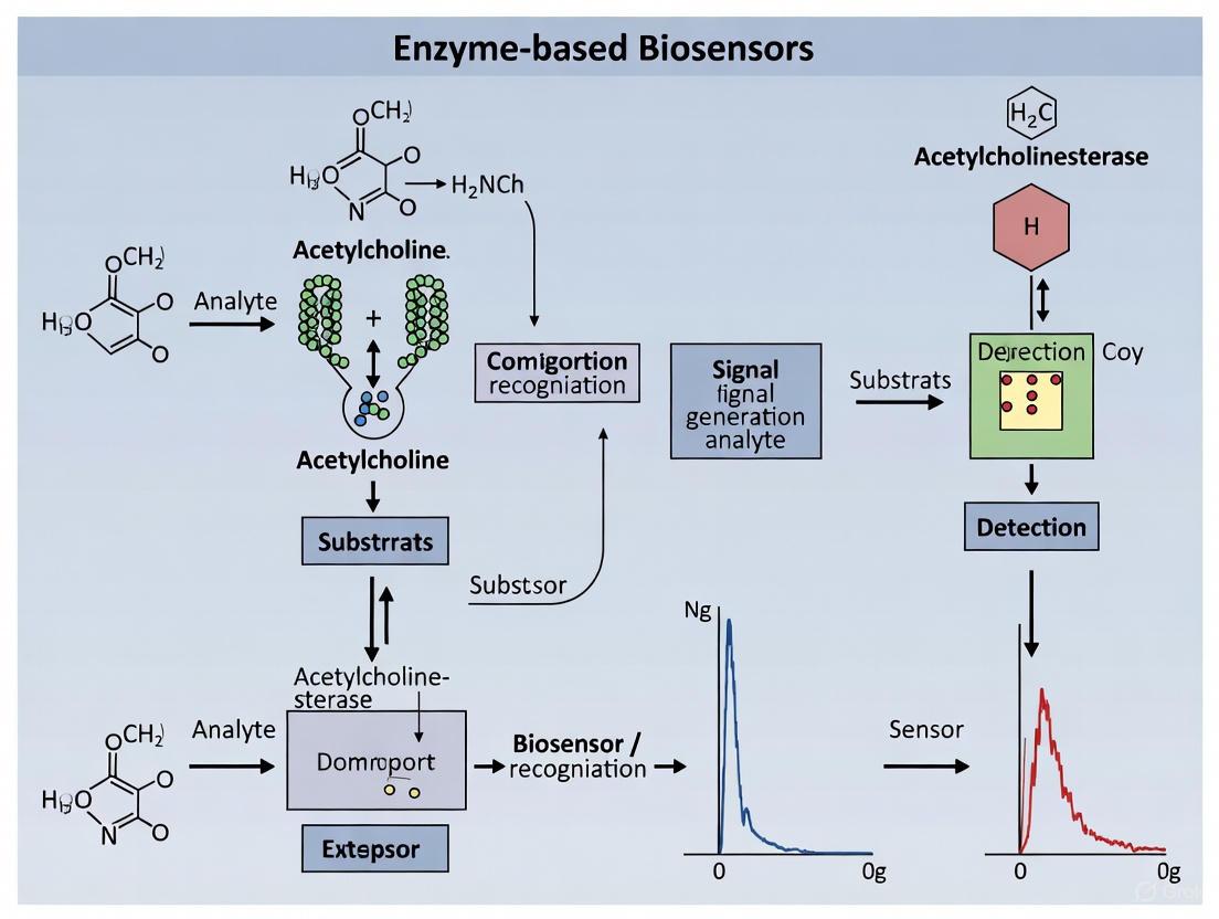
Enzyme-based biosensors are analytical devices that integrate a biological recognition element with a physicochemical transducer to detect target analytes with high specificity and sensitivity [4] [21]. In the critical field of organophosphate (OP) pesticide research, these biosensors leverage the specific inhibition of cholinesterase enzymes (acetylcholinesterase, AChE, or butyrylcholinesterase, BuChE) by OP compounds [22] [7]. The core function of the biosensor rests on its signal transduction system, which converts the biochemical event of enzyme inhibition into a quantifiable electronic or optical signal that researchers can measure and correlate with OP concentration [4] [21]. This conversion is paramount for developing rapid, on-site detection methods that serve as alternatives to complex laboratory techniques like chromatography [7] [23].
The general workflow of an inhibition-based OP biosensor involves exposing the immobilized enzyme to a sample. If OPs are present, they bind to the active site of the enzyme, inhibiting its catalytic activity. Subsequently, the enzyme's substrate is introduced. The degree of inhibition, reflected in the reduced catalytic conversion of the substrate, is then transduced into a measurable signal [22] [7]. The following diagram illustrates this core principle and the subsequent transduction pathways.
Transduction Mechanisms and Methodologies
The transducer is the core component that defines the type of biosensor and its operational principles. For OP detection, the most prevalent transduction mechanisms are electrochemical and optical.
Electrochemical Transduction
Electrochemical biosensors measure the electrical current or potential generated from the enzymatic reaction [4] [21]. In a typical AChE-based sensor, the enzyme catalyzes the hydrolysis of its substrate, acetylthiocholine, producing thiocholine. Thiocholine is an electroactive species that can be oxidized at the surface of an electrode, generating a measurable current [7]. The presence of an OP inhibitor reduces the rate of thiocholine production, leading to a corresponding decrease in the electrochemical signal. This method is widely used due to its high sensitivity, low cost, and potential for miniaturization [4].
Optical Transduction
Optical biosensors transduce the inhibition event into a measurable change in light properties [4]. This can include changes in absorbance, fluorescence, or chemiluminescence. For instance, the chemiluminescence (CL) assay described in the search results utilizes a coupled enzyme system where the product of BuChE activity (choline) is oxidized by choline oxidase (ChOx), producing hydrogen peroxide [22]. The hydrogen peroxide then reacts with luminol in a peroxidase (HRP)-catalyzed reaction, emitting photons. Inhibition of BuChE by OPs reduces the photon count, allowing for quantitative detection [22]. Another optical approach involves colorimetric sensors based on localized surface plasmon resonance (LSPR) of noble metal nanoparticles like silver (Ag) or gold (Au) [7] [24]. The aggregation of these nanoparticles, induced by the products of the enzymatic reaction, causes a visible color shift. OP inhibition prevents this aggregation and color change, providing a visual or spectrophotometric readout [7] [24].
The following diagram details the specific experimental workflow for a chemiluminescence-based assay, illustrating the steps from sample preparation to signal measurement.
Quantitative Performance Data
The performance of different biosensor configurations for OP detection can be evaluated based on key analytical figures of merit such as detection limit, linear range, and stability. The following table summarizes quantitative data for selected methodologies from the search results.
Table 1: Performance Metrics of Selected Enzyme-Based Biosensors for Organophosphate Detection
| Transduction Method | Target OPs | Linear Detection Range | Detection Limit | Stability / Reproducibility | Source |
|---|---|---|---|---|---|
| Chemiluminescence (BuChE inhibition) | Methyl Paraoxon (MPOx) | 0.005 – 50 μg·L⁻¹ | Not Specified | Mean recovery: 93.2–98.6%; RSD: 0.99–1.67% | [22] |
| Chemiluminescence (BuChE inhibition) | Methyl Parathion (MP), Malathion (MT) | 0.5 – 1,000 μg·L⁻¹ | Not Specified | Not Specified | [22] |
| Colorimetric / LSPR (AChE inhibition, AgNP@DANC) | Chlorpyrifos (CPF) | 1 × 10⁻³ to 1 × 10⁻¹⁹ M | Ultralow (from wide range) | Extensive stability for six months | [7] |
| Colorimetric / LSPR (AChE inhibition, AgNP@DANC) | Malathion (MLT) | 1 × 10⁻³ to 1 × 10⁻¹⁷ M | Ultralow (from wide range) | Extensive stability for six months | [7] |
The Scientist's Toolkit: Essential Research Reagents and Materials
The development and execution of enzyme-based biosensors for OP research rely on a suite of specialized reagents and materials. The following table details key components and their functions in typical experimental setups.
Table 2: Key Research Reagent Solutions for OP Biosensor Development
| Reagent / Material | Function / Role in Biosensing | Example from Literature |
|---|---|---|
| Cholinesterase Enzymes (AChE, BuChE) | Biological recognition element; its inhibition by OPs is the basis for detection. | Butyrylcholinesterase from equine serum used in a high-throughput chemiluminescence assay [22]. Acetylcholinesterase from Electrophorus electricus immobilized on a nanocomposite [7]. |
| Enzyme Substrates (Acetylthiocholine, Butyrylcholine) | Converted by the active enzyme into an electroactive or chromogenic product; the reaction rate is measured. | Butyrylcholine chloride used as a substrate for BuChE [22]. Acetylthiocholine chloride hydrolyzed by AChE to produce thiocholine [7]. |
| Signal-Generating Enzymes (Choline Oxidase, Horseradish Peroxidase) | Used in coupled enzyme systems to amplify the signal from the primary enzymatic reaction. | Choline oxidase and horseradish peroxidase used to generate a chemiluminescent signal from choline [22]. |
| Chemiluminescent Probes (e.g., Luminol) | Emits light upon chemical reaction (e.g., with H₂O₂ in the presence of HRP), providing the optical readout. | 5-amino-2,3-dihydro-1,4-phthalazinedione (Luminol) used as the CL substrate [22]. |
| Stabilizing Agents (e.g., Trehalose) | Protects enzymes during drying and storage, enhancing their shelf-life and operational stability at room temperature. | Dextrose and trehalose used to stabilize BuChE pre-loaded in microplates [22]. |
| Nanomaterial Composites (e.g., AgNP@DANC) | Serves as an advanced immobilization matrix; enhances enzyme stability, sensitivity, and can possess nanozyme activity. | Rice husk-derived dialdehyde nanocellulose capped silver nanoparticles (AgNP@DANC) used to immobilize and stabilize AChE [7]. |
| Immobilization Matrices (e.g., Silica Nanoparticles) | Provides a solid support for enzyme attachment, improving reusability and stability in flow-based systems. | Biomimetic silica nanoparticles used to encapsulate BuChE and OPH for continuous aerosol monitoring [23]. |
Enzyme-based biosensors represent a sophisticated class of analytical devices that integrate biological recognition elements with physicochemical transducers to detect specific analytes with high specificity and sensitivity. These devices function by immobilizing biological components onto transducer surfaces, enabling the detection of target substances without reagent addition to sample solutions [25]. The interaction between the target analyte and biological element produces physicochemical changes that transducers convert into measurable signals proportional to analyte concentration [25]. Within the specific context of organophosphate (OP) pesticide detection, enzyme-based biosensors have emerged as vital tools for environmental monitoring and food safety control, offering distinct advantages including high sensitivity and specificity, portability, cost-effectiveness, and potential for miniaturization and point-of-care diagnostic testing [25] [6].
This technical guide examines the three fundamental components constituting enzymatic biosensors for OP detection: the bioreceptor (biological recognition element), the transducer (signal conversion unit), and the immobilization matrix (enzyme stabilization framework). The precise integration of these components dictates biosensor performance metrics including sensitivity, selectivity, reproducibility, and operational stability [26] [27]. For organophosphate detection, specific enzymes including acetylcholinesterase (AChE) and organophosphate hydrolase (OPH) serve as primary biorecognition elements, enabling the development of biosensing systems that provide effective alternatives to traditional, time-consuming analytical methods [6] [28].
Core Component Analysis
Bioreceptor
The bioreceptor constitutes the biological recognition element of a biosensor, responsible for specific interaction with the target analyte. In enzymatic biosensors for organophosphate detection, the bioreceptors are enzymes that either directly catalyze OP hydrolysis or undergo inhibition by OPs, enabling quantitative detection [6].
- Acetylcholinesterase (AChE): This enzyme serves as the primary bioreceptor in inhibition-based biosensors for OP detection. Organophosphates specifically and irreversibly inhibit AChE activity by phosphorylating the serine residue within its active site [6] [28]. The detection mechanism relies on measuring decreased enzyme activity following exposure to OPs, where the inhibition level correlates with pesticide concentration.
- Organophosphate Hydrolase (OPH): Also known as paraoxonase, OPH acts as a catalytic bioreceptor that directly hydrolyzes OP compounds, including paraoxon, coumaphos, and diazinon [6]. The enzymatic reaction yields stoichiometric amounts of protons and chromophoric products, enabling detection through various transduction methods. OPH-based biosensors provide advantages of continuous monitoring and regenerability compared to inhibition-based approaches.
The operating principle of an enzyme-based biosensor involves detecting changes occurring during substrate consumption or product formation in enzymatic reactions, such as proton concentration, gas release/uptake, light emission/absorption, or heat emission [25]. These changes are converted by transducers into quantifiable electrical, optical, or thermal signals [25].
Transducer
The transducer functions as the signal conversion unit, transforming biochemical interactions occurring at the bioreceptor into measurable electronic signals. The choice of transducer significantly influences sensitivity, detection limits, and applicability for field use [25] [29].
Table 1: Transducer Types in Enzyme-Based Biosensors for OP Detection
| Transducer Type | Measurement Principle | Detection Method for OPs | Advantages |
|---|---|---|---|
| Electrochemical | Measures changes in electrical properties (current, potential, impedance) from redox reactions [25] | AChE inhibition: Measures thiocholine oxidation current [25] | Simplicity, portability, low cost, high sensitivity [25] |
| OPH catalysis: Measures pH change or hydrolytic product oxidation [25] | |||
| Optical | Detects changes in light properties (absorbance, fluorescence, luminescence) [25] [6] | AChE inhibition: Monitors colorimetric or fluorimetric substrate conversion [6] | High sensitivity, multiplexing capability, resistance to electromagnetic interference [6] |
| OPH catalysis: Tracks chromophoric product formation (e.g., p-nitrophenol from paraoxon) [6] | |||
| Thermal/Calorimetric | Measures heat emission from enzymatic reactions [25] | Monitors enthalpy changes from substrate hydrolysis | Label-free detection, applicable to various substrates |
| Piezoelectric | Detects mass changes on crystal surface through resonance frequency shifts [25] [29] | Measures mass loading from enzyme-OP binding or product formation | High sensitivity to mass changes, real-time monitoring |
Electrochemical biosensors represent the most extensively used transducer type, with amperometric systems being particularly common [25]. These systems apply a fixed potential to the working electrode and measure current generated from oxidation or reduction of electroactive species involved in the enzymatic reaction [25]. Optical biosensors have gained significant traction for OP detection due to advantages including high sensitivity and selectivity, simple operation, fast response, and relatively inexpensive instrumentation [6].
Immobilization Matrix
Enzyme immobilization represents a critical and essential step in biosensor design, profoundly affecting performance through influences on enzyme orientation, loading, mobility, stability, structure, and biological activity [27]. Effective immobilization maintains enzyme structure and function, ensures tight binding to the transducer surface, and preserves biological activity throughout biosensor operation [27].
Table 2: Enzyme Immobilization Methods for Biosensors
| Immobilization Method | Principle | Advantages | Disadvantages | Relevance to OP Detection |
|---|---|---|---|---|
| Adsorption | Physical attachment via weak bonds (Van der Waals, electrostatic, hydrophobic) [25] [27] | Simple, inexpensive, minimal enzyme modification [25] | Weak bonding sensitive to environmental changes (pH, temperature), potential leaching [25] | Limited use due to stability issues in field applications |
| Covalent Bonding | Formation of stable covalent bonds between enzyme and support [25] [27] | Strong binding, high stability, uniform surface coverage [25] | Potential enzyme activity loss, requires chemical modification [25] | Widely used; provides operational stability for AChE/OPH biosensors |
| Entrapment | Enzyme confinement within porous matrices (polymers, sol-gels, carbon paste) [25] [27] | No chemical modification, simultaneous deposition of enzymes/mediators [27] | Diffusion limitations for substrate/product, potential enzyme leakage [25] | Useful for OPH biosensors to retain cofactors; minimizes matrix effects |
| Cross-linking | Intermolecular covalent bonding between enzymes using bifunctional agents (glutaraldehyde) [25] [27] | Simple, strong chemical binding, high enzyme loading [25] [27] | Potential activity loss due to rigidification, possible diffusion limitations [25] | Enhances stability of immobilized AChE for reusable OP sensors |
| Affinity | Specific bioaffinity interactions (avidin-biotin, lectin-carbohydrate, antibody-antigen) [27] | Controlled orientation, preserves active site accessibility, minimizes denaturation [27] | Requires specific binding groups, more complex implementation [27] | Emerging approach for oriented AChE immobilization to enhance sensitivity |
The selection of appropriate immobilization strategy depends on enzyme characteristics, transducer properties, and intended application requirements. For organophosphate biosensors, covalent bonding and entrapment methods are frequently employed to enhance operational stability and maintain enzyme activity under various environmental conditions [27]. Recent approaches incorporate nanomaterials including carbon nanotubes, metal nanoparticles, and conducting polymer nanowires to create advanced immobilization matrices that increase surface area, enhance electron transfer, and improve biosensor sensitivity [27].
Integration for Organophosphate Detection
The effective integration of bioreceptor, transducer, and immobilization matrix enables the development of sophisticated biosensing platforms for organophosphate pesticide detection. The World Health Organization classifies OPs as extremely toxic compounds due to their specificity for acetylcholinesterase, causing irreversible harm to the nervous system [6] [28]. The excessive use of these pesticides, particularly in developing countries, has necessitated the development of easy, rapid, and sensitive detection methods for monitoring OP residues in food and water [6].
The logical relationship and workflow between core components in an enzyme-based biosensor for organophosphate detection can be visualized as follows:
This workflow illustrates how organophosphate compounds in sample solutions interact specifically with the enzyme bioreceptor (AChE or OPH), which is stabilized by the immobilization matrix. The biochemical recognition event generates physicochemical changes that the transducer converts into measurable signals, enabling quantitative OP detection.
Experimental Protocols for OP Detection
Acetylcholinesterase Inhibition-Based Protocol
This protocol details the procedure for detecting organophosphates using an AChE inhibition-based biosensor with electrochemical transduction [25] [6].
Principle: Organophosphates irreversibly inhibit AChE activity, reducing enzymatic conversion of substrates to electroactive products. The percentage inhibition correlates with OP concentration.
Materials:
- Acetylcholinesterase enzyme (Electric eel or recombinant source)
- Acetylthiocholine iodide or acetylcholine chloride substrate
- Phosphate buffer (0.1 M, pH 7.4)
- Organophosphate standards (paraoxon, parathion, malathion)
- Electrochemical cell with working, reference, and counter electrodes
- Immobilization matrix components (glutaraldehyde, BSA, Nafion, chitosan)
Procedure:
- Enzyme Immobilization: Immobilize AChE on electrode surface using preferred method:
Baseline Measurement: Incubate AChE biosensor in electrochemical cell with acetylthiocholine substrate (1 mM) in phosphate buffer. Apply +0.5V vs Ag/AgCl and record steady-state oxidation current (I₀).
Inhibition Phase: Incubate AChE biosensor with OP standard/sample for 10-15 minutes.
Post-Inhibition Measurement: Re-measure enzymatic activity with substrate as in step 2, record current (Iᵢ).
Quantification: Calculate percentage inhibition: % Inhibition = [(I₀ - Iᵢ)/I₀] × 100. Plot calibration curve using OP standards.
Organophosphate Hydrolase Catalytic Biosensor Protocol
This protocol describes OP detection using direct catalytic hydrolysis by OPH with optical transduction [6].
Principle: OPH catalyzes hydrolysis of organophosphates, generating colored or fluorescent products proportional to OP concentration.
Materials:
- Organophosphate hydrolase enzyme (recombinant source)
- Organophosphate standards (paraoxon, parathion)
- Buffer solution (Tris-HCl or HEPES, pH 8.5-9.0)
- Spectrophotometer or fluorimeter
- Immobilization support (agarose beads, sol-gel, polymer membranes)
Procedure:
- Enzyme Immobilization: Immobilize OPH on selected support:
Assay Setup: Add immobilized OPH to OP standards/samples in buffer.
Incubation: Incubate reaction mixture at 30-37°C for 5-15 minutes with agitation.
Product Measurement: Monitor hydrolytic product formation:
- Spectrophotometric: Measure absorbance at 400-405 nm for p-nitrophenol (ε = 17,000 M⁻¹cm⁻¹) from paraoxon hydrolysis [6].
- Fluorimetric: Monitor fluorescence change for coumaphos hydrolysis (excitation/emission: 360/454 nm).
Quantification: Calculate OP concentration from product standard curve.
The Scientist's Toolkit: Research Reagent Solutions
Table 3: Essential Research Reagents for Enzyme-Based OP Biosensors
| Reagent/Material | Function | Specification Notes |
|---|---|---|
| Acetylcholinesterase (AChE) | Bioreceptor for inhibition-based detection | Source: Electric eel (cheaper) or recombinant (higher purity); Specific activity: ≥500 U/mg [6] |
| Organophosphate Hydrolase (OPH) | Bioreceptor for catalytic detection | Recombinant from Pseudomonas diminuta or Flavobacterium; Specific activity: ≥1000 U/mg [6] |
| Acetylthiocholine iodide | Electrochemical substrate for AChE | Enzymatic product thiocholine oxidizes at electrode; Purity: ≥98% [25] |
| Paraoxon ethyl | Standard OP for calibration and inhibition studies | Model compound for OP detection; Purity: ≥95% [6] |
| Glutaraldehyde | Cross-linking agent for covalent immobilization | Creates stable enzyme linkages; Concentration: 2.5% v/v [25] [27] |
| Nafion polymer | Entrapment matrix for enzyme immobilization | Cation-exchange polymer; Protects electrode from fouling; Concentration: 0.5-1% w/v [27] |
| Sol-gel precursors (TMOS) | Silica matrix for enzyme entrapment | Forms porous network around enzyme; Tetraalkoxysilane precursors [27] |
| Screen-printed electrodes | Disposable electrochemical transducers | Carbon, gold, or platinum working electrodes; Enable mass production [25] |
The strategic selection and integration of these core components—specific bioreceptors, appropriate transducers, and effective immobilization matrices—enables researchers to develop sophisticated biosensing platforms for sensitive and selective detection of organophosphate pesticides. These systems continue to evolve through nanotechnology integration and immobilization strategy optimization, addressing the critical need for rapid environmental and food safety monitoring as emphasized by international health and agricultural organizations [6].
Advanced Sensing Platforms and Real-World Deployments
Electrochemical biosensors, particularly amperometric and potentiometric systems, are pivotal analytical tools that combine the specificity of biological recognition elements with the sensitivity of electrochemical transducers. These devices are integral to modern research, enabling the detection and quantification of various analytes. Within the specific context of organophosphate (OP) research, enzyme-based biosensors function primarily on the principle of enzyme inhibition [1] [30]. The core mechanism involves the interaction between the target OP and a specific enzyme, such as acetylcholinesterase (AChE) or organophosphate hydrolase (OPH). OP compounds irreversibly phosphorylate the serine hydroxyl group in the active site of AChE, leading to a decrease in enzymatic activity [16] [30]. This inhibition directly modulates the production of electroactive species (e.g., protons or electrons) in the enzyme-catalyzed reaction, which is then measured as a change in current (amperometry) or potential (potentiometry) at the transducer surface. This measurable signal is quantitatively related to the concentration of the toxic inhibitor, providing a powerful method for detecting these pesticides [1] [4].
Fundamental Principles and Transducer Mechanisms
Amperometric Biosensors
Amperometric biosensors operate by applying a constant potential and measuring the resulting current generated from the oxidation or reduction of an electroactive species involved in the biocatalytic reaction [31] [4]. The measured current is directly proportional to the concentration of the target analyte.
- Working Principle: In a typical enzyme-based system, the enzyme catalyzes a reaction that produces or consumes an electroactive product. For instance, AChE hydrolyzes acetylcholine to produce choline and acetic acid. The thiocholine produced from subsequent reactions can be oxidized at the electrode surface, generating a measurable anodic current. Inhibition of AChE by OPs reduces this current signal [30]. Alternatively, biosensors using organophosphate hydrolase (OPH) directly catalyze the hydrolysis of OPs, producing electroactive species like p-nitrophenol, which can be oxidized and detected amperometrically [14].
- Key Characteristics: These sensors are known for their high sensitivity, low detection limits, and excellent compatibility with miniaturized, portable devices for on-site analysis [31].
Potentiometric Biosensors
Potentiometric biosensors measure the change in potential (voltage) at an electrode surface under conditions of zero current. This potential change results from the accumulation of ions or charged molecules due to an enzymatic reaction [4].
- Working Principle: The most common approach involves the use of ion-selective electrodes (ISEs) or pH-sensitive field-effect transistors (FETs). An enzymatic reaction that generates or consumes ions (e.g., H⁺, NH₄⁺) leads to a change in the local ion concentration. This shift is measured as a potential difference relative to a reference electrode. For example, the hydrolysis of urea by urease increases the pH, which can be detected by a pH electrode [4]. While less common for OPs than amperometric systems, the inhibition of AChE can be monitored by tracking the reduced production of acetic acid, leading to a smaller pH change.
- Key Characteristics: Potentiometric sensors offer simplicity and low power consumption. However, they can be susceptible to interference from other ions in the sample matrix and may have a slower response time compared to amperometric sensors.
Table 1: Comparison of Amperometric and Potentiometric Transduction Principles
| Feature | Amperometric Biosensors | Potentiometric Biosensors |
|---|---|---|
| Measured Quantity | Current | Potential (Voltage) |
| Operating Condition | Constant applied potential | Zero current flow |
| Signal Dependency | Mass transport & reaction rate | Ionic activity (Nernst equation) |
| Typical Sensitivity | High (nano- to micro-ampere) | Moderate (millivolts per decade) |
| Response Time | Typically fast (seconds) | Can be slower |
| Common Interferences | Other electroactive species | Other ions in sample matrix |
Experimental Protocols for Organophosphate Detection
Protocol 1: Amperometric Biosensor Using Acetylcholinesterase (AChE) Inhibition
This protocol details the fabrication and operation of a classic inhibition-based biosensor for neurotoxic OPs and carbamates [1] [30].
1. Biorecognition Element Immobilization:
- Materials: Acetylcholinesterase (AChE) from electric eel or recombinant mutant enzyme; glutaraldehyde (cross-linker); bovine serum albumin (BSA); chitosan or carbon nanotube nanocomposite for the electrode matrix.
- Procedure: A mixture of AChE (0.5-2 U/µL) and BSA (1% w/v) is prepared in a phosphate buffer (0.1 M, pH 7.4). A 10 µL aliquot is deposited on the surface of a polished glassy carbon electrode (GCE). Then, 5 µL of glutaraldehyde (2.5% v/v) is added as a cross-linking agent to form a stable network. The electrode is dried for 1 hour at 4°C and then rinsed with buffer to remove any unbound enzyme [1].
2. Baseline Activity Measurement:
- Electrochemical Cell Setup: Use a three-electrode system with the AChE-modified GCE as the working electrode, an Ag/AgCl reference electrode, and a platinum wire counter electrode. The cell contains 10 mL of 0.1 M phosphate buffer (pH 7.4) with 0.1 M KCl as the supporting electrolyte.
- Amperometric Measurement: Apply a constant potential of +0.7 V vs. Ag/AgCl. Under stirring, inject a known concentration of the substrate acetylthiocholine iodide (ATCh, 1.0 mM final concentration). Monitor the oxidation current of the enzymatic product, thiocholine, until a stable steady-state current (I₀) is achieved [30].
3. Inhibition and Pesticide Detection:
- Incubation: Immerse the biosensor in a sample solution containing the target OP pesticide (e.g., paraoxon, chlorpyrifos-oxon) for a fixed incubation period (e.g., 10-15 minutes).
- Post-Inhibition Measurement: Wash the electrode gently with buffer and place it in a fresh electrochemical cell. Repeat the amperometric measurement with the same concentration of ATCh substrate (1.0 mM) and record the new steady-state current (Iᵢ).
- Quantification: The percentage of enzyme inhibition is calculated as % Inhibition = [(I₀ - Iᵢ) / I₀] × 100. This value is correlated with the pesticide concentration using a pre-established calibration curve [1].
Protocol 2: Potentiometric Biosensor Using Organophosphate Hydrolase (OPH)
This protocol utilizes OPH, which directly degrades OPs, making it a superior alternative for some applications by eliminating the incubation step required in inhibition-based assays [14].
1. OPH Enzyme Integration:
- Materials: Recombinant Organophosphate Hydrolase (OPH) expressed from the 'opd' gene; polyacrylamide gel or poly(carbamoyl sulfonate) hydrogel for entrapment; pH-sensitive field-effect transistor (FET).
- Procedure: The OPH enzyme (activity ~2.75 U/mL) is mixed with the hydrogel precursor solution. This mixture is spin-coated directly onto the gate surface of the pH-FET device and polymerized via UV exposure to form a thin, enzymatic film approximately 50-100 µm thick [14].
2. Direct Potentiometric Detection:
- Measurement Setup: The OPH-modified FET is integrated into a flow-cell or immersed in a stirred sample solution. A stable potential baseline (E₀) is recorded against a reference electrode.
- Analyte Introduction: The sample containing the OP analyte (e.g., paraoxon) is introduced. OPH catalyzes the hydrolysis reaction: Paraoxon + H₂O → p-Nitrophenol + Diethyl phosphate + H⁺.
- Signal Recording: The release of protons (H⁺) during the reaction causes a local decrease in pH at the FET gate surface. This is measured as a change in potential (∆E) relative to E₀. The rate and magnitude of this potential shift are proportional to the concentration of the OP pesticide in the sample [14].
Signaling Pathways and Workflow Visualization
AChE Inhibition Pathway for OP Detection
The following diagram illustrates the biochemical signaling pathway of AChE inhibition by organophosphates, which forms the basis for many amperometric biosensors.
Experimental Workflow for an Amperometric Biosensor
This workflow outlines the key steps in a typical experiment using an amperometric AChE-based biosensor for pesticide detection.
The Scientist's Toolkit: Essential Research Reagents
Successful development and deployment of electrochemical biosensors for OP research rely on a suite of key materials and reagents.
Table 2: Key Research Reagents for Enzyme-Based OP Biosensors
| Reagent/Material | Function/Role | Specific Examples & Notes |
|---|---|---|
| Acetylcholinesterase (AChE) | Primary biorecognition element; inhibition by OPs enables detection. | Electric eel, bovine erythrocyte, or genetically engineered mutants (e.g., from Drosophila melanogaster) for enhanced sensitivity/selectivity [1]. |
| Organophosphate Hydrolase (OPH) | Biorecognition element; directly catalyzes OP hydrolysis. | Recombinant enzyme expressed from the 'opd' gene; functions without an incubation step [14]. |
| Acetylthiocholine (ATCh) | Enzyme substrate; hydrolysis produces electroactive thiocholine. | Preferred over acetylcholine for amperometric sensors as thiocholine is easily oxidized [30]. |
| Glutaraldehyde | Cross-linking agent for enzyme immobilization. | Creates covalent bonds between enzyme molecules and the support matrix, enhancing stability [31]. |
| Nanomaterials | Transducer surface modification to enhance signal. | Graphene, carbon nanotubes, metal nanoparticles (e.g., gold) increase surface area and electron transfer [16] [4]. |
| Chlorella sp. (Microalgae) | Whole-cell biorecognition element based on PS II inhibition. | Used in novel amperometric biosensors; provides a sustainable, enzyme-free alternative [31]. |
| Buffer Solutions (PBS/Tris-HCl) | Maintain optimal pH and ionic strength for enzyme activity. | Phosphate Buffered Saline (PBS, pH 7.4) is commonly used for AChE-based sensors [31]. |
Amperometric and potentiometric biosensors represent robust and versatile platforms for the sensitive detection of organophosphate pesticides. Amperometric systems, with their high sensitivity and compatibility with miniaturization, are particularly dominant in this field, especially those based on the inhibition of AChE. The ongoing integration of advanced materials like nanomaterials and genetically engineered enzymes, combined with streamlined experimental protocols, continues to push the boundaries of detection limits, selectivity, and operational stability [1] [16] [4]. The choice between amperometric and potentiometric transduction, as well as between inhibitory (AChE) and catalytic (OPH) enzymes, depends on the specific requirements of the analysis, such as the need for speed, sensitivity, or applicability to complex samples. Future research is poised to leverage artificial intelligence for sensor design and data analysis, alongside the development of multifunctional and fully integrated portable devices, to further advance the capabilities of these biosensors in environmental monitoring, food safety, and public health protection [16] [32].
Principles of Enzyme-Based Optical Biosensing for OPs
The core principle of enzyme-based biosensors for organophosphate detection relies on the inhibition of a specific enzyme, primarily acetylcholinesterase (AChE). In its active state, AChE catalyzes the hydrolysis of its substrate (e.g., acetylthiocholine or acetylcholine), leading to a measurable product. The presence of OPs inhibits this catalytic activity, reducing the amount of product formed. This inhibition is directly proportional to the OP concentration, enabling quantitative detection [1] [7]. Optical biosensors transduce this biochemical event into a readable signal through changes in light properties.
- Biological Recognition: AChE is immobilized onto a sensor platform.
- Catalytic Reaction: Upon introduction of the substrate, the active enzyme hydrolyzes it, generating a product.
- Inhibition by OPs: Organophosphates bind to the enzyme's active site, inhibiting its function.
- Optical Transduction: The rate of product formation decreases, which is detected as a change in color, fluorescence intensity, or luminescent signal.
Optical Detection Strategies and Methodologies
The following sections detail the primary optical sensing modalities, with specific experimental considerations for detecting OPs.
Colorimetric Sensing
Colorimetric detection involves a visual or spectrophotometric change in color, often measured by a shift in the UV-Vis absorption peak or a simple color change.
- Mechanism: The most common strategy for OPs exploits the aggregation of metal nanoparticles. The enzymatic reaction product (e.g., thiocholine from acetylthiocholine hydrolysis) induces the aggregation of nanoparticles like silver (AgNPs) or gold (AuNPs), causing a distinct color shift. The presence of OPs inhibits the reaction, preventing aggregation and the associated color change [7].
- Typical Workflow:
- Immobilization: AChE is immobilized on a substrate or in solution with functionalized nanoparticles (e.g., nanocellulose-capped AgNPs for stability and biocompatibility) [7].
- Substrate Introduction: The substrate (acetylthiocholine) is added. In the absence of OPs, the enzyme hydrolyzes it, generating thiocholine.
- Signal Generation: Thiocholine causes the aggregation of AgNPs, leading to a color change from yellow (dispersion) to brown (aggregation) and a decrease in the absorption peak at ~414 nm [7].
- Inhibition Detection: In the presence of OPs, enzyme inhibition reduces thiocholine production, thereby reducing the degree of nanoparticle aggregation and the associated signal change. The percentage inhibition is calculated and correlated to the OP concentration.
Table 1: Key Reagents for a Colorimetric AgNP-based AChE Biosensor [7]
| Research Reagent | Function in the Experiment |
|---|---|
| Acetylcholinesterase (AChE) | Biological recognition element; catalyzes the hydrolysis of acetylthiocholine. |
| Acetylthiocholine Chloride (ATChCl) | Enzyme substrate; hydrolysis product (thiocholine) induces nanoparticle aggregation. |
| Silver Nanoparticles (AgNPs) | Optical transducer; color change upon aggregation signals the enzymatic activity. |
| Functionalized Nanocellulose (DANC) | Serves as a stabilizing and reducing agent for AgNPs; provides a biocompatible matrix for enzyme immobilization. |
| Tris Buffer | Provides a stable pH environment for the enzymatic reaction. |
Fluorescence Sensing
Fluorescence-based biosensors detect changes in the fluorescence intensity, lifetime, or wavelength of a fluorophore.
- Mechanism: This can involve direct fluorescence of metal nanoclusters (MNCs), fluorescence quenching, or fluorescence resonance energy transfer (FRET). Ultra-small MNCs, such as gold or silver nanoclusters, exhibit strong photoluminescence and high photochemical stability, making them excellent transducers [33].
- Typical Workflow:
- Probe Design: A fluorescent probe is designed where the emission is linked to enzymatic activity. This could be a fluorogenic enzyme substrate or MNCs whose fluorescence is modulated by the enzymatic product.
- Enzymatic Activation: In an uninhibited state, the enzymatic reaction produces a change in the fluorescence signal (e.g., turn-on, turn-off, or spectral shift).
- Inhibition Readout: The presence of OPs quenches the fluorescence by inhibiting the production of the activating species. The degree of fluorescence quenching is proportional to the OP concentration. Some advanced sensors use mutant enzymes with enhanced sensitivity patterns to improve selectivity [1] [33].
Electrochemiluminescence (ECL) Sensing
ECL involves the generation of light by an electrochemically initiated reaction. It combines the advantages of electrochemical control with the high sensitivity of optical detection.
- Mechanism: A luminescent compound (luminophore) undergoes a redox reaction at an electrode surface to form an excited state that then emits light. The enzymatic reaction can either generate a coreactant or directly modulate the ECL efficiency.
- Typical Workflow:
- Sensor Fabrication: The luminophore (e.g., ruthenium complexes, quantum dots) and the enzyme (AChE) are co-immobilized on an electrode surface.
- Electrochemical Stimulation: A voltage is applied to the electrode, triggering the ECL reaction and producing a light signal.
- Enzymatic Modulation: The enzymatic hydrolysis of its substrate can produce species that enhance or quench the ECL signal.
- Inhibition Measurement: OPs inhibit the enzyme, altering the production of the ECL-modulating species and resulting in a measurable change (often a decrease) in the ECL intensity.
Experimental Protocol: Colorimetric AgNP@DANC Biosensor
The following detailed protocol is adapted from a recent study for the detection of chlorpyrifos and malathion [7].
Title: Detection of Organophosphates using AChE-Inhibited Aggregation of AgNP@DANC Nanocomposite.
1. Reagents and Materials
- Acetylcholinesterase (AChE, from Electrophorus electricus)
- Acetylthiocholine chloride (ATChCl)
- Dialdehyde nanocellulose-modified silver nanocomposite (AgNP@DANC)
- Tris-HCl buffer (pH ~8.0)
- Organophosphate standards (e.g., chlorpyrifos, malathion)
- Deionized water
2. Equipment
- UV-Vis Spectrophotometer
- Microcentrifuge tubes
- Vortex mixer
- Micropipettes
- Cuvettes
3. Procedure Step 1: Biosensor Incubation
- In a microcentrifuge tube, mix 50 µL of AChE solution with 50 µL of Tris buffer.
- Incubate this mixture with 50 µL of varying concentrations of the target OP (or blank for control) for 10-15 minutes at room temperature to allow for enzyme inhibition.
Step 2: Enzymatic Reaction Initiation
- Add 50 µL of the substrate (ATChCl) to the mixture to initiate the enzymatic reaction.
- Vortex the mixture gently and allow it to incubate for a fixed time (e.g., 10 minutes).
Step 3: Signal Development and Measurement
- Add 100 µL of the AgNP@DANC nanocomposite to the reaction mixture.
- Monitor the solution's color change visually or use a UV-Vis spectrophotometer to measure the absorption spectrum, specifically tracking the decrease in the surface plasmon resonance (SPR) peak at ~414 nm.
Step 4: Data Analysis
- Calculate the percentage inhibition using the formula:
Inhibition (%) = [(A_control - A_sample) / A_control] × 100whereA_controlis the absorbance of the uninhibited reaction andA_sampleis the absorbance in the presence of OPs. - Plot the inhibition percentage against the logarithm of the OP concentration to generate a calibration curve.
Performance Comparison of Optical Biosensor Strategies
Table 2: Comparison of Optical Biosensing Strategies for Organophosphate Detection
| Sensing Strategy | Transduction Principle | Key Materials | Typical LOD (Reported Example) | Advantages | Disadvantages |
|---|---|---|---|---|---|
| Colorimetric | Absorption shift / Nanoparticle aggregation | AgNPs, AuNPs, Nanocellulose | Chlorpyrifos: ~1×10⁻¹⁹ M [7] | Simple, low-cost, rapid, suitable for on-site testing | Susceptible to sample matrix color interference, moderate sensitivity |
| Fluorescence | Change in fluorescence intensity/wavelength | Metal Nanoclusters (Au, Ag), fluorophores | High sensitivity, potential for multiplexing, real-time monitoring | Can be affected by photo-bleaching, may require complex probe design | |
| Electrochemiluminescence | Light emission from electro-generated species | Luminophores (e.g., Ru(bpy)₃²⁺), co-reactants | Very high sensitivity, low background noise, wide dynamic range | Requires electrochemical instrumentation, more complex system setup |
The Scientist's Toolkit: Essential Research Reagents
Table 3: Key Research Reagent Solutions for Enzyme-Based Optical Biosensors [1] [7] [33]
| Reagent / Material | Function and Role in Biosensor Development |
|---|---|
| Acetylcholinesterase (AChE) | The primary biorecognition element. Its inhibition is the basis for OP detection. Wild-type and genetically engineered variants from different sources (e.g., Drosophila melanogaster) offer varying sensitivities [1]. |
| Acetylthiocholine Iodide/Chloride | The preferred substrate for AChE in biosensors. Its hydrolysis product, thiocholine, reacts with metal nanoparticles or other probes to generate the optical signal [7]. |
| Metal Nanoparticles (Ag, Au) | Serve as excellent colorimetric transducers due to their strong, localized surface plasmon resonance (LSPR) that shifts upon aggregation or binding events [7] [34]. |
| Metal Nanoclusters (MNCs) | Ultra-small nanomaterials (e.g., AuNCs, AgNCs) with molecule-like properties, including strong photoluminescence, used as fluorophores in highly sensitive detection [33]. |
| Functionalized Nanocellulose | A biocompatible, renewable nanomaterial used as a stabilizing matrix for nanoparticles and for enzyme immobilization, enhancing sensor stability and performance [7]. |
| Polydimethylsiloxane (PDMS) | A transparent, flexible polymer used as a substrate for building microfluidic devices and wearable optical sensor platforms [35]. |
Experimental Workflow and Data Interpretation
The following diagram illustrates the logical workflow and signaling pathway for a typical enzyme inhibition-based colorimetric biosensor.
Diagram Title: AChE Inhibition Workflow for OP Detection
For data interpretation, the calculated percentage inhibition is plotted against the logarithm of the standard OP concentration to create a calibration curve. This curve is used to interpolate the concentration of OPs in unknown samples. Advanced data analysis using machine learning algorithms, such as artificial neural networks (ANNs), can be employed to resolve mixtures of different insecticides when using an array of biosensors with different AChE enzyme variants [1] [36].
The extensive global use of organophosphate pesticides (OPs) in agriculture has created an urgent need for reliable detection methods to monitor residual levels in environmental and food samples [7] [37]. Organophosphates are extremely toxic compounds that inhibit acetylcholinesterase (AChE), an enzyme crucial for nerve function, leading to neuromuscular paralysis and potential death in acute cases [38] [37]. While conventional detection methods like gas chromatography and high-performance liquid chromatography offer accuracy, they are costly, time-intensive, and require skilled operators, limiting their use for rapid on-site screening [1] [39].
Enzyme-based biosensors have emerged as promising analytical tools that overcome these limitations by combining biological recognition elements with transducers to convert biochemical responses into quantifiable signals [6]. The fundamental principle behind these biosensors exploits the inhibition of AChE by organophosphates – the degree of enzyme inhibition correlates directly with pesticide concentration [7] [1]. Recent advancements have incorporated innovative nanomaterials to significantly enhance biosensor performance through increased surface area for enzyme immobilization, improved electron transfer rates, and amplified signal detection [7] [40] [41].
This technical guide examines three key classes of nanomaterials – functionalized nanocellulose, metal-organic frameworks (MOFs), and noble metal nanoparticles – that are revolutionizing the design and capability of enzyme-based biosensors for organophosphate detection. We explore their unique properties, synthesis methodologies, integration strategies, and performance metrics, providing researchers with a comprehensive resource for developing next-generation biosensing platforms.
Fundamental Principles of Enzyme-Based Biosensors for Organophosphates
Acetylcholinesterase Inhibition Mechanism
The operational principle of AChE-based biosensors centers on the enzyme's catalytic activity toward acetylcholine and its analogs. In normal function, AChE hydrolyzes the neurotransmitter acetylcholine into acetate and choline, maintaining proper neural function [38]. Organophosphate pesticides irreversibly phosphorylate the serine residue in the active site of AChE, inhibiting its catalytic function and leading to acetylcholine accumulation, which causes uncontrolled neuromuscular transmission [37].
In biosensing applications, this inhibition mechanism is utilized by measuring the decrease in enzymatic activity when exposed to OPs. The typical reaction scheme involves:
- Enzymatic reaction: Acetylthiocholine (ATCh) → Thiocholine + Acetate
- Inhibition reaction: AChE + Organophosphate → Phosphorylated AChE (inactive)
The presence of OPs reduces the production of thiocholine, which serves as the electroactive or optically detectable product in most biosensing systems [7] [1]. The percentage of enzyme inhibition is calculated as:
Inhibition (%) = [(I₀ - I)/I₀] × 100
Where I₀ is the response without inhibitor and I is the response with inhibitor [1].
Biosensor Architectures and Signal Transduction
Enzyme-based biosensors for OP detection employ various signal transduction mechanisms, with electrochemical and optical systems being most prevalent:
- Electrochemical biosensors measure current (amperometric), potential (potentiometric), or impedance changes resulting from enzymatic activity inhibition [40] [37].
- Optical biosensors detect changes in light absorption, fluorescence, or surface plasmon resonance resulting from the inhibition mechanism [6] [41].
The following diagram illustrates the core inhibition mechanism and signal transduction pathways:
Figure 1: AChE Inhibition and Signal Transduction Pathway. Organophosphates (OP) inhibit AChE, reducing thiocholine production and decreasing detectable signal.
Functionalized Nanocellulose in Biosensing Platforms
Synthesis and Functionalization Methods
Nanocellulose, derived from renewable sources such as rice husk, wood pulp, or bacterial cellulose, offers exceptional properties for biosensing applications, including high surface area, tunable functionality, renewability, biocompatibility, and commercial feasibility [7]. The extraction and functionalization process typically involves:
Acid Hydrolysis Method:
- Cellulose source (e.g., rice husk) treated with 64% sulfuric acid at 45°C for 30-60 minutes
- Resulting suspension centrifuged and dialyzed to neutral pH
- TEMPO-mediated oxidation to introduce carboxyl groups (COOH)
- Further treatment with sodium periodate to form dialdehyde nanocellulose (DANC) [7]
Dialdehyde Nanocellulose (DANC) Preparation:
- TEMPO-oxidized nanocellulose (1g) reacted with sodium periodate (2g) in water (100mL)
- Reaction maintained at 40°C for 6 hours in darkness
- Resulting DANC washed with distilled water and stored at 4°C [7]
The aldehyde groups in DANC serve as both reducing agents for nanoparticle synthesis and anchoring sites for enzyme immobilization, creating an ideal matrix for biosensor development.
Nanocellulose-Silver Nanoparticle Composite (AgNP@DANC)
A particularly effective biosensing platform utilizes DANC as a template for in-situ synthesis of silver nanoparticles:
Synthesis Protocol:
- DANC suspension (0.1% w/v) in deionized water
- Addition of 10mM silver nitrate (AgNO₃) solution
- Incubation at 80°C for 2 hours with continuous stirring
- Formation of AgNP@DANC composite characterized by UV-Vis spectroscopy (absorption peak at 414nm) [7]
Enzyme Immobilization:
- AChE enzyme (0.5 U/mL) immobilized on AgNP@DANC via Schiff base formation
- Incubation at 4°C for 12 hours in phosphate buffer (pH 7.4)
- Centrifugation and washing to remove unbound enzyme [7]
The AgNP@DANC composite provides exceptional enzyme loading capacity (85-90% immobilization efficiency) and stability, maintaining 95% of initial activity after 30 days of storage at 4°C [7].
Metal-Organic Frameworks (MOFs) for Enhanced Electrochemical Sensing
MOF Synthesis and Modification Strategies
Metal-organic frameworks are crystalline porous materials consisting of metal ions coordinated to organic linkers, offering exceptionally high surface areas, tunable porosity, and diverse functionalization options [40]. Their unique properties make them ideal for enhancing electrochemical biosensor performance:
Common MOF Synthesis Methods:
- Solvothermal/Hydrothermal: Metal salts and organic linkers heated in solvent (e.g., DMF, water) at 80-150°C for 6-48 hours
- Room-temperature precipitation: Rapid mixing of metal and linker solutions with stirring
- Electrochemical deposition: Direct MOF growth on electrode surfaces [40]
MOF Modification Approaches:
- Post-synthetic modification: Introducing functional groups (-NH₂, -COOH, -SH) to enhance enzyme binding
- Composite formation: Combining MOFs with conductive materials (graphene, carbon nanotubes) to improve electron transfer
- Core-shell structures: Creating hierarchical architectures for selective molecular sieving [40]
MOF-Based Biosensor Configurations
MOFs enhance electrochemical biosensors through multiple mechanisms:
Enzyme Immobilization Platforms:
- High surface area (1000-7000 m²/g) allows high enzyme loading
- Tunable pore size (0.5-5 nm) enables molecular sieving of interferents
- Functional groups facilitate covalent enzyme attachment [40]
Signal Amplification:
- Porous structure concentrates analytes near electrode surface
- Metallic nodes (Cu, Zn, Zr) facilitate electron transfer
- Synergistic effects with noble metal nanoparticles enhance catalytic activity [40]
Electrode Modification Protocol:
- MOF suspension (2 mg/mL) in ethanol prepared via ultrasonication
- Drop-casting (5-10 μL) onto polished glassy carbon electrode
- Drying under infrared lamp (30 minutes)
- Enzyme immobilization via cross-linking with glutaraldehyde vapor (2 hours) [40]
Noble Metal Nanoparticles for Signal Amplification
Synthesis and Functionalization Methods
Noble metal nanoparticles, particularly gold (Au) and silver (Ag), play crucial roles in enhancing biosensor sensitivity through their unique optical and electronic properties:
Gold Nanoparticles (AuNPs) Synthesis:
- Citrate reduction: HAuCl₄ (0.25-1 mM) heated to boiling, followed by rapid addition of sodium citrate (1%)
- Seed-mediated growth: Small AuNP seeds (3-5 nm) grown to larger sizes (20-100 nm) via reduction of additional metal salt [41]
Silver Nanoparticles (AgNPs) Synthesis:
- Chemical reduction: AgNO₃ (1 mM) reduced by sodium borohydride (2 mM) or citrate at elevated temperatures
- Green synthesis: Biological reducing agents (plant extracts, microorganisms) for eco-friendly production [7] [38]
Surface Functionalization:
- Thiol chemistry: AuNPs readily form Au-S bonds with thiolated molecules (DNA, antibodies)
- Electrostatic adsorption: Citrate-capped nanoparticles adsorb proteins via charge interactions
- Covalent conjugation: Carbodiimide chemistry for amine-carboxyl coupling [41]
Signal Enhancement Mechanisms
Noble metal nanoparticles enhance biosensor signals through several physical phenomena:
Surface Plasmon Resonance (SPR):
- Collective oscillation of conduction electrons upon light irradiation
- Extremely sensitive to local refractive index changes
- Enables detection of molecular binding events [41]
Surface-Enhanced Raman Scattering (SERS):
- Electromagnetic enhancement via localized surface plasmons
- Enables million-fold signal amplification
- Provides molecular fingerprinting capability [42]
Electrochemical Enhancement:
- High conductivity facilitates electron transfer
- Catalytic activity toward hydrogen peroxide and other electrochemical reactions
- Large surface area increases biomolecule loading [38] [41]
Comparative Performance Analysis of Nanomaterial-Enhanced Biosensors
The integration of nanomaterials significantly enhances biosensor performance for organophosphate detection. The following table summarizes key performance metrics reported in recent studies:
Table 1: Performance Comparison of Nanomaterial-Based Biosensors for Organophosphate Detection
| Nanomaterial Platform | Target OPs | Detection Principle | Linear Range (M) | Detection Limit (M) | Stability | Reference |
|---|---|---|---|---|---|---|
| AgNP@DANC | Chlorpyrifos | Optical absorption | 1×10⁻³ to 1×10⁻¹⁹ | ~1×10⁻¹⁹ | 6 months, 95% | [7] |
| AgNP@DANC | Malathion | Optical absorption | 1×10⁻³ to 1×10⁻¹⁷ | ~1×10⁻¹⁷ | 6 months, 95% | [7] |
| MOF-based electrochemical | Paraoxon | Amperometric | 1×10⁻⁹ to 1×10⁻⁶ | 5×10⁻¹⁰ | 30 days, 90% | [40] |
| AuNP-SERS substrate | Methyl parathion | SERS | 10⁻⁶ to 10⁻³ | 10⁻⁹ | 60 days, 85% | [42] |
| Ce-SAzynme colorimetric | Malathion | Colorimetric | 0.1-5 mg/L | 0.08 mg/L | 30 days, 90% | [39] |
The exceptional sensitivity of the AgNP@DANC biosensor is particularly noteworthy, achieving detection limits as low as 1×10⁻¹⁹ M for chlorpyrifos, which is significantly below the maximum residue limits (MRLs) established by regulatory agencies [7]. For comparison, the MRL for chlorpyrifos is 0.01 ppm (~2.8×10⁻⁸ M) as per FSSAI standards [7].
Experimental Protocols for Biosensor Development
Comprehensive Biosensor Fabrication Workflow
The development of nanomaterial-enhanced biosensors follows a systematic fabrication process:
Figure 2: Biosensor Fabrication Workflow. Systematic process from nanomaterial preparation to performance validation.
Detailed Experimental Methodology
AgNP@DANC Biosensor Fabrication Protocol:
Nanocellulose Extraction (Duration: 24-48 hours)
- Rice husk pretreatment with 2M NaOH at 80°C for 2 hours
- Bleaching with acetate buffer (pH 4.5) and sodium chlorite (1.5%)
- Acid hydrolysis with 64% H₂SO₄ at 45°C for 30 minutes
- Centrifugation at 12,000 rpm for 15 minutes and dialysis until neutral pH [7]
Dialdehyde Nanocellulose Functionalization (Duration: 8 hours)
- TEMPO-mediated oxidation: NC (1g), TEMPO (0.016g), NaBr (0.1g) in water (100mL)
- Addition of NaClO (5mmol) at room temperature with pH maintained at 10
- Reaction for 6 hours, followed by ethanol washing and drying
- Periodate oxidation: TEMPO-NC (1g) with NaIO₄ (2g) in water (100mL)
- Reaction at 40°C for 6 hours in darkness [7]
AgNP@DANC Composite Synthesis (Duration: 3 hours)
- DANC suspension (0.1% w/v) in deionized water
- Addition of 10mM AgNO₃ solution (1:1 v/v)
- Incubation at 80°C for 2 hours with continuous stirring
- Centrifugation and resuspension in phosphate buffer (pH 7.4) [7]
Enzyme Immobilization (Duration: 12 hours)
- AChE enzyme (0.5 U/mL) added to AgNP@DANC suspension
- Incubation at 4°C for 12 hours with gentle shaking
- Centrifugation at 10,000 rpm for 10 minutes to remove unbound enzyme
- Washing with phosphate buffer and resuspension for storage at 4°C [7]
Electrochemical MOF-Biosensor Fabrication:
MOF Synthesis (Duration: 24 hours)
- Solvothermal method: Metal salt (e.g., Zn(NO₃)₂) and organic linker (e.g., 2-methylimidazole) in DMF
- Reaction at 120°C for 24 hours in Teflon-lined autoclave
- Centrifugation, solvent exchange with ethanol, and activation at 150°C under vacuum [40]
Electrode Modification (Duration: 2 hours)
- Glassy carbon electrode polishing with 0.3μm and 0.05μm alumina slurry
- Ultrasonic cleaning in ethanol and water
- MOF dispersion (2mg/mL) in ethanol via sonication for 30 minutes
- Drop-casting 5μL MOF suspension onto electrode surface
- Drying under infrared lamp [40]
Enzyme Immobilization (Duration: 3 hours)
- Electrode exposure to glutaraldehyde vapor (2 hours)
- Incubation with AChE solution (0.5 U/mL) for 1 hour at room temperature
- Rinsing with phosphate buffer to remove physically adsorbed enzyme [40]
The Scientist's Toolkit: Essential Research Reagents and Materials
Successful development of nanomaterial-enhanced biosensors requires carefully selected reagents and materials. The following table outlines essential components and their functions:
Table 2: Essential Research Reagents for Nanomaterial-Enhanced Biosensor Development
| Reagent/Material | Specifications | Function | Example Sources |
|---|---|---|---|
| Acetylcholinesterase (AChE) | From Electrophorus electricus, ≥500 U/mg | Biological recognition element | Sigma-Aldrich [7] |
| Acetylthiocholine chloride (ATChCl) | ≥98% purity | Enzyme substrate | Sigma-Aldrich [7] |
| Organophosphate standards | Chlorpyrifos (96.7%), Malathion (98.7%) | Target analytes | Sigma-Aldrich [7] |
| Silver nitrate (AgNO₃) | ≥99% purity | Silver nanoparticle precursor | Loba Chemicals [7] |
| TEMPO | (2,2,6,6-Tetramethylpiperidinyl-1-oxyl), 98% | Nanocellulose oxidation | Sigma-Aldrich [7] |
| Sodium periodate (NaIO₄) | ≥99% purity | Dialdehyde formation | Loba Chemicals [7] |
| Metal salts for MOFs | Zn(NO₃)₂, Cu(NO₃)₂, ZrCl₄, ≥99% | MOF metal nodes | Sigma-Aldrich [40] |
| Organic linkers | 2-methylimidazole, terephthalic acid, ≥98% | MOF organic connectors | Sigma-Aldrich [40] |
| Glutaraldehyde | 25% aqueous solution | Enzyme cross-linking agent | Loba Chemicals [43] |
| Phosphate buffer | 0.1M, pH 7.4 | Biochemical reactions | Laboratory preparation |
The integration of innovative nanomaterials – functionalized nanocellulose, MOFs, and noble metal nanoparticles – has dramatically advanced the capabilities of enzyme-based biosensors for organophosphate detection. These materials address critical challenges in biosensor development, including enzyme immobilization stability, signal amplification, and detection sensitivity.
Functionalized nanocellulose stands out as particularly promising due to its renewable nature, biocompatibility, and versatile chemistry for constructing sophisticated biosensing architectures. The AgNP@DANC composite demonstrates extraordinary sensitivity, detecting chlorpyrifos at concentrations as low as 1×10⁻¹⁹ M, making it one of the most sensitive biosensors reported to date [7]. MOFs contribute exceptional surface areas and tunable porosity that enhance both enzyme loading and selectivity, while noble metal nanoparticles provide powerful signal amplification through plasmonic and electrochemical effects.
Future research directions should focus on several key areas: developing multimode detection systems that combine complementary sensing mechanisms, creating portable field-deployable devices for on-site monitoring, engineering multiplexed platforms for simultaneous detection of multiple OPs, and enhancing biosensor robustness for complex real-world matrices. As nanomaterials synthesis methodologies continue to advance and our understanding of biological-inorganic interfaces deepens, these innovative materials will undoubtedly play an increasingly central role in protecting public health and environmental safety through sensitive organophosphate monitoring.
The exceptional performance of recently developed nanomaterial-based biosensors, particularly their sub-nanomolar detection limits and extended operational stability, positions them as viable alternatives to conventional analytical methods, with the potential to transform environmental monitoring, food safety testing, and clinical diagnostics.
Organophosphates (OPs) are a class of pesticides widely used in agriculture for crop protection. However, their neurotoxic properties pose significant risks to human health and the environment. Their toxicity mechanism involves the irreversible inhibition of acetylcholinesterase (AChE), a key enzyme in the nervous system that breaks down the neurotransmitter acetylcholine. This inhibition leads to acetylcholine accumulation, causing severe neuromuscular dysfunction that can be fatal [6] [1]. Enzyme-based biosensors leverage this specific biochemical interaction as their core detection mechanism [1].
Unlike conventional detection methods like chromatography (HPLC, GC) or mass spectrometry, which are accurate but time-consuming, expensive, and require skilled personnel and laboratory settings, biosensors offer a rapid, sensitive, and cost-effective alternative [44] [1]. These analytical devices integrate a biological recognition element (e.g., the AChE enzyme) with a physical transducer that converts the biochemical response into a quantifiable signal [44]. The detection is typically based on measuring the degree of enzyme inhibition by the target OP, which is proportional to the pesticide concentration [1]. This makes biosensors particularly suitable for on-site screening of food samples, such as Chinese cabbage and peanuts, enabling quick decisions on food safety.
Biosensor Design and Signaling Pathways
The core of an enzyme-based biosensor for OP detection is the immobilization of a specific enzyme, most commonly acetylcholinesterase (AChE) or organophosphate hydrolase (OPH), onto a transducer surface [6] [1]. The design and signaling pathway can be categorized into two main types: inhibition-based sensors (using AChE) and catalysis-based sensors (using OPH). The fundamental workflow and signal transduction for these two types are illustrated below.
Inhibition-Based Biosensor Workflow (AChE)
Catalysis-Based Biosensor Workflow (OPH)
Quantitative Performance of Enzyme-Based Biosensors
Recent research has demonstrated significant advancements in the sensitivity and detection limits of enzyme-based biosensors. The following tables summarize the analytical performance of various biosensor configurations for detecting organophosphate pesticides relevant to food crops.
Table 1: Performance of Recent Acetylcholinesterase (AChE)-Based Biosensors
| Immobilization Matrix | Target OP | Detection Principle | Linear Detection Range | Limit of Detection | Reference Application |
|---|---|---|---|---|---|
| AgNP@DANC (Nanocellulose) | Chlorpyrifos | Optical (Absorbance) | 1×10⁻³ to 1×10⁻¹⁹ M | Extremely Low | Food stuffs [7] |
| AgNP@DANC (Nanocellulose) | Malathion | Optical (Absorbance) | 1×10⁻³ to 1×10⁻¹⁷ M | Extremely Low | Food stuffs [7] |
| Carboxylic Graphene/Ag-NPs | Malathion | Amperometry | Not Specified | 0.1 pM | Food Analysis [7] |
| Mutant AChE Arrays (Drosophila) | Paraoxon & Carbofuran | Amperometry | 0–5 μg L⁻¹ | 0.4 - 0.5 μg L⁻¹ | Water & Food Samples [1] |
Table 2: Comparison of Biosensor Transducer Types for Food Analysis
| Transducer Type | Working Principle | Advantages | Disadvantages | Suitability for On-Site Use |
|---|---|---|---|---|
| Amperometric | Measures current from redox reactions | High sensitivity, low detection limit, fast response | Less selective, unstable voltage/current | Excellent (portable, miniaturizable) [44] |
| Optical | Measures changes in light properties | High sensitivity and selectivity, no electrical interference | Bulky instruments, may need sample pre-treatment | Good (with simple reader) [6] [44] |
| Potentiometric | Measures potential at zero current | Simple instrumentation | Lower sensitivity and selectivity, slower response | Good [44] |
Detailed Experimental Protocol: A Case Study
This section provides a detailed methodology for fabricating and applying a state-of-the-art biosensor using dialdehyde nanocellulose-modified silver nanoparticles (AgNP@DANC) for the detection of chlorpyrifos and malathion, as reported by Sharma et al. (2024) [7].
Reagents and Materials
- Acetylcholinesterase (AChE): Source: Electrophorus electricus. Function: Biological recognition element that catalyzes the hydrolysis of acetylthiocholine.
- Acetylthiocholine Chloride (ATChCl): Function: Enzymatic substrate. Hydrolysis by AChE produces thiocholine.
- Dialdehyde Nanocellulose (DANC): Derived from rice husk. Function: Biocompatible, economical matrix for enzyme immobilization. Acts as both reducing and stabilizing agent for silver nanoparticles.
- Silver Nitrate (AgNO₃): Precursor for the synthesis of silver nanoparticles (AgNPs).
- Organophosphate Standards: Chlorpyrifos and malathion of high purity (>96%) for preparing calibration standards.
- Buffer Solutions: Tris-HCl buffer for maintaining optimal pH for enzymatic activity.
Fabrication of the AgNP@DANC-AChE Biosensor
- Synthesis of Nanocellulose: Extract microcrystalline cellulose from agro-waste rice husk. Functionalize it via TEMPO-mediated oxidation followed by treatment with sodium periodate to form dialdehyde nanocellulose (DANC) [7].
- Preparation of Nanocomposite: Mix the synthesized DANC with silver nitrate (AgNO₃) solution. The DANC acts as a reducing and stabilizing agent, leading to the in-situ formation of silver nanoparticles and creating the AgNP@DANC nanocomposite.
- Enzyme Immobilization: Immobilize AChE onto the AgNP@DANC nanocomposite. The dialdehyde groups on DANC facilitate covalent bonding with the enzyme, ensuring stable immobilization. The resulting complex (AChE/AgNP@DANC) is the core biosensing platform.
Detection Procedure and Sensing Mechanism
- Baseline Signal Acquisition: Introduce the substrate, acetylthiocholine (ATCh), to the AChE/AgNP@DANC biosensor. AChE catalyzes the hydrolysis of ATCh to produce thiocholine. Thiocholine causes the aggregation of AgNPs, leading to a measurable decrease in the UV-Vis absorption band at 414 nm. This change in absorbance establishes the baseline signal [7].
- Inhibition Step: Incubate the biosensor with a sample extract (e.g., from Chinese cabbage or peanuts) suspected to contain OPs. Organophosphates like chlorpyrifos and malathion will inhibit the AChE enzyme.
- Signal Measurement Post-Inhibition: After incubation, reintroduce the substrate (ATCh). The inhibited enzyme has reduced activity, leading to less production of thiocholine, less aggregation of AgNPs, and a consequently smaller decrease in the absorbance signal.
- Quantification: The degree of signal reduction (i.e., the percentage of enzyme inhibition) is directly proportional to the concentration of the OP in the sample. The concentration can be quantified by referring to a pre-established calibration curve [7].
Sample Preparation for Crops
- Extraction: Homogenize the food sample (e.g., Chinese cabbage leaves, peanuts). Extract the pesticides using a suitable organic solvent like ethyl acetate or acetonitrile.
- Clean-up: Purify the extract using solid-phase extraction (SPE) or a similar technique to remove pigments, fats, and other potential interferents present in the complex food matrix.
- Reconstitution: Evaporate the solvent and reconstitute the residue in a buffer compatible with the biosensor (e.g., Tris-HCl buffer) for analysis.
The Scientist's Toolkit: Essential Research Reagents and Materials
The development and operation of enzyme-based biosensors require a specific set of reagents and materials. The following table details key components and their functions in the biosensing system.
Table 3: Key Research Reagent Solutions for Biosensor Development
| Reagent/Material | Function in the Biosensor | Examples / Specific Types |
|---|---|---|
| Enzyme (Bioreceptor) | Biological recognition element; binds to or is inhibited by the target analyte. | Acetylcholinesterase (AChE), Organophosphate Hydrolase (OPH) [1] [7] |
| Enzyme Substrate | Molecule converted by the enzyme to generate a measurable signal. | Acetylthiocholine chloride (for AChE) [7] |
| Nanomaterial Matrix | Platform for enzyme immobilization; enhances electron transfer and stability. | Nanocellulose (AgNP@DANC), Carboxylic Graphene, Silver/Gold Nanoparticles [7] |
| Immobilization Reagents | Chemicals used to attach the enzyme firmly to the transducer surface. | Glutaraldehyde (crosslinker), EDC/NHS (for covalent bonding) [44] |
| Buffer Solutions | Maintain stable pH for optimal enzymatic activity and stability. | Tris-HCl buffer, Phosphate Buffered Saline (PBS) [7] |
| Standard Analytes | Pure compounds used to prepare calibration curves and validate the sensor. | Chlorpyrifos, Malathion, Paraoxon [1] [7] |
Enzyme-based biosensors represent a powerful and promising technology for ensuring food safety by enabling the rapid, sensitive, and on-site detection of toxic organophosphate pesticides in crops like Chinese cabbage and peanuts. By leveraging the specific inhibition of enzymes like acetylcholinesterase, these devices translate a toxicological mechanism into an effective analytical tool. Continued research in nanomaterials, enzyme engineering, and sensor design is further enhancing their sensitivity, stability, and practicality, paving the way for their widespread adoption in agricultural and food monitoring applications.
Enzyme-based biosensors have emerged as powerful analytical tools for monitoring organophosphate (OP) pesticides in environmental samples, offering a rapid, sensitive, and cost-effective alternative to traditional chromatographic methods [6] [1] [37]. These devices leverage the high specificity of biological enzymes coupled with transducers that convert a biochemical response into a quantifiable signal. The detection of OPs is particularly critical as their extensive agricultural use leads to persistent residues in water and soil, posing significant risks to ecosystems and public health through their action as potent acetylcholinesterase (AChE) inhibitors [6] [16] [37]. This technical guide examines the core principles, operational mechanisms, and practical deployment of enzyme-based biosensors for OP analysis within environmental matrices, providing researchers with detailed methodologies and current technological advances.
Core Principles and Signaling Mechanisms
Enzyme-based biosensors for OP detection primarily operate on the principle of enzyme inhibition rather than direct substrate metabolism [1] [4] [32]. The key enzyme used is acetylcholinesterase (AChE), which catalyzes the hydrolysis of the neurotransmitter acetylcholine. Organophosphates irreversibly inhibit AChE by phosphorylating the serine residue in its active site, reducing enzymatic activity. This reduction is directly correlated to the concentration of the OP compound present in the sample [16] [1].
The fundamental components of these biosensors are:
- Biological Recognition Element: The enzyme (e.g., AChE) that selectively interacts with the target analyte or is inhibited by it.
- Transducer: Converts the biochemical interaction (the reduction in enzymatic activity) into a measurable electrical or optical signal.
- Immobilization Matrix: A critical component that stabilizes the enzyme on the transducer surface, enhancing the sensor's stability and reusability through methods such as physical adsorption, covalent bonding, or entrapment in polymers [4].
The signaling pathway for an AChE inhibition-based biosensor is illustrated in the following diagram:
Biosensor Design and Transduction Methods
The detection of the inhibition event can be achieved through various transduction mechanisms, each with distinct advantages for environmental monitoring.
Electrochemical Transduction
Electrochemical biosensors are the most prevalent type due to their high sensitivity, portability, and cost-effectiveness [16] [37]. Typically, they measure the amperometric response associated with the production of electroactive species in the AChE-catalyzed reaction. For instance, when acetylthiocholine is used as a substrate, the enzymatic hydrolysis produces thiocholine, which can be oxidized at the electrode surface, generating a measurable current. OP inhibition reduces this current signal [1]. Recent advancements focus on integrating nanomaterials to enhance electrode surface area and electron transfer efficiency, thereby lowering detection limits [37].
Optical Transduction
Optical biosensors offer visual or instrumental readouts based on changes in light properties. Colorimetric sensors are particularly valued for on-site applications as they provide a simple visual output [6] [45]. A common assay uses Ellman's reagent (DTNB), which reacts with thiocholine to produce a yellow-colored anion, measurable by absorbance. Inhibition by OPs reduces color development [1]. Fluorometric methods employ substrates that yield fluorescent products upon enzymatic hydrolysis; inhibition results in diminished fluorescence intensity [16]. Recent developments include smartphone-assisted platforms for quantitative colorimetric analysis in the field [16] [45].
Emerging and Other Transduction Methods
Piezoelectric (mass-sensitive) and thermal (thermistor) transducers represent other, less common categories. Piezoelectric sensors detect mass changes on the sensor surface resulting from the binding of inhibitors or the enzymatic reaction itself [1] [4]. While not as widely used as electrochemical or optical methods for OPs, they offer potential for label-free detection.
Table 1: Comparison of Transduction Methods for Acetylcholinesterase-Based OP Biosensors
| Transduction Method | Detection Principle | Typical Substrates | Reported Detection Limits | Key Advantages | Common Challenges |
|---|---|---|---|---|---|
| Electrochemical [16] [1] [37] | Measurement of current from redox reactions | Acetylthiocholine, Acetylcholine | Low µg/L to ng/L levels | High sensitivity, portability, cost-effectiveness, potential for miniaturization | Matrix interference, electrode fouling |
| Colorimetric [6] [16] [45] | Measurement of absorbance or visual color change | Acetylthiocholine with DTNB, Indoxyl acetate | ~0.09 ppm (for ethyl-paraoxon using IOA) [45] | Simplicity, visual readout, suitability for on-site use | Lower sensitivity, susceptibility to colored sample matrices |
| Fluorometric [6] [16] | Measurement of fluorescence intensity/change | Fluorescent probes or enzyme-generated fluorescent products | Varies (often highly sensitive) | High sensitivity and selectivity | Potential photobleaching, interference from autofluorescent compounds |
| Piezoelectric [1] [4] | Measurement of frequency change due to mass load | Not substrate-specific | Information missing | Label-free, real-time monitoring | Susceptible to non-specific binding, complex instrumentation |
Experimental Protocols for Key Biosensor Deployment
This section provides a detailed methodology for deploying a paper-based colorimetric biosensor, a common platform for on-site OP detection in water and on produce surfaces [45].
Protocol: Paper-Based Biosensor for OP Detection on Vegetables
1. Principle: This assay uses AChE immobilized on a paper substrate. The enzyme hydrolyzes the chromogenic substrate, producing a color change. The presence of OPs inhibits AChE, reducing the color intensity proportionally to the OP concentration [45].
2. Reagents and Materials:
- Acetylcholinesterase (AChE) from Electric eel or recombinant sources.
- Chromogenic Substrates: Indoxyl acetate (IOA) or Acetylthiocholine chloride (ATCh).
- Ellman's Reagent (DTNB) if using ATCh.
- Organophosphate Standard (e.g., ethyl-paraoxon).
- Paper substrate (e.g., filter paper, nitrocellulose).
- Buffer solutions (e.g., phosphate buffer, pH ~7-8).
- Produce samples (e.g., lettuce, apples).
3. Immobilization and Sensor Fabrication:
- Enzyme Immobilization: Spot AChE solution onto predefined zones of the paper substrate. Use an immobilization method such as physical adsorption or covalent binding with cross-linkers.
- Stabilization: For long-term storage, employ a stabilization method like the Sandwich Method of Stabilization (SMS), where both enzyme and substrate are dried and stored on the paper in separate layers, protecting them from degradation. This can preserve activity for over five months at ambient conditions [45].
- Device Assembly: Assemble the paper-based device, which may include a sample application zone, a reaction zone containing the enzyme, and a detection zone.
4. Assay Procedure:
- Sample Preparation: Extract potential OP residues from the surface of vegetable samples using a suitable buffer. Centrifuge or filter to obtain a clear solution.
- Inhibition Step: Apply the sample extract to the enzyme-containing zone on the paper sensor and incubate for a fixed time (e.g., 10-20 minutes). During this step, any OPs present will inhibit the immobilized AChE.
- Signal Development: Apply the chromogenic substrate (e.g., IOA or ATCh/DTNB) to the same zone. Incubate for a fixed development time (e.g., 5-10 minutes).
- Detection and Quantification:
- Visual Comparison: Compare the developed color against a calibration card for semi-quantitative results.
- Instrumental Quantification: Use a scanner or smartphone camera to capture the image of the sensor. Analyze the color intensity (e.g., grayscale or RGB values) using image analysis software. The signal is inversely proportional to the OP concentration.
5. Calibration and Data Analysis:
- Prepare a series of standard solutions with known concentrations of the target OP (e.g., ethyl-paraoxon from 0.1 to 100 ppm).
- Run the assay procedure with these standards to generate a calibration curve (signal vs. log(concentration)).
- Fit the data to a suitable model (e.g., logarithmic or four-parameter logistic curve) to determine the concentration of unknown samples.
- Under optimized conditions, an IOA-based method demonstrated a limit of detection (LOD) of 0.09 ppm for ethyl-paraoxon, while an ATCh-based system offered a broader detection range of 1.56–100 ppm [45].
The workflow for this experimental protocol is summarized below:
Advanced Techniques and Enhancing Selectivity
A significant challenge for AChE-based biosensors is their inherent lack of selectivity, as they respond to all AChE-inhibiting compounds. Advanced strategies have been developed to address this limitation.
Chemometrics and Sensor Arrays
Using multiple AChE enzymes with different inhibitory sensitivities in an array format, combined with chemometric data analysis, allows for the discrimination between specific OPs and carbamates. For example, biosensors incorporating wild-type and mutant AChEs from Drosophila melanogaster (e.g., Y408F, F368L) have been used in conjunction with Artificial Neural Networks (ANNs) to successfully resolve mixtures of paraoxon and carbofuran in concentrations ranging from 0–20 µg/L [1]. This approach transforms a limitation into a powerful multi-analyte profiling tool.
Nanomaterial Integration
The incorporation of nanomaterials—such as graphene, carbon nanotubes, metal-organic frameworks (MOFs), and gold nanoparticles—dramatically enhances biosensor performance [16] [37]. These materials increase the effective surface area for enzyme immobilization, facilitate electron transfer in electrochemical sensors, and can even act as nanozymes or quenchers in optical assays, leading to lower detection limits and improved stability [4] [37].
Table 2: Advanced Reagents and Materials for Enhanced Biosensor Development
| Research Reagent / Material | Function in Biosensor Development | Specific Application Example |
|---|---|---|
| Mutant AChE Enzymes [1] | Provide differential sensitivity to various OPs/carbamates for improved selectivity. | Used in sensor arrays with ANN analysis to discriminate between paraoxon and carbofuran in mixtures. |
| Artificial Enzymes (Nanozymes) [4] | Mimic natural enzyme activity with superior stability and lower cost. | Used as stable alternatives or supplements to natural AChE in harsh environmental conditions. |
| Gold Nanoparticles (AuNPs) [16] | Act as colorimetric reporters, signal amplifiers, or enzyme immobilization platforms. | Colorimetric detection of OPs based on aggregation; enhancement of electrochemical signal. |
| Carbon Nanotubes (CNTs) [37] | Enhance electron transfer in electrochemical sensors; provide high surface area for immobilization. | Modified working electrode to achieve lower detection limits for paraoxon and malathion. |
| Metal-Organic Frameworks (MOFs) [16] | Porous structures for high-density enzyme encapsulation and protection. | ZIF-8 used to co-immobilize AChE and ChOx, improving sensor stability and anti-interference ability. |
| Quantum Dots (QDs) [16] | Serve as fluorescent probes in fluorometric inhibition assays. | QD-fluorescence immunoassays for multiplexed detection of antibiotic and pesticide residues. |
| Microfluidic Chips [46] [37] | Integrate sample preparation, reaction, and detection into a miniaturized, automated platform. | Lab-on-a-chip device for on-site, simultaneous detection of multiple OPs in water samples. |
Enzyme-based biosensors represent a rapidly advancing frontier in environmental analytics, providing viable solutions for the demanding task of monitoring organophosphate pesticides in water and soil. The core principle of AChE inhibition, coupled with diverse transduction mechanisms and sophisticated enhancements like nano-material integration and chemometric data processing, has resulted in devices with remarkable sensitivity, selectivity, and suitability for on-site deployment. Current research is squarely focused on overcoming challenges related to enzyme stability in variable environments, matrix effects from complex samples, and the need for multi-analyte detection. Future advancements will likely be driven by the synergy of AI-assisted enzyme design, the development of robust synthetic nanozymes, and the full integration of these systems into portable, user-friendly devices, ultimately strengthening our capacity for environmental surveillance and public health protection.
Overcoming Stability and Sensitivity Challenges
Enzyme-based biosensors represent a powerful analytical technology that combines the exceptional specificity of biological catalysts with the sensitivity of physicochemical transducers. These devices are particularly valuable for detecting organophosphates (OPs)—toxic compounds used as pesticides and nerve agents—by leveraging enzymes whose activity they inhibit, such as acetylcholinesterase (AChE) [1] [4]. However, the widespread deployment and commercial viability of these biosensors are severely hampered by a fundamental challenge: enzyme instability [47] [48].
The complex three-dimensional structure of enzymes, maintained by non-covalent forces, is inherently fragile. Under operational conditions, enzymes can denature, lose their catalytic activity, and result in biosensor signals drifting over time, with lifetimes often limited to mere days or weeks [47]. This instability is particularly problematic in the context of organophosphate research, where reliable, long-term monitoring is essential for environmental safety and toxicological studies [1]. This whitepaper provides an in-depth technical guide to the primary strategies being employed to enhance the operational lifespan of enzymatic biosensors, with a specific focus on applications in OP detection.
Fundamental Causes of Enzyme Instability in Biosensors
The limited lifetime of enzymes in biosensing interfaces stems from several factors:
- Structural Denaturation: The functional conformation of an enzyme's amino acid chain can be disrupted by extremes of temperature, pH, or ionic strength, leading to unfolding and loss of activity [47] [48].
- Deactivation by Sample Matrix: Complex biological or environmental samples can contain proteases, inorganic ions, or other components that chemically modify or degrade the enzyme [48].
- Fouling: Proteins, lipids, and other macromolecules in the sample can adsorb to the sensor surface, creating a diffusion barrier that limits substrate access to the enzyme and alters the sensor's response [48].
- Leaching: Inadequately immobilized enzymes can physically detach from the transducer surface over time, especially under flow conditions, leading to a continuous decline in signal [47].
Core Strategies for Enhancing Enzyme Lifespan
Strategies to combat enzyme instability can be broadly categorized into two paradigms: stabilizing the enzyme itself or devising systems to renew the catalytic element.
Enzyme Stabilization through Immobilization and Engineering
This approach aims to reinforce the enzyme's structure and maintain its proximity to the transducer.
1. Advanced Immobilization Techniques: Immobilization is critical not only for retaining the enzyme on the electrode but also for reducing structural flexibility that leads to denaturation. The method of immobilization introduces covalent or non-covalent binding forces that enhance stability [47] [4].
- Covalent Bonding: Chemically tethering enzymes to the transducer surface or a matrix provides a strong, stable attachment that minimizes leaching.
- Entrapment in Polymers or Gels: Enzymes are physically confined within a porous network (e.g., hydrogels, polymers). A recent innovation for OP detection uses a copper alginate (Cu-Alg) hydrogel, which also participates in the signaling mechanism [8].
- Adsorption onto Nanomaterials: Nanostructured materials like carbon nanotubes, graphene, and metal nanoparticles offer high surface area-to-volume ratios, maximizing enzyme loading and facilitating efficient electron transfer. Their surface functionality can be tailored for optimal enzyme binding [4] [49] [50].
2. Protein Engineering: Genetic engineering techniques are used to create enzyme variants with enhanced intrinsic stability.
- Site-Directed Mutagenesis: This involves substituting specific amino acids to strengthen critical binding forces within the protein structure or to optimize the enzyme's microenvironment, thereby reducing destabilizing interactions [47] [51]. For example, genetically engineered mutants of Drosophila melanogaster acetylcholinesterase have been developed with tailored sensitivities for different OPs [1].
- Directed Evolution: This method screens large libraries of random mutants to identify variants with improved stability under harsh operational conditions.
3. Utilization of Nanozymes: A growing trend involves replacing natural enzymes with nanozymes—engineered nanomaterials that mimic enzymatic activity. Nanozymes offer superior stability, tunable properties, and resistance to denaturation, making them suitable for long-term use in challenging environments [4].
System-Level Strategies for Sustained Operation
These strategies decouple the overall system lifetime from the inherent limited lifespan of the enzyme.
1. Catalyst Renewal: Instead of stabilizing a single enzyme batch, this approach involves periodically supplying fresh, active catalyst to the system.
- Electrolyte Exchange: The electrolyte containing the dissolved or suspended enzyme can be exchanged repeatedly in a flow system [47].
- Microorganism-Based Systems: Integrated microorganisms can be engineered to continuously display enzymes on their surface or secrete them into the electrolyte, enabling unattended, long-term operation [47].
2. Multi-Sensor Arrays and Data Analysis: Using an array of biosensors, each incorporating a different enzyme variant with unique sensitivity and stability profiles, in combination with robust chemometric data analysis, can compensate for the drift or failure of individual sensor elements [1]. Artificial Neural Networks (ANNs) can be trained to resolve analyte concentrations based on complex signal patterns from multiple sensors, enhancing the overall reliability and functional lifespan of the sensing platform [1].
Quantitative Comparison of Stabilization Strategies
Table 1: Comparison of Key Enzyme Stabilization Strategies
| Strategy | Mechanism | Key Advantages | Reported Impact on Stability/Lifespan |
|---|---|---|---|
| Nanomaterial Immobilization [49] [50] | High-surface-area attachment; improved electron transfer. | Enhanced sensitivity; reduced detection limits. | Significant improvement in stability and reusability; specific duration varies with material. |
| Protein Engineering [47] [51] | Reinforcement of internal protein structure. | Tailored sensitivity & stability; fundamental solution. | Can lead to orders-of-magnitude improvement in half-life. |
| Nanozymes [4] | Replaces biological enzyme with stable nanomaterial. | High stability in harsh conditions; tunable. | Greater stability than natural enzymes; long-term use potential. |
| Catalyst Renewal [47] | Replaces inactivated enzyme with fresh catalyst. | Decouples system lifetime from enzyme lifetime. | Theoretically unlimited electrode lifetime. |
Table 2: Experimental Data from Selected OP Detection Biosensors
| Enzyme / Biocatalyst | Immobilization/Stabilization Method | Transduction Method | Target Analyte | Reported Stability / Lifespan |
|---|---|---|---|---|
| Acetylcholinesterase (AChE) [8] | Entrapment in Copper Alginate (Cu-Alg) hydrogel on paper. | Distance-based visual readout. | Malathion (OP) | Not explicitly stated; designed for single-use, point-of-care. |
| AChE Mutants (D. melanogaster) [1] | Not specified (used in array format). | Electrochemical (Amperometric). | Paraoxon, Carbofuran, etc. | Enabled accurate discrimination in mixtures over analysis period. |
| Diisopropyl fluorophosphatase (DFPase) [52] | Solution-based (FTIR assay). | Fourier Transform Infrared (FTIR) Spectroscopy. | DFP, Sarin, Soman | Method allows real-time stability assessment under varying conditions. |
Detailed Experimental Protocol: Enzyme Inhibition-Based Paper Biosensor for OPs
The following protocol is adapted from a recent study detailing a distance-based paper biosensor for organophosphate detection [8]. This method highlights the strategic use of a responsive hydrogel for enzyme immobilization and signal transduction.
1. Objective: To detect organophosphate pesticides (e.g., malathion) via the inhibition of acetylcholinesterase (AChE) using a simple, instrument-free, distance-based paper biosensor.
2. Principle: Acetylcholinesterase (AChE) hydrolyzes acetylthiocholine (ATCh) to produce thiocholine. Thiocholine interacts with Cu²⁺ ions in a pre-formed copper alginate (Cu-Alg) hydrogel, disrupting its cross-linked 3D structure. This disruption releases trapped water, which flows along a strip of pH paper. The distance of water flow is proportional to AChE activity. When AChE is inhibited by OPs, less thiocholine is produced, the hydrogel remains intact, and water flow is reduced. The concentration of OPs is thus quantified by the decrease in water flow distance.
3. Materials and Reagents: Table 3: Research Reagent Toolkit for the EIDP Biosensor
| Reagent/Material | Function in the Experiment |
|---|---|
| Acetylcholinesterase (AChE) | Biological recognition element; catalyzes hydrolysis of ATCh. |
| Acetylthiocholine (ATCh) | Enzyme substrate; hydrolysis product disrupts the hydrogel. |
| Sodium Alginate | Polysaccharide polymer; forms the hydrogel matrix with Cu²⁺. |
| Cupric Chloride (CuCl₂) | Cross-linking agent for alginate; interacts with thiocholine. |
| pH Paper | Porous substrate for visualizing water flow distance. |
| Organophosphate Standard (e.g., Malathion) | Target inhibitor analyte. |
| Tris-HCl Buffer | Provides a stable pH environment for the enzymatic reaction. |
4. Step-by-Step Workflow:
5. Key Optimization Steps:
- Cu²⁺ Concentration: The concentration of Cu²⁺ in the hydrogel must be optimized (e.g., tested between 0.5-2.5 mM) to achieve a gel consistency that is stable yet responsive to thiocholine-induced breakdown [8].
- Enzyme and Substrate Concentration: The activities of AChE (e.g., 0.06 U/mL) and ATCh (e.g., 3 mM) must be calibrated to generate a robust signal within the dynamic range of the hydrogel's response [8].
- Incubation Times: Both the inhibition incubation (AChE with OP) and the enzymatic reaction (AChE-OP mixture with ATCh) require timed optimization to reach completion for accurate quantification.
Enzyme instability remains a significant bottleneck in the development of robust biosensors for organophosphate research. However, as detailed in this guide, a multifaceted arsenal of strategies is available to combat this issue. Advanced immobilization techniques using nanomaterials and hydrogels, coupled with rational protein engineering, directly enhance the enzyme's resilience. Furthermore, innovative concepts like catalyst renewal and multi-sensor arrays with advanced data processing offer system-level solutions that transcend the inherent limitations of biological molecules. The ongoing convergence of materials science, nanotechnology, and synthetic biology promises to yield next-generation enzymatic biosensors with operational lifespans extending to months and even years, unlocking their full potential for continuous environmental monitoring and advanced toxicological studies.
The performance of enzyme-based biosensors for detecting organophosphates (OPs)—a class of highly toxic pesticides and chemical warfare agents—is critically dependent on the method used to immobilize the biological recognition element onto the transducer surface. Immobilization is not merely a procedural step; it directly dictates the biosensor's analytical performance, operational stability, and feasibility for real-world application. Effective immobilization techniques ensure that enzymes such as acetylcholinesterase (AChE) and organophosphate hydrolase (OPH) remain stable, active, and accessible to their target analytes over extended periods, even under non-physiological conditions [43] [9]. Within the context of OP detection, two primary enzymatic mechanisms are exploited: the inhibition-based detection using AChE, where the signal decreases with increasing OP concentration, and the catalytic hydrolysis-based detection using OPH, where the signal is directly proportional to the OP concentration [43] [53] [54]. The immobilization strategy must be tailored to preserve the specific activity required for each of these mechanisms.
This guide provides an in-depth technical examination of three cornerstone immobilization techniques—covalent bonding, entrapment, and cross-linking—as applied to OP-detecting biosensors. It is framed within a broader thesis on how enzyme-based biosensors function in OP research, underscoring that the interface between the enzyme and the transducer, engineered through immobilization, is the ultimate determinant of sensor efficacy. We will summarize quantitative performance data in structured tables, detail experimental protocols, and visualize key workflows and relationships to equip researchers and drug development professionals with the knowledge to design and optimize next-generation biosensing platforms.
Core Principles of Enzyme-Based Biosensors for Organophosphates
Detection Mechanisms
Enzyme-based biosensors for OPs operate on one of two fundamental principles: enzyme inhibition or catalytic hydrolysis. The choice of mechanism dictates the selection of enzyme, transducer, and ultimately, the immobilization strategy.
Inhibition-Based Detection (AChE): Biosensors utilizing acetylcholinesterase (AChE) rely on the irreversible inhibition of the enzyme by organophosphates [9] [53]. In a typical setup, AChE is immobilized on a transducer. In the absence of OPs, the enzyme hydrolyzes its substrate (e.g., acetylcholine or acetylthiocholine), producing a detectable product (e.g., a proton, thiocholine, or choline). The presence of OPs inhibits AChE, leading to a reduction in the catalytic rate and a corresponding decrease in the output signal. Thus, the signal is inversely proportional to the OP concentration [11] [9]. While highly sensitive, this mechanism can lack specificity as AChE is also inhibited by other compounds like carbamate pesticides [54].
Catalytic Hydrolysis-Based Detection (OPH): Biosensors employing organophosphate hydrolase (OPH) offer a direct and selective detection method for OPs [43] [54]. OPH catalyzes the hydrolysis of organophosphate compounds, such as paraoxon or parathion, generating products that can be easily monitored. A key product is protons, leading to a local pH change that can be detected potentiometrically [54]. Other products, like p-nitrophenol, can be detected optically [6]. The rate of signal generation in this scheme is directly proportional to the concentration of the organophosphate analyte [43].
The Crucial Role of Immobilization
The immobilization technique bridges the detection mechanism and the final sensor performance. Its primary objectives are to:
- Maximize Enzyme Activity and Stability: Preserve the enzyme's native conformation and protect it from denaturation, thereby extending the biosensor's operational and shelf life [43] [9].
- Prevent Enzyme Leakage: Firmly anchor the enzyme to the transducer surface to avoid leaching into the sample solution, which would lead to signal drift and inaccurate measurements [9].
- Minimize Analyte Diffusion Barriers: Arrange the enzyme in a manner that allows the substrate and products to diffuse freely, ensuring a rapid response time [43].
- Orient the Enzyme: Position the enzyme's active site favorably toward the analyte solution, enhancing accessibility and catalytic efficiency [9].
The following diagram illustrates the logical relationship between the core elements of an enzyme-based biosensor for OPs, from the detection mechanism to the final output, highlighting the central role of the immobilization layer.
Covalent Bonding
Principle and Workflow
Covalent bonding involves the formation of stable, irreversible covalent bonds between functional groups on the enzyme's surface (e.g., amine, carboxyl, or sulfhydryl groups) and reactive groups on a functionalized support matrix [9]. This method is renowned for producing exceptionally stable conjugates with minimal enzyme leakage, making it ideal for biosensors intended for long-term or continuous monitoring [43]. A critical preparatory step is the functionalization of the sensor surface, often via silanization, to create a reactive layer for subsequent enzyme attachment [43].
The workflow for a covalently immobilized enzyme biosensor involves surface activation, enzyme coupling, and a final step to block any remaining reactive sites to prevent non-specific adsorption.
Detailed Experimental Protocol: APTS-Glutaraldehyde Chemistry on Silica
This protocol details a well-established covalent immobilization method for attaching AChE or OPH to silica-based supports (e.g., sol-gel films, porous silica beads, or FET gate insulators) [43].
Research Reagent Solutions & Materials:
| Reagent/Material | Function in the Protocol |
|---|---|
| 3-aminopropyltriethoxysilane (APTS) | Silane agent for amine-functionalization of the silica surface. |
| Glutaraldehyde | Homobifunctional crosslinker; bridges amine groups on the surface and the enzyme. |
| Toluene (anhydrous) | Solvent for APTS silanization to control hydrolysis and polymerization. |
| Enzyme (AChE or OPH) | The biological recognition element. |
| Tween-20 | Non-ionic surfactant to minimize non-specific protein adsorption. |
| Bovine Serum Albumin (BSA) | Blocking agent to deactivate unreacted aldehyde groups. |
| Porous Silica Beads / Sol-gel Coated Surface | The high-surface-area support for immobilization. |
Procedure:
- Surface Cleaning: Thoroughly clean the silica support (e.g., a sol-gel coated electrode or silica beads) with piranha solution or oxygen plasma to ensure a high density of surface silanol (Si-OH) groups. Caution: Piranha solution is highly corrosive and must be handled with extreme care.
- Amine Silanization: Immerse the cleaned support in a 2% (v/v) solution of APTS in anhydrous toluene for 2 hours at room temperature under an inert atmosphere. This step forms an amine-terminated monolayer on the surface.
- Washing: Rinse the aminated support extensively with toluene followed by methanol to remove physically adsorbed silane.
- Curing: Cure the silanized surface at 110°C for 10-15 minutes to promote covalent bonding.
- Glutaraldehyde Activation: Incubate the aminated support with a 2.5% (v/v) aqueous solution of glutaraldehyde in a phosphate buffer (0.1 M, pH 7.0) for 1 hour at room temperature. This step introduces aldehyde groups.
- Enzyme Immobilization: Prepare a solution of AChE or OPH (e.g., 0.1 - 1 mg/mL) in a phosphate buffer (0.1 M, pH 7.0). To this solution, add 0.1% (v/v) Tween-20 to mitigate non-specific adsorption. Incubate the glutaraldehyde-activated support with the enzyme solution for 2-4 hours at 4°C.
- Blocking and Washing: To quench any unreacted aldehyde groups, incubate the functionalized support with a 1% (w/v) BSA solution or a 1 M ethanolamine solution (pH 8.5) for 1 hour. Finally, wash the biosensor thoroughly with the assay buffer to remove any loosely bound enzyme or reagents.
Performance Data and Applications
The table below summarizes the performance of selected covalently immobilized enzyme biosensors for OP detection as reported in the literature.
Table 1: Performance of Covalently Immobilized Enzyme Biosensors for Organophosphate Detection
| Enzyme | Support/Electrode | Immobilization Chemistry | Target OP | Detection Limit | Linear Range | Stability | Reference |
|---|---|---|---|---|---|---|---|
| OPH | pH electrode | Cross-linking with BSA/Glutaraldehyde | Paraoxon | 2 μM | 2 - 100 μM | >1 month at 4°C | [54] |
| AChE | Porous silica beads | APTS/Glutaraldehyde | Model OPs | N/A | N/A | High long-term stability | [43] |
| AChE | Sol-gel coated surface | MPTS/GMBS (Thiol-Maleimide) | Model OPs | N/A | N/A | Improved reproducibility | [43] |
Key Advantages: Covalent bonding provides robust attachment with no enzyme leaching, leading to excellent operational and storage stability. The method allows for high enzyme loading, especially on porous substrates [43]. Key Challenges: The procedure is multi-step and complex. The use of harsh chemicals during immobilization can lead to enzyme denaturation and a loss of catalytic activity. The random orientation of enzymes can block active sites, reducing specific activity [9].
Entrapment
Principle and Workflow
Entrapment involves physically confining enzymes within the interstitial spaces of a porous, three-dimensional network matrix. The enzyme is not directly bound to the matrix but is caged within it, allowing small substrate and product molecules to diffuse through while retaining the larger enzyme molecules [9] [8]. Common matrices include hydrogels (e.g., alginate, Cu-Alg hydrogel), polymer membranes, and sol-gel derived silica [4] [8]. This method is relatively simple and gentle, as it often avoids direct chemical modification of the enzyme.
Detailed Experimental Protocol: Entrapment within a Copper Alginate (Cu-Alg) Hydrogel for a Paper Biosensor
This protocol details a modern entrapment method used in the development of a distance-based paper biosensor for OPs, where the hydrogel's properties change in response to enzyme activity [8].
Research Reagent Solutions & Materials:
| Reagent/Material | Function in the Protocol |
|---|---|
| Sodium Alginate | Natural polymer that forms a hydrogel network in the presence of divalent cations. |
| Cupric Chloride (CuCl₂) | Source of Cu²⁺ ions to cross-link alginate chains and form the hydrogel; also reacts with enzymatic product. |
| Acetylcholinesterase (AChE) | The inhibition-based recognition element. |
| Acetylthiocholine (ATCh) | Substrate for AChE. |
| pH paper / Chromatography paper | Porous substrate for the hydrogel and medium for fluidic distance-based readout. |
Procedure:
- Hydrogel Preparation: Prepare a 0.2% (w/v) sodium alginate solution in ultrapure water. Ensure it is fully dissolved.
- Gel Formation: Mix the sodium alginate solution with a solution of CuCl₂ to achieve a final Cu²⁺ concentration of 1.5 - 2.0 mM. The solution will instantly form a Cu-Alg hydrogel, trapping water within its 3D structure.
- Enzyme Incorporation and Assay: The entrapped system is used in an inhibition assay. First, pre-incubate a solution of AChE (0.06 U/mL) with the sample (containing OPs or not) for a set time (e.g., 15-30 minutes). Then, add this mixture to the Cu-Alg hydrogel along with the substrate ATCh (3 mM) and incubate for 10 minutes.
- Signal Generation: In the absence of OPs, AChE hydrolyzes ATCh to produce thiocholine. Thiocholine interacts strongly with Cu²⁺ ions in the gel, disrupting the ionic cross-links, altering the gel's 3D structure, and releasing trapped water. This water flows on the underlying pH paper. In the presence of OPs, AChE is inhibited, less thiocholine is produced, the gel remains intact, and water flow is reduced. The distance the water travels on the paper is inversely proportional to the OP concentration [8].
Performance Data and Applications
Table 2: Performance of Entrapment-Based Biosensors for Organophosphate Detection
| Enzyme | Entrapment Matrix | Transducer | Target OP | Detection Limit | Linear Range | Key Feature | Reference |
|---|---|---|---|---|---|---|---|
| AChE | Cu-Alg Hydrogel | Distance (Paper) | Malathion | 18 ng/mL | 18 - 105 ng/mL | Instrument-free, visual readout | [8] |
| AChE | Poly(vinyl)alcohol gel | Potentiometric (pH) | Model OPs | N/A | N/A | Uses recombinant E. coli cells | [54] |
Key Advantages: Entrapment is a simple, one-step procedure that is gentle on the enzyme, often preserving high catalytic activity. It can achieve very high enzyme loadings. Key Challenges: Enzyme leaching can occur if the pore size of the matrix is too large. The matrix can introduce a significant diffusion barrier for the substrate, leading to increased response times. The random entrapment can make the system less reproducible compared to covalent methods [9].
Cross-linking
Principle and Workflow
Cross-linking employs bifunctional or multifunctional reagents (e.g., glutaraldehyde) to create covalent bonds between enzyme molecules, forming a large, insoluble enzyme aggregate. While it can be used alone to form cross-linked enzyme aggregates (CLEAs), it is most often used in combination with other methods, such as covalent bonding or entrapment, to enhance stability and prevent leaching [43] [54]. For instance, enzymes adsorbed or entrapped within a matrix can be further stabilized by cross-linking, creating a more robust composite layer.
Detailed Experimental Protocol: Cross-linking with Glutaraldehyde and BSA on a pH Electrode
This protocol describes the construction of a potentiometric OPH-based enzyme electrode, where cross-linking is the primary immobilization method, creating a stable enzymatic layer directly on the transducer [54].
Research Reagent Solutions & Materials:
| Reagent/Material | Function in the Protocol |
|---|---|
| Organophosphate Hydrolase (OPH) | The catalytic hydrolysis-based recognition element. |
| Glutaraldehyde | Bifunctional cross-linking reagent. |
| Bovine Serum Albumin (BSA) | Inert protein that acts as a spacer and structural reinforcement in the cross-linked matrix. |
| pH-sensitive Electrode | The transducer (e.g., a standard pH electrode). |
Procedure:
- Enzyme Layer Preparation: On a clean, smooth surface, mix a solution containing OPH (e.g., 500 IU) with a 10% (w/v) BSA solution. The BSA acts as a passive protein filler, improving the mechanical strength of the resulting film and providing additional amine groups for cross-linking.
- Cross-linking: Add a small volume of glutaraldehyde (e.g., 0.1% v/v final concentration) to the OPH/BSA mixture and mix gently but thoroughly. The glutaraldehyde will immediately begin to react with the amine groups on both the OPH and BSA.
- Film Deposition: Quickly place a small droplet (5-10 μL) of the mixture onto the sensitive surface of the pH electrode.
- Curing and Drying: Allow the enzyme mixture to dry at room temperature for several hours, or at 4°C overnight, during which the cross-linking reaction is completed, forming an insoluble, hardened membrane on the electrode.
- Post-treatment and Storage: After drying, rinse the electrode gently with a weak buffer (e.g., pH 8.5, 1 mM HEPES) to remove any unreacted reagents. For storage, keep the biosensor in the same buffer at 4°C.
Performance Data and Applications
The OPH-based potentiometric enzyme electrode developed using this cross-linking method demonstrated excellent performance, detecting paraoxon, methyl parathion, ethyl parathion, and diazinon at concentrations as low as 2 μM [54]. The biosensor was noted for its complete stability for over one month when stored under appropriate conditions.
Key Advantages: Cross-linking creates an extremely stable and dense enzyme layer with virtually no leakage. It is a straightforward method that does not require pre-functionalization of the support. When combined with other methods, it significantly enhances the stability of the immobilization layer. Key Challenges: The cross-linking process can be harsh, leading to a substantial loss of enzyme activity due to the potential distortion of the enzyme's active conformation or the creation of mass transfer limitations. It can also result in a random orientation of enzymes [9].
The choice of immobilization technique is a critical design parameter that involves balancing stability, activity, simplicity, and cost. The following table provides a consolidated comparison of the three techniques discussed.
Table 3: Comparative Analysis of Advanced Immobilization Techniques
| Parameter | Covalent Bonding | Entrapment | Cross-linking |
|---|---|---|---|
| Stability | Very High | Low to Moderate | Very High |
| Risk of Leakage | Very Low | Moderate to High | Very Low |
| Enzyme Activity Retention | Moderate (risk of denaturation) | High (gentle process) | Low (harsh process) |
| Procedure Complexity | High (multi-step) | Low (often one-step) | Moderate |
| Cost | High | Low | Low to Moderate |
| Response Time | Fast | Slow (diffusion-limited) | Moderate to Fast |
| Reproducibility | Moderate to High | Low to Moderate | Low to Moderate |
| Ideal Use Case | Long-term, continuous monitoring sensors | Disposable, single-use sensors; gentle enzyme handling | Creating robust, insoluble enzyme membranes; enhancing other methods |
In conclusion, the advancement of enzyme-based biosensors for organophosphate research is inextricably linked to the sophistication of immobilization techniques. While covalent bonding offers robust stability, entrapment provides a gentle and simple approach, and cross-linking delivers unparalleled reinforcement. The current trend in the field leans toward hybrid strategies that combine the strengths of multiple techniques—for example, physical adsorption followed by cross-linking, or covalent attachment onto nanomaterials subsequently entrapped in a hydrogel [11] [4]. Furthermore, the integration of advanced materials like metal-organic frameworks (MOFs), covalent organic frameworks (COFs), and graphene is providing novel matrices that offer high surface area, tailored microenvironments, and enhanced signal transduction, pushing the boundaries of sensitivity, stability, and anti-interference capability [11] [53]. The future of immobilization lies in the rational design of multi-functional interfaces that not only anchor the enzyme but also actively contribute to the sensing mechanism, paving the way for a new generation of biosensors capable of meeting the stringent demands of real-world environmental and food safety monitoring.
Boosting Sensitivity with Nanozymes and Signal Amplification
The detection of organophosphorus pesticides (OPs), a class of extremely toxic compounds widely used in agriculture, is a critical public health and environmental monitoring challenge [6] [55]. Standard laboratory methods like gas chromatography-mass spectrometry (GC-MS), while accurate, are cumbersome, expensive, and ill-suited for rapid, on-site analysis [1] [56]. Biosensors address this need by combining a biological recognition element with a physicochemical detector [57]. For OPs, the most established biosensing mechanism exploits the inhibition of the enzyme acetylcholinesterase (AChE) [55]. In a functioning nervous system, AChE hydrolyzes the neurotransmitter acetylcholine. OPs irreversibly phosphorylate the serine residue in the active site of AChE, inhibiting its activity and leading to the fatal accumulation of acetylcholine [1] [55]. This same mechanism is reproduced in biosensors, where the degree of AChE inhibition is correlated to the concentration of OP present [55].
However, the sensitivity of conventional enzyme-based biosensors can be limited by the environmental fragility and cost of natural enzymes [58]. This is where nanozymes—nanomaterials with intrinsic enzyme-like properties—have emerged as a powerful tool. They offer high stability, low cost, and tunable catalytic activity, making them ideal for signal amplification and significantly boosting biosensor sensitivity [58] [59] [56]. This guide details how integrating nanozymes with innovative strategies creates a new generation of highly sensitive biosensors for organophosphates.
Core Principles: How Nanozymes Amplify Signals
Nanozymes are nanomaterials that mimic the catalytic functions of natural enzymes such as peroxidases (POD), oxidases, and catalases [58] [60]. Their application in biosensors typically follows one of two approaches: as a catalytic label or as a signal generator in an enzyme-inhibition cascade.
A prevalent design uses nanozymes with peroxidase-like (POD-like) activity. These nanozymes can catalyze the oxidation of a colorless chromogenic substrate (e.g., 3,3',5,5'-Tetramethylbenzidine, or TMB) into a colored product (oxTMB) in the presence of hydrogen peroxide (H₂O₂) [59] [60]. This color change provides a strong, measurable optical signal. In a typical inhibition-based biosensor for OPs, the following sequence occurs:
- The active AChE enzyme hydrolyzes its substrate, acetylthiocholine (ATCh), producing thiocholine (TCh).
- TCh acts as a reducing agent, inhibiting the oxidation of TMB by the POD-like nanozyme and thus suppressing color development.
- When OPs are present, they inhibit AChE. This leads to reduced TCh production, which in turn allows the nanozyme-catalyzed TMB oxidation to proceed, resulting in a strong colorimetric signal [59].
The amplification power stems from the high catalytic efficiency and stability of nanozymes, which can generate a multitude of signal molecules per nanozyme, dramatically enhancing the detection signal compared to systems relying on natural enzymes [61] [60].
Advanced Nanozyme Materials and Their Performance
Research has produced a diverse range of nanozyme materials, each with unique properties and catalytic mechanisms. The table below summarizes key nanozyme types used in sensitive OP detection.
Table 1: Performance of Selected Nanozymes in Organophosphate Pesticide Detection
| Nanozyme Material | Enzyme-like Activity | Target Pesticide | Detection Limit | Linear Range | Sample Matrix | Ref. |
|---|---|---|---|---|---|---|
| Hemin@HOF (Hydrogen-bonded Organic Framework) | Peroxidase (POD) | Chlorpyrifos | 3.04 ng/mL | Not Specified | Not Specified | [59] |
| Mn-Au Nanoparticles (Multibranched) | Oxidase (OXD, via Electron Transfer) | E. coli O157:H7 (Model analyte) | 239 CFU mL⁻¹ | Not Specified | Not Specified | [61] |
| FeAC/FeSA-NC (Iron-based Single Atom) | Not Specified | Organophosphates (OPs) | 1.9 pg mL⁻¹ | 0.005–50 ng mL⁻¹ | Water | [56] |
| Cu-N-C (Copper-based Single Atom) | Not Specified | Paraoxon-ethyl | 0.60 ng mL⁻¹ | 1–300 ng mL⁻¹ | Water | [56] |
| CDs (Carbon Dots) | Not Specified | Paraoxon | 0.4 ng mL⁻¹ | 0.001–1.0 μg mL⁻¹ | Water, Rice, Cabbage | [56] |
Material Insights
- HOF-based nanozymes: Bioinspired HOFs, such as Hemin@HOF, mimic the active center of natural peroxidases like horseradish peroxidase (HRP). Their porous structure provides a high surface area and facilitates mass transport, leading to robust catalytic activity and exceptional stability [59].
- Metallic & Carbon-based nanozymes: This category includes noble metals (e.g., Au, Pt), transition metals, and carbon nanomaterials (e.g., graphene quantum dots). Their activity is often derived from their ability to catalyze Fenton-type reactions or facilitate electron transfer [61] [56].
- Nucleic Acid Nanozymes (NANs): These combine the catalytic properties of nanomaterials with the precise molecular recognition and programmable assembly of nucleic acids (like DNA). DNA can act as a template to control the size and shape of metallic nanozymes, and aptamers (single-stranded DNA/RNA) can confer high specificity for target molecules [57].
Experimental Protocols for Key Nanozyme-Based Assays
Protocol: Colorimetric Detection of OPs using a HOF Nanozyme
This protocol details the construction of a hydrogel biosensor incorporating a HOF nanozyme for the smartphone-assisted detection of chlorpyrifos [59].
1. Synthesis of Hemin@HOF Nanozyme:
- Reagents: 6,6’,6’’,6’’’-(Pyrene-1,3,6,8-tetrayl) tetrakis (2-naphthoic acid) (H4PTTNA), Bovine Serum Albumin (BSA), Hemin.
- Procedure: H4PTTNA and BSA are hybridized in aqueous solution via hydrogen-bonding interactions. Hemin is then encapsulated into the HOF structure through a self-assembly process driven by protein-directed hydrogen-bond interaction. The resulting Hemin@HOF composite is collected and washed.
2. Fabrication of Hydrogel Biosensor:
- Reagents: Sodium alginate (SA), Calcium chloride (CaCl₂), Acetylcholinesterase (AChE), Acetylthiocholine iodide (ATCh).
- Procedure: a. Prepare a precursor solution by mixing the synthesized Hemin@HOF nanozyme with sodium alginate. b. Add AChE to the mixture. c. Drop the mixture into a CaCl₂ solution to form stable hydrogel beads via cross-linking.
3. Detection Workflow: a. Incubation with Analyte: Incubate the hydrogel biosensor with a sample solution containing the target OP (e.g., chlorpyrifos) for a specified time (e.g., 25 minutes). The OP inhibits the AChE embedded in the hydrogel. b. Enzymatic Reaction: Add the substrates ATCh and TMB to the system. - In the absence of OP (active AChE), ATCh is hydrolyzed to TCh, which reduces TMB and suppresses color formation. - In the presence of OP (inhibited AChE), less TCh is produced, allowing the Hemin@HOF nanozyme to catalyze the oxidation of TMB to blue oxTMB. c. Signal Readout: The color intensity of the hydrogel bead is captured using a smartphone camera. The intensity is proportional to the OP concentration and can be quantified using color analysis software.
Protocol: LFIA using Multibranched Mn-Au Nanozymes
This protocol describes a cascade lateral flow immunoassay (LFIA) for a bacterial model, demonstrating a principle directly transferable to OP detection using appropriate antibodies or aptamers [61].
1. Synthesis of Multibranched Mn-Au Nanozymes (MnAuNPs):
- Reagents: Chloroauric acid (HAuCl₄), Manganese precursor.
- Procedure: MnAuNPs with a gold core and manganese shell structure are synthesized via a one-pot hydrothermal method. The multibranched structure increases the surface area and enhances electron transfer efficiency.
2. Assay Workflow and Signal Amplification: a. Conjugation: Conjugate the synthesized MnAuNPs with a specific detection antibody (for the target analyte). b. Lateral Flow: Apply the sample to the test strip. The analyte binds to the MnAuNP-antibody conjugate, and this complex migrates to the test line. c. Cascade Amplification: - The MnAuNPs on the test line possess oxidase-like activity and can directly oxidize TMB without the need for H₂O₂. - The oxidation product, oxTMB, is an excellent photothermal agent. - The synergistic photothermal effect of oxTMB and the MnAuNPs themselves significantly enhances the photothermal conversion efficiency, providing a highly sensitive dual-mode (colorimetric and photothermal) readout.
The Scientist's Toolkit: Essential Research Reagent Solutions
Successful development of nanozyme-based biosensors requires a suite of specialized reagents and materials. The following table lists key components and their functions in a typical experimental setup.
Table 2: Essential Research Reagent Solutions for Nanozyme Biosensor Development
| Reagent/Material | Function in the Experiment | Examples & Notes |
|---|---|---|
| Nanozyme Precursors | To synthesize the enzyme-mimicking nanomaterial. | Metal salts (HAuCl₄, FeCl₃), carbon sources, organic ligands (for HOFs/MOFs), hemin. Purity is critical for reproducibility. |
| Enzymes (AChE, OPH) | Primary biological recognition element. AChE for inhibition-based detection; Organophosphorus Hydrolase (OPH) for catalysis-based detection. | Source (electric eel, recombinant), specific activity (U/mg), and stability are key selection criteria. |
| Enzyme Substrates | To probe enzymatic activity. | Acetylthiocholine (ATCh) for AChE; Parathion or other OPs for OPH. |
| Chromogenic Substrates | To generate a measurable signal via nanozyme catalysis. | TMB (peroxidase/oxidase), ABTS (peroxidase). Must be compatible with the nanozyme's catalytic activity. |
| Signal Enhancement Reagents | To further amplify the output signal. | H₂O₂ (for peroxidase-like nanozymes). Concentration must be optimized to avoid nanozyme inhibition. |
| Immobilization Matrices | To provide a solid support for the biosensing elements. | Hydrogels (e.g., sodium alginate), paper membranes (for LFIA), functionalized electrodes (for electrochemical sensors). |
| Buffer Components | To maintain optimal pH and ionic strength for biomolecule activity and stability. | Phosphate buffers are common. Must not interfere with nanozyme activity or signal generation. |
Emerging Trends and Future Perspectives
The field of nanozyme-based biosensing is rapidly evolving, with several cutting-edge trends pushing the boundaries of sensitivity and applicability:
- Multimodal Sensing: Combining multiple detection techniques in a single assay (e.g., colorimetric/fluorescence or photothermal/colorimetric) provides mutual verification and significantly enhances detection reliability and sensitivity. This approach mitigates false positives/negatives and is ideal for complex sample matrices like food [56].
- Nucleic Acid Nanozymes (NANs): The integration of nucleic acids (like DNA aptamers or DNA frameworks) with nanozymes creates sophisticated sensors with enhanced specificity and programmable functionality. DNA can be used as a template to control nanozyme growth, directly modulate nanozyme activity, or provide targeting via aptamers [57].
- Artificial Intelligence (AI) Integration: AI and machine learning are beginning to play a role in the rational design of novel nanozymes and in analyzing complex data from sensor arrays. This helps in predicting catalytic activity and discriminating between multiple analytes simultaneously [56].
- Advanced Material Design: The exploration of single-atom catalysts (SACs) and framework materials (MOFs, HOFs) represents the forefront of nanozyme research. These materials maximize atom utilization efficiency and provide well-defined active sites, leading to catalytic performance that rivals natural enzymes [56].
In conclusion, the integration of nanozymes into biosensing platforms represents a paradigm shift in the detection of organophosphates. By leveraging the catalytic power and stability of these nanomaterials, researchers can achieve unprecedented levels of sensitivity, portability, and robustness, moving analysis from the central laboratory directly to the field.
The unparalleled success of enzyme-based biosensors for detecting organophosphates (OPs) is tempered by a significant challenge: achieving high selectivity in complex matrices. These biosensors leverage the specificity of enzymes like acetylcholinesterase (AChE), which is inhibited by OP neurotoxins, providing a direct mechanism for detection [1] [53]. However, the analytical signal in real-world samples—be it food extracts, environmental water, or biological fluids—is vulnerable to distortion from interfering compounds that either mimic the target reaction, directly influence the enzyme's activity, or generate non-specific signals at the transducer [62]. This interference leads to false positives or inflated detection limits, compromising the reliability of the analysis. For researchers and drug development professionals, addressing these selectivity challenges is paramount to translating laboratory biosensor designs into robust, field-deployable analytical tools. This guide details the interference mechanisms and presents advanced, practical strategies to mitigate them, thereby enhancing the accuracy of OP detection.
Mechanisms of Interference in Complex Matrices
The journey of an analyte from a complex sample to a measurable signal in an enzyme biosensor is fraught with potential interference. Understanding the origin and mechanism of these interferents is the first step in designing effective countermeasures. The primary challenges can be categorized as follows.
Electroactive Compound Interference
In electrochemical biosensors, which represent a significant portion of OP detection platforms, any electroactive species that oxidizes or reduces at a potential similar to the target analyte can contribute directly to the signal [62]. For instance, in a classic AChE-based sensor, the enzymatic hydrolysis of acetylthiocholine produces thiocholine, which is electrochemically oxidized at the electrode surface. Common endogenous interferents such as ascorbic acid, uric acid, and acetaminophen can also be oxidized at comparable potentials, leading to an elevated current that is mistakenly interpreted as the target signal [62]. This problem is particularly acute in first-generation biosensors, which operate at high applied potentials.
Enzyme Activity Interference
The core of the biosensor—the enzyme itself—can be influenced by sample constituents other than the target OP. This form of interference affects the biocatalytic reaction that generates the signal.
- Inhibitors and Activators: Compounds other than OPs can inhibit AChE. Heavy metal ions, for example, can bind to the enzyme and suppress its activity, leading to a false-positive signal for OP presence [63]. Conversely, certain ions or molecules could potentially activate the enzyme, causing a false negative by counteracting the mild inhibition from a low-concentration OP [62].
- Non-Specific Substrates: Some enzymes used in biosensing, such as tyrosinase or laccase, may have class selectivity, meaning they can catalyze reactions with a range of structurally similar phenolic compounds present in environmental or food samples [62]. The consumption of these non-specific substrates can deplete the enzyme's capacity or generate a background signal, masking the response to the target analyte.
Physical and Matrix Effects
The sample matrix itself can pose non-chemical challenges. Proteins in food extracts or biological fluids can foul the sensor surface, reducing sensitivity and signal stability over time [62]. Variations in sample pH or ionic strength can also alter enzyme kinetics and stability, as well as affect the performance of the transducer, leading to drifts in the baseline signal and inaccurate quantification [4].
Table 1: Common Interferents in OP Biosensing and Their Mechanisms
| Interferent Category | Example Compounds | Primary Interference Mechanism | Typical Sample Matrices |
|---|---|---|---|
| Electroactive Species | Ascorbic Acid, Uric Acid, Acetaminophen | Direct oxidation/reduction at electrode surface | Biological Fluids (serum, sweat), Food Homogenates |
| Enzyme Inhibitors | Heavy Metals (e.g., Arsenic, Chromium) | Binding to and inhibition of the enzyme | Environmental Water, Soil Extracts |
| Non-Specific Substrates | Phenolic Compounds, Other Esters | Competition for the enzyme's active site | Food Samples, Agricultural Runoff |
| Surface-Fouling Agents | Proteins, Lipids | Non-specific adsorption on sensor surface | Blood Serum, Milk, Food Extracts |
Strategic Approaches to Mitigate Interference
A multifaceted strategy is required to shield the biosensor's signal from the complex sample milieu. The following approaches, often used in combination, have proven effective in enhancing selectivity.
Physical and Membrane-Based Shielding
A direct method to prevent interferents from reaching the transducer or the enzyme is to incorporate a physical barrier.
- Permselective Membranes: Coatings like Nafion (a negatively charged polymer) or cellulose acetate can be applied over the electrode. Nafion repels anionic interferents like ascorbate and urate through electrostatic repulsion, while cellulose acetate acts as a size-exclusion layer, blocking larger molecules like proteins [62]. A composite membrane of Nafion and cellulose acetate has been successfully used in implantable glucose sensors to mitigate acetaminophen interference, a strategy that can be adapted for OP biosensors.
- Hydrogel Entrapment: As demonstrated in a distance-based paper biosensor, enzymes can be immobilized within a copper alginate (Cu-Alg) hydrogel [8]. This matrix not only stabilizes the enzyme but also creates a microenvironment that can limit the diffusion of larger interfering molecules, thereby enhancing selectivity for the target reaction.
Advanced Sensor Design and Data Processing
Innovations in sensor architecture and signal interpretation provide powerful tools to distinguish the target signal from noise.
- Sentinel (Blank) Sensors: This approach uses a dual-sensor system. Alongside the functional biosensor, an identical "sentinel" sensor is deployed that lacks the biorecognition element (e.g., the enzyme is replaced with an inert protein like BSA) [62]. The sentinel sensor captures all non-specific signals from the matrix (electrochemical interferences, surface fouling). By electronically subtracting the sentinel signal from the biosensor signal, a more accurate, analyte-specific response is obtained.
- Chemometrics and Multi-Sensor Arrays: For complex mixtures of inhibitors, using an array of biosensors, each incorporating a slightly different AChE enzyme (e.g., wild-type and mutant forms from different species with varying inhibition profiles), generates a unique response pattern for each OP [1]. This multivariate data can be deconvoluted using Artificial Neural Networks (ANNs) or Partial Least Squares (PLS) regression, enabling the discrimination and quantification of individual OPs like paraoxon and carbofuran in a mixture [1].
Material and Enzyme Engineering
The intrinsic properties of the sensing interface and biological element can be tailored for superior performance.
- Nanomaterial Integration: Nanomaterials like graphene, carbon nanotubes (CNTs), and metal nanoparticles (e.g., functionalized silver nanoparticles) enhance selectivity in multiple ways [4] [7]. They facilitate direct electron transfer in third-generation biosensors, allowing operation at lower potentials where fewer interferents are active [62]. Their high surface area also allows for more controlled enzyme immobilization and can be functionalized to preferentially attract or repel certain molecules.
- Enzyme Engineering and Selection: Employing genetically engineered mutant enzymes with altered active sites can dramatically improve specificity. For example, mutant AChE enzymes from Drosophila melanogaster have been developed with selectively enhanced sensitivity towards specific OP subclasses, reducing cross-reactivity [1]. Alternatively, using enzyme-coupled systems can help eliminate interferents; for instance, incorporating ascorbate oxidase into the biosensor matrix can convert interfering ascorbic acid into non-electroactive products before it reaches the transducer [62].
The following diagram illustrates the coordinated operation of these strategies in a refined biosensor design to ensure selective detection.
Table 2: Summary of Selectivity Enhancement Strategies
| Strategy | Underlying Principle | Key Advantage | Potential Limitation |
|---|---|---|---|
| Permselective Membranes | Size/charge-based exclusion of interferents | Simple, effective for common electroactive species | May slow sensor response time; requires optimization |
| Sentinel Sensors | Mathematical subtraction of non-specific signal | Directly accounts for complex matrix effects | Requires fabrication of a matched, inert sensor |
| Chemometrics with Sensor Arrays | Pattern recognition from multiple biorecognition elements | Can resolve mixtures of analytes/inhibitors | Requires large dataset for model training; complex setup |
| Nanomaterial Electrodes | Lower operational potential; enhanced surface control | Reduces electrochemical interferences; improves sensitivity | Batch-to-batch variation in nanomaterial synthesis |
| Engineered/Mutant Enzymes | Altered active site for tailored specificity | High specificity for target analyte class | Requires advanced protein engineering capabilities |
Experimental Protocols for Selectivity Assessment
Validating the selectivity of a newly developed OP biosensor is a critical step. The following protocol provides a standardized methodology to rigorously challenge the biosensor's performance against potential interferents.
Protocol: Evaluating Selectivity Against Common Interferents
This experiment assesses the biosensor's response in the presence of structurally similar compounds and common electroactive species found in target matrices.
1. Reagent Preparation:
- Stock Solutions: Prepare 1 mM stock solutions of the target OP (e.g., chlorpyrifos) and potential interferents. Key interferents to test include:
- Other Pesticides: Carbamates (e.g., carbofuran), triazines, or other OPs (e.g., malathion).
- Heavy Metal Ions: Cd²⁺, Pb²⁺, As³⁺ (as their soluble salts).
- Electroactive Species: Ascorbic acid, uric acid, acetaminophen.
- Matrix Ions: Na⁺, K⁺, Ca²⁺, Mg²⁺, Cl⁻, SO₄²⁻.
- Buffer Solution: Use an appropriate buffer (e.g., 0.1 M Tris-HCl or phosphate buffer, pH 7.4) for all dilutions.
2. Biosensor Measurement:
- Calibrate the biosensor in the pure buffer to establish a baseline response.
- Measure the biosensor's signal for the following solutions, ensuring consistent incubation times and measurement conditions:
- A: Buffer only (blank).
- B: A known, low concentration of the target OP (e.g., 50 ng/mL) in buffer.
- C: A mixture containing the same concentration of the target OP (from Step B) and a ten-fold higher concentration (e.g., 500 ng/mL) of a single interferent.
- D: The interferent alone at the high concentration (500 ng/mL).
3. Data Analysis and Interpretation:
- Calculate the signal inhibition for solutions B and C. The signal in the mixture (C) should be comparable to that of the target OP alone (B) if the biosensor is selective.
- The signal from the interferent alone (D) should be negligible. A significant signal from (D) or a significant deviation in (C) compared to (B) indicates interference.
- Express the degree of interference as % Signal Change:
[(Signal_Mixture - Signal_OP_alone) / Signal_OP_alone] * 100. A value within ±10% is generally considered to indicate good selectivity against that particular interferent.
Protocol: Validating with Real Samples via Standard Addition
To confirm performance in real-world conditions, the standard addition method is employed to account for the entire sample matrix.
1. Sample Preparation:
- Obtain a real sample (e.g., lake water, fruit extract) confirmed to be free of the target OP via a reference method like GC-MS.
- Split the sample into several aliquots.
2. Standard Addition Curve:
- Spike the sample aliquots with increasing known concentrations of the target OP (e.g., 0, 20, 40, 60, 80 ng/mL).
- Measure the biosensor's response for each spiked aliquot.
- Plot the response versus the spiked OP concentration. The slope of this curve is used for quantification, while the x-intercept (after correcting for any signal in the unspiked sample) indicates the concentration of the native OP.
3. Recovery Calculation:
- Assess accuracy by calculating the % Recovery for each spiked level:
[(Measured Concentration - Native Concentration) / Spiked Concentration] * 100. - Recoveries between 90-110% demonstrate that the biosensor's response is not significantly affected by the sample matrix, confirming the robustness of the selectivity strategies employed [8].
The Scientist's Toolkit: Essential Reagents and Materials
The development of selective enzyme-based biosensors for OPs relies on a core set of reagents and advanced materials. The following table details these essential components and their critical functions in the sensing architecture.
Table 3: Research Reagent Solutions for Selective OP Biosensors
| Reagent/Material | Function/Role | Specific Example & Use Case |
|---|---|---|
| Acetylcholinesterase (AChE) | Primary biorecognition element; inhibited by OPs. | Wild-type from Electrophorus electricus for general screening; mutant forms (e.g., from Drosophila melanogaster) for enhanced specificity [1] [7]. |
| Acetylthiocholine (ATCh) | Enzymatic substrate; hydrolysis product is electroactive. | Used as a chromogenic/electrogenic substrate in AChE inhibition assays; produces thiocholine [7] [8]. |
| Nanocellulose (Functionalized) | Biocompatible, high-surface-area immobilization matrix. | Dialdehyde nanocellulose (DANC) from rice husk acts as a reducing/stabilizing agent for metal NPs and provides a scaffold for enzyme attachment [7]. |
| Silver Nanoparticles (AgNPs) | Transducer element; signal generation via aggregation. | AgNPs capped with DANC aggregate in presence of AChE/ATCh reaction product, causing a measurable color/absorbance change [7]. |
| Permselective Membranes | Physical barrier to exclude interferents. | Nafion (charge-based exclusion) and Cellulose Acetate (size-based exclusion) coated on electrode surface [62]. |
| Metal-Organic Frameworks (MOFs) | Nanostructured support for enzyme stabilization. | ZIF-8 or similar MOFs used to co-immobilize enzymes, enhancing stability and reusability in OP degradation/detection [64]. |
| Artificial Neural Networks (ANNs) | Computational tool for data deconvolution. | Software algorithms used to resolve signals from multi-sensor arrays for discriminating specific OPs in a mixture [1]. |
The path to achieving high selectivity in enzyme-based biosensors for organophosphates is not reliant on a single silver bullet but on a strategic, layered defense. By integrating physical barriers like permselective membranes, intelligent sensor designs employing sentinels and arrays, and cutting-edge materials and enzymes, researchers can effectively insulate the analytical signal from the noise of complex matrices. The experimental protocols outlined provide a framework for rigorously validating these strategies, ensuring that biosensor data is both accurate and reliable. As the field advances, the fusion of these mitigation approaches with emerging technologies like synthetic biology and advanced machine learning will undoubtedly unlock new levels of specificity and robustness, paving the way for the widespread, real-world application of these vital analytical devices in environmental monitoring, food safety, and public health.
For enzyme-based biosensors, long-term stability and reusability are not merely convenient attributes but are critical determinants of their practical utility and commercial viability. This is particularly true in the field of organophosphate (OP) detection, where biosensors serve as vital tools for environmental monitoring and food safety assurance [1]. The core functionality of these biosensors hinges on the precise inhibition of enzymes, such as acetylcholinesterase (AChE), by toxic OP compounds [9]. When an OP molecule binds to the active site of AChE, it forms a covalent bond with a serine residue, irreversibly inhibiting the enzyme's ability to hydrolyze its substrate, acetylcholine [9]. This inhibition event is transduced into a measurable electrochemical or optical signal, enabling the detection of the OP compound. The reproducibility and reliability of this inhibition mechanism over extended periods are fundamentally dependent on the preservation of enzyme activity, making advanced stabilization strategies a cornerstone of biosensor research and development.
Breakthrough in Biosensor Stability: The Silk Fibroin Hydrogel Approach
A significant advancement in the pursuit of long-term biosensor stability was demonstrated through the development of a silk fibroin hydrogel film encapsulating the acetylcholinesterase enzyme and pH test strips [5]. This innovative immobilization matrix conferred exceptional properties to the biosensor, including flexibility and time efficiency. Most notably, the encapsulated AChE enzymes retained significant sensitivity for over 18 months, even when stored at an elevated temperature of 37°C [5]. Furthermore, the biosensor strips exhibited remarkable resistance to sensitivity loss caused by inhibitors, a common failure mode in less stable configurations.
The operational principle of this stable biosensor is based on the inhibition of AChE activity upon exposure to OPs or aflatoxin B1 (AFB1). This inhibition leads to a quantifiable change in the production of hydrogen ions during the enzymatic reaction, which in turn produces a discernible color change on an integrated pH test strip, enabling rapid and sensitive visual detection [5]. The performance of this biosensor is summarized in Table 1.
Table 1: Performance Metrics of a Stable Silk Fibroin-Based AChE Biosensor
| Analyte | Limit of Detection (LOD) | Stability | Key Application |
|---|---|---|---|
| Paraoxon (OP) | 6.57 ng mL⁻¹ | >18 months at 37°C | Detection in Chinese cabbage and lake water [5] |
| Aldicarb | 8.92 ng mL⁻¹ | >18 months at 37°C | Detection in agricultural samples [5] |
| Aflatoxin B1 (AFB1) | 0.274 ng mL⁻¹ | >18 months at 37°C | Detection in peanuts [5] |
Detailed Experimental Protocol for Fabricating Stable Biosensors
The following section outlines a generalized experimental workflow, synthesizing key methodologies from recent research for creating enzyme-based biosensors with enhanced long-term stability.
Bioreceptor Preparation and Purification
The process begins with the preparation of the biological recognition element. For an AChE-based biosensor, this involves the overexpression and purification of the enzyme. A common protocol involves transforming E. coli BL21 (DE3) cells with a plasmid vector containing the gene for the desired enzyme (e.g., AChE or a mutant of a thermostable esterase like EST2) [65]. Protein production is induced, and the enzyme is purified from the bacterial culture through a multi-step process that typically includes:
- Cell Lysis: Using sonication to break open the cells.
- Thermoprecipitation: A heat step to precipitate and remove many host cell proteins, leveraging the thermostability of enzymes like EST2 [65].
- Chromatography: Sequential purification using anion exchange and gel filtration chromatography to achieve high purity (>95%), as monitored by SDS-PAGE and enzymatic activity assays [65].
Enzyme Immobilization via Silk Fibroin Encapsulation
The core stabilization strategy involves immobilizing the purified enzyme within a silk fibroin hydrogel matrix [5]. The general procedure is as follows:
- Matrix Preparation: A silk fibroin solution is prepared from purified silk.
- Enzyme Incorporation: The purified AChE enzyme is mixed thoroughly with the silk fibroin solution under gentle conditions to avoid denaturation.
- Film Casting and Hydration: The enzyme-silk fibroin mixture is cast onto a suitable substrate and allowed to dry or is processed to form a thin film. The film is subsequently hydrated to form a stable hydrogel that entraps the enzyme.
- Sensor Integration: The silk fibroin hydrogel film encapsulating the AChE is integrated with a transducer, such as a pH test strip for optical detection or an electrode for electrochemical detection [5].
Activity Assay and Inhibition Testing
The enzymatic activity of the fabricated biosensor is characterized and used for inhibition studies.
- Substrate: Use a chromogenic or electroactive substrate, such as acetylthiocholine or p-nitrophenyl caprylate (pNP-C8) [65].
- Activity Measurement: For a spectrophotometric assay, monitor the release of the product (e.g., p-nitrophenol from pNP-C8) at 405 nm [65].
- Inhibition Testing: Incubate the biosensor with samples containing the target inhibitor (e.g., an OP pesticide). The degree of enzyme inhibition is calculated by comparing the activity after incubation to the initial activity, which is proportional to the concentration of the toxic compound [9] [65].
Diagram 1: Biosensor fabrication and use workflow.
The Scientist's Toolkit: Essential Reagents and Materials
Successful replication of this stable biosensor technology requires a specific set of high-quality reagents and materials. Table 2 details the key components and their functions within the experimental protocol.
Table 2: Research Reagent Solutions for Stable Biosensor Fabrication
| Reagent/Material | Function/Description | Experimental Role |
|---|---|---|
| Acetylcholinesterase (AChE) | Biological recognition element; catalyzes substrate hydrolysis. | Source of biosensor specificity and sensitivity to OP inhibitors [5] [9]. |
| Silk Fibroin | Natural polymer used to form a hydrogel encapsulation matrix. | Provides a biocompatible environment, stabilizing enzyme structure and activity for over 18 months [5]. |
| Organophosphate Analytes | Target inhibitors (e.g., paraoxon, aldicarb). | Used for calibration, limit of detection (LOD) determination, and inhibition studies [5]. |
| Acetylthiocholine / pNP-C8 | Enzyme substrate. | Hydrolyzed by AChE to produce a measurable signal (electrical or colorimetric); decrease in signal indicates inhibition [9] [65]. |
| pH Test Strip / Electrode | Transducer element. | Converts the biochemical reaction (production of H⁺ or thiocholine) into a quantifiable output (color change or current) [5] [9]. |
Underlying Signaling Pathways and Detection Logic
The detection mechanism of AChE-based biosensors for OPs is fundamentally based on an inhibition pathway, not a classical signaling pathway. The logical sequence of this process is visualized in the following diagram.
Diagram 2: Biosensor detection logic via enzyme inhibition.
The achievement of over 18-month stability in enzyme-based biosensors represents a transformative advancement for the field of organophosphate research and detection. The encapsulation of AChE within a silk fibroin hydrogel matrix demonstrates a potent and practically viable strategy to overcome the historical limitation of enzyme instability. This breakthrough, providing robust, sensitive, and long-lasting biosensing platforms, paves the way for their widespread deployment in real-world scenarios. Such reliable tools are indispensable for ensuring food safety, monitoring environmental health, and protecting public health from the dangers of organophosphate contamination. The continued refinement of these stabilization techniques will undoubtedly expand the frontiers of biosensor application and efficacy.
Assaying Performance Against Gold-Standard Methods
In the development and validation of enzyme-based biosensors for organophosphates, rigorously characterizing analytical performance is not merely a procedural formality but a fundamental requirement for ensuring data reliability and practical applicability. These metrics define the boundaries within which a biosensor produces trustworthy data and ultimately determine its fitness for purpose in real-world scenarios, such as environmental monitoring or food safety testing. For enzyme-based biosensors targeting organophosphorus pesticides (OPs)—which function by detecting the inhibition of acetylcholinesterase (AChE)—understanding these parameters is critical for evaluating sensor capability and translating laboratory research into deployable technology [16].
This guide provides an in-depth technical examination of four core analytical performance metrics: the Limit of Detection (LOD), the Limit of Quantification (LOQ), the Linear Range, and Reproducibility. The discussion is specifically framed within the context of AChE inhibition biosensors, complete with experimental protocols and performance data extracted from contemporary research. A nuanced appreciation of these parameters enables researchers to optimize biosensor design, validate performance claims, and accurately assess the practical utility of their developed sensors for detecting OPs.
Definitions and Statistical Foundations
A clear grasp of the definitions and statistical underpinnings of LOD and LOQ is essential for their correct determination and interpretation. These metrics define the lowest levels at which an analyte can be reliably detected or quantified, which is particularly important for detecting trace levels of toxic OPs.
- Limit of Blank (LoB): The LoB is the highest apparent analyte concentration expected to be found when replicates of a blank sample (containing no analyte) are tested. It is calculated from the mean and standard deviation of the blank signal:
LoB = mean_blank + 1.645 * SD_blank(assuming a 95% confidence level for a one-tailed test) [66]. - Limit of Detection (LOD): The LOD is the lowest analyte concentration that can be reliably distinguished from the LoB. It is determined using both the LoB and test replicates of a sample containing a low concentration of the analyte. The formula is:
LOD = LoB + 1.645 * SD_low concentration sample[66]. A common practical definition states that at the LOD, the signal (S) is three times greater than the noise (N), i.e., S/N > 3, or the signal is greater than three standard deviations of the blank measurement [67]. - Limit of Quantification (LOQ): The LOQ is the lowest concentration at which the analyte can not only be reliably detected but also quantified with acceptable precision and accuracy. It is often defined as the concentration where the signal-to-noise ratio is greater than 10 (S/N > 10) or the signal is greater than ten standard deviations of the blank [67]. The LOQ is the point where predefined goals for bias and imprecision are met, and it cannot be lower than the LOD [66].
The following diagram illustrates the statistical relationship between LoB, LOD, and LOQ, showing how these thresholds are derived from the distribution of blank and low-concentration sample measurements.
The Critical Role of Metrics in OP Biosensor Research
For enzyme-based biosensors targeting OPs, analytical metrics are not abstract numbers but direct indicators of a sensor's potential for real-world impact. The intense focus on achieving lower LODs has driven significant advances in sensitivity. However, a paradox emerges when this pursuit overshadows other critical factors. An ultra-sensitive biosensor capable of detecting picomolar concentrations is an impressive technical feat, but if the clinical or regulatory relevant concentration for a specific OP occurs in the nanomolar range, such extreme sensitivity may be redundant. This can unnecessarily complicate the device, potentially compromising its detection range, robustness, and user-friendliness without adding practical value [68].
Therefore, a balanced approach to biosensor development is paramount. The required performance metrics must be guided by the intended application. For instance:
- Diagnosing poisoning requires high sensitivity at very low, biologically relevant concentrations.
- Monitoring food safety against Maximum Residue Limits (MRLs) demands a LOQ well below the regulatory threshold and a linear range that covers it [69].
- On-site, rapid screening prioritizes reproducibility, robustness, and ease-of-use, sometimes accepting slightly higher LODs in exchange for field-deployability [5].
Furthermore, the unique working principle of AChE-based biosensors—where the signal is a drop in enzyme activity due to inhibition by OPs—directly influences metric determination. The calibration curve plots signal (e.g., current, color intensity) against the logarithm of pesticide concentration, and the LOD is the lowest concentration that causes a statistically significant inhibition compared to the uninhibited enzyme signal (blank) [16].
Performance Metrics of OP Detection Methodologies
Modern research into OP detection employs a diverse array of techniques, from traditional chromatographic methods to innovative biosensors. The table below summarizes the performance metrics reported in recent studies for detecting specific OPs, providing a benchmark for evaluating analytical capabilities.
Table 1: Analytical Performance Metrics for Organophosphorus Pesticide Detection in Recent Studies
| Detection Method / Technology | Target Analyte(s) | Sample Matrix | Linear Range | LOD | LOQ | Reproducibility (RSD%) | Citation |
|---|---|---|---|---|---|---|---|
| GDME/GC-MS | Diazinon, Chlorpyrifos | Urine | 0.01 - 100 µg/L | 0.0058 µg/L (Diazinon) | 0.019 µg/L (Diazinon) | Data not specified | [70] |
| Silk Fibroin Strip Biosensor | Paraoxon | Chinese cabbage, peanuts | Data not specified | 6.57 ng/mL | Data not specified | Remarkable reproducibility reported | [5] |
| SPME/GC-FPD | 11 OPs (e.g., Chlorpyrifos) | Vegetables (cabbage, kale) | 0.1 - 100 µg/L | 0.01 - 0.14 µg/L | 0.03 - 0.42 µg/L | 2.44 - 17.9% | [69] |
| Organo-LDH Needle Trap/GC-MS | Diazinon, Parathion, etc. | Air | Data not specified | 0.02 - 0.05 mg/m³ | 0.09 - 0.18 mg/m³ | 3.8 - 10.1% | [71] |
Experimental Protocols for Metric Determination
Determining LOD and LOQ
The following protocol, adapted from the GDME/GC-MS study for diazinon detection in urine, outlines a robust empirical approach for determining LOD and LOQ [70]:
- Preparation of Calibration Standards: Prepare a series of standard solutions across the expected concentration range (e.g., 0.01 to 100 µg/L). Ensure the matrix of the standards closely matches that of the real samples (e.g., urine, diluted extract).
- Sample Analysis and Signal Measurement: Analyze at least 20 replicates of a blank sample (containing no analyte) and a low-concentration sample. The low-concentration sample should be near the expected LOD.
- Data Calculation:
- Calculate the mean and standard deviation (SD) of the signals from the blank and the low-concentration sample.
- LOD Calculation: The LOD can be determined using the statistical method:
LOD = LoB + 1.645 * SD_low_concentration_sample, whereLoB = mean_blank + 1.645 * SD_blank[66]. Alternatively, from the calibration curve, LOD can be calculated as3.3 * σ / S, where σ is the standard deviation of the regression line's residuals and S is its slope [70]. - LOQ Calculation: Similarly, the LOQ can be calculated as
LOQ = 10 * σ / Sfrom the calibration curve [67] [70].
- Verification: Analyze several samples at the calculated LOD concentration to confirm that the signal is reliably distinguishable from the blank.
Establishing Linear Range
The linear range is the concentration interval over which the analytical response is directly proportional to the analyte concentration, and the sensor has been demonstrated to be precise [67].
- Calibration Curve Generation: Analyze a minimum of six standard solutions spanning from below the LOQ to above the expected upper limit of linearity. The concentrations should be evenly spaced.
- Regression Analysis: Plot the measured signal (y-axis) against the analyte concentration (x-axis). Perform linear regression analysis to obtain the equation
y = ax + band the coefficient of determination (R²). - Assessment of Linearity: A value of R² > 0.995 is typically considered indicative of excellent linearity [70]. Visually inspect the plot for any systematic deviations from the regression line.
- Defining the Range: The lower end of the linear range is often defined by the LOQ, while the upper end is the highest concentration for which the response remains linear and the sensor is precise.
Assessing Reproducibility
Reproducibility (inter-assay precision) and repeatability (intra-assay precision) evaluate the precision of the biosensor under different conditions.
- Inter-Assay Precision: Prepare identical samples at low, medium, and high concentrations within the linear range. Analyze these samples using different instruments, on different days, and/or by different analysts. A minimum of three replicates per concentration per day over at least three days is recommended.
- Intra-Assay Precision: Prepare identical samples at low, medium, and high concentrations. Analyze these samples in multiple replicates (e.g., n=5 or n=10) within a single analytical run.
- Data Analysis: For both experiments, calculate the mean concentration, standard deviation (SD), and relative standard deviation (RSD%) for each concentration level. An RSD% of less than 10-15% is generally considered acceptable, depending on the application and concentration level [72] [71].
The following workflow summarizes the key experimental stages for characterizing an enzyme-based biosensor for OPs.
Essential Research Reagents and Materials
The development and validation of enzyme-based biosensors for OPs rely on a specific set of reagents and instruments. The following table details key materials and their functions as derived from the cited experimental protocols.
Table 2: Research Reagent Solutions and Essential Materials for OP Biosensor Development
| Material / Reagent | Function / Role | Example from Research |
|---|---|---|
| Acetylcholinesterase (AChE) | Biological recognition element; its inhibition by OPs generates the analytical signal. | The core enzyme in strip biosensors and electrochemical sensors for OP detection [5] [16]. |
| Silk Fibroin Hydrogel | Biocompatible encapsulation matrix for enzyme stabilization, enabling long-term storage. | Used to encapsulate AChE, retaining significant sensitivity for over 18 months [5]. |
| Methylene Blue (MeB) | An electroactive label for signal transduction in electrochemical biosensors. | Covalently attached to a DNA strand in a 4-way junction electrochemical biosensor [72]. |
| Gas Chromatograph-Mass Spectrometer (GC-MS) | Gold-standard instrument for separation, identification, and quantification of OPs; used for method validation. | Used as the detection system in GDME and Needle Trap methods for definitive analysis [70] [71]. |
| Sodium Dodecyl Sulfate (SDS) | Surfactant used to modify adsorbents, enhancing their hydrophobicity and extraction capacity for OPs. | Modified a layered double hydroxide (LDH) adsorbent in a needle trap device for air sampling [71]. |
| 6-mercapto-1-hexanol (MCH) | A passivating agent that forms a self-assembled monolayer on gold electrodes to prevent non-specific adsorption. | Used to backfill electrodes in electrochemical biosensors after probe immobilization [72]. |
A deep and practical understanding of LOD, LOQ, linear range, and reproducibility is indispensable for advancing the field of enzyme-based biosensors for organophosphates. These metrics are not isolated figures of merit but are deeply interconnected parameters that collectively define the analytical window and practical utility of a biosensor. The drive for ultra-low LODs must be balanced against the clinically or environmentally significant concentration range, the required linear dynamic range, and the imperative for robust reproducibility. By adhering to rigorous experimental protocols for determining these metrics—as outlined in this guide—researchers can develop more reliable, fit-for-purpose, and ultimately more impactful analytical tools for safeguarding food safety, environmental health, and public security.
Benchmarking Against HPLC, GC-MS, and ELISA
This technical guide provides a comprehensive benchmarking analysis of enzyme-based biosensors against established chromatographic and immunological methods—High-Performance Liquid Chromatography (HPLC), Gas Chromatography-Mass Spectrometry (GC-MS), and Enzyme-Linked Immunosorbent Assay (ELISA)—for organophosphate (OP) pesticide detection. Within the broader thesis context of how enzyme-based biosensors function in OP research, this evaluation reveals that biosensors offer a transformative approach for rapid, sensitive, and cost-effective screening, though they face limitations in multi-residue analysis and confirmatory identification compared to traditional techniques. The data and protocols presented herein equip researchers and drug development professionals with the necessary framework to select appropriate methodologies based on specific analytical requirements.
Organophosphorus pesticides (OPs), classified as extremely toxic by the World Health Organization, specifically target the enzyme acetylcholinesterase (AChE), causing irreversible harm to the nervous system [6]. The need for efficient monitoring has driven the development of various analytical techniques. While conventional methods like HPLC, GC-MS, and ELISA provide gold standard measurements, enzyme-based biosensors have emerged as powerful alternatives that leverage biological recognition principles for detection. These biosensors typically utilize enzymes such as AChE or organophosphate hydrolase (OPH) as recognition elements, transducing the biochemical interaction with the analyte into a quantifiable optical or electrochemical signal [6] [1]. Understanding the comparative strengths and limitations of these methods is crucial for advancing OP research and developing effective monitoring strategies.
Technical Comparison of Analytical Methods
The following tables provide a detailed comparison of the core characteristics and analytical performance of HPLC, GC-MS, ELISA, and enzyme-based biosensors.
Table 1: Core Characteristics and Workflow Comparison
| Method | Detection Principle | Key Instrumentation | Sample Throughput | Required Operator Skill Level |
|---|---|---|---|---|
| HPLC | Physicochemical separation | HPLC pump, column, detector (e.g., UV, MS) | Medium | High (skilled personnel required) |
| GC-MS | Separation followed by mass-based identification | GC unit, MS detector | Medium | High (skilled personnel required) |
| ELISA | Antibody-antigen immunoreaction | Microplate reader | High (can analyze 42+ samples in duplicate in 40 min) [73] | Medium |
| Enzyme Biosensor | Enzyme inhibition or catalysis | Biosensor platform (electrochemical/optical) | Very High (potential for real-time) | Low (designed for simple operation) [74] |
Table 2: Analytical Performance and Operational Cost Benchmarking
| Method | Limit of Detection (LOD) | Selectivity / Specificity | Analysis Time | Cost per Analysis |
|---|---|---|---|---|
| HPLC | Low ppm-ppb range [73] | High (for targeted compounds) | Hours (incl. sample prep) | High (expensive instrumentation, skilled labor) [74] |
| GC-MS | Very High (sub-ppb) | Very High (confirmatory) | Hours (incl. sample prep) | Very High (complex instrumentation and maintenance) [74] |
| ELISA | ~20 μg/L (ppb) for OPs [73] | Broad-specificity for a class (e.g., OPs) [73] | ~40 minutes [73] | Low (high-throughput, low cost) [73] |
| Enzyme Biosensor | Very High (sub-ppb to ppt; e.g., LOD of 0.28 μM for a lactate model) [75] | High for toxicologically relevant compounds, can be tuned with enzyme mutants [1] | Minutes (minimal sample prep) [6] [74] | Very Low (cheap instrumentation, minimal reagents) [6] [74] |
Detailed Experimental Protocols
Protocol for Enzyme-Based Electrochemical Biosensor for OP Detection
This protocol details the construction and use of an AChE-based biosensor, a common architecture for OP detection [6] [1].
- Principle: OPs inhibit the activity of AChE. The degree of enzyme inhibition, measured electrochemically, is proportional to the concentration of the OP present in the sample.
Key Reagents:
- Acetylcholinesterase (AChE), sourced from electric eel or genetically engineered Drosophila melanogaster [1].
- Acetylthiocholine (substrate).
- 5,5′-dithiobis(2-nitrobenzoic) acid (DTNB, Ellman's reagent) for colorimetric signal generation in some formats [1].
- Phosphate buffer saline (PBS, pH 7.2-8.0) [73].
- Nanomaterial-modified electrode (e.g., Laser-Induced Graphene (LIG) [75] or other carbon-based electrodes).
Procedure:
- Electrode Modification: Fabricate a working electrode. For LIG electrodes, a CO2 laser is used to convert a polyimide film surface into a porous, multilayered graphene structure [75]. The electrode may be further modified with a mediator (e.g., noble metals like Pd) to enhance electron transfer and lower overpotential [75].
- Enzyme Immobilization: Immobilize AChE onto the surface of the modified working electrode. This can be achieved via physical adsorption, cross-linking with glutaraldehyde, or encapsulation in a polymer matrix [6] [1].
- Baseline Activity Measurement: Place the biosensor in an electrochemical cell containing buffer and the substrate (acetylthiocholine). Measure the amperometric or voltammetric response generated by the enzymatic hydrolysis of the substrate. This signal represents 100% enzyme activity.
- Inhibition (Sample Assay): Incubate the biosensor with the sample solution containing the target OP for a fixed period (e.g., 10-15 minutes). Rinse the biosensor to remove the sample.
- Post-Inhibition Activity Measurement: Measure the electrochemical signal again under the same conditions as step 3. The decreased signal is due to the inhibition of AChE by the OP.
- Quantification: The percentage of inhibition is calculated as
(I_0 - I)/I_0 * 100, whereI_0is the initial current andIis the current after incubation with the inhibitor. The concentration of the OP is determined by interpolating the inhibition percentage against a calibration curve prepared with standard OP solutions.
Protocol for Monoclonal Antibody-Based Direct Competitive ELISA (dcELISA)
This protocol is adapted from methods used for multi-analyte determination of OPs in vegetables [73].
- Principle: OP analytes in the sample and an OP-enzyme conjugate (tracer) compete for binding sites on a limited amount of immobilized capture antibody. The signal is inversely proportional to the OP concentration in the sample.
Key Reagents:
- Monoclonal antibody (MAb) with broad-specificity for OPs.
- Coating antigen (hapten–ovalbumin conjugate).
- Horseradish peroxidase (HRP)-labelled tracer (e.g., MAb–HRP).
- Substrate solution: 3,3′,5,5′-tetramethylbenzidine (TMB)/H2O2.
- Stop solution (e.g., 1 M H2SO4).
- Washing buffer (e.g., PBS with Tween 20).
Procedure:
- Coating: Coat a 96-well microplate with the coating antigen (hapten-OVA) in carbonate buffer. Incubate overnight at 4°C, then wash the plate to remove unbound antigen.
- Blocking: Add a blocking agent (e.g., OVA or gelatin) to cover any remaining protein-binding sites. Incubate, then wash.
- Competition and Detection: Simultaneously add the sample (or OP standard) and the HRP-labelled antibody (MAb–HRP) to the wells. Incubate for a set time (e.g., 30 min) to allow competitive binding. Wash thoroughly to remove unbound reagents.
- Signal Development: Add the TMB substrate solution to all wells. Incubate in the dark for a fixed time (e.g., 15 min) for color development.
- Reaction Stopping and Measurement: Add the stop solution to terminate the enzyme reaction. Measure the absorbance of the solution in each well at 450 nm using a microplate reader.
- Quantification: Construct a standard curve by plotting the absorbance against the logarithm of the standard OP concentrations. The concentration of OPs in unknown samples is determined by interpolation from this standard curve.
Signaling Pathways and Workflows
Enzyme-Based Biosensor Signaling Pathway
Comparative Workflow for OP Analysis
The Scientist's Toolkit: Research Reagent Solutions
Table 3: Essential Reagents for Enzyme-Based OP Biosensor Research
| Reagent / Material | Function / Role | Specific Examples & Notes |
|---|---|---|
| Acetylcholinesterase (AChE) | Primary biological recognition element; its inhibition is the basis for detection. | Available from electric eel, bovine erythrocytes. Genetically engineered variants from Drosophila melanogaster (e.g., mutants F368L, Y408F) offer tailored sensitivity to different OPs [1]. |
| Organophosphate Hydrolase (OPH) | Catalytic recognition element; directly hydrolyzes OPs, often producing a detectable product. | Used in biosensors where the reaction product, rather than inhibition, is measured [6]. |
| Electrode Materials | Serves as the physical platform and transducer. | Laser-Induced Graphene (LIG): Porous, high-surface-area graphene formed by laser irradiation of polyimide [75]. Screen-printed carbon electrodes (SPCEs): Low-cost, disposable. |
| Electrochemical Mediators | Facilitates electron transfer, lowering working potential and reducing interference. | Prussian Blue (PB): Common mediator. PdCu nanoparticles: Novel mediator that significantly increases sensitivity for H₂O₂ detection [75]. |
| Immobilization Matrices | Provides a stable environment for enzyme attachment to the transducer. | Polymers (e.g., Nafion), hydrogels, sol-gels, and cross-linking agents like glutaraldehyde. Critical for maintaining enzyme stability and activity [6] [76]. |
| Chemometric Software | For data analysis and resolving complex mixtures. | Artificial Neural Networks (ANNs), Partial Least Squares (PLS): Used with sensor arrays to discriminate between different insecticides in a mixture [1]. |
The benchmarking analysis conclusively demonstrates that enzyme-based biosensors are not intended to outright replace gold-standard chromatographic methods but rather to serve as a complementary, high-performance tool for rapid screening and on-site monitoring. Their unparalleled advantages in speed, cost, and portability make them ideal for the initial triaging of samples, thereby enhancing the efficiency of laboratory-based confirmatory analysis by HPLC-MS or GC-MS. Future advancements in enzyme-based biosensors will be driven by the development of more robust and specific engineered enzymes, the integration of novel nanomaterials and mediators to boost sensitivity, and the increased use of artificial intelligence for data interpretation in multi-analyte environments [77] [1]. This synergistic approach, leveraging the strengths of both rapid screening and precise confirmatory techniques, represents the future of efficient organophosphate pesticide monitoring.
The transition of enzyme-based biosensors from controlled laboratory settings to the analysis of real-world samples is a critical step in their development pathway. For biosensors targeting organophosphate (OP) pesticides, this step introduces complex challenges, primarily due to the composition of the sample matrix itself. The matrix effect—whereby components of the sample interfere with the biosensor's operation—can significantly alter analytical signals, leading to inaccurate measurements [78]. Consequently, rigorous recovery studies are indispensable for validating biosensor performance, quantifying accuracy, and demonstrating reliability for applications in food safety, environmental monitoring, and clinical diagnostics [79]. This guide details the theoretical and practical aspects of evaluating and mitigating matrix effects in enzyme-based biosensors for organophosphate analysis, providing a structured framework for researchers.
The Critical Impact of Matrix Effects on Biosensor Performance
The performance of an enzyme-based biosensor is governed by the interaction between its biological recognition element (e.g., the enzyme), the transducer, and the analyte within a specific sample milieu. In real samples, the "milieu" contains a host of non-target compounds that can interfere with this interaction.
Sources and Types of Interference: The matrix effect can manifest through several mechanisms. Nonspecific adsorption of proteins or other macromolecules onto the sensor surface can foul the electrode, limiting analyte access to the enzyme and reducing sensitivity [78]. In electrochemical sensors, this fouling can increase the impedance of the electrode surface. Furthermore, matrix components can directly interact with the immobilized enzyme, potentially inhibiting its activity or altering its kinetic parameters. For example, in the analysis of vegetable oils, the fatty acid content was found to exert its own inhibitory effect on acetylcholinesterase (AChE), and synergistic effects between the oil matrix and the carbamate pesticide carbofuran were observed, leading to significant deviations from the sensor's performance in a simple buffer [79]. In cell-free biosensor systems, clinical samples like serum, plasma, and urine have been shown to strongly inhibit reporter production, with serum and plasma causing over 98% inhibition [80].
Consequences for Analytical Performance: The primary impacts of matrix effects are a loss of sensitivity, a decrease in accuracy, and impaired reliability. A biosensor that demonstrates a superb limit of detection (LOD) in a clean buffer may fail to detect clinically or environmentally relevant concentrations in a real sample. Moreover, the matrix effect can vary between sample types (e.g., serum vs. urine) and even between individual samples of the same type, leading to poor reproducibility and high inter-patient or inter-sample variability [80]. This underscores why achieving a low LOD under pristine laboratory conditions does not guarantee success with real samples [78].
Table 1: Common Matrix Interferents in Different Sample Types for AChE-based Biosensors
| Sample Type | Common Matrix Interferents | Primary Impact on Biosensor |
|---|---|---|
| Vegetable Oils | Fatty acids, triglycerides, antioxidants | Synergistic enzyme inhibition, surface fouling [79] |
| Blood/Serum | Proteins (albumin), lipids, urea, salts | Nonspecific adsorption, enzyme inhibition, increased viscosity [78] [80] |
| Urine | Urea, salts, creatinine, hormones | Ionic strength effects, enzyme inhibition [80] |
| Environmental Water | Humic acids, heavy metals, suspended solids | Enzyme inhibition, electrode fouling |
Methodologies for Recovery Studies and Matrix Effect Evaluation
Recovery studies are conducted to quantify the effect of the sample matrix and determine the accuracy of the biosensor. The recovery percentage indicates how close the measured value is to the true value in the specific sample.
Standard Addition and Calibration Curve Method
The most robust approach involves constructing two calibration curves: one in a pure buffer solution and another in the pre-treated sample matrix.
- Experimental Protocol:
- Sample Pre-treatment: Prepare the real sample (e.g., oil, serum) using standard procedures such as solid-phase extraction, liquid-liquid extraction, or simple dilution/filtration to remove particulates while aiming to preserve the native matrix composition as much as possible [79].
- Matrix-matched Calibration: Spike a series of the pre-treated sample aliquots with known concentrations of the target organophosphate analyte (e.g., chlorpyrifos, paraoxon) across the dynamic range of the biosensor.
- Buffer Calibration: Prepare an equivalent series of spiked standards in an ideal buffer solution.
- Biosensor Measurement: Analyze all spiked samples and standards using the biosensor, recording the analytical signal (e.g., current for electrochemical sensors, fluorescence for optical sensors).
- Data Analysis: Plot the calibration curves for both the buffer and the matrix. Calculate the percent recovery for each spike level using the formula: > Recovery (%) = (Measured Concentration in Matrix / Known Spiked Concentration) × 100% The measured concentration is derived from the buffer calibration curve. A recovery of 85-115% is generally considered acceptable for analytical methods.
This method directly accounts for the matrix effect by quantifying the difference in response between the ideal and real-sample environments. As demonstrated in the analysis of vegetable oils, this necessitates drafting appropriate calibration curves for each type of vegetable oil to achieve highly reproducible and accurate determination of pesticides [79].
Assessment of Enzyme Inhibition Kinetics
For inhibition-based biosensors (e.g., AChE for OPs), evaluating the kinetics of enzyme inhibition in the matrix provides crucial information.
- Experimental Protocol:
- Measure Enzyme Activity: Determine the initial activity of the immobilized enzyme in both buffer and sample matrix (without inhibitor).
- Expose to Inhibitor: Incubate the biosensor with the sample containing the OP and/or the matrix for a fixed time.
- Measure Residual Activity: After incubation and washing, measure the remaining enzyme activity using a standard substrate solution.
- Calculate Inhibition: The percentage inhibition is calculated as
[(Activity_initial - Activity_residual) / Activity_initial] × 100%. - Kinetic Analysis: Compare the apparent inhibition constants (e.g., IC₅₀) and the rate of inhibition between buffer and matrix to identify any synergistic or protective effects the matrix may have on the inhibitor [79] [1].
Strategies for Mitigating Matrix Effects
Successfully overcoming matrix effects is key to developing a practical biosensor. Strategies can be categorized into sample preparation, sensor design, and data processing.
Sample Preparation and Pre-treatment
- Dilution: The simplest strategy, which reduces the concentration of interferents. However, it also dilutes the analyte and may impair the limit of detection.
- Extraction and Cleanup: Techniques like solid-phase extraction (SPE) can selectively isolate the analyte from the interfering matrix. This was employed in the analysis of pesticides in vegetable oils, where a sample pre-treatment step was essential before biosensor interrogation [79].
- Deproteinization: For biological fluids, adding organic solvents or acids to precipitate proteins can reduce nonspecific adsorption and interference.
Sensor Surface Engineering and Interface Design
- Advanced Immobilization Matrices: Using nanostructured materials like graphene, carbon nanotubes, or metal-organic frameworks (MOFs) can enhance enzyme stability and create a more selective micro-environment for the enzyme [4] [49].
- Anti-fouling Coatings: Modifying the transducer surface with hydrophilic polymers (e.g., polyethylene glycol), hydrogels, or zwitterionic materials can dramatically reduce nonspecific adsorption of proteins and other bio-foulants [78].
- Use of Permselective Membranes: Coating the sensor with membranes (e.g., Nafion) can repel charged interferents like uric acid or ascorbic acid in biological samples.
Advanced Data Processing and Chemometrics
When complete physical mitigation is impossible, computational methods can deconvolute the signal.
- Multi-sensor Arrays: Using an array of biosensors, each with a different enzyme variant (e.g., wild-type and mutant AChEs from Drosophila melanogaster), generates a unique response pattern for each analyte or mixture [1]. These patterns can be interpreted by artificial neural networks (ANNs) to discriminate between different insecticides (e.g., paraoxon vs. carbofuran) in mixtures, even at concentrations below 5 μg L⁻¹ [1]. This approach effectively enhances selectivity in complex environments.
Table 2: Research Reagent Solutions for Mitigating Matrix Effects
| Reagent / Material | Function | Example Application |
|---|---|---|
| RNase Inhibitor | Protects cell-free systems from degradation by RNases in clinical samples. Critical for maintaining signal in serum, plasma, urine [80]. | Cell-free biosensor diagnostics |
| Protease Inhibitor Cocktails | Inhibits proteases from both the sample and the biosensor's biological components, preserving enzyme activity. | Biosensors using biological extracts |
| Nafion Membrane | A cation-exchange polymer that repels negatively charged interferents (e.g., ascorbate, urate) at the electrode surface. | Electrochemical sensors in biological fluids |
| Polyethylene Glycol (PEG) | An anti-fouling polymer that forms a hydrophilic layer, reducing nonspecific protein adsorption. | Sensor surfaces for serum/blood analysis |
| Genetically Engineered AChE Mutants | Enzyme variants with tailored sensitivity and selectivity profiles for specific inhibitors. Used in arrays for differential detection [1]. | Discrimination of OP and carbamate pesticides |
Experimental Protocol: Evaluating an AChE-biosensor in Vegetable Oils
The following workflow, based on a published study, provides a concrete example of how to conduct a recovery study for an AChE-based electrochemical biosensor in a complex food matrix [79].
Title: Workflow for Recovery Study in Oils
Procedure in Detail:
- Sample Pre-treatment: Subject the vegetable oil samples (e.g., olive, sunflower, corn oil) to a standardized liquid-liquid extraction procedure to isolate the pesticide fraction. The goal is to transfer the pesticides into a suitable aqueous-based buffer for analysis while leaving the bulk of the oil matrix behind.
- Spiking Protocol: Divide the pre-treated sample extract into multiple aliquots. Spike these aliquots with the target OP or carbamate pesticide (e.g., carbofuran) at various known concentrations spanning the expected detection range (e.g., from 1 to 100 μg/L).
- Biosensor Analysis: For each spiked sample, incubate the AChE-based electrochemical biosensor with the sample for a fixed period (e.g., 10-15 minutes). This allows the pesticide to inhibit the enzyme. After incubation and a washing step, measure the residual activity of the AChE by adding a fixed concentration of its substrate (acetylcholine) and measuring the electrochemically detectable product (e.g., thiocholine from acetylthiocholine).
- Data Analysis and Recovery Calculation:
- Calculate the percentage inhibition for each spiked sample:
Inhibition % = [(I_0 - I_s) / I_0] × 100, whereI_0is the current from an un-inhibited sensor andI_sis the current after exposure to the spiked sample. - Plot the inhibition percentage against the logarithm of the known spiked concentration to create a matrix-matched calibration curve.
- Prepare a separate calibration curve in a pure buffer solution following the same procedure.
- For a given inhibition signal, determine the "measured concentration" from the buffer calibration curve. Then, calculate the recovery as
(Measured Concentration / Spiked Concentration) × 100%.
- Calculate the percentage inhibition for each spiked sample:
Expected Outcome: The study will reveal the extent of the matrix effect. The recovery values may deviate from 100%, and the calibration curve in the matrix may have a different slope or intercept compared to the buffer curve, indicating suppression or enhancement of the inhibitory effect. This protocol allows for the accurate quantification of pesticides by using the matrix-matched curve for real samples [79].
The path to commercializing enzyme-based biosensors for organophosphate detection is paved with the rigorous characterization of matrix effects and recovery. Relying solely on performance metrics obtained in idealized buffer systems is insufficient. By systematically employing robust recovery study methodologies, such as the standard addition method with matrix-matched calibration, and by implementing mitigation strategies—ranging from physical sample clean-up and sophisticated sensor surface engineering to computational data analysis—researchers can significantly enhance the accuracy, reliability, and practical utility of their biosensors. Mastering the analysis in real samples is, therefore, not merely a final validation step but a central and iterative component of the biosensor development process.
Enzyme-based biosensors represent a transformative technology for detecting organophosphate (OP) pesticides, offering a compelling alternative to traditional chromatographic methods. This technical guide provides a detailed cost-benefit analysis for researchers and development professionals, evaluating these biosensors against core performance metrics of portability, speed, and operational expense. Standard laboratory techniques like gas chromatography (GC) and high-performance liquid chromatography (HPLC), while highly accurate, are characterized by high costs, lengthy analysis times, requirement for skilled personnel, and complex sample pre-treatment, making them unsuitable for rapid, on-site screening [1] [7]. In contrast, biosensors that utilize enzymes such as acetylcholinesterase (AChE) leverage the inherent toxicity of OPs—their ability to inhibit the enzyme's catalytic activity—to create sensitive, specific, and biologically relevant detection platforms [1] [6]. The ongoing integration of advanced nanomaterials, novel immobilization techniques, and intelligent design is pushing the boundaries of these biosensors, making them increasingly viable for applications in food safety, environmental monitoring, and clinical diagnostics [81] [82].
Technical Mechanisms of Enzyme-Based Biosensors for OPs
Fundamental Sensing Principles
The operation of enzyme-based biosensors for OP detection primarily relies on two mechanistic principles: enzyme inhibition and catalytic hydrolysis.
The most common approach exploits the enzyme inhibition mechanism. Acetylcholinesterase (AChE), the key enzyme in cholinergic neurons, is the primary biological target for OP neurotoxicity [1] [7]. Biosensors designed on this principle immobilize AChE on a transducer surface. In the absence of OPs, the enzyme catalyzes the hydrolysis of its substrate (e.g., acetylthiocholine or acetylcholine), producing a measurable electroactive or chromogenic product (e.g., thiocholine). The presence of OPs in the sample irreversibly inhibits AChE, leading to a reduction in the product formation. The degree of inhibition is quantitatively correlated to the OP concentration, enabling detection [6] [7]. This mechanism is depicted in the following workflow.
An alternative, less common principle involves the catalytic hydrolysis mechanism using enzymes like organophosphate hydrolase (OPH). Unlike inhibition-based sensors, OPH-based sensors directly catalyze the hydrolysis of OPs. This reaction produces detectable byproducts (e.g., protons, p-nitrophenol), leading to a signal increase that is proportional to the OP concentration [6].
Signaling Pathways and Transduction Mechanisms
The biological recognition event (inhibition or hydrolysis) must be converted into a quantifiable signal. This transduction occurs through various mechanisms, each with distinct advantages.
Electrochemical Transduction: This is the most prevalent transduction method in AChE biosensors [1]. The enzymatic reaction often produces or consumes ions or electrons, changing the electrical properties of the solution. For instance, the hydrolysis of acetylthiocholine produces thiocholine, which can be oxidized at an electrode surface, generating a measurable current (amperometry) [83] [7]. Other electrochemical techniques include potentiometry and impedimetry [82].
Optical Transduction: This category encompasses a range of techniques where the detection event causes a change in optical properties. This can include colorimetric changes (visible to the eye or via spectrophotometry), fluorescence, chemiluminescence, and refractometric sensing using photonic crystals or gratings [84] [6] [83]. For example, the aggregation of silver nanoparticles (AgNPs) due to enzymatic products can cause a visible color shift, providing a simple detection method [7].
Piezoelectric Transduction: This less common method utilizes crystals that oscillate at a characteristic frequency. The mass change resulting from the binding of an analyte or an inhibition event on the crystal surface alters this frequency, allowing for detection [1].
Quantitative Cost-Benefit Analysis
The following tables summarize the key performance metrics and cost factors of enzyme-based biosensors compared to traditional methods and among different biosensor types.
Table 1: Performance Comparison with Traditional Methods
| Metric | Traditional Chromatography (GC/HPLC) | Enzyme-Based Biosensors |
|---|---|---|
| Analysis Time | Time-consuming (hours) [1] | Rapid (seconds to minutes) [83] [7] |
| Portability | Laboratory-bound, bulky equipment [1] | High; portable, handheld, and on-site formats available [85] [83] |
| Equipment Cost | High (expensive instrumentation) [1] [7] | Low to Moderate (cheaper instrumentation) [7] |
| Operational Cost | High (skilled personnel, costly solvents/gases) [1] [7] | Low (minimal training, minimal reagents) [7] |
| Sample Prep | Extensive pre-treatment required [1] | Minimal to no pre-treatment [85] [7] |
| Sensitivity | Very High (ppt-ppb) [1] | High (ultrasensitive to ppb level) [7] |
| Throughput | High for multi-analyte | Lower, but ideal for single-analyte screening |
Table 2: Cost & Performance Profile of Biosensor Technologies
| Biosensor Technology | Key Materials / Components | Detection Limit (Example) | Stability / Lifespan |
|---|---|---|---|
| Nanocellulose-AgNP Sensor [7] | Rice husk nanocellulose, Silver NPs, AChE | Chlorpyrifos: ~1x10⁻¹⁹ M [7] | 6 months storage [7] |
| Self-Powered Biofuel Cell [81] | Enzymes (e.g., Glucose Oxidase), Nanomaterials, Mediators | Dependent on target analyte | Limited by enzyme stability [81] |
| Portable Photonic Crystal [84] [86] | GaN/Si gratings, Laser source, Detector | Avidin: 2.1 ng/mL [84] | High (solid-state) [86] |
| Chemiluminescence Reader [83] | Smartphone, 3D-printed cartridge, Enzymes | Lactate: 0.1 mmol/L [83] | Dependent on reagent shelf-life |
Table 3: Operational Expense Breakdown
| Cost Factor | Traditional Methods | Advanced Biosensors |
|---|---|---|
| Capital Equipment | Very High ($10,000s - $100,000s) | Low - Moderate ($100s - $5,000) |
| Consumables & Reagents | High (costly solvents, columns) | Low (nanomaterials, enzymes) |
| Personnel & Training | Requires highly skilled technicians | Minimal training required |
| Per-Test Cost | High | Very Low |
| Waste Disposal | Significant cost (hazardous solvents) | Negligible cost |
Detailed Experimental Protocols
Protocol: Nanocellulose-Silver Nanoparticle AChE Biosensor
This protocol details the construction and use of a highly sensitive, low-cost biosensor for OP detection, as described by Sharma et al. [7].
The Scientist's Toolkit: Key Research Reagents
| Reagent / Material | Function in the Experiment |
|---|---|
| Acetylcholinesterase (AChE) | Biorecognition element; catalyzes substrate hydrolysis, inhibited by OPs. |
| Dialdehyde Nanocellulose (DANC) | Biocompatible, economic support matrix; reduces and stabilizes silver ions. |
| Silver Nitrate (AgNO₃) | Precursor for forming silver nanoparticles (AgNPs) on the DANC matrix. |
| Acetylthiocholine Chloride (ATChCl) | Enzyme substrate; hydrolysis product causes AgNP aggregation. |
| Organophosphate Standard | Target analyte (e.g., Chlorpyrifos, Malathion) for inhibition studies. |
| Tris-HCl Buffer | Provides a stable pH environment for the enzymatic reaction. |
Step-by-Step Methodology:
Synthesis of AgNP@DANC Nanocomposite:
- Extract microcrystalline cellulose from agro-waste rice husk.
- Functionalize the cellulose via TEMPO-mediated oxidation to produce nanocellulose.
- Further treat the nanocellulose with sodium periodate to create dialdehyde nanocellulose (DANC).
- Use DANC as both a reducing and stabilizing agent to synthesize silver nanoparticles in situ by adding silver nitrate (AgNO₃) solution. The formation of AgNPs is confirmed by a color change and a characteristic UV-Vis absorption peak at ~414 nm [7].
Enzyme Immobilization:
- Immobilize the AChE enzyme onto the AgNP@DANC nanocomposite matrix. The dialdehyde groups on DANC facilitate effective binding and stabilization of the enzyme [7].
Biosensing and Detection of OPs:
- Introduce the substrate, acetylthiocholine (ATCh), to the biosensor system.
- In the absence of OPs, AChE catalyzes the hydrolysis of ATCh to produce thiocholine.
- Thiocholine induces the aggregation of the AgNPs, leading to a decrease in the intensity of the UV-Vis absorption peak at 414 nm.
- For detection, pre-incubate the biosensor with the sample containing OPs. The OPs inhibit AChE, reducing the amount of thiocholine produced upon subsequent addition of ATCh.
- The reduction in enzyme activity is directly proportional to the OP concentration, resulting in a lesser decrease in the AgNP absorption peak. This change in signal (ΔAbsorption) is used for quantification [7].
Protocol: Portable Smartphone-Based Lactate Biosensor
This protocol exemplifies the integration of biosensors with consumer electronics for ultimate portability and rapid detection, as conceptualized from Roda et al. [83].
Step-by-Step Methodology:
Device Fabrication:
- Fabricate a miniaturized cartridge using 3D printing.
- Immobilize the requisite enzymes (e.g., lactate oxidase coupled with a peroxidase for chemiluminescence generation) within a membrane or directly on the cartridge's detection zone.
- Design the cartridge to be inserted between a smartphone or tablet and a dark box, effectively turning the device's camera into a luminometer [83].
Assay Procedure:
- Apply a small volume of the sample (e.g., saliva, sweat) to the injection port of the cartridge.
- The sample rehydrates and mixes with the dry reagents in the cartridge, initiating the enzyme-catalyzed reaction that produces chemiluminescence.
- The smartphone camera acquires the emitted light over a set exposure time (e.g., <5 minutes).
- A dedicated mobile application processes the acquired image, quantifying the light intensity and correlating it with the analyte concentration using a pre-calibrated curve [83].
The logical flow of this integrated system is shown below.
Discussion and Future Perspectives
The cost-benefit analysis conclusively demonstrates that enzyme-based biosensors offer a superior alternative to traditional methods for rapid, on-site screening of organophosphates, particularly when portability, speed, and low operational costs are critical. The primary trade-off, which is a lower multiplexing capability compared to chromatography, is acceptable for many field-deployment and point-of-care scenarios.
Future advancements are focused on mitigating remaining challenges and enhancing functionality. Key research directions include:
- Integration of Machine Learning (ML) and Artificial Intelligence (AI): ML algorithms are being deployed to interpret complex data from sensor arrays, improving analyte discrimination and classification accuracy, especially in complex mixtures [1] [86] [82].
- Enhanced Stability and Lifetime: Research continues into advanced enzyme immobilization strategies using polyelectrolytes and novel nanomaterials to improve operational and storage stability, a current limitation for some biosensor designs [87] [81].
- Self-Powered Systems: The development of enzymatic biofuel cells (EBFCs) that can power biosensors using biological fuels from the sample environment promises fully autonomous, battery-free operation, further enhancing portability [81].
- Multiplexing and Wearable Integration: The convergence of biosensors with wearable technology and microfluidics is opening new avenues for continuous, multi-analyte monitoring in healthcare, sports medicine, and environmental health [81] [83] [82].
In conclusion, the ongoing integration of nanotechnology, materials science, and data analytics is steadily solidifying the role of enzyme-based biosensors as indispensable, cost-effective tools for modern analytical science within the "One Health" paradigm.
Commercial Viability and Regulatory Pathway Considerations
Organophosphates (OPs), a class of neurotoxic insecticides widely used in global agriculture, pose significant health risks through environmental contamination and food residue accumulation. The World Health Organization classifies many OPs as extremely toxic compounds due to their specificity for acetylcholinesterase (AChE), causing irreversible harm to the nervous system [6]. Traditional detection methods like gas chromatography and high-performance liquid chromatography, while accurate, are time-consuming, require expensive instrumentation, skilled personnel, and sophisticated sample preparation, making them unsuitable for rapid field testing [1] [7]. Enzyme-based biosensors have emerged as transformative analytical tools that leverage biological recognition elements integrated with transducers to provide highly sensitive, selective, portable, and cost-effective solutions for real-time OP detection [4]. This technical guide examines the commercial viability and regulatory pathway considerations for these biosensing platforms within the broader context of how enzyme-based biosensors function for organophosphate research.
Technical Foundations: Enzyme-Based Detection Mechanisms
Primary Enzymatic Recognition Elements
Enzyme-based biosensors for OP detection primarily utilize two distinct enzymatic mechanisms: inhibition-based and hydrolysis-based systems.
Acetylcholinesterase (AChE) Inhibition Systems: AChE-based biosensors exploit the toxicity mechanism of neurotoxic OPs and carbamates, which irreversibly inhibit AChE activity [1]. In normal function, AChE catalyzes the hydrolysis of the neurotransmitter acetylcholine to choline and acetate. OP compounds phosphorylate the serine residue in the enzyme's active site, inhibiting its catalytic function [4]. This inhibition is directly proportional to OP concentration and can be measured via various transduction methods. AChE from different biological sources (electric eel, bovine erythrocytes, Drosophila melanogaster) exhibits varying sensitivity patterns to different insecticides, enabling discrimination between analyte classes when used in sensor arrays [1].
Organophosphate Hydrolase (OPH) Catalytic Systems: Unlike inhibition-based sensors, OPH-based biosensors employ a direct catalytic mechanism where OPH hydrolyzes OPs into less toxic, electrochemically or optically detectable products [14]. The 'opd' gene encodes this enzyme, which offers functional superiority over AChE by eliminating the need for multiple-step operations, time-consuming incubation, and reactivation/regeneration steps [14]. OPH exhibits optimal activity at pH 8.0 with thermal inactivation above 37°C, making it suitable for environmental testing conditions [14].
Signal Transduction Methodologies
The enzymatic recognition event is converted into a quantifiable signal through various transduction mechanisms:
- Electrochemical Transducers: Measure current (amperometric) or potential (potentiometric) changes resulting from enzymatic reactions. For AChE-based sensors, the hydrolysis product thiocholine is easily oxidized at electrode surfaces, generating a measurable current that decreases with enzyme inhibition [1] [4].
- Optical Transducers: Detect changes in light properties including absorbance, fluorescence, or chemiluminescence. Recent advances include solvatochromic approaches where environment-sensitive fluorophores conjugated to binding scaffolds (SuCESsFul biosensors) exhibit fluorescence changes upon analyte binding [88].
- Piezoelectric Transducers: Measure mass changes on the sensor surface resulting from enzymatic binding or conversion processes [4].
Experimental Protocols and Methodologies
Nanomaterial-Enhanced Biosensor Fabrication
Recent advances focus on incorporating nanomaterials to enhance sensitivity, stability, and detection limits. A representative protocol for developing a nanocellulose-silver nanoparticle biosensor demonstrates this approach [7]:
Nanocellulose Substrate Preparation:
- Extract microcrystalline cellulose from agro-waste rice husk through alkaline treatment and bleaching.
- Functionalize cellulose using TEMPO-mediated oxidation (using TEMPO, NaBr, and NaOCl) to form nanocellulose.
- Further treat TEMPO-oxidized nanocellulose with sodium periodate to form dialdehyde nanocellulose (DANC), which serves as both reducing and stabilizing agent.
Nanocomposite Formation:
- Mix DANC with silver nitrate solution under stirring at 60°C for 2 hours.
- The color change to brown indicates reduction of silver ions and formation of silver nanoparticles capped with DANC (AgNP@DANC).
- Purify the nanocomposite through centrifugation and re-dispersion in buffer.
Enzyme Immobilization:
- Incubate AChE enzyme with AgNP@DANC nanocomposite in tris buffer at 4°C for 12 hours.
- Wash the immobilized enzyme system to remove unbound enzyme.
- Characterize using UV-Vis spectroscopy, FTIR, and TEM to confirm successful immobilization.
Detection Protocol:
- Introduce substrate acetylthiocholine chloride to the biosensor system.
- Measure initial absorbance at 414 nm corresponding to thiocholine-induced AgNP aggregation.
- Expose to OP samples and monitor decrease in absorption band due to inhibited thiocholine production.
- Quantify OP concentration based on inhibition percentage relative to control.
OPH-Based Biosensor Development
For OPH-based systems, an alternative methodology has been established [14]:
- Enzyme Production: Express OPH from the 'opd' gene through recombinant DNA technology in suitable microbial hosts.
- Purification: Recover and purify OPH using affinity chromatography, achieving specific activity of 2.75 U/mL at λmax 410 nm.
- Sensor Integration: Immobilize purified OPH in 96-well plate format compatible with portable imaging array technology.
- Detection: Measure decreasing signal as OPH directly hydrolyzes OP compounds, with signal reduction proportional to OP concentration.
The Scientist's Toolkit: Essential Research Reagents
Table 1: Key Research Reagent Solutions for Enzyme-Based OP Biosensors
| Reagent/Category | Function and Role in Biosensing | Examples and Specifications |
|---|---|---|
| Recognition Enzymes | Biological recognition element that specifically interacts with target OP analytes | Acetylcholinesterase (from Electrophorus electricus), Organophosphate Hydrolase (expressed from 'opd' gene) [1] [14] |
| Enzyme Substrates | Compounds converted by enzymes to generate measurable signals | Acetylthiocholine chloride (hydrolyzed to thiocholine), Acetylcholine [1] [7] |
| Nanomaterial Matrices | Enhance electron transfer, provide high surface area for enzyme immobilization | Silver nanoparticles (AgNP), Dialdehyde nanocellulose (DANC), Carboxylic graphene, Gold nanoparticles [7] |
| Immobilization Chemicals | Facilitate stable enzyme attachment to transducer surfaces | Glutaraldehyde (cross-linking), Sodium periodate (cellulose functionalization), Nafion (polymer entrapment) [7] |
| Buffer Systems | Maintain optimal pH for enzymatic activity and stability | Tris buffer (pH 8.0 for OPH), Phosphate buffer saline (PBS) [14] [7] |
Performance Metrics and Analytical Parameters
Rigorous evaluation of biosensor performance is essential for assessing commercial potential and regulatory compliance. Key analytical parameters must be quantified under standardized conditions.
Comparative Biosensor Performance Analysis
Table 2: Performance Comparison of Enzyme-Based Biosensors for OP Detection
| Biosensor Platform | Detection Mechanism | Linear Detection Range | Limit of Detection | Analysis Time | Stability |
|---|---|---|---|---|---|
| AChE-AgNP@DANC [7] | Inhibition-based, colorimetric | 1×10⁻³ to 1×10⁻¹⁹ M (chlorpyrifos) | Ultra-sensitive | Rapid (minutes) | 6 months |
| OPH-based UIISScan [14] | Hydrolysis-based, optical imaging | 0.1 to 100 ng/mL | 0.1 ng/mL | Rapid (on-spot) | Not specified |
| AChE-CHEMFET [1] | Inhibition-based, electrochemical | 0-20 μg/L | 0.4-1.6 μg/L | ~10-15 minutes | 2-3 months |
| AChE-Chitosan/Ag-NPs [7] | Inhibition-based, amperometric | Not specified | Nanomolar range | <10 minutes | 30 days |
Multiplexing and Discrimination Capabilities
Advanced biosensing platforms incorporate multiple enzyme variants to discriminate between different insecticides:
- Enzyme Array Systems: Employ AChE from different biological sources (electric eel, bovine erythrocytes, Drosophila melanogaster) with varying sensitivity patterns to resolve pesticide mixtures [1].
- Genetically Engineered Variants: Utilize modified enzymes (Drosophila melanogaster AChE mutants Y408F, F368L, F368H, F368W) with tailored sensitivity profiles to discriminate between paraoxon and carbofuran in binary mixtures (0-5 μg/L) with prediction errors of 0.4 μg/L for paraoxon and 0.5 μg/L for carbofuran [1].
- Chemometric Integration: Combine biosensor responses with artificial neural networks (ANNs) and partial least squares (PLS) models to resolve complex insecticide mixtures in environmental samples [1].
Regulatory Pathway and Commercialization Framework
The translation of enzymatic biosensors from research laboratories to commercially viable diagnostic tools requires navigating complex regulatory landscapes and implementing rigorous validation protocols.
Biosensor Translation Pathway
The holistic pathway to biosensor translation encompasses multiple critical stages that extend beyond technical development [89]:
- Biomarker Selection: Identify optimal biomarkers with established clinical utility, considering relevant concentration ranges across different biological fluids, stability, and detectability. For OP detection, the biomarker is the inhibitory effect on AChE or the hydrolytic activity of OPH [89].
- Body Fluid Selection: Choose appropriate sample matrices (blood, saliva, sweat, tears) based on collection convenience, biomarker concentration, and minimal processing requirements. For environmental and food safety applications, water extracts and food homogenates are common samples [89].
- Clinical Validation: Conduct rigorous trials to establish sensitivity, specificity, and reliability compared to gold standard methods (e.g., GC-MS, HPLC) using statistically significant sample sizes [89].
- Regulatory Approval: Submit comprehensive performance data to relevant regulatory bodies (FDA, EPA, CE Mark) for diagnostic or environmental monitoring approval [89].
- Manufacturing Scale-Up: Transition from laboratory prototypes to mass-produced devices while maintaining quality control, reproducibility, and cost-effectiveness [89].
Regulatory Considerations for OP Biosensors
Effective regulatory strategy must address several key aspects [89]:
- Performance Standards: Establish analytical sensitivity (limit of detection), specificity, accuracy, precision, and robustness that meet or exceed existing regulatory thresholds for environmental monitoring and food safety.
- Environmental Protection Agency (EPA) Requirements: For environmental applications, demonstrate reliability under real-world conditions and compliance with established maximum residue limits (MRLs) for specific crops [7].
- Food Safety Regulations: Align with Food and Drug Administration (FDA) and Food Safety and Standards Authority of India (FSSAI) requirements for food contaminant monitoring, particularly for specified MRLs (e.g., 0.01 ppm for chlorpyrifos, 8 ppm for malathion) [7].
- Quality Management Systems: Implement ISO 13485 standards for design and manufacturing processes to ensure consistent product quality.
- Post-Market Surveillance: Establish mechanisms for monitoring long-term performance and detecting any field failures or performance degradation.
Commercial Viability and Market Considerations
Market Analysis and Growth Potential
The global biosensor market demonstrates substantial growth potential, with bioelectronics and biosensor markets projected to expand at significant compound annual growth rates from 2025 to 2033 [90]. Key market segments include:
- Point-of-Care Testing: Rapid screening for environmental and agricultural applications
- Home Healthcare Diagnostics: Personal exposure monitoring
- Food Industry: Quality control and contamination screening
- Research Laboratories: Academic and industrial R&D applications
Major companies in the biosensor space include Bayer, Abbott Point of Care, F. Hoffmann-La Roche, Medtronic, and Nova Biomedical, indicating established commercial interest and infrastructure [90].
Economic Manufacturing Considerations
Scalable production requires attention to several economic factors:
- Enzyme Production Costs: Recombinant expression of AChE and OPH in microbial systems to reduce production expenses compared to native purification [14].
- Nanomaterial Sourcing: Utilization of agro-waste like rice husk for nanocellulose production provides a cost-effective, biocompatible, and renewable material source [7].
- Manufacturing Processes: Development of roll-to-roll printing, screen printing, and injection molding compatible processes for mass production [89].
- Stability and Shelf-Life: Optimization of immobilization matrices and storage conditions to extend operational stability to 6 months or more, reducing replacement frequency [7].
Enzyme-based biosensors for organophosphate detection represent a technologically mature field with significant commercial potential. The dual approaches of AChE inhibition and OPH catalytic systems offer complementary advantages for different application scenarios. Current research demonstrates exceptional sensitivity reaching attomolar detection limits, with operational stability extending to six months through advanced nanomaterial integration. The regulatory pathway, while complex, is well-defined and can be navigated through rigorous validation against established chromatographic methods. Commercial viability is enhanced by the growing demand for rapid, on-site environmental and food safety monitoring tools, with market projections indicating sustained growth. Future developments will likely focus on multiplexed detection platforms, integration with wireless technologies for data transmission, and further miniaturization for field-deployable devices, ultimately expanding the impact of these biosensors in protecting human health and environmental quality.
Conclusion
Enzyme-based biosensors represent a transformative technology for organophosphate detection, offering a powerful combination of high sensitivity, specificity, and potential for portability that challenges conventional chromatographic methods. The key takeaways highlight the mature understanding of the AChE inhibition mechanism, successful integration with advanced nanomaterials for signal enhancement, and proven applicability in real-world agricultural and environmental samples. Future directions point toward the development of multi-analyte arrays for combined pesticide screening, the integration of biosensors into wearable or smartphone-based devices for unprecedented field deployment, and the ambitious pursuit of continuous in-vivo monitoring systems. For biomedical research, this progress paves the way for novel biosensors that can monitor exposure biomarkers or therapeutic drug levels, ultimately contributing to advanced diagnostic and public health tools.
References
- 1. Advances in Enzyme-Based Biosensors for Pesticide ... [https://pmc.ncbi.nlm.nih.gov/articles/PMC6022933/]
- 2. Organophosphate Toxicity - StatPearls - NCBI Bookshelf - NIH [https://www.ncbi.nlm.nih.gov/books/NBK470430/]
- 3. Organophosphate Poisoning: What It Is, Symptoms ... [https://my.clevelandclinic.org/health/diseases/organophosphate-poisoning]
- 4. Advances in Enzyme-Based Biosensors: Emerging Trends ... [https://www.mdpi.com/2673-4591/106/1/5]
- 5. Development of stable and sensitive strip biosensors for ... [https://pubs.rsc.org/en/content/articlelanding/2025/ay/d4ay02229g]
- 6. Enzyme-based optical biosensors for organophosphate ... [https://pubs.rsc.org/en/content/articlelanding/2020/cp/d0cp01647k]
- 7. Development of enzyme-based biosensor for residual ... [https://www.sciencedirect.com/science/article/abs/pii/S0026265X24019684]
- 8. Enzyme Inhibition-Mediated Distance-Based Paper ... [https://www.mdpi.com/2227-9040/13/4/147]
- 9. Acetylcholinesterase Biosensors for Electrochemical ... [https://pmc.ncbi.nlm.nih.gov/articles/PMC3872028/]
- 10. Principles of Enzyme Biosensors [https://link.springer.com/protocol/10.1385/0-89603-410-0:3]
- 11. Advances in acetylcholinesterase-based biosensing ... [https://www.sciencedirect.com/science/article/abs/pii/S030881462504021X]
- 12. A comprehensive review on enzyme-based biosensors [https://www.sciencedirect.com/science/article/pii/S0141813024016210]
- 13. Information visualization and machine learning driven ... [https://www.sciencedirect.com/science/article/abs/pii/S0165993623002029]
- 14. On-spot biosensing device for organophosphate pesticide ... [https://www.sciencedirect.com/science/article/pii/S2590262821000320]
- 15. Fluorometric and Colorimetric Biosensors for the Assay of ... [https://pmc.ncbi.nlm.nih.gov/articles/PMC12074314/]
- 16. Advance in Organophosphorus Pesticides Detection ... [https://www.sciencedirect.com/science/article/abs/pii/S0957582025011139]
- 17. Structures of Human Acetylcholinesterase Bound to ... [https://pmc.ncbi.nlm.nih.gov/articles/PMC4027152/]
- 18. A novel acetylcholinesterase biosensor with dual ... [https://www.sciencedirect.com/science/article/abs/pii/S0925400521003294]
- 19. A Novel Irreversible Inhibitor of Acetylcholinesterase ... [https://www.sciencedirect.com/science/article/pii/S0026895X25148098]
- 20. Flow-Through Acetylcholinesterase Sensor with ... [https://www.mdpi.com/2079-6374/12/9/676]
- 21. Biosensor - an overview [https://www.sciencedirect.com/topics/engineering/biosensor]
- 22. A High-Throughput Enzyme Assay for Organophosphate ... [https://pmc.ncbi.nlm.nih.gov/articles/PMC3231061/]
- 23. Biosensor system for continuous monitoring of ... [https://www.sciencedirect.com/science/article/abs/pii/S0956566307002540]
- 24. Colorimetric Biosensors: Advancements in Nanomaterials ... [https://pmc.ncbi.nlm.nih.gov/articles/PMC12190540/]
- 25. Immobilized Enzymes in Biosensor Applications - PMC [https://pmc.ncbi.nlm.nih.gov/articles/PMC6337536/]
- 26. Introduction to biosensors - PMC [https://pmc.ncbi.nlm.nih.gov/articles/PMC4986445/]
- 27. Immobilization strategies to develop enzymatic biosensors [https://www.sciencedirect.com/science/article/abs/pii/S073497501100156X]
- 28. - Enzyme optical based for biosensors class of... organophosphate [https://pubmed.ncbi.nlm.nih.gov/32613964/]
- 29. Biosensing Basics [https://www.intechopen.com/chapters/89025]
- 30. Recent advances in enzyme-based metal nanoparticles as ... [https://www.sciencedirect.com/science/article/abs/pii/S1359511325000352]
- 31. Development of a novel amperometric biosensor utilising ... [https://www.sciencedirect.com/science/article/abs/pii/S2589014X25002609]
- 32. A Review on the Application of Biosensors for Monitoring ... [https://pmc.ncbi.nlm.nih.gov/articles/PMC12390133/]
- 33. Optical and Electrochemical Biosensors for Detection of ... [https://www.mdpi.com/2079-6374/15/7/460]
- 34. Optical biosensors - PMC [https://pmc.ncbi.nlm.nih.gov/articles/PMC4986466/]
- 35. Recent Advances in Flexible Materials for Wearable ... [https://pmc.ncbi.nlm.nih.gov/articles/PMC12467128/]
- 36. Machine Learning Algorithms for Prediction of Optical ... [https://www.mdpi.com/2072-666X/14/6/1174]
- 37. Combatting Organophosphate Toxicity: Novel Strategies ... [https://www.sciencedirect.com/science/article/abs/pii/S016599362500319X]
- 38. Advances in Noble-Metal Nanoparticle-Based ... [https://pmc.ncbi.nlm.nih.gov/articles/PMC9366967/]
- 39. Research Advances in Nanosensor for Pesticide Detection in ... [https://pmc.ncbi.nlm.nih.gov/articles/PMC12298915/]
- 40. Metal-organic framework-based nanoarchitectonics: A ... [https://www.sciencedirect.com/science/article/pii/S0010854523005234]
- 41. Nano-Biosensors Based on Noble Metal and ... [https://www.mdpi.com/2075-4701/13/4/792]
- 42. Recent Advances in SERS-Based Detection of ... [https://pmc.ncbi.nlm.nih.gov/articles/PMC12610478/]
- 43. Development of sensors for direct detection ... [https://www.sciencedirect.com/science/article/abs/pii/S0956566399000445]
- 44. Recent Advances in Electrochemical Enzyme-Based ... [https://pmc.ncbi.nlm.nih.gov/articles/PMC10529900/]
- 45. Development and stabilization of a paper-based biosensor ... [https://chemrxiv.org/engage/chemrxiv/article-details/690e4a25a482cba122b2aef1]
- 46. Biosensor Technologies for Water Quality - PubMed Central [https://pmc.ncbi.nlm.nih.gov/articles/PMC11940157/]
- 47. Strategies to extend the lifetime of bioelectrochemical ... [https://pubmed.ncbi.nlm.nih.gov/21190104/]
- 48. Enzyme Biosensors for Biomedical Applications: Strategies ... [https://www.mdpi.com/1424-8220/16/6/780]
- 49. Synergistic convergence of materials and enzymes for ... [https://www.nature.com/articles/s43246-024-00557-6]
- 50. A comprehensive review on enzyme-based biosensors: ... [https://www.sciencedirect.com/science/article/abs/pii/S0141813024016210]
- 51. Advanced strategies for enzyme–electrode interfacing in ... [https://www.sciencedirect.com/science/article/pii/S0167779924003445]
- 52. Quantification of hydrolysis of toxic organophosphates and ... [https://www.sciencedirect.com/science/article/abs/pii/S0003269708007690]
- 53. Recent advances and perspectives of enzyme-based ... [https://www.sciencedirect.com/science/article/abs/pii/S0039914021010675]
- 54. Biosensor for direct determination of organophosphate ... [https://www.sciencedirect.com/science/article/abs/pii/S0956566398000967]
- 55. Bio-sensing of organophosphorus pesticides: A review [https://www.sciencedirect.com/science/article/abs/pii/S095656631930421X]
- 56. Nanozyme-Powered Multimodal Sensing for Pesticide ... [https://pmc.ncbi.nlm.nih.gov/articles/PMC12154461/]
- 57. Exploring Nucleic Acid Nanozymes: A New Frontier in Biosensor Development [https://www.mdpi.com/2079-6374/15/3/142]
- 58. Nanozymes: a bibliometrics review | Journal of Nanobiotechnology [https://link.springer.com/article/10.1186/s12951-024-02907-5]
- 59. Bioinspired HOF nanozyme-enhanced hydrogel biosensor ... [https://www.sciencedirect.com/science/article/abs/pii/S0925400525015229]
- 60. Multifunctional nanozymes for disease diagnosis and therapy - PMC [https://pmc.ncbi.nlm.nih.gov/articles/PMC11344012/]
- 61. Electron Transfer-Driven Nanozymes Boost Biosensor Sensitivity via a Synergistic Signal Amplification Strategy - PubMed [https://pubmed.ncbi.nlm.nih.gov/40026065/]
- 62. Addressing the Selectivity of Enzyme Biosensors: Solutions ... [https://www.mdpi.com/1424-8220/21/9/3038]
- 63. Recent Advances of Enzyme-Free Electrochemical Sensors ... [https://pmc.ncbi.nlm.nih.gov/articles/PMC9918968/]
- 64. Innovative strategies for organophosphorus remediation [https://www.sciencedirect.com/science/article/pii/S2211715625002814]
- 65. Use of biosensors for rapid and sensitive detection ... [https://pmc.ncbi.nlm.nih.gov/articles/PMC9749373/]
- 66. Limit of Blank, Limit of Detection and Limit of Quantitation [https://pmc.ncbi.nlm.nih.gov/articles/PMC2556583/]
- 67. Biosensors Metrics Glossary of Definitions - Zimmer & Peacock [https://www.zimmerpeacocktech.com/knowledge-base/faq/biosensors-glossary/]
- 68. When lower isn't always better in biosensor research and ... [https://www.sciencedirect.com/science/article/abs/pii/S0956566324006766]
- 69. Determination of organophosphorus pesticide residues in ... [https://www.sciencedirect.com/science/article/pii/S1878535214003414]
- 70. A novel method for the determination of organophosphorus ... [https://pmc.ncbi.nlm.nih.gov/articles/PMC11927743/]
- 71. Investigation of organophosphorus (OPs) compounds by a ... [https://pubs.rsc.org/en/content/articlehtml/2023/ra/d3ra01732j]
- 72. Highly reproducible electrochemical biosensor for Influenza A ... [https://pmc.ncbi.nlm.nih.gov/articles/PMC11439509/]
- 73. Monitoring of organophosphorus pesticides in vegetables ... [https://www.sciencedirect.com/science/article/abs/pii/S0308814611014531]
- 74. What are the Main Sensor Methods for Quantifying Pesticides ... [https://pmc.ncbi.nlm.nih.gov/articles/PMC6680408/]
- 75. A performance improvement of enzyme-based ... [https://www.sciencedirect.com/science/article/abs/pii/S1567539422002109]
- 76. Enzyme-Based Single Solid-State Nanochannel Biosensors [https://www.mdpi.com/2227-9040/13/8/275]
- 77. Recent advances in biosensors: structure, principles ... [https://www.sciencedirect.com/science/article/abs/pii/S1046202325001665]
- 78. Transformative biomedical devices to overcome biomatrix ... [https://www.sciencedirect.com/science/article/abs/pii/S0956566325002477]
- 79. AChE-based electrochemical biosensor for pesticide ... [https://pubmed.ncbi.nlm.nih.gov/36445454/]
- 80. Evaluating and mitigating clinical samples matrix effects on ... [https://pmc.ncbi.nlm.nih.gov/articles/PMC9374283/]
- 81. The Emergence of Self-Powered Enzymatic Biofuel Cell ... [https://pmc.ncbi.nlm.nih.gov/articles/PMC11274387/]
- 82. EBS 2025 [https://www.ebs2025.com/]
- 83. Recent Advances in Portable Biosensors for Biomarker ... [https://pmc.ncbi.nlm.nih.gov/articles/PMC7559030/]
- 84. Pedestal High-Contrast Gratings for Biosensing - PMC [https://pmc.ncbi.nlm.nih.gov/articles/PMC9145707/]
- 85. Advancing biological investigations using portable sensors ... [https://www.sciencedirect.com/science/article/pii/S2405844023098870]
- 86. Design and analysis of a GaN-based 2D photonic crystal ... [https://www.nature.com/articles/s41598-025-25893-6]
- 87. Improved operational stability of biosensors based on ... [https://www.sciencedirect.com/science/article/abs/pii/S0956566398000669]
- 88. Design principles for reagentless biosensors [https://pmc.ncbi.nlm.nih.gov/articles/PMC5048519/]
- 89. A holistic pathway to biosensor translation - RSC Publishing [https://pubs.rsc.org/en/content/articlehtml/2024/sd/d4sd00088a]
- 90. Global Bioelectronics And BioSensor Market Report 2025 ... [https://www.cognitivemarketresearch.com/bioelectronics-and-biosensor-market-report]