Evaluating Biosensor Reproducibility: A Strategic Framework for Reliable Research and Drug Development
Reproducibility is a critical determinant of reliability for biosensor platforms, directly impacting their utility in research, drug development, and clinical diagnostics.
Evaluating Biosensor Reproducibility: A Strategic Framework for Reliable Research and Drug Development
Abstract
Reproducibility is a critical determinant of reliability for biosensor platforms, directly impacting their utility in research, drug development, and clinical diagnostics. This article provides a comprehensive evaluation framework for scientists and industry professionals, addressing the foundational principles, methodological applications, and optimization strategies that underpin reproducible biosensor performance. By exploring the core challenges—from bioreceptor immobilization and nanomaterial integration to signal processing and rigorous validation—we synthesize current advances and practical guidance. The content covers electrochemical, optical, and portable systems, offering a comparative analysis of platforms and outlining future directions driven by AI, machine learning, and standardized manufacturing to enhance consistency and foster trust in biosensor-generated data.
The Reproducibility Imperative: Core Principles and Challenges in Biosensor Performance
Defining Reproducibility, Repeatability, and Stability in Biosensing
In the development and deployment of biosensors, three performance metrics are paramount for ensuring reliable and trustworthy data: reproducibility, repeatability, and stability. These parameters form the foundation for evaluating whether a biosensing technology can transition from a research setting to real-world applications, particularly in clinical diagnostics and drug development. Reproducibility refers to the agreement between results when the same biosensing process is performed under different conditions, such as different operators, instruments, or laboratories. Repeatability measures the precision obtained when the same biosensing assay is repeated multiple times under identical conditions. Stability defines the ability of a biosensor to maintain its performance characteristics over time and throughout its stated shelf-life. For point-of-care (POC) applications, the Clinical and Laboratory Standards Institute (CLSI) has established specific guidelines, requiring a coefficient of variation (CV) of less than 10% for reproducibility, accuracy, and stability to be considered clinically viable [1]. This guide provides a comparative analysis of how different biosensor platforms perform against these critical benchmarks, supported by experimental data and methodological details.
Quantitative Comparison of Biosensor Platforms
The performance of biosensor platforms can vary significantly based on their underlying technology. The following tables summarize key experimental findings from comparative studies, highlighting the strengths and weaknesses of various platforms.
Table 1: Comparison of Impedance-Based Biosensor Platforms for Cell Monitoring
| Platform | Key Measured Parameter | Sensitivity to TNFα & IL1β | Ability to Model Barrier Components | Key Advantage |
|---|---|---|---|---|
| ECIS | Impedance of cellular monolayers | Highest sensitivity | Yes, can attribute responses to specific components | Superior sensitivity and resolving ability |
| xCELLigence | Impedance of cellular monolayers | Detected transient changes | No, limited frequency data cannot be modelled | High-throughput capability |
| cellZscope | Impedance of cellular monolayers | Detected transient changes | Yes, but with reduced resolving ability | Electrode configuration allows basolateral access |
Source: Adapted from a direct comparison study of instruments measuring human endothelial barrier properties [2].
Table 2: Performance Trade-offs in Kinetic Characterization Biosensors
| Platform | Technology | Data Quality & Consistency | Throughput | Overall Characteristic |
|---|---|---|---|---|
| Biacore T100 | Surface Plasmon Resonance (SPR) | Excellent | Lower | High data reliability |
| ProteOn XPR36 | SPR | Good | Medium | Balanced performance |
| Octet RED384 | Bio-Layer Interferometry (BLI) | Compromised | High | High flexibility and throughput |
| IBIS MX96 | SPR | Compromised | High | High flexibility and throughput |
Source: Adapted from a benchmark study evaluating antibody-antigen binding kinetics [3].
Table 3: Key Reagent Solutions for Biosensor Development and Function
| Research Reagent | Function in Biosensor Development |
|---|---|
| Streptavidin-Biotin System | Serves as a high-affinity biomediator to immobilize bioreceptors (e.g., antibodies, aptamers), enhancing stability [1]. |
| GW Linker | A unique peptide linker fused to streptavidin, providing ideal flexibility and rigidity to optimize bioreceptor orientation and function [1]. |
| Nanosructured Materials (e.g., Au NPs, CNTs) | Enhance electrode surface area, improve loading efficacy of bioreceptors, and influence charge transfer characteristics, boosting sensitivity [4] [5]. |
| EDC/NHS Chemistry | A common cross-linking system for covalent immobilization of bioreceptors (e.g., antibodies) onto sensor surfaces [1]. |
| CRISPR/Cas12a System | Used in conjunction with isothermal amplification (e.g., RPA, LAMP) for highly specific nucleic acid detection, enabling trans-cleavage of reporter probes [6]. |
Experimental Protocols for Assessing Key Metrics
A critical step in evaluating biosensors involves standardized experimental protocols to quantitatively assess reproducibility, repeatability, and stability.
Protocol for Evaluating an Electrochemical Biosensor Platform
The following methodology outlines a comprehensive approach for evaluating a label-free electrochemical biosensor platform to meet POC standards [1].
- 1. Sensor Fabrication: Electrodes are produced using semiconductor manufacturing technology (SMT). Key production settings are calibrated: the thin-film metal thickness should be greater than 0.1 μm and the surface roughness should be less than 0.3 μm to ensure consistency in conductivity and topography.
- 2. Bioreceptor Immobilization: A recombinant streptavidin biomediator, fused with a GW linker, is immobilized on the electrode surface. This linker optimizes the orientation and flexibility of subsequently attached biotinylated bioreceptors (e.g., antibodies, DNA probes). This step is crucial for achieving both high stability and accuracy.
- 3. Data Acquisition and Analysis: The sensor's response to specific targets (e.g., cardiac troponin I) is measured using electrochemical techniques such as voltammetry or impedance spectroscopy. To assess performance:
- Repeatability: Measure the same sample multiple times (n≥5) in a single session under identical conditions.
- Reproducibility: Measure the same sample across different sensors, by different operators, or on different days.
- Stability: Perform assays over an extended period (e.g., 30 days) while storing the sensors under recommended conditions.
- 4. Statistical Evaluation: Calculate the coefficient of variation (CV%) for the results from the repeatability, reproducibility, and stability tests. A CV of less than 10% for each parameter is required to meet CLSI guidelines for POC use [1].
Protocol for Comparing Impedance-Based Biosensors
This protocol describes the experimental workflow for directly comparing the performance of different impedance-based biosensors in a biological application [2].
- 1. Biological Model Setup: Culture human brain endothelial cells to form confluent monolayers on the specialized surfaces of each instrument (e.g., ECIS, xCELLigence, cellZscope).
- 2. Experimental Treatment: Simultaneously expose the cellular monolayers on all platforms to pro-inflammatory cytokines (TNFα and IL1β). These cytokines induce predictable, transient changes in the endothelial barrier, which are detected as changes in impedance.
- 3. Data Acquisition: Monitor impedance in real-time across all platforms. For systems capable of multi-frequency measurements (ECIS, cellZscope), collect data across a range of frequencies.
- 4. Data Modeling and Analysis: For platforms with multi-frequency data (ECIS, cellZscope), use mathematical modeling to deconvolute the impedance signals and attribute changes to specific cellular components (e.g., barrier function vs. cell-matrix interactions). Compare the sensitivity (magnitude of response) and the ability of each platform to distinguish between subtle changes in monolayer properties.
Analysis of Performance Data and Trends
The data from comparative studies reveals clear trends and trade-offs. The impedance platform comparison shows that ECIS offers the highest sensitivity and is most capable of distinguishing subtle biological changes, while xCELLigence and cellZscope offer other practical advantages like throughput or specialized electrode designs [2]. This underscores that the "best" platform is often application-dependent. Similarly, the study on kinetic biosensors highlights a fundamental trade-off: platforms like Biacore T100 provide exceptional data quality and are ideal for critical characterization work, while high-throughput systems like Octet RED384 sacrifice some data accuracy for speed and flexibility, making them suitable for screening applications [3]. A "fit-for-purpose" approach is therefore essential when selecting a biosensor platform.
Furthermore, systematic optimization during development is critical for achieving high performance. Methods like Design of Experiments (DoE) are powerful chemometric tools that move beyond traditional "one-variable-at-a-time" approaches. DoE allows researchers to efficiently optimize multiple fabrication and assay parameters (e.g., immobilization conditions, material compositions) simultaneously, accounting for complex interactions between variables. This leads to a more robust and reproducible biosensor design [7].
In summary, reproducibility, repeatability, and stability are non-negotiable metrics for validating biosensor performance. Experimental data consistently shows that while some platforms excel in data reliability (e.g., Biacore, ECIS), others prioritize throughput (e.g., Octet, xCELLigence). Meeting the CLSI guideline of CV<10% is a key benchmark for clinical translation [1]. Future developments will likely focus on integrating advanced data analytics, machine learning, and systematic optimization frameworks like DoE to further enhance these performance metrics. As biosensor technologies continue to evolve, a rigorous and standardized approach to evaluating reproducibility, repeatability, and stability will remain the cornerstone of their credibility and adoption in research and clinical diagnostics.
Biosensors represent a critical convergence of biological recognition and physicochemical detection, serving as powerful tools in diagnostics, environmental monitoring, and drug development. At their core, these devices integrate a biological recognition element (bioreceptor) with a transducer that converts the biological response into a quantifiable signal. The analytical performance of any biosensing platform—particularly its reproducibility, accuracy, and stability—is fundamentally governed by the design choices and integration of these components. Within the context of advancing reproducible biosensor research, this guide objectively compares how different bioreceptor and transducer technologies influence analytical variability, supported by experimental data and standardized protocols.
The Fundamental Architecture of a Biosensor
A typical biosensor consists of three primary components: (a) a bioreceptor that specifically recognizes the target analyte, (b) a transducer that converts the recognition event into a measurable signal, and (c) an electronic system that processes and displays the output [5] [8]. The bioreceptor can be an enzyme, antibody, nucleic acid, aptamer, whole cell, or tissue. The transducer, in turn, can operate on electrochemical, optical, thermal, or piezoelectric principles [5]. The precise integration of these elements dictates the sensor's performance characteristics, including its sensitivity, selectivity, and, most critically for this analysis, its reproducibility and stability across manufacturing batches and operational cycles.
Table 1: Core Components of a Biosensor and Their Influence on Variability
| Component | Function | Key Variability Factors |
|---|---|---|
| Bioreceptor | Specifically binds to the target analyte (e.g., glucose, DNA, a virus) [5]. | Binding affinity stability, immobilization method, orientation, production batch consistency [9] [1]. |
| Transducer | Converts the biorecognition event into a measurable signal (e.g., electrical, optical) [5]. | Material properties (e.g., electrode roughness), signal-to-noise ratio, susceptibility to environmental interference [1]. |
| Electronics/Readout | Amplifies, processes, and displays the signal from the transducer [5]. | Calibration drift, signal amplification consistency, user interpretation [1]. |
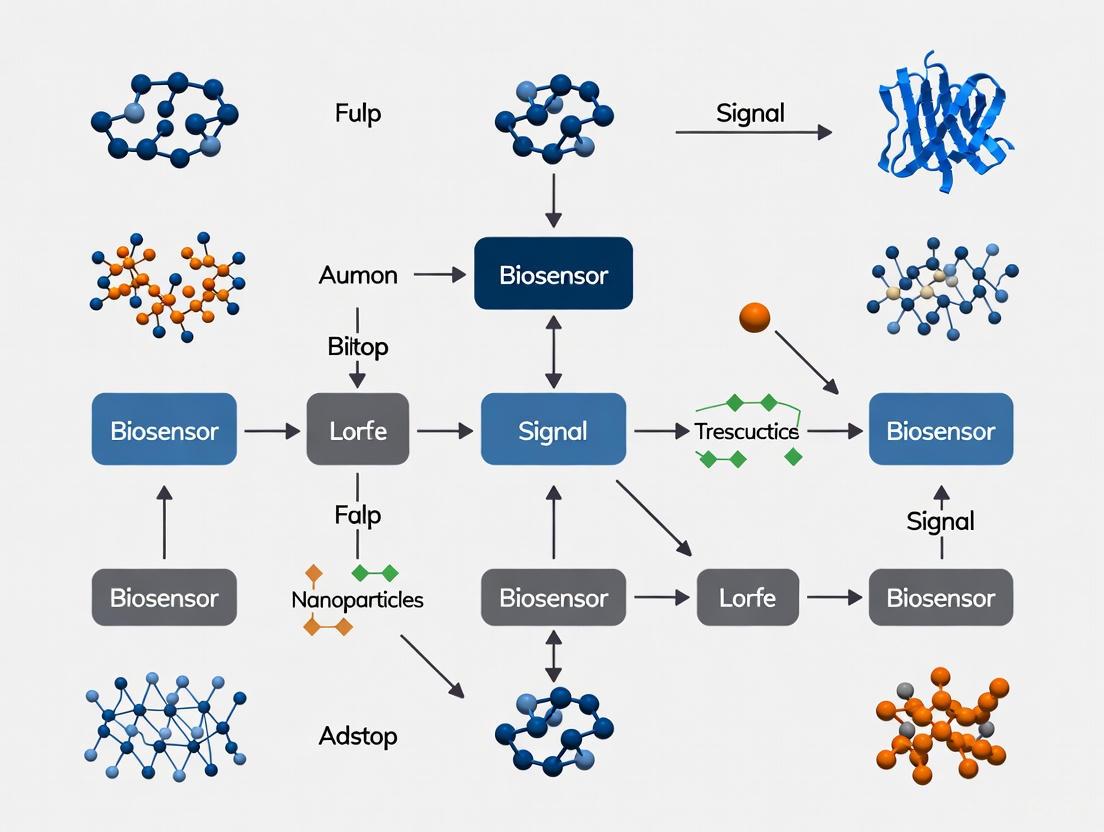
Diagram 1: Core biosensor signal pathway.
Comparative Analysis of Bioreceptor Platforms
The selection of a bioreceptor is a primary determinant of biosensor specificity and a significant contributor to performance variability. Different bioreceptor classes offer distinct advantages and limitations concerning reproducibility, stability, and ease of production.
Antibodies
Antibodies are high-affinity, three-dimensional protein structures that provide exceptional specificity through unique recognition patterns formed by their light and heavy chains [9]. Their primary drawback is production complexity, as generation relies on animal hosts, leading to potential batch-to-batch variability that can compromise reproducibility [9]. Furthermore, antibodies are relatively large (~150 kDa) and can be sensitive to changes in pH and temperature, impacting operational stability [9] [8].
Enzymes
Enzymes function as biocatalytic bioreceptors, converting the target analyte into a measurable product [9]. They are reusable and offer the advantage of signal amplification through catalysis. However, their activity is highly dependent on maintaining their delicate three-dimensional structure. Factors such as immobilization method, temperature, and pH can lead to denaturation and a subsequent drift in signal output over time, directly affecting reproducibility and sensor lifetime [8].
Nucleic Acids and Aptamers
Nucleic acid-based genosensors rely on the highly predictable and stable hybridization of complementary strands, enabling excellent reproducibility when sequences are synthetically produced [9] [10]. Aptamers are single-stranded oligonucleotides selected in vitro (via SELEX) to bind specific targets, from small molecules to whole cells [9]. As synthetic molecules, they offer superior production consistency compared to antibodies. Their inherent stability and ability to be chemically modified also enhance the reusability and shelf-life of the biosensor [9].
Molecularly Imprinted Polymers (MIPs)
MIPs are synthetic polymers with cavities templated for a specific analyte, serving as robust and cost-effective artificial receptors [9]. Their fully synthetic nature avoids biological variability, making them highly stable across wide pH and temperature ranges. This grants them a significant advantage in reproducibility and shelf-life for applications where the extreme specificity of biological receptors is not required [9].
Table 2: Bioreceptor Performance Comparison for Reproducibility
| Bioreceptor | Sensitivity | Reproducibility & Stability | Key Advantages | Key Limitations for Reproducibility |
|---|---|---|---|---|
| Antibodies | High (catalytic amplification possible) | Moderate; Batch-to-batch variability, sensitive to conditions [9]. | Very high specificity, well-established protocols. | Animal-based production, stability issues [9]. |
| Enzymes | High (catalytic amplification) | Moderate; Activity loss over time, dependent on immobilization [8]. | Signal amplification, reusability. | Denaturation, sensitivity to environment [8]. |
| Nucleic Acids (Aptamers) | High | High; Synthetic production, stable, reusable [9]. | Consistent production, high stability, tunable. | SELEX process can be complex/costly [9]. |
| Molecularly Imprinted Polymers (MIPs) | Moderate to High | Very High; Synthetic, robust, long shelf-life [9]. | Low-cost, high stability, no biological variability. | Can lack specificity of biological receptors. |
Comparative Analysis of Transducer Platforms
The transducer is the interface that quantifies the biorecognition event. Its design and material properties are critical for converting a biological interaction into a stable, low-noise electrical or optical signal.
Electrochemical Transducers
Electrochemical biosensors measure electrical changes—such as current (amperometric), potential (potentiometric), or impedance (impedimetric)—resulting from a biorecognition event [5] [10]. They are prized for their portability, low cost, and ease of miniaturization. A key source of variability in these systems is the electrode surface properties. Research has demonstrated that calibrating semiconductor manufacturing settings to produce electrodes with a thickness greater than 0.1 μm and a surface roughness below 0.3 μm is essential for achieving a high signal-to-noise ratio and consistent performance, which are prerequisites for reproducibility [1].
Optical Transducers
Optical biosensors, including those based on surface plasmon resonance (SPR), detect changes in light properties like intensity, wavelength, or refractive index [11] [12]. SPR biosensors enable label-free, real-time monitoring of binding events. Methodological validation of an SPR biosensor for chloramphenicol demonstrated high accuracy, with intra-day and inter-day accuracies of 98%–114% and 110%–122%, respectively, meeting analytical requirements [11]. While highly sensitive, these systems can be influenced by ambient light fluctuations and require precise optical alignment, potentially introducing variability if not carefully controlled.
Nanomaterial-Enhanced Transducers
The integration of nanomaterials like graphene, carbon nanotubes, gold nanoparticles, and quantum dots has revolutionized transducer design [5] [10] [12]. These materials offer high surface-to-volume ratios for efficient bioreceptor immobilization and improved electrical conductivity or optical properties for enhanced signal transduction [5]. However, the synthesis and functionalization consistency of these nanomaterials is a critical challenge. Slight variations in nanoparticle size, shape, or distribution can significantly impact transducer sensitivity and reproducibility across sensor batches [5].
Experimental Protocols for Assessing Reproducibility
Standardized experimental validation is paramount for objectively comparing biosensor platforms. The following protocols are adapted from studies focused on quantifying reproducibility, accuracy, and stability.
Protocol 1: Reproducibility and Accuracy Testing for Electrochemical Biosensors
This protocol is based on work that aimed to meet the standards for point-of-care testing (POCT) set by the Clinical and Laboratory Standards Institute (CLSI) [1].
- Objective: To determine the inter-assay coefficient of variation (CV) and accuracy of a biosensor platform.
- Materials:
- Biosensors fabricated with calibrated SMT electrodes (thickness >0.1 μm, roughness <0.3 μm) [1].
- Streptavidin biomediator with a GW linker for optimized bioreceptor (e.g., antibody) orientation [1].
- Standard solutions of the target analyte (e.g., cardiac troponin I) at known concentrations.
- Electrochemical analyzer.
- Method:
- Immobilize the biotinylated bioreceptor onto the streptavidin-modified electrode.
- Measure the electrochemical response (e.g., impedance or current) for at least 20 replicate sensors across multiple production batches, using standard solutions covering the dynamic range.
- For accuracy, compare the measured concentration to the known reference value for each standard.
- Data Analysis:
- Reproducibility: Calculate the CV (%) for the signal output at each concentration. A CV of less than 10% is typically required for POCT applications [1].
- Accuracy: Determine the recovery rate (%) as (Measured Concentration / Known Concentration) × 100%. Recovery rates between 90-110% are generally acceptable.
Protocol 2: Validation of an SPR Biosensor for Small Molecules
This protocol outlines the methodological verification for an SPR biosensor, as demonstrated for the detection of chloramphenicol in blood [11].
- Objective: To validate the precision, accuracy, and detection limits of an SPR biosensor.
- Materials:
- SPR instrument with a sensor chip functionalized with the appropriate bioreceptor (e.g., antibody or MIP).
- Standard solutions of the target small molecule.
- Blank and spiked complex matrices (e.g., blood, serum).
- Method:
- Inject standard solutions at various concentrations (e.g., 0.1–50 ng/mL) to establish a calibration curve.
- Assess intra-day precision by analyzing quality control samples (low, mid, high concentrations) at least five times within a day.
- Assess inter-day precision by analyzing the same samples over three consecutive days.
- Evaluate the matrix effect by comparing the signal from standards prepared in buffer versus the complex biological matrix.
- Data Analysis:
- Precision: Calculate the CV for intra-day and inter-day measurements.
- Accuracy: Determine the recovery rate from the spiked matrix samples.
- Limit of Detection (LOD): Calculate based on the signal of the blank plus three times its standard deviation (3σ).
Diagram 2: Reproducibility assessment workflow.
The Scientist's Toolkit: Essential Reagents and Materials
The following reagents and materials are critical for developing reproducible biosensor platforms, as identified in the featured experimental protocols.
Table 3: Key Research Reagent Solutions for Reproducible Biosensor Development
| Reagent/Material | Function in Biosensor Development | Rationale for Reproducibility |
|---|---|---|
| SMT-Produced Electrodes | Provides a consistent, solid-state transduction interface [1]. | Calibrated thickness and roughness minimize signal noise and batch-to-batch variability [1]. |
| Streptavidin Biomediator with GW Linker | Serves as a universal layer for immobilizing biotinylated bioreceptors (antibodies, aptamers) [1]. | The GW linker provides ideal flexibility and rigidity, ensuring proper bioreceptor orientation and consistent binding capacity [1]. |
| N-Hydroxysuccinimide (NHS)/EDC | A carbodiimide crosslinker used for covalent immobilization of bioreceptors to transducer surfaces [12]. | Enables stable, covalent attachment that reduces bioreceptor leaching, enhancing biosensor stability and operational lifetime. |
| Locked Nucleic Acids (LNA) | Modified nucleic acid probes used in genosensors [9]. | "Locks" the ribose conformation, reducing flexibility and improving binding stability and hybridization consistency [9]. |
| Nanosructured Composites (e.g., Porous Gold, Polyantiline) | Used to modify transducer surfaces to enhance signal and sensitivity [12]. | High surface area increases bioreceptor loading, while good conductivity improves electron transfer, leading to a higher and more stable signal. |
The pursuit of highly reproducible biosensor platforms demands a meticulous, component-level approach to design and manufacturing. Evidence indicates that synthetic bioreceptors like aptamers and MIPs offer superior production consistency and stability compared to traditional biological elements. Similarly, transducer performance is profoundly affected by physical characteristics, such as electrode topography, which can be optimized through controlled fabrication processes. The integration of engineered biomediators and linkers further minimizes variability by standardizing bioreceptor orientation. For researchers and drug development professionals, the path to reducing analytical variability lies in selecting platforms that leverage these reproducible design principles: synthetic biology for recognition and precision engineering for transduction. The future of reliable biosensing, particularly for point-of-care applications and rigorous clinical trials, depends on this foundational strategy.
Reproducibility is a cornerstone of reliable biosensor development, yet achieving consistent performance remains a significant challenge across the field. Despite the publication of over 100,000 articles on electrochemical enzyme biosensors since their inception, very few have reached practical application and commercialization, with inconsistent performance being a critical barrier [13]. This guide objectively compares the impact of four key irreproducibility sources—bioreceptor denaturation, immobilization inconsistency, electrode fouling, and signal drift—on biosensor performance, providing researchers with experimental data and methodologies to evaluate and improve their systems. The analysis is framed within a broader thesis on evaluating the reproducibility of different biosensor platforms, offering direct comparisons between common challenges and established mitigation strategies.
The table below summarizes the core characteristics, impacts on analytical performance, and prevalence across different biosensor types for the four key irreproducibility sources.
Table 1: Comparative Analysis of Key Irreproducibility Sources in Biosensors
| Irreproducibility Source | Main Impact on Performance | Common Affected Biosensor Types | Key Mitigation Strategies |
|---|---|---|---|
| Bioreceptor Denaturation [13] | Decreased sensitivity and selectivity over time; reduced operational life | Enzyme-based biosensors; immunosensors | Enzyme stabilization strategies; improved immobilization matrices; controlled storage conditions |
| Immobilization Inconsistency [14] | Poor batch-to-batch reproducibility; variable sensitivity and signal output | All affinity-based biosensors (aptasensors, immunosensors) | Optimized semiconductor manufacturing; biotin-streptavidin systems; standardized linker chemistry [14] |
| Electrode Fouling [15] | Reduced sensitivity, higher detection limit, unreliable signal in complex media | Sensors used in blood, serum, or other biofluids | PEG-modified surfaces; zwitterionic materials; nanoporous electrodes as diffusion filters [15] |
| Signal Drift [16] | Measurement inaccuracy over time; requires frequent recalibration | Ion-Sensitive Field-Effect Transistor (ISFET) sensors; continuous monitoring systems | Gate oxide surface treatment; signal processing algorithms; stable reference electrodes |
Experimental Protocols for Investigating Reproducibility
Investigating Signal Drift in ISFET Biosensors
Objective: To quantify and minimize the sensing signal drift error in an Ion-Sensitive Field-Effect Transistor (ISFET) biosensor caused by undesirable ion interactions in the sample media [16].
Materials:
- Gate Oxide Layer (GOL): SnO₂ thin film (80 nm) sputtered on ITO glass.
- Surface Modifiers: 3-Aminopropyltriethoxysilane (APTES), Succinic Anhydride, EDC, Sulfo-NHS.
- Bioreceptor: Prostate-Specific Membrane Antigen (PSMA) Antibody.
- Blocking Agent: Bovine Serum Albumin (BSA).
- Measurement Setup: Semiconductor Parameter Analyzer, Ag/AgCl Reference Electrode.
Methodology:
- Sensing Gate Fabrication: Deposit an 80 nm SnO₂ film on ITO glass using RF magnetron sputtering. Attach a PDMS reservoir to the GOL using O₂ plasma treatment.
- Surface Functionalization:
- Treat the GOL surface with O₂ plasma to form OH groups.
- Silanize with 5% APTES to create NH₂ functional groups.
- React with 5% succinic anhydride in DMF to form COOH groups.
- Activate with EDC/Sulfo-NHS chemistry.
- Immobilize PSMA antibodies (100 nM).
- Block non-specific sites with 1 M ethanolamine and 10% BSA.
- Drift Measurement:
- Add 1× PBS or 0.01× PBS solution to the reservoir.
- Measure the current-voltage (I–V) characteristics at 0, 1, 3, 5, and 10-minute intervals using a semiconductor parameter analyzer.
- Calculate the sensing voltage drift error (ΔVdf) as the change in voltage over time.
Expected Outcome: The surface-treated GOL (ST-GOL) with antibodies shows significantly reduced ΔVdf (e.g., 2.3 mV/min in diluted PBS) compared to a bare GOL (e.g., 4.3 mV/min) [16].
Assessing Anti-Fouling Performance in Blood
Objective: To evaluate the effectiveness of anti-fouling surface modifications in preventing non-specific adsorption from complex biofluids like whole blood [15].
Materials:
- Electrode Substrate: Gold electrodes.
- Anti-fouling Materials: Poly(ethyleneglycol) (PEG), Hyaluronic Acid, Hydrogels, Anti-fouling Peptides.
- Test Medium: Undiluted human blood or plasma.
- Measurement Technique: Electrochemical Impedance Spectroscopy (EIS).
Methodology:
- Surface Modification: Covalently graft anti-fouling polymers (e.g., PEG) or peptides onto the gold electrode surface using self-assembled monolayer (SAM) chemistry.
- Exposure to Complex Media: Incubate the modified electrode in undiluted human blood or plasma for a predetermined time (e.g., 30-60 minutes).
- Signal Measurement:
- Perform EIS measurements in a standard redox mediator (e.g., [Fe(CN)₆]³⁻/⁴⁻) before and after exposure to blood.
- Monitor changes in charge transfer resistance (Rₑₜ), which indicates fouling.
- Data Analysis: Compare the Rₑₜ shift of the anti-fouling modified electrode with a bare gold electrode. A smaller change in Rₑₜ indicates superior anti-fouling performance.
Expected Outcome: Electrodes modified with highly hydrated polymer brushes like PEG show a significantly reduced increase in Rₑₜ after blood exposure, indicating effective suppression of non-specific protein adsorption [15].
Visualizing Biosensor Irreproducibility and Mitigation Pathways
The following diagram illustrates the logical relationship between the four key sources of irreproducibility, their direct consequences on the biosensor's function, and the resulting analytical errors, culminating in the final unreliable output.
The subsequent diagram outlines a generalized experimental workflow for assessing biosensor reproducibility, integrating the investigation of multiple failure sources—specifically fouling and signal drift—as detailed in the provided experimental protocols.
The Scientist's Toolkit: Key Reagents and Materials
The table below lists essential reagents and materials critical for conducting reproducibility experiments, particularly those focused on mitigating fouling and signal drift.
Table 2: Key Research Reagent Solutions for Biosensor Reproducibility Studies
| Item Name | Function/Brief Explanation | Example Application in Protocols |
|---|---|---|
| Poly(ethyleneglycol) (PEG) [15] | Forms a hydrated, anti-fouling brush layer on surfaces to minimize non-specific protein adsorption. | Coating electrode surfaces to enable sensing in full blood. |
| 3-Aminopropyltriethoxysilane (APTES) [16] | A silane coupling agent that introduces primary amine (-NH₂) groups onto oxide surfaces for further functionalization. | Creating a functionalized surface on SnO₂ gate oxide for antibody immobilization. |
| EDC / Sulfo-NHS Chemistry [16] [17] | A zero-length crosslinking system for activating carboxyl groups to form stable amide bonds with primary amines. | Covalently immobilizing antibodies or aptamers onto a functionalized sensor surface. |
| Bovine Serum Albumin (BSA) [16] | A common blocking agent used to passivate unreacted sites on the sensor surface, reducing non-specific binding. | Blocking step after bioreceptor immobilization to minimize background signal. |
| Nanoporous Gold (NPG) [15] [18] | A high-surface-area electrode material that can act as a diffusion filter, alleviating fouling from larger proteins. | Used as an electrode substrate to allow analyte access while hindering fouling agents. |
| Redox Mediators (e.g., [Fe(CN)₆]³⁻/⁴⁻) [17] | Soluble molecules that shuttle electrons between the biorecognition element and the electrode transducer. | Used in Electrochemical Impedance Spectroscopy (EIS) to characterize electrode fouling and surface changes. |
The journey toward highly reproducible biosensors requires a systematic and multifaceted approach. As this guide has detailed, critical sources of irreproducibility—bioreceptor denaturation, immobilization inconsistency, electrode fouling, and signal drift—each have distinct impacts and require specific mitigation strategies. The experimental data and protocols presented highlight that solutions range from advanced material engineering, such as nanoporous electrodes and anti-fouling polymers, to precision manufacturing and sophisticated surface chemistry [15] [16] [14]. For researchers evaluating biosensor platforms, a rigorous assessment of these four factors is paramount. Future progress hinges on the adoption of more consistent reporting standards for stability data and the continued integration of stabilized bioreceptors, robust immobilization frameworks, and intelligent materials that resist biofouling, ultimately paving the way for biosensors that fulfill their promise in both clinical and point-of-care diagnostics [13].
The Impact of Nanomaterials and Functional Layers on Signal Stability and Reproducibility
The integration of nanomaterials and functional layers has become a cornerstone in the development of modern biosensors, directly addressing the critical challenges of signal stability and measurement reproducibility. For researchers evaluating biosensor platforms, these engineered interfaces are not merely enhancements but fundamental components that dictate analytical performance. Advances in nanomaterial science have enabled the precise control of interfacial properties, allowing for the creation of sensing platforms that can consistently transduce biological recognition events into reliable, quantifiable signals [19]. This progression is pivotal for applications ranging from point-of-care diagnostics to continuous monitoring in complex biological matrices, where consistent performance is as crucial as high sensitivity.
The quest for reproducibility drives the shift from bulk materials to nanostructured interfaces. Traditional biosensor surfaces often suffer from heterogeneous activity and non-specific binding, leading to signal drift and poor batch-to-batch consistency. The strategic implementation of functional layers, including two-dimensional materials, metal-organic frameworks, and engineered polymers, provides a pathway to overcome these limitations. These layers enhance signal stability by offering uniform surface chemistry, high biomolecule loading capacity, and efficient electron transfer pathways, thereby forming the foundation for reproducible biosensing platforms required for rigorous scientific and clinical validation [20] [19].
Comparative Performance of Nanomaterial-Enhanced Biosensing Platforms
The selection of nanomaterial and the architecture of the functional layer significantly influence key biosensor performance metrics. The table below provides a structured comparison of different platforms, highlighting their impact on signal stability and reproducibility.
Table 1: Performance Comparison of Nanomaterial-Based Biosensing Platforms
| Platform Description | Target Analyte | Key Performance Metrics | Implications for Stability/Reproducibility |
|---|---|---|---|
| ATA-monolayer on HOPG [19] | Epinephrine (EP) | Superior electron transfer, sub-micromolar LOD, stable covalent bonding. | High reproducibility from well-defined monolayer; excellent stability from covalent grafting. |
| WS₂-based SPR Sensor [21] | Cancer Cells (e.g., Jurkat) | Sensitivity: 342.14 deg/RIU; FOM: 124.86 RIU⁻¹. | Enhanced field confinement improves signal-to-noise ratio and measurement consistency. |
| ZIF-8 Fluorescent Biosensor [22] | COVID-19 RNA | LOD: 6.24 pM; Detection time: 8 min; 78.39% quenching efficiency. | High crystallinity and porosity ensure uniform probe loading and consistent fluorescence response. |
| Graphene/Si₃N₄ SPR [23] | Malaria DNA | Sensitivity: up to 353.14 °/RIU; High Quality Factor. | Graphene's consistent lattice structure aids in reducing signal variance during biomolecular binding. |
Key Insights from Comparative Data
- Well-Defined Interfaces are Crucial: The superior performance of the ATA-monolayer on HOPG underscores a central theme in modern biosensor design: molecular-level control over the interface is a primary determinant of reproducibility. Unlike disordered multilayers that lead to inconsistent performance, well-defined monolayers provide a uniform landscape for analyte binding and electron transfer, directly enhancing signal stability [19].
- Material Properties Dictate Consistency: The high performance of 2D materials like WS₂ and graphene is linked to their intrinsic properties. Their large, uniform surface areas allow for consistent immobilization of biorecognition elements (antibodies, DNA strands), while their excellent electrical and optical properties minimize batch-to-batch variation in transducer manufacturing [21] [23].
- Structural Order Underpins Reliability: The use of highly crystalline and porous frameworks, such as ZIF-8, ensures that each sensor platform has a nearly identical structure and density of active sites. This structural predictability is a key factor in achieving a low limit of detection while maintaining a high degree of reproducibility across different sensor units and production batches [22].
Experimental Protocols for Evaluating Stability and Reproducibility
To objectively compare biosensor platforms, standardized experimental protocols are essential. The following sections detail key methodologies for assessing the impact of functional layers.
Protocol for Electrochemical Grafting of Functional Layers
This protocol, adapted from a study on rational biosensor design, details the creation of a well-defined carboxy-functionalized interface on a carbon electrode [19].
- Primary Reagents: Highly Oriented Pylytic Graphite (HOPG) electrode (e.g., SPI-2 ZYB grade); 3,4,5-tricarboxybenzenediazonium (ATA) salt or para-aminobenzoic acid (PAB); sodium nitrite (NaNO₂); supporting electrolyte (e.g., 0.1 M H₂SO₄).
- Procedure:
- Electrode Preparation: Freshly cleave the HOPG basal plane to obtain an atomically flat, clean surface.
- Diazonium Solution Preparation: Dissolve the ATA or PAB precursor (e.g., 2 mM) in an acidic aqueous solution (e.g., 0.1 M HCl). Add NaNO₂ to initiate in situ diazotization, forming the reactive diazonium species.
- Electrochemical Grafting: Place the HOPG electrode in the diazonium solution within a controlled electrochemical cell. Perform Cyclic Voltammetry (CV), typically for 1-5 cycles, between +0.6 V and -0.4 V (vs. Ag/AgCl) at a scan rate of 50 mV/s. Monitor for the characteristic irreversible reduction peak near +0.1 V in the first cycle, which confirms the grafting process.
- Post-treatment: Rinse the grafted electrode thoroughly with deionized water and solvent (e.g., acetone) to remove any physisorbed species.
- Validation: Characterize the modified surface using Raman spectroscopy (looking for the D-band at ~1336 cm⁻¹ indicating successful covalent modification) and Atomic Force Microscopy (AFM) to verify layer uniformity [19].
Protocol for Assessing Signal Stability and Reproducibility
This general protocol evaluates the long-term and operational stability of the biosensor platform.
- Primary Reagents: Functionalized biosensor; target analyte at known concentrations; relevant buffer solution (e.g., PBS, pH 7.4).
- Procedure:
- Intra-assay Reproducibility: Perform replicate measurements (n ≥ 5) of the target analyte at a fixed concentration using the same biosensor unit within a single experimental session. Calculate the relative standard deviation (RSD) of the output signal (e.g., current, angle shift, fluorescence intensity).
- Inter-assay Reproducibility: Repeat the measurement of the fixed analyte concentration using multiple, independently fabricated biosensor units (n ≥ 3). Calculate the RSD across these different units.
- Operational Stability: Subject the biosensor to continuous operation in a relevant buffer or a flowing system. Record the baseline signal and the response to a standard analyte concentration at regular intervals (e.g., every hour) over an extended period (e.g., 8-24 hours). The signal decay over time is a key metric of stability.
- Storage Stability: Store the biosensors under defined conditions (e.g., 4°C in dry state or in buffer). Periodically test their response to a standard analyte over days or weeks to determine shelf-life.
- Data Analysis: Report both intra- and inter-assay RSD values. A low RSD (typically <5-10%) indicates high reproducibility. For stability, report the percentage of initial response retained after a specific duration.
Table 2: Research Reagent Solutions for Functional Layer Engineering
| Reagent / Material | Function in Experiment | Key Characteristic for Reproducibility |
|---|---|---|
| HOPG (Highly Oriented Pyrolytic Graphite) [19] | Provides an atomically flat, defined substrate for functionalization. | Low defect density and uniform surface terraces minimize substrate-induced variability. |
| Aryl Diazonium Salts (e.g., ATA, PAB) [19] | Precursors for creating covalently grafted functional layers. | Enables formation of robust, ordered monolayers with specific chemical termini (e.g., -COOH). |
| 2D Materials (e.g., WS₂, Graphene) [21] [23] | Serve as signal-amplifying and biomolecule-adsorbing layers in optical/electrical sensors. | High crystallinity and uniform surface chemistry ensure consistent layer properties. |
| Zeolitic Imidazolate Framework-8 (ZIF-8) [22] | A porous MOF used as a fluorescence quencher and probe-loading platform. | High crystallinity and thermal stability ensure uniform pore structure and quenching efficiency. |
| Silicon Nitride (Si₃N₄) [23] | Used as a high-refractive-index dielectric layer in SPR sensors. | Low optical loss and consistent film quality from CVD processes enhance sensor-to-sensor uniformity. |
Visualization of Biosensor Functionalization and Signal Transduction
The following diagram illustrates the logical workflow and key interactions involved in creating and utilizing a functionalized biosensor interface for stable and reproducible detection.
Biosensor Functionalization and Signal Pathway
The strategic implementation of nanomaterials and functional layers is a decisive factor in advancing biosensor technology from promising prototypes to reliable analytical tools. The experimental data consistently demonstrates that platforms engineered with well-defined interfaces, such as covalently grafted monolayers, highly crystalline 2D materials, and porous MOFs, exhibit superior signal stability and reproducibility compared to their non-engineered counterparts. The pursuit of reproducibility is fundamentally a pursuit of control at the nanoscale, where uniformity in surface chemistry, structure, and biomolecule orientation directly translates to consistent and dependable sensor performance.
For researchers and drug development professionals, this underscores the necessity of prioritizing material characterization and surface engineering in biosensor design. The choice of nanomaterial and functionalization protocol is not merely a technical detail but a core determinant of the platform's validity and commercial viability. Future research will likely focus on standardizing these nanofabrication protocols and developing new classes of functional materials that offer even greater control over the bio-interface, further closing the gap between laboratory innovation and real-world clinical application.
Platform-Specific Performance: Assessing Reproducibility Across Electrochemical, Optical, and Portable Systems
Electrochemical biosensors have emerged as powerful analytical tools, transforming diagnostic capabilities in healthcare, environmental monitoring, and food safety. These devices integrate biological recognition elements with electrochemical transducers to detect target analytes with high specificity and sensitivity. Among the critical performance parameters for these biosensors—including sensitivity, selectivity, and stability—reproducibility stands as a fundamental characteristic determining their reliability and practical applicability. Reproducibility refers to the ability of a biosensor to generate identical responses for a duplicated experimental setup, characterized by the precision and accuracy of the transducer and electronics [24].
The evaluation of reproducibility transcends academic interest, representing a pivotal requirement for regulatory approval and clinical adoption, particularly in point-of-care (POC) settings where single-use devices must perform consistently across mass production. The Clinical and Laboratory Standards Institute (CLSI) has established stringent guidelines requiring a coefficient of variation (CV) of less than 10% for reproducibility, accuracy, and stability for POC applications [25] [14]. Despite these clear benchmarks, achieving consistent reproducibility across different electrochemical sensing modalities presents distinct challenges and opportunities.
This review provides a systematic comparison of three primary electrochemical biosensor platforms—amperometric, potentiometric, and impedimetric—focusing specifically on their reproducibility characteristics. By synthesizing recent research advances, experimental data, and technological innovations, we aim to provide researchers and drug development professionals with a comprehensive framework for evaluating and selecting appropriate biosensing platforms based on reproducibility requirements.
Fundamental Principles of Electrochemical Biosensing
Electrochemical biosensors function by converting a biological recognition event into a quantifiable electrical signal. All biosensors share core components: (1) a bioreceptor (enzymes, antibodies, aptamers, DNA) that specifically recognizes the target analyte; (2) a transducer that transforms the biological interaction into a measurable electrical signal; and (3) electronics that process and display the result [24] [26]. The stability and reproducibility of these components directly determine the overall biosensor performance.
The bioreceptor dictates specificity through molecular recognition, while the transducer determines the sensitivity and reproducibility of signal conversion. Reproducibility challenges often originate from inconsistencies in bioreceptor immobilization, electrode surface properties, or signal transduction mechanisms [24]. Even with highly specific bioreceptors, inadequate transducer design or manufacturing inconsistencies can compromise reproducibility.
Table 1: Core Components of Electrochemical Biosensors and Their Impact on Reproducibility
| Component | Function | Impact on Reproducibility |
|---|---|---|
| Bioreceptor | Specific target recognition | High specificity reduces cross-reactivity interference |
| Transducer | Signal conversion from biological to electrical | Consistent fabrication determines signal uniformity |
| Electrode Surface | Platform for bioreceptor immobilization | Uniform morphology ensures consistent binding kinetics |
| Electronics | Signal processing and amplification | Stable circuitry reduces measurement variability |
Comparative Analysis of Electrochemical Techniques
Amperometric Biosensors
Amperometric biosensors measure current generated by electrochemical oxidation or reduction of an electroactive species at a constant applied potential. These sensors typically employ enzymes such as glucose oxidase or horseradish peroxidase, which generate or consume electroactive products during catalytic reactions [27] [28]. The measured current is directly proportional to the analyte concentration.
Recent studies demonstrate significant advances in amperometric biosensor reproducibility through improved electrode design and enzyme immobilization techniques. Research on phenoloxidase-based Sonogel-Carbon biosensors for detecting polyphenols in beers revealed that a Nafion-Lac/Sonogel-Carbon system maintained 84% of its initial response for at least three weeks with a relative standard deviation (RSD) of 3.3% (n=10), indicating high reproducibility [27]. Similarly, a study on BOBzBT₂ pentamer-modified carbon electrodes for creatinine detection highlighted the importance of hydrophobic properties in enhancing sensor stability and enabling reusable applications without performance degradation [29].
The historical development of amperometric biosensors includes important innovations such as the introduction of ferrocene mediators in 1984, which enhanced electron transfer efficiency and improved reproducibility by reducing dependence on dissolved oxygen [24]. Comparative studies of different amperometric strategies—including ferrocene mediation, redox hydrogels, and conducting polymers—have demonstrated that mediator-based systems generally offer superior reproducibility for continuous monitoring applications [28].
Potentiometric Biosensors
Potentiometric biosensors measure the potential difference between working and reference electrodes under conditions of zero current flow. These sensors have evolved from conventional ion-selective electrodes (ISEs) to solid-contact ISEs, which eliminate the inner filling solution to enhance miniaturization and mechanical stability [30]. The potential difference arises from selective ion partitioning across an ion-selective membrane, following the Nernst equation.
The reproducibility of potentiometric biosensors heavily depends on the solid-contact material between the ion-selective membrane and the electron conductor. Early "coated-wire" electrodes suffered from significant potential drift due to the formation of an aqueous layer between the metal and membrane interface [30]. The introduction of conducting polymers (e.g., polypyrrole, PEDOT) as intermediate layers marked a substantial improvement, with potential drifts as low as 10 µV/h over eight days, significantly reducing recalibration needs [30].
Recent innovations in nanomaterial-based solid contacts have further enhanced reproducibility. Materials such as carbon nanotubes, graphene, and MXenes provide high double-layer capacitance and hydrophobicity, minimizing potential drift and improving electrode-to-electrode consistency [30]. These advances have facilitated the development of wearable potentiometric sensors for monitoring ions (sodium, potassium, calcium) in sweat, demonstrating reproducible performance across multiple measurements during physical activity [30].
Impedimetric Biosensors
Impedimetric biosensors monitor changes in the electrical impedance of the electrode-electrolyte interface, typically using electrochemical impedance spectroscopy (EIS). These sensors operate through either faradaic processes (involving redox mediators) or non-faradaic processes (measuring double-layer capacitance changes) [31]. A significant advantage of impedimetric biosensors is their label-free detection capability, allowing direct measurement of binding events without secondary labels.
Electrode design critically influences the reproducibility of impedimetric biosensors. Comparative studies between interdigitated electrodes (IDEs) and micro-gap parallel plate electrodes (PPEs) have revealed substantial differences in device-to-device variations [32]. Computational simulations show that IDEs exhibit highly concentrated current density at electrode edges, which are susceptible to damage during fabrication, resulting in poor reproducibility. In contrast, PPEs demonstrate uniform current distribution across the electrode surface, yielding significantly improved reproducibility [32].
Experimental validation with Protein G-based immunoglobulin G (IgG) biosensors confirmed that PPE structures provide small device-to-device variations compared to IDEs, while simultaneously achieving ultrasensitive detection with a linear range from 1 × 10⁻¹³ to 1 × 10⁻⁷ mol/L [32]. This demonstrates that proper electrode design can enhance both reproducibility and sensitivity simultaneously, addressing a common trade-off in biosensor development.
Table 2: Quantitative Reproducibility Comparison of Electrochemical Biosensor Platforms
| Biosensor Type | Reproducibility Metric | Experimental Conditions | Reference |
|---|---|---|---|
| Amperometric | RSD = 3.3% (n=10) | Nafion-Lac/Sonogel-Carbon for polyphenols | [27] |
| Potentiometric | Potential drift < 10 µV/h for 8 days | Solid-contact ISEs with conducting polymers | [30] |
| Impedimetric | CV < 10% (meets CLSI POC standards) | SMEB platform with optimized SMT electrodes | [25] [14] |
| Impedimetric | Significantly lower device-to-device variation | Micro-gap PPE vs. IDE for IgG detection | [32] |
Experimental Protocols for Reproducibility Assessment
Electrode Fabrication and Optimization
The foundation of reproducible biosensing begins with consistent electrode fabrication. Semiconductor manufacturing technology (SMT) has demonstrated exceptional capability for producing electrodes with high reproducibility. Recent research optimized SMT production settings by calibrating electrode thickness to greater than 0.1 μm and surface roughness to less than 0.3 μm, resulting in biosensors that meet CLSI standards for point-of-care use [25] [14].
For impedimetric biosensors, the micro-gap parallel plate electrode (PPE) fabrication process involves creating two planar electrodes with edges covered by a SiO₂ layer, placed face-to-face with a precisely controlled gap. This structure ensures uniform current distribution over the planar electrode surface, maximizing the contribution of the well-defined surface to sensing and minimizing variations from edge defects [32]. The gap between electrodes is typically controlled using a spacer layer with thicknesses ranging from 2-10 μm, depending on the target sensitivity and application requirements.
Bioreceptor Immobilization Strategies
Consistent bioreceptor immobilization is crucial for biosensor reproducibility. The streptavidin-biotin system has been widely employed due to its strong binding affinity (K_d ≈ 10⁻¹⁵ M) and stability. Recent innovations incorporate a GW linker (glycine-tryptophan) between the streptavidin biomediator and the bioreceptor, providing ideal flexibility and rigidity to optimize orientation and function [25]. This approach minimizes random orientation and steric hindrance, significantly improving assay reproducibility.
For amperometric biosensors, immobilization techniques such as Nafion ion exchange doping have demonstrated excellent reproducibility. In phenoloxidase-based biosensors, a mixture of enzyme and Nafion was applied to the Sonogel-Carbon electrode surface, providing both protective encapsulation and enhanced electron transfer [27]. This method maintained 84% of the initial biosensor response after three weeks, indicating outstanding operational stability and reproducibility.
Standardized Testing Protocols
Reproducibility assessment requires standardized testing methodologies. The CLSI guidelines (EP05-A3, EP24-A2, EP25-A) recommend measuring the coefficient of variation (CV) across multiple devices (typically n≥10) from the same production batch, with CV values below 10% considered acceptable for POC applications [25] [14].
For impedimetric biosensors, reproducibility is evaluated through Nyquist plot analysis using a Randles equivalent circuit model. Parameters such as charge-transfer resistance (Rct), constant phase element (CPE), and Warburg impedance (Zw) are extracted from fitting the impedance spectra. The variation in R_ct values across multiple sensors exposed to the same analyte concentration provides a quantitative measure of reproducibility [32]. This method was successfully applied to Protein G-based IgG biosensors, demonstrating significantly lower device-to-device variations for PPE structures compared to conventional IDEs.
Essential Research Reagents and Materials
The consistent performance of electrochemical biosensors depends heavily on the quality and proper selection of research reagents and materials. The following table summarizes key components essential for developing reproducible biosensing platforms.
Table 3: Essential Research Reagents and Materials for Reproducible Biosensor Development
| Reagent/Material | Function | Role in Enhancing Reproducibility |
|---|---|---|
| Sonogel-Carbon Electrode | Electrochemical transducer | Provides porous, stable matrix for consistent enzyme immobilization [27] |
| Nafion Ion Exchanger | Protective additive | Stabilizes bioreceptor and prevents fouling, extending operational lifetime [27] |
| GW Linker | Bioreceptor immobilization | Optimizes orientation and flexibility for consistent binding kinetics [25] |
| Streptavidin Biomediator | Bioreceptor anchoring | Strong, stable binding to biotinylated molecules reduces batch variations [25] [14] |
| Conducting Polymers (PEDOT) | Solid contact material | Enhances potential stability in potentiometric sensors [30] |
| Redox Probes ([Fe(CN)₆]³⁻/⁴⁻) | Electron transfer mediator | Provides consistent faradaic reaction for impedimetric/amperometric sensing [32] |
| BOBzBT₂ Pentamer | Surface modification | Hydrophobic properties enable reusable sensors with stable performance [29] |
Technological Innovations Enhancing Reproducibility
Recent technological advances have substantially improved the reproducibility of electrochemical biosensors. Semiconductor manufacturing technology (SMT) has enabled the mass production of electrodes with minimal batch-to-batch variations. By optimizing SMT settings to control electrode thickness (>0.1 μm) and surface roughness (<0.3 μm), researchers have developed biosensor platforms that meet CLSI standards for point-of-care use [25] [14].
Nanomaterial integration has provided another pathway to enhanced reproducibility. Carbon-based nanomaterials such as graphene, carbon nanotubes, and MXenes offer large surface-to-volume ratios and excellent electrical properties, facilitating consistent bioreceptor immobilization and electron transfer [31]. These materials have been incorporated into all three biosensor types, demonstrating improved reproducibility through more uniform surface properties and enhanced signal-to-noise ratios.
Microfluidic integration represents a third innovation stream, improving reproducibility by precisely controlling sample delivery and wash conditions. Automated fluid handling minimizes operator-induced variations and ensures consistent binding kinetics across multiple assays [26]. This approach has been particularly beneficial for impedimetric biosensors used in continuous monitoring applications, where flow conditions significantly impact measurement consistency.
Strategies for Enhancing Biosensor Reproducibility
The comprehensive evaluation of amperometric, potentiometric, and impedimetric biosensors reveals distinct reproducibility characteristics and optimization strategies for each platform. Impedimetric biosensors with optimized electrode designs, particularly micro-gap parallel plate electrodes, demonstrate superior reproducibility by ensuring uniform current distribution and minimizing edge effects. Amperometric biosensors benefit from advanced immobilization matrices like Nafion-doped Sonogel-Carbon, which stabilize enzymatic activity and enhance operational lifetime. Potentiometric biosensors have achieved significant reproducibility improvements through solid-contact materials like conducting polymers and nanomaterials that minimize potential drift.
Across all platforms, consistent trends emerge: standardized manufacturing processes, optimized bioreceptor orientation, and controlled microenvironments significantly enhance reproducibility. Semiconductor manufacturing technology has proven particularly valuable for mass production of electrodes with minimal variations. Furthermore, the adoption of CLSI guidelines for reproducibility assessment provides a standardized framework for performance validation across different research groups and commercial entities.
Future research directions should focus on integrating artificial intelligence for quality control during manufacturing, developing self-calibrating systems to compensate for device-to-device variations, and advancing multi-analyte detection platforms with minimal cross-talk. As biosensor technologies continue to evolve toward point-of-care applications, wearable devices, and continuous monitoring systems, reproducibility will remain a critical parameter determining their successful translation from research laboratories to real-world applications.
Alanine aminotransferase (ALT) is a crucial biomarker for liver health, with elevated levels in the bloodstream indicating hepatocellular damage from conditions such as hepatitis, liver cirrhosis, or fatty liver disease [33] [34]. In clinical practice, the accurate detection of ALT activity is vital for diagnosis and monitoring. Traditional laboratory methods for ALT detection, including spectrophotometric and chromatographic techniques, are often expensive, time-consuming, and require trained personnel, limiting their use for rapid point-of-care testing [33] [34].
Electrochemical biosensors represent a promising alternative, offering potential for portability, cost-effectiveness, and rapid results. A central challenge in developing these biosensors is the selection of an optimal biorecognition element. Since ALT itself is not electroactive, its activity is typically measured indirectly by detecting the reaction products—pyruvate or glutamate—using secondary enzymes. The two predominant enzymatic systems for this purpose are Pyruvate Oxidase (POx) and Glutamate Oxidase (GlOx) [33]. While both have been utilized, a direct, systematic comparison under controlled conditions to guide rational biosensor design has been lacking. This case study, set within a broader thesis on biosensor reproducibility, provides a direct experimental comparison of GlOx and POx-based amperometric biosensors, evaluating their analytical performance, robustness, and practical utility for researchers and drug development professionals [33].
Experimental Protocols & Biosensor Fabrication
Biosensor Design and Principle of Operation
Both biosensor configurations are amperometric and operate on a similar fundamental principle: the secondary enzyme (POx or GlOx) generates hydrogen peroxide (H₂O₂) as a product, which is then oxidized at a platinum electrode. This oxidation produces a measurable current change that is proportional to the original ALT activity [33]. The core difference lies in which ALT product they detect.
The following diagram illustrates the distinct signaling pathways for the two biosensor systems:
Detailed Fabrication Methodologies
A critical differentiator between the two biosensors was the enzyme immobilization strategy, which was optimized separately for each enzyme [33].
POx-Based Biosensor:
- Immobilization Method: Entrapment within a polyvinyl alcohol with steryl pyridinium groups (PVA-SbQ) photopolymer.
- Optimized Conditions: The enzyme gel was prepared in 25 mM HEPES buffer (pH 7.4), containing glycerol, Bovine Serum Albumin (BSA), and 4.86 U/µL POx. This gel was mixed with the PVA-SbQ photopolymer in a 1:2 ratio, resulting in final concentrations of 1.62 U/µL POx and 13.2% PVA-SbQ.
- Immobilization Process: 0.15 µL of the mixture was applied to the platinum electrode surface and photopolymerized under UV light (365 nm) for approximately 8 minutes. The electrodes were rinsed with working buffer before use [33].
GlOx-Based Biosensor:
- Immobilization Method: Covalent crosslinking with glutaraldehyde (GA).
- Optimized Conditions: The enzyme gel was prepared in 100 mM phosphate buffer (pH 6.5), containing glycerol, BSA, and 8% GlOx. It was mixed with a 0.5% GA solution in a 1:2 ratio, yielding final concentrations of 2.67% GlOx and 0.3% GA.
- Immobilization Process: A smaller volume of 0.05 µL of the mixture was deposited on the electrode and air-dried for 35 minutes, followed by rinsing [33].
Shared Platform and Interference Mitigation: To ensure a valid comparison, both biosensors were fabricated on identical platforms using the same type of platinum disc working electrodes, a platinum counter electrode, and an Ag/AgCl reference electrode. Amperometric measurements were conducted at an applied potential of +0.6 V vs. Ag/AgCl in a stirred cell at room temperature [33]. To enhance selectivity in complex fluids like serum, the platinum electrodes were first modified with a semi-permeable poly(meta-phenylenediamine) (PPD) membrane. This membrane allows H₂O₂ to diffuse through while blocking larger, electroactive interferents such as ascorbic acid, thereby improving signal accuracy [33].
Comparative Performance Analysis
A systematic evaluation of the two biosensors revealed a clear trade-off between sensitivity and robustness.
Table 1: Direct Comparison of Analytical Performance for GlOx and POx-based ALT Biosensors
| Analytical Parameter | POx-Based Biosensor | GlOx-Based Biosensor |
|---|---|---|
| Linear Range | 1–500 U/L | 5–500 U/L |
| Limit of Detection (LOD) | 1 U/L | 1 U/L |
| Sensitivity (at 100 U/L ALT) | 0.75 nA/min | 0.49 nA/min |
| Optimized Immobilization pH | 7.4 | 6.5 |
| Stability in Complex Solutions | Lower | Higher |
| Assay Cost | Higher | Lower (simpler working solution) |
| Specificity for ALT | High (unique to ALT pathway) | Subject to interference from AST activity |
The data shows that the POx-based biosensor offers superior analytical sensitivity and a wider linear range at the lower end, making it suitable for applications where detecting very low ALT levels is critical [33]. In contrast, the GlOx-based biosensor demonstrated greater stability when challenged with complex sample matrices, a vital characteristic for clinical serum analysis [33]. Furthermore, the GlOx system benefits from a simpler working solution, which translates to lower per-test costs [33].
The Scientist's Toolkit: Key Research Reagents
The fabrication and operation of these biosensors rely on a set of specific reagents and materials. The following table details the essential components and their functions in the experimental workflow.
Table 2: Essential Research Reagents for ALT Biosensor Fabrication
| Reagent / Material | Function in the Experiment |
|---|---|
| Pyruvate Oxidase (POx) | Biorecognition element for the POx-based biosensor; catalyzes the conversion of pyruvate (from ALT reaction) to generate H₂O₂ [33]. |
| Glutamate Oxidase (GlOx) | Biorecognition element for the GlOx-based biosensor; catalyzes the conversion of glutamate (from ALT reaction) to generate H₂O₂ [33]. |
| Alanine Aminotransferase (ALT) | The target enzyme; used for calibration and performance testing of the fabricated biosensors [33]. |
| Polyvinyl Alcohol (PVA-SbQ) | Photocopolymer matrix used for the entrapment immobilization of POx [33]. |
| Glutaraldehyde (GA) | Crosslinking agent used for the covalent immobilization of GlOx and BSA on the electrode surface [33]. |
| meta-Phenylenediamine (m-PD) | Monomer for electropolymerization to create a selective membrane that blocks interferents [33]. |
| Platinum (Pt) Electrode | The working electrode; serves as the solid support for enzyme immobilization and the surface for H₂O₂ oxidation [33]. |
| Thiamine Pyrophosphate (TPP) | Essential cofactor for the enzymatic activity of pyruvate oxidase [33]. |
| Bovine Serum Albumin (BSA) | Used as a stabilizing protein in the enzyme immobilization gels to help maintain enzyme activity and reduce leaching [33]. |
Discussion and Research Implications
The comparative data underscores that the choice between GlOx and POx is not a matter of one being universally superior, but rather depends on the specific requirements of the intended application. The higher sensitivity of the POx-based system makes it ideal for scenarios demanding low limits of detection [33]. Its primary advantage is its high specificity for ALT, as the pyruvate it detects is a direct product of the ALT-catalyzed reaction [33].
Conversely, the GlOx-based system excels in robustness. Its stability in complex solutions is a significant advantage for clinical diagnostics involving blood serum [33]. However, a key limitation is its potential vulnerability to cross-reactivity. Aspartate aminotransferase (AST) is another important liver enzyme that also produces glutamate. In samples with elevated AST levels, the GlOx-based biosensor could overestimate ALT activity [33] [34]. Interestingly, this same property can be leveraged to develop biosensors targeted specifically for AST detection.
From a reproducibility standpoint, the immobilization chemistry plays a critical role. Covalent crosslinking (used for GlOx) typically provides a more stable and durable enzyme layer compared to entrapment methods (used for POx), potentially leading to a longer operational lifetime and more consistent performance across different sensor batches [35].
This direct comparison reveals a definitive trade-off in biosensor design for ALT detection. The POx-based biosensor is the optimal choice for applications where maximum sensitivity and specificity for ALT are the primary goals. In contrast, the GlOx-based biosensor is better suited for environments requiring robust performance in complex matrices and where cost-effectiveness is a major driver.
For the research community, this study provides a clear, data-driven framework for selecting an enzymatic system. The findings emphasize that the optimal biosensor configuration is application-dependent. Future work in this field should focus on further enhancing the stability of the sensitive POx system and engineering the GlOx system for improved selectivity, ultimately advancing the development of reliable, point-of-care diagnostic devices for liver health monitoring.
Reproducibility stands as a critical performance parameter in the validation and adoption of biosensing technologies for research and clinical applications. Achieving consistent results across different instruments, operators, and experimental runs is fundamental for establishing reliability in data interpretation, particularly in pharmaceutical development where decisions hinge on precise molecular interaction data. This guide objectively evaluates the reproducibility of three prominent optical biosensor platforms—fluorescence-based, surface plasmon resonance (SPR), and colorimetric systems—by comparing direct experimental evidence from recent studies. We examine the key performance metrics, experimental methodologies, and technical factors that contribute to reproducible outcomes in each platform, providing researchers with a structured comparison to inform their technology selection process.
Performance Comparison of Optical Biosensor Platforms
The quantitative comparison of reproducibility and key performance metrics across fluorescence, SPR, and colorimetric biosensor platforms provides critical insights for technology selection. Table 1 summarizes experimental data from recent studies, highlighting the distinct performance characteristics of each platform.
Table 1: Reproducibility and Performance Metrics Across Biosensor Platforms
| Platform | Target Analyte | Reproducibility (RSD/Other Metrics) | Linear Range | Detection Limit | Key Advantages for Reproducibility |
|---|---|---|---|---|---|
| Fluorescence (DNA-AgNCs/DSN) [36] | miRNA-155 | Excellent (<5% RSD) | 1–600 nM | 0.86 nM | Label-free design; DSN signal amplification enhances consistency |
| Fluorescence (G-quadruplex/Exo III) [37] | Silver ions (Ag⁺) | High specificity vs. interfering ions | 5–1500 pM | 2 pM | Dual enzymatic recycling amplification; C-Ag⁺-C structure specificity |
| SPR (Graphene-BP Heterostructure) [38] | Refractive Index | Machine learning validation (R²: 92-100%) | 1.29-1.38 RIU | 0.018 RIU | 2D material enhancement; ML predictive modeling reduces experimental variance |
| SPR (Laccase Enzymatic) [39] | Dopamine | High specificity (no ascorbic acid/L-dopa interference) | 0.01–189 μg/mL | 0.1 ng/mL | Regenerable surface; controlled orientation immobilization preserves activity |
| Colorimetric (ACCbi-PVAc Membrane) [40] | P. aeruginosa (via HCN) | Functionality retained after 2-year storage | N/A | Visual detection in 10-12 hours | Exceptional material stability; simplified readout minimizes user variability |
The data reveal distinct reproducibility advantages across platforms. Fluorescence biosensors achieve excellent precision (RSD <5%) through enzymatic amplification strategies and label-free detection that minimize preparation variability [36]. SPR platforms demonstrate reproducibility through regenerable surfaces and advanced material designs that maintain stability across multiple measurement cycles [38] [39]. The graphene-black phosphorus heterostructure SPR sensor further enhances reliability through machine learning validation with R² values between 92-100%, significantly reducing experimental variance in refractive index detection [38]. Colorimetric sensors offer exceptional long-term stability, with some membranes maintaining functionality after two years of storage, making them valuable for resource-limited settings [40].
Experimental Protocols for Reproducibility Assessment
Fluorescence-Based miRNA Detection Protocol
The reproducible fluorescence detection of miRNA-155 employs a duplex-specific nuclease (DSN) assisted amplification strategy with DNA-templated silver nanoclusters (DNA-AgNCs) as label-free probes [36]. The experimental workflow begins with sample preparation requiring extraction of miRNA from biological samples using standard TRIzol-based methods, followed by dilution in appropriate reaction buffer (typically Tris-HNO₃, pH 7.0). The assay procedure involves incubating the miRNA-155 target with DSN enzyme and specific DNA probes at 37°C for 60 minutes. During this step, DSN selectively cleaves the DNA strand in DNA-miRNA heteroduplexes, releasing the miRNA intact for repeated cycling that generates significant signal amplification. Following amplification, DNA-AgNCs are introduced as fluorescent probes that emit strong fluorescence upon binding to the amplified DNA products. Signal measurement is performed using a standard fluorescence spectrometer with excitation at 399 nm and emission detection at 614 nm. For reproducibility assessment, researchers should conduct inter-day precision tests with triplicate measurements across three separate days using identical miRNA concentrations, calculating the relative standard deviation (RSD) to quantify precision. The method demonstrates excellent reproducibility (<5% RSD) attributable to the specificity of DSN enzyme and the consistent fluorescence properties of DNA-AgNCs across experimental runs [36].
SPR Biosensor Experimental Methodology
The protocol for evaluating SPR biosensor reproducibility, as demonstrated in the graphene-black phosphorus heterostructure sensor, involves precise sensor fabrication and angular interrogation [38]. The fabrication process begins with a BK7 glass prism substrate onto which a 50-nm silver plasmonic film is deposited using magnetron sputtering. This is followed by sequential transfer of a monolayer graphene sheet and a black phosphorus dielectric layer using deterministic transfer methods to create the heterostructure interface. For refractive index measurements, the sensor is integrated into a Kretschmann configuration SPR system where a polarized light source is directed through the prism at varying angles of incidence. The reflected light intensity is monitored using a photodetector array, with the resonance angle (θSPR) identified as the angle of minimum reflectance. Reproducibility assessment involves measuring multiple analyte solutions with known refractive indices (range: 1.29-1.38 RIU) across separately fabricated sensor chips, with the angular shift (ΔθSPR) recorded for each measurement. The sensitivity is calculated as the ratio of angular shift to refractive index change (ΔθSPR/ΔRI), with reproducibility determined through statistical analysis of sensitivity values across multiple sensor chips and measurement cycles. The incorporation of machine learning algorithms (K-nearest neighbors regression) further validates reproducibility by predicting sensor behavior and comparing predicted versus experimental values across datasets [38].
Colorimetric Biosensor Implementation
The experimental protocol for the colorimetric detection of Pseudomonas aeruginosa via hydrogen cyanide (HCN) measurement emphasizes material stability and visual assessment reproducibility [40]. The sensor fabrication involves electrospinning polyvinyl acetate (PVAc) polymer solutions containing aquacyanocobinamide (ACCbi) indicator onto appropriate substrates to create nanofiber membranes. The ACCbi indicator undergoes a distinct color change from orange to violet upon interaction with HCN, providing a visual detection mechanism. For bacterial detection, the optimized PVAc membranes are exposed to P. aeruginosa cultures or infected wound exudates, with color development monitored over time (typically 10-12 hours). Reproducibility assessment includes both inter-batch consistency evaluation (comparing color change kinetics across separately fabricated membrane batches) and long-term stability testing (measuring performance maintenance after extended storage periods up to two years). The structural integrity of the PVAc membranes, confirmed through scanning electron microscopy, contributes significantly to reproducible performance by maintaining consistent indicator distribution and accessibility to target analyte [40].
Technical Factors Influencing Reproducibility
Signal Amplification and Stability Mechanisms
The fundamental mechanisms governing signal generation and stability significantly impact biosensor reproducibility. Enzymatic amplification strategies, particularly those employing duplex-specific nucleases (DSN) and exonuclease III (Exo III), enhance reproducibility by generating consistent signal amplification across experimental replicates [36] [37]. In the fluorescence-based miRNA detection system, DSN demonstrates precise specificity for cleaving DNA in DNA-RNA heteroduplexes while sparing single-stranded DNA and RNA, ensuring uniform amplification efficiency. Similarly, the Exo III-assisted dual recycling amplification for silver ion detection creates a cascading signal enhancement that reduces the impact of initial reaction variability. For colorimetric systems, the material stability of the polymer matrix, particularly polyvinyl acetate (PVAc) membranes, provides consistent microenvironments for indicator molecules, maintaining reliable performance even after prolonged storage [40]. SPR platforms achieve enhanced reproducibility through two-dimensional material heterostructures (graphene-black phosphorus) that create highly uniform sensing interfaces with consistent electromagnetic field confinement, minimizing signal drift between measurement cycles [38].
Surface Functionalization and Immobilization
The consistency of bioreceptor immobilization on sensor surfaces represents a critical factor in biosensor reproducibility, particularly for SPR and fluorescence platforms. Controlled orientation immobilization strategies, such as the amine coupling procedure used for laccase enzyme immobilization on carboxymethyl dextran (CMD) SPR chips, preserve bioactivity and ensure consistent binding capacity across sensor surfaces [39]. The reproducibility of this process can be verified through atomic force microscopy (AFM) analysis, which demonstrates homogeneous enzyme distribution with increased surface roughness (from 0.256 nm to 0.424 nm) following successful immobilization. For fluorescence-based DNA-silver nanocluster systems, the consistency of DNA templating directly impacts silver nanocluster formation and subsequent fluorescence emission, with optimized DNA sequences and incubation conditions ensuring uniform cluster formation [36]. In colorimetric systems, the electrospinning parameters for creating indicator-embedded nanofiber membranes must be rigorously controlled to produce consistent fiber morphology, diameter distribution, and indicator loading, all of which directly influence the reproducibility of colorimetric responses [40].
Research Reagent Solutions for Reproducible Biosensing
Successful implementation of reproducible biosensing requires specific reagent systems with optimized functions. Table 2 catalogues essential research reagents and their roles in ensuring experimental consistency across different biosensor platforms.
Table 2: Essential Research Reagents for Biosensor Reproducibility
| Reagent/Material | Function | Platform Applicability | Reproducibility Consideration |
|---|---|---|---|
| Duplex-Specific Nuclease (DSN) [36] | Selective cleavage of DNA in DNA-RNA heteroduplexes for signal amplification | Fluorescence | Maintains consistent activity across batches; specific recognition reduces off-target amplification |
| DNA-Templated Silver Nanoclusters (DNA-AgNCs) [36] | Label-free fluorescence probes | Fluorescence | Template sequence and formation conditions critical for consistent fluorescence quantum yield |
| Exonuclease III (Exo III) [37] | Stepwise removal of mononucleotides from blunt 3'-termini for enzymatic recycling amplification | Fluorescence | Requires standardized activity units; minimal recognition site requirements enhance consistency |
| Graphene-Black Phosphorus Heterostructure [38] | Enhanced electromagnetic field confinement and sensing interface | SPR | Deterministic transfer method ensures uniform layer formation and interfacial properties |
| Carboxymethyl Dextran (CMD) SPR Chip [39] | Sensor surface for covalent immobilization via amine coupling | SPR | Consistent surface chemistry and carboxyl group density across chips |
| Aquacyanocobinamide (ACCbi) [40] | Vitamin B12 derivative that changes color from orange to violet upon HCN binding | Colorimetric | Encapsulation stability in polymer matrix affects long-term response consistency |
| Polyvinyl Acetate (PVAc) Electrospun Membranes [40] | Porous carrier matrix for indicator immobilization | Colorimetric | Fiber morphology and porosity must be controlled for consistent diffusion kinetics |
Workflow Diagrams
Fluorescence miRNA Biosensor Workflow
SPR Biosensor Reproducibility Assessment
This comparative analysis demonstrates that each optical biosensor platform offers distinct pathways to achieving reproducibility, driven by different technical approaches. Fluorescence platforms leverage enzymatic amplification and label-free probes to achieve high precision (RSD <5%) for nucleic acid detection. SPR systems utilize advanced materials and regenerable surfaces combined with machine learning validation to maintain consistency in refractive index-based detection. Colorimetric sensors achieve reliability through stable material matrices that preserve functionality over extended periods, making them suitable for resource-limited settings. The selection of an appropriate platform should balance reproducibility requirements with application-specific needs including sensitivity, multiplexing capability, and operational environment. Future developments in standardized fabrication protocols, reference materials, and data validation algorithms will further enhance reproducibility across all biosensor platforms, strengthening their role in pharmaceutical research and diagnostic applications.
The expansion of point-of-care (POC) biosensors from controlled laboratory environments to diverse field settings represents a paradigm shift in diagnostic testing. This transition demands a critical balance between the practical requirements of field-deployability—including portability, ease of use, and robustness—and the analytical necessity for consistent, reproducible performance. As these technologies increasingly support decisions in clinical medicine, public health, and drug development, understanding the factors influencing their reliability across different platforms becomes paramount. This guide objectively compares the performance of contemporary POC biosensor platforms, with a specific focus on evaluating the reproducibility of results across different systems and operational environments, providing researchers with a structured framework for technology selection and validation.
The fundamental challenge lies in the inherent tension between technical sophistication and practical utility. Field-deployable systems must operate reliably in settings with variable temperature, humidity, and operator expertise, often while maintaining stability without refrigeration. Simultaneously, they must deliver consistent performance that meets regulatory standards and produces reproducible data for clinical or research applications. This balance is particularly crucial for applications in resource-limited settings where infrastructure support may be minimal, yet diagnostic accuracy cannot be compromised [41]. The analytical framework for evaluating these systems must therefore extend beyond basic sensitivity and specificity to include operational parameters directly influencing reproducible implementation.
Biosensor Architecture: Functional Components and Performance Implications
All portable biosensors share a common architectural foundation comprising several functional components that work in concert to generate a measurable signal from a biological sample. Understanding this architecture is essential for dissecting performance variations across platforms.
A biosensor can be partitioned into four functional "shells," each contributing distinctly to its overall performance and field-deployability [41]:
- Sensitive Element: This component creates the initial interaction with the target analyte (e.g., pathogen, biomarker) using biological or biomimetic elements like antibodies, DNA strands, or aptamers. Its design determines the assay's fundamental specificity.
- Transducer: This element converts the biological interaction into a quantifiable parameter. Examples include functionalized surfaces for optical detection or electrodes for electrochemical sensing. The transducer's design heavily influences sensitivity.
- Detection System: This system measures the signal from the transducer. It may include photodiodes for optical signals or potentiostats for electrochemical signals. Its complexity directly impacts portability and power requirements.
- Transporter: This is the physical structure housing all components, which may also integrate sample pre-processing steps. Its design dictates robustness, user-friendliness, and suitability for field use.
The following diagram illustrates the information flow and the critical role of each shell in the biosensing process:
Comparative Analysis of Leading POC Biosensor Platforms
The POC biosensor landscape encompasses diverse technological approaches, each with distinct advantages and trade-offs. The table below provides a performance comparison of major platform types, highlighting their key characteristics relevant to field-deployability and consistent performance.
Table 1: Performance Comparison of Major POC Biosensor Platforms
| Platform Type | Key Technology | Reported Sensitivity / LoD | Key Strengths | Key Limitations | Best-Suited Applications |
|---|---|---|---|---|---|
| Electrochemical (e.g., Glucose Meters) | Measurement of current/ impedance changes | Cortisol: 1 ng/mL [42] | High portability, cost-effective, low power, quantitative readout | Susceptible to matrix effects, may require sample prep | Home health monitoring, metabolic panels (glucose, lactate) [43] [44] |
| Optical (Lateral Flow, Colorimetric LAMP) | Visual/optical signal detection (color change, fluorescence) | Mpox Virus: 100 genome copies [45] | Rapid, highly user-friendly, equipment-light, suitable for multiplexing | Subject to subjective interpretation, generally less quantitative | Infectious disease screening (Mpox, Influenza), pregnancy testing [45] [46] |
| PNA-Based Electrochemical | Peptide Nucleic Acid probes for target recognition | High specificity for DNA/RNA targets [47] | Superior stability vs. DNA, strong hybridization, resistant to enzymatic degradation | Higher probe synthesis cost, newer technology with less validation | Molecular diagnostics for pathogens, genetic biomarkers [47] |
Reproducibility Analysis Across Platforms
Reproducibility is not a single metric but a composite outcome influenced by multiple factors inherent to each platform. The following diagram maps the critical factors that directly impact the reproducibility of results from point-of-care biosensors, highlighting the interconnectedness of technical and operational elements.
- Electrochemical Biosensors: These platforms demonstrate high reproducibility in controlled measurements due to digital signal output. However, consistency can be compromised by variations in sample matrix (e.g., viscosity, interfering substances) and electrode fouling, requiring robust calibration protocols [43] [42].
- Optical Biosensors (including LFA): Reproducibility challenges often stem from subjective visual interpretation and batch-to-batch variations in nitrocellulose membranes and conjugate pads. Automated readers can mitigate this but add cost and complexity [45].
- PNA-Based Biosensors: The inherent stability of PNA probes against nuclease degradation provides a significant advantage for reproducible performance, especially in field settings where precise temperature control is absent. Their neutral backbone also reduces susceptibility to salt concentration variations that can affect DNA-based assays [47].
Experimental Protocols for Performance Validation
To ensure the reliability of data generated by POC platforms, rigorous and standardized experimental validation is essential. Below are detailed methodologies for key performance tests, as cited in recent literature.
Protocol: Clinical Validation of a Molecular POC Diagnostic
This protocol is adapted from the validation of the "Dragonfly" platform for Mpox detection [45].
- Sample Collection: Collect clinical specimens (e.g., lesion swabs) in an inactivating transport medium (e.g., COPAN eNAT).
- Nucleic Acid Extraction: Use a power-free extraction method. Employ a magnetic lid (SmartLid) to capture and transfer magnetic beads with bound DNA/RNA through lysis-binding, washing, and elution steps. Process time: <5 minutes.
- Amplification & Detection: Resuspend the eluted nucleic acids in a lyophilized colorimetric LAMP reaction mix. Incubate in a low-cost isothermal heat block at 65°C for 35 minutes.
- Result Readout: Visually inspect the reaction tube for a color change from pink (negative) to yellow (positive), indicating a pH drop due to amplification.
- Data Analysis: Compare results against a gold-standard qPCR test. Calculate clinical sensitivity and specificity based on a sufficiently large sample set (e.g., N=164 samples with 51 positives).
Protocol: Analytical Characterization of a Wearable Biosensor
This protocol is adapted from the validation of a sweat cortisol sensor [42].
- Sensor Functionalization: Immobilize cortisol antibodies onto the surface of MoS₂ nanosheets embedded in a nanoporous flexible electrode using cross-linker chemistry.
- Dose-Response Calibration: Apply low-volume (1–5 µL) standard solutions of known cortisol concentrations (e.g., 0.5, 5, 50, 500 ng/mL) to the sensor.
- Measurement: Perform non-faradaic electrochemical impedance spectroscopy (EIS). Measure impedance changes associated with cortisol binding to the functionalized MoS₂ surface.
- Specificity Testing: Challenge the sensor with a solution containing a structurally similar metabolite (e.g., Ethyl Glucuronide) to confirm the absence of cross-reactivity.
- Continuous Monitoring: For wearable form factors, perform continuous dosing studies over several hours to assess stability and refresh rate.
The Scientist's Toolkit: Essential Research Reagents & Materials
Successful development and deployment of POC biosensors rely on a suite of specialized reagents and materials. The following table details key components and their functions in typical experimental workflows.
Table 2: Essential Research Reagents and Materials for POC Biosensor Development
| Reagent / Material | Function | Example in Use |
|---|---|---|
| Affinity Receptors (Antibodies, Aptamers) | Provides specific molecular recognition for the target analyte. | Anti-cortisol antibodies for stress monitoring [42]. DNA aptamers for tobramycin detection [41]. |
| PNA (Peptide Nucleic Acid) Probes | Synthetic probes for DNA/RNA target detection; offer high stability and strong hybridization. | Used in electrochemical biosensors for pathogen detection due to nuclease resistance [47]. |
| Lyophilized Reagents | Stable, room-temperature storage of enzymes and primers for assays like LAMP or PCR. | Enables room-temperature storage and transport of the Dragonfly Mpox test without a cold chain [45]. |
| Magnetic Beads | Solid-phase support for nucleic acid extraction and purification in sample preparation. | Used in the power-free SmartLid extraction method for isolating DNA from swab samples [45]. |
| 2D Nanomaterials (e.g., MoS₂) | High-surface-area transduction elements that enhance signal sensitivity. | MoS₂ nanosheets in a flexible electrode system for low-volume sweat cortisol sensing [42]. |
| Paper/Polymer Substrates | Porous matrices for fluidic handling and reagent storage in microfluidic devices. | Used in lateral flow assays and paper-based microfluidic devices for low-cost, pump-free fluid control [48]. |
The ongoing evolution of portable and point-of-care biosensors is characterized by a concerted effort to harmonize the competing demands of field-deployability and consistent performance. As the comparison tables and protocols in this guide illustrate, no single platform universally outperforms all others; rather, the choice depends heavily on the specific application, required performance thresholds, and the constraints of the deployment environment.
Future advancements are likely to focus on several key areas. The integration of artificial intelligence and machine learning is poised to enhance data analysis, improve diagnostic accuracy by recognizing complex patterns, and enable predictive calibration to maintain performance over time [46]. Furthermore, the push for greater multiplexing capabilities on a single, portable platform will continue, allowing for comprehensive diagnostic panels from a single sample [44] [48]. Finally, the trend toward miniaturization and enhanced connectivity through IoT and cloud platforms will further entrench these devices in decentralized healthcare and remote monitoring frameworks, making the rigorous assessment of their reproducibility more critical than ever for researchers, clinicians, and regulatory bodies.
The journey of a biosensor from a promising laboratory prototype to a reliable, mass-produced device is a critical yet challenging engineering endeavor. While academic literature presents numerous innovative biosensing concepts, only a select few achieve commercial success, with glucose monitors representing a notable exception that dominates over half of the global biosensor market [49]. This disparity highlights a significant translational gap, where manufacturing complexities and reproducibility requirements become the primary determinants of real-world utility.
For researchers and drug development professionals evaluating biosensor platforms, understanding this scaling process is essential. The core challenge lies in maintaining analytical performance—sensitivity, specificity, and reliability—while transitioning from hand-crafted, optimized laboratory prototypes to automated, high-volume production. This process involves navigating critical trade-offs between data reliability and manufacturing throughput, optimizing biorecognition element stability, and implementing rigorous quality control measures that can screen for consistency across production batches [50] [49] [3].
Comparative Performance of Biosensor Platforms
Key Performance Metrics for Manufacturing Scale-Up
When evaluating biosensor platforms for manufacturability, researchers must consider multiple technical and operational parameters beyond basic analytical performance. The stability of biological recognition elements during storage and operation directly impacts shelf life and usability. Signal-to-noise ratio in complex matrices affects reliability in real-world applications, while ease of manufacturing each component determines scalability and cost-effectiveness [49]. Additionally, standardization potential—the ability to maintain consistent performance across production batches—is crucial for commercial viability.
Direct Platform Comparison: Data Quality vs. Throughput
A direct comparison of four label-free biosensor platforms reveals fundamental trade-offs between data quality and throughput—a critical consideration for manufacturing scale-up. The findings from a systematic evaluation of ten high-affinity monoclonal antibodies binding to the same antigen across platforms are summarized in the table below [3]:
Table 1: Performance comparison of major biosensor platforms for antibody-antigen binding kinetics
| Platform | Technology | Data Quality & Consistency | Throughput & Flexibility | Best Application Context |
|---|---|---|---|---|
| Biacore T100 | Surface Plasmon Resonance (SPR) | Excellent data quality and consistency | Lower throughput | Research and development requiring high data fidelity |
| ProteOn XPR36 | SPR | Excellent data quality and consistency | Moderate throughput | Process optimization studies |
| Octet RED384 | Bio-Layer Interferometry (BLI) | Compromised data accuracy and reproducibility | High throughput | Early screening and development phases |
| IBIS MX96 | SPR | Compromised data accuracy and reproducibility | High throughput | High-volume screening applications |
This comparative analysis demonstrates that no single platform excels simultaneously in data reliability and throughput, necessitating a "fit-for-purpose" approach to instrument selection based on the specific stage of biosensor development and manufacturing [3].
Experimental Protocols for Assessing Reproducibility
Standardized Methodology for Biosensor Reproducibility Testing
Establishing robust experimental protocols is essential for meaningful comparison of biosensor platforms and their scalability potential. The following methodology, adapted from comparative biosensor studies, provides a framework for systematic evaluation [3] [51]:
1. Surface Functionalization Protocol:
- Clean sensor surface according to manufacturer specifications
- Activate surface with EDC/NHS chemistry (400 mM EDC, 100 mM NHS)
- Immobilize capture probe (e.g., anti-IL-17A or anti-CRP antibodies) at 25-50 μg/mL in 10 mM sodium acetate buffer (pH 5.0)
- Block remaining active groups with 1M ethanolamine-HCl (pH 8.5)
- Include reference surfaces with control proteins (BSA, isotype controls) for nonspecific binding assessment
2. Binding Kinetics Assay:
- Prepare analyte dilutions in relevant matrix (PBS, serum, or growth medium)
- Establish flow conditions (30 μL/min for microfluidic systems)
- Record association phase (5-10 minutes)
- Monitor dissociation phase (10-30 minutes)
- Regenerate surface with 10 mM glycine-HCl (pH 2.0) between cycles
- Repeat measurements across multiple sensor chips (n≥3) and production batches
3. Data Analysis Procedure:
- Subtract reference sensor signals to correct for bulk shift and nonspecific binding
- Fit corrected data to 1:1 Langmuir binding model
- Calculate kinetic parameters (ka, kd, KD) and maximal binding response (Rmax)
- Determine coefficient of variation (CV) for parameters across multiple sensors
Reference Control Optimization for Manufacturing-Quality Biosensors
A critical methodological consideration for manufacturing-scale biosensor development is the implementation of appropriate reference controls. Systematic studies reveal that the optimal control probe varies significantly depending on the specific capture probe and target analyte. For instance [51]:
- In IL-17A assays, BSA scored highest (83%) as a reference control
- For CRP detection, rat IgG1 isotype control antibody performed best (95%)
- Isotype-matching to the capture antibody does not guarantee optimal performance
This finding underscores the necessity of empirical optimization of reference controls for each new biosensor application, particularly when transitioning to manufacturing where consistent performance across thousands of sensors is required [51].
Diagram 1: Biosensor reproducibility assessment workflow for manufacturing readiness
Essential Research Reagent Solutions for Biosensor Development
The transition from laboratory prototype to manufacturable device requires carefully selected reagents and materials that balance performance with scalability. The following table details key research reagent solutions essential for rigorous biosensor evaluation and manufacturing preparation [52] [51]:
Table 2: Essential research reagents for biosensor development and manufacturing preparation
| Reagent Category | Specific Examples | Function in Biosensor Development | Manufacturing Considerations |
|---|---|---|---|
| Capture Probes | Anti-IL-17A (mouse IgG1), Anti-CRP (mouse IgG2b) [51] | Target-specific recognition element | Batch-to-batch consistency, stability during storage |
| Reference Controls | BSA, Isotype-matched antibodies, Anti-FITC [51] | Nonspecific binding correction | Standardization across production batches |
| Surface Chemistry | EDC/NHS, 4-mercaptobenzoic acid (MBA), (3-Aminopropyl)triethoxysilane (APTES) [50] [52] | Immobilization of recognition elements | Reproducibility of functionalization process |
| Nanomaterial Enhancers | Gold nanoparticles (30nm), graphene, carbon nanotubes [50] [12] | Signal amplification | Controlled synthesis and functionalization |
| Stabilizing Agents | Bovine serum albumin (BSA), trehalose, glycerol [49] | Preservation of bioreceptor activity | Compatibility with mass production processes |
Advanced Manufacturing Technologies and Future Directions
AI-Enhanced Manufacturing and Quality Control
The integration of artificial intelligence and machine learning represents a transformative approach to addressing biosensor manufacturing challenges. AI-driven models can predict optimal material compositions, surface topographies, and bioreceptor configurations, significantly reducing the traditional trial-and-error approach to process optimization [50]. Specifically:
- Machine learning algorithms analyze complex relationships between surface properties and sensor performance metrics
- AI-guided molecular dynamics simulations provide atomic-level understanding of bioreceptor-substrate interactions
- Generative adversarial networks have been employed to design novel nanomaterials with tailored plasmonic or catalytic properties for signal amplification
These technologies enable unprecedented precision in tailoring biosensors for mass production while maintaining performance consistency across manufacturing batches [50].
Microfabrication and Nanotechnology for Scalable Production
Advances in microfabrication technologies and nanomaterial engineering are critical enablers for scalable biosensor manufacturing. Photonic integrated circuits fabricated at wafer scale using 300 mm CMOS processes demonstrate the potential for high-volume production of consistent, reliable sensor platforms [51]. Key developments include:
- Silicon nitride photonic ring resonators with bulk sensitivity up to 220 nm/RIU
- Nanostructured composite electrodes integrated on printed circuit boards for enzyme-free sensing
- Highly porous gold with polyaniline and platinum nanoparticles achieving high sensitivity (95.12 ± 2.54 µA mM−1 cm−2) and excellent stability
These technological advances support the trend toward miniaturization, integration, and multiplexing—essential characteristics for next-generation biosensor platforms destined for mass production [12] [51].
The successful transition of biosensors from laboratory prototypes to mass-produced devices requires a strategic approach that prioritizes manufacturing considerations from the earliest development stages. Based on comparative platform evaluations and reproducibility studies, the most critical factors include:
- Early Selection of Scalable Materials and Processes that maintain performance across production batches
- Implementation of Robust Reference Control Strategies tailored to specific analyte and matrix combinations
- Adoption of High-Throughput Characterization Methods that can screen for consistency during development
- Integration of AI-Enhanced Optimization Tools to predict and correct scaling issues before manufacturing
For researchers and drug development professionals, this comprehensive analysis demonstrates that understanding manufacturing constraints and reproducibility requirements is not merely a final-stage consideration but a fundamental aspect of biosensor platform evaluation that should inform development from its inception.
Optimizing for Consistency: Advanced Strategies to Enhance Biosensor Reproducibility
The reproducibility of biosensor platforms is a cornerstone of their reliable translation from research tools to clinical diagnostics. At the heart of this reproducibility lies the immobilization technique, which dictates the orientation, stability, and accessibility of the biological recognition element on the transducer surface [50]. This guide provides a comparative analysis of three advanced immobilization methods—covalent cross-linking, entrapment/encapsulation, and affinity-based immobilization—focusing on their performance characteristics, experimental protocols, and impact on biosensor reproducibility.
The selection of an immobilization strategy involves balancing multiple factors: the retention of biological activity, the robustness of the attachment, the simplicity of the protocol, and the final cost. Achieving oriented immobilization, where the active site of a biomolecule is consistently presented towards the solution, is often critical for maximizing the sensitivity and reproducibility of the biosensing platform [53] [50]. The following sections objectively compare these techniques, supported by experimental data and detailed methodologies.
Comparative Performance Analysis
The table below summarizes the key characteristics and performance metrics of the three immobilization techniques, based on recent research findings.
Table 1: Comparative Analysis of Advanced Immobilization Techniques
| Immobilization Technique | Key Characteristics & Binding Mechanism | Reported Performance Metrics | Impact on Biosensor Reproducibility |
|---|---|---|---|
| Covalent Cross-linking | Forms stable, irreversible covalent bonds between functional groups on the biomolecule (e.g., -NH2, -COOH) and an activated support [54]. | • No enzyme leakage due to strong bonds [54].• High thermal stability of the immobilized form [54].• Can lead to a 79% immobilization yield and 75% activity retention after 40 days for covalently bonded laccase [55]. | High operational stability and reusability reduce run-to-run variance. Potential for random orientation can lead to inconsistent activity between sensor batches. |
| Entrapment & Encapsulation | Physically confines enzymes within a porous polymer matrix or nanofiber network without direct binding [55]. | • 100% immobilization yield and 90% activity retention after 40 days for laccase encapsulated in PMMA/Fe3O4 nanofibers [55].• >80% degradation of pollutants (e.g., sulfamethoxazole) by encapsulated horseradish peroxidase in SA/PVC nanofibers [55].• High sensitivity (410 μA cm⁻²) reported for glucose oxidase in laponite clay [56]. | The physical barrier protects from proteolysis and denaturation, enhancing lifespan and signal stability. Pore size distribution can affect diffusion and cause variability. |
| Affinity-Based | Utilizes highly specific, reversible biological interactions (e.g., His-tag/NTA, streptavidin/biotin) for oriented binding [53]. | • Superior binding capacity and stability compared to traditional coatings [53].• Preserves enzymatic activity upon immobilization [53].• Enables oriented binding, facilitating optimal interaction with the analyte [53]. | Oriented immobilization ensures consistent surface density and activity, directly improving sensor-to-sensor reproducibility. Reversibility may require careful handling. |
Experimental Protocols & Methodologies
Covalent Cross-linking on Silanized Surfaces
This protocol is widely used for creating stable biosensor interfaces, such as for the detection of the C1 inhibitor (C1-INH) protein [57].
- Surface Hydroxylation: Clean the electrode surface (e.g., ITO-PET) and treat it with a solution of NH4OH, H2O2, and H2O (in a 1:1:5 ratio) to generate surface hydroxyl (-OH) groups [57].
- Silanization: Incubate the surface with 3-Aminopropyltrimethoxysilane (3-APTES). The alkoxy groups of APTES react with surface -OH groups, forming a self-assembled monolayer with exposed primary amine (-NH2) groups [57].
- Cross-linking: Treat the aminated surface with glutaraldehyde, a homo-bifunctional cross-linker that reacts with the -NH2 groups on the surface.
- Biomolecule Immobilization: Incubate the activated surface with the solution containing the biological recognition element (e.g., antibody, enzyme). The free amine groups on the biomolecule covalently attach to the free end of the glutaraldehyde [57].
- Blocking: Treat the surface with a non-interfering protein, like Bovine Serum Albumin (BSA), to block any remaining reactive sites and minimize non-specific binding [57].
- Characterization: Each step can be characterized using electrochemical impedance spectroscopy (EIS) and cyclic voltammetry (CV) to monitor the increase in charge transfer resistance (Rct) and confirm successful layer-by-layer assembly [57].
Encapsulation of Enzymes in Electrospun Nanofibers
Encapsulation within nanofibers is a powerful entrapment method for creating highly stable and reusable enzymatic biosensors or biocatalytic platforms [55].
- Polymer Solution Preparation: Dissolve a suitable polymer (e.g., Poly(methyl methacrylate) - PMMA, sodium alginate) in an appropriate solvent to form a spinning solution.
- Enzyme Incorporation: Mix the enzyme of interest (e.g., laccase, horseradish peroxidase) directly into the polymer solution under gentle conditions to prevent denaturation.
- Electrospinning: Load the polymer-enzyme solution into a syringe with a metallic needle. Apply a high voltage (typically 5-30 kV) to the needle, which is positioned at a specific distance (10-20 cm) from a grounded collector. The electric field draws a jet of the solution that rapidly thins and solidifies into nanofibers deposited on the collector, with the enzyme encapsulated within [55].
- Parameter Optimization: Critical parameters include the polymer concentration, applied voltage, flow rate, and distance to the collector, all of which influence fiber morphology and, consequently, enzyme activity and stability [55].
- Activity Assay: The activity of the encapsulated enzymes is typically measured by immersing the nanofiber mat in a solution of the enzyme's substrate and monitoring the formation of the product, often via spectrophotometry or electrochemical methods [55].
Affinity-Based Immobilization via NTA-His-Tag Interaction
This protocol describes the use of a novel terpolymer coating for the oriented immobilization of His-tagged proteins, ideal for biosensor development [53].
- Coating Application: Coat the sensor surface (e.g., magnetic beads, glass, gold) with the functional terpolymer. This polymer incorporates nitrilotriacetic acid (NTA) ligands directly into its chains and can form a film through physic-chemisorption in minutes [53].
- Metal Ion Charging: Load the polymer-coated surface with a transition metal cation, such as Ni2+ or Co2+, which chelates with the NTA ligands.
- Protein Binding: Incubate the charged surface with the solution containing the His-tagged protein. The histidine residues in the tag coordinate with the immobilized metal ions, resulting in a oriented and specific binding [53].
- Reversibility and Regeneration: The interaction can be reversed by adding a competitive agent like imidazole, which allows for the dissociation of the His-tagged protein and the regeneration of the biosensor surface [53].
Workflow and Material Visualizations
Experimental Workflow for Biosensor Surface Functionalization
The following diagram illustrates the key decision points and steps involved in functionalizing a biosensor surface using the three compared techniques, highlighting their divergent paths to a finalized biosensor.
Signaling Pathways in Electrochemical Biosensing
Electrochemical biosensors transduce a biological binding event into a measurable electrical signal. The diagram below outlines the general signaling pathways for different electrochemical detection techniques used to characterize and operate biosensors.
The Scientist's Toolkit: Essential Research Reagent Solutions
The table below lists key reagents and materials essential for implementing the discussed immobilization techniques, along with their primary functions in biosensor development.
Table 2: Key Reagent Solutions for Immobilization Techniques
| Reagent/Material | Function in Biosensor Development |
|---|---|
| 3-Aminopropyltriethoxysilane (APTES) | A silane coupling agent used to introduce primary amine (-NH2) groups onto oxide surfaces (e.g., ITO, glass, SiO₂) for subsequent covalent cross-linking [50] [57]. |
| Glutaraldehyde | A homo-bifunctional cross-linker that forms Schiff base linkages between amine groups on a functionalized surface and amine groups on biomolecules (e.g., antibodies, enzymes), enabling strong covalent attachment [54] [57]. |
| Nitrilotriacetic Acid (NTA) | A chelating ligand that, when immobilized on a surface, can bind transition metal ions (Ni²⁺, Co²⁺) to create a site for the specific and oriented capture of polyhistidine-tagged recombinant proteins [53]. |
| Electrospinning Polymers (e.g., PMMA, PLA) | Polymers used to form nanofiber mats via electrospinning. They act as a high-surface-area, porous scaffold for the physical entrapment and encapsulation of enzymes, protecting them from harsh environments [55]. |
| Polyethylenimine (PEI) / Polydopamine (PDA) | Polymers used for surface coating and functionalization. They provide a versatile platform rich in amine or catechol groups that can facilitate both covalent and non-covalent immobilization of biomolecules and improve surface adhesion [50]. |
| Bovine Serum Albumin (BSA) | A blocking agent used to passivate unoccupied binding sites on a functionalized surface after biomolecule immobilization. It is critical for reducing non-specific adsorption and minimizing background noise [57]. |
Leveraging Machine Learning for Parametric Optimization and Signal Calibration
Reproducibility remains a critical challenge in the development and deployment of biosensing technologies, influencing their reliability in medical diagnostics, environmental monitoring, and pharmaceutical research. Variability in sensor response stemming from fabrication inconsistencies, environmental fluctuations, and signal drift often compromises analytical accuracy and hinders widespread adoption [58]. The integration of machine learning (ML) frameworks offers a transformative approach to these challenges by enabling data-driven optimization of sensor parameters and intelligent calibration of output signals. This paradigm shift from traditional trial-and-error methods to computational intelligence is accelerating the development of robust, reproducible biosensor platforms capable of maintaining performance across different production batches and operational environments [59] [60].
The reproducibility crisis in biosensor technology is multifaceted, encompassing issues from nanomaterial synthesis and bioreceptor immobilization to signal transduction and data interpretation. Next-generation biosensors increasingly incorporate advanced materials and complex architectures that introduce additional variables affecting performance consistency [61] [62]. Within this context, ML serves not only as a tool for enhancing individual sensor performance but also as a bridge toward standardized, reproducible biosensing systems that can deliver reliable results across laboratories and real-world conditions [58] [60].
Machine Learning Approaches for Biosensor Optimization
Comparative Performance of ML Algorithms
Machine learning algorithms systematically address biosensor reproducibility by modeling complex, nonlinear relationships between fabrication parameters and sensor performance metrics. Recent comprehensive studies have evaluated numerous regression models to identify optimal approaches for predictive biosensor design.
Table 1: Performance Comparison of Machine Learning Models for Biosensor Optimization
| Model Category | Specific Algorithms | Key Performance Metrics | Interpretability Features | Best Use Cases |
|---|---|---|---|---|
| Tree-Based Models | Decision Tree Regressors, Random Forest, XGBoost | RMSE ≈ 0.1465, R² = 1.00 [58] | Feature importance rankings, SHAP values [58] | High-dimensional parameter spaces, tabular data |
| Gaussian Process Models | Gaussian Process Regression (GPR) | RMSE ≈ 0.1465, R² = 1.00 [58] | Uncertainty quantification, probabilistic outputs [58] | Small datasets, calibration tasks |
| Neural Networks | Wide Artificial Neural Networks | RMSE ≈ 0.1465, R² = 1.00 [58] | Lower intrinsic interpretability, requires XAI [60] | Complex nonlinear relationships, large datasets |
| Ensemble Methods | Stacked Ensembles (GPR, XGBoost, ANN) | RMSE = 0.143, improved generalization [58] | Combined strengths of multiple algorithms [58] | Maximizing predictive accuracy and stability |
| Kernel-Based Methods | Support Vector Regression (SVR) | Higher RMSE compared to tree-based methods [58] | Limited interpretability for complex kernels [58] | Non-linear but low-dimensional problems |
The systematic evaluation of 26 regression models across six methodological families revealed that tree-based models, Gaussian Process Regression, and wide artificial neural networks consistently achieved near-perfect performance in predicting biosensor responses [58]. A stacked ensemble model combining GPR, XGBoost, and ANN further improved prediction stability and generalization across validation folds, demonstrating the power of hybrid approaches [58].
Identification of Critical Parameters
Beyond prediction, ML interpretability frameworks are crucial for identifying which parameters most significantly impact biosensor reproducibility. Permutation feature importance and SHAP (SHapley Additive exPlanations) analysis have revealed that enzyme amount, pH, and analyte concentration collectively account for more than 60% of predictive variance in electrochemical biosensor responses [58]. This insight allows researchers to prioritize control and optimization of these key parameters during sensor fabrication and operation.
For graphene-based biosensors designed for breast cancer detection, ML-driven optimization of structural parameters—including silver-silica-silver multilayer architecture and graphene spacer integration—significantly enhanced detection sensitivity to 1785 nm/RIU while improving reproducibility across sensor batches [62]. The ability to systematically refine these architectural elements through computational modeling rather than physical experimentation substantially reduces development time and cost while enhancing consistency [62] [60].
ML-Driven Biosensor Optimization Workflow
ML-Enhanced Signal Calibration Methodologies
Advanced Calibration Frameworks
Signal calibration represents another critical aspect of biosensor reproducibility, addressing issues of drift, environmental interference, and measurement variability. Machine learning enables sophisticated calibration approaches that adapt to changing conditions and compensate for non-ideal sensor behaviors.
For FRET (Förster Resonance Energy Transfer) biosensors, which are particularly susceptible to imaging parameter fluctuations, ML-integrated calibration using multiplexed biosensor barcoding has demonstrated significant improvements in measurement consistency [63] [64]. This approach incorporates high-FRET and low-FRET calibration standards into experimental designs, enabling normalization that compensates for variability in excitation intensity, detector sensitivity, and photobleaching effects [64]. The calibrated FRET ratios become independent of specific imaging conditions, facilitating direct comparison of results across different experiments and laboratories [63].
In electrochemical biosensing, ML algorithms effectively compensate for temperature drift, electrode fouling, and matrix effects in complex biological samples [59]. Gaussian Process Regression is particularly valuable in these applications due to its inherent uncertainty quantification, providing not only calibrated measurements but also confidence intervals that enhance reliability assessment [58] [59].
Table 2: Machine Learning Approaches for Biosensor Signal Calibration
| Calibration Challenge | ML Solution | Key Advantages | Reported Performance |
|---|---|---|---|
| FRET ratio variability due to imaging parameters | Multiplexed biosensor barcoding with reference standards [63] [64] | Enables cross-experimental comparisons, compensates for photobleaching | Calibrated FRET ratios independent of imaging settings [64] |
| Electrode fouling and signal drift in continuous monitoring | Gaussian Process Regression with uncertainty quantification [58] [59] | Provides confidence intervals, adapts to sensor degradation | Maintains accuracy over extended operation periods [59] |
| Temperature sensitivity and environmental fluctuations | Support Vector Regression combined with physical models [58] | Compensates for nonlinear responses to environmental factors | Reduced RMSE compared to polynomial calibration [58] |
| Multi-analyte interference in complex samples | Convolutional Neural Networks for spectral unmixing [59] [60] | Resolves overlapping signals from multiple targets | Enabled multiplexed detection without physical separation [59] |
| Batch-to-batch variation in sensor fabrication | Transfer learning between sensor production batches [60] | Reduces recalibration burden for new sensor batches | Decreased calibration data requirements by 30-50% [60] |
Implementation Workflows
The implementation of ML-enhanced calibration follows structured workflows that integrate physical measurement models with data-driven correction. For FRET biosensors, this begins with theoretical modeling of energy transfer kinetics, followed by experimental validation using engineered reference standards [64]. The calibration model dynamically adjusts for instrument-specific parameters and environmental factors, effectively decoupling the biological signal from measurement artifacts [63] [64].
In electrochemical systems, ML calibration typically involves collecting comprehensive training data across expected operational conditions, followed by model training that maps distorted signals to reference measurements [59]. The resulting models can correct for multiple interference sources simultaneously, restoring signal fidelity without requiring physical sensor modifications [58] [59].
ML-Enhanced Biosensor Calibration Workflow
Experimental Protocols and Research Toolkit
Detailed Methodologies for Reproducible Research
Protocol 1: ML-Optimized Electrochemical Biosensor Fabrication
This protocol outlines the procedure for developing reproducible electrochemical biosensors using machine learning guidance, adapted from comprehensive studies on ML-driven biosensor optimization [58]:
Parameter Space Definition: Systematically identify critical fabrication parameters including enzyme amount (0.1-1.0 mg/mL), glutaraldehyde concentration (0.5-2.5%), pH (6.0-8.0), conducting polymer scan number (5-25 cycles), and analyte concentration range [58].
Dataset Generation: Fabricate sensors across parameter combinations using design-of-experiments principles. Record corresponding performance metrics (sensitivity, selectivity, response time, signal-to-noise ratio) for each configuration [58] [65].
Model Training: Implement 10-fold cross-validation while training multiple regression algorithms including Decision Trees, Random Forests, Gaussian Process Regression, and Artificial Neural Networks. Evaluate using RMSE, MAE, MSE, and R² metrics [58].
Interpretability Analysis: Apply SHAP analysis and permutation feature importance to identify parameters with greatest influence on sensor performance. Use partial dependence plots to understand individual parameter effects [58].
Validation: Fabricate sensors using ML-optimized parameters and compare performance to traditionally optimized controls. Assess reproducibility across multiple batches (typically n≥5) [58] [65].
Protocol 2: FRET Biosensor Calibration Using Reference Standards
This protocol describes the implementation of calibrated, multiplexed FRET biosensor imaging based on recently published methodologies [63] [64]:
Standard Preparation: Engineer "FRET-ON" and "FRET-OFF" reference standards using CFP-YFP FRET pairs with fixed conformations. Include donor-only and acceptor-only controls for signal correction [64].
Barcoding Implementation: Transduce cells with distinct barcoding proteins (blue or red FPs targeted to different subcellular locations) to enable multiplexed identification [64].
Image Acquisition: Acquire fluorescence signals across appropriate excitation/emission wavelengths for all biosensors and barcodes within the same experimental setup [63].
Data Normalization: Apply reference-standard normalized FRET ratio calculation using both high and low FRET standards to compensate for imaging parameter variations [64].
Efficiency Calculation: Compute actual FRET efficiency using normalized ratios and reference values, verifying reciprocal donor and acceptor signal changes [63] [64].
Cross-Validation: Compare calibrated results across multiple imaging sessions and instruments to quantify reproducibility improvements [64].
Essential Research Reagent Solutions
Table 3: Key Research Reagents for ML-Enhanced Biosensor Studies
| Reagent/Material | Function | Example Applications | Reproducibility Considerations |
|---|---|---|---|
| Chitosan-based conducting polymers [65] | Enzyme immobilization matrix with functional groups for covalent binding | Glucose biosensors, electrochemical platforms | Batch-to-batch consistency in polymer conductivity and film formation |
| Reduced Graphene Oxide (rGO) [62] [65] | Enhanced electron transfer, large surface area for bioreceptor immobilization | Breast cancer detection sensors, electrochemical transducers | Reproducible oxidation levels and reduction methods critical for performance |
| Genetically encoded FRET standards [63] [64] | Calibration references for fluorescence imaging | Live-cell biosensing, multiplexed activity monitoring | Consistent expression levels and photostability across experiments |
| Glutaraldehyde crosslinker [58] [65] | Biocompatible crosslinking for enzyme stabilization | Enzyme-based biosensors, immobilization chemistry | Concentration optimization minimizes activity loss while ensuring stability |
| MXenes and 2D nanomaterials [58] [59] | High surface-area substrates with tunable electronic properties | Ultrasensitive diagnostics, wearable sensors | Controlled synthesis protocols for consistent layer thickness and composition |
| Silver-Silica nanostructures [62] | Plasmonic enhancement for optical biosensing | Breast cancer biomarker detection, reflectance-based sensors | Precise control of layer thickness and interface quality in multilayer fabrication |
The integration of machine learning into biosensor development represents a paradigm shift in addressing reproducibility challenges. By enabling data-driven optimization of fabrication parameters and intelligent calibration of output signals, ML frameworks are transforming biosensor technology from artisanal craftsmanship to standardized engineering. The comparative analysis presented in this review demonstrates that tree-based models, Gaussian Process Regression, and ensemble methods currently offer the most compelling balance of predictive accuracy and interpretability for parameter optimization [58], while multiplexed calibration standards combined with ML correction algorithms significantly enhance measurement reproducibility [63] [64].
The future of reproducible biosensing lies in the continued convergence of materials science, transducer engineering, and machine intelligence. Emerging approaches including explainable AI (XAI) for model interpretability [60], transfer learning for accelerated optimization of related sensor platforms [59], and hybrid physical-ML models that incorporate domain knowledge [58] will further strengthen the reliability and adoption of biosensing technologies. As these methodologies mature, they will catalyze the development of truly reproducible biosensor platforms that deliver consistent performance across laboratories, production batches, and real-world conditions—ultimately enhancing their impact in pharmaceutical research, clinical diagnostics, and environmental monitoring.
For researchers and drug development professionals, the journey of a biosensor from a promising lab prototype to a reliable, commercialized tool is often hindered by performance instability. A core challenge lies at the sensor interface: biofouling, the non-specific adsorption of proteins and other biomolecules, and electrode passivation, which degrade signal integrity over time [66] [67]. The reproducibility of biosensor data across different batches, samples, and time is fundamentally linked to the materials used at the electrode-electrolyte interface. This guide objectively compares two primary material solutions—nanostructured electrodes and advanced anti-fouling coatings—by synthesizing recent experimental data to aid in the selection of platforms that ensure stable performance in complex biofluids.
Comparative Analysis of Anti-fouling Coating Technologies
Anti-fouling coatings act as physical or chemical barriers on the electrode surface. Recent innovations focus not only on repelling fouling agents but also on maintaining, or even enhancing, electrochemical sensitivity.
Porous Nanocomposite Coating
One advanced approach uses a micrometer-thick, porous nanocomposite created via nozzle-printing of an oil-in-water emulsion. This coating consists of cross-linked bovine serum albumin (BSA) with interconnected pores and embedded gold nanowires (AuNWs) [68].
- Experimental Protocol: The coating is formed by nozzle-printing a stabilized emulsion of hexadecane (oil phase) and PBS containing BSA and AuNWs (water phase), followed by cross-linking with glutaraldehyde and heating to evaporate the oil and form a porous matrix. Emulsion stability is optimized via sonication for 25 minutes, yielding a droplet size of ~325 nm and a zeta potential of -75.5 mV [68].
- Performance Data: The table below summarizes its exceptional performance compared to a thinner, drop-cast nanocomposite coating.
| Performance Metric | Thick (~1 µm) Porous Emulsion Coating | Thin (~10 nm) Drop-Cast Coating |
|---|---|---|
| Fouling Resistance | Maintained electron transfer kinetics for over one month in serum and nasopharyngeal secretions [68]. | Good initial fouling resistance, but durability challenged by physical shear stress over time [68]. |
| Sensitivity Enhancement | 3.75 to 17-fold enhancement for different target biomolecules [68]. | Baseline sensitivity. |
| Application Example | Simultaneous detection of SARS-CoV-2 nucleic acid, antigen, and host antibody in clinical specimens with high sensitivity and specificity [68]. | Not specified for this specific application. |
Surface Chemistry and Polymer Films
Other strategies modulate surface chemistry using hydrophilic polymers or zwitterionic materials to create a hydration layer that resists protein adsorption [66]. While these can be effective, their performance is highly dependent on achieving high surface coverage and stable immobilization.
Performance Comparison of Nanostructured Electrode Geometries
The intrinsic geometry and chemistry of the electrode material itself significantly influence its susceptibility to fouling. Nanostructuring provides a powerful alternative or complement to applied coatings.
Carbon-Based Nanostructures
A comparative study evaluated three carbon-based electrodes with distinct morphologies for dopamine detection in complex media, assessing both biofouling (in cell-culture media with proteins) and electrochemical fouling (from dopamine byproducts) [67].
- Experimental Protocol: Electrodes were exposed to a biological medium (F12-K) with and without proteins (15% horse serum, 2.5% fetal bovine serum) and to phosphate buffer saline (PBS). The electrochemical performance of an inner-sphere redox probe, dopamine, was monitored to evaluate the combined effect of biofouling and electrochemical fouling. Surface regeneration was attempted via PBS washing [67].
- Performance Data: The following table quantifies the fouling resistance of different electrode geometries.
| Electrode Material & Geometry | Key Morphological Characteristics | Fouling Resistance & Performance |
|---|---|---|
| Planar Pyrolytic Carbon (PyC) | Flat surface (reference) [67]. | Marked effect of fouling; most severely affected performance [67]. |
| Carbon Nanofiber on ta-C (CNF/ta-C) | Vertically aligned fibers (~1 µm length, 75 nm diameter) [67]. | Much less seriously affected by fouling; performed better after PBS washing [67]. |
| MWCNT on ta-C (MWCNT/ta-C) | Spaghetti-like, porous network of intertwined fibers [67]. | Much less seriously affected by fouling compared to planar geometry [67]. |
The study concluded that the composite nanostructures showed less surface fouling than the planar geometry, highlighting the critical role of morphology in designing reproducible sensors [67].
The Scientist's Toolkit: Essential Research Reagents and Materials
The table below details key materials used in the featured studies, providing a starting point for experimental design.
| Material / Reagent | Function in Experimental Protocol |
|---|---|
| Bovine Serum Albumin (BSA) | Biocompatible matrix protein for cross-linked anti-fouling coatings; provides charge repulsion and is readily available [68]. |
| Gold Nanowires (AuNWs) | Conductive nanomaterial embedded in coatings to enhance electron transfer while maintaining antifouling properties [68]. |
| l-Amino Acids | Monomers for electropolymerization into non-toxic, biocompatible poly(amino acid) films on electrodes, improving conductivity and providing immobilization sites [69]. |
| Tetrachloroauric Acid (HAuCl₄) | Precursor for electrodepositing gold nanoparticles (AuNPs) onto electrode surfaces to enhance conductivity and enable bioconjugation [69]. |
| Glutaraldehyde | Cross-linking agent used to stabilize the BSA matrix in nanocomposite coatings, creating a robust porous structure [68]. |
| Carbon Nanotubes (CNTs) / Nanofibers (CNFs) | Nanostructured materials for fabricating electrodes with high surface area and geometry-dependent fouling resistance [67]. |
The pursuit of reproducible biosensor platforms demands a deliberate choice of material solutions. The experimental data presented herein demonstrates that:
- Thick, porous nanocomposite coatings offer a superior combination of long-term fouling resistance and enhanced sensitivity compared to traditional thin films.
- Nanostructured electrode geometries, particularly spaghetti-like MWCNT and vertically-aligned CNF forests, provide inherent resistance to combined biofouling and electrochemical fouling compared to planar surfaces.
The optimal strategy may ultimately involve combining these approaches, such as applying a porous anti-fouling coating onto a nanostructured electrode, to leverage the benefits of both physical and chemical resistance mechanisms. Future research directions include developing stimuli-responsive surfaces for on-demand biosensing and improving the long-term stability of these material interfaces to bridge the gap between laboratory innovation and commercial application [66].
The translation of biosensors from controlled laboratory settings to clinical use for analyzing real biological samples, such as serum, is significantly hampered by the matrix effect [70]. This phenomenon refers to the interference caused by the complex components of biological samples, which can alter the sensor's response, leading to reduced sensitivity, specificity, and overall unreliable results [71] [70]. Molecules present in the sample can interact with the analytes or the sensor surface itself, causing issues like nonspecific adsorption and signal drift [70]. Consequently, a biosensor that demonstrates an excellent low limit of detection (LOD) under pristine conditions may fail entirely when confronted with the complexity of serum or plasma [71] [70]. Mitigating these effects is therefore a critical frontier in biosensor research, directly impacting the reproducibility and reliability of these platforms for drug development and clinical diagnostics. This guide compares several established and emerging strategies for overcoming matrix effects, providing experimental data and protocols to aid researchers in selecting and developing robust biosensing systems.
Comparative Analysis of Mitigation Strategies
The following table summarizes the core principles, experimental findings, and key advantages of three strategies documented in recent research for mitigating matrix effects in complex samples.
Table 1: Comparison of Strategies for Mitigating Matrix Effects in Biosensors
| Strategy | Principle & Mechanism | Key Experimental Findings | Advantages & Limitations |
|---|---|---|---|
| RNase Inhibitor in Cell-Free Systems [71] | Addition of RNase inhibitor to protect cell-free expression systems from nucleases in clinical samples. | - Serum/Plasma: >98% inhibition without inhibitor; 20-40% signal recovery with inhibitor.- Urine: >90% inhibition; ~70% signal recovery with inhibitor.- Identified glycerol in commercial inhibitor buffers as a source of signal reduction. | - Advantage: Directly targets a key degradation pathway (RNA degradation).- Limitation: Commercial formulations can introduce new interferents (e.g., glycerol). |
| Multichannel EGGFET Immunoassay with In-situ Calibration [72] | Uses a multi-sensor chip with built-in standards and a negative control for in-situ calibration and statistical validation to account for electrolyte matrix variations. | - Detection of human IgG in serum with a CV <20%.- Achieved a recovery rate of 85-95% for spiked IgG in serum.- Demonstrated that electrolyte ionic strength and pH significantly modulate graphene's Fermi level, affecting sensitivity. | - Advantage: Compensates for matrix-induced electronic drift; enables label-free detection.- Limitation: More complex fabrication and readout infrastructure required. |
| Paper-Based Competitive Biosensor [73] | A competitive immunoassay format on a paper platform that reduces sample viscosity and matrix interference through a simple, rapid workflow. | - Detection of pyocyanin (PYO) in sputum with a LOD of 4.7 nM.- Showed lower relative standard deviation in sputum vs. traditional ELISA.- Qualitative differentiation between spiked/non-spiked patient samples where ELISA failed. | - Advantage: Simple, low-cost, and rapid (6 min); effective with viscous samples.- Limitation: Primarily qualitative/semi-quantitative; competitive format may have inherent dynamic range constraints. |
Detailed Experimental Protocols
Protocol: Mitigating Matrix Effects in Cell-Free Biosensors
This protocol is adapted from systematic evaluations of cell-free systems in serum, plasma, urine, and saliva [71].
1. Key Research Reagent Solutions
- E. coli TX-TL Cell-Free Extract: The core reaction environment for transcription and translation.
- Optimized Buffer: Contains salts, energy sources (e.g., ATP, GTP), and building blocks (amino acids) for protein synthesis.
- Reporter Plasmid: A plasmid DNA constitutively expressing a reporter protein (e.g., sfGFP or firefly luciferase).
- RNase Inhibitor: A commercial protein-based inhibitor or an extract from an engineered strain that produces its own inhibitor.
- Clinical Sample: Serum, plasma, urine, or saliva. Samples are typically used with minimal processing (e.g., centrifugation for serum/plasma) and added at 10% of the final reaction volume.
2. Experimental Workflow
- Reaction Setup: In a reaction tube, mix the cell-free extract, optimized buffer, reporter plasmid, and RNase inhibitor.
- Sample Introduction: Add the clinical sample to the reaction mixture.
- Incubation and Reaction: Allow the reaction to proceed at a constant temperature (e.g., 37°C) for a set period (e.g., several hours).
- Signal Measurement: Quantify the output using a plate reader or similar instrument—fluorescence for sfGFP or luminescence for luciferase.
- Data Analysis: Compare the signal from reactions with the clinical sample to a positive control (no sample) and a negative control (no sample and no inhibitor) to calculate the percentage inhibition and recovery.
Protocol: EGGFET Immunoassay for Serum Analysis
This protocol outlines the procedure for using a multichannel electrolyte-gated graphene field-effect transistor (EGGFET) for detecting targets in serum, incorporating in-situ calibration [72].
1. Key Research Reagent Solutions
- EGGFET Immunoassay Chip: A chip with multiple graphene sensor sets (for standards, sample, and negative control) and an integrated Ag/AgCl gate electrode.
- Functionalized Graphene Channels: Biosensor surfaces immobilized with a specific biorecognition element (e.g., antibody).
- Standard Solutions: Solutions with known concentrations of the target analyte for calibration.
- Buffer Solutions: For washing and maintaining a consistent electrolyte environment.
2. Experimental Workflow
- Chip Preparation: The EGGFET chip is fabricated, and graphene channels are functionalized with the capture antibody.
- Calibration: Standard solutions with known analyte concentrations are applied to the designated sensor sets.
- Sample Application: The serum sample is applied to the sample measurement sensor set.
- Negative Control: A control solution is applied to the negative control sensor set.
- Signal Acquisition: The gate potential is applied, and the conductance change of each graphene channel is measured simultaneously.
- Data Processing: A calibration curve is generated from the standard sensor responses. The sample signal is interpolated from this curve, and the result is validated against the negative control and through statistical analysis of the replicate sensors.
The quantitative performance of the discussed strategies, as reported in their respective studies, is consolidated below.
Table 2: Quantitative Performance of Mitigation Strategies in Complex Samples
| Strategy | Target Analyte | Complex Sample | Key Performance Metrics |
|---|---|---|---|
| RNase Inhibitor [71] | Constitutively expressed sfGFP/Luciferase | Serum, Plasma, Urine, Saliva | - Inhibition without inhibitor: 40% (Saliva-sfGFP) to >98% (Serum/Plasma).- Recovery with inhibitor: ~70% (Urine-sfGFP) to ~50% (Plasma-Luc). |
| EGGFET Immunoassay [72] | Human Immunoglobulin G (IgG) | Serum | - Detection Range: 2–50 nM.- Recovery Rate: 85–95%.- Coefficient of Variation (CV): <20%. |
| Paper Biosensor [73] | Pyocyanin (PYO) | Sputum | - Limit of Detection (LOD): 4.7 nM.- Dynamic Range: 0.47 μM to 47.6 μM.- Qualitative identification in patient samples where ELISA failed. |
The reproducibility and reliability of biosensor platforms in real-world applications hinge on effectively overcoming the matrix effects posed by complex samples like serum. As the data demonstrates, no single strategy is universally superior; the choice depends on the specific application, required precision, and available resources. RNase inhibition is a direct biochemical intervention for nucleic acid-based systems but requires careful reagent selection. Multichannel EGGFET platforms offer a powerful electronic solution with built-in calibration for high-fidelity measurements. In contrast, paper-based biosensors provide a simple, cost-effective, and rapid alternative that physically and chemically simplifies the sample matrix. For researchers in drug development, selecting a platform that explicitly details its protocol for handling matrix effects and reports performance metrics like recovery rate and CV in real clinical samples is paramount for ensuring reproducible and trustworthy results.
Standardized Protocols for Storage, Recalibration, and Operational Workflow
Biosensors are analytical devices that integrate a biological recognition element with a physicochemical transducer to detect specific analytes [74]. For researchers and drug development professionals, the reproducibility of biosensor data across different platforms and laboratories is a critical concern, directly impacting the reliability of scientific findings and regulatory decisions. This guide objectively compares the operational workflows of two predominant biosensor classes—whole-cell bacterial biosensors and electrochemical biosensors—by examining their standardized protocols for storage, recalibration, and operation. Evaluating these protocols is essential for any broader thesis on biosensor reproducibility, as they are key determinants of analytical performance and data consistency.
Performance Comparison of Biosensor Platforms
The comparative performance of whole-cell bacterial and electrochemical biosensors varies significantly across key operational parameters, as summarized in the table below.
Table 1: Performance and Protocol Comparison of Biosensor Platforms
| Performance Parameter | Whole-Cell Bacterial Biosensors (for Metabolite Detection) | Electrochemical Biosensors (Glucose/Lactate Model) |
|---|---|---|
| Key Application Example | Detection of bile acids, benzoate, and lactate in human fecal samples [75] [76] | Monitoring of glucose and lactate in biofluids [77] |
| Sensitivity | Demonstrated detection of endogenous bile acids in complex fecal matrix [75] [76] | High sensitivity enabled by amperometric detection of H₂O₂ [77] |
| Operational Stability | Reduced inhibition from fecal matrix when encapsulated in hydrogel [75] [76] | Stability varies with immobilization matrix; monitored over 120 days [77] |
| Key Storage Temperature | -80°C for fecal samples [75] [76] | -80°C [77] |
| Impact of Storage | Preserves sample integrity for subsequent biosensor analysis [75] | Significantly improves stability and can enhance VMAX and LRS over 120 days [77] |
| Recalibration Frequency | Requires case-by-case optimization due to individual sample matrix effects [75] [76] | Regular recalibration required; stability monitored via VMAX and KM parameters [77] |
| Calibration Method | Functional assessment in derived fecal media with external calibrants [75] [76] | Full calibration curves from analyte additions; data fitting with Michaelis-Menten model [77] |
| Susceptibility to Matrix Effects | High and variable between individuals; a major challenge [75] [76] | Addressed by specific designs; a key consideration in real-sample applications [77] |
Detailed Experimental Protocols
Standardized Workflow for Bacterial Biosensor Assessment in Feces
This protocol is designed for the functional assessment of bacterial biosensors using human fecal samples, a key complex matrix in gut microbiome research [75] [76].
1. Sample Collection and Pre-Storage:
- Collection: Approximately 100-120 mg of fecal sample is collected using a Copan Liquid Amies Elution Swab (ESwab) system. This buffer contains salts and sodium thioglycollate to maintain reducing conditions and prevent metabolite oxidation [75] [76].
- Initial Storage: Immediately after resuspension in the ESwab buffer, samples are stored at -80°C to preserve metabolite integrity until processing [75] [76].
2. Feces Processing to Create Analysis Media:
- Thawing and Homogenization: Frozen samples are thawed and homogenized by vortexing for 2 minutes [75] [76].
- Centrifugation: The sample is centrifuged at 4,000 rpm for 10 minutes to pellet host microbes and solid debris [75] [76].
- Filtration: The supernatant is recovered and filtered through a 0.45 µm or 0.2 µm pore size hydrophilic PVDF membrane syringe filter. This step generates a sterile, particle-free fecal supernatant that retains soluble metabolites and serves as the physiological-derived media for biosensor testing [75] [76]. This filtrate can be stored at -20°C for later use.
3. Biosensor Measurement and Calibration:
- Sample Dilution: The fecal supernatant is diluted to the desired concentration (e.g., 10%, 25%, 50%) using ESwab buffer. A typical assay mixture is prepared as follows [75] [76]:
- 75 µl of 2X LB medium
- 1.5 µl of biosensor culture
- 3 µl of inducer stock solution
- 70.5 µl of diluted fecal supernatant (for a total volume of 150 µl)
- Functional Assessment: The biosensor response (e.g., fluorescence or luminescence) is measured after incubation. To calibrate and account for matrix effects, a standard curve is generated by spiking known concentrations of the target analyte (e.g., bile acids, lactate) into the fecal supernatant [75] [76].
- Matrix Effect Mitigation: To reduce fecal matrix inhibition, bacterial biosensors can be encapsulated in hydrogel prior to exposure [75] [76].
Stability and Calibration Protocol for Electrochemical Biosensors
This protocol outlines the stability testing and calibration of enzyme-based electrochemical biosensors, critical for ensuring long-term reliability [77].
1. Biosensor Construction and Storage Conditions:
- Manufacture: A platinum wire electrode is insulated, and an active surface is exposed. A permselective membrane (e.g., poly-o-phenylenediamine) is electrosynthesized. The enzyme (e.g., Glucose Oxidase or Lactate Oxidase) is immobilized via layer-by-layer deposition, and a containment net (e.g., polyurethane (PU) or glutaraldehyde (GTA) with Bovine Serum Albumin (BSA)) is applied [77].
- Storage: After manufacture, biosensors are stored under different conditions to test shelf-life, typically at +4°C, -20°C, and -80°C. They are kept in sealed tubes in dry conditions to prevent damage and moisture exposure [77].
2. Stability Monitoring and Recalibration:
- Calibration Schedule: Biosensors are calibrated at Day 1 after construction, then weekly for the first month (e.g., days 7, 14, 21, 28), and then monthly for a total period of up to 120 days [77].
- Calibration Procedure: Biosensors are placed in a stirred calibration buffer (e.g., PBS at pH 7.4) at a constant temperature (e.g., 37°C). The response is measured (e.g., amperometrically at +700 mV vs. Ag/AgCl). The analyte (e.g., glucose or lactate) is added in successive increments to generate a full calibration curve [77] [78].
- Data Analysis: The resulting current vs. concentration data is fitted to a Michaelis-Menten model to extract two key parameters [77]:
- VMAX: The maximum enzymatic rate, indicating the number of active enzyme molecules on the sensor surface.
- KM: The Michaelis constant, indicating the enzyme's affinity for its substrate. Changes can suggest alterations in enzyme-substrate binding.
- Stability Assessment: The Linear Region Slope (LRS), which defines the analytical sensitivity, is tracked over time. An increase in VMAX and LRS at -80°C indicates improved stability and performance under these storage conditions [77].
Workflow and Signaling Visualizations
Bacterial Biosensor Workflow
The following diagram illustrates the integrated workflow for processing fecal samples and testing bacterial biosensors, highlighting steps critical to reproducibility.
Electrochemical Biosensor Signaling
Amperometric biosensors for metabolites like glucose and lactate operate on a well-defined biochemical signaling pathway.
Diagram Title: Electrochemical Biosensor Signaling Pathway
Research Reagent Solutions
Table 2: Essential Research Reagents and Materials
| Item | Function in Protocol |
|---|---|
| Copan ESwab System | Standardized clinical sample collection and transport in a liquid Amies medium that preserves metabolite stability [75] [76]. |
| Hydrophilic PVDF Membrane Filter (0.2 µm / 0.45 µm) | Processes fecal supernatant by removing host microbes and particles to create a clear assay media [75] [76]. |
| Hydrogel (e.g., Alginate) | Encapsulates bacterial biosensors to mitigate inhibitory matrix effects from complex samples like feces [75] [76]. |
| Glucose Oxidase (GOx) / Lactate Oxidase (LOx) | Biological recognition elements in enzymatic biosensors that selectively catalyze the oxidation of their target analytes [77]. |
| Polyurethane (PU) / Glutaraldehyde (GTA) | Polymers used as containment nets to immobilize and stabilize the enzyme layer on the electrochemical sensor surface [77]. |
| Phosphate Buffered Saline (PBS) | A standard calibration buffer solution used to maintain stable pH and ionic strength during electrochemical testing [77] [78]. |
Benchmarking and Validation: Establishing Confidence in Biosensor Data
The transition from laboratory research to clinically viable diagnostic tools poses significant challenges for biosensor technology. Validation frameworks serve as the critical bridge between innovative sensor designs and their real-world application, ensuring that performance claims are reliable, reproducible, and clinically relevant. At the core of these frameworks lies clinical cross-validation—the process of verifying a biosensor's performance against established reference methods using clinically relevant samples. This rigorous methodology is essential for evaluating reproducibility across different biosensor platforms, as it directly tests a sensor's ability to deliver consistent and accurate results amid the complex matrix of biological samples.
The fundamental components of a robust validation framework include reference standards that provide ground truth measurements, statistical protocols for comparing results across methods, and clearly defined performance metrics that quantify analytical performance. As biosensors increasingly target point-of-care applications, the importance of these frameworks has escalated, moving beyond academic exercises to become prerequisites for regulatory approval and clinical adoption. This comparative guide examines how different biosensor platforms perform when subjected to standardized validation methodologies, with particular focus on their reproducibility across sample types and operational conditions.
Core Principles of Clinical Cross-Validation
The Role of Reference Standards
Reference standards establish the benchmark against which new biosensor technologies are evaluated. These standards must demonstrate traceability to internationally recognized references and maintain stability throughout the validation process. In clinical biosensing, reference standards typically fall into two categories: certified reference materials with precisely defined analyte concentrations, and standardized methodological protocols performed using gold-standard laboratory equipment. For example, in a study evaluating a laser-induced graphene (LIG) electrochemical sensor for burn wound monitoring, researchers used standardized commercial kits including an L-乳酸检测试剂盒 for lactate concentration and a 精密pH计 for pH validation [79]. This approach ensures that measurements obtained from novel biosensors can be directly correlated to established clinical methods.
The selection of appropriate reference standards must consider the intended clinical application. For continuous monitoring sensors, this includes not only concentration accuracy but also temporal response characteristics. Similarly, for multi-analyte sensors, reference standards must cover all target analytes with appropriate specificity. The validation of a multi-modal sensing platform for xanthine oxidase activity monitoring, for instance, required comparison against multiple reference methods including traditional electrochemical detection and chemical luminescence to establish its superiority [80].
Clinical Cross-Validation Methodologies
Clinical cross-validation employs standardized experimental designs to quantify a biosensor's performance relative to reference methods. The fundamental approach involves parallel testing of clinical samples using both the novel biosensor and the reference method, followed by statistical correlation analysis to determine the degree of agreement. A key requirement is the use of clinically relevant sample matrices—such as serum, blood, or wound exudate—rather than simplified buffer solutions, to properly assess matrix effects on sensor performance.
Table 1: Key Statistical Measures for Clinical Cross Validation
| Metric | Calculation Method | Acceptance Criterion | Clinical Significance |
|---|---|---|---|
| Correlation Coefficient (r) | Pearson or Spearman correlation | r ≥ 0.95 | Strength of linear relationship with reference method |
| Slope and Intercept | Linear regression analysis | Slope: 0.95-1.05, Intercept: not statistically different from zero | Proportional and constant bias |
| Bland-Altman Analysis | Mean difference ± 1.96 SD | Limits of agreement clinically acceptable | Agreement between methods accounting for measurement range |
| Coefficient of Variation (CV) | (Standard deviation / Mean) × 100% | CV < 15% for biological samples | Precision and reproducibility |
The LIG multimodal electrochemical sensor study exemplifies proper cross-validation methodology, where researchers divided prepared standard test system solutions into 30 samples each, then compared lactate sensor readings against an L-乳酸检测试剂盒, pH sensor values against a precision pH meter, and bacterial sensor results against a microspectrophotometer [79]. This comprehensive approach generated sufficient data for meaningful statistical analysis, with correlation coefficients of 0.97, 0.96, and 0.95 respectively, demonstrating strong agreement across all three sensing modalities.
Comparative Performance of Biosensor Platforms
Electrochemical Biosensors
Electrochemical biosensors represent one of the most mature platforms for clinical applications, with well-established validation protocols. These sensors transform biological recognition events into measurable electrical signals through techniques including voltammetry, amperometry, and electrochemical impedance spectroscopy (EIS) [81]. Their validation framework typically emphasizes sensitivity, selectivity, and stability metrics obtained through clinical cross-validation.
The LIG multimodal electrochemical sensor demonstrates the capabilities of this platform when subjected to rigorous validation. For lactate sensing, it exhibited a significant linear correlation (r=0.98, P<0.05) across the physiologically relevant concentration range of 10-60 mmol/L [79]. The pH sensor showed similiary strong correlation (r=0.96, P<0.05) across pH 3-8, while the bacterial sensor maintained a correlation of r=0.95 with bacterial concentration log values from 1×10³ to 1×10⁸ CFU/mL [79]. During cross-validation with clinical standards, no statistically significant differences were observed between the sensor readings and reference method values (P>0.05), supporting its potential for clinical deployment.
Table 2: Performance Comparison of Featured Biosensor Platforms
| Platform | Sensitivity | Detection Range | Correlation with Reference (r) | Sample Matrix | Key Advantage |
|---|---|---|---|---|---|
| LIG Multimodal Electrochemical [79] | Current change: 0.228±0.117 μA (vs 0.025±0.041 μA control) | Lactate: 10-60 mmol/L; Bacteria: 1×10³-1×10⁸ CFU/mL | 0.97 (lactate), 0.96 (pH), 0.95 (bacteria) | Burn wound exudate | Multi-analyte capability, high stability |
| SPR Biosensor (CaF₂/BCB/Au₂₀Ag₈₀) [82] | 342°/RIU | Not specified | <0.1% error vs commercial tools | Standard buffer solutions | Ultra-high sensitivity, real-time monitoring |
| Fluorescent Tri-Metal Organic Framework [80] | 0.00095 U/L (fluorescence), 0.0058 U/L (colorimetric) | 0.1-50 U/L | R²>0.998 | Human serum | Multi-mode detection, high specificity |
Optical Biosensors
Surface Plasmon Resonance (SPR) biosensors represent a prominent optical platform with distinct validation requirements centered on sensitivity quantification in refractive index units (RIU) and real-time binding kinetics. The recent development of specialized software (SPR-Soft) has standardized the validation process for SPR biosensors, enabling more consistent performance comparisons across platforms [82]. Through this standardized validation framework, an optimized SPR structure (CaF₂/BCB/Au₂₀Ag₈₀/BCB/SM) demonstrated exceptional sensitivity of 342°/RIU and a quality factor (FoM) of 53.12/RIU [82].
Validation of optical platforms typically emphasizes different performance metrics compared to electrochemical systems, with greater focus on refractive index resolution, nonspecific binding rejection, and surface regeneration capability. The SPR-Soft validation approach enabled direct comparison with established commercial tools like COMSOL and Lumerical FDTD, demonstrating minimal relative error (typically <0.1%) in resonance angle determination [82]. This standardized validation approach facilitates more meaningful comparisons between optical biosensor platforms and accelerates performance optimization.
Multi-Mode Sensing Platforms
Multi-mode sensing platforms represent an emerging trend in biosensing, combining multiple detection methodologies to enhance validation confidence through internal self-verification. The fluorescent tri-metal-organic framework (Fe/Co/Cu-MOF) platform for xanthine oxidase activity monitoring exemplifies this approach, incorporating fluorescence quenching, colorimetric change, and smartphone-assisted visual detection in a single platform [80]. This multi-mode capability enables a unique validation approach where each mode cross-validates the others, significantly enhancing result reliability.
When subjected to clinical cross-validation with human serum samples, this tri-mode system demonstrated impressive performance, achieving 96.3% accuracy with healthy donor samples and 94.7% accuracy with gout patient samples, with a specificity of 89.2% [80]. The platform also enabled high-throughput inhibitor screening, successfully evaluating 23 Chinese herbal extracts and 12 fruit juices for xanthine oxidase inhibitory effects [80]. The multi-mode approach addresses a key validation challenge by providing built-in verification mechanisms that reduce false positives/negatives and enhance reliability in complex clinical matrices.
Experimental Protocols for Cross-Validation
Sensor Fabrication and Functionalization
Standardized fabrication protocols are essential for ensuring reproducible biosensor performance across different batches and research groups. The LIG multimodal electrochemical sensor provides a detailed fabrication methodology: First, electrode patterns are designed using AutoCAD software, then transferred to polyimide film using a CO₂ laser cutter to create LIG three-electrode bases [79]. Each electrode measures 2mm in width with 2mm spacing between adjacent electrodes. Critical functionalization steps include:
- Lactate sensor: Application of a chitosan solution containing L-lactate oxidase (40 g/L final concentration) to the working electrode, followed by drying at 4°C for 4 hours [79].
- pH sensor: Electrodeposition of polyaniline film from a solution containing 1 mol/L sulfuric acid and 0.1 mol/L aniline at 1.5V for 300 seconds [79].
- Bacterial sensor: Sequential application of chitosan solution, sortase A solution, and glutaraldehyde to the working electrode, with drying between each application [79].
Similar methodological rigor is demonstrated in the preparation of the fluorescent tri-metal-organic framework, where precise control of metal ratios (Fe:Co:Cu atomic ratio of 1:1:1) achieved through optimized synthesis conditions resulted in a material with a high specific surface area of 1200 m²/g [80]. This precise control of material properties is essential for achieving reproducible performance across different production batches.
Cross-Validation Experimental Design
Robust cross-validation requires carefully controlled experiments that test biosensor performance across clinically relevant ranges and conditions. The experimental protocol for validating the LIG multimodal sensor exemplifies this approach:
- Electrochemical performance testing: Using an electrochemical workstation, cyclic voltammetry assessed the electrochemical performance of lactate and bacterial sensors, recording voltammetric characteristic curves [79].
- Lactate response assessment: Chronoamperometry evaluated lactate sensor response in L-lactate solutions (10-60 mmol/L), recording current-time curves and plotting calibration curves [79].
- pH response assessment: Open circuit potential method evaluated pH sensor response in standard buffer solutions (pH 3-8), recording open circuit potential-time curves and plotting calibration curves [79].
- Bacterial response assessment: Differential pulse voltammetry evaluated bacterial sensor response to S. aureus gradient solutions (1×10³-1×10⁸ CFU/mL), recording current-voltage curves and plotting calibration curves [79].
For each experiment, sample size was set at n=3, and correlation analysis was performed between sensor signals and target concentrations [79]. This comprehensive validation protocol ensures that sensor performance is characterized across the entire operational range rather than at single points, providing more meaningful performance data for clinical applications.
Visualization of Validation Workflows
Biosensor Cross-Validation Methodology
Biosensor Cross-Validation Methodology
This workflow outlines the systematic process for clinically cross-validating biosensor platforms, highlighting the parallel analysis of split samples by both the novel biosensor and reference methods, followed by statistical comparison to generate performance metrics.
Multi-Mode Sensing Verification Logic
Multi-Mode Sensing Verification Logic
This diagram illustrates the internal validation mechanism of multi-mode sensing platforms, where multiple detection methodologies analyze the same sample independently, with results fused through algorithms to generate consensus results with reliability scoring.
Essential Research Reagent Solutions
The implementation of robust validation frameworks requires carefully selected reagents and materials that ensure reproducible performance across experiments. Based on the analyzed biosensor platforms, the following research reagent solutions represent critical components for biosensor development and validation:
Table 3: Essential Research Reagent Solutions for Biosensor Validation
| Reagent/Material | Function in Validation | Example Specifications | Application Context |
|---|---|---|---|
| L-Lactate Oxidase | Biological recognition element | 40 g/L in chitosan solution [79] | Lactate sensing in wound monitoring |
| Sortase A | Bacterial detection enzyme | 1.2 g/L in HEPES buffer [79] | S. aureus detection in biosensors |
| Chitosan Matrix | Enzyme immobilization | Degree of deacetylation ≥95% [79] | Biocompatible enzyme stabilization |
| Polyaniline | pH-sensitive transducer | Electrodeposited from 0.1 mol/L aniline [79] | pH sensing in electrochemical platforms |
| Fe/Co/Cu-MOF | Multi-mode sensing material | 1200 m²/g surface area, 1:1:1 metal ratio [80] | Xanthine oxidase activity monitoring |
| Certified Reference Materials | Analytical calibration | Traceable to international standards | Quantification and method validation |
| HEPES Buffer | Biochemical reactions | pH stabilization for enzymatic assays [79] | Maintaining consistent reaction conditions |
These essential reagents form the foundation of reproducible biosensor development and validation. Their consistent quality and proper application directly impact key performance metrics including sensitivity, specificity, and operational stability. The integration of these reagents within standardized experimental protocols enables meaningful comparisons across different biosensor platforms and research laboratories.
Clinical cross-validation and reference standards provide the critical foundation for evaluating biosensor reproducibility across platforms and research environments. This comparative analysis demonstrates that while different sensing modalities (electrochemical, optical, multi-mode) have distinct performance characteristics, they all benefit from standardized validation frameworks employing clinical samples, appropriate reference methods, and statistical rigor. The consistent demonstration of strong correlation (r≥0.95) with reference methods across multiple biosensor platforms highlights the maturity of current biosensor technologies and their advancing readiness for clinical implementation.
Future directions in biosensor validation will likely emphasize real-world clinical performance through expanded cross-validation studies across multiple sites and patient populations. Additionally, as biosensors increasingly target continuous monitoring applications, validation frameworks must evolve to assess temporal stability and drift characteristics alongside traditional sensitivity and specificity metrics. Through continued refinement of these validation frameworks, the biosensor research community can accelerate the translation of promising technologies from laboratory demonstrations to clinically impactful diagnostic tools.
The acquisition of reliable binding kinetics is a critical component in drug discovery and development, making the reproducibility of biosensor platforms a subject of paramount importance for researchers and scientists [3]. Biosensors, which integrate a biological element with a transducer to convert a biological response into a measurable signal, have become indispensable tools in life sciences [5]. While recent advancements have introduced sophisticated materials like covalent organic frameworks and graphene into biosensing platforms to enhance sensitivity, the fundamental need for consistent and reproducible data across experiments and laboratories remains a cornerstone of scientific validity [83] [62]. This review objectively compares the reproducibility metrics of several leading commercial biosensor platforms, providing experimental data and methodologies to guide instrument selection for specific research applications.
This analysis focuses on four prominent commercial biosensor platforms evaluated for their performance in characterizing high-affinity biomolecular interactions: the Biacore T100 from GE Healthcare, the ProteOn XPR36 from Bio-Rad, the Octet RED384 from ForteBio, and the IBIS MX96 from Wasatch Microfluidics [3]. These instruments utilize different core technologies for detection. The Biacore T100 and ProteOn XPR36 are optical biosensors based on surface plasmon resonance (SPR), which measures changes in the refractive index at a sensor surface [3]. The Octet RED384 employs Bio-Layer Interferometry (BLI), a technology that analyzes interference patterns of white light reflected from a biosensor tip to measure biomolecular binding. The IBIS MX96 also utilizes SPR but incorporates a continuous flow microfluidics (CFM) system and imaging capabilities for higher throughput [3].
A critical differentiator affecting reproducibility is the flow system design. The Biacore T100 uses a microfluidic cartridge with multiple flow cells, while the ProteOn XPR36 employs a unique "one-shot" kinetic injection method where the analyte is flowed simultaneously over multiple ligand channels. In contrast, the Octet systems use a dip-and-read format where biosensor tips are immersed into analyte solutions in microplates, a format that forgoes continuous flow [3].
Table: Key Characteristics of Featured Biosensor Platforms
| Platform | Core Technology | Flow System | Key Feature |
|---|---|---|---|
| Biacore T100 | Surface Plasmon Resonance (SPR) | Continuous Flow Microfluidics | Multi-flow cell design |
| ProteOn XPR36 | Surface Plasmon Resonance (SPR) | "One-shot" kinetic injection | Parallel interaction analysis |
| Octet RED384 | Bio-Layer Interferometry (BLI) | Dip-and-read (no flow) | High throughput in 384-well format |
| IBIS MX96 | Imaging SPR | Continuous Flow Microfluidics (CFM) | High-throughput SPR imaging |
Experimental Protocol for Comparative Evaluation
Assay Design and Reagent Preparation
A standardized experimental protocol was followed to ensure a fair comparison across platforms [3]. A panel of ten high-affinity monoclonal antibodies (mAbs) was selected as the analyte, with the same antigen (Proprotein Convertase Subtilisin Kexin type 9, PCSK9) used as the ligand across all systems. Ligands were immobilized onto respective sensor chips using amine-coupling chemistry, involving the activation of carboxyl groups on the sensor surface with a mixture of EDC (1-ethyl-3-(3-dimethylaminopropyl)carbodiimide hydrochloride) and NHS (N-hydroxysuccinimide) [3]. The antigen was then injected over the activated surface, and any remaining reactive groups were deactivated with ethanolamine. For systems utilizing nickel-nitrilotriacetic acid (Ni-NTA) sensors, his-tagged antigens were captured directly. All reagents, including running buffers and samples, were prepared from a common stock to minimize variability.
Data Acquisition and Analysis
Serial dilutions of each monoclonal antibody were prepared and injected over the ligand surface in a concentration series to collect binding sensorgrams. For each platform, the association phase was monitored during analyte injection, followed by a dissociation phase where only running buffer was flowed over the surface. Regeneration of the ligand surface was performed between cycles using conditions that removed bound analyte without denaturing the immobilized ligand. The resulting sensorgrams for all ten mAbs were locally reference subtracted, and the binding data were fitted to a 1:1 Langmuir binding model using each instrument's native software to determine the association rate constant (k~a~), dissociation rate constant (k~d~), and equilibrium dissociation constant (K~D~) [3].
Diagram Title: Biosensor Kinetic Assay Workflow
Comparative Reproducibility and Performance Metrics
Data Reliability and Throughput
The comparative study revealed a clear trade-off between data reliability and sample throughput [3]. The Biacore T100, followed by the ProteOn XPR36, exhibited excellent data quality and consistency, attributed to their well-controlled continuous flow microfluidics and sophisticated referencing capabilities. These systems provided highly reproducible kinetic rate constants across replicates. In contrast, the Octet RED384 and IBIS MX96 demonstrated high flexibility and throughput, with the Octet platform being particularly suited for rapid screening due to its 384-well format and parallel processing. However, this increased throughput came with compromises in data accuracy and reproducibility, partly due to the lack of continuous flow in the Octet system, which can lead to issues with mass transport and mixing [3].
Quantitative Kinetic Data Comparison
Despite the differences in data quality, the rank orders of both the association and dissociation rate constants for the panel of ten antibodies were highly correlated across all four instruments [3]. This indicates that while the absolute values of kinetic parameters might vary between platforms, the relative comparison of molecular interactions remains consistent. This finding is crucial for researchers, as it suggests that lead selection and ranking based on kinetic data from one platform can be translatable to another, though caution should be exercised when comparing absolute values, especially for very high or low affinity binders.
Table: Comparative Performance Metrics of Biosensor Platforms
| Platform | Data Quality & Consistency | Throughput | Key Strength | Key Compromise |
|---|---|---|---|---|
| Biacore T100 | Excellent | Medium | Gold standard for data reliability | Lower throughput, higher cost |
| ProteOn XPR36 | Excellent | Medium-High | Parallel interaction analysis | Discontinued, but data is informative |
| Octet RED384 | Good (with compromises) | High | High flexibility and speed | Lower data accuracy vs. SPR |
| IBIS MX96 | Good (with compromises) | High | High-throughput SPR imaging | Lower reproducibility vs. T100/ProteOn |
The Scientist's Toolkit: Essential Research Reagents and Materials
The following table details key reagents and materials essential for conducting reproducible biosensor experiments, as derived from the featured study and general biosensor operation [3] [5].
Table: Key Research Reagent Solutions for Biosensor Experiments
| Reagent/Material | Function in Experiment | Example from Study |
|---|---|---|
| Monoclonal Antibodies (Analytes) | The binding molecule whose interaction kinetics are being measured. | Panel of ten high-affinity mAbs against PCSK9 [3]. |
| Recombinant Antigen (Ligand) | The immobilized molecule that captures the analyte. | PCSK9 antigen, his-tagged for capture on Ni-NTA sensors [3]. |
| Amine-coupling Reagents (EDC/NHS) | Activates carboxylated sensor surfaces for covalent ligand immobilization. | Standard chemistry used for immobilization on multiple platforms [3]. |
| Regeneration Buffers | Removes bound analyte from the ligand surface without denaturing it, allowing surface re-use. | Low-pH buffer or other specific solutions optimized for each mAb [3]. |
| HBS-EP Buffer | A common running buffer; provides a consistent chemical environment for interactions. | Composed of HEPES, NaCl, EDTA, and a surfactant (Polysorbate 20) [3]. |
| Ni-NTA Sensor Chips | Surfaces that allow for directed capture of his-tagged ligands, simplifying regeneration. | Used as an alternative to amine-coupling for his-tagged PCSK9 [3]. |
This comparative analysis underscores that there is no single "best" biosensor platform; rather, the choice of instrument should follow a "fit-for-purpose" approach [3]. For applications demanding the highest data accuracy and reproducibility, such as definitive characterization of lead therapeutic candidates, the Biacore T100 remains the benchmark. For scenarios requiring high-throughput screening where absolute kinetic precision can be sacrificed for speed, platforms like the Octet RED384 offer a compelling solution. The integration of advanced materials like graphene and covalent organic frameworks holds promise for further enhancing the sensitivity and functionality of all these platforms [83] [62]. Furthermore, the application of machine learning for data analysis and optimization, as seen in the development of advanced graphene-based sensors, is poised to improve the precision and predictive power of biosensor data, potentially mitigating some reproducibility challenges in the future [62].
This guide provides an objective comparison of three fundamental statistical tools used to evaluate the reproducibility of biosensor platforms, a critical step in method validation and drug development.
In biosensor research, reproducibility (the precision under changed conditions, like different operators or laboratories) and repeatability (the precision under constant conditions) are distinct yet crucial concepts [84]. Quantifying them is essential for determining whether a sensor is fit for point-of-care (POC) use, where guidelines often demand a coefficient of variation (CV%) of less than 10% [1].
The following table summarizes the core tools discussed in this guide.
| Tool | Primary Function | Key Outputs | Best Used For |
|---|---|---|---|
| Coefficient of Variation (CV%) | Quantifies relative variability and repeatability [85] [86]. | A single percentage; lower values indicate better precision. | Assessing consistency of repeated measurements under identical conditions [84]. |
| Bland-Altman (B&A) Analysis | Assesses agreement between two measurement methods [87] [88] [89]. | Mean difference (bias) and Limits of Agreement (LoA) [87]. | Identifying systematic bias (mean difference) and expected range of differences between two methods [87]. |
| Total Error | Combines multiple uncertainty components into a single estimate [90]. | A combined uncertainty value or interval. | Understanding the overall uncertainty in a single measurement from all known sources [90]. |
Detailed Tool Comparison and Application
Coefficient of Variation (CV%)
The Coefficient of Variation (CV%) is a normalized measure of dispersion that describes the standard deviation of repeated measurements as a percentage of the mean. A lower CV% indicates higher precision and better repeatability [85] [86].
Typical Workflow:
- Collect Data: Perform multiple (n) measurements of the same sample under constant conditions (same operator, instrument, and short time period).
- Calculate Mean and Standard Deviation (SD): Compute the mean (average) and standard deviation of the n measurements.
- Compute CV%: CV% = (Standard Deviation / Mean) × 100.
Interpretation in Practice: A study on a handheld G6PD biosensor demonstrated excellent repeatability with CV% values of 11.1%, 17.2%, and 26.0% for high, intermediate, and low G6PD activity controls, respectively. The higher CV% at lower concentrations is a common phenomenon [85] [86]. For POC applications, the Clinical and Laboratory Standards Institute (CLSI) often recommends a CV% of less than 10% as a benchmark for acceptable reproducibility [1].
Bland-Altman Analysis
Bland-Altman analysis is the preferred method for assessing agreement between two quantitative measurement methods, such as a new biosensor and a reference standard [87] [88]. It quantifies the bias (average difference between methods) and establishes Limits of Agreement (LoA) (bias ± 1.96 standard deviations of the differences), within which 95% of the differences between the two methods are expected to fall [87].
Standard Protocol for B&A Analysis [87] [89]:
- A Priori Definition: Before the study, define clinically or analytically acceptable limits for the bias and LoA.
- Data Collection: Obtain paired measurements from each subject using both methods. Ensure the sample covers the entire expected measurement range.
- Calculate Differences and Means: For each pair, calculate the difference (Method A - Method B) and the average of the two measurements ((A+B)/2).
- Plot and Analyze: Create a scatter plot (B&A plot) with the mean of the two measurements on the x-axis and the difference between them on the y-axis.
- Plot and Report: Add the following to the plot:
- The mean difference (bias) as a solid line.
- The Limits of Agreement (LoA) as dashed lines.
- The 95% confidence intervals for the bias and LoA should be calculated and reported numerically [89].
- Check Assumptions: Visually assess the plot to ensure differences are normally distributed and their variance is consistent across the measurement range.
The following diagram illustrates the core workflow and key outputs of a Bland-Altman analysis.
Interpretation of Results: The B&A plot allows for a direct visual assessment of agreement. The key is to determine if the pre-defined acceptable limits are wider than the LoA and their confidence intervals. A new biosensor may be considered to agree sufficiently with the reference method if the observed bias is small and the LoA are narrow enough for the intended clinical or analytical purpose [87] [89].
Total Error
Total Error is a concept that acknowledges a single measurement's uncertainty arises from multiple sources. It is the sum of all possible systematic errors (affecting accuracy/trueness) and random errors (affecting precision) [90] [84].
Calculation Approach: While there are different models, a common approach is to combine the major sources of uncertainty. For example, in a power meter detector, the total measurement uncertainty might be calculated as the square root of the sum of the squares of individual uncertainties [90]:
- Total Error = √(Calibration Uncertainty² + Non-linearity Error² + Beam Positioning Error² + ...)
Components of Total Error: Systematic error consistently shifts measurements from the true value, while random error causes unpredictable variation [84]. In biosensor systems, contributors to total error can include [90]:
- Calibration Uncertainty: Inherent uncertainty in the calibration standard.
- Sensor Non-uniformity: Variability in the sensor's response across its active area.
- Angular Dependence: Changes in responsivity based on the angle of the input signal.
- Non-linearity: Deviation from a linear response at high or low signal levels.
Essential Research Reagent Solutions
The table below lists key materials and reagents commonly used in experiments aimed at improving and quantifying biosensor reproducibility.
| Reagent/Material | Function in Reproducibility Assessment |
|---|---|
| Lyophilized Controls [85] [86] | Standardized samples with known analyte levels used to test consistency and repeatability across different devices, operators, and laboratories. |
| Isotype Control Antibodies [51] | Negative control probes used in label-free biosensors to measure and subtract nonspecific binding signals, improving assay accuracy. |
| GW Linker [1] | A specific protein linker (Glycine-Tryptophan) fused to a biomediator (e.g., streptavidin) to optimize bioreceptor orientation and immobilization, enhancing biosensor accuracy and stability. |
| Streptavidin Biomediator [1] | A protein with strong, stable binding to biotin; used to immobilize biotinylated bioreceptors (e.g., antibodies, DNA) uniformly on a sensor surface, improving reproducibility. |
Key Takeaways for Researchers
Selecting the right tool depends on the specific validation question. Use CV% for internal consistency and repeatability. Use Bland-Altman analysis when comparing a new method against a reference standard to understand bias and agreement. Use Total Error analysis to understand the combined effect of all uncertainty sources on a single measurement. For any of these tools, defining acceptability benchmarks a priori is critical for objective decision-making [89].
A core thesis in modern biosensor research is that analytical performance—often established under controlled laboratory conditions—does not always translate to reliable operation in real-world environments. For researchers and drug development professionals, selecting an appropriate biosensor platform requires a critical understanding of how these systems perform after deployment, where factors like environmental perturbation, sensor drift, and biofouling introduce significant challenges to reproducibility and data reliability [91]. While traditional approaches focus on improving individual sensor components to enhance stability, a paradigm shift is emerging towards system-level solutions that use data redundancy and intelligent algorithms to achieve reliability with lower-cost sensors [91]. This guide objectively compares the performance of various biosensor platforms across healthcare and environmental monitoring applications, providing explicit experimental data and methodologies to inform platform selection for field-based research and clinical studies.
Performance Comparison of Major Biosensor Platforms
The tables below summarize quantitative performance data from deployed systems in environmental monitoring and healthcare, providing a direct comparison of key metrics including accuracy, stability, and reproducibility.
Table 1: Performance Comparison of Biosensor Platforms for Environmental Monitoring
| Platform / Sensor Type | Target Analyte | Real-World Performance Metrics | Deployment Duration | Key Challenges Observed |
|---|---|---|---|---|
| Alphasense Electrochemical Gas Sensors [92] | O₃, NO, NO₂, CO | R²: 0.82-0.96, RMSE: 1.46-2.46 ppb, MBE: -0.42 to 1.34 ppb | 3 years | Minimal performance degradation over time with proper calibration. |
| Microbial Electrochemical Cell-Based Biosensors [93] | Bioavailable Heavy Metals, Organic Pollutants | Successfully evaluated bioavailability of Cd, toluene, Hg, and phenanthrene in groundwater and river water. | Not Specified | Selectivity can be hampered by pollutants with similar structures (e.g., benzene regulatory protein targeting toluene, ethylbenzene). |
| Pedestal High-Contrast Grating (PHCG) Biosensor [94] | Avidin (Model Analyte) | Limit of Detection (LoD): 2.1 ng/mL, Limit of Quantification (LoQ): 85 ng/mL, Bulk Sensitivity: 536 nm/RIU | Laboratory | Demonstrates potential for high-sensitivity, label-free detection in solutions. |
Table 2: Performance Comparison of Biosensor and Wearable Platforms in Healthcare
| Platform / System | Application / Measured Parameter | Real-World Performance Metrics | Deployment Context | Key Challenges Observed |
|---|---|---|---|---|
| BioStamp nPoint System [95] | Biometric Monitoring (Heart Rate, Activity, Sleep) | HR Correlation: 0.957, HRV Correlation: 0.965, Respiration Rate MAE: 1.3 breaths/min, Activity Classification Agreement: 98.7% | 2-day clinical trial (30 subjects) | Validated for clinical-quality data in remote (home) environments. |
| Fitbit Charge HR [96] | Physical Function Assessment in Cancer Patients | Sensor-derived daily activity and heart rate were strong predictors of patient-reported physical function (marginal R²: 0.429–0.433). | 6-week multicenter study (84% usable data rate) | More feasible than cardiopulmonary exercise testing during routine cancer care. |
| SMEB Platform (Label-free Electrochemical) [1] | Point-of-Care Detection (e.g., Proteins, CTCs) | Reproducibility, accuracy, and stability met CLSI POC standards (CV < 10%). | Laboratory validation | Requires optimized SMT production and a unique linker (GW linker) to achieve POC standards. |
| Biacore T100 [3] | Antibody-Antigen Binding Kinetics | Excellent data quality and consistency. | Laboratory comparison | Represents a benchmark for data quality in laboratory kinetic studies. |
| Octet RED384 [3] | Antibody-Antigen Binding Kinetics | High flexibility and throughput with compromises in data accuracy and reproducibility. | Laboratory comparison | Trade-off between throughput and data reliability observed. |
Experimental Protocols from Key Studies
Protocol: Validating a Novel Wearable Sensor System
Objective: To validate the accuracy and performance of the BioStamp nPoint system, an end-to-end wearable sensor system for capturing clinical-quality physiological data in remote environments [95].
Methodology:
- Study Design: A prospective, non-randomized clinical trial involving 30 healthy adult volunteers over two continuous days and nights.
- Data Collection: The study combined supervised (clinic) and unsupervised ("at-home") activities. System outputs for heart rate (HR), heart rate variability (HRV), activity classification, step count, posture, and sleep metrics (e.g., respiration rate) were evaluated.
- Comparator Devices: System outputs were validated against FDA-cleared devices: Actiheart for HR and HRV, and Capnostream35 for respiration rate. Ground truth for activity, posture, and step count was established by investigator observation.
Key Workflow Steps:
- Participants were fitted with BioStamp nPoint sensors and comparator devices.
- In the clinic, participants performed prescribed activities (lying, sitting, standing, walking, biking) under investigator observation.
- Participants continued wearing the system at home for unsupervised monitoring, including sleep.
- Data from the BioStamp system was compared to comparator data and ground truth observations for agreement and error analysis.
Protocol: Ensuring Reliability from Drifting Field Sensors
Objective: To develop and validate a method for achieving high-precision measurement from a network of low-cost electrochemical sensors that may undergo drift and degradation in the field [91].
Methodology:
- Core Concept: Introduce redundancy by deploying multiple sensors to measure the same analyte. The true signal is estimated by aggregating sensor outputs weighted by their dynamically updated credibility.
- Algorithm: A Maximum Likelihood Estimation (MLE) framework is used. The credibility of a sensor is based on its historical performance and how closely its current reading agrees with the majority of other credible sensors.
- Drift Correction: The estimated true signal from the MLE is used to perform on-the-fly correction of systematic drifts in individual sensors during operation.
- Validation: The approach was tested by measuring pH under gamma-ray irradiation for over three months and nitrate in an agricultural field over 22 days, comparing results to a high-precision laboratory sensor.
Logical Workflow: The following diagram illustrates the core process for reliable data estimation from a sensor network.
Protocol: Enhancing Reproducibility of Electrochemical Biosensors
Objective: To improve the reproducibility, accuracy, and stability of a label-free electrochemical biosensor platform to meet point-of-care (POC) standards defined by the Clinical and Laboratory Standards Institute (CLSI) [1].
Methodology:
- Electrode Optimization: Semiconductor manufacturing technology (SMT) production settings were calibrated. Electrode thickness was set to >0.1 μm and surface roughness to <0.3 μm to enhance consistency and conductivity.
- Biomediator Improvement: A unique linker (GW linker) was fused to a streptavidin biomediator. This linker provides an optimal balance of flexibility and rigidity, improving the orientation and function of the immobilized bioreceptor (e.g., antibody).
- Performance Validation: The platform was tested by constructing biosensors for targets like cardiac troponin I (cTnI). Performance was evaluated against CLSI guidelines (EP05-A3, EP24-A2, EP25-A), which require a coefficient of variation (CV) of less than 10% for reproducibility, accuracy, and stability.
The Scientist's Toolkit: Essential Research Reagents and Materials
Critical to the performance and reproducibility of any biosensor platform are the reagents and materials used in its fabrication and operation. The following table details key components cited in the featured studies.
Table 3: Essential Research Reagents and Materials for Biosensor Development
| Item / Reagent | Function / Application | Example from Search Results |
|---|---|---|
| Streptavidin Biomediator | Serves as a stable base for immobilizing biotinylated bioreceptors (e.g., antibodies, DNA) on the sensor surface. | Used in the SMEB platform; improved with a GW linker for better bioreceptor orientation [1]. |
| Biotinylated Bioreceptors | Recognition elements (antibodies, aptamers) that bind specifically to the target analyte. | Used to functionalize a sensor surface for avidin detection [94]. |
| Electrochemical Sensor Electrodes | Transduce a biochemical binding event into a measurable electrical signal. | SMT-produced electrodes with calibrated thickness and roughness are critical for reproducibility [1]. |
| High-Contrast Grating (HCG) Chips | Dielectric nanostructures that act as the transducing element in optical biosensors. | Pedestal HCGs showed improved bulk and surface sensitivity for label-free avidin detection [94]. |
| Aptamers (ssDNA/RNA) | Synthetic single-stranded DNA or RNA molecules that serve as recognition elements for specific targets. | Used in nucleic acid-based biosensors (aptasensors) for detecting heavy metals and organic pollutants [93] [97]. |
| Whole Microbial Cells | Serve as the biological recognition element in whole-cell biosensors, providing information on bioavailability and toxicity. | Engineered with reporter genes (e.g., GFP) to assess bioavailable heavy metals in soils [93]. |
| Functionalization Chemicals (e.g., APTMS) | Used to chemically modify sensor surfaces for the covalent attachment of bioreceptors. | The HCG surface was silanized with APTMS prior to biotin functionalization for avidin sensing [94]. |
| Calibration Gas Standards | Essential for calibrating and validating the performance of electrochemical gas sensors in air quality networks. | Used for monthly calibration of the BEACO2N network sensors against reference instruments [92]. |
The deployed performance data and experimental protocols summarized in this guide underscore a critical reality: there is a inherent trade-off between the high throughput and flexibility of some platforms and the superior data quality and reproducibility of others [3]. For environmental monitoring, long-term, reliable data is achievable with low-cost sensors, but it is contingent on robust calibration techniques and novel data analysis approaches that can correct for drift and failure [91] [92]. In healthcare, wearable sensors have transitioned from mere activity trackers to sources of clinical-quality data, enabling remote monitoring of patient status with high accuracy [95] [96]. The future of reproducible biosensing, particularly for field deployment, appears to lie in system-level innovations—such as the MLE-based credibility weighting of redundant sensors—that compensate for the inherent weaknesses of individual, low-cost components. This allows for the development of pervasive sensing systems that maintain high precision without the prohibitive cost and complexity traditionally associated with it [91]. For researchers, this necessitates a "fit-for-purpose" approach to platform selection, carefully weighing the requirements for data reliability against those for throughput, cost, and flexibility within the specific context of their application.
For researchers and drug development professionals, the journey from biosensor innovation to clinical implementation hinges upon successfully navigating complex regulatory pathways. Demonstrating reproducibility—the ability of a biosensor to perform consistently across multiple production batches and throughout its lifecycle—is a fundamental requirement for both FDA (U.S. Food and Drug Administration) clearance and CE (Conformité Européenne) Marking under the EU Medical Device Regulation (MDR). Reproducibility is not merely a technical performance metric; it is a direct reflection of the manufacturer's control over their design and production processes, serving as a proxy for the device's long-term safety and reliability [98] [99]. Regulatory bodies perceive high reproducibility as evidence of a mature, well-controlled manufacturing system, which in turn reduces the risk of device failure in the field. This guide provides a comparative analysis of the experimental protocols and standards required to validate reproducibility for FDA and CE Marking, offering a practical framework for research and development planning.
Regulatory Frameworks and Key Definitions
The FDA and the European Union approach medical device regulation with different structures and philosophical underpinnings, though both employ a risk-based classification system.
FDA Framework: The FDA operates a centralized review system. Its approach is often predicate-based, particularly for moderate-risk (Class II) devices via the 510(k) pathway, where demonstrating substantial equivalence to a legally marketed device can reduce the regulatory burden. The FDA's risk classification (Class I, II, or III) directly determines the rigor of required reproducibility evidence [100] [101].
EU MDR Framework: The EU MDR employs a decentralized system reliant on Notified Bodies. Its process is performance-based, requiring conformity with General Safety and Performance Requirements (GSPRs). Classification (Class I, IIa, IIb, or III) is determined by 22 specific rules based on the device's invasiveness, duration of contact, and affected body system [100]. A critical difference is that under MDR, a clinical evaluation is mandatory for all devices, regardless of class, which places a greater emphasis on consistent performance data [100].
Table: Foundational Regulatory Concepts for Reproducibility
| Concept | FDA Perspective | EU MDR Perspective | Impact on Reproducibility Strategy |
|---|---|---|---|
| Regulatory Philosophy | Centralized, predicate-based review [100] | Decentralized, performance-based conformity [100] | FDA may accept predicate comparison; EU MDR requires direct demonstration of performance. |
| Risk Classification | Class I (Low), II (Moderate), III (High) [98] [100] | Class I (Low), IIa, IIb, III (High) [98] [100] | Level of reproducibility validation escalates with class/risk for both. |
| Key Standard for QMS | 21 CFR 820 (Transitioning to QMSR aligned with ISO 13485) [100] | ISO 13485:2016 compliance mandated [100] [101] | Implementation of a robust Quality Management System is foundational for both. |
| Post-Market Surveillance | Medical Device Reporting (MDR), Annual Reports (for PMA) [100] | Vigilance Reporting, Periodic Safety Update Reports (PSUR) [100] | Ongoing monitoring of device performance in the field is required to confirm reproducibility. |
Experimental Protocols for Demonstrating Reproducibility
A staged validation strategy is critical for building a compelling case for reproducibility. Investors and regulators expect a clear, staged plan showing accuracy, reliability, and real-world utility [102].
The Evidence Ladder: Staged Validation Strategy
A robust validation protocol follows a sequential evidence ladder [102]:
Analytical Validation (Bench): This initial stage establishes fundamental performance characteristics.
Technical/Engineering Verification: This stage focuses on the hardware and software's inherent reliability.
- Objective: Conduct hardware/software stress tests, and assess electromagnetic compatibility (EMC), electrical safety (IEC 60601), and battery performance. This ensures the physical device can operate consistently [102].
Controlled Clinical Accuracy: This stage introduces human factors under clinical supervision.
- Objective: Compare biosensor readings against a validated gold standard (e.g., 12-lead ECG for cardiac rhythm, clinical-grade lab analyzer for metabolites) in an ideal, controlled clinical setting. This is often used for initial sensitivity and specificity estimates [102].
- Reporting: Follow STARD (Standards for Reporting Diagnostic Accuracy Studies) guidelines [102].
Prospective Clinical Validation: This is the definitive stage for proving real-world performance.
- Objective: Deploy the biosensor in its intended-use population and environment. Data collection involves consecutive enrolment, pre-specified endpoints, and real-world conditions (motion, temperature variations, user diversity) [102].
Real-World Performance & Utility: This final stage assesses long-term consistency and clinical impact.
- Objective: Monitor long-term adherence, device failure rates, and impact on clinical decisions or health economics in a deployment study [102].
The following workflow visualizes this multi-stage validation process and its key decision points:
Key Statistical Methods and Sample Size Considerations
The statistical analysis plan must be pre-specified in the protocol to avoid bias [102].
- Primary Endpoints: These should be tied to clinical use. Examples include:
- Patient-level sensitivity & specificity for event detection (e.g., atrial fibrillation).
- Mean Absolute Error (MAE) for continuous measurements (e.g., heart rate, glucose concentration) [102].
- Statistical Analysis:
- Bland-Altman Plots: Used for continuous measures to visualize mean bias and 95% limits of agreement between the biosensor and the gold standard [102].
- Sensitivity/Specificity with CIs: Report with exact (Clopper-Pearson) 95% confidence intervals for diagnostic accuracy [102].
- Intra-class Correlation Coefficient (ICC): Measures test-retest reliability or consistency between multiple device batches [102].
- Sample Size Calculation: A worked example for an atrial fibrillation-detecting wearable illustrates the process. To achieve a sensitivity of 0.95 with a 95% CI half-width of 0.03, approximately 203 positive AF cases are needed. If the disease prevalence in the recruitment pool is 5%, this necessitates a total sample size of about 4,060 participants. Such calculations are essential for ensuring studies are adequately powered, a key point scrutinized by regulators and investors [102].
Comparative Analysis: FDA vs CE Marking Requirements
While the underlying scientific principles of reproducibility are universal, the regulatory expression and emphasis differ between the two jurisdictions.
Table: Comparative Analysis of Reproducibility Requirements
| Aspect | FDA Requirements | CE Marking (EU MDR) Requirements |
|---|---|---|
| Core Philosophy | Safety & Effectiveness, often based on Substantial Equivalence to a predicate [100]. | Safety & Performance, based on conformity with GSPRs; no concept of predicate [100]. |
| Clinical Evidence | Clinical data may be waived for 510(k) if performance testing vs. a predicate is sufficient [100]. | Clinical evaluation is mandatory for all devices, requiring a Clinical Evaluation Report (CER) [100]. |
| Quality Management | 21 CFR 820 (transitioning to QMSR aligning with ISO 13485 by Feb 2026) [100]. | ISO 13485:2016 certification is mandatory for Class IIa, IIb, and III devices [100] [101]. |
| Post-Market Vigilance | Medical Device Reporting (MDR) for adverse events [100]. | Proactive Post-Market Surveillance (PMS) plan, Post-Market Clinical Follow-up (PMCF), and Periodic Safety Update Reports (PSUR) required [100]. |
Case Study: Quality Control in Biosensor Fabrication
A 2025 study published in Scientific Reports provides a groundbreaking example of integrating Quality Control (QC) directly into the biosensor fabrication process to enhance reproducibility [99]. The research focused on developing electrochemical Molecularly Imprinted Polymer (MIP) biosensors for detecting metabolites and proteins.
- Innovative QC Strategy: The protocol embedded Prussian Blue nanoparticles (PB NPs) as an internal redox probe within the MIP structure. The current intensity of these PB NPs was monitored in real-time using cyclic voltammetry (CV) at key fabrication stages: electrodeposition, electropolymerization, and template extraction [99].
- Experimental Outcome: This QC strategy resulted in a dramatic reduction in the Relative Standard Deviation (RSD), a key metric of reproducibility. For the detection of glial fibrillary acidic protein (GFAP), the RSD was reduced to 1.44% with QC, compared to 11.67% without QC—an 87% improvement in consistency [99].
- Regulatory Alignment: The study explicitly designed this QC protocol to meet the demands of ISO 13485 certification, demonstrating how controlled manufacturing processes are directly linked to regulatory compliance and commercial viability [99].
The following diagram illustrates this integrated QC workflow:
The Scientist's Toolkit: Essential Reagents and Materials
The following table details key materials and their functions in biosensor development and validation, as derived from the cited experimental protocols [102] [104] [99].
Table: Research Reagent Solutions for Biosensor Validation
| Item / Reagent | Function in Development & Validation | Experimental Context |
|---|---|---|
| Prussian Blue Nanoparticles | Embedded redox probe for real-time, non-destructive monitoring of electropolymerization and template extraction efficiency during fabrication [99]. | Quality Control Strategy |
| Polypyrrole | A conductive polymer used as a solid contact material and for creating Molecularly Imprinted Polymer (MIP) films via electropolymerization [104] [99]. | Potentiometric Sensor & MIP Biosensor |
| Gold Standard Device | A validated reference device (e.g., 12-lead ECG, clinical-grade blood pressure monitor) used as a comparator to assess the biosensor's accuracy [102]. | Clinical Validation |
| Electrochemical Cell Setup | Equipment for Cyclic Voltammetry (CV) and Electrochemical Impedance Spectroscopy (EIS) to characterize electrode properties and monitor fabrication steps [99]. | Analytical Validation & QC |
| Validated Sphygmomanometer | An automated upper-arm blood pressure monitor validated per ISO 81060 or AAMI protocols, used as a comparator for cuffless blood pressure biosensors [102]. | Clinical Validation (Specific Use Case) |
Navigating the regulatory landscapes of the FDA and CE Marking requires a strategic and scientifically rigorous approach to demonstrating biosensor reproducibility. While the FDA's 510(k) pathway can offer a more streamlined route for devices with valid predicates, the EU MDR demands a more self-contained, evidence-based approach with mandatory clinical evaluations and proactive post-market surveillance for all devices [100]. The experimental protocols for proving reproducibility are universally demanding, necessitating a staged strategy that progresses from analytical bench testing to robust prospective clinical validation [102]. As demonstrated by cutting-edge research, integrating real-time quality control directly into the manufacturing process is a powerful method for achieving the high levels of reproducibility that regulators require [99]. For researchers and developers, understanding these nuances is not just about regulatory compliance—it is about building a foundational commitment to quality and reliability that accelerates the translation of innovative biosensors from the lab to the clinic.
Conclusion
Achieving high reproducibility in biosensors is a multifaceted challenge that requires a systematic approach, spanning from meticulous foundational design and material selection to robust validation protocols. This review underscores that consistency is not a single feature but the product of integrated advancements in immobilization chemistry, nanomaterial engineering, and intelligent data processing. The integration of AI and machine learning heralds a new era for predictive optimization and drift correction, moving the field beyond traditional calibration. For biosensors to fully realize their potential in precision medicine and global health, future efforts must prioritize the development of universal reproducibility standards, sustainable manufacturing practices, and extensive clinical cross-validation. By addressing these areas, the next generation of biosensors will deliver not only exquisite sensitivity but also the unwavering reliability required for critical decision-making in biomedical research and clinical diagnostics.
References
- 1. Improving the reproducibility, accuracy, and stability of an ... [https://www.sciencedirect.com/science/article/abs/pii/S0956566320301081]
- 2. Comparison of Leading Biosensor Technologies to Detect ... [https://pubmed.ncbi.nlm.nih.gov/34069959/]
- 3. Comparison of biosensor platforms in the evaluation ... [https://www.sciencedirect.com/science/article/pii/S0003269716301592]
- 4. Electrochemical Biosensors: A Prospective Insight to ... [https://www.sciencedirect.com/science/article/abs/pii/S2451910325001358]
- 5. A Review on Biosensors and Recent Development of ... [https://pmc.ncbi.nlm.nih.gov/articles/PMC7915135/]
- 6. Comparison of Biosensing Methods Based on Different ... [https://www.mdpi.com/2079-6374/12/12/1174]
- 7. Perspectives on systematic optimization of ultrasensitive ... [https://pubs.rsc.org/en/content/articlehtml/2024/tc/d4tc02131b]
- 8. - Wikipedia Biosensor [https://en.wikipedia.org/wiki/Biosensor]
- 9. Guide to Selecting a Biorecognition Element for Biosensors [https://pmc.ncbi.nlm.nih.gov/articles/PMC6416154/]
- 10. Advancing protein biosensors: redefining detection through ... [https://pubs.rsc.org/en/content/articlehtml/2025/ra/d4ra06791f]
- 11. Development of a surface plasmon resonance biosensor ... [https://www.sciencedirect.com/science/article/pii/S2095177922000557]
- 12. Biosensors, Volume 15, Issue 9 (September 2025) [https://www.mdpi.com/2079-6374/15/9]
- 13. Stability and Stabilization of Enzyme Biosensors [https://pubmed.ncbi.nlm.nih.gov/29580138/]
- 14. Improving the reproducibility, accuracy, and stability of an ... [https://pubmed.ncbi.nlm.nih.gov/32217334/]
- 15. Do not let electrode fouling be the enemy of bioanalysis [https://www.sciencedirect.com/science/article/abs/pii/S1567539423001160]
- 16. A strategy to minimize the sensing voltage drift error in ... [https://pmc.ncbi.nlm.nih.gov/articles/PMC5396965/]
- 17. Immobilization Techniques for Aptamers on Gold ... [https://www.mdpi.com/2079-6374/10/5/45]
- 18. Biosensors—Recent Advances and Future Challenges in ... [https://www.mdpi.com/1424-8220/20/12/3561]
- 19. Towards a Rational Design of Biosensors [https://pmc.ncbi.nlm.nih.gov/articles/PMC12114450/]
- 20. Nanomaterial-Powered Biosensors: A Cutting-Edge ... [https://www.mdpi.com/2072-666X/16/9/1042]
- 21. On the sensing performance improvement in SPR ... [https://www.nature.com/articles/s41598-025-14131-8]
- 22. Novel metal–organic framework biosensing platform for ... [https://www.nature.com/articles/s41598-024-75668-8]
- 23. Design and simulation of a graphene-integrated SPR ... [https://www.frontiersin.org/journals/bioengineering-and-biotechnology/articles/10.3389/fbioe.2025.1580344/full]
- 24. Introduction to biosensors - PMC [https://pmc.ncbi.nlm.nih.gov/articles/PMC4986445/]
- 25. Improving the reproducibility, accuracy, and stability of an ... [https://www.sciencedirect.com/science/article/pii/S0956566320301081]
- 26. Innovations in Biosensor Technologies for Healthcare ... [https://www.mdpi.com/1424-8220/24/16/5143]
- 27. A comparison of three amperometric phenoloxidase-Sonogel-Carbon... [https://pubmed.ncbi.nlm.nih.gov/26047296/]
- 28. Development and Comparison of Biosensors for In-Vivo ... [https://pubmed.ncbi.nlm.nih.gov/11197486/]
- 29. A Study of Repeatability And Reproducibility of Amperometric ... [https://scispace.com/papers/a-study-of-repeatability-and-reproducibility-of-amperometric-1f0f7w1p4v]
- 30. Next-Generation Potentiometric Sensors: A Review of ... [https://www.mdpi.com/2079-6374/15/1/51]
- 31. Advances in Impedimetric Biosensors: Current Applications ... [https://www.mdpi.com/2072-666X/16/11/1244]
- 32. Toward a Practical Impedimetric Biosensor: A Micro-Gap ... [https://pmc.ncbi.nlm.nih.gov/articles/PMC8991901/]
- 33. Development and Comparative Evaluation of Two Enzyme ... [https://pmc.ncbi.nlm.nih.gov/articles/PMC12566202/]
- 34. Current Advancement in Biosensing techniques for ... [https://www.sciencedirect.com/science/article/abs/pii/S1359511322000162]
- 35. Recent Advances in Electrochemical Enzyme-Based ... [https://www.mdpi.com/2304-8158/12/18/3355]
- 36. Label-free miRNA fluorescent based on duplex-specific... biosensors [https://pubs.rsc.org/en/content/articlelanding/2025/an/d4an01407c]
- 37. A Sensitive Fluorescence for Silver Ions (Ag+) Detection... Biosensor [https://pmc.ncbi.nlm.nih.gov/articles/PMC6421803/]
- 38. Enhanced surface with... | PLOS One plasmon resonance biosensor [https://journals.plos.org/plosone/article?id=10.1371/journal.pone.0332356]
- 39. The potential of a novel enzyme-based surface ... plasmon resonance [https://www.nature.com/articles/s41598-024-64796-w?error=cookies_not_supported&code=a744832e-9210-49bb-b5ce-fb441e9d534d]
- 40. A simple and specific colorimetric biosensor for early ... [https://www.sciencedirect.com/science/article/pii/S095656632500572X]
- 41. Assessing the Potential Deployment of Biosensors for Point-of ... [https://pmc.ncbi.nlm.nih.gov/articles/PMC6316672/]
- 42. Portable biosensor for monitoring cortisol in low-volume ... [https://www.nature.com/articles/s41598-017-13684-7]
- 43. Recent Advances in Portable Biosensors for Biomarker ... [https://pmc.ncbi.nlm.nih.gov/articles/PMC7559030/]
- 44. Point-of-Care Biosensors 2025-2033 Analysis [https://www.archivemarketresearch.com/reports/point-of-care-biosensors-837457]
- 45. Portable molecular diagnostic platform for rapid point-of- ... [https://www.nature.com/articles/s41467-025-57647-3]
- 46. Emerging trends in AI-integrated optical biosensors for point ... [https://pubs.rsc.org/en/content/articlehtml/2025/cc/d5cc04899k]
- 47. PNA-Based Biosensors for Portable Electrochemical ... [https://www.sciencedirect.com/science/article/pii/S2590137025001293]
- 48. Point-of-Care Diagnostics: Recent Developments in a ... [https://pmc.ncbi.nlm.nih.gov/articles/PMC5793870/]
- 49. The Example of Blood Glucose Meters [https://www.mdpi.com/2227-9040/13/8/300]
- 50. AI-Enhanced Surface Functionalization in Biosensors [https://www.sciencedirect.com/science/article/abs/pii/S0165993625003887]
- 51. Everything's under Control: Maximizing Biosensor ... [https://pmc.ncbi.nlm.nih.gov/articles/PMC11840803/]
- 52. Peptide-based biosensors for variant-specific detection of ... [https://pubs.rsc.org/en/content/articlehtml/2025/ma/d5ma00485c]
- 53. Affinity-Based Copolymer Coating for Oriented Protein ... [https://pmc.ncbi.nlm.nih.gov/articles/PMC12564766/]
- 54. Review of research progress in immobilization and ... [https://pmc.ncbi.nlm.nih.gov/articles/PMC12273361/]
- 55. Progress on encapsulation and entrapment of enzymes in ... [https://link.springer.com/article/10.1007/s00449-025-03244-z]
- 56. Entrapment of enzyme within organic and inorganic ... [https://www.sciencedirect.com/science/article/abs/pii/S0928493105003930]
- 57. Development of a low-cost, disposable biosensor for ... [https://link.springer.com/article/10.1007/s44373-025-00053-7]
- 58. Machine learning-based prediction and interpretation of ... [https://www.sciencedirect.com/science/article/abs/pii/S0026265X25030048]
- 59. Machine‐Learning‐Aided Advanced Electrochemical ... [https://pmc.ncbi.nlm.nih.gov/articles/PMC12369699/]
- 60. Influence of machine learning technology on the ... [https://www.sciencedirect.com/science/article/abs/pii/S0026265X25027559]
- 61. The measures of biosensor performance and how to ... [https://blog.igii.uk/the-measures-of-biosensor-performance-and-how-to-improve-accuracy]
- 62. Enhancement and optimization of a graphene-based ... [https://www.nature.com/articles/s41598-025-20570-0]
- 63. Robust calibration and quantification of FRET signals using ... [https://www.sciencedirect.com/science/article/pii/S2589004225020048]
- 64. Robust calibration and quantification of FRET signals using ... [https://pmc.ncbi.nlm.nih.gov/articles/PMC12595091/]
- 65. Highly Stable and Reproducible Biosensor Interface based ... [https://www.sciencedirect.com/science/article/abs/pii/S0254058422007039]
- 66. Recent advances in surface modification and antifouling ... [https://www.sciencedirect.com/science/article/pii/S2451910323001126]
- 67. Nanostructured Geometries Strongly Affect Fouling of ... [https://pmc.ncbi.nlm.nih.gov/articles/PMC8515610/]
- 68. Micrometer-thick and porous nanocomposite coating for ... [https://www.nature.com/articles/s41467-024-44822-1]
- 69. Optimization of Poly(l-Amino Acids)-Based Platforms for ... [https://www.mdpi.com/1424-8220/25/23/7230]
- 70. Transformative biomedical devices to overcome biomatrix ... [https://www.sciencedirect.com/science/article/abs/pii/S0956566325002477]
- 71. Evaluating and mitigating clinical samples matrix effects on ... [https://pmc.ncbi.nlm.nih.gov/articles/PMC9374283/]
- 72. Matrix Effect Study and Immunoassay Detection Using ... [https://pmc.ncbi.nlm.nih.gov/articles/PMC6187333/]
- 73. A paper biosensor for overcoming matrix effects interfering ... [https://link.springer.com/article/10.1007/s00604-023-06017-1]
- 74. A Closer Look at Biosensors: A Practical Tutorial for ... [https://www.spectroscopyonline.com/view/a-closer-look-at-biosensors-a-practical-tutorial-for-spectroscopists]
- 75. Frontiers | A rapid and standardized for functional... workflow [https://www.frontiersin.org/journals/bioengineering-and-biotechnology/articles/10.3389/fbioe.2022.859600/full]
- 76. A rapid and standardized workflow for functional ... [https://pmc.ncbi.nlm.nih.gov/articles/PMC9444133/]
- 77. Low-Temperature Storage Improves the Over-Time ... [https://pmc.ncbi.nlm.nih.gov/articles/PMC6359396/]
- 78. Biosensor calibrations [https://bio-protocol.org/exchange/minidetail?id=9088998&type=30]
- 79. 用于烧伤创面微环境监测的激光诱导石墨烯多模态电化学 ... [https://pmc.ncbi.nlm.nih.gov/articles/PMC12308298/]
- 80. 用于黄嘌呤氧化酶活性监测和抑制剂筛选,该框架具有高效的 ... [https://www.ebiotrade.com/newsf/2025-11/20251126093814105.htm]
- 81. 综述:电化学生物传感器:近期发展的前瞻性见解与未来方向 [https://www.ebiotrade.com/newsf/2025-10/20251030094933503.htm]
- 82. 面向SPR生物传感器仿真优化的独立软件应用开发与性能验证 [https://www.ebiotrade.com/newsf/2025-10/20251019005330116.htm]
- 83. Biosensors, Volume 15, Issue 7 (July 2025) – 74 articles [https://www.mdpi.com/2079-6374/15/7]
- 84. What are accuracy, error and repeatability in sensors? [https://www.sensortips.com/featured/what-are-accuracy-error-and-repeatability-in-sensors/]
- 85. Repeatability and reproducibility of a handheld quantitative ... [https://journals.plos.org/plosntds/article?id=10.1371/journal.pntd.0010174]
- 86. Repeatability and reproducibility of a handheld quantitative ... [https://pmc.ncbi.nlm.nih.gov/articles/PMC8853557/]
- 87. Understanding Bland Altman analysis - PMC [https://pmc.ncbi.nlm.nih.gov/articles/PMC4470095/]
- 88. Bland-Altman analysis: A paradigm to understand ... [https://www.sciencedirect.com/science/article/pii/S2452247318302462]
- 89. Reporting Standards for a Bland–Altman Agreement Analysis [https://www.mdpi.com/2075-4418/10/5/334]
- 90. Understanding Total Measurement Uncertainty [https://www.newport.com/n/measurement-uncertainty]
- 91. A new paradigm of reliable sensing with field-deployed ... [https://www.nature.com/articles/s41598-022-25920-w]
- 92. Sustained Performance of Low-Cost Air Quality Sensors in ... [https://pmc.ncbi.nlm.nih.gov/articles/PMC12210265/]
- 93. A review of biosensor for environmental monitoring [https://pmc.ncbi.nlm.nih.gov/articles/PMC10308875/]
- 94. Pedestal High-Contrast Gratings for Biosensing [https://www.mdpi.com/2079-4991/12/10/1748]
- 95. A Pivotal Study to Validate the Performance of a Novel ... [https://pubmed.ncbi.nlm.nih.gov/32095764/]
- 96. A pilot multicenter study from the Alliance for Clinical Trials in ... [https://journals.plos.org/digitalhealth/article?id=10.1371/journal.pdig.0000178]
- 97. A Review on the Application of Biosensors for Monitoring ... [https://www.mdpi.com/1424-8220/25/16/4945]
- 98. Biosensors in wearable medical devices: Regulatory ... [https://www.sciencedirect.com/science/article/pii/S0003450925000380]
- 99. A novel strategy for controllable electrofabrication of ... [https://www.nature.com/articles/s41598-025-93025-1]
- 100. EU MDR vs FDA: 2025 US-EU Medical Device Regulatory ... [https://www.complizen.ai/post/eu-mdr-vs-fda-complete-regulatory-comparison-guide-2025]
- 101. What Are the Latest Standards in Wearable Biosensor ... [https://eureka.patsnap.com/report-latest-standards-in-wearable-biosensor-regulation]
- 102. Clinical Validation of Biosensors: Protocols That Investors ... [https://medstartups.org/clinical-validation-biosensors-protocols-investors-expect]
- 103. Validation of DHTs (Digital Health Technologies) for use in ... [https://improvita.com/validation-of-dhts/]
- 104. Stability and reproducibility study for the development of ... [https://www.sciencedirect.com/science/article/pii/S2666831925001572]