Fluorescence and Surface Plasmon Resonance: Principles and Advancements in Optical Biosensing
This article provides a comprehensive review of the principles and applications of fluorescence- and surface plasmon resonance (SPR)-based optical biosensors, tailored for researchers and drug development professionals.
Fluorescence and Surface Plasmon Resonance: Principles and Advancements in Optical Biosensing
Abstract
This article provides a comprehensive review of the principles and applications of fluorescence- and surface plasmon resonance (SPR)-based optical biosensors, tailored for researchers and drug development professionals. It explores the foundational physics behind label-free SPR detection and sensitive fluorescence signaling, detailing their methodologies in drug discovery, clinical diagnostics, and environmental monitoring. The content addresses key challenges such as sensitivity limits and sample matrix interference, offering optimization strategies and comparative analyses of emerging technologies. Finally, it synthesizes future trajectories, including the integration of artificial intelligence, nanotechnology, and IoT for next-generation point-of-care diagnostic systems.
The Fundamental Physics of Optical Biosensors: From Light-Matter Interactions to Signal Generation
Surface Plasmon Resonance (SPR) is a label-free optical biosensing technology that enables real-time, quantitative analysis of biomolecular interactions [1] [2]. When implemented in analytical instrumentation, SPR provides researchers with the ability to monitor binding events without requiring fluorescent or radioactive labels, making it indispensable for investigating interactions between proteins, nucleic acids, lipids, and small molecules [3]. The technology's core strength lies in its exceptional sensitivity to minute changes in refractive index (RI) occurring at a metal-dielectric interface, which correspond directly to mass changes caused by molecular binding [4]. This physical principle has established SPR as a cornerstone technique in drug discovery, where it is extensively used for characterizing antibody-antigen recognition, fragment-based screening, and kinetic evaluation of therapeutic candidates [3].
The fundamental SPR phenomenon involves the collective oscillation of free electrons at the surface of a thin metal film when excited by incident light under specific conditions [4]. These charge density waves, known as surface plasmons, generate an evanescent electromagnetic field that extends approximately 200 nanometers from the metal surface into the adjacent medium [4] [3]. This decaying field makes the resonance exquisitely sensitive to alterations in the local refractive index, forming the physical basis for refractometric sensing in SPR biosensors [2]. The resonance condition depends critically on the angle, wavelength, and polarization of incident light, as well as the optical properties of both the metal film and the dielectric medium in contact with it [5].
Fundamental Principles of Refractometric Sensing
The Physical Phenomenon of SPR
Surface Plasmon Resonance occurs when energy from incident photons is transferred to collective oscillations of free electrons at a metal-dielectric interface [4]. This energy transfer happens only at a specific combination of incident angle and wavelength, satisfying the momentum-matching condition between the incoming light and the surface plasmons [5]. The resulting resonance manifests as a sharp dip in the intensity of reflected light, as energy is absorbed by the metal film to excite the surface plasmons [3]. The precise resonance condition is highly sensitive to the refractive index of the dielectric medium within the evanescent field, enabling detection of molecular adsorption events in real-time [4].
The underlying physics can be described by the dispersion relation for surface plasmon polaritons:
$$ k(\omega) = \frac{\omega}{c} \sqrt{\frac{\varepsilon1 \varepsilon2 \mu1 \mu2}{\varepsilon1 \mu1 + \varepsilon2 \mu2}} $$
Where $k(\omega)$ represents the wave vector of the surface plasmon, $\omega$ is the angular frequency of light, $c$ is the speed of light in vacuum, $\varepsilon$ denotes dielectric functions, and $\mu$ represents permeability [4]. For biosensing applications, the critical parameter is the resonance condition's dependence on the refractive index of the dielectric medium adjacent to the metal surface.
Refractometric Sensing Mechanism
SPR biosensors function as refractometers, detecting changes in the local refractive index resulting from molecular binding at the sensor surface [4]. When target molecules (analytes) in solution bind to their immobilized interaction partners (ligands) on the sensor chip, the accumulated mass increases the refractive index in the region probed by the evanescent field [3]. This alteration shifts the SPR resonance condition, which can be monitored as a change in either the resonance angle (angle interrogation) or resonance wavelength (wavelength interrogation) [5]. The magnitude of this shift is directly proportional to the mass concentration of bound analyte, allowing for quantitative measurements of binding kinetics and affinity [3].
The exceptional sensitivity of SPR biosensors stems from the enhanced electromagnetic field associated with surface plasmon excitation. Traditional SPR configurations can detect refractive index changes on the order of 10⁻⁵ to 10⁻⁷ refractive index units (RIU), corresponding to picogram amounts of bound protein per square millimeter [1]. This sensitivity can be further enhanced by several orders of magnitude through techniques such as surface plasmon-enhanced fluorescence spectroscopy (SPFS), which combines the field enhancement of SPR with fluorescence detection [6] [7].
The Kretschmann Configuration: Theory and Implementation
Fundamental Architecture
The Kretschmann configuration represents the most widely adopted and practical implementation of SPR for biosensing applications [5] [4]. In this arrangement, a thin metal film (typically gold) is directly deposited onto the base of a high-refractive-index prism [3]. Polarized light is directed through the prism and undergoes total internal reflection at the prism-metal interface, generating an evanescent wave that penetrates through the metal film [8]. When the momentum of this evanescent wave matches that of the surface plasmon at the outer metal-dielectric interface, resonance occurs, resulting in a characteristic drop in reflected light intensity [4].
This configuration effectively overcomes the momentum mismatch between incident photons and surface plasmons by utilizing the prism to increase the wave vector of the excitation light [5]. The Kretschmann configuration provides more efficient plasmon generation compared to the alternative Otto configuration, where an air gap exists between the prism and the metal layer [5]. This efficiency advantage, combined with practical implementation benefits, has established the Kretschmann configuration as the foundation for most commercial SPR instruments, including widely used systems such as Cytiva's Biacore platforms [3].
Technical Implementation
In a standard Kretschmann configuration setup, several optical and mechanical components work in concert to generate and measure the SPR phenomenon. A monochromatic, polarized light source (typically a laser diode) is directed through a prism toward the metal-coated interface [8]. The prism material is selected for its high refractive index (commonly fused silica or BK7 glass) to enable efficient momentum matching [8]. The metal film, generally comprising a 50-nanometer layer of gold, provides the conductive surface necessary for plasmon generation while offering excellent chemical stability for biological applications [9]. A detector, such as a position-sensing device (PSD) or charged-coupled device (CCD), measures the intensity of reflected light across a range of angles [4].
The following visualization represents the fundamental components and light path in the Kretschmann configuration:
Diagram 1: Kretschmann configuration setup for SPR sensing.
The detection mechanism relies on monitoring the reflectivity of the prism-metal interface as a function of incident angle. At the specific resonance angle where SPR occurs, reflected light intensity reaches a minimum [8]. Molecular binding at the sensing surface increases the local refractive index, shifting this resonance angle to higher values [3]. By tracking this angular shift in real-time, researchers can obtain quantitative information about binding kinetics, including association and dissociation rates [3].
Performance Metrics and Quantitative Comparison of SPR Platforms
The performance of SPR biosensors is evaluated using several key metrics that quantify their detection capabilities and overall efficiency. Wavelength sensitivity (WS), expressed in nanometers per refractive index unit (nm/RIU), measures the spectral shift in resonance wavelength for a given refractive index change [9]. Amplitude sensitivity (AS), reported in RIU⁻¹, quantifies changes in resonance intensity [9]. The figure of merit (FOM), also measured in RIU⁻¹, combines spectral and amplitude characteristics to provide a comprehensive performance indicator [9]. Additionally, the quality factor (QF) represents another important parameter for assessing sensor resolution and detection limits [8].
Recent advances in SPR platform development have yielded significant improvements in these performance metrics, particularly through innovative materials and structural designs. The table below summarizes the performance characteristics of various state-of-the-art SPR sensing platforms as reported in recent literature:
Table 1: Performance comparison of advanced SPR biosensor platforms
| SPR Platform | Sensing Structure | Sensitivity | Figure of Merit (FOM) | Refractive Index Range | Key Applications |
|---|---|---|---|---|---|
| D-shaped PCF with Au/TiO₂ [9] | Photonic crystal fiber | 42,000 nm/RIU (WS) | 1393.128 RIU⁻¹ | 1.3-1.4 | Multi-cancer cell detection |
| Ag-ZnSe layered structure [8] | Kretschmann prism | 451 °/RIU | 173.46 RIU⁻¹ (QF) | 1.2-1.36 | Broad-range detection |
| Graphene-integrated Otto [10] | Terahertz SPR | 3.1043×10⁵ °/RIU (phase) | N/R | N/R | Liquid and gas sensing |
| Ag/Au membrane SPR [10] | Conventional SPR | 8x enhancement vs. Au | N/R | N/R | Human Immunoglobulin G detection |
| Dual Au/MgF₂ [9] | D-shaped PCF | 31,800 nm/RIU | N/R | 1.27-1.43 | Broad-range sensing |
These performance metrics demonstrate the remarkable progress in SPR sensor technology, with modern platforms achieving sensitivities orders of magnitude higher than traditional configurations. The development of photonic crystal fiber-based sensors and the integration of novel materials like TiO₂ and graphene have been particularly instrumental in these advancements [9] [10].
Experimental Protocols for SPR Analysis
Sensor Surface Preparation and Ligand Immobilization
A typical SPR experiment begins with careful preparation of the sensor surface and immobilization of the ligand molecule. The gold sensor chip is first cleaned using plasma treatment or piranha solution to remove organic contaminants and ensure a uniform surface [3]. For protein immobilization, the gold surface is often functionalized with a carboxymethylated dextran matrix that provides a hydrophilic environment and facilitates covalent coupling through amine, thiol, or other specific chemistries [3]. The ligand (typically at concentrations of 10-100 μg/mL in low-salt buffer at optimal pH) is then injected over the sensor surface, either directly or via capture-based methods such as antibody-mediated immobilization [3]. Unreacted groups on the surface are subsequently blocked using ethanolamine or other suitable capping agents to minimize non-specific binding during analyte exposure.
Binding Kinetics Measurement
The core application of SPR technology involves quantifying the binding kinetics between immobilized ligands and analytes in solution. The experimental workflow follows a well-defined sequence that generates a characteristic sensorgram. The following diagram illustrates the key phases of an SPR binding experiment and the corresponding processes occurring at the molecular level:
Diagram 2: SPR sensorgram phases and molecular events.
The experiment initiates with a baseline phase, where buffer alone flows across the sensor surface to establish a stable reference signal [3]. The association phase begins at time t=0 when analyte solution is introduced into the flow system, resulting in binding to the immobilized ligand and a corresponding increase in SPR response [3]. From this binding phase, the association rate constant (kₐₙ) can be determined. The system may reach a steady state where association and dissociation rates equalize, allowing calculation of the equilibrium dissociation constant (K_D) [3]. Subsequently, during the dissociation phase, buffer flow is restored, and the decrease in signal as complexes dissociate provides the dissociation rate constant (kₒff) [3]. For tight-binding interactions with slow dissociation, a regeneration phase using solutions with altered pH or high salt concentration may be necessary to remove bound analyte and prepare the surface for subsequent analysis cycles [3].
Data Analysis and Kinetic Parameter Extraction
Analysis of SPR sensorgram data enables quantification of key kinetic and affinity parameters that characterize the molecular interaction. The equilibrium dissociation constant KD is calculated as the ratio of dissociation to association rate constants (KD = kd/ka) [4]. Modern SPR instruments include sophisticated software that performs global fitting of the entire binding curve to appropriate interaction models (1:1 Langmuir, bivalent, heterogeneous, etc.), extracting parameters with high precision and statistical validation [3]. For quality control, replicates at multiple analyte concentrations are essential to confirm the binding model and obtain reliable kinetic parameters.
The Scientist's Toolkit: Essential Research Reagents and Materials
Successful implementation of SPR biosensing requires careful selection of materials and reagents optimized for specific experimental requirements. The following table catalogizes essential components for SPR experiments, along with their functions and typical specifications:
Table 2: Essential research reagents and materials for SPR biosensing
| Component | Function | Common Types/Specifications | Considerations |
|---|---|---|---|
| Sensor Chips [3] | Platform for ligand immobilization | Dextran-based, carboxymethylated, NTA, SA | Choice depends on ligand properties and coupling chemistry |
| Gold Film [9] | Plasmon-active metal layer | 50 nm thickness, chromium or titanium adhesion layer | High purity (>99.99%) ensures sharp resonance |
| Coupling Prism [8] | Optical component for light coupling | Fused silica, BK7 glass, SF10 | Higher index prisms enable wider angular scans |
| Immobilization Reagents [3] | Covalent attachment of ligands | EDC/NHS chemistry, amine coupling, thiol coupling | pH optimization critical for efficient coupling |
| Regeneration Solutions [3] | Remove bound analyte without damaging ligand | Glycine-HCl (pH 1.5-3.0), high salt, NaOH | Must be optimized for each ligand-analyte pair |
| Running Buffers [3] | Maintain physiological conditions during analysis | HBS-EP, PBS with surfactant | Minimize non-specific binding; ensure compatibility |
Gold remains the predominant metal for SPR biosensing due to its favorable optical properties, chemical stability, and well-established surface chemistry for biomolecule immobilization [9]. Recent innovations have explored enhancements using materials like TiO₂, graphene, and zinc selenide to improve sensitivity and functionality [9] [8] [10]. The choice of sensor chip matrix directly impacts ligand activity, accessibility, and non-specific binding propensity, requiring careful matching to the specific experimental system.
Advanced SPR Configurations and Emerging Applications
Alternative SPR Platforms
While the Kretschmann configuration remains the workhorse of commercial SPR systems, several alternative platforms offer unique advantages for specialized applications. Grating-coupled sensors replace the prism with a diffraction grating etched into the substrate, enabling more compact device architectures [5]. Optical fiber SPR sensors utilize the evanescent field at the core-cladding interface of a fiber waveguide, facilitating miniaturization and remote sensing capabilities [5]. Waveguide-coupled SPR systems guide light through thin-film waveguides, allowing control of both transverse electric (TE) and transverse magnetic (TM) modes for simultaneous measurement of density and thickness of adsorbed layers [5].
Enhanced SPR Modalities
Several enhanced SPR modalities have been developed to address specific limitations of conventional SPR. Surface Plasmon Resonance Microscopy (SPRM) combines SPR with high-resolution imaging to visualize binding events across the sensor surface with micrometer-scale spatial resolution [2]. Localized Surface Plasmon Resonance (LSPR) utilizes metallic nanoparticles rather than continuous films, generating highly localized plasmon fields with sensitivity to single-molecule binding events [2]. Electrochemical SPR (EC-SPR) integrates electrochemical control with SPR detection, enabling simultaneous monitoring of binding events and redox processes [2]. Most notably, Surface Plasmon-Enhanced Fluorescence Spectroscopy (SPFS) dramatically enhances detection sensitivity by leveraging the intensified electromagnetic field of SPR to excite fluorophore-labeled molecules, achieving detection limits several orders of magnitude lower than conventional SPR [6] [7].
Cutting-Edge Applications in Medical Diagnostics
SPR biosensors have found particularly significant applications in medical diagnostics, where their label-free detection capability and real-time monitoring enable rapid and precise analysis of clinically relevant biomarkers. Recent developments have demonstrated SPR platforms capable of detecting cancer cells (including Basal, MDA-MB-231, Jurkat, PC-12, and HeLa lines) with exceptional sensitivity by monitoring refractive index changes in cell cytoplasm [9]. SPR immunosensors have been configured for detection of pathogens, viruses, and bacteria, with applications in point-of-care diagnostics [1]. The technology has also been successfully applied to monitoring therapeutic antibodies, nucleic acid biomarkers, and exosomes in complex biological matrices [1]. These applications highlight the translation of SPR principles from basic research tools to clinically implemented diagnostic platforms.
Surface Plasmon Resonance biosensing based on the Kretschmann configuration represents a mature yet continuously evolving technology that has revolutionized the study of biomolecular interactions. The fundamental principles of refractometric sensing through excitation of surface plasmons provide a robust physical foundation for label-free, real-time monitoring of binding events with exceptional sensitivity. Ongoing advancements in materials science, particularly the integration of novel nanomaterials like graphene and TiO₂, alongside innovative optical designs such as photonic crystal fibers, continue to push the detection limits of SPR platforms. These developments ensure that SPR technology remains at the forefront of biomedical research, drug discovery, and clinical diagnostics, enabling increasingly sophisticated analysis of the molecular interactions underlying biological function and disease pathology.
Optical biosensors represent a powerful class of analytical tools that transduce biological binding events into measurable optical signals, with fluorescence-based techniques standing at the forefront due to their exceptional sensitivity and versatility [11]. These biosensors have revolutionized biomarker identification by enabling researchers to detect and quantify specific analytes—from small ions to complex proteins and pathogens—with unprecedented precision in diverse matrices including biological fluids, environmental samples, and food products [12] [13]. The fundamental principle governing fluorescence-based detection lies in the photophysical properties of fluorophores, which are molecular entities that absorb light at specific wavelengths and subsequently emit light at longer wavelengths through a process known as photoluminescence [12]. This absorption-emission cycle provides a detectable signal that can be quantitatively correlated to the concentration of a target biomarker, forming the basis for a wide array of diagnostic and research applications.
The significance of fluorescence-based biosensors extends across multiple domains, including clinical diagnostics, drug discovery, environmental monitoring, and food safety [12] [14]. Their integration into point-of-care (POC) devices has been particularly transformative, facilitating rapid detection at the patient's bedside or in field settings without the need for sophisticated laboratory infrastructure [12]. This transition from laboratory-based techniques to portable platforms has been enabled by advancements in miniaturization, smartphone-based readout systems, and the development of novel fluorescent materials with enhanced optical properties [12]. As we delve deeper into the mechanisms of light absorption and emission, it becomes evident that the strategic design of these biosensing platforms hinges on a thorough understanding of photophysical principles and their practical implementation for specific biomarker recognition.
Fundamental Photophysics of Fluorescence
Light Absorption and Emission Mechanisms
The process of fluorescence begins when a fluorophore absorbs a photon of specific energy, typically in the ultraviolet or visible region of the electromagnetic spectrum, promoting an electron from its ground state (S₀) to a higher vibrational level of an excited singlet state (S₁, S₂, etc.) [12]. This absorption event occurs on an extremely fast timescale (approximately 10⁻¹⁵ seconds), and the excited electron rapidly relaxes to the lowest vibrational level of S₁ through internal conversion (10⁻¹² to 10⁻¹⁴ seconds) [14]. The molecule resides in this excited state for a characteristic period known as the fluorescence lifetime (typically 1-10 nanoseconds), after which the electron returns to the ground state, emitting a photon with energy lower than that of the absorbed photon—a phenomenon described as the Stokes shift [12].
The magnitude of the Stokes shift is crucial for practical applications as it enables the separation of excitation light from emitted fluorescence, thereby improving signal-to-noise ratio in detection systems [13]. Several deactivation pathways compete with fluorescence emission, including non-radiative relaxation (heat dissipation) and intersystem crossing to triplet states, which can lead to phosphorescence or photobleaching [14]. The efficiency of fluorescence emission is quantified by the quantum yield (Φ), defined as the ratio of photons emitted to photons absorbed, with ideal fluorophores for biosensing applications possessing high quantum yields and photostability [14].
Molecular Design Strategies for Fluorescent Probes
The development of effective fluorescent probes requires careful molecular engineering to optimize aqueous solubility, binding affinity, and optical properties [13]. Key design strategies include the incorporation of hydrophilic functional groups (e.g., sulfonates, carboxylates, quaternary ammonium salts) to ensure water compatibility while maintaining sufficient lipophilicity for cell membrane permeation when intracellular imaging is desired [13]. The absorption and emission characteristics can be tuned through extended π-conjugation systems, electron-donating or withdrawing substituents, and structural rigidity to reduce non-radiative decay pathways [13].
Recent advances have produced probes with emission tailored to the "biological window" (650-900 nm) where tissue absorption and autofluorescence are minimal, thereby enhancing penetration depth and signal clarity for in vivo applications [13]. Additionally, environmental sensitivity can be engineered into fluorophores such that their emission intensity, lifetime, or spectral position changes in response to specific physicochemical parameters (pH, viscosity, polarity) or the presence of target analytes [12]. These designed responses form the mechanistic basis for sensing applications and are frequently mediated through photoinduced electron transfer (PET), intramolecular charge transfer (ICT), excited-state intramolecular proton transfer (ESIPT), or aggregation-induced emission (AIE) processes [13].
Table 1: Key Photophysical Mechanisms in Fluorescent Probe Design
| Mechanism | Process Description | Signal Change | Applications |
|---|---|---|---|
| Photoinduced Electron Transfer (PET) | Electron transfer between fluorophore and receptor quenches fluorescence | Turn-on upon analyte binding | Ion and small molecule detection |
| Intramolecular Charge Transfer (ICT) | Change in dipole moment upon excitation alters emission energy | Spectral shift | Polarity sensing, molecular recognition |
| Excited-State Intramolecular Proton Transfer (ESIPT) | Proton transfer in excited state creates tautomer with distinct emission | Dual emission, large Stokes shift | Microenvironment monitoring |
| Aggregation-Induced Emission (AIE) | Restriction of intramolecular rotation reduces non-radiative decay | Emission enhancement in aggregate state | Biomarker assembly detection |
| Förster Resonance Energy Transfer (FRET) | Non-radiative energy transfer between donor and acceptor fluorophores | Ratio metric response | Molecular interactions, proximity assays |
Key Fluorescence Mechanisms in Biosensing
Förster Resonance Energy Transfer (FRET)
FRET represents a powerful sensing mechanism based on the non-radiative transfer of energy from an excited donor fluorophore to a proximal acceptor molecule through dipole-dipole interactions [14]. This process occurs when several conditions are met: (1) the emission spectrum of the donor significantly overlaps with the absorption spectrum of the acceptor (typically >30%), (2) the donor and acceptor transition dipoles are favorably oriented, and (3) the molecules are within a characteristic distance known as the Förster radius (R₀), typically ranging from 1-10 nanometers [14]. The efficiency of FRET (E_FRET) exhibits an inverse sixth-power dependence on the distance between donor and acceptor (R), as described by the equation:
E_FRET = R₀⁶ / (R₀⁶ + R⁶) [14]
where R₀ represents the distance at which energy transfer efficiency is 50% [14]. This strong distance dependence makes FRET exceptionally sensitive to molecular-scale displacements, conformational changes, and binding events, rendering it ideal for monitoring biomolecular interactions in real-time [14]. In practice, FRET-based biosensors translate molecular recognition events into measurable changes in fluorescence intensity, lifetime, or emission ratios, enabling quantitative detection of targets ranging from ions and small molecules to proteins and nucleic acids [14] [15].
The design of FRET biosensors requires careful selection of donor-acceptor pairs with appropriate spectral overlap, quantum yields, and photostability [14]. Common pairs include cyan fluorescent protein (CFP)/yellow fluorescent protein (YFP) for genetically encoded sensors, and organic dyes such as fluorescein (FAM)/tetramethylrhodamine (TAMRA) or cyanine dyes (Cy3/Cy5) for in vitro applications [15]. Recent advancements have incorporated nanomaterials like quantum dots, graphene oxide, and MoS₂ nanosheets as either donors or acceptors, leveraging their broad absorption spectra and high quenching efficiencies to enhance sensitivity [15] [16].
Fluorescence Quenching Mechanisms
Fluorescence quenching encompasses processes that decrease the emission intensity of a fluorophore through various deactivation pathways [15]. In biosensing applications, two primary quenching mechanisms are exploited: dynamic (collisional) quenching and static (complex formation) quenching [12]. Dynamic quenching occurs when the excited-state fluorophore interacts with a quencher molecule through collisions, facilitating non-radiative energy transfer, while static quenching involves the formation of a non-fluorescent ground-state complex between fluorophore and quencher [15]. Both mechanisms result in a reduction of fluorescence intensity and lifetime, though the specific photophysical signatures differ.
The Stern-Volmer equation describes the relationship between fluorescence intensity and quencher concentration:
F₀/F = 1 + K_SV[Q]
where F₀ and F represent fluorescence intensities in the absence and presence of quencher, respectively, [Q] is the quencher concentration, and K_SV is the Stern-Volmer quenching constant [15]. This relationship forms the basis for quantitative sensing applications, particularly in "turn-off" assays where analyte binding enhances quenching. More sophisticated "turn-on" sensors utilize the displacement or separation of quencher from fluorophore upon analyte recognition, resulting in fluorescence recovery [15]. Nanomaterial quenchers like graphene oxide (GO) and MoS₂ nanosheets have gained prominence due to their exceptional quenching efficiencies through energy transfer processes and their large surface areas for biomolecular assembly [15] [16].
Table 2: Comparison of Fluorescence Quenching Materials in Biosensing
| Quenching Material | Quenching Mechanism | Advantages | Limitations | Application Examples |
|---|---|---|---|---|
| Graphene Oxide (GO) | FRET, π-π stacking | High quenching efficiency, water dispersibility | Non-specific binding | CRP detection [15] |
| MoS₂ Nanosheets | FRET, charge transfer | High surface area, tunable properties | Complex synthesis | Parasite detection [16] |
| Gold Nanoparticles | Nanometal surface energy transfer | Tunable plasmonics, versatile conjugation | Potential cytotoxicity | Pathogen detection [12] |
| Carbon Nanomaterials | FRET, photoinduced electron transfer | Low cost, chemical stability | Batch variability | Ion sensing [12] |
| Organic Dyes | FRET, inner filter effect | Well-characterized, commercial availability | Photobleaching | Cellular imaging [14] |
Additional Fluorescence Sensing Mechanisms
Beyond FRET and quenching, several other photophysical mechanisms contribute to the diverse toolbox of fluorescence-based biosensing. Photoinduced Electron Transfer (PET) involves the transfer of an electron from a receptor moiety to the excited fluorophore (or vice versa), resulting in fluorescence quenching until analyte binding disrupts this pathway and restores emission [13]. This mechanism forms the basis for many small-molecule sensors, particularly for metal ions and pH detection, where coordination or protonation alters the redox properties of the receptor [13].
Intramolecular Charge Transfer (ICT) occurs in fluorophores with separated electron donor and acceptor groups, leading to a large change in dipole moment upon excitation [13]. The emission characteristics of ICT fluorophores are highly sensitive to local environment polarity, enabling their use as molecular reporters for solvation, membrane potential, and binding events that alter microenvironment polarity [13]. Ratiometric measurements based on ICT can provide internal calibration, improving quantification accuracy by mitigating artifacts from probe concentration variations or instrumental fluctuations [13].
Aggregation-Induced Emission (AIE) represents a more recent development where fluorophores that are non-emissive in molecularly dispersed states become highly fluorescent upon aggregation [13]. This phenomenon contrasts with traditional aggregation-caused quenching and has been exploited for sensing applications where analyte binding induces fluorophore assembly, such as in the detection of proteins, nucleic acids, and enzymatic activities [13]. AIE-based sensors often exhibit significant signal amplification and excellent photostability, making them attractive for high-sensitivity applications.
Experimental Implementation and Protocols
FRET-Based Aptasensor for C-Reactive Protein Detection
The following protocol details the implementation of a FRET-based aptasensor for ultrasensitive detection of C-reactive protein (CRP), an important inflammatory biomarker, utilizing graphene oxide (GO) as a quenching platform [15]. This method exemplifies the integration of fluorescence principles with biomolecular recognition elements for specific analyte detection.
Materials and Reagents:
- FAM-labeled aptamer (sequence: 5'-FAM-GGC AGG AAG ACA AAC ATA TAA TTG AGA TCG TTT GAT GAC TTT GTA AGA GTG TGG AAT GGT CTG TGG TGC TGT-3')
- Graphene oxide (GO) suspension (4 mg/mL in Milli-Q water)
- CRP protein standards
- Phosphate buffered saline (PBS, pH 7.4)
- Bovine serum albumin (BSA)
- Interfering substances for selectivity testing (TNF-α, hemoglobin, herceptin)
- Human serum samples (positive and negative)
- 300-μL quartz cuvette
Instrumentation:
- Fluorescence spectrophotometer (e.g., Hitachi F-7000)
- Ultrasonic bath
- NanoDrop UV-Vis spectrophotometer
- TEM for GO characterization
- XRD for GO structural analysis
Procedure:
GO Synthesis and Characterization (Hummers' Method):
- Oxidize natural graphite powder with NaNO₃, KMnO₄, and concentrated H₂SO₄ under controlled temperature [15].
- Terminate reaction with H₂O₂, then wash and exfoliate through ultrasonication to obtain GO suspension.
- Characterize GO by XRD (peak at 2θ = 11.6°), UV-Vis (λ_max = 230 nm), and TEM to confirm morphology [15].
Aptamer-GO Complex Preparation:
- Dilute FAM-aptamer stock to 330 nM in Milli-Q water.
- Mix 1 μL aptamer with 0.5 μL GO suspension (0.03 mg/mL final concentration).
- Dilute mixture to 300 μL with Milli-Q water.
- Incubate for 5 minutes at room temperature to allow π-π stacking between aptamer nucleobases and GO surface [15].
Fluorescence Quenching Verification:
- Transfer aptamer-GO complex to quartz cuvette.
- Measure fluorescence intensity with excitation at 450 nm, emission at 520 nm.
- Confirm >90% quenching efficiency compared to free aptamer.
CRP Detection Assay:
- Add CRP standards (concentration range: 33-207 fg/mL) to aptamer-GO complex.
- Incubate for 5 minutes with gentle shaking.
- Measure fluorescence recovery at 520 nm.
- Generate calibration curve from fluorescence intensity versus CRP concentration.
Selectivity Assessment:
- Repeat assay with potential interferents (TNF-α, hemoglobin, herceptin).
- Compare fluorescence response to CRP-specific signal.
Real Sample Analysis:
- Dilute human serum samples 1:100 in assay buffer.
- Apply 1 μL to detection system.
- Quantify CRP concentration from calibration curve.
Data Analysis: The assay exhibits two linear ranges (33-82 fg/mL and 114-207 fg/mL) with a limit of detection (LOD) of 2.27 fg/mL [15]. Calculate CRP concentration in unknown samples using the regression equation derived from the standard curve. The exceptional sensitivity stems from the high quenching efficiency of GO and the specific conformational change in the aptamer upon CRP binding, which displaces the FAM label from the GO surface, restoring fluorescence [15].
Research Reagent Solutions for Fluorescence-Based Detection
Table 3: Essential Research Reagents for Fluorescence-Based Biomarker Detection
| Reagent Category | Specific Examples | Function in Assay | Key Characteristics |
|---|---|---|---|
| Fluorophores | 6-Carboxyfluorescein (FAM), Cyanine dyes (Cy3, Cy5), BODIPY derivatives, Quantum dots | Signal generation | High quantum yield, photostability, appropriate excitation/emission profiles [14] [15] |
| Quenching Materials | Graphene oxide (GO), MoS₂ nanosheets, Gold nanoparticles, Carbon nanomaterials | Signal modulation | High quenching efficiency, biocompatibility, surface functionalization capability [15] [16] |
| Biological Recognition Elements | Aptamers, Antibodies, Enzymes, Molecularly imprinted polymers | Target recognition | High affinity and specificity, stability, reproducible production [15] |
| Signal Amplification Components | Recombinase polymerase amplification (RPA), Horseradish peroxidase (HRP), Alkaline phosphatase (AP) | Sensitivity enhancement | Efficient signal multiplication, compatibility with detection system [16] |
| Sample Matrix Modifiers | Bovine serum albumin (BSA), Surfactants (Tween-20), Protease inhibitors | Reduction of non-specific binding | Blocking interference, stabilizing delicate components [15] |
Advanced Applications and Future Directions
The evolution of fluorescence-based detection systems continues to expand their application domains and technical capabilities. In clinical diagnostics, FRET-based biosensors have enabled real-time monitoring of disease biomarkers with exceptional sensitivity, as demonstrated by the CRP detection assay achieving attomolar sensitivity [15]. Similarly, pathogen detection platforms have been developed for bacterial infections including Staphylococcus aureus, Streptococcus pyogenes, Escherichia coli, and Pseudomonas aeruginosa with detection limits below 50 CFU/mL, leveraging the differential affinities of various fluorescent nanoclusters to bacterial cell wall components [12].
The integration of CRISPR/Cas systems with fluorescence detection methodologies represents a cutting-edge advancement, particularly for nucleic acid detection [16]. For instance, the combination of MoS₂ nanosheets as quenching platforms with CRISPR/Cas12a has enabled attomolar sensitivity for food-borne parasite detection within 35 minutes, showcasing the potential for rapid, on-site diagnostics [16]. This platform utilizes the collateral cleavage activity of Cas12a, which is activated upon target recognition and cleaves reporter DNA molecules, leading to fluorescence dequenching and signal amplification [16].
Future directions in fluorescence-based detection focus on enhancing multiplexing capabilities, improving quantification accuracy through ratiometric measurements, and developing increasingly portable platforms for point-of-care applications [12] [14]. The synthesis of novel fluorescent materials with enhanced brightness, photostability, and environmental responsiveness will further push detection limits, while machine learning approaches for signal analysis may enable more robust interpretation of complex fluorescence signatures in heterogeneous samples [12]. As these technologies mature, fluorescence-based detection will continue to serve as a cornerstone methodology in biomarker identification, enabling fundamental biological discoveries and transformative diagnostic applications.
The investigation of molecular interactions—the fundamental language of biological processes—relies heavily on biosensing technologies capable of translating these subtle biochemical conversations into quantifiable signals. Within this domain, a fundamental methodological divide exists between label-free and label-based sensing approaches, each offering distinct pathways to observe molecular behavior. Label-free biosensors detect target analytes in their natural, unmodified state by directly transducing binding events into measurable signals through various physical principles [17] [18]. In contrast, label-based methods rely on chemical tags—such as fluorescent dyes, enzymes, or radioactive isotopes—attached to the target molecule to generate a detectable signal [19] [20]. This distinction is particularly crucial for research focused on understanding native molecular states, where the very act of observation should ideally introduce minimal perturbation to the system under study.
The core principle of label-free optical biosensing rests on detecting changes in the physical properties of the sensing interface—typically the refractive index—when a target analyte binds to its immobilized receptor [21]. As most biological molecules have a higher refractive index (1.45–1.55 RIU) than the aqueous buffers they are typically dissolved in (1.33 RIU), molecular binding increases the refractive index in a thin surface layer, which optical transducers can detect with remarkable sensitivity [21]. Surface Plasmon Resonance (SPR), a cornerstone of label-free sensing, exemplifies this principle by exploiting collective electron oscillations at a metal-dielectric interface to probe binding events in real-time without any molecular modification [22] [21].
Within the context of optical biosensor research, the choice between label-free and label-based strategies represents more than a technical preference; it embodies a philosophical approach to experimental design that balances the desire for native-state observation against practical considerations of sensitivity and multiplexing capability. This review examines the advantages and limitations of both paradigms, providing researchers with a framework for selecting the optimal sensing strategy for investigating molecular interactions in their most natural form.
Core Principles and Technological Foundations
Label-Free Sensing: Direct Detection of Molecular Presence
Label-free biosensing technologies operate by directly transducing the physical presence of molecules at a sensing interface into an analyzable signal. The primary advantage of this approach lies in its ability to monitor molecular interactions in real-time without requiring additional labeling steps that might alter molecular function or binding characteristics [17] [18]. Various physical principles are harnessed for this purpose:
Optical Transducers: These represent the most mature category of label-free biosensors. Surface Plasmon Resonance (SPR) and its localized counterpart (LSPR) detect binding-induced changes in the local refractive index through shifts in resonance conditions [22] [21]. Interferometric methods, such as Interference Scattering Microscopy (iSCAT), leverage the interference between light scattered by a biomolecule and a reference wave to achieve single-molecule sensitivity [22]. Photonic crystal structures and whispering gallery mode resonators confine light to create highly sensitive electromagnetic fields that respond to molecular binding [21].
Mechanical Transducers: Devices like microcantilevers and quartz crystal microbalances (QCM) detect the mass change associated with molecular binding. As molecules accumulate on the sensor surface, they induce measurable changes in resonance frequency or deflection [19].
Electrical Transducers: Field-effect transistors (FETs) functionalized with specific receptors can detect the charge perturbations associated with molecular binding, enabling direct electronic readout of binding events [23].
The fundamental workflow of label-free detection involves immobilizing a molecular recognition element (e.g., an antibody, aptamer, or receptor) on the transducer surface, establishing a baseline signal, introducing the analyte sample, and monitoring the signal change in real-time as binding occurs [17]. This process enables the determination of binding kinetics (association and dissociation rates) and affinity constants without any secondary detection steps.
Label-Based Sensing: Amplification Through Molecular Tags
Label-based sensing employs molecular tags to generate a detectable signal, transforming the challenge of direct detection into one of specific recognition followed by amplified reporting [19] [20]. This approach typically requires a sandwich assay format where the target analyte is captured between a surface-immobilized receptor and a solution-phase detection reagent that carries the label [17]. Several classes of labels dominate current methodologies:
Fluorescent Labels: These include small organic dyes, fluorescent proteins, and quantum dots that absorb light at specific wavelengths and re-emit it at longer wavelengths [19] [22]. Fluorescence detection offers exceptional sensitivity and multiplexing capability through different spectral signatures but may suffer from photobleaching and relatively large label size that can perturb molecular function [22].
Enzymatic Labels: Enzymes such as horseradish peroxidase (HRP) and alkaline phosphatase (ALP) catalyze the conversion of substrates into detectable products, providing significant signal amplification through enzymatic turnover [20]. These systems enable highly sensitive colorimetric, chemiluminescent, or electrochemical detection but introduce additional washing and substrate addition steps [19] [20].
Nanoparticle Labels: Gold nanoparticles, quantum dots, and other nanomaterials provide versatile labeling platforms offering unique optical, electrochemical, or magnetic properties [19] [20]. Their high surface-area-to-volume ratio allows for multi-valent labeling and significant signal enhancement, though conjugation chemistry and potential non-specific binding require careful optimization [20].
Isotopic Labels: Radioisotopes such as ³²P and ¹²⁵I provide extremely sensitive detection through radiation measurement but present safety concerns and regulatory hurdles that limit their widespread adoption [19].
Label-based methods typically involve more complex experimental workflows than label-free approaches, including multiple incubation and washing steps to ensure specific signal generation. However, they often achieve superior sensitivity and are more readily adaptable to multiplexed detection formats [19] [20].
Comparative Analysis: Advantages and Limitations
Quantitative Comparison of Sensing Modalities
The choice between label-free and label-based sensing involves trade-offs across multiple performance parameters. The table below summarizes key characteristics of each approach:
Table 1: Performance Comparison of Label-Free and Label-Based Sensing Methods
| Parameter | Label-Free Sensing | Label-Based Sensing |
|---|---|---|
| Sample Preparation | Simpler, less time-consuming [24] [25] | More complex, requires labeling/conjugation steps [19] [25] |
| Cost | Lower, no labeling reagents needed [24] [25] | Higher, due to labeling reagents and additional chemicals [24] [25] |
| Native-State Preservation | Excellent, no molecular modifications [22] [18] | Poor, labels may alter function/behavior [22] |
| Real-Time Kinetics | Yes, enables monitoring of association/dissociation [17] [21] | Limited, typically endpoint measurements [17] |
| Multiplexing Capability | Limited with conventional SPR, improving with new platforms [19] [18] | Excellent, through different spectral signatures [19] [20] |
| Sensitivity (Traditional) | Moderate (nM-pM range) [21] | High (pM-fM range) [20] |
| Sensitivity (Advanced) | Single-molecule level with latest innovations [22] [21] | Single-molecule level with fluorescence [22] |
| Throughput | Medium | High [25] |
| Dynamic Range | Wider [25] | Narrower [25] |
| Susceptibility to Matrix Effects | Higher, due to direct detection [17] | Lower, washing steps remove interferents [17] |
Advantages and Limitations for Native-State Studies
When investigating molecular interactions in their native state, label-free sensing offers distinct advantages that have established it as the preferred method for fundamental biophysical studies:
Minimal Perturbation: By avoiding molecular modifications, label-free approaches preserve natural conformation, dynamics, and interaction interfaces [22]. Fluorescent labels, particularly those comparable in size to the molecule being studied (e.g., with small molecules), can significantly alter binding affinities or interfere with native conformational dynamics [22].
Real-Time Kinetic Monitoring: The ability to continuously monitor binding events provides direct access to association (kₐₙ) and dissociation (kₐₚₚ) rate constants, enabling a more complete understanding of interaction mechanisms than equilibrium endpoint measurements [17] [21]. This is particularly valuable for characterizing transient complexes or multi-step binding processes.
Simplified Experimental Workflow: The elimination of labeling and associated purification steps reduces preparation time and potential artifacts [24] [25]. This simplicity also facilitates the study of interactions that might be disrupted by labeling procedures.
Despite these advantages, traditional label-free sensing faces limitations that have restricted its broader adoption:
Sensitivity Limitations: Conventional SPR and related techniques have struggled to match the sensitivity of well-established label-based methods, particularly for detecting low-abundance analytes [21]. This limitation becomes critical when studying rare biological species or working with limited sample volumes.
Limited Multiplexing: Traditional label-free platforms have offered limited capability for parallel detection of multiple analytes, though recent developments in imaging SPR and photonic crystal arrays are addressing this limitation [18].
Vulnerability to Non-Specific Binding: Without the specificity conferred by secondary recognition elements and washing steps, label-free sensors may be more susceptible to interference from non-specific binding, particularly in complex biological matrices [17].
Label-based methods, while potentially perturbing native molecular states, continue to offer compelling advantages for specific applications:
Signal Amplification: Enzymatic labels provide substantial signal amplification through catalytic turnover, while fluorescent nanoparticles offer high brightness per binding event [20]. This amplification enables detection of rare species that might otherwise escape notice.
Spatial Resolution: Fluorescent labeling enables super-resolution localization far beyond the diffraction limit, providing nanoscale spatial information about molecular distribution and interaction [22].
Established Protocols: Well-characterized labeling chemistry and detection protocols reduce method development time and facilitate cross-laboratory reproducibility [19] [20].
Recent Technological Advances
Overcoming Sensitivity Barriers in Label-Free Sensing
Recent innovations have substantially addressed the historical sensitivity limitations of label-free sensing, with several platforms now achieving single-molecule detection capability:
Plasmonic Phase Sensing: Traditional SPR monitors intensity changes (reflectance dips), but newer approaches exploit phase singularities that occur at points of minimum reflectance where the optical phase undergoes an abrupt jump [21]. This phase response can be up to 1000× more sensitive than conventional intensity-based measurements, enabling detection limits below 1 fg/mm² and making single-protein detection routine [21].
Interferometric Scattering Microscopy (iSCAT): This technique interferes light scattered from a nanoparticle or biomolecule with a reference wave reflected from a substrate, creating contrast proportional to particle mass [22]. With optimized illumination and detection, iSCAT can detect single proteins as small as 60 kDa and track their motion in real-time, effectively serving as an "optical mass spectrometer" for quantitative single-molecule imaging [22].
Metamaterial-Enhanced Sensing: Engineered materials with optical properties not found in nature, such as hyperbolic metamaterials and coupled plasmonic nanostructures, concentrate electromagnetic fields into nanoscale volumes, dramatically enhancing light-matter interactions [21]. These platforms have demonstrated detection of attomolar concentrations of analytes, rivaling the most sensitive label-based assays [21].
Nanoparticle-Enhanced Sensing: The combination of label-free transducers with nanoparticles as signal enhancers creates hybrid approaches that preserve real-time monitoring while boosting sensitivity [18]. For example, antibody-conjugated gold nanoparticles binding to captured viral proteins can amplify SPR signals, enabling direct detection of viruses at clinically relevant concentrations [18].
Emerging Applications and Methodologies
These technological advances have unlocked new application areas previously inaccessible to label-free sensing:
Digital Detection of Single Molecules: By partitioning samples into femtoliter volumes where the presence or absence of single molecules creates a digital readout, researchers can achieve absolute quantification without calibration curves [18]. This approach has been applied to protein biomarkers and nucleic acids with sensitivities approaching those of digital PCR.
Direct Virus Detection and Enumeration: Unlike PCR-based methods that detect genomic material or immunoassays that detect viral proteins, advanced label-free imaging techniques can directly count intact virus particles as diffraction-limited spots [18]. This capability provides a more direct measure of infectious potential and has been demonstrated for SARS-CoV-2 and influenza viruses.
Single-Molecule Dynamics and Heterogeneity: Label-free techniques now enable observation of individual biomolecules without averaging across populations, revealing transient intermediate states, conformational fluctuations, and molecular heterogeneities that are masked in ensemble measurements [22].
Experimental Considerations and Protocols
Researcher's Toolkit: Essential Reagents and Materials
Successful implementation of sensing experiments requires careful selection of recognition elements, surfaces, and detection components:
Table 2: Essential Research Reagents for Molecular Sensing Studies
| Reagent Category | Specific Examples | Function and Application |
|---|---|---|
| Recognition Elements | Antibodies, aptamers, recombinant proteins, peptide arrays [19] [26] | Molecular specificity for target capture; immobilized on sensor surface |
| Sensor Substrates | Gold films (SPR), functionalized glass, graphene, nano-structured metasurfaces [21] | Transducer surface for immobilization; determines sensitivity and noise characteristics |
| Labeling Reagents | Fluorescent dyes (Cy3, Cy5, FITC), enzymes (HRP, ALP), gold nanoparticles, quantum dots [19] [20] | Signal generation in label-based approaches; selected based on detection modality |
| Surface Chemistry | SAMs (alkanethiols), PEG layers, carboxylated dextran, biotin-streptavidin systems [19] | Controlled immobilization of recognition elements; minimizes non-specific binding |
| Signal Generation | Chemiluminescent substrates, enzymatic substrates, electrochemical mediators [19] [20] | Convert molecular recognition into detectable signals in label-based formats |
| Reference Materials | Isotopically labeled standards (SILAC), purified target analytes [24] [19] | Quantification standards and internal controls for assay validation |
Method Selection Workflow
The following diagram illustrates a systematic approach for selecting between label-free and label-based sensing strategies based on experimental objectives and sample characteristics:
Diagram 1: Method Selection Workflow for Molecular Sensing Studies
Detailed Protocol: Real-Time Binding Kinetics Using Surface Plasmon Resonance
The following protocol provides a detailed methodology for characterizing molecular interactions using label-free SPR technology:
I. Sensor Surface Preparation
- Substrate Selection: Use a commercially available SPR sensor chip with a gold film (≈50 nm) on a glass substrate with an adhesion-promoting chromium or titanium layer (≈2 nm).
- Surface Functionalization: Create a self-assembled monolayer (SAM) of carboxylated alkanethiols (e.g., 16-mercaptohexadecanoic acid) by immersing the gold substrate in a 1 mM ethanol solution for 24 hours [21].
- Receptor Immobilization: Activate the carboxyl groups with a mixture of 0.4 M EDC (1-ethyl-3-(3-dimethylaminopropyl)carbodiimide) and 0.1 M NHS (N-hydroxysuccinimide) for 7 minutes. Dilute the capture molecule (antibody, receptor) to 10-50 μg/mL in 10 mM sodium acetate buffer (pH 5.0) and inject until desired immobilization level is reached (typically 5-15 kRU). Deactivate remaining esters with 1 M ethanolamine-HCl (pH 8.5) [17].
II. Binding Kinetics Measurement
- System Preparation: Prime the SPR instrument with running buffer (e.g., HBS-EP: 10 mM HEPES, 150 mM NaCl, 3 mM EDTA, 0.005% surfactant P20, pH 7.4). Maintain constant temperature (±0.03°C) and flow rate (typically 30 μL/min).
- Analyte Injection: Dilute analytes in running buffer spanning a concentration range of 0.1-10 × expected K({}_{D}). Inject samples for 2-5 minutes (association phase) followed by running buffer for 5-30 minutes (dissociation phase). Include blank buffer injections for double-referencing.
- Surface Regeneration: Remove tightly bound analyte using a 30-second pulse of regeneration solution (e.g., 10 mM glycine-HCl, pH 2.0) that does not damage the immobilized receptor. Verify surface stability through repeated control injections.
III. Data Analysis
- Reference Subtraction: Subtract signals from reference flow cells and blank injections to remove bulk refractive index changes and systematic artifacts.
- Kinetic Modeling: Fit processed sensorgrams to appropriate interaction models (1:1 Langmuir, two-state reaction, conformational change, etc.) using global fitting algorithms that simultaneously analyze multiple concentrations.
- Affinity Determination: Calculate equilibrium dissociation constant (K({}{D})) both from kinetic rate constants (K({}{D}) = k({}{d})/k({}{a})) and from steady-state response levels at different concentrations.
Critical Considerations: Include concentration series spanning at least a 10-fold range above and below K({}_{D}). Verify mass transport limitations are not affecting measured rates. Perform experiments in triplicate to ensure reproducibility. Include control surfaces to assess specificity [17] [21].
The historical divide between label-free and label-based sensing is narrowing as technological innovations address their respective limitations. Label-free methods, once considered insufficiently sensitive for many applications, now approach single-molecule detection through advanced photonic approaches that exploit phase measurements, interference phenomena, and engineered metamaterials [22] [21]. Similarly, label-free platforms are gaining multiplexing capabilities through spatial encoding and imaging detection schemes [18]. Meanwhile, label-based methods continue to evolve through brighter fluorophores, more efficient enzymatic systems, and novel nanomaterials that reduce the size and potential perturbance of labels while maintaining excellent sensitivity [20].
For researchers investigating native molecular states, label-free sensing remains the preferred approach when real-time kinetic information and minimal perturbation are paramount. The choice between these paradigms should be guided by specific experimental requirements rather than preconceived preferences, with hybrid approaches offering a middle ground that leverages the advantages of both strategies [18]. As both technologies continue to mature, their convergence promises a future where researchers can routinely observe molecular interactions with unprecedented spatial and temporal resolution while preserving the native state of the system under study.
The ongoing integration of these sensing technologies with microfluidics, automated sample processing, and advanced data analytics will further transform their capabilities, potentially enabling comprehensive characterization of molecular interactions in contexts that more closely resemble their native physiological environments. This evolution will continue to enhance our fundamental understanding of biological processes and accelerate the development of novel therapeutic interventions.
The performance of an optical biosensor is fundamentally determined by its biorecognition element, the biological component that confers specificity for the target analyte. These elements interact specifically with targets such as pathogens, proteins, or DNA sequences, and this binding event is transduced into a quantifiable optical signal [27] [28]. In the context of optical biosensors, particularly those based on fluorescence and surface plasmon resonance (SPR), the choice and proper implementation of the biorecognition element are critical for achieving high sensitivity, specificity, and reliability [29] [28]. This technical guide provides an in-depth examination of the three primary classes of biorecognition elements—enzymes, antibodies, and nucleic acids—framed within the principles of optical biosensing.
Biorecognition elements function as the exquisite "locks" designed to fit specific molecular "keys." Their inherent biological specificity allows for the precise detection of target analytes, even within highly complex sample matrices like blood, serum, or environmental samples [27]. When this specific binding occurs on the surface of an optical transducer, it alters the local refractive index or generates a fluorescent signal, enabling real-time, label-free detection in the case of SPR, or highly sensitive detection in fluorescence-based systems [29] [28]. The effectiveness of these devices is highly dependent on their biorecognition capabilities, which must combine selective and potent affinity towards the bioanalyte with stability and suitability for immobilization on sensor surfaces [27].
Fundamental Principles of Optical Biosensing
Surface Plasmon Resonance (SPR) Biosensing
Surface Plasmon Resonance is a powerful label-free detection technique that has emerged as a cornerstone for studying biomolecular interactions in real-time. The principle relies on the excitation of surface plasmons—coherent oscillations of free electrons at a metal-dielectric interface, typically a thin gold film [29]. In the common Kretschmann configuration, a polarized light source is directed through a prism onto the metal film. At a specific angle of incidence, the energy of the photons couples with the electron oscillations, resulting in a sharp drop in the intensity of the reflected light, known as the resonance angle [29].
This resonance angle is exquisitely sensitive to changes in the refractive index within the evanescent field, which extends a few hundred nanometers from the metal surface. When a biorecognition event, such as an antibody binding to its antigen, occurs on the sensor surface, it increases the local refractive index, causing a measurable shift in the resonance angle [29]. This shift is monitored in real-time, providing a direct measure of binding kinetics—including association (k_on) and dissociation (k_off) rate constants—and affinity without the need for fluorescent or radioactive labels [29]. The technique's versatility allows it to monitor a wide range of interactions, including protein-protein, protein-DNA, receptor-drug, and cell-virus-protein interactions [29].
Fluorescence-Based Biosensing
Fluorescence biosensing employs a different principle, relying on the detection of light emitted by a fluorophore following its excitation at a specific wavelength. In biosensor design, fluorescence can be generated or modulated through several mechanisms, including Förster Resonance Energy Transfer (FRET), where energy is transferred from a donor fluorophore to an acceptor molecule when they are in close proximity [30]. The efficiency of this transfer is highly dependent on the distance between the donor and acceptor, making FRET an exceptionally powerful mechanism for reporting conformational changes in biorecognition elements upon analyte binding [30].
Genetically Encoded Fluorescent Biosensors (GEFBs) represent a sophisticated application of this principle, where a sensory domain is fused to one or more fluorescent proteins (FPs). Upon binding the target analyte, a conformational change alters the FP's emission properties or FRET efficiency between two FPs [30]. This design enables intrinsic, ratiometric sensing, allowing for the quantification of analytes like hormones, calcium ions, or reactive oxygen species in living cells with high spatiotemporal resolution, while controlling for optical artefacts [30]. The high sensitivity of fluorescence detection, capable of reaching single-molecule levels, makes it particularly valuable for detecting low-abundance biomarkers [28].
Comparative Analysis of Key Biorecognition Elements
The selection of an appropriate biorecognition element is a critical design decision that directly impacts biosensor performance. The table below provides a structured comparison of the three primary biorecognition elements across key technical parameters relevant to optical biosensor design.
Table 1: Comparative Analysis of Key Biorecognition Elements for Optical Biosensors
| Characteristic | Enzymes | Antibodies | Nucleic Acids (Aptamers & DNAzymes) |
|---|---|---|---|
| Primary Mechanism | Catalytic turnover & substrate conversion | High-affinity antigen-binding | Sequence-specific hybridization (DNA/RNA) or 3D structure-based binding (aptamers) |
| Key Advantage | Signal amplification through catalysis | Exceptional specificity & commercial availability | High chemical stability, reusability, & tailorability via SELEX |
| Common Immobilization Methods | Covalent bonding, entrapment in polymers | Adsorption, covalent attachment to SAMs | Thiol-gold chemistry, biotin-streptavidin, covalent bonding |
| Typical Targets | Substrates, inhibitors, cofactors | Proteins, pathogens, hormones | Ions, small molecules, proteins, whole cells |
| Stability | Moderate (sensitive to T, pH) | Moderate (can denature) | High (robust to T, long shelf-life) |
| Development Time/Cost | Low (if commercially available) | High (animal immunization required) | Moderate (in vitro selection) |
| Susceptibility to Matrix Effects | High (inhibitors may be present) | High (nonspecific binding) | Lower (can be engineered for robustness) |
Enzymes as Biorecognition Elements
Enzymes are biocatalysts that accelerate specific biochemical reactions. In biosensors, their inherent specificity for their substrate is leveraged for detection. Enzyme-based biosensors typically operate by measuring the consumption of a reactant or the generation of a product, which can be optically detected [28]. A classic example is the use of glucose oxidase in electrochemical glucose sensors, a principle that can be adapted for optical detection using fluorescent or chemiluminescent products [28].
The principal advantage of enzymes is catalytic signal amplification. A single enzyme molecule can convert millions of substrate molecules to a detectable product, significantly enhancing sensitivity [27]. Furthermore, the catalytic activity can be modulated by inhibitors, allowing for the development of biosensors for toxins, pesticides, or heavy metals [28]. However, the practical application of enzyme-based optical biosensors can be limited by the stability of the enzyme, which is often sensitive to temperature, pH, and denaturing agents in the sample matrix. Additionally, the requirement for a detectable product often adds steps to the assay protocol [27].
Antibodies as Biorecognition Elements
Antibodies, or immunoglobulins, are proteins produced by the immune system that bind to specific antigens with high affinity. Biosensors utilizing antibodies are termed immunosensors and represent a dominant technology in clinical diagnostics, such as for the detection of cardiac biomarkers, pathogens, and cytokines [27] [28]. The antibody-antigen interaction is highly specific and strong, making it ideal for detecting analytes in complex mixtures like blood or serum.
Immunosensors can be configured in various formats. In direct SPR immunosensors, the binding of the antigen to the surface-immobilized antibody causes a direct change in the refractive index, which is measured in real-time [29]. In fluorescent immunoassays, the signal may be generated by a labeled secondary antibody (sandwich format) or through competitive binding assays for smaller molecules [28]. A significant challenge with antibodies is their susceptibility to nonspecific adsorption of other proteins from the sample, which can lead to false-positive signals. This necessitates careful surface chemistry and the use of blocking agents [28]. Furthermore, the production of antibodies involves animal systems, making it a time-consuming and costly process, and the resulting molecules can be prone to batch-to-batch variation and degradation [27].
Nucleic Acids as Biorecognition Elements
Nucleic acid-based biorecognition encompasses both natural oligonucleotides and engineered molecules. Traditional DNA biosensors rely on the principle of complementary base pairing (hybridization) to detect specific DNA or RNA sequences, which is crucial for genetic mutation analysis, pathogen identification, and gene expression profiling [27] [29].
More recently, engineered nucleic acids like aptamers and DNAzymes have gained prominence. Aptamers are single-stranded DNA or RNA oligonucleotides selected in vitro (via SELEX) to bind specific targets, from small molecules to proteins and whole cells, with affinity and specificity rivaling antibodies [27] [29]. DNAzymes are catalytic DNA molecules that can perform specific chemical reactions, such as cleavage of a RNA substrate, upon binding a co-factor like a metal ion [27]. The key advantages of nucleic acid-based receptors include their high chemical stability, ease of synthesis and modification, and reusability (as they can often be denatured and regenerated) [27]. Their robustness and lower production cost make them attractive alternatives to antibodies, especially for use in rugged or resource-limited settings [27].
Experimental Protocols for Biosensor Development
Protocol: Fabrication of a Pedestal High-Contrast Grating (PHCG) for Label-Free Detection
High-contrast gratings (HCGs) are dielectric sensing structures that support guided-mode resonances with narrow linewidths, making them highly sensitive to refractive index changes. The following protocol details the fabrication of a pedestal HCG (PHCG), which has demonstrated enhanced sensitivity over conventional designs [31].
- Substrate Preparation: Begin with a 500 µm thick Si ⟨100⟩ wafer. Perform a standard RCA cleaning procedure to remove organic and ionic contaminants.
- Thermal Oxidation: Oxidize the wafer in a furnace using a wet oxidation process (H₂O at 1100 °C) to grow a 1.1 µm thick SiO₂ layer.
- Amorphous Silicon Deposition: Deposit a 500 nm thick layer of amorphous silicon (aSi) via Low-Pressure Chemical Vapor Deposition (LPCVD) using silane (SiH₄) at 560 °C, creating a custom silicon-on-insulator (SOI) structure.
- Patterning via Deep-UV Lithography:
- Spin-coat the wafer with a 65 nm bottom anti-reflective coating (BARC) and a 360 nm layer of positive photoresist.
- Bake, expose (dose of 240 J/m²) using a photomask defining a 1D periodic pattern (e.g., period Λ = 820 nm, bar width w = 340 nm), and develop the resist.
- Deep Reactive Ion Etching (DRIE): Etch the exposed pattern through the aSi layer using a DRIE process (e.g., SPTS Pegasus) with SF₆ chemistry. Maintain the process at 0 °C and 10 mTorr. Follow with an O₂ plasma step to remove residual resist.
- Pedestal Formation (Isotropic Etching): Use vapor-phase hydrofluoric acid (HF) etching (e.g., Primaxx uEtch tool) to isotropically and controllably under-etch the SiO₂ layer beneath the silicon grating bars. An etch time of 600 seconds creates the pedestal structure, which increases the surface area interacting with the analyte's electric field.
- Characterization: Inspect the final PHCG structure using scanning electron microscopy (SEM) to confirm critical dimensions and etch profile.
Table 2: Key Reagents and Materials for PHCG Fabrication
| Item | Function/Description |
|---|---|
| Si ⟨100⟩ Wafer | Primary substrate material. |
| RCA Clean Chemicals | Standard mixture of SCI and SC2 solutions for ultra-cleaning. |
| Silane (SiH₄) Gas | Precursor for LPCVD of amorphous silicon. |
| Deep-UV Photoresist & BARC | Light-sensitive polymer and anti-reflective layer for patterning. |
| SF₆ and O₂ Gases | Etchant and cleaning gases for the DRIE process. |
| Vapor-Phase HF | Isotropic etchant for silicon dioxide to create the pedestal. |
Protocol: Development of an Aptamer-Based SPR Biosensor
This protocol outlines the steps for creating a biosensor by immobilizing a DNA aptamer on an SPR gold chip for the detection of a specific protein target, such as a disease biomarker.
- Surface Pre-conditioning: Rinse the bare gold sensor chip with absolute ethanol and deionized water, then dry under a stream of nitrogen. Clean the surface with an oxygen plasma treatment for 2-5 minutes to remove any organic contaminants.
- Formation of a Self-Assembled Monolayer (SAM): Immerse the clean chip in a 1 mM solution of a thiolated alkane, such as 6-mercapto-1-hexanol (MCH), in ethanol for 12-24 hours at room temperature. This forms a dense, ordered SAM that passivates the surface against non-specific binding.
- Aptamer Immobilization:
- Design: Use an aptamer with a 5' or 3' modification, typically a thiol (-SH) or biotin group.
- Activation: If using a thiolated aptamer, dilute it in a suitable buffer (e.g., Tris-EDTA with Mg²⁺) to a concentration of 0.5-1 µM. For biotinylated aptamers, proceed to step 4.
- Incubation: Inject the aptamer solution over the SAM-functionalized SPR chip surface for 30-60 minutes. The thiol group will covalently bind to the gold, displacing some MCH molecules and tethering the aptamer to the surface.
- Alternative: Streptavidin-Biotin Immobilization:
- First, immobilize a streptavidin layer on the chip (e.g., via amine coupling to a carboxymethylated dextran surface).
- Then, inject the biotinylated aptamer solution, which will bind with high affinity to the immobilized streptavidin.
- Surface Blocking: Rinse the chip with buffer to remove loosely bound aptamers. Inject a solution of a blocking agent, such as bovine serum albumin (BSA, 1% w/v) or casein, for 30 minutes to cover any remaining gold surface and minimize non-specific adsorption.
- Binding Kinetics Analysis:
- Set the SPR instrument temperature to a constant value (e.g., 25 °C).
- Use a continuous flow of running buffer (e.g., HEPES buffered saline).
- Inject a series of concentrations of the target protein (e.g., 10, 25, 50, 100 nM) for 3-5 minutes each (association phase), followed by running buffer for 5-10 minutes (dissociation phase).
- Regenerate the surface between cycles with a mild regeneration solution (e.g., 10 mM glycine-HCl, pH 2.0) to dissociate the bound target without damaging the aptamer.
- Data Analysis: Fit the resulting sensorgrams (response vs. time) globally using a 1:1 Langmuir binding model or a more complex model if needed to determine the kinetic rate constants (
k_a,k_d) and the equilibrium dissociation constant (K_D).
Advanced Concepts and Emerging Trends
The field of biorecognition is continuously evolving, with research pushing the boundaries of sensitivity, multiplexing, and application scope. SPR imaging (SPRI) is a powerful extension of conventional SPR that enables high-throughput, multiplexed analysis. Instead of monitoring a few flow cells, SPRI uses a coherent light source and a CCD camera to visualize molecular binding events across a large biochip formatted as a microarray [29]. This allows for the simultaneous screening of hundreds of biomolecular interactions, such as an entire library of drug candidates against a protein target or the profiling of multiple disease biomarkers in a single sample [29].
Another significant trend is the integration of computational and omics data for biosensor development. Tools like OmicSense represent a paradigm shift by using entire omics datasets (e.g., transcriptome, metabolome) as a comprehensive "biosensor" [32]. This method constructs a probability distribution from a library of simple regression models between a target physiological state and each variable in the omics dataset. The resulting mixture of Gaussian distributions yields the most likely prediction of the target state, effectively using the entire molecular profile of a sample as a biomarker assemblage, which is highly robust against data noise and multidimensionality [32].
Furthermore, the development of Genetically Encoded Fluorescent Biosensors (GEFBs) has revolutionized the quantitative analysis of dynamic processes in living cells. Unlike indirect transcriptional reporters, direct GEFBs, such as the ABACUS sensor for abscisic acid, consist of a sensory domain fused between two fluorescent proteins that undergo a change in FRET efficiency upon analyte binding [30]. This allows for ratiometric, real-time quantification of hormones, second messengers, and enzyme activities with high spatiotemporal resolution, independent of the cell's transcriptional/translational machinery [30]. This provides robust data for mathematical modeling of complex biological systems.
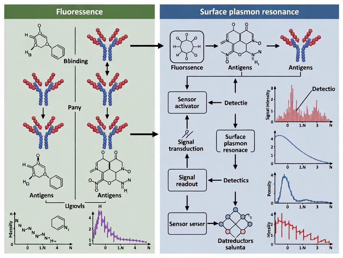
Diagram 1: Optical Biosensing Mechanisms. The top section illustrates the principle of a direct, FRET-based fluorescent biosensor, where analyte binding induces a conformational change that alters energy transfer between two fluorophores. The bottom section depicts the label-free detection principle of SPR, where binding of an analyte to an immobilized bioreceptor changes the local refractive index (RI), shifting the resonance angle of reflected light.
The strategic selection and implementation of biorecognition elements—enzymes, antibodies, and nucleic acids—form the cornerstone of effective optical biosensor design. Each class offers a unique set of advantages and limitations, making them suited to different applications. Enzymes provide catalytic amplification, antibodies deliver exceptional specificity, and nucleic acids offer robustness and design flexibility. The choice of element must be aligned with the target analyte, the required sensitivity and specificity, and the operational environment.
The ongoing convergence of materials science, nanotechnology, and data analytics is propelling the field forward. Enhancements in transducer design, such as pedestal high-contrast gratings, and novel computational approaches, like OmicSense, are pushing the limits of detection and quantification. For researchers and drug development professionals, a deep understanding of these core biorecognition principles is not merely academic; it is essential for innovating the next generation of diagnostic tools, monitoring systems, and research assays that will underpin advancements in personalized medicine, environmental safety, and fundamental biology.
The Role of Nanomaterials and Plasmonics in Enhancing Optical Signal and Sensitivity
Surface Plasmon Resonance (SPR) and Localized Surface Plasmon Resonance (LSPR) have emerged as foundational principles in modern optical biosensing, enabling the detection of biological analytes with exceptional sensitivity and without the need for fluorescent labels [33]. These phenomena leverage the collective oscillation of free electrons at the surfaces of metals and nanostructures, which can concentrate electromagnetic energy into subwavelength volumes [34]. The integration of nanomaterials into these plasmonic systems has catalyzed a paradigm shift in biosensor capabilities, yielding dramatic enhancements in signal strength, sensitivity, and specificity [33] [35]. This technical guide examines the underlying mechanisms, material systems, and experimental protocols that harness nanomaterial-plasmonic synergies, providing a comprehensive resource for researchers and drug development professionals working at the frontier of optical biosensing.
The evolution of plasmonic sensing from a laboratory phenomenon to a commercial technology began with the first demonstration of SPR biosensing in 1983, followed by the introduction of the first commercial SPR instrument by Biacore [33]. Concurrently, advances in nanotechnology have enabled precise fabrication of metallic nanostructures with tailored LSPR properties, opening new avenues for signal enhancement [36] [37]. Today, plasmonic-enhanced sensors find applications across medical diagnostics, pharmaceutical development, food safety, and environmental monitoring, driven by their capacity for real-time, label-free detection of biomolecular interactions [36] [33] [35].
Fundamental Principles of Plasmonics
Surface Plasmon Resonance Physics
Surface Plasmon Resonance occurs when incident photons couple with the collective oscillations of free electrons at a metal-dielectric interface under specific resonance conditions [37] [35]. This coupling requires momentum matching, typically achieved using prism couplers in the Kretschmann configuration, grating couplers, or waveguide structures [37] [35]. The resonance condition is highly sensitive to changes in the refractive index within the evanescent field region (typically extending 100-200 nm from the metal surface), forming the fundamental basis for SPR sensing [37].
The propagation constant of surface plasmon polaritons is given by: $$k{SPP} = \frac{\omega}{c} \cdot \sqrt{\frac{\varepsilon{metal} \cdot \varepsilon{diel}}{\varepsilon{metal} + \varepsilon{diel}}}$$ where $\omega$ is the angular frequency of light, $c$ is the speed of light, and $\varepsilon{metal}$ and $\varepsilon{diel}$ are the dielectric constants of the metal and dielectric medium, respectively [36]. When the wavevector component of incident light matches $k{SPP}$, resonance occurs, resulting in a characteristic dip in reflected light intensity at a specific angle or wavelength [35].
Localized Surface Plasmon Resonance
Localized Surface Plasmon Resonance occurs in metallic nanoparticles and nanostructures with dimensions smaller than the wavelength of incident light, where the confined electrons oscillate collectively without propagating [37] [34]. Unlike propagating SPR, LSPR can be excited by direct illumination without the need for momentum matching techniques [37]. The LSPR frequency is highly dependent on the nanoparticle's size, shape, composition, and the local dielectric environment, enabling tunable optical properties across the visible and near-infrared spectrum [37] [38].
For a spherical nanoparticle, the extinction cross-section derived from Mie theory is: $$\sigma{ext} = 9\left(\frac{\omega}{c}\right)\varepsilon{diel}^{3/2}V\frac{\varepsilon{metal}''}{(\varepsilon{metal}' + 2\varepsilon{diel})^2 + (\varepsilon{metal}'')^2}$$ where $V$ is the nanoparticle volume, and $\varepsilon{metal}'$ and $\varepsilon{metal}''$ are the real and imaginary parts of the metal dielectric function [37]. The resonance condition occurs when $\varepsilon{metal}' = -2\varepsilon{diel}$, leading to enhanced local electromagnetic fields near the nanoparticle surface [37].
Field Enhancement and Energy Localization
A paramount consequence of plasmon resonance is the significant enhancement of local electromagnetic fields, which can exceed the incident field intensity by several orders of magnitude [34]. This field enhancement is spatially confined to nanoscale volumes near the metal surface, typically within 30 nm for LSPR and 100-200 nm for propagating SPR [37]. The enhanced fields directly amplify optical processes including fluorescence, Raman scattering, and infrared absorption, enabling detection of single molecules in some configurations [37] [34].
The quality factor ($Q{LSPR}$) of plasmonic resonances determines the number of oscillation cycles before energy dissipation and is given by: $$Q{LSPR} \equiv \frac{\omega \partial Re[\varepsilonm(\omega)]/\partial\omega}{2Im[\varepsilonm(\omega)]}$$ which for simple cases reduces to $Q{LSPR} = -\frac{Re[\varepsilonm]}{Im[\varepsilon_m]}$ [34]. Higher Q-factors correspond to narrower resonance linewidths and greater sensitivity to environmental changes [34].
Table 1: Comparison of SPR and LSPR Sensing Modalities
| Characteristic | SPR (Propagating) | LSPR (Localized) |
|---|---|---|
| Excitation Mechanism | Momentum matching required (prisms, gratings) | Direct illumination |
| Field Penetration Depth | ~100-200 nm [37] | ~30 nm [37] |
| Measurement Setup | Complex optical alignment | Simplified optics |
| Miniaturization Potential | Challenging | Excellent |
| Quality Factor | Higher | Lower |
| Application Focus | Kinetic studies, biomolecular interactions | Point-of-care, portable sensing |
Nanomaterials for Plasmonic Enhancement
Metallic Nanoparticles
Gold and silver nanoparticles serve as the workhorse materials for plasmonic enhancement due to their favorable optical properties and chemical stability [33] [37]. Gold nanoparticles exhibit strong plasmon resonances across the visible spectrum, high chemical stability, and facile surface functionalization, making them ideal for biological applications [33]. Silver nanoparticles provide stronger plasmon resonances and higher field enhancement but are more susceptible to oxidation [37]. The plasmonic properties of these nanoparticles can be precisely tuned through control of their size, shape, and architecture:
- Nanospheres: Exhibit size-dependent resonance positions; 20-100 nm diameters typically used for biosensing [37]
- Nanorods: Offer tunable longitudinal plasmon modes across visible to near-infrared via aspect ratio control [38]
- Nanostars: Feature enhanced field localization at tip structures for superior signal amplification [38]
- Nanoshells: Consist of dielectric cores with metallic shells enabling precise resonance tuning through core-shell dimensions [37]
Bimetallic and Hybrid Nanostructures
Bimetallic nanoparticles combine the advantages of different metallic components to enhance stability, tune resonance positions, and improve sensing performance [36]. Core-shell architectures with silica-gold or silver-gold configurations enable precise tuning of plasmon resonances while maintaining chemical stability [36] [38]. Hybrid nanostructures integrating plasmonic metals with functional materials such as graphene, transition metal dichalcogenides, or metal-organic frameworks provide additional capabilities for selective analyte capture and signal amplification [36] [39].
2D Materials and Advanced Substrates
Two-dimensional materials including graphene, transition metal dichalcogenides (e.g., MoS₂), and MXenes have emerged as powerful components in plasmonic sensors [34] [39]. These materials can enhance plasmonic sensitivity through several mechanisms: increased surface area for analyte adsorption, energy transfer between materials, and modification of the local refractive index [35] [39]. Graphene specifically protects metallic nanostructures from oxidation while providing π-π stacking interactions for aromatic molecules, enhancing both stability and selectivity [39].
Table 2: Nanomaterial Properties and Their Impact on Sensing Performance
| Nanomaterial | Key Properties | Enhancement Mechanism | Typical Enhancement Factor |
|---|---|---|---|
| Gold Nanospheres | Tunable 20-100 nm, functionalizable | Local field enhancement, LSPR shift | 10-100x for SERS [37] |
| Gold Nanorods | Aspect ratio 2-5, NIR resonance | Longitudinal plasmon mode, photothermal | Up to 10^5 for fluorescence [38] |
| Silver Nanocubes | Sharp corners, high field enhancement | Hot spots at vertices | 10^6-10^8 for SERS [37] |
| Graphene-Gold Hybrid | Atomic thickness, high quinching | Energy transfer, adsorption enhancement | 2-5x sensitivity improvement [35] |
| Plasmonic Vesicles | Self-assembled, coupled nanoparticles | Plasmon coupling, spectral shifting | 2-4x PA signal enhancement [38] |
Enhancement Mechanisms and Signal Amplification
Plasmon-Enhanced Fluorescence
Metal-Enhanced Fluorescence (MEF) leverages the interaction between fluorophores and plasmonic nanostructures to significantly increase fluorescence intensity [33] [37]. This enhancement arises from two primary mechanisms: (1) increased excitation rate due to the enhanced local electromagnetic field, and (2) improved quantum yield through modification of the radiative decay rate [33]. The optimal enhancement occurs at an intermediate distance (5-20 nm) where non-radiative energy transfer to the metal is minimized while still benefiting from field enhancement [37]. MEF has enabled detection of biomarkers at previously inaccessible concentrations, with reported enhancement factors reaching 10²-10³ for optimized configurations [33].
Surface-Enhanced Raman Scattering
Surface-Enhanced Raman Scattering demonstrates the most dramatic signal amplification among plasmon-enhanced techniques, with enhancement factors typically ranging from 10⁶ to 10⁸ and reaching single-molecule sensitivity under optimal conditions [37]. This extraordinary enhancement originates primarily from electromagnetic mechanisms involving localized plasmon resonances, with additional contributions from chemical mechanisms involving charge transfer [37]. The largest enhancements occur at "hot spots" - nanoscale gaps between plasmonic nanoparticles where electromagnetic fields are most concentrated [37] [38]. SERS-based biosensors have achieved detection limits in the attomolar to zeptomolar range for various biomarkers, making them among the most sensitive analytical tools available [37].
Plasmon-Enhanced Photoacoustic Sensing
The photoacoustic effect combines optical excitation with acoustic detection, providing deep-tissue imaging capability while maintaining spectral specificity [38]. Plasmonic nanoparticles significantly enhance photoacoustic signals by increasing optical absorption and optimizing the thermoelastic conversion efficiency [38]. The generated pressure wave ($p0$) follows the relationship: $$p0 = \left(\frac{\beta c^2}{Cp}\right)\mua F = \Gamma A$$ where $\beta$ is the thermal expansion coefficient, $c$ is the speed of sound, $Cp$ is the heat capacity, $\mua$ is the absorption coefficient, $F$ is the fluence, $\Gamma$ is the Grüneisen parameter, and $A$ is the local energy deposition [38]. Engineering these parameters through nanomaterial design has enabled order-of-magnitude improvements in photoacoustic imaging contrast and sensitivity [38].
Experimental Protocols and Methodologies
Kretschmann Configuration SPR Setup
The Kretschmann configuration remains the most widely used implementation for high-sensitivity SPR biosensing [35] [8]. The following protocol details a standard setup for biomolecular interaction analysis:
Substrate Preparation: Use a high-refractive-index prism (e.g., SF10 glass, n = 1.723) cleaned with piranha solution and dried under nitrogen [35] [8].
Metal Film Deposition: Deposit a 50 nm gold or silver film onto the prism surface using thermal or electron-beam evaporation at a rate of 0.1-0.5 Å/s under high vacuum (<10⁻⁶ Torr) [35] [8]. Silver provides sharper resonances but requires protection from oxidation.
Functionalization: Immerse the metal film in a 1 mM solution of alkanethiol (e.g., 16-mercaptohexadecanoic acid) in ethanol for 12-24 hours to form a self-assembled monolayer [35]. Activate carboxyl groups with EDC/NHS chemistry for biomolecule immobilization.
Ligand Immobilization: Inject the ligand solution (typically 10-100 μg/mL in appropriate buffer) over the activated surface until saturation is achieved, followed by blocking with ethanolamine or BSA [35].
SPR Measurement: Use a polarized light source (λ = 633 nm or 785 nm) and rotate the prism or detector to monitor angular shifts with a resolution of <0.001° [8]. Maintain constant temperature (±0.1°C) throughout measurements.
Kinetic Analysis: Inject analyte at various concentrations (typically 1 nM-10 μM) in running buffer, monitor association and dissociation phases, and regenerate the surface between cycles [35].
LSPR Biosensor Fabrication and Measurement
LSPR biosensors based on colloidal nanoparticles offer simplified optical systems while maintaining high sensitivity:
Nanoparticle Synthesis: Prepare gold nanorods using a seed-mediated growth method: First, generate 3-4 nm gold seeds by reducing HAuCl₄ with ice-cold NaBH₄ in CTAB solution [38]. Then, grow nanorods by adding seeds to a growth solution containing HAuCl₄, CTAB, AgNO₃, and ascorbic acid [38].
Surface Functionalization: Functionalize nanoparticles with thiolated aptamers or antibodies by incubating overnight at room temperature in buffer, followed by centrifugation and resuspension [33] [39].
Substrate Immobilization: Immobilize functionalized nanoparticles on silanized glass substrates through covalent bonding or electrostatic interactions to create stable LSPR sensors [33].
Spectroscopic Measurement: Measure extinction spectra using a UV-Vis spectrophotometer with 1 nm resolution. Monitor LSPR peak shifts in real-time upon analyte introduction [33] [37].
Data Analysis: Determine analyte concentration from the magnitude of LSPR wavelength shift, typically achieving detection limits of 1-100 pM for protein biomarkers [33].
SERS-Based Detection Protocol
Surface-Enhanced Raman Scattering provides exceptional sensitivity for molecular detection:
Substrate Fabrication: Create SERS-active substrates by assembling gold or silver nanoparticles into closely-packed monolayers on functionalized silicon or glass substrates [37]. Alternatively, create patterned nanostructures using electron-beam lithography for reproducible hot spots [37].
Analyte Immobilization: Incubate substrates with analyte solution (typically 1-10 μL) and allow to dry, or use microfluidics for continuous flow measurements [37].
Spectral Acquisition: Illuminate with a laser source matching the plasmon resonance (commonly 532, 633, or 785 nm) with power 1-10 mW to avoid sample damage [37]. Collect spectra with a Raman spectrometer equipped with a CCD detector, typically integrating for 1-10 seconds.
Signal Processing: Subtract background fluorescence, normalize by integration time and laser power, and identify characteristic Raman peaks for quantitative analysis [37].
The Scientist's Toolkit: Essential Research Reagents and Materials
Table 3: Essential Materials for Plasmonic Biosensing Research
| Category | Specific Items | Function/Purpose | Key Considerations |
|---|---|---|---|
| Substrate Materials | SF10/SF11 glass prisms, Fused silica slides | High refractive index for SPR coupling | Refractive index >1.7 for optimal sensitivity [8] |
| Plasmonic Metals | Gold and silver targets (99.999%) | Film deposition for SPR substrates | Silver provides sharper resonance but oxidizes [35] [8] |
| Nanoparticles | Gold nanospheres (20-100 nm), Gold nanorods (aspect ratio 2-5) | LSPR substrates, signal amplification | CTAB-coated for stability, functionalizable [33] [38] |
| Surface Chemistry | Alkanethiols (e.g., 16-mercaptohexadecanoic acid), Silanes (APTES) | Self-assembled monolayers for functionalization | Thiol-gold chemistry for stable conjugation [35] |
| Coupling Reagents | EDC, NHS, Sulfo-SMCC | Covalent immobilization of biomolecules | EDC/NHS for carboxyl-amine coupling [35] |
| Recognition Elements | Antibodies, Aptamers, Molecularly imprinted polymers | Target capture and specificity | Aptamers offer stability and batch consistency [39] |
| Instrumentation | Polarized laser sources (633 nm, 785 nm), High-resolution rotation stages, CCD detectors | Optical measurement and data acquisition | Angular resolution <0.001° for kinetic studies [35] [8] |
Advanced Sensing Architectures and Performance
Performance Metrics and Optimization
Quantifying the performance of plasmonic biosensors requires careful attention to multiple metrics. Sensitivity defines the change in sensor output per unit change in refractive index or analyte concentration, typically expressed in nm/RIU (refractive index unit) or response units/nM [35] [8]. The quality factor combines sensitivity with resonance linewidth to provide a comprehensive performance metric [34] [8]. The limit of detection represents the smallest detectable analyte concentration, while the dynamic range defines the concentration span over which the sensor responds quantitatively [35].
Optimization strategies include engineering nanostructure geometry to enhance field confinement, utilizing high-quality factor materials to narrow resonance linewidths, and implementing noise reduction techniques in optical detection systems [34] [8]. Recent advances have demonstrated sensitivities exceeding 450°/RIU for angular interrogation and quality factors above 170 RIU⁻¹ in optimized multilayer structures [8].
Emerging Applications and Future Directions
Plasmonic-enhanced sensors are finding transformative applications across multiple domains. In neuroengineering, plasmonic nanoparticles enable minimally invasive neuromodulation and diagnosis of central nervous system disorders through enhanced light-tissue interactions [40]. For therapeutic monitoring, SPR aptasensors provide real-time kinetic analysis of drug-target interactions with picomolar sensitivity [39]. In point-of-care diagnostics, LSPR-based platforms offer rapid, label-free detection of biomarkers in complex biological fluids [33].
Future directions include the development of multifunctional plasmonic nanostructures for theranostic applications, integration with artificial intelligence for enhanced data analysis, and creation of massively multiplexed arrays for high-throughput biomarker screening [33] [40]. The convergence of plasmonics with emerging materials such as two-dimensional semiconductors and metal-organic frameworks promises to further expand the capabilities of optical biosensing platforms [34] [39].
Methodologies and Real-World Applications in Biomedical Research and Point-of-Care Diagnostics
Surface Plasmon Resonance (SPR) has established itself as a cornerstone technology in modern drug discovery, enabling the label-free detection and quantitative analysis of biomolecular interactions in real-time. This optical technique measures changes in the refractive index at a metal surface, typically gold, which occur when biomolecules bind to or dissociate from their partners immobilized on this surface [29]. The capability to monitor these interactions as they happen, without requiring fluorescent dyes or other reporter tags, provides significant advantages over traditional endpoint methods, including the reduced risk of false-negative results for interactions with fast kinetics and the ability to extract detailed kinetic parameters [41]. Within the broader context of optical biosensors, SPR stands out for its direct application in measuring the affinity and kinetics of molecular binding events—parameters that are critically important for understanding therapeutic efficacy and specificity.
The fundamental principle of SPR biosensing revolves around the generation of surface plasmons—collective oscillations of free electrons on a metal-dielectric interface. In a typical SPR configuration based on the Kretschmann geometry, a polarized light source is directed through a prism onto a thin gold film. At a specific angle of incidence, the energy from the photons couples with the electron oscillations, resulting in a sharp drop in the reflected light intensity, known as the resonance angle [29]. When biomolecular binding occurs on the gold surface, it alters the local refractive index, leading to a measurable shift in this resonance angle. These shifts, recorded in resonance units (RU) over time, produce a continuous data trace or sensorgram, providing a rich source of information on the association and dissociation phases of an interaction [29] [42]. The following diagram illustrates the core components and signal generation process of an SPR biosensor.
Figure 1: Core Principle of an SPR Biosensor. Polarized light strikes a gold film via a prism, generating a plasmon wave at a specific resonance angle. Molecular binding in the flow channel alters the refractive index, shifting the resonance angle detected in the reflected light.
The Critical Advantage of Real-Time Kinetic Analysis
A primary limitation of traditional endpoint assays in characterizing biomolecular interactions is their inherent vulnerability to missing transient interactions. These assays rely on a single measurement taken after incubation and wash steps, which can fail to capture interactions that form but dissociate rapidly before detection occurs [41]. In the high-stakes environment of drug discovery, such false negatives can have serious consequences, as off-target binding interactions—even weak ones—can lead to dose-limiting toxicity and are a major contributor to late-stage drug failures [41].
SPR technology directly addresses this limitation by providing a continuous, real-time view of the entire binding event. This allows researchers to not only confirm that binding occurs but also to characterize the kinetic profile of the interaction. The key parameters derived from SPR sensorgrams are:
- Association rate constant (kₐ or kon): Measures how quickly a complex forms.
- Dissociation rate constant (kd or koff): Measures how quickly a complex falls apart.
- Equilibrium dissociation constant (KD): The ratio kd/ka, representing the affinity of the interaction.
The following table summarizes a key comparative study that highlights the advantage of real-time SPR over an endpoint assay.
Table 1: Case Study Comparison: Endpoint Fluorescence vs. Real-Time SPR for Detecting Antibody Binding [41]
| Aspect | Endpoint Fluorescence Assay | Real-Time SPR Assay |
|---|---|---|
| Detection Principle | Fluorescence signal after washes | Label-free, real-time refractive index shift |
| Result for Antibody #1 | Strong positive signal | Strong binding signal |
| Result for Antibody #2 | False negative (no signal) | Positive binding signal confirmed |
| Identified Reason | Antibody #2 had a fast dissociation rate (koff), leading to complex disassembly during wash steps | Real-time monitoring captured the transient binding before dissociation |
| Risk | High risk of missing critical interactions | Reduced false-negative rate |
This kinetic information is not merely academic; it provides profound insight into the mechanism of action and can inform therapeutic strategy. For instance, a drug targeting a receptor may require slow dissociation (small kd) to achieve prolonged efficacy. Conversely, in emerging modalities like CAR-T therapy and targeted protein degradation, moderate affinity or finely tuned binding kinetics have been shown to correlate better with clinical efficacy [41].
Key Applications in the Drug Discovery Pipeline
SPR biosensing is deeply integrated across multiple stages of the drug discovery and development workflow, from initial target validation to lead optimization and beyond.
Off-Target Binding Screening
Secondary pharmacological profiling to identify off-target interactions is a regulatory requirement for investigational new drugs. SPR is exceptionally well-suited for this application because it can detect even weak, transient interactions with off-target proteins that might be missed by endpoint methods [41]. By screening drug candidates against a panel of immobilized "putative unsafe" off-target proteins (e.g., GPCRs, ion channels), researchers can identify compounds with promiscuous binding profiles early in the development process, thereby reducing the risk of adverse drug reactions (ADRs) in later clinical stages [41].
Antibody Characterization and Affinity Maturation
The development of biologic therapeutics, particularly monoclonal antibodies, relies heavily on SPR for kinetics characterization. SPR is used to characterize epitope binning (identifying antibodies that bind to overlapping regions on an antigen) and to precisely measure the affinity and kinetics of antibody-antigen interactions [42]. High-throughput SPR imaging (SPRI) systems can simultaneously analyze hundreds of interactions, dramatically accelerating the screening of antibody libraries and the process of affinity maturation [29].
Characterization of Complex Therapeutic Modalities
As drug modalities evolve, SPR has proven adaptable and vital for their characterization:
- Antibody-Drug Conjugates (ADCs): SPR can be used to independently verify the binding kinetics of the antibody component to its target, ensuring that affinity is optimized for both efficacy and safety [41].
- Targeted Protein Degraders (TPD): These molecules, such as PROTACs, facilitate a ternary complex between a target protein and an E3 ubiquitin ligase. SPR is instrumental in quantifying the binding events that lead to productive complex formation, helping to avoid the "hook effect" where high degrader concentrations inhibit degradation [41].
Experimental Workflow and Methodologies
A typical SPR experiment involves a series of carefully orchestrated steps to ensure the collection of high-quality, interpretable data. The following diagram outlines the generalized workflow for an SPR-based binding kinetics experiment.
Figure 2: Generalized SPR Experimental Workflow. The process involves preparing the surface, immobilizing the ligand, collecting binding data through association and dissociation phases, and finally analyzing the sensorgram.
Surface Functionalization and Ligand Immobilization
The first critical step is the immobilization of one interacting partner (the ligand) onto the sensor chip surface. The goal is to achieve a stable and functional layer that presents the ligand in a native conformation. Common immobilization strategies include:
- Direct Covalent Coupling: Amine coupling is the most prevalent method, where the gold surface is modified with a carboxymethylated dextran matrix. The ligand is coupled to this matrix via primary amines.
- Capture-Based Immobilization: This method uses a high-affinity capture molecule (e.g., Protein A for antibodies, streptavidin for biotinylated ligands, or an anti-tag antibody) immobilized on the surface. The ligand is then captured from solution, which often leads to more uniform orientation and better preservation of activity [41]. The SPOC (sensor-integrated proteome on chip) technology exemplifies an advanced capture approach, leveraging cell-free expressed proteins fused to a HaloTag that is captured in situ onto a chloroalkane-coated biosensor [41].
Binding Kinetics Measurement Protocol
Once the ligand is immobilized, the other interacting partner (the analyte) is injected over the surface in a series of concentrations dissolved in running buffer.
- Baseline: The system is stabilized with a continuous flow of running buffer to establish a stable baseline signal.
- Association Phase: The analyte solution is injected. As analyte molecules bind to the immobilized ligand, the mass on the surface increases, leading to an increase in the SPR signal (response units, RU). This part of the sensorgram is used to determine the association rate constant (kₐ).
- Dissociation Phase: The flow is switched back to running buffer. The decrease in signal as the analyte dissociates from the ligand is used to determine the dissociation rate constant (kd).
- Regeneration (Optional): A regeneration solution (e.g., low pH or a mild detergent) may be used to remove any remaining bound analyte, restoring the surface for a new cycle.
Data Analysis and Curve Fitting
The reference-subtracted binding time courses (sensorgrams) are fitted to a kinetic binding model to extract the rate constants. The most common model is the 1:1 Langmuir binding model, which assumes a simple bimolecular interaction [42]. The differential equations describing this model are:
Association: Rₜ = Rshiftᵢ + (Rmaxᵢ × kₐ × Cᵢ) / (kₐ × Cᵢ + k<sub>d</sub>) × (1 - e^(-(kₐ × Cᵢ + k<sub>d</sub>) × (t - t₀ᵢ))) [42]
Dissociation: Rₜ = Rdriftᵢ + (Rmaxᵢ × kₐ × Cᵢ) / (kₐ × Cᵢ + k<sub>d</sub>) × (1 - e^(-(kₐ × Cᵢ + k<sub>d</sub>) × (t_assoc - t₀ᵢ))) × e^(-k<sub>d</sub> × (t - t_assoc)) [42]
Where Rₜ is the response at time t, Cᵢ is the analyte concentration, Rmaxᵢ is the maximum binding capacity, and kₐ and k<sub>d</sub> are the rate constants.
High-throughput analysis tools, such as the TitrationAnalysis package for Mathematica, have been developed to automatically fit sensorgrams from multiple platforms (e.g., Biacore, Carterra LSA, ForteBio Octet), globaly estimating kₐ, k<sub>d</sub>, and the resulting K<sub>D [42].
Advanced SPR Technologies and Future Directions
The field of SPR biosensing continues to advance, with innovations enhancing its sensitivity, throughput, and application scope.
SPR Imaging (SPRI) for High-Throughput Screening
SPR Imaging (SPRI) represents a major technological evolution. Instead of monitoring a single spot, SPRI uses a CCD camera to visualize binding events across a large array format in real-time [29]. This allows for the simultaneous processing of hundreds or even thousands of interactions, making it ideal for applications like epitope binning of large antibody panels, high-throughput off-target screening, and clinical diagnostics using multi-array detection systems [41] [29]. Technologies like SPOC have further pushed the multiplex capacity, reporting up to ~864 protein ligand spots on a single biosensor [41].
Integration of Machine Learning
Machine learning (ML) is beginning to play a role in optimizing and interpreting SPR data. For instance, ML models like Random Forest (RF) and k-Nearest Neighbors (KNN) have been successfully applied to detect and classify DNA attachment on SPR biosensors based on reflective light angles and associated properties, achieving high accuracy [43]. This approach shows potential for developing more intelligent biosensors and for improving the analysis of complex datasets generated in disease diagnosis and prognosis.
Standardization of Reporting
With the increasing number of publications featuring biosensor data, there is a growing movement towards standardizing how this data is reported. The STROBE (Standards for Reporting Optical Biosensor Experiments) guidelines have been proposed to ensure that critical information—such as sample preparation, method settings, sensor type, and data evaluation details—is comprehensively documented. This enhances the reproducibility and reliability of biosensor-based research [44].
The Scientist's Toolkit: Essential Reagents and Materials
Table 2: Key Research Reagent Solutions for SPR Experiments
| Reagent / Material | Function in SPR Experiments | Example from Literature |
|---|---|---|
| Sensor Chips | Provides the gold surface and matrix for ligand attachment. Functionalized surfaces (e.g., CM5 for amine coupling, Protein A for antibody capture) are standard. | CM5 sensor chip (carboxymethylated dextran) is a classic platform [29]. |
| Capture Tags / Ligands | Enables oriented and uniform immobilization of the ligand, preserving activity. | HaloTag fusion proteins captured onto chloroalkane-coated biosensors in SPOC technology [41]. |
| Anti-Tag Antibodies | Act as capture molecules for specific tags fused to the protein of interest. | Commercial anti-HaloTag antibodies used to verify protein capture [41]. |
| Running Buffers | Provide the liquid environment for analyte dilution and flow. Must be optimized for pH and ionic strength to minimize non-specific binding. | Phosphate-buffered saline (PBS) is commonly used; often supplemented with surfactants (e.g., Tween-20) [41]. |
| Regeneration Solutions | Removes tightly bound analyte from the ligand to regenerate the sensor surface for subsequent analysis cycles without damaging the ligand. | Low pH buffers (e.g., Glycine-HCl) or mild detergents are typical [42]. |
| High-Throughput Analysis Software | Automates the fitting of sensorgrams to kinetic models, enabling efficient processing of large datasets. | TitrationAnalysis, a Mathematica package compatible with data from Biacore, Carterra LSA, and ForteBio platforms [42]. |
Surface Plasmon Resonance has fundamentally transformed the landscape of drug discovery by providing an unparalleled window into the dynamics of biomolecular interactions. Its capacity for real-time, label-free kinetic analysis directly addresses critical challenges in therapeutic development, from mitigating off-target toxicity to optimizing the affinity of advanced biologic and modality-specific drugs. As the technology evolves through advancements like high-throughput SPRI, integration with machine learning, and rigorous reporting standards, its role as an indispensable tool in the researcher's arsenal is only solidified. By enabling the precise quantification of how potential drugs interact with their targets, SPR continues to be a pivotal force in guiding the development of safer and more effective therapeutics.
Fluorescence Imaging and Assays for Intracellular Tracking and Cell-Drug Interaction Studies
Fluorescence molecular imaging (FMI) has established itself as a powerful imaging technique in biomedical research and clinical applications for visualizing molecular and cellular processes in tumors and other diseases [45]. This methodology leverages the fundamental properties of fluorescent molecules (fluorophores) that absorb light at specific wavelengths and emit it at longer wavelengths, providing researchers with a versatile toolkit for investigating drug delivery mechanisms, drug-target interactions, and cellular responses in single cells and whole tissues [46]. The core advantage of fluorescence-based methodologies lies in their ability to generate and enhance contrast that reveals central processes of drug delivery, offering non-radioactive, safe, real-time imaging with higher spatial resolution for superficial tumors compared to modalities like positron emission tomography (PET) [45].
The significance of fluorescence imaging is particularly evident in its application to more physiologically relevant 3D cell cultures, which better recapitulate the complex microenvironment and cell-cell interactions of actual tissues compared to traditional 2D monolayer cultures [47]. A growing body of evidence indicates that cells grown in monolayers lose some of their physiological properties and do not faithfully recapitulate drug responses, whereas 3D models provide structural and cellular morphological complexity that yields different—and more clinically relevant—responses to therapeutic compounds [47]. This capability to provide in-depth information about individual cell structure, function, and relative location within complex systems makes fluorescence imaging indispensable for modern drug discovery pipelines.
Fundamental Principles and Instrumentation
The Fluorescence Process and Probe Design
The physics and photochemistry of fluorescence can be understood through three consecutive stages: (1) formation of one or more excited state(s) by absorption (excitation), (2) non-radiative transitions between excited states, and (3) energy loss accompanied by emission of radiation [46]. These processes are linked reactions, each describable by statistical probability distributions, reaction rate constants, or decay lifetimes.
Effective fluorescence assays rely on probes consisting of two main components: a targeting portion (ligand) and a chromophore portion that presents the signal to be measured [46]. The targeting portion increases optical signal by localizing to its target through processes including covalent or non-covalent bonding, functional modification, or compartmentalization. This can involve chemical agents for covalent coupling, non-covalent DNA-binding probes, biotinylation of proteins, antibodies, ion-binding chelators, or other affinity molecules. The robustness and dynamic range of the assay is dictated by both the efficacy of the ligand-target interaction and the quantum efficiency of the chromophore [46].
Instrumentation Platforms and Selection Criteria
Choosing an appropriate imaging platform is crucial for obtaining meaningful data. The selection should be guided by the concept of "Dimensions of Imaging," which involves assessing experimental requirements for lateral (x-y) and axial (z) resolution, acquisition speed, sensitivity, and spectral separation [48].
Table 1: Fluorescence Microscopy Platforms and Their Characteristics
| Microscopy Type | Resolution | Imaging Speed | Key Advantages | Best Applications |
|---|---|---|---|---|
| Widefield Epifluorescence | Limited by out-of-focus light | Fast, whole sample illumination | Accessible, flexible, affordable | Screening large sample sets, thin samples |
| Laser Scanning Confocal (LSCM) | High, with optical sectioning | Slower, point scanning | Rejects out-of-focus light with pinhole | High-contrast 3D imaging, thicker samples |
| Spinning Disk Confocal | High, with optical sectioning | Very fast (∼100+ frames/s) | Reduced photobleaching | Live-cell dynamics, fast 3D collection |
| Super-Resolution | 2-10× below diffraction limit | Varies by technique | Nanoscale resolution | Sub-organellar studies, nanoscale interactions |
For most routine experiments, widefield and confocal microscopes are suitable, with widefield being particularly advantageous for efficiently screening large sample sets and detecting weak signals in thin specimens [48]. Laser scanning confocal microscopy (LSCM) creates thin, high-contrast "optical sections" through point-scanning and pinhole rejection of out-of-focus light, making it ideal for 3D reconstruction of thicker samples [48]. When higher imaging speeds are required for dynamic processes like vesicle trafficking or calcium imaging, spinning disk confocal microscopy is preferred due to its ability to capture data at rates of ∼100+ frames/s while reducing photobleaching [48]. For resolution beyond the diffraction limit (approximately 250 nm with green light), super-resolution techniques such as single molecule localization microscopy (SMLM), structured illumination microscopy (SIM), and stimulated emission depletion (STED) microscopy enable visualization of fine subcellular structures [48].
Advanced Fluorescence Methodologies for Drug Studies
Fluorescence Imaging in 3D Cell Culture Systems
The application of fluorescence imaging to three-dimensional (3D) cellular models represents a significant advancement in drug discovery, as these systems more accurately mimic the in vivo tissue environment than traditional 2D monolayers [47]. The most commonly employed 3D models are single-cell type and mixed co-culture spheroids due to their relative ease, reproducibility, and scalability for high-throughput screens. These spheroids, particularly in cancer research, mimic avascular tumor nodules with characteristic oxygen and nutrient gradients, extracellular matrix contacts, and cell-cell interactions that influence drug responses [47].
Imaging 3D cellular structures presents unique challenges, including light scattering and absorption in thick samples, heterogeneous cell labeling, and the need for specialized analysis techniques [47]. Standard procedures from 2D cultures, such as fixation and antibody staining with multiple washes, do not readily transfer to non-adherent 3D systems, as penetration of reagents into the 3D structure and potential volumetric collapse with fixation must be empirically determined [47]. Furthermore, certain subcellular events resolvable in 2D systems, such as nuclear translocation or vesicle trafficking, may not be detectable in 3D models due to sample background and physical constraints, making 3D imaging screens primarily suitable for macro-level readouts like cellular identification or viability [47].
Emerging Techniques for Visualizing Drug-Target Interactions
Recent advancements have enabled more precise visualization of drug-target interactions within native subcellular environments. The subcellular target-tracking fluorescent-visualization-based interaction screening (SubTrack-FVIS) platform combines super-resolution imaging with target-specific fluorescent tagging to map nanoscale spatial distributions of drug targets within living cells [49]. This approach screens compound libraries to identify molecules specifically binding to target-enriched domains and quantifies drug-target interactions through super-resolution imaging tracking, significantly reducing off-target effects by evaluating compound binding within native subcellular architectures [49].
When applied to lysosomal vacuolar H+-ATPases (V-ATPase), SubTrack-FVIS enabled real-time visualization of drug-target interactions and subsequent lysosomal destabilization, uncovering a unique mechanism of blocked autophagosome-lysosome fusion at sub-100 nm resolution [49]. This platform establishes a visualization framework for discovering drugs within physiological subcellular contexts while simultaneously decoding their mechanistic impacts.
Label-Free Alternatives with Machine Learning Integration
While fluorescence imaging provides exceptional specificity, label-free approaches that avoid potential artifacts from fluorescent probes are emerging as valuable alternatives. High-throughput bright-field imaging combined with machine learning can identify drug-induced morphological variations too subtle for human detection [50]. This method acquires numerous bright-field images of drug-treated and untreated cells at high throughput (up to 10,000 cells/s) and applies machine learning algorithms to classify cells based on morphological changes [50].
In practice, this approach has achieved 92% accuracy in distinguishing drug-treated from untreated cells without any labeling, with the degree of morphological change inferred from classification accuracy increasing with drug concentration and treatment time [50]. The multivariate data provided by single-cell images captures various types of information including geometry, granularity, intensity, and texture, making this label-free approach effective for identifying cellular responses to drugs.
Experimental Protocols and Methodologies
Workflow for SubTrack-FVIS Platform
The SubTrack-FVIS protocol for real-time visualization of drug-target interactions involves a multi-stage process [49]:
- Target Tagging: Genetically tag the drug target of interest (e.g., ATP6V1A subunit of V-ATPase) with a fluorescent protein in living cells.
- Super-Resolution Mapping: Use super-resolution microscopy to map the nanoscale spatial distribution of the target within native subcellular architectures.
- Compound Screening: Screen compound libraries against the target-tagged cells, maintaining physiological conditions.
- Interaction Visualization: Employ super-resolution imaging to track compound binding to target-enriched domains in real time.
- Functional Assessment: Quantify downstream effects of drug-target engagement, such as lysosomal alkalization or organelle dysfunction.
- Mechanistic Analysis: Resolve mechanistic impacts at sub-100 nm resolution, such as blockade of autophagosome-lysosome fusion.
This workflow enables direct visualization of drug-target interactions within native subcellular environments, providing both binding information and functional consequences.
Workflow for 3D Spheroid Imaging and Analysis
Imaging drug responses in 3D tumor spheroids requires specific adaptations to account for structural complexity [47]:
- Spheroid Generation: Culture cells in low-adhesion plates or with scaffold support to promote self-assembly into 3D structures. Optimize for size uniformity, critical for standardized analysis.
- Compound Treatment: Apply drug treatments using concentrations and duration relevant to the biological question. Include appropriate controls.
- Staining Protocol: For live-cell imaging, use fluorescent viability markers (e.g., calcein-AM for live cells, propidium iodide for dead cells). For fixed samples, optimize permeabilization and antibody incubation times for full penetration.
- Image Acquisition: Use confocal microscopy to obtain optical sections through the entire spheroid. Set z-stack parameters to ensure complete coverage with appropriate step size.
- Image Analysis: Employ 3D analysis software to quantify parameters such as spheroid volume, live/dead cell distribution, fluorescence intensity gradients, and invasion metrics.
Diagram 1: 3D Spheroid Imaging Workflow. This flowchart outlines the key steps in preparing and imaging 3D spheroids for drug response studies.
Controls and Validation Measures
Robust experimental design requires appropriate controls and validation measures to ensure data reliability [51]:
- Autofluorescence Controls: Include "no transfection," "no antibody," or "no dye" controls to account for sample autofluorescence.
- Antibody Specificity: Control for antibody specificity with no primary antibody controls.
- Spectral Bleed-Through: Reduce or account for bleed-through signal between channels using single-stained controls.
- Photobleaching Monitoring: Account for or reduce photobleaching during time-lapse experiments.
- Cell Health Assessment: Monitor cell health and phototoxicity throughout live-cell imaging.
- Hardware Calibration: Regularly verify light source alignment, objective lens performance, and channel registration using reference beads.
The Scientist's Toolkit: Research Reagent Solutions
Table 2: Essential Reagents and Materials for Fluorescence Imaging Studies
| Reagent/Material | Function | Application Notes |
|---|---|---|
| Polydimethylsiloxane (PDMS) | Flexible substrate for sensors | Excellent flexibility, optical transparency, biocompatibility [52] |
| Fluorescent Protein Fusions | Target labeling in live cells | Enables tracking of protein localization and dynamics [48] |
| Immunofluorescent Probes | Target labeling in fixed cells | Requires fixation and permeabilization; potential penetration issues in 3D models [47] |
| Viability Markers (Calcein-AM/PI) | Live/dead cell discrimination | Commonly used in 3D spheroid viability assays [47] |
| Quantum Dots | Photostable fluorescent probes | ZnO-PDMS composites used for high-sensitivity detection [52] |
| MXenes and Nanomaterials | Signal enhancement | Improve sensitivity and response speed in detection [52] |
| Organelle-Specific Dyes | Subcellular localization | Track drug accumulation in specific compartments [46] |
Technical Considerations and Best Practices
Ensuring Rigor and Reproducibility
Reproducibility in fluorescence imaging requires careful attention to experimental design and execution. Several key factors should be addressed to minimize bias and ensure reliable data [51]:
- Blinded Acquisition: Label samples with codes so that sample identity remains unknown during image acquisition to prevent selection bias.
- Systematic Sampling: Image predetermined random locations within wells rather than selecting "representative" areas, which can introduce bias.
- Statistical Power: Use preliminary imaging sessions to determine the number of samples needed for sufficient statistical power.
- Environmental Control: Maintain consistent temperature, CO₂ concentration, humidity, and pH throughout experiments where applicable.
- Image Quality Optimization: Avoid pixel saturation, increase dynamic range for optimal contrast, and follow the Shannon-Nyquist criterion for spatial and temporal sampling [51].
Addressing Plant-Specific Challenges
While many fluorescence imaging principles apply universally, plant specimens present unique challenges including waxy cuticles, strong autofluorescence, recalcitrant cell walls, and air spaces that impede fixation or live imaging [48]. These issues require specific adaptations:
- Autofluorescence Management: Plant tissues often exhibit broad-spectrum autofluorescence that can interfere with signals of interest, requiring careful filter selection and control experiments.
- Sample Preparation Optimization: The presence of cell walls necessitates specialized protocols for probe penetration and immunolabeling.
- Mounting Techniques: Air spaces within plant tissues require specific mounting solutions and techniques to minimize light scattering and refractive index mismatches.
Diagram 2: Fluorescence Imaging Challenges and Solutions. This diagram maps common challenges in fluorescence imaging to their potential solutions, highlighting strategies for improving image quality and experimental reliability.
Future Perspectives and Emerging Technologies
The field of fluorescence imaging for drug discovery continues to evolve with several promising directions. Ongoing improvements in probe development, imaging technologies, and analytical methods are addressing current challenges related to tissue penetration depth, background autofluorescence, photobleaching, and signal-to-noise ratio in deep tissues [45]. Future advancements will likely focus on:
- Multifunctional Miniaturized Devices: Integration of fluorescence sensing capabilities into smaller, more versatile platforms for increased accessibility and application range [52].
- Advanced Materials: Development of novel flexible materials with enhanced optical properties, biocompatibility, and environmental resilience for wearable and implantable sensors [52].
- Intelligent Data Analytics: Incorporation of artificial intelligence and machine learning for automated image analysis, pattern recognition, and predictive modeling of drug responses [50].
- Wireless Integration: Combination of fluorescence sensing with wireless communication technologies for real-time monitoring and data transmission in clinical and consumer healthcare applications [52].
These innovations are expected to accelerate the transition of fluorescence imaging technologies from laboratory research to practical clinical and consumer healthcare applications, ultimately revolutionizing personalized health management, early disease diagnosis, and continuous physiological monitoring [52].
Optical biosensors have emerged as transformative tools in biomedical research and drug development, enabling real-time, label-free investigation of biomolecular interactions. Among these, surface plasmon resonance (SPR) and fluorescence spectroscopy represent two powerful techniques with complementary strengths. SPR provides quantitative, kinetic data on binding events by detecting changes in the refractive index at a sensor surface, while fluorescence analysis offers high sensitivity and specificity for verifying molecular presence and localization. The integration of these technologies into synchronous dual-mode systems creates a powerful analytical platform that overcomes the limitations of either technique used independently, particularly for complex cellular analyses [53] [54].
The fundamental advantage of dual-mode systems lies in their ability to provide correlated datasets from a single experimental run, combining SPR's quantification of binding kinetics with fluorescence's spatial and specificity information. This approach is particularly valuable for studying cell-drug interactions, membrane protein dynamics, and intracellular signaling events where both binding kinetics and cellular localization are critical parameters. As pharmaceutical research increasingly focuses on cell-based assays and personalized medicine, these synchronous systems offer unprecedented capability for comprehensive cellular analysis in drug discovery workflows [54].
Fundamental Principles and Technological Synergy
Surface Plasmon Resonance Fundamentals
SPR technology operates on the principle of measuring refractive index changes occurring at the interface between a metal film (typically gold) and a dielectric medium when biomolecular binding events occur. In the Kretschmann configuration, which is most common in commercial instruments, polarized light is directed through a prism onto a thin metal film, exciting surface plasmons at specific resonance angles. As molecules bind to the functionalized sensor surface, the refractive index changes, causing a measurable shift in the resonance angle that is proportional to the mass concentration of bound analyte [53]. This label-free detection capability makes SPR ideal for monitoring molecular interactions in real-time without requiring fluorescent or other tags that might alter biomolecular behavior.
SPR's particular strength lies in providing quantitative information about association and dissociation rates, binding affinity, and stoichiometry of interactions. However, conventional SPR has limitations in detecting small molecules and lacks spatial resolution for cellular studies. It also provides complex information from samples by detecting refractive index changes, which can be challenging to deconvolute in complex biological mixtures [53].
Fluorescence Spectroscopy Principles
Fluorescence detection involves the emission of light from molecules that have absorbed photons at a specific excitation wavelength. In biosensing applications, fluorophores—either intrinsic to the target molecules or introduced as labels—provide highly sensitive and specific detection capabilities. Key parameters include excitation and emission wavelengths, intensity, lifetime, and polarization, all of which can provide information about molecular environment, conformation, and interactions [53] [55].
Modern fluorescence detection systems can switch between multiple wavelength channels (DAPI, FITC, TX RED, and CY5), enabling multiplexed detection of different targets. Advanced techniques like synchronous modulation and demodulation fluorescence spectroscopy can significantly improve signal-to-noise ratio and accuracy by reducing influences from dark current, ambient light, and background noise [53] [55]. While fluorescence provides exceptional sensitivity and specificity, it traditionally lacks the robust quantitative capabilities of SPR for determining binding kinetics and can be affected by photobleaching and environmental quenching.
Synergistic Benefits of Dual-Mode Systems
The combination of SPR and fluorescence creates a complementary system where the whole becomes greater than the sum of its parts. SPR enhances the sensitivity of fluorescence detection for low molecular weight analytes, while fluorescence adds molecular specificity to SPR's refractive index-based detection [53]. This synergy is particularly evident in cellular studies, where SPR can monitor membrane binding events while fluorescence simultaneously tracks intracellular responses, providing temporal correlation between external binding and internal signaling events.
Research has demonstrated that in epidermal growth factor (EGF) testing, the response time of fluorescence occurred earlier than SPR, indicating that signal transduction first occurred throughout the entire cell before propagating to specific membrane regions [54]. This type of insight is only possible when both techniques are applied synchronously to the same cellular sample, highlighting the unique capability of dual-mode systems for elucidating complex biological mechanisms.
System Architecture and Implementation
Instrumentation Design
A synchronous SPR-fluorescence detection system integrates both technologies around a shared flow cell and detection chamber. The SPR module typically employs a prism-coupled configuration with a red laser light source (635 nm peak wavelength), polarizing filter, and CCD camera mounted on precision rotation stages for angle scanning. The fluorescence detector is positioned above the SPR sensor and consists of a mercury lamp excitation source, filter wheel for wavelength selection, beam splitter, objective lens, and a separate CCD camera for detection [53].
Critical to the system's operation is the integrated sensor chip, which serves as the shared detection surface for both techniques. These chips are typically fabricated by magnetron sputtering technology on glass slides, with a 2 nm chromium adhesion layer followed by a 50 nm gold film in a defined detection area. A polydimethylsiloxane (PDMS) flow cell is then immobilized on the glass surface to form a sealed chamber for biochemical reactions and sample introduction [53]. This configuration enables both SPR and fluorescence measurements to be performed on the same sample simultaneously, ensuring perfect temporal correlation between the detected signals.
The following diagram illustrates the core architecture and signal flow of a synchronous SPR-fluorescence detection system:
Figure 1: System Architecture of Synchronous SPR-Fluorescence Detection
Key Research Reagents and Materials
Successful implementation of dual-mode SPR-fluorescence systems requires carefully selected reagents and materials optimized for both detection modalities. The table below details essential components and their functions in typical experimental workflows:
Table 1: Essential Research Reagents and Materials for Dual-Mode SPR-Fluorescence Experiments
| Component | Specification/Example | Function in Dual-Mode System |
|---|---|---|
| Sensor Chips | Glass slides with 2 nm Cr + 50 nm Au sputtered coating [53] | Provides SPR-active surface compatible with fluorescence imaging |
| Flow Cells | PDMS (Sylgard 184, 10:1 ratio) cured at 80°C for 8h [53] | Creates sealed chamber for sample introduction and washing |
| Fluorescent Labels | Hoechst 33258, FITC-conjugated antibodies, green fluorescent microspheres [53] | Enables fluorescence detection and spatial localization |
| Cell Culture Reagents | RPMI 1640 medium with 10% FBS, penicillin-streptomycin [53] | Maintains cell viability during real-time monitoring |
| Ligand Immobilization | Biotin-streptavidin systems, carbodiimide crosslinking [56] | Anchors binding partners to sensor surface for interaction studies |
| Reference Materials | Phosphate buffered saline (PBS), deionized water [53] [56] | Provides baseline measurements and controls for system characterization |
Signal Processing and Data Correlation
A critical aspect of dual-mode systems is the temporal and spatial matching of SPR and fluorescence signals. Advanced algorithms enable adaptive adjustment of potentially distorted SPR images and correlation with fluorescence data. The system characterized by Zhang et al. employed an adaptive adjustment algorithm for distorted SPR images, allowing for precise temporal and spatial matching of the dual-mode detection [54]. This synchronization is essential for accurately correlating binding events detected by SPR with cellular responses monitored via fluorescence.
The correlation between SPR and fluorescence signals has been quantitatively demonstrated in studies with fluorescent microspheres, where the correlation coefficient reached up to 0.9866 [53]. This high degree of correlation validates the synchronous detection capability and enables confident interpretation of complementary data streams. For cellular studies, this means that changes in SPR response units can be directly linked to specific fluorescence signals from cellular compartments or labeled components, providing a comprehensive view of cell-drug interactions or signaling events.
Experimental Protocols and Methodologies
System Characterization and Calibration
Before conducting cellular experiments, the dual-mode system requires comprehensive characterization and calibration to ensure optimal performance of both detection modalities. The following workflow outlines the essential calibration procedures:
Figure 2: System Calibration and Characterization Workflow
For SPR calibration, an angular scan is conducted in Milli-Q water to locate the absorption peak and corresponding coupling angle. The incident light angle is changed via motor and mechanical device, typically scanning from 90,000 to 140,000 steps in characterized systems [53]. Once the resonance angle is determined, the system can be fixed at this angle for intensity-based detection or continue angular scanning for more comprehensive characterization.
Fluorescence calibration involves testing the excitation and detection efficiency across all available wavelength channels (DAPI, FITC, TX RED, and CY5). The system's capability to switch between these wavelengths enables multiplexed detection of different cellular targets. Validation using fluorescent microspheres of known size and emission characteristics (e.g., 16 μm diameter green fluorescent microspheres with excitation/emission at 495/519 nm) confirms proper alignment and functionality [53].
Cell Preparation and Immobilization Protocols
For cellular studies, appropriate preparation and immobilization of cells on the sensor surface is crucial for obtaining meaningful data. The following protocol outlines the key steps for cell-based experiments:
Cell Culture and Maintenance: A549 non-small-cell lung cancer cells (or other relevant cell lines) are cultured at 37°C in 5% CO₂ in RPMI 1640 medium supplemented with 10% heat-inactivated fetal bovine serum, 100 units/mL penicillin, and 100 μg/mL streptomycin [53].
Cell Harvesting and Labeling: Immediately prior to experiments, cells are trypsinized, centrifuged, and resuspended in supplemented culture medium. For fluorescence detection, cells are fixed and labeled with appropriate fluorophores such as Hoechst 33258 for nuclear staining or specific antibody conjugates for target proteins [53].
Sensor Surface Preparation: The gold sensor surface may be functionalized with extracellular matrix proteins (e.g., fibronectin, collagen) or specific capture molecules to promote cell adhesion and mimic physiological conditions.
Cell Immobilization: A cell droplet of 30 μL at a density of approximately 1×10⁶ cells/mL is added to the sensor surface and allowed to adhere under appropriate conditions [53]. The flow cell is then assembled, taking care not to disrupt the attached cells.
Viability Assessment: Using fluorescence markers, cell viability and general morphology are confirmed before initiating experimental treatments.
Synchronous Detection Experimental Procedure
Once cells are properly immobilized and the system is calibrated, synchronous SPR-fluorescence detection experiments can be performed:
Baseline Establishment: Begin with a continuous flow of buffer or culture medium to establish stable SPR and fluorescence baselines.
Treatment Introduction: Introduce the experimental treatment (e.g., drug candidate, growth factor, signaling molecule) using a precise flow system to ensure rapid and complete exchange.
Real-Time Monitoring: Simultaneously collect SPR and fluorescence data throughout the experiment. SPR typically monitors changes in resonance angle or intensity, while fluorescence tracks specific molecular events or cellular responses.
Multiplexed Fluorescence Imaging: If multiple fluorescence channels are available, sequentially image different cellular compartments or signaling molecules to build a comprehensive picture of cellular responses.
Data Correlation: Use established algorithms to temporally and spatially correlate SPR and fluorescence signals, identifying relationships between binding events and cellular responses.
For EGF stimulation experiments, researchers have successfully demonstrated that the platform can quantitatively detect changes in the cell membrane region, with SPR response signals increasing with upticks of epidermal growth factor within stimulated cells [54]. In these studies, when adding EGF, a peak was observed in the SPR curve, while the corresponding cells in the SPR image exhibited whitening, demonstrating the platform's capability to simultaneously monitor SPR response signals and image changes [54].
Performance Metrics and Quantitative Analysis
System Sensitivity and Detection Limits
Dual-mode SPR-fluorescence systems offer enhanced sensitivity by combining the strengths of both technologies. The detection limit of the synchronized method for epidermal growth factor (EGF) has been demonstrated to be 20 IU/mL [54]. This sensitivity is sufficient for monitoring many biologically relevant concentrations of signaling molecules and drugs.
In fluorescence-only mode, advanced techniques like synchronous modulation and demodulation fluorescence spectroscopy can significantly improve quantitative analysis. When applied to urea detection in serum, this method achieved a correlation coefficient (Rc) of 0.916753 and root mean square error of calibration (RMSEC) of 2.05848 mmol/L, representing significant improvement over unmodulated fluorescence spectroscopy [55].
Table 2: Performance Metrics of Dual-Mode SPR-Fluorescence Systems
| Performance Parameter | SPR Mode | Fluorescence Mode | Dual-Mode Advantage |
|---|---|---|---|
| Detection Limit | Dependent on molecular weight; challenges with small molecules [53] | Single molecule detection possible with optimal labeling | Fluorescence enhances small molecule detection via SPR sensitivity enhancement [53] |
| Quantitative Accuracy | High for binding kinetics and affinity measurements [56] | Improved with modulation/demodulation techniques (Rc = 0.916753) [55] | Complementary data improves overall quantification reliability |
| Temporal Resolution | Real-time (sub-second) monitoring capability [53] | Limited by exposure times and frame rates | Synchronous detection enables correlated kinetic analysis |
| Spatial Resolution | Limited to sensor surface interactions | Subcellular resolution possible with microscopy | Correlate membrane events (SPR) with intracellular responses (fluorescence) |
| Correlation Coefficient | - | - | Up to 0.9866 for fluorescent microspheres [53] |
Comparative Analysis with Single-Mode Systems
The advantages of dual-mode systems become apparent when comparing their capabilities with single-mode approaches. Traditional SPR alone struggles with detecting small molecules and lacks molecular specificity in complex mixtures, while fluorescence alone provides limited quantitative information about binding kinetics [53]. The integration of these technologies creates a system where SPR enhances the quantitative capabilities of fluorescence analysis, and fluorescence adds specificity to SPR detection.
Research has shown that in cell-drug interaction studies, the response time of fluorescence occurred earlier than SPR when adding EGF, indicating that signal transduction first occurred throughout the entire cell before propagating to the cell membrane region [54]. This type of insight would be impossible to obtain with either technique alone and demonstrates the unique value of synchronous detection for elucidating temporal sequences in cellular signaling pathways.
Applications in Cell Analysis and Drug Development
Cell-Drug Interaction Studies
Dual-mode SPR-fluorescence systems excel in pharmaceutical research by enabling comprehensive analysis of cell-drug interactions. These systems can simultaneously assess binding kinetics at the cell membrane (via SPR) and subsequent intracellular responses (via fluorescence), providing a complete picture of drug activity. The technology has been successfully applied to study the interactions between various drug candidates and cellular targets, particularly membrane receptors and their signaling pathways [54].
In practice, when adding epidermal growth factor to stimulated cells, researchers observed that the SPR response signal increased with EGF concentration, demonstrating the platform's capability for quantitative detection of the cell membrane region [54]. Simultaneous fluorescence imaging revealed that cellular responses initiated internally before manifesting at the membrane level, providing valuable insights into the sequence of signaling events [54].
Receptor Signaling and Pathway Analysis
The synchronous nature of these systems makes them ideal for studying receptor activation and subsequent signaling cascades. By labeling different components of signaling pathways with distinct fluorophores and monitoring initial binding events via SPR, researchers can establish direct correlations between receptor engagement and downstream effects. This approach is particularly valuable for characterizing targeted therapeutics and understanding mechanisms of drug resistance.
Cancer Research and Therapeutic Development
In cancer research, dual-mode systems offer powerful capabilities for studying tumor cell responses to chemotherapeutic agents, targeted therapies, and immunotherapies. Using cancer cell lines like A549 lung cancer cells, researchers can monitor drug binding kinetics while simultaneously assessing cell viability, apoptosis induction, and changes in expression of specific biomarkers [53] [54]. This comprehensive analysis accelerates the drug screening process and provides deeper mechanistic insights than single-technique approaches.
Emerging Trends and Future Perspectives
Integration of Artificial Intelligence
The future of dual-mode SPR-fluorescence systems lies in sophisticated data analysis and integration with emerging technologies. Artificial intelligence, particularly machine learning and deep learning algorithms, is being increasingly applied to enhance the analytical capabilities of optical biosensors [57]. AI can improve sensitivity and specificity through intelligent signal processing, pattern recognition, and automated decision-making, helping to extract more meaningful information from the complex datasets generated by dual-mode systems.
Miniaturization and Point-of-Care Applications
As the field advances, there is growing interest in miniaturizing dual-mode systems for point-of-care diagnostics and decentralized testing. Recent advances in flexible materials and wearable optical biosensors demonstrate the potential for more compact and accessible platforms [52]. Integration with Internet of Things (IoT) and cloud computing technologies could enable real-time data analysis and remote monitoring capabilities, expanding the applications of these systems beyond traditional laboratory settings [57] [52].
Enhanced Nanomaterial Integration
The incorporation of advanced nanomaterials represents another promising direction for improving dual-mode system performance. Nanostructured materials, including metallic nanoparticles, quantum dots, and two-dimensional materials like MXenes, can enhance sensitivity and functionality through their unique optical and electronic properties [52] [58]. These materials can enhance both SPR and fluorescence signals, potentially leading to orders-of-magnitude improvements in detection limits and enabling new application areas.
Despite these exciting developments, challenges remain in the widespread adoption of dual-mode systems, including integration complexity, data management requirements, and the need for standardized validation protocols across different platforms [57]. Addressing these challenges will require collaborative efforts between researchers, engineers, and clinical specialists to fully realize the potential of synchronous SPR-fluorescence technology in revolutionizing biomedical research and therapeutic development.
Wearable optical biosensors represent a paradigm shift in personalized healthcare, enabling the continuous, non-invasive monitoring of vital physiological parameters. Among these technologies, photoplethysmography (PPG) has emerged as a predominant methodology, integrated into millions of consumer wearables and clinical devices. PPG biosensors operate on the principle of optical detection of blood volume variations in the microvascular tissue bed, typically using a light-emitting diode (LED) and a photodetector (PD) to capture cardiovascular pulsations [59]. The significance of these sensors extends beyond basic heart rate monitoring, encompassing measurements of oxygen saturation, respiratory rate, blood pressure, and vascular health through sophisticated signal processing techniques [60].
This technical guide examines wearable PPG biosensors within the broader context of optical biosensing principles, including fluorescence and surface plasmon resonance (SPR) research. SPR sensors detect minute interactions between sensing materials and chemicals through changes in refractive index, enabling accurate detection of even the smallest molecular changes [36]. While traditional SPR systems have been predominantly laboratory-based, recent advancements in miniaturization, including grating- and fiber-based SPR sensors that eliminate the need for prisms, are paving the way for their integration into wearable platforms [36]. The convergence of PPG's physiological monitoring capabilities with the molecular sensitivity of SPR technologies represents the next frontier in wearable biosensing, potentially enabling continuous monitoring of biochemical markers alongside physical parameters.
The global market for PPG biosensors is projected to grow from USD 648.5 million in 2025 to USD 3,064.8 million by 2035, reflecting a compound annual growth rate (CAGR) of 16.8% [60]. This growth is driven by increasing adoption of wearable health technologies, advancements in remote patient monitoring, and rising demand for non-invasive, continuous biometric tracking. Simultaneously, the broader wearable sensors market is forecast to reach US$7.2 billion by 2035, with PPG playing a significant role in this expansion [61].
Fundamental Principles: From Light-Tissue Interaction to Signal Acquisition
Photoplethysmography Operating Mechanism
PPG biosensors function by emitting light into the skin and detecting the variations in light absorption or reflection caused by pulsatile changes in arterial blood volume. A typical PPG sensor consists of a light source (usually an LED) that illuminates the tissue, and a photodetector that measures the intensity of light after it has interacted with the tissue [59]. The detected signal contains both a direct current (DC) component and an alternating current (AC) component. The DC component arises from light absorption by non-pulsatile arterial blood, venous blood, and stationary tissues, while the AC component corresponds to the pulsatile changes in arterial blood volume synchronized with the cardiac cycle [59].
The relationship between light attenuation and blood volume changes follows the modified Beer-Lambert law, which describes light propagation in scattering media like biological tissue. The PPG waveform morphology provides valuable clinical information, with characteristic systolic peaks, diastolic peaks, and dicrotic notches reflecting cardiovascular dynamics. Advanced signal processing techniques can extract additional parameters including heart rate variability, pulse wave velocity, and respiratory-induced variations [59].
SPR and LSPR Sensing Modalities
Surface Plasmon Resonance (SPR) and Localized Surface Plasmon Resonance (LSPR) sensors represent more advanced optical sensing techniques that are increasingly being explored for wearable applications. SPR sensors detect changes in the refractive index near a metal surface (typically gold or silver) through the excitation of surface plasmon polaritons by polarized light [36]. When the wavevector of incident light matches the propagation constant of surface plasmons, resonance occurs at a specific angle and wavelength that is highly sensitive to molecular binding events at the metal surface [36].
LSPR sensors operate on similar principles but utilize metal nanoparticles rather than continuous thin films. The confined surface plasmons in nanoparticles result in strong light absorption at specific wavelengths that depend on the nanoparticle size, shape, composition, and local dielectric environment [36]. LSPR sensors offer advantages for miniaturization and integration into wearable platforms due to their simpler optical arrangements and potential for colorimetric detection [36]. Recent research has focused on innovative materials including metal nanoparticles, bimetallic nanoparticles, and metal-organic materials to enhance the sensitivity and specificity of LSPR and SPR sensors [36].
Table 1: Comparison of Optical Biosensing Technologies for Wearable Applications
| Parameter | PPG | LSPR | SPR |
|---|---|---|---|
| Sensing Principle | Light absorption/ scattering by blood volume changes | Local refractive index changes via nanoparticle plasmons | Refractive index changes via thin film plasmons |
| Measured Analytes | Physiological parameters (heart rate, SpO₂, etc.) | Molecular targets (proteins, biomarkers, chemicals) | Molecular targets (proteins, biomarkers, chemicals) |
| Detection Limit | Macroscopic physiological changes | ppb level for chemical analytes [36] | High sensitivity to molecular binding events |
| Miniaturization Potential | High (integrated into watches, patches) | High (nanoparticle-based, colorimetric) | Moderate (advances in fiber- and grating-based systems) [36] |
| Power Consumption | Low to moderate | Low | Moderate to high |
| Current Commercial Status | Widespread in consumer wearables | Emerging in specialized sensors | Predominantly laboratory systems |
Modes of PPG Signal Acquisition
Wearable PPG sensors implement two primary configurations for signal acquisition:
Transmission Mode: In this configuration, the photodetector is positioned opposite the light source, detecting light that passes through the tissue. Transmission-mode PPG provides higher signal-to-noise ratio (SNR) and superior accuracy for SpO₂ and heart-rate measurements, making it suitable for clinical settings using finger clips or ear probes [59]. However, this approach requires placement on anatomical sites with sufficient light penetration, limiting wearability options.
Reflection Mode: Reflection-mode PPG positions both the light source and detector on the same side of the tissue, measuring back-scattered light. This configuration offers greater flexibility in sensor placement, enabling integration into wrist-worn devices, chest patches, and other wearable form factors [59]. While reflection-mode generally provides lower SNR compared to transmission-mode, advances in sensor design and signal processing have significantly improved its performance for continuous monitoring applications.
Recent research has focused on enhancing reflection-mode PPG through flexible, conformable sensor designs that improve skin contact and motion artifact rejection. For example, ultra-thin, flexible PPG sensors with total thickness of just 3 μm have been developed, thinner than the epidermis, enabling unobtrusive monitoring with improved signal quality [59]. Similarly, patch-type systems incorporating flexible perovskite photodetectors and 3D wrinkled-serpentine interconnections maintain functionality even at significant bending angles (60°), adapting comfortably to curved body surfaces [59].
Market Analysis and Technological Adoption Trends
The PPG biosensors market exhibits robust growth and evolving application segments across various geographical regions and product categories. The market expansion is fueled by technological advancements, increasing health consciousness, and the growing prevalence of cardiovascular diseases that require continuous monitoring.
Table 2: PPG Biosensors Market Forecast and Regional Analysis (2025-2035)
| Region/Country | Market Size 2025 (USD million) | Projected Market Size 2035 (USD million) | CAGR (%) | Key Drivers and Applications |
|---|---|---|---|---|
| Global Market | 648.5 [60] | 3,064.8 [60] | 16.8 [60] | Wearable health tech, remote patient monitoring, cardiovascular disorders |
| North America | Largest market share | Significant growth | 17.1 (US) [60] | Strong wearable tech ecosystem, FDA clearance pathways, insurance-backed RPM |
| Europe | Established market | Steady expansion | 16.7 (EU) [60] | Data interoperability mandates, telehealth infrastructure, preventive cardiovascular care |
| Asia-Pacific | Rapidly growing | Highest growth rate | 17.3 (South Korea) [60] | Affordable wearables, smartphone penetration, lifestyle disease awareness |
| Japan | Progressive growth | Continued expansion | 16.2 [60] | Aging demographic, smart healthcare devices, precision monitoring |
The product segment landscape is dominated by smartwatches, which hold a 42.8% value share of the PPG biosensors market by product type [60]. Other significant product categories include fitness bands, chest straps, smart patches, and emerging form factors such as smart rings and ear-worn devices. The period from 2025 to 2035 is projected to witness a transformation in PPG biosensor capabilities, shifting from basic physiological monitoring to comprehensive health diagnostics through several key technological shifts:
- Regulatory Landscape: Expansion from basic FDA and CE clearances for consumer-grade wearables to clinical-grade PPG standards and AI/ML model validation for health diagnostics [60].
- Technological Innovation: Evolution from optical sensors using green/red/IR LEDs for basic parameters to multimodal, AI-calibrated PPG with simultaneous ECG, hydration, respiration, and blood pressure sensing [60].
- Industry Adoption: Transition from mainstream use in smartwatches and fitness bands to adoption in chronic care, geriatric health, hospital-at-home, and remote ICU monitoring [60].
- Product Design: Advancement from wrist-worn wearables with basic LED/PD arrays to flexible printed sensors, invisible skin-integrated wearables, and adaptive light-sensing arrays for any skin tone [60].
Experimental Methodologies and Technical Protocols
Fabrication of Flexible PPG Sensors
The development of high-performance wearable PPG sensors requires specialized fabrication approaches to achieve flexibility, comfort, and reliable skin contact. The following protocol outlines the methodology for creating ultraflexible organic PPG sensors based on published research:
Materials and Equipment:
- Substrate: 1-μm-thick parylene film
- Active layers: Polymer light-emitting diodes (PLEDs) and organic photodetectors (OPDs)
- Electrodes: Transparent conductive oxide (e.g., ITO) or metal thin films
- Encapsulation: Thin-film barrier layers (e.g., Al₂O₃, SiO₂)
- Fabrication tools: Thermal evaporation system, spin coater, photolithography equipment
Procedure:
- Substrate Preparation: Deposit a 1-μm-thick parylene film on a handling wafer. Parylene provides excellent flexibility, biocompatibility, and moisture barrier properties.
- Electrode Patterning: Pattern bottom electrodes using photolithography and thermal evaporation of conductive materials.
- Active Layer Deposition: Sequentially deposit organic semiconductor layers for PLEDs and OPDs using solution processing or thermal evaporation. For PLEDs, typical stack includes hole injection layer, hole transport layer, emission layer, and electron transport layer. For OPDs, utilize donor-acceptor bulk heterojunction structures.
- Top Electrode Deposition: Evaporate semi-transparent top electrodes (e.g., thin metal films or transparent conductive oxides).
- Encapsulation: Deposit thin-film barrier layers (e.g., 100-nm Al₂O₃) using atomic layer deposition to protect organic layers from oxygen and moisture.
- Device Release: Carefully release the completed device stack from the handling wafer, resulting in a freestanding, ultraflexible PPG sensor.
Validation and Testing:
- Perform electo-optical characterization to determine LED output intensity and detector responsivity.
- Validate PPG functionality on volunteers at rest and after exercise, comparing waveform quality and heart rate accuracy with reference devices.
- Conduct mechanical testing to verify performance under bending (e.g., up to 60° curvature) and after repeated flexing cycles [59].
Hybrid PPG-SPR Sensing Platform
Integrating PPG with SPR sensing capabilities creates a multimodal platform for simultaneous physiological and molecular monitoring. The following protocol describes the development of a hybrid sensing system:
Materials and Equipment:
- SPR substrate: Gold-coated (50 nm) glass slides or optical fibers
- PPG components: Miniature LEDs (green, red, IR) and photodetectors
- Surface functionalization: Thiolated capture molecules (e.g., antibodies, aptamers)
- Microfluidic system: For sample delivery to SPR sensing area
- Optical components: Polarizers, lenses, light source (for SPR)
- Signal processing: Microcontroller with separate processing channels for PPG and SPR
Procedure:
- SPR Substrate Preparation: Clean gold-coated substrates with oxygen plasma treatment. Functionalize surface with self-assembled monolayers of thiolated capture molecules specific to target analytes (e.g., cardiac biomarkers).
- PPG Sensor Integration: Mount miniature LEDs and photodetectors adjacent to SPR sensing area, ensuring optical isolation between systems.
- Fluidic System Integration: Bond microfluidic channels to SPR substrate using PDMS or thermoplastic materials, ensuring precise delivery of samples to sensing regions.
- Optical Alignment: Align SPR excitation optics (if using traditional prism configuration) or integrate fiber-optic SPR elements. For miniaturized systems, implement grating-coupled SPR to eliminate bulky prisms [36].
- Electronic Integration: Develop separate but synchronized signal acquisition systems for PPG and SPR, with appropriate filtering for each modality.
- Software Development: Implement algorithms for real-time SPR angle shift detection and PPG waveform analysis, with correlation analysis between physiological parameters and molecular concentrations.
Validation:
- Test SPR sensitivity using solutions with known refractive index changes.
- Validate molecular detection with model analytes (e.g., albumin, specific antibodies).
- Conduct in-vitro testing with whole blood to verify PPG functionality in presence of SPR sensing elements.
- Perform simultaneous monitoring of physiological parameters (via PPG) and biomarker concentrations (via SPR) during controlled perturbations [36] [59].
Emerging Research Directions and Technical Challenges
Advanced Materials and Structures
Research in wearable PPG biosensors is increasingly focused on novel materials and structures that enhance performance, wearability, and compatibility with diverse user populations. Key developments include:
Organic Semiconductor-Based Photodetectors: Traditional PPG systems utilize silicon photodiodes, which are rigid and have limited flexibility. Recent advances in organic photodetectors (OPDs) offer mechanical flexibility, tunable spectral response, and compatibility with large-area fabrication processes. Huang et al. developed a photodetector using a novel ultranarrow-bandgap nonfullerene acceptor, achieving responsivity over 0.5 A/W in the NIR region (920–960 nm), the highest reported for organic photodiodes [59]. These devices demonstrated specific detectivity up to 10¹² Jones, comparable to commercial silicon photodiodes, while offering superior flexibility.
Geometrically Adaptive Designs: Conventional rigid PPG sensors struggle to maintain consistent skin contact on curved body surfaces, leading to motion artifacts and signal degradation. Three-dimensional wrinkled-serpentine interconnections have been developed to improve device adaptability to curved surfaces, maintaining excellent functionality even at significant bending angles (60°) [59]. These designs enable conformal contact with skin, reducing motion artifacts and improving signal quality during daily activities.
Skin-Tone Adaptive Sensing: A significant challenge in reflective PPG is accuracy variation across different skin complexions, particularly with higher melanin content which strongly absorbs green light commonly used in wrist-worn devices [59]. Research is addressing this through multi-wavelength approaches, adaptive LED power control, and advanced signal processing algorithms that compensate for skin tone variations. Next-generation sensors are incorporating adaptive light-sensing arrays designed specifically for accuracy across all skin tones [60].
AI-Enhanced Signal Processing and Multimodal Integration
The integration of artificial intelligence with PPG biosensing represents one of the most significant advancements in the field:
AI-Driven Signal Enhancement: Machine learning algorithms are being deployed to address fundamental PPG limitations including motion artifacts, ambient light interference, and low perfusion signals. Deep learning models, particularly convolutional neural networks and recurrent neural networks, can effectively separate noise from physiological signals, enabling reliable monitoring in real-world conditions [60]. These approaches are especially valuable for extracting clean signals from high-motion scenarios like physical exercise.
Multimodal Sensor Fusion: Combining PPG with complementary sensing modalities enhances measurement accuracy and expands detectable parameters. Hybrid platforms integrating PPG with electrocardiogram (ECG), bioimpedance, temperature, and inertial measurement units (IMUs) enable comprehensive physiological profiling [60]. Sensor fusion algorithms correlate data from multiple sources to derive advanced metrics including blood pressure, cardiac output, vascular aging, and autonomic nervous system activity.
Predictive Health Analytics: Beyond real-time monitoring, AI-enabled PPG systems are evolving toward predictive health assessment. By analyzing long-term PPG trend data combined with clinical outcomes, machine learning models can identify subtle patterns preceding adverse cardiovascular events [60]. Research is exploring PPG-based prediction of conditions like atrial fibrillation, hypertension exacerbation, and sleep disorders, potentially enabling early interventions.
Technical Challenges and Limitations
Despite significant advances, wearable PPG biosensors face several persistent technical challenges:
Motion Artifacts: Physical movement remains the most significant source of error in PPG signals, particularly in reflection-mode configurations. Motion can cause sensor-tissue displacement, changes in venous blood distribution, and hemodynamic alterations unrelated to cardiac activity [60] [59]. Advanced signal processing, multi-axis accelerometer-based cancellation, and mechanical stabilization approaches are being developed to address this limitation.
Power Consumption: Continuous PPG monitoring imposes substantial power demands, limiting operational time in battery-powered wearables. Research focuses on low-power LED driving strategies, energy-efficient photodetectors, duty cycling approaches, and energy harvesting solutions [60]. Emerging organic photodetectors show promise for reduced power consumption while maintaining adequate sensitivity [59].
Inter-Subject Variability: PPG signal quality and morphology vary significantly between individuals due to anatomical, physiological, and demographic factors [59]. Skin thickness, subcutaneous fat distribution, age-related vascular changes, and metabolic conditions all influence PPG waveforms. Personalized calibration approaches and population-specific algorithms are needed to address this variability.
Regulatory and Validation Hurdles: As PPG applications expand from wellness to clinical diagnostics, regulatory requirements become more stringent. Validation against gold-standard measurements, demonstration of accuracy across diverse populations, and reliability under real-world conditions present significant challenges [60]. The transition from consumer wellness to clinical-grade diagnostics requires substantial evidence generation and regulatory clearance processes.
The Scientist's Toolkit: Essential Research Reagents and Materials
Table 3: Key Research Reagents and Materials for Wearable PPG Biosensor Development
| Category | Specific Materials/Functions | Research Applications | Technical Considerations |
|---|---|---|---|
| Substrate Materials | Parylene films (1-3 μm thickness), Polyimide, Polydimethylsiloxane (PDMS), Ecoflex | Flexible sensor foundations, conformal skin interfaces | Biocompatibility, moisture barrier properties, mechanical flexibility, thickness optimization |
| Optical Components | Inorganic LEDs (green: ~530nm, red: ~660nm, IR: ~940nm), Organic LEDs (PLEDs), Silicon photodiodes, Organic photodetectors (OPDs) | Light emission and detection in PPG systems | Wavelength selection, power efficiency, responsivity, flexibility, compatibility with flexible substrates |
| Functional Materials | Gold nanoparticles (5-100 nm), Silver nanostars, Graphene sheets, Metal-organic frameworks (MOFs) | LSPR/SPR enhancement, conductive traces, sensing interfaces | Plasmonic properties, conductivity, functionalization chemistry, stability in biological environments |
| Surface Chemistry | Thiolated antibodies/aptamers, Poly dopamine coatings, Self-assembled monolayers (SAMs), PEG spacers | Biorecognition elements, antifouling coatings, molecular immobilization | Binding specificity, density control, orientation, stability, non-fouling properties |
| Encapsulation | Thin-film Al₂O₃/SiO₂ barriers, Parylene C, SU-8 epoxy, Medical-grade silicones | Device protection from moisture and biological fluids | Water vapor transmission rates, conformality, thickness uniformity, mechanical integrity |
| Conductive Elements | Silver nanowires, PEDOT:PSS, Graphene inks, Stretchable eutectic gallium-indium (eGaIn) | Flexible electrodes and interconnects | Conductivity, stretchability, stability under deformation, oxidation resistance |
The development of advanced wearable PPG biosensors requires careful selection and integration of these materials to achieve optimal performance. Recent research emphasizes the importance of mechanical compatibility between all components to ensure reliable operation under flexing, stretching, and other deformations encountered during wear. Additionally, biocompatibility remains paramount for extended skin contact applications, necessitating rigorous testing of all materials against relevant ISO 10993 standards.
Wearable optical biosensors based on photoplethysmography have evolved from simple heart rate monitors to sophisticated platforms for comprehensive physiological assessment. The integration of PPG with advanced optical sensing modalities like SPR and LSPR presents opportunities for simultaneous monitoring of physiological parameters and molecular biomarkers, enabling a more complete picture of individual health status.
The future trajectory of wearable PPG biosensors points toward several key developments. First, continued miniaturization and flexibility will enable more discreet, comfortable wearables that maintain high signal quality during extended use. Second, the integration of artificial intelligence will transform raw PPG data into actionable health insights, potentially enabling predictive health monitoring rather than reactive assessment. Third, standardization and regulatory frameworks will mature to support the transition from wellness applications to clinical diagnostics, particularly for cardiovascular disease management.
The convergence of materials science, optical engineering, and artificial intelligence positions wearable PPG biosensors as central components in the future of digital health. These technologies will play a crucial role in shifting healthcare from episodic, facility-based interventions to continuous, personalized health management across diverse populations and settings.
Point-of-care (POC) testing represents a paradigm shift in clinical diagnostics, moving complex laboratory analyses to the bedside, clinic, or resource-limited environments. The fundamental goal of POC diagnostics is to provide rapid, accurate, and actionable medical information at or near the site of patient care. Optical biosensors have emerged as powerful tools in this domain, leveraging the principles of fluorescence, surface plasmon resonance (SPR), and other optical phenomena to convert biological recognition events into quantifiable signals. These sensors fulfill the REASSURED criteria—a standard for ideal POC tests that are Real-time connectivity, Ease of sample collection, Affordable, Sensitive, Specific, User-friendly, Rapid and robust, Equipment-free, and Delivered to end-users [62].
The core architecture of an optical biosensor comprises three essential components: a biological recognition element (such as antibodies, aptamers, or nucleic acids) that specifically interacts with the target analyte, a transducer that converts this biological interaction into a measurable optical signal, and a signal processor that interprets and displays the result [62]. The superiority of optical biosensors for POC applications stems from their high sensitivity, capability for real-time monitoring, immunity to electromagnetic interference, and potential for miniaturization and integration into portable platforms [52]. This technical guide explores the principles, methodologies, and applications of optical biosensors, with a specific focus on fluorescence and SPR technologies for rapid pathogen detection and biomarker quantification in clinical settings.
Fundamental Principles of Optical Biosensing Technologies
Fluorescence and FRET-Based Sensing
Fluorescence-based biosensing relies on the detection of light emitted by fluorophores when they return to their ground state after being excited by specific wavelengths of light. A particularly powerful fluorescence technique is Förster Resonance Energy Transfer (FRET), a distance-dependent phenomenon where energy non-radiatively transfers from an excited donor fluorophore to a nearby acceptor fluorophore through dipole-dipole interactions [63].
The efficiency of FRET is inversely proportional to the sixth power of the distance between the donor and acceptor, making it exceptionally sensitive to molecular-scale changes in the 1-10 nm range [63] [64]. This exquisite distance dependence enables FRET biosensors to detect subtle conformational changes in proteins, nucleic acid hybridization, and molecular interactions. FRET-based biosensors typically incorporate the donor and acceptor fluorophores into a single molecular construct, where biological binding events alter the distance or orientation between the fluorophores, resulting in measurable changes in FRET efficiency [65]. The dynamic range of these biosensors—defined as the difference in signal between bound and unbound states—can be significantly enhanced through strategic engineering, such as incorporating rigid, helical ER/K linkers that maximize distance changes upon target binding [65].
Surface Plasmon Resonance (SPR) Sensing
Surface Plasmon Resonance is a label-free optical technique that detects changes in refractive index near a metal surface, typically gold or silver. When polarized light strikes a metal film under conditions of total internal reflection, it generates electromagnetic waves called surface plasmons. At a specific resonance angle, energy is transferred to these surface plasmons, resulting in a measurable reduction in reflected light intensity [66] [52].
The resonance angle is exquisitely sensitive to changes in the refractive index within approximately 200 nanometers of the metal surface, making SPR ideal for monitoring biomolecular interactions in real-time without requiring fluorescent labeling. When target analytes bind to recognition elements immobilized on the sensor surface, the resulting mass change alters the local refractive index and shifts the resonance angle, providing quantitative information about binding kinetics and affinity [66]. Recent advancements have integrated novel materials like graphene into SPR configurations to enhance sensitivity and enable active tuning of the sensor response through electrical modulation [66].
Emerging Optical Sensing Modalities
Beyond fluorescence and SPR, several other optical sensing mechanisms show promise for POC applications. Surface-Enhanced Raman Spectroscopy (SERS) utilizes nanostructured metallic surfaces to dramatically enhance Raman scattering signals, enabling single-molecule detection with specific molecular fingerprinting [66]. Optical fiber-based sensors leverage the evanescent field surrounding optical fibers to probe local environmental changes, offering advantages for miniaturization and remote sensing [52]. Chemiluminescence-based detection, which generates light through chemical reactions, provides high sensitivity with minimal background noise, particularly valuable for detecting low-abundance biomarkers [52].
Advanced Materials and Transduction Mechanisms
Flexible Materials for Wearable Biosensors
The integration of advanced flexible materials has revolutionized wearable optical biosensors, enhancing their functionality, sensitivity, and adaptability for continuous health monitoring [52]. These materials enable conformal contact with biological tissues while maintaining sensor performance under mechanical stress.
Table 1: Advanced Flexible Materials for Optical Biosensors
| Material Category | Examples | Key Properties | POC Applications |
|---|---|---|---|
| Polymer Substrates | PDMS, PI, PET | Flexibility, optical transparency, biocompatibility, thermal stability | Wearable patches, implantable sensors, skin-conformal devices |
| Nanostructured Materials | Gold nanostructures, quantum dots, carbon nanotubes | High surface area, enhanced sensitivity, tunable optical properties | SERS substrates, fluorescence enhancement, signal amplification |
| Two-Dimensional Materials | MXenes, graphene | Electrical conductivity, mechanical strength, surface functionality | SPR enhancement, flexible electrodes, composite sensors |
| Hydrogels | Polyacrylamide, alginate, chitosan | High water content, tissue-like mechanics, biocompatibility | Implantable sensors, drug delivery integration, tissue interfaces |
| Textile-Based Platforms | Smart fabrics, fiber optics | Breathability, wearability, large-area sensing | Clothing-integrated sensors, health monitoring garments |
Polymer substrates like polydimethylsiloxane (PDMS) offer exceptional flexibility, optical transparency, and biocompatibility, making them ideal foundations for wearable sensors [52]. Nanostructured materials, including gold nanostructures and quantum dots, provide enhanced sensitivity through their high surface area and unique optical properties. For instance, spiky Au-Ag nanostars serve as powerful SERS platforms due to their sharp-tipped morphology that creates intense electromagnetic hot spots for signal amplification [66]. Two-dimensional materials like MXenes and graphene offer exceptional electrical conductivity and mechanical properties that can enhance SPR sensitivity and enable active tuning of optical responses [66] [52].
Signal Transduction and Amplification Strategies
Achieving sufficient sensitivity for detecting low-abundance pathogens and biomarkers requires sophisticated signal transduction and amplification strategies. CRISPR-Cas systems represent a groundbreaking approach for nucleic acid detection, leveraging the collateral cleavage activity of Cas enzymes to amplify signals [67]. The TCC (Target-amplification-free Collateral-cleavage-enhancing CRISPR-CasΦ) method achieves remarkable sensitivity down to 0.11 copies/μL without target pre-amplification by employing a dual-stem-loop DNA amplifier that triggers exponential signal amplification through cyclic cleavage and activation cycles [67].
Enzyme-free amplification strategies utilizing nanostructured materials offer alternative pathways for signal enhancement. Porous gold structures functionalized with polyaniline and platinum nanoparticles create highly sensitive electrochemical and optical platforms that achieve exceptional sensitivity for targets like glucose [66]. These nanostructured interfaces provide large surface areas for biomolecular immobilization and enhance local electromagnetic fields for optical signal amplification.
Experimental Protocols for POC Biosensor Development
Development of FRET-Based Biosensors
The construction of high-performance FRET biosensors requires careful optimization of multiple components to maximize dynamic range and specificity. The following protocol outlines the key steps for developing a FRET biosensor for detecting Rac1 activity, as described by [65]:
Molecular Construct Design: Design a fusion protein with the following domain order (N- to C-terminus): FRET acceptor fluorescent protein, Rac1 binding domain (PBD), ER/K α-helical linker, FRET donor fluorescent protein, and full-length Rac1.
Linker Optimization: Incorporate ER/K linkers of varying lengths (10 nm, 20 nm, 30 nm) to empirically determine the optimal separation between donor and acceptor fluorophores. These rigid, helical linkers enhance dynamic range by reducing baseline FRET in the unbound state.
Fluorophore Selection: Select appropriate FRET pairs with strong spectral overlap, such as mCerulean/YPet or Tb(III)/EGFP for lanthanide-based FRET. Consider using circularly permuted fluorescent proteins (cpFPs) to optimize orientation factors.
Expression Vector Construction: Clone the designed construct into mammalian expression vectors suitable for the intended application (e.g., transient transfection, stable cell line generation).
Sensor Validation: Express the biosensor in relevant cell lines and validate functionality by measuring FRET changes in response to known Rac1 activators and inhibitors using fluorescence microscopy or plate readers.
This protocol can be adapted for other targets by replacing the Rac1-specific components with appropriate binding domains while maintaining the core architectural principles of FRET biosensor design.
CRISPR-CasΦ-Based Pathogen Detection (TCC Method)
The TCC method enables ultrasensitive pathogen detection without target amplification through sophisticated enzyme cascades [67]. The following protocol details the procedure for detecting bacterial pathogens in serum samples:
Table 2: Key Reagents for TCC Pathogen Detection
| Reagent | Function | Specifications |
|---|---|---|
| CasΦ Protein | CRISPR enzyme for specific target recognition and collateral cleavage | Purified CasΦ (Cas12j family, ~80 kDa) |
| gRNA1 and gRNA2 | Guide RNAs for target recognition and amplifier detection | Designed complementary to target pathogen DNA and TCC amplifier |
| TCC Amplifier | Dual stem-loop DNA structure for signal amplification | Unmodified ssDNA that folds into dsDNA with two stem-loops |
| Fluorescent Reporter | Signal generation | ssDNA with fluorophore and quencher at opposite ends |
| Lysis Buffer | Pathogen lysis and DNA release | Thermal lysis buffer compatible with subsequent reactions |
Procedure:
Sample Preparation: Mix 10 μL of serum sample with 5 μL of lysis buffer. Incubate at 95°C for 5 minutes to lyse pathogens and release genomic DNA. Centrifuge briefly to pellet debris.
Reaction Setup: Combine the following components in a reaction tube:
- 5 μL of lysed sample supernatant
- 2 μL of CasΦ protein (100 nM)
- 2 μL of gRNA1/gRNA2 mix (200 nM each)
- 2 μL of TCC amplifier (500 nM)
- 2 μL of fluorescent reporter (1 μM)
- 2 μL of 10× reaction buffer
- 5 μL of nuclease-free water
One-Pot Reaction: Incubate the reaction mixture at 37°C for 40 minutes in a real-time PCR instrument or fluorescence plate reader to monitor fluorescence development.
Signal Detection: Measure fluorescence intensity every 30 seconds using excitation/emission wavelengths appropriate for the reporter fluorophore (e.g., FAM: 495/520 nm).
Data Analysis: Calculate the rate of fluorescence increase or endpoint fluorescence values. Compare to standard curves generated with known pathogen concentrations for quantification.
The TCC method achieves exceptional sensitivity (1.2 CFU/mL in serum) within 40 minutes by leveraging exponential signal amplification through the cyclic cleavage of stem-loop amplifiers and subsequent activation of collateral cleavage activity [67].
Quantitative Performance Comparison of POC Biosensors
The evaluation of POC biosensors requires careful assessment of multiple performance parameters to determine their clinical utility. The following table compares the quantitative performance of various optical biosensing technologies based on recent advancements:
Table 3: Performance Comparison of Optical Biosensors for POC Applications
| Biosensor Technology | Target Analyte | Detection Principle | Limit of Detection | Assay Time | Dynamic Range |
|---|---|---|---|---|---|
| CRISPR-CasΦ (TCC) | Bacterial pathogens | Collateral cleavage enhancement | 0.11 copies/μL [67] | 40 min [67] | 6 orders of magnitude |
| FRET Biosensor | Rac1 activity | FRET with ER/K linkers | N/A | Real-time monitoring | Up to 1100% signal change [65] |
| SERS Immunoassay | α-Fetoprotein | Au-Ag nanostars platform | Sub-pg/mL level [66] | < 1 hour | 4 orders of magnitude |
| Enzyme-free Glucose Sensor | Glucose | Porous gold with polyaniline/Pt nanoparticles | High sensitivity (95.12 ± 2.54 µA mM−1 cm−2) [66] | Continuous monitoring | Clinical glucose range |
| SPR Biosensor | Various biomarkers | Graphene-coupled Otto structure | Enhanced refractive index sensitivity [66] | Real-time (minutes) | N/A |
| Digital ELISA | Infectious disease biomarkers | Digital signal readout | Single molecule level [66] | 2-3 hours | 4-5 orders of magnitude |
The exceptional sensitivity of the TCC method positions it as a transformative technology for detecting low-abundance pathogens that challenge conventional methods like qPCR [67]. Similarly, FRET biosensors with optimized linkers achieve remarkable dynamic ranges exceeding 1000%, enabling robust detection of subtle changes in protein activity within complex biological environments [65].
Implementation Challenges and Future Perspectives
Despite significant advancements, several challenges remain in translating optical biosensors from research laboratories to widespread clinical POC applications. Ensuring long-term stability under variable environmental conditions, maintaining consistent performance across different sample matrices, and achieving cost-effective manufacturing at scale represent significant hurdles [62] [52].
Integration with digital health technologies represents a critical frontier for next-generation POC biosensors. The incorporation of wireless connectivity, cloud-based data analytics, and artificial intelligence for result interpretation can enhance the utility of these devices while facilitating remote patient monitoring and personalized treatment recommendations [52]. Additionally, the development of multiplexed detection platforms capable of simultaneously measuring multiple pathogens or biomarkers from a single sample will provide more comprehensive diagnostic information and improve clinical decision-making.
Future research directions should focus on enhancing the environmental resilience of biosensors, developing novel antifouling strategies to maintain performance in complex biological fluids, and creating increasingly miniaturized platforms that maintain high sensitivity while reducing reagent consumption and cost [52]. The convergence of materials science, nanotechnology, molecular engineering, and data science will ultimately drive the development of next-generation POC biosensors that provide laboratory-quality diagnostics in virtually any setting.
The Scientist's Toolkit: Essential Research Reagents and Materials
Table 4: Essential Research Reagents for Optical Biosensor Development
| Category | Reagent/Material | Function/Application | Key Considerations |
|---|---|---|---|
| Recognition Elements | Aptamers | Target-specific binding | SELEX selection, stability, modification sites |
| Antibodies | High-affinity protein detection | Specificity, cross-reactivity, immobilization | |
| gRNAs | CRISPR-Cas systems | Target specificity, minimal off-target effects | |
| Nanomaterials | Gold nanoparticles | SPR enhancement, SERS substrates | Size, shape, surface functionalization |
| Quantum dots | Fluorescence donors/acceptors | Brightness, photostability, biocompatibility | |
| Graphene/MXenes | SPR signal enhancement | Layer number, functional groups, conductivity | |
| Signal Generation | Organic fluorophores | FRET pairs, direct labeling | Extinction coefficient, quantum yield, photostability |
| Tb(III) complexes | LRET donors | Long lifetime, time-gated detection | |
| Fluorescent proteins | Genetically encoded biosensors | Brightness, maturation time, oligomerization | |
| Platform Materials | PDMS | Flexible substrates | Optical transparency, permeability, patterning |
| Hydrogels | Biocompatible interfaces | Swelling ratio, pore size, functionalization | |
| Textile composites | Wearable sensors | Wicking properties, flexibility, durability |
This toolkit provides researchers with essential components for developing advanced optical biosensors. The selection of appropriate reagents and materials should be guided by the specific application requirements, including target analyte, sample matrix, required sensitivity, and intended use environment.
Overcoming Technical Challenges: Strategies for Enhancing Sensitivity, Specificity, and Stability
Addressing Matrix Interference and Non-Specific Binding in Complex Samples
Matrix interference and non-specific binding (NSB) represent two of the most significant challenges in optical biosensing, particularly when analyzing complex biological samples such as serum, plasma, urine, and saliva. These phenomena can severely compromise assay accuracy, sensitivity, and reliability by generating false-positive or false-negative results. Matrix effects refer to the sum of all influences from sample components other than the analyte of interest, including variations in pH, ionic strength, organic content, and the presence of interfering substances [68]. Simultaneously, NSB occurs when non-target molecules interact with the biorecognition element or sensor substrate through non-covalent interactions such as electrostatic forces, hydrogen bonding, and van der Waals interactions [69].
The fundamental principle of optical biosensors like surface plasmon resonance (SPR) and fluorescence-based platforms relies on detecting specific binding events between target analytes and immobilized biorecognition elements. SPR biosensors function by measuring refractive index changes in the vicinity of a thin metal film (typically gold) in response to biomolecular interactions [29]. This label-free detection method enables real-time monitoring of binding kinetics but remains vulnerable to signal distortion from matrix components and NSB. In complex media such as human serum, the presence of abundant proteins, lipids, carbohydrates, and salts can produce substantial background signal, obscuring specific binding events and reducing detection reliability [69] [70]. Understanding and mitigating these interference mechanisms is therefore essential for developing robust biosensing platforms suitable for clinical diagnostics, drug discovery, and environmental monitoring.
Fundamental Principles of Interference
The analytical performance of optical biosensors is fundamentally governed by the physicochemical environment at the biorecognition interface. Matrix effects arise when sample components modulate the biological interaction between the target analyte and its recognition element or directly interfere with the detection signal. The biological recognition element—whether antibody, aptamer, enzyme, or nucleic acid—operates within a specific optimal range of environmental conditions. Deviations from these parameters can significantly alter binding affinity and specificity [68].
The binding efficiency between antigen and antibody, for instance, depends mainly on van der Waals forces and hydrophobic interactions, which are greatly affected by environmental factors present in real samples [68]. Similarly, the performance of cell-free biosensing systems can be strongly inhibited by clinical samples, with serum and plasma demonstrating >98% inhibition of reporter production compared to control conditions without clinical samples [70]. Even saliva, which shows the least interference among biological fluids, still inhibits 70% of luciferase reporter signal and 40% of sfGFP production [70].
Table 1: Major Sources of Matrix Interference in Optical Biosensors
| Interference Category | Specific Examples | Impact on Biosensing |
|---|---|---|
| pH Variation | Low pH (<6) or high pH (>8) in samples | 70% reduction in fluorescence signal in evanescent wave immunosensors [68] |
| Ionic Strength | High phosphate buffer concentration (10×PBS) | Altered antibody-antigen binding affinity; shifted calibration curves [68] |
| Divalent Cations | Copper ions (Cu²⁺) at >5 mg/L | 70% reduction in fluorescence signal; chelation with EDTA required [68] |
| Organic Content | Humic acid, proteins, lipids | Non-specific adsorption; surface fouling; signal suppression [68] |
| Enzymatic Activities | RNases, proteases in clinical samples | Degradation of biorecognition elements; false negatives [70] |
| Complex Matrices | Serum, plasma, urine, saliva | >90% inhibition of cell-free reporter production [70] |
Molecular Mechanisms of Non-Specific Binding
Non-specific binding occurs through multiple physicochemical mechanisms that enable non-target molecules to adhere to sensor surfaces. Electrostatic interactions play a significant role, particularly when the sensor surface and interfering molecules carry opposite charges. The importance of electrostatic considerations is highlighted by the relationship between protein isoelectric point (pI) and NSB: the closer the solution pH is to a protein's pI, the more neutral its charge, potentially increasing NSB due to enhanced hydrophobic interactions [69]. This phenomenon explains why pH optimization represents a critical strategy for mitigating NSB.
Hydrophobic interactions constitute another major NSB mechanism, especially concerning serum proteins like albumin, which are notorious for binding nonspecifically to bioreceptors and underlying sensor substrates [69]. The combination of these non-covalent interactions results in substantial NSB that can dominate the sensor response, particularly in complex matrices like human serum. This underscores why label-free sensors, despite their advantages for real-time monitoring, are particularly vulnerable to NSB compared to labeled assays that incorporate washing steps and signal amplification [69].
Systematic Mitigation Strategies
Reference Control Strategies
The use of reference controls represents the most fundamental approach for compensating for both matrix effects and NSB in optical biosensors. Properly designed reference channels enable subtraction of nonspecific binding contribution from the overall binding curve, isolating the specific binding signal [69]. In SPR experiments, this typically involves immobilizing a non-interacting or mutant biomolecule of the same class and density as the capture probe in the reference flow cell [69].
A systematic framework for optimal control probe selection has been developed, inspired by FDA validation guidelines. This approach evaluates potential control probes based on bioanalytical parameters of linearity, accuracy, and selectivity [69]. Research findings surprisingly indicate that while isotype-matching to the capture antibody appears logically sound, the optimal reference control must be empirically determined for each specific assay. For instance, in an interleukin-17A (IL-17A) assay, bovine serum albumin (BSA) scored highest (83%) as a reference control, while a mouse IgG1 isotype control antibody placed second (75%). Conversely, for a C-reactive protein (CRP) assay, a rat IgG1 isotype control antibody performed best (95%), with anti-fluorescein isothiocyanate (anti-FITC) second (89%) [69].
Table 2: Performance Evaluation of Reference Control Probes
| Target Analyte | Control Probe | Performance Score | Key Characteristics |
|---|---|---|---|
| IL-17A | Bovine Serum Albumin (BSA) | 83% | Common blocking reagent, neutral charge |
| IL-17A | Mouse IgG1 Isotype Control | 75% | Isotype-matched to capture antibody |
| CRP | Rat IgG1 Isotype Control | 95% | Isotype-matched, species-aligned |
| CRP | Anti-FITC | 89% | Specific to non-existent antigen in samples |
| CRP | Mouse IgG2a | <89% | Non-matched isotype |
| General Use | Cytochrome c | Variable | Charged non-antibody protein |
Surface Chemistry and Immobilization Optimization
Strategic surface modification and careful optimization of biorecognition element immobilization significantly reduce NSB. Effective immobilization must maintain biological activity while ensuring stability through adsorption, covalent attachment, entrapment, or affinity-based anchoring [28]. Self-assembled monolayers (SAMs) using reagents like 3-mercaptopropyl-trimethoxysilane (MTS) and heterobifunctional crosslinkers such as GMBS [N-(4-maleimidobutyryloxy) succinimide] create well-ordered interfaces that minimize nonspecific interactions while providing functional groups for controlled bioreceptor attachment [68].
Blocking agents represent another critical component for mitigating NSB. After immobilizing the biorecognition element, remaining non-specific binding sites on the sensor surface must be blocked with inert proteins or polymers. Bovine serum albumin (BSA) at concentrations of 2 mg/mL has been successfully employed to reduce non-specific binding of antibodies in evanescent wave all-fiber immunosensors [68]. The careful selection of blocking agents should consider the specific sample matrix, as their effectiveness varies across different biological fluids.
Advanced antifouling coatings have emerged as powerful tools for combating NSB in complex samples. Surface modifications with hydrophilic polymers, zwitterionic materials, and polyethylene glycol (PEG) derivatives create hydration barriers that resist protein adsorption. These coatings are particularly valuable for biosensors analyzing clinical samples like serum and plasma, where abundant proteins readily foul unprotected surfaces.
Buffer Composition and Additive Optimization
Strategic optimization of buffer composition represents a straightforward yet highly effective approach for mitigating matrix effects. The systematic evaluation and adjustment of pH, ionic strength, and specialized additives can dramatically improve assay robustness in complex matrices.
pH optimization is crucial, as most biological recognition elements exhibit optimal binding within a specific pH range. Studies on microcystin-LR fluorescent immunoassays demonstrated that pH values below 6 or above 8 significantly reduced fluorescence signals, while the pH range of 6-8 maintained assay performance with IC50 values of 1.01-1.04 μg/L and a linear working range of 0.12-10.5 μg/L [68]. The use of adequate buffering capacity (e.g., 10-50 mM buffer) helps maintain the optimal pH despite variations in sample composition.
Chelating agents like ethylenediaminetetraacetic acid (EDTA) effectively mitigate interference from divalent metal cations. Research demonstrated that copper ions at concentrations above 5 mg/L dramatically reduced fluorescence signals, with 10 mg/L CuSO4 causing a 70% signal reduction [68]. The addition of EDTA (1 mg/mL final concentration) to the pre-reaction mixture effectively compromised this copper interference, restoring assay performance [68].
Enzyme inhibitors protect biorecognition elements from degradation in complex samples. RNase inhibitors have been shown to improve cell-free protein production by approximately 70% in urine, 20% in serum, and 40% in plasma [70]. However, commercial enzyme inhibitors often contain stabilizing agents like glycerol, which itself can inhibit cell-free reactions—50% glycerol in commercial RNase inhibitor buffers reduced protein production by approximately 50% [70]. Innovative solutions include engineering expression strains that produce protective proteins like murine RNase inhibitor during extract preparation, eliminating the need for exogenous additives with inhibitory components [70].
Experimental Design and Optimization Protocols
Systematic Optimization Using Design of Experiments
The development of robust biosensing assays for complex samples requires systematic optimization rather than one-variable-at-a-time approaches. Experimental design (DoE) methodologies provide powerful chemometric tools for efficiently exploring multiple variables and their interactions while minimizing experimental effort [71]. Full factorial designs, central composite designs, and mixture designs have all been successfully applied to optimize biosensor fabrication and assay conditions.
DoE is particularly valuable for navigating the complex interplay between multiple factors affecting matrix tolerance, including immobilization chemistry, buffer composition, blocking agents, and reference controls. For instance, a central composite design could simultaneously optimize pH, ionic strength, and blocking agent concentration to maximize signal-to-noise ratio in serum samples. The application of DoE enables researchers to identify optimal conditions and develop a comprehensive understanding of factor interactions that would remain obscured using traditional optimization approaches [71].
Comprehensive Experimental Workflow
The following workflow diagram illustrates a systematic approach for developing matrix-tolerant biosensing assays:
Practical Implementation Protocol
For researchers implementing matrix-tolerant biosensing assays, the following step-by-step protocol provides a practical framework:
Preliminary Surface Preparation
- Clean sensor surface with piranha solution (concentrated H₂SO₄/H₂O₂ 2:1) for 2 minutes, then rinse extensively with distilled deionized water [68].
- Functionalize surface with appropriate silane (e.g., 2% 3-mercaptopropyl-trimethoxysilane in toluene for 2 hours under inert atmosphere) [68].
- Activate with heterobifunctional crosslinker (e.g., 2 mM GMBS in ethanol for 1 hour) [68].
Biorecognition Element Immobilization
- Immobilize capture probe (antibody, aptamer, etc.) at concentration 10-100 μg/mL in appropriate buffer for 2 hours.
- Block remaining reactive groups with 2 mg/mL BSA or alternative blocking agent for 1 hour [68].
- Prepare reference surface with control probe (BSA, isotype control, etc.) at matched density.
Buffer Optimization and Additive Selection
- Prepare assay buffer with optimal pH (typically 7.0-7.4 for biological samples) and ionic strength (equivalent to 1×PBS).
- Incorporate necessary additives: 1-5 mg/mL BSA, 0.01-0.1% Tween-20, and/or 1-10 mM EDTA as required.
- For cell-free systems, consider RNase inhibitor (with glycerol-free formulation) at 0.5-2 U/μL [70].
Sample Pretreatment Protocol
- For complex samples, implement appropriate dilution in optimized assay buffer (typically 2-10× dilution for serum/plasma).
- For heavy metal contamination, add EDTA to final concentration 0.1-1 mM [68].
- Adjust pH if necessary using dilute HCl or NaOH.
Assay Execution with Reference Subtraction
- Simultaneously measure sample in both active and reference channels.
- Use reference signal for real-time background subtraction.
- Employ calibration curves generated in target matrix for accurate quantification.
Research Reagent Solutions
Table 3: Essential Reagents for Mitigating Matrix Interference
| Reagent Category | Specific Examples | Function & Mechanism | Optimal Concentration |
|---|---|---|---|
| Blocking Agents | Bovine Serum Albumin (BSA) | Occupies non-specific binding sites on sensor surface | 1-5 mg/mL [68] |
| Surface Chemistry | GMBS crosslinker | Creates controlled orientation for bioreceptor immobilization | 2 mM in ethanol [68] |
| Chelating Agents | EDTA | Binds divalent cations (e.g., Cu²⁺) that interfere with assays | 0.1-10 mM [68] |
| Enzyme Inhibitors | RNase Inhibitor | Protects RNA-based recognition elements from degradation | 0.5-2 U/μL (glycerol-free) [70] |
| Reference Controls | Isotype-matched antibodies | Controls for non-specific antibody interactions in immunoassays | Matched to capture antibody density [69] |
| Detergents | Tween-20 | Reduces hydrophobic interactions causing NSB | 0.01-0.1% v/v [69] |
| Buffer Systems | Phosphate Buffered Saline (PBS) | Maintains physiological pH and ionic strength | 10 mM phosphate, 150 mM NaCl [68] |
Molecular Interactions in NSB and Mitigation
The following diagram illustrates the key molecular interactions responsible for non-specific binding and corresponding mitigation strategies at the biosensor surface:
Successfully addressing matrix interference and non-specific binding requires a multifaceted approach combining appropriate surface chemistry, strategic buffer formulation, empirical reference control selection, and systematic optimization. No single solution applies universally across all biosensing platforms and sample matrices. Rather, researchers must empirically optimize conditions for their specific application while leveraging fundamental principles of intermolecular interactions and interference mechanisms.
The most robust biosensing implementations combine multiple mitigation strategies—surface passivation with optimized blocking, reference subtraction with carefully validated controls, and buffer optimization with specific additives. Through systematic application of these principles, researchers can develop optical biosensors capable of reliable operation in even the most challenging complex samples, enabling accurate biomarker detection, drug discovery applications, and clinical diagnostics across diverse biological matrices.
This technical guide explores recent material and design innovations that are advancing the performance of optical biosensors, with a specific focus on surface plasmon resonance (SPR) and fluorescence-based platforms. The integration of nanostructured materials, novel prism configurations, and hybrid composites has led to significant improvements in sensitivity, specificity, and miniaturization potential. This whitepaper provides an in-depth analysis of these developments, structured quantitative comparisons of performance metrics, detailed experimental methodologies, and visualization of key operational principles. Aimed at researchers, scientists, and drug development professionals, this review synthesizes cutting-edge advancements that are pushing the boundaries of what is possible in biomedical diagnostics, environmental monitoring, and pharmaceutical development.
Optical biosensors have revolutionized biomedical analysis by enabling sensitive, specific, and label-free detection of biological analytes. The fundamental operating principles of these sensors rely on the transduction of biological binding events into measurable optical signals. Surface Plasmon Resonance (SPR) and fluorescence-based detection represent two of the most prominent techniques in this domain, each with distinct advantages and applications. Recent advancements have focused on overcoming inherent limitations in traditional configurations through strategic integration of nanomaterials and innovative design modifications [58] [72].
The emergence of nanotechnology has been particularly transformative, enabling the development of biosensors with dramatically enhanced performance characteristics. Nanostructured materials provide exceptional surface-to-volume ratios, facilitate strong electromagnetic field confinement, and offer versatile surface chemistry for biomolecular functionalization [73]. Concurrently, innovations in prism design and hybrid composite materials have addressed challenges related to sensitivity, portability, and operational stability. These advancements are increasingly important for meeting the demanding requirements of point-of-care diagnostics, real-time monitoring, and drug discovery applications where sensitivity, specificity, and robustness are paramount [74] [57].
This whitepaper examines the most promising material and design innovations across three primary categories: (1) metallic and semiconductor nanostructures for signal enhancement; (2) novel prism configurations and two-dimensional material integrations for SPR platforms; and (3) hybrid composite systems that combine multiple nanomaterials for synergistic performance improvements. For each category, we provide quantitative performance comparisons, detailed experimental protocols, and mechanistic visualizations to serve as a comprehensive resource for researchers working at the intersection of nanotechnology, photonics, and biosensing.
Nanostructured Materials for Signal Enhancement
Metallic Nanostructures and Plasmonic Effects
Metallic nanostructures, particularly those made from noble metals such as gold and silver, leverage the Localized Surface Plasmon Resonance (LSPR) phenomenon to concentrate electromagnetic fields at nanoscale dimensions. This field enhancement forms the basis for dramatically improved sensitivity in biosensing applications. The design parameters of these nanostructures—including size, shape, composition, and arrangement—critically influence their optical properties and sensing performance [58].
Anisotropic structures such as nanorods, nanostars, and nanoflowers demonstrate superior enhancement capabilities compared to their spherical counterparts due to the lightning rod effect, which generates intensely localized fields at sharp tips and edges. Recent research has developed an Au-Ag nanostars platform for SERS-based immunoassay that addresses limitations in cancer biomarker detection, achieving a limit of detection (LOD) of 16.73 ng/mL for α-fetoprotein without dependence on conventional Raman reporters [66]. The spiky morphology of these nanostars provides multiple hot spots for signal amplification, enabling sensitive detection of clinically relevant biomarkers in complex biological matrices.
Table 1: Performance Metrics of Metallic Nanostructures in Optical Biosensing
| Nanostructure Type | Sensing Mechanism | Analytes Detected | Sensitivity | Limit of Detection | Reference |
|---|---|---|---|---|---|
| Au-Ag Nanostars | SERS immunoassay | α-fetoprotein (cancer biomarker) | - | 16.73 ng/mL | [66] |
| Silver-based MIM waveguide | Fano resonance, SPR | Refractive index changes | 2820 nm/RIU | - | [75] |
| Gold nanoparticles (AuNPs) | LSPR, SERS | Proteins, nucleic acids, pathogens | Varies with functionalization | pM-fM range | [58] [73] |
Semiconductor Nanostructures
Semiconductor nanomaterials, particularly zinc oxide (ZnO), have emerged as versatile transducer materials in optical biosensing platforms due to their unique optical and electronic properties. ZnO nanostructures exhibit a wide bandgap, defect-mediated photoluminescence, high surface-to-volume ratio, and tunable morphology that can be optimized for specific biosensing applications [73]. The dimensionality of these nanostructures significantly influences biosensor performance through effects on charge transport, signal transduction, and biomolecule immobilization efficiency.
One-dimensional (1D) ZnO nanostructures such as nanowires and nanorods provide direct electrical pathways and strong confinement effects that enhance sensitivity. These structures can be synthesized using various approaches including chemical vapor deposition (CVD) and hydrothermal methods, with each technique offering distinct advantages in terms of crystallinity, defect density, and compatibility with different substrates [73]. Functionalization of ZnO nanowires with DNA capture molecules has enabled the electrical detection of specific DNA sequences, while decoration with AuNPs has substantially enhanced the sensitivity of fiber-optic plasmonic biosensors, achieving detection limits as low as 0.51 pg·mL⁻¹ for prostate-specific antigen [73].
Table 2: Zinc Oxide Nanostructures in Optical Biosensing Applications
| ZnO Morphology | Synthesis Method | Functionalization | Detection Mechanism | Target Analyte | Performance | |
|---|---|---|---|---|---|---|
| Nanowires (1D) | CVD, Hydrothermal | DNA capture probes | Electrical transduction | Specific DNA sequences | - | [73] |
| Nanowires (1D) | CVD | AuNPs decoration | Fiber-optic plasmonics | Prostate-specific antigen | 0.51 pg·mL⁻¹ LOD | [73] |
| Nanoflowers (3D) | Hydrothermal, Solution-based | Enzymes, antibodies | Photoluminescence, SPR | Glucose, dopamine, cancer biomarkers | pM-fM range | [73] |
Experimental Protocol: Functionalization of Nanostructured Platforms
A critical step in developing effective biosensors involves the proper functionalization of nanostructured surfaces with biorecognition elements. The following protocol outlines a standardized approach for functionalizing gold surfaces with antibodies, as employed in SPR and SERS-based biosensors [66] [76]:
Surface Cleaning: Immerse the gold nanostructured sensor in a base piranha solution (5:1:1 mixture of H₂O, NH₄OH, H₂O₂) for 15 minutes. Thoroughly rinse with deionized water and dry under a stream of nitrogen gas.
Spacer Layer Formation: Incubate the cleaned surface with 11-Mercaptoundecanoic acid (MUA) (2.5 mM in absolute ethanol) overnight to form a self-assembled monolayer (SAM). This layer provides carboxyl groups for subsequent biomolecule attachment and creates appropriate spacing to maintain biomolecular activity.
Carboxyl Group Activation: Prepare a fresh solution of EDC (1-Ethyl-3-(3-dimethylaminopropyl) carbodiimide) and s-NHS (N-Hydroxysuccinimide) (5 mM in MES buffer: 100 mM MES, 500 mM NaCl, pH 6.0). Incubate the MUA-functionalized surface with this activation solution for 15 minutes to convert carboxyl groups to amine-reactive esters.
Antibody Immobilization: Remove excess activation solution and incubate the surface with the specific antibody (e.g., anti-mouse-AF568) diluted in printing buffer (final concentration: 100 mM Na₃PO₄, 300 mM NaCl, 0.01% Triton X-100, pH 7.2) for 2-4 hours at room temperature or overnight at 4°C.
Surface Passivation: Remove excess antibody solution and incubate the sensor surface with blocking buffer (50 mM sodium phosphate, 1 M ethanolamine, pH 7.2) for 1 hour at room temperature to deactivate any remaining reactive groups and minimize non-specific binding.
Validation: Verify successful antibody immobilization using fluorescence scanning (for fluorescently-tagged antibodies) or through SPR response monitoring during the functionalization process.
Diagram 1: Surface Functionalization Workflow for Gold Nanostructures
Novel Prism Architectures and 2D Material Integration
Advancements in Prism-Based Configurations
Prism-based SPR configurations represent the classical approach to exciting surface plasmons, typically employing the Kretschmann configuration where a thin metal film is deposited on a prism and total internal reflection conditions are established. Recent innovations have focused on enhancing this traditional architecture through improved optical designs and integration with novel materials [74]. The SPECTRA system exemplifies such advancements—a compact, portable optical bench functioning as a spectrophotometer add-on that performs SPR measurements in grating configuration without requiring complex instrumentation [76].
This novel system addresses key limitations of conventional SPR instruments, including their bulkiness, expense, and operational complexity. The SPECTRA device is available in both vertical and horizontal configurations, with the vertical structure achieving lower incident light angles (~20°) that generate deeper plasmonic dips, while the horizontal configuration accommodates fluid dynamic measurements with microfluidic cells [76]. Experimental validation of this system demonstrated its capability to detect molecular functionalization on gold grating surfaces, confirming peak wavelength shifts from 552.19 nm to 553.44 nm after MUA incubation and to 556.19 nm following antibody functionalization [76]. Such compact, versatile systems significantly enhance the accessibility and application scope of SPR sensing in point-of-care and resource-limited settings.
Two-Dimensional Material Enhancements
The integration of two-dimensional (2D) materials with traditional SPR platforms has introduced unprecedented opportunities for performance enhancement. Materials such as graphene, transition metal dichalcogenides (TMDCs), black phosphorous (BP), and MXenes possess exceptional optical, electrical, and chemical properties that significantly improve SPR biosensor performance [74]. These materials enhance sensor sensitivity through several mechanisms, including increased adsorption of analyte molecules due to their substantial surface area, enhanced electric field confinement, and efficient charge transfer processes [74].
Graphene, with its unique Dirac cone electronic structure, demonstrates remarkable properties including high electron mobility, excellent optical transparency, and strong biomolecular adsorption via π-π stacking interactions. In theoretical studies of terahertz SPR biosensors utilizing graphene-integrated Otto configurations, researchers have achieved exceptional phase sensitivity up to 3.1043×10⁵ deg/RIU in liquid sensing environments [66]. Beyond graphene, other 2D materials like black phosphorous offer anisotropic optical responses that enable polarization-dependent sensing capabilities, while TMDCs such as MoS₂ exhibit strong light-matter interactions and layer-dependent bandgaps that can be tuned for specific sensing applications [74].
Table 3: Performance Enhancement of SPR Sensors with 2D Materials
| 2D Material | Key Properties | SPR Configuration | Sensitivity Enhancement | Figure of Merit (FOM) | |
|---|---|---|---|---|---|
| Graphene | High electron mobility, π-π stacking, chemical stability | Prism-based, Otto configuration | Up to 3.1043×10⁵ deg/RIU (phase sensitivity) | Varies with layer number | [74] [66] |
| Black Phosphorous | Anisotropic, direct bandgap, puckered structure | Prism-based | 20-30% improvement reported in simulations | Layer-dependent | [74] |
| MoS₂ | Layer-dependent bandgap, strong light-matter interaction | Prism-based | 1155.71 nm/RIU in some configurations | Improved vs. conventional SPR | [74] |
| MXenes | High conductivity, tunable surface chemistry | Prism-based | Significant electric field enhancement reported | Under investigation | [74] |
Experimental Protocol: SPECTRA System for SPR Measurements
The SPECTRA system provides a novel approach to performing SPR measurements with grating configurations. The following protocol details its operation for characterizing molecular functionalization on gold grating surfaces [76]:
System Setup: Install the SPECTRA device into the sample compartment of a compatible spectrophotometer (e.g., Mapada Instruments UV-1600). Ensure proper alignment so the light beam follows the correct path through the device optics to the detector.
Baseline Measurement: Place a bare gold grating sensor (720 nm period) in the appropriate holder (vertical or horizontal configuration depending on experimental requirements). Record the SPR spectrum in air as a baseline, identifying characteristic plasmonic peaks (typically at approximately 552 nm and 974 nm for 720 nm gratings).
Surface Modification: Functionalize the gold grating surface following the protocol outlined in Section 2.3. This typically involves sequential incubation with MUA spacer, EDC/s-NHS activation, antibody immobilization, and ethanolamine passivation.
Post-Functionalization Measurement: After each functionalization step (MUA incubation, antibody immobilization), carefully rinse the sensor surface with appropriate buffer, dry under nitrogen, and measure the SPR spectrum using the SPECTRA system.
Data Analysis: Determine the resonance wavelength shift (Δλ) by comparing peak positions before and after functionalization. Typical results show shifts from 552.19 nm (bare gold) to 553.44 nm (after MUA) to 556.19 nm (after antibody immobilization) [76].
Fluid Dynamic Measurements (Horizontal Configuration): For solution-based measurements, install a microfluidic cell and flow solutions with varying refractive indices (e.g., glycerol solutions at different concentrations) across the sensor surface. Monitor SPR peak shifts in real-time to characterize binding kinetics or detect analyte presence.
Diagram 2: SPECTRA System Operation and SPR Detection Principle
Hybrid Composite Systems
Metallic-Semiconductor Hybrids
The strategic combination of metallic and semiconductor nanomaterials in hybrid structures creates synergistic effects that significantly enhance biosensing capabilities. These composites leverage the complementary properties of their constituents—typically the plasmonic enhancement from metals and the charge transfer capabilities or catalytic properties from semiconductors. A prominent example includes ZnO nanostructures decorated with AuNPs, which combine the high surface area and electron transfer efficiency of ZnO with the localized surface plasmon resonance of gold nanoparticles [73].
This hybrid approach addresses individual material limitations while amplifying their respective advantages. The strong electromagnetic fields generated by AuNPs under LSPR conditions enhance the excitation and emission rates of adjacent semiconductor components, while the semiconductor nanostructures provide additional platforms for biomolecule immobilization and signal transduction. Similarly, graphene and iron triple-ring nested resonators have demonstrated exceptional performance with a maximum sensitivity of 1266.67 nm/RIU and a peak figure of merit (FOM) of 199.67, highlighting the potential of carefully designed hybrid systems [75]. These architectures enable multi-cavity cooperative coupling and phase modulation that suppress stray modes while enhancing robustness and tolerance to manufacturing variations [75].
Nano-Silica and Biopolymer Composites
Hybrid composites incorporating nano-silica and biopolymers represent an emerging class of sustainable materials for biosensing applications. These systems combine the mechanical stability, high surface area, and facile surface chemistry of silica nanoparticles with the biocompatibility, flexibility, and functional groups of natural biopolymers [77]. The resulting materials exhibit enhanced biomolecule immobilization capacity, improved stability in biological environments, and tunable optical properties that can be exploited for various sensing mechanisms.
The development of these sustainable composites addresses growing concerns about the environmental impact of sensor manufacturing while maintaining high performance standards. Nano-silica can be derived from alternative sources such as agricultural waste, electrical and electronic equipment waste, or as a by-product of the phosphate industry, reducing reliance on non-renewable quartz sand [77]. When combined with biopolymers like chitosan, cellulose, or polydopamine, these composites form versatile platforms for optical biosensors that balance performance with environmental considerations. Their applications span medical diagnostics, environmental monitoring, and food safety analysis, where they contribute to the development of greener sensing technologies without compromising analytical capabilities.
Experimental Protocol: Fabrication of ZnO-Au Hybrid Nanostructures
The following protocol details the synthesis of ZnO nanowires decorated with gold nanoparticles for enhanced optical biosensing applications [73]:
ZnO Nanowire Growth:
Substrate Preparation: Clean appropriate substrate (e.g., silicon, glass, or flexible polymer) sequentially in acetone, isopropanol, and deionized water using ultrasonication. Deposit a thin seed layer of ZnO (5-10 nm) using radio-frequency sputtering or spin-coating of zinc acetate solution followed by annealing at 350°C for 20 minutes.
Hydrothermal Growth: Prepare growth solution containing 25 mM zinc nitrate hexahydrate and 25 mM hexamethylenetetramine in deionized water. Transfer the solution to a Teflon-lined autoclave and immerse the seeded substrate vertically in the solution. Heat at 90-95°C for 2-6 hours depending on desired nanowire length. Remove substrate, rinse thoroughly with deionized water, and dry at 80°C.
Gold Nanoparticle Decoration:
Chemical Reduction Method: Prepare 1 mM chloroauric acid (HAuCl₄) solution. Immerse the ZnO nanowire substrate in the solution for 30 minutes with gentle agitation. Add freshly prepared sodium borohydride (NaBH₄) solution (0.1 M) dropwise while stirring. Continue reaction for 1 hour until a color change indicates nanoparticle formation.
Thermal Annealing: Alternatively, deposit a thin gold film (2-5 nm) using sputtering or evaporation followed by thermal annealing at 400-500°C for 10-30 minutes in air or inert atmosphere to form discrete nanoparticles.
Characterization:
- Use scanning electron microscopy (SEM) to verify nanowire morphology and nanoparticle distribution.
- Employ UV-Vis spectroscopy to confirm LSPR peak of gold nanoparticles (typically 520-580 nm depending on size and shape).
- Perform X-ray diffraction (XRD) to verify crystal structure of both ZnO and gold components.
The Scientist's Toolkit: Essential Research Reagents and Materials
Table 4: Key Research Reagent Solutions for Advanced Optical Biosensing
| Reagent/Material | Function | Application Example | Key Considerations | |
|---|---|---|---|---|
| 11-Mercaptoundecanoic acid (MUA) | Self-assembled monolayer formation | SPR sensor functionalization | Creates carboxyl-terminated surface for biomolecule conjugation; controls probe density and orientation | [76] |
| EDC/s-NHS | Carboxyl group activation | Covalent antibody immobilization | Fresh preparation critical; optimal pH 6.0 for activation efficiency; quench with ethanolamine | [66] [76] |
| Gold nanoparticles (AuNPs) | Plasmonic enhancers | LSPR, SERS biosensors | Size, shape, and surface chemistry tune optical properties; functionalization with thiolated ligands | [58] [73] |
| ZnO nanostructure precursors | Nanostructure growth | Semiconductor-enhanced biosensors | Control of morphology through synthesis parameters (temperature, concentration, time) | [73] |
| Graphene and derivatives | 2D enhancement material | SPR sensitivity improvement | Layer number, oxidation level, and transfer method affect electrical and optical properties | [74] |
| Polydopamine | Bio-inspired adhesion layer | Surface modification for biosensors | Aqueous preparation; polymerizes on various surfaces; provides universal coating for further functionalization | [77] |
| Blocking buffers (e.g., ethanolamine, BSA) | Non-specific binding reduction | Surface passivation | Critical for reducing background signal; compatibility with detection system must be verified | [66] [76] |
The strategic integration of nanostructures, novel prism architectures, and hybrid composites has substantially advanced the performance capabilities of optical biosensors based on surface plasmon resonance and fluorescence detection. These material and design innovations have collectively addressed key challenges in sensitivity, specificity, miniaturization, and operational stability. Metallic and semiconductor nanostructures leverage unique nanoscale phenomena to enhance electromagnetic fields and improve biomolecule immobilization, while two-dimensional materials and advanced prism configurations have pushed the boundaries of traditional SPR sensing. Hybrid composite systems demonstrate the powerful synergy that can be achieved through thoughtful material combinations, often yielding performance metrics that surpass those of individual components.
Looking forward, several emerging trends are poised to further transform this field. The integration of artificial intelligence and machine learning with optical biosensing enables enhanced signal processing, pattern recognition, and data analysis capabilities that can extract more information from complex sensing data [57]. Additionally, the growing emphasis on sustainable materials and manufacturing processes aligns with broader environmental goals while maintaining high performance standards [77]. The continued development of point-of-care diagnostic platforms will benefit from these material innovations, particularly through the creation of compact, portable systems like the SPECTRA device that bring sophisticated analytical capabilities to non-laboratory settings [76]. As these trends converge, we anticipate the emergence of increasingly sophisticated, accessible, and environmentally conscious biosensing platforms that will further expand the applications of optical biosensors in biomedical research, clinical diagnostics, and pharmaceutical development.
The detection of single molecules represents the ultimate frontier in analytical science, pushing the boundaries of what is measurable in biological research and diagnostic applications. The fundamental challenge in single-molecule analysis lies in the inherent limitations of detecting signals from individual molecular entities against background noise. In their native biological environments, functionally critical molecules such as transcription factors and cell-surface cytokine receptors often exist at expression levels below the detection threshold of conventional affinity reagents [78]. The distribution of target molecules within cellular systems is neither spatially uniform nor temporally static, creating a dynamic detection landscape that demands sophisticated amplification strategies [78].
Signal amplification techniques have emerged as indispensable tools for overcoming these detection barriers, enabling researchers to investigate low-abundance molecular targets either in situ or ex vivo. These methodologies essentially fall into two broad categories: enzyme labeling and macrofluorophore labeling, though these approaches are not mutually exclusive and may be deployed in combination for additive effect [78]. The development of these techniques has been driven by the remarkable span of molecular abundances in biological systems—proteins in mammalian cells, for instance, vary by at least seven orders of magnitude (~101–108 copies per cell) [78]. This vast dynamic range necessitates amplification strategies that can bridge the gap between abundant housekeeping proteins and rare signaling molecules.
Within the context of optical biosensors, particularly those utilizing fluorescence and surface plasmon resonance (SPR), signal amplification takes on additional dimensions. SPR biosensors constitute an advanced optical technology that has been widely adopted in molecular biology for investigating biomolecular interactions and in bioanalytics for detecting biological species [1]. The principle of SPR sensing hinges on monitoring changes in the refractive index at a metal-dielectric interface, typically a thin metal film such as gold or silver, when target analytes bind to recognition elements on the surface [36] [79]. This label-free detection mechanism provides exceptional sensitivity for monitoring binding events in real-time, but still requires amplification strategies to achieve single-molecule detection limits.
Fundamental Principles of Signal Amplification
Enzyme-Based Amplification Systems
Enzyme labeling strategies utilize an enzyme linked to a target-specific affinity reagent, either through direct conjugation or via a secondary complex. The enzyme catalyzes the turnover of multiple copies of a fluorogenic or chromogenic substrate, resulting in significantly higher target-associated signal levels than achievable with dye-labeled affinity reagents alone [78]. The two most widely employed enzymes for this purpose are horseradish peroxidase (HRP) and alkaline phosphatase, each offering distinct advantages for different experimental applications.
The effectiveness of enzyme-based amplification is fundamentally governed by the enzyme's turnover rate—the number of substrate molecules converted per unit time—and the detectability of the reaction product. For applications requiring spatial information, such as immunocytochemistry and immunohistochemistry, it is essential that the reaction product remains localized in the vicinity of the enzyme conjugate to accurately report the distribution of the target [78]. Tyramide substrates for HRP and ELF substrates for alkaline phosphatase fulfill this critical requirement by generating precipitated products at the site of enzyme activity.
In contrast, for macroscopic quantitation applications such as enzyme-linked immunosorbent assays (ELISAs), substrates yielding diffusible products are typically preferred. Fluorogenic substrates like Amplex UltraRed for HRP and chemiluminescent substrates including CSPD and CDP-Star for alkaline phosphatase are commonly employed in these formats [78]. A crucial consideration in all enzyme-based detection systems is the time-dependent nature of the amplified signal. Consequently, careful control of reaction timing is an essential prerequisite for obtaining quantitative and reproducible results across experiments [78].
Macrofluorophore and Nanomaterial Labels
Macrofluorophores represent collections of fluorophores numbering from tens (as in phycobiliproteins) to millions (as in fluorescent microspheres) attached to or incorporated within a common scaffold [78]. This scaffold is coupled to a target-specific affinity reagent such as an antibody or streptavidin, collectively associating the incorporated fluorophores with the target upon binding. From a physical perspective, quantum dot nanocrystals function as single fluorophores but possess extraordinary photon output capacity that resembles macrofluorophores in practical application [78].
Unlike enzyme-based systems, macrofluorophores are not subject to time-dependent signal development, but they are generally more susceptible to nonspecific binding effects. Even phycobiliproteins, the smallest and most biocompatible macrofluorophores, remain vulnerable to these limitations [78]. Recent advancements have yielded increasingly sophisticated nanofluorophores including quantum dots, upconverting nanoparticles, carbon dots, metal nanoclusters, single-wall carbon nanotubes, fluorescent nanodiamonds, and polymeric nanoparticles [80]. Each category offers distinct photophysical properties that make them suitable for different single-molecule applications.
Quantum dots, for instance, are semiconductor nanocrystals characterized by bright fluorescence, narrow and tunable emission bands, and long-term photostability [80]. Their unique optical properties stem from the confinement of electron motion in all three spatial directions, making them particularly valuable for single-molecule localization microscopy techniques [80]. Single-wall carbon nanotubes emit near-infrared fluorescence, exhibit large Stokes shifts, maintain constant single-molecule fluorescence intensity over time, and resist photobleaching under ambient conditions [80]. These properties have enabled label-free single-molecule detection of DNA hybridization kinetics using SWCNT-based platforms [80].
Surface Plasmon Resonance and Related Techniques
Surface plasmon resonance technologies have evolved significantly as detection platforms that can benefit from integration with signal amplification strategies. SPR sensors detect minute interactions between sensing materials and chemicals through changes in absorbance and refractive index, enabling the accurate detection of even minimal changes in the local environment [36]. The theoretical foundation for these techniques dates back to Mie's work in 1908, with experimental applications gaining momentum in the 1980s and 1990s as nanofabrication techniques advanced [36].
A particularly powerful variant is localized surface plasmon resonance (LSPR), which utilizes the unique optical properties of metal nanoparticles rather than continuous metal films. LSPR sensors detect chemical interactions through changes in absorption wavelength caused by analyte binding to nanoparticle surfaces [36]. The excellent sensing properties of LSPR sensors at the nanoscale allow them to detect minute amounts of target molecules at parts-per-billion levels, enhancing the real-time validity of detection results for trace analytes [36].
Surface plasmon-coupled emission (SPCE) represents a reverse process of SPR where emission is studied rather than absorbance [81]. This phenomenon occurs for fluorophores within 200 nm of a thin metal film, constituting a near-field effect where excited fluorophores couple with surface plasmons that subsequently radiate into a prism at the resonance angle [81]. SPCE displays enhanced, directional, and polarized fluorescence, providing significant improvements in collection efficiency compared to conventional fluorescence detection [81]. Research has demonstrated that SPCE can be excited using light-emitting diodes (LEDs), enabling approximately 40-fold enhancement of fluorescence signals compared to free-space emission [81]. This combination of high sensitivity gains with inexpensive illumination sources opens possibilities for developing low-cost, high-sensitivity sensors for field applications.
Advanced Materials for Enhanced Signal Detection
Metallic and Hybrid Nanostructures
The strategic implementation of metallic nanostructures has dramatically advanced signal amplification capabilities for single-molecule detection. Gold and silver nanoparticles form the foundation of many plasmonic sensing platforms due to their exceptional optical properties and biocompatibility. Gold-based materials, in particular, offer significant advantages as they can be easily synthesized, are structurally robust, and their optical properties can be fine-tuned by controlling particle size, shape, and environmental conditions [79]. These characteristics make gold nanoparticles ideal for biosensing chips in SPR platforms [79].
Core-shell nanostructures represent a more sophisticated architecture that combines multiple functionalities within a single entity. For example, Au@MnO₂ nanoparticles exhibit both the LSPR properties of gold cores and the oxidative characteristics of manganese dioxide shells [82]. In these structures, the MnO₂ shell can oxidize non-fluorescent thiamine (vitamin B1) into highly fluorescent thiochrome, while also contributing to LSPR absorption changes when the shell is chemically reduced [82]. This dual-functionality enables the development of sensing platforms that simultaneously monitor multiple signal modalities.
Hybrid materials that combine metallic components with other functional materials have demonstrated remarkable performance enhancements. Graphene-based SPR platforms, for instance, exhibit excellent analyte adsorption properties, corrosion resistance, and thermal stability [79]. The integration of graphene with gold in hybrid platforms maximizes plasmonic performance through graphene-gold interactions [79]. Similarly, the combination of barium titanate with graphene in SPR biosensors has been shown to yield higher detection sensitivity compared to single-component graphene-based sensors [79]. These hybrid approaches leverage the complementary properties of constituent materials to achieve performance metrics unattainable with single-material systems.
Quantum Dots and Fluorescent Nanomaterials
Quantum dots have emerged as transformative tools for single-molecule fluorescence detection due to their exceptional brightness, photostability, and tunable emission properties. Compared to conventional organic fluorophores, QDs offer advantages in spectral tunability, quantum yield, and resistance to photodegradation [80]. Their application in quantitative single-molecule localization microscopy (qSMLM) enables accurate counting of single molecules using QDs as small as 10 nm on conventional microscopes without complex laser control systems [80].
Despite these advantages, quantum dots face several limitations in single-molecule applications. They inherently lack the photoswitching properties crucial for sequential detection and localization in super-resolution imaging, and their non-specific and multivalent nature may induce off-target effects such as oligomerization, activation, internalization, or redistribution of molecules [80]. Furthermore, their relatively large size compared to organic dyes and fluorescent proteins can hinder effective passage through intact cellular membranes [80].
Fluorescent nanodiamonds represent another promising class of nanomaterials whose fluorescence originates from vacancy-impurity complexes within the diamond crystal lattice [80]. FNDs are highly compatible with multimodal imaging techniques including stimulated emission depletion microscopy, two-photon microscopy, photoacoustic microscopy, and live-cell fluorescence microscopy [80]. Their exceptional biocompatibility, non-toxicity, and efficient cellular internalization via endocytosis make them particularly suitable for single-molecule detection in biological systems [80]. Challenges in their application include difficulties in process control, high environmental sensitivity, and surface modification requirements for coupling with bioactive agents [80].
Table 1: Performance Comparison of Nanomaterials for Single-Molecule Detection
| Nanomaterial | Brightness | Photostability | Size Range | Key Advantages | Primary Limitations |
|---|---|---|---|---|---|
| Quantum Dots | Very High | Excellent | 2-10 nm | Narrow emission, tunable spectra | Blinking, potential cytotoxicity |
| Gold Nanoparticles | N/A | N/A | 5-100 nm | Tunable LSPR, easy functionalization | No intrinsic fluorescence |
| Fluorescent Nanodiamonds | Moderate | Exceptional | 5-100 nm | Non-blinking, excellent biocompatibility | Challenges in surface modification |
| Carbon Nanotubes | Low | Excellent | 0.5-2 nm (diameter) | NIR emission, no photobleaching | Polydispersity, complex purification |
| Polymer Nanoparticles | High | Good | 20-200 nm | High dye loading, versatile chemistry | Structural heterogeneity |
Two-Dimensional Materials and Composite Structures
Two-dimensional materials have recently entered the realm of signal amplification with exceptional properties derived from their atomic-scale thickness and extensive lateral dimensions. Graphene oxide, with its oxygen-functionalized edges and abundant π-conjugation structure, offers a large surface-to-volume ratio that enables robust immobilization of biomolecules on SPR biosensor platforms [79]. The edge structure of GO rich in oxygen functional groups facilitates specific intrinsic features that enhance biomolecular interactions [79].
Composite structures that integrate multiple nanomaterial types have demonstrated synergistic effects for signal amplification. The combination of graphene oxide with carboxyl functional groups and quantum dots significantly increases mechanical stability while amplifying SPR signals [79]. This approach was successfully implemented in a platform for detecting dengue virus E-protein, where cadmium sulfide quantum dots adsorbed on a graphene oxide-based SPR sensor achieved a remarkable detection limit of 0.001 nM [79]. The estimable linear range of this sensor spanned from 0.0001 to 0.01 nM, demonstrating the exceptional sensitivity enabled by composite nanomaterial approaches.
Three-dimensional structures represent another architectural innovation that provides higher surface area compared to their two-dimensional counterparts, thereby increasing the probability of analyte binding to the platform [79]. These structures can be engineered to create optimized microenvironments for biomolecular interactions while simultaneously enhancing signal generation and detection. The integration of these 3D architectures with plasmonic components creates sensing platforms with enhanced sensitivity and specificity for single-molecule applications.
Methodologies and Experimental Protocols
Surface Plasmon-Coupled Emission (SPCE) Protocol
The implementation of surface plasmon-coupled emission requires specific configuration and preparation steps to achieve optimal signal enhancement. The following protocol outlines the key steps for establishing an SPCE detection system based on published methodologies [81]:
Substrate Preparation: Begin by vapor depositing a 50-nm thick layer of silver onto a clean glass microscope slide, followed by a 5-nm thick layer of SiO₂ to protect the silver from oxidation and serve as a spacer. This deposition process typically requires specialized equipment and should be performed by qualified personnel using appropriate safety protocols.
Sample Application: Prepare a 4 mM solution of Rhodamine B in 1% poly(vinyl alcohol) (PVA) and deposit this solution onto the silica surface. Spin-coat at 3000 rpm to create a thin, uniform layer approximately 40 nm thick. The precise thickness can be estimated through established correlation between spin speed and film thickness.
Optical Configuration: Attach the prepared slide to a hemi-cylindrical prism made of BK7 glass using non-fluorescent index-matching fluid. Place this assembly on a rotary stage that allows excitation and observation at all angles relative to the vertical axis of the prism center.
Excitation Setup: For reverse Kretschmann configuration, focus light from a 525 nm LED through a lens, filtered with a 550 nm short-pass filter to remove longer wavelengths. Place a polarizer in front of the LED to create s-polarized incident light, which facilitates separation of excitation and emission signals.
Emission Collection: Observe emission through a 600 nm long-pass filter placed in front of a 3 mm diameter liquid light guide positioned approximately 10 cm from the assembly. The light guide should be covered with a 200 μm vertical slit to define angular resolution.
Signal Detection: Direct the output of the light guide to a spectrofluorometer for spectral analysis or to a photodetector for intensity measurements. Align the detection system to collect radiation at the calculated SPR angle for the emission wavelength (typically around 53° for the described system).
This configuration typically yields highly p-polarized emission (approximately 92-93% polarization) and can provide up to 40-fold enhancement of fluorescence signals compared to free-space emission [81]. The directional nature of SPCE dramatically improves collection efficiency, making it particularly valuable for detecting low-abundance species.
Dual-Signal Detection Using LSPR and Fluorescence
Dual-signal detection methodologies provide enhanced accuracy and reliability through complementary measurement principles. The following protocol describes a representative dual-signal approach for ascorbic acid detection using Au@MnO₂ nanoparticles, which can be adapted for other analytes with appropriate modifications [82]:
Nanoparticle Synthesis: Prepare Au@MnO₂ core-shell nanoparticles by first synthesizing gold nanoparticles via citrate reduction of chloroauric acid (HAuCl₄·3H₂O). Then, form the MnO₂ shell by reacting the Au nanoparticles with KMnO₄ under controlled conditions. Characterize the resulting nanoparticles using UV-vis spectroscopy, transmission electron microscopy, and X-ray photoelectron spectroscopy to verify core-shell structure and composition.
Sensor System Assembly: Create the sensing system by combining Au@MnO₂ nanoparticles with thiamine (VB1) in phosphate buffer solution (pH 7.4). Allow the system to incubate for a predetermined time to facilitate the oxidation of thiamine to fluorescent thiochrome by the MnO₂ shell.
Dual-Signal Measurement: Expose the sensor system to various concentrations of the target analyte (ascorbic acid in the reference method). Monitor two independent signal modalities simultaneously:
- LSPR Signal: Measure UV-vis absorption spectra and record the shift in LSPR absorption peak position (Δλ) caused by reduction of the MnO₂ shell thickness.
- Fluorescence Signal: Monitor fluorescence intensity at 445 nm (excitation at 375 nm) to track changes in thiochrome formation resulting from altered MnO₂ oxidative capacity.
Calibration and Quantification: Construct calibration curves for both LSPR peak shift and fluorescence intensity decrease versus analyte concentration. The referenced method achieved linear ranges of 0.75-17.5 μM for both detection modes, with detection limits of 0.18 μM (LSPR) and 0.47 μM (fluorescence) for ascorbic acid [82].
Validation and Application: Validate the method using standard addition techniques or comparison with established reference methods. Apply the validated approach to real-world samples such as pharmaceutical formulations or biological fluids with appropriate sample preparation.
This dual-signal strategy provides built-in verification through two independent measurement principles, enhancing the reliability of quantitative results compared to single-modality approaches. The methodology can be adapted to other analytes by selecting appropriate recognition elements and corresponding signal transduction mechanisms.
Table 2: Performance Metrics of Representative Signal Amplification Techniques
| Technique | Enhancement Factor | Detection Limit | Key Applications | Implementation Complexity |
|---|---|---|---|---|
| Enzyme Amplification (HRP) | 10³-10⁶ | ~fM- pM | ELISA, immunohistochemistry | Moderate |
| SPCE | 40× (vs. free-space) | Single molecule | Near-surface fluorescence assays | High |
| LSPR (Au@MnO₂) | N/A | 0.18 μM (ascorbic acid) | Small molecule detection | Moderate |
| Quantum Dot Labeling | 10-100× vs. dyes | Single molecule | Single-particle tracking, super-resolution microscopy | Moderate |
| Graphene-enhanced SPR | 2-5× vs. conventional SPR | 0.001 nM (dengue E-protein) | Pathogen detection, biomolecular interaction studies | High |
Single-Molecule Fluorescence Microscopy Setup
Single-molecule fluorescence microscopy requires specialized instrumentation and careful optimization to detect individual fluorophores against background signals. The following protocol outlines key considerations for establishing a single-molecule fluorescence detection system [80]:
Microscope Configuration: Utilize either wide-field epifluorescence or total internal reflection fluorescence (TIRF) microscopy to minimize background fluorescence. Implement high-numerical-aperture objectives (NA ≥ 1.4) to maximize photon collection efficiency.
Excitation Source Selection: Choose appropriate laser sources matched to the absorption maxima of selected fluorophores. Consider implementation intensity stabilization to minimize excitation fluctuations that can impact quantitative measurements.
Emission Pathway Optimization: Employ efficient bandpass or notch filters to separate emission from excitation light. Consider using additional emission filters to reduce background autofluorescence from samples.
Detector Selection: Utilize high-sensitivity detectors such as electron-multiplying charge-coupled device (EMCCD) cameras or scientific complementary metal-oxide-semiconductor (sCMOS) cameras with high quantum efficiency and low readout noise. For highest time resolution, consider avalanche photodiode (APD) detectors for point detection.
Environmental Control: Implement temperature stabilization and vibration isolation to minimize drift during extended acquisitions. For live-cell applications, incorporate environmental chambers to maintain physiological conditions.
Calibration Procedures: Regularly perform alignment checks and calibrations using reference standards such as fluorescent beads with known sizes and intensities. Determine localization precision through repeated measurements of immobilized single fluorophores.
This foundational setup can be extended with advanced capabilities such as multicolor imaging, polarization detection, or fluorescence correlation spectroscopy depending on specific application requirements. The exceptional sensitivity of single-molecule detection enables observation of conformational heterogeneity, identification of transient states, tracking of conformational changes over time, and revelation of parallel reaction pathways that are obscured in ensemble measurements [80].
Research Reagent Solutions Toolkit
Table 3: Essential Research Reagents for Signal Amplification Applications
| Reagent Category | Specific Examples | Key Functions | Application Notes |
|---|---|---|---|
| Enzyme Labels | Horseradish peroxidase (HRP), Alkaline phosphatase | Catalytic amplification of signal through substrate turnover | Time-dependent signal development requires careful timing control |
| Enzyme Substrates | Tyramide reagents, ELF substrates, Amplex UltraRed, CSPD, CDP-Star | Convert enzyme activity to detectable optical signals | Selection depends on required signal localization and detection modality |
| Metallic Nanostructures | Gold nanoparticles, silver nanoparticles, Au@MnO₂ core-shell NPs | LSPR signal generation, catalytic activity, fluorescence quenching | Size, shape, and composition tune optical properties |
| Fluorescent Nanomaterials | Quantum dots, fluorescent nanodiamonds, polymer nanoparticles | Bright, photostable labels for single-molecule tracking | Consider size, blinking behavior, and functionalization options |
| 2D Materials | Graphene oxide, transition metal dichalcogenides | Enhanced analyte adsorption, fluorescence quenching, SPR signal amplification | High surface area improves immobilization capacity |
| Plasmonic Substrates | Silver films (50 nm) with SiO₂ spacer (5 nm) | SPCE signal enhancement | Require precise vapor deposition and protection layers |
| Blocking Reagents | BlockAid blocking solution, Image-iT FX signal enhancer | Reduce nonspecific binding and background signals | Critical for improving signal-to-noise ratio in complex samples |
Emerging Trends and Future Perspectives
The field of signal amplification for single-molecule analysis continues to evolve with emerging trends focusing on integration, miniaturization, and multimodal detection. The convergence of advanced nanomaterials with sophisticated optical techniques is creating unprecedented opportunities for pushing detection limits while maintaining spatial and temporal resolution. Several key trends are shaping the future trajectory of this field.
Miniaturization and portability represent a significant direction, particularly for diagnostic applications where field deployment is essential. The successful demonstration of SPCE excitation using LEDs rather than lasers suggests a pathway toward compact, cost-effective detection systems [81]. Similarly, ongoing research into fiber-optic SPR sensors and grating-based configurations that eliminate the need for bulky prism components facilitates the development of portable sensing platforms [36]. These advances align with the growing demand for point-of-care diagnostic tools that deliver laboratory-quality results in non-laboratory settings.
Multimodal detection strategies that combine complementary sensing principles are gaining prominence as researchers seek to enhance measurement reliability and information content. The dual-signal approach utilizing both LSPR and fluorescence detection exemplifies this trend, providing built-in verification through independent measurement principles [82]. The integration of SPR with other techniques such as mass spectrometry, electrochemical detection, or Raman spectroscopy creates powerful hybrid platforms that leverage the strengths of each methodology while mitigating their individual limitations.
Advanced material systems with precisely engineered properties represent another frontier in signal amplification technology. The development of stimuli-responsive materials that change their optical properties in response to specific biochemical signals enables new signal transduction mechanisms with built-in amplification [36] [79]. Similarly, the creation of multifunctional nanocomposites that combine recognition, amplification, and reporting capabilities within single entities simplifies assay design while improving performance. These material advances are complemented by progress in fabrication techniques that enable precise control over nanoscale architecture with increasing reproducibility and scalability.
The application of machine learning and advanced data analysis techniques to single-molecule data represents a methodological shift that extends conventional amplification approaches. These computational methods can extract subtle patterns from noisy datasets, effectively amplifying weak signals through statistical processing [80]. As these algorithms become more sophisticated and accessible, they are likely to become integrated components of single-molecule analysis workflows, complementing physical and chemical amplification strategies.
The implementation of these advanced signal amplification techniques requires careful consideration of several practical aspects. Researchers must match the amplification strategy to their specific application requirements, considering factors such as required sensitivity, spatial resolution, temporal resolution, sample compatibility, and available instrumentation. The tables provided throughout this review offer comparative information to guide these selections, while the detailed protocols provide starting points for implementation. As the field continues to advance, these signal amplification methodologies will undoubtedly play an increasingly central role in enabling the detection and characterization of individual molecules across diverse scientific and application domains.
Signal Amplification Techniques Overview
Integration with AI and Machine Learning for Intelligent Signal Processing and Noise Reduction
The integration of Artificial Intelligence (AI) and Machine Learning (ML) represents a paradigm shift in optical biosensing, transforming these devices from simple data collection tools into intelligent analytical systems. Optical biosensors, particularly those based on surface plasmon resonance (SPR) and fluorescence, have become cornerstone technologies in life science research and drug development due to their label-free, real-time monitoring capabilities [57]. However, their analytical performance is often limited by challenges such as low signal-to-noise ratios in multiplexed detection, non-specific binding, and complex data interpretation in heterogeneous samples [83]. AI and ML algorithms are uniquely positioned to address these limitations by enabling intelligent signal processing, advanced pattern recognition, and automated decision-making [57] [83].
The fundamental synergy between optical biosensors and AI lies in the ability of ML algorithms to extract meaningful information from complex, high-dimensional data generated by modern sensing platforms [83]. For researchers and drug development professionals, this integration translates to enhanced analytical capabilities, including improved sensitivity and specificity for low-abundance biomarkers, robust noise reduction without sacrificing critical signal information, and the ability to perform real-time analysis of dynamic molecular interactions [84]. This technical guide explores the core principles, methodologies, and experimental protocols for effectively leveraging AI and ML in fluorescence and SPR biosensing applications, providing a comprehensive framework for implementation in advanced research settings.
Core AI/ML Techniques for Biosensor Signal Enhancement
Machine Learning Algorithms for Signal Processing
Machine learning algorithms enhance biosensor performance through multiple approaches, including noise filtration, feature extraction, and pattern classification. These algorithms can be categorized into supervised, unsupervised, and deep learning techniques, each with distinct advantages for specific biosensing applications [83].
Supervised learning algorithms, such as support vector machines (SVM) and random forests, are particularly valuable for classification tasks in multiplexed detection. These algorithms learn from labeled training data to establish decision boundaries that can differentiate between specific biomolecular interaction patterns, even in the presence of significant signal overlap [83]. For regression tasks, such as quantitative analyte concentration determination, partial least squares regression (PLSR) and neural networks demonstrate exceptional performance in establishing robust calibration models that remain stable against environmental interferences [85].
Deep learning architectures, particularly convolutional neural networks (CNNs) and recurrent neural networks (RNNs), offer superior capabilities for processing complex spectral data from SPR and fluorescence biosensors [83] [86]. CNNs excel at identifying spatially-localized features in spectral data, while RNNs are optimized for analyzing time-series data from real-time binding kinetics experiments. These networks automatically learn hierarchical feature representations from raw sensor data, reducing the need for manual feature engineering and often achieving higher accuracy than traditional machine learning approaches [86].
Table 1: Machine Learning Algorithms for Optical Biosensor Enhancement
| Algorithm Category | Specific Examples | Key Applications in Biosensing | Reported Performance Benefits |
|---|---|---|---|
| Supervised Learning | Support Vector Machines (SVM), Partial Least Squares Discriminant Analysis (PLS-DA) | Pathogen classification, disease diagnosis from complex samples [86] [85] | 100% sensitivity, 76% specificity for SARS-CoV-2 antibody detection [85] |
| Deep Learning | Convolutional Neural Networks (CNNs), Recurrent Neural Networks (RNNs) | Processing SERS spectra, analyzing binding kinetics, image-based multiplex detection [83] [86] | >95% accuracy in pathogen classification across diverse food matrices [86] |
| Unsupervised Learning | Principal Component Analysis (PCA), k-means clustering | Dimensionality reduction, identifying hidden patterns in high-dimensional sensor data [83] | Efficient feature extraction from sensor arrays with minimal human intervention [83] |
AI-Driven Noise Reduction Strategies
Noise in optical biosensors originates from various sources, including instrumental fluctuations, environmental interferences, and non-specific binding events. Traditional filtering approaches often struggle to distinguish between noise and low-abundance analyte signals, particularly in complex biological matrices. AI-enhanced noise reduction strategies overcome this limitation through adaptive learning of signal characteristics.
ML algorithms can be trained to recognize and filter specific noise patterns while preserving critical analytical information. For fluorescence biosensors, deep learning models have demonstrated exceptional capability in suppressing background autofluorescence and shot noise, enabling detection of weak signals at ultra-low concentrations [83]. In SPR systems, recurrent neural networks effectively compensate for baseline drift and temperature-induced fluctuations, significantly improving the accuracy of binding affinity measurements [57].
Transfer learning approaches allow models pre-trained on large spectral databases to be fine-tuned for specific biosensing applications with limited datasets, addressing the common challenge of insufficient training data in specialized domains [83]. Furthermore, generative adversarial networks (GANs) can create realistic training data through data augmentation, enhancing model robustness and generalizability across varying experimental conditions [86].
Experimental Implementation and Workflows
AI-Enhanced Surface Plasmon Resonance Biosensing
The integration of machine learning with SPR biosensing creates a powerful platform for analyzing biomolecular interactions with enhanced sensitivity and reliability. The following workflow illustrates the optimized experimental protocol for implementing AI-enhanced SPR biosensing.
Sensor Preparation and Functionalization: Begin with standard cleaning and activation of the gold sensor surface. Immobilize the appropriate biorecognition element (e.g., antibodies, aptamers, or receptors) using established covalent coupling chemistry. For multiplexed detection, employ patterned array surfaces with different capture molecules in distinct regions. Ensure consistent surface density across the sensor by optimizing injection flow rate and concentration of the immobilization solution [57].
Data Acquisition and Pre-processing: Collect real-time sensorgram data at high temporal resolution (≥10 Hz) to capture rapid binding events. For ML-enhanced analysis, increase the number of replicate measurements across different analyte concentrations to generate robust training datasets. Implement initial signal processing steps including baseline alignment, reference subtraction, and smoothing using Savitzky-Golay filters to remove high-frequency noise without distorting kinetic features [83].
Machine Learning Implementation: For quantitative analysis, employ regression algorithms such as Gaussian process regression or neural networks trained on standard curves generated from known analyte concentrations. These models can accurately predict concentrations in unknown samples while accounting for non-linear sensor responses and matrix effects [87]. For applications requiring high specificity, such as variant-specific detection, implement classification algorithms including SVM or random forests trained on binding profiles of known variants to identify specific biomarkers in complex samples [85].
Validation and Interpretation: Validate ML model performance using cross-validation techniques and independent test datasets not used during training. Compare results with traditional kinetic analysis methods to ensure consistency. For drug discovery applications, focus on accurate determination of binding kinetics (ka, kd) and affinity constants (KD) from the enhanced sensorgrams, leveraging the improved signal quality for more reliable parameter estimation [57].
AI-Augmented Fluorescence Biosensing
Fluorescence biosensors benefit significantly from AI integration through enhanced sensitivity, improved multiplexing capability, and superior background rejection. The protocol below details the implementation for an AI-augmented fluorescence biosensing platform.
Experimental Setup and Assay Configuration: Configure the fluorescence detection system with appropriate excitation/emission filters for your target fluorophores. For multiplexed detection, select fluorophores with minimal spectral overlap or implement temporal separation techniques. Incorporate the biological recognition element (e.g., fluorescently-labeled antibodies, molecular beacons, or fluorescent proteins) using optimized labeling protocols to maintain functionality while maximizing brightness [83].
Data Collection and Pre-processing: Acquire fluorescence intensity, lifetime, or polarization data according to experimental requirements. For imaging-based systems, collect high-resolution images with sufficient dynamic range. Implement flat-field correction for imaging systems and dark count subtraction for all measurements. For single-molecule applications, apply thresholding to distinguish specific signals from background [83].
AI-Enhanced Analysis: For spectral unmixing in multiplexed detection, employ non-negative matrix factorization or CNN-based decomposition algorithms to accurately resolve overlapping emission spectra, enabling simultaneous detection of multiple targets beyond traditional limits [86]. To address autofluorescence in complex samples, implement ML classifiers trained to distinguish specific fluorescence signals from background based on spectral or temporal signatures, significantly improving signal-to-noise ratio in biological matrices like serum or cell lysates [85].
Quantification and Validation: Utilize regression models trained on calibration curves to convert enhanced fluorescence signals to quantitative analyte concentrations. These models effectively compensate for non-linearities and quenching effects that often limit traditional fluorescence quantification. Validate results against reference methods and assess improvement in detection limits and dynamic range compared to conventional analysis approaches [83].
Table 2: Quantitative Performance of AI-Enhanced Optical Biosensors
| Biosensor Platform | AI Integration | Analytical Performance Metrics | Application Reference |
|---|---|---|---|
| SERS Biosensor | Partial Least Squares Discriminant Analysis (PLS-DA) | 100% Sensitivity, 76% Specificity | SARS-CoV-2 antibody detection in human serum [85] |
| Metasurface Refractive Index Sensor | Machine Learning-based predictive modeling | Sensitivity: 400 GHz/RIU, Figure of Merit: 5.000 RIU⁻¹, R²: 0.90 for refractive index prediction [87] | COVID-19 detection |
| Microring Resonator Biosensor | Not specified | Sensitivity range: 150.3 to 167 nm/RIU | Cancer cell detection [88] |
| General ML-enhanced Biosensors | Machine Learning algorithms | >95% accuracy in classification and quantification | Pathogen detection in food matrices [86] |
Essential Research Reagent Solutions
Successful implementation of AI-enhanced optical biosensing requires carefully selected reagents and materials that ensure analytical consistency and reproducibility.
Table 3: Essential Research Reagents for AI-Enhanced Optical Biosensing
| Reagent/Material | Specification | Function in Experimental Workflow |
|---|---|---|
| Gold Nanoparticles (AuNPs) | 30-50 nm, functionalized with specific ligands [85] | Signal amplification in SPR and SERS biosensing; biorecognition element immobilization |
| Biorecognition Elements | Antibodies, aptamers, peptides (e.g., P44 sequence: TGKIADYNYKLPDDF) [85] | Target-specific capture and molecular recognition |
| Raman Reporter Molecules | 4-mercaptobenzoic acid (MBA) [85] | Generate enhanced Raman signals in SERS-based detection |
| Surface Functionalization Reagents | Carboxylate, amine, or thiol coupling chemistries | Immobilization of biorecognition elements to sensor surfaces |
| Blocking Buffers | BSA, casein, or specialized commercial formulations | Minimize non-specific binding and reduce background noise |
| Reference Samples | Known positive/negative controls, standard concentration series | Training and validation of machine learning models |
Validation and Performance Assessment
Rigorous validation of AI-enhanced optical biosensors is essential to establish reliability and translational potential. The validation framework should address both the analytical performance of the biosensor and the predictive accuracy of the ML algorithms.
Analytical Performance Metrics: Evaluate standard parameters including sensitivity, specificity, limit of detection (LOD), and dynamic range using established protocols. Compare these metrics between AI-enhanced analysis and conventional processing methods to quantify improvement [83]. For kinetic analysis in SPR, assess consistency of determined rate constants (ka, kd) and equilibrium constants (KD) with literature values for well-characterized molecular interactions [57].
Machine Learning Validation: Implement robust cross-validation strategies such as k-fold cross-validation to prevent overfitting and ensure model generalizability. For classification models, construct receiver operating characteristic (ROC) curves and calculate area under curve (AUC) values to objectively assess performance [85]. For regression models, report coefficients of determination (R²), mean absolute error, and root mean square error against reference values [87].
Clinical and Practical Validation: For translational applications, validate against gold standard methods using clinically relevant sample sets with appropriate statistical power. Include assessment of robustness to matrix effects by testing in complex biological samples such as serum, blood, or cell lysates [84]. Evaluate real-world practicality through analysis of processing time, computational requirements, and operational complexity compared to conventional approaches [83].
The following diagram illustrates the integrated validation framework for AI-enhanced biosensing systems:
Future Perspectives and Challenges
The integration of AI with optical biosensors, while promising, faces several significant challenges that represent opportunities for future research and development. Data quality and quantity remain fundamental constraints, as ML algorithms typically require large, high-quality datasets for effective training [83]. Generating such datasets in biosensing can be time-consuming and resource-intensive, particularly for novel biomarkers or rare analytes. Algorithm transparency and interpretability present another hurdle, as the "black box" nature of some complex ML models can hinder clinical adoption and regulatory approval [86]. Developing explainable AI approaches that provide insight into decision-making processes will be crucial for building trust in these systems [83].
Future advancements will likely focus on several key areas. Adaptive learning systems that continuously improve with new data will enable biosensors to maintain accuracy amid changing conditions or emerging biomarkers [83]. Edge computing implementations will facilitate real-time AI processing directly on portable biosensing platforms, expanding point-of-care applications without reliance on cloud connectivity [57]. Multi-modal data integration approaches that combine optical sensing data with complementary information sources (e.g., electrical, thermal) will provide more comprehensive analytical insights [83]. Additionally, the development of standardized validation protocols specifically for AI-enhanced biosensors will be essential for establishing reliability across different platforms and applications [86].
As these technologies mature, the synergy between AI and optical biosensors will continue to transform biomedical research and diagnostic applications, enabling unprecedented sensitivity, multiplexing capability, and analytical intelligence that will advance both fundamental understanding and clinical translation of biosensing technologies.
Improving Sensor Stability and Miniaturization for Robust Field-Deployable Devices
The transition of optical biosensors from controlled laboratory environments to real-world field deployment represents a pivotal challenge in modern sensor research. For researchers and drug development professionals, the core obstacles lie in enhancing long-term operational stability and achieving effective miniaturization without compromising the exquisite sensitivity of techniques like surface plasmon resonance (SPR) and fluorescence. These challenges are particularly acute in point-of-care diagnostics and environmental monitoring, where sensors must function reliably in dynamic conditions with variable temperature, humidity, and complex sample matrices [89] [17]. The principles of SPR and fluorescence biosensing provide a powerful foundation for real-time, label-free detection, but their practical implementation demands innovative approaches in materials science, device architecture, and system integration. This technical guide examines the most current advances and methodologies aimed at creating robust, field-deployable optical biosensing platforms, with a specific focus on material strategies, fabrication protocols, and performance validation metrics essential for reliable field operation.
Core Stability Challenges in Optical Biosensing
The stability of optical biosensors in field environments is governed by multiple interdependent factors that can compromise performance through signal drift, biofouling, and material degradation.
Material-Centric Degradation Pathways
The functional interfaces of biosensors are susceptible to various degradation mechanisms. Surface plasmon resonance (SPR) sensors, which typically utilize noble metal thin films (e.g., gold, silver), experience performance decay due to oxidation, sulfurization, or delamination of the metallic layer under fluctuating environmental conditions [36] [52]. For fluorescence-based sensors, photobleaching of fluorophores and quenching effects in complex biological samples significantly reduce signal-to-noise ratios over extended operational periods [89]. Furthermore, the bio-recognition layer—comprising enzymes, antibodies, or nucleic acids—can undergo denaturation or lose activity when exposed to temperature variations or non-physiological conditions common in field deployment [90] [17].
Environmental and Matrix Effects
Field-deployable sensors encounter complex sample matrices without the benefit of laboratory-grade sample preparation. Matrix effects from complex fluids like blood, wastewater, or soil extracts can cause non-specific binding, leading to false positives and reduced specificity [17]. The sample matrix can also alter the local refractive index, creating significant interference in SPR measurements [36] [91]. Additionally, mechanical stresses from vibration, pressure changes, or flexing in wearable applications can cause microcracks in optical components or delamination of functional layers, particularly in miniaturized devices [92] [52].
Table 1: Primary Stability Challenges and Their Impact on Sensor Performance
| Challenge Category | Specific Mechanism | Impact on Sensor Performance |
|---|---|---|
| Material Degradation | Metal layer oxidation/delamination (SPR) | Reduced plasmonic efficiency, signal drift |
| Photobleaching (Fluorescence) | Decreased fluorescence intensity, signal loss | |
| Denaturation of bio-recognition elements | Reduced specificity and binding affinity | |
| Environmental Effects | Non-specific binding from sample matrix | Increased false positive rates, reduced accuracy |
| Refractive index fluctuations (SPR) | Resonance angle/wavelength shift, calibration error | |
| Mechanical stress & vibration | Physical damage to optical components, connection failure | |
| Operational Limitations | Biofouling in complex samples | Reduced sensitivity, surface passivation |
| Temperature/humidity variations | Material expansion/contraction, signal instability |
Material Strategies for Enhanced Stability
Advanced material systems provide the foundational approach to addressing sensor stability challenges, focusing on protective coatings, innovative composites, and surface functionalization techniques.
Protective and Functional Coatings
The application of thin, durable coatings represents a primary strategy for shielding sensitive optical components. Metal oxide layers such as zinc oxide (ZnO) and silicon nitride (Si₃N₄) have demonstrated exceptional effectiveness in protecting SPR metal films from corrosion and physical abrasion while simultaneously enhancing the electric field and sensing performance [91]. Research indicates that a ZnO/Ag/Si₃N₄/WS₂ layered structure can achieve sensitivity up to 342.14 deg/RIU for cancer cell detection while providing superior environmental protection [91]. For fluorescence sensors, encapsulation matrices incorporating polymers or sol-gels protect fluorophores from oxygen and moisture, significantly extending their functional lifespan. These materials must be carefully engineered to provide a barrier function while maintaining optical transparency and allowing analyte diffusion to the sensing elements [89] [52].
Stable Biorecognition Elements
The operational lifetime of biosensors is often limited by the stability of their biological recognition elements. Nucleic acid aptamers offer superior thermal stability and can be chemically modified for surface attachment, making them excellent alternatives to protein-based receptors for field-deployable sensors [93]. Engineered peptides and synthetic molecular imprinted polymers (MIPs) provide additional options for creating robust recognition interfaces that withstand temperature variations and exposure to organic solvents better than natural antibodies [92]. Recent advances in artificial biomimetic receptors have demonstrated particular promise for applications requiring extended deployment in harsh environmental conditions where biological elements would typically degrade [89] [17].
Advanced Composite Materials
The integration of multiple material types into composite structures enables synergistic stability enhancement. Polymer-nanomaterial composites such as ZnO-polydimethylsiloxane (ZnO-PDMS) combine the flexibility and environmental resistance of polymers with the optical and electronic properties of nanomaterials, creating sensing platforms that maintain functionality under mechanical stress [52]. Two-dimensional (2D) materials including transition metal dichalcogenides (TMDCs) like WS₂ and MoS₂ are increasingly incorporated near SPR active surfaces due to their atomic thinness, high stability, and ability to enhance light-matter interaction [36] [91]. These materials provide a robust platform for biomolecule immobilization while protecting the underlying plasmonic structure.
Table 2: Material Solutions for Sensor Stability Enhancement
| Material Category | Specific Examples | Stability Benefits | Implementation Considerations |
|---|---|---|---|
| Protective Coatings | ZnO, Si₃N₄, Al₂O₃ | Prevents metal oxidation, reduces fouling | Thickness control critical for optical performance |
| Stable Bioreceptors | Nucleic acid aptamers, MIPs | Thermal stability, solvent resistance | Potential trade-off in specificity vs. natural antibodies |
| 2D Materials | TMDCs (WS₂, MoS₂), MXenes | Chemical inertness, high surface area | Layer number affects enhancement factor |
| Polymer Composites | ZnO-PDMS, Graphene-PI | Mechanical flexibility, environmental sealing | Refractive index matching for optical applications |
| Hydrogels | PVA, PEG-based polymers | Biocompatibility, reduced non-specific binding | Swelling behavior must be controlled |
Miniaturization Strategies and System Integration
Device miniaturization requires coordinated advances in optical design, fluidic management, and electronics integration to maintain performance while reducing footprint.
Optical Path Miniaturization
Traditional SPR systems rely on bulky optical components including prisms, polarizers, and precision mechanical stages. Recent innovations have successfully replaced these with optical fibers and waveguides that guide light to the sensing region without complex free-space optics, dramatically reducing system size and improving mechanical stability [36] [89]. Grating-coupled SPR configurations eliminate the need for prisms by embedding diffraction gratings that directly couple light to surface plasmons, enabling more compact sensor architectures [36]. For fluorescence-based systems, miniaturized LEDs and photodetectors have been integrated with microfabricated optical filters and fluidic channels to create compact, robust analysis systems suitable for field use [89] [52].
Microfluidic Integration
Precise sample handling is critical for reliable sensor operation, particularly with small sample volumes common in point-of-care testing. Lab-on-chip microfluidics enable controlled delivery of samples and reagents to the sensing area while minimizing dead volume and reducing analysis time [92]. These systems incorporate passive mixers, valves, and pumps that can be fabricated using soft lithography or injection molding with polymers such as PDMS or PMMA [92]. The integration of on-chip sample preparation capabilities including filtration, separation, and concentration further enhances the autonomy of field-deployable sensors by reducing the need for manual preprocessing steps [93] [17].
Electronic Integration and Power Management
Miniaturized sensors require correspondingly compact electronic systems for signal processing, data transmission, and power management. The development of application-specific integrated circuits (ASICs) allows for custom-tailored signal conditioning and processing in a miniature footprint, significantly improving signal-to-noise ratios while reducing power consumption [89]. For wireless data transmission, low-power Bluetooth and LoRa technologies enable remote data access from field-deployed sensors. Power management systems that efficiently harvest energy from environmental sources (solar, thermal gradients) or optimize battery usage are essential for long-term autonomous operation [52].
Experimental Protocols for Stability Assessment
Rigorous experimental validation is essential to ensure sensor reliability under anticipated field conditions. The following protocols provide standardized methodologies for assessing key stability parameters.
Accelerated Aging Protocol
Purpose: To predict long-term stability through controlled exposure to elevated stress conditions. Materials: Functionalized sensors, environmental chamber, reference analytes, data acquisition system. Procedure:
- Characterize initial sensor performance (sensitivity, specificity, signal-to-noise ratio) using standard analytes.
- Place sensors in environmental chamber set to accelerated conditions (e.g., 40-60°C, 70-80% relative humidity).
- Remove sensor subsets at predetermined intervals (24h, 48h, 96h, 168h) and retest performance under standard conditions.
- Monitor control sensors stored at 4°C as a reference.
- Analyze performance degradation kinetics and extrapolate to normal storage conditions using the Arrhenius equation for thermal acceleration. Data Analysis: Plot performance metrics versus time and fit with appropriate decay models to estimate operational lifetime and identify failure mechanisms [92] [52].
Matrix Interference Testing
Purpose: To quantify sensor performance in complex, realistic sample matrices. Materials: Functionalized sensors, target analytes, interfering substances (e.g., proteins, lipids, salts, humic acids), sample spiking solutions. Procedure:
- Prepare calibration curves for target analytes in simple buffer solutions to establish baseline performance.
- Spike the same analyte concentrations into complex matrices (e.g., blood serum, wastewater, soil extracts).
- Measure sensor response in these complex matrices and compare to baseline calibration.
- Specifically test common interferents at physiologically or environmentally relevant concentrations.
- Calculate signal recovery (measured concentration/expected concentration × 100%) for each matrix condition. Data Analysis: Determine the limit of detection (LOD) and sensitivity in each matrix, and calculate the percent signal suppression or enhancement compared to ideal conditions [17].
Mechanical Endurance Testing
Purpose: To evaluate sensor integrity under physical stress encountered during field use. Materials: Sensor prototypes, mechanical testing apparatus (vibration table, bend tester), performance characterization setup. Procedure:
- Characterize initial sensor optical and electrical performance.
- Subject sensors to defined mechanical stresses:
- Vibration testing: Expose to variable frequencies (10-200 Hz) for 24-72 hours.
- Flex testing: For flexible sensors, perform repeated bend cycles (1-10 mm radius) up to 10,000 cycles.
- Thermal cycling: Cycle between temperature extremes (-5°C to 50°C) for 50-100 cycles.
- After defined stress intervals, characterize sensor performance and inspect for physical damage.
- Document failure modes including layer delamination, crack formation, or electrical connection failure. Data Analysis: Plot performance parameters versus stress cycles to identify degradation patterns and determine mean time before failure [52].
The following workflow diagram illustrates the comprehensive stability assessment process for field-deployable optical biosensors:
Stability Assessment Workflow for Optical Biosensors
Performance Metrics and Validation
Quantitative assessment of sensor performance under simulated field conditions provides critical data for comparing different stabilization approaches and predicting real-world behavior.
Key Stability Metrics
The performance of stabilized, miniaturized sensors should be evaluated against standardized metrics that reflect field-deployment requirements. Signal drift rate quantifies the change in baseline signal over time, typically measured as percentage change per hour or day under constant conditions. Sensitivity retention measures the percentage of original sensitivity maintained after exposure to stress conditions or extended operation. Operational lifetime defines the duration over which the sensor maintains specified performance criteria, often determined through accelerated aging studies. Recalibration interval establishes the maximum time between calibrations needed to maintain measurement accuracy, a critical parameter for autonomous field operation [36] [89] [52].
Table 3: Quantitative Performance Comparison of Miniaturized Optical Biosensors
| Sensor Platform | Miniaturization Strategy | Stability Approach | Key Performance Metrics | Field Validation Outcome |
|---|---|---|---|---|
| Fiber-Optic SPR [36] | Grating-coupled, no prism | ZnO protective coating | Sensitivity: 298.55 deg/RIU | 85% sensitivity retention after 30 days |
| Smartphone Fluorescence [89] | LED & camera detection | Quantum dot encapsulation | LOD: 0.1 nM for model analyte | 15% CV in complex matrix |
| Waveguide Interferometer [89] | Integrated photonic chip | Si₃N₄ waveguide platform | Drift: <0.5% per hour | Stable 72h continuous operation |
| Wearable SPR Patch [52] | Flexible PDMS substrate | Au/WS₂ nanocomposite | Temperature stability: ±2% response | 90% functionality after 1000 flex cycles |
| Autonomous Genetic Sensor [93] | Microfluidic cartridge | Stable, non-refrigerated reagents | Multi-pathogen detection | 30-day autonomous operation demonstrated |
The Scientist's Toolkit: Research Reagent Solutions
Successful development of stable, miniaturized biosensors requires specialized materials and reagents optimized for enhanced performance and durability.
Table 4: Essential Research Reagents for Stable Biosensor Development
| Reagent Category | Specific Examples | Function in Sensor Development | Implementation Notes |
|---|---|---|---|
| Surface Functionalization | Carboxylated PEG thiols, NHS-ester silanes | Create stable bioreceptor attachment sites | Suppresses non-specific binding; enables covalent immobilization |
| Stable Biorecognition Elements | DNA aptamers, engineered peptides | Target capture with enhanced stability | Superior thermal stability vs. antibodies |
| Protective Nanomaterials | WS₂, MoS₂ dispersions, ZnO nanoparticles | Enhance stability & signal transduction | Solution-processable for thin film deposition |
| Encapsulation Matrices | UV-curable polyurethane, sol-gel precursors | Protect sensitive elements from environment | Maintain optical transparency while providing barrier |
| Reference Materials | Certified analyte standards, matrix mimics | System calibration & validation | Essential for quantifying field performance |
The development of robust field-deployable optical biosensors requires a multidisciplinary approach addressing stability and miniaturization challenges simultaneously. Material innovations—particularly protective metal oxide coatings, 2D materials, and stable biorecognition elements—provide the foundation for enhanced environmental resilience. Miniaturization strategies focusing on integrated optical systems, microfluidics, and low-power electronics enable compact form factors suitable for field use without sacrificing performance. Rigorous experimental validation using standardized protocols for accelerated aging, matrix interference testing, and mechanical endurance provides critical data for predicting field performance. As these technologies continue to mature, the integration of wireless connectivity, energy harvesting, and machine learning for data analysis will further enhance the capabilities of field-deployable optical biosensors, enabling their widespread adoption in point-of-care diagnostics, environmental monitoring, and pharmaceutical development applications.
Performance Validation and Comparative Analysis of Optical Sensing Modalities
In the fields of medical diagnostics, environmental monitoring, and drug discovery, optical biosensors have emerged as transformative tools for the specific and rapid detection of analytes. The performance of these biosensors, particularly those based on fluorescence and surface plasmon resonance (SPR), is critically evaluated through three core metrics: sensitivity, resolution, and limit of detection (LOD). These parameters collectively define the analytical capability and practical utility of a biosensing device. High sensitivity indicates the device's ability to produce a significant signal change in response to a minute variation in the target analyte concentration. Resolution refers to the smallest detectable change in the measured parameter that the sensor can reliably distinguish. The LOD represents the lowest concentration of an analyte that can be consistently differentiated from a blank sample with no analyte present. For researchers and drug development professionals, a deep understanding of these metrics is indispensable for selecting appropriate sensing platforms, optimizing experimental protocols, and validating results for clinical and commercial applications. This guide provides an in-depth technical examination of these benchmarks, framed within the context of SPR and fluorescence biosensing principles, supported by contemporary research data and detailed methodologies.
Defining the Core Performance Metrics
Sensitivity
Sensitivity is a primary measure of a biosensor's responsiveness. In the context of optical biosensors, it is defined as the ratio of the change in the sensor's output signal to the change in the measured property of the analyte, such as its concentration or the refractive index (RI) of the medium. For label-free SPR biosensors, the working principle is based on the detection of changes in the refractive index at the sensor surface upon biomolecular binding. The resulting shift in the resonance angle, wavelength, or intensity is measured against the change in refractive index units (RIU). Therefore, the sensitivity ((S)) of an SPR sensor is most commonly expressed as a shift in the resonance angle (deg/RIU) or resonance wavelength (nm/RIU) per unit change in refractive index. A study investigating an SPR biosensor with a BK7/ZnO/Ag/Si3N4/WS2 architecture for cancer cell detection reported an angular sensitivity of 342.14 deg/RIU for blood cancer detection, demonstrating high performance [91]. Another sensor utilizing a long-wavelength (LW) excitation strategy demonstrated a dramatically higher bulk sensitivity of 11,773.93 nm/RIU, which is an order of magnitude greater than conventional SPR systems [94]. For fluorescence-based biosensors, sensitivity is often related to the smallest change in analyte concentration that produces a measurable change in fluorescence intensity, quantum yield, or lifetime.
Limit of Detection (LOD)
The Limit of Detection (LOD) is the lowest concentration or quantity of an analyte that can be reliably distinguished from its absence. It is a critical parameter for applications in early disease diagnosis, where biomarkers may be present at ultralow concentrations. The LOD is typically calculated based on the signal-to-noise ratio (S/N), often defined as three times the standard deviation of the blank (or background) signal divided by the sensitivity of the calibration curve. SPR biosensors have achieved remarkably low LODs through various enhancement strategies. For instance, a self-referencing LW-SPR biosensor using graphene oxide-assisted Au nanoparticle conjugates for signal amplification achieved an ultralow LOD of 0.2 fM for target DNA determination [94]. Another SPR biosensor featuring a PtSe2 and BlueP/WS2 heterostructure demonstrated a low LOD of 4.26 × 10⁻⁶ RIU, ensuring its capability for early and accurate detection [95]. Fluorescence biosensors can achieve low LODs through the use of high-quantum-yield fluorophores, amplification strategies, and effective background suppression.
Resolution
Resolution, in the context of biosensing, refers to the smallest detectable change in the measured parameter, such as refractive index or concentration. For SPR sensors, refractive index resolution is the minimum change in RI (ΔRI) that the sensor can discern. It is intrinsically linked to the sensitivity and the system's ability to measure the shift in the resonance curve accurately. A sensor with high sensitivity but poor resolution may not detect small changes if the noise level is too high. Resolution is influenced by the full width at half maximum (FWHM) of the resonance curve, the signal-to-noise ratio, and the stability of the instrumentation. A narrower FWHM and a higher SNR generally lead to better resolution, allowing the sensor to detect finer changes in the analyte concentration or binding kinetics.
Table 1: Key Performance Metrics from Recent SPR Biosensor Studies
| Sensor Architecture | Sensitivity | LOD | Figure of Merit (FOM) | Primary Application |
|---|---|---|---|---|
| BK7/ZnO/Ag/Si3N4/WS2 [91] | 342.14 deg/RIU | - | 124.86 RIU⁻¹ | Blood Cancer Cell Detection |
| PtSe2 & BlueP/WS2 Heterostructure [95] | 234 deg/RIU | 4.26 × 10⁻⁶ RIU | - | General Biosensing |
| Long-Wavelength (LW) Excitation SPR [94] | ~11,774 nm/RIU | 0.2 fM (for DNA) | - | DNA Hybridization |
| Au, PtSe₂, BlueP/WS2 [95] | - | - | 390 RIU⁻¹ | General Biosensing |
Experimental Protocols for Performance Benchmarking
Protocol 1: Finite Element Method (FEM) Simulation for SPR Sensor Design
This computational protocol is used to model and predict the performance of an SPR biosensor before fabrication.
- Sensor Geometry Definition: Using simulation software like COMSOL Multiphysics, define the layered structure of the sensor. A typical configuration may include a prism (e.g., BK7), a plasmonic metal layer (e.g., Gold, 45 nm), a 2D material layer (e.g., PtSe₂, 0.7 nm), a signal enhancement layer (e.g., BlueP/WS₂, 0.7 nm), and the sensing medium (e.g., analyte with RI 1.330–1.350) [95].
- Material Property Assignment: Assign the complex refractive indices to each layer at the operating wavelength (e.g., 633 nm). For instance, the refractive index for BK7 is 1.5151, Gold is defined by its complex permittivity (0.13774 + 3.6183i), PtSe₂ is 2.9189 + 0.9593i, and BlueP/WS₂ is 2.48 + 0.17i [95].
- Physics Setup: Implement the electromagnetic wave (frequency domain) physics. Set the boundary conditions, including the port for incident light (TM-polarized) and the perfectly matched layers (PMLs) to absorb scattered radiation.
- Meshing: Generate a computationally efficient mesh, typically finer at the interfaces between layers where the electromagnetic field gradients are highest.
- Simulation and Analysis:
- Perform a parameter sweep over the incident angle or wavelength.
- Extract the reflectance spectrum to determine the resonance angle/wavelength at different refractive indices of the sensing medium.
- Calculate sensitivity from the slope of the resonance shift vs. refractive index change plot.
- Analyze the electric field distribution across the interfaces at resonance to visualize field enhancement [91] [95].
Protocol 2: Angular Interrogation for Sensitivity and LOD Measurement
This experimental protocol details the steps to characterize a fabricated SPR biosensor using the angular interrogation method.
- Instrument Calibration: Calibrate the SPR instrument (e.g., equipped with a BK7 prism) using standard solutions with known refractive indices.
- Sensor Chip Preparation: Immobilize a biorecognition element (e.g., antibody, DNA probe) onto the sensor surface (e.g., a gold film functionalized with a 2D material like WS₂) using chemical coupling (e.g., EDC/NHS chemistry) [91] [94].
- Baseline Establishment: Flow a running buffer over the sensor surface to establish a stable baseline signal. Record the resonance angle ((θ_0)).
- Sample Introduction: Introduce analyte solutions with varying known concentrations ((C)) in a serial flow. Monitor the sensorgram in real-time.
- Data Collection and Analysis:
- For each concentration, record the steady-state shift in resonance angle ((Δθ)).
- Plot (Δθ) versus (C) (for specific detection) or versus (ΔRI) (for bulk refractometric sensing) to generate a calibration curve.
- The sensitivity ((S)) is the slope of the linear region of this calibration curve (e.g., in deg/RIU or deg/nM) [91].
- LOD Determination:
- Measure the noise level of the system, typically the standard deviation ((σ)) of the baseline signal over a period of time.
- The LOD is calculated as (LOD = 3σ / S), where (S) is the sensitivity derived from the calibration curve [94].
Visualization of Biosensor Principles and Workflows
Diagram 1: SPR Biosensor Kretschmann Configuration Workflow
Diagram 2: General Biosensing Experimental Sequence
The Scientist's Toolkit: Essential Research Reagents and Materials
The performance of optical biosensors is heavily dependent on the materials used in their construction and the reagents employed in assays.
Table 2: Key Research Reagent Solutions for Optical Biosensors
| Material/Reagent | Function in Biosensing | Example Use Case |
|---|---|---|
| Gold (Au) & Silver (Ag) Films | Plasmonic layer for SPR excitation. Au offers better chemical stability, while Ag provides sharper resonance [95] [96]. | Standard 45-50 nm films deposited on prisms or optical fibers for SPR. |
| 2D Materials (WS₂, MoS₂, Graphene) | Signal enhancement layers; increase surface area and electric field intensity, improve adsorption of biomolecules [91] [95]. | Coating on metal films to enhance sensitivity and LOD in SPR sensors. |
| Platinum Diselenide (PtSe₂) | A transition metal dichalcogenide (TMDC) with high refractive index and excellent electrical properties for performance enhancement [95]. | Used in heterostructures with other 2D materials to boost SPR sensor performance. |
| Graphene Oxide (GO)-Au Nanoparticle Conjugates | Signal amplification tags; provide large surface area and enhance local field for ultralow LOD detection [94]. | Used in sandwich assays for DNA detection to amplify signal and achieve fM LOD. |
| EDC/NHS Chemistry | Crosslinkers for covalent immobilization of biorecognition elements (e.g., antibodies, DNA) onto sensor surfaces [94]. | Functionalizing carboxylated sensor surfaces to attach proteins or nucleic acid probes. |
| Specific Biorecognition Elements | Provide selectivity by binding the target analyte (antibody-antigen, DNA complementary strand) [91] [94]. | Immobilized on the sensor surface as the primary detection element. |
The rigorous benchmarking of sensitivity, resolution, and LOD is fundamental to advancing optical biosensor technology. As demonstrated by contemporary research, innovations in material science—such as the integration of 2D materials like WS₂ and PtSe₂ into heterostructures—coupled with novel optical approaches like long-wavelength excitation and sophisticated signal amplification strategies, are consistently pushing the boundaries of these performance metrics. The experimental protocols and benchmarking frameworks outlined in this guide provide a foundation for researchers to systematically evaluate and optimize biosensor designs. The continuous improvement of these core parameters is pivotal for meeting the growing demands of high-precision applications in clinical diagnostics, drug discovery, and environmental monitoring, ultimately enabling earlier disease detection and more reliable analytical results.
Optical biosensing technologies are indispensable tools in modern biological research and drug development, enabling the precise detection and characterization of molecular interactions. Among the most prominent techniques are Surface Plasmon Resonance (SPR) and fluorescence-based detection. These methods operate on distinct physical principles, leading to significant differences in their quantitative capabilities, specificity, and application suitability. SPR is a label-free technique that detects changes in refractive index near a metal surface, allowing real-time monitoring of biomolecular interactions [97]. In contrast, fluorescence-based methods rely on the detection of light emitted from labeled molecules following excitation [98]. This review provides a comprehensive technical comparison of these two foundational biosensing platforms, examining their underlying mechanisms, analytical performance parameters, and specific applications within life sciences research. The analysis is framed within the broader context of optical biosensor principles, highlighting how these technologies complement each other in addressing diverse experimental challenges from basic research to pharmaceutical development.
Fundamental Principles and Instrumentation
Surface Plasmon Resonance (SPR)
The SPR phenomenon occurs when plane-polarized light interacts with a thin metal film (typically gold) at the interface between media of different refractive indices, under conditions of total internal reflection [99] [97]. At a specific resonance angle, energy from the incident light is transferred to excite surface plasmon waves, creating an evanescent field that extends approximately 100-300 nanometers from the sensor surface [99] [100]. When biomolecular binding events occur within this evanescent field, they alter the local refractive index, causing a measurable shift in the resonance angle [97]. This shift, recorded in resonance units (RU), is directly proportional to the mass concentration of bound analyte, enabling real-time monitoring of interaction kinetics without requiring molecular labels [99]. SPR instrumentation typically incorporates a microfluidic system that passes analytes over a sensor surface where the interaction partner (ligand) is immobilized, allowing for precise control over experimental conditions [97].
Table 1: Key Components of SPR Instrumentation
| Component | Function | Common Specifications |
|---|---|---|
| Optical System | Generates polarized light and detects resonance angle shifts | Laser or LED source; CCD or photodiode array detector [97] |
| Sensor Chip | Provides surface for ligand immobilization | Gold film (≈50 nm thick) with functionalized coating [99] [97] |
| Fluidics System | Delivers analyte to sensor surface in controlled flow | Microfluidic channels; flow rates typically 1-100 μL/min [97] |
| Software | Controls instrument and analyzes binding data | Kinetic analysis algorithms; data fitting models [97] |
Fluorescence-Based Detection
Fluorescence sensing operates on fundamentally different principles, relying on molecular labels that absorb light at specific wavelengths and subsequently emit light at longer wavelengths [98]. The key instrumentation components include an excitation source (laser, LED, or lamp), optical filters to separate excitation and emission wavelengths, and a detector (typically a photomultiplier tube or CCD camera) to quantify the emitted light [98]. The sensitivity of fluorescence detection depends on multiple factors including the quantum yield of the fluorophore, excitation source intensity, and detector efficiency. Various fluorescence modalities exist, including evanescent wave fluorescence that utilizes a similar penetration depth as SPR [97], total internal reflection fluorescence (TIRF) [100], and more recently, surface plasmon field-enhanced fluorescence spectroscopy (SPFS) which combines principles of both SPR and fluorescence to achieve ultra-sensitive detection [101]. In SPFS, the enhanced electromagnetic field associated with surface plasmon resonance is used to excite surface-confined fluorophores, resulting in significantly increased fluorescence intensity compared to conventional excitation methods [101].
Figure 1: Fundamental workflow of fluorescence-based detection showing the pathway from light excitation to emission detection.
Quantitative Capabilities: Sensitivity and Detection Limits
SPR Performance Metrics
SPR excels at quantifying biomolecular interactions in real-time without requiring labels that might alter binding properties [97]. The technique provides exceptional sensitivity for monitoring binding events, with typical detection limits in the low nanomolar to picomolar range for protein-protein interactions [97]. With signal amplification strategies, these limits can be extended significantly. For instance, incorporating antibody-quantum dot (QD) conjugates in a sandwich assay format has enabled detection of tumor markers such as α-fetoprotein (AFP), carcinoembryonic antigen (CEA), and cytokeratin fragment 21-1 (CYFRA 21-1) at concentrations as low as 0.1 ng/mL [102]. This dual amplification approach using AuNP-antibody and antibody-QD conjugates demonstrated a 50-fold increase in signal response compared to conventional SPR detection [102]. SPR's quantitative strength lies in its ability to precisely measure kinetic parameters (association rate constant, kₒₙ; dissociation rate constant, kₒff) and equilibrium constants (dissociation constant, K_D) from a single experiment, providing a comprehensive understanding of interaction dynamics [97].
Fluorescence Performance Metrics
Fluorescence-based detection offers exceptional sensitivity, often achieving single-molecule detection under optimized conditions, though this typically requires specialized instrumentation [98]. The use of bright fluorophores with high quantum yields and low background noise is crucial for maximizing sensitivity. However, fluorescence quantification can be complicated by photobleaching (irreversible loss of fluorescence due to dye degradation), inner filter effects (absorption of excitation light by the sample matrix), and environmental sensitivity of fluorophores to factors such as pH and polarity [98]. These factors can introduce nonlinearities in the relationship between analyte concentration and fluorescence signal. The hybrid technique of surface plasmon field-enhanced fluorescence spectroscopy (SPFS) combines advantages of both approaches, using the enhanced electromagnetic field of surface plasmons to excite fluorophores, resulting in significantly increased fluorescence intensity while maintaining the specificity provided by fluorescent labels [101]. This approach has demonstrated particular utility for detecting binding to surfaces with extremely low antigen density, where conventional SPR signals were insufficient [101].
Table 2: Quantitative Performance Comparison
| Parameter | SPR | Fluorescence |
|---|---|---|
| Detection Limit | ~0.1 ng/mL (with amplification) [102] | Single molecule (specialized systems) [98] |
| Dynamic Range | ~3-4 orders of magnitude | ~4-6 orders of magnitude |
| Label Requirement | Label-free | Requires labeling |
| Real-time Monitoring | Yes (native interactions) | Yes (labeled interactions) |
| Key Limitations | Mass transport effects at high concentrations [97] | Photobleaching, environmental quenching [98] |
Specificity and Selectivity
Specificity Mechanisms in SPR
SPR achieves specificity primarily through the immobilization of a highly selective capture molecule on the sensor surface. Common recognition elements include antibodies [102] [103], DNA probes [103], aptamers [103], and sometimes whole cells [103] or molecularly imprinted polymers [103]. The real-time monitoring capability of SPR allows researchers to distinguish specific binding from non-specific interactions through careful analysis of binding curves and the use of appropriate reference surfaces [97]. For instance, in lipid-protein interaction studies, specificity is confirmed by comparing binding to lipid vesicles containing the target lipid versus control vesicles (e.g., phosphatidylcholine only) [99]. Challenges to specificity in SPR include non-specific binding to the sensor chip and mass transport effects, which can be mitigated through surface chemistry optimization and careful experimental design [99] [97].
Specificity Mechanisms in Fluorescence
Fluorescence-based methods achieve specificity through both biological recognition elements and the spectral properties of the fluorescent labels. The ability to use multiple fluorophores with distinct emission spectra enables multiplexed detection, where several analytes can be monitored simultaneously in a single sample [98]. Specificity can be compromised by autofluorescence from biological matrix components and background scattering, though these can be minimized through appropriate optical filtering and sample preparation techniques [98]. The surface plasmon field-enhanced fluorescence spectroscopy (SPFS) approach provides exceptional specificity by combining the surface sensitivity of SPR with the molecular specificity of fluorescence detection, making it particularly useful for studying interactions at surfaces with extremely low densities of recognition elements [101].
Experimental Protocols and Methodologies
SPR Experimental Protocol for Protein-Ligand Interaction Analysis
Sensor Surface Preparation: The first critical step involves functionalizing the SPR sensor chip. For a carboxymethylated dextran-coated chip (such as those used in Biacore systems), the surface is activated with a mixture of N-hydroxysuccinimide (NHS) and N-ethyl-N'-(3-dimethylaminopropyl)carbodiimide (EDC) to create reactive esters [97]. The ligand (e.g., an antibody or receptor) is then immobilized in 10 mM sodium acetate buffer (pH 4.0-5.5) at concentrations typically ranging from 1-50 μg/mL, followed by deactivation of remaining active esters with ethanolamine [97]. For lipid-protein interaction studies, intact lipid vesicles are captured on an L1 chip by injecting 80 μL of 0.5 mg/mL lipid vesicles at a flow rate of 5 μL/min, followed by surface stabilization with 0.1 M NaOH [99].
Binding Measurement: The analyte is diluted in running buffer (typically HBS-EP: 10 mM HEPES, 150 mM NaCl, 3 mM EDTA, 0.005% surfactant P20, pH 7.4) and injected over the sensor surface at flow rates of 10-100 μL/min [97]. The association phase is monitored for 1-5 minutes, followed by dissociation phase monitoring in running buffer alone. The sensor surface is regenerated between cycles using conditions that remove bound analyte without damaging the immobilized ligand (e.g., 10-100 mM glycine-HCl, pH 1.5-3.0, or 0.1 M NaOH for antibody-antigen interactions) [97].
Data Analysis: Sensorgram data (response units vs. time) are processed by subtracting signals from reference flow cells and buffer blanks. Kinetic parameters are obtained by fitting processed data to appropriate binding models (e.g., 1:1 Langmuir binding) using software such as BIAevaluation [97]. The equilibrium dissociation constant (KD) is calculated from the ratio of dissociation and association rate constants (koff/k_on) [97].
Figure 2: SPR experimental workflow showing the key steps from surface preparation to data analysis.
Fluorescence-Based Binding Assay Protocol
Labeling Procedure: Proteins or other biomolecules are labeled with fluorescent dyes according to manufacturer's protocols. For amine-reactive dyes (e.g., NHS ester derivatives of Cy5 or Alexa Fluor dyes), the protein is incubated with a 5-20 molar excess of dye in bicarbonate buffer (pH 8.3) for 30-60 minutes at room temperature [101]. Unconjugated dye is removed using size exclusion chromatography or dialysis. The degree of labeling (DOL) is determined spectrophotometrically and typically maintained at 1-4 dyes per protein molecule to minimize effects on binding activity [101].
Binding Measurement: For direct binding assays, the fluorescently labeled analyte is added to immobilized ligand in microtiter plates or on biosensor surfaces. Fluorescence intensity is measured at appropriate excitation and emission wavelengths with background subtraction using control wells/surfaces without ligand. For surface plasmon field-enhanced fluorescence spectroscopy, the excitation light is set at the surface plasmon resonance angle to utilize the field enhancement effect, which can increase fluorescence intensity by orders of magnitude compared to conventional excitation [101].
Data Analysis: Binding curves are generated by plotting fluorescence intensity (or polarization, for FP assays) against analyte concentration. Data are fitted to appropriate binding models (e.g., hyperbolic fit for 1:1 binding) to determine apparent K_D values. For kinetic measurements, time-dependent fluorescence changes are monitored and fitted to exponential functions to determine rate constants.
The Scientist's Toolkit: Essential Research Reagents and Materials
Table 3: Key Research Reagents for SPR and Fluorescence Experiments
| Reagent/Material | Function | Application Notes |
|---|---|---|
| CM5 Sensor Chip | Gold surface with carboxymethylated dextran for ligand immobilization | Standard choice for protein immobilization via amine coupling [97] |
| L1 Sensor Chip | Hydrophobic surface for capturing lipid vesicles | Maintains lipid bilayer integrity for membrane-protein studies [99] |
| NHS/EDC | Crosslinking reagents for activating carboxyl groups on sensor surfaces | Fresh preparation required due to hydrolysis; standard immobilization chemistry [97] |
| HBS-EP Buffer | Running buffer for SPR experiments | Contains surfactant to minimize non-specific binding [97] |
| Quantum Dots | Semiconductor nanocrystals for signal amplification | High mass and brightness enhance SPR and fluorescence signals [102] |
| Alexa Fluor Dyes | Bright, photostable fluorescent labels | Preferred over Cy5 for SPFS due to minimal self-quenching [101] |
| Gold Nanoparticles | Plasmonic nanoparticles for signal enhancement | Used to increase surface area and resonance response in SPR [102] |
Applications in Biomedical Research and Drug Development
SPR Applications
SPR has established a central role in characterizing biomolecular interactions, including protein-protein, protein-DNA, and protein-small molecule interactions [97]. In pharmaceutical research, SPR is extensively used for epitope mapping, antibody characterization, and lead compound selection during drug discovery [97]. The technology enables precise quantification of binding kinetics and affinity, critical parameters for predicting in vivo efficacy [97]. In clinical diagnostics, SPR biosensors have been developed for detection of various biomarkers, with applications in cancer diagnosis (detection of tumor markers like CEA and AFP) [102] and infectious disease diagnosis (detection of viral antigens or antibodies) [103]. SPR imaging (SPRi) extends these capabilities to multiplexed analysis, allowing simultaneous monitoring of hundreds to thousands of interactions on a single sensor surface, which is particularly valuable for high-throughput screening applications [97].
Fluorescence Applications
Fluorescence-based detection offers versatile applications across biological research, particularly in cellular imaging and high-throughput screening [98]. The technique enables visualization of drug distribution and target engagement within living cells and organisms, providing spatial and temporal information about molecular interactions [98]. Recent advances in super-resolution microscopy have extended fluorescence imaging to the nanoscale, allowing researchers to visualize molecular processes at unprecedented resolution [98]. In drug discovery, fluorescence polarization/anisotropy assays are widely used for measuring binding affinities in solution, while fluorescence resonance energy transfer (FRET) assays provide information about molecular proximity and conformational changes [104]. The hybrid technique of surface plasmon field-enhanced fluorescence spectroscopy is particularly valuable for detecting low-abundance analytes and studying interactions at low surface densities where conventional SPR lacks sufficient sensitivity [101].
SPR and fluorescence-based biosensing technologies offer complementary strengths for quantitative analysis of molecular interactions. SPR provides superior capabilities for label-free, real-time kinetic analysis of biomolecular binding events, while fluorescence techniques offer exceptional sensitivity and versatility for multiplexed detection and cellular imaging. The choice between these techniques depends heavily on specific experimental requirements, including the need for kinetic information, sensitivity requirements, and whether molecular labeling is feasible or desirable. Emerging hybrid approaches such as surface plasmon field-enhanced fluorescence spectroscopy demonstrate how combining principles from both technologies can overcome individual limitations, offering enhanced sensitivity while maintaining molecular specificity. As both technologies continue to evolve, they will undoubtedly remain essential tools in the researcher's arsenal for elucidating biological mechanisms and accelerating drug development processes.
Optical biosensors have become indispensable tools in modern bioanalysis, enabling the specific and sensitive detection of chemical and biological analytes across healthcare, environmental monitoring, and pharmaceutical development [97] [105]. These sensors combine a biological recognition element with a transducer that converts molecular interactions into measurable optical signals. Among the diverse transduction mechanisms, fluorescence and surface plasmon resonance (SPR) represent cornerstone technologies that have driven substantial advances in real-time, label-free biomolecular interaction analysis [97] [28]. The principle of SPR, which occurs at a metal-dielectric interface when illuminated with polarized light at a specific angle, enables direct monitoring of binding events without requiring fluorescent labels [97]. Similarly, fluorescence-based techniques provide exceptional sensitivity through the detection of photon emissions from excited states of fluorophores attached to target molecules [28].
The expanding application landscape for optical biosensing necessitates critical evaluation of instrumentation platforms to match technological capabilities with specific analytical requirements. This review provides a systematic comparative analysis of three prominent optical sensing approaches—spectrophotometry, LED photometry using Paired Emitter-Detector Diode (PEDD) methodology, and imaging-based systems—within the context of advanced optical biosensor research. Our examination focuses on performance characteristics, practical implementation considerations, and suitability for various biosensing applications, with particular emphasis on their integration with fluorescence and SPR-based detection paradigms.
Comparative Analysis of Optical Platforms
Performance Metrics and Technical Specifications
Table 1: Quantitative Performance Comparison of Optical Sensing Platforms
| Performance Parameter | Spectrophotometry | LED Photometry (PEDD) | Imaging-Based Systems |
|---|---|---|---|
| Detection Limit | Moderate | Superior | Variable [106] |
| Sensitivity | Standard | 107.53×improvement over spectrophotometry [106] | Moderate [106] |
| Dynamic Range | Baseline | 147.06×improvement over spectrophotometry [106] | Limited [106] |
| Measurement Range | Standard | 16.39×improvement over spectrophotometry [106] | Moderate [106] |
| Accuracy | Reference standard | 1.79×improvement over spectrophotometry [106] | Subject to processing algorithms [106] |
| Cost | High (laboratory instrumentation) | Low | Moderate (depends on camera specs) [106] [107] |
| Scalability | Limited to central labs | High for decentralized systems | High for multiplexed assays [106] [107] |
| Multiplexing Capability | Low | Low | High (spatially resolved detection) [107] |
Spectrophotometry
Traditional spectrophotometry represents the laboratory gold standard for colorimetric bio/chemical detection, providing full-spectrum analysis across ultraviolet and visible wavelengths [106] [108]. These systems employ monochromators and precision optical components to measure absorbance or transmission through samples contained in cuvettes. While offering validated performance and reliability for central laboratory settings, spectrophotometers impose significant cost barriers and lack portability for field applications [106]. Their operation depends on complex optical pathways and requires sophisticated instrumentation generally operated by trained personnel in controlled environments.
LED Photometry (PEDD)
The Paired Emitter-Detector Diode (PEDD) approach represents a paradigm shift in photometric sensing, utilizing LEDs in both emitter and detector roles through a capacitive discharge technique [106] [109]. In this methodology, the detector LED is reverse-biased to build up charge, then allowed to discharge photometrically, with the discharge time correlating directly with light intensity incident on the LED detector [109]. This approach demonstrates exceptional performance across multiple metrics while dramatically reducing cost and complexity compared to conventional spectrophotometry [106]. Recent investigations have revealed that embedded software implementation critically influences PEDD measurement outcomes, with discharge timing algorithms affecting detection performance by up to 67% in some configurations [109]. The technique has been successfully applied to diverse analytical challenges including pH measurement, turbidity monitoring, and detection of specific biomarkers in physiological fluids [106] [109].
Imaging-Based Sensing
Imaging-based optical sensing encompasses technologies ranging from smartphone camera colorimetry to advanced CMOS-based detection of nanophotonic biosensors [106] [107]. These systems leverage spatial resolution capabilities to enable multiplexed analyses through microarray formats, simultaneously monitoring multiple sensing elements within a single field of view [107]. Advanced implementations incorporate dielectric metasurfaces supporting bound states in the continuum (BICs) that generate high-quality factor resonances sensitive to biomarker binding [107]. A significant innovation in this domain involves spectrometer-less operation using single-wavelength illumination combined with computational imaging algorithms to reconstruct spectral shift information, eliminating the need for complex wavelength scanning instrumentation while maintaining detection sensitivity [107]. This approach has demonstrated capability for detecting extracellular vesicles from breast cancer samples at concentrations as low as 204 femtomolar, highlighting the potential for clinical diagnostic applications [107].
Figure 1: Experimental workflow for comparative analysis of optical biosensing platforms
Research Reagent Solutions and Materials
Table 2: Essential Research Reagents and Materials for Optical Biosensor Development
| Category | Specific Example | Function/Application | Reference |
|---|---|---|---|
| Chemical Indicators | Bromocresol Green (BCG) | pH-sensitive colorimetric dye for method validation | [106] [108] |
| Biological Recognition Elements | Antibodies (anti-FKBP12) | Target capture for SPR-based binding kinetics studies | [97] |
| DNA aptamers | Specific molecular recognition in label-free biosensors | [28] | |
| Nanophotonic Materials | Gold nanoparticles (Colloidal) | LSPR substrates for enhanced field confinement | [97] |
| Dielectric metasurfaces (Silicon) | Quasi-BIC resonators for high-Q factor sensing | [107] | |
| Immobilization Chemistry | NHS/EDC coupling | Covalent attachment of biomolecules to sensor surfaces | [97] |
| Carboxymethylated dextran | Hydrogel matrix for SPR chip functionalization | [97] | |
| Calibration Standards | Formazin turbidity standards | Reference materials for sensor calibration and validation | [109] |
| Microfluidic Components | PDMS flow cells | Automated sample delivery for real-time binding studies | [107] |
Methodology and Experimental Protocols
Sample Preparation and Reference Measurements
For comparative studies of optical sensing platforms, consistent sample preparation is fundamental. In a standardized approach for pH sensing validation, bromocresol green (BCG) stock solutions are prepared at 50 µM concentration in ultrapure water [106] [108]. Solutions spanning pH 2-8 are created through controlled titration using 0.1 M HCl and 0.1 M KOH, with each solution stabilized for 5 minutes before analysis to ensure equilibrium [106]. Reference measurements should be performed using a calibrated pH meter with triplicate measurements and proper rinsing between samples to prevent cross-contamination [106]. For spectrophotometric characterization, absorption spectra are typically collected from 350 nm to 750 nm in 1 nm increments to establish baseline performance metrics [106] [108].
LED PEDD Photometry Implementation
The PEDD capacitive discharge technique employs a specific electronic configuration where detector LEDs operate in reverse bias [109]. The implementation involves a sequential process: (1) charging the detector LED's photo-capacitance by setting the microcontroller I/O pin to output mode for a predetermined duration (typically 100 ms), (2) switching the I/O pin to input mode to initiate the discharge process, and (3) measuring the discharge time (tDischarge) by counting iterations until the pin's logic state falls below the threshold [109]. Critical to this methodology is the software implementation, where direct register access via bitwise operators (Methods 2 and 3) demonstrates superior performance compared to standard library functions, reducing measurement disagreement from 23% to 0.18% in optimized code [109]. The optical setup typically utilizes a 3D-printed cuvette holder fabricated in black ABS to minimize stray light, with emitter and detector LEDs positioned opposingly to maximize light collection efficiency [106] [109].
Figure 2: Operational principle of PEDD capacitive discharge methodology for LED photometry
Imaging-Based Biosensing with Computational Analysis
For imaging-based biosensors incorporating dielectric metasurfaces, experimental implementation involves illuminating the sensor surface with a single-wavelength light source while capturing time-resolved images using a CMOS camera [107]. The metasurface chips are typically functionalized with specific capture molecules (e.g., antibodies for extracellular vesicle detection) and integrated with microfluidics to enable controlled sample introduction [107]. Rather than relying solely on intensity changes at individual pixels, advanced processing employs an optimal linear estimator algorithm to reconstruct spectral shift information from intensity variations across the sensor array [107]. This approach significantly enhances signal-to-noise ratio by mitigating inconsistencies arising from fabrication imperfections or non-uniform illumination, enabling detection of minimal refractive index changes corresponding to biomarker binding events [107].
Surface Plasmon Resonance Methodology
SPR biosensing experiments require careful experimental design to obtain meaningful kinetic data [97]. The fundamental protocol involves: (1) immobilization of the ligand on the sensor chip surface using appropriate chemistry (typically NHS/EDC activation of carboxymethylated dextran), (2) establishment of a stable baseline with running buffer, (3) injection of analyte solutions at varying concentrations to monitor association phases, (4) switching to buffer flow to monitor dissociation, and (5) surface regeneration to remove bound analyte for subsequent measurements [97]. For kinetic analysis, data fitting to a 1:1 Langmuir binding model yields association (kon) and dissociation (koff) rate constants, from which the equilibrium dissociation constant (Kd) is calculated [97]. SPR imaging (SPRi) extends this capability to multiplexed analysis by monitoring binding events across patterned microarrays, enabling high-throughput characterization of molecular interactions [97].
Emerging Trends and Future Perspectives
The field of optical biosensing continues to evolve through integration with emerging technologies including artificial intelligence (AI), machine learning (ML), and the Internet of Medical Things (IoMT) [105]. These integrations enable the development of fifth- and sixth-generation sensing systems that leverage computational power for enhanced data analysis and connectivity [105]. Quantum biosensing represents another frontier, applying quantum phenomena such as interference and entanglement to achieve unprecedented detection sensitivity [105]. Additionally, the convergence of optical biosensing with microfluidics and nanotechnology continues to produce devices with enhanced capabilities for point-of-care diagnostics, particularly through the development of miniaturized, portable systems that maintain laboratory-level performance [107] [105]. These advancements promise to further expand the application scope of optical biosensing platforms across clinical diagnostics, environmental monitoring, and drug discovery.
Correlation of SPR and Fluorescence Signals in Synchronous Detection Systems
The integration of surface plasmon resonance (SPR) and fluorescence detection modalities represents a significant advancement in optical biosensing, enabling a more comprehensive analysis of biomolecular interactions. SPR provides robust, label-free quantification of binding kinetics and affinity, while fluorescence confers high sensitivity and spatial resolution for intracellular events and verification of surface binding. This whitepaper delineates the operational principles, experimental methodologies, and data correlation frameworks for synchronous SPR-fluorescence systems. Designed for researchers and drug development professionals, this guide synthesizes current technical implementations. It demonstrates how this dual-mode approach offers unparalleled insights in applications ranging from single-cell analysis to pharmaceutical screening, effectively bridging the gap between macroscopic binding data and microscopic cellular responses.
The evolution of optical biosensors has been marked by a continuous pursuit of higher sensitivity, specificity, and information density. Among these technologies, surface plasmon resonance (SPR) has established itself as a cornerstone technique for label-free, real-time monitoring of biomolecular interactions [97] [29]. SPR operates by detecting changes in the refractive index at a metal-dielectric interface, typically a thin gold film, which occur when biomolecules bind to immobilized ligands on the sensor surface [110] [97]. The resulting sensorgram provides quantitative data on association and dissociation rates, enabling precise determination of binding kinetics and affinity constants without requiring fluorescent labels or other reporters that might sterically hinder interactions or alter binding properties [111] [29].
Despite its strengths, conventional SPR sensing faces limitations in spatial resolution and can struggle to differentiate between specific binding and non-specific adsorption. Conversely, fluorescence detection offers exceptional sensitivity down to the single-molecule level, enables subcellular spatial resolution, and provides unambiguous verification of specific molecular binding through targeted labeling [112]. However, fluorescence microscopy typically requires extensive sample preparation, introduces potential artifacts from labels, and often lacks the robust quantification of binding kinetics in real time.
The synchronous integration of SPR and fluorescence creates a dual-mode detection system that transcends the limitations of either technique alone. This powerful combination allows researchers to correlate macroscopic binding events measured by SPR with microscopic visualization and confirmation provided by fluorescence [112]. The fundamental thesis underlying this integrated approach is that these complementary signals, when acquired simultaneously from the same biological event, provide a more complete and validated understanding of complex molecular interactions, particularly in heterogeneous cellular environments or during the screening of therapeutic compounds.
Technical Foundations of Individual Modalities
Surface Plasmon Resonance (SPR) Biosensing
The physical principle of SPR arises when incident light photons interact with free electrons at a metal-dielectric interface under conditions of total internal reflection [110] [29]. At a specific angle of incidence known as the resonance angle, energy from the incident light is transferred to excite surface plasmons—collective oscillations of electrons at the metal surface. This energy transfer manifests as a sharp dip in the intensity of reflected light [97]. The precise resonance condition is exquisitely sensitive to changes in the refractive index within the evanescent field, which typically extends a few hundred nanometers from the metal surface [29]. When biomolecules bind to the sensor surface, the local refractive index increases, causing a measurable shift in the resonance angle or wavelength that is proportional to the mass concentration of bound analyte [97].
Modern SPR platforms have evolved beyond simple intensity measurements. SPR imaging (SPRI) extends the capability to monitor hundreds or thousands of interactions simultaneously in an array format by using a CCD camera to capture reflectivity changes across the entire sensor surface [113] [29]. More recently, hyperspectral SPR microscopy (HSPRM) has emerged, which captures the complete resonance spectrum at each pixel of the image. This advanced approach enables two-dimensional quantification of thin films with high spatial resolution, overcoming the limited dynamic range of traditional monochromatic SPR imaging [113].
Fluorescence Detection and Imaging
Fluorescence detection operates on fundamentally different principles, relying on the excitation of fluorescent labels or dyes and measurement of their emitted light. When integrated with SPR systems, fluorescence typically serves to confirm the specificity of binding interactions observed via SPR, provide spatial information about the distribution of bound molecules, or report on intracellular processes that are inaccessible to SPR's evanescent field [112]. The combination is particularly powerful because fluorescence can validate that SPR response units correspond to desired specific binding rather than non-specific adsorption, thereby increasing confidence in kinetic measurements [112].
Advanced implementations use evanescent wave fluorescence, where the same evanescent field that excites surface plasmons also excites fluorescent labels in close proximity to the sensor surface. This optical coupling ensures that only bound species are excited, effectively eliminating background signal from unbound fluorophores in solution and significantly enhancing signal-to-noise ratio [97].
Synchronous SPR-Fluorescence Detection Systems
Instrumental Configuration and Working Principle
The operational schematic of a synchronous SPR-fluorescence detection system involves precise optical alignment to ensure both measurements interrogate the same region of interest simultaneously. A representative system, as documented by Zhang et al., incorporates a Kretschmann-type SPR configuration with a high-brightness broadband light source for SPR excitation, coupled with an epi-fluorescence pathway for excitation and collection of fluorescence signals [112].
In this configuration, a polarized light beam undergoes total internal reflection at the sensor chip interface, generating an evanescent field that probes the immediate vicinity of the surface. Changes in molecular binding at the surface are detected as shifts in the SPR signal. Simultaneously, the same region is illuminated by an appropriate excitation source (typically a laser or LED at specific wavelengths) to excite fluorescent labels. The emitted fluorescence is collected through a high-numerical-aperature objective, separated from excitation light using dichroic mirrors and emission filters, and detected by a sensitive camera [112]. Critical to this setup is the spatial and temporal registration of both detection channels, ensuring that the signals originate from the same physical events and can be directly correlated.
Experimental Protocol for Dual-Mode Analysis
The following detailed protocol for synchronous SPR-fluorescence detection is adapted from the methodology applied to study cell-drug interactions [112]:
System Calibration and Alignment
- Align the optical path of the SPR and fluorescence detection systems to ensure overlapping fields of view.
- Perform spatial calibration using reference structures or fluorescent beads to create a coordinate mapping between SPR and fluorescence images.
- Temporally synchronize the data acquisition systems to ensure correlated signal recording.
Sensor Surface Preparation
- Use standard SPR sensor chips with a gold film (∼50 nm thick).
- Functionalize the gold surface with appropriate capture molecules (e.g., antibodies, receptors) using chemisorption or covalent coupling chemistry.
- Block non-specific binding sites with suitable blocking agents (e.g., BSA, casein, or commercial blocking buffers).
Sample Introduction and Data Acquisition
- Introduce the analyte solution (e.g., drugs, proteins, or whole cells) to the sensor surface while simultaneously initiating data acquisition from both SPR and fluorescence channels.
- For cellular studies, seed cells directly on the sensor surface and allow them to adhere under appropriate culture conditions.
- Acquire SPR data as continuous sensorgrams or time-lapsed images.
- Simultaneously acquire fluorescence images at defined intervals or under continuous illumination, depending on photostability considerations.
Stimulation and Response Monitoring
- Introduce experimental stimuli (e.g., drug compounds, ligands, or environmental changes) while maintaining continuous dual-mode monitoring.
- Record SPR response units and fluorescence intensity changes throughout the association and dissociation phases.
Data Processing and Correlation
- Extract quantitative parameters from SPR sensorgrams (binding kinetics, affinity constants).
- Quantify fluorescence intensity changes, spatial distribution, and temporal dynamics.
- Correlate the temporal and spatial patterns from both detection modalities to validate specific binding and extract complementary information.
The Researcher's Toolkit: Essential Reagents and Materials
Table 1: Key Research Reagent Solutions for Synchronous SPR-Fluorescence Experiments
| Reagent/Material | Function/Application | Examples/Specifications |
|---|---|---|
| SPR Sensor Chips | Provides the gold surface for plasmon excitation and biomolecule immobilization | Traditional gold films; carboxymethylated dextran chips (e.g., CM5 from Biacore) [97] |
| Capture Ligands | Immobilized molecules that specifically bind analytes of interest | Antibodies, antigens, receptors, nucleic acids, enzymes [97] [29] |
| Fluorescent Labels | Tags for visualization and confirmation of specific binding | Cy3, Cy5, FITC, Alexa Fluor dyes; should be chosen to avoid spectral overlap with SPR light source [112] [41] |
| Cell Culture Reagents | Maintenance of living cells during dynamic experiments | DMEM medium, fetal bovine serum (FBS), penicillin-streptomycin, trypsin [112] |
| Blocking Buffers | Reduction of non-specific binding to the sensor surface | PBS with 0.2% Tween-20 and 5% fat-free milk (PBST-M), BSA solutions, commercial blocking buffers [112] [41] |
| Microfluidic Components | Precise delivery and removal of samples and reagents | Flow cells, tubing, precision pumps for controlled laminar flow [97] |
Data Correlation and Interpretative Framework
The principal advantage of synchronous detection lies in the correlative analysis of complementary data streams. The SPR signal provides a label-free quantification of the total mass accumulation at the sensor surface, including both specifically bound target molecules and any non-specifically adsorbed material. The fluorescence signal, in contrast, specifically reports only on the labeled target molecules, providing unambiguous confirmation of specific binding [112].
In a typical correlative experiment, the temporal dynamics of both signals are compared. A strong correlation between increasing SPR response and increasing fluorescence intensity confirms that the observed binding is specific to the target analyte. Discrepancies between the signals, however, can be highly informative. For instance, a rising SPR signal without a corresponding increase in fluorescence may indicate non-specific adsorption of unlabeled components in the sample mixture [112]. Furthermore, the fluorescence channel can reveal spatial heterogeneity in binding distribution that might be averaged in the bulk SPR response.
Zhang et al. demonstrated this elegantly in their study of drug-cell interactions, where they observed that upon epidermal growth factor (EGF) stimulation, "the response time of fluorescence was several seconds earlier than SPR, revealing that signal transduction first occurred in the whole cell and then propagated to the cell membrane region" [112]. This temporal offset, only detectable through synchronous monitoring, provides valuable insight into the sequence of intracellular signaling events.
Table 2: Quantitative Performance Characteristics of Synchronous SPR-Fluorescence Detection
| Performance Parameter | SPR Detection Capability | Fluorescence Detection Capability | Synergistic Advantage |
|---|---|---|---|
| Detection Limit | ~10 pg/mL for proteins [29] | Single-molecule sensitivity | Fluorescence validates SPR specificity at low concentrations |
| Temporal Resolution | Real-time (sub-second measurements) [97] | Real-time to seconds, depending on brightness | Simultaneous kinetic tracking |
| Spatial Resolution | ~1.2 μm with HSPRM [113] | Sub-diffraction limit (<200 nm) with super-resolution | Correlate bulk binding with subcellular localization |
| Information Type | Label-free binding kinetics and affinity [111] | Specific molecular localization and verification | Distinguish specific from non-specific binding |
| Dynamic Range | Limited in monochromatic SPRi; wider in hyperspectral SPR [113] | Can be limited by fluorophore saturation | Extended effective dynamic range |
The relationship between SPR and fluorescence signals in a synchronous detection system can be visualized as a coordinated workflow where each technology validates and enhances the interpretation of the other:
Applications in Biomedical Research and Drug Development
The synchronous SPR-fluorescence platform has demonstrated particular utility in pharmaceutical screening and basic biomedical research, where understanding the precise dynamics of molecular interactions is critical.
In drug discovery, SPR has become a gold-standard technique for characterizing the kinetics of therapeutic candidates, such as monoclonal antibodies (mAbs), with their targets [111]. The integration of fluorescence detection adds a crucial layer of validation, especially for detecting transient interactions with fast dissociation rates that might be missed in endpoint assays [41]. This is particularly important for identifying off-target interactions that could lead to adverse effects, as "approximately 75% of adverse drug reactions (ADRs) are due to dose-limiting toxicity which constrain therapeutic windows" [41].
In cell-based assays, the dual-mode system enables researchers to correlate binding events at the cell membrane (detected by SPR) with subsequent intracellular responses (visualized by fluorescence). For example, the system has been employed to monitor the real-time inhibition of cancer cell (A549) proliferation by the anticancer drug Gefitinib, completing the analysis within 20 minutes while providing both quantitative binding data and visual confirmation of cellular response [112]. This capability to simultaneously monitor whole-cell responses and specific membrane region changes provides a more complete picture of drug mechanism of action.
Another advanced application involves characterizing the influence of post-translational modifications on protein function. For instance, SPR has been extensively used to study how N-glycosylation of therapeutic antibodies affects their binding to Fcγ receptors, which modulates immune responses [111]. The addition of fluorescence detection in such studies could help visualize the spatial distribution of different glycoforms on sensor surfaces or cellular membranes, further elucidating the structure-function relationships that govern these critical biological interactions.
Synchronous SPR-fluorescence detection represents a powerful technological integration that overcomes the inherent limitations of each individual method. By providing simultaneous, correlated data on mass binding and specific molecular localization, this dual-mode approach delivers unprecedented insight into biomolecular interactions. The SPR signal offers robust, label-free quantification of binding kinetics, while fluorescence microscopy confirms specificity and provides spatial context. As both technologies continue to advance—with developments in hyperspectral SPR imaging [113] and brighter, more photostable fluorophores—the synergy between these modalities will only strengthen. For researchers and drug development professionals, adopting this integrated approach enables more confident characterization of complex biological systems, from fundamental protein interactions to cellular responses, ultimately accelerating scientific discovery and therapeutic development.
Optical biosensors have become indispensable tools in biological research and drug development, with fluorescence microscopy and surface plasmon resonance (SPR) serving as long-standing workhorses for studying biomolecular interactions [114]. However, the inherent limitations of these methods—including photobleaching, the need for fluorescent labeling that can alter biomolecular behavior, and the inability to perform long-term measurements—have driven the exploration of label-free, fluorescence-free imaging techniques [115] [114]. Among the most promising emerging modalities are Interferometric Scattering (iSCAT) microscopy and Nanofluidic Scattering Microscopy (NSM). These techniques leverage the fundamental physical principles of light scattering and interference to detect single biomolecules and nanoparticles without the requirement for fluorescent tags, enabling the study of native biological systems with exceptional spatial and temporal resolution [116] [115]. This review provides an in-depth technical evaluation of iSCAT and NSM, detailing their theoretical foundations, experimental implementations, and applications within the broader context of optical biosensing.
Theoretical Foundations and Working Principles
Core Principles of Interferometric Scattering (iSCAT)
iSCAT functions as a common-path interferometer that detects nano-objects by interfering the light they scatter with a reference light field [116] [117]. The detected intensity ((I{\text{det}})) can be described by the equation: [ I{\text{det}} \propto |\vec{Er} + \vec{Es}|^2 = Ir + Is + 2 Er Es \cos \phi ] where ( \vec{Er} = Er e^{i\phir} ) is the reference field, ( \vec{Es} = Es e^{i\phis} ) is the field scattered by the object, and ( \phi = \phir - \phis ) is their phase difference [117]. The signal of interest originates predominantly from the interference term (( 2 Er Es \cos \phi )), which can be several orders of magnitude larger than the pure scattering term ((I_s)) and enables the detection of subwavelength objects like single proteins and viruses [116] [114]. The reference beam is typically the light reflected from the cover-slip-sample interface, ensuring optical path stability [117]. The signal's polarity (bright or dark against the background) depends on the phase difference ( \phi ), which is influenced by the refractive indices of the particle and the surrounding medium [116] [115].
Core Principles of Nanofluidic Scattering Microscopy (NSM)
NSM extends interferometric detection to biomolecules freely diffusing in solution by confining them within nanochannels [115]. This confinement addresses two major challenges: it restricts Brownian motion to the microscope's focal plane and dramatically enhances the optical contrast via interferometric amplification. When a biomolecule is inside a nanochannel, the total scattered light is the coherent sum of the fields from the molecule and the channel itself [115]. The differential signal, obtained by subtracting the image of an empty channel, is ( \Delta It \approx -2\sqrt{Ic Im} ), where ( Ic ) is the scattering intensity of the channel and ( Im ) is the scattering intensity of the molecule [115]. This interference term makes the molecule "visible." Furthermore, the integrated optical contrast (iOC) of this signal is linearly proportional to the molecular weight (MW) of the biomolecule, as the molecular polarizability ( \alpham \cong a \cdot \text{MW} ), where ( a = 0.46 \, \mathrm{\AA}^3 \cdot \text{Da}^{-1} ) for proteins [115].
Table 1: Fundamental Principles and Capabilities of iSCAT and NSM
| Feature | Interferometric Scattering (iSCAT) | Nanofluidic Scattering Microscopy (NSM) |
|---|---|---|
| Core Principle | Interference between scattered light and a reference wave [117] [114] | Interference within a nanochannel enhances contrast of diffusing molecules [115] |
| Typical Sample State | Surface-immobilized or on a membrane [116] [117] | Freely diffusing in a nanochannel [115] |
| Key Measured Signal | Interference contrast ((2 Er Es \cos \phi)) [117] | Differential interferometric contrast (( \Delta I_t )) [115] |
| Molecular Weight Determination | Yes, via signal intensity (e.g., Mass Photometry) [117] | Yes, via linear relationship between iOC and MW [115] |
| Hydrodynamic Radius (Rs) Determination | Indirectly via diffusion analysis if not immobilized | Directly from measured diffusivity [115] |
Figure 1: iSCAT Basic Optical Path. Light is split and focused onto a sample. The reflected light (Er) and scattered light (Es) interfere at the camera [116] [117].
Figure 2: NSM Working Principle. Dark-field illumination of a nanochannel containing a biomolecule. The signal from the empty channel (Ic) is subtracted to reveal the enhanced interferometric contrast (ΔIt) of the molecule [115].
Experimental Protocols and Methodologies
Standard iSCAT Protocol for Single Protein Detection and Tracking
1. Sample Preparation:
- Substrate Functionalization: Use a clean glass coverslip. For studies of membrane proteins, a supported lipid bilayer (SLB) can be formed on the glass surface. For other proteins, the surface may be passivated with bovine serum albumin (BSA) or polyethylene glycol (PEG) to minimize non-specific binding [116] [117].
- Protein Solution: Dilute the protein of interest in an appropriate physiological buffer. Labeling is not required. For tracking motor proteins like myosin, the protocol may include adenosine triphosphate (ATP) in the buffer to initiate movement [116] [118].
2. Instrument Setup and Data Acquisition:
- Microscope Configuration: Implement a common-path interferometer in an epi-illumination setup. A laser or high-power light-emitting diode (LED) is used as the illumination source [116] [117].
- Optical Components: A beam splitter directs light to a high-numerical-aperture (NA) objective lens, which focuses the light onto the sample. The reflected reference field and the scattered light from the sample are collected by the same objective and imaged onto a camera [114].
- Camera Settings: Use a high-speed, back-illuminated sCMOS camera with low noise and high sensitivity. Acquire movies at frame rates of hundreds to thousands of frames per second to capture fast protein dynamics [116] [115].
3. Data Processing and Analysis:
- Background Subtraction: A running average of acquired frames is typically used to create a dynamic background reference, which is then subtracted from each frame to isolate the interference signal of the nano-object [116] [36].
- Single-Particle Tracking: The positions of individual proteins in each frame are determined by fitting the point spread function (PSF). Trajectories are reconstructed by linking positions across consecutive frames [117].
- Quantification: The signal intensity can be calibrated to determine the molecular mass, as in Mass Photometry, allowing for the quantification of oligomeric states and binding stoichiometries [117] [58].
Standard NSM Protocol for Label-Free Single Molecule Sizing
1. Nanofluidic Chip Preparation:
- Chip Fabrication: Nanochannels are typically fabricated on a SiO₂ substrate using standard electron-beam lithography and etching techniques. Channel cross-sections can range from 100 nm × 27 nm to 110 nm × 72 nm, depending on the size of the target biomolecules [115].
- Sample Loading: The protein or nanoparticle solution is introduced into the nanofluidic chip via capillary action or pressure-driven flow [115].
2. Instrument Setup and Data Acquisition:
- Microscopy System: A dark-field microscope (e.g., Mad City Labs RM21) is used with a polychromatic light source (e.g., NKT Photonics SuperK EXTREME EXB-6) [115].
- Imaging Parameters: Movies of the nanochannels are recorded at a high frame rate (e.g., 200 fps) using a high-speed CMOS camera (e.g., Andor Zyla). The system must be stable enough to achieve a camera noise level of approximately 0.005% [115].
3. Data Processing and Analysis:
- Image Stabilization and Normalization: Correct each frame for mechanical drift and intensity instabilities. Estimate and subtract the background (empty nanochannel signal) for each frame. Normalize the differential image by the intensity profile of an empty channel to correct for illumination inhomogeneity [115].
- Kymograph Generation and Analysis: To visualize molecular motion, create kymographs by plotting the optical signal averaged across the short axis of the nanochannel over time [115].
- Molecular Weight and Hydrodynamic Radius Calculation:
- MW: Determine the integrated Optical Contrast (iOC) for each detected molecule. Use a calibration curve relating iOC to known molecular weights to determine the MW of unknown samples [115].
- Rs: Calculate the mean-squared displacement (MSD) from the molecule's trajectory. The diffusion coefficient (D) is derived from the MSD, and the hydrodynamic radius (Rs) is calculated using the Stokes-Ein equation: ( Rs = \frac{kB T}{6 \pi \eta D} ), where ( k_B ) is Boltzmann's constant, T is temperature, and ( \eta ) is the viscosity [115].
Table 2: Key Experimental Parameters and Requirements for iSCAT and NSM
| Parameter | iSCAT Protocol | NSM Protocol |
|---|---|---|
| Sample Preparation | Surface immobilization (e.g., SLB, PEG passivation) [116] [117] | Solution in nanochannel; no immobilization [115] |
| Key Instrument | Epi-illumination microscope, laser/LED, beam splitter [116] | Dark-field microscope, polychromatic light [115] |
| Critical Camera Specs | High-speed sCMOS, high sensitivity, low noise [116] | High-speed CMOS, very low noise (~0.005%) [115] |
| Typical Frame Rate | 100 - 10,000 fps [116] [115] | ~200 fps [115] |
| Primary Data Output | Particle position & interferometric contrast [117] | Molecular trajectory, iOC, and diffusivity [115] |
| Key Analytical Outputs | Single-particle tracking, molecular mass [117] [58] | Molecular weight, hydrodynamic radius (Rs) [115] |
The Scientist's Toolkit: Essential Research Reagents and Materials
Successful implementation of iSCAT and NSM requires specific materials and reagents. The following table details key components for experiments in these fields.
Table 3: Essential Research Reagent Solutions and Materials
| Item | Function/Role | Example Use Cases |
|---|---|---|
| High-Speed sCMOS Camera | Detects weak, high-speed interferometric signals with low noise [116] [115]. | Core component in both iSCAT and NSM setups. |
| Nanofluidic Chips | Confines biomolecules for detection and enables diffusivity measurement [115]. | Essential substrate for NSM experiments. |
| Supported Lipid Bilayers (SLBs) | Mimics a native cell membrane environment for studying membrane proteins [117] [118]. | iSCAT sample substrate for protein tracking studies. |
| PEG Passivation Reagents | Reduces non-specific binding of biomolecules to glass surfaces [116]. | Surface treatment in iSCAT to ensure specific binding. |
| Supercontinuum Laser / High-Power LED | Provides high-intensity, coherent (laser) or partially coherent (LED) illumination [116] [117]. | Light source for iSCAT. LEDs offer stability and lower cost. |
| Polychromatic Light Source | Provides broad-spectrum illumination for dark-field microscopy [115]. | Key light source for NSM setups. |
Comparative Analysis with Conventional Optical Biosensors
iSCAT and NSM occupy a unique and complementary niche within the ecosystem of optical biosensors. While SPR and its variant, Localized Surface Plasmon Resonance (LSPR), are powerful for measuring binding kinetics and concentrations in a label-free manner, they typically provide ensemble-average data and have a detection limit in the femtometer range for biomolecular binding [36] [58]. In contrast, iSCAT and NSM push detection to the level of single molecules, providing a direct window into heterogeneity and discrete molecular events that are obscured in ensemble measurements [115] [114].
Fluorescence microscopy, despite its versatility and specificity, fundamentally alters the system under observation. The labels can be large relative to the biomolecule, potentially interfering with function, and photobleaching limits long-term observation [115] [114]. iSCAT and NSM overcome these limitations by relying on the inherent contrast provided by the biomolecule's own refractive index, allowing for the observation of unperturbed dynamics over extended periods. The combination of these techniques with fluorescence, such as iSCAT with TIRF, demonstrates their synergistic potential, allowing researchers to correlate label-free quantitative information with specific fluorescent labeling [116].
Applications in Research and Drug Development
The capabilities of iSCAT and NSM have enabled groundbreaking applications across biochemistry and biophysics:
- Single Protein Sensing and Oligomerization: Both iSCAT (via Mass Photometry) and NSM can determine the molecular weight of single proteins, allowing researchers to monitor oligomeric state transitions, protein aggregation, and complex assembly in real-time [115] [117] [58].
- Single-Particle Tracking of Viruses and Proteins: iSCAT has been used to track the position and orientation of single viruses during early-stage infection and to monitor the stepping motion of motor proteins like myosin V and kinesin on their tracks with nanometer precision [116] [117] [117].
- Analysis of Complex Biofluids: NSM has been demonstrated for the analysis of conditioned cell culture medium, enabling the label-free sizing and concentration measurement of heterogeneous populations of extracellular vesicles (EVs) and other secreted nanoparticles [115].
- Binding Kinetics and Interactions: The single-molecule nature of these techniques allows for the direct observation of binding and unbinding events without the need for synchronization, providing detailed kinetic information for drug discovery and biomolecular engineering [116] [117].
Interferometric Scattering and Nanofluidic Scattering Microscopy represent a significant paradigm shift in optical biosensing. By moving beyond the constraints of fluorescence and ensemble averaging, they provide unparalleled access to the nanoscopic world of single biomolecules in their native state. iSCAT excels in providing ultra-high-speed tracking and mass measurement of surface-bound species, while NSM unlocks the ability to perform similar quantitative analyses on molecules freely diffusing in solution. As these technologies continue to mature, particularly with the integration of advanced data analysis tools like artificial intelligence [57], they are poised to become central tools for unraveling complex biological mechanisms, characterizing biopharmaceuticals, and accelerating the drug development process. Their capacity to provide label-free, quantitative, and single-molecule data makes them indispensable additions to the modern biophysicist's and biochemist's toolkit.
Conclusion
The convergence of fluorescence and SPR technologies establishes a powerful paradigm in optical biosensing, enabling unparalleled sensitivity and real-time analytical capabilities for biomedical research. Future progress will be driven by synergies with artificial intelligence for data analysis, advances in nanotechnology for signal enhancement, and integration with IoT for connected health monitoring. These innovations promise to transform diagnostic workflows through highly sensitive, multiplexed, and accessible point-of-care tools, ultimately accelerating drug discovery and enabling personalized medicine.
References
- 1. Surface plasmon resonance biosensors and their medical ... [https://www.sciencedirect.com/science/article/pii/S0956566325001824]
- 2. Emergent Biosensing Technologies Based on ... [https://www.mdpi.com/1424-8220/21/3/906]
- 3. An Introduction to Surface Plasmon Resonance [https://www.jacksonimmuno.com/secondary-antibody-resource/immuno-techniques/an-introduction-to-surface-plasmon-resonance/]
- 4. Surface plasmon resonance [https://en.wikipedia.org/wiki/Surface_plasmon_resonance]
- 5. Configurations [https://www.sprpages.nl/spr-overview/configurations]
- 6. Biosensors based on surface plasmon-enhanced ... [https://pubmed.ncbi.nlm.nih.gov/20408695/]
- 7. Plasmon-enhanced fluorescence (bio)sensors and other ... [https://www.sciencedirect.com/science/article/pii/S0165993624005430]
- 8. Highly-sensitive surface plasmon resonance optical sensor ... [https://link.springer.com/article/10.1007/s12596-025-02879-1]
- 9. Advanced SPR biosensor with optimized gold-TiO2 D ... [https://www.nature.com/articles/s41598-025-24697-y]
- 10. Highly Sensitive THz SPR Biosensor Based on Graphene ... [https://pmc.ncbi.nlm.nih.gov/articles/PMC12467783/]
- 11. Optical biosensors: a decade in review [https://www.sciencedirect.com/science/article/pii/S1110016822008249]
- 12. Fluorescence-Based Portable Assays for Detection ... [https://pmc.ncbi.nlm.nih.gov/articles/PMC10255701/]
- 13. Water-soluble organic fluorescence-based probes for ... [https://www.sciencedirect.com/science/article/pii/S2590137024000748]
- 14. Expanding Horizons in Advancements of FRET Biosensing ... [https://pmc.ncbi.nlm.nih.gov/articles/PMC12293601/]
- 15. Very simple and ultra-sensitive fluorescence resonance ... [https://www.nature.com/articles/s41598-025-21858-x]
- 16. FRET with MoS 2 nanosheets integrated CRISPR/Cas12a ... [https://www.sciencedirect.com/science/article/pii/S092540052301208X]
- 17. Advances and challenges in biosensor-based diagnosis of ... [https://pmc.ncbi.nlm.nih.gov/articles/PMC4104499/]
- 18. Label-free optical biosensors in the pandemic era - PMC [https://pmc.ncbi.nlm.nih.gov/articles/PMC11502114/]
- 19. Label and Label-Free Detection Techniques for Protein ... [https://pmc.ncbi.nlm.nih.gov/articles/PMC4996399/]
- 20. Ultrasensitive Materials for Electrochemical Biosensor Labels [https://www.mdpi.com/1424-8220/21/1/89]
- 21. Label-free optical biosensing: going beyond the limits [https://pubs.rsc.org/en/content/articlehtml/2023/cs/d3cs00155e]
- 22. Label-free single-molecule optical detection | npj Biosensing [https://www.nature.com/articles/s44328-025-00048-9]
- 23. A review on label free biosensors [https://www.sciencedirect.com/science/article/pii/S2590137022001091]
- 24. Label-Free vs. Label-Based Quantitative Proteomics [https://silantes.com/label-vs-label-free-quantitative-proteomics/]
- 25. Label-free vs Label-based Proteomics [https://www.creative-proteomics.com/resource/label-free-vs-labe-proteomics.htm]
- 26. Optical Biosensors for Label-Free Detection of Small ... [https://pmc.ncbi.nlm.nih.gov/articles/PMC6308632/]
- 27. Novel Biorecognition Elements against Pathogens in the ... [https://pmc.ncbi.nlm.nih.gov/articles/PMC8615483/]
- 28. A Closer Look at Biosensors: A Practical Tutorial for ... [https://www.spectroscopyonline.com/view/a-closer-look-at-biosensors-a-practical-tutorial-for-spectroscopists]
- 29. Surface Plasmon Resonance: A Versatile Technique for ... [https://pmc.ncbi.nlm.nih.gov/articles/PMC4481982/]
- 30. Looking through the lens of quantitative biology [https://pmc.ncbi.nlm.nih.gov/articles/PMC10095858/]
- 31. Pedestal High-Contrast Gratings for Biosensing [https://www.mdpi.com/2079-4991/12/10/1748]
- 32. A quantitative prediction method utilizing whole omics data ... [https://www.nature.com/articles/s41598-024-84323-1]
- 33. Nanoplasmonic Biosensors: A Comprehensive Overview ... [https://pmc.ncbi.nlm.nih.gov/articles/PMC12067471/]
- 34. Optical Processes behind Plasmonic Applications [https://www.mdpi.com/2079-4991/13/7/1270]
- 35. The Principle of Nanomaterials Based Surface Plasmon ... [https://www.mdpi.com/1420-3049/25/12/2769]
- 36. Advancements in surface plasmon resonance sensors for real ... [https://pubs.rsc.org/en/content/articlehtml/2025/tc/d4tc04890c]
- 37. Plasmon-Enhanced Optical Sensors: A Review - PMC [https://pmc.ncbi.nlm.nih.gov/articles/PMC4274271/]
- 38. Engineering Plasmonic Nanoparticles for Enhanced ... [https://pmc.ncbi.nlm.nih.gov/articles/PMC8043768/]
- 39. Surface Plasmon Resonance Aptasensors: Emerging ... [https://www.mdpi.com/2079-6374/15/6/359]
- 40. Plasmonics for neuroengineering [https://www.nature.com/articles/s43246-023-00429-5]
- 41. Real-Time SPR Biosensing to Detect and Characterize ... [https://pmc.ncbi.nlm.nih.gov/articles/PMC12190930/]
- 42. TitrationAnalysis: a tool for high throughput binding kinetics ... [https://pmc.ncbi.nlm.nih.gov/articles/PMC10667272/]
- 43. Machine learning for detecting DNA attachment on SPR ... [https://www.nature.com/articles/s41598-023-29395-1]
- 44. Improving standards in the reporting of optical biosensor ... [https://www.sciencedirect.com/science/article/pii/S2472555224000546]
- 45. An update on recent advances in fluorescent materials for ... [https://pubs.rsc.org/en/content/articlelanding/2025/ra/d5ra03102h]
- 46. Fluorescence techniques for drug delivery research: theory ... [https://www.sciencedirect.com/science/article/abs/pii/S0169409X04001838]
- 47. High throughput fluorescence imaging approaches for drug ... [https://pmc.ncbi.nlm.nih.gov/articles/PMC4863443/]
- 48. Best practices in plant fluorescence imaging and reporting [https://pmc.ncbi.nlm.nih.gov/articles/PMC12284399/]
- 49. Real-Time Visualization of Drug-Target Interactions in ... [https://www.sciencedirect.com/science/article/pii/S209517792500245X]
- 50. Label-free detection of cellular drug responses by high- ... [https://www.nature.com/articles/s41598-017-12378-4]
- 51. A beginner's guide to rigor and reproducibility in fluorescence ... [https://pmc.ncbi.nlm.nih.gov/articles/PMC6080651/]
- 52. Recent Advances in Flexible Materials for Wearable ... [https://pmc.ncbi.nlm.nih.gov/articles/PMC12467128/]
- 53. Development of a Surface Plasmon Resonance and ... [https://pmc.ncbi.nlm.nih.gov/articles/PMC6680379/]
- 54. and Drug Interactions Studied Using... | Spectroscopy Online Cell [https://www.spectroscopyonline.com/view/cell-and-drug-interactions-studied-using-surface-plasmon-resonance-and-fluorescence]
- 55. of urea in serum by Quantitative modulation... analysis synchronous [https://pubmed.ncbi.nlm.nih.gov/34838422/]
- 56. Comparison of surface plasmon resonance spectroscopy ... [https://www.sciencedirect.com/science/article/abs/pii/S0956566305000199]
- 57. Emerging trends in AI-integrated optical biosensors for point ... [https://pubs.rsc.org/en/content/articlehtml/2025/cc/d5cc04899k]
- 58. Surface plasmon resonance optical sensors involving ... [https://www.sciencedirect.com/science/article/pii/S0003267025010876]
- 59. State-of-the-art wearable sensors for cardiovascular health [https://www.nature.com/articles/s44325-025-00090-6]
- 60. PPG Biosensors Market Size & Trends 2025 to 2035 [https://www.futuremarketinsights.com/reports/ppg-biosensors-market]
- 61. Wearable Sensors Market 2025-2035: Technologies, ... [https://www.idtechex.com/en/research-report/wearable-sensors-market-2025/1051]
- 62. Point-of-care biosensors for infectious disease diagnosis [https://pmc.ncbi.nlm.nih.gov/articles/PMC12377050/]
- 63. FRET Based Biosensor: Principle Applications Recent ... [https://pmc.ncbi.nlm.nih.gov/articles/PMC10136898/]
- 64. Recent Advances in Fluorescence Resonance Energy ... [https://pmc.ncbi.nlm.nih.gov/articles/PMC12025574/]
- 65. Förster resonance energy transfer biosensors for ... [https://www.nature.com/articles/s41598-022-09364-w]
- 66. Biosensors, Volume 15, Issue 9 (September 2025) [https://www.mdpi.com/2079-6374/15/9]
- 67. Ultrasensitive detection of clinical pathogens through a ... [https://www.nature.com/articles/s41467-025-59219-x]
- 68. on the Microcystin-LR Fluorescent Immunoassay Based... Matrix Effects [https://pmc.ncbi.nlm.nih.gov/articles/PMC3348817/]
- 69. Everything's under Control: Maximizing Biosensor ... [https://pmc.ncbi.nlm.nih.gov/articles/PMC11840803/]
- 70. Evaluating and mitigating clinical samples matrix on TX-TL... effects [https://www.nature.com/articles/s41598-022-17583-4?error=cookies_not_supported&code=640504c2-4289-498f-8618-c9338e7047b5]
- 71. Perspectives on systematic optimization of ultrasensitive biosensors ... [https://pubs.rsc.org/en/content/articlelanding/2024/tc/d4tc02131b]
- 72. Recent advances in nanomaterial-based optical ... [https://www.sciencedirect.com/science/article/pii/S2095177925001662]
- 73. A Review on ZnO Nanostructures for Optical Biosensors [https://pmc.ncbi.nlm.nih.gov/articles/PMC12609013/]
- 74. 2D material assisted Prism based Surface Plasmon ... [https://www.sciencedirect.com/science/article/abs/pii/S0143816624004305]
- 75. A novel surface plasmon-based sensor for liquid refractive ... [https://www.frontiersin.org/journals/physics/articles/10.3389/fphy.2025.1676348/full]
- 76. A Novel Compact System for Surface Plasmon Resonance ... [https://www.mdpi.com/1424-8220/23/9/4309]
- 77. Nano-Silica and Biopolymer Hybrid Composites for ... [https://savvysciencepublisher.com/index.php/jcbp/article/view/1115]
- 78. Introduction to Signal Amplification—Section 6.1 [https://www.thermofisher.com/us/en/home/references/molecular-probes-the-handbook/ultrasensitive-detection-technology/introduction-to-detection-methods.html]
- 79. Recent Advances in Surface Plasmon Resonance Sensors ... [https://www.mdpi.com/2079-6374/12/3/180]
- 80. Advancements in Single-Molecule Fluorescence Detection ... [https://pmc.ncbi.nlm.nih.gov/articles/PMC12109833/]
- 81. First Observation of Surface Plasmon-Coupled Emission ... [https://pmc.ncbi.nlm.nih.gov/articles/PMC6830068/]
- 82. Dual signal AA detection based on fluorescence and local ... [https://www.sciencedirect.com/science/article/abs/pii/S1386142523012556]
- 83. Advancement of machine learning algorithms in biosensors [https://www.sciencedirect.com/science/article/abs/pii/S000989812500556X]
- 84. Artificial intelligence and biosensors: Transforming cancer ... [https://www.sciencedirect.com/science/article/pii/S2590093525000293]
- 85. Peptide-based biosensors for variant-specific detection of ... [https://pubs.rsc.org/en/content/articlehtml/2025/ma/d5ma00485c]
- 86. Integration of Artificial Intelligence in Biosensors for Enhanced ... [https://pmc.ncbi.nlm.nih.gov/articles/PMC12564411/]
- 87. Next-generation COVID-19 detection using a metasurface ... [https://www.nature.com/articles/s41598-025-18753-w]
- 88. Dual-function analysis of triple unsymmetrical microring ... [https://www.sciencedirect.com/science/article/abs/pii/S0030402625003559]
- 89. Advances and challenges in portable optical biosensors for ... [https://www.sciencedirect.com/science/article/abs/pii/S0165993624001225]
- 90. Optical Biosensors: A Comprehensive Review of Their ... [https://pubmed.ncbi.nlm.nih.gov/40947682/]
- 91. On the sensing performance improvement in SPR ... [https://www.nature.com/articles/s41598-025-14131-8]
- 92. Device Processing Challenges for Miniaturized Sensing ... [https://link.springer.com/article/10.1007/s44174-022-00034-z]
- 93. Water Innovation Hub - Nucleic Sensing Systems [https://www.climatehaven.tech/press/announcing-the-winner-of-the-2025-water-innovation-challenge-nucleic-sensing-systems]
- 94. Self-referencing SPR biosensing with an ultralow limit-of- ... [https://www.sciencedirect.com/science/article/abs/pii/S092540052031282X]
- 95. An ultra-sensitive surface plasmon resonance biosensor ... [https://pmc.ncbi.nlm.nih.gov/articles/PMC11471480/]
- 96. Surface Plasmon Resonance Sensors on Raman and ... [https://pmc.ncbi.nlm.nih.gov/articles/PMC5751530/]
- 97. Optical biosensors - PMC [https://pmc.ncbi.nlm.nih.gov/articles/PMC4986466/]
- 98. Enhanced optical imaging and fluorescent labeling for ... [https://www.sciencedirect.com/science/article/pii/S2211383524000364]
- 99. Surface plasmon resonance: a useful technique for cell ... [https://pmc.ncbi.nlm.nih.gov/articles/PMC3608497/]
- 100. High resolution surface plasmon resonance imaging for single ... [https://pmc.ncbi.nlm.nih.gov/articles/PMC4289309/]
- 101. Surface plasmon field-enhanced fluorescence ... [https://pubmed.ncbi.nlm.nih.gov/12948127/]
- 102. A SPR biosensor based on signal amplification using ... [https://www.nature.com/articles/srep33140]
- 103. A comprehensive review on plasmonic-based biosensors ... [https://www.nature.com/articles/s42003-020-01615-8]
- 104. The use of surface plasmon resonance (SPR) and ... [https://www.sciencedirect.com/science/article/abs/pii/S0168165604002391]
- 105. Review Navigating the landscape of optical biosensors [https://www.sciencedirect.com/science/article/abs/pii/S1385894724031486]
- 106. A Comparative Study of Optical Sensing Methods for ... [https://pmc.ncbi.nlm.nih.gov/articles/PMC12251617/]
- 107. Imaging-based spectrometer-less optofluidic biosensors ... [https://www.nature.com/articles/s41467-021-23257-y]
- 108. A Comparative Study of Optical Sensing Methods for ... [https://www.mdpi.com/1424-8220/25/13/3850]
- 109. LED PEDD Discharge Photometry: Effects of Software ... [https://pmc.ncbi.nlm.nih.gov/articles/PMC8879031/]
- 110. Surface Plasmon Resonance - an overview [https://www.sciencedirect.com/topics/physics-and-astronomy/surface-plasmon-resonance]
- 111. On the Use of Surface Plasmon Resonance Biosensing to ... [https://pmc.ncbi.nlm.nih.gov/articles/PMC8235063/]
- 112. Study of cell and drug interactions based on dual-mode ... [https://www.sciencedirect.com/science/article/abs/pii/S1386142524003366]
- 113. Flexible hyperspectral surface plasmon resonance ... [https://www.nature.com/articles/s41467-022-34196-7]
- 114. Interferometric Scattering Microscopy: Seeing Single ... [https://pmc.ncbi.nlm.nih.gov/articles/PMC6750867/]
- 115. Label-free nanofluidic scattering microscopy of size and ... [https://www.nature.com/articles/s41592-022-01491-6]
- 116. iSCAT Microscopy [https://www.teledynevisionsolutions.com/learn/learning-center/scientific-imaging/iscat-microscopy/]
- 117. Interferometric scattering microscopy [https://en.wikipedia.org/wiki/Interferometric_scattering_microscopy]
- 118. Nanomaterial-based single-molecule optical ... [https://www.sciencedirect.com/science/article/pii/S2590137022000851]