Gold-Based Biosensors for Salmonella Detection: Protocols, Advancements, and Clinical Applications
This article provides a comprehensive resource for researchers and scientists on the development and application of gold-based biosensors for detecting Salmonella.
Gold-Based Biosensors for Salmonella Detection: Protocols, Advancements, and Clinical Applications
Abstract
This article provides a comprehensive resource for researchers and scientists on the development and application of gold-based biosensors for detecting Salmonella. It covers the foundational principles of transducer mechanisms and biorecognition elements, detailed protocols for biosensor fabrication and testing, strategies for troubleshooting and enhancing performance in complex matrices, and rigorous validation against established methods. By synthesizing recent advancements, this review serves as a critical guide for the implementation of these rapid, sensitive, and specific detection platforms in food safety, clinical diagnostics, and drug development.
Principles and Components of Gold Biosensors for Pathogen Detection
Gold nanomaterials, particularly gold nanoparticles (AuNPs), have become a cornerstone of modern biosensing due to their unique and tunable physical properties. Their exceptional optical characteristics, rooted in the phenomenon of Localized Surface Plasmon Resonance (LSPR), and their excellent electrochemical properties make them ideal transducers in biosensor design [1] [2]. The surface plasmon resonance generates strong electromagnetic fields on the nanoparticle surface, enhancing radiative properties like absorption and scattering, while also facilitating rapid photothermal conversion via non-radiative processes [1]. Furthermore, AuNPs exhibit high conductivity, stability, and biocompatibility, allowing for efficient electron transfer and straightforward functionalization with biological recognition elements such as antibodies and DNA [2]. This document details the application of these properties within a specific protocol for detecting Salmonella, a significant foodborne pathogen, using a gold-based biosensor.
Key Properties of Gold Nanomaterials for Biosensing
The utility of AuNPs in biosensing is driven by several key properties, summarized in the table below.
Table 1: Key Properties of Gold Nanomaterials and Their Role in Biosensing
| Property | Description | Relevance to Biosensing |
|---|---|---|
| Localized Surface Plasmon Resonance (LSPR) | Collective oscillation of conduction electrons upon light irradiation, leading to strong absorption and scattering [1]. | Enables label-free detection; LSPR shift upon target binding is a direct signal transducer [1] [2]. |
| Surface-Enhanced Raman Scattering (SERS) | Dramatic enhancement of Raman signal for molecules adsorbed on or near AuNP surfaces [2]. | Allows for highly sensitive and specific spectroscopic detection of pathogens [2]. |
| High Conductivity & Electrochemical Activity | Facilitates efficient electron transfer between the biomolecule and the electrode surface [2]. | Improves sensitivity in electrochemical biosensors (e.g., Cyclic Voltammetry) [3]. |
| Biocompatibility & Easy Functionalization | Au surfaces allow for stable immobilization of biomolecules via thiol chemistry or other linkages [4] [2] [3]. | Provides a platform for creating robust biorecognition layers on the sensor. |
| Quenching & Photothermal Effect | Strong light absorption and conversion to heat; ability to quench fluorophores [1]. | Used in photothermal therapy and in fluorescence-based "turn-on" sensing schemes [1]. |
Application Note: Detection ofSalmonellaUsing a Gold Biosensor
Research Reagent Solutions and Essential Materials
The following table lists the critical reagents and materials required for the construction of a gold electrochemical immunosensor for Salmonella detection, as referenced in the cited studies [4] [3].
Table 2: Essential Materials and Reagents for the Gold Biosensor
| Item | Function / Description |
|---|---|
| Gold Electrode | The transducer surface; serves as the substrate for antibody immobilization and electrochemical signal generation [3]. |
| Anti-Salmonella Antibodies | Biorecognition element; specifically binds to Salmonella O-antigen for capture and detection [4] [3]. |
| Mercaptoacetic Acid (MAA) / 11-Mercaptoundecanoic acid (MUA) | Used to form a Self-Assembled Monolayer (SAM) on the gold surface, providing functional carboxyl groups for subsequent antibody conjugation [4] [3]. |
| EDC & NHS | Crosslinking agents (carbodiimide chemistry); activate the carboxyl groups on the SAM to form stable amide bonds with antibodies [4] [3]. |
| Gold Nanoparticles (AuNPs) | Signal amplification tags; can be conjugated to secondary antibodies or streptavidin-biotin systems to increase mass or catalytic activity, enhancing the sensor's signal [4]. |
| Phosphate Buffered Saline (PBS) | A common buffer used for washing steps and for diluting biological reagents to maintain a stable pH [4]. |
| Quartz Crystal Microbalance (QCM) Chip | For mass-sensitive detection; the resonant frequency shift is proportional to the mass of captured Salmonella and AuNPs [4]. |
Experimental Protocol: Electrochemical Immunosensor forSalmonella
This protocol outlines the steps for constructing and operating a highly sensitive gold electrode-based electrochemical immunosensor for the rapid detection of Salmonella enterica [3].
Title: Protocol for Gold Electrode-Based Electrochemical Detection of Salmonella
Workflow Overview: The following diagram illustrates the sequential steps involved in the sensor fabrication and detection process.
Detailed Procedure:
Gold Electrode Pretreatment:
- Clean the gold electrode surface thoroughly with alumina slurry (e.g., 0.05 µm) and subsequently sonicate in ethanol and deionized water to remove any organic contaminants. Dry the electrode under a stream of nitrogen gas [3].
Formation of Self-Assembled Monolayer (SAM):
- Immerse the clean gold electrode in a solution of mercaptoacetic acid (MAA, e.g., 10 mM in ethanol) for a specified period (e.g., 1-2 hours) to form a SAM. This creates a surface functionalized with carboxyl (-COOH) groups. Rinse the electrode with ethanol and deionized water to remove physically adsorbed MAA [3].
Antibody Immobilization:
- Prepare a fresh mixture of EDC (e.g., 5 mM) and NHS (e.g., 5 mM) in water. Activate the carboxyl groups on the SAM by incubating the electrode in the EDC/NHS solution for 30-60 minutes. This step forms amine-reactive esters.
- Rinse the electrode with a buffer (e.g., PBS).
- Incubate the activated electrode with a solution of anti-Salmonella antibodies (e.g., 10-50 µg/mL in PBS) for several hours (e.g., 2 hours) at room temperature or overnight at 4°C. The antibodies form stable amide bonds with the activated SAM surface [3].
Blocking:
- To minimize non-specific binding, incubate the functionalized electrode with a blocking agent such as 1% Bovine Serum Albumin (BSA) or 1 M ethanolamine for 1 hour. Rinse with PBS to remove excess blocking agent [3].
Salmonella Capture and Detection:
- Introduce the sample solution (suspected to contain Salmonella) to the sensor surface and incubate for a defined period (e.g., 20 minutes).
- Wash the electrode with PBS to remove unbound cells and impurities.
- Perform electrochemical measurement using Cyclic Voltammetry (CV) in a solution containing a redox probe (e.g., [Fe(CN)₆]³⁻/⁴⁻). The binding of Salmonella cells to the immobilized antibodies alters the electron transfer kinetics at the electrode-solution interface, resulting in a measurable change in the peak current. This current change is proportional to the concentration of the captured Salmonella [3].
Performance Data and Comparison
The performance of the described AuNP-enhanced biosensor is benchmarked against other detection methods and its key metrics are summarized below.
Table 3: Performance Comparison of Salmonella Detection Methods
| Detection Method / Platform | Limit of Detection (LOD) | Assay Time | Key Advantages |
|---|---|---|---|
| Gold Electrode Electrochemical Immunosensor [3] | 10 CFU/mL | ~20 minutes | Extreme sensitivity, rapid analysis, portability for on-site use. |
| QCM with AuNP Signal Amplification [4] | 10³ CFU/mL (Improved with AuNPs) | ~30 minutes (after sample prep) | Real-time mass measurement, enhanced signal with AuNPs. |
| Traditional Culture-Based Methods [3] | Varies, generally higher | Multiple days | Considered the "gold standard" but slow and labor-intensive. |
| PCR / ELISA [3] | Moderate | Several hours | High specificity but requires specialized equipment and training. |
Specific Quantitative Data from Studies:
- The QCM biosensor, without amplification, showed frequency shifts ranging from -3.65 Hz (for 10³ CFU/mL) to -26.91 Hz (for 10⁹ CFU/mL) upon Salmonella binding [4].
- The introduction of 100 nm streptavidin-conjugated AuNPs for signal amplification in the QCM system resulted in a significant frequency shift of -28.04 Hz for a 10³ CFU/mL sample, thereby improving the limit of detection [4].
- The gold electrode-based electrochemical immunosensor demonstrated high specificity, showing no cross-reactivity with non-target bacteria like E. coli, Listeria, and Staphylococcus [4] [3].
Signaling and Amplification Mechanisms
Title: AuNP-Enhanced QCM Detection Mechanism
The following diagram illustrates the mechanism of signal amplification in a QCM biosensor using the biotin-streptavidin-AuNP system.
The rapid and accurate detection of foodborne pathogens like Salmonella is a critical challenge in ensuring food safety and public health. Traditional methods, while reliable, are often time-consuming and labor-intensive, creating a pressing need for innovative biosensing technologies [5]. Biosensors, which combine a biorecognition element with a transducer, have emerged as powerful tools for rapid, sensitive, and specific pathogen detection [5] [6]. Among the various sensing platforms, those utilizing gold and other nanomaterials have demonstrated exceptional performance, leveraging the unique optical and electrical properties of these materials to enhance sensitivity and facilitate miniaturization for point-of-care use [7] [3]. This article details the core transducer mechanisms—electrochemical, colorimetric, and surface plasmon resonance (SPR)/localized surface plasmon resonance (LSPR)—within the context of a broader research thesis on protocols for detecting Salmonella with gold biosensors. It provides structured application notes and detailed experimental protocols tailored for researchers, scientists, and drug development professionals working at the intersection of analytical chemistry, microbiology, and sensor engineering.
Core Transducer Mechanisms and Application inSalmonellaDetection
The fundamental principle of a biosensor involves the specific binding of a target analyte (e.g., Salmonella cells) by a biorecognition element (e.g., an antibody) immobilized on a sensor surface. This binding event produces a physicochemical change that is converted into a measurable signal by the transducer. The choice of transducer mechanism directly impacts the sensor's sensitivity, specificity, speed, and potential for field deployment. The following sections and Table 1 compare the three primary transducer platforms discussed in this protocol.
Table 1: Comparison of Gold-Based Biosensor Transducer Platforms for Salmonella Detection
| Transducer Mechanism | Detection Principle | Reported Limit of Detection (LOD) for Salmonella | Approximate Detection Time | Key Advantages |
|---|---|---|---|---|
| Electrochemical [3] | Measurement of electrical properties (current, impedance) change due to antibody-Salmonella binding on a gold electrode. | 10 CFU/mL | 20 minutes | High sensitivity, portability, compatibility with miniaturized systems. |
| Colorimetric / Plasmonic [7] [8] | Visual color change from red to blue due to gold nanoparticle aggregation upon binding to Salmonella DNA or cells. | 56 CFU/mL (via nanozymes); 1 CFU/mL (capture efficiency of MNPs) | ~25 minutes | Simplicity, visual readout (often with smartphone quantification), high throughput. |
| SPR / LSPR & Microscopy [9] [10] | Shift in plasmon resonance angle or wavelength due to change in refractive index from target binding; or direct visualization of captured cells. | Visual enumeration of captured cells; high specificity confirmed. | ~2.5 hours | Label-free detection, real-time monitoring, direct observation of bacteria. |
The selection of a transducer platform depends on the application's specific requirements. Electrochemical sensors excel in sensitivity and are ideal for miniaturized, portable devices [3]. Colorimetric assays offer simplicity and are well-suited for rapid, on-site screening without complex instrumentation [7]. SPR/LSPR and imaging techniques provide powerful label-free and visualization capabilities, which are valuable for fundamental studies and confirmation of results [9] [10].
Detailed Experimental Protocols
Protocol 1: Gold Electrode-Based Electrochemical Immunosensor
This protocol describes the development of a highly sensitive and specific electrochemical immunosensor for the rapid detection of Salmonella enterica using a gold (Au) electrode [3].
Research Reagent Solutions & Essential Materials:
- Gold Electrode: Serves as the solid support for antibody immobilization and the transducer surface.
- Mercaptoacetic Acid (MAA): Forms a self-assembled monolayer (SAM) on the gold surface, creating a functionalized interface.
- EDC and NHS: Cross-linking agents that activate carboxyl groups on the SAM for stable antibody conjugation.
- Anti-Salmonella Antibodies: Biorecognition element that specifically binds to Salmonella cells.
- Phosphate Buffered Saline (PBS): Washing and dilution buffer.
- Cyclic Voltammetry (CV) Setup: Potentiostat, a three-electrode system (Au working electrode, reference electrode, counter electrode), and data analysis software.
Step-by-Step Procedure:
- Gold Electrode Pretreatment: Clean the gold electrode surface thoroughly with alumina slurry, followed by sequential sonication in ethanol and deionized water. Dry the electrode under a stream of nitrogen gas.
- SAM Formation: Immerse the cleaned Au electrode in a 10 mM aqueous solution of mercaptoacetic acid (MAA) for 12 hours at room temperature to form a self-assembled monolayer. Rinse the electrode with PBS to remove physically adsorbed MAA.
- Antibody Immobilization: Activate the carboxyl terminal groups of the SAM by treating the electrode with a mixture of EDC and NHS for 1 hour. Subsequently, incubate the electrode with a solution of anti-Salmonella antibodies (optimized concentration) for 2 hours at room temperature. Wash with PBS to remove unbound antibodies.
- Blocking: Treat the antibody-functionalized electrode with 1% Bovine Serum Albumin (BSA) for 1 hour to block any non-specific binding sites.
- Antigen Incubation and Detection: Expose the biosensor to the sample containing Salmonella for 20 minutes. After binding, wash the electrode with PBS. Perform Cyclic Voltammetry (CV) in a solution containing a redox probe (e.g., [Fe(CN)₆]³⁻/⁴⁻). The binding of Salmonella cells to the antibodies insulates the electrode surface, leading to a decrease in the Faradaic current. The peak current is inversely proportional to the logarithm of the Salmonella concentration.
Protocol 2: Gold Nanoparticle (GNP) Plasmonic/Colorimetric Biosensor
This protocol outlines a method for detecting Salmonella DNA using magnetic separation and a gold nanoparticle-based colorimetric assay [7].
Research Reagent Solutions & Essential Materials:
- Glycan-coated Magnetic Nanoparticles (MNPs): Used to capture and concentrate Salmonella cells from complex samples like fecal suspensions via glycan-glycoprotein interactions.
- Gold Nanoparticles (GNPs): Act as the colorimetric probe; their aggregation results in a visible color shift from red to blue.
- DNA Probes for Salmonella: Specific oligonucleotides complementary to Salmonella DNA targets.
- Bovine Fecal Suspension: A complex matrix used to simulate a real-world sample.
- Magnetic Rack: For separating MNP-bacteria complexes from the sample mixture.
- Spectrophotometer or Smartphone Camera: For quantifying the color change.
Step-by-Step Procedure:
- Sample Preparation and MNP Capture: Spike a known concentration of Salmonella culture (e.g., 1.5 × 10⁸ CFU/mL) into a bovine fecal suspension. Add 10 µL of glycan-coated MNPs (5 mg/mL) to 1 mL of the spiked sample. Incubate the mixture in a shaker at 32°C for 10 minutes to allow bacteria capture.
- Magnetic Separation: Place the tube on a magnetic rack for 5 minutes. The MNP-Salmonella complexes will be pulled to the side of the tube. Carefully remove and discard the supernatant.
- DNA Extraction and Hybridization: Extract DNA from the captured Salmonella cells. Hybridize the extracted Salmonella DNA with specific oligonucleotide probes functionalized on the GNPs.
- Colorimetric Detection: The hybridization of target DNA induces the aggregation of GNPs in a salt solution. Observe the color change of the solution. A positive result is indicated by a color shift from ruby red (dispersed GNPs) to blue/purple (aggregated GNPs). The Limit of Detection (LOD) for this method has been reported to be as low as 2.9 µg/µL for DNA [7].
Protocol 3: Gold Biosensor with Light Microscope Imaging System (GB-LMIS)
This protocol involves a gold biosensor coupled with a light microscope for the direct visualization and enumeration of captured Salmonella cells [9].
Research Reagent Solutions & Essential Materials:
- Gold-coated Glass Sensor (5 mm × 5 mm): The platform for antibody immobilization.
- Anti-Salmonella Polyclonal Antibodies (pAbs): Biorecognition element (used at 100 µg/mL for GB-LMIS).
- Acetone, Ethanol, Filtered Distilled Water: For sensor cleaning.
- Chromium (Cr) Sputter: Adhesion layer for gold on glass.
- Light Microscope with CCD Camera: For imaging and counting captured bacteria.
Step-by-Step Procedure:
- Gold Sensor Fabrication: Cut a glass square to 5 mm × 5 mm. Clean it ultrasonically in sequence with acetone, ethanol, and filtered distilled water. Use a sputter coater to deposit a thin layer of chromium (adhesion layer) followed by a 40 nm gold layer onto the glass substrate.
- Antibody Immobilization: Immobilize anti-Salmonella pAbs onto the gold sensor surface. The optimal concentration for GB-LMIS was determined to be 100 µg/mL [9].
- Sample Incubation: Expose the antibody-functionalized sensor to the enriched sample (e.g., after incubation in brilliant green broth) for a set period to allow Salmonella to bind.
- Washing and Visualization: Gently wash the sensor to remove unbound cells and food matrix components. Place the sensor under a light microscope equipped with a charge-coupled device (CCD) camera.
- Detection and Enumeration: Directly observe and enumerate the Salmonella cells bound to the sensor surface. The detection can be completed within approximately 2.5 hours and demonstrates competitive specificity against non-target bacteria [9].
The Scientist's Toolkit
Table 2: Essential Research Reagent Solutions for Gold-Based Salmonella Biosensors
| Item | Function/Brief Explanation | Example Use Case |
|---|---|---|
| Gold Electrodes | Provides a stable, conductive surface that can be easily functionalized with biorecognition elements via thiol-gold chemistry. | Electrochemical immunosensors [3]. |
| Gold Nanoparticles (GNPs) | Act as colorimetric labels due to their surface plasmon resonance, which causes a visible color change upon aggregation. | Plasmonic detection of bacterial DNA [7]. |
| Anti-Salmonella Antibodies | The primary biorecognition element that confers specificity by binding to Salmonella surface antigens. | All immunosensors described [9] [3]. |
| Magnetic Nanoparticles (MNPs) | Used to isolate, concentrate, and purify target bacteria from complex sample matrices, reducing background interference. | Pre-concentration of Salmonella from fecal samples prior to GNP detection [7]. |
| EDC/NHS Chemistry | A standard carbodiimide crosslinking chemistry used to covalently conjugate antibodies to functionalized sensor surfaces. | Immobilizing antibodies on SAM-coated gold electrodes [3]. |
The accurate and timely detection of Salmonella is a critical objective in food safety and clinical diagnostics. Traditional culture-based methods, while reliable, are often time-consuming and labor-intensive, making them suboptimal for rapid response scenarios [11]. The development of biosensors, particularly those employing gold-based transducers, has opened new avenues for rapid, sensitive, and specific pathogen detection. The performance of these biosensors is fundamentally dependent on the specificity and affinity of the biorecognition elements immobilized on their surface [12].
This document provides detailed application notes and protocols for the use of three primary classes of biorecognition elements—antibodies, nucleic acid aptamers, and bacteriophages—for the specific capture of Salmonella on gold-based biosensor platforms. The content is framed within a broader research project aimed at establishing a standardized protocol for Salmonella detection, providing researchers and scientists with a comparative and practical guide for selecting and implementing these biorecognition strategies. We summarize key performance metrics in structured tables, outline detailed experimental methodologies, and visualize workflows to facilitate adoption and replication in the lab.
Biorecognition elements are the core components of a biosensor that confer specificity by binding to target analytes. The choice of element directly influences the sensor's sensitivity, selectivity, stability, and overall applicability [12]. Below, we detail the three elements central to this protocol.
Antibodies are immunological proteins that bind with high specificity to particular antigenic epitopes on the surface of Salmonella, such as O-antigens or lipopolysaccharides (LPS). They are a well-established and widely used recognition element in platforms like ELISA and immunochromatographic strips [12]. While monoclonal antibodies offer superior specificity, their production is time-consuming and costly, and they can be sensitive to environmental conditions [12].
Aptamers are short, single-stranded DNA or RNA oligonucleotides selected in vitro through the Systematic Evolution of Ligands by EXponential enrichment (SELEX) process to bind specific targets. They offer advantages over antibodies, including better stability, easier modification, and lower production costs. Aptamers can be selected to bind various Salmonella surface components, providing a versatile tool for capture [12].
Bacteriophages (Phages) are viruses that specifically infect bacteria. Their natural ability to bind to specific receptors on the bacterial cell wall makes them excellent organic probes for capture and detection. A key advantage of phage-based detection is the ability to distinguish between viable and non-viable cells, which is a limitation for molecular methods like PCR that detect genetic material regardless of cell viability [11]. Phages can be used whole or as engineered reporter phages to facilitate signal generation.
Table 1: Comparative Analysis of Biorecognition Elements for Salmonella Capture
| Feature | Antibodies | Aptamers | Bacteriophages |
|---|---|---|---|
| Origin | Immunological (in vivo) | Nucleic Acid (in vitro SELEX) | Biological (Virus) |
| Target | Epitopes (e.g., O-antigen, LPS) | 3D Structures on cell surface | Specific cell wall receptors |
| Specificity | High (monoclonal) to Moderate (polyclonal) | High | Very High (strain-specific) to Moderate (broad host range) |
| Stability | Moderate (sensitive to temperature/pH) | High (thermostable) | High (robust particles) |
| Production & Cost | High cost, time-consuming | Moderate cost, chemical synthesis | Low cost, easy propagation |
| Key Advantage | Well-established, high affinity | Small size, modifiable, stable | Distinguishes viable cells, self-replicating |
| Key Limitation | Batch-to-batch variation, sensitivity to environment | Susceptibility to nuclease degradation | Potential for host resistance, larger size |
Table 2: Reported Performance Metrics in Salmonella Detection Assays
| Biorecognition Element | Assay Platform | Detection Limit | Assay Time | Reference/Context |
|---|---|---|---|---|
| Antibodies | Immunoassays (e.g., ELISA, Lateral Flow) | Varies (e.g., 10³ - 10⁴ CFU/mL) | Several hours | [12] |
| Aptamers | Electrochemical Biosensor | Not specified in search results | Rapid | [12] |
| Bacteriophages | Phage-based assays with electrochemistry/fluorescence | 7 - 8 CFU/mL | Within 30 minutes | [11] |
| Nucleic Acids | ddPCR | 7-9 copies/20µL reaction | Several hours (including extraction) | [13] |
The Scientist's Toolkit: Essential Research Reagent Solutions
The following table catalogues key materials and reagents required for the development of a gold-biosensor utilizing the described biorecognition elements.
Table 3: Essential Research Reagents and Materials
| Item Name | Function/Application | Brief Explanation |
|---|---|---|
| Gold Electrode/Substrate | Biosensor Transducer Platform | Provides a surface for immobilizing biorecognition elements and transducing binding events into a measurable signal via electrochemistry or surface plasmon resonance [14] [15]. |
| Self-Assembled Monolayer (SAM) Reagents (e.g., 11-MUA) | Surface Functionalization | Creates a well-ordered, chemically active layer on the gold surface for covalent attachment of biorecognition elements, improving orientation and stability [14]. |
| Carbodiimide Crosslinkers (e.g., EDC, NHS) | Immobilization Chemistry | Activates carboxyl groups on the SAM to form stable amide bonds with amine groups on antibodies, aptamers, or phage capsid proteins [12]. |
| Monoclonal Anti-Salmonella Antibody | Specific Biorecognition | Specifically binds to surface antigens of Salmonella, serving as the capture agent. Monoclonal antibodies are preferred for consistency [12]. |
| Salmonella-specific Aptamer | Specific Biorecognition | Synthetic DNA/RNA molecule engineered to bind Salmonella with high affinity; often modified with a thiol or amine group for surface attachment [12]. |
| Salmonella-specific Bacteriophage | Specific Biorecognition & Viability Detection | Naturally binds to and infects Salmonella; can be used directly for capture or engineered to carry reporter genes for signal amplification [11]. |
| Blocking Agents (e.g., BSA, Casein) | Assay Optimization | Reduces non-specific binding of non-target molecules to the sensor surface, thereby lowering background noise and improving signal-to-noise ratio. |
| Immunomagnetic Beads | Sample Pre-concentration | Antibody-coated magnetic beads used to separate and concentrate Salmonella from complex food matrices prior to analysis, enhancing detection sensitivity [12]. |
Experimental Protocols for Biorecognition Element Immobilization and Testing
Protocol A: Immobilization of Antibodies on a Gold Electrode Surface
Principle: This protocol describes the covalent attachment of anti-Salmonella antibodies onto a gold electrode via a self-assembled monolayer (SAM) of 11-mercaptoundecanoic acid (11-MUA), using EDC/NHS chemistry to activate the carboxyl termini.
Materials:
- Gold electrode (e.g., disk electrode or SPR chip)
- Absolute ethanol
- 1 mM 11-mercaptoundecanoic acid (11-MUA) in ethanol
- 0.1 M MES buffer (pH 5.5)
- 400 mM EDC (1-ethyl-3-(3-dimethylaminopropyl)carbodiimide) in MES buffer
- 100 mM NHS (N-hydroxysuccinimide) in MES buffer
- PBS (Phosphate Buffered Saline, pH 7.4)
- Purified monoclonal anti-Salmonella antibody (1 mg/mL in PBS)
- 1% (w/v) Bovine Serum Albumin (BSA) in PBS
Procedure:
- Electrode Pretreatment: Clean the gold electrode by polishing with alumina slurry (0.05 µm) and sonicating in ethanol and deionized water. Perform electrochemical cleaning via cyclic voltammetry in 0.5 M H₂SO₄.
- SAM Formation: Incubate the clean, dry gold electrode in 1 mM 11-MUA solution for a minimum of 12 hours at room temperature to form a dense, oriented SAM. Rinse thoroughly with absolute ethanol to remove unbound thiols and dry under a stream of nitrogen.
- Carboxyl Group Activation: Prepare a fresh mixture of EDC (400 mM) and NHS (100 mM) in MES buffer. Pipette this activation solution onto the SAM-functionalized electrode surface and incubate for 30 minutes at room temperature in a humid chamber. Rinse gently with MES buffer to stop the reaction.
- Antibody Coupling: Apply the purified anti-Salmonella antibody solution (1 mg/mL in PBS) to the activated surface. Incubate for 2 hours at room temperature or overnight at 4°C to allow covalent amide bond formation.
- Blocking: Rinse the electrode with PBS to remove unbound antibody. Incubate the surface with 1% BSA solution for 1 hour to block any remaining non-specific binding sites.
- Storage: Rinse the functionalized biosensor with PBS and store in PBS at 4°C until use.
Protocol B: Selection and Immobilization of DNA Aptamers
Principle: This protocol outlines the in silico validation and thiol-based covalent immobilization of a Salmonella-specific DNA aptamer onto a gold surface.
Materials:
- Salmonella-specific aptamer sequence (e.g., from literature), modified with a 5' or 3' thiol (C6-SH) group.
- Tris-EDTA (TE) buffer (pH 8.0)
- TCEP (Tris(2-carboxyethyl)phosphine) solution
- Gold substrate (electrode or SPR chip)
- PBS (Phosphate Buffered Saline, pH 7.4)
Procedure:
- Aptamer Selection and In Silico Validation: Select an aptamer sequence from published literature targeting Salmonella. Use software like DNAStar or mFold to perform an in silico validation, checking for potential secondary structures (e.g., hairpins) that could interfere with binding [13].
- Aptamer Reduction: To cleave disulfide bonds and ensure a free thiol group, incubate the thiol-modified aptamer (e.g., 100 µM) with a 10-fold molar excess of TCEP in TE buffer for 1 hour at room temperature.
- Purification: Purify the reduced aptamer using a desalting column or ethanol precipitation to remove excess TCEP.
- Substrate Preparation: Clean the gold substrate as described in Protocol A, step 1.
- Aptamer Immobilization: Dilute the reduced aptamer to a concentration of 1 µM in PBS. Incubate the clean gold substrate in this solution for 16-24 hours at 4°C. This allows the thiol group to form a stable Au-S bond with the gold surface.
- Rinsing and Storage: Rinse the substrate thoroughly with PBS to remove physically adsorbed aptamers. The aptamer-functionalized biosensor can be stored in PBS at 4°C.
Protocol C: Utilization of Bacteriophages for Capture and Detection
Principle: This protocol describes the use of whole Salmonella-specific bacteriophages as a capture element, leveraging their natural specificity and ability to distinguish viable cells.
Materials:
- High-titer lysate of Salmonella-specific bacteriophage (e.g., >10⁹ PFU/mL)
- Purified phage particles (purified by PEG precipitation or CsCl gradient)
- Gold biosensor platform
- Enrichment broth (e.g., Nutrient Broth)
- Test food sample (e.g., 25 g of chicken homogenate)
Procedure:
- Sample Enrichment (if required): Inoculate the test food sample into enrichment broth and incubate at 37°C for 4-6 hours to moderately increase the bacterial population [11].
- Phage Immobilization (Optional): While phages can be introduced in solution, they can also be immobilized on the sensor surface. This can be achieved by physical adsorption or by conjugating purified phage particles to a gold surface previously functionalized with a SAM, using EDC/NHS chemistry similar to Protocol A, targeting amine groups on the phage capsid.
- Capture and Detection:
- Direct Capture: Incubate the enriched sample (or a pure culture) with the phage-functionalized biosensor surface. Alternatively, mix the sample with a known quantity of phages in solution.
- Signal Generation: The phage-bacteria interaction can be transduced into a signal in multiple ways:
- Electrochemical: Binding events alter the interfacial properties of the electrode, measurable via impedance [11].
- Optical: Use of reporter phages engineered with luciferase or fluorescent protein genes. Upon infection, the reporter gene is expressed, generating a detectable signal that confirms the presence of viable Salmonella [11].
- Analysis: Measure the signal (current, impedance, luminescence) and compare it to a calibration curve for quantification. Phage-based assays have been shown to achieve detection limits as low as 7-8 CFU/mL within 30 minutes in research settings [11].
Workflow and Data Analysis Visualization
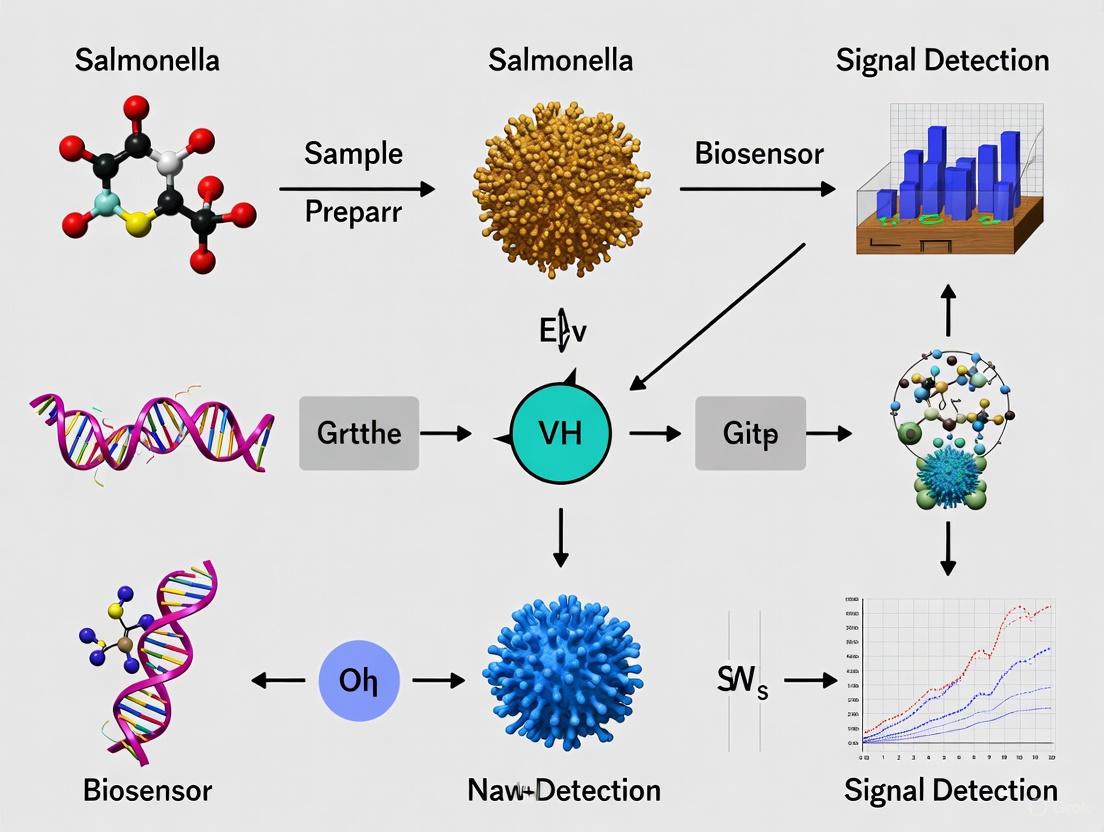
The following diagram illustrates the logical workflow for selecting and applying biorecognition elements within the context of a gold-biosensor research project.
Diagram 1: A generalized workflow for developing a Salmonella detection assay using a gold biosensor, highlighting the critical decision point of selecting a biorecognition element.
The Critical Role of Surface Functionalization and Antibody Immobilization
The performance of a biosensor is fundamentally dictated by the careful engineering of its interface. For gold-based biosensors targeting the detection of foodborne pathogens like Salmonella, the strategies employed for surface functionalization and antibody immobilization are paramount. These steps directly control the density, orientation, and biological activity of the immobilized biorecognition elements, thereby determining the sensor's sensitivity, specificity, and limit of detection (LOD) [16]. A robust and well-characterized protocol ensures that antibodies are presented optimally to the analyte, maximizing binding efficiency while minimizing non-specific interactions. This document details a standardized protocol for functionalizing gold biosensor surfaces and immobilizing antibodies, framed within the context of detecting Salmonella, to achieve highly sensitive and reliable pathogen detection.
Surface Functionalization & Antibody Immobilization Protocol
The following section provides a detailed, step-by-step methodology for preparing the gold biosensor surface, creating a functionalized monolayer, and immobilizing antibodies for the specific detection of Salmonella.
Reagents and Materials
Table 1: Essential Reagents and Materials for Biosensor Functionalization.
| Item Name | Function / Role | Specifications / Notes |
|---|---|---|
| Custom-made Gold Leaf Electrodes (GLEs) or commercial screen-printed gold electrodes [17] | Transducer substrate | Provides an excellent conductive surface for functionalization and electrochemical measurements. |
| 11-mercaptoundecanoic acid (MUA) [17] | Self-assembled monolayer (SAM) formation | Creates a stable, ordered layer on gold. Exposes carboxyl groups for subsequent biomolecule conjugation. |
| 1-Ethyl-3-(3-dimethylaminopropyl)carbodiimide (EDC) and N-hydroxysuccinimide (NHS) [17] | Carboxyl group activation | EDC/NHS chemistry activates MUA's terminal carboxyl groups, enabling covalent coupling to amine groups on proteins. |
| Protein L [17] | Antibody capture ligand | Binds to the light chain of antibodies, promoting a favorable orientation for antigen binding. |
| Trastuzumab (or anti-Salmonella antibody) [17] | Biorecognition element | The specific antibody that binds the target analyte (Salmonella). |
| Bovine Serum Albumin (BSA) [17] | Blocking agent | Reduces non-specific binding by occupying uncovered areas on the sensor surface. |
| Phosphate-Buffered Saline (PBS) [17] | Washing and dilution buffer | Provides a physiologically compatible ionic strength and pH for biological reactions. |
| Absolute Ethanol [17] | Solvent | Used for preparing the MUA solution. |
Step-by-Step Experimental Procedure
Gold Surface Preparation: Clean the gold electrode surface (e.g., GLEs or commercial screen-printed gold electrodes) thoroughly to remove organic contaminants. This can be done via oxygen plasma treatment or by piranha solution (Note: Handle with extreme caution), followed by rinsing with deionized water and drying under a stream of nitrogen [17] [16].
Self-Assembled Monolayer (SAM) Formation:
- Prepare a 1 mM solution of 11-mercaptoundecanoic acid (MUA) in absolute ethanol [17].
- Pipette a 1 µL droplet of the MUA solution onto the working electrode area.
- Incubate the electrode at 4°C in the dark for 16 hours to allow a dense, ordered SAM to form [17].
- After incubation, rinse the electrode surface thoroughly first with absolute ethanol, then with deionized water (DIW) to remove unbound MUA molecules.
Activation of Carboxyl Groups:
- Prepare a fresh solution containing 50 mM EDC and 50 mM NHS in PBS buffer [17].
- Apply 10 µL of the EDC/NHS solution to the working electrode.
- Incubate for 1 hour in the dark at room temperature. This step converts the terminal carboxyl groups of MUA into amine-reactive NHS esters.
- Rinse the electrode gently with DIW to stop the activation reaction and remove excess EDC/NHS.
Immobilization of Protein L:
- Apply 10 µL of a Protein L solution (0.1 mg mL⁻¹ in an appropriate buffer, such as PBS) to the activated surface [17].
- Incubate for 1 hour in the dark. The amine groups on Protein L will covalently bind to the NHS-activated surface.
- Rinse with DIW and PBS to remove any unbound Protein L.
Surface Blocking:
- To minimize non-specific adsorption, apply a solution of BSA (e.g., 50 µg mL⁻¹) to the electrode and incubate for 20 minutes in the dark [17].
- Rinse the electrode with buffer to remove excess BSA.
Antibody Immobilization:
- Apply 10 µL of the specific antibody (e.g., anti-Salmonella antibody) at a predetermined optimal concentration to the Protein L-modified surface.
- Incubate for 20 minutes to allow the antibody to bind to Protein L. This interaction helps orient the antibody correctly, presenting its antigen-binding sites towards the solution [17].
- Rinse thoroughly with PBS to remove any loosely attached antibodies. The biosensor is now ready for exposure to the sample.
Diagram 1: Workflow for Gold Biosensor Functionalization. This diagram outlines the sequential steps for preparing the biosensor surface, from the bare gold electrode to the final antibody-immobilized, ready-to-use state.
Experimental Validation & Performance Metrics
After functionalization, the biosensor's performance must be rigorously validated. Electrochemical Impedance Spectroscopy (EIS) is a powerful technique for this purpose, as it can monitor the step-by-step modification of the electrode surface and the subsequent binding of the target Salmonella.
Detection Principle and Assay Procedure
Electrochemical Measurement Setup: Use a potentiostat to perform EIS measurements. A common redox probe is a solution containing 5 mM K₃[Fe(CN)₆]/K₄[Fe(CN)₆] (1:1 mixture) in PBS [17].
Baseline Measurement: Record the EIS spectrum of the functionalized antibody-modified biosensor in the redox probe solution. This serves as the baseline signal.
Analyte Incubation: Expose the biosensor to a sample containing Salmonella cells. Incubate for a defined period (e.g., 20-30 minutes) to allow the antigen-antibody binding to occur.
Post-Assay Measurement: Rinse the biosensor gently and record the EIS spectrum again in the fresh redox probe solution.
Signal Analysis: The binding of Salmonella cells to the antibody on the sensor surface acts as an insulating layer, increasing the charge-transfer resistance (Rₛᵢ). The change in Rₛᵢ (ΔRₛᵢ) is directly proportional to the concentration of the target pathogen [17]. A calibration curve can be constructed by plotting ΔRₛᵢ against the logarithm of Salmonella concentration.
Diagram 2: Biosensor Detection Mechanism. The diagram illustrates the core detection principle: the binding of the target pathogen to the immobilized antibodies increases the impedance signal, which is quantified for analysis.
Performance Data
The following table summarizes performance benchmarks achievable with optimized surface functionalization, as demonstrated in recent literature for pathogen and biomarker detection.
Table 2: Performance Metrics of Optimized Biosensor Platforms.
| Target Analyte | Biosensor Platform | Immobilization Strategy | Limit of Detection (LOD) | Linear Range | Reference Context |
|---|---|---|---|---|---|
| HER2 (Cancer Biomarker) | Gold Leaf Electrode (GLE) | Protein L / Trastuzumab | 2.7 ng mL⁻¹ (in culture medium) | Not Specified | [17] |
| Salmonella (Pathogen) | SG4MB/SRCA Colorimetric | Nucleic Acid Hybridization | 4.33 CFU/mL | 5.2 × 10¹ to 5.2 × 10⁶ CFU/mL | [18] |
| Interleukin-6 (IL6) | Optical Immunosensor | Optimized Anti-IL6 Immobilization | 16% improvement in LOD* | Not Specified | [16] |
| E. coli & Salmonella | Gold Leaf Electrode (GLE) | Not Specified | Detection without enrichment | Not Specified | [17] |
Note: The 16% improvement highlights the impact of optimized functionalization, rather than an absolute LOD value [16].
The Scientist's Toolkit: Research Reagent Solutions
Table 3: Key Research Reagent Solutions for Biosensor Development.
| Reagent / Material | Critical Function |
|---|---|
| Protein L | An immunoglobulin-binding protein that binds to the light chains of antibodies without interfering with the antigen-binding site, promoting optimal orientation [17]. |
| PEG-based Nanoparticles | Thiolated nanoparticles create a 3D matrix on the gold surface, increasing the surface area for ligand immobilization and enhancing sensor sensitivity. They can also provide anti-fouling properties [19]. |
| EDC/NHS Chemistry | The cornerstone of carbodiimide crosslinking chemistry for covalently conjugating carboxyl groups to primary amines, essential for stable biomolecule immobilization [17]. |
| BSA (Bovine Serum Albumin) | A standard blocking agent used to passivate any remaining uncovered surface sites, drastically reducing non-specific binding and background signal [17]. |
| Thiolated Aptamers | Single-stranded DNA or RNA molecules that can be directly immobilized on gold via thiol-gold chemistry. Serve as synthetic, stable recognition elements for specific targets [20]. |
Troubleshooting and Optimization Guidelines
- Low Sensitivity/High LOD: This often results from low antibody density or poor orientation. To optimize, systematically characterize each functionalization step using techniques like Atomic Force Microscopy (AFM) or X-ray Photoelectron Spectroscopy (XPS) to ensure complete and homogeneous surface coverage [16]. Increase the concentration or incubation time during Protein L and antibody immobilization steps.
- High Non-Specific Binding: Inadequate blocking is a common cause. Ensure the BSA solution is fresh and the concentration is sufficient. Consider using other blocking agents like casein or surfactant-based blockers. The use of PEG-based coatings can also significantly mitigate fouling in complex matrices [19].
- Poor Reproducibility: Inconsistent SAM formation is a frequent culprit. Strictly control the concentration of the MUA solution (1 mM is often optimal), use high-purity solvents, and ensure consistent incubation times and temperatures across all experiments [17].
Step-by-Step Protocols and Real-World Application Workflows
The detection of pathogenic bacteria like Salmonella enterica is crucial for public health and food safety. Traditional methods, while reliable, are often time-consuming, requiring several days to yield results [3]. Electrochemical immunosensors offer a powerful alternative, combining the high specificity of antibody-antigen interactions with the sensitivity and rapid response of electrochemical transducers. This protocol details the fabrication of a gold electrode-based immunosensor using Self-Assembled Monolayers (SAMs) and EDC/NHS chemistry for the specific and sensitive detection of Salmonella [3]. The principle of this label-free immunosensor is that the binding of Salmonella cells to the capture antibodies immobilized on the electrode surface alters the interface's electrical properties, which can be monitored via cyclic voltammetry (CV) [3].
Research Reagent Solutions
The following table lists the essential materials and reagents required for the successful fabrication of the immunosensor.
Table 1: Essential reagents and materials for immunosensor fabrication.
| Item | Function/Description |
|---|---|
| Gold Electrodes (e.g., screen-printed) | Serves as the sensing platform and transducer surface. |
| Mercaptoacetic Acid (MAA) | Forms a self-assembled monolayer (SAM) on the gold surface, providing carboxyl groups for antibody immobilization [3] [21]. |
| EDC (1-Ethyl-3-(3-dimethylaminopropyl)carbodiimide) | Activates carboxyl groups, forming an amine-reactive O-acylisourea intermediate [3]. |
| NHS (N-Hydroxysuccinimide) | Stabilizes the EDC-activated carboxyl groups, forming an amine-reactive NHS ester for efficient antibody coupling [3]. |
| Anti-Salmonella Antibodies | Biorecognition element that specifically binds to Salmonella antigens [3]. |
| Phosphate Buffered Saline (PBS) | Buffer for diluting antibodies and for washing steps. |
| Bovine Serum Albumin (BSA) | Used as a blocking agent to passivate unreacted sites and minimize non-specific adsorption [21]. |
| Potassium Ferrocyanide/Ferricyanide | Redox probe used in electrochemical characterization (e.g., CV, EIS) [21] [22]. |
| Ethanolamine | An alternative blocking agent for quenching unreacted NHS esters [23]. |
Methodology
Electrode Pretreatment and Cleaning
- Clean the bare gold electrode by dipping it in 1 M sulfuric acid (H₂SO₄) for 3 minutes [22].
- Rinse the electrode thoroughly with a generous amount of deionized water [22].
- Alternatively, an electrochemical pretreatment in 0.5 M H₂SO₄ within a potential range of -2.5 V to +2.5 V at a scan rate of 100 mV/s for two cycles can be performed [21].
- Dry the electrode under a gentle stream of nitrogen gas or air.
Formation of Self-Assembled Monolayer (SAM)
- Spot 20 µL of a 10 mM aqueous solution of mercaptoacetic acid (MAA) onto the cleaned gold working electrode surface [3] [21].
- Incubate for 2 hours at room temperature to allow the thiol groups to form a covalent bond with the gold, creating a well-ordered SAM.
- Wash the modified electrode with ultrapure water to remove any physically adsorbed MAA and dry it with a low stream of nitrogen [21].
Activation of Carboxyl Groups with EDC/NHS
- Prepare a fresh activation solution containing 10 mM EDC and 20 mM NHS in 100 mM MES buffer (pH 6.0) [21].
- Spot 20 µL of the EDC/NHS solution onto the MAA-modified electrode.
- Incubate for 1 hour at room temperature. This step converts the terminal carboxyl groups of the SAM into amine-reactive NHS esters.
- Rinse the electrode gently with MES buffer (100 mM, pH 6.0) to remove excess EDC/NHS [21].
Antibody Immobilization
- Spot 20 µL of anti-Salmonella antibody solution (e.g., 1 µg/mL in 0.1 M PBS, pH 7.4) onto the activated surface [21].
- Incubate for 1 hour at room temperature, allowing the primary amine groups of the antibodies to form stable amide bonds with the NHS esters on the surface.
- Wash the electrode with PBS (0.1 M, pH 7.4) to remove any unbound antibodies [21].
Blocking of Non-Specific Sites
- Incubate the functionalized electrode with 20 µL of a 1% (w/v) Bovine Serum Albumin (BSA) solution in PBS for 30–60 minutes [21] [22].
- Wash the electrode with PBS to remove excess BSA. The immunosensor is now ready for use.
- For storage, the sensor can be dipped in a stabilizing solution (e.g., 2% BSA, 2% glucose in DI water) for 1 hour, dried, and stored refrigerated or frozen. Before use, wash with PBS to rehydrate [22].
Performance and Characterization
The performance of the fabricated immunosensor for Salmonella detection can be characterized electrochemically and by its analytical figures of merit.
Electrochemical Characterization
Cyclic Voltammetry (CV) and Electrochemical Impedance Spectroscopy (EIS) using a [Fe(CN)₆]³⁻/⁴⁻ redox probe are standard methods to monitor the modification process. A successful fabrication is indicated by a decrease in the voltammetric peak current or an increase in the electron transfer resistance (Rₑₜ) after each modification step (SAM formation, antibody immobilization, blocking) due to the increased insulating layer on the electrode surface [21] [22].
Analytical Performance
When tested, immunosensors fabricated with this methodology have demonstrated excellent performance for pathogen detection, as summarized in the table below.
Table 2: Performance metrics of a representative SAM-based gold immunosensor for Salmonella detection [3].
| Parameter | Performance |
|---|---|
| Detection Principle | Label-free, using Cyclic Voltammetry (CV) |
| Target Pathogen | Salmonella enterica serovar Typhimurium |
| Limit of Detection (LOD) | 10 CFU/mL |
| Total Analysis Time | < 20 minutes |
| Specificity | No cross-reactivity with other tested bacteria |
| Linear Range | Peak current proportional to concentration (e.g., 10–10⁶ CFU/mL) |
Experimental Workflow and Detection Mechanism
The following diagram illustrates the step-by-step fabrication process and the subsequent detection of the target pathogen.
Diagram 1: Schematic of the immunosensor fabrication and detection workflow. The electrode surface is sequentially modified with a SAM, activated, functionalized with antibodies, and blocked. The specific capture of Salmonella cells alters the electrochemical signal.
Salmonella species are among the leading causative agents of foodborne illnesses, resulting in significant rates of sickness, hospitalization, and deaths worldwide [24] [25]. The existence of approximately 2,000 Salmonella serotypes necessitates the development of rapid, sensitive, and comprehensive detection methods capable of identifying multiple strains simultaneously [24]. While traditional detection methods like plating culture, enzyme-linked immunosorbent assays (ELISA), and polymerase chain reaction (PCR) remain valuable, they often lack the speed, simplicity, or multi-target capability desired for modern food safety monitoring [26].
Colorimetric assays utilizing functionalized gold nanoparticles (f-AuNPs) have emerged as a powerful biosensing platform, combining high sensitivity with the simplicity of visual readout [24] [27]. Gold nanoparticles (AuNPs) within the 13–20 nm diameter range possess excellent dispersity, biocompatibility, and ease of functionalization [24]. Their unique optical properties, particularly the color change from red (dispersed state) to purplish-blue (aggregated state), provide a robust mechanism for detection that can be observed with the naked eye or quantified with simple instrumentation [24] [26]. This protocol details the development of a colorimetric assay using oligonucleotide-functionalized AuNPs for the specific and simultaneous detection of multiple Salmonella strains, achieving superior detection limits of less than 10 CFU/mL or g in both pure culture and complex food matrices [24].
Principle of the Detection Method
The fundamental principle of this colorimetric assay is sandwich hybridization, which utilizes the aggregation state of f-AuNPs as a visual indicator for the presence of target Salmonella DNA [24]. The assay employs two single-stranded oligonucleotide probes (30-mer each) functionalized onto the surface of 13 nm AuNPs. These probes are designed to hybridize with adjacent sequences within a conserved 192-base genomic region of the ttrRSBCA locus, which is found across a broad range of Salmonella spp. strains [24].
In the absence of the target DNA, the f-AuNPs remain dispersed in solution at an optimized salt concentration, resulting in a red color. In the presence of the target Salmonella DNA, a sandwich hybridization structure forms, creating highly stable oligonucleotide/AuNPs-DNA complexes. This aggregation state remains stable even at high salt concentrations (up to 2 M), leading to a visible color change from red to purplish-blue [24]. This color shift serves as the direct readout for a positive detection event.
Visual Workflow of the f-AuNP Colorimetric Assay
The following diagram illustrates the experimental workflow and the underlying detection mechanism.
Research Reagent Solutions and Essential Materials
Successful execution of this protocol requires the following key reagents and materials. Their specific functions are outlined in the table below.
Table 1: Essential Research Reagents and Materials
| Item | Function/Description in the Assay |
|---|---|
| Gold (III) chloride trihydrate (HAuCl₄·3H₂O) | Precursor for the synthesis of gold nanoparticles (AuNPs) [24]. |
| Sodium citrate (C₆H₅Na₃O₇) | Reducing and stabilizing agent for AuNP synthesis, preventing aggregation [24]. |
| Thiol-modified oligonucleotide probes (Probe 1 & Probe 2) | Detection probes that are covalently attached to AuNPs via thiol groups; designed to hybridize with the conserved ttrRSBCA region of Salmonella [24]. |
| Sodium chloride (NaCl) | Used in the salt concentration step to induce aggregation in non-target reactions, differentiating positive from negative results [24]. |
| Immunomagnetic Separation (IMS) beads | Used for concentrating target Salmonella cells from complex food matrices (e.g., blueberries, chicken meat) prior to DNA preparation [24]. |
| DNeasy Blood & Tissue Kit | Commercial kit for efficient and reliable extraction of genomic DNA from bacterial cells [24]. |
| Asymmetric PCR primers | Primers (For-192-Sal and Rev-192-Sal) for amplifying the 192-base target region within the ttrRSBCA locus, generating single-stranded DNA for more efficient hybridization with the probes [24]. |
| Culture Media (BHI broth, HE Agar) | For the initial activation and growth of bacterial cultures [24]. |
Detailed Experimental Protocol
Synthesis and Functionalization of Gold Nanoparticles (AuNPs)
4.1.1. Synthesis of Citrate-capped AuNPs (13 nm)
- Prepare a 1 mM HAuCl₄ solution in purified water (>18.3 MΩ/cm).
- Bring the solution to a rolling boil under reflux conditions with continuous stirring.
- Rapidly add 38.8 mM sodium citrate solution (1% w/v) to the boiling solution.
- Continue heating and stirring for an additional 15 minutes until the solution develops a deep red color, indicating the formation of ~13 nm AuNPs.
- Allow the solution to cool to room temperature while stirring.
- Characterize the AuNPs by UV-Vis spectroscopy (peak absorbance ~520 nm) and Transmission Electron Microscopy (TEM) to confirm size and monodispersity [24].
4.1.2. Functionalization of AuNPs with Oligonucleotide Probes
- Probe Design: Design two thiol-modified 30-mer oligonucleotide probes (Probe 1 and Probe 2) complementary to adjacent sequences on the conserved 192-bp region of the ttrRSBCA locus [24].
- Probe 1 (P1-Sal): 5′-AGC AAC TGG CGG GAG AAA GCG GTC TTG ACG-3′
- Probe 2 (P2-Sal): 5′-GCA GGA ACA CCC GAT TGA CTC GTC CGT CCC-3′
- Functionalization: Incubate the thiol-modified oligonucleotide probes with the synthesized AuNPs. The thiol groups form stable Au-S bonds, anchoring the probes to the nanoparticle surface.
- Aging and Purification: Age the functionalized AuNPs (f-AuNPs) in a buffer solution (e.g., phosphate buffer) overnight. Remove unbound oligonucleotides via centrifugation and washing [24].
Sample Preparation and DNA Extraction
- Culture Enrichment: Activate frozen stocks of Salmonella strains in Brain Heart Infusion (BHI) broth overnight at 37°C [24].
- Sample Concentration from Food Matrices: For contaminated food samples (e.g., chicken meat, blueberries), use Immunomagnetic Separation (IMS) with anti-Salmonella beads to isolate and concentrate viable Salmonella cells from the complex matrix [24].
- DNA Extraction: Extract genomic DNA from the enriched culture or IMS-concentrated cells using a commercial DNA extraction kit (e.g., DNeasy Blood & Tissue Kit) according to the manufacturer's instructions. The DNA can be used directly or after asymmetric PCR amplification of the target region [24].
Colorimetric Detection Assay
- Hybridization Reaction:
- In a 96-well microplate, combine the f-AuNP solution with the prepared DNA sample (extracted or amplified).
- Incubate the mixture at 55°C for 30 minutes to allow for sandwich hybridization to occur [24].
- Salt Addition and Result Interpretation:
- After hybridization, introduce a high concentration of salt (NaCl) to the reaction mixture, achieving a final concentration of up to 2 M.
- Visual Readout: Observe the color of the solution.
- Positive Result (Target DNA Present): The solution remains red due to the stable hybridization complex preventing AuNP aggregation.
- Negative Result (No Target DNA): The solution turns purplish-blue due to salt-induced aggregation of the f-AuNPs [24].
- Validation: Confirm the results using spectrophotometric analysis (absorbance shift) or gel electrophoresis to visualize the hybridization complexes [24].
Performance Data and Analysis
The performance of the f-AuNP colorimetric assay was rigorously evaluated for sensitivity, specificity, and application in complex matrices. The quantitative data are summarized in the tables below.
Table 2: Assay Performance Characteristics
| Parameter | Result | Experimental Condition |
|---|---|---|
| Detection Limit (DL) | < 10 CFU/mL or g | For both pure culture and complex food matrices (blueberries, chicken meat) [24]. |
| Specificity | 100% | Successfully distinguished 19 different Salmonella spp. strains from non-target bacteria [24]. |
| Number of Strains Detected | 19 | Simultaneous detection of environmental and outbreak Salmonella strains in a single assay [24]. |
| Assay Time | ~30 min (post-DNA preparation) | The hybridization and readout step is rapid [24]. |
| Target Locus | ttrRSBCA | A conserved genomic region near Salmonella Pathogenicity Island 2 (SPI-2) [24]. |
Table 3: Comparison with Other Colorimetric Methods for Salmonella
| Method / Biosensor Type | Target Analyte | Detection Limit (DL) | Key Advantage / Disadvantage |
|---|---|---|---|
| Oligonucleotide-f-AuNPs (This Protocol) | ttrRSBCA DNA (19 strains) | < 10 CFU/mL or g [24] | Superior sensitivity, simultaneous multi-strain detection. |
| Aptamer-based Colorimetric Assay | S. Enteritidis | 10³ CFU/mL (in milk) [24] | Lower sensitivity, single pathogen detection. |
| Fiber Optic Sensor (BARDOT) | S. Enteritidis & S. Typhimurium | 10³ CFU/mL (poultry) [24] | Lower sensitivity, limited serovar detection. |
| Aptasensor (COF-AuNPs) | S. Typhimurium | 7 CFU/mL [26] | High sensitivity, uses nanozyme activity; single pathogen detection. |
Troubleshooting and Technical Notes
- Low Color Change Contrast: Ensure the AuNPs are monodisperse and of the correct size (~13 nm) after synthesis. Verify the salt concentration is optimized for clear discrimination between aggregated and non-aggregated states.
- Non-Specific Aggregation: Confirm the quality of the oligonucleotide functionalization. Inadequate functionalization can lead to probe detachment and instability of the f-AuNPs at higher salt concentrations.
- False Negatives in Food Matrices: The use of IMS for pre-concentration of cells is critical for removing PCR inhibitors and matrix components that can interfere with the hybridization reaction [24].
- Assay Robustness: The high stability of the sandwich hybridization complex, which remains undisturbed at 2 M salt concentration, is a key factor contributing to the assay's 100% specificity and reliability [24].
Application Notes: Microfluidic Systems for Pathogen Detection
The integration of microfluidic technology with biosensors represents a significant advancement in the detection of foodborne pathogens like Salmonella. These systems merge the precise fluid handling capabilities of microfluidic devices with the high sensitivity of biosensors, creating portable, efficient, and automated platforms ideal for rapid on-site analysis [28]. This document details the application of such systems within the context of a thesis focused on protocols for detecting Salmonella with a gold biosensor, providing notes and methodologies for researchers and scientists.
The core advantage of microfluidic biosensors lies in their ability to perform multiple laboratory functions—such as sample preparation, concentration, and detection—on a single, compact chip. This "lab-on-a-chip" approach minimizes reagent consumption, reduces analysis time, and enhances detection sensitivity by improving transport conditions and increasing the mixing rate of reagents [29] [28]. For Salmonella detection, this translates to the ability to identify low pathogen concentrations (as low as 1-2 CFU/mL) directly in complex food matrices like raw chicken wash, with overall detection times as short as 40-50 minutes, a significant improvement over traditional culture methods which can take 5-7 days [30] [31].
Key Design Considerations for Sample Concentration and Analysis
Effective microfluidic design is paramount for automating sample preparation and enhancing sensor sensitivity. Two primary strategies are employed:
- Pump-Free Fluidic Control: Traditional syringe pumps can be replaced by gravity-driven flow, significantly reducing system cost and complexity. Research has shown that height differences in fluid reservoirs can effectively drive samples through microfluidic channels, with strategic "bottleneck" designs integrated into the channels to slow cell velocity, thereby improving the quality of subsequent image analysis [32].
- On-Chip Sample Concentration: To achieve a low limit of detection, microfluidic chips can be engineered with specific regions that actively concentrate the target analyte. For instance, the use of dielectrophoresis (DEP) in a focusing and trapping region can concentrate Salmonella antigens from a large sample volume onto a small detection zone, dramatically enhancing the sensor's signal and enabling the detection of just 1-2 cells per mL [31].
The table below summarizes performance metrics of various microfluidic biosensing platforms for pathogen detection, as reported in the literature:
Table 1: Performance Comparison of Microfluidic Biosensors for Pathogen Detection
| Detection Technique | Target Analyte | Limit of Detection (LOD) | Total Analysis Time | Key Features | Source |
|---|---|---|---|---|---|
| Fluidic Impedance Biosensor | Salmonella Typhimurium | 1-2 cells/mL | 40-50 min | Integrated focusing/trapping region; distinguishes live/dead cells | [31] |
| SmartFlow (Computer Vision) | Cells in Body Fluid | N/A (R²=0.96 for counting) | N/A | 3D-printed, gravity-driven, pump-free, smartphone-based | [32] |
| Quartz Crystal Microbalance (QCM) | Salmonella Typhimurium | 10³ CFU/mL (without AuNPs) | ~30 min (after incubation) | Gold nanoparticle (AuNP) signal amplification | [4] |
| Cell-Based Bioelectric (BERA) | Salmonella spp. | 1 log CFU g⁻¹ (10 CFU g⁻¹) | 3 min assay (post-enrichment) | Membrane-engineered Vero cells, portable device | [30] |
| Automated Spectrophotometric System | Nitrite Ions | 1×10⁻⁴ μg mL⁻¹ | 600 analyses/hour | Arduino-controlled, high-throughput, for environmental samples | [33] |
Experimental Protocols
This section provides detailed methodologies for fabricating microfluidic chips and conducting detection assays, suitable for replication in a research setting.
Protocol 1: Fabrication of a 3D-Printed, Pump-Free Microfluidic Chip for Sample Concentration
This protocol outlines the creation of a low-cost microfluidic chip that uses gravity for flow control and a bottleneck design for cell velocity management, ideal for pre-concentrating samples for visual analysis [32].
Research Reagent Solutions & Essential Materials
Table 2: Materials for Pump-Free Microfluidic Chip
| Item | Function | Specification/Note |
|---|---|---|
| 3D Printer & Resin | Chip fabrication | Creates the monolithic chip structure with microchannels. |
| Polydimethylsiloxane (PDMS) | Channel molding | Alternative to 3D printing; requires soft lithography. |
| Biological Sample | Analysis target | e.g., diluted sheep blood or pre-processed food sample. |
| Smartphone with Camera | Detection & analysis | Records cell flow for computer vision algorithms. |
| Microscope Setup | Visualization | Provides magnification for the microfluidic channel. |
Methodology:
- Chip Design: Using computer-aided design (CAD) software, design a microfluidic chip that incorporates a long, straight main channel. Integrate a "bottleneck" section—a segment of the channel with a significantly reduced cross-sectional area—to increase fluidic resistance and slow down passing cells [32].
- Fabrication: a. 3D Printing: Transfer the design to a high-resolution 3D printer. Print the chip as a single, monolithic unit using a biocompatible resin. Post-process the print according to the resin manufacturer's instructions, which may include washing and UV curing. b. PDMS Molding (Alternative): Create a master mold via 3D printing or photolithography. Pour a mixture of PDMS base and curing agent (typically 10:1 ratio) over the mold and cure at 65°C for 2 hours. Peel off the cured PDMS and bond it to a glass slide or another PDMS slab using oxygen plasma treatment.
- Flow Setup: Attach fluidic reservoirs to the chip's inlets and outlets via tubing. To drive flow by gravity, place the sample reservoir at a higher vertical position than the waste reservoir. Systematically vary this height difference (e.g., from 1 cm to 7 cm) to control and calibrate the flow velocity [32].
- Validation and Analysis: a. Introduce a diluted cell sample (e.g., sheep blood) into the system. b. Record the flow of cells through both the straight and bottleneck sections using a smartphone mounted on a microscope. c. Analyze the video sharpness and cell velocity using computer vision software. The bottleneck design should demonstrate a slower cell velocity and better video quality (higher sharpness) at equivalent height differences compared to the straight channel [32].
The following workflow diagram illustrates the fabrication and validation process:
Protocol 2: Detection ofSalmonellaspp. using a Gold Nanoparticle-Amplified QCM Biosensor in a Microfluidic System
This protocol describes the use of a Quartz Crystal Microbalance (QCM) integrated with a flow system. The QCM sensor's surface is functionalized with antibodies, and detection is enhanced using gold nanoparticles (AuNPs), making it highly relevant for a thesis on gold biosensors [4].
Research Reagent Solutions & Essential Materials
Table 3: Reagents and Materials for QCM Biosensor
| Item | Function | Specification/Note |
|---|---|---|
| QCM Sensor Chip (5 MHz) | Piezoelectric transducer | Gold-coated quartz crystal for mass-sensitive detection. |
| 11-Mercaptoundecanoic acid (MUA) | Forms self-assembled monolayer (SAM) | Creates a functionalized surface on the gold electrode. |
| EDC & NHS | Cross-linkers | Activate carboxyl groups for antibody immobilization. |
| Polyclonal Anti-Salmonella Antibodies | Biorecognition element | Binds specifically to Salmonella O-antigen. |
| Gold Nanoparticles (AuNPs) | Signal amplification | Conjugated with streptavidin for mass enhancement. |
| Biotinylated Anti-Salmonella | Secondary antibody | Binds to captured Salmonella and links to AuNPs. |
| Peristaltic Pump & Flow Cell | Fluid handling | Controls reagent delivery to the sensor surface. |
Methodology:
- Sensor Surface Functionalization: a. Place the gold-coated QCM sensor into the flow cell. b. Prime the system with ethanol, then flow 1 mM 11-Mercaptoundecanoic acid (MUA) in ethanol over the sensor surface for at least 1 hour to form a self-assembled monolayer (SAM). c. Flush with ethanol and deionized water to remove unbound MUA. d. Activate the carboxyl terminals of the SAM by flowing a fresh mixture of 5 mM EDC and 5 mM NHS in water for 15 minutes. e. Immobilize polyclonal anti-Salmonella antibodies by flowing a solution (e.g., 5 µg/mL in PBS) over the activated surface for 1 hour. The antibodies will covalently bind to the SAM. f. Flush with PBS to remove any loosely attached antibodies [4].
- Sample Introduction and Pathogen Capture: a. Establish a stable baseline frequency by flowing PBS buffer through the system. b. Inject the prepared sample (e.g., enriched food sample or bacterial suspension in PBS) over the sensor surface for a defined period (e.g., 30 minutes). c. The specific binding of Salmonella cells to the immobilized antibodies will cause a decrease in the resonant frequency of the QCM crystal. Monitor this shift in real-time. d. Flush with PBS again to remove unbound or weakly bound cells and record the stable frequency value. The frequency shift (∆f₁) is proportional to the mass of captured Salmonella [4].
- Signal Amplification with Gold Nanoparticles: a. To further enhance the signal, flow a solution of biotinylated anti-Salmonella antibodies over the sensor. These will bind to the captured Salmonella cells. b. Flush with PBS. c. Introduce streptavidin-conjugated gold nanoparticles (100 nm). The streptavidin will bind strongly to the biotin on the secondary antibodies, adding significant mass to the sensor surface. d. Flush with PBS and record the new stable frequency. The additional frequency shift (∆f₂) corresponds to the mass of the bound AuNPs [4].
- Data Analysis: a. The total frequency shift (∆f₁ + ∆f₂) is used for quantification. The use of AuNPs typically results in a much larger frequency shift, thereby improving the limit of detection. b. Generate a calibration curve by plotting frequency shifts against the logarithmic concentration of known Salmonella standards.
The following diagram illustrates the key steps of the assay and signal amplification principle:
The detection of Salmonella in complex matrices such as food and clinical specimens presents significant challenges due to the presence of fats, proteins, biofilms, and salts that can interfere with analytical accuracy. [34] Gold-based biosensors have emerged as transformative tools to overcome these limitations, offering enhanced sensitivity, rapid detection, and adaptability for real-time monitoring. [34] This application note details standardized protocols and experimental data for applying gold biosensor technology to detect Salmonella in meat products and milk, providing researchers with validated methodologies for complex sample analysis.
The following table summarizes the performance characteristics of different gold biosensor platforms when applied to various complex sample matrices.
Table 1: Performance of Gold Biosensor Methods for Salmonella Detection in Complex Matrices
| Detection Platform | Sample Matrix | Detection Time | Limit of Detection (LOD) | Accuracy/Specificity | Key Advantage |
|---|---|---|---|---|---|
| B.EL.D Bioelectric Biosensor [35] | Meat products | ~24 h (including enrichment), 3-min analysis | 1 log CFU g⁻¹ [35] | 86.1% accuracy [35] | Portable, real-time notification via mobile device |
| SG4MB/SRCA Colorimetric Biosensor [18] | Milk | ~90 min [18] | 4.33 CFU/mL [18] | 95.0-105.4% recovery in spiked milk [18] | Visual or absorbance readout, high sensitivity |
| Gold Biosensor with Light Microscope Imaging (GB-LMIS) [9] | Chicken | ~2.5 h [9] | Not specified | Competitive specificity; no cross-reactivity with 13 other bacteria species [9] | Direct visual enumeration of captured bacteria |
Detailed Experimental Protocols
Protocol 1: B.EL.D Bioelectric Biosensor for Meat Products
This protocol utilizes a cell-based biosensor technology that gauges changes in cell membrane potential based on the Bioelectric Recognition Assay (BERA) principle. [35]
Research Reagent Solutions
Table 2: Essential Reagents for B.EL.D Biosensor Protocol
| Reagent/Material | Function | Specifications/Notes |
|---|---|---|
| Vero Cells (African green monkey kidney) | Biosensor transducer element | LGC Promochem, Teddington, UK [35] |
| Dulbecco's Modified Eagle Medium (DMEM) | Cell culture maintenance | Supplemented with 10% FBS, streptomycin/penicillin, L-glutamine/L-alanine [35] |
| Anti-Salmonella Antibodies | Specific biorecognition element | Purified polyclonal or monoclonal antibodies [35] |
| Phosphate-Buffered Saline (PBS) | Electroporation buffer | Biomedicals, Illkrich, France [35] |
| Trypsin/EDTA | Cell detachment | Biosera, Cholet, France [35] |
| B.EL.D Portable Device | Signal measurement and processing | EMBIO Diagnostics; connects to Android/iOS via Bluetooth [35] |
Step-by-Step Procedure
Sample Preparation and Pre-enrichment
- Collect meat samples (e.g., burgers, sausages, turkey, chicken fillets) and transport under refrigerated conditions.
- Homogenize 25 g of sample with 225 mL of non-selective pre-enrichment broth (e.g., Buffered Peptone Water).
- Incubate at 37°C for 18-24 hours to revive stressed bacteria and allow initial growth.
Biosensor Fabrication
- Culture Vero cells in DMEM complete medium at 37°C with 5% CO₂ until 80-90% confluency.
- Detach cells using trypsin/EDTA (10 min at 37°C) and collect by centrifugation.
- Resuspend cell pellet in PBS containing anti-Salmonella antibodies.
- Incubate on ice for 20 minutes to allow antibody binding.
- Transfer cell-antibody mixture to electroporation cuvettes.
- Apply two square electric pulses at 1800 V/cm using an Eppendorf Eporator to electroinsert antibodies into cell membranes.
- Incubate the membrane-engineered cells in nutrient medium at 37°C with 5% CO₂ for 24 hours.
Measurement and Detection
- After incubation, remove medium and mechanically detach biosensors (Vero/anti-Salmonella cells).
- Mix 100 μL of enriched sample with prepared biosensors in the measurement chamber.
- Insert into B.EL.D portable device and initiate measurement.
- Record changes in cell membrane potential during a 3-minute analysis.
- Results are transmitted via Bluetooth to a mobile device for immediate interpretation.
Workflow Visualization
Protocol 2: SG4MB/SRCA Colorimetric Biosensor for Milk
This protocol employs a saltatory rolling circle amplification (SRCA) combined with a split G-quadruplex molecular beacon (SG4MB) for highly sensitive detection in dairy matrices. [18]
Research Reagent Solutions
Table 3: Essential Reagents for SG4MB/SRCA Colorimetric Biosensor
| Reagent/Material | Function | Specifications/Notes |
|---|---|---|
| SRCA Primers | Specific DNA amplification | Designed according to SRCA version 2.0 for stability and specificity [18] |
| DNA Polymerase | Isothermal amplification | Bst polymerase or similar with strand displacement activity [18] |
| Split G-Quadruplex Molecular Beacon (SG4MB) | Signal generation | Binds to SRCA products; forms G-quadruplex with peroxidase activity [18] |
| Hemin | Cofactor for DNAzyme | Enables horseradish peroxidase-like activity [18] |
| ABTS/H₂O₂ Substrate | Colorimetric reaction | Produces color change when oxidized by G-quadruplex-hemin DNAzyme [18] |
| Lysis Buffer | DNA extraction | For releasing bacterial DNA from sample matrix |
Step-by-Step Procedure
Sample Preparation and DNA Extraction
- Centrifuge 10 mL of milk sample at 10,000 ×g for 10 minutes to concentrate bacterial cells.
- Resuspend pellet in 500 μL of lysis buffer and incubate at 95°C for 10 minutes.
- Centrifuge at 12,000 ×g for 5 minutes and collect supernatant containing DNA.
Saltatory Rolling Circle Amplification (SRCA)
- Prepare SRCA reaction mixture containing:
- 10 μL of extracted DNA template
- 25 μL of 2× reaction buffer
- 1 μL of SRCA primers (10 μM each)
- 1 μL of Bst DNA polymerase (8 U/μL)
- Nuclease-free water to 50 μL final volume
- Incubate reaction at 60°C for 60 minutes for isothermal amplification.
- Heat-inactivate at 80°C for 10 minutes.
- Prepare SRCA reaction mixture containing:
Colorimetric Detection with SG4MB
- Prepare detection mixture containing:
- 10 μL of SRCA product
- 5 μL of SG4MB probe (2 μM)
- 5 μL of hemin (1 mM)
- 25 μL of detection buffer
- Incubate at room temperature for 10 minutes.
- Add 10 μL of ABTS/H₂O₂ substrate solution.
- Incubate for 5-10 minutes for color development.
- Prepare detection mixture containing:
Result Interpretation
- Qualitative assessment: Visual observation of color change from colorless to green.
- Quantitative assessment: Measure absorbance at 405-420 nm using a microplate reader.
- Calculate bacterial concentration based on a pre-constructed standard curve.
Workflow Visualization
Troubleshooting and Optimization
Addressing Matrix Interference
Complex food matrices contain components that can inhibit molecular detection methods. [34] To minimize interference:
- For fatty matrices: Add 1% Tween-20 to lysis buffer to emulsify lipids.
- For protein-rich samples: Incorporate proteinase K treatment (10 mg/mL, 10-minute incubation at 56°C) before DNA extraction.
- Inhibit PCR inhibitors: Use dilution series or additional purification steps if amplification efficiency is suboptimal.
Enhancing Sensitivity
The infectious dose of Salmonella can be less than 1000 cells, necessitating highly sensitive detection methods. [35]
- Extended enrichment: Increase pre-enrichment time to 24-48 hours for very low bacterial loads.
- Antibody optimization: Test multiple antibody clones and concentrations (typically 12.5-100 μg/mL) for maximum capture efficiency. [9]
- Signal amplification: For colorimetric methods, extend substrate incubation time to 15 minutes for low-concentration samples.
Gold biosensor technologies offer robust solutions for detecting Salmonella in complex matrices like meat and milk, overcoming traditional limitations of sensitivity and detection time. The protocols detailed herein provide researchers with standardized methods for implementing these advanced detection platforms, contributing to improved food safety monitoring and clinical diagnostics. The integration of gold biosensors with portable detection devices and straightforward colorimetric readouts makes these technologies particularly valuable for both laboratory and point-of-care applications.
Enhancing Performance and Overcoming Matrix Interference
The development of robust and reliable biosensors for the detection of Salmonella is a critical pursuit in food safety and clinical diagnostics. Gold-based biosensors, in particular, have emerged as a prominent platform due to the favorable properties of gold, such as its excellent conductivity and ease of functionalization. The performance of these biosensors is not merely a function of their design but is profoundly dependent on the meticulous optimization of key experimental parameters. This application note provides detailed protocols and consolidated data to guide researchers in optimizing the triumvirate of critical parameters—antibody concentration, incubation time, and reaction volume—to enhance the sensitivity, specificity, and efficiency of gold biosensor-based Salmonella detection assays. The guidance herein is framed within a broader research protocol, enabling the translation of a conceptual biosensor into a validated analytical tool.
Research Reagent Solutions
The following table details essential materials and reagents commonly used in the preparation and operation of gold-based biosensors for Salmonella detection.
Table 1: Essential Research Reagents for Gold Biosensor-Based Salmonella Detection
| Reagent/Material | Function/Description | Example Source / Component |
|---|---|---|
| Gold Electrode/Sensor | Transducer platform; provides a surface for antibody immobilization and electrochemical signal measurement. | Screen-printed gold electrodes; gold-sputtered glass chips [3] [9]. |
| Anti-Salmonella Antibodies | Biorecognition element; specifically binds to Salmonella antigens. | Polyclonal or monoclonal antibodies specific to Salmonella surface antigens [9]. |
| Mercaptoacetic Acid (MAA) | Forms a self-assembled monolayer (SAM) on the gold surface, facilitating subsequent antibody attachment. | Linker molecule for covalent bonding [3]. |
| EDC/NHS | Cross-linking agents; activate carboxyl groups on the SAM for stable antibody immobilization. | N-(3-Dimethylaminopropyl)-N′-ethylcarbodiimide (EDC) and N-Hydroxysuccinimide (NHS) [3]. |
| Blocking Agent | Reduces non-specific binding on the sensor surface, improving specificity. | Bovine Serum Albumin (BSA) [9]. |
| Phosphate Buffered Saline | Washing and dilution buffer; maintains a stable pH and ionic strength. | PBS, pH 7.2 - 7.4 [9]. |
| Electrochemical Probe | Generates the measurable electrochemical signal. | Potassium ferricyanide/ferrocyanide ([Fe(CN)₆]³⁻/⁴⁻) [3] [36]. |
Optimized Parameter Tables
Systematic optimization is key to achieving high performance. The data below, synthesized from recent studies, provides a benchmark for key parameters.
Table 2: Optimized Parameters for Biosensor Assay Steps
| Assay Stage | Key Parameter | Optimized Value / Range | Impact on Performance |
|---|---|---|---|
| Antibody Immobilization | Antibody Concentration | 12.5 - 100 μg/mL | Lower end (12.5 μg/mL) for ELISA; higher end (100 μg/mL) for direct surface imaging biosensors. Ensures sufficient surface coverage without overcrowding [9]. |
| Incubation Time | 1 - 2 hours | Allows for complete self-assembly and stable covalent attachment, ensuring a robust and reproducible sensor surface [3] [9]. | |
| Target Capture & Detection | Incubation Time (Sample) | 20 minutes - 2.5 hours | Faster sensors (20 min) use electrochemical transduction; ~2.5 hours for microscopic counting. Balances speed with sufficient pathogen binding [3] [9]. |
| Reaction Volume | 100 μL (typical for electrode assays) | A standard volume that ensures the sensor surface is fully immersed and analytes are efficiently transported to the binding sites. |
Table 3: Performance Outcomes of Optimized Gold Biosensors
| Biosensor Type | Limit of Detection (LOD) | Total Detection Time | Key Optimized Features |
|---|---|---|---|
| Electrochemical Immunosensor [3] | 10 CFU/mL | ~20 minutes | High-sensitivity antibody immobilization via SAM/EDC-NHS. |
| Gold Biosensor with Light Microscopy [9] | Not Specified | ~2.5 hours | High antibody concentration (100 μg/mL) for direct visual detection. |
| Molecularly Imprinted Polymer (MIP) Sensor [36] | 10 CFU/mL | Rapid (specific time not given) | LPS imprinting on polydopamine; avoids need for antibodies. |
Experimental Protocols
Protocol: Antibody Immobilization on a Gold Electrode Surface
This protocol details the functionalization of a gold electrode with anti-Salmonella antibodies to create the sensing interface [3].
I. Materials
- Gold electrode (e.g., screen-printed or disk electrode)
- Mercaptoacetic Acid (MAA)
- EDC and NHS
- Anti-Salmonella antibodies
- Phosphate Buffered Saline (PBS, pH 7.4)
- Bovine Serum Albumin (BSA)
II. Procedure
- Gold Surface Cleaning: Clean the gold electrode surface thoroughly with acetone, ethanol, and deionized water to remove any organic contaminants.
- SAM Formation: Incubate the electrode in a 10 mM aqueous solution of Mercaptoacetic Acid (MAA) for 1 hour at room temperature. The thiol group of MAA will form a covalent bond with the gold, creating a monolayer with exposed carboxyl groups.
- Washing: Rinse the electrode gently with PBS to remove any physically adsorbed MAA.
- Activation: Incubate the electrode in a fresh mixture of EDC (400 mM) and NHS (100 mM) for 1 hour. This activates the carboxyl groups on the SAM, forming amine-reactive esters.
- Antibody Immobilization: Apply 100 μL of the anti-Salmonella antibody solution (optimized concentration, e.g., 12.5-100 μg/mL in PBS) onto the activated electrode surface. Incubate for 2 hours at room temperature in a humidified chamber to prevent evaporation.
- Blocking: Rinse the electrode with PBS to remove unbound antibodies. Incubate the electrode with a 1% (w/v) solution of BSA in PBS for 1 hour to block any remaining non-specific sites on the electrode surface.
- Storage: The functionalized biosensor can be stored in PBS at 4°C until use.
The following diagram illustrates this antibody immobilization workflow.
Protocol: Electrochemical Detection of Salmonella
This protocol describes the use of the functionalized biosensor for the rapid detection of Salmonella in a sample [3].
I. Materials
- Functionalized gold biosensor (from Protocol 3.1)
- Sample containing Salmonella
- Potentiostat
- PBS containing 5 mM [Fe(CN)₆]³⁻/⁴⁻ as a redox probe
II. Procedure
- Baseline Measurement: Place the functionalized biosensor in an electrochemical cell containing the PBS/redox probe solution. Record a Cyclic Voltammetry (CV) or Differential Pulse Voltammetry (DPV) scan to establish the baseline current.
- Sample Incubation: Apply 100 μL of the sample solution directly onto the sensing area of the biosensor. Incubate for 20 minutes at room temperature to allow Salmonella cells to bind to the captured antibodies.
- Washing: Gently rinse the biosensor with PBS to remove unbound cells and matrix components.
- Detection Measurement: Place the biosensor back into the electrochemical cell with the PBS/redox probe solution. Record a second CV or DPV scan under the same conditions as the baseline.
- Analysis: Compare the peak current from the detection scan to the baseline. The binding of Salmonella cells on the electrode surface creates a barrier, hindering electron transfer and resulting in a measurable decrease in the peak current. This change is proportional to the concentration of the target pathogen.
The mechanism of signal generation upon target capture is summarized below.
Discussion and Concluding Notes
The data and protocols presented herein underscore a clear correlation between parameter optimization and biosensor efficacy. The choice of antibody concentration is highly dependent on the transduction method, with direct imaging sensors requiring higher density for visual detection compared to electrochemical sensors. The incubation time for the immunoreaction is a critical determinant of the assay's rapidity, with electrochemical methods offering a significant advantage in speed. The standardized reaction volume of 100 μL represents a practical balance for most electrode-based systems.
Integrating these optimized parameters into a gold biosensor protocol for Salmonella detection ensures a method that is not only highly sensitive, with limits of detection as low as 10 CFU/mL, but also rapid, providing results within 20 minutes. This positions gold biosensors as a robust, efficient, and practical solution for on-site pathogen screening, contributing substantially to the overarching goal of enhancing public health safety. Future work should focus on further multiplexing capabilities and integrating these sensors into automated, sample-to-answer microfluidic devices.
Strategies to Mitigate Signal Interference from Complex Food Matrices
The detection of foodborne pathogens, such as Salmonella spp., using gold-based biosensors is a rapidly advancing field promising rapid, on-site diagnostics. However, a significant challenge impeding the transition from research to real-world application is signal interference caused by complex food matrices. Food components including fats, proteins, carbohydrates, pigments, and biofilms can profoundly reduce detection sensitivity and accuracy by causing nonspecific binding, fouling the sensor surface, or quenching optical signals [37] [34]. This document outlines practical strategies and detailed protocols to mitigate these interferences, ensuring the reliability and performance of gold biosensor-based detection systems within a research context focused on Salmonella.
Key Mitigation Strategies
Several approaches can be employed to manage matrix-derived interference, often used in combination.
2.1 Sample Pre-processing and Target Separation This is the most common first line of defense, aiming to separate the target pathogen from the interfering substances in the food sample.
- Filter-Assisted Sample Preparation (FASP): A double-filtration system can effectively remove food residues. A primary filter with a larger pore size (e.g., glass fiber) removes large particles, while a secondary filter (e.g., 0.45 µm cellulose acetate) captures target microorganisms [37].
- Immunomagnetic Separation (IMS): This technique uses antibody-coated magnetic beads to specifically bind and concentrate the target pathogen from a complex sample homogenate. The beads, along with the captured bacteria, can then be separated using a magnet and washed to remove impurities [30].
2.2 Sensor Surface Engineering and Signal Amplification The design of the biosensor itself can be optimized to resist fouling and enhance signal.
- Use of Gold Nanomaterials: Gold nanoparticles (AuNPs) provide a high surface-area-to-volume ratio and unique optical-electrical properties. They can be functionalized with specific antibodies or other recognition elements. Their conjugation can be optimized using commercial kits that ensure covalent attachment, improving stability and reducing nonspecific binding compared to passive adsorption methods [38] [39].
- Membrane-Engineering for Cell-Based Biosensors: In electric cell-based sensors, cells (e.g., Vero cells) can be engineered to have specific anti-Salmonella antibodies electroinserted into their membranes. This creates a highly specific biosensor where the electrical response is triggered only by the binding of the target pathogen [30].
Detailed Experimental Protocols
Protocol: Filter-Assisted Sample Preparation (FASP) for Vegetable and Meat Matrices
This protocol is adapted from a system demonstrating a detection limit of 10¹ CFU/mL for pathogens in complex foods within 3 minutes of preprocessing [37].
1. Materials
- Stomacher or laboratory blender
- Vacuum pump and filtration apparatus
- Primary Filter: Glass microfiber filter (GF/D)
- Secondary Filter: Cellulose acetate membrane (0.45 µm pore size)
- Sterile peptone water or buffered peptone water
2. Procedure
- Step 1: Homogenization. Aseptically weigh 25 g of the food sample (e.g., lettuce, chicken). Add it to 225 mL of sterile peptone water and homogenize in a stomacher for 1-2 minutes.
- Step 2: Primary Filtration. Pour the homogenate through the primary glass fiber filter under vacuum. This step removes large particulate matter and vegetable fibers.
- Step 3: Secondary Filtration. Pass the filtrate from Step 2 through the 0.45 µm cellulose acetate membrane. This membrane captures bacterial cells, including Salmonella.
- Step 4: Elution (Optional). The bacteria can be eluted from the filter by back-flushing with a small volume (e.g., 1-5 mL) of an appropriate buffer. This concentrated sample is then suitable for analysis with the gold biosensor.
3. Performance Data The bacterial recovery rate of this FASP method varies by food matrix [37]:
Table 1: Bacterial Recovery from Food Matrices using FASP
| Food Matrix | Relative Bacterial Recovery |
|---|---|
| Vegetables | 1-log reduction (vs. initial inoculum) |
| Meats, Melon, Cheese Brine | 2-log reduction (vs. initial inoculum) |
Protocol: Immunomagnetic Separation (IMS) Coupled with a Gold Biosensor
This protocol concentrates Salmonella and purifies it from inhibitors prior to detection.
1. Materials
- Anti-Salmonella antibody-coated magnetic beads
- Magnetic separation rack
- Washing buffer (e.g., phosphate-buffered saline with 0.05% Tween-20, PBS-T)
- Sample enrichment broth (if analyzing low pathogen levels)
2. Procedure
- Step 1: Enrichment (if required). Incubate the food sample in a non-selective broth to increase bacterial numbers. For highly contaminated samples, this step may be omitted.
- Step 2: Binding. Add the prepared sample homogenate to a tube containing the immunomagnetic beads. Incubate for 10-15 minutes with continuous mixing to allow antibodies on the beads to bind to Salmonella.
- Step 3: Separation and Washing. Place the tube in a magnetic rack for 2-3 minutes. While the beads are captured against the magnet, carefully aspirate and discard the supernatant containing the food matrix. Remove the tube from the magnet and resuspend the beads in washing buffer. Repeat this washing step 2-3 times.
- Step 4: Detection. Resuspend the washed beads (now with captured Salmonella) in a small volume of buffer. This purified concentrate can be applied directly to the gold biosensor for detection.
Protocol: Conjugating Antibodies to Gold Nanoparticles for a Lateral Flow Assay
Stable conjugation is critical for the performance and shelf-life of the biosensor.
1. Materials
- Commercial Gold Conjugation Kit (e.g., 40 nm Gold Conjugation Kit, ab154873) [39]
- Purified anti-Salmonella antibody
- Amine-free buffer (e.g., 10 mM MES, MOPS, or HEPES, pH 6.5-8.5)
2. Procedure
- Step 1: Prepare Antibody. Ensure the antibody is in a compatible, amine-free buffer. Use a purification kit if necessary to exchange the buffer or concentrate the antibody.
- Step 2: Reconstitute Nanoparticles. Pipette the calculated volume of antibody directly into the vial containing the freeze-dried gold nanoparticles from the kit. Mix gently.
- Step 3: Conjugate. Allow the conjugation reaction to proceed at room temperature. Using the specified kit, the conjugate is ready in under 1 hour with less than 5 minutes of hands-on time, with 100% antibody recovery.
- Step 4: Quench and Store. Add the provided "Gold Quencher Reagent" to stabilize the conjugate. Store the final product at -20°C.
The Scientist's Toolkit: Research Reagent Solutions
Table 2: Essential Reagents for Gold Biosensor-Based Salmonella Detection
| Reagent / Material | Function & Importance |
|---|---|
| Gold Conjugation Kit (e.g., 40nm) | Enables rapid, covalent, and stable attachment of antibodies to gold nanoparticles, ensuring high efficiency and consistent batch-to-batch reproducibility [39]. |
| Anti-Salmonella Antibodies | The primary recognition element that confers specificity to the biosensor by binding selectively to Salmonella surface antigens. |
| Immunomagnetic Beads | Coated with anti-Salmonella antibodies, these beads are used for specific target capture and concentration from sample homogenates, purifying the analyte [30]. |
| Cellulose Acetate Filters (0.45 µm) | Used in filter-assisted preprocessing to physically separate and capture bacterial cells from liquid sample fractions after larger debris has been removed [37]. |
| Membrane-Engineered Vero Cells | Serve as the core of a cell-based biosensor. The electroinserted antibodies allow the cell membrane to act as a highly specific recognition element for Salmonella, transducing binding into an electric signal [30]. |
Workflow Visualization
Sample Preparation and Detection Workflow
Gold Nanoparticle Conjugation for Biosensing
Improving Sensitivity with Nanomaterial Composites and Signal Amplification Techniques
The detection of low-abundance pathogens, such as Salmonella, in complex matrices like food samples presents a significant challenge for food safety and public health. Achieving ultra-sensitive detection requires a synergistic approach that combines advanced nanomaterials with sophisticated signal amplification strategies. This Application Note details a protocol for detecting Salmonella Typhimurium (S.T.) using a biosensor that integrates a gold nanoparticle (AuNP)-based nanocomposite with a cascade quadruple-signal amplification strategy. This methodology demonstrates a substantial enhancement in sensitivity, achieving a detection limit as low as 5 CFU/mL, which is a 3569-fold improvement over conventional horseradish peroxidase-based systems [40]. The entire assay is designed for high performance while maintaining operational simplicity, using a two-pot, ready-to-use reagent workflow that can be completed within 50 minutes, making it suitable for rapid and sensitive on-site testing [40].
Core Amplification Strategy and Principle
The exceptional sensitivity of this biosensing platform is achieved through a meticulously engineered, cascade quadruple-signal amplification strategy. This approach leverages the unique properties of nanomaterials to create a powerful and synergistic amplification effect.
2.1. First Dual Amplification: Multivalent Recognition and High Nanozyme Loading The initial amplification stage focuses on enhancing the recognition and signal-generating capacity of the detection probe.
- Multivalent Target Recognition: Rolling circle amplification (RCA) is employed to generate a long, single-stranded DNA product containing repetitive aptamer motifs. This structure allows a single probe to bind multiple targets, significantly increasing the capture efficiency of Salmonella cells [40].
- High-Density Nanozyme Loading: The RCA product is densely anchored onto generation 6.5 poly(amidoamine) dendrimers (G6.5), which act as a three-dimensional scaffold. This scaffold is then hybridized with a high density of gold nanoparticles (AuNPs), forming the RCA-G6.5-AuNP nanozyme. This architecture dramatically increases the number of signal-generating units (AuNPs) per binding event [40].
2.2. Second Dual Amplification: Catalytic Cascade Reaction Upon target recognition, a second stage of amplification is triggered through a catalytic cascade.
- Nanozyme Catalysis: The AuNPs in the RCA-G6.5-AuNP nanozyme catalyze the oxidation of glucose, producing gluconic acid and hydrogen peroxide (H₂O₂) [40].
- Substrate Decomposition and Signal Inhibition: The generated H₂O₂ and the acidic environment from gluconic acid synergistically decompose MnO₂ nanosheets. The decomposition of MnO₂ nanosheets inhibits the oxidation of the chromogenic substrate 3,3',5,5'-tetramethylbenzidine (TMB), leading to a measurable reduction in colorimetric signal [40].
The logical flow of this cascade amplification is illustrated in the following diagram.
Experimental Protocol for S.T. Detection
This section provides a detailed, step-by-step methodology for the detection of Salmonella Typhimurium using the described biosensor.
3.1. Materials and Reagent Preparation
- Capture Probe Preparation: Synthesize or procure magnetic nanoparticles (MNPs) functionalized with anti-Salmonella antibodies.
- Detection Probe Preparation (RCA-G6.5-AuNP Nanozyme):
- RCA Template Design: Design a padlock probe specific to the target Salmonella sequence that includes an aptamer region.
- RCA Reaction: Perform the RCA reaction using a circularized padlock probe as a template, DNA polymerase, and dNTPs in an appropriate buffer. Incubate at 37°C for 90 minutes to generate long, repetitive single-stranded DNA products.
- Dendrimer Assembly: Incubate the RCA product with G6.5 PAMAM dendrimers to allow for dense anchoring via hybridization.
- AuNP Hybridization: Add citrate-stabilized AuNPs (e.g., 20 nm diameter) to the RCA-dendrimer complex and incubate to form the final RCA-G6.5-AuNP nanozymes. Purify the nanozymes via centrifugation and resuspend in a suitable storage buffer (e.g., PBS) [40].
- Substrate Solution Preparation: Prepare a solution containing MnO₂ nanosheets and glucose in a buffer.
- Chromogen Solution: Prepare a TMB solution.
3.2. Detailed Assay Workflow The following diagram outlines the complete experimental workflow, from sample preparation to signal detection.
Magnetic Capture (Pot 1):
- Incubate the prepared food sample (e.g., 1 mL of pre-enriched broth) with the MNP-antibody capture probes for 20-25 minutes at room temperature with gentle shaking.
- This allows the formation of MNP-Salmonella complexes.
Sandwich Complex Formation:
- Add the prepared RCA-G6.5-AuNP nanozyme detection probes directly to the mixture from Step 1.
- Incubate for an additional 20 minutes to form the sandwich complex: MNP-Salmonella-Nanozyme.
Magnetic Separation and Washing:
- Place the reaction tube on a magnetic separator rack until the solution clears.
- Carefully aspirate and discard the supernatant.
- Wash the magnetic complex with an appropriate washing buffer (e.g., PBS-Tween) 2-3 times to remove any unbound nanozymes and sample matrix components.
Catalytic Reaction and Signal Readout (Pot 2):
- Resuspend the washed magnetic complex in the substrate solution containing glucose and MnO₂ nanosheets.
- Incubate for 10-15 minutes to allow the AuNP nanozymes to catalyze glucose oxidation and the subsequent decomposition of MnO₂ nanosheets.
- Add the TMB solution and observe the color development. The presence of Salmonella inhibits the color change.
- Signal Measurement: The absorbance can be measured spectrophotometrically at 652 nm, or the result can be visually assessed for qualitative analysis [40].
Performance Data and Analysis
The performance of the biosensor was rigorously evaluated. The key quantitative data are summarized in the table below for easy comparison.
Table 1: Performance Metrics of the Quadruple-Amplification Biosensor for S.T. Detection
| Parameter | Performance Value | Comparative Benchmark | Reference |
|---|---|---|---|
| Detection Limit (LOD) | 5 CFU/mL | 3569x lower than HRP-based systems | [40] |
| Dynamic Range | 10 - 10^6 CFU/mL | N/A | [40] |
| Total Assay Time | ~50 minutes | Culture-based methods: 24-72 hours | [40] [41] |
| Enhancement in Sensitivity | 21-fold enhancement | Compared to non-amplified systems | [40] |
| Recovery Rate (in real food samples) | 93.3% - 107.3% | N/A | [40] |
| Precision (RSD) | < 9.85% | N/A | [40] |
The Scientist's Toolkit: Research Reagent Solutions
This section lists the essential materials and reagents required to implement the described protocol, along with their critical functions.
Table 2: Essential Research Reagents and Their Functions
| Reagent/Material | Function in the Assay | Key Characteristics & Notes |
|---|---|---|
| Gold Nanoparticles (AuNPs) | Core of the nanozyme; catalyzes glucose oxidation to produce H₂O₂, triggering the signal cascade. | ~20 nm diameter; citrate-stabilized; high catalytic (peroxidase-like) activity is crucial [40] [42]. |
| PAMAM Dendrimer (G6.5) | 3D nanoscaffold for loading a high density of RCA products and AuNPs, enabling the first signal amplification. | Provides a high density of functional groups for conjugation [40]. |
| Magnetic Nanoparticles (MNPs) | Solid support for immobilization of capture antibodies; enables rapid separation and washing via an external magnetic field. | Typically superparamagnetic iron oxide particles coated with streptavidin or carboxyl groups [40]. |
| Rolling Circle Amplification (RCA) Reagents | Generates a long DNA chain with repetitive aptamer sequences for multivalent target binding and nanozyme assembly. | Includes padlock probe, phi29 DNA polymerase, dNTPs, and reaction buffer [40]. |
| Manganese Dioxide (MnO₂) Nanosheets | Signal transduction probe; its decomposition by the nanozyme-catalyzed products inhibits TMB oxidation. | High surface area and reactivity are essential for efficient decomposition [40]. |
| TMB (3,3',5,5'-Tetramethylbenzidine) | Chromogenic substrate; its oxidation (inhibited in the presence of the target) produces a color change for readout. | Common substrate for peroxidase-like reactions; allows colorimetric and spectrophotometric detection [40]. |
The detection of Salmonella spp. remains a critical objective in ensuring food safety and public health. While gold-based biosensors offer a promising platform for rapid and sensitive detection, their practical deployment is often hindered by several technical challenges. This application note details a standardized protocol for detecting Salmonella using a gold electrode-based electrochemical immunosensor, with a specific focus on mitigating non-specific binding, enhancing bioreceptor stability, and addressing manufacturing scalability. The methodologies herein are designed to provide researchers and development professionals with a robust framework to produce reliable and consistent results, bridging the gap between laboratory innovation and commercial application.
Performance Metrics and Comparative Analysis
The developed gold electrode-based immunosensor achieves performance benchmarks that surpass many conventional detection methods, as summarized in Table 1.
Table 1: Performance Summary of the Gold Electrode-Based Immunosensor for Salmonella Detection
| Performance Parameter | Result | Comparative Traditional Method | Performance of Traditional Method |
|---|---|---|---|
| Detection Limit | 10 CFU/mL [3] | Culture-based methods [3] | Higher than 10 CFU/mL [3] |
| Total Analysis Time | 20 minutes [3] | Culture-based methods & PCR [3] [43] | Several hours to days [3] [43] |
| Selectivity (Cross-reactivity) | No observable cross-reactivity with other tested bacteria [3] | Immunoassay-based methods [30] | Potential for false positives due to cross-reactivity [30] |
| Accuracy in Food Matrices | Consistent with traditional methods in artificially contaminated samples [3] | ISO 6579-1:2017 (Gold Standard) [30] | High accuracy, but time-consuming [30] |
| Detection in Food Samples | 86.1% accuracy, LOD of 1 log CFU g⁻¹ in meat [30] | N/A | N/A |
Reagent Solutions and Materials
The successful execution of this protocol relies on specific reagents and materials, each selected to ensure assay robustness and reproducibility.
Table 2: Essential Research Reagent Solutions and Materials
| Item Name | Function / Role in the Protocol | Specific Example / Note |
|---|---|---|
| Gold (Au) Electrode | Sensor transducer platform; provides a surface for bioreceptor immobilization [3]. | |
| Mercaptoacetic Acid (MAA) | Forms a self-assembled monolayer (SAM) on the gold surface, creating a functionalized base layer [3]. | |
| EDC & NHS | Cross-linking agents that activate carboxyl groups on the SAM for stable covalent antibody attachment [3]. | N-(3-Dimethylaminopropyl)-N′-ethylcarbodiimide (EDC) and N-Hydroxysuccinimide (NHS). |
| Anti-Salmonella Antibodies | Biorecognition element that specifically binds to Salmonella antigens [3] [30]. | Specificity and affinity are critical for sensor performance. |
| Vero Cells | Used in an alternative cell-based biosensor (BERA) for bioelectric detection of the pathogen [30]. | Monkey African green kidney cells. |
| B.EL.D Device | A portable instrument used with the cell-based biosensor for rapid, on-site measurement [30]. | Portable device from EMBIO Diagnostics. |
Detailed Experimental Protocols
Protocol A: Gold Electrode Functionalization and Antibody Immobilization
This protocol is critical for ensuring bioreceptor stability and minimizing non-specific binding [3].
- Electrode Pretreatment: Clean the gold electrode surface using standard piranha solution (3:1 mixture of concentrated sulfuric acid to 30% hydrogen peroxide). CAUTION: Piranha solution is highly corrosive and must be handled with extreme care. Rinse thoroughly with deionized water and ethanol, then dry under a stream of nitrogen gas.
- SAM Formation: Immerse the clean gold electrode in a 10 mM aqueous solution of mercaptoacetic acid (MAA) for a minimum of 12 hours at room temperature. This forms a carboxylate-terminated self-assembled monolayer.
- Surface Activation: Rinse the SAM-modified electrode with deionized water to remove physically adsorbed MAA. Subsequently, incubate the electrode in a fresh mixture of 20 mM EDC and 50 mM NHS in water for 30 minutes. This step activates the carboxyl groups to form amine-reactive esters.
- Antibody Conjugation: Rinse the activated electrode gently with a suitable buffer (e.g., 10 mM PBS, pH 7.4). Apply a solution containing anti-Salmonella antibodies (recommended concentration: 10-50 µg/mL in PBS) onto the electrode surface and incubate for 1 hour at room temperature in a humidified chamber.
- Quenching and Storage: After incubation, rinse the electrode with PBS to remove unbound antibodies. To block any remaining activated esters and minimize future non-specific binding, incubate the electrode with 1 M ethanolamine solution (pH 8.5) for 15 minutes. The functionalized biosensor can be stored at 4°C in PBS for several days.
Protocol B: Electrochemical Detection and Quantification of Salmonella
This protocol outlines the sample analysis procedure using cyclic voltammetry (CV).
- Sample Preparation: Pre-enrich food samples (e.g., 25 g of meat in 225 mL of buffered peptone water) for 18-24 hours as per standard microbiological methods [30]. Serial dilutions can be made in PBS for quantitative analysis.
- Assay Setup: Place the functionalized biosensor into an electrochemical cell containing a suitable redox couple, such as 5 mM potassium ferricyanide in PBS.
- Baseline Measurement: Perform a cyclic voltammetry scan (e.g., from -0.1 to +0.5 V vs. Ag/AgCl at a scan rate of 50 mV/s) to record the baseline peak current.
- Pathogen Capture and Detection: Incubate the biosensor with the prepared sample solution for 15 minutes. The binding of Salmonella cells to the immobilized antibodies introduces a barrier to electron transfer, reducing the observed peak current in the CV measurement [3].
- Quantification: The percentage decrease in the peak current is proportional to the concentration of Salmonella in the sample. Quantify the bacterial load by comparing the signal reduction to a standard calibration curve prepared with known concentrations of Salmonella.
Protocol C: Cell-Based Biosensor Assay for Salmonella Detection
This protocol provides an alternative, label-free detection method based on bioelectric recognition [30].
- Cell Membrane Engineering: Culture Vero cells in Dulbecco's medium supplemented with 10% FBS. Detach the cells using trypsin/EDTA. Resuspend the cell pellet in PBS containing anti-Salmonella antibodies and incubate on ice for 20 minutes. Transfer the mixture to an electroporation cuvette and apply two square electric pulses at 1800 V/cm. This electroinserts the antibodies into the cell membranes, creating the biorecognition layer.
- Biosensor Preparation: Incubate the membrane-engineered cells in a nutrient medium at 37°C with 5% CO₂ for 24 hours to recover.
- Measurement: Detach the cells and place them in the B.EL.D portable device. Introduce the sample extract. The specific binding of Salmonella to the membrane-embedded antibodies causes a measurable change in the cell membrane potential.
- Result Interpretation: The device records the electric response, and the result is displayed on a connected smartphone or tablet, indicating the presence or absence of the pathogen within minutes.
Workflow and System Diagrams
Biosensor Fabrication and Detection Workflow
The following diagram illustrates the key steps in fabricating the gold electrode-based immunosensor and the mechanism of detection.
System Setup for Electrochemical Measurement
This diagram outlines the components and data flow in the complete electrochemical detection system.
The protocols detailed in this application note provide a comprehensive strategy for overcoming key challenges in biosensor development. The use of a well-constructed SAM and EDC/NHS chemistry directly addresses bioreceptor stability, while effective blocking steps and the inherent specificity of antibodies mitigate non-specific binding. The clear, step-by-step fabrication and measurement protocols, coupled with performance data from peer-reviewed studies, lay a foundation for the scalable manufacturing of these devices. By adhering to these guidelines, researchers can accelerate the translation of laboratory biosensors into reliable tools for on-site Salmonella detection, ultimately contributing to enhanced food safety and public health outcomes.
Assaying Analytical Performance and Benchmarking Against Gold Standards
The accurate detection of foodborne pathogens like Salmonella is a critical public health objective, vital for preventing outbreaks and ensuring food safety. Within this domain, gold-based biosensors have emerged as a prominent technology, promising rapid, sensitive, and specific detection. The performance of these biosensors is quantitatively evaluated through three core analytical parameters: the Limit of Detection (LOD), which defines the lowest concentration of an analyte that can be reliably distinguished from background noise; Sensitivity, which reflects the method's ability to correctly identify true positive samples; and Specificity, which indicates its ability to correctly identify true negative samples, avoiding false positives from non-target organisms [30] [44]. This document details standardized protocols and application notes for the rigorous quantification of these parameters, specifically within the context of developing a gold biosensor for Salmonella detection, providing a framework for robust assay validation.
Core Analytical Parameters: Definitions and Quantification
A foundational understanding of LOD, sensitivity, and specificity is essential for developing and validating any diagnostic biosensor.
Limit of Detection (LOD): The LOD is the lowest analyte concentration that can be consistently detected but not necessarily quantified under stated experimental conditions. It is a crucial metric for assessing a biosensor's ability to identify low-level contamination. The LOD is typically determined using a dose-response curve from serially diluted samples spiked with known concentrations of the target pathogen (e.g., Colony Forming Units per milliliter, CFU/mL). The standard calculation is LOD = Meanblank + 3(SDblank), where Meanblank is the average signal from negative control samples and SDblank is the standard deviation of those signals [44]. For example, a gold electrode-based electrochemical immunosensor achieved an impressively low LOD of 10 CFU/mL for S. typhimurium [3], while a microfluidic impedance biosensor reported an LOD of 1–2 cells/mL in chicken rinsate [31].
Sensitivity (Analytical): This refers to the lowest concentration of an analyte that produces a detectable signal change. A highly sensitive biosensor can detect minute quantities of the pathogen, which is critical given that the infectious dose for Salmonella can be very low—sometimes less than 1000 cells [30]. In a clinical or diagnostic context, sensitivity is also expressed as the percentage of true positive samples correctly identified by the assay.
Specificity: This parameter measures the biosensor's ability to respond exclusively to the target analyte and not to other similar, non-target components. High specificity is necessary to prevent false-positive results from cross-reacting organisms. Specificity is validated by challenging the biosensor with non-target pathogens commonly found in the sample matrix, such as Listeria monocytogenes and Escherichia coli O157:H7 [31] [44]. A specific biosensor will show a significantly stronger signal for Salmonella compared to these related bacteria.
Table 1: Performance Comparison of Selected Salmonella Biosensors
| Biosensor Technology | Detection Principle | LOD (CFU/mL) | Assay Time | Specificity Tested Against |
|---|---|---|---|---|
| Gold Electrode Immunosensor [3] | Electrochemical (Cyclic Voltammetry) | 10 | 20 min | No cross-reactivity to other pathogens |
| Microfluidic Impedance Biosensor [31] | Impedance / Dielectrophoresis | 1–2 | 40–50 min | L. monocytogenes, E. coli O157:H7 |
| QCM-based Immunosensor [44] | Mass-Sensitive (Frequency Shift) | < 10 (after 2h enrichment) | ~2.5 hours (incl. enrichment) | E. coli |
| Non-Faradaic EIS Biosensor [45] | Electrochemical Impedance Spectroscopy | 9 | 5 min | Validated against ELISA |
| MIP-based Electrochemical Sensor [36] | Electrochemical (DPV/CV) | 10 | Rapid (specific time not given) | E. coli, S. aureus, K. pneumoniae |
Experimental Protocols for Performance Quantification
Protocol 1: Gold Electrode-Based Electrochemical Immunosensor
This protocol outlines the procedure for a highly sensitive and specific electrochemical biosensor that immobilizes antibodies on a gold electrode [3].
1. Reagents and Materials:
- Gold (Au) electrode
- Mercaptoacetic acid (MAA)
- EDC/NHS crosslinking reagents
- Anti-Salmonella antibodies
- Phosphate Buffered Saline (PBS), pH 7.4
- Cultures of Salmonella typhimurium and non-target bacteria (e.g., E. coli, L. monocytogenes) for specificity testing
2. Electrode Functionalization and Assay Workflow:
- Step 1 (SAM Formation): Clean the gold electrode surface. Immerse the electrode in a solution of mercaptoacetic acid (MAA) to form a self-assembled monolayer (SAM) on the gold surface.
- Step 2 (Antibody Immobilization): Activate the terminal carboxylic acid groups of the SAM using a mixture of EDC and NHS. This creates an amine-reactive ester. Subsequently, incubate the electrode with a solution of anti-Salmonella antibodies, which covalently bind to the activated surface.
- Step 3 (Blocking): Treat the functionalized electrode with a blocking agent (e.g., BSA or SuperBlock) to cover any remaining exposed surface and minimize non-specific binding.
- Step 4 (Sample Incubation): Expose the biosensor to a sample solution (e.g., spiked buffer or food homogenate) containing the target Salmonella for a defined period (e.g., 20 minutes).
- Step 5 (Detection & Measurement): Wash the electrode to remove unbound cells. Using an electrochemical workstation, perform Cyclic Voltammetry (CV) in a suitable redox solution. The binding of bacterial cells to the antibodies alters the electrode's electrical properties, resulting in a measurable change in peak current that is proportional to the Salmonella concentration.
3. Quantifying LOD, Sensitivity, and Specificity:
- LOD: Test a logarithmic series of S. typhimurium concentrations (e.g., from 10¹ to 10⁶ CFU/mL). The LOD is the lowest concentration that produces a signal statistically significant from the negative control (Meanblank + 3SD).
- Specificity: Challenge the biosensor with high concentrations (e.g., 10⁵-10⁶ CFU/mL) of non-target bacteria. A highly specific sensor will show a strong signal only for S. typhimurium and negligible response to others [3].
Protocol 2: Gold Nanoparticle (GNP)-Based Plasmonic Detection
This protocol utilizes the colorimetric properties of Gold Nanoparticles (GNPs) for the detection of bacterial DNA, offering a simple and rapid visual readout [7].
1. Reagents and Materials:
- Synthesized or commercially acquired Gold Nanoparticles (GNPs), typically ~13 nm diameter
- Thiol-linked oligonucleotide probes specific to the conserved 3D gene of Salmonella [46]
- DNA extraction kit (for complex samples)
- Salt solution for aggregation induction
- Pathogen-specific primers for PCR/rRT-PCR if used in a nano-PCR format
2. GNP Biosensor Functionalization and Assay Workflow:
- Step 1 (Probe Design): Design oligonucleotide probes complementary to the target Salmonella DNA sequence. Modify these probes at the 3' or 5' end with a poly(A) spacer and a thiol (-SH) group to facilitate conjugation to GNPs [46].
- Step 2 (GNP Conjugation): Deprotect the thiol-linked oligonucleotides and incubate them with the colloidal GNP solution. The thiol groups form stable Au-S bonds, functionalizing the GNP surface with the specific DNA probes.
- Step 3 (DNA Extraction): Extract DNA from the sample (e.g., from pre-enriched food or fecal samples). The use of Magnetic Nanoparticles (MNPs) for initial capture and concentration of bacteria from complex matrices like fecal samples can significantly improve the overall LOD [7].
- Step 4 (Detection):
- Colorimetric Method: Hybridize the extracted DNA with the GNP biosensor. A positive reaction, indicating the presence of target DNA, induces GNP aggregation, causing a visible color change from ruby red (dispersed) to blue/purple (aggregated) [7].
- Nano-PCR Enhancement: Alternatively, add the functionalized GNPs directly into a PCR or real-time PCR (rRT-PCR) mix. The GNPs act as enhancers, increasing the reaction's specificity and efficiency, leading to a significantly lower LOD—reported to be as low as 1 copy number of RNA standard in rRT-PCR for virus detection, demonstrating the principle's potential [46].
3. Quantifying LOD, Sensitivity, and Specificity:
- LOD: Determine the LOD by testing serially diluted DNA standards or bacterial suspensions. The LOD can be defined as the lowest concentration producing a consistent color change or a positive PCR signal. One study reported a GNP biosensor LOD of 2.9 µg/µL for bacterial DNA in a plasmonic detection format [7].
- Specificity: Test the biosensor against DNA from non-target but genetically similar foodborne pathogens (e.g., E. coli O157:H7, C. jejuni, L. monocytogenes) to ensure no cross-reactivity and a specific colorimetric or amplification signal only for Salmonella [7].
The Scientist's Toolkit: Essential Research Reagents
The successful implementation of the protocols above relies on a set of key reagents and materials.
Table 2: Essential Research Reagents for Gold-Based Salmonella Biosensors
| Reagent/Material | Function/Application | Example from Protocols |
|---|---|---|
| Gold Electrode | Serves as the transduction platform for electrochemical sensing; allows for surface functionalization. | Used in electrochemical immunosensors [3] and non-Faradaic EIS platforms [45]. |
| Anti-Salmonella Antibodies | Serves as the biological recognition element for immuno-based sensors, providing specificity. | Monoclonal or polyclonal antibodies immobilized on electrodes [3] [45] [44] or used in LFIA [47]. |
| Gold Nanoparticles (GNPs) | Act as colorimetric reporters, signal enhancers in PCR, or platforms for DNA probe immobilization. | ~13 nm GNPs for plasmonic DNA detection [7] and nano-PCR [46]. |
| Thiol-Linked Oligonucleotides | Functionalize GNP surfaces for DNA-based detection via stable Au-S bonds. | Probes targeting the conserved 3D gene of pathogens [46]. |
| Magnetic Nanoparticles (MNPs) | Capture and concentrate target bacteria from complex sample matrices, improving LOD. | Glycan-coated MNPs for pre-concentrating bacteria from fecal samples [7]. |
| EDC / NHS | Crosslinkers for covalent immobilization of antibodies on functionalized sensor surfaces. | Used to activate carboxyl groups on SAMs for antibody attachment [3]. |
Discussion and Concluding Remarks
The data and protocols presented demonstrate that gold-based biosensors are a versatile and powerful tool for the rapid and accurate detection of Salmonella. When quantifying analytical performance, it is imperative to align the target LOD with the clinical or regulatory need. For example, while an LOD of 1-2 CFU/mL is technologically impressive [31], many regulatory frameworks require detection at specified threshold levels. The focus should be on achieving a clinically significant detection range rather than an ultra-low LOD for its own sake [48].
Furthermore, the choice of biosensor platform involves trade-offs. Electrochemical sensors offer high sensitivity and rapid results [3] [45], while GNP-based colorimetric sensors can provide simplicity and visual readouts suitable for field use [7] [47]. A critical advancement in the field is the development of biosensors capable of distinguishing live from dead cells, such as the non-Faradaic EIS biosensor [45], which provides a more accurate assessment of contamination risk compared to methods like PCR that can detect DNA from non-viable bacteria.
In conclusion, the rigorous quantification of LOD, sensitivity, and specificity through standardized protocols is fundamental to the development and validation of reliable gold biosensors for Salmonella. By adhering to these application notes, researchers can ensure their detection methods are not only technically sound but also fit-for-purpose in enhancing food safety and public health.
The rapid and accurate detection of Salmonella is a critical public health objective, crucial for preventing foodborne illnesses and ensuring food safety. Traditional methods, while reliable, often involve a trade-off between time, sensitivity, and practicality for on-site use. This application note provides a detailed comparative analysis of an advanced gold electrode-based electrochemical immunosensor against established cultural, molecular, and immunological techniques. Framed within a broader research protocol for Salmonella detection, this document offers detailed methodologies and data to guide researchers in selecting and implementing the most appropriate detection strategy for their specific applications. The core advantage of the featured biosensor is its ability to combine the high sensitivity of molecular methods with the rapid, on-site potential of immunological assays [3] [43].
Experimental Protocols
Protocol A: Gold Electrode-Based Electrochemical Immunosensor
This protocol details the procedure for fabricating and using a highly sensitive immunosensor for the direct detection of Salmonella enterica [3].
- Step 1: Electrode Functionalization. Clean the gold electrode thoroughly. Immerse it in a solution of mercaptoacetic acid (MAA) to form a self-assembled monolayer (SAM) on the gold surface. This layer provides carboxyl groups for subsequent antibody immobilization [3].
- Step 2: Antibody Immobilization. Activate the carboxyl groups on the SAM using a mixture of EDC (1-ethyl-3-(3-dimethylaminopropyl) carbodiimide) and NHS (N-hydroxysuccinimide). Subsequently, incubate the electrode with a solution of anti-Salmonella antibodies, which form stable amide bonds with the activated surface, ensuring oriented and secure attachment [3].
- Step 3: Blocking and Sample Incubation. Block any remaining active sites on the electrode surface with a blocking agent like Bovine Serum Albumin (BSA) to prevent non-specific binding. Then, incubate the functionalized electrode with the prepared sample for a set period (e.g., 20 minutes) to allow Salmonella cells to bind to the captured antibodies [3].
- Step 4: Electrochemical Detection and Analysis. After a washing step, perform Cyclic Voltammetry (CV) in an appropriate redox solution. The binding of bacterial cells insulates the electrode surface, causing a measurable decrease in the peak current. This change in current is proportional to the concentration of Salmonella in the sample [3].
The following workflow diagram illustrates the key steps in this biosensor protocol:
Protocol B: Traditional and Reference Methods
For a meaningful comparison, standard protocols for culture, PCR, and ELISA are summarized below.
- Culture-Based Method (Gold Standard). This method involves homogenizing the sample in a buffered peptone water for pre-enrichment, followed by selective enrichment in media such as Rappaport-Vassiliadis or Tetrathionate broth. The enriched culture is then streaked onto selective agar plates. Presumptive Salmonella colonies are confirmed through biochemical and serological tests. This process typically requires 3–5 days to complete [49] [43].
- Enzyme-Linked Immunosorbent Assay (ELISA). In a typical sandwich ELISA, a multi-well plate is coated with a capture antibody. After blocking, the sample is added, and Salmonella antigens bind to the capture antibody. A detector antibody, conjugated to an enzyme, is then added. Finally, a substrate solution is introduced, and the enzymatic reaction produces a color change measurable with a plate reader. The entire procedure, while faster than culture, often takes several hours to a full day [9] [50].
- Polymerase Chain Reaction (PCR). DNA is first extracted from the sample. Specific primers targeting Salmonella genes are used to amplify the DNA in a thermal cycler. The amplified products are typically analyzed using gel electrophoresis or quantitative real-time PCR. While highly sensitive, this method requires sophisticated equipment and is prone to inhibitors in the sample matrix, with a total time of 4 to 24 hours depending on the protocol [51] [43] [50].
Comparative Performance Data
The quantitative performance of the gold biosensor against traditional methods is summarized in the table below.
Table 1: Comparative performance of gold biosensor versus traditional methods for Salmonella detection.
| Method | Detection Limit (CFU/mL) | Total Analysis Time | Key Advantages | Key Limitations |
|---|---|---|---|---|
| Gold Electrochemical Biosensor | 10 [3] | ~20 minutes [3] | Ultra-sensitive, rapid, suitable for on-site use [3] | Requires specialized electrode fabrication |
| Culture Plate Method | Varies; requires growth | 3–5 days [49] [43] | Gold standard, viable count, identifies live bacteria [43] | Time-consuming, labor-intensive |
| PCR / Real-time PCR | 7 - 10² [43] [50] | 4 - 24 hours [51] [43] | High sensitivity, species identification [49] [43] | Requires DNA extraction, complex equipment, prone to inhibitors [49] |
| ELISA | 10⁴ [52] | Several hours to a day [9] [50] | High-throughput, relatively easy to use [50] | Lower sensitivity, requires enzyme-antibody conjugates [52] |
The data reveals a clear hierarchy in sensitivity and speed. The gold electrochemical biosensor demonstrates a significantly lower detection limit (10 CFU/mL) in a dramatically shorter time (20 minutes) compared to all other methods. PCR is more sensitive than ELISA but is more complex and time-consuming than the biosensor. Culture methods, while definitive, are impractical when rapid results are needed.
Research Reagent Solutions
The following table lists essential materials and their functions for the gold electrochemical immunosensor protocol, based on the cited research.
Table 2: Key research reagents and materials for gold biosensor fabrication and use.
| Item | Function / Role in the Protocol |
|---|---|
| Gold Electrode | The transducer surface for antibody immobilization and electrochemical signal generation [3]. |
| Mercaptoacetic Acid (MAA) | Forms a self-assembled monolayer (SAM) on the gold surface, presenting carboxyl groups for covalent binding [3]. |
| EDC & NHS | Cross-linking agents that activate carboxyl groups, facilitating stable immobilization of antibodies [3]. |
| Anti-Salmonella Antibodies | Biorecognition element that specifically binds to Salmonella antigens; can be polyclonal or monoclonal [4] [3]. |
| Bovine Serum Albumin (BSA) | Blocking agent used to cover non-specific binding sites on the sensor surface, reducing background signal [9]. |
| Phosphate Buffered Saline (PBS) | Standard buffer for dilution of antibodies, washing steps, and sample preparation to maintain pH and ionic strength [4] [9]. |
Discussion and Implementation
The comparative analysis underscores the transformative potential of gold-based biosensors for rapid, sensitive, and on-site detection of Salmonella. The primary advantage lies in the significant reduction of analysis time from days to minutes while maintaining high sensitivity and specificity [3]. This makes the technology particularly suitable for applications in food processing facilities for quality control and in clinical settings for rapid diagnosis.
The decision-making process for selecting the appropriate detection method, based on the core requirements of the application, can be visualized as follows:
For researchers implementing the gold biosensor protocol, attention must be paid to the critical steps of electrode functionalization and antibody immobilization, as these directly impact the sensor's stability and specificity. Future development in this field is geared towards integrating these sensors with microfluidics for automated sample handling and with IoT platforms for real-time data transmission, further enhancing their utility in connected health and smart food safety monitoring systems [43].
The detection and monitoring of pathogenic bacteria like Salmonella are critical for ensuring public health and food safety. Traditional culture-based methods, while reliable, are often labor-intensive and time-consuming, requiring several days to yield results. Biosensor technologies have emerged as promising alternatives, offering rapid, sensitive, and specific detection capabilities. However, the adoption of any novel analytical method in research and diagnostics hinges on its rigorous validation against established standard methods. This application note details a validation study correlating results from a Gold Biosensor combined with a Light Microscope Imaging System (GB-LMIS) with the standard Enzyme-Linked Immunosorbent Assay (ELISA) for detecting Salmonella in spiked chicken samples [9]. The protocols and data presented herein are designed to provide researchers and scientists with a framework for assessing the performance of biosensor platforms in a simulated real-world context.
Experimental Protocols
Biosensor Principle and Workflow
The GB-LMIS operates on an immunoassay principle where anti-Salmonella polyclonal antibodies (pAbs) are immobilized on a gold-coated glass sensor. When this sensor is exposed to a sample, the antibodies selectively capture Salmonella cells. The bound bacteria are then directly visualized and enumerated using a light microscope equipped with a charge-coupled device (CCD) camera, allowing for quantitative detection without the need for secondary enzymes or labels [9]. The following diagram and protocol outline the core experimental workflow.
Diagram Title: GB-LMIS Experimental Workflow
Detailed Methodology
Gold Biosensor Preparation and Functionalization
- Sensor Fabrication: Cut glass squares (5 mm × 5 mm, thickness 0.17 mm) using a micro-dicing saw. Clean the glass sensors sequentially in an ultrasonic bath with acetone, ethanol, and filtered distilled water. Sputter-coat the cleaned sensors with a 40 nm layer of gold using a sputter coater [9].
- Antibody Immobilization: Apply 100 µL of purified anti-Salmonella polyclonal antibodies (pAbs) at a concentration of 100 µg/mL onto the gold sensor surface. Incubate to allow for stable immobilization of the antibodies on the gold layer. The sensor is now ready for use and should be stored appropriately if not used immediately [9].
Sample Preparation and Spiking Procedure
- Bacterial Culture and Spiking: Inoculate Salmonella Typhimurium and Salmonella Enteritidis in Trypticase Soy Broth (TSB) and incubate at 37°C for 16 hours. Wash the bacterial cultures three times with phosphate-buffered saline (PBS) via centrifugation (5,000 ×g for 5 min) and resuspend in PBS to the desired concentration. Create a Salmonella cocktail by mixing equal amounts of each serotype. Artificially contaminate (spike) chicken samples with this cocktail to simulate contamination. For enrichment, incubate the spiked samples in brain heart infusion broth or brilliant green broth [9].
Detection Protocol using GB-LMIS
- Assay Execution: Expose the functionalized gold sensor to the spiked and enriched sample solution.
- Pathogen Capture: Incubate the sensor in the sample to allow Salmonella cells to bind specifically to the immobilized pAbs.
- Washing: Gently rinse the sensor with PBST (PBS with 0.1% Tween 20) to remove unbound cells and sample matrix.
- Imaging and Analysis: Place the sensor under a light microscope. Capture images of the sensor surface using a CCD camera. Visually identify and enumerate the captured Salmonella cells. The result is expressed as the number of cells per square millimeter (cells/mm²) of the sensor surface [9].
Reference Method: Indirect ELISA
- Plate Coating: Add 100 µL of the bacterial sample (approximately 10⁸ CFU/mL) to each well of an ELISA plate. Incubate at 37°C for 1 hour for adsorption.
- Washing and Blocking: Wash the wells three times with PBST. Block unbound sites with 200 µL of 1% Bovine Serum Albumin (BSA) for 1 hour at 37°C, followed by another washing step.
- Primary Antibody Binding: Add 100 µL of anti-Salmonella pAbs (optimized concentration: 12.5 µg/mL) to each well and incubate at room temperature for 2 hours. Wash with PBST.
- Secondary Antibody Binding: Add 100 µL of alkaline phosphatase-conjugated anti-rabbit goat IgG (0.5 µg/mL) and incubate for 1 hour at room temperature. Wash with PBST.
- Signal Development and Detection: Add 100 µL of p-nitrophenyl phosphate (p-npp) substrate. Measure the absorbance at 405 nm using a microplate reader [9].
Results and Data Analysis
Specificity Testing of the Gold Biosensor
A critical step in validation is assessing the specificity of the biosensor to ensure it does not cross-react with non-target organisms. The following table summarizes the performance of the GB-LMIS compared to ELISA when tested against a panel of various bacteria.
Table 1: Specificity of the GB-LMIS and ELISA for Salmonella Detection
| Bacterial Species | GB-LMIS (cells/mm²) | ELISA (Absorbance at 405 nm) |
|---|---|---|
| Salmonella Typhimurium | 23,127 ± 3,264 | 1.693 ± 0.054 |
| Salmonella Enteritidis | 28,221 ± 2,997 | 1.724 ± 0.028 |
| Salmonella Heidelberg | 20,765 ± 4,375 | 1.166 ± 0.19 |
| Citrobacter freundii | 68 ± 82 | 0.154 ± 0.014 |
| Escherichia coli O157:H7 | 325 ± 205 | 0.218 ± 0.017 |
| Listeria monocytogenes | 151 ± 169 | 0.238 ± 0.009 |
| Staphylococcus aureus | 75 ± 89 | 0.254 ± 0.037 |
| Negative Control | 19 ± 66 | 0.129 ± 0.013 |
The data demonstrate that the GB-LMIS showed high specificity for Salmonella serotypes, with significantly higher binding (20,765–28,221 cells/mm²) compared to non-target bacteria (68–371 cells/mm²). This performance was consistent with the results from the ELISA, confirming the minimal cross-reactivity of the anti-Salmonella pAbs used in the study [9].
Detection in Spiked Food Samples
To validate the method in a relevant matrix, chicken samples were spiked with a Salmonella cocktail and stored under chilling conditions to simulate real-world scenarios. After enrichment, the samples were analyzed using both GB-LMIS and ELISA. The GB-LMIS successfully detected Salmonella with a total detection time of approximately 2.5 hours, demonstrating its feasibility as a rapid, specific, and reliable method for detecting Salmonella in a complex food matrix like poultry [9].
The Scientist's Toolkit: Research Reagent Solutions
The following table lists the key reagents and materials essential for executing the GB-LMIS and ELISA protocols described in this application note.
Table 2: Essential Research Reagents and Materials
| Item | Function / Description | Example / Source |
|---|---|---|
| Anti-Salmonella pAbs | Primary bioreceptor that specifically binds to Salmonella cells. | Purified from rabbit ascites fluid [9]. |
| Gold Biosensor | Transducer platform; a glass chip sputter-coated with a 40 nm gold layer for antibody immobilization. | Fabricated in-lab (5x5 mm) [9]. |
| ALK-Conj. Anti-Rabbit IgG | Secondary antibody for signal generation in ELISA. Conjugated with alkaline phosphatase. | Goat anti-rabbit IgG (Sigma-Aldrich) [9]. |
| p-nitrophenyl phosphate (p-npp) | Enzyme substrate for alkaline phosphatase. Yields a measurable color change (yellow) in ELISA. | Sigma Chemical Co. [9]. |
| Phosphate-Buffered Saline (PBS) | Washing and dilution buffer, maintaining a stable pH and isotonic environment. | pH 7.2 (Sigma-Aldrich) [9]. |
| Bovine Serum Albumin (BSA) | Blocking agent to cover non-specific binding sites on the sensor or ELISA well surface. | 1% solution (Sigma-Aldrich) [9]. |
| Tween 20 | Surfactant added to PBS to form PBST, improving washing efficiency by reducing non-specific binding. | 0.1% in PBST [9]. |
| Culture Media (TSB, BHI) | Used for bacterial cultivation and enrichment of spiked samples. | Trypticase Soy Broth, Brain Heart Infusion [9]. |
Point-of-care testing (POCT) represents a transformative diagnostic approach defined by performing analyses at or near the patient location, providing rapid results that enable immediate clinical decision-making [53]. The fundamental value proposition of POCT lies in its ability to accelerate diagnostic workflows, thereby shortening time to treatment and potentially improving patient outcomes [53] [54]. In the specific context of pathogen detection, such as for Salmonella spp., POCT technologies offer the potential to significantly reduce the reliance on centralized laboratory facilities that typically require 24-48 hours for results using culture-based methods [30] [23]. This application note evaluates the commercial potential of gold biosensor platforms for Salmonella detection by analyzing critical performance parameters including cost, speed, and portability, while providing detailed experimental protocols for assay implementation.
The commercial adoption of POCT depends on demonstrating not only analytical performance but also operational and economic advantages over traditional diagnostic pathways. Health economic evaluations indicate that more than 75% of POCT implementations are recommended for adoption, with some studies demonstrating significant time savings that translate into reduced staffing costs and improved patient flow [53] [54]. For foodborne pathogen detection, rapid on-site screening can prevent contaminated products from entering the food supply chain, potentially averting costly recalls and disease outbreaks [30]. Gold-based biosensors have emerged as particularly promising platforms due to the unique optical and electronic properties of gold nanoparticles (AuNPs), including their exceptional stability, high conductivity, and extensive light interaction properties that make them ideal for biological sensing applications [2].
Commercial Potential Analysis: Performance Metrics of Detection Platforms
Comparative Analysis of Salmonella Detection Methods
Table 1: Performance Comparison of Salmonella Detection Technologies
| Method Category | Specific Technology | Detection Limit | Time to Result | Key Commercial Advantages | Key Commercial Limitations |
|---|---|---|---|---|---|
| Culture-Based (Reference) | ISO 6579-1:2017 | N/A (enrichment dependent) | 5-7 days [30] | High accuracy; isolates live bacteria [30] | Labor-intensive; requires specialized personnel [30] |
| Molecular-Based | Real-Time PCR (qPCR) | Varies by assay | Several hours [30] | High sensitivity and specificity [30] | Requires specialized equipment and training [30] |
| Gold Biosensors | Electrochemical Immunosensor | 10 CFU/mL [3] | 20 minutes [3] | Extreme sensitivity; rapid results [3] | Requires electrode functionalization [3] |
| Molecularly Imprinted Electrochemical Sensor | 10¹ CFU/mL [55] | 4 minutes [55] | Exceptional speed; specific binding cavities [55] | Polymer optimization required [55] | |
| Cell-Based Biosensor (BERA) | 1 log CFU g⁻¹ [30] | 24h + 3min analysis [30] | Portable device; accurate discrimination [30] | Requires pre-enrichment [30] | |
| AuNP-Enhanced SPR | 4.2 × 10¹ CFU/mL [23] | 50 minutes [23] | High sensitivity with enrichment-free detection [23] | Requires SPR instrumentation [23] |
Economic Viability of Point-of-Need Testing
The economic assessment of POCT must extend beyond direct per-test costs to encompass the full value proposition, including time savings and operational efficiencies. A randomized controlled trial examining upfront POCT implementation in an emergency department setting demonstrated that the most cost-effective combination (i-STAT + CBC) ultimately saved money when implemented, with time savings translating into financial benefits through optimized staffing [54]. The study calculated staffing costs at approximately $0.75 per patient per minute, making even modest time savings economically meaningful at scale [54].
Nevertheless, the economic picture remains complex. A 1995 cost analysis found that POCT for glucose and electrolyte panels exceeded central laboratory stat costs by 1.1 to 4.6 times, with costs escalating with increased usage [56]. However, modern health economic evaluations now frequently demonstrate POCT cost-effectiveness, suggesting that technological advancements and optimized implementation strategies have improved the economic profile of point-of-need testing [53]. More than 75% of health economic evaluations now recommend POCT implementation, indicating a shifting economic perspective driven by both technological improvements and a more comprehensive assessment of value that includes clinical outcomes and operational efficiencies [53].
Experimental Protocols: Gold-Based Biosensor Implementation
Protocol 1: Gold Electrode-Based Electrochemical Immunosensor
This protocol details the construction and implementation of a highly sensitive gold electrode-based immunosensor for Salmonella enterica detection, achieving a remarkably low detection limit of 10 CFU/mL within 20 minutes [3].
Reagents and Materials
- Gold working electrode (2 mm diameter)
- Anti-Salmonella antibodies (specific to target serovar)
- Mercaptoacetic acid (MAA, 10 mM in ethanol)
- EDC (1-ethyl-3-(3-dimethylaminopropyl)carbodiimide, 4 mM)
- NHS (N-hydroxysuccinimide, 1 mM)
- Phosphate buffered saline (PBS, 0.1 M, pH 7.4)
- Potassium ferricyanide (10 mM in PBS)
- Salmonella cultures (serial dilutions in PBS or food matrix)
Procedure
Step 1: Electrode Pretreatment
- Polish gold electrode with 0.3 µm and 0.05 µm alumina slurry sequentially
- Rinse thoroughly with deionized water between polishing steps
- Electrochemically clean in 0.5 M H₂SO₄ by cyclic voltammetry (CV) scanning from -0.2 to +1.5 V until stable CV profile is obtained
- Rinse with deionized water and dry under nitrogen stream
Step 2: Self-Assembled Monolayer (SAM) Formation
- Incubate cleaned gold electrode in 10 mM mercaptoacetic acid (MAA) ethanol solution for 2 hours at room temperature
- This forms a carboxyl-terminated SAM on the gold surface
- Rinse thoroughly with ethanol to remove physically adsorbed MAA
- Dry under gentle nitrogen stream
Step 3: Antibody Immobilization
- Activate carboxyl groups by immersing SAM-modified electrode in freshly prepared EDC/NHS solution (4 mM EDC, 1 mM NHS) for 30 minutes
- Rinse with PBS (pH 7.4) to remove excess EDC/NHS
- Incubate electrode with anti-Salmonella antibody solution (10 µg/mL in PBS) for 2 hours at room temperature
- Rinse with PBS to remove unbound antibodies
- Block remaining active sites with 1% BSA in PBS for 30 minutes to minimize nonspecific binding
Step 4: Electrochemical Detection
- Assemble three-electrode system with prepared immunosensor as working electrode
- Incubate immunosensor in sample containing Salmonella for 15 minutes
- Transfer to electrochemical cell containing 10 mM potassium ferricyanide in PBS
- Perform cyclic voltammetry from -0.1 to +0.5 V at scan rate of 50 mV/s
- Measure peak current response, which decreases with Salmonella concentration due to binding-induced electron transfer inhibition
Step 5: Regeneration (Optional)
- For reusable sensors, regenerate by immersing in glycine-HCl buffer (pH 2.5) for 2 minutes to dissociate antigen-antibody complexes
- Rinse thoroughly with PBS before next use
Commercial Implementation Notes
This platform demonstrates exceptional sensitivity (10 CFU/mL) and rapid detection (20 minutes), surpassing many conventional techniques [3]. The miniaturization potential of the electrochemical platform facilitates development of portable devices for field deployment. For commercial implementation, consistency in SAM formation is critical for batch-to-batch reproducibility. Shelf-life studies of functionalized electrodes should be conducted to determine optimal storage conditions and expiration dating.
Protocol 2: AuNP-Enhanced Surface Plasmon Resonance (SPR) Detection
This protocol combines immunomagnetic separation with AuNP-enhanced SPR for highly sensitive Salmonella detection, achieving a limit of detection of 4.2 × 10¹ CFU/mL in 50 minutes [23].
Reagents and Materials
- Carboxylated magnetic beads (200 nm, PuriMag G-COOH or equivalent)
- Rabbit polyclonal anti-Salmonella antibody (Abcam ab35156 or equivalent)
- EDC/NHS coupling solution (0.4 M EDC, 0.1 M NHS, pH 6.0)
- Gold nanoparticles (20 nm diameter, Sigma-Aldrich or equivalent)
- SPR chip with carboxymethylated dextran surface (CMS chip)
- Phosphate buffered saline with Tween-20 (PBST, 0.01%)
- Ethanolamine hydrochloride (1.0 M, pH 8.5)
- Salmonella cultures in appropriate matrices
Procedure
Step 1: Preparation of Immunomagnetic Beads (IMBs)
- Resuspend carboxylated magnetic beads (10 mg/mL) by vortexing
- Add 1 mL beads to coupling tube and separate using magnetic rack
- Remove supernatant and wash beads twice with activation buffer (0.1 M MES, pH 6.0)
- Resuspend beads in activation buffer containing 0.4 M EDC and 0.1 M NHS
- Incubate with rotation for 30 minutes at room temperature
- Wash twice with coupling buffer (0.1 M PBS, pH 7.4)
- Add anti-Salmonella antibody at bead-to-antibody mass ratio of 4:1
- Rotate mixture for 2 hours at room temperature
- Block unreacted sites with 0.1 M ethanolamine (pH 8.5) for 30 minutes
- Wash three times with PBST and resuspend in PBST at 1 mg/mL
- Store at 4°C until use (stable for up to 4 weeks)
Step 2: Immunomagnetic Separation
- Prepare serial dilutions of Salmonella culture in PBS or food matrix
- Mix 50 µL bacterial suspension with 12.5 µg IMBs (12.5 µL of 1 mg/mL suspension)
- Incubate with vertical rotation for 30 minutes at room temperature
- Separate bead-bacteria complexes using magnetic rack
- Remove supernatant and wash twice with PBST
- Resuspend in 50 µL PBST for SPR analysis
Step 3: SPR Chip Preparation
- Dock CMS chip in SPR instrument and prime with running buffer (HBS-EP)
- Activate carboxyl groups with EDC/NHS mixture (35 µL each) at flow rate 10 µL/min
- Inject anti-Salmonella antibody (30 µg/mL in 10 mM sodium acetate, pH 5.0) for 7 minutes
- Deactivate remaining esters with 1.0 M ethanolamine (pH 8.5) for 7 minutes
- Wash with running buffer until stable baseline achieved
Step 4: AuNP Enhancement and Detection
- Inject captured Salmonella-IMB complexes over SPR chip for 10 minutes
- Wash with running buffer to remove unbound material
- Inject AuNP-antibody conjugates (prepared by conjuging 20 nm AuNPs with anti-Salmonella antibodies) for 10 minutes
- Wash with running buffer and monitor signal enhancement
- Measure resonance unit (RU) change relative to reference flow cell
- Regenerate chip surface with 10 mM glycine-HCl (pH 2.0) for 30 seconds between cycles
Commercial Implementation Notes
The AuNP-enhanced SPR platform achieves exceptional sensitivity (4.2 × 10¹ CFU/mL) without pre-enrichment, significantly reducing total analysis time compared to culture methods [23]. The combination of immunomagnetic concentration with nanoparticle signal amplification makes this platform particularly suitable for complex food matrices like milk, where capture efficiencies exceed 91.66% [23]. For commercial deployment, automation of the immunomagnetic separation step would enhance reproducibility and throughput. The method demonstrates excellent specificity with minimal cross-reactivity to other foodborne pathogens.
The Scientist's Toolkit: Essential Research Reagents and Materials
Table 2: Key Research Reagent Solutions for Gold Biosensor Development
| Reagent/Material | Function/Application | Commercial Examples/Specifications |
|---|---|---|
| Gold Electrodes | Working electrode for electrochemical biosensors | 2 mm diameter disk electrodes; polished with 0.05 µm alumina slurry [3] |
| Carboxylated Magnetic Beads | Immunomagnetic separation platform | PuriMag G-COOH (200 nm); functionalized with anti-Salmonella antibodies [23] |
| Gold Nanoparticles (AuNPs) | Signal amplification in SPR and colorimetric assays | 20-100 nm spherical AuNPs (Sigma-Aldrich); functionalized with antibodies or aptamers [57] [2] |
| Anti-Salmonella Antibodies | Specific biorecognition element | Polyclonal anti-Salmonella (Abcam ab35156); recognizes S. Enteritidis, S. Typhimurium, S. Heidelberg [23] |
| EDC/NHS Coupling Kit | Carboxyl group activation for biomolecule immobilization | 0.4 M EDC/0.1 M NHS in MES buffer (pH 6.0); standard carbodiimide chemistry [3] [23] |
| SPR Chips | Optical detection platform | CMS chips with carboxymethylated dextran surface for antibody immobilization [23] |
| Screen-Printed Electrodes | Disposable electrochemical platforms | Carbon or gold working electrodes with integrated counter and reference electrodes [55] |
The commercial potential of gold-based biosensors for Salmonella detection is substantial, driven by compelling performance advantages in speed, sensitivity, and portability compared to traditional methods. Successful commercialization will require optimizing the balance between performance characteristics and economic considerations, with particular attention to manufacturing scalability, regulatory compliance, and user-friendly implementation. The protocols detailed in this application note provide robust methodological foundations for further development and validation of these promising diagnostic platforms. As point-of-need testing continues to evolve, gold biosensor technologies are positioned to play an increasingly significant role in transforming food safety monitoring and clinical diagnostics.
Conclusion
Gold-based biosensors represent a transformative advancement in Salmonella detection, offering a powerful combination of high sensitivity, rapid analysis, and potential for portability that far surpasses traditional methods. The successful implementation of these biosensors hinges on a deep understanding of their foundational principles, meticulous protocol optimization, and rigorous validation against established standards. Future directions should focus on the development of multiplexed platforms for simultaneous pathogen detection, integration with smart technologies like IoT for real-time monitoring, and the creation of robust, user-friendly devices for widespread point-of-care and industrial applications. For biomedical researchers, these biosensors open new avenues for rapid diagnostics and more effective management of foodborne outbreaks and antimicrobial resistance.
References
- 1. Review article Gold nanoparticles: Optical properties and ... [https://www.sciencedirect.com/science/article/pii/S2090123210000056]
- 2. Gold nanoparticles (AuNPs): A versatile material for ... [https://www.sciencedirect.com/science/article/pii/S2666831924000419]
- 3. A highly sensitive and specific Gold Electrode-Based ... [https://www.sciencedirect.com/science/article/pii/S0026265X24003023]
- 4. Detection of Salmonella Typhimurium with Gold ... [https://pmc.ncbi.nlm.nih.gov/articles/PMC9697148/]
- 5. Frontiers | Microfluidic biosensors for rapid detection of foodborne... [https://www.frontiersin.org/journals/chemistry/articles/10.3389/fchem.2025.1536928/full]
- 6. Biosensors Designed for Clinical Applications - PMC [https://pmc.ncbi.nlm.nih.gov/articles/PMC8301448/]
- 7. Gold Nanoparticle-Based Plasmonic Detection ... [https://pmc.ncbi.nlm.nih.gov/articles/PMC11205818/]
- 8. Rapid and Sensitive Biosensing of Salmonella Using Mechanical Step... [https://link.springer.com/article/10.1007/s41664-024-00313-2]
- 9. Comparison of Gold Combined with Light Microscope... Biosensor [https://pmc.ncbi.nlm.nih.gov/articles/PMC9998200/]
- 10. Continuously Tunable, Polarization Controlled, Colour ... [https://www.nature.com/articles/srep28062]
- 11. Efficiency of Bacteriophage-Based Detection Methods for Non ... [https://pmc.ncbi.nlm.nih.gov/articles/PMC11680155/]
- 12. Recognition Element-Based Strategies for Rapid Detection ... [https://www.mdpi.com/2079-6374/15/11/717]
- 13. A ddPCR method for simultaneous detection and ... [https://www.nature.com/articles/s41598-025-17272-y]
- 14. Optimizing gold electrode surface treatments for enhanced ... [https://www.sciencedirect.com/science/article/pii/S0927775725007769]
- 15. Advanced SPR biosensor with optimized gold-TiO2 D ... [https://www.nature.com/articles/s41598-025-24697-y]
- 16. Strategy to optimize the surface functionalization of ... [https://www.sciencedirect.com/science/article/pii/S0169433225012449]
- 17. A versatile gold leaf immunosensor with a novel surface ... [https://www.nature.com/articles/s41598-024-83961-9]
- 18. Detection of Salmonella in food by SG4MB/SRCA based ... [https://www.sciencedirect.com/science/article/abs/pii/S0889157525004922]
- 19. Preparation of Nanoparticle-Immobilized Gold Surfaces for ... [https://pmc.ncbi.nlm.nih.gov/articles/PMC12191028/]
- 20. Colorimetric sensor array based on aptamer [https://www.sciencedirect.com/science/article/abs/pii/S0026265X25030206]
- 21. Electrochemical immunosensors using electrodeposited ... [https://pmc.ncbi.nlm.nih.gov/articles/PMC8853172/]
- 22. Forming a SAM on a gold electrode - Zimmer & Peacock [https://www.zimmerpeacocktech.com/knowledge-base/faq/forming-sam-on-gold-electrodes/]
- 23. Rapid and Sensitive Detection of Salmonella via ... [https://www.mdpi.com/2076-2607/13/8/1914]
- 24. Frontiers | Simultaneous Colorimetric Detection of a Variety of Salmonella spp. in Food and Environmental Samples by Optical Biosensing Using Oligonucleotide-Gold Nanoparticles [https://www.frontiersin.org/journals/microbiology/articles/10.3389/fmicb.2019.01138/full]
- 25. Simultaneous Colorimetric Detection of a Variety of Salmonella spp. in Food and Environmental Samples by Optical Biosensing Using Oligonucleotide-Gold Nanoparticles - PMC [https://pmc.ncbi.nlm.nih.gov/articles/PMC6554661/]
- 26. On-site colorimetric detection of Salmonella typhimurium | npj Science of Food [https://www.nature.com/articles/s41538-022-00164-0]
- 27. Development and characterization of gold nanoparticle ... [https://www.sciencedirect.com/science/article/pii/S2405844025009946]
- 28. Microfluidics Integrated Biosensors: A Leading Technology ... [https://pmc.ncbi.nlm.nih.gov/articles/PMC4721704/]
- 29. a promising analytical tool for on-site detection of mycotoxins [https://www.nature.com/articles/s41538-025-00444-5]
- 30. Biosensor-Based Detection of Salmonella spp. in Food [https://pmc.ncbi.nlm.nih.gov/articles/PMC11674448/]
- 31. A highly sensitive microfluidic biosensor for rapid and ... [https://www.sciencedirect.com/science/article/pii/S2666053924000730]
- 32. Pump-Free Microfluidics for Cell Concentration on... Analysis [https://pmc.ncbi.nlm.nih.gov/articles/PMC11704648/]
- 33. of an Analytical System and... | IIETA Design Automated Microfluidic [https://iieta.org/journals/ijei/paper/10.18280/ijei.080418]
- 34. Nanoparticle-based detection of foodborne pathogens [https://www.sciencedirect.com/science/article/pii/S2590157525005437]
- 35. Biosensor-Based Detection of Salmonella spp. in Food [https://www.mdpi.com/2079-6374/14/12/582]
- 36. Rapid and simple on-site salmonella detection in food via ... [https://pmc.ncbi.nlm.nih.gov/articles/PMC11974074/]
- 37. An Integrated System Combining Filter-Assisted Sample ... [https://pmc.ncbi.nlm.nih.gov/articles/PMC12428675/]
- 38. Gold Nanomaterials-Based Electrochemical Sensors and ... [https://pmc.ncbi.nlm.nih.gov/articles/PMC8950254/]
- 39. Gold Conjugation Kit (40nm, 20 OD) [https://www.abcam.com/en-us/products/conjugation-kits/gold-conjugation-kit-40nm-20-od-ab154873]
- 40. Two-pot ready-to-use reagents achieved quadruple-signal ... [https://pubmed.ncbi.nlm.nih.gov/40441105/]
- 41. Advancements in Detection Methods for Salmonella in Food [https://www.mdpi.com/2076-0817/13/12/1075]
- 42. Theoretical and experimental study into laser-induced gold ... [https://www.nature.com/articles/s41598-025-20662-x]
- 43. Recent Advances in Electrochemical Biosensors for the ... [https://pmc.ncbi.nlm.nih.gov/articles/PMC9221498/]
- 44. QCM-based immunosensor for rapid detection of ... [https://www.nature.com/articles/s41598-018-34285-y]
- 45. Label-Free Electrochemical Biosensor for Real-Time ... [https://www.sciencedirect.com/science/article/abs/pii/S0956566325008371]
- 46. Development of gold nanoparticles biosensor for ... [https://jnanobiotechnology.biomedcentral.com/articles/10.1186/s12951-018-0374-x]
- 47. Optimized Microfluidic Biosensor for Sensitive C-Reactive ... [https://pmc.ncbi.nlm.nih.gov/articles/PMC12024833/]
- 48. When lower isn't always better in biosensor research and ... [https://www.sciencedirect.com/science/article/abs/pii/S0956566324006766]
- 49. The Development of Foodborne Pathogen Detection and ... [https://www.mdpi.com/2079-6374/15/12/774]
- 50. Comparison of real-time PCR and ELISA-based methods for the detection of beef and pork in processed meat products - ScienceDirect [https://www.sciencedirect.com/science/article/abs/pii/S0956713516303802]
- 51. Biosensor and molecular-based methods for the detection of human coronaviruses: A review - PMC [https://pmc.ncbi.nlm.nih.gov/articles/PMC7477626/]
- 52. Development of gold nanoparticle-aptamer-based LSPR ... [https://www.nature.com/articles/s41598-017-10188-2]
- 53. Health Economic Evidence of Point-of-Care Testing [https://pmc.ncbi.nlm.nih.gov/articles/PMC8160040/]
- 54. The cost-effectiveness of upfront point-of-care testing in the ... [https://sjtrem.biomedcentral.com/articles/10.1186/s13049-019-0687-2]
- 55. Rapid detection of Salmonella typhimurium in food ... [https://www.sciencedirect.com/science/article/pii/S0023643824008466]
- 56. Cost analysis of point-of-care laboratory testing in a ... [https://pubmed.ncbi.nlm.nih.gov/7856570/]
- 57. Investigation of Performance Parameters of Spherical Gold ... [https://pmc.ncbi.nlm.nih.gov/articles/PMC10535430/]