Magnetic Bead-Based Sample Preparation for Biosensors: Enhancing Sensitivity, Specificity, and Automation in Biomedical Detection
This article provides a comprehensive overview of magnetic bead-based strategies for sample preparation in biosensing applications, tailored for researchers, scientists, and drug development professionals.
Magnetic Bead-Based Sample Preparation for Biosensors: Enhancing Sensitivity, Specificity, and Automation in Biomedical Detection
Abstract
This article provides a comprehensive overview of magnetic bead-based strategies for sample preparation in biosensing applications, tailored for researchers, scientists, and drug development professionals. It explores the foundational principles of magnetic beads and their unique properties that make them indispensable in modern diagnostics. The content delves into a wide array of methodological applications, from isolating extracellular vesicles and pathogens to integrating with electrochemical and optical transducers. It further addresses critical troubleshooting and optimization challenges, including bead synthesis and mitigating matrix effects. Finally, the article offers a rigorous validation and comparative analysis of the technology's performance against traditional methods, highlighting its superior sensitivity, specificity, and potential for full automation in point-of-care and clinical settings.
Core Principles and Advantages of Magnetic Beads in Biosensing
Core Concepts and Material Composition
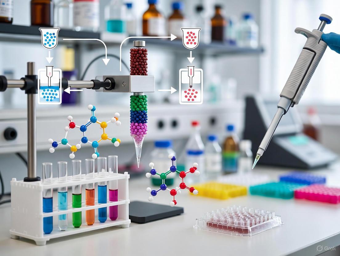
Magnetic beads are a cornerstone technology in modern life sciences, providing a versatile platform for the separation, isolation, and analysis of biological molecules. These beads are typically composed of a magnetic core surrounded by a polymer coating that can be functionalized with various chemical groups or biomolecules to enable specific interactions with targets of interest.
The global magnetic beads market, valued at USD 3.62 billion in 2025, is projected to expand at a compound annual growth rate (CAGR) of 7.85%, reaching USD 6.18 billion by 2032, reflecting their critical role in biotechnology and diagnostics [1].
Structural Characteristics and Classification
Magnetic beads are classified based on their magnetic properties, material composition, and physical dimensions. The table below summarizes the key structural parameters.
Table 1: Structural Classification and Composition of Magnetic Beads
| Classification Parameter | Types and Size Ranges | Common Material Compositions |
|---|---|---|
| Magnetic Properties | Paramagnetic, Superparamagnetic [1] | |
| Bead Size | 1–10 µm, 20–40 µm, 70–120 µm [1] | |
| Core Material | Iron oxide (Fe₃O₄ or γ-Fe₂O₃), Cobalt ferrite (CoFe₂O₄), Nickel ferrite (NiFe₂O₄) [1] | |
| Shell/Coating | Polymer matrices (e.g., poly(glycidyl methacrylate)), Natural biopolymers (e.g., Sodium Alginate, Chitosan) [2] [3] |
Superparamagnetic beads are particularly valuable for biomedical applications because they exhibit strong magnetism only in the presence of an external magnetic field, preventing aggregation and ensuring easy redispersion after the field is removed.
Functionalization Strategies and Surface Engineering
Surface functionalization is the process of modifying the bead's outer layer to introduce specific chemical groups (e.g., carboxyl, amine, epoxy) or biomolecules (e.g., antibodies, streptavidin, oligonucleotides). This enables the selective capture of target analytes from complex mixtures.
Advanced Chemical Functionalization
A powerful strategy involves using amino acids as building blocks for surface modification. Different amino acids impart distinct surface properties:
- Phenylalanine (Hydrophobic): Creates a hydrophobic surface that exhibits strong adsorption for proteins like mouse immunoglobulin (IgG), with a capacity of up to 53.5 μg/mg. Beads modified with phenylalanine are highly effective as carriers in chemiluminescent immunoassays (CLIA), significantly reducing background levels [2].
- Aspartic Acid (Acidic): Introduces carboxyl groups, conferring a negative surface charge.
- Arginine (Basic): Introduces amine groups, conferring a positive surface charge.
- Glycine (Neutral): Provides a neutral, hydrophilic surface [2].
The interactions between these functionalized surfaces and proteins are governed by a combination of electrostatic forces and hydrophobic interactions [2]. A common synthesis method is surface-initiated atom transfer radical polymerization (SI-ATRP), which grows polymer brushes (e.g., from glycidyl methacrylate monomers) on the bead surface, providing epoxy groups for subsequent ring-opening reactions with amino acids [2].
Biopolymer-Based Encapsulation
For applications in biocatalysis or environmental remediation, magnetic beads can be encapsulated within natural biopolymers like sodium alginate (SA) and chitosan (CS). These materials form a core-shell structure through layer-by-layer self-assembly, physically cross-linked via covalent bonds between –NH₂ groups on CS and –COOH groups on SA [3]. This matrix is non-toxic, biodegradable, and excellent for immobilizing microorganisms or enzymes while protecting them from environmental inhibition [3].
Applications in Biosensor-Based Sample Preparation
Magnetic beads functionalized with specific capture molecules are indispensable for preparing clean and concentrated samples for biosensor detection.
Diagnostic Immunoassays for Small Molecules
A prime example is the detection of cocaine in biological fluids using a magnetic bead-based competitive immunoassay [4] [5].
Experimental Protocol: Competitive Immunoassay for Cocaine Detection
- Bead Preparation: Immobilize cocaine-BSA conjugates on functionalized magnetic beads.
- Competitive Incubation: Mix the prepared beads with the sample (saliva or urine) and horseradish peroxidase (HRP)-labeled anti-cocaine antibodies. The cocaine in the sample competes with the bead-bound cocaine-BSA for the limited antibody binding sites.
- Magnetic Separation: Place the tube in a magnetic rack to pull the beads to the side of the vessel. Carefully remove and discard the supernatant to wash away unbound substances.
- Electrochemical Detection: Transfer the beads to a screen-printed carbon electrode (SPCE). Add an amperometric redox solution (hydrogen peroxide/hydroquinone, H₂O₂/HQ). The HRP enzyme on the bound antibodies catalyzes a reaction, generating an electrical current measured by the sensor.
- Quantification: The measured current is inversely proportional to the cocaine concentration in the sample.
This platform achieves a remarkable detection limit of 0.1 ng mL⁻¹ with a rapid analysis time of under 30 minutes, demonstrating high sensitivity and suitability for point-of-care testing [4] [5].
Competitive immunoassay workflow for cocaine detection.
Isolation of Complex Biomarkers
Magnetic beads also excel at isolating specific biomarkers, such as extracellular vesicles (EVs). EVs are membrane-bound carriers of molecular information but are challenging to purify from biofluids due to their heterogeneity [6].
Experimental Protocol: Immunocapture of Extracellular Vesicles
- Bead Selection: Use magnetic beads functionalized with antibodies against specific surface proteins (e.g., CD9, CD63, CD81) present on the target EVs.
- Sample Incubation: Mix the antibody-conjugated beads with the biological sample (e.g., blood plasma, urine) and incubate to allow EVs to bind to the antibodies on the bead surface.
- Magnetic Capture: Place the tube in a magnetic rack. The bead-bound EVs are pulled to the side, while contaminating proteins and particles remain in the supernatant.
- Washing: Remove the supernatant and wash the bead-EV complex with a suitable buffer to remove non-specifically bound impurities.
- Elution (Optional): Release the captured EVs from the beads using a low-pH buffer or a competitive agent for downstream analysis (e.g., RNA sequencing, proteomics) [6].
This affinity-based method offers superior specificity, reproducibility, and efficiency compared to traditional techniques like ultracentrifugation [6].
Purification of Post-Translational Modifications
In biotherapeutic development, analyzing protein glycosylation is critical. Cellulose-functionalized magnetic beads (CMBs) provide a high-throughput method for purifying fluorescently labeled N-glycans after their release from proteins.
Experimental Protocol: High-Throughput N-Glycan Purification
- Release and Label: Enzymatically release N-glycans from the protein (e.g., a therapeutic antibody) using PNGase F and label them with a fluorescent tag (e.g., RapiFluor-MS).
- Binding: Add CMBs to the solution. The hydrophilic N-glycans bind to the cellulose via hydrogen bonding, while impurities like proteins and excess dye do not.
- Magnetic Separation: Use a magnet to capture the beads. Remove the supernatant containing impurities.
- Washing: Wash the beads with an organic solvent (e.g., ethanol) to further remove hydrophobic contaminants.
- Elution: Release the purified glycans by adding pure water, which disrupts the hydrogen bonds. The eluted glycans are then ready for analysis by LC-FLR-MS [7].
This method is cost-effective, robust, and easily automated on a robotic liquid handler, making it ideal for characterizing biotherapeutic products [7].
The Scientist's Toolkit: Essential Reagent Solutions
The table below lists key materials and reagents required for implementing magnetic bead-based protocols.
Table 2: Key Research Reagent Solutions for Magnetic Bead-Based Workflows
| Reagent/Material | Function and Application | Specific Example |
|---|---|---|
| Functionalized Magnetic Beads | Core solid phase for capture and separation; available with carboxyl, amine, epoxy, or streptavidin surfaces. | Iron oxide core with polymer shell [1] [2]. |
| Coupling Agents | Facilitate covalent attachment of proteins/ligands to bead surfaces. | Crosslinkers for amino-acid based functionalization [2]. |
| Capture Ligands | Provide specificity for target analyte (antigen, vesicle, etc.). | Anti-cocaine antibodies [4], CD63 antibodies for EV isolation [6]. |
| Blocking Buffers | Reduce non-specific binding to the bead surface, improving assay signal-to-noise. | Bovine Serum Albumin (BSA) solutions. |
| Wash Buffers | Remove unbound and non-specifically bound materials during separation steps. | Phosphate Buffered Saline (PBS) with detergents (e.g., Tween-20). |
| Detection Probes | Enable readout of the binding event, often via optical or electrochemical means. | Horseradish Peroxidase (HRP)-labeled antibodies [4] [5]. |
| Elution Buffers | Release captured targets from beads for downstream analysis. | Low-pH buffer or pure water [6] [7]. |
General structure of a functionalized magnetic bead.
Magnetic beads are a cornerstone of modern sample preparation in biosensor research. Their integration significantly enhances the sensitivity, specificity, and speed of detecting analytes across diverse fields—from medical diagnostics and drug discovery to environmental monitoring [6] [8]. The utility of these nanoscale tools is anchored in three fundamental properties: an exceptionally high surface-to-volume ratio, superparamagnetism, and proven biocompatibility. These characteristics work in concert to enable efficient target capture, concentration, and separation from complex sample matrices, thereby purifying and pre-concentrating analytes for highly accurate biosensor detection [9] [10]. This protocol outlines the core principles, quantitative properties, and practical methodologies for leveraging magnetic beads in biosensor research, providing a framework for robust and reproducible experimental design.
Core Properties and Quantitative Analysis
The performance of magnetic beads in biosensing applications is dictated by a set of interdependent physicochemical properties. The table below summarizes these key characteristics and their direct impact on biosensor functionality.
Table 1: Key Properties of Magnetic Beads and Their Impact on Biosensing
| Property | Description | Impact on Biosensor Performance |
|---|---|---|
| High Surface-to-Volume Ratio | Provides a large functional surface area for immobilizing antibodies, aptamers, or other capture probes relative to bead volume [10]. | Increases the loading capacity for target biomolecules, enhancing capture efficiency and ultimately the signal intensity of the biosensor [9]. |
| Superparamagnetism | Exhibits strong magnetization under an external magnetic field while retaining no residual magnetism once the field is removed, preventing aggregation [11] [10]. | Enables rapid magnetic separation and concentration of targets from complex samples (e.g., blood, food) while maintaining a stable colloidal dispersion during assay steps [8]. |
| Biocompatibility | The surface chemistry of the beads is compatible with biological systems, often achieved through coatings like silica (TEOS), polyethylene glycol (PEG), or dextran [10]. | Preserves the biological activity of immobilized probes and target analytes, minimizes non-specific binding, and reduces cytotoxic effects for in vitro applications [10] [6]. |
| Surface Functionalization | The ability to chemically modify the bead surface with various functional groups (e.g., carboxyl, amine, streptavidin) [6]. | Allows for covalent and stable immobilization of a wide range of biorecognition elements, tailoring the beads for specific assays [9] [12]. |
| Size Uniformity (Monodispersity) | Beads with a highly consistent and narrow size distribution, typically in the 1-3 µm range for many separation applications [9]. | Ensures predictable and uniform binding kinetics, magnetic response, and flow characteristics, leading to high reproducibility in assays [9]. |
The following diagram illustrates the logical relationships between a magnetic bead's intrinsic properties, the subsequent engineering strategies, and the final performance outcomes in a biosensing workflow.
Diagram 1: From bead properties to biosensor performance.
Application Notes: Biosensing Enhanced by Magnetic Beads
Target Isolation and Pre-Concentration
A primary application of magnetic beads is the isolation and purification of specific targets from complex, heterogeneous samples. For instance, in the detection of inflammatory cytokines like interleukin-1β (IL-1β), magnetic beads functionalized with capture antibodies are used to pull the target protein out of solution, concentrating it and removing contaminants that could interfere with the biosensor's detection mechanism [13]. This process directly enhances the signal-to-noise ratio. Similarly, extracellular vesicles (EVs) can be efficiently isolated using magnetic beads coated with antibodies against specific surface markers (e.g., CD63, CD81), overcoming the limitations of traditional methods like ultracentrifugation in terms of yield and purity [6] [12]. This is critical for downstream analysis of EV cargo for diagnostic purposes.
Signal Amplification in Transduction Systems
Magnetic beads are not merely passive carriers; they actively participate in the signal generation of certain biosensors. In giant magnetoresistance (GMR) biosensors, the magnetic fringe field from beads bound to the sensor surface causes a measurable change in electrical resistance [11] [14]. The use of superparamagnetic nanoparticles, such as TEOS-coated Fe₃O₄, optimizes this effect by providing a strong, stable magnetic signal without agglomeration, thereby lowering the limit of detection [11]. In electrochemical biosensors like the Magnetic Bead Electrochemical Sandwich Assay (MBESA), beads serve as a mobile solid support for immunocomplex formation, facilitating efficient washing steps and enabling highly sensitive detection with minimal reagent use [13].
Experimental Protocols
Protocol: Magnetic Bead-Based Immunoassay for Cytokine Detection
This protocol details the steps for detecting a protein biomarker (e.g., IL-1β) using an electrochemical sandwich assay, adapted from the MBESA method [13].
Workflow Overview: The process involves capturing the target cytokine from a sample using antibody-coated magnetic beads, forming a sandwich complex with a detection antibody, and finally generating an electrochemical signal for quantification.
Diagram 2: Immunoassay workflow for cytokine detection.
Materials:
- Magnetic Beads: Carboxyl-functionalized superparamagnetic beads (e.g., 1-3 µm diameter).
- Bioprobes: Capture antibody (specific to the target cytokine), enzyme-conjugated detection antibody (e.g., Horseradish Peroxidase, HRP).
- Buffers: Coupling buffer (e.g., 0.1 M MES, pH 5.5), blocking buffer (e.g., 1% BSA in PBS), wash buffer (e.g., PBS with 0.05% Tween 20).
- Activation Reagents: N-Hydroxysuccinimide (NHS) and 1-Ethyl-3-(3-dimethylaminopropyl)carbodiimide (EDC) for covalent coupling.
- Sample: Cell culture supernatant, serum, or plasma.
- Equipment: Magnetic separation rack, microcentrifuge tubes, electrochemical workstation.
Step-by-Step Procedure:
- Bead Functionalization:
- Activate 1 mg of carboxylated magnetic beads in 1 mL of coupling buffer using a fresh mixture of NHS and EDC for 30 minutes with gentle mixing.
- Magnetically separate the beads and remove the supernatant.
- Resuspend the activated beads in coupling buffer containing 10-50 µg of capture antibody.
- Incubate for 2 hours at room temperature with gentle rotation.
- Separate the beads and wash twice with wash buffer.
- Block non-specific sites by incubating with 1% BSA for 1 hour.
- Wash twice and resuspend in storage buffer at 4°C until use.
Target Capture:
- Combine 100 µL of functionalized bead suspension with 100 µL of sample or standard (e.g., IL-1β in the range of 10-600 pg/mL) [13].
- Incubate for 60 minutes with gentle mixing.
Magnetic Washing:
- Place the tube on a magnetic rack for 2 minutes until the solution clears.
- Carefully aspirate and discard the supernatant.
- Wash the bead-analyte complex three times with 200 µL of wash buffer to remove unbound substances.
Detection Complex Formation:
- Add 100 µL of HRP-conjugated detection antibody to the washed beads.
- Incubate for 45 minutes with gentle mixing.
- Perform another magnetic washing cycle (Step 3) to remove excess detection antibody.
Electrochemical Measurement:
- Transfer the bead complex to an electrochemical cell containing a suitable substrate (e.g., TMB/H₂O₂ for amperometric detection).
- Apply the appropriate potential and measure the resulting current.
- The magnitude of the electrochemical signal is proportional to the amount of captured target.
Protocol: Enhancing GMR Biosensor Sensitivity with Superparamagnetic Nanoparticles
This protocol describes the synthesis and application of coated magnetic nanoparticles to optimize the performance of Giant Magnetoresistance (GMR) biosensors [11].
Workflow Overview: Superparamagnetic Fe₃O₄ nanoparticles are synthesized and coated with TEOS to enhance stability and performance. These optimized nanoparticles are then used as magnetic labels in a GMR-based immunoassay.
Diagram 3: GMR biosensor enhancement workflow.
Materials:
- Precursors: Ferric chloride hexahydrate (FeCl₃·6H₂O), ferrous sulfate heptahydrate (FeSO₄·7H₂O).
- Coating Agent: Tetraethyl orthosilicate (TEOS).
- Base: Ammonium hydroxide (NH₄OH, 25%).
- GMR Sensor Setup: GMR chip, biasing magnet, readout electronics.
Step-by-Step Procedure:
- Synthesis of TEOS-coated Fe₃O₄ (Fe₃O₄@TEOS):
- Synthesize Fe₃O4 nanoparticles via the co-precipitation method by rapidly adding NH₄OH to a mixed solution of Fe²⁺ and Fe³⁺ ions under an inert atmosphere and vigorous stirring [11] [10].
- Wash the precipitated nanoparticles with deionized water and ethanol.
- Re-disperse the Fe₃O₄ nanoparticles in a mixture of ethanol, water, and NH₄OH.
- Add varying volumes of TEOS (1-3 mL) dropwise under continuous stirring and react for 6-12 hours to form the silica shell (Fe₃O₄@TEOS) [11].
- Collect the coated nanoparticles magnetically and wash thoroughly.
Characterization of Nanoparticles:
- Confirm the core-shell structure and crystallinity using X-ray diffraction (XRD) and Transmission Electron Microscopy (TEM).
- Measure magnetic properties with a Vibrating Sample Magnetometer (VSM). Note that saturation magnetization decreases with higher TEOS volume, but stability improves [11].
- Assess colloidal stability via Zeta potential measurements.
GMR Biosensor Assay:
- Functionalize the GMR sensor surface with a capture probe.
- Incubate the sensor with the sample containing the target analyte.
- Add Fe₃O₄@TEOS nanoparticles conjugated with a detection probe to form a sandwich complex on the sensor.
- Apply an optimal bias magnetic field (e.g., 1.7 Oe as reported) [11].
- Measure the change in the sensor's resistance caused by the fringe field of the bound magnetic nanoparticles. The signal correlates with the target concentration, achieving high sensitivity with a low limit of detection.
The Scientist's Toolkit: Essential Research Reagents
Table 2: Key Research Reagent Solutions for Magnetic Bead-Based Biosensing
| Reagent / Material | Function / Application | Key Considerations |
|---|---|---|
| Carboxyl-Modified Magnetic Beads | A versatile platform for covalent coupling of proteins (antibodies) and amine-containing ligands via EDC/NHS chemistry [13] [6]. | Available in various sizes (nm to µm). The choice of size balances surface area (capture efficiency) and magnetic response (separation speed) [9]. |
| Streptavidin-Coated Magnetic Beads | Universal capture particles for any biotinylated molecule (e.g., biotinylated antibodies, aptamers), offering high affinity and flexibility in assay design [8]. | High binding capacity and consistency are critical. The stability of the streptavidin-biotin bond is essential for rigorous washing steps. |
| Tetraethyl Orthosilicate (TEOS) | A silica precursor used to create a protective, hydrophilic shell around magnetic nanoparticles, enhancing colloidal stability and providing a surface for further functionalization [11]. | The volume of TEOS used during coating is a key optimization parameter, trading off saturation magnetization for improved stability and dispersibility [11]. |
| EDC & NHS Crosslinkers | Activating agents for carboxyl groups, enabling efficient and stable covalent conjugation of biomolecules to the bead surface. | Fresh preparation of the activation mixture is crucial for high coupling efficiency. The reaction pH must be carefully controlled. |
| Superparamagnetic Iron Oxide Nanoparticles (SPIONs) | The core magnetic material (Fe₃O₄ or γ-Fe₂O₃) for many bead formulations, prized for its strong magnetic moment and lack of magnetic remanence [10]. | Must be coated (e.g., with polymers or silica) to ensure biocompatibility and prevent oxidation and aggregation in physiological buffers [10]. |
| Antibodies & Aptamers | Biorecognition elements that confer specificity to the magnetic bead complex, allowing for the selective isolation of targets from proteins to whole cells [15] [8]. | Aptamers, as synthetic oligonucleotides, offer advantages in stability and cost over antibodies but require careful selection via SELEX [8]. |
Biosensors represent a powerful convergence of biological recognition and physicochemical detection, serving critical roles in healthcare, food safety, and environmental monitoring. A persistent challenge in these fields involves detecting low-abundance analytes within complex sample matrices such as blood, saliva, or food homogenates. Magnetic beads (MBs), also referred to as magnetic nanoparticles (MNPs), have emerged as a transformative tool in biosensor design, effectively addressing key limitations in conventional detection methodologies [8]. These superparamagnetic particles, typically ranging from nanometers to micrometers in diameter, provide a versatile platform for enhancing all aspects of biosensor performance.
The fundamental properties of MBs—including their high surface-area-to-volume ratio, superparamagnetism, and biocompatibility—make them ideal for biosensing applications [8] [16]. Their functionalized surfaces can be modified with various biorecognition elements such as antibodies, aptamers, or DNA probes, enabling specific target capture. When coupled with an external magnetic field, MBs facilitate precise manipulation and separation of target analytes from complex samples, leading to significant improvements in sensitivity, specificity, and speed [8]. This application note examines the mechanisms through which magnetic beads enhance biosensor performance and provides detailed protocols for their implementation in research settings, framed within the broader context of magnetic bead-based sample preparation for biosensor detection.
Performance Enhancement Mechanisms
Sensitivity Enhancement
Magnetic beads dramatically improve biosensor sensitivity through two primary mechanisms: target preconcentration and signal amplification. By efficiently capturing and isolating target analytes from large sample volumes, MBs effectively concentrate them into significantly smaller volumes for detection, thereby increasing the effective concentration presented to the biosensor transducer [8]. This magnetic enrichment capability is particularly valuable for detecting low-abundance targets that would otherwise fall below the detection limit of conventional assays.
The substantial surface area of MNPs allows for immobilization of numerous signal-generating molecules or labels, leading to enhanced signal amplification. In electrochemical biosensors, MB-biomarker complexes localized on electrode surfaces via external magnets significantly amplify redox signals, enabling detection of clinically relevant biomarkers at ultralow concentrations [17]. For Alzheimer's disease biomarkers in saliva, this approach achieved detection limits of 2 μg/mL for lactoferrin and 0.1 pg/mL for amyloid β-protein 1-42, outperforming commercial ELISA kits [17]. Similarly, in CRISPR-Cas13a-based electrochemical biosensors for SARS-CoV-2 detection, immunocapture magnetic beads reduced background noise signals and enhanced detection ability, achieving ultrasensitive detection down to 1.66 aM [18].
Specificity Improvement
The specificity of biosensors employing magnetic beads stems from two complementary factors: the biological recognition elements immobilized on MB surfaces and the physical separation of target-bound complexes from interfering substances. Magnetic separation enables rigorous washing steps that effectively remove non-specifically bound molecules, matrix components, and contaminants that could generate false-positive signals [8] [16].
This enhancement is exemplified in a fluorescent biosensor for cancer-associated miRNA let-7a detection, where MBs served as an efficient separation tool for capturing and separating targets from complex samples without pretreatment [16]. The combination of MBs with graphene oxide (GO) enabled highly specific differentiation between target and non-target sequences through differential adsorption capabilities, achieving a limit of detection of 15.015 pM even in complex human serum samples [16]. For eukaryotic cell detection using giant magnetoresistance (GMR) sensors, a two-stage GMR biochip with face-to-face sensors allowed differentiation between biological objects magnetically labeled by numerous beads from bead aggregates, significantly reducing false positives and improving specificity [19].
Acceleration of Assay Workflow
The integration of magnetic beads streamlines biosensing protocols by consolidating multiple processing steps and reducing or eliminating requirements for complex instrumentation. The rapid magnetic responsiveness of MBs facilitates quick separation and washing cycles without the need for centrifugation, which is typically time-consuming and requires multiple manual steps [8] [20].
This acceleration is particularly evident in foodborne pathogen detection, where conventional culture-based methods require 24-48 hours and PCR-based methods need at least 36 hours [21]. In contrast, an immunomagnetic bead biosensor detected 100 cells of E. coli in just 2 hours without pre-enrichment or incubation [21]. Similarly, a miniaturized electrochemical sensor for Alzheimer's biomarkers achieved output within one minute after MBs captured the biomarkers [17]. Automated magnetic bead-based sample preparation systems further enhance speed and reproducibility by eliminating manual pipetting steps, reducing hands-on time by 5-6 hours while improving walkaway automation for applications with 10-100 moderate daily throughput samples [20].
Quantitative Performance Comparison
The table below summarizes representative performance metrics of magnetic bead-enhanced biosensors across various detection platforms and target analytes.
Table 1: Performance Metrics of Magnetic Bead-Enhanced Biosensors
| Target Analyte | Biosensor Platform | Detection Mechanism | Limit of Detection | Assay Time | Reference |
|---|---|---|---|---|---|
| SARS-CoV-2 | Electrochemical | CRISPR-Cas13a with magnetic separation | 1.66 aM | ~1 hour | [18] |
| E. coli | Immunomagnetic | Antibody-functionalized MBs with frequency shift measurement | 100 cells | 2 hours | [21] |
| let-7a miRNA | Fluorescent | MB and GO-assisted enzyme-free amplification | 15.015 pM | - | [16] |
| Amyloid β-protein 1-42 | Electrochemical | Immunomagnetic beads with AuNP-modified electrode | 0.1 pg/mL | <1 minute (post-capture) | [17] |
| Lactoferrin | Electrochemical | Immunomagnetic beads with AuNP-modified electrode | 2 μg/mL | <1 minute (post-capture) | [17] |
| miR-21-5p | Dark-field imaging | CRISPR/Cas13a with magnetic-gold nanoparticle complexes | 25 pM (500 attomoles) | 30 minutes | [22] |
| Murine cancer cells (NS1) | Giant Magnetoresistance (GMR) | Antibody-functionalized magnetic particles | 5 × 10² cells/mL | - | [19] |
| Salmonella typhimurium | Electrochemical | Gold leaf electrode with magnetic beads | Quantitative detection demonstrated | - | [23] |
| Listeria monocytogenes | Electrochemical | Gold leaf electrode with magnetic beads | Quantitative detection demonstrated | - | [23] |
Experimental Protocols
Protocol: Immunomagnetic Bead Modification for Protein Detection
This protocol details the functionalization of magnetic beads with capture antibodies for specific protein biomarker detection, adapted from the Alzheimer's biomarker sensor development [17].
Materials:
- Streptavidin-coated magnetic beads (3 μm for low-abundance targets, 10 μm for higher concentration targets)
- Biotinylation kit
- Target-specific capture antibodies
- Phosphate-buffered saline (PBS), pH 7.4
- Bovine serum albumin (BSA)
- Magnetic separator
- Rotator or mixer
Procedure:
- Biotinylation of Antibodies:
- Use a commercial biotinylation kit following manufacturer's instructions.
- Typically involves incubating antibodies with biotin reagent at room temperature for 30-60 minutes.
- Remove excess biotin using desalting columns or dialysis.
Magnetic Bead Preparation:
- Resuspend streptavidin-modified magnetic bead suspension (20 μL of 20 mg/mL) in 200 μL of PBS.
- Place tube on magnetic separator for 1-2 minutes until beads collect on tube wall.
- Carefully remove and discard supernatant.
- Remove tube from magnet and resuspend beads in 200 μL PBS.
- Repeat washing step twice.
Antibody Immobilization:
- Add biotinylated capture antibodies to washed magnetic beads (typical ratio: 5-10 μg antibody per mg beads).
- Incubate with continuous mixing on a rotator for 30-60 minutes at room temperature.
- Separate beads using magnetic separator and remove supernatant.
- Wash three times with PBS to remove unbound antibodies.
Blocking:
- Resuspend antibody-conjugated beads in PBS containing 1-5% BSA.
- Incubate for 30 minutes to block non-specific binding sites.
- Wash three times with PBS and resuspend in appropriate storage buffer.
Storage:
- Store functionalized beads at 4°C in PBS with 0.1% BSA and 0.02% sodium azide.
- Use within one month for optimal performance.
Protocol: Magnetic Bead-Enhanced Electrochemical Detection
This protocol describes the application of functionalized magnetic beads for electrochemical detection of biomarkers, based on the Alzheimer's salivary biomarker sensor [17] and foodborne pathogen detection systems [23].
Materials:
- Functionalized immunomagnetic beads (from Protocol 4.1)
- Screen-printed carbon electrodes or custom-fabricated gold leaf electrodes
- Electrochemical workstation
- External magnet
- Sample containing target analyte
- Detection antibodies (biotinylated or enzyme-conjugated)
- Redox mediator (e.g., ferri/ferrocyanide solution)
- Washing buffer (PBS with 0.05% Tween-20)
Procedure:
- Target Capture:
- Incubate 50 μL of functionalized magnetic beads with 500 μL-1 mL sample.
- Mix continuously for 15-30 minutes at room temperature to facilitate target binding.
Magnetic Separation and Washing:
- Place reaction tube on magnetic separator for 2 minutes.
- Carefully remove and discard supernatant.
- Remove tube from magnet and resuspend beads in 500 μL washing buffer.
- Repeat washing step three times.
Signal Generation Complex Formation:
- Add detection antibodies (biotinylated) to washed beads.
- Incubate for 15 minutes with mixing.
- Wash three times to remove unbound detection antibodies.
- For enzymatic signal amplification, add enzyme-conjugated streptavidin and incubate for 15 minutes.
- Perform final three washes.
Electrochemical Measurement:
- Resuspend final bead complex in 40-100 μL of appropriate buffer.
- Transfer suspension to electrochemical cell containing the working electrode.
- Apply external magnet beneath electrode to localize bead complexes on electrode surface.
- Allow 1-2 minutes for bead settlement and localization.
- Perform electrochemical measurement (e.g., square wave voltammetry, electrochemical impedance spectroscopy) using appropriate parameters.
- For square wave voltammetry, typical parameters include: frequency 50 Hz, amplitude 50 mV, potential range -0.5 to 0 V [18].
Data Analysis:
- Measure current value (peak current after subtracting background).
- Compare to standard curve for quantitative analysis.
Research Reagent Solutions
Table 2: Essential Research Reagents for Magnetic Bead-Based Biosensing
| Reagent/Material | Function/Application | Examples/Specifications |
|---|---|---|
| Streptavidin-coated Magnetic Beads | Universal platform for biotinylated bioreceptor immobilization | Dynabeads M-280 Streptavidin; 2.8 μm diameter [18] |
| Carboxyl-modified Magnetic Beads | Covalent attachment of proteins/antibodies via EDC-NHS chemistry | Various suppliers; available in multiple size ranges (50 nm-10 μm) |
| Amine-functionalized Magnetic Beads | Covalent conjugation to carboxylated biomolecules | Suitable for DNA/aptamer immobilization |
| Screen-printed Carbon Electrodes | Disposable electrochemical transducers | Red Matrix China Co., Ltd. [18] |
| Gold Leaf Electrodes | Cost-effective alternative to fabricated gold electrodes | 24-karat gold leaves laminated on PVC sheets [23] |
| CRISPR-Cas13a Protein | RNA-targeting CRISPR enzyme for nucleic acid detection | LwaCas13a protein [18] |
| Biotin-RNA-MB Reporter | Electrochemical reporter for CRISPR assays | Biotin-labeled RNA with methylene blue tag [18] |
| HRP-Conjugated Streptavidin | Enzymatic signal amplification | Compatible with TMB substrate for colorimetric/electrochemical detection |
| Magnetic Separator | Magnetic separation of bead complexes | Various formats (stand, rack, handheld) for different tube sizes |
Workflow and Signaling Pathways
Magnetic beads serve as a versatile and powerful tool for enhancing critical performance parameters in biosensing applications. Their unique properties enable significant improvements in sensitivity through target preconcentration and signal amplification, enhance specificity via efficient separation of target complexes from matrix interferents, and accelerate assay workflows through rapid magnetic manipulation. The integration of magnetic beads with emerging technologies such as CRISPR systems, GMR sensors, and novel electrode fabrication methods continues to expand their utility across diverse diagnostic and monitoring applications. As research progresses, further optimization of magnetic bead synthesis, surface functionalization, and integration with automated platforms will undoubtedly unlock new capabilities in biosensor performance and point-of-care applicability.
Magnetic beads are a cornerstone of modern biotechnology, serving as versatile solid-phase supports for the separation and analysis of biomolecules, cells, and pathogens. Their significance is particularly pronounced in the field of biosensor-based detection, where they facilitate the efficient purification and pre-concentration of analytes from complex biological samples, thereby dramatically enhancing biosensor sensitivity and specificity [24]. The global magnetic bead market reflects this importance, with an estimated size of USD 4620 million in 2025 and a projected growth to USD 11603.38 million by 2033, driven largely by demands in in-vitro diagnostics and genomic research [25]. The performance of magnetic beads in these applications—including their binding capacity, colloidal stability, and magnetic responsiveness—is intrinsically governed by their synthesis and functionalization. This article provides a detailed overview of the chemical, physical, and biological approaches used to synthesize and tailor magnetic beads, framing them within the context of biosensor development and providing actionable protocols for researchers.
Chemical Synthesis Approaches
Chemical methods offer precise control over the composition, size, and surface chemistry of magnetic beads, making them the most prevalent synthesis route.
Thermolysis Decomposition
This method involves the high-temperature decomposition of organometallic precursors in high-boiling-point organic solvents.
- Protocol: In a representative synthesis for creating magnetic mesoporous silica beads, iron(III) acetylacetonate (Fe(acac)₃) is used as the precursor [26]. The precursor is dissolved in 2-pyrrolidone (which acts as both solvent and stabilizer) at a concentration of 0.5 M. Dendritic silica colloid (dSiO₂) templates are dispersed in this solution. The reaction mixture is then heated to 245°C under a nitrogen atmosphere and maintained at this temperature for 1 hour to allow for the thermal decomposition of the precursor and the formation of superparamagnetic Fe₃O₄ nanoparticles within the silica channels. The resulting composite, Fe₃O₄@dSiO₂, is collected via magnetic separation and washed repeatedly with ethanol and acetone [26].
Activated Swelling Method
A widely used commercial method, exemplified by Dynabeads, this technique produces monodisperse polymeric beads with incorporated magnetic material.
- Protocol: The process begins with the synthesis of monodisperse macroporous polymer beads (e.g., polystyrene) via seed polymerization [26]. The surfaces of these beads are then modified with hydrophilic functional groups (e.g., -SO₃ or -NO₂). The modified beads are immersed in an aqueous solution of iron salts (e.g., FeCl₂ and FeCl₃). Under controlled pH and conditions, magnetic iron oxides (Fe₃O₄ or γ-Fe₂O₃) are precipitated and formed within the porous network of the polymer beads. Finally, a monomer with desired functional groups is introduced to swell the beads, followed by polymerization to seal the pores and create a functional outer shell [26].
Co-precipitation
This is a straightforward aqueous method for synthesizing iron oxide nanoparticles, often used as cores for more complex beads.
- Protocol: A mixture of ferric and ferrous chlorides (e.g., at a molar ratio of 2:1 Fe³⁺:Fe²⁺) is dissolved in deoxygenated water. Under an inert atmosphere and vigorous stirring, a base, such as ammonium hydroxide (NH₄OH), is added dropwise to the solution until the pH reaches 10-11. The black precipitate of magnetite (Fe₃O₄) forms instantly. The reaction is typically continued for 30 minutes at room temperature. The nanoparticles are then separated magnetically and washed thoroughly with deionized water until neutral pH is achieved [24].
Table 1: Comparison of Primary Chemical Synthesis Methods for Magnetic Beads
| Method | Key Reagents | Reaction Conditions | Key Characteristics of Resultant Beads |
|---|---|---|---|
| Thermolysis Decomposition | Fe(acac)₃, 2-pyrrolidone, dSiO₂ templates | 245°C, N₂ atmosphere, 1 hour | High magnetic content (59%), uniform, superparamagnetic, mesoporous structure [26] |
| Activated Swelling | Polymer seeds, iron salts (FeCl₂/FeCl₃), functional monomers | Aqueous solution, controlled pH | Monodisperse, micron-sized (1-100 μm), high functional group density [26] |
| Co-precipitation | FeCl₃, FeCl₂, NH₄OH | Room temperature, N₂ atmosphere, pH 10-11 | Simple, cost-effective, aqueous process; can yield agglomerated particles [24] |
Physical and Biological Synthesis Approaches
While chemical synthesis dominates, physical and biological methods offer alternative pathways with unique advantages.
Physical Synthesis: Sonochemical Assembly
Physical energy, such as ultrasound, can be used to assist in the synthesis and functionalization of magnetic beads.
- Principle: High-intensity ultrasound creates cavitation bubbles in a liquid, generating localized spots of extremely high temperature and pressure. This energy can drive chemical reactions, fragment particles, and enhance the mixing and deposition of materials onto surfaces [24]. While not a standalone method for creating the magnetic core, it is highly effective for functionalizing pre-synthesized magnetic nanoparticles with silica or polymer shells, or for creating nanocomposites.
Biological Synthesis: Bio-inspired Functionalization
This approach leverages biological molecules to impart specific functionalities and responsiveness to magnetic beads.
- Protocol (pH-Sensitive Oligopeptide Functionalization): Magnetic mesoporous silica beads (Fe₃O₄@dSiO₂) are first synthesized and their surfaces are activated with maleimide functional groups. A custom-synthesized oligopeptide, such as a decamer of histidine and glutamate (HE)₁₀, is dissolved in a phosphate buffer (pH 7.0). The peptide solution is added to the bead suspension and allowed to react for 12 hours at 4°C. The conjugation occurs via a thiol-maleimide "click" reaction. The resulting Fe₃O₄@dSiO₂-(HE)₁₀ beads are magnetically separated and washed to remove unbound peptide [26]. These beads exhibit pH-sensitive behavior, altering their charge and conformation in response to environmental pH, which is advantageous for controlled binding and release of biomolecules like DNA.
Functionalization and Application-Oriented Protocols
The bare magnetic bead is often inert; its utility is unlocked through strategic surface functionalization tailored to specific applications.
Core Functionalization Strategies
- Streptavidin-Coating: Beads are coated with streptavidin, enabling strong biotin-streptavidin binding to conjugate biotinylated antibodies, oligonucleotides, or other probes. Beads in the ~100-150 nm size range have shown superior performance in immunomagnetic capture applications [27].
- Antibody Immobilization: Antibodies are directly covalently immobilized onto beads functionalized with carboxyl, amine, or tosyl groups for specific antigen capture (e.g., anti-EpCAM for circulating tumor cells) [27].
- Oligonucleotide Probes: Single-stranded DNA or RNA probes are attached for the hybridization and capture of complementary nucleic acid sequences [28].
- Polymer Encapsulation: Coating with polymers like polyethylene glycol (PEG) reduces non-specific binding and improves stability in biological fluids [24].
Application Note 1: DNA Extraction for Next-Generation Sequencing
Efficient DNA isolation from dried blood spots (DBS) is critical for genomic newborn screening.
- Recommended Method: Magnetic bead-based protocols, particularly semi-automated systems, offer the best balance of DNA yield, purity, and operational feasibility for targeted amplicon sequencing [28].
- Detailed Protocol (Semi-Automated Magnetic Bead-based DNA Isolation):
- Punching: Punch one 3.2 mm disc from a DBS sample.
- Lysis & Binding: Transfer the punch to a deep-well plate. Add a lysis/binding buffer (e.g., from the Chemagic DNA Blood Spot 13 Kit). Seal the plate and incubate at 56°C for 3 hours with agitation at 800 rpm [28].
- Magnetic Separation: Transfer the plate to a semi-automated instrument (e.g., Chemagic 360). The instrument automatically adds magnetic beads, mixes for binding, captures the bead-DNA complexes with a magnet, and performs wash steps.
- Elution: The purified DNA is eluted in 55 µL of elution buffer [28].
- Performance Metrics: This method provides sufficient DNA quality for NGS, with high read output, mapping efficiency, and coverage depth, while minimizing hands-on time [28].
Application Note 2: Extracellular Vesicle (EV) Isolation for Diagnostic Biosensing
EVs are promising biomarkers, and their isolation using magnetic beads significantly improves yield and purity over ultracentrifugation.
- Detailed Protocol (Immunomagnetic EV Isolation):
- Bead Preparation: Incubate streptavidin-coated magnetic beads (~150 nm) with a biotinylated antibody against a specific EV surface marker (e.g., CD9, CD63, or CD81) for 30 minutes at room temperature.
- Washing: Separate the beads and wash twice with PBS containing 0.1% BSA to remove unbound antibody.
- Sample Incubation: Incubate the antibody-conjugated beads with a biofluid sample (e.g., plasma or urine) for 1-2 hours with continuous mixing.
- Washing & Elution: Capture the beads, wash thoroughly with PBS to remove non-specifically bound contaminants. Isolated EVs can be eluted using a low-pH buffer or directly lysed for downstream RNA/protein analysis [6].
Table 2: Key Operational Parameters for Different Magnetic Bead-based Isolation Methods
| Parameter | Column-based DNA Isolation | Lysis-based DNA Isolation | Semi-automated Magnetic Bead DNA Isolation | Immunomagnetic EV Isolation |
|---|---|---|---|---|
| Hands-on Time | High | Moderate | Low | Moderate to High |
| Turnaround Time | Long | Short | Moderate | 1.5 - 2.5 hours |
| Scalability | Poor | Fair | Excellent | Good (with automation) |
| Estimated Cost | Low | Very Low | High | Moderate to High |
| Purity/Yield | Variable | Variable | High and Consistent | High Purity [28] [6] |
The Scientist's Toolkit: Essential Research Reagents
Table 3: Essential Reagents and Materials for Magnetic Bead-based Research
| Item | Function/Description | Example Use Case |
|---|---|---|
| Streptavidin Magnetic Beads | Universal solid support for capturing any biotinylated molecule (antibodies, probes). | Immunoassays, nucleic acid extraction, CTC enrichment [27]. |
| Oligopeptides (e.g., (HE)₁₀) | Imparts smart, pH-responsive behavior for controlled binding and release of biomolecules. | pH-sensitive DNA capture and elution [26]. |
| Biotinylated Antibodies | Targeting ligands for specific capture of cells (e.g., via EpCAM) or EVs (e.g., via CD63). | Isolation of circulating tumor cells or extracellular vesicles [6] [27]. |
| Dendritic Silica Colloids (dSiO₂) | Templates/scaffolds for creating mesoporous beads with high surface area and pore volume. | Synthesis of high-capacity magnetic mesoporous silica beads [26]. |
| Functional Monomers (e.g., Tosyl) | Provides reactive groups for direct, covalent immobilization of proteins or ligands. | Creating antibody-coated beads for direct capture assays. |
| Magnetic Separation Rack | Enables rapid separation of bead-bound complexes from solution in standard tubes. | A crucial tool for all manual magnetic bead protocols. |
Workflow and Pathway Visualization
The following diagram illustrates the logical decision pathway for selecting and applying magnetic bead synthesis and functionalization methods based on the desired application in biosensor research.
Magnetic Bead Synthesis and Application Workflow: This chart outlines the decision-making process for developing magnetic beads for biosensing, from selecting a core synthesis method to choosing a functionalization strategy for the target application.
The synthesis and functionalization of magnetic beads are critical determinants of their success in biosensor-based sample preparation. Chemical methods like thermolysis and activated swelling provide robust, scalable production of high-performance beads, while biological functionalization introduces "smart" capabilities for enhanced control. The choice of method must be aligned with the final application, whether it is the high-throughput isolation of DNA for sequencing, the specific capture of rare EVs for diagnostic purposes, or the enrichment of CTCs for cancer monitoring. As the field advances, the integration of these tailored magnetic beads with automated platforms and high-throughput systems will continue to be a key driver in the evolution of sensitive, reliable, and point-of-care biosensor diagnostics [25].
Implementation Strategies and Cross-Disciplinary Applications
The isolation and enrichment of specific biomolecules, extracellular vesicles, and pathogens from complex biological matrices constitute a critical preliminary step in biosensor-based detection research. Magnetic bead-based sample preparation has emerged as a powerful technique that leverages the unique properties of magnetic particles for efficient target isolation. These beads, typically composed of a magnetic iron oxide core such as magnetite (Fe₃O₄) or maghemite (γ-Fe₂O₃), are coated with stabilizing polymers and functionalized with specific binding ligands that provide affinity toward target compounds [29] [30]. The fundamental advantage of this methodology lies in the ability to separate target analytes from complex samples using an external magnetic field, eliminating the need for time-consuming procedures such as centrifugation, filtration, or traditional solid-phase extraction [29]. This magnetic solid-phase extraction (MSPE) approach has revolutionized sample preparation by enabling facile, rapid, and high-throughput processing of diverse sample types, including serum, urine, saliva, blood, and tissue samples [29].
The growing importance of magnetic bead-based enrichment strategies is particularly evident in the detection of low-abundance analytes, which presents a significant challenge in the early diagnosis of diseases, environmental monitoring, and biological research [31]. By concentrating target molecules and simultaneously removing interfering substances from complex matrices, these techniques significantly enhance the sensitivity and specificity of downstream biosensing platforms [29] [31]. The versatility of magnetic beads allows for their functionalization with various capture agents, including antibodies, aptamers, oligonucleotides, and other molecular probes, enabling the selective isolation of diverse targets ranging from proteins and nucleic acids to entire cells and pathogens [29] [30]. This application note provides a comprehensive overview of magnetic bead-based enrichment methodologies, detailed experimental protocols, and their integration with biosensing platforms within the context of advanced research and diagnostic applications.
Fundamental Principles and Methodologies
Magnetic Bead Structure and Functionalization
Magnetic beads used in biosensing applications typically feature a core-shell structure engineered for optimal performance in biological systems. The magnetic core consists of superparamagnetic iron oxide nanoparticles (SPIONs), which exhibit strong magnetic responsiveness in an external magnetic field while retaining no residual magnetism once the field is removed, thus preventing aggregation [29] [30]. This core is encapsulated within a surface coating that provides physical and chemical stability, prevents particle aggregation, and offers functional groups for subsequent conjugation with biological ligands. Common coating materials include polymers such as dextran, polyethylene glycol, polyvinyl alcohol, and silica, as well as non-polymeric stabilizers like oleic acid and lactobionic acid [30].
The functionalization of magnetic beads with specific binding ligands transforms them into highly selective capture agents. Two primary immobilization strategies are employed: (1) direct covalent coupling of ligands to activated functional groups (e.g., carboxyl, amine, epoxy) on the bead surface using chemistries such as EDC/NHS, and (2) affinity-based attachment, such as the use of streptavidin-coated beads with biotinylated ligands [30]. Aptamers, which are single-stranded DNA or RNA molecules selected for high-affinity binding to specific targets, offer particular advantages as capture ligands due to their thermal stability, cost-effective production, and ease of chemical modification for controlled orientation on the bead surface [30]. Similarly, antibodies can be immobilized through protein A or G mediators to ensure proper orientation and maximize binding capacity [30].
Modes of Magnetic Separation
Magnetic bead-based separations employ two principal methodologies: direct and indirect capture. In the direct method, the magnetic beads are pre-functionalized with specific capture ligands (e.g., antibodies, aptamers) that directly bind to the target analyte when added to the sample solution [29]. This approach simplifies the workflow by combining capture and separation into a single step. In the indirect method, a free affinity ligand (typically an antibody) is first introduced to the sample to form target-ligand complexes, which are subsequently captured by secondary-modified magnetic beads (e.g., protein A/G beads for antibody capture) [29]. While requiring an additional incubation step, the indirect method can offer enhanced sensitivity for certain applications by allowing more efficient antigen-antibody interactions in solution before capture.
The separation process itself leverages the magnetic properties of the beads. When an external magnetic field is applied, the bead-analyte complexes migrate toward the magnet, allowing for efficient concentration and washing to remove non-specifically bound contaminants [29] [30]. The target analytes can then be eluted under appropriate conditions (e.g., change in pH, ionic strength, or using specific elution agents) for subsequent analysis or directly introduced into biosensing systems [29].
Table 1: Comparison of Magnetic Separation Methods
| Parameter | Direct Method | Indirect Method |
|---|---|---|
| Workflow | Simplified single-step | Multi-step process |
| Hands-on Time | Shorter | Longer |
| Flexibility | Lower | Higher |
| Capture Efficiency | Dependent on bead-surface kinetics | Enhanced by solution-phase binding |
| Typical Applications | High-throughput screening, DNA/RNA extraction | Complex protein analyses, low-abundance targets |
Research Reagent Solutions and Materials
The successful implementation of magnetic bead-based enrichment protocols requires carefully selected reagents and materials optimized for specific applications. The following table summarizes essential components and their functions in typical enrichment workflows.
Table 2: Essential Research Reagents for Magnetic Bead-Based Enrichment
| Reagent/Material | Function | Examples/Specifications |
|---|---|---|
| Magnetic Beads | Core solid-phase support for target capture | Carboxyl-terminated beads, streptavidin-coated beads, amine-functionalized beads |
| Capture Ligands | Provide specificity for target recognition | Antibodies, aptamers, oligonucleotide probes, peptides |
| Binding Buffers | Optimize conditions for ligand-target interaction | Varying pH, ionic strength, containing surfactants or carrier proteins |
| Wash Buffers | Remove non-specifically bound materials | PBS, Tris-based buffers with controlled stringency (e.g., containing low detergent concentrations) |
| Elution Buffers | Release captured targets from beads | Low pH buffers, high salt buffers, polarity-reducing agents, specific competitors |
| Blocking Agents | Reduce non-specific binding | BSA, skim milk, synthetic blocking polymers, salmon sperm DNA |
| Coupling Reagents | Facilitate ligand immobilization on beads | EDC, NHS, sulfo-NHS for covalent conjugation |
The selection of appropriate magnetic beads is particularly critical and depends on the specific application requirements. Beads with different surface functionalities, sizes, and magnetic properties are available for various applications. For instance, carboxyl-terminated beads enable straightforward covalent coupling of amine-containing ligands via EDC/NHS chemistry, while streptavidin-coated beads offer universal capture capability for biotinylated probes [29] [30]. Size considerations are also important, with smaller beads (50-500 nm) offering higher surface-area-to-volume ratios for enhanced binding capacity, while larger beads (1-5 μm) enabling faster magnetic separation [29].
Application-Specific Protocols
Protocol 1: Nucleic Acid Extraction and Enrichment
Principle: This protocol describes the extraction of PCR-ready genomic DNA (gDNA) from biological samples such as blood and urine using carboxylated magnetic nanoparticles (CMNPs) [29]. The method leverages the selective binding of nucleic acids to functionalized magnetic particles under specific buffer conditions, followed by magnetic separation and washing to remove contaminants, and final elution of purified DNA.
Materials:
- Carboxylated magnetic nanoparticles (CMNPs, 100-200 nm)
- Lysis buffer (e.g., GuHCl-based)
- Binding buffer (e.g., PEG/NaCl)
- Wash buffers (e.g., ethanol-based)
- Elution buffer (TE buffer, pH 8.0)
- Magnetic separation rack
- Thermonixer
Procedure:
- Cell Extraction: Add 100 μL of CMNPs to 1 mL of urine or blood sample. Incubate for 10 minutes with continuous mixing to allow cell capture.
- Magnetic Separation: Place the tube in a magnetic rack for 2 minutes. Discard the supernatant while retaining the bead-cell complex.
- Cell Lysis: Resuspend the bead-cell complex in 500 μL of lysis buffer. Vortex vigorously and incubate at 56°C for 10 minutes.
- DNA Binding: Add 1.5 volumes of binding buffer to the lysate. Mix thoroughly and incubate for 10 minutes at room temperature.
- Washing: Separate beads using a magnet. Wash twice with 500 μL of wash buffer.
- Elution: Resuspend beads in 50-100 μL of elution buffer. Incubate at 65°C for 5 minutes. Separate and collect supernatant containing purified gDNA.
Applications: This rapid (30-minute) protocol is particularly suitable for preparing PCR-ready DNA for downstream detection of bacterial pathogens [29] or analysis of human DNA in diagnostic applications. The method eliminates hazardous reagents like phenol or chloroform used in traditional extraction protocols and can be automated for high-throughput processing.
Protocol 2: Pathogen Detection from Food Samples
Principle: This protocol employs antibody-functionalized magnetic beads for the specific capture and concentration of foodborne pathogens such as E. coli, Listeria, and Salmonella from complex food matrices [21]. The enriched pathogens can subsequently be detected using various biosensing platforms.
Materials:
- Antibody-functionalized magnetic beads (e.g., anti-E. coli antibody conjugated)
- Food sample homogenate
- Enrichment broth
- Phosphate buffered saline (PBS) with 0.05% Tween-20
- Magnetic separation device
Procedure:
- Sample Preparation: Homogenize 25 g food sample in 225 mL enrichment broth. Pre-enrich by incubating for 16-24 hours at 37°C.
- Immunocapture: Add 100 μL of antibody-functionalized magnetic beads to 1 mL of pre-enriched sample. Incubate with continuous mixing for 60 minutes at room temperature.
- Magnetic Separation: Place tube in magnetic separator for 5 minutes. Discard supernatant.
- Washing: Resuspend bead-pathogen complex in 1 mL of PBS-Tween. Repeat magnetic separation and washing twice.
- Detection: Resuspend captured pathogens in appropriate buffer for downstream biosensor analysis. The entire detection process, from sample preparation to result display, can be completed within 2 hours with sensitivity of approximately 100 cells [21].
Applications: This method significantly reduces detection time compared to traditional culture-based methods (which require 24-48 hours) and PCR-based methods (requiring at least 36 hours) [21]. It is particularly valuable for food safety monitoring and outbreak investigations.
Protocol 3: Protein and Peptide Isolation
Principle: This protocol describes the isolation of specific proteins or peptides from complex biological samples using magnetic beads functionalized with appropriate affinity ligands [29]. Both direct and indirect capture methods can be employed, with the indirect method often providing enhanced sensitivity for low-abundance targets.
Materials:
- Magnetic beads with appropriate surface functionality
- Specific antibody or aptamer for target protein
- Binding buffer (e.g., PBS, pH 7.4)
- Wash buffer (e.g., PBS with 0.1% Tween-20)
- Elution buffer (e.g., low pH glycine buffer or high salt buffer)
Procedure:
- Bead Preparation: If using covalent coupling, immobilize capture antibody on carboxylated magnetic beads using EDC/NHS chemistry. Block remaining active sites with BSA.
- Sample Incubation: Incubate protein sample with functionalized magnetic beads for 60 minutes with continuous mixing.
- Separation and Washing: Separate using magnet and wash 3-4 times with wash buffer.
- Elution: Elute bound protein using appropriate elution buffer. Neutralize immediately if using low pH elution.
Applications: This method has been successfully applied for the isolation of various proteins, including immunoglobulin G from blood serum [29], IgE antibodies from allergic patient sera [29], and prostate-specific antigen (PSA) for cancer diagnostics [21]. The approach can be tailored for specific requirements by selecting appropriate capture ligands and elution conditions.
Diagram 1: Magnetic bead-based protein isolation workflow showing key steps from sample incubation to target elution.
Integration with Biosensing Platforms
The enrichment of target analytes using magnetic beads significantly enhances the performance of various biosensing platforms by improving sensitivity, specificity, and detection limits. The concentrated and purified targets obtained through magnetic enrichment reduce matrix effects and increase the availability of analytes for detection, which is particularly crucial for identifying low-abundance biomarkers in early disease diagnosis [31].
Magnetic beads can be directly integrated into biosensing systems in several configurations. In electrochemical biosensors, beads with captured targets can be concentrated on electrode surfaces, amplifying the electrochemical signal and lowering detection limits [29] [30]. For optical biosensors, the enrichment process reduces background interference from complex matrices, improving signal-to-noise ratios [29]. Magnetoresistive sensors directly detect the magnetic field perturbations caused by magnetic beads bound to the sensor surface, enabling highly sensitive and wash-free detection [32]. The superparamagnetic properties of the beads allow for their manipulation within microfluidic systems, facilitating automated and miniaturized diagnostic devices suitable for point-of-care testing [29] [30].
The combination of aptamer-modified magnetic beads with biosensors represents a particularly powerful approach. Aptamers offer advantages including thermal stability, animal-free production, and the ability to undergo target-induced structural changes that can be exploited for novel sensing strategies [30]. These include target-induced structural switching (TISS) and target-induced dissociation (TID) of complementary oligonucleotides, which enable the development of highly specific and sensitive detection assays [30].
Table 3: Performance Metrics of Magnetic Bead-Based Enrichment in Different Applications
| Application | Target | Sample Matrix | Detection Limit | Processing Time | Reference |
|---|---|---|---|---|---|
| Pathogen Detection | E. coli | Food homogenate | 100 cells | 2 hours | [21] |
| DNA Extraction | Genomic DNA | Blood, urine | PCR-ready in 30 min | 30 minutes | [29] |
| Protein Detection | Prostate-specific antigen | Serum | Not specified | < 4 hours | [21] |
| NGS Target Enrichment | Genomic regions | FFPE, cfDNA | 0.1% variant frequency | 3-24 hours | [33] [34] |
Advanced Target Enrichment Strategies for Next-Generation Sequencing
Target enrichment methodologies have become integral to next-generation sequencing (NGS) applications, enabling focused analysis of specific genomic regions of interest. Two primary strategies dominate this field: hybridization capture-based enrichment and amplicon sequencing (multiplex PCR-based) [33] [34]. Both approaches employ magnetic beads in critical separation steps, but differ in their underlying mechanisms and application suitability.
Hybridization capture utilizes biotinylated oligonucleotide probes that are complementary to genomic regions of interest. These probes hybridize to target sequences in the library, and the resulting complexes are captured using streptavidin-coated magnetic beads [33] [35]. This method is particularly suitable for targeting large genomic regions (typically > 50 genes), including whole exome sequencing, and provides comprehensive variant profiling capabilities [33] [34]. The key advantages of hybridization capture include its ability to discover novel variants and handle high sequence complexity, making it ideal for cancer research and identification of rare variants [33] [35].
Amplicon sequencing employs multiplexed PCR primers to directly amplify targeted regions, followed by purification using magnetic beads to remove excess primers and enzymes [34]. This approach is more suitable for smaller target regions (typically < 50 genes) and offers a faster, more cost-effective workflow with higher on-target rates for focused panels [34]. Amplicon sequencing is particularly valuable for applications requiring high sensitivity for low-frequency variants and when working with challenging sample types such as formalin-fixed paraffin-embedded (FFPE) and cell-free DNA (cfDNA) samples [34].
Diagram 2: Comparison of NGS target enrichment methods showing hybridization capture and amplicon sequencing workflows.
The incorporation of unique molecular identifiers (UMIs) in conjunction with magnetic bead-based enrichment has further advanced the detection of ultra-low frequency variants, particularly in liquid biopsy applications [35]. These molecular barcodes enable bioinformatic correction of errors introduced during sample preparation and sequencing, facilitating reliable variant calling at frequencies as low as 0.1% [35]. This approach has proven valuable for monitoring cancer mutations in circulating tumor DNA, with applications in treatment response monitoring and resistance mutation detection.
Technical Considerations and Optimization Strategies
Successful implementation of magnetic bead-based enrichment protocols requires careful attention to several technical parameters that influence efficiency and specificity. The binding kinetics between target analytes and bead-immobilized ligands are influenced by factors including incubation time, temperature, mixing efficiency, and buffer composition [29]. Optimization of these parameters is essential for maximizing capture efficiency, particularly for low-abundance targets. Adequate mixing during incubation prevents bead sedimentation and promotes uniform contact between targets and capture ligands.
Non-specific binding represents a significant challenge in magnetic separation protocols, potentially leading to reduced purity and false-positive signals in downstream detection. Several strategies can mitigate this issue: (1) inclusion of blocking agents such as BSA, skim milk, or synthetic polymers during incubation; (2) optimization of wash buffer stringency through detergent concentration or salt adjustments; (3) pre-clearing samples with non-functionalized beads to remove molecules with non-specific binding propensity [29]. The choice of appropriate magnetic separation equipment is also crucial, as insufficient magnetic force or incomplete separation can result in sample loss.
The size and magnetic properties of beads should be matched to specific application requirements. Smaller beads provide higher surface-area-to-volume ratios for enhanced binding capacity but require longer separation times and stronger magnetic fields [29]. Larger beads separate more rapidly but offer lower binding capacity. For automated and high-throughput applications, bead uniformity and colloidal stability are particularly important to ensure consistent performance [29] [30].
Statistical considerations in bead distribution and detection also warrant attention, particularly when using magnetoresistive sensors. As demonstrated by Henriksen et al., the random distribution of magnetic beads across a functionalized area larger than the sensor can introduce configurational fluctuations that affect signal reproducibility [32]. These fluctuations may reduce sensitivity and dynamic range, highlighting the importance of sensor design that accounts for statistical sampling variations [32].
Magnetic bead-based target capture and enrichment methodologies have revolutionized sample preparation for biosensing applications by providing efficient, specific, and versatile platforms for isolating biomolecules, extracellular vesicles, and pathogens from complex matrices. The integration of these enrichment strategies with advanced detection technologies has significantly enhanced the sensitivity, specificity, and reliability of diagnostic assays, particularly for challenging applications involving low-abundance targets. As research continues to advance, further innovations in magnetic material synthesis, surface functionalization chemistries, and integration with microfluidic systems promise to expand the capabilities and applications of these powerful techniques. The ongoing development of automated, high-throughput magnetic separation platforms will continue to drive the adoption of these methodologies in both research and clinical settings, ultimately contributing to improved diagnostic outcomes and advanced biological research.
Magnetic bead-based sample preparation has become a cornerstone of modern biosensing, significantly enhancing the sensitivity, specificity, and efficiency of detection assays. These beads function as versatile mobile solid supports, enabling the selective capture, concentration, and purification of target analytes from complex biological samples. The integration of this pre-analytical step with sophisticated transduction platforms—electrochemical, optical, and giant magnetoresistive (GMR)—creates powerful biosensing systems capable of detecting targets with high precision. This integration mitigates common challenges such as matrix effects and low analyte concentration, thereby improving overall assay reliability and facilitating the development of point-of-care diagnostics. This document provides detailed application notes and protocols for coupling magnetic bead-based sample preparation with these three primary transduction mechanisms, framed within ongoing research for advanced biosensor detection.
Magnetic Beads as a Universal Sample Preparation Platform
Magnetic beads, often composed of iron oxide cores (e.g., magnetite) coated with a polymer shell (e.g., polystyrene, silica), are functionalized with biorecognition elements such as antibodies, oligonucleotides, or receptor proteins. The core advantage lies in their high surface-to-volume ratio, which allows for a greater density of immobilized capture molecules and creates more binding sites for target biomolecules [36]. This property is crucial for enhancing the sensitivity of biosensors, as it enables the efficient capture of low-abundance analytes.
A generalized workflow involves incubating the beads with the sample, allowing the target analyte to bind to the surface-bound capture probes. Subsequently, an external magnetic field is applied to separate the bead-analyte complexes from the sample matrix. After washing to remove unbound constituents, the purified complexes are presented to the transduction element for detection. This process concurrently enriches the target and reduces background interference, which is particularly vital for optical methods like Raman spectroscopy that are sensitive to sample matrix contributions [37]. The beads' versatility allows the same preparation scheme to be adapted for different transducers simply by modifying the final detection step.
Integration with Specific Transduction Platforms
The following sections detail the integration of magnetic bead-based sample preparation with three distinct transduction platforms, summarizing key performance characteristics for comparison.
Table 1: Comparison of Biosensor Transduction Platforms Integrated with Magnetic Beads
| Transduction Platform | Key Measurand | Key Advantages | Typical Assay Format | Example Limit of Detection (LoD) |
|---|---|---|---|---|
| Electrochemical | Current, Potential, or Impedance change | High sensitivity, miniaturization potential, cost-effectiveness, low sample volume requirements [38] [13] | Sandwich immunoassay (e.g., MBESA) [13] | Interleukin-1β (IL-1β): 10 pg/mL [13] |
| Optical | Change in refractive index, absorbance, fluorescence, or Raman signal | Real-time monitoring, visually interpretable outputs, high specificity, wealth of molecular information [37] [38] | Direct or sandwich capture on beads followed by spectroscopic reading | SARS-CoV-2 differentiation via Raman spectroscopy and correlation analysis [37] |
| Giant Magnetoresistive (GMR) | Magnetoresistance change due to nanomagnetic labels | Ultra-high sensitivity, direct digital readout, potential for massive multiplexing, minimal background in biological samples [38] | Sandwich immunoassay with magnetic nanoparticle labels | Not explicitly quantified in results, but noted for high sensitivity [38] |
Electrochemical Biosensors
Electrochemical biosensors convert biochemical reactions into measurable electrical signals, such as current, potential, or impedance [38]. The integration with magnetic beads is highly synergistic; the beads are used to isolate the target, and the subsequent electrochemical measurement occurs directly on the concentrated bead-analyte complex, often on or near the electrode surface.
Protocol 1: Magnetic Bead Electrochemical Sandwich Assay (MBESA) for Interleukin-1β (IL-1β) This protocol is adapted from a study detecting inflammatory cytokines [13].
- Objective: To quantitatively detect IL-1β in a cell culture supernatant using a magnetic bead-based electrochemical sandwich immunoassay.
- Principle: Magnetic beads functionalized with a capture antibody (Ab1) are used to isolate IL-1β. A second, enzyme-labeled detection antibody (Ab2) completes the sandwich complex. The beads are magnetically captured on a screen-printed electrode, and upon addition of an enzymatic substrate, the generated electroactive product is measured via amperometry.
Materials:
- Streptavidin-functionalized magnetic beads (e.g., M-280 Streptavidin, ThermoFisher Scientific)
- Biotinylated anti-human IL-1β capture antibody (Ab1)
- Anti-human IL-1β detection antibody (Ab2) conjugated with Horseradish Peroxidase (HRP)
- Recombinant human IL-1β standard
- Phosphate Buffered Saline (PBS), pH 7.3
- PIPES buffer (0.05 M) with NaCl (0.1 M), pH 6.5
- Blocking buffer (e.g., 1% BSA in PBS)
- TMB (3,3',5,5'-Tetramethylbenzidine) substrate solution
- Screen-printed carbon electrode (SPCE) and potentiostat
Procedure:
- Bead Preparation: Wash 50 µL of streptavidin beads twice with 500 µL PBS. Resuspend in PBS and add 4.5 µg of biotinylated Ab1. Incubate for 30 minutes at room temperature under end-over-end rotation.
- Blocking: Wash the Ab1-functionalized beads twice with PBS and once with PIPES buffer. Resuspend in blocking buffer and incubate for 1 hour to minimize non-specific binding.
- Antigen Capture & Sandwich Formation:
- Wash beads and resuspend in PIPES buffer.
- Add 200 µL of sample (or IL-1β standard in culture medium) to the beads. Incubate for 1 hour with mixing.
- Wash beads twice to remove unbound antigen.
- Add HRP-conjugated Ab2 and incubate for 1 hour.
- Perform a final wash series to remove unbound detection antibody.
- Electrochemical Detection:
- Resuspend the final bead complex in PBS and pipette onto the working area of the SPCE.
- Apply a magnet beneath the SPCE to capture the beads on the electrode surface.
- Add TMB substrate solution. The HRP enzyme catalyzes the oxidation of TMB, producing an electroactive product.
- Apply a constant potential of -0.1 V (vs. Ag/AgCl reference) and measure the resulting reduction current (amperometry). The current is proportional to the amount of captured IL-1β.
- Data Analysis: Generate a calibration curve by plotting the amperometric signal against the known concentrations of the IL-1β standard. Use this curve to interpolate the concentration of IL-1β in unknown samples.
Diagram 1: Electrochemical sandwich assay workflow.
Optical Biosensors
Optical biosensors detect changes in the properties of light (e.g., intensity, wavelength, polarization) induced by the interaction between the target analyte and a biorecognition element on the sensor surface [38]. Magnetic beads can be used to pre-concentrate the analyte, which is then detected either directly on the bead surface or after transfer to an optical transducer.
Protocol 2: ACE2-Functionalized Magnetic Bead Assay for Raman Spectroscopic Detection of SARS-CoV-2 This protocol is based on a study for virus detection using conventional Raman spectroscopy [37].
- Objective: To differentiate SARS-CoV-2 from other viruses (e.g., Influenza A H1N1) using ACE2-functionalized magnetic beads for capture and Raman spectroscopy for detection.
- Principle: Magnetic beads are functionalized with the angiotensin-converting enzyme 2 (ACE2) receptor, which selectively enriches SARS-CoV-2 virions on their surface. The bead-virus complexes are analyzed via Raman spectroscopy. While the spectra are dominated by the bead substrate, subtle spectral differences are extracted using correlation coefficients (e.g., Pearson coefficient) relative to a negative control.
Materials:
- Streptavidin-functionalized magnetic beads (e.g., Dynabeads M-270)
- Biotinylated Human ACE2 protein
- Virus samples (e.g., SARS-CoV-2, Influenza A virus) and negative control (cell culture medium)
- PBS buffer, pH 7.3
- PIPES buffer (0.05 M) with NaCl (0.1 M), pH 6.5
- Raman spectrometer
Procedure:
- ACE2 Bead Preparation:
- Wash 50 µL of streptavidin beads twice with PBS.
- Resuspend in PBS and add 4.5 µg of biotinylated ACE2 per sample. Incubate for 30 minutes at room temperature.
- Wash the ACE2-beads twice with PBS and twice with PIPES buffer. Resuspend in PIPES buffer.
- Virus Isolation:
- Add 200 µL of virus culture (or negative control) to the prepared ACE2-beads.
- Incubate to allow virus capture (specific time and temperature to be optimized, e.g., 30-60 minutes at room temperature).
- Wash the beads to remove unbound material.
- Raman Measurement:
- Spot the washed bead suspension onto a suitable substrate (e.g., aluminum slide) and allow to air dry.
- Acquire Raman spectra from multiple points on the bead aggregate for each sample type (e.g., 8 independent replicates). Use consistent laser power, integration time, and wavelength.
- Data Analysis:
- Pre-process spectra (background subtraction, normalization).
- Calculate correlation coefficients (e.g., Pearson coefficient, Normalized Cross-Correlation) between the spectrum of each test sample (SARS-CoV-2, Influenza) and the average spectrum of the negative control.
- Differentiate between virus types based on the degree of correlation; SARS-CoV-2-positive samples will show lower correlation with the negative control compared to non-target viruses.
- ACE2 Bead Preparation:
Giant Magnetoresistive (GMR) Biosensors
GMR biosensors measure changes in electrical resistance in a multi-layer thin-film structure when an external magnetic field is applied. In biosensing, magnetic nanoparticles (MNPs) are used as labels. When an MNP binds to the sensor surface, it creates a localized magnetic field that alters the sensor's resistance, producing a quantifiable signal [38]. The combination of magnetic beads for sample preparation and GMR for detection is powerful, as both rely on magnetic properties, minimizing background.
Protocol 3: GMR-based Sandwich Immunoassay Using Magnetic Nanolabels
- Objective: To detect a target protein biomarker using a GMR sensor array with magnetic nanoparticle labels.
- Principle: The GMR sensor is functionalized with a capture antibody. The sample is pre-incubated with magnetic beads conjugated with a detection antibody to form bead-analyte complexes. These complexes are then injected over the sensor. Alternatively, the assay can be performed directly on the sensor surface. The binding of MNPs to the sensor surface is detected as a change in the magnetoresistance of the GMR sensor.
Materials:
- GMR sensor array chip
- Capture antibody (Ab1) specific to the target
- Magnetic nanoparticles (MNPs, ~30-50 nm) conjugated with detection antibody (Ab2)
- Microfluidic flow cell and pumping system
- Electronic readout system for resistance measurement
- Blocking buffer (e.g., 1% BSA or casein)
- Wash buffer
Procedure:
- Sensor Functionalization:
- Immobilize the capture antibody (Ab1) onto the surface of the GMR sensors using standard chemistries (e.g., NHS/EDC on a gold layer, or protein A/G).
- Block the sensor surface with blocking buffer to prevent non-specific adsorption.
- Assay Format Options:
- Option A (On-sensor assay): Introduce the sample containing the target analyte to the sensor. After washing, introduce the MNP-Ab2 conjugates to form the sandwich complex directly on the chip.
- Option B (Bead-based pre-mixing): Pre-mix the sample with MNP-Ab2 conjugates to form MNP-analyte complexes in solution. Then, inject this mixture over the sensor surface functionalized with Ab1.
- Detection:
- Apply a small, constant in-plane magnetic field to the GMR sensors.
- Continuously monitor the electrical resistance of each GMR sensor element throughout the assay.
- The binding of MNPs to the sensor surface generates a localized magnetic field, causing a measurable shift in the magnetoresistance of the sensor.
- Data Analysis:
- The signal is typically proportional to the number of bound MNPs, which in turn is proportional to the target analyte concentration.
- Quantify the target by comparing the resistance change to a standard curve.
- Sensor Functionalization:
Diagram 2: GMR biosensor assay workflow options.
The Scientist's Toolkit: Essential Research Reagent Solutions
Table 2: Key Reagents for Magnetic Bead-Based Biosensor Assays
| Reagent / Material | Function / Description | Example Use Case |
|---|---|---|
| Streptavidin-Functionalized Magnetic Beads | Serve as a universal scaffold; the high-affinity biotin-streptavidin interaction allows easy immobilization of any biotinylated biorecognition element [37] [13]. | Foundation for ACE2-virus capture [37] and MBESA [13]. |
| Biotinylated Bioreceptors | Capture molecules (antibodies, ACE2 receptor, DNA probes) modified with biotin for stable conjugation to streptavidin-coated beads. | Biotinylated ACE2 for virus capture [37]; Biotinylated anti-IL-1β for MBESA [13]. |
| Magnetic Nanoparticles (MNPs) | Act as labels for detection. Their magnetic moment induces a measurable signal in GMR sensors and can be manipulated with magnets. | Magnetic nanolabels in GMR sandwich immunoassays [38]. |
| Enzyme-Conjugated Detection Antibodies | Secondary antibodies linked to an enzyme (e.g., HRP) for signal generation in electrochemical and colorimetric assays. | HRP-conjugated anti-IL-1β for TMB reaction in MBESA [13]. |
| Screen-Printed Electrodes (SPEs) | Disposable, low-cost electrodes for electrochemical detection, ideal for decentralized testing. | Used as the transduction platform in the MBESA protocol [13]. |
| GMR Sensor Chips | Miniaturized arrays of magnetoresistive sensors for highly sensitive, multiplexed detection of magnetic labels. | Core transduction element for detecting MNP labels in sandwich assays [38]. |
The integration of magnetic bead-based sample preparation with electrochemical, optical, and GMR transduction platforms provides a robust and flexible framework for developing next-generation biosensors. This synergy enhances key assay parameters—sensitivity, specificity, and speed—by effectively separating the target enrichment and purification steps from the detection event. The protocols outlined herein for detecting cytokines, viruses, and proteins showcase the practical application of this integrated approach. As research progresses, the trend is moving towards multi-modal biosensors that combine two or more of these transduction techniques (e.g., electro-optical) to enable cross-validation and extract complementary information, thereby further improving diagnostic accuracy and reliability in complex biological matrices [38] [39].
The demand for rapid, sensitive, and automated diagnostic systems has catalyzed significant innovation at the intersection of microfluidics and magnetic bead-based methodologies. These sample-to-answer platforms integrate complex laboratory procedures into compact, automated systems capable of delivering reliable results with minimal user intervention, thereby making sophisticated diagnostics accessible in point-of-care (POC) and resource-limited settings [40] [41]. Magnetic beads serve as a versatile cornerstone for these systems, enabling the precise manipulation of biological samples—such as nucleic acids, proteins, and viruses—through magnetic forces for extraction, purification, and concentration directly within microfluidic chips [42] [5]. This article details the application notes and experimental protocols central to implementing these automated workflows, framed within a broader thesis on magnetic bead-based preparation for biosensor detection. We summarize quantitative performance data from established platforms, provide detailed protocols for key assays, and visualize the underlying workflows and signaling pathways to equip researchers and drug development professionals with practical tools for advancing this field.
Automated sample-to-answer platforms leverage magnetic digital microfluidics (MDM) and magnetofluidic manipulations to execute multi-step bioassays. The core principle involves using functionalized magnetic beads as solid supports to capture, transport, and process analytes through a series of pre-programmed steps within a disposable microfluidic cartridge, all controlled by a portable analyzer [40] [41]. This section summarizes the operational parameters and analytical performance of several representative systems.
Table 1: Key Performance Metrics of Sample-to-Answer Platforms
| Platform Name | Target Analytes | Assay Principle | Limit of Detection (LOD) | Assay Time | Key Features |
|---|---|---|---|---|---|
| DropLab [40] | C-reactive protein, Troponin C, anti-SARS-CoV-2 IgG | Microbead-based ELISA | Matched conventional ELISA | Not Specified | Fully automated; 4 parallel tests; image-based optical detection |
| MagPEA-POCT [41] | IL-6, IL-8, IFN-γ | Magnetic bead-based Proximity Extension Assay (PEA) with qPCR | IL-6: 62.3 fg/mLIL-8: 168.0 fg/mLIFN-γ: 231.9 fg/mL | 90 minutes | Fully integrated; multiplexed detection; 2-order magnitude improvement over ELISA |
| Droplet Magnetofluidic MSP Chip [42] | Methylated DNA (e.g., PC3 cells) | Methylation-Specific PCR (MSP) | Not Specified | >2 hours (est.) | Integrates DNA extraction, bisulfite conversion, and qPCR |
| Magnetic Bead-Based Electrochemical Platform [5] | Cocaine | Competitive Immunoassay | 0.1 ng/mL | < 30 minutes | Rapid analysis in saliva/urine; uses screen-printed carbon electrodes |
Table 2: Comparison of Magnetic Bead-Based Detection Modalities
| Detection Method | Measurable Signal | Application Example | Advantages |
|---|---|---|---|
| Optical (Absorbance) [40] | Optical Density (OD) / Colorimetric intensity | DropLab ELISA | Simple, cost-effective, well-established |
| Fluorescence (qPCR) [41] | Fluorescence intensity from DNA amplification | MagPEA-POCT | Extremely high sensitivity and multiplexing capability |
| Electrochemistry [5] | Amperometric current from enzyme reaction | Cocaine biosensor | High sensitivity, portability, low cost |
| Raman Spectroscopy [43] | Spectral fingerprint after virus capture | SARS-CoV-2 detection | Label-free, rich molecular information |
Experimental Protocols
This section provides detailed, actionable methodologies for implementing key automated assays using magnetic bead-based protocols.
Protocol: Automated Microbead-Based ELISA on the DropLab Platform
This protocol describes an automated immunoassay for quantifying protein biomarkers, such as C-reactive protein or antibodies, using the DropLab system [40].
I. Research Reagent Solutions Table 3: Essential Reagents for DropLab ELISA
| Item | Function |
|---|---|
| Disposable DropLab Chip | Thermoformed polypropylene chip with micro-wells and channels for housing reagents and reactions. |
| Magnetic Microbeads | Solid support pre-coated with capture antibodies. |
| Sample & Pre-loaded Reagents | Includes wash buffers, enzyme-conjugated detection antibodies, and chromogenic enzyme substrates. |
| DropLab Analyzer | Integrated instrument with magnetic manipulation module, thermal control, and optical detection. |
II. Step-by-Step Procedure
- Chip Loading: Apply the patient sample to the designated sample reservoir on the DropLab chip. All other necessary reagents (wash buffers, detection antibodies, substrate) are pre-loaded at their specified locations within the chip's parallel units [40].
- Chip Insertion and Initiation: Place the loaded chip into the universal adapter and insert it into the DropLab analyzer. Select the appropriate pre-stored assay program via the touchscreen interface and initiate the run [40].
- Automated Assay Execution: The platform executes the following steps automatically via magnetic manipulation: a. Sample Incubation: The magnet moves the bead packet into the sample droplet for target antigen capture. b. Washing: The bead packet is sequentially transported through multiple wash buffer droplets to remove unbound material. c. Detection Antibody Incubation: The beads are moved into the droplet containing the enzyme-linked detection antibody. d. Washing: Beads are washed again to remove excess, unbound detection antibody. e. Signal Development: The bead packet is moved into the droplet containing the colorimetric enzyme substrate. The enzymatic reaction produces a colored product [40].
- Optical Detection and Analysis: The integrated camera captures images of the reaction droplets. The intensity of the colorimetric signal, inversely correlated with the blue-channel intensity, is quantified and converted to analyte concentration using a pre-loaded calibration curve [40].
Protocol: Integrated DNA Methylation Analysis via Droplet Magnetofluidics
This protocol enables the detection of DNA methylation status from crude biological samples (e.g., cells) on a single microfluidic chip, integrating DNA isolation, bisulfite conversion, and methylation-specific PCR (qMSP) [42].
I. Research Reagent Solutions Table 4: Essential Reagents for Integrated MSP Assay
| Item | Function |
|---|---|
| PDMS-Glass Assay Chip | Microfluidic chip with patterned droplets for reagent containment. Coated with Teflon AF for hydrophobicity. |
| Magnetic Beads | For solid-phase DNA extraction and manipulation (e.g., Silica-coated beads). |
| Bisulfite Lysis Reagent | Contains conversion reagent (e.g., Zymo Lightning Conversion Reagent) and Proteinase K for combined cell lysis and bisulfite conversion. |
| M-Binding & M-Wash Buffers | For DNA binding to beads and subsequent washing. |
| L-Desulphonation Buffer | Critical for completing the bisulfite conversion chemistry. |
| qPCR Mix | Contains primers specific for methylated DNA sequences, Taq polymerase, dNTPs, and a fluorescent probe (e.g., FAM-labeled). |
II. Step-by-Step Procedure
- Chip Pre-loading: Pre-load droplets of reagents into the chip in the following order:
- Position 1: Bisulfite Lysis Reagent mixed with sample cells.
- Position 2: M-Binding Buffer.
- Position 3, 5, 6: M-Wash Buffer.
- Position 4: L-Desulphonation Buffer.
- Position 7: PCR Mix [42].
- Automated DNA Extraction and Bisulfite Conversion: a. The chip is placed on a thermal unit and undergoes incubation (e.g., 55°C for 1 hour, 98°C for 8 minutes, 70°C for 1 hour) to facilitate cell lysis and bisulfite conversion. b. The magnetic beads are moved through the sequence of droplets via an external magnet. The process involves: * Merging with M-Binding buffer to bind DNA. * Transfer through multiple M-Wash buffer droplets for purification. * Incubation in L-Desulphonation buffer. * Further washing in M-Wash buffers [42].
- On-Chip qMSP: a. The purified beads with bound, bisulfite-converted DNA are transferred into the final droplet containing the PCR mix. b. The droplet is thermally cycled (e.g., 95°C for 5 min initial denaturation, followed by 40-50 cycles of denaturation/annealing/extension) while a confocal fluorometer mounted above the chip monitors the fluorescence in real-time [42]. c. The quantification cycle (Cq) is determined, indicating the methylation status and quantity of the target sequence.
Workflow and Signaling Pathway Visualizations
Generic Sample-to-Answer Magnetofluidic Workflow
The following diagram illustrates the core logical process for a sample-to-answer system using magnetic bead manipulation, common to platforms like DropLab and the MSP chip.
Magnetic Bead-Based Proximity Extension Assay (MagPEA) Signaling Pathway
The MagPEA platform combines immunoassay specificity with the sensitivity of nucleic acid amplification, as described in the protocol for MagPEA-POCT [41]. The signaling pathway is visualized below.
Magnetic bead-based sample preparation has emerged as a powerful methodology for isolating and enriching target analytes in complex biological matrices. By leveraging the unique properties of functionalized magnetic beads, researchers can significantly improve the sensitivity, specificity, and speed of biosensor detection systems. These platforms facilitate the selective capture of target molecules or organisms from challenging samples like biofluids and food matrices, enabling more accurate downstream analysis. This article presents three detailed case studies demonstrating the application of magnetic bead-based sample preparation for detecting cocaine in biofluids, foodborne pathogens, and low-abundance protein biomarkers including IL-6 and IL-8. Each case study includes comprehensive experimental protocols, performance data, and practical implementation guidelines to assist researchers in adapting these methodologies to their specific biosensor research applications.
Case Study 1: Cocaine Detection in Biofluids
Magnetic Bead-Based Electrochemical Platform
The detection of psychoactive substances in biological samples presents significant challenges for clinical diagnostics and forensic analysis. An innovative magnetic bead-based electrochemical platform has been developed to address the critical need for rapid, sensitive, and field-deployable cocaine testing methods [44]. This platform integrates functionalized magnetic beads with screen-printed carbon electrodes in a competitive immunoassay format that leverages the molecular recognition capabilities of antibodies while maintaining operational simplicity.
The biosensor operates via a competitive binding mechanism where cocaine present in the sample competes with cocaine-bovine serum albumin conjugates immobilized on magnetic beads for binding sites on horseradish peroxidase-labeled anti-cocaine antibodies. Electrochemical detection is achieved through amperometric measurement of enzyme activity using a redox system consisting of hydrogen peroxide/hydroquinone. The optimized biosensor demonstrates excellent analytical performance with a linear response range from 0.3 to 300 ng mL−1 and a detection limit of 0.1 ng mL−1 in biological matrices including human saliva and urine [44].
Table 1: Performance Metrics for Cocaine Detection Platforms
| Platform | Detection Mechanism | Linear Range | Limit of Detection | Analysis Time | Sample Types |
|---|---|---|---|---|---|
| Magnetic Bead-Based Electrochemical Platform [44] | Competitive immunoassay with amperometric transduction | 0.3 - 300 ng mL−1 | 0.1 ng mL−1 | < 30 min | Human saliva, urine |
| Aptamer-Based Evanescent Wave Fibre Biosensor [45] | Competitive affinity with fluorescence detection | 10 - 5000 µM | 10.5 µM | 16.5 min (6.5 min detection interval) | Standard solutions |
Experimental Protocol: Magnetic Bead-Based Cocaine Immunoassay
Materials and Reagents:
- Streptavidin-coupled superparamagnetic beads (1 μm diameter)
- Cocaine-bovine serum albumin conjugates
- Horseradish peroxidase-labeled anti-cocaine antibodies
- Screen-printed carbon electrodes
- Hydrogen peroxide/hydroquinone redox system
- Phosphate buffer saline (PBS, pH 7.4)
- Bovine serum albumin for blocking
- Wash buffer (PBS with 0.05% Tween-20)
Procedure:
- Magnetic Bead Functionalization: Incubate streptavidin-coated magnetic beads with biotinylated cocaine-BSA conjugates for 60 minutes at room temperature with gentle mixing.
- Blocking: Treat the conjugated beads with 1% BSA solution for 30 minutes to prevent non-specific binding.
- Sample Incubation: Mix 50 μL of prepared magnetic beads with 100 μL of standard or sample solution and 50 μL of HRP-labeled anti-cocaine antibody solution. Incubate for 20 minutes with continuous mixing.
- Magnetic Separation: Place the reaction tube on a magnetic separator for 2 minutes and carefully remove the supernatant.
- Washing: Resuspend the magnetic beads in 200 μL wash buffer and repeat the magnetic separation process three times.
- Electrochemical Detection: Transfer the washed magnetic beads to the screen-printed carbon electrode. Add 100 μL of H₂O₂/HQ solution and measure the amperometric response at -0.2 V versus Ag/AgCl.
- Quantification: Generate a standard curve using known cocaine concentrations and interpolate sample concentrations from the curve.
Critical Notes: The assay maintains performance in complex biological matrices with minimal sample preparation. The single-use disposable electrodes enable point-of-care testing and field applications in clinical, forensic, and roadside testing scenarios [44].
Case Study 2: Foodborne Pathogen Detection
Phage-Derived Bacterial-Binding Protein with Nano Magnetic Beads
Foodborne pathogen infection poses a significant threat to public health and is considered one of the most serious hazards in global food safety. A sensitive and efficient method for on-site monitoring of foodborne pathogens has been developed using a smartphone-assisted paper-sensor combined with phage-derived bacterial-binding proteins-nano magnetic beads [46]. This approach utilizes PBPs including tail fiber protein, cell-wall binding domain of endolysin, and tailspike protein coated on the surface of magnetic beads for rapid separation and enrichment of targeted bacteria from food samples.
The system enables specific capture of Escherichia coli O157:H7, Staphylococcus aureus, and Salmonella typhimurium within 20 minutes before detection on paper-based sensors. The paper-based sensor is loaded with polymyxin B as a lytic agent to induce bacterial lysis and release specific endogenous enzymes. Subsequently, three distinct chromogenic substrates are hydrolyzed by their corresponding enzymes, resulting in characteristic color changes on the paper. A smartphone application for RGB color analysis directly detects the three foodborne pathogens without expensive equipment or specialized technicians [46].
Table 2: Foodborne Pathogen Detection Performance Using PBP-MBs
| Pathogen | Capture Element | Detection Enzyme | Chromogenic Substrate | Limit of Detection (CFU/mL) |
|---|---|---|---|---|
| Escherichia coli O157:H7 | Tail fiber protein (TFP:gp13) | β-galactosidase | Chlorophenol red β-galactopyranoside (CPRG) | 2.44 × 10² |
| Staphylococcus aureus | Cell-wall binding domain (CBD) | α-glucosidase | p-Nitrophenyl α-D-glucopyranoside (pNPG) | 2.68 × 10⁴ |
| Salmonella typhimurium | Tailspike protein (TSP) | Esterase | 5-bromo-6-chloro-3-indolyl caprylate (MC) | 4.62 × 10³ |
Experimental Protocol: PBP-MB Pathogen Separation and Detection
Materials and Reagents:
- Carboxyl magnetic beads (100-200 nm diameter)
- Purified phage-derived bacterial-binding proteins (TFP, CBD, TSP)
- 1-(3-Dimethylaminopropyl)-3-ethylcarbodiimide hydrochloride and N-Hydroxysuccinimide
- Phosphate buffer saline (PBS, pH 7.4)
- Blocking buffer (PBS with 1% BSA)
- Food samples (diluted in PBS if necessary)
- Polymyxin B solution (1 mg/mL)
- Chromogenic substrates: CPRG, pNPG, and MC
- Paper-based sensors
Procedure:
- Magnetic Bead Functionalization:
- Activate carboxyl magnetic beads with EDC/NHS chemistry for 30 minutes.
- Wash beads twice with PBS and resuspend in activation buffer.
- Add purified PBPs (approximately 50 μg per mg beads) and incubate for 4 hours at room temperature with gentle mixing.
- Block remaining active sites with 1% BSA for 1 hour.
Pathogen Capture and Separation:
- Mix 100 μL of functionalized PBP-MBs with 1 mL of food sample homogenate.
- Incubate for 20 minutes with continuous mixing to allow pathogen binding.
- Separate bead-pathogen complexes using a magnetic rack and discard supernatant.
- Wash twice with PBS to remove unbound materials.
Sample Lysis and Detection:
- Resuspend the washed bead-pathogen complexes in 50 μL of polymyxin B solution.
- Incubate for 5 minutes to lyse bacterial cells and release endogenous enzymes.
- Apply the lysate to the paper-based sensor containing specific chromogenic substrates.
- Incubate for 10-15 minutes to allow color development.
Signal Measurement:
- Capture sensor image using a smartphone camera.
- Analyze RGB values using a dedicated application.
- Quantify pathogen concentration based on pre-established calibration curves.
Critical Notes: The method does not require pre-enrichment and provides results within 40 minutes. The PBP-MBs show high specificity with minimal cross-reactivity to non-target bacteria. The smartphone-based detection makes this platform particularly suitable for resource-limited settings [46].
Case Study 3: Protein Biomarker Detection
Magnetic Bead-Based Proximity Extension Assay
Protein biomarker detection is essential for clinical decision-making, particularly in immune profiling and infectious disease monitoring. Current point-of-care testing platforms for protein biomarkers often encounter challenges related to analytical sensitivity, multiplexing capability, and automated sample preparation. The MagPEA-POCT platform addresses these limitations by integrating magnetofluidic manipulations for automated on-cartridge sample preparation with magnetic bead-based proximity extension assay [41].
This portable, fully integrated protein detection system provides a true sample-in, answer-out workflow, enabling simultaneous quantification of IL-6, IL-8, and IFN-γ directly from serum within 90 minutes. The platform utilizes a compact automated analyzer and disposable microfluidic cartridge. Analytical validation demonstrated exceptional detection limits of 62.3 fg/mL for IL-6, 168.0 fg/mL for IL-8, and 231.9 fg/mL for IFN-γ, representing a two order-of-magnitude improvement over standard ELISA methods [41].
Online Protein Capture on Magnetic Beads
An alternative approach for sensitive protein biomarker detection incorporates an online chamber to capture cancer biomarker proteins on magnetic beads derivatized with approximately 300,000 enzyme labels and 40,000 antibodies into a modular microfluidic immunoarray [47]. This system facilitates rapid, sensitive, repetitive protein separation and measurement in 30 minutes in a semi-automated system adaptable to multiplexed protein detection.
In simultaneous assays, the microfluidic system gave ultralow detection limits of 5 fg/mL for interleukin-6 and 7 fg/mL for IL-8 in serum. Accuracy was demonstrated by measuring IL-6 and IL-8 in conditioned media from oral cancer cell lines and showing good correlations with standard ELISAs. The integration of capture and detection chambers enables complete automation of the immunoassay process including protein capture, washing, and measurement [47].
Table 3: Performance Comparison for Protein Biomarker Detection
| Platform | Target Biomarkers | Detection Limit | Dynamic Range | Analysis Time | Sample Volume |
|---|---|---|---|---|---|
| MagPEA-POCT [41] | IL-6, IL-8, IFN-γ | 62.3 fg/mL (IL-6), 168.0 fg/mL (IL-8), 231.9 fg/mL (IFN-γ) | 4-5 orders of magnitude | 90 min | 50-100 μL serum |
| Online Capture Microfluidic System [47] | IL-6, IL-8 | 5 fg/mL (IL-6), 7 fg/mL (IL-8) | Not specified | 30 min | Not specified |
Experimental Protocol: MagPEA-POCT for Protein Biomarkers
Materials and Reagents:
- Carboxyl-functionalized magnetic beads (Dynabeads MyOne)
- Sulfosuccinimidyl 4-(N-maleimidomethyl) cyclohexane-1-carboxylate
- Anti-IL-6, anti-IL-8, and anti-IFN-γ antibodies for capture and detection
- Oligonucleotide-conjugated detection antibodies
- DNA polymerase for proximity extension
- PCR reagents including primers, nucleotides, and fluorescent probes
- Serum samples
- Wash buffers
Procedure:
- Magnetic Bead Functionalization:
- Conjugate capture antibodies to carboxylated magnetic beads using EDC/NHS chemistry or sulfo-SMCC crosslinker for thiol-based coupling.
- Block remaining active sites with 1% BSA for 1 hour.
- Wash and resuspend in storage buffer at 4°C until use.
Automated Sample Processing (On-Cartridge):
- Load serum sample (50-100 μL) into the microfluidic cartridge.
- The system automatically mixes the sample with functionalized magnetic beads.
- Incubate for 30 minutes with continuous mixing to allow protein capture.
Washing and Target Enrichment:
- Apply magnetic field to retain bead-protein complexes.
- Remove supernatant and unbound materials through automated washing cycles.
- Introduce oligonucleotide-conjugated detection antibodies.
- Incubate for 20 minutes to form magnetic bead-protein-detection antibody complexes.
Proximity Extension and Amplification:
- Add DNA polymerase to initiate proximity-dependent DNA polymerization.
- Generate unique amplifiable barcode sequences for each protein target.
- Transfer the barcode DNA to the amplification chamber.
- Perform qPCR amplification with target-specific primers and probes.
Detection and Quantification:
- Monitor real-time fluorescence during thermal cycling.
- Determine protein concentrations based on threshold cycle values.
- Generate quantitative reports for all three biomarkers simultaneously.
Critical Notes: The MagPEA-POCT platform utilizes three distinct epitopes (one for magnetic capture and two for PEA probe binding) to enhance assay specificity. The system's modular design facilitates easy adaptation to diverse biomarker panels suitable for different disease applications. This technology effectively integrates automated sample preparation with high-sensitivity multiplex analysis, significantly advancing accessible and reliable point-of-care diagnostics [41].
The Scientist's Toolkit: Essential Research Reagents
Table 4: Key Research Reagent Solutions for Magnetic Bead-Based Assays
| Reagent/Material | Function | Example Applications |
|---|---|---|
| Streptavidin-Coated Magnetic Beads | Universal platform for biotinylated ligand immobilization | Cocaine immunoassay [44], aptamer-based biosensors [45] |
| Carboxyl-Functionalized Magnetic Beads | Covalent coupling of proteins and antibodies via EDC/NHS chemistry | Pathogen separation [46], protein biomarker capture [47] [41] |
| Phage-Derived Bacterial-Binding Proteins | High-affinity, specific recognition elements for bacterial capture | Foodborne pathogen separation [46] |
| Screen-Printed Carbon Electrodes | Disposable electrochemical transduction platform | Cocaine detection [44] |
| Chromogenic Enzyme Substrates | Generate visible color changes for detection | Paper-based pathogen detection [46] |
| Horseradish Peroxidase Enzyme Labels | Signal amplification for electrochemical and optical detection | Immunoassays [44] [47] |
| Oligonucleotide-Conjugated Antibodies | Enable proximity extension assays for ultrasensitive protein detection | MagPEA-POCT platform [41] |
Workflow Diagrams
Diagram 1: General magnetic bead-based sample preparation workflow
Diagram 2: Competitive immunoassay for cocaine detection
Diagram 3: Foodborne pathogen detection workflow
Diagram 4: Magnetic bead-based proximity extension assay workflow
These case studies demonstrate the versatility and effectiveness of magnetic bead-based sample preparation methods across diverse applications in biosensor research. The integration of functionalized magnetic beads with various detection platforms enables researchers to achieve exceptional sensitivity, specificity, and speed in analyzing complex samples. The detailed protocols and performance metrics provided in this article serve as valuable resources for researchers developing next-generation biosensing platforms for clinical diagnostics, food safety monitoring, and biomarker discovery. As magnetic bead technology continues to evolve, we anticipate further improvements in automation, multiplexing capability, and field-deployability, ultimately expanding the impact of biosensor research across scientific disciplines and practical applications.
Addressing Technical Challenges and Enhancing Performance
In magnetic bead-based sample preparation for biosensor detection, the affinity of the biorecognition elements—be they aptamers or antibodies—is a paramount determinant of overall performance. High binding efficiency directly influences the sensitivity, specificity, and reliability of the biosensor by ensuring effective capture and enrichment of target analytes from complex samples. This application note details standardized protocols and strategic optimizations for enhancing the binding affinity of both aptamers and antibodies, framed within the context of biosensor research utilizing magnetic beads as a versatile platform. The methodologies outlined herein are designed to provide researchers, scientists, and drug development professionals with practical tools to maximize the efficacy of their affinity reagents, thereby improving downstream detection capabilities.
Strategic Approaches for Affinity Optimization
Enhancing the binding efficiency of affinity reagents involves a multi-faceted strategy, encompassing initial characterization, systematic optimization of experimental conditions, and rigorous validation. The following sections delineate these approaches separately for aptamers and antibodies, acknowledging their distinct properties while emphasizing shared principles of rigorous characterization.
Aptamer Affinity Maturation and Characterization
Aptamers, single-stranded DNA or RNA oligonucleotides, offer advantages of chemical stability and in vitro selection. Their affinity can be optimized post-selection through several methods:
- Sequence Truncation and Mutagenesis: Following the initial SELEX (Systematic Evolution of Ligands by Exponential Enrichment) process, aptamer sequences can be systematically truncated to identify the minimal functional domain responsible for binding. This process often removes non-essential nucleotides, potentially increasing affinity and reducing synthesis costs. Furthermore, introducing mutations at specific positions and screening the resulting variants can yield aptamers with improved dissociation constants (Kd) [48].
- In Silico Design and Modeling: Computational tools provide a powerful platform for aptamer optimization. As demonstrated in the development of aptamers against SARS-CoV-2, an integrated workflow utilizing molecular docking and molecular dynamics (MD) simulations can predict how sequence alterations affect tertiary structure and interaction energy with the target protein. This in silico guidance allows for the rational design of optimized aptamers before costly experimental synthesis and testing [49].
- Use of Modified Nucleotides: Incorporating modified nucleotides (e.g., 2'-Fluoro pyrimidines) during the selection process or via post-SELEX modification can enhance aptamer stability against nucleases and improve binding affinity by introducing novel functional groups for target interaction [50].
Antibody and Protein-Based Receptor Optimization
For antibody-based capture and other protein receptors, optimization focuses on maximizing the density and accessibility of binding sites on the solid support.
- Oriented Immobilization: The method of immobilizing antibodies onto magnetic beads is critical. Random conjugation can block antigen-binding sites. Strategies that promote oriented immobilization, such as using secondary antibodies [51] or protein-based receptors like human serum albumin (HSA) [52], can significantly increase the number of available binding sites. For instance, pre-modifying a surface with a secondary antibody that binds the Fc region of the primary antibody presents the primary antibody's antigen-binding sites uniformly towards the solution, enhancing capture efficiency.
- Surface Functionalization and Density: The chemical activation of magnetic beads and the density of the immobilized receptor must be optimized. A study on HSA-coupled magnetic beads for ochratoxin A (OTA) capture found that the ratio of HSA to beads was critical; a 1:1 ratio yielded the highest recovery (>90%), while higher ratios led to saturation without further benefit [52].
- Microfluidic Flow Engineering: The physical delivery of the analyte to the capture surface is a key factor in binding kinetics. The formation of a diffusion boundary layer can limit binding efficiency. Introducing obstacles within a microfluidic channel can engineer fluid streams, creating vortices that disrupt this layer and enhance mass transport of the analyte to the ligand-functionalized surface, thereby accelerating both association and dissociation phases of the binding reaction [53].
Table 1: Summary of Optimization Strategies and Their Measured Outcomes
| Affinity Reagent | Optimization Strategy | Experimental Outcome | Reference |
|---|---|---|---|
| Anti-Ang2 DNA Aptamer | QPASS Parallel Characterization: Microfluidic selection with in-situ synthesized arrays for parallel Kd measurement. | Identification of >12 high-affinity aptamers; lowest Kd = 20.5 ± 7.3 nM. | [54] |
| Anti-RBC Aptamers (N1, N4, BB1) | Affinity Maturation: Sequence truncation and in silico modelling (HDOCK) coupled with in vitro assays (ELONA, MST). | Enhanced binding affinity; Kd values characterized in nanomolar to low micromolar range. | [48] |
| SARS-CoV-2 RNA Aptamer (Ta) | In Silico Optimization (CAAMO): Computational redesign of an existing aptamer using docking and MD simulations. | ~3-fold affinity enhancement over the parental aptamer (TaG34C variant). | [49] |
| F1 Antibody on Microcantilever | Secondary Antibody Layer: Introducing a secondary antibody to increase binding sites and order the primary antibodies. | Significant increase in fluorescence signal from captured antigen; enhanced binding efficiency. | [51] |
| HSA on Magnetic Beads | Coupling Ratio Optimization: Systematic variation of HSA-to-bead ratio for OTA capture. | Optimal 1:1 ratio achieved >95% recovery; maximum adsorption capacity >80 ng. | [52] |
Standardized Experimental Protocols
Protocol: Standardized Flow Cytometry for Aptamer Validation
This protocol provides a standardized method for evaluating cell-surface-targeting aptamers, critical for ensuring specificity and reproducibility [50].
1. Primary Materials:
- Fluorescently labeled aptamer (e.g., 5'-Cy5 modified)
- Target cell line and negative control cell line
- Non-specific competitor DNA (e.g., random ssDNA sequence)
- Flow cytometry buffer (e.g., culture media with 10% FBS)
- Flow cytometer
2. Staining Procedure: 1. Cell Preparation: Harvest and wash cells, then resuspend in flow cytometry buffer at a concentration of 1-5 x 10^6 cells/mL. 2. Competitor Addition: Add non-specific competitor DNA (e.g., 1 mg/mL) to the cell suspension to minimize charge-based non-specific binding. 3. Aptamer Incubation: Add the fluorescently labeled aptamer to the cell suspension at a final concentration determined empirically (e.g., 100-500 nM). Incubate at 37°C. 4. Time-Course Analysis: To avoid artifacts from non-specific uptake, keep incubation times relatively short (e.g., 30-60 minutes). Prolonged incubation (>6 hours) can lead to significant non-specific signal even in the presence of a competitor. 5. Washing and Analysis: Wash cells twice with buffer to remove unbound aptamer. Resuspend in fresh buffer and analyze immediately via flow cytometry.
3. Key Controls:
- Always include a non-binding, scrambled-sequence aptamer labeled with the same fluorophore.
- Use a cell line known to be negative for the target receptor.
- Validate binding specificity via siRNA knockdown of the target protein.
Protocol: Enhancing Antibody Binding Efficiency via a Secondary Antibody Layer on a Microcantilever
This protocol describes a method to increase the effective binding sites of a primary antibody on a sensor surface, using a microcantilever biosensor as an example [51].
1. Primary Materials:
- Silicon microcantilever array
- 3-aminopropyltriethoxysilane (APTES)
- Glutaraldehyde (GA) solution (5% v/v in DI water)
- Secondary Antibody (specific to the Fc region of the primary antibody)
- Primary F1 Antibody (specific to Yersinia)
- Bovine Serum Albumin (BSA)
- Phosphate Buffered Saline (PBS), pH 7.4
2. Surface Functionalization Steps: 1. Silanization: Treat the silicon cantilever array with oxygen plasma. Subsequently, incubate the array in a 10% APTES solution in ethanol for 1 hour at room temperature. Rinse with ethanol and dry. 2. Cross-linking: Incubate the silanized cantilever in a 5% glutaraldehyde solution in deionized water for 1 hour at room temperature. Wash thoroughly with deionized water. 3. Secondary Antibody Immobilization: Incubate the cantilever with the secondary antibody solution for 1 hour at 37°C. Wash with PBS to remove unbound antibody. 4. Primary Antibody Attachment: Incubate the cantilever with the primary F1 antibody solution at 37°C for 1 hour. The secondary antibody will specifically capture the primary antibody, presenting its antigen-binding sites in an ordered, accessible orientation. 5. Blocking: To passivate any remaining reactive sites, incubate the cantilever with a BSA solution (e.g., 1% w/v) at 4°C for at least 1 hour. Wash with PBS before use.
3. Validation:
- The enhancement in binding efficiency can be verified by comparing the signal from a target antigen (e.g., using a fluorescently labeled antigen) with and without the secondary antibody modification. A significant increase in fluorescence signal confirms the efficacy of this approach [51].
Table 2: Key Research Reagent Solutions for Affinity Optimization
| Reagent / Material | Function / Role in Optimization | Example Application / Note |
|---|---|---|
| Streptavidin Magnetic Beads | Versatile solid support for immobilizing biotinylated affinity reagents (antibodies, aptamers, receptors). | Used for ACE2-mediated SARS-CoV-2 capture [37] and HSA-based OTA capture [52]. |
| Human Serum Albumin (HSA) | A stable, low-cost protein receptor that can replace antibodies for capturing specific targets like mycotoxins. | High binding constant for OTA (5.2 × 10⁶ M⁻¹); optimal coupling ratio with magnetic beads is 1:1 [52]. |
| Non-specific Competitor DNA | Single-stranded DNA used to block non-specific binding sites during aptamer incubation, reducing false positives. | Critical for flow cytometry validation of cell-targeting aptamers; used at 1 mg/mL [50]. |
| Secondary Antibody | Used to create an ordered layer for oriented immobilization of primary antibodies, increasing binding site availability. | Enhanced fluorescence signal in microcantilever biosensor for Yersinia detection [51]. |
| Microfluidic Channel with Obstacle | A passive fluid mixing system that disrupts the diffusion boundary layer, enhancing mass transport to the sensor surface. | Optimized obstacle position can significantly reduce biosensor response time [53]. |
Optimizing the binding efficiency of aptamers and antibodies is a critical step in developing robust magnetic bead-based biosensing platforms. As detailed in this note, a combination of strategies—including careful reagent characterization, strategic surface functionalization, computational design, and fluidic engineering—can yield significant improvements in affinity and specificity. The standardized protocols provided offer researchers reproducible methods to validate and enhance their affinity reagents. By adopting these practices, scientists can improve the capture efficiency in their sample preparation workflows, thereby enhancing the sensitivity and reliability of downstream biosensor detection for applications ranging from diagnostic development to environmental monitoring.
Mitigating Matrix Effects and Non-Specific Binding in Biological Fluids
Matrix effects and non-specific binding (NSB) present significant challenges in the development of robust analytical methods for biosensor applications, particularly when working with complex biological fluids. Matrix effects occur when extraneous components in a sample interfere with the detection and accurate quantification of the target analyte, leading to signal suppression or enhancement [55] [56]. Simultaneously, NSB refers to the undesirable adhesion of non-target molecules to sensor surfaces or capture agents, which increases background noise and reduces assay sensitivity and specificity [37] [21]. Within the context of magnetic bead-based sample preparation for biosensor detection, effectively mitigating these phenomena is paramount for achieving reliable, reproducible, and accurate results in critical fields such as diagnostic testing, drug development, and biomedical research [37] [55]. This document outlines detailed protocols and application notes to address these challenges.
Understanding the Challenges
Matrix Effects
Matrix interference originates from components such as proteins, lipids, salts, and metabolites present in biological samples like plasma, serum, and urine [55] [56]. In mass spectrometry, these interferents can co-elute with the analyte and alter its ionization efficiency, a phenomenon particularly pronounced with electrospray ionization (ESI) sources [56]. The consequences include compromised reproducibility, linearity, accuracy, and sensitivity during method validation [56]. The extent of matrix effects is variable and can be dependent on the specific interactions between the analyte and the interfering compounds [56].
Non-Specific Binding (NSB)
NSB involves the unintended adsorption of non-target molecules to the solid surfaces of the assay, such as the magnetic beads or the biosensor transducer surface. This can result in elevated background signals, reduced dynamic range, and false-positive or false-negative results [37] [21]. For instance, in an immunoassay, NSB can cause proteins or other biomolecules to bind to regions of the magnetic beads not specifically coated with the capture antibody, or directly to the sensor surface itself.
Strategies and Protocols for Mitigation
A multi-faceted approach is required to overcome matrix effects and NSB. The following sections provide detailed strategies and step-by-step protocols.
Sample Preparation Techniques to Minimize Interference
Effective sample clean-up is the first line of defense against matrix effects and NSB.
- Sample Dilution: Diluting the sample with an appropriate assay-compatible buffer can reduce the concentration of interfering components. The dilution factor must be optimized to minimize interference without compromising the detection of the target analyte [55].
- Buffer Exchange: Using pre-calibrated buffer exchange columns or centrifugal filters allows for the replacement of the native sample matrix with a clean, compatible buffer. This effectively removes salts, small molecules, and other interferents [55].
- Centrifugation and Filtration: Simple centrifugation can precipitate and remove particulate matter and large proteins. Subsequent filtration (e.g., using 0.22 µm filters) clarifies the sample [37] [55].
- pH Neutralization: Adjusting the sample pH to the optimal range for the assay using buffering concentrates can rectify pH-related issues that contribute to NSB and suboptimal binding [55].
Magnetic Bead-Based Specific Capture
Functionalized magnetic beads can be used to selectively isolate the target analyte from the complex matrix, thereby reducing both matrix effects and NSB in the subsequent detection step.
Protocol: Functionalization of Magnetic Beads with ACE2 Receptor for Virus Capture [37]
Materials:
- Streptavidin-functionalized magnetic beads (e.g., M-280 Streptavidin, ThermoFisher Scientific)
- Biotinylated ligand (e.g., Biotinylated Human ACE2, Acro Biosystems)
- Phosphate Buffered Saline (PBS), pH 7.3
- PIPES buffer (0.05 M PIPES + 0.1 M NaCl, pH 6.5)
- 1.5 mL LoBind protein tubes
- Sterile syringe filters (0.22 µm)
Procedure:
- Resuspend the stock magnetic beads by vortexing.
- Aliquot 50 µL of beads (10 mg/mL) per sample into a LoBind tube.
- Place the tube on a magnetic separator for 1-2 minutes and carefully remove the supernatant.
- Wash the beads twice with 500 µL of 1x PBS, resuspending thoroughly each time.
- Resuspend the washed beads in 50 µL of 1x PBS per sample.
- Add 4.5 µg of biotinylated ACE2 per sample to the bead suspension.
- Adjust the total volume to 200 µL with additional 1x PBS.
- Incubate the mixture for 30 minutes at room temperature under end-over rotation (12 rpm).
- Separate the beads and wash twice with 500 µL of 1x PBS, followed by two washes with 500 µL of PIPES buffer.
- Finally, resuspend the ACE2-functionalized beads in 50 µL of PIPES buffer per sample. They are now ready for virus isolation.
Assay Optimization and Calibration
- Use of Blocking Agents: Incorporating blocking agents like bovine serum albumin (BSA), casein, or non-fat dry milk in assay buffers helps to occupy non-specific binding sites on the magnetic beads and sensor surface [55].
- Antibody Optimization: Utilizing antibodies with high specificity and affinity for the target analyte minimizes cross-reactivity and reduces the potential for NSB [55].
- Matrix-Matched Calibration: For quantitative accuracy, prepare standard curves by spiking the target analyte into a blank (analyte-free) sample of the same biological matrix as the experimental samples. This accounts for the residual matrix effects that persist after sample preparation [55] [56].
- Internal Standards: The use of isotope-labeled internal standards (IS) is a powerful technique in mass spectrometry to compensate for matrix effects. The IS co-elutes with the analyte and experiences the same ion suppression/enhancement, allowing for accurate correction [56].
Quantitative Data and Analysis
The following table summarizes the primary methods for evaluating matrix effects as discussed in the literature [56].
Table 1: Methods for the Evaluation of Matrix Effects (ME)
| Method Name | Description | Output | Key Limitations |
|---|---|---|---|
| Post-Column Infusion | A blank sample extract is injected into the LC system while the analyte is infused post-column via a T-piece. | Qualitative identification of chromatographic regions with ion suppression/enhancement. | Does not provide quantitative data; can be laborious for multi-analyte methods. |
| Post-Extraction Spike | The signal of a pure standard is compared to the signal of the analyte spiked into a blank matrix after extraction. | Quantitative measurement of ME at a specific concentration. | Requires access to a blank matrix. |
| Slope Ratio Analysis | Compares the slope of a calibration curve in a pure solvent to the slope of a matrix-matched calibration curve. | Semi-quantitative assessment of ME across a concentration range. | Only semi-quantitative; requires a blank matrix. |
Quantitative data from model assays demonstrate the efficacy of these strategies. For instance, a magnetic bead-based biosensor utilizing a specific antibody sandwich assay could generate dose-response curves. The following table illustrates hypothetical data comparing assay performance with and without mitigation strategies.
Table 2: Hypothetical Assay Performance Data with and without Mitigation Strategies
| Condition | Limit of Detection (LOD) | Signal-to-Noise Ratio | % Recovery (Spiked Analyte) | Inter-assay CV (%) |
|---|---|---|---|---|
| No Mitigation | 1.0 nM | 5:1 | 65% | 25% |
| With Bead Capture + Blocking | 0.1 nM | 50:1 | 95% | 8% |
Visualizing the Workflow
The following diagram illustrates the logical workflow for mitigating matrix effects and non-specific binding in a magnetic bead-based assay.
Diagram 1: Workflow for matrix effect and NSB mitigation.
The specific protocol for preparing and using functionalized magnetic beads can be visualized as follows:
Diagram 2: Magnetic bead functionalization and use protocol.
The Scientist's Toolkit: Research Reagent Solutions
Table 3: Essential Materials for Magnetic Bead-Based Sample Preparation
| Item | Function / Application |
|---|---|
| Streptavidin-functionalized Magnetic Beads | Serves as a universal solid support for immobilizing any biotinylated capture agent (antibodies, receptors, oligonucleotides). |
| Biotinylated Capture Ligands (e.g., Antibodies, ACE2 receptor protein) | Provides the specific recognition element for the target analyte. The biotin-streptavidin interaction is used for stable immobilization [37]. |
| Blocking Agents (e.g., BSA, Casein, Non-fat Dry Milk) | Reduces non-specific binding by saturating reactive sites on the bead surface and assay vessel [55]. |
| Low-Binding Microcentrifuge Tubes | Minimizes the loss of protein/analyte by adsorption to the tube walls. |
| Magnetic Separation Rack | Enables quick and efficient separation of beads from the supernatant during washing and buffer exchange steps. |
| Assay-Compatible Buffers (e.g., PBS, PIPES) | Provides a stable chemical environment for binding and washing steps. May contain additives (e.g., detergents like Tween-20) to further reduce NSB. |
1. Introduction
Magnetic bead-based sample preparation has emerged as a cornerstone technique in biosensing, enabling the specific enrichment and purification of target analytes from complex samples. However, the ultimate sensitivity of biosensors employing this method is often constrained by two intertwined challenges: sensor self-heating and a limited signal-to-noise ratio (SNR). Self-heating, caused by Joule heating from high sensor bias currents, can denature biomolecules and create unstable baseline signals. This, in turn, compromises the SNR, obscuring the detection of low-abundance targets. This document details protocols and application notes for overcoming these limitations, focusing on strategies for thermal management and signal enhancement to achieve robust and sensitive detection.
2. Summarized Quantitative Data
The following tables consolidate key quantitative relationships and design parameters for managing self-heating and optimizing the SNR in magnetic biosensors.
Table 1: Impact of Sensor Bias Current on Performance Parameters [57]
| Parameter | Relationship with Bias Current (I) | Impact on Biosensing |
|---|---|---|
| Signal from Magnetic Beads | Proportional to I² | Higher current dramatically increases the signal. |
| Joule Heating (Self-Heating) | Proportional to I² | Higher current causes a non-linear increase in sensor temperature. |
| Signal-to-Noise Ratio (SNR) | Improves with higher I, until limited by thermal noise | Maximizing current is beneficial, but only within a safe thermal window. |
Table 2: Thermal Management and Design Guidelines for a Model Sensor Stack [57]
| Design Parameter | Value / Relationship | Implication for Sensor Design |
|---|---|---|
| Primary Thermal Barrier | 1 μm thick Silicon Dioxide (SiO₂) layer | Heat conductance is proportional to sensor area and inversely proportional to oxide thickness. |
| Maximum Bias Current | 30 mA | The current allowed for a 25 μm wide sensor on a 1 μm SiO₂ layer, limiting temperature increase to 5 °C. |
| Allowed Temperature Increase | 5 °C | A common limit to prevent damage to biological components and ensure assay reliability. |
3. Experimental Protocols
3.1. Protocol: Functionalization of Magnetic Beads for Target Capture
This protocol describes the preparation of magnetic beads with the Angiotensin-Converting Enzyme 2 (ACE2) receptor for the specific capture of viruses, such as SARS-CoV-2 [37].
- Objective: To functionalize streptavidin-coated magnetic beads with a biotinylated capture protein for subsequent target enrichment.
- Materials:
- Streptavidin-functionalized dynabeads (e.g., M-280 Streptavidin, ThermoFisher Scientific)
- Biotinylated ligand (e.g., Biotinylated Human ACE2)
- Phosphate Buffered Saline (PBS), pH 7.3
- PIPES buffer (0.05 M PIPES + 0.1 M NaCl, pH 6.5)
- 1.5 mL LoBind protein tubes
- Sterile syringe filters (0.22 μm pore size)
- Methodology:
- Wash Beads: Resuspend the stock beads by vortexing. For three samples, transfer 150 μL of bead suspension to a LoBind tube. Place the tube on a magnetic separator for 1-2 minutes. Carefully remove and discard the supernatant. Wash the beads twice with 500 μL of sterile-filtered 1x PBS, resuspending thoroughly each time.
- Ligate Biotinylated Protein: Resuspend the washed beads in 150 μL of 1x PBS. Add 13.5 μg of biotinylated ACE2 (4.5 μg per sample) to the bead suspension. Adjust the total volume to 200 μL with additional 1x PBS.
- Incubate: Incubate the sample for 30 minutes at room temperature under end-over rotation (12 rpm) to ensure mixing.
- Wash and Resuspend: Post-incubation, place the tube on a magnetic separator and discard the supernatant. Wash the beads twice with 500 μL of 1x PBS, followed by two washes with 500 μL of PIPES buffer.
- Final Preparation: After the final wash, resuspend the bead pellet in 155 μL of PIPES buffer. Aliquot 50 μL per sample into new vials. The ACE2-functionalized beads are now ready for virus isolation.
3.2. Protocol: Determining Maximum Sensor Bias Current to Limit Self-Heating
This protocol provides a methodology to empirically determine the maximum safe bias current for a magnetoresistive sensor, preventing detrimental self-heating [57].
- Objective: To establish the relationship between sensor bias current and self-heating, and to define a maximum operating current for a predefined temperature limit.
- Materials:
- Magnetoresistive sensor system (e.g., Planar Hall Effect Bridge sensor)
- Precision current source
- Voltage measurement unit
- Temperature-controlled stage or environmental chamber
- Methodology:
- Temperature Calibration: Place the sensor chip on the temperature-controlled stage. Measure the sensor's bridge resistance (R = Vx / Ix) at several known temperatures (e.g., from 20 °C to 50 °C) with a low, non-heating sense current. Fit the data to a linear model, ( R(T) = R0[1 + α(T - T0)] ), to determine the temperature coefficient of resistance (α).
- Self-Heating Measurement: At a stable baseline temperature (e.g., 25 °C), apply a series of increasing bias currents (Ix) to the sensor. For each current, measure the resulting bridge resistance.
- Data Analysis: Use the calibrated α and the measured resistance change to calculate the temperature increase (ΔT) for each applied bias current: ( ΔT = (R(Ix) - R0) / (α R_0) ).
- Determine Maximum Current: Plot ΔT versus Ix. Identify the bias current that corresponds to the maximum allowed temperature increase (e.g., 5 °C) for your specific biosensing application. This current is the recommended maximum for all subsequent experiments.
4. Visualization of Workflows and Relationships
The following diagrams, generated using Graphviz, illustrate the core concepts and experimental workflows.
Diagram 1: Self-heating cause and effect.
Diagram 2: Integrated SNR enhancement strategy.
5. The Scientist's Toolkit: Research Reagent Solutions
Table 3: Essential Materials for Magnetic Bead-Based Biosensor Development
| Item | Function / Role in Research | Example / Note |
|---|---|---|
| Streptavidin-Coated Magnetic Beads | Solid-phase support for immobilizing biotinylated capture probes (antibodies, proteins, DNA). | Serves as the core platform for sample preparation and target enrichment [37]. |
| Biotinylated Capture Probes | Confers specificity to the assay by binding the target analyte (e.g., virus, bacterial RNA). | ACE2 receptor for SARS-CoV-2; DNA probes for bacterial 16S rRNA [37] [58]. |
| Magnetoresistive Sensors | Transducer that detects the magnetic field from magnetically labeled targets. | Planar Hall Effect Bridge (PHEB) sensors can use their self-field to magnetize beads, simplifying design [57]. |
| Electrokinetic Stringency Control | On-chip technique to reduce non-specific binding and matrix effects using AC fields. | Generates localized heating and fluid motion to wash away weakly bound material, improving SNR [58]. |
| Thermal Management Model | Analytical tool to predict sensor self-heating and guide bias current selection. | Critical for maximizing signal (∝ I²) while maintaining biological integrity [57]. |
Magnetic bead-based sample preparation has emerged as a transformative approach in biosensor detection research, offering significant advantages for isolating and concentrating analytes from complex biological matrices. These platforms leverage the unique properties of magnetic nanoparticles (MNPs) for efficient target capture, purification, and signal enhancement, enabling highly sensitive detection of biomarkers, pathogens, and other analytes relevant to clinical diagnostics and drug development [8]. The integration of magnetic beads with various transduction mechanisms—including electrochemical, optical, and fluorescent detection systems—has demonstrated remarkable potential for advancing point-of-care diagnostics and personalized medicine [36].
Despite substantial technical advancements, the transition of magnetic bead-based biosensors from laboratory demonstrations to widespread clinical implementation faces significant scalability and commercialization challenges. These barriers include manufacturing reproducibility, regulatory compliance, cost-effective production, and integration with existing clinical workflows. This application note systematically examines these barriers and provides detailed protocols to facilitate the development of robust, scalable magnetic bead-based biosensing platforms suitable for clinical translation.
Technical Advantages and Performance Metrics of Magnetic Bead-Based Platforms
Magnetic bead-based biosensing platforms offer several distinct technical advantages that make them particularly attractive for clinical applications. Their high surface-to-volume ratio significantly enhances the immobilization capacity for capture molecules, creating more binding sites for target biomolecules and improving overall sensitivity [36]. This property is especially valuable for detecting low-abundance biomarkers present in complex biological samples such as blood, serum, or saliva.
The magnetic separation capability of these platforms enables efficient capture and concentration of target analytes while reducing background interference from complex matrices. This purification step significantly enhances assay specificity and signal-to-noise ratios, as demonstrated in platforms for detecting circulating tumor DNA (ctDNA), where magnetic beads efficiently capture and purify amplified DNA from samples, substantially reducing background interference [59]. The versatility and modularity of magnetic bead systems allow integration with various detection methodologies, including fluorescent, electrochemical, and Raman-based techniques, providing flexibility in assay design and optimization [59] [37].
Table 1: Performance Metrics of Representative Magnetic Bead-Based Biosensing Platforms
| Target Analyte | Detection Method | Linear Range | Limit of Detection | Assay Time | Reference |
|---|---|---|---|---|---|
| ctDNA (PIK3CA gene) | Electrochemical (DPV) | 100 pM - 500 nM | 100 pM | < 3 hours | [59] |
| Interleukin-1β (IL-1β) | Electrochemical sandwich assay | 10-600 pg/mL | < 10 pg/mL | ~ 1 hour | [13] |
| SARS-CoV-2 | Raman spectroscopy with ACE2-beads | N/A | Qualitative discrimination | ~ 2 hours | [37] |
| Foodborne pathogens | Electrochemical (GLE-based) | Culture-dependent | Culture-dependent | ~ 4 hours | [23] |
Key Commercialization Barriers and Scalability Challenges
Manufacturing and Reproducibility Hurdles
The synthesis and functionalization of magnetic nanoparticles present significant manufacturing challenges that impact commercial scalability. Inconsistent MNP synthesis can lead to batch-to-batch variations in size, shape, and magnetic properties, directly affecting assay performance and reliability [8]. Reproducible surface functionalization is equally critical, as uneven coating with streptavidin, antibodies, or other capture molecules can result in variable binding capacities and detection sensitivities [36]. Scale-up from laboratory synthesis (milligram quantities) to industrial production (gram to kilogram quantities) often introduces additional variability that must be carefully controlled through standardized protocols and rigorous quality control measures.
The selection of appropriate substrate materials and fabrication methods for sensor interfaces also presents manufacturing challenges. While traditional approaches like physical vapor deposition (PVD) and chemical vapor deposition (CVD) offer precision, they require expensive equipment and cleanroom facilities, increasing production costs [23]. Recent innovations in alternative fabrication methods, such as gold leaf electrodes (GLEs) combined with laser ablation, offer more cost-effective pathways for electrode production while maintaining performance standards necessary for clinical applications [23].
Regulatory and Clinical Validation Requirements
Navigating regulatory landscapes represents a substantial barrier to clinical adoption of magnetic bead-based biosensors. The complex regulatory pathways for in vitro diagnostic devices require extensive validation studies, including analytical performance verification (sensitivity, specificity, accuracy, precision) and clinical utility demonstrations [60]. For biosensors targeting infectious diseases or cancer biomarkers, regulatory agencies typically require evidence of performance comparable to or superior than existing gold-standard methods [61].
The clinical validation process necessitates large-scale patient studies across diverse populations to establish diagnostic accuracy, clinical sensitivity, and specificity. For example, a ctDNA detection platform for cancer monitoring must demonstrate robust performance across different cancer types, stages, and demographic groups [59]. Additionally, reagent stability and shelf-life studies are required to establish expiration dates under various storage conditions, presenting particular challenges for biological components such as enzymes, antibodies, and aptamers integrated with magnetic beads [8].
Economic and Market Adoption Considerations
The cost-effectiveness of magnetic bead-based biosensors must be demonstrated relative to existing diagnostic technologies to justify adoption in cost-conscious healthcare systems. While these platforms offer potential long-term savings through rapid results and point-of-care testing, initial implementation costs including instrumentation, training, and infrastructure can be prohibitive [13]. The competitive landscape against established technologies like ELISA, PCR, and next-generation sequencing requires clear demonstration of superior performance, faster turnaround times, or unique capabilities not available with current methods [36].
Manufacturing costs are significantly influenced by the availability and cost of raw materials, particularly specialized MNPs with specific surface functionalizations. For example, streptavidin-coated magnetic beads used in ctDNA detection platforms represent a substantial portion of assay costs [59]. Additionally, intellectual property landscapes surrounding magnetic bead technologies, detection methods, and specific assay configurations can create barriers to commercial development, requiring careful navigation of patents and licensing agreements [60].
Detailed Experimental Protocols
Protocol 1: Magnetic Bead-Assisted Fluorescent and Electrochemical Detection of ctDNA
This protocol describes an integrated approach for detecting circulating tumor DNA (ctDNA) using magnetic bead-assisted sample preparation combined with ligase chain reaction (LCR) amplification and dual-mode detection, adapted from published work with enhanced scalability considerations [59].
Reagents and Materials
- Streptavidin-coated magnetic beads (e.g., M-280 Streptavidin, ThermoFisher Scientific)
- LCR probes: Ferrocene-labeled (5' end) and biotin-labeled (3' end) oligonucleotides
- Target ctDNA: Synthetic oligonucleotides containing PIK3CA mutations or patient-derived samples
- Thermostable DNA ligase and corresponding reaction buffer
- Wash buffers: Phosphate-buffered saline (PBS), 0.05 M PIPES + 0.1 M NaCl (pH 6.5)
- Detection reagents: Ferri/ferrocyanide redox couple for electrochemical detection
Step-by-Step Procedure
Sample Preparation and LCR Amplification
- Dilute target ctDNA in appropriate buffer (serum or synthetic matrix)
- Set up LCR reaction mixture containing:
- 10 μL 10X LCR buffer
- 5 μL each of ferrocene- and biotin-labeled LCR probes (10 μM each)
- 2 μL thermostable DNA ligase (5 U/μL)
- 10-50 ng target ctDNA
- Nuclease-free water to 100 μL final volume
- Perform thermal cycling: 30 cycles of 94°C for 30 s (denaturation) and 60°C for 60 s (ligation)
Magnetic Bead-Based Capture and Purification
- Resuspend streptavidin-coated magnetic beads by vortexing
- Transfer 50 μL bead suspension (10 mg/mL) to a clean tube
- Place tube on magnetic separator for 1 min, remove supernatant
- Wash beads twice with 500 μL PBS buffer
- Resuspend beads in 50 μL PBS
- Add entire LCR reaction product to washed beads
- Incubate 30 min at room temperature with end-over-end mixing
- Separate on magnetic rack, discard supernatant
- Wash beads twice with 500 μL PIPES/NaCl buffer (pH 6.5)
Dual-Mode Detection
Fluorescent Detection
- Resuspend bead-ctDNA complex in 100 μL detection buffer
- Transfer to quartz cuvette or microplate
- Measure fluorescence intensity (excitation/emission wavelengths specific to reporter dye)
- Generate calibration curve using ctDNA standards
Electrochemical Detection
- Resuspend bead-ctDNA complex in 100 μL PBS containing 10 mM ferri/ferrocyanide
- Transfer to electrochemical cell
- Perform differential pulse voltammetry (DPV) measurements:
- Potential range: -0.2 to +0.6 V (vs. Ag/AgCl reference)
- Pulse amplitude: 50 mV
- Pulse width: 50 ms
- Scan rate: 20 mV/s
- Quantify ctDNA based on ferrocene oxidation current
Scalability Considerations
- For high-throughput applications, implement automated liquid handling systems for bead washing and transfer
- Optimize bead concentration to maintain performance while reducing reagent costs
- Utilize multi-well electrode arrays for parallel electrochemical detection
Protocol 2: Magnetic Bead Electrochemical Sandwich Assay (MBESA) for Cytokine Detection
This protocol details the MBESA platform for sensitive detection of inflammatory cytokines such as interleukin-1β (IL-1β), with specific adaptations for scalability and commercial translation [13].
Reagents and Materials
- Magnetic beads functionalized with capture antibodies (e.g., anti-IL-1β)
- Detection antibodies: Enzyme-conjugated (e.g., horseradish peroxidase, HRP)
- Target analyte: Recombinant IL-1β standards or clinical samples
- Blocking buffer: PBS with 1% bovine serum albumin (BSA)
- Wash buffer: PBS with 0.05% Tween-20
- Electrochemical substrate: TMB/H2O2 or other enzyme-compatible substrates
Step-by-Step Procedure
Immunoassay Setup and Capture
- Resuspend antibody-functionalized magnetic beads by vortexing
- Aliquot 25 μL bead suspension (approximately 2.5 mg beads) per assay
- Separate beads magnetically, remove storage buffer
- Add 100 μL sample or standard (in PBS/1% BSA) to beads
- Incubate 45 min at room temperature with gentle mixing
- Separate beads magnetically, remove supernatant
- Wash beads three times with 200 μL wash buffer
Signal Generation and Amplification
- Add 100 μL detection antibody solution (0.5-1.0 μg/mL in PBS/1% BSA)
- Incubate 30 min at room temperature with gentle mixing
- Separate beads magnetically, remove supernatant
- Wash beads three times with 200 μL wash buffer
- For enzymatic signal amplification, add 100 μL enzyme substrate solution
- Incubate 10-15 min for color development
Electrochemical Detection and Quantification
- Transfer bead suspension to electrochemical cell
- Perform amperometric measurements:
- Applied potential: -0.1 V (vs. Ag/AgCl reference) for H2O2 detection
- Measurement time: 60 s
- Stirring rate: 500 rpm
- Alternatively, use differential pulse voltammetry (DPV) for direct electrochemical tags
- Generate standard curve using IL-1β calibrators (10-600 pg/mL)
- Calculate unknown concentrations from standard curve
Scalability Considerations
- Implement single-use, disposable electrode chips to minimize cross-contamination
- Optimize antibody concentrations to maintain sensitivity while reducing production costs
- Develop lyophilized reagent formulations for improved shelf-life and point-of-care applicability
The Scientist's Toolkit: Essential Research Reagent Solutions
Table 2: Key Research Reagents and Materials for Magnetic Bead-Based Biosensor Development
| Reagent/Material | Function | Examples/Specifications | Scalability Considerations |
|---|---|---|---|
| Streptavidin-coated Magnetic Beads | Capture and separation of biotinylated targets | M-280 Streptavidin (ThermoFisher); 2.8 μm diameter | Batch-to-batch consistency; surface density optimization |
| Functionalized MNPs | Target-specific capture | Antibody-conjugated; aptamer-modified; size: 50-500 nm | Controlled functionalization at scale; stability testing |
| Electrochemical Substrates | Signal generation in enzymatic assays | TMB/H2O2; ferri/ferrocyanide redox couple | Lot-to-lot consistency; low-background formulations |
| Specialized Buffers | Maintain assay performance and stability | PIPES/NaCl (pH 6.5); PBS with surfactants | Robust formulation; minimal purification requirements |
| Biological Recognition Elements | Target-specific binding | Antibodies; aptamers; oligonucleotide probes | Renewable sourcing; thermal stability; modification sites |
| Signal Reporters | Detection and quantification | Enzymes (HRP, AP); fluorescent dyes; electroactive tags | Brightness/activity consistency; conjugation efficiency |
Workflow and System Integration Diagrams
Integrated Magnetic Bead-Based Biosensing Workflow
Diagram 1: Integrated workflow for magnetic bead-based biosensing showing parallel detection pathways.
Scalability Challenges in Commercial Translation
Diagram 2: Key scalability challenges and requirements for transitioning from laboratory prototypes to commercial clinical products.
Magnetic bead-based biosensing platforms represent a promising technology for advanced clinical diagnostics, offering significant advantages in sensitivity, specificity, and operational flexibility. However, their successful translation from research laboratories to widespread clinical use requires careful attention to manufacturing scalability, regulatory compliance, and economic viability. The protocols and analyses presented in this application note provide a framework for addressing these challenges systematically.
Future development efforts should focus on standardized manufacturing protocols, automated instrumentation for consistent results, and comprehensive validation studies meeting regulatory requirements. Additionally, integration with digital health technologies and electronic medical records will enhance the clinical utility and adoption of these platforms. By addressing these scalability and commercialization barriers, magnetic bead-based biosensors can realize their full potential to transform clinical diagnostics and patient care.
Benchmarking Performance Against Established Methodologies
Analytical performance metrics are the cornerstone of robust biosensor development, providing critical validation for research findings and potential clinical translations. Within the rapidly advancing field of magnetic bead-based biosensing, three metrics are particularly fundamental: the limit of detection (LOD), defining the lowest analyte concentration detectable above background noise; the dynamic range, specifying the concentration interval over which the sensor provides a quantifiable response; and reproducibility, which measures the precision and reliability of results across repeated experiments. For researchers utilizing magnetic beads in sample preparation and detection, a deep understanding of these metrics is not merely academic—it is essential for designing sensitive, reliable, and meaningful experiments. This application note details the protocols and methodologies for rigorously characterizing these metrics, framed within the context of magnetic bead-based biosensing platforms such as giant magnetoresistive (GMR) sensors and electrochemical immunoassays.
Core Performance Metrics in Magnetic Bead-Based Biosensing
The integration of magnetic beads (MBs) or magnetic nanoparticles (MNPs) into biosensing platforms enhances performance by improving target capture efficiency, facilitating washing steps, and amplifying signals [62]. Their high surface-to-volume ratio increases the immobilization of capture molecules, which enhances sensitivity and lowers the LOD [36]. The superparamagnetic properties of high-quality MNPs allow for efficient magnetic separation and concentration of analytes from complex biological matrices, directly contributing to improved assay reproducibility and an extended dynamic range by reducing background interference [63] [62].
The table below summarizes the typical performance ranges for these metrics across various magnetic bead-based biosensing platforms.
Table 1: Typical Performance Metrics of Magnetic Bead-Based Biosensors
| Biosensor Type | Typical LOD | Dynamic Range | Key Factors Influencing Reproducibility |
|---|---|---|---|
| GMR Sensors [64] [14] | As low as 10 CFU/mL for bacteria [14] | 4-5 orders of magnitude (theoretical) | Probe design, surface functionalization density, MNP consistency [64] [32] |
| Electrochemical (Amperometric) [4] [13] | 0.1 ng mL⁻¹ for small molecules [4] | 3 orders of magnitude (e.g., 0.3-300 ng mL⁻¹) [4] | Electrode surface fouling, consistency of enzyme-labeled antibodies [13] |
| Rotational (AMBR) [65] | Single-cell level for bacteria [65] | Not specified | Cluster stability, viscosity/temperature control, bead aggregation [65] |
Experimental Protocols for Metric Characterization
Protocol: Determining Limit of Detection (LOD) and Dynamic Range for a GMR Biosensor
This protocol outlines the procedure for determining the LOD and dynamic range of a Giant Magnetoresistive (GMR) biosensor detecting a DNA target, based on established methodologies [64].
1. Materials and Reagents
- GMR Biosensor Chip: Fabricated with an array of sensors [64] [14].
- Oligonucleotide Probes: Amine-modified for surface immobilization (e.g., P01-P19) [64].
- Biotinylated Target Oligonucleotides: (e.g., T1, T2) in a dilution series.
- Streptavidin-coated Magnetic Nanoparticles (MNPs): e.g., ~50 nm diameter [64].
- Buffers: 2x Saline-Sodium Citrate (SSC), phosphate-buffered saline (PBS) with 0.1% BSA and 0.05% Tween-20 [64].
- Equipment: GMR reader station with Helmholtz coil, temperature-controlled shaker and cartridge, contactless arrayer for probe spotting [64].
2. Procedure
1. Probe Immobilization: Dilute amine-modified oligonucleotide probes to 20 µM in 2x SSC. Deposit onto the GMR sensors using a contactless robotic arrayer. Incubate the chip overnight at 4°C in a humid chamber [64].
2. Surface Blocking: Rinse the chip with washing buffer. Block the surface with 1% BSA at room temperature for 1 hour to minimize non-specific binding [64].
3. Target Hybridization:
* Prepare a serial dilution of the biotinylated target oligonucleotide in 2x SSC buffer, spanning a concentration range from expected sub-picomolar to nanomolar levels.
* For each concentration C_i, apply 100 µL to the chip and incubate for 1 hour in a temperature-controlled shaker. Include a negative control (blank) with no target.
* Rinse the chip three times with 2x SSC buffer to remove unbound targets [64].
4. MNP Labeling and Signal Detection:
* Add 70 µL of streptavidin-coated MNPs to the chip.
* Insert the chip into the reader station and apply a uniform magnetic field.
* Record the baseline-corrected sensor signal (e.g., change in magnetoresistance ratio, ΔMR) for each target concentration C_i. The signal S_i is the average reading from replicate sensors [64] [14].
5. Data Analysis:
* Plot the mean sensor signal S against the target concentration C to generate the calibration curve.
* Fit an appropriate function (e.g., 4-parameter logistic/sigmoidal curve) to the data.
* Dynamic Range: Determine the concentration range between the lower limit of quantitation (LLOQ) and the upper limit of quantitation (ULOQ), typically defined as the linear portion of the sigmoidal curve or the range where the coefficient of variation (CV) is <20% [36].
* LOD Calculation: Calculate the LOD using the formula: LOD = S_blank + 3σ_blank, where S_blank is the mean signal of the blank sample and σ_blank is its standard deviation. Convert the signal value to a concentration using the calibration curve [14] [36].
Protocol: Assessing Reproducibility for a Magnetic Bead Electrochemical Sandwich Assay (MBESA)
This protocol describes how to evaluate the reproducibility of an electrochemical sensor, such as the Magnetic Bead Electrochemical Sandwich Assay (MBESA) for interleukin-1β (IL-1β) [13].
1. Materials and Reagents
- Screen-Printed Carbon Electrodes (SPCEs) [4] [13].
- Functionalized Magnetic Beads: Beads coated with capture antibodies (e.g., anti-IL-1β) [13].
- Analyte: Recombinant IL-1β standard.
- Detection Antibody: Horseradish peroxidase (HRP)-conjugated detection antibody.
- Electrochemical Substrate: Hydrogen peroxide (H₂O₂) / Hydroquinone (HQ) redox system [4].
- Equipment: Potentiostat, magnetic separator [13].
2. Procedure
1. Assay Execution:
* Incubation: For a given concentration of IL-1β (e.g., within the dynamic range, 10-600 pg/mL), incubate the sample with antibody-functionalized magnetic beads and the HRP-conjugated detection antibody to form a sandwich complex. Perform this assay in multiple replicates (n ≥ 5) across different days, by different operators, or using different lots of beads to evaluate inter-assay, inter-operator, or inter-lot reproducibility, respectively [13].
* Washing and Separation: Use a magnetic separator to wash the beads and remove unbound components.
* Signal Measurement: Resuspend the beads in the H₂O₂/HQ solution. Transfer the suspension to the SPCE and measure the amperometric current [4] [13].
2. Data Analysis:
* For each set of replicates, calculate the mean amperometric signal and the standard deviation (SD).
* Calculate the Coefficient of Variation (CV) as: CV (%) = (SD / Mean) × 100.
* A CV of < 10% is typically considered excellent for biosensor reproducibility, while < 15% is generally acceptable for most bioassays [13].
The Scientist's Toolkit: Key Research Reagent Solutions
The following table outlines essential materials and their critical functions in magnetic bead-based biosensing research.
Table 2: Essential Research Reagents for Magnetic Bead-Based Biosensing
| Reagent / Material | Function & Importance in Performance Metrics |
|---|---|
| Monodispersed Magnetic Beads (1-3 µm) [9] | Provide uniform size and consistent magnetic response, which is crucial for achieving high reproducibility and predictable binding kinetics. |
| Streptavidin-Coated MNPs [64] | Enable universal labeling of biotinylated detection molecules (e.g., antibodies, DNA), ensuring high sensitivity and simplifying assay development. |
| Amine-Modified Oligonucleotide Probes [64] | Allow for covalent immobilization onto sensor surfaces, creating a stable and reproducible probe layer for DNA hybridization assays. |
| HRP-Conjugated Antibodies [4] [13] | Serve as enzyme labels in electrochemical and optical assays for signal amplification, directly lowering the LOD. |
| Casein Hydrolysate / BSA [64] [65] | Used as blocking agents to passivate surfaces, reducing non-specific binding of beads or proteins, which improves the signal-to-noise ratio and reproducibility. |
Workflow and Signaling Visualizations
GMR Biosensing Workflow
The following diagram illustrates the key experimental and data analysis steps for characterizing a GMR biosensor, from probe immobilization to the calculation of key metrics.
GMR Sensor Characterization Workflow
Magnetic Bead Detection Principle
This diagram conceptualizes the principle of magnetic bead detection using a GMR sensor, highlighting how the bead's position and the magnetic flux concentrator (MFC) influence the signal, which is a key factor in sensor design for optimal LOD [14] [32].
Magnetic Bead Detection Principle
The pursuit of sensitive and specific biosensing platforms, particularly for the detection of biomarkers such as extracellular vesicles (EVs), necessitates robust and reproducible sample preparation methods. Magnetic bead-based preparation has emerged as a powerful technique for isolating and concentrating target analytes from complex biological matrices, thereby enhancing the sensitivity and specificity of downstream biosensor detection [66]. However, the performance of any novel method must be rigorously evaluated against established laboratory standards. This application note provides a detailed comparative analysis of three traditional methods—Ultracentrifugation, ELISA, and Size-Exclusion Chromatography (SEC)—within the context of developing and validating magnetic bead-based protocols for biosensor applications. We present standardized protocols, quantitative performance data, and workflow visualizations to guide researchers and drug development professionals in selecting and optimizing sample preparation strategies for their specific research needs.
Comparative Analysis of Traditional Methods
The following table summarizes the key characteristics, advantages, and limitations of Ultracentrifugation, ELISA, and Size-Exclusion Chromatography, providing a direct comparison for method selection.
Table 1: Direct comparison of traditional methods relevant to biosensor sample preparation.
| Method | Primary Principle | Key Advantages | Major Limitations | Typical Applications in Biosensor Workflows |
|---|---|---|---|---|
| Ultracentrifugation (UC) | Separation based on size and density using high centrifugal forces [67] [68]. | Considered the historical "gold standard"; yields highly enriched EV fractions; no requirement for specific labels or ligands [68] [69]. | Time-consuming (up to 12 hours); low throughput; potential for vesicle damage and aggregation; requires specialized, expensive equipment; moderate yield and recovery [67] [68] [69]. | Preparative isolation of extracellular vesicles (e.g., exosomes) from large volumes of cell culture media or biofluids for subsequent biomarker discovery or sensor characterization. |
| Enzyme-Linked Immunosorbent Assay (ELISA) | Immunoaffinity-based detection using an enzyme-labeled antibody for colorimetric, fluorescent, or chemiluminescent signal generation [70]. | High specificity and sensitivity; capable of multiplexing; well-standardized and widely adopted; quantitative results [66]. | Requires specific, high-quality antibodies; limited to the detection of known antigens; potential for cross-reactivity; typically provides bulk analysis without information on analyte heterogeneity. | Validation of biosensor results; quantification of specific biomarkers (e.g., tetraspanins CD9, CD63, CD81) in isolated samples; quality control of conjugated reagents [70]. |
| Size-Exclusion Chromatography (SEC) | Separation based on hydrodynamic size as samples pass through a porous stationary phase [68] [69]. | Gentle process that preserves vesicle integrity and function; high purity by removing soluble proteins; good reproducibility; relatively fast and compatible with various biofluids [67] [68] [71]. | Lower resolution for particles of similar size (e.g., exosomes and lipoproteins); sample dilution; requires specialized columns; potential for co-isolation of similar-sized contaminants [68] [69]. | Rapid isolation of intact EVs with high biological activity for functional studies; post-isolation purification to remove contaminating proteins from UC pellets; ideal for processing multiple samples. |
Quantitative data reinforces these comparative characteristics. In studies isolating small extracellular vesicles (sEVs), Ultracentrifugation, while producing consistent sEV populations, is reported to yield significantly fewer sEVs compared to more modern techniques like tangential flow filtration [67]. When assessing purity, SEC consistently demonstrates very high purity with low carryover of soluble protein, whereas UC requires density gradient optimization to achieve comparable purity [68]. The combination of SEC with density gradient ultracentrifugation (DGUC) has been shown to produce high-purity sEV isolates, effectively removing both lipoproteins and plasma proteins, with the SEC-DGUC sequence offering superior protein and RNA yield [71].
Experimental Protocols
Protocol for Small Extracellular Vesicle Isolation via Ultracentrifugation
This protocol is adapted for isolating sEVs from cell culture conditioned media and is a common pre-processing step for biosensor analysis [67] [69].
- Step 1: Cell Culture and Media Conditioning. Culture cells (e.g., HeLa, MDA-MB-231) in media supplemented with EV-depleted fetal bovine serum (FBS) to minimize background vesicle contamination. Seed cells into 150 mm dishes and allow them to adhere overnight. Replace the growth media with a conditioning media (e.g., DMEM with 5% EV-depleted FBS) and incubate for 48 hours [67].
- Step 2: Clarification of Conditioned Media. Collect the cell culture conditioned media and centrifuge at (500 \times g) for 10 minutes at 4°C to remove detached cells. Transfer the supernatant to a new tube and centrifuge at (2,000 \times g) for 20 minutes to remove apoptotic bodies and large debris. Filter the supernatant through a 0.22 µm membrane to remove other large particles [67] [69].
- Step 3: Ultracentrifugation. Transfer the clarified media to ultracentrifugation tubes. Pellet the sEVs using a fixed-angle rotor (e.g., Beckman Coulter 50.2 Ti) at (100,000 \times g) at 4°C for 120 minutes. Carefully decant the supernatant. Resuspend the crude sEV pellet in 1 mL of ice-cold, sterile phosphate-buffered saline (PBS) [67].
- Step 4: Washing (Optional). For higher purity, a second round of ultracentrifugation can be performed. Resuspend the pellet in a large volume of PBS and centrifuge again at (100,000 \times g) for 120 minutes. Finally, resuspend the final sEV pellet in 50-100 µL of PBS [67] [69].
- Step 5: Storage. Aliquot the isolated sEVs and store at -80°C for downstream applications, such as characterization or detection via biosensors.
Protocol for Protein Detection and Conjugation Validation via ELISA
This protocol outlines a generic sandwich ELISA, useful for validating the presence of specific markers on isolated EVs or for characterizing antibody-enzyme conjugates used in biosensor development [70].
- Step 1: Coating. Dilute the capture antibody in a carbonate-bicarbonate coating buffer (pH 9.6). Add 100 µL per well to a 96-well microplate and incubate overnight at 4°C.
- Step 2: Blocking. Empty the wells and wash three times with PBS containing 0.05% Tween 20 (PBST). Add 200 µL of a blocking buffer (e.g., 1-5% BSA or non-fat dry milk in PBS) to each well and incubate for 1-2 hours at room temperature. Wash three times with PBST.
- Step 3: Sample and Standard Incubation. Add 100 µL of the sample (e.g., purified EVs, conjugated antibody) or standard of known concentration to the appropriate wells. Include a blank well with only dilution buffer. Incubate for 2 hours at room temperature. Wash three times with PBST.
- Step 4: Detection Antibody Incubation. Add 100 µL of the biotinylated or enzyme-labeled detection antibody to each well. Incubate for 1-2 hours at room temperature. Wash three times with PBST.
- Step 5: Signal Development and Detection. If using a biotinylated antibody, add Streptavidin-Horseradish Peroxidase (HRP) conjugate and incubate for 30-45 minutes. Wash again. Add 100 µL of a substrate solution (e.g., TMB for HRP) to each well and incubate in the dark until color develops. Stop the reaction with 50 µL of 1M sulfuric acid (for TMB). Measure the absorbance immediately using a microplate reader.
Protocol for Vesicle Purification via Size-Exclusion Chromatography
This gentle protocol is ideal for obtaining functional, intact vesicles for biosensor functionalization or as a polishing step after other isolation methods [68] [71].
- Step 1: Sample Preparation and Column Equilibration. Pre-clarify the biofluid (e.g., blood plasma, cell culture supernatant) via low-speed centrifugation ((2,000 \times g) for 20 minutes) and 0.22 µm filtration. Equilibrate the SEC column (e.g., qEV original, IZON) according to the manufacturer's instructions, typically with PBS or a similar isotonic buffer.
- Step 2: Sample Loading and Fractionation. Carefully load the clarified sample onto the top of the SEC resin. The maximum loading volume is column-dependent. As the sample enters the resin, begin adding the elution buffer (e.g., PBS) and start collecting sequential fractions. The void volume containing the vesicles will elute first.
- Step 3: Fraction Collection. Collect the eluate in a series of small-volume fractions (e.g., 0.5 mL or 1 mL). The first few fractions (typically fractions 7-10 for a 10 mL column) will contain the EVs, followed by later fractions containing smaller proteins and contaminants [71].
- Step 4: Concentration (If Needed). The EV-containing fractions are often dilute. If a higher concentration is required for downstream biosensing, the pooled fractions can be concentrated using ultrafiltration devices with an appropriate molecular weight cutoff (e.g., 100 kDa).
- Step 5: Storage. Aliquot the purified vesicles and store at -80°C. Avoid repeated freeze-thaw cycles.
Workflow Visualization
The following diagram illustrates the logical relationship and typical sequencing of the three traditional methods within a research workflow, particularly in the context of preparing samples for advanced biosensor detection.
Research Workflow Relationships
The Scientist's Toolkit: Research Reagent Solutions
Successful implementation of the protocols above relies on key reagents and materials. The following table details essential items for these experimental workflows.
Table 2: Key research reagents and materials for method implementation.
| Item | Function/Application | Example Use Case |
|---|---|---|
| EV-Depleted FBS | Serum for cell culture that has been processed to remove bovine EVs, reducing background contamination in isolations from conditioned media. | Used in cell culture during the media conditioning step prior to UC or SEC isolation of sEVs [67]. |
| Iodixanol (OptiPrep) | A sterile, ready-to-use density gradient medium for isolating EVs and organelles based on buoyant density. | Forming density gradients for Density Gradient Ultracentrifugation (DGUC) to achieve high-purity EV separation from contaminants like lipoproteins [71]. |
| SEC Columns (e.g., qEV) | Pre-packed columns with porous polymer resin for fast, gentle, and standardized size-based separation of EVs from proteins. | Rapid isolation of intact, functional EVs from plasma or cell culture supernatant for downstream biosensor analysis [68] [71]. |
| Tetraspanin Antibodies (CD9, CD63, CD81) | Antibodies against canonical EV surface proteins, used for characterization, immunoaffinity capture, and detection. | Serving as capture/detection probes in ELISA for validating EV identity or functionalizing surfaces in magnetic bead and biosensor platforms [66] [69]. |
| Horseradish Peroxidase (HRP) | A glycoprotein enzyme commonly used to label antibodies for detection in immunoassays like ELISA. | Conjugated to secondary or detection antibodies for signal amplification in ELISA via colorimetric, fluorescent, or chemiluminescent substrates [70]. |
| Magnetic Beads (e.g., Streptavidin M-280) | Micron-sized paramagnetic particles functionalized with streptavidin for binding biotinylated ligands, enabling highly specific capture. | Used as a solid support for immobilizing biotinylated antibodies or receptors (e.g., anti-CD63, ACE2) to isolate specific EVs or viruses from complex samples [4] [43]. |
The demand for diagnostic and bioanalytical methods that can detect multiple targets in a single assay has grown significantly. Multiplexed detection platforms address critical limitations of single-analyte systems, including their higher sample volume requirements, longer analysis times, and inability to provide comprehensive diagnostic profiles from limited sample quantities [72] [73]. Magnetic bead-based technologies have emerged as a powerful foundation for multiplexed biosensing due to their high surface-to-volume ratio, superior binding efficiency, and flexibility in functionalization [36]. This application note explores the technical capabilities of multiplexed detection systems compared to single-analyte platforms, with specific protocols for implementing magnetic bead-based assays in research settings.
Comparative Advantages of Multiplexed Platforms
Technical Superiority and Practical Benefits
Multiplexed biosensing platforms demonstrate clear advantages over single-analyte detection systems across multiple performance parameters. By enabling the simultaneous measurement of several biomarkers in a single sample, these systems provide a more efficient and information-rich analytical approach, particularly valuable in complex diagnostic scenarios [72] [73].
Table 1: Performance Comparison Between Single-Analyte and Multiplexed Platforms
| Parameter | Single-Analyte Platforms | Multiplexed Platforms |
|---|---|---|
| Sample Volume | High requirement | Reduced by 50-80% |
| Analysis Time | Longer for multiple biomarkers | Simultaneous detection |
| Diagnostic Reliability | Limited by single parameter | Enhanced through biomarker panels |
| Cost per Analyte | Higher | Significantly lower |
| Throughput | Low | High |
| Information Content | Limited | Comprehensive profiling |
The limitations of single-analyte detection become particularly evident in conditions like metabolic syndrome (MS), where diagnosis relies on identifying a cluster of simultaneous conditions including obesity, high blood pressure, high blood sugar, and abnormal cholesterol levels [72]. A single biomarker often lacks the specificity for definitive diagnosis, as seen with prostate-specific antigen (PSA), which can elevate in both prostate cancer and benign prostate conditions [36]. Assessing groups of four to ten biomarkers simultaneously provides more statistically robust prognostic information and significantly increased diagnostic value [36].
Magnetic Beads as a Versatile Multiplexing Platform
Magnetic beads serve as an ideal platform for multiplexed biosensing due to several inherent advantages. Their high surface-to-volume ratio enables increased immobilization of capture molecules, creating more binding sites for target biomolecules and enhancing detection sensitivity [36]. This characteristic also improves the signal-to-noise ratio by facilitating stronger signals from bound biomolecules while minimizing non-specific interactions that contribute to background noise [36].
The functionalization flexibility of magnetic beads allows researchers to create distinct populations with different surface coatings or embedded codes. For example, polystyrene spheres can be impregnated with varying amounts of two fluorophores to create different bead types, each associated with a unique capture probe and distinguishable by flow cytometry [74]. This forms the basis for highly multiplexed assays where different bead types, each targeting a specific analyte, can be mixed in a single tube and exposed to the same sample solution [74].
Multiplexed Detection Methodologies
Magnetic Frequency Mixing Techniques
Frequency Mixing Magnetic Detection (FMMD) represents a powerful approach for multiplexed detection using magnetic beads. This technique leverages the nonlinear magnetization properties of superparamagnetic beads to generate distinct harmonic signatures that enable bead discrimination and quantification [75] [76].
Table 2: Magnetic Bead Types and Their FMMD Signatures
| Bead Type | Hydrodynamic Diameter (nm) | Surface Coating | Distinguishing FMMD Features |
|---|---|---|---|
| Synomag-D | 70 | Streptavidin | Specific phase response and zero-crossings |
| Synomag-D | 1000 | Streptavidin | Distinct extreme points in offset field sweeps |
| Perimag | 130 | Streptavidin | Unique harmonic amplitude ratios |
| Nanomag CLD | 300 | Streptavidin | Characteristic relaxation behavior |
| Nanomag/Synomag | 1000 | Streptavidin | Specific harmonic phase relationships |
In FMMD, beads are subjected to two alternating magnetic fields: a driving field (f₂ = 62.95 Hz, B₂ = 16.4 mT) that drives magnetization toward saturation, and an excitation field (f₁ = 30.786 kHz, B₁ = 0.31 mT) that probes the magnetization state [75]. The nonlinear magnetization generates mixing harmonics at frequencies f₁ ± n·f₂, with the harmonic amplitudes being proportional to the bead quantity. By applying a static magnetic offset field (0-24 mT) and analyzing the resulting harmonic responses, different bead types can be distinguished based on their characteristic patterns of extremes and zero-crossings [75].
The discrimination capability enables quantitative analysis of mixtures. In studies with binary mixtures, the mixing ratios were determined with greater than 14% accuracy using a quadratic programming algorithm to find the best linear combination of pure constituent signals that resembled the measured mixture signals [75].
Optical Encoding and Flow Cytometry
Multiplexed bead-based detection can also be achieved through optical encoding strategies. In this approach, microscopic polystyrene spheres are impregnated with varying ratios of two fluorescent dyes (red and orange), creating optically distinct bead populations [74]. Each bead type is functionalized with a unique capture probe specific to a particular target sequence. A third fluorophore (green) serves as the reporter signal after target binding [74].
Flow cytometry instrumentation enables detection by measuring the red, orange, and green emission intensities from individual beads as they pass single-file through a laser interrogation zone. This allows simultaneous identification of the bead type (based on the red and orange classification signals) and quantification of target binding (based on the green reporter signal) [74]. This approach demonstrated superior sequence discrimination compared to DNA microarrays using the same sequences, while also facilitating accurate quantitation of target abundances [74].
Experimental Protocols
Protocol: ACE2-Functionalized Magnetic Beads for Virus Detection
Principle: This protocol describes the functionalization of magnetic beads with angiotensin-converting enzyme 2 (ACE2) receptor protein for selective enrichment and detection of SARS-CoV-2 viruses using Raman spectroscopy [37].
Materials:
- Streptavidin-functionalized dynabeads (M-280 Streptavidin, ThermoFisher Scientific)
- Biotinylated Human ACE2 (AC2-H82E6, Acro Biosystems)
- Phosphate Buffered Saline (PBS), pH 7.3
- 0.05 M PIPES + 0.1 M NaCl, pH 6.5
- 1.5 mL LoBind protein tubes (Eppendorf)
- Syringe filters (0.22 µm pore size)
Procedure:
- Bead Preparation: Transfer 150 µL of streptavidin-functionalized magnetic beads (10 mg/mL) to a 1.5 mL LoBind tube.
- Washing: Wash beads twice with 500 µL sterile PBS using magnetic separation.
- ACE2 Binding: Resuspend beads in 150 µL PBS. Add 13.5 µg biotinylated ACE2 (4.5 µg per sample) and adjust total volume to 200 µL with additional PBS.
- Incubation: Incubate for 30 minutes at room temperature under end-over rotation (12 rpm).
- Washing and Storage: Wash beads twice with 500 µL PBS and twice with 500 µL PIPES/NaCl buffer. Resuspend in 155 µL PIPES buffer and aliquot 50 µL per sample into fresh vials.
- Virus Isolation: Add 200 µL of virus culture or cell medium (negative control) to each vial containing functionalized beads.
- Detection: Perform Raman spectroscopy measurements directly on the magnetic bead substrate. Calculate Pearson correlation coefficient and normalized cross correlation coefficient relative to negative control to differentiate virus types [37].
Protocol: Multiplexed Detection Using Frequency Mixing Magnetic Detection
Principle: This protocol enables simultaneous detection of two different analytes by distinguishing magnetic bead types based on their FMMD responses under varying static offset fields [75].
Materials:
- Superparamagnetic beads with different core properties (e.g., Synomag-D 70 nm and 1000 nm)
- Immunofiltration columns (abicap HP columns, Senova)
- Distilled water
- FMMD measurement system with excitation and driving field coils
Procedure:
- Bead Immobilization: Immobilize different bead types on separate equilibrated immunofiltration columns by applying bead solutions and allowing gravity flow.
- Washing: Remove unbound beads by washing columns twice with 500 µL distilled water.
- Sample Preparation for Mixtures: For binary mixtures, combine two bead types in varying volume ratios (0:100, 25:75, 50:50, 75:25, 100:0) with total bead solution volume maintained at 10 µL.
- Measurement Setup: Place samples in FMMD measurement head with three-coil configuration. Set excitation field to f₁ = 30.786 kHz, B₁ = 0.31 mT and driving field to f₂ = 62.95 Hz, B₂ = 16.4 mT.
- Offset Field Sweep: Apply static magnetic offset field from 0 to 24 mT in steps of 0.48 mT.
- Signal Acquisition: Record mixing harmonics at frequencies f₁ + n·f₂ for n = 1, 2, 3, 4.
- Data Analysis: Identify bead types by locations of extremes and zeros in their offset field-dependent responses. For mixtures, determine mixing ratios using quadratic programming to find the best linear combination of pure constituent signals [75].
Protocol: Bead-Based Gene Expression Analysis Using QGprofiler
Principle: This protocol describes mRNA quantification using the QuantiGene Plex 2.0 platform combined with QGprofiler for data analysis in high-throughput drug discovery settings [77].
Materials:
- QuantiGene Plex 2.0 assay kit (ThermoFisher Scientific)
- QuantiGene Sample Processing Kit (Cultured Cells)
- Luminex FlexMAP 3D platform
- Cell lysates or tissue homogenates
Procedure:
- Sample Preparation: Prepare cell lysates according to manufacturer's instructions using the QuantiGene Sample Processing Kit.
- Assay Setup: Run QuantiGene assay on Luminex FlexMAP 3D platform following manufacturer's protocol.
- Bead Counting Optimization: Ensure >50 beads per gene per well for stable median fluorescence intensity values. Use subsampling analysis (10-50 beads) to determine minimal required bead count.
- Data Preprocessing:
- Subtract mean background signal from sample-free wells.
- Calculate relative gene expression by dividing background-corrected signals by geometric mean of housekeeping genes (HKG).
- Compute fold changes by dividing relative expression values by median relative expression from untreated controls.
- Quality Control: Remove HKG with fold changes outside [0.8; 1.2] range until remaining HKG meet this criterion.
- Dose Response Modeling: Fit log-logistic (Hill) models to fold change values using drm() function in R package drc [77].
The Scientist's Toolkit: Essential Research Reagents
Table 3: Key Reagent Solutions for Magnetic Bead-Based Multiplexed Detection
| Reagent/Category | Specific Examples | Function and Application |
|---|---|---|
| Magnetic Beads | Dynabeads M-280 Streptavidin | Solid phase for biomolecule immobilization |
| Functionalization Reagents | Biotinylated ACE2 receptor protein | Virus capture and enrichment |
| Detection Enzymes | Horseradish peroxidase (HRP) | Signal generation in electrochemical detection |
| Signal Substrates | Hydrogen peroxide/Hydroquinone (H₂O₂/HQ) | Redox system for amperometric detection |
| Bead Discrimination | Synomag-D, Perimag, Nanomag-CLD | Multiplexing through distinct magnetic signatures |
| Assay Platforms | QuantiGene Plex 2.0 | High-throughput gene expression analysis |
| Detection Instruments | Luminex FlexMAP 3D, FMMD systems | Multiplexed signal acquisition and analysis |
Workflow and Signaling Pathways
Diagram 1: Integrated workflow for multiplexed detection using magnetic bead-based platforms. The process begins with sample input and progresses through bead functionalization, target capture, and magnetic separation before branching into various detection modalities suitable for multiplexed analysis.
Diagram 2: Frequency Mixing Magnetic Detection (FMMD) principle for multiplexed bead discrimination. The technique employs multiple magnetic fields to generate distinctive harmonic signatures from different bead types, enabling simultaneous identification and quantification through their characteristic response patterns.
Assessment of Cost-Effectiveness, Portability, and Suitability for Point-of-Care Testing
Magnetic bead-based methodologies represent a transformative advancement in the realm of point-of-care (POC) diagnostics, offering a powerful synergy of cost-effectiveness, portability, and technical robustness [8]. This sample preparation technique has become increasingly vital for enabling precise biosensor detection in non-laboratory settings, from clinical wards to resource-limited environments. The core principle leverages functionalized magnetic nanoparticles to isolate, concentrate, and purify target analytes—such as nucleic acids, proteins, or whole pathogens—directly from complex biological samples [8]. This process is particularly suited to POC biosensing, where it effectively replaces bulky, time-consuming laboratory equipment with compact, automated, or semi-automated systems [78] [79]. By integrating seamlessly with various detection platforms, including electrochemical and optical biosensors, magnetic bead-based preparation facilitates the development of complete, sample-to-answer diagnostic systems that meet the ASSURED (Affordable, Sensitive, Specific, User-friendly, Rapid and Robust, Equipment-free, and Deliverable) criteria defined by the World Health Organization for ideal POC devices [80].
The growing significance of this approach is underscored by the expanding POC biosensors market, which is projected to grow at a compound annual growth rate (CAGR) of 12%, reaching an estimated value of $45 billion by 2033 [81]. This growth is largely driven by the demand for rapid, decentralized testing, as exemplified by the critical role of POC diagnostics during the COVID-19 pandemic [82] [79]. This document provides a detailed assessment of magnetic bead-based sample preparation, evaluating its cost structures, portability features, and operational suitability for POC applications. It further offers standardized protocols to ensure reproducibility and performance consistency for researchers and developers in the field.
Cost-Effectiveness Analysis
The economic viability of magnetic bead-based sample preparation is a cornerstone of its suitability for POC testing. A comprehensive cost analysis must extend beyond the initial commodity price to include the total expenditure over the device's lifecycle and the economic value it delivers within the healthcare system.
Direct and Indirect Cost Considerations
A detailed breakdown of the cost structure reveals several key components. The direct costs primarily include the magnetic beads themselves, specialized buffers (lysis, binding, wash, and elution), and disposable plastics. The production cost of magnetic nanoparticles is variable, influenced by the synthesis method (chemical, physical, or biological), with chemical methods like co-precipitation being the most widely adopted due to their balance of cost and scalability [8]. Furthermore, the trend toward miniaturization and reduced reagent volumes in microfluidic systems directly lowers these per-test costs [81].
Indirect costs, often overlooked, are equally critical. Automated extraction systems, such as the T-Prep24, reduce hands-on time and minimize the risk of human error, leading to significant long-term savings in labor and training [78]. The implementation of a single, automated platform can standardize procedures across multiple settings, from central labs to satellite clinics, reducing the costs associated with quality control and result verification [83]. A study on integrating quantitative glucose-6-phosphate dehydrogenase (G6PD) testing in Vietnam highlighted that per-patient costs are sensitive to test kit size and case load; switching from a 25-unit to a 10-unit kit in low-volume settings can substantially reduce commodity costs and waste [83].
Comparative Cost and Value Analysis
Table 1: Comparative Cost Analysis of Nucleic Acid Extraction Systems
| Extraction System | Estimated Cost per Test | Throughput (Samples/Run) | Hands-on Time | Equipment Footprint |
|---|---|---|---|---|
| Manual (Spin-Column) | Low | 1-12 | High | Moderate |
| T-Prep24 (Automated Magnetic Bead) | Medium | 24 | Low | Compact [78] |
| TANBead System (Automated Magnetic Bead) | Medium | High (varies by model) | Low | Moderate [78] |
| Qiagen System (e.g., QIAcube) | High | 12-96 | Low | Large [78] |
When compared to traditional methods, magnetic bead-based systems offer compelling value. While conventional laboratory PCR remains the gold standard for sensitivity, its requirement for sophisticated equipment and skilled technicians makes it less economical for decentralized use [82]. Lateral flow immunoassays (LFIs), though inexpensive and instrument-free, often suffer from lower sensitivity, potentially leading to higher long-term costs due to false results and the need for confirmatory testing [82] [84]. Magnetic bead-based sample preparation bridges this gap by providing high-quality nucleic acids or purified analytes that enhance the performance of downstream POC biosensors, thereby reducing the overall diagnostic error rate and associated healthcare costs [8].
The U.S. point-of-care infectious disease testing market, valued at $2.3 billion in 2025 and projected to reach $5.01 billion by 2034 (9.34% CAGR), reflects a clear shift toward more sophisticated, multi-parameter tests [84]. This growth is partly fueled by the recognition that the higher upfront cost of advanced sample preparation and detection systems is offset by improved health outcomes, reduced antibiotic misuse, and shorter hospital stays [84].
Portability and Decentralized Use
The physical and operational characteristics of magnetic bead-based systems make them exceptionally suitable for use outside the central laboratory. Portability is a multi-faceted attribute, encompassing not just device dimensions but also power requirements, environmental resilience, and user-friendliness.
System Design and Integration
A key advantage of magnetic bead-based protocols is their compatibility with miniaturization. The entire process—from sample lysis to elution—can be engineered into a compact, single-use cartridge or a microfluidic chip [79]. These integrated cartridges are a defining feature of modern POC molecular platforms, such as rapid molecular NAAT (Nucleic Acid Amplification Test) and multiplexed cartridge systems, which are the fastest-growing segment in the POC infectious disease market [84]. The T-Prep24 system, for instance, is a bench-top instrument with a small footprint, capable of processing 24 samples in approximately 30 minutes, demonstrating the space-efficient nature of automated magnetic bead handling [78].
The core mechanics are simple and require minimal moving parts: an electromagnetic or permanent magnet is used to immobilize the bead-bound complexes while washing solutions are passed over them, followed by movement to an elution chamber for final release [78] [4]. This simplicity facilitates robust design, allowing devices to maintain functionality in environments with variable temperature and humidity. Furthermore, the power demands for these magnetic manipulation steps are low, making them compatible with battery operation, a critical feature for field use in resource-limited areas [83].
Connectivity and Data Management
Modern portable systems are increasingly incorporating connectivity features. The integration of Bluetooth or other wireless technologies allows for the seamless transfer of results to electronic health records (EHRs), enabling real-time data tracking and improving population health management [81] [84]. This "connected health" capability is a significant trend, turning a simple diagnostic test into a node in a broader healthcare information network, which is essential for disease surveillance and outbreak management.
Suitability for Point-of-Care Biosensing
The technical performance of magnetic bead-based sample preparation directly enables the high sensitivity and specificity required for reliable POC biosensors.
Performance Metrics and Biosensor Integration
The primary goal of sample preparation is to deliver a purified, concentrated analyte to the biosensor while removing contaminants that could cause interference. Magnetic beads excel in this role. Their high surface-to-volume ratio allows for efficient binding of targets, leading to high recovery rates and concentration factors that are crucial for detecting low-abundance biomarkers [8]. This is vital for applications like early cancer detection or monitoring of minimal residual disease. In a development study, the T-Prep24 system demonstrated effective extraction of SARS-CoV-2 RNA from respiratory specimens, with PCR results showing strong agreement (minimal systematic bias, slope=1.015) with those from another established magnetic bead system [78].
The purified output from magnetic bead protocols is compatible with a wide array of POC biosensors. For electrochemical biosensors, which are prized for their high sensitivity, miniaturization potential, and low cost, magnetic beads can be used both in sample preparation and as signal amplifiers within the sensor itself [85] [4]. For example, a magnetic bead-based electrochemical platform was developed for cocaine detection in biological fluids, achieving a detection limit of 0.1 ng mL−1, showcasing the high sensitivity attainable through this integration [4]. Similarly, in optical biosensors, beads can be functionalized with enzymes or fluorescent tags to generate a strong, measurable signal [8].
Application Spectrum and Operational Workflow
The versatility of magnetic bead-based preparation is evidenced by its application across a wide diagnostic spectrum. Key areas include:
- Infectious Diseases: Critical for safe radical cure of Plasmodium vivax malaria through G6PD deficiency testing [83]. Also central to rapid testing for respiratory infections (e.g., COVID-19, influenza) and sepsis [84].
- Chronic Disease Monitoring: Enabling the detection of biomarkers for conditions like diabetes and cardiac events [85] [81].
- Food Safety: Used in aptasensors for the rapid detection of foodborne pathogens like E. coli and Salmonella [8].
- Therapeutic Drug Monitoring and Forensic Toxicology: As demonstrated by the sensitive detection of drugs like cocaine in saliva and urine [4].
The operational workflow is designed for simplicity. A health worker or even a patient can initiate the test by adding a raw sample (e.g., swab, blood, saliva) to a cartridge. The cartridge is then inserted into a reader instrument that automates all subsequent steps: sample mixing with lysis buffer and beads, magnetic separation, washing, elution, and final detection. This "sample-in, answer-out" paradigm minimizes user intervention and the potential for error, making sophisticated diagnostics accessible to non-specialists [79] [84].
Experimental Protocols
Protocol 1: Magnetic Bead-based Nucleic Acid Extraction from Respiratory Swabs
This protocol is adapted from the methodology for the T-Prep24 automated system and is designed for manual or semi-automated implementation in a research setting [78].
Research Reagent Solutions: Table 2: Essential Reagents for Nucleic Acid Extraction
| Reagent/Material | Function | Notes |
|---|---|---|
| Silica-coated Magnetic Beads | Binds nucleic acids via chaotropic salt-mediated interaction. | Core reagent for capture and isolation. |
| Lysis/Binding Buffer (w/ Guanidine Thiocyanate) | Disrupts cells/virions, inactivates nucleases, and creates conditions for NA binding to beads. | Chaotropic agent is key; handle with care. |
| Wash Buffer 1 (w/ Salt Additives) | Removes proteins and other contaminants. | Optimized salt concentration is critical for purity [78]. |
| Wash Buffer 2 (w/ Ethanol) | Removes salts and residual solvents; prepares NA for elution. | Ethanol concentration impacts drying speed and final yield [78]. |
| Nuclease-free Water (Elution Buffer) | Releases purified nucleic acids from beads into a low-salt solution. | Pre-heating to 70°C can improve elution efficiency. |
| Virus Transport Medium (VTM) Sample | Preserves the clinical sample (e.g., nasopharyngeal swab). | Starting material. |
Procedure:
- Sample Lysis: Transfer 200 µL of VTM containing the respiratory swab sample to a 1.5 mL microcentrifuge tube. Add 300 µL of Lysis/Binding Buffer and mix thoroughly by vortexing for 15 seconds. Incubate at room temperature for 5 minutes.
- Binding: Add 20 µL of thoroughly resuspended silica-coated magnetic beads to the lysate. Mix by pipetting or gentle vortexing. Incubate for 5 minutes at room temperature to allow nucleic acids to bind to the beads.
- Magnetic Separation: Place the tube on a magnetic separation rack for 2 minutes or until the solution clears. Carefully aspirate and discard the supernatant without disturbing the bead pellet.
- Washing (First): Remove the tube from the magnetic rack. Add 500 µL of Wash Buffer 1 and vortex thoroughly to resuspend the beads. Return the tube to the magnetic rack for 2 minutes. Aspirate and discard the supernatant completely.
- Washing (Second): Repeat Step 4 using 500 µL of Wash Buffer 2. Ensure all ethanol residue is removed after this step.
- Drying: Briefly centrifuge the tube while it is on the magnetic rack to collect any residual liquid. Aspirate the last traces of wash buffer and air-dry the bead pellet for 5-10 minutes to ensure complete ethanol evaporation.
- Elution: Remove the tube from the magnetic rack. Add 50-100 µL of pre-heated (70°C) Nuclease-free Water or Elution Buffer. Vortex thoroughly to resuspend the beads. Incubate at 70°C for 5 minutes to facilitate nucleic acid release.
- Final Separation: Place the tube back on the magnetic rack for 2 minutes. Carefully transfer the clarified supernatant (containing the purified nucleic acids) to a new, clean tube.
- Storage and Analysis: Store extracted nucleic acids at -20°C or below if not used immediately. Analyze concentration and purity using a spectrophotometer/fluorometer, and proceed with downstream applications (e.g., RT-PCR).
Protocol 2: Functionalization of Magnetic Beads for Target Capture
This general protocol outlines the process of conjugating specific antibodies to magnetic beads for use in an immunoassay-based biosensor, such as the cocaine detection platform [4].
Procedure:
- Bead Activation: Wash 1 mg of carboxyl-terminated magnetic beads twice with 100 µL of MES buffer (pH 6.0) using magnetic separation.
- Cross-linking: Resuspend the beads in 100 µL of MES buffer containing 10 µL of a freshly prepared solution of EDC (1-ethyl-3-(3-dimethylaminopropyl)carbodiimide) and NHS (N-hydroxysuccinimide), both at 10 mg/mL. Incubate with gentle mixing for 30 minutes at room temperature to activate the carboxyl groups.
- Antibody Conjugation: Separate the activated beads and wash once with coupling buffer (e.g., PBS, pH 7.4). Resuspend the beads in 100 µL of coupling buffer containing 10-20 µg of the target-specific antibody (e.g., HRP-labeled anti-cocaine antibody). Incubate for 2 hours at room temperature with gentle rotation.
- Quenching and Blocking: Separate the beads and resuspend in 100 µL of 1M Tris-HCl (pH 8.0) for 15 minutes to quench any remaining active esters. Separate again and resuspend in 200 µL of blocking buffer (e.g., PBS with 1% BSA) for 1 hour to block non-specific binding sites.
- Storage: Wash the functionalized beads twice with storage buffer (e.g., PBS with 0.1% BSA and 0.02% sodium azide). Resuspend in 100 µL of the same buffer and store at 4°C until use.
Workflow and Signaling Visualization
Diagram 1: Magnetic Bead-based Nucleic Acid Extraction Workflow. This flowchart outlines the core steps for purifying nucleic acids from a raw sample, preparing it for biosensor analysis.
Diagram 2: Competitive Immunoassay with Magnetic Beads. This diagram visualizes the signaling pathway for a competitive assay, where target analytes in the sample compete with a labeled conjugate for binding sites on antibody-functionalized beads, with the final signal measured electrochemically.
Conclusion
Magnetic bead-based sample preparation has unequivocally established itself as a transformative technology in the biosensing landscape, directly addressing the critical needs for high sensitivity, specificity, and automation in biomedical research and clinical diagnostics. By enabling efficient target isolation from complex samples and seamless integration with diverse detection platforms, this approach facilitates the development of robust, field-deployable devices. Future directions should focus on standardizing protocols, advancing bead synthesis for greater consistency, leveraging artificial intelligence for optimized assay design, and fostering interdisciplinary collaborations to overcome remaining commercialization hurdles. The continued innovation in this field promises to significantly impact personalized medicine, rapid infectious disease monitoring, and the next generation of point-of-care diagnostics.
References
- 1. Magnetic Beads Market Size, Competitors & Forecast to 2030 [https://www.researchandmarkets.com/report/magnetic-bead]
- 2. Fabrication of functional interface on magnetic beads via ... [https://www.sciencedirect.com/science/article/abs/pii/S0927776524006234]
- 3. Core-shell structured magnetic beads based on sodium alginate/chitosan for nitrogen removal enhancement - ScienceDirect [https://www.sciencedirect.com/science/article/abs/pii/S1385894725030293]
- 4. Innovative magnetic bead-based electrochemical platform ... [https://www.sciencedirect.com/science/article/abs/pii/S1567539425001756]
- 5. Innovative magnetic bead-based electrochemical platform ... [https://pubmed.ncbi.nlm.nih.gov/40782582/]
- 6. Extracellular vesicles isolation: a focus on magnetic beads ... [https://www.frontiersin.org/journals/bioengineering-and-biotechnology/articles/10.3389/fbioe.2025.1646385/full]
- 7. Cellulose functionalized magnetic beads for high ... [https://www.nature.com/articles/s41598-024-80649-y]
- 8. Recent advances and trends in magnetic nanoparticle ... [https://pmc.ncbi.nlm.nih.gov/articles/PMC12593589/]
- 9. A Strategic Guide to Optimizing Magnetic Bead Technology ... [https://www.labmanager.com/a-strategic-guide-to-optimizing-magnetic-bead-technology-for-drug-discovery-and-biomedical-applications-33563]
- 10. Promising biomedical applications using superparamagnetic ... [https://eurjmedres.biomedcentral.com/articles/10.1186/s40001-025-02696-z]
- 11. Enhancing giant magnetoresistance biosensor sensitivity ... [https://www.sciencedirect.com/science/article/abs/pii/S0263224125017452]
- 12. Extracellular vesicles isolation: a focus on magnetic beads ... [https://pmc.ncbi.nlm.nih.gov/articles/PMC12504249/]
- 13. Magnetic Bead Electrochemical Sandwich Assay (MBESA) ... [https://www.sciencedirect.com/science/article/pii/S092540052501500X]
- 14. A Highly Sensitive Giant Magnetoresistive (GMR ... [https://www.mdpi.com/2072-666X/16/5/559]
- 15. A Review on the Application of Biosensors for Monitoring ... [https://pmc.ncbi.nlm.nih.gov/articles/PMC12390133/]
- 16. Magnetic beads and GO-assisted enzyme-free signal ... [https://www.sciencedirect.com/science/article/abs/pii/S0003267024003829]
- 17. AuNP/Magnetic Bead-Enhanced Electrochemical Sensor ... [https://www.mdpi.com/1424-8220/25/13/4088]
- 18. Imunocapture Magnetic Beads Enhanced and ... [https://www.mdpi.com/2079-6374/13/6/597]
- 19. Innovative and sensitive detection of a cancer cell line using a ... [https://pubs.rsc.org/en/content/articlehtml/2025/sd/d5sd00029g]
- 20. Automation for Liquid Chromatography / Mass... Sample Prep [https://www.idex-hs.com/capabilities/fluidics/applications/sample-preparation]
- 21. -based Magnetic bead biosensor [https://bme240.eng.uci.edu/students/09s/shanshax/apps.html]
- 22. CRISPR/Cas13a-assisted amplification-free miRNA biosensor via... [https://pubs.rsc.org/en/content/articlelanding/2024/sd/d4sd00081a]
- 23. A Cost-Effective and Rapid Manufacturing Approach for ... [https://pmc.ncbi.nlm.nih.gov/articles/PMC11944730/]
- 24. Advances in magnetic nanomaterials for biomedical ... [https://www.sciencedirect.com/science/article/pii/S2772582025000609]
- 25. Magnetic Bead Market Report 2025 (Global Edition) [https://www.cognitivemarketresearch.com/magnetic-bead-market-report]
- 26. pH-Sensitive oligopeptide magnetic mesoporous silica beads ... [https://pubs.rsc.org/en/content/articlehtml/2025/na/d4na00987h]
- 27. Performance comparison of streptavidin magnetic beads for ... [https://journals.plos.org/plosone/article?id=10.1371/journal.pone.0322375]
- 28. Comparing DNA Isolation and Preparation Protocols for ... [https://pmc.ncbi.nlm.nih.gov/articles/PMC12452450/]
- 29. Magnetic Particles: Their Applications from Sample ... [https://pmc.ncbi.nlm.nih.gov/articles/PMC7142445/]
- 30. Aptamer-Modified Magnetic Beads in Biosensing - PMC [https://pmc.ncbi.nlm.nih.gov/articles/PMC5948603/]
- 31. Advanced Strategies for the Recognition, Enrichment and Detection of Low Abundance Target Bioanalytes | Frontiers Research Topic [https://www.frontiersin.org/research-topics/23097/advanced-strategies-for-the-recognition-enrichment-and-detection-of-low-abundance-target-bioanalytes/magazine]
- 32. Configurational Statistics of Magnetic Bead Detection with ... [https://journals.plos.org/plosone/article?id=10.1371/journal.pone.0141115]
- 33. Target Enrichment | NGS-based hybrid capture method [https://www.illumina.com/techniques/sequencing/dna-sequencing/targeted-resequencing/target-enrichment.html]
- 34. Target Enrichment Solutions for NGS: Amplicon Sequencing and Hybrid Capture - Paragon Genomics [https://www.paragongenomics.com/target-enrichment/]
- 35. Target Enrichment With Hybridization Capture | IDT [https://www.idtdna.com/pages/products/next-generation-sequencing/methods/targeted-dna-sequencing/xgen-hybridization-capture]
- 36. Recent Trends and Innovations in Bead-Based Biosensors ... [https://www.mdpi.com/1424-8220/24/9/2904]
- 37. Simple, Fast and Convenient Magnetic Bead-Based ... [https://pmc.ncbi.nlm.nih.gov/articles/PMC10296004/]
- 38. Recent advancements in dual modality biosensors for ... [https://www.sciencedirect.com/science/article/abs/pii/S016599362500322X]
- 39. Dual-Mode Electro-Optical Techniques for Biosensing ... [https://pmc.ncbi.nlm.nih.gov/articles/PMC5620729/]
- 40. DropLab: an automated magnetic digital microfluidic ... [https://www.nature.com/articles/s41378-022-00475-y]
- 41. A Portable High-Sensitivity Multiplex Platform with ... [https://www.sciencedirect.com/science/article/abs/pii/S0956566325009479]
- 42. A sample-to-answer droplet magnetofluidic assay platform ... [https://pmc.ncbi.nlm.nih.gov/articles/PMC6341997/]
- 43. Simple, Fast and Convenient Magnetic Bead-Based ... [https://www.mdpi.com/2079-6374/13/6/594]
- 44. Innovative magnetic bead-based electrochemical platform for rapid and sensitive cocaine detection in biological fluids - ScienceDirect [https://www.sciencedirect.com/science/article/pii/S1567539425001756]
- 45. Rapid detection of cocaine using aptamer-based biosensor on an evanescent wave fibre platform - PMC [https://pmc.ncbi.nlm.nih.gov/articles/PMC6227954/]
- 46. Specific separation and sensitive detection of foodborne ... [https://www.sciencedirect.com/science/article/pii/S0956566323008539]
- 47. On-line Protein Capture on Magnetic Beads for ... [https://pmc.ncbi.nlm.nih.gov/articles/PMC3855669/]
- 48. In silico and in vitro characterisation and affinity maturation of human... [https://pubs.rsc.org/en/content/articlehtml/2025/ra/d5ra00645g]
- 49. In silico design and validation of high- affinity RNA aptamers for... [https://elifesciences.org/reviewed-preprints/107785v1/reviews]
- 50. A comparative analysis of cell surface targeting aptamers [https://www.nature.com/articles/s41467-021-26463-w]
- 51. Enhanced Binding Efficiency of Microcantilever Biosensor ... [https://pmc.ncbi.nlm.nih.gov/articles/PMC6696187/]
- 52. Development of a Novel Magnetic-Bead-Based Automated ... [https://www.mdpi.com/2072-6651/15/4/270]
- 53. Optimization of microfluidic biosensor efficiency by means ... [https://www.nature.com/articles/s41598-017-06204-0]
- 54. Quantitative selection and parallel characterization of ... [https://pmc.ncbi.nlm.nih.gov/articles/PMC3831996/]
- 55. What is Matrix Interference and How Does It Affect Testing? [https://www.arborassays.com/what-is-matrix-interference/?srsltid=AfmBOoqLtEtgXmfE8lE0q8Do8P-T4DbTAV8Ozkolg0NMYNs0TMJ3cHjh]
- 56. Compensate for or Minimize Matrix Effects? Strategies ... - PMC [https://pmc.ncbi.nlm.nih.gov/articles/PMC7412464/]
- 57. Optimization of magnetoresistive sensor current for on-chip magnetic bead detection using the sensor self-field - ScienceDirect [https://www.sciencedirect.com/science/article/abs/pii/S0304885314008786]
- 58. Electrokinetic Stringency Control in Self-Assembled Monolayer-based Biosensors for Multiplex Urinary Tract Infection Diagnosis [https://www.ncbi.nlm.nih.gov/pmc/articles/PMC3858494/]
- 59. Detection of circulating tumor DNA (ctDNA) using a ... [https://www.sciencedirect.com/science/article/abs/pii/S0026265X25030152]
- 60. Revolutionizing healthcare with nanosensor technology [https://pubs.rsc.org/en/content/articlehtml/2025/ra/d5ra04412j]
- 61. Point-of-care biosensors for infectious disease diagnosis [https://pubs.rsc.org/en/content/articlehtml/2025/ra/d5ra03897a?page=search]
- 62. Advancements in magnetic nanoparticle-based biosensors ... [https://www.frontiersin.org/journals/bioengineering-and-biotechnology/articles/10.3389/fbioe.2024.1393789/full]
- 63. Application of high-performance magnetic nanobeads to ... [https://pmc.ncbi.nlm.nih.gov/articles/PMC6453870/]
- 64. Design and Validation of Specific Oligonucleotide Probes ... [https://pmc.ncbi.nlm.nih.gov/articles/PMC12588398/]
- 65. Self-Assembled Magnetic Bead Biosensor for Measuring ... [https://pmc.ncbi.nlm.nih.gov/articles/PMC3625966/]
- 66. Recent advances in bioreceptor-based sensing for ... [https://www.sciencedirect.com/science/article/abs/pii/S0956566325003069]
- 67. analysis of tangential flow filtration and... Comparative [https://pmc.ncbi.nlm.nih.gov/articles/PMC9486818/]
- 68. vs Ultracentrifugation - Size : Optimizing... Exclusion Chromatography [https://www.sepscience.com/ultracentrifugation-vs-size-exclusion-chromatography-optimizing-ev-purification-in-biotech-and-pharma-labs-12145]
- 69. Exosome isolation and detection : Methods and applications | Abcam [https://www.abcam.com/en-us/knowledge-center/cell-biology/exosome-isolation-and-detection]
- 70. Multi-Detection Size Exclusion Chromatography as an ... [https://www.mdpi.com/1420-3049/28/11/4567]
- 71. Isolating Small Extracellular Vesicles from Small Volumes of Blood... [https://elifesciences.org/reviewed-preprints/92796]
- 72. Towards Multiplexed and Multimodal Biosensor Platforms ... [https://pmc.ncbi.nlm.nih.gov/articles/PMC9323394/]
- 73. Multiplexed immunosensors for point-of-care diagnostic ... [https://www.sciencedirect.com/science/article/pii/S0956566322000902]
- 74. A Bead-Based Method for Multiplexed Identification and ... [https://pmc.ncbi.nlm.nih.gov/articles/PMC92294/]
- 75. Multiplex Detection of Magnetic Beads Using Offset Field ... [https://pmc.ncbi.nlm.nih.gov/articles/PMC8433651/]
- 76. Multiplex Detection of Different Magnetic Beads Using ... [https://www.mdpi.com/1424-8220/19/11/2599]
- 77. Analyzing magnetic bead QuantiGene® Plex 2.0 gene ... [https://bmcbioinformatics.biomedcentral.com/articles/10.1186/s12859-019-2975-2]
- 78. Development and Performance Evaluation of T-prep24 [https://www.mdpi.com/2075-4418/15/12/1528]
- 79. Molecular point-of-care testing technologies: Current status ... [https://www.sciencedirect.com/science/article/pii/S2950160125000063]
- 80. Advancing transistor-based point-of-care (POC) biosensors [https://pubs.rsc.org/en/content/articlehtml/2025/nr/d4nr04441j]
- 81. Point-of-Care Biosensors 2025-2033 Analysis [https://www.archivemarketresearch.com/reports/point-of-care-biosensors-837457]
- 82. Point-of-care testing: a critical analysis of the market and ... [https://www.frontiersin.org/journals/lab-on-a-chip-technologies/articles/10.3389/frlct.2024.1394752/full]
- 83. Assessing the Operational Feasibility of Integrating Point-of ... [https://pmc.ncbi.nlm.nih.gov/articles/PMC10222310/]
- 84. US Point-of-Care Infectious Disease Testing Market Size ... [https://www.towardshealthcare.com/insights/us-point-of-care-infectious-disease-testing-market-sizing]
- 85. Biomedical field applications of electrochemical biosensors ... [https://www.sciencedirect.com/science/article/abs/pii/S2451910325000973]