Non-Specific Adsorption in Biosensors: A Complete Guide to Mechanisms, Impacts, and Cutting-Edge Reduction Strategies
Non-specific adsorption (NSA) is a critical challenge that compromises the sensitivity, specificity, and reproducibility of biosensors, particularly in complex clinical and biological samples.
Non-Specific Adsorption in Biosensors: A Complete Guide to Mechanisms, Impacts, and Cutting-Edge Reduction Strategies
Abstract
Non-specific adsorption (NSA) is a critical challenge that compromises the sensitivity, specificity, and reproducibility of biosensors, particularly in complex clinical and biological samples. This article provides a comprehensive overview for researchers and drug development professionals, covering the fundamental mechanisms of NSA, its detrimental effects on analytical performance, and a thorough analysis of both established and emerging reduction strategies. We explore passive surface coatings, active removal methods, innovative material solutions, and AI-driven design, alongside essential protocols for evaluating antifouling efficacy and the specific hurdles in translating these technologies into clinical diagnostics. The content synthesizes recent scientific advances to offer a actionable guide for troubleshooting and optimizing biosensor interfaces to achieve reliable detection in real-world applications.
What is Non-Specific Adsorption? Defining the Fundamental Challenge in Biosensing
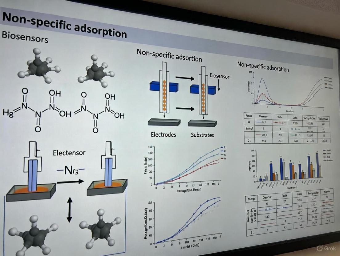
Non-specific adsorption (NSA), often termed biofouling, represents a fundamental challenge in the development and deployment of reliable biosensors. It refers to the undesirable accumulation of non-target molecules (e.g., proteins, cells, or other biomolecules) from a sample matrix onto the sensor's interface [1] [2]. This phenomenon is distinct from the specific, selective binding between a bioreceptor (like an antibody) and its target analyte. The primary consequence of NSA is the generation of a false-positive signal or the masking of a true positive signal, severely compromising the biosensor's sensitivity, specificity, and reproducibility [2]. In complex samples such as blood, serum, or milk, which contain a high concentration of various proteins and other biomolecules, the impact of NSA is particularly pronounced, posing a major barrier to the widespread adoption of biosensors in clinical diagnostics, food safety, and environmental monitoring [1].
The analytical problem extends beyond simple signal interference. NSA can lead to signal drift over time, passivate the sensing interface, restrict the conformational freedom of immobilized bioreceptors (such as structure-switching aptamers), and ultimately cause the degradation of the biosensor's coating [1]. Effectively addressing NSA requires a deep understanding of its physicochemical origins, which predominantly stem from two primary adsorption mechanisms: physisorption and chemisorption.
Fundamental Mechanisms: Physisorption vs. Chemisorption
The accumulation of non-target species on a biosensor surface occurs through two main types of interactions: physical adsorption (physisorption) and chemical adsorption (chemisorption). A clear distinction between these mechanisms is critical for designing effective antifouling strategies. Table 1 provides a comparative summary of their characteristics.
Table 1: Characteristics of Physisorption and Chemisorption in Biosensor NSA
| Characteristic | Physisorption | Chemisorption |
|---|---|---|
| Interaction Forces | van der Waals, electrostatic, hydrophobic, hydrogen bonding [1] [2] | Covalent or ionic bonding [2] |
| Binding Energy | Weak (typically < 50 kJ/mol) | Strong (typically > 50 kJ/mol) |
| Reversibility | Often reversible by changes in buffer, shear forces, or washing [2] | Largely irreversible under normal sensor operation conditions |
| Specificity | Non-specific | Can be more specific to surface chemistry |
| Typical Foulants | Proteins, lipids, polysaccharides via hydrophobic or ionic patches [1] | Molecules forming covalent bonds with surface functional groups |
| Temperature Dependence | May decrease with increasing temperature | Often increases with increasing temperature (activated process) |
Physisorption
Physisorption is the most common mechanism behind NSA in biosensing. It is driven by non-covalent, intermolecular forces and does not involve the sharing or transfer of electrons between the adsorbate and the sensor surface [2]. The combined effect of these weak forces can lead to substantial and problematic fouling, especially in complex biological fluids. The weaker nature of physisorption also means it can sometimes be addressed by active removal methods that generate surface shear forces to overpower the adhesive interactions [2].
Chemisorption
Chemisorption involves the formation of chemical bonds between the foulant molecules and the functional groups on the sensor's surface. This process is characterized by a higher binding energy and is typically irreversible under the mild conditions used for biosensor operation [2]. While chemisorption is less frequently the primary driver of NSA from complex samples compared to physisorption, it can occur with certain surface-reactive molecules. Once a molecule is chemisorbed, it is exceedingly difficult to remove without harsh chemical or physical treatments that could damage the sensor interface.
Experimental Protocols for Evaluating NSA
Quantifying and characterizing NSA is essential for diagnosing biosensor performance issues and validating the efficacy of antifouling strategies. A range of experimental techniques, from simple solution-depletion methods to sophisticated in-situ analysis platforms, are employed.
Solution-Depletion and Ex-Situ Analysis
Traditional methods involve exposing the sensor surface or a representative substrate to a solution containing a potential foulant (e.g., a protein like BSA). The amount of adsorption is determined by measuring the depletion in the foulant's concentration in the bulk solution after a set incubation time and subsequent separation (e.g., via centrifugation) [3]. The concentration can be measured using various offline (ex-situ) techniques like UV-Vis spectroscopy. A significant limitation of this approach is its inability to capture instantaneous information or adsorption kinetics, and the separation step itself may disrupt weakly adsorbed layers [3].
In-Situ Quantification Using UV-Vis Spectroscopy
Recent advancements have demonstrated the use of in-situ UV-Vis spectroscopy coupled with advanced algorithms to quantitatively monitor heterogeneous adsorption processes in real-time [3]. The following protocol is adapted from studies quantifying the adsorption of organic molecules onto suspended microparticles:
- Setup: A suspension containing the adsorbent material (e.g., polymer microparticles or nanomaterials representing the sensor coating) is placed in a cuvette within a UV-Vis spectrophotometer equipped with an integrating sphere to collect both transmitted and reflected light.
- Data Acquisition: The target foulant molecule (e.g., bisphenol A) is introduced, and the time-dependent total transmittance and total reflectance spectra of the suspension are recorded continuously.
- Data Processing - Scattering Correction: The recorded spectra contain contributions from both light absorption and scattering by the suspended particles. The Inverse Adding-Doubling (IAD) method is applied to the reflectance and transmittance data to extract the pure absorption coefficient spectra of the system, effectively isolating the absorption signal from scattering [3].
- Data Processing - Spectral Deconvolution: The resulting time-evolving pure absorption spectra are then analyzed using the Multivariate Curve Resolution-Alternating Least Squares (MCR-ALS) method. This algorithm decomposes the complex spectral dataset into the pure spectral profiles of the chemical components (e.g., free and adsorbed foulant) and their relative concentrations over time, without requiring prior reference spectra [3].
- Validation: The resolved spectral profiles from MCR-ALS can be assigned to specific molecular states (aqueous vs. adsorbed) by comparing them with theoretical spectra generated via computational chemistry simulations [3].
Gas Chromatography for VOC Adsorption Analysis
For volatile organic compounds (VOCs) or to study the adsorption and desorption characteristics of filter materials, gas chromatography (GC) provides a robust quantitative method [4].
- Adsorption: A zeolite filter (or other adsorbent) is exposed to a VOC vapor in a controlled chamber until saturation.
- Sampling: The filter is cut into a precise, small piece (e.g., 2 x 3 cm²) and immersed in a known mass of a solvent (e.g., ethanol).
- Extraction: The vial is shaken on a vortex to desorb the VOCs from the filter into the solvent.
- GC Analysis: The solution is sampled with a syringe, filtered, and injected into a Gas Chromatograph. The GC peak areas of the detected VOC and the solvent are compared.
- Quantification: The mass of VOC adsorbed is calculated using a pre-determined calibration curve that relates the GC peak area ratio (VOC to solvent) to the known mass ratio, incorporating a correction factor specific to the VOC and GC setup [4].
- Desorption Studies: To test filter reusability, the loaded filter can be heated in a furnace at various temperatures and for different durations to desorb (regenerate) the VOCs. The residual VOC amount is then quantified again using the same GC method to determine optimal regeneration conditions [4].
Visualization of NSA Mechanisms and Evaluation Workflows
The following diagrams illustrate the core concepts of NSA mechanisms and a generalized experimental workflow for its evaluation.
NSA Mechanisms and Impact on Biosensor Signals
Workflow for In-Situ NSA Evaluation
The Scientist's Toolkit: Key Reagents and Materials
Developing biosensors and studying NSA requires a suite of specialized materials and reagents. The following table details several key components used in the field.
Table 2: Key Research Reagent Solutions for NSA Studies
| Reagent/Material | Function/Description | Example Application Context |
|---|---|---|
| Blocking Proteins (BSA, Casein) | Passive NSA reduction; adsorb to vacant surface sites to prevent non-specific binding of interferents [2]. | Commonly used in ELISA and immunosensor fabrication as a post-functionalization blocking step [2]. |
| Self-Assembled Monolayers (SAMs) | Chemical surface modification; create a well-defined, ordered interface that can be engineered with specific terminal groups (e.g., oligo(ethylene glycol)) to resist protein adsorption [2]. | Used on gold or other metal transducer surfaces to create a conformal antifouling layer. |
| Zwitterionic Materials | Passive NSA reduction; form a hydrated layer via electrostatic interactions that create a thermodynamic barrier to protein adsorption [5]. | Applied as polymer brushes or surface grafts on sensor interfaces for extreme fouling resistance in complex media. |
| Zeolite Filters (ZSM-11) | Porous adsorbent material for studying adsorption/desorption kinetics and isotherms of volatile species [4]. | Used as a model system in GC-based protocols to quantify VOC adsorption and filter regeneration efficiency [4]. |
| Polyamide Microparticles | A model adsorbent with well-characterized properties for studying heterogeneous adsorption processes in aqueous suspension [3]. | Used in in-situ UV-Vis spectroscopic studies to quantify the adsorption kinetics of organic molecules like bisphenol A [3]. |
| Avidin-Biotin System | Immobilization strategy; provides a universal, high-affinity linkage for attaching biotinylated bioreceptors to sensor surfaces, often improving orientation and activity [6]. | A common intermediate layer in optical fiber and SPR biosensors to immobilize antibodies or nucleic acid probes [6]. |
Non-specific adsorption (NSA) is a persistent challenge that negatively affects biosensors by decreasing their sensitivity, specificity, and reproducibility [2]. This phenomenon, also known as non-specific binding or biofouling, occurs when molecules adsorb indiscriminately to a sensor's surface through physicochemical interactions, resulting in high background signals that are often indistinguishable from specific binding events [2]. For biosensors, particularly those used in diagnostic biomarker protein detection, NSA can lead to false-positive signals that adversely affect the dynamic range, limit of detection, reproducibility, and selectivity [2]. The reduction of NSA is therefore crucial in biosensor development, especially for point-of-care clinical diagnostics where accuracy and reliability are paramount [2].
NSA fundamentally arises from physisorption (physical adsorption), which results from intermolecular forces including hydrophobic forces, ionic interactions, van der Waals forces, and hydrogen bonding, rather than chemisorption which involves chemical covalent bonding [2]. When biosensor surfaces come into contact with complex biological mixtures containing proteins and other biomolecules, these physicochemical forces drive the uncontrolled adsorption of non-target species to both active and inactive sensor regions [2]. Understanding the primary forces—hydrophobic, electrostatic, and van der Waals interactions—that govern NSA is essential for developing effective strategies to mitigate its effects and improve biosensor performance.
Fundamental Forces Governing NSA
Hydrophobic Interactions
Hydrophobic interactions, also known as the hydrophobic effect, represent a fundamental driving force in non-specific adsorption processes. These interactions are entropically driven, arising from the tendency of nonpolar molecules or molecular regions to associate in aqueous environments to minimize the disruption of hydrogen bonding between water molecules [7]. In biological systems, hydrophobic interactions play an essential role in determining three-dimensional molecular structures and driving association events [7].
In the context of NSA, hydrophobic interactions occur between nonpolar residues on biomolecules (such as proteins) and hydrophobic regions on biosensor surfaces. Nucleic acids also exhibit hydrophobic character through their base-stacking forces, where the accumulation of these forces can cause nucleic acids with certain base compositions and chain lengths to display properties similar to thermal-responsive polymers [7]. The tunability of hydrophobic interactions with environmental conditions such as temperature and ionic strength makes them particularly challenging to control in biosensing applications, as these interactions can promote the irreversible adsorption of biomolecules to sensor surfaces even in the absence of specific recognition elements.
Electrostatic Interactions
Electrostatic interactions constitute another major force driving NSA in biosensing systems. These electric forces occur between charged molecules and surfaces, and are particularly relevant for biosensors operating in aqueous environments where most biomolecules carry surface charges [2] [7]. For instance, DNA possesses a negatively charged phosphate-sugar backbone that can participate in strong electrostatic attractions with positively charged molecules or surfaces [7].
In biosensing applications, electrostatic NSA typically manifests as non-specific electrostatic binding to charged surfaces [2]. This is methodologically distinct from immunological non-specificity and can significantly interfere with accurate detection signals. The prevalence of electrostatic interactions in NSA has led to the development of various mitigation strategies, including the use of charged surfactants like sodium dodecyl sulfate (SDS) and cetyl trimethyl ammonium bromide (CTAB) to electrostatically block functional groups responsible for non-specific binding [8]. These approaches aim to create a thin hydrophilic and non-charged boundary layer to prevent protein adsorption through electrostatic attractions [2].
van der Waals Forces
van der Waals forces represent a class of weak intermolecular forces that include dipole-dipole interactions, dipole-induced dipole interactions, and London dispersion forces. While individually weak, these forces collectively contribute significantly to NSA, particularly at nanoscale separations where their influence becomes more pronounced [2] [9]. In biosensing systems, van der Waals interactions facilitate the initial approach and temporary adhesion of biomolecules to sensor surfaces, potentially leading to more permanent adsorption through other force mechanisms.
The emergence of van der Waals (vdW) materials in nanophotonic biosensing has highlighted both the challenges and opportunities associated with these interactions [10]. Low-dimensional vdW materials can harness tightly confined polaritonic waves to deliver unique advantages for biosensing, but他们也 also present surfaces prone to NSA through van der Waals interactions [10]. For example, graphene surfaces can facilitate nonspecific binding via π-stacking, a form of van der Waals interaction that occurs between aromatic systems [10]. This ease of attachment via π-stacking implies increased nonspecific binding of interferents from biological samples, necessitating proper blocking procedures in biosensor design [10].
Table 1: Comparative Analysis of Primary Physicochemical Forces in NSA
| Force Type | Strength Range | Distance Dependence | Key Characteristics | Impact on NSA |
|---|---|---|---|---|
| Hydrophobic | Moderate to Strong | Long-range (nm scale) | Entropy-driven; enhanced in aqueous environments | High; causes irreversible adsorption |
| Electrostatic | Strong (in low ionic strength) | Long-range (1/r) | Highly dependent on pH and ionic strength | Moderate to High; significant for charged molecules |
| van der Waals | Weak (0.5-5 kcal/mol) | Short-range (1/r⁶) | Always present; operates at short distances | Moderate; facilitates initial adhesion |
Experimental Characterization of NSA Forces
Quantitative Measurement Techniques
Characterizing the individual contributions of different physicochemical forces to NSA requires specialized experimental approaches that can probe interactions at the molecular level. Several quantitative methods have been developed to measure these forces directly:
Binding isotherm analysis provides fundamental insights into NSA forces by measuring the adsorption capacity of surfaces as a function of analyte concentration. Studies comparing molecularly imprinted polymers (MIPs) with non-imprinted polymers (NIPs) have demonstrated higher adsorption capacity in MIPs due to specific cavities, while also revealing the extent of non-specific binding through comparative analysis [8]. This approach allows researchers to quantify the relative contributions of specific and non-specific binding events.
Kinetic adsorption studies further elucidate the role of different forces in NSA by examining the time-dependent adsorption behavior of molecules to sensor surfaces. These studies have demonstrated the efficacy of surfactant modifications (SDS or CTAB) in selectively reducing non-specific adsorption while preserving specific recognition capabilities [8]. By analyzing adsorption rates and equilibrium states, researchers can distinguish between rapidly-established non-specific interactions and slower specific binding processes.
Surface plasmon resonance (SPR) instruments represent another powerful tool for measuring binding kinetics and affinities resulting from NSA forces [10]. State-of-the-art SPR instruments can achieve limits of detection comparable to ELISA and are increasingly applied to clinical analysis of patient biofluids [10]. The refractive index-sensing transduction mechanism of SPR eliminates the need for labeling and washing steps while providing real-time kinetic information valuable for characterizing NSA forces [10].
Technical Protocols for Force Analysis
Protocol 1: Isotherm Analysis for NSA Quantification
This protocol describes how to generate and analyze binding isotherms to quantify NSA:
- Surface Preparation: Prepare sensing surfaces with and without specific recognition elements (e.g., MIPs and NIPs) [8]
- Sample Preparation: Prepare a concentration series of the target analyte (e.g., sulfamethoxazole) in relevant buffer solutions [8]
- Incubation: Expose surfaces to each analyte concentration for a fixed duration under controlled temperature and agitation
- Measurement: Quantify adsorbed analyte using appropriate detection methods (spectrophotometry, radiometry, etc.)
- Data Analysis: Plot adsorbed amount versus concentration and fit with appropriate models (Langmuir, Freundlich)
- NSA Calculation: Calculate non-specific binding by subtracting MIP binding from NIP binding at each concentration [8]
Protocol 2: Surfactant Modification for Electrostatic NSA Suppression
This protocol details the use of charged surfactants to mitigate electrostatic-driven NSA:
- Surface Selection: Select MIPs with known external functional groups (e.g., poly(4-vinylpyridine) or polymethacrylic acid) [8]
- Surfactant Pairing: Pair anionic surfaces with cationic surfactants (CTAB) and cationic surfaces with anionic surfactants (SDS) [8]
- Modification Process: Incubate MIPs with surfactant solutions at optimal concentrations determined through preliminary testing
- Characterization: Validate surfactant modification through binding isotherm analysis and comparison with unmodified controls
- Performance Assessment: Test modified MIPs in complex matrices to confirm NSA reduction while maintaining specific binding [8]
Table 2: Experimental Techniques for Characterizing NSA Forces
| Technique | Force Sensitivity | Information Obtained | Limitations | Applications |
|---|---|---|---|---|
| Binding Isotherm Analysis | All forces collectively | Adsorption capacity, affinity constants | Cannot distinguish individual force types | Surface characterization, NSA quantification |
| SPR Kinetics | High for electrostatic | Real-time binding rates, affinity constants | Requires specialized equipment | Drug discovery, biomarker detection |
| FTIR Spectroscopy | Hydrogen bonding, hydrophobic | Molecular-level interaction information | Complex data interpretation | Material characterization, mechanism studies |
| Competitive Adsorption | Hydrophobic, electrostatic | Binding specificity, surface blocking efficacy | Indirect measurement | Method development, optimization |
Force Relationships and Experimental Workflows
The following diagram illustrates the interconnected nature of different physicochemical forces in NSA and the experimental approaches used to characterize them:
The Scientist's Toolkit: Research Reagent Solutions
Table 3: Essential Reagents for NSA Force Research and Their Applications
| Reagent/Category | Specific Examples | Primary Function in NSA Research | Force Target |
|---|---|---|---|
| Blocking Proteins | BSA, Casein, Milk Proteins | Physical barrier to occupy NSA sites | Hydrophobic, Electrostatic |
| Charged Surfactants | SDS, CTAB | Electrostatic blocking of functional groups | Electrostatic |
| Functional Monomers | MAA, 4-Vinylpyridine | MIP creation with specific cavities | All forces |
| vdW Materials | Graphene, Antimonene | Enhanced sensing with polaritonic waves | van der Waals |
| Nucleic Acid Probes | Aptamers, ssDNA, dsDNA | Molecular recognition elements | Hydrophobic, Electrostatic |
| Surface Coatings | PEG, Zwitterionic polymers | Create hydrophilic non-fouling surfaces | Hydrophobic |
Emerging Trends and Future Perspectives
Recent advances in understanding and controlling NSA forces have shifted from traditional passive methods toward more sophisticated active removal approaches and advanced material solutions [2]. The development of van der Waals material-based sensors represents a promising frontier, where the reduced dimensionality of these materials enhances plasmonic field confinement while their much-reduced dielectric screening confers sensitive electrostatic tunability [10]. These materials enable the excitation of different polariton modes including plasmons, excitons, and phonons for new sensing modalities that can potentially circumvent traditional NSA challenges [10].
The integration of molecularly imprinted polymers (MIPs) with nanozymes forms hybrid nanozyme@MIP systems that combine catalytic efficiency with molecular recognition while addressing NSA concerns [11]. These advanced materials exhibit enhanced selectivity and sensitivity, enabling their application in diverse biosensing platforms including colorimetric, fluorescence, and electrochemical assays [11]. Key challenges being addressed in current research include the trade-off between selectivity and catalytic activity, non-specific adsorption reduction, and optimization for complex matrices [11].
Future directions in NSA force management will likely focus on multi-force suppression strategies that simultaneously address hydrophobic, electrostatic, and van der Waals interactions through sophisticated surface engineering and material design. The incorporation of artificial intelligence-assisted data analysis and the development of standardized protocols will further enhance our ability to predict and control NSA across diverse biosensing platforms and application environments [12] [13]. As these technologies mature, researchers, scientists, and drug development professionals will have access to increasingly powerful tools for overcoming the persistent challenge of non-specific adsorption in biosensor applications.
Non-specific adsorption (NSA) represents a fundamental challenge in the development and deployment of reliable biosensors. NSA refers to the accumulation of species other than the target analyte on the biosensing interface, a phenomenon that significantly compromises analytical performance [1]. In complex matrices such as blood, serum, or milk, numerous biological components including proteins, lipids, and cells can adhere to sensor surfaces through various physical and chemical interactions, leading to false readings, reduced sensitivity, and signal instability [1] [14]. The COVID-19 pandemic has highlighted that no diagnostic tool is infallible, with false positives and false negatives occurring even in AI-powered biosensors, underscoring the critical importance of addressing NSA for accurate clinical diagnostics [15].
The persistence of NSA as a barrier to widespread biosensor adoption is evidenced by substantial ongoing research efforts aimed at understanding and mitigating its effects [1]. As biosensors continue to evolve toward point-of-care testing and continuous monitoring applications, the ability to maintain performance in real-world sample matrices becomes increasingly crucial. This technical review examines the multifaceted impact of NSA on biosensor performance, with particular focus on its consequences for diagnostic reliability, and discusses established and emerging strategies for its minimization.
Fundamental Mechanisms of NSA
Physicochemical Basis of NSA
The accumulation of non-target sample components on biosensor surfaces occurs primarily through physical adsorption, facilitated by a combination of electrostatic interactions, hydrophobic interactions, hydrogen bonds (or other dipole-dipole interactions), and van der Waals interactions between the interface and components of the sample matrix [1]. The relative contribution of each interaction type depends on the chemical properties of both the sensor surface and the foulant molecules present in the sample.
The complex, multilayered initiative to understand and minimize NSA must address: (1) the foulant-containing sample, (2) the interaction between the sample matrix and the interface, and (3) the nature and coating of the biosensor surface [1]. This comprehensive approach must also consider the intended biosensor application and operational setup, including whether operation will occur under static or hydrodynamic conditions, in vivo or in vitro, for single use or repetitive measurements, and whether the measurement protocol incorporates washing steps.
Progression of Fouling at the Biosensor Interface
Table 1: Stages of NSA Impact on Biosensor Performance
| Stage | Time Frame | Primary Effects | Consequences |
|---|---|---|---|
| Initial Exposure | Seconds to minutes | Rapid adsorption of prevalent proteins | Formation of conditioning film that alters surface properties |
| Intermediate Fouling | Minutes to hours | Accumulation of additional matrix components | Steric hindrance of bioreceptor-target binding |
| Long-Term Degradation | Hours to days | Passivation layer formation, possible biofilm initiation | Significant signal drift, reduced sensitivity, potential false negatives |
Initially, when a biosensor is exposed to a complex sample, molecules with high surface affinity (such as abundant proteins in serum) rapidly adsorb to the interface, forming a conditioning film [1]. This initial layer can fundamentally alter the surface characteristics, potentially increasing its attractiveness to other foulants. Over time, this accumulation progresses, leading to the various performance issues detailed in subsequent sections.
Impact of NSA on Biosensor Performance Parameters
Induction of False Positive Results
False positives represent one of the most clinically significant consequences of NSA in biosensing. In surface plasmon resonance (SPR) biosensors, for instance, the adsorption of foulant molecules and the specific binding of the target analyte produce similar changes in reflectivity [1]. NSA therefore contributes directly to the amplitude of the analytical signal, compromising its correlation with the actual concentration of the target analyte and leading to potential false positive diagnoses.
In electrochemical biosensors, fouling has dramatic effects on the characteristics of the sensing interface and the rate of electron transfer at the electrode surface [1]. Non-specifically adsorbed molecules may undergo redox reactions at applied potentials, generating faradaic currents that are indistinguishable from those produced by the target analyte. Similarly, in catalytic biosensors, the electrochemical transformation of adsorbed sample components can mask signals originating from the enzymatic reaction of interest [1].
Reduction in Analytical Sensitivity
NSA diminishes biosensor sensitivity through multiple mechanisms. Adsorbed, passivating molecules or those interfering with the recognition event by inhibition or steric effects can lead to underestimation of the analyte concentration in the sample [1]. This effect is particularly pronounced at low analyte concentrations, where the specific signal is already weak and more easily masked by non-specific interactions.
In electrochemical aptamer-based (E-AB) biosensors, non-specifically adsorbed molecules may restrict the ability of structure-switching aptamers to undergo the large conformational change required for target binding and specific signal generation [1]. This steric hindrance effectively reduces the number of functional bioreceptors available for target capture, diminishing the overall signal response even when the target analyte is present at clinically relevant concentrations.
Signal Drift and Instability
Signal drift represents a persistent challenge in biosensor operation, particularly for continuous monitoring applications. NSA contributes significantly to this phenomenon through the progressive accumulation of foulants on the sensing interface over time [1]. In the short term, the contribution of NSA to biosensor signal might be negligible due to intrinsic detection mechanisms or implemented drift correction measures. However, over extended operational periods, progressing fouling leads to significant degradation of the biosensor surface that can no longer be adequately addressed by correction algorithms [1].
The impact of sensor drift on diagnostic reliability was clearly demonstrated in electronic nose (E-Nose) studies, where significant drift was observed after just two days of measurement despite blowing procedures to maintain baseline [16]. This temporal variation in sensor output affected diagnostic algorithms, compromising the accuracy of disease-specific detection models until appropriate drift correction methodologies were implemented.
Quantitative Assessment of NSA Impacts
Comparative Performance Degradation Across Biosensor Platforms
Table 2: Quantitative Impact of NSA on Different Biosensor Types
| Biosensor Type | Primary Signal Interference | Reported Sensitivity Loss | False Positive Rate Increase |
|---|---|---|---|
| Electrochemical Aptamer-Based (E-AB) | Steric hindrance of conformation change | Up to 70% signal reduction in complex media | Significant due to non-faradaic currents |
| SPR Immunosensors | Reflectivity changes from foulants | EC₅₀ shifts of 1-2 orders of magnitude | >30% in undiluted serum samples |
| Electrochemical Enzyme Biosensors | Substrate diffusion barrier; enzyme inhibition | 40-60% current reduction | Variable, depends on interferents |
| Lateral Flow Immunoassays | Matrix effects (proteins, fats) | LOD increase from 0.006 to 0.184 ng/mL for aflatoxins [14] | Visible background coloration |
The data in Table 2 illustrates the significant and variable impact of NSA across different biosensing platforms. The sensitivity loss is particularly pronounced in systems relying on conformational changes of bioreceptors or enzymatic reactions, where even minor surface fouling can dramatically impact function. The increase in false positive rates highlights the critical importance of NSA mitigation for clinical diagnostic applications where treatment decisions depend on accurate results.
Methodologies for NSA Evaluation
Understanding the dimension of NSA requires appropriate evaluation methods. The perceived fouling is strictly related to the sensitivity of the method used for its evaluation, and a combination of analytical methods typically provides better insight than a single method [1]. Commonly employed techniques include:
- Surface Plasmon Resonance (SPR): Provides real-time, label-free monitoring of adsorption processes with high sensitivity to surface mass changes.
- Electrochemical Impedance Spectroscopy (EIS): Sensitive to changes in interfacial properties and charge transfer resistance resulting from surface fouling.
- Quartz Crystal Microbalance (QCM): Measures mass uptake on surfaces with nanogram sensitivity.
- Fluorescence Microscopy: Allows visualization of protein adsorption when fluorescently labeled foulants are used.
Each method offers distinct advantages and limitations, with sensitivity ranges spanning from ng/cm² for QCM to sub-monolayer detection for SPR. Coupled electrochemical-surface plasmon resonance (EC-SPR) biosensors offer particularly interesting opportunities for comprehensive NSA evaluation as they enable larger detection ranges, improved spatial resolution, and more detailed information on interfacial, catalytic, and affinity binding events compared to single detection procedures [1].
Experimental Approaches to NSA Investigation
Standardized Protocol for NSA Assessment
A generalized experimental workflow for evaluating NSA in biosensors involves several critical stages. First, the biosensor surface is prepared with appropriate functionalization and bioreceptor immobilization. Baseline measurements are then recorded in pure buffer solution to establish the initial signal. The sensor is subsequently exposed to the complex sample matrix (e.g., serum, blood, milk) or simplified model foulant solutions for a predetermined period. After exposure, the sensor undergoes carefully controlled washing steps to remove loosely bound material, followed by post-exposure measurement. The difference between pre- and post-exposure signals provides a quantitative measure of NSA [1].
Superficial protocols represent a significant limitation in NSA studies. Comprehensive evaluation requires testing under conditions that closely mimic the intended operational environment, including relevant foulant concentrations, flow conditions, temperature, and exposure duration. The resistance to fouling must be adapted to particular static or hydrodynamic operational conditions, different time lengths, and samples with various pH levels and ionic strengths and complex compositions [1].
Research Reagent Solutions for NSA Studies
Table 3: Essential Reagents for NSA Investigation and Mitigation
| Reagent Category | Specific Examples | Primary Function | Application Notes |
|---|---|---|---|
| Antifouling Polymers | Polyethylene glycol (PEG), Zwitterionic polymers | Form hydration barrier preventing protein adsorption | PEG density and molecular weight critical for efficacy |
| Surface Blockers | Bovine serum albumin (BSA), Casein, Salmon sperm DNA | Passivate unused binding sites on sensor surface | Can introduce background in certain detection methods |
| Surfactants | Tween-20, Triton X-100 | Reduce hydrophobic interactions driving NSA | Concentration optimization essential to avoid bioreceptor denaturation |
| Stabilizing Additives | Sucrose, Trehalose, Glycerol | Maintain bioreceptor activity in complex media | Particularly important for enzymatic biosensors |
| Reference Sensors | Backfilling thiols (EG6), Deactivated bioreceptors | Distinguish specific from non-specific binding | Crucial for quantitative NSA assessment in real-time sensing |
The reagents listed in Table 3 represent foundational tools for both studying and mitigating NSA effects. Recent advances have expanded these traditional categories to include new peptide-based antifouling agents, cross-linked protein films, and hybrid materials with tunable conductivity, thickness, and functional groups [1]. The optimal combination of these reagents depends strongly on the specific biosensing platform, detection mechanism, and sample matrix.
Emerging Solutions and Future Perspectives
Advanced Antifouling Strategies
Recent research has focused on developing increasingly sophisticated antifouling strategies that extend beyond traditional surface blocking approaches. For electrochemical biosensors, developments in the last five years include new peptides, cross-linked protein films, and hybrid materials [1]. These materials are designed to create surfaces that are inherently resistant to protein adsorption while maintaining the conductivity necessary for electrochemical transduction.
For SPR and combined EC-SPR biosensors, promising antifouling solutions include zwitterionic materials, which demonstrate exceptional resistance to non-specific protein adsorption due to their strong hydration via electrostatic interactions [1] [5]. Other approaches utilize molecular simulations and machine learning-assisted evaluations to design and screen new antifouling materials with optimized properties [1].
Material Science Innovations
Nanomaterial innovations continue to drive progress in NSA mitigation. Graphene and its derivatives show particular promise due to their unique properties, including exceptional electrical conductivity, mechanical strength, and high surface area [17]. Graphene's functionalization versatility enables the creation of biosensing interfaces with enhanced antifouling properties through both covalent and non-covalent modifications [17].
Metal-organic frameworks (MOFs) represent another material class with significant potential for addressing NSA challenges. ZrFe-MOF@PtNPs nanocomposites, for instance, have demonstrated improved performance in complex samples like milk, where they help mitigate interference from proteins and fats that would otherwise cause non-specific binding in traditional lateral flow immunoassays [14].
Integrated Approaches and Future Directions
The future of NSA mitigation lies in integrated approaches that combine material innovations with advanced sensing modalities and data processing. The incorporation of artificial intelligence and machine learning shows particular promise for distinguishing specific signals from non-specific background, potentially enabling accurate biosensing even in the presence of some fouling [15] [18]. Coupled detection methods like EC-SPR offer opportunities for more sophisticated NSA correction through multimodal signal acquisition [1].
As biosensor technology continues to evolve toward point-of-care testing, wearable monitoring, and implantable devices, addressing the fundamental challenge of NSA will remain critical for translating laboratory demonstrations into clinically viable diagnostic tools. Future research directions will likely focus on developing universal functionalization strategies that provide robust antifouling protection while maintaining bioreceptor activity and compatibility with diverse transduction mechanisms.
Non-specific adsorption (NSA) represents a fundamental challenge in biosensor development, particularly when working with complex biological matrices. NSA occurs when molecules adsorb to a sensor's surface through physisorption rather than specific biorecognition, resulting in high background signals that are often indiscernible from specific binding events [2]. This phenomenon negatively affects biosensor performance by decreasing sensitivity, specificity, and reproducibility [2]. In complex matrices such as serum, cell lysate, and blood products, the diversity and concentration of interfering compounds—including proteins, lipids, and metabolites—exacerbate NSA, leading to false-positive signals, altered dynamic range, and elevated limits of detection [2].
The core of the NSA problem lies in the interplay between surface chemistry and biological components. Most biomolecular surfaces experience hindrance from non-specific species, with proteins being particularly prone to irreversible adsorption [2]. This creates a critical barrier for biosensors intended for clinical diagnostics, environmental monitoring, and food safety applications where complex samples are the norm rather than the exception [19].
Fundamental Mechanisms of NSA in Complex Environments
Molecular Interactions Driving NSA
The primary mechanisms underlying NSA involve intermolecular forces that facilitate physisorption. These include:
- Hydrophobic interactions: Non-polar regions of proteins and other biomolecules adhere to hydrophobic surfaces
- Ionic interactions: Charged molecules interact with oppositely charged surfaces
- Van der Waals forces: Weak electromagnetic forces between adjacent molecules
- Hydrogen bonding: Polar interactions between hydrogen donors and acceptors [2]
In complex matrices, these interactions occur not only with the sensor surface but also with previously adsorbed molecules, leading to multilayered fouling that further complicates detection.
Matrix-Specific Challenges
Different biological matrices present distinct NSA challenges:
Serum: Contains high concentrations of albumin, immunoglobulins, and fibrinogen that compete with target analytes for surface binding sites. The protein-rich nature of serum makes it particularly prone to rapid surface fouling [2].
Cell Lysate: Comprises intracellular components including enzymes, nucleic acids, metabolites, and membrane fragments. The metabolic pathways active in lysate-based systems can deplete reporter molecules, as demonstrated in E. coli lysate where endogenous glycolytic activity rapidly consumed glucose [20].
Blood Products: Contain cellular components, platelets, and coagulation factors that adhere to surfaces. Hemoglobin from lysed red blood cells can generate strong background signals in colorimetric assays [2].
Quantitative Impact of NSA on Biosensor Performance
The consequences of NSA can be quantified through specific performance metrics, as demonstrated across multiple studies.
Table 1: Quantitative Impacts of NSA on Biosensor Performance
| Performance Metric | Impact of NSA | Experimental Evidence |
|---|---|---|
| Limit of Detection | Increases | Fabric-based E. coli sensor achieved 537 CFU/mL despite complex sample matrix [19] |
| Detection Time | Increases | Minimum detection time of 20 minutes reported for fabric-based biosensor [19] |
| Signal-to-Noise Ratio | Decreases | Non-specific binding creates background signals indistinguishable from specific binding [2] |
| Reproducibility | Decreases | Variability in surface fouling leads to inconsistent results between samples [2] |
Methodologies for Investigating and Mitigating NSA
Experimental Protocols for NSA Characterization
Surface Plasmon Resonance (SPR) Protocol:
- Functionalize gold sensor chip with biorecognition elements
- Establish baseline signal with buffer solution
- Introduce complex sample (serum, lysate, or blood product)
- Monitor association phase for 5-10 minutes
- Switch to buffer flow to monitor dissociation phase
- Quantify irreversible adsorption by comparing pre- and post-sample baselines [2]
Microfluidic Biosensor Protocol:
- Chemically modify microchannel surfaces (e.g., with PEG silane)
- Introduce fluorescently-labeled complex sample at controlled flow rates
- Image adsorption patterns using fluorescence microscopy
- Quantify surface coverage using image analysis software
- Apply shear forces by increasing flow rate to assess adhesion strength [2]
Cell-Free Expression Biosensor Protocol:
- Prepare bacterial lysate containing genetic circuits for reporter expression
- Add serum samples spiked with target analytes
- Incubate for 45 minutes for LacZ production
- Terminate transcription with naproxen-lactose mixture
- Incubate for 15 minutes for glucose production via LacZ activity
- Measure glucose output using personal glucose monitor [20]
Research Reagent Solutions for NSA Challenges
Table 2: Essential Reagents for NSA Mitigation in Complex Matrices
| Reagent/Chemical | Function in NSA Reduction | Application Context |
|---|---|---|
| Bovine Serum Albumin (BSA) | Blocking agent that occupies vacant surface sites | ELISA, Western blotting, microfluidic biosensors [2] |
| Casein | Protein blocker that reduces non-protein interactions | Enzyme-based assays, immunohistochemistry [2] |
| Self-Assembled Monolayers (SAMs) | Create controlled surface chemistry with reduced stickiness | Electrochemical sensors, surface-based detection [2] |
| Poly(ethylene glycol) Derivatives | Form hydrated barrier preventing protein adsorption | SPR sensors, implantable devices, marine equipment [2] |
| Naproxen-Lactose Mixture | Terminates cell-free reactions while enabling reporter readout | CFE-based biosensors with personal glucose monitor detection [20] |
| β-glucuronidase Substrate (MUG) | Enzyme substrate that generates fluorescent signal upon target recognition | Fabric-based biosensors for E. coli detection [19] |
Advanced Solutions for NSA Reduction in Complex Matrices
Passive Reduction Methods
Passive methods focus on preventing undesired adsorption by coating surfaces with anti-fouling materials. The goal is to create a thin hydrophilic and non-charged boundary layer that thwarts protein adsorption [2]. These methods include:
Physical Blocking: Using proteins like serum albumins (BSA), casein, and other milk proteins to occupy vacant surface sites [2]. These blockers are particularly effective for ELISA, Western blotting, and other enzyme-based assays [2].
Chemical Modification: Employing self-assembled monolayers (SAMs), poly(ethylene glycol) derivatives, and other synthetic polymers to create surfaces that resist protein adsorption through steric repulsion and hydration forces [2].
Active Removal Methods
Active methods dynamically remove adsorbed molecules after surface fouling has occurred:
Electromechanical Transducers: Generate surface forces to shear away weakly adhered biomolecules through piezoelectric or electrostatic actuation [2].
Acoustic Wave Devices: Utilize surface acoustic waves to create mechanical vibrations that dislodge non-specifically bound molecules [2].
Hydrodynamic Removal: Relies on controlled fluid flow in microfluidic channels to generate shear forces that overpower adhesive forces [2].
Innovative Approaches for Complex Matrices
Metabolic Decoupling: In cell-free expression systems, researchers have successfully decoupled reporter enzyme production from glucose conversion to overcome endogenous glycolytic activity in E. coli lysate that depletes signal [20]. This approach enables one-pot removal of confounding glucose present in complex samples like human serum without customizing reagent volumes to individual samples [20].
Fabric-Based Biosensors: Cotton fabric platforms provide inherent advantages for complex sample analysis due to their flexibility, mechanical robustness, and ease of functionalization [19]. These sensors can be chemically modified to load targeting substrate molecules that produce color changes in response to specific enzymes secreted by pathogens like E. coli [19].
Computational Prediction Tools: Methods like OmicSense use multidimensional omics data to build prediction models that are robust against background noise, enabling accurate biosensing even with noisy biological data [13]. This approach uses a mixture of Gaussian distributions as probability distribution, yielding the most likely objective variable predicted for each biomarker [13].
The challenge of non-specific adsorption in complex matrices remains a significant hurdle in biosensor development, particularly for applications requiring analysis of serum, cell lysate, and blood products. The interplay between surface chemistry and biological components creates a demanding environment where NSA can compromise detection limits, specificity, and reliability. However, advanced strategies including metabolic decoupling, fabric-based sensors, and computational prediction methods offer promising avenues for overcoming these challenges. As biosensor technology continues to evolve, the integration of multiple NSA reduction approaches—combining passive surface modifications with active removal mechanisms—will be essential for developing robust detection platforms capable of operating in real-world complex matrices.
Strategies to Combat NSA: From Surface Chemistry to Active Removal Methods
In the field of biosensing, non-specific adsorption (NSA) presents a fundamental challenge that critically compromises sensor performance. NSA occurs when molecules other than the target analyte, such as proteins, DNA, or other biomolecules, adhere to the biosensor's surface through physisorption mechanisms like hydrophobic forces, ionic interactions, van der Waals forces, and hydrogen bonding [2]. This phenomenon results in elevated background signals that are often indistinguishable from specific binding events, leading to false-positive responses, reduced sensitivity and specificity, compromised reproducibility, and an increased limit of detection [2] [21]. For affinity-based biosensors, which rely on specific bioreceptor-analyte binding (e.g., antibody-antigen), the methodological non-specificity can arise from surface protein denaturation, mis-orientation, substrate stickiness, non-specific electrostatic binding to charged surfaces, and adsorption of molecules in free spaces not occupied by the bioreceptor [2]. The persistent nature of NSA has driven extensive research into surface modification strategies aimed at creating bio-inert interfaces that resist fouling, thereby improving the reliability and accuracy of biosensors, particularly for critical applications such as point-of-care clinical diagnostics [2].
Passive blocking methods constitute a primary defense against NSA by creating a permanent or semi-permanent barrier on the biosensor surface. These methods operate on the principle of preventing undesired adsorption through coating the surface with materials that minimize intermolecular interactions with non-target species [2]. Unlike active removal methods that dynamically generate surface forces to shear away adsorbed molecules, passive techniques are typically applied during sensor fabrication or preparation and remain functional throughout the assay [2]. Passive methods can be broadly categorized into two groups: protein-based blockers and synthetic chemical coatings. Protein blockers like Bovine Serum Albumin (BSA) and casein function by occupying vacant sites on the sensor surface through their own adsorption, thereby preventing subsequent non-specific binding of interferents [2] [21]. Synthetic coatings, such as poly(ethylene glycol) and zwitterionic polymers, create a thermodynamically unfavorable, hydrophilic, and charge-neutral interface that resists the initial adsorption of biomolecules through the formation of a tightly bound hydration layer [22] [23] [24]. The selection between these approaches depends on factors including the sensor platform, sample matrix, target analyte, and required sensitivity.
Protein-Based Blocking Agents
Bovine Serum Albumin (BSA)
Bovine Serum Albumin is one of the most extensively utilized protein blocking agents in biosensor development and immunoassays. This 66.5 kDa protein functions by adsorbing to vacant sites on the sensor surface, forming a physical barrier that prevents subsequent non-specific binding of interferents present in complex samples [21]. The effectiveness of BSA stems from its ability to cover hydrophobic and charged surfaces, thereby reducing available sites for unwanted protein adsorption. In practice, BSA is typically applied at concentrations ranging from 1% to 2% in buffer solutions, often supplemented with surfactants like Tween 20 to enhance its blocking efficiency [21]. However, a significant limitation of BSA is its potential for cross-reactivity against certain hapten-conjugates, which can occasionally contribute to background signals rather than reducing them [21]. Studies optimizing blocking agents for electrochemical biosensors targeting ovarian cancer biomarker miRNA-204 demonstrated that 1% BSA in Tween 20 provided good blocking characteristics, though it was outperformed by gelatin-based formulations in some specific applications [21].
Casein and Milk Protein Blends
Casein, along with other milk-derived proteins, represents another class of protein-based blocking agents commonly employed in diagnostic assays such as ELISAs and Western blots [2]. These proteins function similarly to BSA by adsorbing to surfaces and creating a protective layer against non-specific binding. The primary advantage of casein lies in its lack of cross-reactivity compared to BSA, making it preferable for certain applications where immunological interference is a concern [21]. However, a notable disadvantage is casein's tendency to potentially block specific surface binding regions required for analyte detection if not properly optimized [21]. Experimental evidence from BLI studies has shown that casein (at 0.2% concentration) can sometimes produce even larger NSB signals compared to assays without any blocking agent, highlighting the importance of empirical optimization for each specific biosensor platform [25]. This counterproductive effect underscores the critical need for thorough testing and validation of blocking conditions rather than relying on standardized protocols.
Experimental Protocol for Protein Blocker Optimization
The following protocol provides a methodology for evaluating and optimizing protein-based blocking agents for electrochemical biosensors, based on approaches documented in the literature [21]:
Surface Preparation: Begin with functionalized biosensor surfaces (e.g., carbon screen-printed electrodes modified with citrate-reduced gold nanoparticles and immobilized with specific capture probes such as 5'-amine modified ssDNA).
Blocking Solution Preparation: Prepare candidate blocking solutions:
- 1-2% BSA: Dissolve 1-2 g of BSA in 100 mL of 0.01 M phosphate-buffered saline (PBS, pH 7.4) containing 0.05% Tween 20.
- 1% Gelatin: Dissolve 1 g of gelatin in 100 mL of 0.01 M PBS (pH 7.4) containing 0.05% Tween 20.
- Alternative Buffers: Consider including surfactants like Triton X-100 or specific buffers such as HEPES in the screening process.
Blocking Procedure: Apply 50-100 μL of each blocking solution to the prepared sensor surfaces and incubate for 1 hour at room temperature in a humidified chamber to prevent evaporation.
Washing: Gently rinse the sensors three times with 0.01 M PBS (pH 7.4) to remove excess blocking agent.
Performance Evaluation:
- Chronoamperometry: Test blocked sensors by spiking target analyte (e.g., miRNA-204) into both simple buffer (0.01 M PBS) and complex matrix (fetal bovine serum). Record the saturation current.
- Data Analysis: Calculate the difference in saturation current between measurements in PBS and FBS for each blocking agent. A smaller difference indicates superior blocking efficiency against non-specific binding in complex matrices.
- Interference Testing: Challenge the biosensor with a cocktail of potential interferents (e.g., non-target miRNAs, DNA, proteins) to confirm specific signal retention.
Diagram: Experimental workflow for optimizing protein-based blocking agents on biosensors.
Synthetic Chemical Coatings
Poly(Ethylene Glycol) and Derivatives
Poly(ethylene glycol) represents one of the most established synthetic polymers for creating anti-fouling surfaces in biosensing applications. PEG operates through the formation of a hydration layer and the imposition of a steric barrier that entropically discourages protein adsorption [24]. The molecular weight and chain conformation of PEG significantly influence its antifouling performance, with shorter chains forming densely packed monolayers while longer chains may undergo bending and become less effective [21]. Comparative studies between PEG and zwitterionic polymers have revealed that while both provide substantial antifouling properties, they differ in their performance characteristics. Research on wearable microprojection arrays for biomarker capture demonstrated that PEG coatings effectively reduced non-specific adsorption in single protein solutions, diluted plasma, and when applied to skin tissue [24]. However, a critical limitation of conventional PEG coatings is their susceptibility to oxidative degradation over time, which can compromise long-term stability in biosensing applications [23]. This has motivated the development of PEG derivatives and alternative synthetic coatings with improved stability profiles.
Zwitterionic Polymers
Zwitterionic polymers have emerged as a highly effective class of antifouling materials that surpass PEG in certain applications. These polymers contain both positive and negative charged groups within the same monomer unit, creating a super-hydrophilic surface that strongly binds water molecules via electrostatic interactions [22] [23]. This results in the formation of a tightly bound hydration layer that acts as a physical and energetic barrier to protein adsorption. The three major classes of zwitterionic polymers include:
- Sulfobetaine polymers: Featuring quaternary ammonium cations and sulfonate anions, offering high hydrophilicity and resistance to protein and bacterial adhesion [23].
- Carboxybetaine polymers: Containing quaternary ammonium cations with carboxylate anions, providing non-fouling properties combined with functional groups for further biomolecule conjugation [23].
- Phosphorylcholine polymers: Mimicking natural phospholipid headgroups found in cell membranes, exhibiting exceptional hemocompatibility for blood-contacting devices [23].
The effectiveness of zwitterionic coatings was demonstrated in a study where a sulfobetaine-based copolymer reduced protein adsorption by approximately 67% compared to bare gold surfaces when incubated with human plasma [22]. Furthermore, electrochemical biosensors coated with this zwitterionic polymer maintained stable performance with only a 5% decrease in anodic current after incubation in 1% human serum albumin, compared to an 83% decrease observed with bare gold electrodes [22].
Application Protocols for Chemical Coatings
Surface-Initiated Atom Transfer Radical Polymerization for Zwitterionic Coatings: This method grows polymer brushes directly from the sensor surface, providing high grafting density and excellent anti-fouling performance [23].
- Surface Activation: Clean the substrate (e.g., gold, silicon) thoroughly and functionalize with initiator molecules (e.g., bromoester-based ATRP initiator) through self-assembled monolayer formation.
- Polymerization Solution: Prepare a degassed solution containing zwitterionic monomer (e.g., sulfobetaine methacrylate - SBMA), solvent (water/methanol mixture), and ATRP catalyst complex (e.g., CuBr/bipyridine).
- Grafting Process: Immerse the initiator-functionalized substrate in the polymerization solution and react under inert atmosphere (nitrogen or argon) for 2-24 hours at controlled temperature (20-40°C).
- Termination and Cleaning: Remove the substrate, rinse extensively with deionized water and ethanol to remove physisorbed polymer and catalyst residues.
- Characterization: Verify polymer brush formation and thickness using ellipsometry, and assess antifouling performance using model proteins (e.g., fibrinogen, serum albumin).
Dip-Coating Method for Zwitterionic Polymers: A simpler approach suitable for creating uniform coatings over large surface areas [23].
- Polymer Solution Preparation: Dissolve the zwitterionic polymer (e.g., copolymers containing sulfobetaine, carboxylic, aldehyde, and thiol groups) at 0.1-5 mg/mL in an appropriate solvent.
- Substrate Preparation: Clean the substrate thoroughly (e.g., oxygen plasma treatment for enhanced wettability).
- Coating Process: Immerse the substrate in the polymer solution for 1-60 minutes, then withdraw at a controlled speed (0.1-10 mm/s) to ensure uniform deposition.
- Curing: Dry the coated substrate under nitrogen flow and optionally cure at elevated temperature (40-80°C) for enhanced adhesion.
- Quality Control: Characterize coating thickness (~16 nm as reported for Zwitter-repel coating) and homogeneity using AFM or ellipsometry [22].
Comparative Performance Analysis
Quantitative Comparison of Blocking Efficiency
Table: Performance comparison of different passive blocking methods for biosensors
| Blocking Method | Reduction in Protein Adsorption | Key Advantages | Limitations | Optimal Application Context |
|---|---|---|---|---|
| BSA (1-2%) | Not quantified in results but significantly reduces background in ELISAs and electrochemical sensors [21] | Easy to apply, cost-effective, widely established | Potential cross-reactivity with certain hapten-conjugates [21] | Routine immunoassays, electrochemical biosensors in buffer-based assays |
| Casein | Variable performance; may increase NSB in some BLI applications at 0.2% [25] | Minimal cross-reactivity compared to BSA [21] | May block specific binding sites if not optimized [21] | Immunoassays where BSA shows interference |
| PEG | Comparable to zwitterions in single protein solutions and diluted plasma [24] | Well-established chemistry, effective steric hindrance | Susceptible to oxidative degradation over time [23] | Short-term biosensing applications, wearable devices |
| Zwitterionic Polymers | ~67% reduction vs. bare gold in human plasma [22] | Superior stability, salt-resistant hydration, high ligand density capability [23] [24] | More complex coating procedures required [23] | Complex biological fluids (blood, saliva), long-term implants |
Advanced Formulations and Combinatorial Approaches
Recent research has demonstrated that combinatorial approaches using multiple blocking mechanisms can achieve superior NSA reduction compared to single-component systems. A notable example comes from Biolayer Interferometry studies, where a tri-component admixture of 1% BSA, 20 mM imidazole, and 0.6 M sucrose significantly suppressed nonspecific binding across multiple protein analytes at high concentrations (>10 μM) that typically challenge conventional blockers [25]. This formulation leverages multiple mechanisms: BSA provides surface coverage, imidazole blocks specific interactions with Ni-NTA biosensors, and sucrose enhances protein solvation through osmolyte effects [25]. In another innovative approach, researchers have developed reversible blocking strategies using amphiphilic sugars like n-Dodecyl β-D-maltoside, which can be adsorbed on hydrophobic surfaces during assays and subsequently removed, enabling simplified surface preparation while maintaining anti-fouling properties [26]. These advanced formulations highlight a trend toward context-specific blocking solutions tailored to particular biosensor platforms and application environments.
The Scientist's Toolkit: Research Reagent Solutions
Table: Essential reagents and materials for implementing passive blocking methods
| Reagent/Material | Function | Example Applications | Key Considerations |
|---|---|---|---|
| Bovine Serum Albumin (BSA) | Protein-based blocker that adsorbs to vacant surface sites | ELISA, Western blot, electrochemical biosensors [21] | Use 1-2% in PBS with 0.05% Tween 20; monitor for cross-reactivity |
| Casein | Milk-derived protein blocker with low cross-reactivity | Immunoassays where BSA causes interference [21] | Optimize concentration carefully to avoid blocking specific binding sites |
| Polyethylene Glycol | Synthetic polymer creating steric hindrance and hydration layer | Coating for various biosensors, wearable devices [21] [24] | Molecular weight affects packing density; shorter chains form denser monolayers |
| Sulfobetaine Methacrylate | Zwitterionic monomer for anti-fouling polymer coatings | Biomedical implants, biosensors in complex fluids [23] | Can be polymerized via SI-ATRP; forms durable hydration layer |
| Carboxybetaine Acrylamide | Zwitterionic monomer with functionalizable groups | Biosensors requiring subsequent biomolecule conjugation [23] | Carboxylate groups allow attachment of peptides or drugs |
| n-Dodecyl β-D-maltoside | Amphiphilic sugar for reversible surface blocking | Label-free immunoassays with simplified chemistry [26] | Added directly to analyte solutions; enables non-covalent probe attachment |
Passive blocking methods utilizing protein blockers and synthetic chemical coatings represent essential tools for mitigating non-specific adsorption in biosensors. While traditional protein blockers like BSA and casein offer simplicity and effectiveness for many applications, advanced materials such as zwitterionic polymers demonstrate superior performance in challenging environments like complex biological fluids [22] [24]. The future development of passive blocking strategies will likely focus on combinatorial formulations that leverage multiple mechanisms simultaneously [25], stimuli-responsive coatings that can adapt to different environments, and high-throughput screening approaches to identify optimal blocking conditions for specific applications. Furthermore, the integration of artificial intelligence in surface design promises to accelerate the development of next-generation antifouling coatings by predicting material properties and optimizing surface-analyte interactions without extensive trial-and-error experimentation [27]. As biosensing technologies continue to advance toward more complex applications in point-of-care diagnostics and continuous monitoring, the role of sophisticated passive blocking methods will become increasingly critical for achieving the required reliability and accuracy in real-world biological samples.
The performance of a biosensor is fundamentally determined by the interactions that occur at the interface between its physical transducer and the complex biological sample it is designed to analyze. A persistent challenge in this domain is non-specific adsorption (NSA), also referred to as non-specific binding or biofouling [2]. NSA occurs when proteins, lipids, or other biomolecules physisorb onto the sensing surface through hydrophobic forces, ionic interactions, van der Waals forces, and hydrogen bonding, rather than through the specific, selective recognition mechanism the biosensor is designed for [2]. This phenomenon leads to elevated background signals that are often indistinguishable from the specific binding signal, resulting in false positives, a reduced dynamic range, an increased limit of detection, and diminished sensor reproducibility and reliability [2]. For affinity-based biosensors, such as immunosensors, which rely on the specific binding between an antibody and its target antigen, the negative impact of NSA is particularly acute [2]. Effective surface functionalization strategies to mitigate NSA are therefore not merely beneficial but essential for developing sensitive, selective, and robust biosensors, especially for point-of-care clinical diagnostics [2].
Self-Assembled Monolayers (SAMs) as Bioinert Surfaces
Fundamentals of SAMs
Self-assembled monolayers (SAMs) are highly ordered, molecularly thin films that form spontaneously when molecules with a specific affinity for a substrate are adsorbed onto its surface [28]. They serve as a versatile platform for creating well-defined interfaces with tailored chemical properties. In biosensing, SAMs are primarily used to transform a non-specific surface (like gold or metal oxides) into a bioinert surface that resists NSA, or to provide functional groups for the subsequent, oriented immobilization of biorecognition elements (e.g., antibodies, DNA strands) [28]. The formation of a dense, uniform SAM is critical to its effectiveness in creating a non-adsorptive barrier.
Key Experimental Protocols: Forming Dendrimer SAMs
Protocol: Formation of Poly(amidoamine) (PAMAM) Dendrimer SAMs on Gold [29]
- Substrate Preparation: Begin with clean, template-stripped gold substrates. Standard cleaning procedures (e.g., oxygen plasma treatment or piranha solution cleaning) are recommended to ensure a pristine, contaminant-free surface.
- SAM Formation: Immerse the gold substrate in an aqueous solution of the desired generation of PAMAM dendrimer (e.g., G2, G4, G6) for a period of 12-24 hours at room temperature.
- Rinsing and Drying: After incubation, remove the substrate from the dendrimer solution and rinse it thoroughly with ultrapure ethanol and water to remove any physisorbed molecules. Gently dry the substrate under a stream of inert gas (e.g., nitrogen).
- Characterization: The successful formation and quality of the dendrimer SAM can be confirmed through:
- Contact Angle Goniometry: A decrease in the water contact angle (e.g., from ~61° for bare gold to ~30° after dendrimer modification) indicates increased surface hydrophilicity [29].
- Surface Plasmon Resonance (SPR): Used to quantify the adsorption of proteins onto the SAM in real-time.
- X-ray Photoelectron Spectroscopy (XPS) and Fourier-Transform Infrared Reflection–Absorption Spectroscopy (FTIR-RAS): Used to verify the chemical composition and molecular structure of the formed monolayer [29].
Performance of SAMs in Reducing NSA
The efficacy of SAMs, particularly dendrimer SAMs, in resisting protein adsorption is highly dependent on the generation (size and terminal group density) of the dendrimer. The table below summarizes quantitative data on the adsorption of various proteins onto different generations of PAMAM dendrimer SAMs, as measured by SPR [29].
Table 1: Protein Adsorption on PAMAM Dendrimer SAMs of Different Generations [29]
| Surface | Lysozyme (pI ~11) | Fibrinogen (pI ~5.5) | BSA (pI ~4.7) |
|---|---|---|---|
| Bare Gold | 105.0 ng/cm² | 285.0 ng/cm² | Data Not Provided |
| G2 Dendrimer SAM | 52.5 ng/cm² | 162.5 ng/cm² | 42.5 ng/cm² |
| G4 Dendrimer SAM | 32.5 ng/cm² | 122.5 ng/cm² | 27.5 ng/cm² |
| G6 Dendrimer SAM | 22.5 ng/cm² | 72.5 ng/cm² | 12.5 ng/cm² |
The data demonstrates that as the dendrimer generation increases, protein adsorption consistently decreases, regardless of the protein's isoelectric point (pI). This highlights that the density of the terminal groups and the resulting steric repulsion and hydration layer are key factors in conferring bioinert properties, surpassing the influence of surface charge alone [29].
Polymer Brushes for Dynamic Surface Passivation
Fundamentals of Polymer Brushes
Polymer brushes are composed of long polymer chains tethered by one end to a surface at a sufficiently high density that the chains are forced to stretch away from the substrate, forming a brush-like morphology [30]. This structure creates a physical and chemical barrier that is highly effective at resisting the approach and adhesion of biomolecules. The properties of polymer brushes, such as their thickness, density, and responsiveness to environmental stimuli (e.g., pH, temperature, solvent), can be precisely tuned by varying the polymer chain length, grafting density, and chemical composition [30]. Their effectiveness stems from a combination of steric repulsion, the formation of a highly hydrated layer, and entropic exclusion that makes it thermodynamically unfavorable for proteins to penetrate the brush layer.
Synthesis Methodologies: "Grafting-To" vs. "Grafting-From"
Two primary synthetic strategies are employed to create polymer brushes, each with distinct advantages and limitations.
Table 2: Comparison of Polymer Brush Synthesis Methods [30]
| Characteristic | Grafting-To | Grafting-From |
|---|---|---|
| Process | Pre-synthesized, end-functionalized polymer chains are attached to a compatible surface. | Polymer chains are grown directly from initiator molecules pre-anchored to the surface. |
| Control | High control over polymer architecture, molecular weight, and dispersity. | Limited control due to steric hindrance from already-attached chains. |
| Grafting Density | Lower grafting densities due to steric hindrance during the attachment process. | Very high grafting densities achievable. |
| Common Techniques | Chemical coupling (e.g., EDC/NHS), physical adsorption. | Controlled Radical Polymerization (e.g., ATRP, RAFT). |
Key Experimental Protocols: Forming Polymer Brushes via Surface-Initiated ATRP
Protocol: Forming Anti-Fouling Polymer Brushes via Surface-Initiated Atom Transfer Radical Polymerization (SI-ATRP) [30]
- Surface Initiator Immobilization: A gold or silicon substrate is functionalized with a SAM containing initiator groups for ATRP (e.g., an ω-mercapto alkyl bromoisobutyrate for gold surfaces).
- Reaction Mixture Preparation: In a Schlenk flask, deglass a mixture of the monomer (e.g., oligo(ethylene glycol) methacrylate for anti-fouling properties), a ligand (e.g., PMDETA or bipyridine), and a solvent (e.g., water/methanol mixture).
- Polymerization: Degas the reaction mixture by purging with an inert gas (e.g., nitrogen or argon). Add the catalyst (e.g., Cu(I)Br) and quickly transfer the solution to the reaction vessel containing the initiator-functionalized substrate. Seal the vessel and allow the polymerization to proceed for a predetermined time at a controlled temperature (e.g., 20-40°C) to control brush thickness.
- Termination and Cleaning: Once the desired reaction time is reached, open the vessel to air to terminate the polymerization. Remove the substrate and rinse it extensively with an appropriate solvent (e.g., water, ethanol) to remove any physisorbed polymer and catalyst residues.
The Scientist's Toolkit: Essential Research Reagents
Table 3: Key Reagents for SAM and Polymer Brush Research
| Reagent / Material | Function in Research | Example Application |
|---|---|---|
| PAMAM Dendrimers | Well-defined dendritic polymer for forming dense, generation-dependent SAMs. | Creating bioinert surfaces on gold substrates for SPR sensing [29]. |
| 11-Mercaptoundecanoic Acid | A linear alkanethiol molecule used to form carboxyl-terminated SAMs on gold. | Providing a surface for subsequent immobilization of biomolecules via EDC/NHS chemistry. |
| ATRP Initiator (e.g., Bromoisobutyrate) | Surface-bound molecule to initiate the "grafting-from" polymerization process. | Growing poly(oligo(ethylene glycol) methacrylate) brushes from silicon or gold surfaces [30]. |
| Oligo(ethylene glycol) methacrylate | Monomer for synthesizing highly hydrophilic, protein-resistant polymer brushes. | Fabricating anti-fouling coatings for microfluidic biosensors and medical devices [30]. |
| EDC / NHS Crosslinkers | Activating agents for carboxyl groups, enabling amide bond formation with amines. | Covalently immobilizing antibodies or other proteins onto functionalized SAMs. |
Visualizing Functionalization Strategies and Workflows
SAM Formation Workflow
Polymer Brush Synthesis Pathways
The strategic implementation of Self-Assembled Monolayers and Polymer Brushes represents a cornerstone of modern biosensor development. As the data and protocols outlined in this guide demonstrate, the precision engineering of surfaces at the molecular level is not an ancillary consideration but a primary determinant of analytical performance. The choice between SAMs and polymer brushes—or their potential combination—depends on the specific requirements of the biosensing application, including the required grafting density, the nature of the biological sample, and the transducer platform. A deep understanding of the synthesis, characterization, and structure-function relationships of these advanced coatings empowers researchers to systematically overcome the pervasive challenge of non-specific adsorption, thereby paving the way for the next generation of highly sensitive, specific, and reliable diagnostic devices.
In biosensor research, non-specific adsorption (NSA) represents a fundamental challenge to reliability and accuracy. This phenomenon, often referred to as biofouling, occurs when proteins, cells, or other biomolecules inadvertently adhere to sensor surfaces, obscuring detection sites and generating false signals. Within the context of a broader thesis on NSA, active removal techniques emerge as critical strategies for maintaining sensor functionality in complex biological environments. Unlike passive antifouling coatings which aim to prevent initial adhesion, active removal techniques physically dislodge fouling agents that have accumulated on sensor surfaces, enabling real-time recovery of sensor performance without requiring disassembly or replacement.
These techniques are particularly valuable for long-term monitoring applications in drug development and clinical diagnostics, where biosensors are exposed to protein-rich fluids, whole blood, or bacterial suspensions that rapidly degrade sensor performance. Acoustic shearing and related electromechanical methods offer a promising approach to on-line fouling control by applying precisely tuned physical forces to remove fouling layers while the sensor remains operational. This technical guide examines the underlying mechanisms, experimental implementations, and performance characteristics of these active removal strategies, providing researchers with practical methodologies for integrating fouling control into biosensor design.
Acoustic Shearing Fundamentals and Mechanisms
Acoustic shearing techniques utilize precisely controlled sound waves to generate mechanical forces that disrupt and remove fouling layers from sensor surfaces. The effectiveness of these techniques stems from several physical phenomena that occur when acoustic energy interacts with fouling materials and the fluid medium in which they are suspended.
Primary Mechanisms of Fouling Removal
Acoustic Streaming: When high-frequency acoustic waves propagate through a fluid medium, they can induce steady streaming flows that create shear forces at the sensor surface. These localized fluid currents effectively dislodge weakly attached fouling materials and enhance mass transfer away from the critical surface interface. Research has demonstrated that acoustically excited microstructures can generate sufficient streaming velocities to remove cake layer aggregations in less than 100 milliseconds [31].
Cavitation Effects: Under appropriate acoustic energy densities, microscopic bubbles can form, grow, and collapse violently near fouled surfaces. The implosive collapse generates localized microjets and shock waves that mechanically disrupt biofilms and chemical deposits. While particularly effective for tenacious fouling, cavitation requires careful energy control to prevent damage to sensitive sensor components.
Structural Resonance: Specific fouling layers and biofilm matrices possess natural vibrational frequencies that, when matched by applied acoustic energy, can induce internal stresses and fracture propagation within the fouling material. This targeted mechanical energy transfer can break adhesive bonds without exposing the underlying sensor to excessive energy loads.
Acoustic Sensor Integration for Real-Time Monitoring
The integration of Passive Acoustic Emission (PAE) sensing represents an advanced approach to monitoring fouling states and cleaning progress in real-time. This non-intrusive technique employs piezoelectric acoustic sensors to detect acoustic signals generated by fluid-structure interactions during cleaning processes. Through time-domain and frequency-domain analyses, including Power Spectral Density (PSD), researchers can correlate specific acoustic signatures with different fouling conditions and cleaning stages [32].
Table 1: Acoustic Signal Correlations with Fouling and Cleaning States
| Acoustic Parameter | Fouled State Signature | Cleaning Transition | Clean State Signature |
|---|---|---|---|
| Signal Energy | Dampened, lower amplitude | Progressive restoration | Higher amplitude, stable |
| Waveform Regularity | Irregular, disrupted | Increasing periodicity | Regular, predictable |
| PSD Peaks | Shifted frequency domains | Migration to baseline | Characteristic clean reference |
| Response to Flow Changes | Blunted response | Increasing sensitivity | Sharp, defined response to perturbations |
Experimental Protocols for Acoustic Fouling Control
Protocol 1: Passive Acoustic Emission Monitoring for Cleaning Validation
This protocol outlines the procedure for monitoring cleaning-in-place (CIP) operations using passive acoustic emission sensors to detect fouling removal in real-time, based on validated experimental approaches [32].
Materials and Equipment:
- VS900-M piezoelectric acoustic sensor or equivalent
- Data acquisition system with minimum 100 kHz sampling rate
- Signal processing software capable of time-domain and frequency-domain analysis
- Test rig with controlled flow capabilities (2150-3150 L/hr range)
- Glucose solutions (20-30% w/w) for simulated fouling
- Surfactant solutions for cleaning cycles
Methodology:
- System Setup: Mount the acoustic sensor directly onto the external surface of the flow system using acoustic coupling gel to ensure efficient signal transmission.
- Baseline Acquisition: Record acoustic signatures of the clean system under standardized flow conditions (e.g., 2150 L/hr) for at least 5 minutes to establish reference spectra.
- Fouling Phase: Circulate glucose solution (30% w/w) through the system for a predetermined fouling period, continuously monitoring acoustic signal changes.
- Cleaning Phase: Initiate cleaning cycles using water-surfactant mixtures while maintaining constant flow rates. Continue acoustic monitoring throughout the cleaning process.
- Signal Processing:
- Perform Power Spectral Density (PSD) analysis on acquired signals using Fast Fourier Transform (FFT) algorithms
- Extract key features including signal energy, peak frequencies, and waveform regularity indices
- Compare real-time PSD spectra against clean reference condition
- Endpoint Determination: Identify cleaning completion when acoustic spectra stabilize and closely match baseline characteristics, typically occurring after 3-4 cleaning cycles in validated systems.
Validation Metrics:
- PSD similarity index exceeding 0.95 compared to clean reference
- Signal energy restoration to within 5% of baseline values
- Stable waveform periodicity across minimum 3-minute observation window
Protocol 2: Acoustic Streaming for Cake Layer Removal
This protocol describes the procedure for implementing acoustically excited microstructures to remove cake layer fouling in membrane systems, adapted from demonstrated microfluidic platform technology [31].
Materials and Equipment:
- Microfluidic membrane device with integrated oscillating microstructures
- Function generator capable of producing 100-500 kHz signals
- Piezoelectric transducer with impedance matching network
- High-speed camera system (>1000 fps) for flow visualization
- Synthetic feed solutions with calibrated particulate loads
Methodology:
- Fouling Induction: Circulate particulate suspension through the membrane device under controlled pressure conditions to establish consistent cake layer formation.
- Flow Visualization: Activate high-speed camera system to document pre-intervention fouling state and baseline cross-flow conditions.
- Acoustic Activation:
- Apply specific frequency signal (typically 100-200 kHz) to piezoelectric transducer
- Gradually increase power until microstructure oscillation is visually confirmed
- Maintain acoustic excitation for predetermined duration (typically <1 second)
- Removal Monitoring: Document cake layer disruption and particle resuspension via high-speed imaging throughout the acoustic excitation period.
- Performance Assessment: Quantify foulant removal efficiency through image analysis and permeability restoration measurements.
Optimization Parameters:
- Resonant frequency matching for specific foulant types
- Minimum power requirement for effective removal
- Maximum exposure duration before potential surface damage
Figure 1: Acoustic fouling removal mechanism and outcome pathway
Performance Data and Quantitative Outcomes
Acoustic Technique Efficacy Metrics
Table 2: Quantitative Performance of Acoustic Fouling Control Techniques
| Technique | Removal Efficiency | Time Scale | Energy Input | Applications Demonstrated |
|---|---|---|---|---|
| Passive Acoustic Monitoring | Cycle detection: >95% accuracy | Real-time continuous | Sensor only: <5W | CIP staging, cleaning validation [32] |
| Acoustic Streaming Microstructures | Cake removal: >90% visual clearance | <100 milliseconds | 100-200 kHz range | Microfiltration, membrane systems [31] |
| Flow Cytometry Detection | Biofouling预警: 5-7 day lead time | Daily monitoring | Sample flow only | RO membrane biofouling [33] |
| Thermal Biofilm Sensors | Signal correlation: R²=0.89 vs. permeability | Continuous monitoring | <10W heating element | Biofilm quantification [34] |
Comparative Analysis of Detection Methods
Table 3: Early Detection Capabilities for Biofouling
| Monitoring Method | Detection Principle | Early Warning Advantage | Implementation Complexity |
|---|---|---|---|
| Flow Cytometry (FCM) | Bacterial cell concentration in cross-flow | 5-7 days before pressure drop increase | Medium (requires staining and specialized equipment) [33] |
| Thermal Biofilm Sensor | Heat transfer resistance changes | Corresponds with permeability decline | Low (easily integrated into existing systems) [34] |
| Passive Acoustic Emission | Fluid-structure interaction sounds | Real-time cleaning stage detection | Medium (requires signal processing expertise) [32] |
| O₂ Sensing Optodes | Oxygen consumption by biofilm | Before performance decline | High (specialized imaging equipment required) [33] |
Implementation Considerations and Technical Challenges
Integration with Sensor Architectures
The implementation of acoustic shearing techniques must be carefully matched to specific biosensor architectures and operational requirements. Surface-based biosensors, such as those utilizing gold substrates in quartz crystal microbalances or surface plasmon resonance devices, present particular integration challenges due to their sensitivity to surface modifications [35]. Successful implementation requires consideration of several key factors:
Transducer Compatibility: Acoustic excitation systems must operate at frequencies and power levels that effectively remove fouling without damaging sensitive recognition elements or altering surface chemistry. For biosensors employing immobilized antibodies or aptamers, typical operational frequencies between 100-500 kHz provide sufficient fouling removal while preserving biorecognition element functionality.
Fluidic Design Considerations: The efficiency of acoustic shearing is highly dependent on fluidic chamber geometry, which influences acoustic standing wave formation and streaming patterns. Microfluidic designs with precisely positioned acoustically excited microstructures have demonstrated particularly effective fouling control, enabling cake layer removal in under 100 milliseconds [31].
Surface Material Properties: Acoustic impedance matching between transducers, sensor surfaces, and the fluid medium significantly impacts energy transfer efficiency. Gold surfaces, commonly used in biosensors, may require interface layers to optimize acoustic energy transmission for fouling removal.
Complementary Antifouling Strategies
While acoustic shearing provides effective active removal of established fouling, its performance is enhanced when combined with passive antifouling strategies:
Zwitterionic Peptide Coatings: Surfaces modified with EKEKEKEKEKGGC peptides demonstrate superior antibiofouling properties compared to conventional PEG coatings, reducing nonspecific adsorption from complex biofluids by up to 90% [36]. These coatings minimize initial fouling attachment, reducing the frequency and intensity required for acoustic removal cycles.
Monoethylene Glycol Silane Layers: Ultrathin antifouling coatings on gold surfaces can reduce fouling by 88% through the formation of a hydrophilic barrier that prevents surface fouling [35]. When combined with periodic acoustic cleaning, such coatings significantly extend operational longevity in complex biological media.
Polymer Brush Modifications: Dense polymer brush coatings provide steric hindrance against macromolecular adsorption while remaining compatible with acoustic cleaning protocols. The flexible nature of polymer brushes allows them to withstand the mechanical stresses induced by acoustic shearing better than rigid coatings.
Figure 2: Integrated fouling control strategy combining passive and active approaches
The Scientist's Toolkit: Essential Research Reagents and Materials
Table 4: Key Research Reagents and Materials for Fouling Control Studies
| Reagent/Material | Function/Application | Experimental Notes |
|---|---|---|
| VS900-M Piezoelectric Sensor | Passive acoustic emission monitoring | Provides non-intrusive real-time detection of fouling states and cleaning progress [32] |
| Zwitterionic Peptides (EKEKEKEKEKGGC) | Surface passivation against nonspecific adsorption | Superior to PEG coatings; reduces protein adsorption by >90% in complex biofluids [36] |
| Si-MEG-TFA Precursor | Antifouling monolayer formation on gold surfaces | Forms covalent siloxane network; 88% fouling reduction in serum applications [35] |
| β-mercaptoethanol (βME) | Gold surface hydroxylation | Enables subsequent silane coating applications; forms foundational layer for tandem coatings [35] |
| Flow Cytometry with Staining Kits | Bacterial cell quantification in cross-flow | Enables early biofouling detection 5-7 days before performance decline [33] |
| Thermal Biofilm Sensors | Biofouling quantification via heat transfer resistance | Economical alternative to OCT; correlates with permeability decline (R²=0.89) [34] |
| Acoustically Excited Microstructures | Cake layer disruption in membrane systems | Enables fouling removal in <100 ms; integrated into microfluidic devices [31] |
| Sodium Dodecyl Sulfate (SDS) | Reduction of non-specific adsorption in MIP sensors | Electrostatic immobilization minimizes interference in conductive polymer sensors [37] |
Active removal techniques employing acoustic shearing principles represent a powerful approach to maintaining biosensor functionality in fouling-prone environments. The experimental protocols and performance data presented in this technical guide demonstrate that acoustic methods can effectively remove established fouling layers while providing real-time monitoring capabilities for cleaning validation. When integrated with passive antifouling strategies such as zwitterionic peptides and optimized surface chemistries, these active techniques enable extended operational longevity for biosensors in complex biological media relevant to drug development and clinical diagnostics.
The quantitative outcomes observed in controlled studies – including cake layer removal in under 100 milliseconds and detection of biofouling events 5-7 days before system performance decline – highlight the significant potential of these techniques to transform biosensor maintenance paradigms [31] [33]. As biosensor applications continue to expand into more challenging environments, including implantable devices and continuous monitoring platforms, the development of robust active fouling control strategies will become increasingly essential for reliable operation.
Non-specific adsorption (NSA) is a pervasive problem that negatively affects biosensors by decreasing their sensitivity, specificity, and reproducibility [2]. This phenomenon, also referred to as biofouling, occurs when non-target molecules—such as proteins, cells, or other biomolecules—adhere to a sensor's surface through physisorption, generating background signals that are often indistinguishable from specific binding events [2]. The consequences of NSA are particularly severe for applications requiring high precision, including medical diagnostics, environmental monitoring, and continuous health monitoring, where false positives can lead to incorrect conclusions or improper treatments.
The fundamental challenge stems from the fact that most biosensor surfaces are prone to irreversible adsorption of proteins and other biological components from complex mixtures [2]. This is especially problematic for affinity-based biosensors (e.g., immunosensors) that rely on specific molecular recognition events between bioreceptors and target analytes. NSA can lead to four distinct interference scenarios: (1) molecules adsorbed on vacant spaces, (2) molecules adsorbed on non-immunological sites, (3) molecules adsorbed on immunological sites while still allowing antigen access, and (4) molecules adsorbed on immunological sites that block antigen binding [2]. The persistence of this challenge has driven extensive research into innovative material solutions that can effectively mitigate fouling while maintaining biosensor functionality.
Passive Anti-Fouling Strategies: Surface Coatings and Functionalization
Fundamental Mechanisms of Passive NSA Prevention
Passive anti-fouling methods aim to prevent undesired adsorption by creating a physical or chemical barrier on the sensor surface. These approaches work by establishing a thin, hydrophilic, and non-charged boundary layer that thwarts protein adsorption through various mechanisms [2] [38]. The primary goal is to minimize intermolecular forces and interactions between adsorbing molecules and the substrate, enabling easy detachment under low shear stresses such as gentle washing [2].
Table 1: Primary Mechanisms of Passive Anti-Fouling Materials
| Mechanism | Fundamental Principle | Key Material Examples | Target Applications |
|---|---|---|---|
| Hydration Layer | Forms a physical barrier through tightly bound water molecules that prevent foulant contact with the surface | Poly(ethylene glycol), Zwitterionic polymers, Polyacrylamide hydrogels | Implantable biosensors, Medical devices |
| Elastic Modulus Control | Utilizes low modulus materials that reduce adhesion strength and promote foulant release under flow | Soft hydrogels (0.1-10 kPa), Silicone elastomers | Marine sensors, Implantable devices |
| Antifoulant Modification | Incorporates bioactive compounds that repel or degrade foulants | Peptide-based coatings, Antimicrobial peptides | Biomedical implants, Water quality sensors |
| Micro/Nanostructuring | Creates topological features that reduce effective contact area | Wrinkled surfaces, Nanopillars, Micropatterns | Membrane sensors, Anti-biofouling surfaces |
| Self-Renewal Surfaces | Designs materials that continuously shed fouled layers | Self-peeling polymers, Degradable coatings | Long-term implantables, Marine equipment |
Hydrogel-Based Anti-Fouling Solutions
Hydrogels represent a particularly promising class of anti-fouling materials due to their unique physicochemical properties. These three-dimensional polymer networks with high water content create effective physical and chemical barriers against non-specific adsorption. The hydration layer formed through hydrogen bonding between hydrophilic groups and water molecules serves as both a physical separation and energy barrier that prevents adhesion of proteins, polysaccharides, and microorganisms [38].
Recent combinatorial approaches have identified novel polyacrylamide-based copolymer hydrogels that outperform traditional "gold standard" materials like poly(ethylene glycol) in preventing platelet adhesion and protein adsorption [39]. These hydrogels are particularly valuable for implantable biosensors where they can be synthesized with stiffness values mimicking human vein or artery tissues (typically 20 wt% monomer concentration), making them biologically compatible while providing excellent anti-fouling properties [39]. The modular nature of these hydrogels allows researchers to systematically explore chemical compositions to optimize anti-fouling performance while maintaining the mechanical integrity required for specific applications.
Peptide-Based Coatings and Functionalization
Peptides offer exceptional versatility as anti-fouling agents due to their high specificity, biocompatibility, tunable properties, and ability to self-assemble into complex structures. Their building-block nature enables precise chemical design of surfaces with specific functionalities. For instance, laminin-derived peptides such as CAS-IKVAV-S (IKV) have been successfully conjugated to polyimide surfaces to create coatings that support neuronal adhesion and neurite sprouting while reducing fibroblast contamination [40]. This selective adhesion property is particularly valuable for neural interfaces where specific cell integration is desired while minimizing general biofouling.
Antifouling peptides can be integrated into sensing platforms through various strategies, including covalent conjugation to functionalized surfaces [40], incorporation into polymer matrices [41], or as self-assembled monolayers. In wearable electrochemical sensors, hydrophilic polypeptides combined with conducting polyaniline hydrogels have demonstrated superior antifouling properties in complex biological fluids like sweat, enabling accurate cortisol detection without interference from non-specific adsorption [41]. The peptides function by creating a hydrated barrier that resists protein adhesion while maintaining accessibility for target analytes.
Active NSA Removal Methods and Advanced Composites
Transducer-Based Active Removal Systems
Active removal methods represent a paradigm shift from traditional passive approaches by dynamically removing adsorbed molecules after fouling has occurred. These systems typically utilize transducers to generate surface forces that shear away weakly adhered biomolecules [2]. Electromechanical transducers create controlled vibrations or surface waves that disrupt molecular adhesion, while acoustic devices employ high-frequency sound waves to generate cleaning forces. The primary advantage of active systems is their ability to extend functional sensor lifetime through periodic regeneration, making them particularly valuable for long-term monitoring applications where passive coatings may degrade over time.
Active methods are especially beneficial in microfluidic biosensors, where the small dimensions enhance signal-to-noise ratio by increasing signal density and reducing background signals [2]. The integration of active cleaning mechanisms directly into microfluidic architectures allows for continuous operation without manual intervention. However, these approaches require additional energy input and more complex fabrication processes compared to passive coatings, presenting trade-offs that must be balanced according to application requirements.
Nanomaterial-Enhanced Surfaces and Composites
Nanomaterials have revolutionized anti-fouling strategies by leveraging unique physicochemical properties such as high surface-to-volume ratios, quantum confinement effects, and tailored surface functionalities. The integration of nanostructures including nanoparticles, nanowires, nanosheets, and nanotubes enhances biosensor performance by providing more precise control over surface interactions at molecular scales [42]. These materials can be engineered to create topographical features that minimize contact area with potential foulants or to display specific chemical motifs that repel non-target molecules.
The combination of nanomaterials with hydrogel matrices creates composite systems with synergistic properties. For instance, conducting polyaniline hydrogels incorporating nanomaterials have been developed for wearable electrochemical sensors, leveraging the hydrogel's water retention capabilities and three-dimensional structure to prevent non-specific adsorption while enhancing electrical conductivity for improved sensing performance [41]. Similarly, peptide-nanomaterial conjugates exploit the molecular recognition capabilities of peptides with the enhanced physical properties of nanomaterials to create highly selective anti-fouling surfaces [43] [44].
Experimental Protocols and Methodologies
High-Throughput Screening of Anti-Fouling Hydrogels
Protocol: Combinatorial Hydrogel Library Screening for Platelet Resistance
This methodology enables rapid evaluation of anti-biofouling materials under conditions that realistically simulate the complex milieu of biomolecules in blood [39].
Hydrogel Fabrication: Prepare polyacrylamide-based copolymer hydrogels from acrylamide-derived monomers (20 wt% monomer content) via photopolymerization using lithium phenyl-2,4,6-trimethylbenzoylphosphinate (LAP) as a radical photoinitiator and a simple LED (λ = 350 nm) light source [39].
Library Design: Create binary combinatorial mixtures (100:0, 75:25, 50:50, 25:75) of selected monomers to generate an extensive library of hydrogel formulations. Include control materials such as PEG and zwitterionic polymers as reference standards.
Mechanical Characterization: Perform oscillatory shear rheology on representative samples to confirm consistent mechanical properties across the library, ensuring that subsequent assays screen specifically for chemical effects rather than mechanical variations.
Protein Adsorption Assay: Incubate hydrogel samples in undiluted serum or platelet-rich plasma for prolonged timeframes (24-72 hours) to simulate severe fouling conditions. Avoid using diluted proteins or short incubation times that don't represent realistic application environments.
Platelet Adhesion Quantification: Apply platelet-rich plasma to hydrogel surfaces and incubate for predetermined intervals. Quantify adhesion using platelet counting methods or fluorescence techniques for automated high-throughput assessment.
Data Analysis: Employ machine learning algorithms to identify key molecular features correlating with anti-fouling performance, enabling rational design of improved materials [39].
Peptide-Based Surface Functionalization
Protocol: Covalent Conjugation of Laminin-Derived Peptides to Polyimide Surfaces
This protocol describes two alternative methods for creating peptide-functionalized surfaces with enhanced biocompatibility and reduced fouling [40].
Method A: Vinyl-Functionalized Surface Conjugation
Surface Preparation: Develop polyimide films using thin-film technology by spin-coating polyimide resin onto cleaned glass substrates, followed by soft-baking at 130°C and curing at 350°C in nitrogen atmosphere.
Vinyl Functionalization: Treat polyimide films overnight in methanol with N-(3-Aminopropyl)methacrylamide hydrochloride and tributylamine in the dark to create methacrylamide-modified surfaces (PI_v).
Peptide Preparation: Prepare 1.2 mM solution of CAS-IKVAV-S peptide and 1.2 mM tris(2-carboxyethyl)phosphine (TCEP) in phosphate buffer (PBS, pH 7.4). Mix in equal amounts and heat at 40°C for 45 minutes to reduce disulphide bonds.
Conjugation: Treat PI_v samples with the reduced peptide solution for 72 hours at room temperature with gentle shaking in the absence of light.
Washing and Storage: Remove solution and wash first with PBS to eliminate unreacted peptides, then with deionized water to remove excess salts. Store at 4°C in PBS with antibiotics (100 IU/mL penicillin, 100 µL/mL streptomycin) before biological testing.
Method B: Amino-Functionalized Surface Conjugation
Amino Functionalization: Incubate polyimide samples overnight at room temperature in methanol with ethylendiamine to introduce amino functional groups (PI_a).
Peptide Activation: Prepare 1.2 mM IKV peptide solution with TCEP reduction as in Method A. Simultaneously, prepare a 1.2 mM reactive solution with 1-Ethyl-3-(3-dimethylaminopropyl)carbodiimide (EDC) and 4-Dimethylaminopyridine (DMAP) in PBS.
Activation and Conjugation: Mix the peptide and activation solutions in equal amounts and store for 1 hour at room temperature. Then treat PI_a samples with this activated solution for 72 hours as described in Method A.
Surface Characterization: Use Atomic Force Microscopy to measure surface morphology changes quantified by root-mean-square roughness values (Rq). Perform wettability measurements to assess hydrophilicity changes.
Surfactant Modification of Molecularly Imprinted Polymers
Protocol: Electrostatic Modification with Surfactants to Eliminate Non-Specific Adsorption
This approach addresses non-specific binding in molecularly imprinted polymers (MIPs) by blocking external functional groups that contribute to fouling [8].
MIP Preparation: Synthesize molecularly imprinted polymers using appropriate functional monomers (e.g., methacrylic acid or 4-vinylpyridine), cross-linkers (e.g., ethylene glycol dimethacrylate), and template molecules (e.g., sulfamethoxazole for antibiotic detection) via bulk, precipitation, or surface imprinting polymerization.
Surfactant Selection: Choose surfactants with complementary charges to the MIP surface:
- For poly(4-vinylpyridine): Use anionic sodium dodecyl sulfate (SDS)
- For polymethacrylic acid: Use cationic cetyl trimethyl ammonium bromide (CTAB)
Modification Process: Incubate MIPs with surfactant solutions (approximately 1.2 mM concentration) for sufficient time to allow electrostatic interaction with external functional groups while preserving the specific binding cavities.
Validation: Analyze binding isotherms of target molecules on modified and unmodified MIPs and non-imprinted polymers (NIPs) to confirm reduction of non-specific adsorption while maintaining specific binding capacity.
Performance Testing: Evaluate modified MIPs in complex matrices (e.g., milk, water samples) to verify maintained selectivity and detection capability under realistic conditions.
Performance Comparison and Quantitative Analysis
Table 2: Quantitative Performance of Anti-Fouling Materials in Biosensing Applications
| Material Platform | Target Application | Detection Performance | Anti-Fouling Efficacy | Stability/Lifetime |
|---|---|---|---|---|
| Polyaniline Hydrogel + Antifouling Peptides [41] | Wearable cortisol sensor in sweat | Detection range: 10⁻¹⁰ to 10⁻⁶ g/mL; LOD: 33 pg/mL | Prevents NSA in complex human sweat; maintains accuracy | Excellent selectivity and reproducibility; stable in wearable format |
| Combinatorial Polyacrylamide Hydrogels [39] | Implantable electrochemical biosensors | Enabled continuous in vivo measurement of small-molecule drugs | Superior platelet resistance vs. gold standard PEG coatings | Extended functional lifetime in rodent venous implants |
| Molecularly Imprinted Polymers + Surfactants [8] | Sulfamethoxazole detection in milk/water | LOD: 6 ng/mL using MIP+-SDS | Effectively eliminated non-specific adsorption in MIPs | Stable at high temperatures; suitable for on-site applications |
| Peptide-Based Optical Biosensors [45] | Early-stage cancer detection | High sensitivity for tumor tissue detection | Antifouling peptides reduce background interference | Rapid, real-time detection capability |
| Zwitterionic Polymer Coatings [2] [39] | General biosensor applications | Maintained sensor sensitivity | Excellent resistance to protein adsorption | Potential hydrolysis issues in long-term applications |
The Scientist's Toolkit: Essential Research Reagents and Materials
Table 3: Key Research Reagents for Anti-Fouling Biosensor Development
| Reagent/Material | Function | Example Applications | Key Characteristics |
|---|---|---|---|
| Acrylamide-derived Monomers [39] | Hydrogel backbone formation | Combinatorial anti-fouling hydrogels | Biocompatible, tunable properties, statistical copolymerization |
| LAP Photoinitiator [39] | Radical initiation for photopolymerization | Hydrogel synthesis under mild conditions | Water-soluble, biocompatible, works with 350 nm LED light |
| CAS-IKVAV-S Peptide [40] | Bioactive surface functionalization | Neural interfaces, selective cell adhesion | Laminin-derived, supports neuronal growth, reduces fibroblasts |
| SDS and CTAB Surfactants [8] | Electrostatic blocking of non-specific sites | MIP modification for enhanced selectivity | Ionic complementary to polymer surfaces, effectively reduce NSA |
| 4-Vinylpyridine & Methacrylic Acid [8] | Functional monomers for MIP synthesis | Creating specific molecular recognition cavities | Form complexes with template molecules, polymerizable |
| TCEP Reducing Agent [40] | Disulphide bond reduction in peptides | Peptide activation for surface conjugation | Effective at 1.2 mM concentration, works in phosphate buffer |
| EDC/DMAP Crosslinking System [40] | Carbodiimide-mediated conjugation | Peptide attachment to amino-functionalized surfaces | Activates carboxyl groups for amide bond formation |
| Poly(ethylene glycol) Derivatives [2] [39] | Gold standard anti-fouling control | Reference material for performance comparison | Forms hydration layer, known oxidation limitations |
The ongoing challenge of non-specific adsorption in biosensing continues to drive innovation in material science, with current research increasingly focusing on multi-mechanism approaches that combine the advantages of different anti-fouling strategies. The integration of hydration layer formation, controlled mechanical properties, topographical features, and bioactive components represents the next frontier in developing surfaces that maintain functionality in complex biological environments over extended periods [38].
Future developments will likely see increased implementation of high-throughput screening methods combined with machine learning algorithms to accelerate the discovery of novel anti-fouling materials [39]. Similarly, the convergence of nanomaterials, stimuli-responsive polymers, and biological recognition elements will enable the creation of smart surfaces that can adapt their properties in response to changing environmental conditions or fouling states. As biosensing applications expand into more challenging environments—from continuous in vivo monitoring to remote environmental sensing—the development of robust anti-fouling strategies will remain essential for reliable operation and meaningful data generation.
In biosensor research, non-specific adsorption (NSA) is a predominant challenge that critically compromises sensor performance. NSA refers to the undesirable, non-targeted physisorption of molecules (e.g., proteins, cells, or other biomolecules) onto the sensor's surface. This phenomenon occurs through weak interactions like hydrophobic forces, ionic interactions, and van der Waals forces [2]. The consequences of NSA are severe, leading to elevated background signals, false positives, reduced sensitivity and specificity, and poor reproducibility [27] [2]. This is particularly problematic in complex matrices like blood or serum, where a multitude of proteins can foul the sensor surface. Effectively, NSA obscures the specific signal from the target analyte, diminishing the biosensor's reliability and accuracy. The core challenge in surface functionalization is to engineer interfaces that promote specific biorecognition while simultaneously resisting NSA.
Fundamentals of Surface Functionalization
Surface functionalization involves modifying the transducer surface of a biosensor to precisely control the immobilization of biorecognition elements (e.g., antibodies, enzymes, DNA). The primary goals are to ensure high density, optimal orientation, and stability of these bioreceptors, thereby maximizing the specific signal and minimizing NSA [27].
- Covalent Immobilization: This strategy creates stable, irreversible bonds between the surface and bioreceptors, often using linkers like (3-Aminopropyl)triethoxysilane (APTES) or self-assembled monolayers (SAMs) of alkanethiols on gold [27].
- Non-Covalent Immobilization: This approach relies on physical adsorption, affinity interactions (e.g., biotin-streptavidin), or electrostatic forces. While simpler, it can be less stable and more prone to NSA [27].
- Nanomaterial-Enhanced Surfaces: The integration of nanomaterials such as gold nanoparticles (AuNPs), graphene, and carbon nanotubes (CNTs) provides high surface-to-volume ratios and unique optoelectronic properties that enhance signal transduction and can be further functionalized to resist fouling [27].
- Passive Anti-Fouling Coatings: A common method to combat NSA is to create a physical or chemical barrier using blocker proteins like Bovine Serum Albumin (BSA) or casein, or by applying polymer coatings such as polyethylene glycol (PEG), polydopamine (PDA), or zwitterionic materials that create a hydrated, neutral layer resistant to protein adsorption [2].
The AI and Machine Learning Revolution in Biosensor Design
The traditional development of biosensor surfaces has largely relied on trial-and-error experimentation, which is time-consuming, resource-intensive, and often fails to navigate the complex multivariate optimization landscape. The integration of Artificial Intelligence (AI) and Machine Learning (ML) represents a paradigm shift, enabling the predictive design and optimization of surface architectures with unprecedented speed and accuracy [27] [46].
AI-driven approaches leverage computational power and data-driven insights to transform the design pipeline. Key applications include:
- Predictive Modeling: ML models can forecast the performance of a biosensor (e.g., its sensitivity and limit of detection) based on input design parameters, drastically reducing the number of physical experiments needed [46] [47].
- Material and Property Prediction: AI models analyze vast datasets to predict optimal material compositions, surface topographies, and the behavior of biological systems, facilitating the rational design of high-performance interfaces [27] [46].
- Analysis of Complex Data: Machine learning, particularly deep learning, is adept at processing complex, high-dimensional data from spectroscopic or imaging techniques (e.g., SEM, FTIR), extracting meaningful patterns that relate surface properties to experimental outcomes [27].
Key Machine Learning Algorithms and Their Biosensor Applications
Table 1: Key Machine Learning Algorithms in Biosensor Development
| Algorithm | Primary Application in Biosensors | Key Advantage | Example Use-Case |
|---|---|---|---|
| Artificial Neural Networks (ANNs) / Deep Neural Networks (DNNs) | Predicting optical properties (effective index, confinement loss); Classifying disease states from sensor data [46] [47]. | High accuracy in modeling complex, non-linear relationships [46]. | Predicting effective refractive index and confinement loss in a PCF-SPR biosensor [47]. |
| Random Forest (RF) / Decision Tree (DT) | Regression tasks for predicting sensor performance metrics; Feature importance analysis [47]. | Handles non-linear data; provides insights into feature significance [47]. | Used in an ensemble with other regressors to predict biosensor amplitude sensitivity and wavelength sensitivity [47]. |
| Gradient Boosting (GB) / XGBoost | Optimizing sensor design parameters for enhanced performance [47]. | High predictive accuracy; effective with mixed data types. | Identifying optimal design parameters (pitch, gold thickness) for a PCF-SPR biosensor [47]. |
| Convolutional Neural Networks (CNN) | Processing and analyzing image-based data from microfluidic systems or microscopy [46]. | Superior performance in image recognition and classification. | Diagnosing pyruvate kinase disease in red blood cells within a microfluidic platform [46]. |
| Explainable AI (XAI) / SHAP | Interpreting "black-box" ML models to identify the most influential design parameters [47]. | Provides transparency and trust in model predictions; guides rational design. | Revealing that wavelength and analyte RI are the most critical factors influencing PCF-SPR sensor sensitivity [47]. |
Experimental Protocols for AI-Enhanced Biosensor Development
The following section details specific methodologies that illustrate the synergy between AI/ML and experimental biosensor research.
Protocol 1: ML-Guided Optimization of a Photonic Crystal Fiber Biosensor
This protocol, adapted from Khatun & Islam (2025), outlines the use of ML regression and XAI to optimize a PCF-SPR biosensor design [47].
1. Sensor Design and Simulation:
- Use COMSOL Multiphysics or an equivalent finite-element analysis software to design the initial PCF-SPR structure.
- Define key geometric parameters: pitch (Λ), air hole radius (r), gold layer thickness (tg), and analyte refractive index (na).
- Execute simulations across a range of input parameters (e.g., wavelength: 0.5-2.0 μm; n_a: 1.31-1.42) to generate a dataset of output performance metrics, including effective refractive index (Neff), confinement loss (CL), wavelength sensitivity (Sλ), and amplitude sensitivity (SA) [47].
2. Data Collection and Preprocessing:
- Compile simulation results into a structured dataset where each row represents a unique design configuration and its resulting performance.
- Clean the data and split it into training and testing sets (e.g., 80/20 split).
3. Machine Learning Model Training and Prediction:
- Train multiple ML regression models (e.g., Random Forest, Gradient Boosting, Decision Tree) on the training set. The input features (X) are the design parameters, and the target variables (y) are Neff, CL, and SA.
- Evaluate model performance on the test set using metrics like Mean Absolute Error (MAE) and R-squared (R²). The cited study achieved high predictive accuracy, with R² values often exceeding 0.99 for Neff and 0.98 for CL [47].
4. Explainable AI (XAI) Analysis:
- Apply the SHAP (SHapley Additive exPlanations) framework to the trained ML model.
- Calculate SHAP values to quantify the contribution of each input feature (wavelength, na, tg, pitch) to the model's output for a specific performance metric.
- Use summary plots to identify the most influential design parameters. In the case study, SHAP analysis revealed that wavelength and analyte refractive index were the most critical factors, followed by gold thickness and pitch [47].
5. Design Optimization and Validation:
- Use the ML model and SHAP insights to propose new design configurations predicted to yield superior performance.
- Validate the top-performing designs through additional COMSOL simulations.
- The optimized biosensor achieved a maximum wavelength sensitivity of 125,000 nm/RIU and an amplitude sensitivity of -1422.34 RIU⁻¹, demonstrating the efficacy of the ML-guided approach [47].
Protocol 2: Biosensor and ML-Aided Engineering of a Plant Enzyme
This protocol summarizes the work of Cravens et al. (2024), which combined a genetically encoded biosensor with ML to engineer an enzyme for alkaloid biosynthesis [48].
1. Development of a Genetic Biosensor:
- Select a malleable transcription factor (e.g., RamR from Salmonella typhimurium) as a starting scaffold.
- Use directed evolution to engineer biosensor specificity and sensitivity towards a target metabolite (e.g., 4’-O-methylnorbelladine, 4NB).
- Employ a high-throughput screening method like Seamless Enrichment of Ligand Inducible Sensors (SELIS) to select for variants with desired properties. The evolved 4NB2.1 biosensor achieved a limit of detection of ~2.5 μM and >80-fold selectivity over its unmethylated precursor [48].
2. Machine Learning-Guided Enzyme Design:
- Develop a structure-based residual neural network (3DResNet), such as MutComputeX, trained on protein structures and sequences.
- Generate a model of the target enzyme (e.g., norbelladine 4’-O-methyltransferase, Nb4OMT) and use the ML model to design a library of enzyme variants predicted to have enhanced activity.
- The ML model should be trained to generalize at protein-cofactor-substrate interfaces to suggest beneficial mutations [48].
3. High-Throughput Screening of Enzyme Variants:
- Express the library of ML-designed enzyme variants in a microbial host (e.g., E. coli).
- Use the evolved genetic biosensor (from Step 1) to rapidly screen the library for clones producing high levels of the target metabolite (4NB). The biosensor provides a faster and more sensitive readout than HPLC [48].
4. Validation and Characterization:
- Ferment top-performing clones and quantify final product titer using analytical methods like LC-MS.
- Purify the engineered enzyme and determine its catalytic activity (e.g., kcat/Km) and selectivity. The best variant (Nb4OMT-M10) showed a 60% improvement in product titer, a 2-fold higher catalytic activity, and a 3-fold reduction in off-product formation [48].
- Solve the crystal structure of the engineered enzyme to elucidate the structural mechanisms behind the improvements.
Essential Research Reagent Solutions
The following table catalogs key materials and computational tools essential for implementing the AI-enhanced strategies described in this guide.
Table 2: Essential Research Reagents and Tools for AI-Enhanced Biosensor R&D
| Reagent / Tool | Function and Application | Specific Example / Note |
|---|---|---|
| COMSOL Multiphysics | Finite-element analysis software for simulating biosensor performance (e.g., optical properties) based on design parameters [47]. | Used to generate training data for ML models by simulating PCF-SPR sensor behavior. |
| ML Regression Models (RF, XGBoost) | Predict biosensor performance metrics and optimize design parameters from simulated or experimental data [47]. | Random Forest and XGBoost were top performers in predicting PCF-SPR sensor sensitivity and loss [47]. |
| Explainable AI (XAI) - SHAP | Interprets complex ML models to identify the most influential input features, guiding rational design [47]. | SHAP analysis revealed wavelength and analyte RI as the most critical factors for PCF-SPR sensitivity [47]. |
| Genetic Biosensors (e.g., RamR-derived) | High-throughput screening of enzyme variants or metabolic output in microbial factories [48]. | An evolved RamR sensor detected 4'-O-methylnorbelladine with high specificity and sensitivity [48]. |
| Structure-Based Neural Networks (e.g., MutComputeX) | AI-driven protein design tool for generating activity-enriched enzyme variants from a 3D structure [48]. | Used to engineer a plant methyltransferase with improved catalytic activity and reduced off-target activity [48]. |
| Anti-Fouling Coatings (PEG, Zwitterions) | Passivate biosensor surfaces to reduce non-specific adsorption (NSA) from complex samples [27] [2]. | Creates a hydrated barrier that resists protein physisorption. |
| Functionalization Reagents (APTES, SAMs) | Covalently immobilize biorecognition elements (antibodies, aptamers) onto transducer surfaces [27]. | (3-Aminopropyl)triethoxysilane (APTES) is used for silanizing glass/silicon surfaces. |
The convergence of AI, nanotechnology, and synthetic biology is paving the way for a new generation of intelligent biosensing platforms. Future trends point towards autonomous labs, where AI systems not only predict optimal designs but also direct robotic platforms to synthesize and test them in a closed loop [27]. The integration of multi-omics data (genomics, proteomics) with biosensor data using ML will further enable personalized medicine applications, tailoring diagnostics and treatments to individual patient profiles [27] [46]. However, challenges remain, including the need for large, high-quality datasets, model interpretability, and addressing potential data bias [27] [46]. As these technical hurdles are overcome, AI-enhanced biosensors are poised to become indispensable tools in precision medicine, environmental monitoring, and food safety, fundamentally transforming how we detect and measure biological signals.
Optimizing Biosensor Interfaces: Practical Protocols and AI-Driven Solutions
The performance of a biosensor is fundamentally dictated by the molecular interactions at its surface. Non-specific adsorption (NSA), the undesirable, non-targeted binding of molecules to the sensor interface, is a pervasive challenge that compromises analytical performance. Also known as biofouling, NSA occurs when proteins, lipids, or other matrix components from complex samples like blood, serum, or food products physisorb onto the biosensor surface [2] [49]. This phenomenon is primarily driven by hydrophobic forces, ionic interactions, and van der Waals forces [2]. The consequences are severe: NSA leads to elevated background signals, false positives, reduced sensitivity and selectivity, and poor reproducibility [2] [50]. For biosensors to achieve reliable operation in real-world clinical, environmental, or food safety monitoring—the core thesis of much contemporary biosensor research—developing universal functionalization workflows that robustly balance effective bioreceptor immobilization with potent antifouling properties is paramount.
Foundational Principles: Bioreceptor Immobilization and Antifouling
A biosensor's biointerface must perform two simultaneous, and often competing, functions: presenting bioreceptors for specific target capture and resisting the non-specific adsorption of interferents.
Bioreceptor Immobilization Strategies
The immobilization of bioreceptors—such as antibodies, aptamers, or DNA probes—must ensure uniform coverage, optimal orientation, and preserved biological activity [50].
- Physical Adsorption: This simplest method relies on electrostatic and hydrophobic interactions. While straightforward, it often leads to random orientation and denaturation of biomolecules, resulting in poor reproducibility and stability [50].
- Covalent Immobilization: This approach uses chemical cross-linkers to form stable bonds between the bioreceptor and a functionalized sensor surface. It offers superior stability and is widely used. Common strategies involve creating self-assembled monolayers (SAMs) on gold (via Au-S bonds) or on oxides (using silane chemistry) that present reactive groups (e.g., NHS esters, epoxies) for biomolecule coupling [50].
- Affinity-Based Immobilization: This method utilizes high-affinity biological pairs, such as streptavidin-biotin or Protein G-antibody Fc regions. It provides excellent control over orientation, helping to maximize the antigen-binding capacity of immobilized antibodies [51] [49].
Antifouling Mechanisms and Materials
Antifouling strategies aim to create a molecular barrier that is inert to physisorption. Effective antifouling coatings are typically neutral or weakly negative and highly hydrophilic, forming a hydrated layer that sterically repels biomolecules [2].
- Polymer-Based Coatings: Zwitterionic polymers, polyethylene glycol (PEG), and other superhydrophilic coatings are highly effective due to their strong water-binding capacity, which creates a physical and energetic barrier to protein adsorption [52] [2].
- Biomimetic Coatings: Lipid membranes and other biomimetic structures can replicate the non-fouling properties of cell surfaces [50].
- Peptide-Based Coatings: Engineered peptides, such as trifunctional branched-cyclopeptides (TBCPs), can be designed to present both antifouling properties and specific functional groups for bioreceptor attachment [52].
Universal Workflow Strategies
Several advanced strategies exemplify the successful integration of antifouling and immobilization functions.
The Pt-S Chemistry Workflow
Traditional biosensor interfaces often rely on gold-sulfur (Au-S) chemistry, which suffers from limited stability as biothiols in complex fluids can displace the bound ligands [52]. An innovative workflow utilizes platinum-sulfur (Pt-S) interactions, which offer significantly higher bond strength and stability [52].
Table 1: Key Experimental Reagents and Materials for Pt-S Antifouling Workflow
| Reagent/Material | Function in the Workflow |
|---|---|
| Platinum Nanoparticles (PtNP) | Form the electrode substrate for robust Pt-S bond formation [52]. |
| Trifunctional Branched-Cyclopeptide (TBCP) | Core functional layer; provides thiol for Pt-S binding, antifouling properties, and groups for bioreceptor attachment [52]. |
| Glutathione | A biothiol used in ligand substitution experiments to validate the superior stability of Pt-S vs. Au-S bonds [52]. |
| Anti-ErbB2 Antibody | Model bioreceptor (e.g., for breast cancer detection) immobilized onto the TBCP layer [52]. |
Experimental Protocol [52]:
- Electrode Modification: Modify a clean electrode surface with platinum nanoparticles (PtNP) to create a substrate for Pt-S bonding.
- Peptide Self-Assembly: Incubate the PtNP-modified electrode with a solution of the synthetic trifunctional branched-cyclopeptide (TBCP), allowing the thiol groups to form stable Pt-S bonds.
- Bioreceptor Immobilization: Couple the specific bioreceptor (e.g., antibody) to the functional groups presented by the oriented TBCP layer.
- Stability Validation:
- Electrochemical Desorption: Use cyclic voltammetry (CV) in 1.0 M KOH to determine the reductive desorption potential. The Pt-S interface requires a more negative potential (-0.48 V) than Au-S, confirming higher bond strength.
- Ligand Substitution: Challenge the functionalized surface with high concentrations of glutathione (e.g., 1 mM) and monitor signal retention. The Pt-S-based sensor maintains stability with less than 10% signal degradation over 8 weeks.
- Analytical Application: Test the biosensor's performance in undiluted human serum, demonstrating specific detection of the target (e.g., ErbB2) with minimal non-specific adsorption.
Material-Centric Antifouling Workflows
The intrinsic properties of the material used to fabricate the sensor or its microfluidic channels play a critical role in determining the NSA baseline. A comparative study of common dielectric materials revealed significant differences in protein adsorption [49].
Experimental Protocol for Material NSA Evaluation [49]:
- Sample Preparation: Fabricate films of different materials (e.g., CYTOP fluoropolymer grades, silica, SU-8) on silicon wafers using standardized spin-coating and baking protocols.
- Surface Cleaning: Clean all surfaces with isopropanol (IPA) and deionized (DI) water, followed by a UV-Ozone treatment immediately before experiments to ensure a consistent starting state.
- Protein Exposure: Introduce a solution of a model protein, such as 100 µg/mL of Bovine Serum Albumin (BSA) labeled with Fluorescein isothiocyanate (FITC), into microfluidic channels made of the test materials.
- Quantification: Use fluorescence microscopy to measure the intensity of bound FITC-BSA. Calculate the averaged intensity over multiple chips, corrected by subtracting the auto-fluorescence of negative-control samples not exposed to BSA.
Table 2: Quantitative Comparison of Protein Adsorption on Microfluidic Materials
| Material | Terminal Group | Relative Fluorescence Intensity (a.u.) | Key Surface Property |
|---|---|---|---|
| SU-8 | Epoxy resin | ~12 (Lowest) | Hydrophilic (post-cleaning) |
| CYTOP S-grade | -CF₃ (Trifluoromethyl) | ~25 | Low surface energy, hydrophobic |
| CYTOP M-grade | -CONH-Si(OR)ₙ (Amide-silane) | ~50 | Intermediate hydrophilicity |
| CYTOP A-grade | -COOH (Carboxyl) | ~75 | Charged, hydrophilic |
| Silica (SiO₂) | -OH (Hydroxyl) | ~90 (Highest) | Fixed positive charge trapped in layer |
The data reveals that SU-8, due to its hydrophilic character post-cleaning, exhibits the lowest NSA. Among the fluoropolymers, the S-grade with a non-polar -CF₃ terminal group shows superior antifouling performance compared to the more functionalized A and M grades. Silica, despite being hydrophilic, showed high NSA due to a fixed positive charge in the layer that attracted the negatively charged BSA [49]. This highlights that surface charge, in addition to wettability, is a critical design parameter.
Advanced and Emerging Workflows
Innovations in Bioreceptor Design
Beyond antibodies, alternative bioreceptors can simplify functionalization and enhance stability. Aptamers (single-stranded DNA or RNA) can be chemically synthesized with specific functionalities and exhibit high stability [50]. Recombinant nanobodies and bioengineered antibodies offer potential for more site-specific, oriented immobilization without the need for secondary binders like Protein G [50]. Furthermore, molecularly imprinted polymers (MIPs) create synthetic, robust recognition sites, though their affinity and specificity often lag behind biological receptors [50].
The Role of Artificial Intelligence
Artificial intelligence (AI) is emerging as a powerful tool for optimizing functionalization workflows. Machine learning (ML) and deep learning models can analyze complex datasets from biosensor outputs to distinguish specific signals from non-specific background noise, thereby improving accuracy even in the presence of residual fouling [53]. AI can also be applied to guide the design of novel antifouling peptides or polymers by predicting molecular interactions with interfering species, accelerating the development of next-generation universal coatings [53].
Achieving a balance between dense, oriented bioreceptor immobilization and potent antifouling properties is non-trivial but essential for the transition of biosensors from research laboratories to real-world applications. The workflows described herein—ranging from the application of robust chemogenetic interfaces like Pt-S chemistry to the informed selection of inherent low-fouling materials—provide a roadmap for developing reliable and universal functionalization strategies. The integration of these advanced surface chemistries with novel bioreceptor designs and AI-driven optimization promises to create a new generation of biosensors capable of precise and reliable operation in the most complex biological milieus, fundamentally addressing the persistent challenge of non-specific adsorption.
Non-specific adsorption (NSA), also referred to as non-specific binding or biofouling, represents a persistent challenge that negatively affects biosensor performance by decreasing sensitivity, specificity, and reproducibility [2]. This phenomenon occurs when molecules indiscriminately adsorb to a sensor's surface through physisorption, generating background signals often indistinguishable from specific binding events [2]. In the context of biosensing, NSA leads to elevated background signals, false positives, reduced dynamic range, and compromised limits of detection, ultimately affecting the reliability of analytical measurements [2] [1].
The core of the NSA problem lies in the complex interplay of intermolecular forces including hydrophobic interactions, ionic interactions, van der Waals forces, and hydrogen bonding [2]. These interactions facilitate the accumulation of non-target species on biosensing interfaces, which is particularly problematic when analyzing complex matrices such as blood, serum, milk, or other biological fluids [1]. For synthetic receptors like Molecularly Imprinted Polymers (MIPs), often called "plastic antibodies," NSA poses a unique challenge as functional groups located outside specific binding cavities can participate in non-specific interactions, thereby reducing their effectiveness in sensing applications [54].
The NSA Challenge in Molecularly Imprinted Polymers
Molecularly Imprinted Polymers are synthetic receptors engineered to bind specific target molecules with selectivity and affinity comparable to natural antibody-antigen interactions [55]. These polymers are created by forming a highly cross-linked polymeric network around a template molecule, which after extraction leaves behind complementary nanocavities that serve as specific recognition sites [56]. Despite their advantages over biological receptors—including superior physical and chemical stability, lower production costs, and reusability—MIPs remain susceptible to NSA [57] [56].
The fundamental issue for MIPs lies in the distinction between specific binding (occurring within the designed cavities) and non-specific binding (occurring at functional groups outside these cavities) [54]. This non-specific binding reduces the effectiveness of MIPs in sensing applications by contributing to background signal and reducing selectivity. The problem is typically quantified by comparing the performance of MIPs with their non-imprinted polymer (NIP) counterparts, which are synthesized without the template molecule and thus lack specific recognition cavities [54].
Table 1: Comparative Analysis of MIPs and Biological Receptors
| Characteristic | Molecularly Imprinted Polymers (MIPs) | Natural Antibodies |
|---|---|---|
| Production Cost | Low-cost synthesis procedures [56] | High-cost biological production |
| Stability | Resistant to harsh pH and temperatures [56] | Sensitive to denaturation |
| Storage Requirements | Ambient conditions, long shelf life [56] | Often requires refrigeration |
| Reproducibility | Batch-to-batch variability can be a challenge [56] | Generally high reproducibility |
| Susceptibility to NSA | Functional groups outside cavities cause NSA [54] | Immunological and methodological NSA [2] |
Mechanistic Insights into NSA in MIPs
The binding isotherms of target molecules on MIPs and NIPs provide critical insights into the extent of non-specific adsorption. Studies have demonstrated that MIPs consistently show higher adsorption capacity compared to NIPs due to the presence of specific cavities [54]. However, the NIPs still exhibit considerable binding, primarily through non-specific interactions. This non-specific binding in MIP systems primarily occurs through:
- Electrostatic interactions between charged functional groups on the polymer surface and molecules in the sample matrix [1]
- Hydrophobic interactions between non-polar regions of the polymer and analyte molecules [1]
- Hydrogen bonding between functional groups outside the imprinted cavities and non-target molecules [1]
- van der Waals forces that facilitate physisorption of various molecules [2]
The following diagram illustrates the fundamental difference between specific and non-specific binding in MIP systems:
Specific vs Non-Specific Binding in MIPs: The diagram contrasts target molecule binding within complementary cavities (specific) versus non-target molecule adsorption to external functional groups (non-specific).
Innovative Strategies to Suppress NSA in MIPs
Electrostatic Modification with Surfactants
Recent innovative approaches have focused on electrostatic modification of MIPs with surfactants to eliminate non-specific adsorption. A 2024 study demonstrated that using surfactants such as sodium dodecyl sulfate (SDS) and cetyl trimethyl ammonium bromide (CTAB) effectively suppressed NSA in MIPs designed for sulfamethoxazole (SMX) detection [54]. The methodology for this approach involves:
- MIP Synthesis: Prepare MIPs using standard polymerization techniques with the target molecule (e.g., SMX) as a template [54]
- Surfactant Modification: Incubate the synthesized MIPs with surfactant solutions (SDS or CTAB) at optimized concentrations [54]
- Characterization: Analyze the binding isotherms of the target molecule on both modified and unmodified MIPs and NIPs [54]
- Performance Evaluation: Assess the selectivity and sensitivity of the modified MIPs in complex matrices [54]
This electrostatic modification approach demonstrated remarkable success, with the SDS-modified MIPs achieving a limit of detection of 6 ng mL⁻¹ for SMX in milk and water samples, significantly outperforming unmodified MIPs [54]. The surfactant modification effectively neutralizes functional groups outside the specific cavities that would otherwise participate in non-specific binding.
Computational Design of High-Affinity MIPs
Computational approaches have emerged as powerful tools for designing MIPs with minimized non-specific binding potential. An automated protocol for rapidly screening functional monomers enables the identification of optimal monomer combinations that maximize specific interactions while minimizing non-specific adsorption [58]. The workflow involves:
Computational MIP Design Workflow: The process for rational design of high-affinity MIPs through automated screening of monomer libraries and interaction energy calculations.
The foundational principle of computational MIP design is that the stability of the template-monomer complex directly correlates with the quality of imprinted sites. The interaction energy (ΔE) is calculated as:
ΔE = EC - (ET + ΣE_M) [58]
Where EC represents the energy of the template-monomer complex, ET the template energy, and ΣE_M the sum of monomer energies. Comparison of ΔE values guides the selection of appropriate monomers and their optimal ratios [58]. This rational design approach represents a significant advancement over traditional trial-and-error methods that often rely on chemical intuition rather than quantitative parameters.
Advanced Material Combinations
The integration of MIPs with other advanced materials has shown promise in reducing NSA. Core-shell structures combining metal-organic frameworks (MOFs) with MIPs create hybrid materials with enhanced selectivity. For instance, a MIL-101(Cr)@MIPs composite demonstrated superior adsorption efficiency (94.3%) compared to its non-imprinted counterpart (9.9%) for H₂S adsorption [59]. The MOF core provides high surface area and structural stability, while the MIP shell offers specific recognition sites, collectively minimizing non-specific interactions.
Similarly, biomass-based MIPs derived from sustainable resources offer environmentally friendly alternatives with potentially reduced NSA due to their unique surface properties and abundant active functional groups [60]. These bio-based MIPs can be categorized as biomass-derived carbon-based MIPs and polysaccharide-based MIPs, both offering distinct advantages in terms of cost, sustainability, and binding characteristics.
Table 2: Quantitative Performance Comparison of NSA Reduction Strategies for MIPs
| Strategy | Material System | Target Analyte | Performance Improvement | Reference |
|---|---|---|---|---|
| Surfactant Modification | MIP+SDS | Sulfamethoxazole | LOD: 6 ng mL⁻¹; Effective NSA suppression | [54] |
| Core-Shell Structure | MIL-101(Cr)@MIPs | H₂S | Adsorption efficiency: 94.3% vs. 9.9% for NIP | [59] |
| Computational Design | Computationally designed MIPs | Various | High-affinity binding sites with reduced NSA | [58] |
| Biomass-Based MIPs | Polysaccharide-based MIPs | Environmental contaminants | Sustainable materials with competitive performance | [60] |
Experimental Protocols for NSA Evaluation in MIPs
Binding Isotherm Analysis Protocol
A critical step in evaluating NSA in MIPs involves comprehensive binding studies following this experimental protocol:
- Sample Preparation: Prepare MIP and NIP samples with identical composition except for the imprinting process [54]
- Equilibrium Binding Experiments: Incubate fixed amounts of MIP/NIP with varying concentrations of the target molecule in appropriate buffer solutions [54]
- Separation: Centrifuge or filter the polymers to separate them from the solution [54]
- Quantification: Measure the concentration of free analyte in the supernatant using appropriate analytical techniques (e.g., HPLC, UV-Vis) [54]
- Data Analysis: Calculate the amount bound to the polymer and plot binding isotherms [54]
- NSA Assessment: Compare MIP and NIP binding profiles to quantify specific vs. non-specific binding [54]
Surfactant Modification Protocol
For surfactant modification of MIPs to suppress NSA:
- MIP Synthesis: Synthesize MIPs using standard polymerization techniques with functional monomer (e.g., methacrylic acid), cross-linker (e.g., EGDMA), and template in appropriate solvent [54]
- Template Removal: Extract template molecules using Soxhlet extraction with acetic acid/methanol (20:80 v/v) until no template is detected [54]
- Surfactant Treatment: Incubate MIPs with optimized concentration of SDS or CTAB solution (typically 0.1-1% w/v) for 2-4 hours [54]
- Washing: Remove excess surfactant by repeated washing with deionized water [54]
- Drying: Dry the modified MIPs under vacuum at room temperature [54]
- Characterization: Validate modification success through FTIR, zeta potential measurements, and binding studies [54]
The Scientist's Toolkit: Essential Research Reagents
Table 3: Key Research Reagents for MIP Development and NSA Suppression
| Reagent Category | Specific Examples | Function in MIP Development | Role in NSA Suppression |
|---|---|---|---|
| Functional Monomers | Methacrylic acid (MAA), Acrylamide | Form interactions with template during imprinting | Proper selection reduces external functional groups |
| Cross-linkers | Ethylene glycol dimethacrylate (EGDMA), Trimethylolpropane trimethacrylate (TRIM) | Create rigid polymer network around template | High cross-linking density reduces polymer flexibility and NSA |
| Surfactants | Sodium dodecyl sulfate (SDS), Cetyl trimethyl ammonium bromide (CTAB) | Not traditionally used in synthesis | Electrostatic modification to block non-specific sites |
| Initiators | 2,2'-Azobisisobutyronitrile (AIBN), Ammonium persulfate | Initiate free-radical polymerization | Proper selection controls morphology and surface properties |
| Porogenic Solvents | Acetonitrile, Toluene, Dimethylformamide (DMF) | Create pore structure during polymerization | Polarity affects template-monomer complex stability |
| Template Removal Agents | Acetic acid/methanol mixtures, Soxhlet extraction systems | Extract template molecules after polymerization | Complete removal essential to eliminate NSA sources |
The suppression of non-specific adsorption in Molecularly Imprinted Polymers represents a critical advancement toward their successful implementation in biosensing applications. Through strategies such as electrostatic modification with surfactants, computational rational design, and advanced material combinations, researchers have demonstrated significant progress in overcoming the limitations posed by NSA. These approaches collectively contribute to enhancing the selectivity, sensitivity, and reliability of MIP-based sensors, positioning them as viable alternatives to biological receptors in challenging applications ranging from clinical diagnostics to environmental monitoring.
Future research directions will likely focus on the integration of machine learning-assisted evaluations, high-throughput screening of novel antifouling materials, and the development of standardized protocols for NSA assessment [1]. As these synthetic receptors continue to evolve, their translation into commercial biosensing platforms will depend on effectively addressing the fundamental challenge of non-specific binding while maintaining the inherent advantages of stability, cost-effectiveness, and versatility that make MIPs so promising for the future of biosensing.
The performance and reliability of biosensors are critically dependent on the interactions that occur at the interface between the sensor surface and the complex biological sample. Non-specific adsorption (NSA), often termed biofouling, refers to the undesirable accumulation of non-target molecules (e.g., proteins, cells, lipids) on the biosensing interface [1]. This phenomenon poses a major barrier to the widespread adoption of biosensors in clinical and diagnostic applications [61]. NSA impacts nearly all analytical characteristics of a biosensor: it can mask the specific signal from the target analyte, cause false positives or negatives, reduce sensitivity and selectivity, lead to signal drift, and ultimately compromise the sensor's accuracy and operational lifespan [1] [62]. The challenge is particularly acute when analyzing complex biological fluids like blood, serum, or saliva, which contain a high concentration of potential foulants [62]. Consequently, the selection of appropriate structural materials and the implementation of effective surface treatments are paramount for developing robust biosensors capable of functioning in real-world environments. This guide provides a detailed comparison of two polymers, CYTOP and SU-8, and outlines surface modification strategies to mitigate NSA.
Material Properties and Antifouling Capabilities of SU-8 and CYTOP
SU-8: An Epoxy-Based Photoresist for Microfabrication
SU-8 is an epoxy-based, negative-tone photoresist renowned in microelectromechanical systems (MEMS) and microfluidic device fabrication. Its key characteristics are summarized in the table below.
Table 1: Properties and Antifouling Considerations of SU-8
| Property | Description | Implication for Biosensing/Fouling |
|---|---|---|
| Base Material | Epoxy-based polymer | Intrinsic biocompatibility is debated and often requires surface modification [63]. |
| Key Fabrication Advantage | Can form very thick films (>500 µm) and high-aspect-ratio structures with high resolution [63]. | Ideal for creating complex microfluidic channel architectures and master molds for PDMS [64]. |
| Mechanical Properties | Young’s modulus of 2-3 GPa; relatively flexible and high yield strength [63]. | Suitable for flexible devices and as a structural component in implantable sensors [63]. |
| Optical Properties | Highly transparent at wavelengths >400 nm; large refractive index [63]. | Suitable for optical biosensing applications, such as waveguides. |
| Native Surface & Fouling | Potential cytotoxicity reported for some cell lines; may cause enhanced platelet adhesion [63]. | The source of cytotoxicity is postulated to be antimony (Sb) salts from the photoacid generator, though leaching may be minimal after cross-linking [63]. |
| Surface Modification Need | High—Often requires treatment to support cell growth, reduce protein adsorption, and improve hemocompatibility [63]. |
CYTOP: An Amorphous Fluoropolymer with Innate Low-Fouling Properties
CYTOP is an amorphous, perfluorinated polymer whose unique properties make it particularly interesting for integrated biophotonics.
Table 2: Properties and Antifouling Considerations of CYTOP
| Property | Description | Implication for Biosensing/Fouling |
|---|---|---|
| Base Material | Amorphous fluoropolymer | Inherently strong chemical resistance, non-toxicity, and resistance to biodegradation [65]. |
| Key Sensing Advantage | Refractive index (n = 1.34) closely matches water and biological solutions [65]. | Minimizes optical discontinuity at the sensor-sample interface, a core principle in reducing nonspecific adhesion in optical sensors. |
| Fabrication Properties | High transparency (200 nm to 2+ µm); soluble in fluorinated solvents; low glass transition temperature (Tg ~108°C) [65]. | Enables spin-coating and low-temperature processing. Untreated surface is hydrophobic, which can cause adhesion challenges in multi-layer structures [65]. |
| Native Surface & Fouling | The untreated surface is highly hydrophobic (contact angle ~110°) [65]. | Hydrophobicity can promote NSA of certain biomolecules; surface activation is needed for biofunctionalization. |
| Surface Modification Need | Medium—Not primarily for biocompatibility but to render the surface hydrophilic and enable biomolecule immobilization [65]. |
Surface Treatment Strategies to Minimize Non-Specific Adsorption
Neither SU-8 nor CYTOP is "fouling-proof" in its native state. Surface modification is a critical step to tailor their interfacial properties. The strategies can be broadly classified into physical/chemical treatments and the application of antifouling coatings.
Surface Activation and Functionalization
For SU-8:
- O2 Plasma Treatment: A common method to increase surface hydrophilicity and introduce reactive oxygen-containing groups, thereby improving cell adhesion and preparing the surface for further modification [63].
- Heat Treatment: Baking SU-8 at 150°C under vacuum for several days has been shown to significantly improve neuronal cell viability on the surface [63].
- Chemical Grafting: Grafting of biocompatible polymers (e.g., PEG) can be performed after plasma activation to create a non-fouling brush layer [63].
- Parylene Coating: Conformal deposition of parylene can act as a biocompatible barrier and has been used to improve SU-8's biocompatibility [63].
For CYTOP:
- Plasma Treatment and Aluminum Etching: Argon plasma treatment can reduce the water contact angle. A more effective method involves depositing a thin Al layer on CYTOP and then wet-etching it away, which leaves the polymer's carboxylate end-groups oriented towards the surface, reducing the contact angle to about 80° and improving adhesion for subsequent layers [65].
- Silanization: Using aminosilanes like (3-Aminopropyl)triethoxysilane (APTES) after surface activation to create a functional layer for bonding biomolecules or microfluidic components [65].
Application of Antifouling Coatings
Beyond material-specific treatments, general antifouling coatings can be applied to both polymers once their surfaces are properly activated.
- Poly(Ethylene Glycol) (PEG) / Oligo(Ethylene Glycol) (OEG): A gold-standard antifouling material. PEG chains form a hydrated brush layer that creates steric repulsion, effectively preventing protein adsorption and cell attachment [62] [66].
- Zwitterionic Polymers: Materials like poly(carboxybetaine) or poly(sulfobetaine) possess both positive and negative charges on the same monomer unit, resulting in a net neutral but highly hydrophilic surface. They bind water molecules even more strongly than PEG, forming a super-hydrophilic layer that acts as a physical and energetic barrier to foulants [67] [66]. They are noted for their high stability and non-toxic, passive antifouling mechanism.
- Self-Assembled Monolayers (SAMs): For sensors with gold layers, alkanethiolate SAMs terminating in PEG or zwitterionic groups can be used to create highly ordered, dense antifouling surfaces [62].
- Biomimetic Coatings: Inspired by biological systems, these include polydopamine (PDA) coatings, which can facilitate the immobilization of antifouling polymers, or coatings that mimic the structure of cell membranes [68].
The following workflow diagram illustrates the decision-making process for selecting and applying these materials and coatings.
Diagram 1: Material Selection and Surface Treatment Workflow for SU-8 and CYTOP.
Experimental Protocols for Evaluating Non-Specific Adsorption
Rigorous testing is essential to validate the efficacy of any antifouling strategy. The following protocols are standard in the field for quantifying NSA.
Protocol for Protein Fouling Assessment using Surface Plasmon Resonance (SPR)
Objective: To quantitatively evaluate the non-specific adsorption of proteins onto a modified SU-8 or CYTOP surface in real-time [1] [62].
Materials:
- Research Reagent Solutions:
- Phosphate Buffered Saline (PBS): Provides a stable, physiologically relevant running buffer.
- Bovine Serum Albumin (BSA) Solution (1 mg/mL): A model protein used to challenge the surface and simulate fouling.
- Human Serum or Plasma: A complex biological matrix for testing under realistic conditions.
- Sodium Dodecyl Sulfate (SDS) Solution (0.5% w/v): A regeneration solution to clean the sensor surface between experiments.
Methodology:
- Surface Preparation: Fabricate a thin gold film (~50 nm) on a substrate. Spin-coat and process the SU-8 or CYTOP layer according to the desired fabrication recipe. Perform the chosen surface modification (e.g., PEG grafting, zwitterionic coating).
- SPR Instrument Priming: Prime the SPR instrument with degassed PBS at a constant flow rate (e.g., 10-30 µL/min) until a stable baseline is achieved.
- Baseline Establishment: Monitor the SPR angle shift (response units, RU) in PBS buffer for at least 5 minutes to establish a stable baseline.
- Sample Injection & Association: Inject the BSA solution or diluted human serum over the sensor surface for 10-15 minutes, monitoring the increase in RU due to molecular adsorption.
- Buffer Rinse & Dissociation: Switch back to PBS flow for another 10-15 minutes to wash away loosely bound molecules. The remaining RU shift indicates irreversibly adsorbed material.
- Surface Regeneration: Inject a short pulse of SDS solution to remove all adsorbed proteins and regenerate the surface for the next experiment.
- Data Analysis: Calculate the total RU change after the dissociation phase. A lower final RU value indicates superior antifouling performance. Compare modified surfaces against untreated controls.
Protocol for Cell Viability and Adhesion Assay (MTT Assay)
Objective: To assess the cytotoxicity of material leachates and the compatibility of the material surface for cell culture [63].
Materials:
- Research Reagent Solutions:
- Cell Culture Medium: Suitable for the chosen cell line (e.g., primary cortical neurons, SH-SY5Y neuroblastoma cells).
- MTT Reagent (3-(4,5-dimethylthiazol-2-yl)-2,5-diphenyltetrazolium bromide): A yellow tetrazolium salt reduced to purple formazan by living cells.
- Extraction Solvent (e.g., DMSO or Isopropanol): Used to dissolve the formed formazan crystals for quantification.
Methodology:
- Sample Extraction (for Leachate Testing): Incubate sterile SU-8 or CYTOP samples in cell culture medium at 37°C for 24-72 hours to create conditioned media containing potential leachates.
- Cell Seeding: Plate the chosen cell line in a 96-well plate and allow them to adhere overnight.
- Exposure: Replace the standard culture medium with the conditioned medium (for leachate testing) or seed cells directly onto the material surfaces.
- Incubation: Incubate cells for a predetermined period (e.g., 24, 48, 72 hours).
- MTT Assay: Add MTT reagent to each well and incubate for 2-4 hours. Metabolically active cells will convert MTT to insoluble purple formazan crystals.
- Solubilization and Measurement: Carefully remove the medium, add the extraction solvent to dissolve the formazan crystals, and measure the absorbance of the solution at 570 nm using a plate reader.
- Data Analysis: Calculate cell viability as a percentage relative to the control group (cells grown on a standard tissue culture polystyrene plate). Viability of >90% is typically considered indicative of good cytocompatibility [63].
The Scientist's Toolkit: Essential Reagents for Antifouling Research
Table 3: Key Research Reagent Solutions for Fouling Experiments
| Reagent / Material | Function in Experimental Protocol | Key Consideration |
|---|---|---|
| Bovine Serum Albumin (BSA) | A model foulant protein for initial, standardized screening of antifouling coatings [1]. | High purity is recommended to ensure consistent results. |
| Human Serum or Plasma | A complex biological matrix used to test antifouling performance under clinically relevant conditions [1] [62]. | Batch-to-batch variability should be noted; pooling samples can help. |
| Phosphate Buffered Saline (PBS) | A standard, physiologically relevant buffer used for dilution, rinsing, and as a running buffer in flow systems [63] [1]. | Must be free of contaminants and degassed before use in SPR. |
| Poly(Ethylene Glycol) (PEG) | The benchmark polymer for creating antifouling surfaces via grafting or copolymerization [62] [66]. | Molecular weight and grafting density critically impact performance. |
| Zwitterionic Monomers (e.g., SBMA, CBMA) | Used to synthesize ultra-low-fouling polymer brushes or hydrogels on activated surfaces [67] [66]. | Known for exceptional hydration capacity and stability vs. PEG. |
| Aminosilanes (e.g., APTES) | A coupling agent to introduce reactive amine (-NH₂) groups onto oxide surfaces for biomolecule immobilization [65]. | Reaction conditions must be controlled to prevent multilayer formation. |
| MTT Reagent | A colorimetric indicator for assessing cell metabolic activity and cytotoxicity (MTT assay) [63]. | The resulting formazan crystals must be fully dissolved for accurate reading. |
Selecting between SU-8 and CYTOP for a biosensing application is not a matter of declaring one superior to the other, but rather identifying which polymer's intrinsic properties best align with the technical requirements of the device. SU-8 is the unequivocal choice for applications demanding high-aspect-ratio microstructures, mechanical robustness, and complex microfluidic patterning. However, its susceptibility to biofouling and debated cytocompatibility necessitate robust surface modification, such as O₂ plasma treatment followed by grafting of PEG or zwitterionic polymers. In contrast, CYTOP offers a distinct advantage for optical biosensing, particularly evanescent wave-based sensors, due to its near-perfect index-matching with aqueous biological samples. Its inherent chemical inertness and low toxicity are significant benefits, though its surface often requires activation to transition from a hydrophobic, bio-inert state to a hydrophilic, biofunctionalized one.
The mitigation of non-specific adsorption is a multifaceted challenge that extends beyond the choice of bulk polymer. As research advances, emerging strategies such as AI-driven materials design are being used to predict optimal surface architectures and accelerate the development of novel antifouling coatings [27]. Furthermore, the integration of nanomaterials like graphene oxide or gold nanoparticles can provide synergistic benefits, enhancing both sensing capabilities and fouling resistance [66]. Ultimately, the successful development of a biosensor for clinical use hinges on a holistic approach that combines judicious material selection with a tailored surface chemistry strategy, rigorous validation in complex biological media, and a clear path to cost-effective and scalable manufacturing.
AI and Molecular Dynamics Simulations for Atomic-Level Optimization of Interfacial Chemistry
Non-specific adsorption (NSA) represents a persistent challenge in biosensing, detrimentally affecting sensitivity, specificity, and reproducibility by causing indiscernible background signals and false positives [2]. This whitepaper details a paradigm shift from traditional passive NSA reduction methods towards active, AI-driven approaches. We provide an in-depth technical guide on leveraging artificial intelligence-accelerated ab initio molecular dynamics (AI2MD) to achieve atomic-level optimization of electrochemical interfaces, thereby mitigating NSA. The document includes structured quantitative data, detailed experimental protocols for AI2MD simulations, and visual workflows to equip researchers and drug development professionals with the tools to advance biosensor design.
Non-specific adsorption (NSA), also referred to as non-specific binding or biofouling, occurs when molecules such as proteins physisorb onto a biosensor's surface through intermolecular forces like hydrophobic interactions, ionic bonds, and van der Waals forces [2]. This phenomenon is particularly problematic for surface-based affinity biosensors (e.g., immunosensors, microfluidic biosensors) used in diagnostic biomarker protein detection. NSA leads to elevated background signals that are often indistinguishable from specific analyte binding, resulting in false positives, reduced dynamic range, an increased limit of detection, and compromised reproducibility [2].
The core of the challenge lies in controlling atomic-scale structures at electrochemical interfaces, which dictate the interactions between the sensor surface and the complex biological mixtures it contacts [69]. Traditional methods to reduce NSA have primarily involved passive blocking using physical coatings like bovine serum albumin (BSA) or casein, or chemical surface functionalization to create a hydrophilic, non-charged boundary layer [2]. However, these coatings are often incompatible with sensing or ineffective at providing a complete solution, necessitating a move towards more dynamic control strategies.
AI-Accelerated Molecular Dynamics: A Paradigm for Atomic-Scale Insight
Understanding and controlling interfacial chemistry requires a detailed, atomic-scale picture. Experimental methods like X-ray reflectivity and vibrational spectroscopy offer valuable insights but face significant limitations, such as an inability to directly detect hydrogen atoms or signal interference from bulk water [69]. Computational modeling provides a powerful alternative.
Ab initio molecular dynamics (AIMD) simulations treat solid and liquid phases at the same electronic-structure level, offering high accuracy. However, their extreme computational cost typically restricts simulations to picosecond timescales, which is often insufficient for the proper equilibration of interface structures [69]. AI-driven molecular dynamics surmounts this barrier by using machine learning potentials (MLPs) trained on AIMD data. This approach, known as AI2MD or MLMD, extends accessible simulation times to nanoseconds while maintaining ab initio accuracy, making it feasible to capture the complex dynamics of solid-liquid interfaces [69].
Key Computational Datasets and Tools
The emergence of large, open datasets and specialized software tools is accelerating research in this field.
Table 1: Key Research Reagent Solutions for AI-Driven MD Simulations
| Item / Resource | Function / Description | Relevance to NSA & Interface Optimization |
|---|---|---|
| ElectroFace Dataset [69] | A curated collection of over 60 distinct AIMD and MLMD trajectories for charge-neutral aqueous interfaces of 2D materials, semiconductors, oxides, and metals. | Provides benchmarked, atomic-scale structural data for building accurate interface models and training new MLPs. |
| DeePMD-kit [69] | An open-source code for training and running machine learning potentials. | Core software for building the MLPs that enable fast, accurate AI2MD simulations. |
| DP-GEN & ai2-kit [69] | Concurrent learning packages for automating the active learning workflow in MLP generation. | Systematically expands training datasets to ensure MLP reliability and transferability. |
| CP2K/QUICKSTEP [69] | A mixed Gaussian and plane-wave basis set code for performing AIMD simulations. | Generates the high-accuracy initial data used to train MLPs. |
| LAMMPS [69] [70] | A classical MD simulation package that can be integrated with MLPs via the ML-IAP-Kokkos interface. | The primary engine for running large-scale, GPU-accelerated MLMD production simulations. |
| ML-IAP-Kokkos Interface [70] | An interface integrating PyTorch-based MLIPs with the LAMMPS MD package, enabling end-to-end GPU acceleration. | Allows researchers to seamlessly connect custom ML models with LAMMPS for scalable simulations. |
Quantitative Data from AI2MD Simulations
AI2MD simulations generate rich, quantitative data on interfacial properties. The following table summarizes key metrics that can be extracted to inform biosensor surface design and understand NSA.
Table 2: Quantitative Metrics from AI2MD for Interface Analysis
| Metric Category | Specific Measurable Outputs | Interpretation for NSA Mitigation |
|---|---|---|
| Interfacial Water Structure | Water density profiles; Hydrogen-bond network statistics; Molecular orientation (dipole angles). | Reveals surface hydrophilicity/hydrophobicity. Surfaces that promote ordered, strongly hydrogen-bonded water layers are more resistant to protein adsorption. |
| Ion Adsorption & Distribution | Ion density profiles; Ion residence times; Specific ion-surface binding energies. | Identifies charge screening and electrostatic interactions that can attract or repel charged biomolecules, a primary driver of NSA. |
| Surface Functional Group Dynamics | Protonation state populations; pKa values of surface sites; Kinetics of proton transfer. | Determines the surface charge and reactivity under different pH conditions, directly impacting electrostatic NSA. |
| Molecular Adsorption Energies | Binding (adsorption) energies of water, ions, and representative organic molecules (e.g., amino acids). | Quantifies the intrinsic "stickiness" of the surface. Lower non-specific adsorption energies are desirable. |
| Dynamic Trajectories | Time-evolution of atomic positions, forces, and velocities for all atoms in the system. | Allows direct observation of adsorption/desorption events and the calculation of diffusion coefficients and binding kinetics. |
Experimental Protocol: Implementing an AI2MD Workflow
This section provides a detailed methodology for conducting AI2MD simulations to study and optimize electrochemical interfaces, based on established procedures [69] [70].
Initial System Setup and AIMD Simulation
- Slab Model Creation: Cleave the bulk material along the desired crystallographic facet (e.g., Pt(111), SnO2(110)) to generate a symmetric, stoichiometric slab model. The slab thickness must be determined through convergence tests of properties like band alignment and water adsorption energy.
- Water Box Equilibration: Create an orthorhombic simulation box with lateral dimensions matching the slab and a height of ~25 Å. Fill the box with water molecules using the PACKMOL package to achieve a density of 1 g/cm³. Equilibrate the water box using classical MD (e.g., with the SPC/E force field) in the NVT ensemble.
- Interface Model Construction: Merge the equilibrated slab and water box. Where possible, pre-saturate under-coordinated surface atoms with water molecules.
- Density Check and Adjustment: Perform a short (5 ps) AIMD simulation. Check that the water density in the bulk-like region is 1.0 g/cm³ within a 5% error margin. If not, iteratively add/remove water molecules and repeat the short AIMD until the requirement is met. Use the final structure as the initial state for production.
- Production AIMD: Run a 20-30 ps AIMD simulation using CP2K/QUICKSTEP. Recommended parameters include the PBE functional with D3 dispersion correction, a DZVP basis set, a plane-wave cutoff of 400-600 Ry, GTH pseudopotentials, and an NVT ensemble at 330 K controlled by a Nosé-Hoover thermostat.
Machine Learning Potential Development via Active Learning
- Initial Dataset Creation: Extract 50-100 structures evenly distributed from the production AIMD trajectory.
- Iterative Active Learning Loop:
- Training: Train four MLPs (using DeePMD-kit) on the current dataset with different random initializations.
- Exploration: Use one of the MLPs to run an MD simulation (e.g., at a higher temperature) to explore new configurations.
- Screening: Calculate the maximum disagreement (standard deviation) on atomic forces predicted by the four MLPs for each sampled structure. Categorize structures into "good," "decent," and "poor" based on this disagreement.
- Labeling: Randomly select 50 structures from the "decent" group and recompute their energies and forces with AIMD using CP2K. Add these new, accurately labeled structures to the training dataset.
- Termination: The iterative process is complete when over 99% of sampled structures are consistently categorized as "good" for two consecutive iterations. The final MLP is now ready for production MLMD simulations.
Production MLMD Simulation and Analysis
- Run MLMD: Use the validated MLP within LAMMPS (via the ML-IAP-Kokkos interface for GPU acceleration) to perform nanosecond-scale simulations of the interface.
- Analysis: Utilize toolkits like ECToolkits and ai2-kit to analyze the resulting trajectories, calculating the quantitative metrics listed in Table 2 (e.g., density profiles, proton transfer pathways).
Connecting AI2MD Insights to NSA Reduction Strategies
The atomic-scale insights from AI2MD simulations directly inform the design of surfaces resistant to NSA. By calculating the binding energies and dynamics of water, ions, and representative protein fragments, simulations can predict how surface chemistry and topography influence the first steps of biofouling [69]. For instance, simulations can reveal why a hydrophilic surface with strong water binding creates a protective hydration layer that is energetically unfavorable for proteins to displace, thereby reducing NSA [2]. Furthermore, MLMD can guide the in-silico design of novel functionalization layers or patterned surfaces before costly and time-consuming wet-lab experiments are undertaken.
The integration of AI with molecular dynamics represents a transformative tool for combating the persistent challenge of non-specific adsorption in biosensors. By providing unprecedented, atomic-level access to the structure and dynamics of electrochemical interfaces, AI2MD enables the rational design of surfaces with inherently low fouling propensities. This whitepaper has outlined the critical problem of NSA, detailed the computational methodologies and tools required for AI-driven simulation, and presented a clear pathway from atomic-scale insight to practical biosensor optimization. As these computational techniques continue to mature and become more accessible, they will play an increasingly vital role in accelerating the development of next-generation, highly reliable biosensing platforms for clinical diagnostics and drug development.
High-Throughput Screening of Novel Antifouling Materials and Compositions
The functional lifetime and accuracy of biosensors are critically limited by a ubiquitous phenomenon known as non-specific adsorption (NSA) or biofouling [2]. In biosensing, NSA occurs when proteins, lipids, cells, or other biomolecules from a complex sample (such as blood, serum, or plasma) physisorb onto the sensor's surface, rather than interacting specifically with the immobilized biorecognition elements [71] [2]. This fouling layer introduces substantial background noise, obscures the specific signal from the target analyte, and can lead to false positives, reduced sensitivity, and poor reproducibility [71] [2]. For continuous monitoring devices, such as those used for therapeutic drug monitoring or glucose sensing, biofouling is the primary factor leading to a rapid and irreversible decline in performance in vivo, often necessitating invasive device replacement [39]. The "broader thesis" of research in this field is that overcoming NSA is not merely an optimization step but a fundamental prerequisite for the development of reliable, long-term implantable biosensors for real-world applications in medical diagnostics, environmental monitoring, and food safety [71] [39].
Antifouling Material Mechanisms and High-Throughput Screening (HTS) Rationale
Mechanisms of Antifouling Materials
The design of effective antifouling materials is guided by two primary theoretical mechanisms that prevent the initial, non-specific adsorption of biomolecules:
- Hydration Layer Theory: This mechanism involves the formation of a tightly bound water layer at the material surface through hydrogen bonding. This hydration shell creates a physical and energetic barrier that proteins must disrupt to adsorb, a process that is thermodynamically unfavorable [71]. Poly(ethylene glycol) (PEG) and its derivatives, long considered the "gold standard" antifouling material, function primarily through this mechanism [39].
- Steric Hindrance Theory: This mechanism proposes that surface-grafted polymer chains, when present at sufficient density and thickness, create a physical barrier that sterically excludes approaching foulants. The repulsive forces generated by the compression of these polymer chains prevent biomolecules from reaching the underlying substrate [71].
Several material properties are known to influence antifouling performance, including molecular structure, surface charge, hydrophilicity, and the grafting density and thickness of polymer brushes [71].
The Need for High-Throughput Screening
Despite the established understanding of antifouling mechanisms, discovering superior materials has been challenging. Traditional materials like PEG suffer from oxidative degradation in vivo, leading to a loss of antifouling performance over time [39]. While zwitterionic polymers have emerged as promising alternatives, their long-term stability can also be compromised by hydrolytically unstable ester bonds [39]. The development of new materials is further complicated by the fact that superior antifouling properties can arise from non-intuitive copolymer compositions that are difficult to predict theoretically [39]. Therefore, a high-throughput screening (HTS) approach allows researchers to empirically test a vast landscape of material compositions against biologically relevant fouling conditions, dramatically accelerating the discovery of novel, high-performance antifouling coatings.
HTS Experimental Protocol for Antifouling Hydrogels
The following protocol is adapted from a combinatorial screening study of polyacrylamide-based hydrogels, which identified materials outperforming PEG and zwitterionic coatings [39].
Library Design and Hydrogel Synthesis
Objective: To fabricate a diverse library of copolymer hydrogels for parallelized fouling assays.
- Monomer Selection: Select a range of commercially available acrylamide-derived monomers (e.g., acrylamide, [tris(hydroxymethyl)methyl]acrylamide, N-(3-sulfopropyl)-N-methacryloxyethyl-N,N-dimethylammonium betaine, etc.) to maximize chemical diversity [39].
- Combinatorial Formulation: For each binary monomer pair, create prepolymer solutions at multiple compositional ratios (e.g., 100:0, 75:25, 50:50, 25:75) to generate a library of dozens to hundreds of unique formulations [39].
- Photopolymerization: Incorporate a radical photoinitiator (e.g., Lithium phenyl-2,4,6-trimethylbenzoylphosphinate (LAP) at 1 mM) into each prepolymer solution. Pipette the solutions into a multi-well plate or mold and initiate polymerization using an LED light source (λ = 350 nm) for a standardized duration (e.g., 5-10 minutes) [39].
- Post-processing and Quality Control: After polymerization, wash the hydrogels to remove unreacted monomers. Visually inspect and exclude any formulations that turn opaque, indicating macroscopic phase separation and insolubility [39].
High-Throughput Biofouling Assay
Objective: To screen the hydrogel library for resistance to protein adsorption and platelet adhesion under severe, clinically relevant conditions.
- Sample Incubation:
- Challenge Solution: Use undiluted blood serum or platelet-rich plasma (PRP) to mimic the complex fouling environment in vivo [39].
- Incubation: Add the challenge solution to each hydrogel in the multi-well plate. Incubate for a prolonged period (e.g., 60 minutes) under static conditions to simulate a worst-case scenario [39].
- Platelet Adhesion Analysis:
- Washing: Gently rinse each hydrogel with a buffer (e.g., phosphate-buffered saline) to remove non-adherent cells and proteins.
- Quantification: Use an automated platelet counter or a fluorescent DNA-binding dye (e.g., CyQuant) to quantify the number of platelets adhered to each hydrogel. Platelet count provides a sensitive, biologically relevant measure of thrombogenicity [39].
- Protein Adsorption Analysis (Alternative or Complementary):
- Labeling: Incubate the challenge solution with fluorescently tagged proteins (e.g., FITC-albumin) prior to the assay.
- Quantification: After incubation and washing, measure the fluorescence intensity of each hydrogel using a plate reader. Higher fluorescence indicates greater protein adsorption [39].
Data Analysis and Machine Learning
Objective: To identify top-performing materials and elucidate the molecular features governing their performance.
- Performance Normalization: Normalize the platelet count or fluorescence intensity of each formulation against positive (e.g., a bare surface) and negative (e.g., a known PEG coating) controls.
- Hit Identification: Rank all tested formulations based on their fouling resistance. Top-performing "hits" are selected for further validation [39].
- Feature Analysis: Employ machine learning algorithms (e.g., random forest regression) to correlate molecular descriptors of the monomers (e.g., hydrophilicity, charge, functional groups) with the anti-biofouling performance data. This helps identify non-intuitive structure-property relationships that can guide future material design [39].
Diagram 1: High-Throughput Screening Workflow for Antifouling Materials.
This diagram outlines the key stages of the HTS pipeline, from combinatorial library creation to the identification and validation of top-performing hydrogel coatings.
Key Research Reagent Solutions
Table 1: Essential reagents and materials for HTS of antifouling hydrogels.
| Reagent/Material | Function in the Protocol | Example / Key Characteristics |
|---|---|---|
| Acrylamide Monomers | Building blocks for creating a diverse library of copolymer hydrogels. | e.g., Acrylamide, Sulfobetaine acrylamide, Hydroxyl-functionalized acrylamides [39]. |
| Photoinitiator | Initiates radical polymerization upon exposure to light. | Lithium phenyl-2,4,6-trimethylbenzoylphosphinate (LAP); water-soluble, UV-A activated [39]. |
| Platelet-Rich Plasma (PRP) | Biologically relevant challenge solution to test thrombogenicity and fouling. | Undiluted PRP provides a severe test for platelet adhesion [39]. |
| Fluorescent Dye (e.g., CyQuant) | Binds to DNA to enable quantification of adhered cells. | Allows for high-throughput, plate-reader-based quantification of platelet adhesion [39]. |
| Machine Learning Software | Analyzes high-dimensional data to identify structure-property relationships. | e.g., Random Forest algorithms to correlate monomer features with performance [39]. |
HTS Data and Material Performance Analysis
Quantitative Screening Results
Table 2: Performance comparison of selected material categories from a combinatorial HTS study [39].
| Material Category / Example | Key Characteristic | Relative Fouling Performance (vs. PEG) | Identified Molecular Feature for Performance |
|---|---|---|---|
| Poly(Ethylene Glycol) (PEG) | "Gold Standard"; operates via hydration layer. | Baseline | High hydrogen bonding capacity [71] [39]. |
| Zwitterionic Polymers | Betaine-based; strong electrostatic hydration. | Can surpass PEG | Zwitterionic, charged groups [71] [39]. |
| Polyacrylamide Homopolymers | Neutral, hydrophilic hydrogels. | Variable | Hydrophilicity and hydrogen bonding [39]. |
| Novel Copolymer Hydrogels | Non-intuitive binary compositions. | Superior | Combination of hydrophilicity and specific functional groups (e.g., sulfobetaine, hydroxyl) identified via ML [39]. |
Validation on Functional Biosensors
The ultimate validation of HTS hits involves coating functional biosensors and testing in vivo. In one study, electrochemical biosensors coated with a top-performing polyacrylamide-based copolymer hydrogel demonstrated significantly extended functional lifetime when implanted intravenously in rodent models. These sensors successfully enabled the continuous, real-time monitoring of a small-molecule drug, outperforming sensors coated with PEG, the previous gold standard [39].
Diagram 2: The HTS-Driven Material Discovery Pipeline.
This diagram contrasts the limitations of traditional antifouling materials with the data-driven discovery process enabled by high-throughput screening, leading to validated sensor coatings with enhanced performance.
High-throughput screening represents a paradigm shift in the discovery of antifouling materials for biosensors. By moving beyond intuition-based design and leveraging combinatorial libraries and machine learning, researchers can efficiently identify novel material compositions that exhibit exceptional resistance to non-specific adsorption. The rigorous validation of these materials on functional biosensors in complex in vivo environments confirms that this HTS approach is a powerful tool for overcoming the persistent challenge of biofouling, paving the way for the development of reliable, long-term implantable diagnostic devices.
Evaluating Antifouling Efficacy: Analytical Methods and Clinical Translation Challenges
Non-specific adsorption (NSA), often termed biofouling, represents a fundamental challenge in biosensor development, particularly for applications in complex biological matrices such as serum, blood, and cell lysate. NSA occurs when non-target molecules (e.g., proteins, lipids) physisorb onto the biosensor surface through hydrophobic forces, ionic interactions, van der Waals forces, and hydrogen bonding [2]. This phenomenon leads to elevated background signals, false positives, reduced sensitivity, and compromised reproducibility, ultimately limiting biosensor reliability for quantitative analysis [2] [1]. The imperative for robust NSA assessment protocols stems from the escalating demand for clinical, point-of-care, and environmental biosensors capable of direct operation in real-world samples without extensive pre-processing [72] [1].
This technical guide details established and emerging protocols for the quantitative assessment of NSA using three cornerstone analytical techniques: Surface Plasmon Resonance (SPR), Electrochemical Impedance Spectroscopy (EIS), and fluorescence-based methods. The focus is on providing experimentally viable methodologies, complete with quantitative benchmarks and procedural details, to enable researchers to critically evaluate and minimize NSA in their biosensing platforms.
Surface Plasmon Resonance (SPR) Protocols
Principles of NSA Quantification via SPR
SPR transduces changes in the refractive index at a metal (typically gold) sensor surface into a quantifiable signal (resonance units, RU). As molecules adsorb to the surface, the local mass increases, shifting the SPR angle or minimum in reflectivity [73]. NSA manifests as a positive baseline drift upon exposure to a complex sample, which is often indistinguishable from the specific binding signal of a target analyte [1]. The high sensitivity of SPR allows for the real-time, label-free monitoring of fouling, making it an invaluable tool for evaluating antifouling surface chemistries.
Standardized SPR Experimental Protocol
Objective: To quantify the NSA of a complex biological sample (e.g., serum, cell lysate) on a functionalized sensor surface.
Materials:
- SPR instrument (e.g., Biacore, P4SPR)
- Gold sensor chips
- Running buffer (e.g., 10 mM PBS, pH 7.4)
- Complex sample: Undiluted bovine serum (76 mg/mL total protein) or crude cell lysate (30-60 mg/mL total protein) [74] [75]
- Regeneration solution (e.g., 10 mM Glycine-HCl, pH 2.0) – use only if necessary, as it may damage some antifouling layers
Procedure:
- Surface Preparation: Functionalize the gold sensor chip with the coating under investigation (e.g., peptide SAM, PEG, dextran) using established immobilization chemistry.
- Baseline Stabilization: Prime the SPR system with running buffer at a constant flow rate (e.g., 20-30 µL/min) until a stable baseline is achieved (±1 RU/min).
- Sample Injection: Inject the complex sample (undiluted or minimally diluted) for a defined period (typically 10-20 minutes).
- Buffer Rinse: Switch back to running buffer for a 5-10 minute wash to remove loosely bound molecules.
- Data Acquisition: Record the sensorgram throughout the process.
- Quantification: Calculate the total NSA response (ΔRU) as the difference between the baseline before injection and the stabilized signal after the buffer rinse.
- Surface Regeneration (Optional): If the surface is to be reused, apply a regeneration solution to remove all adsorbed material, followed by re-equilibration with running buffer.
Quantitative Benchmarks and Data Interpretation
The following table summarizes NSA levels reported in the literature for various surface chemistries exposed to challenging biological matrices, providing a benchmark for performance evaluation.
Table 1: Quantitative NSA Assessment via SPR in Complex Media
| Surface Chemistry | Sample Matrix | Reported NSA Level | Key Findings | Source |
|---|---|---|---|---|
| Peptide SAM (3-MPA-(Ser)₅-OH) | Crude cell lysate (30-60 mg/mL) | ~200 RU (for n=5) | Enabled direct quantification of β-lactamase; minimal NSA and high antibody activity retention. | [74] |
| Afficoat (Zwitterionic Peptide SAM) | Bovine serum (76 mg/mL) | Lowest among tested peptides | Superior performance; used for detecting methotrexate, testosterone, and SARS-CoV-2 antibodies in clinical samples. | [75] |
| Surface-Initiated Polymerization (SIP) | Serum & Cell lysate | Lower NSA vs. PEG & cyclodextrin | Identified as a promising universal platform with high sensitivity and minimal fouling. | [76] |
| Carboxylated Surfaces (e.g., CM-Dextran) | Bovine serum (76 mg/mL) | High NSA | Post-carboxylation significantly increases NSA, complicating detection in complex samples. | [76] [75] |
Advanced SPR Imaging (SPRi) Protocol
SPRi extends the capability of SPR to spatially resolve NSA across a sensor surface, enabling high-throughput comparison of multiple surface chemistries simultaneously [76]. The protocol is similar to conventional SPR, but the analysis involves measuring the reflectivity change (Δ%R) from distinct spots on the array. Matrix-Assisted Laser Desorption/Ionization Time-of-Flight Mass Spectrometry (MALDI-TOF/MS) can be coupled with SPRi to identify the specific proteins and lipids responsible for the fouling, providing deeper insight into the mechanisms of NSA [76].
Electrochemical Impedance Spectroscopy (EIS) Protocols
Principles of NSA Quantification via EIS
EIS probes the impedance (Z) of an electrochemical system, measuring the opposition to current flow when a small sinusoidal potential is applied across a range of frequencies. In biosensing, the formation of an insulating layer of non-specifically adsorbed proteins on the electrode surface alters the interfacial properties, primarily increasing the charge-transfer resistance (Rₜ) and modifying the double-layer capacitance [72] [77]. EIS is exceptionally sensitive to these nanoscale interfacial changes, making it a powerful tool for label-free NSA assessment.
Standardized EIS Experimental Protocol
Objective: To monitor the change in electrochemical impedance parameters resulting from the NSA of proteins or other molecules from a complex sample.
Materials:
- Potentiostat with EIS capability
- Standard electrochemical cell: Working Electrode (e.g., Gold, Glassy Carbon), Counter Electrode (Pt wire), and Reference Electrode (Ag/AgCl)
- Redox probe solution: e.g., 5 mM [Fe(CN)₆]³⁻/⁴⁻ in PBS (for Faradaic EIS)
- Complex sample: Serum, saliva, or diluted cell lysate
Procedure:
- Electrode Preparation: Clean and functionalize the working electrode. For gold, common methods include electrochemical cycling and piranha treatment (handle with extreme caution).
- Baseline EIS Measurement: Immerse the electrode in the redox probe solution. Perform an EIS scan over a wide frequency range (e.g., 100 kHz to 0.1 Hz) with a small AC amplitude (e.g., 10 mV) at the formal potential of the redox couple.
- Sample Exposure: Incubate the functionalized working electrode in the complex sample for a set time (e.g., 10-30 minutes).
- Rinsing: Gently rinse the electrode with running buffer to remove unbound molecules.
- Post-Fouling EIS Measurement: Perform a second EIS measurement in the fresh redox probe solution under identical parameters.
- Data Fitting: Fit the obtained Nyquist plots to an equivalent circuit model. The Modified Randles Circuit is most commonly used for Faradaic EIS.
The key parameters to monitor are:
- ΔRₜ: The increase in charge-transfer resistance, directly related to the degree of surface passivation by NSA.
- ΔCₕ: The change in the double-layer capacitance or constant phase element.
Data Interpretation and Quantitative Analysis
A successful antifouling strategy will result in minimal change in Rₜ after exposure to the complex sample. The percentage of surface coverage (θ) by foulants can be estimated using the equation:
θ = (1 - (Rₜ,baseline / Rₜ,fouled)) × 100%
A significant challenge for EIS-based biosensors operating in complex samples is the inherently low sensitivity of impedance transduction (low ΔRct/decade), which can be overwhelmed by the large background signal from NSA [72]. Therefore, a primary goal of surface functionalization is to achieve a ΔRₜ from NSA that is negligible compared to the ΔRₜ generated by specific target binding.
Table 2: EIS NSA Assessment and Antifouling Strategies
| Aspect | Description | Implication for NSA | |
|---|---|---|---|
| Measurement Modes | Faradaic: Uses a redox probe. Sensitive to blocking effects.Non-Faradaic: No redox probe. Measures capacitance changes in the double layer. | Both are effective for NSA monitoring; mode selection depends on the sensor design. | [72] |
| Key Parameter | Charge-Transfer Resistance (Rₜ) | A large increase in Rₜ after sample exposure indicates significant surface fouling. | [72] [77] |
| Antifouling Strategy | Integration of nanomaterials (graphene, CNTs) and conductive polymers. | Enhances signal-to-noise ratio and can be functionalized with NSA-resistant coatings. | [72] [27] |
| Critical Challenge | Low ΔRct/decade sensitivity of impedance transduction. | NSA can easily obscure the specific signal of low-abundance targets. | [72] |
Fluorescence-Based Protocols
Principles of NSA Quantification via Fluorescence
Fluorescence-based methods quantify NSA by leveraging the inherent fluorescence of some biomolecules or, more commonly, by using fluorescent probes that non-specifically adsorb to the sensor surface. The high sensitivity of fluorescence detection allows for the visualization and quantification of even low levels of fouling. A common approach is to use semiconductor quantum dots (QDs) as bright, photostable probes to mimic the behavior of biomolecules and study their non-specific adsorption on different substrates [78].
Quantum Dot Fluorescence Immunosorbent Assay (QD-FLISA) Protocol
Objective: To quantitatively evaluate the NSA of fluorescent probes on a functionalized glass substrate and its impact on immunoassay sensitivity.
Materials:
- Glass substrates (e.g., soda-lime glass slides)
- Functionalization reagents: PSS (poly(styrene sulfonic acid) sodium salt) and/or TSPP (sulfonated porphyrin)
- Aqueous Quantum Dots (QDs) or QD-antibody probes
- Fluorescence scanner or microarray scanner
- Blocking agents (e.g., BSA, casein)
Procedure:
- Substrate Functionalization: Create a dense, negatively charged film on the glass slide to repel the negatively charged QDs. The protocol by Lv et al. [78] uses a layer-by-layer self-assembly of TSPP and PSS.
- Clean glass slides in a piranha solution.
- Assemble a film by alternately dipping the slide in a cationic polymer (e.g., PDDA) and an anionic polymer (PSS or TSPP). The optimal performance was found with a 2-layer TSPP / 4-layer PSS structure.
- Probe Adsorption (NSA Simulation): Apply aqueous QDs or QD-antibody probes to the functionalized and non-functionalized (control) areas of the substrate.
- Incubation and Washing: Incubate, then thoroughly wash the substrate to remove any unbound probes.
- Fluorescence Imaging: Scan the substrate using a fluorescence scanner.
- Quantification: Measure the photoluminescence (PL) intensity from different areas. The NSA level is directly proportional to the PL intensity on the control surface after washing.
Quantitative Benchmarks and Data Interpretation
This method provides a direct, visual assessment of NSA. The performance of an antifouling coating is quantified by the reduction in PL intensity compared to an untreated control.
Table 3: Fluorescence-Based NSA Quantification Using QDs
| Substrate Treatment | Relative NSA Reduction | Impact on Assay Performance (CRP Detection) | Source |
|---|---|---|---|
| Untreated Glass | Baseline (PL intensity ~30,000 counts) | High background, poor sensitivity. | [78] |
| PSS-modified | ~300-fold reduction | LOD for CRP: 1.3 ng/mL | [78] |
| TSPP-modified | ~400-fold reduction | LOD for CRP: 5.2 ng/mL (FRET quenching issue) | [78] |
| TSPP/PSS co-treated | Optimal reduction (minimized FRET) | LOD for CRP: 0.69 ng/mL (1.9x and 7.5x more sensitive than PSS and TSPP alone) | [78] |
The Scientist's Toolkit: Essential Reagents and Materials
Table 4: Key Research Reagent Solutions for NSA Assessment
| Reagent/Material | Function in NSA Assessment | Example Use Case |
|---|---|---|
| Peptide SAMs (e.g., Afficoat, 3-MPA-(Ser)₅-OH) | Form ultrathin, hydrophilic, zwitterionic antifouling monolayers on gold. | SPR sensing in crude cell lysate and serum [74] [75]. |
| Poly(Ethylene Glycol) (PEG) | Traditional polymer coating that resists protein adsorption via hydrophilicity and steric repulsion. | A common benchmark for comparing new antifouling surfaces in SPR and EIS [2] [75]. |
| Negatively Charged Polymers (PSS, TSPP) | Create a dense, negatively charged surface to repel negatively charged biomolecules and QD probes. | Functionalizing glass substrates to reduce NSA in fluorescence-based biochips [78]. |
| Blocking Proteins (BSA, Casein) | Passive method to "block" leftover reactive sites on a surface after functionalization. | Used in ELISA and Western blots; can be applied before sample introduction in various biosensors [2]. |
| Redox Probes ([Fe(CN)₆]³⁻/⁴⁻) | Essential for Faradaic EIS measurements; their electron transfer is hindered by adsorbed foulants. | Quantifying the increase in charge-transfer resistance (Rₜ) due to NSA on electrodes [72] [77]. |
| Aqueous Quantum Dots (QDs) | Act as bright, stable fluorescent probes to simulate and quantify the NSA of nanoprobes. | Evaluating the antifouling performance of modified glass slides in QD-FLISA [78]. |
Cross-Technique Workflow and Conceptual Framework
The following diagram illustrates the logical workflow and relationship between the sources of NSA, the detection techniques, and the resulting analytical artifacts, underpinning the protocols described in this guide.
Figure 1: Logical workflow mapping the cause, detection, and impact of Non-Specific Adsorption (NSA). NSA from complex samples is detected via changes in refractive index (SPR), electrode impedance (EIS), or fluorescence, leading to analytical artifacts. Antifouling strategies directly mitigate the initial adsorption event.
Quantitative assessment of NSA is a non-negotiable step in the development of robust biosensors for real-world applications. SPR, EIS, and fluorescence-based protocols offer complementary and powerful means to quantify fouling, each with distinct advantages. SPR provides real-time, label-free kinetic data; EIS is highly sensitive to interfacial changes and is easily miniaturized; and fluorescence offers exceptional sensitivity and direct visualization. The protocols and benchmarks outlined in this guide provide a foundation for researchers to systematically evaluate antifouling strategies, such as peptide SAMs, zwitterionic coatings, and charged polymer films, with the ultimate goal of achieving reliable biosensing in the most challenging clinical and environmental samples.
The transition of a biosensor from a controlled laboratory setting to real-world application is a critical juncture, often revealing a significant performance gap primarily due to the complex composition of biological matrices. At the heart of this challenge lies non-specific adsorption (NSA), a phenomenon where non-target molecules present in real samples adhere to the sensor surface. This adsorption occurs primarily through physisorption—weaker intermolecular forces like hydrophobic interactions, ionic bonds, van der Waals forces, and hydrogen bonding—rather than through specific, covalent (chemisorption) bonds [2]. In biosensing, NSA leads to elevated background signals, false positives, reduced dynamic range, and compromised sensitivity and reproducibility [2]. When analyzing complex samples like serum, milk, and whole blood, the sensor surface encounters a multitude of proteins, lipids, and other biomolecules that can foul the surface, making the validation of biosensor performance in these matrices a fundamental requirement for clinical, food industry, and point-of-care applications [79] [80]. This guide provides an in-depth technical framework for conducting these essential validation studies, with a particular focus on mitigating the pervasive effects of NSA.
Non-Specific Adsorption: The Core Challenge in Complex Matrices
The Impact of NSA on Biosensor Performance
NSA negatively affects nearly all biosensor performance parameters. The consequences are particularly pronounced in microfluidic and surface-based biosensors (e.g., immunosensors, SPR, electrochemical sensors), where the sensing area is directly exposed to the sample matrix [2]. Key impacts include:
- Reduced Sensitivity and Specificity: Non-specifically bound molecules obscure the sensing area and generate background noise that can mask the specific signal from the target analyte [2].
- Elevated Limit of Detection (LOD): The increased background signal raises the lowest detectable concentration of the analyte [2].
- False-Positive Responses: Molecules adsorbed to immunological or non-immunological sites can produce signals indistinguishable from specific binding, leading to incorrect positive readings [2].
- Poor Reproducibility: The variable and uncontrolled nature of NSA can lead to significant signal drift and inconsistency between assays [2].
Real Samples and Their Fouling Potential
The samples central to this guide—serum, milk, and whole blood—present unique and significant fouling challenges due to their composition.
- Serum: This matrix is rich in proteins like albumin, immunoglobulins, and fibrinogen, which have a high propensity for non-specific adsorption on most sensor surfaces. A comparative study using Surface Plasmon Resonance Imaging (SPRi) demonstrated significant NSA from serum on various "non-fouling" surfaces [80].
- Whole Blood: In addition to serum proteins, whole blood introduces cellular components like erythrocytes, leukocytes, and platelets, which can cause more complex fouling and potentially clog microfluidic channels.
- Milk: As a complex food matrix, milk contains proteins (casein, whey proteins), fats, lactose, and cells, all of which can contribute to NSA. Its high turbidity can also interfere with optical sensing methods [79].
Table 1: Key Interfering Components in Real Samples
| Sample Type | Major Interfering Components | Primary Fouling Mechanisms |
|---|---|---|
| Serum | Albumin, Immunoglobulins, Fibrinogen | Protein adsorption (physisorption) |
| Whole Blood | All serum proteins, Erythrocytes, Platelets | Protein adsorption, cellular adhesion |
| Milk | Casein, Whey proteins, Fat globules | Protein adsorption, lipid deposition, light scattering |
Methodologies for NSA Reduction and Surface Passivation
Effective management of NSA is achieved through two primary strategies: passive methods (surface coating) and active methods (physical removal). A combination of both is often employed for robust performance in real samples.
Passive Methods: Surface Functionalization and Blocking
Passive methods aim to prevent NSA by creating a physical or chemical barrier on the sensor surface. The goal is to form a thin, hydrophilic, and non-charged boundary layer that minimizes intermolecular forces between the adsorbing molecules and the substrate [2].
Chemical Coatings:
- Polyethylene Glycol (PEG) and Derivatives: PEG forms a hydrated, steric barrier that dramatically reduces protein adsorption. SPRi studies have shown PEGylated surfaces to be effective against NSA from serum and cell lysates [80].
- Hydrogel Dextran: A commonly used matrix in SPR biosensors, dextran hydrogels provide a 3D structure that can be functionalized with bioreceptors while offering some resistance to fouling [80].
- Surface-Initiated Polymerization (SIP): This method involves growing polymer brushes directly from the sensor surface. In comparative studies, SIP has produced superior results, showing high sensitivity and minimal NSA against serum and cell lysate, making it a promising universal platform [80].
- α-Cyclodextrin (CD): These cyclic oligosaccharides can form inclusion complexes and have been explored as antifouling layers, though with variable success compared to SIP and dextran [80].
Physical Adsorption (Protein Blockers):
- Serum Albumins (e.g., Bovine Serum Albumin - BSA): BSA is widely used to block vacant sites on surfaces after immobilization of the bioreceptor (e.g., antibody) [2].
- Casein and Milk Proteins: These mixtures of proteins are effective blocking agents for assays like ELISA and Western blots, and can be applied to biosensors [2].
Table 2: Comparison of Passive NSA Reduction Methods
| Method | Mechanism | Advantages | Limitations | Effectiveness (Serum/Milk) |
|---|---|---|---|---|
| PEGylation | Hydration, steric repulsion | Well-established, high reduction | Can oxidize over time | High |
| SIP Brushes | Dense polymer barrier | High density, custom chemistry | Complex fabrication | Very High [80] |
| Dextran Hydrogel | 3D hydrated network | High binding capacity | Can be unstable | Moderate [80] |
| BSA/Casein | Site blocking | Simple, low-cost | Can leach, variable batches | Moderate |
Active Removal Methods
Active methods involve the application of external energy to dynamically remove adsorbed molecules after they have bound to the surface. These are gaining traction, especially in microfluidic systems.
- Electromechanical Transducers: Use piezoelectric materials to generate high-frequency vibrations or surface waves that create shear forces to dislodge weakly adhered molecules [2].
- Acoustic Methods: Employ surface acoustic waves (SAWs) to generate similar shear forces for NSA removal [2].
- Hydrodynamic Removal: Relies on controlled fluid flow within microchannels to create shear forces that sweep away non-specifically bound species. This method is simple but may be less effective for strongly adsorbed molecules [2].
Diagram 1: Strategic workflow for tackling non-specific adsorption (NSA) in biosensors, outlining the two core approaches of passive prevention and active removal.
Experimental Protocols for Validation in Real Samples
General Workflow for Validation Studies
A systematic approach is required to accurately assess biosensor performance and NSA in complex matrices.
Diagram 2: The core experimental workflow for validating biosensor performance in real samples like serum, milk, and whole blood.
Detailed Protocol: SPRi-Based NSA Assessment
The following protocol, adapted from a comparative study on biosensor surfaces, provides a detailed methodology for quantifying NSA using SPRi [80].
Objective: To evaluate and compare the non-specific adsorption of serum and other complex samples on various functionalized biosensor surfaces.
Materials and Reagents:
- Biosensor Chips with different surface chemistries (e.g., PEG, SIP, Dextran, CD).
- Real Samples: Human serum, whole milk, cell lysate.
- Running Buffer: e.g., Phosphate Buffered Saline (PBS), pH 7.4.
- Blocking Solutions: Solutions of BSA (1% w/v) or casein (1% w/v) in buffer.
- SPRi Instrument equipped with flow cells and temperature control.
- Data Analysis Software.
Procedure:
- Surface Pre-conditioning: Dock the sensor chips in the SPRi instrument and prime the system with running buffer at a constant flow rate (e.g., 10-50 µL/min) until a stable baseline is achieved.
- Baseline Establishment: Record the baseline reflectivity (or resonance angle) in buffer for at least 5-10 minutes.
- Sample Injection: Switch the injection source from buffer to the undiluted or diluted real sample (e.g., 10% serum in buffer, whole milk diluted 1:10 in buffer). Inject for a fixed period (e.g., 15-20 minutes) to allow for NSA to reach saturation.
- Wash Phase: Switch back to running buffer and continue flowing for another 10-15 minutes to remove loosely bound material and establish a stable post-adsorption signal.
- Regeneration (Optional): If reusing the chip, a regeneration step (e.g., injection of a low pH buffer or surfactant) may be required to strip off adsorbed molecules.
- Data Collection: Repeat steps 1-5 for each surface type and each sample matrix. Each condition should be performed in replicate (n≥3).
Data Analysis:
- The response unit (RU) shift between the stable baseline and the stable signal after the wash step is directly proportional to the mass adsorbed on the surface.
- Calculate the average RU shift for each surface-sample combination.
- Normalize the response by the surface area if comparing different sensor platforms.
- A higher RU shift indicates greater NSA and a poorer-performing antifouling surface.
Detailed Protocol: Amperometric Biosensor Validation in Whole Blood
This protocol is typical for validating an electrochemical biosensor, such as a lactate biosensor, for use in blood [79].
Objective: To determine the sensitivity, selectivity, and LOD of an amperometric biosensor in whole blood.
Materials and Reagents:
- Fabricated Biosensor (e.g., Lactate oxidase or Lactate dehydrogenase-based electrode).
- Whole Blood Samples (heparinized or EDTA-treated to prevent clotting).
- Standard Solutions: Known concentrations of the target analyte (e.g., lactate) in PBS.
- Potentiostat for applying potential and measuring current.
- Blood Gas/Metabolite Analyzer (reference method).
Procedure:
- Calibration in Buffer: First, calibrate the biosensor by measuring the amperometric response (e.g., H₂O₂ oxidation current for LOD-based sensors) to a series of standard analyte solutions in PBS. This establishes the baseline performance.
- Standard Addition in Blood: Spike known concentrations of the analyte into whole blood samples. For example, prepare blood samples with lactate concentrations of 2, 4, 6, 8, and 10 mM.
- Sample Measurement: Measure the amperometric response of the biosensor to each spiked blood sample. A common strategy is to use a small sample volume (e.g., 10-50 µL) deposited on the sensor.
- Reference Analysis: Simultaneously, measure the analyte concentration in each spiked sample using the reference method (e.g., clinical analyzer).
- Control for NSA: Perform a negative control by measuring the sensor's response to unspiked blood. This signal is largely due to NSA and endogenous interferents.
Data Analysis:
- Plot the sensor response (current) against the reference concentration to create the calibration curve in blood.
- Calculate the correlation coefficient (R²), slope (sensitivity), and LOD (3×standard deviation of blank/slope).
- Compare the sensitivity and LOD in blood to the values obtained in buffer. A significant degradation indicates interference from the matrix, including NSA.
- The negative control response gives a direct measure of the background signal caused by NSA.
Table 3: Key Experimental Parameters for Real Sample Validation
| Parameter | SPRi NSA Assessment [80] | Amperometric Validation [79] |
|---|---|---|
| Sample Volume | 10s-100s µL (flow system) | 10-50 µL (drop or microfluidic) |
| Incubation/Response Time | 15-20 minutes | 10-60 seconds |
| Key Measured Output | Reflectivity / Resonance Angle Shift (RU) | Electric Current (Amperes) |
| Data Interpretation | Higher RU = More NSA | Higher Background Current = More NSA |
| Primary NSA Metric | Absolute RU shift at saturation | % Signal suppression vs. buffer |
The Scientist's Toolkit: Essential Reagents and Materials
Table 4: Key Research Reagent Solutions for Biosensor Validation
| Reagent/Material | Function | Example Use Case |
|---|---|---|
| PEG-based Thiols | Forms dense, hydrophilic monolayer on gold surfaces to resist protein adsorption. | SPR sensor chip functionalization [2] [80]. |
| BSA Fraction V | Protein blocker that adsorbs to vacant sites on the sensor surface. | Blocking step in immunosensor development [2]. |
| Casein | Protein mixture used as an effective blocking agent to reduce NSA. | Alternative to BSA in food analysis sensors (e.g., for milk) [2]. |
| Hydrogel Dextran Matrix | 3D carboxymethylated dextran for immobilizing bioreceptors and providing a mild antifouling environment. | Commercial SPR chips (e.g., CM5 from Cytiva) [80]. |
| Lactate Oxidase (LOD) | Biological recognition element for lactate. Key component of the lactate biosensor. | Fabrication of amperometric lactate biosensor for blood analysis [79]. |
| NAD+ Coenzyme | Mediator for dehydrogenase enzymes; shuttles electrons in LDH-based biosensors. | Essential reagent for L-lactate dehydrogenase (LDH) biosensors [79]. |
Data Analysis and Interpretation
Quantifying the extent of NSA is crucial for evaluating the success of antifouling strategies. The following metrics should be calculated:
- % Signal Suppression:
(1 - (Sensitivity_in_Real_Matrix / Sensitivity_in_Buffer)) * 100 - % Background Increase:
(Background_Real_Matrix / Background_Buffer) * 100 - Fouling Resistance (Rₙ): In SPR, it can be defined as
Rₙ = 1 - (Response_Serum / Response_Reference_Surface)
A successful surface modification will show low values for all these metrics. For instance, in the cited SPRi study, SIP-based surfaces showed the lowest NSA response to serum and cell lysate, making them the most promising platform [80].
Validating biosensor performance in real samples such as serum, milk, and whole blood is an indispensable step in the development of reliable diagnostic and analytical tools. The core challenge of non-specific adsorption must be addressed through rigorous experimental design that incorporates both passive surface chemistry (e.g., SIP, PEG) and active removal methods. The protocols outlined here for techniques like SPRi and amperometry provide a framework for quantitatively assessing NSA and sensor efficacy. Future advancements will likely involve the creation of even more robust "non-fouling" surfaces, the integration of smart materials that can reversibly resist and release fouling agents, and the incorporation of machine learning algorithms to differentiate specific signals from non-specific noise in complex datasets. By systematically applying these validation principles, researchers can bridge the gap between laboratory promise and real-world utility, enabling the creation of biosensors that are truly fit for purpose in clinical and industrial settings.
Non-specific adsorption (NSA) and biofouling present significant challenges across fields, from biomedical sensing to marine engineering, by compromising sensitivity, specificity, and operational efficiency. This review provides a comparative analysis of contemporary antifouling strategies, categorizing them into passive, active, and emerging intelligent approaches. We evaluate their mechanisms, effectiveness, operational stability, and commercial viability, with a particular focus on applications in biosensor research. The analysis integrates quantitative performance data, detailed experimental protocols, and a visual toolkit to guide researchers and professionals in selecting and implementing optimal antifouling solutions for their specific developmental and operational contexts.
Non-specific adsorption (NSA) is the undesirable adhesion of atoms, ions, or molecules—such as proteins, microorganisms, or other biomolecules—to a surface through physisorption [2]. In biosensing, NSA leads to elevated background signals, false positives, reduced sensitivity, and compromised reproducibility, making it a central problem in developing reliable diagnostic tools [2] [5]. This phenomenon extends beyond the lab; in marine environments, biofouling, the colonization of submerged surfaces by organisms, increases drag on vessels, raising fuel consumption by up to 62.5% and contributing significantly to operational costs and environmental emissions [81]. The core challenge lies in creating surfaces and systems that can specifically interact with target analytes or remain entirely inert while resisting the complex, non-specific forces that drive fouling in real-world environments.
Classification and Mechanisms of Antifouling Strategies
Antifouling strategies can be broadly classified into three categories based on their operational principle: passive methods that prevent adhesion, active methods that remove adhered species, and emerging data-driven approaches [2] [82].
Passive Methods: Surface Modification and Coatings
Passive methods aim to create a surface that is inherently resistant to adsorption. The goal is to form a thin, hydrophilic, and non-charged boundary layer that minimizes intermolecular forces, thus thwarting the initial attachment of foulants [2].
- Physical Blocking: This involves coating a surface with blocker proteins like bovine serum albumin (BSA) or casein, which occupy vacant sites before non-target molecules can adsorb [2].
- Chemical Functionalization: This strategy uses chemical layers to create an anti-fouling surface.
- Self-Assembled Monolayers (SAMs): Alkanethiol SAMs on gold surfaces are popular linker molecules in microfluidic biosensors. Their efficacy is highly dependent on parameters like incubation time, surface roughness, and gold crystal orientation. Optimizing these can reduce NSA to concentrations as low as 0.05 ng mm⁻² [83].
- Zwitterionic Materials: Surfaces modified with materials like sulfobetaine create a hydrated layer that strongly resists protein adsorption [5].
- Surfactant Modification: In molecularly imprinted polymers (MIPs), surfactants like sodium dodecyl sulfate (SDS) or cetyl trimethyl ammonium bromide (CTAB) can be used to occupy external functional groups responsible for non-specific binding, thereby enhancing selectivity for the target molecule [8].
- Fouling-Release Coatings: Commonly used in maritime industries, silicone-based coatings exhibit low surface energy, making it difficult for adhered organisms to maintain a strong grip, allowing them to be easily sheared off by water flow [84].
Active Methods: Dynamic Removal Techniques
Active methods dynamically remove adsorbed molecules after initial attachment, typically by generating forces that overpower the adhesive bonds [2].
- Hydrodynamic Removal: This relies on fluid flow to create shear forces. Techniques include backflushing (reversing flow using permeate) and pulsatile flow (generating pressure pulses in the feed channel) [85].
- Electromechanical and Acoustic Removal: These methods use transducers to generate surface forces. Examples include hypersonic resonators that use high-frequency vibrations to shear away biomolecules and ultrasonic systems that interfere with biofilm formation on ship hulls [84] [5].
- Periodic Transmembrane Pressure (PTMP): A recently developed hydraulic technique for membrane filtration where the transmembrane pressure is periodically dropped to zero. This eliminates permeation drag, allowing crossflow drag to dislodge pinned foulants, resulting in a "clean as-new" surface [85].
Emerging and Data-Driven Strategies
The field is shifting towards integrated, intelligent systems.
- AI and Robotics: AI-powered robots are being deployed for autonomous in-water inspection and grooming of ship hulls, removing fouling before it becomes severe [81].
- Biomimicry: Surfaces are being engineered with nano-textures inspired by nature (e.g., shark skin) to passively discourage organism attachment [84] [82].
- Sustainable and Biological Solutions: Driven by environmental regulations, research is focusing on biocide-free coatings that incorporate natural compounds, enzymes, or oxygen microbubbles to inhibit fouling [81] [82] [86].
The following diagram illustrates the logical decision-making process for selecting an appropriate antifouling strategy based on the application's primary constraints.
Comparative Analysis: Effectiveness, Stability, and Viability
A critical comparison of antifouling strategies requires evaluating their performance against key operational metrics. The following tables summarize quantitative data and viability assessments.
Table 1: Quantitative Performance Comparison of Physical Antifouling Techniques in Oily Wastewater Filtration [85]
| Technique | Steady-State Permeate Flux (LMH) | Fouling Reversibility (%) | Final Surface Condition | Key Mechanism |
|---|---|---|---|---|
| PTMP (Periodic Transmembrane Pressure) | Highest | Highest | Clean, as-new | Periodic TMP drop to zero eliminates permeation drag |
| Pulsatile Flow | Moderate | Moderate | Some residual fouling | Destabilizes pinned droplets with pressure pulses |
| Backflushing | Lower | Lower | Visible fouling layer | Reverses flow to push foulants from pores |
Table 2: Commercial Viability and Stability Assessment of Antifouling Strategies
| Strategy | Operational Stability | Environmental Impact | Relative Cost | Integration Complexity | Primary Application Context |
|---|---|---|---|---|---|
| BSA/Casein Blocking | Low (can leach) | Low | Low | Low | Research biosensors (ELISA) |
| SAMs (Optimized) | High | Low | Medium | Medium | Microfluidic biosensors |
| Silicone FRC | High | Low | High | Medium | Ship hulls, maritime |
| PTMP | High | Low | Low | Low-Medium | Industrial membrane filtration |
| AI Robotic Grooming | Data-dependent | Low | High | High | Ship hull maintenance |
| Surfactant-Modified MIPs | High (stable at high temps) [8] | Medium | Low | Low | Chemical sensing, diagnostics |
Detailed Experimental Protocols
To ensure reproducibility, this section outlines key methodologies from cited research.
This protocol details the preparation of a gold surface with minimized NSA for biosensing applications.
- Objective: To functionalize a gold transducer surface with alkanethiol SAMs to minimize non-specific adsorption of proteins like fibrinogen and lysozyme.
- Materials:
- Gold-coated substrates (e.g., SPR chips)
- Alkanethiol solutions (e.g., both short-chain (C2) and long-chain (C10))
- Absolute ethanol
- Fibrinogen, lysozyme, and target antibody (e.g., anti-fibrinogen)
- Surface Plasmon Resonance (SPR) instrument
- Methodology:
- Surface Preparation: Anneal the gold substrate to promote crystal re-growth along the (1 1 1) orientation. Characterize surface roughness via Atomic Force Microscopy (AFM) to ensure a low RMS (e.g., 0.8 nm).
- SAM Formation: Incubate the gold substrate in the alkanethiol solution for a defined period (e.g., 24-48 hours). Increased incubation time reduces NSA, with short-chain SAMs responding more favorably.
- NSA Quantification: Mount the SAM-functionalized chip in the SPR instrument. Flow a solution of non-target proteins (e.g., 1 mg/mL fibrinogen) over the surface and monitor the resonance unit (RU) shift. A lower RU indicates less NSA.
- Expected Outcome: With optimized parameters (long-chain SAMs, low surface roughness, and (1 1 1) crystal orientation), NSA can be reduced to 0.05 ng mm⁻² for fibrinogen and 0.075 ng mm⁻² for lysozyme, significantly enhancing biosensor sensitivity [83].
This protocol describes using surfactants to eliminate non-specific binding in molecularly imprinted polymers.
- Objective: To modify molecularly imprinted polymers (MIPs) with surfactants SDS or CTAB to eliminate non-specific adsorption, enhancing selectivity for the target molecule (e.g., sulfamethoxazole, SMX).
- Materials:
- Synthesized MIPs and Non-Imprinted Polymers (NIPs)
- Surfactants: Sodium Dodecyl Sulfate (SDS) and Cetyl Trimethyl Ammonium Bromide (CTAB)
- Target analyte (SMX) and interferents (e.g., sulfadiazine)
- Buffer solutions (e.g., phosphate buffer, pH 7.4)
- Spectrophotometer or HPLC for concentration analysis
- Methodology:
- Polymer Synthesis: Synthesize MIPs and NIPs via bulk or precipitation polymerization using the target molecule (SMX) as a template.
- Surfactant Modification: Incubate the synthesized MIPs with an optimized concentration of SDS or CTAB. For a poly(4-vinylpyridine) MIP, use SDS; for a polymethacrylic acid MIP, use CTAB.
- Binding Study: Conduct batch adsorption experiments. Add a known amount of modified MIP to solutions containing the target SMX and/or interferents. Shake for a predetermined time to reach adsorption equilibrium.
- Analysis: Separate the polymer and analyze the supernatant concentration using a calibrated spectrophotometric or chromatographic method. Calculate the adsorption capacity.
- Expected Outcome: The adsorption capacity of MIPs for the target SMX will be significantly higher than for non-target molecules. Modification with SDS/CTAB will effectively eliminate the non-specific adsorption observed in unmodified NIPs, confirming enhanced selectivity [8].
The workflow for this MIP modification and testing protocol is visualized below.
The Scientist's Toolkit: Essential Research Reagents and Materials
Successful implementation of antifouling strategies, particularly in biosensing, relies on a set of key reagents and materials.
Table 3: Essential Reagents for Antifouling Research in Biosensing
| Reagent/Material | Function | Typical Application Context |
|---|---|---|
| Bovine Serum Albumin (BSA) | Blocker protein; occupies non-specific binding sites on surfaces. | ELISA, Western Blot, surface pre-treatment [2]. |
| Alkanethiols (e.g., C2, C10) | Forms self-assembled monolayers (SAMs) on gold; creates a tunable, ordered surface. | Microfluidic biosensors, SPR chips [83]. |
| Sodium Dodecyl Sulfate (SDS) | Anionic surfactant; modifies external functional groups on polymers to reduce NSA. | Electrostatic modification of MIPs [8]. |
| Cetyl Trimethyl Ammonium Bromide (CTAB) | Cationic surfactant; counterpart to SDS for modifying positively charged polymer surfaces. | Electrostatic modification of MIPs [8]. |
| Zwitterionic Compounds (e.g., Sulfobetaine) | Forms a highly hydrophilic, hydrated surface that strongly resists protein adsorption. | Coating for optical biosensors, biomedical devices [5]. |
| Ethylene Glycol Dimethacrylate (EGDMA) | Common cross-linker agent; provides structural integrity in polymer networks like MIPs. | Synthesis of molecularly imprinted polymers [8]. |
The comparative analysis reveals that no single antifouling strategy is universally superior. The choice is a multivariable optimization problem balancing effectiveness (e.g., PTMP's superior flux in filtration), stability (e.g., the thermal resilience of surfactant-modified MIPs), and commercial viability (e.g., the low cost and complexity of protein blocking versus the high investment in AI robotics). The trend is moving from static, single-action methods (passive coatings) toward dynamic, integrated, and intelligent systems (active removal, AI-driven grooming) [2] [84] [81].
Future development will be heavily influenced by environmental regulations, pushing for biocide-free and sustainable solutions [82] [86]. In biosensing, the integration of advanced materials like zwitterionic polymers with active removal techniques such as electromechanical transducers presents a promising path to achieving the ultra-low NSA required for next-generation point-of-care diagnostics. Success will depend on interdisciplinary collaboration, translating physical principles and biological insights into robust, real-world applications.
The journey of a biosensor from a promising research concept to a clinically adopted tool is a complex multidisciplinary endeavor. While scientific innovation in biosensing has accelerated, the translation into commercial products has not kept pace, with a comparatively small number of biosensors successfully reaching the market despite extensive research publications [87]. A critical technical barrier impacting this translation is non-specific adsorption (NSA), a phenomenon where molecules adsorb indiscriminately to sensor surfaces, causing elevated background signals, false positives, and reduced sensitivity [2]. This whitepaper provides a comprehensive technical guide for researchers and drug development professionals, framing the clinical adoption pathway within the context of overcoming NSA challenges through rigorous regulatory strategy and economic validation.
Technical Foundations: Non-Specific Adsorption in Biosensor Research
Fundamental Mechanisms and Impact
Non-specific adsorption refers to the unwanted physisorption of atoms, ions, or molecules to a biosensor's surface through intermolecular forces such as hydrophobic interactions, ionic bonds, van der Waals forces, and hydrogen bonding [2]. This differs from chemical adsorption (chemisorption) which involves covalent binding. For biosensors, NSA typically occurs when biomolecular surfaces contact complex mixtures of proteins and other molecules during use, leading to several performance issues [2].
The primary impacts of NSA on biosensor performance include:
- Decreased sensitivity and specificity due to interference with target analyte binding
- Elevated background signals that are indistinguishable from specific binding events
- Reduced reproducibility and reliability across measurements
- False-positive results that compromise diagnostic accuracy
- Impaired dynamic range and limit of detection [2]
In immunosensors, a common biosensor format, methodological non-specificity can arise from multiple sources including protein-protein interactions, surface protein denaturation, substrate stickiness, non-specific electrostatic binding to charged surfaces, and adsorption of molecules in free spaces on the sensor surface [2].
Experimental Methodologies for NSA Characterization and Mitigation
Quantitative NSA Assessment Protocols
Surface Plasmon Resonance (SPR) Methodology:
- Principle: Measure changes in refractive index near a sensor surface to monitor binding events in real-time
- Protocol:
- Functionalize gold sensor chip with appropriate biorecognition elements
- Establish baseline signal with running buffer (e.g., PBS, pH 7.4)
- Inject complex test solution (e.g., 10% serum in buffer) for 5-10 minutes
- Monitor association phase during injection
- Switch to running buffer and monitor dissociation phase
- Calculate response units (RU) corresponding to adsorbed mass
- Regenerate surface with mild acid or base for repeated measurements
- Data Analysis: Compare RU values from test solutions to negative controls; higher RU indicates greater NSA
Microfluidic Biosensor NSA Quantification:
- Principle: Utilize pressure-driven flow in microchannels with immobilized bioreceptors
- Protocol:
- Pattern bioreceptors in specific regions of microfluidic channel
- Introduce fluorescently-labeled non-target proteins (e.g., BSA, lysozyme) at 1 mg/mL concentration
- Apply controlled flow rates (0.5-5 µL/min) to simulate operational conditions
- Image adsorption using fluorescence microscopy at timed intervals
- Quantify fluorescence intensity in sensing vs. non-sensing regions
- Calculate selectivity ratio (target signal vs. non-specific signal)
- Data Analysis: Fluorescence intensity correlates with adsorbed proteins; optimal biosensors demonstrate >10:1 target-to-non-specific signal ratio
NSA Reduction Experimental Workflows
Diagram 1: Experimental workflow for NSA reduction method evaluation
Research Reagent Solutions for NSA Management
Table 1: Essential Research Reagents for NSA Mitigation Experiments
| Reagent Category | Specific Examples | Concentration Range | Mechanism of Action | Application Notes |
|---|---|---|---|---|
| Protein Blockers | Bovine Serum Albumin (BSA) | 1-5% solution | Adsorbs to vacant surface sites, creating steric barrier | Compatible with most biosensor types; may require optimization |
| Casein | 0.1-1% solution | Forms hydrophilic coating resistant to protein adsorption | Effective for immunoassays; potential background in fluorescence | |
| Polymer Coatings | Polyethylene Glycol (PEG) | 0.1-10 mM | Creates hydrated barrier through molecular flexibility | Chain length impacts effectiveness; functionalization required |
| Zwitterionic polymers | Varies by type | Forms electrostatically neutral super-hydrophilic surface | Excellent antifouling properties; more complex application | |
| Surface Modifiers | Self-Assembled Monolayers (SAMs) | Varies by system | Creates controlled surface chemistry with specific terminal groups | Requires gold or other compatible substrates |
| Pluronic surfactants | 0.1-1% solution | Adsorbs to hydrophobic surfaces via PPO blocks | Particularly effective for nanoparticle-based sensors |
Navigating Regulatory Hurdles for Biosensor Approval
Global Regulatory Frameworks and Classification
The regulatory landscape for biosensors varies significantly across jurisdictions, with classification typically based on intended use, risk level, and technological characteristics [88]. Understanding these frameworks is essential for strategic planning of clinical translation.
Table 2: Comparative Analysis of Biosensor Regulatory Frameworks
| Regulatory Body | Governing Regulations | Device Classification | Key Requirements | NSA-Specific Considerations |
|---|---|---|---|---|
| U.S. FDA | Federal Food, Drug, and Cosmetic Act | Class I (low risk): General controls Class II (moderate risk): 510(k) premarket notification Class III (high risk): Premarket Approval (PMA) | Clinical validation, Quality System Regulation (QSR), Labeling requirements, Post-market surveillance | Demonstration of specificity against complex matrices; stability data showing consistent performance |
| EU MDR | Medical Device Regulation (MDR) | Class I (low risk) Class IIa/IIb (medium risk) Class III (high risk) | Clinical evidence, Technical documentation, CE marking, Unique Device Identification (UDI) | Extensive performance evaluation with biological fluids; detailed risk management file addressing NSA |
| Japan PMDA | Pharmaceutical and Medical Devices Act | Class I-IV based on risk | Premarket approval, Clinical trial data, GMP compliance | Rigorous testing with Japanese population samples; matrix effect studies |
| China NMPA | Medical Device Regulations | Class I-III | Clinical trial data (Class II/III), Technical testing, Manufacturing quality systems | Local clinical data requirements; specific standards for different biosensor types |
Regulatory Strategy Development: Addressing NSA Challenges
Developing a comprehensive regulatory strategy requires early and systematic attention to NSA mitigation throughout the device development lifecycle. Key considerations include:
Preclinical Validation Requirements:
- Specificity Testing: Demonstrate minimal cross-reactivity with structurally similar molecules and expected interferents present in the sample matrix
- Matrix Effect Studies: Evaluate biosensor performance in target biological fluids (e.g., blood, saliva, urine) from multiple donors to account for variability
- Stability Testing: Provide evidence that NSA reduction methods remain effective throughout the device's shelf life and operational period
- Limit of Detection (LOD) and Quantification (LOQ): Establish with clinically relevant samples rather than clean buffers [87]
Quality Management System Implementation:
- Establish design controls that specifically address NSA as a potential failure mode
- Implement rigorous risk management procedures (ISO 14971) with NSA as a considered hazard
- Document all NSA mitigation strategies and validation testing comprehensively
- Maintain material traceability for all surface modification reagents [88]
Clinical Validation and Trial Design
Designing robust clinical trials for biosensors requires special consideration of NSA-related performance metrics:
Key Trial Endpoints:
- Analytical Sensitivity: LOD in target population samples
- Analytical Specificity: Percentage of false positives attributable to NSA
- Clinical Sensitivity: Detection rate in confirmed positive cases
- Clinical Specificity: Negative rate in confirmed negative cases [87]
Sample Collection and Handling Protocols:
- Standardize sample collection methods to minimize pre-analytical variables
- Include samples with expected interferents (e.g., lipids in hemolyzed blood, mucins in saliva)
- Test across clinically relevant temperature and humidity ranges
- Evaluate performance in intended use settings (clinics, home use, etc.) [87]
Cost-Benefit Analysis for Laboratory Adoption
Comprehensive Cost Assessment Framework
Evaluating the economic viability of biosensor implementation requires analysis of both direct and indirect costs across the technology lifecycle.
Table 3: Biosensor Implementation Cost-Benefit Analysis Framework
| Cost Category | Specific Components | Traditional Methods | Biosensor Technology | Impact of NSA on Costs |
|---|---|---|---|---|
| Development Costs | R&D, prototyping, optimization | High (established methods) | Very high (specialized expertise) | Significant (25-40% of R&D may address NSA) |
| Validation & Regulatory Costs | Clinical trials, regulatory submissions | Moderate (known pathways) | High (evolving frameworks) | Moderate (additional specificity studies required) |
| Production Costs | Materials, manufacturing, quality control | Low to moderate (economies of scale) | High (specialized materials) | High (NSA reduction reagents add recurring cost) |
| Operational Costs | Training, sample processing, data analysis | High (trained technicians) | Low (automation potential) | Moderate (calibration and maintenance frequency) |
| Economic Benefits | Throughput, time-to-result, labor reduction | Baseline | 15x faster, 15x lower cost potential [89] | Benefit reduction if NSA causes repeat testing |
Health Economic Evaluation Methodology
The value proposition of biosensors extends beyond direct costs to encompass broader health economic impacts. Economic evaluations should consider:
Cost-Effectiveness Analysis (CEA) Parameters:
- Population-level net health effects (NHEs): Health effects minus health opportunity costs associated with funding the intervention [90]
- Time savings: Reduction in technician time compared to conventional methods (e.g., ELISA)
- Clinical outcome improvements: Earlier detection and intervention leading to reduced treatment costs
- Healthcare system efficiencies: Reduced infrastructure requirements and point-of-care testing capabilities [90]
Value-Based Pricing Considerations:
- Manufacturer pricing strategies often set prices such that the incremental cost-effectiveness ratio (ICER) is just below the cost-effectiveness threshold used by decision makers [90]
- Confidential discounts are frequently used to achieve this pricing in systems using CEA to inform decisions [90]
- The total value proposition should account for benefits beyond analytical performance, including improved patient experience and accessibility
Technology Adoption Decision Framework
Diagram 2: Biosensor technology adoption decision framework
Integrated Translation Pathway: From NSA Management to Clinical Adoption
Successfully navigating the path to clinical adoption requires systematic attention to both technical and commercial considerations. The following integrated approach addresses key challenges:
Stage-Gated Development Process:
- Proof of Concept Stage: Demonstrate fundamental detection capability with initial NSA assessment in buffer solutions
- Technical Validation Stage: Optimize NSA reduction methods in target biological matrices with defined performance specifications
- Preclinical Verification Stage: Validate performance across relevant sample types with reproducibility testing
- Clinical Trial Stage: Demonstrate safety and effectiveness in intended use population
- Regulatory Submission Stage: Compile comprehensive data for regulatory review
- Commercialization Stage: Implement manufacturing scale-up and market development [87]
Stakeholder Engagement Strategy:
- Early Regulatory Consultation: Engage with regulatory agencies during development for feedback on validation requirements
- Clinical Collaborations: Partner with clinical sites for sample access and real-world performance assessment
- Health Economic Analysis: Work with payers and providers to establish appropriate reimbursement pathways
- Manufacturing Partnerships: Collaborate with experienced manufacturers to address scale-up challenges [88]
The successful clinical adoption of biosensors requires a holistic approach that addresses both technical challenges like non-specific adsorption and complex regulatory and economic considerations. By implementing robust NSA mitigation strategies early in development, constructing comprehensive regulatory pathways, and conducting thorough cost-benefit analyses, researchers and drug development professionals can significantly enhance their prospects for translation success. The integrated framework presented in this whitepaper provides a structured approach to navigating these complex considerations, ultimately supporting the advancement of biosensor technologies from promising laboratory concepts to clinically impactful tools that enhance patient care and diagnostic capabilities.
The convergence of electrochemical (EC) and surface plasmon resonance (SPR) sensing modalities creates a powerful synergistic platform for biodetection. Coupled electrochemical–surface plasmon resonance biosensors (EC-SPR) offer unique opportunities to achieve larger detection ranges, improve spatial resolution, and acquire more detailed information on interfacial, catalytic, and affinity binding events [1]. However, the widespread adoption of these sophisticated biosensors is significantly impeded by a persistent and fundamental challenge: non-specific adsorption (NSA). NSA refers to the accumulation of species other than the analyte of interest on the biosensing interface, a phenomenon that severely compromises signal fidelity, sensitivity, and reliability [2] [1]. In the context of multiplexed EC-SPR systems, which aim to simultaneously detect multiple distinct analytes from a single, often complex sample, the ramifications of NSA are magnified, potentially leading to cross-talk, false positives, and erroneous quantification. This technical guide explores the future prospects of multiplexed EC-SPR biosensors, framing the discussion within the critical need to address NSA, and provides a detailed examination of the integrated detection modalities and advanced materials poised to overcome these limitations.
Fundamentals of Non-Specific Adsorption (NSA) in Biosensors
Mechanisms and Impact on Analytical Signals
Non-specific adsorption is primarily a process of physisorption, driven by a combination of intermolecular forces including hydrophobic interactions, electrostatic attractions, van der Waals forces, and hydrogen bonding between the sensor surface and non-target components in the sample matrix [2] [1]. In biosensors, NSA leads to elevated background signals that are frequently indiscernible from specific binding events, thereby affecting key analytical performance metrics.
The impact of NSA varies with the detection method, as shown in the diagram below.
For electrochemical biosensors, fouling can cause signal drift and passivate the electrode surface, restricting electron transfer and degrading the signal over time [1]. In SPR biosensors, the adsorption of foulant molecules produces changes in reflectivity nearly identical to those generated by the specific binding of the target analyte, making them difficult to discriminate [1]. In a multiplexed EC-SPR configuration, these issues compound, where NSA can lead to cross-talk between adjacent sensing spots and a overall reduction in the accuracy of multi-analyte quantification.
Advanced Antifouling Strategies for EC-SPR Biosensors
Combating NSA requires a multi-faceted approach. The strategies can be broadly classified into passive methods (which aim to prevent adsorption by coating the surface) and active methods (which dynamically remove adsorbed molecules) [2]. For EC-SPR biosensors, the antifouling coatings must meet specific, and sometimes competing, requirements: they must provide adequate conductivity for the EC component, possess an optimal thickness for the SPR evanescent field, and offer sufficient bioreceptor loading capacity for both components [1].
Passive Antifouling Materials and Coatings
Passive methods involve creating a thin, hydrophilic, and non-charged boundary layer to thwart protein adsorption. The table below summarizes key advanced materials developed for this purpose.
Table 1: Advanced Antifouling Materials for EC and SPR Biosensors
| Material Class | Key Examples | Mechanism of Action | Compatibility/Advantage |
|---|---|---|---|
| Peptide-Based Films | New synthetic peptides [1] | Form dense, hydrophilic layers that resist protein adhesion | Tunable chemistry, molecular-level control over packing |
| Cross-Linked Protein Films | Albumin, casein, hybrid protein films [2] [1] | Physically block vacant spaces on the sensor surface | Well-established, effective for many diagnostic assays |
| Hybrid/Polymer Materials | PEG derivatives, zwitterionic polymers, hydrogels [2] [1] | Create a hydration barrier via strong water-binding capacity | Tunable conductivity and thickness; high resilience |
| 2D Nanomaterial Layers | WS₂, functionalized graphene [91] | Provide atomically flat, chemically inert surfaces | Enhances EM field (SPR) & conductivity (EC); protects metal layer |
The selection of an antifouling material is highly dependent on the sample matrix. For instance, analysis of blood and serum requires coatings resistant to a high concentration of diverse proteins, whereas applications in food safety (e.g., analysis of milk) must address challenges posed by fats and other interfering substances [1].
Active NSA Removal Methods
Active methods represent a more recent and technologically advanced approach. Instead of merely blocking adsorption, these methods generate forces to shear away weakly adhered biomolecules after they have bound to the surface. The main categories are:
- Electromechanical Transducers: Utilize piezoelectric elements to generate surface acoustic waves that create shear forces to dislodge adsorbed molecules [2].
- Acoustic Transducers: Function on a similar principle, using ultrasonic frequencies to agitate the surface and fluid interface [2].
- Hydrodynamic Removal: Relies on controlled, high-velocity fluid flow within microfluidic channels to generate sufficient shear stress to overpower the adhesive forces of physisorbed molecules [2].
Multiplexing and Integrated Detection Modalities
The Paradigm of Multiplexed Detection
Multiplexed biosensors are designed for the simultaneous detection and quantification of multiple distinct analytes from a single, small-volume sample. This capability is invaluable in complex fields like disease diagnostics, where a single biomarker is often insufficient for a definitive diagnosis. Multiplex nanophotonic diagnostics enable rapid and simultaneous detection of viral infections and specific biomarkers with high sensitivity and specificity, often without the need for nucleic acid amplification [92]. Technologies like the Luminex xMAP system, which uses fluorescently coded microbeads, exemplify this principle [93]. In such a system, different bioreceptors are bound to spectrally unique bead sets, which are mixed with the sample and then identified via flow cytometry, allowing for a high degree of multiplexing [93].
Synergy in Coupled EC-SPR Biosensing
The integration of electrochemical and SPR detection creates a platform with complementary strengths. The working principle and information flow of this coupled system is illustrated below.
The SPR component provides label-free, real-time data on affinity binding events and kinetics by monitoring changes in the refractive index at the sensor surface [94] [1]. Concurrently, the EC component translates a chemical reaction (e.g., an enzymatic conversion or redox event) into a quantifiable electrical signal, often offering superior sensitivity for concentration determination [95] [96]. When correlated, this dual-stream data provides a more robust and information-rich analysis, helping to discriminate specific binding from NSA by cross-verifying signals from both modalities [1].
Quantitative Performance and Experimental Protocols
Performance Metrics and Benchmarking
The development of high-performance biosensors involves optimizing a set of key metrics. For SPR sensors, these typically include sensitivity, detection accuracy (DA), quality factor (QF), and limit of detection (LoD) [91]. Advanced material stacks can significantly enhance these figures of merit.
Table 2: Performance Comparison of SPR Sensor Architectures for Nucleic Acid Detection
| Sensor Architecture | Angular Sensitivity (°/RIU) | Quality Factor (RIU⁻¹) | Limit of Detection (RIU) | Key Feature |
|---|---|---|---|---|
| Conventional Gold (Au) Film | ~120 [91] | Low | ~10⁻⁶ | Benchmark; stable but damped plasmon |
| Silver (Ag) with Si₃N₄/WS₂ (Sys3) | 167 [91] | 56.9 [91] | 2.99 × 10⁻⁵ [91] | Impedance-matched, high field concentration |
| Reversed Dielectric (Sys4) | 201 [91] | N/P | N/P | Highest sensitivity, lower stability |
N/P: Not explicitly provided in the source.
Detailed Experimental Protocol: EC-SPR with Antifouling Coating
The following protocol outlines the key steps for fabricating and evaluating an NSA-resistant, multiplexed EC-SPR biosensor for the detection of viral DNA, based on methodologies described in the search results [96] [91].
Part 1: Sensor Chip Fabrication and Functionalization
- Substrate Preparation: Use a BK7 glass prism as the base substrate. Clean thoroughly with piranha solution and oxygen plasma to ensure a pristine, hydrophilic surface.
- Metal Deposition: Deposit a 50 nm thick silver film via magnetron sputtering or thermal evaporation under high vacuum. A thin chromium layer (1-2 nm) may be used as an adhesion promoter if necessary.
- Dielectric Spacer Deposition: Apply a 7 nm silicon nitride (Si₃N₄) layer using plasma-enhanced chemical vapor deposition (PECVD). This layer acts as an optical spacer and protects the silver from oxidation.
- 2D Nanomaterial Transfer: Transfer a monolayer of WS₂ onto the Si₃N₄ surface using a deterministic dry transfer method. This layer enhances the local electromagnetic field.
- Antifouling Coating: Functionalize the WS₂ surface with a zwitterionic polymer (e.g., poly(sulfobetaine methacrylate)) via solution-phase grafting. This creates a dense, hydrophilic brush layer to minimize NSA.
- Bioreceptor Immobilization: Employ EDC/NHS chemistry to covalently immobilize specific DNA capture probes (e.g., for HIV DNA) onto predefined regions of the antifouling layer. For multiplexing, use a microfluidic spotter to create an array of different probe types.
Part 2: Biosensing Assay and Integrated Detection
- Sample Preparation and Pre-concentration: Dilute the sample (e.g., serum or buffer spiked with target DNA) in a suitable hybridization buffer. Optionally, pre-concentrate the sample using magnetic beads functionalized with a complementary sequence.
- Microfluidic Introduction: Load the sample into a polydimethylsiloxane (PDMS) microfluidic channel bonded to the sensor chip. Use a syringe pump to maintain a constant flow rate (e.g., 10 μL/min).
- Hybridization and Real-Time SPR Monitoring: Allow the target DNA to hybridize with the immobilized probes for a set period (e.g., 30 minutes) under continuous flow. Monitor the binding event in real-time using the SPR instrument, recording the shift in the resonance angle.
- Electrochemical Interrogation: After hybridization, switch the buffer to one containing a redox mediator (e.g., [Fe(CN)₆]³⁻/⁴⁻). Perform electrochemical measurements such as electrochemical impedance spectroscopy (EIS) or cyclic voltammetry (CV). The formation of dsDNA hinders electron transfer to the electrode, leading to a measurable change in the charge transfer resistance (Rₜₜ).
- Data Correlation and Analysis: Correlate the SPR angle shift with the change in Rₜₜ for each sensing spot. The combined signal from both modalities provides a more reliable quantification of the target analyte, as NSA that affects one signal may not identically affect the other.
The Scientist's Toolkit: Essential Reagents and Materials
Table 3: Key Research Reagent Solutions for Multiplexed EC-SPR Development
| Item | Function/Benefit | Example Application/Note |
|---|---|---|
| High-Affinity Monoclonal Antibodies (mAbs) | Biorecognition element for immunoassays; provide specificity. | Critical for detecting protein targets (e.g., SEs) with low picogram/mL LOD [93]. |
| DNA/Aptamer Capture Probes | Biorecognition element for nucleic acids or small molecules. | Used for detecting viral DNA (HIV) [91]; aptamers offer stability and design flexibility. |
| Zwitterionic Polymers (e.g., PSBMA) | Form highly effective antifouling coatings via strong hydration. | Resists NSA from complex samples like blood serum [1]. |
| Transition Metal Dichalcogenides (WS₂, MoS₂) | 2D nanomaterial that enhances EM field & provides functional groups. | Increases SPR sensitivity and protects the metal film [91]. |
| Silicon Nitride (Si₃N₄) | Low-loss dielectric spacer for tuning evanescent field profile. | CMOS-compatible; moves field maximum toward analyte [91]. |
| Electrochemical Redox Mediators (e.g., [Fe(CN)₆]³⁻/⁴⁻) | Facilitate electron transfer in EC detection. | Used in EIS to probe binding-induced changes in charge transfer resistance. |
| Fluorescently Coded Microbeads (Luminex) | Solid support for multiplexed suspension assays. | Enable simultaneous detection of up to 50+ targets in a single well [93]. |
The future of multiplexed EC-SPR biosensors is intrinsically linked to the development of more robust and intelligent interfaces. Key research frontiers include:
- Intelligent Coatings and High-Throughput Screening: The use of machine learning and molecular simulations to rapidly design and screen vast libraries of novel polymer and peptide sequences for optimal antifouling properties will accelerate material discovery [1].
- Advanced Nanocomposites: Exploring hybrid materials that combine the field-enhancing properties of plasmonic nanoparticles with the electrical properties of graphene or MXenes could further push the limits of sensitivity [94] [97].
- Closed-Loop Therapeutic Systems: Integrating multiplexed EC-SPR biosensors with actuator components will enable the creation of closed-loop "sense-and-act" systems, which are particularly promising for the management of chronic diseases like diabetes [95].
- Standardized NSA Evaluation Protocols: The field will benefit from more rigorous and universal protocols for evaluating NSA, moving beyond simplified buffer tests to standardized complex matrices like blood serum and milk [1].
In conclusion, while non-specific adsorption remains a formidable challenge, the confluence of advanced antifouling materials, synergistic multi-modal detection like EC-SPR, and sophisticated multiplexing platforms charts a clear course for the future. By systematically addressing the issue of NSA, researchers can unlock the full potential of these sophisticated biosensors, paving the way for transformative applications in clinical diagnostics, food safety, and personalized medicine.
Conclusion
Non-specific adsorption remains a pivotal challenge that dictates the real-world applicability of biosensors. Effectively addressing NSA requires a multifaceted strategy, integrating foundational knowledge of interfacial interactions with advanced material science and computational design. The future of biosensing lies in the intelligent combination of robust passive coatings, smart active removal systems, and AI-accelerated material discovery. These integrated approaches are essential to develop next-generation, clinically viable biosensors that offer the reliability, sensitivity, and specificity required for transformative impacts in personalized medicine, point-of-care diagnostics, and biomedical research. Overcoming the NSA barrier is not merely a technical improvement but a fundamental enabler for the widespread adoption of biosensor technology in clinical biochemistry laboratories and beyond.
References
- 1. Promising Solutions to Address the Non-Specific ... [https://www.mdpi.com/2227-9040/13/3/92]
- 2. Non-Specific Adsorption Reduction Methods in Biosensing [https://pmc.ncbi.nlm.nih.gov/articles/PMC6603772/]
- 3. In Situ Quantitative Monitoring of Adsorption from Aqueous ... [https://pmc.ncbi.nlm.nih.gov/articles/PMC11348127/]
- 4. Quantitative analysis of adsorption and desorption of volatile ... [https://journals.plos.org/plosone/article?id=10.1371/journal.pone.0227430]
- 5. Non-Specific Adsorption Reduction Methods in Biosensing [https://www.semanticscholar.org/paper/Non-Specific-Adsorption-Reduction-Methods-in-Lichtenberg-Ling/498c19ce009e78ec3a6f75ba09e8b3c7be162139]
- 6. Biosensor Method - an overview [https://www.sciencedirect.com/topics/chemistry/biosensor-method]
- 7. Hydrophobic Interaction: A Promising Driving Force for the ... [https://pmc.ncbi.nlm.nih.gov/articles/PMC7435255/]
- 8. Innovative approaches to suppress non-specific adsorption ... [https://www.sciencedirect.com/science/article/abs/pii/S0956566324000563]
- 9. Advances in aptameric biosensors designed to detect toxic ... [https://www.sciencedirect.com/science/article/pii/S2214158822000319]
- 10. Nanophotonic biosensors harnessing van der Waals ... [https://www.nature.com/articles/s41467-021-23564-4]
- 11. Molecularly imprinted polymer-integrated nanozymes for ... [https://pubmed.ncbi.nlm.nih.gov/41191384/]
- 12. Biosensor technologies for natural medicine discovery [https://www.sciencedirect.com/science/article/abs/pii/S0165993625003668]
- 13. A quantitative prediction method utilizing whole omics data ... [https://www.nature.com/articles/s41598-024-84323-1]
- 14. A novel triple-signal biosensor based on ZrFe-MOF ... [https://www.sciencedirect.com/science/article/abs/pii/S0956566324008030]
- 15. Biosensors, Artificial Intelligence Biosensors, False Results ... [https://pmc.ncbi.nlm.nih.gov/articles/PMC12025796/]
- 16. Electronic Nose Sensor Drift Affects Diagnostic Reliability ... [https://www.mdpi.com/1424-8220/22/23/9246/review_report]
- 17. Graphene-Based Biosensors: Enabling the Next Generation of ... [https://pmc.ncbi.nlm.nih.gov/articles/PMC12467994/]
- 18. Exploring Biosensors: Distinctive Features and Emerging ... [https://www.sciencedirect.com/science/article/abs/pii/S0003269725002325]
- 19. Fabric-based visualization biosensor for real-time ... [https://www.sciencedirect.com/science/article/abs/pii/S0304389424031388]
- 20. Point-of-care analyte quantification and digital readout via ... [https://pmc.ncbi.nlm.nih.gov/articles/PMC9807263/]
- 21. An optimization of blocking agents for designing reliable ... [https://www.sciencedirect.com/science/article/abs/pii/S2214785323023866]
- 22. Zwitter-repel: An anti-fouling coating promoting ... [https://www.sciencedirect.com/science/article/pii/S1385894724050113]
- 23. Hydrophilic Zwitterionic Coatings: Advanced Biomedical ... [https://hydromer.com/hydrophilic-zwitterionic-coatings-biomedical/]
- 24. Comparison between polyethylene glycol and zwitterionic ... [https://pubmed.ncbi.nlm.nih.gov/26446192/]
- 25. Suppressing Nonspecific Binding in Biolayer Interferometry ... [https://pmc.ncbi.nlm.nih.gov/articles/PMC8945191/]
- 26. Reduction of non-specific adsorption in label-free assays ... [https://www.sciencedirect.com/science/article/pii/S0925400522002994]
- 27. AI-Enhanced Surface Functionalization in Biosensors [https://www.sciencedirect.com/science/article/abs/pii/S0165993625003887]
- 28. Self-assembled monolayers for biosensors [https://pubmed.ncbi.nlm.nih.gov/9177074/]
- 29. Bioinert surface to protein adsorption with higher ... [https://www.sciencedirect.com/science/article/abs/pii/S0927776511000087]
- 30. Fundamentals and Applications of Polymer Brushes in Air [https://pmc.ncbi.nlm.nih.gov/articles/PMC9112284/]
- 31. Acoustically excited microstructure for on-demand fouling ... [https://www.sciencedirect.com/science/article/pii/S277242122100012X]
- 32. On/In-line monitoring of cleaning in place operation using a ... [https://www.sciencedirect.com/science/article/pii/S0924424725009057]
- 33. Application of Online Flow Cytometry for Early Biofouling ... [https://pmc.ncbi.nlm.nih.gov/articles/PMC11434271/]
- 34. Application of online biofilm sensors for membrane ... [https://www.sciencedirect.com/science/article/abs/pii/S1383586623021081]
- 35. Antifouling Modification of Gold Surfaces for Acoustic Wave ... [https://pmc.ncbi.nlm.nih.gov/articles/PMC12191305/]
- 36. Porous silicon biosensors meet zwitterionic peptides [https://pubs.rsc.org/en/content/articlehtml/2025/nh/d5nh00478k]
- 37. Improving MIP-based electrochemical sensors by ... [https://www.sciencedirect.com/science/article/abs/pii/S0026265X25024634]
- 38. Antifouling hydrogel with different mechanisms ... [https://www.sciencedirect.com/science/article/abs/pii/S0001868624002823]
- 39. Combinatorial Polyacrylamide Hydrogels for Preventing ... [https://pmc.ncbi.nlm.nih.gov/articles/PMC9793805/]
- 40. Peptide-based coatings for flexible implantable neural ... [https://www.nature.com/articles/s41598-017-17877-y]
- 41. A wearable electrochemical biosensor based on ... [https://www.sciencedirect.com/science/article/pii/S1001841725000713]
- 42. Nanomaterial-Enhanced Biosensing: Mechanisms and ... [https://pubmed.ncbi.nlm.nih.gov/40167299/]
- 43. Overview on peptide-based electrochemical biosensors [https://www.sciencedirect.com/science/article/pii/S145239812319849X]
- 44. Recent Advances on Peptide-Based Biosensors and ... [https://pmc.ncbi.nlm.nih.gov/articles/PMC9954637/]
- 45. A promising approach for early-stage cancer detection [https://www.sciencedirect.com/science/article/pii/S2590137022001522]
- 46. Influence of machine learning technology on the ... [https://www.sciencedirect.com/science/article/abs/pii/S0026265X25027559]
- 47. Design optimization of high-sensitivity PCF-SPR biosensor ... [https://journals.plos.org/plosone/article?id=10.1371/journal.pone.0330944]
- 48. Biosensor and machine learning-aided engineering of an ... [https://www.nature.com/articles/s41467-024-46356-y]
- 49. Non-specific adsorption of protein to microfluidic materials [https://www.sciencedirect.com/science/article/abs/pii/S0927776521005828]
- 50. Biochemistry strategies for label-free optical sensor ... [https://pmc.ncbi.nlm.nih.gov/articles/PMC9242939/]
- 51. Biosensors, Volume 15, Issue 11 (November 2025) [https://www.mdpi.com/2079-6374/15/11]
- 52. An electrochemical biosensor with enhanced antifouling ... [https://www.sciencedirect.com/science/article/abs/pii/S0925400524010517]
- 53. Integration of Artificial Intelligence in Biosensors for Enhanced ... [https://pmc.ncbi.nlm.nih.gov/articles/PMC12564411/]
- 54. Innovative approaches to suppress non-specific adsorption ... [https://pubmed.ncbi.nlm.nih.gov/38266615/]
- 55. Molecularly imprinted polymers: synthetic receptors in ... [https://pubmed.ncbi.nlm.nih.gov/20845034/]
- 56. MIPs for commercial application in low-cost sensors and assays [https://pmc.ncbi.nlm.nih.gov/articles/PMC7525251/]
- 57. Recent advances in molecularly imprinted polymer-based ... [https://www.sciencedirect.com/science/article/pii/S0956566324000216]
- 58. A Protocol for the Computational Design of High Affinity ... [https://www.biolscigroup.us/articles/GJBBS-3-109.php]
- 59. A novel method for selective adsorption of toxic H₂S gas ... [https://www.nature.com/articles/s41598-025-16898-2]
- 60. Advances in biomass-based molecularly imprinted polymers [https://pubs.rsc.org/en/content/articlehtml/2025/gc/d5gc01018g]
- 61. Challenges in the application of biosensor technology ... [https://www.frontiersin.org/journals/sensors/articles/10.3389/fsens.2025.1641221/full]
- 62. Low-Fouling Substrates for Plasmonic Sensing of ... [https://pmc.ncbi.nlm.nih.gov/articles/PMC7345924/]
- 63. Biocompatibility of SU-8 and Its Biomedical Device Applications [https://pmc.ncbi.nlm.nih.gov/articles/PMC8304786/]
- 64. Conventional and emerging strategies for the fabrication and ... [https://pubs.rsc.org/en/content/articlehtml/2021/lc/d1lc00288k]
- 65. Integrated Biophotonics with CYTOP [https://www.mdpi.com/2072-666X/3/1/114]
- 66. Biofouling-resistant nanomaterials for non-enzymatic ... [https://pmc.ncbi.nlm.nih.gov/articles/PMC12020842/]
- 67. Zwitterions in Antifouling Surfaces [https://zwitterco.com/blog/zwitterions-in-antifouling-surfaces/]
- 68. Nature-inspired surface modification strategies for ... [https://www.sciencedirect.com/science/article/pii/S2590006425001735]
- 69. An artificial intelligence accelerated ab initio molecular ... [https://www.nature.com/articles/s41597-025-05338-5]
- 70. Enabling Scalable AI-Driven Molecular Dynamics ... [https://developer.nvidia.com/blog/enabling-scalable-ai-driven-molecular-dynamics-simulations/]
- 71. Design and mechanisms of antifouling materials for surface ... [https://www.sciencedirect.com/science/article/abs/pii/S1742706116300782]
- 72. Electrochemical Impedance Spectroscopy-Based Biosensors ... [https://pmc.ncbi.nlm.nih.gov/articles/PMC12293632/]
- 73. Label-free single-molecule optical detection | npj Biosensing [https://www.nature.com/articles/s44328-025-00048-9]
- 74. Peptide self-assembled monolayers for label-free and unamplified... [https://pubmed.ncbi.nlm.nih.gov/19606821/]
- 75. Afficoat™ for Reducing Non - Specific in Adsorption Experiments SPR [https://www.affiniteinstruments.com/post/afficoat-for-reducing-non-specific-adsorption]
- 76. (PDF) Non - specific of serum and cell lysate on... adsorption [https://www.academia.edu/35758030/Non_specific_adsorption_of_serum_and_cell_lysate_on_3D_biosensor_platforms_A_comparative_study_based_on_SPRi_Production_and_hosting_by_Elsevier]
- 77. Applications of electrochemical impedance spectroscopy in ... [https://www.sciencedirect.com/science/article/pii/S2666053924000213]
- 78. Self-assembly strategy to reduce non-specific adsorption ... [https://www.sciencedirect.com/science/article/abs/pii/S0003267022009382]
- 79. Biosensors based on electrochemical lactate detection [https://pmc.ncbi.nlm.nih.gov/articles/PMC5600356/]
- 80. Non-specific adsorption of serum and cell lysate on 3D ... [https://www.sciencedirect.com/science/article/pii/S1878535215002099]
- 81. AI Takes on Biofouling Prevention and Removal in 2025 [https://maritime-innovations.com/ai-takes-on-biofouling-prevention-and-removal-in-2025/]
- 82. Strategies for Biofouling Control: A Review from an ... [https://www.mdpi.com/2079-6412/15/10/1185]
- 83. Methods of reducing non-specific adsorption in microfluidic ... [https://asu.elsevierpure.com/en/publications/methods-of-reducing-non-specific-adsorption-in-microfluidic-biose]
- 84. The Maritime Industry's Biofouling Management Focus in ... [https://evac.com/blog/how-maritime-industry-manages-biofouling-2025/]
- 85. A comparative study between three of the physical ... [https://www.sciencedirect.com/science/article/abs/pii/S2214714423004403]
- 86. Development of Antifouling Strategies for Marine Applications [https://pmc.ncbi.nlm.nih.gov/articles/PMC10305701/]
- 87. A holistic pathway to biosensor translation - RSC Publishing [https://pubs.rsc.org/en/content/articlehtml/2024/sd/d4sd00088a]
- 88. How Do Wearable Biosensors Meet Current Regulatory ... [https://eureka.patsnap.com/report-how-do-wearable-biosensors-meet-current-regulatory-standards]
- 89. Startup's Biosensor Makes Drug Development and ... [https://alum.mit.edu/slice/startups-biosensor-makes-drug-development-and-manufacturing-cheaper]
- 90. A Framework for Using Cost-effectiveness Analysis ... [https://pmc.ncbi.nlm.nih.gov/articles/PMC12227514/]
- 91. Numerical analysis of WS 2 /Si 3 N 4 for improved SPR ... [https://www.frontiersin.org/journals/bioengineering-and-biotechnology/articles/10.3389/fbioe.2025.1577925/full]
- 92. Recent advances in the development of multiplexed ... [https://www.sciencedirect.com/science/article/pii/S1572100024002837]
- 93. Highly Sensitive Suspension Immunoassay for Multiplex ... [https://pmc.ncbi.nlm.nih.gov/articles/PMC12197356/]
- 94. Nanoplasmonic Biosensors: A Comprehensive Overview ... [https://pmc.ncbi.nlm.nih.gov/articles/PMC12067471/]
- 95. Biosensor-Integrated Drug Delivery Systems as New ... [https://pmc.ncbi.nlm.nih.gov/articles/PMC9496590/]
- 96. Integrated Biosensor Assay for Rapid Uropathogen ... [https://pmc.ncbi.nlm.nih.gov/articles/PMC5538928/]
- 97. Advances in biosensor technologies for infectious diseases ... [https://www.sciencedirect.com/science/article/pii/S016599362400462X]