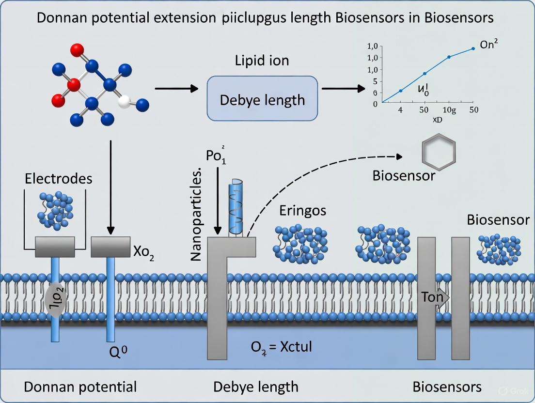
Overcoming the Debye Limit: How Donnan Potential Extends Biosensor Capabilities in Physiological Solutions
This article provides a comprehensive review of the Donnan potential effect as a transformative mechanism for extending the Debye length in field-effect transistor (FET) biosensors.
Overcoming the Debye Limit: How Donnan Potential Extends Biosensor Capabilities in Physiological Solutions
Abstract
This article provides a comprehensive review of the Donnan potential effect as a transformative mechanism for extending the Debye length in field-effect transistor (FET) biosensors. Aimed at researchers, scientists, and drug development professionals, it explores the foundational electrostatics of the Donnan equilibrium, its practical implementation using advanced materials like polymer brushes and supported lipid bilayers, and systematic strategies for optimizing sensor performance. The content critically examines solutions for persistent challenges such as signal drift and steric hindrance, validates the approach through comparative analysis with experimental data, and discusses the implications for developing ultrasensitive, point-of-care diagnostic platforms capable of operating in biologically relevant ionic strength solutions.
The Donnan Equilibrium: Unraveling the Electrostatic Principle that Defeats Ionic Screening
The Fundamental Challenge: Debye Screening in Physiological Fluids
BioFETs are semiconductor-based devices that detect the electrical field generated by charged biomolecules, such as proteins, DNA, or ions, which bind to their sensitive surface. This binding event changes the local charge density, which in turn modulates the current flowing through the semiconductor channel, allowing for direct, label-free detection. The exceptional sensitivity, potential for miniaturization, and compatibility with complementary-metal-oxide-silicon (CMOS) processes make BioFETs promising platforms for ultra-sensitive, multiplexed diagnostics.
However, the operational principle of BioFETs is severely compromised in physiologically relevant media. Biological samples like serum, blood, or urine are high-ionic-strength solutions, containing a high concentration of mobile ions. When a charged biomolecule, such as a protein, approaches the sensor surface in such an environment, it attracts a cloud of counter-ions from the solution. This ion cloud electrically screens the charge of the target molecule, preventing its electric field from reaching and influencing the BioFET's channel.
The spatial range over which an electric field persists in an electrolyte is defined by the Debye screening length (λD). It is the characteristic distance at which the charge's influence drops by a factor of 1/e. The Debye length is inversely proportional to the square root of the ionic strength of the solution. In standard phosphate-buffered saline (PBS, ~0.165 M) or other physiological fluids, the Debye length is typically less than 1 nanometer [1] [2] [3]. This creates a critical dimensional mismatch: while the charge of a target biomolecule like an antibody (which can be 10-15 nm in size) must be detected, its electric field is effectively neutralized beyond a distance shorter than its own physical dimensions. Consequently, the sensitivity of conventional BioFETs is drastically reduced in high-ionic-strength environments, confining their operation to artificially diluted, low-ionic-strength buffers and limiting their practical application [1] [2].
The following diagram illustrates this fundamental screening problem.
Diagram 1: The Debye screening problem in BioFETs. The charge of a target biomolecule is screened by a cloud of counter-ions within the short Debye length, preventing its electric field from reaching the sensor surface.
Strategic Approaches to Overcome the Debye Length Limitation
Researchers have developed innovative strategies to circumvent the Debye screening problem. These methods can be broadly categorized into three approaches: electrostatic control of the interface, geometric and steric confinement, and the use of novel materials with inherent advantages.
Electrostatic and Donnan Potential Engineering
This strategy involves actively modifying the electrostatic environment at the solution-solid interface to reduce the local ion concentration and thereby extend the effective sensing range.
- Meta-Nano-Channel (MNC) BioFET: This novel design decouples the electrostatics of the double layer from the electrodynamics of the conducting channel. Fabricated in a CMOS process, it allows for the independent electrostatic tuning of the double layer. By applying specific potentials, the population of ions in the double layer is decreased, forcing the double layer ion concentration to match the bulk concentration and effectively increasing the screening length. This approach has been demonstrated to enhance the sensing signal for prostate-specific antigen (PSA) from 70 mV to 133 mV [4] [5].
- Exploiting Donnan Potentials in Soft Layers: When a charged, ion-permeable layer (like a polymer hydrogel or a polyelectrolyte multilayer (PEM)) is grafted onto the sensor surface, a Donnan potential is established at the interface between this layer and the bulk solution. The existence and magnitude of this potential depend on the properties of the layer. This modified interface can lead to a longer, intra-layer Debye screening length. The key condition is that the layer thickness must be significantly larger than this operative Debye length within the shell. The effective screening length inside such layers can be increased by an order of magnitude, allowing for charge detection beyond the conventional Debye limit [6] [2].
Geometric and Steric Confinement: The "Debye Volume" Concept
A more recent concept moves beyond the Debye length to consider the Debye volume—the total space available around a charge for ions to form a screening cloud.
- Nanostructured Surfaces: Concave geometries, such as those found in nanogaps, nanopores, or at the base of nanowires, inherently restrict the volume available for double layers to form. This spatial confinement introduces energetic constraints that reduce charge screening, making these geometries more sensitive than planar surfaces [2].
- Surface Grafting with Neutral Polymers: Coating the sensor surface with a dense, partially hydrated layer of large, neutral polymers like poly(ethylene glycol) (PEG) physically limits the space into which screening ions can diffuse. The confined volume within the polymer brush extends the reach of electric fields from bound analytes. Studies have shown that this method can enable the detection of proteins like PSA and thyroid-stimulating hormone in undiluted serum, with reported sensitivity improvements of 3 to 5-fold [2].
Novel Material Platforms
Certain materials offer unique properties that can intrinsically mitigate screening effects.
- Epitaxial Graphene on SiC: Unlike exfoliated or chemically deposited graphene, single-crystal epitaxial graphene exhibits electrical characteristics that are nearly independent of the solution concentration. Its very low quantum capacitance may render the device less sensitive to the ionic strength of the solution, effectively creating a large effective screening length. This allows antibody-modified epitaxial graphene FETs to detect antigens without the need for sample desalting or complex device fabrication [3].
- Three-Dimensional Nanostructures: Using 3D crumpled graphene structures increases the effective sensing area and has been reported to effectively increase the Debye length, reducing charge screening and enabling ultrasensitive detection of small biomolecules like dopamine in complex media such as human urine and serum [7].
Table 1: Summary of Key Strategies for Overcoming the Debye Screening Length
| Strategy | Core Principle | Exemplar Technology/Method | Reported Performance |
|---|---|---|---|
| Electrostatic Control [4] [5] | Actively manipulate surface potentials to deplete the double layer of ions. | Meta-Nano-Channel (MNC) BioFET | PSA detection signal increased from 70 mV to 133 mV. |
| Donnan Potential [6] [2] | Use a charged, porous layer to establish a constant potential that extends the sensing range. | Polyethylene Glycol (PEG) brushes; Polyelectrolyte Multilayers (PEM) | 3 to 5-fold sensitivity improvement for protein detection in serum; Order-of-magnitude increase in screening length predicted. |
| Geometric Confinement [2] | Restrict the physical volume (Debye volume) available for double-layer formation. | Nanogaps, nanopores, concave nanowire structures. | Enhanced sensitivity compared to planar sensor geometries. |
| Novel Materials [3] | Leverage intrinsic material properties that are less susceptible to ionic screening. | Epitaxial Graphene FETs on SiC | Successful antigen detection with antibodies in physiological buffers; Device characteristics independent of solution concentration. |
Detailed Experimental Protocol: Overcoming Screening with Polymer-Modified BioFETs
The following protocol provides a detailed methodology for functionalizing a BioFET sensor with a dense PEG brush to overcome Debye screening, based on strategies highlighted in the literature [2].
Research Reagent Solutions
Table 2: Essential Materials and Reagents
| Item Name | Function/Description |
|---|---|
| BioFET Chip | The foundational sensor, e.g., a SiNW-FET, graphene FET, or MNC-BioFET. |
| Oxygen Plasma Cleaner | For cleaning and activating the sensor surface to enhance subsequent chemical binding. |
| Silane-PEG-NHS Ester | A heterobifunctional linker: silane group anchors to SiO₂ surfaces, while the NHS ester reacts with amine groups. The long PEG chain provides the steric barrier. |
| Aptamer or Antibody | The biological recognition element (probe) that specifically binds the target analyte. |
| Ethanolamine or BSA | Used to block any remaining reactive sites on the sensor surface after probe immobilization, reducing non-specific binding. |
| Phosphate Buffered Saline (PBS) | A standard buffer for preparing biological solutions and for conducting control experiments. |
| Target Analyte | The molecule of interest (e.g., a protein, hormone) to be detected. |
Step-by-Step Functionalization and Assay Procedure
Part A: Surface Preparation and PEGylation
- Surface Cleaning and Activation: Place the BioFET chip in an oxygen plasma cleaner. Treat it for 2-5 minutes at a medium power setting (e.g., 100 W). This step removes organic contaminants and creates a hydrophilic surface rich in hydroxyl (-OH) groups, which is crucial for the next step.
- Silane-PEG Coupling: Immediately after plasma treatment, prepare a 1-5 mM solution of Silane-PEG-NHS ester in anhydrous toluene. Immerse the activated chip in this solution and incubate for 4-12 hours at room temperature under an inert atmosphere (e.g., in a sealed vial with nitrogen gas). The silane group will covalently bind to the hydroxylated surface, forming a stable monolayer. The high-density PEG brush is established in this step.
- Washing and Curing: Remove the chip from the reaction solution and rinse it thoroughly with toluene followed by ethanol to remove any unbound molecules. Gently dry the chip with a stream of nitrogen gas. To ensure complete covalent bonding, cure the chip at 100-110 °C for 10-15 minutes.
- Probe Immobilization: Prepare a 1-10 µM solution of the amino-modified aptamer or antibody in a neutral buffer (e.g., 1x PBS, pH 7.4). Pipette this solution onto the PEGylated sensor surface and incubate in a humidified chamber for 1-2 hours at room temperature. The NHS ester group at the distal end of the PEG chain will react with primary amine groups on the probe, covalently tethering it to the surface.
- Surface Blocking: To passivate any remaining reactive NHS esters and minimize non-specific adsorption, incubate the sensor with a 1 M ethanolamine solution (or a 1% w/v BSA solution) for 30-60 minutes. After incubation, rinse the chip thoroughly with the running buffer (e.g., PBS) to remove any unbound blocking agents.
Part B: Biosensing Measurement and Data Acquisition
- Electrical Characterization: Integrate the functionalized BioFET chip into a liquid-gated measurement system with an Ag/AgCl reference electrode. Under a constant drain-source voltage (VDS), perform a gate voltage (VGS) sweep in pure buffer to obtain the baseline transfer characteristic (IDS vs. VGS).
- Analyte Introduction: Introduce the sample containing the target analyte at a known concentration into the measurement chamber. Allow the system to equilibrate for 10-20 minutes to ensure sufficient binding kinetics, as diffusion through the PEG layer can be slowed [2].
- Real-Time Monitoring: Continuously monitor the drain-source current (IDS) at a fixed, optimized gate voltage. The specific binding of the charged target to the immobilized probes will cause a shift in the IDS over time.
- Signal Quantification: After the signal stabilizes, perform another full transfer characteristic sweep. The shift in the threshold voltage (∆VTH) or the change in current (∆IDS) between the baseline and post-binding curves is the quantitative sensing signal.
- Control and Calibration: Repeat the process with solutions of different analyte concentrations to build a calibration curve, and use control experiments (e.g., with a non-complementary protein) to confirm specificity.
The experimental workflow for this protocol is summarized below.
Diagram 2: Workflow for BioFET functionalization and sensing.
The Debye screening problem represents a fundamental barrier to the widespread adoption of BioFETs in clinical and point-of-care settings. However, as outlined in this note, it is not an insurmountable one. Innovative strategies ranging from electrostatic engineering and the application of Donnan potentials in soft materials to the clever use of geometry and novel semiconductors provide a robust toolkit for overcoming this limitation. The successful demonstration of specific, label-free detection of biomarkers in undiluted, physiologically relevant fluids like serum, urine, and sweat signals a promising future for this technology. As these approaches mature and are integrated with wearable platforms, they will unlock the full potential of BioFETs for quantitative, real-time health monitoring and advanced diagnostic applications [4] [7] [8].
Field-effect transistor (FET)-based biosensors represent a powerful tool for label-free, rapid biological testing, with applications spanning from pathogen detection to biomarker quantification [9]. A significant challenge confronting these devices, especially when operating in physiologically relevant ionic strength solutions (e.g., 1X PBS), is the Debye screening effect [10]. In aqueous solutions, dissolved ions form an Electrical Double Layer (EDL) at charged surfaces. The characteristic thickness of this layer, the Debye length (λD), typically ranges from angstroms to a few nanometers in biological fluids [10]. This short length scale means that charged analyte molecules, such as antibodies (~10 nm in size), binding beyond this distance are electrically screened from the sensor surface, rendering them undetectable [10].
The Donnan potential phenomenon provides a mechanism to overcome this fundamental limitation. This principle is established when an ion-permeable layer (such as a polymer brush or a layer of immobilized bioreceptors) containing fixed structural charges is equilibrated with an electrolyte solution [6] [9]. A constant electrostatic potential, the Donnan potential (ΔφD), develops within this layer due to charge-driven accumulation of counterions and exclusion of co-ions [6]. This potential effectively extends the sensing distance beyond the native Debye length, enabling the detection of larger biomolecules in high ionic strength environments [10] [9]. This Application Note details the core principles and provides practical protocols for leveraging the Donnan potential to achieve effective Debye length extension in biosensor applications.
Theoretical Foundation
The Donnan Potential
The Donnan potential arises from a partitioning of ions between a bulk electrolyte solution and a charged, ion-permeable surface layer. The magnitude of this potential for a soft, charged layer is given by [9]:
$$\begin{array}{c}\Delta {\phi }{D}={\varphi }{th}\,ln\frac{(\sqrt{4{c}{s}^{2}+{c}{x}^{2}}+{c}{x})}{2{c}{s}}\end{array}$$
where:
- ΔφD is the Donnan potential.
- φth is the thermal voltage (≈ 26 mV at room temperature).
- cs is the bulk electrolyte concentration (ionic strength).
- cx is the effective charge concentration within the ion-permeable layer.
This equation shows that the Donnan potential increases with the charge density (cx) of the immobilized layer and decreases with increasing bulk ion concentration (cs). It is important to note that the existence of a stable Donnan potential is conditional. It requires that the thickness of the surface layer well exceeds the intra-particulate Debye screening length and that steric effects mediated by the sizes of the electrolyte ions and structural layer charges do not prevent its formation [6].
Debye Length and its Effective Extension
The Debye length (λD) in an aqueous solution can be approximated by [9]:
$$\begin{array}{c}\lambdaD ≈ \frac{0.3}{\sqrt{cs}} \text{ (in nanometers)}\end{array}$$
In a standard phosphate-buffered saline (PBS) solution, cs is high, resulting in a very short λD of about 0.7 nm. When a charged, ion-permeable layer like a polymer brush is immobilized on the sensor surface, the resulting Donnan potential creates a much larger region of electric field influence. From an electrical perspective, the system can be modeled with the bulk liquid as the gate of a transistor, and the combined Donnan region and the native Debye layer as the effective dielectric [9]. This effectively extends the sensing zone from a few nanometers to the entire thickness of the polymer layer, which can be tens of nanometers, thus overcoming the charge screening limitation [10].
Table 1: Key Parameters Governing Donnan Potential and Debye Length Extension.
| Parameter | Symbol | Description | Impact on Sensing |
|---|---|---|---|
| Bulk Ion Concentration | cs | Ionic strength of the solution (e.g., PBS). | Higher cs reduces both λD and ΔφD, challenging detection. |
| Layer Charge Density | cx | Effective charge concentration within the immobilized layer. | Higher cx increases ΔφD, enhancing the sensing distance. |
| Layer Thickness | δ | Physical thickness of the ion-permeable polymer/bioreceptor layer. | Must significantly exceed the intra-layer Debye length for a stable Donnan potential to exist [6]. |
| Steric Factor | - | Finite sizes of ions and layer charges. | At high concentrations, can limit ion partitioning and prevent Donnan potential establishment [6]. |
Experimental Protocols
Protocol: Fabrication of a Graphene FET (gFET) Biosensor Platform
This protocol outlines the creation of a foundational gFET biosensor, which can subsequently be functionalized to exploit the Donnan effect.
1. Materials
- Pre-patterned wafer with source/drain electrodes (e.g., Au/Cr).
- High-quality graphene (CVD-grown).
- Transfer medium (e.g., PMMA).
- Etching solutions (e.g., FeCl3 for Au, APS for Cu).
- Deionized water and organic solvents (acetone, isopropanol).
- Photolithography or electron-beam lithography system.
- High-κ dielectric material (e.g., Al2O3, HfO2) for top-gating (optional).
2. Procedure
- Graphene Transfer: Transfer a monolayer of CVD graphene onto the pre-patterned electrode structures using a wet-transfer process with a PMMA support layer [9].
- Patterning: Use lithography and oxygen plasma etching to define the graphene channel dimensions.
- Dielectric Deposition (For top-gated devices): Deposit a thin layer of high-κ dielectric via atomic layer deposition (ALD).
- Passivation: Apply a passivation layer (e.g., SiO2 or SU-8 epoxy) to protect the contacts and define the active sensing area [10].
- Encapsulation: Implement encapsulation strategies to mitigate signal drift and leakage current, crucial for stable liquid gating [10].
- Electrical Characterization: Validate device performance in buffer solution by measuring the I-Vg transfer characteristics to determine carrier mobility and the Dirac point.
Protocol: Surface Functionalization for Debye Length Extension
This critical protocol details the application of a polymer brush layer to create the ion-permeable membrane necessary for the Donnan effect.
1. Materials
- Functionalized gFETs from Protocol 3.1.
- Poly(oligo(ethylene glycol) methyl ether methacrylate) (POEGMA) or similar PEG-based polymer.
- Initiator for polymer growth (e.g., for ATRP or photo-initiated polymerization).
- Suitable solvent (e.g., water, ethanol).
- Capture antibodies (cAb) or other bioreceptors (e.g., DNA aptamers).
- Crosslinkers (e.g., NHS-EDC chemistry).
2. Procedure
- Surface Activation: Clean and activate the gFET channel surface (e.g., with oxygen plasma) to generate functional groups for initiator attachment [10].
- Initiator Immobilization: Covalently anchor the polymerization initiator molecules to the activated surface.
- Polymer Brush Growth: Grow the POEGMA brush layer from the surface via a controlled polymerization technique (e.g., ATRP). Control polymerization time to achieve the desired brush thickness (≥ 10 nm is typical) [10].
- Prepare a degassed solution of POEGMA monomer and catalyst in solvent.
- Immerse the initiator-functionalized sensor into the solution.
- Allow polymerization to proceed for a controlled duration.
- Rinse thoroughly with solvent and DI water to remove unreacted monomer.
- Bioreceptor Immobilization: Print or spot the capture antibodies into the polymer brush matrix [10]. The brush can be designed to contain functional groups for covalent attachment via bio-orthogonal chemistry.
- Blocking: Incubate the functionalized sensor with a blocking agent (e.g., BSA, casein) to passivate any non-specific binding sites.
Protocol: Biosensing Measurement with the D4-TFT Workflow
This protocol describes a stable measurement methodology ("D4-TFT") for detecting biomarkers in high ionic strength solution [10].
1. Materials
- Functionalized gFET biosensor from Protocol 3.2.
- Assay buffer (e.g., 1X PBS).
- Sample containing the target analyte.
- Dissolvable trehalose layer containing detection antibodies (dAb) [10].
- Stable electrical testing setup with a pseudo-reference electrode (e.g., Pd) [10].
- Source-meter unit for DC sweeps.
2. Procedure
- Dispense (Baseline): Place a drop of assay buffer onto the sensor. Perform infrequent DC current-voltage (I-V) sweeps to establish a stable baseline current (Ibase). Avoid continuous DC measurement to minimize drift [10].
- Dispense (Sample): Replace the buffer with the sample solution containing the target analyte.
- Dissolve & Diffuse: The sample droplet dissolves the overlying trehalose layer, releasing the detection antibodies, which then diffuse to the sensor surface [10].
- Detect (Binding): If the target analyte is present, a sandwich complex (cAb-analyte-dAb) forms within the POEGMA brush. The associated change in charge density (Δcx) modulates the Donnan potential, leading to a measurable shift in the transistor's drain current (ΔI).
- Data Analysis: Calculate the sensor response as the percent change in current, %ΔI = [(I - Ibase) / Ibase] × 100. Compare against a negative control (a sensor without capture antibodies) to confirm specific binding [10].
Diagram 1: D4-TFT biosensing workflow for reliable biomarker detection.
The Scientist's Toolkit: Essential Research Reagents & Materials
Table 2: Key research reagents and materials for implementing Donnan-based biosensing.
| Item | Function/Description | Example Use Case |
|---|---|---|
| CVD Graphene | High-mobility, chemically stable channel material for FETs. | Core transducer material in gFETs for sensitive charge detection [9]. |
| POEGMA Brush | Non-fouling polymer brush that establishes a Donnan potential. | Creates an ion-permeable layer to extend Debye length in 1X PBS [10]. |
| Palladium (Pd) Electrode | Stable pseudo-reference electrode for liquid gating. | Enables compact, point-of-care device design without bulky Ag/AgCl electrodes [10]. |
| Capture Antibodies | High-affinity bioreceptors immobilized in the polymer brush. | Specific capture of target biomarkers (e.g., viruses, cytokines) from solution [10]. |
| ATRP Initiator | Molecule to initiate controlled radical polymerization. | Covalently grafting POEGMA brushes from sensor surfaces [10]. |
| NHS-EDC Chemistry | Crosslinking reagents for covalent biomolecule immobilization. | Coupling antibodies or other bioreceptors to functionalized polymer brushes. |
Data Presentation & Analysis
The following table summarizes experimental data and performance metrics achievable with Donnan potential-based biosensing platforms, as reported in the literature.
Table 3: Performance summary of Donnan-enabled biosensing platforms.
| Sensor Platform / Assay | Target / Application | Key Performance Metric | Result | Reference Context |
|---|---|---|---|---|
| CNT-based D4-TFT | General Biomarker Detection | Detection Limit in 1X PBS | Sub-femtomolar (aM) | [10] |
| gFET Immunoassay | Infectious Disease Biomarker | Sensitivity in Serum | 500 ng/mL | [9] |
| gFET Immunoassay | Infectious Disease Biomarker | Sensitivity in Buffer | 18 ng/mL | [9] |
| POEGMA-functionalized FET | Debye Length Extension | Sensing Distance | Increased to ~10s of nm | [10] |
Diagram 2: Electrical model showing Donnan and Debye layer capacitances in a gFET.
The Critical Role of Ion-Impermeable and Ion-Permeable Layers
In the field of electrochemical biosensors, the interface between the biological recognition element and the transducer is critical for determining performance characteristics. The strategic application of ion-impermeable and ion-permeable layers at this interface provides a powerful mechanism for controlling the local ionic environment, directly addressing the fundamental challenge of Debye length screening in physiological solutions. When operating at biologically relevant ionic strengths, conventional biosensors suffer from limited detection capabilities because the electrical double layer (EDL) formed at the sensor surface screens charged analytes beyond a very short distance (typically <1 nm) [10].
The Donnan potential effect establishes an equilibrium at the interface between ion-permeable and ion-impermeable layers, creating a stable interfacial potential that can effectively extend the sensing distance beyond the traditional Debye length [10]. This principle enables the detection of larger biomolecules, such as antibodies (~10-15 nm), in high ionic strength solutions like blood or phosphate-buffered saline (PBS), where conventional field-effect transistor (FET) biosensors would normally fail [10]. This application note details the theoretical foundation, experimental protocols, and practical implementations of these critical layers for advancing biosensor research and development.
Theoretical Foundation: Donnan Potential and Debye Length Extension
Donnan Equilibrium Principles
The Donnan potential arises when an ion-impermeable layer containing fixed charges establishes equilibrium with an adjacent ion-permeable solution phase [11]. This phenomenon occurs extensively in ion-exchange membrane systems, where fixed charged groups attached to a polymer backbone create a selective barrier that excludes co-ions while allowing counter-ions to pass [11]. At thermodynamic equilibrium, the unequal distribution of ions between the hydrated membrane and aqueous phases generates an electrical potential at the interface—the Donnan potential—which can be described by:
EDon = RT/ziℱ ln(ais/aim) [11]
Where:
- EDon = Donnan electrical potential
- R = Ideal gas constant
- T = Temperature
- zi = Valence of ion i
- ℱ = Faraday constant
- ai = Activity of ion i in solution (s) or membrane (m) phase
Extension of the Debye Length in Biosensing
In biosensor applications, this Donnan equilibrium principle is leveraged by creating a structured interface where a polymer layer with specific ionic permeability characteristics is grafted onto the sensor surface. The poly(oligo(ethylene glycol) methyl ether methacrylate) (POEGMA) polymer brush has emerged as a particularly effective material for this purpose [10]. When functionalized with charged groups or biomolecular recognition elements, this layer establishes a Donnan potential that modulates the local ionic distribution, effectively extending the sensing distance beyond the native Debye length in high ionic strength solutions [10].
Table 1: Key Theoretical Parameters in Donnan Potential-Mediated Biosensing
| Parameter | Symbol | Typical Range/Value | Impact on Biosensing |
|---|---|---|---|
| Fixed Charge Density | q | 0.1-2.0 eq/L [11] | Determines magnitude of Donnan potential and exclusion capability |
| Donnan Potential | EDon | Variable based on system | Creates interfacial potential that extends sensing distance |
| Debye Length | λD | ~0.7 nm in 1X PBS [10] | Native screening distance in physiological buffers |
| Effective Sensing Distance | - | Enhanced via Donnan effect [10] | Determines size of detectable biomolecules |
| Ionic Strength | I | 150 mM for physiological | Challenges conventional FET sensing without Donnan extension |
Experimental Protocols
Fabrication of POEGMA-Modified BioFETs with Donnan Extension Capability
This protocol details the creation of carbon nanotube-based BioFETs incorporating a POEGMA polymer brush to extend the Debye length via the Donnan potential effect [10].
Materials Required
- Semiconducting carbon nanotube (CNT) solution
- Poly(oligo(ethylene glycol) methyl ether methacrylate) (POEGMA)
- Atom transfer radical polymerization (ATRP) initiator
- High-κ dielectric substrate (e.g., Al2O3, HfO2)
- Phosphate buffered saline (PBS), 1X concentration
- Target-specific capture and detection antibodies
- Palladium (Pd) pseudo-reference electrode
- Trehalose excipient layer material
Procedure
CNT Thin-Film Transistor Fabrication
- Deposit CNTs onto high-κ dielectric substrate using solution-processing or printing techniques
- Pattern source and drain electrodes (typically Pd or Au) using photolithography or shadow masking
- Encapsulate device edges to minimize leakage current and enhance stability [10]
POEGMA Polymer Brush Growth
- Functionalize the CNT channel surface with ATRP initiator
- Grow POEGMA brush layer via surface-initiated ATRP
- Control polymer thickness (typically 10-100 nm) by adjusting monomer concentration and reaction time
- Verify polymer growth and uniformity using ellipsometry or AFM
Antibody Functionalization
- Inkjet-print capture antibodies (cAb) into the POEGMA matrix above the CNT channel
- Print detection antibodies (dAb) onto a dissolvable trehalose excipient layer on a separate pad
- Include control devices without antibodies printed over CNT channel for validation [10]
Device Integration and Packaging
- Integrate Pd pseudo-reference electrode to avoid bulky Ag/AgCl references
- Mount device on printed circuit board with automated testing capability
- Implement solution reservoir with growth medium for bacterial-based sensors if applicable [12]
Biosensing Operation Using D4-TFT Platform
The D4-TFT platform operates through four sequential steps that enable ultrasensitive detection in high ionic strength solutions [10]:
Dispense
- Dispense liquid sample onto the sensor cartridge
- Ensure contact with both the trehalose pad (containing dAb) and the POEGMA/antibody-functionalized CNT channel
Dissolve
- Allow the trehalose excipient layer to dissolve (typically 1-2 minutes)
- Release detection antibodies into the solution
Diffuse
- Detection antibodies and target analytes diffuse through the solution
- Form sandwich complexes (cAb-analyte-dAb) within the POEGMA layer above the CNT channel
- Incubate for sufficient time to allow binding (typically 5-15 minutes)
Detect
- Apply infrequent DC voltage sweeps (rather than static or AC measurements) to minimize signal drift
- Measure changes in CNT channel current resulting from antibody sandwich formation
- Compare signals between active and control devices to confirm specific detection
- Quantify analyte concentration based on calibrated current response
Figure 1: D4-TFT biosensing workflow illustrating the four operational steps that enable detection in high ionic strength solutions [10].
Research Reagent Solutions
Table 2: Essential Research Reagents for Donnan Potential-Based Biosensing
| Reagent/Material | Function | Application Notes |
|---|---|---|
| POEGMA Polymer Brush | Creates ion-permeable layer with Donnan potential [10] | Extends Debye length in physiological solutions; thickness critical for performance |
| Ion-Exchange Membranes | Provides fixed charge density for Donnan exclusion [11] | High charge density required for high-salinity systems; affects permselectivity |
| Carbon Nanotubes (CNTs) | High-sensitivity transducer material [10] | High mobility in thin-film; chemical inertness; solution-phase processability |
| Palladium Pseudo-Reference Electrode | Stable potential measurement in miniaturized systems [10] | Enables point-of-care form factor without bulky Ag/AgCl electrodes |
| Trehalose Excipient | Stabilizes and contains detection antibodies [10] | Forms readily-dissolvable layer for controlled antibody release |
| Specific Antibodies | Biorecognition elements for target analytes | Printed into POEGMA matrix; form sandwich complexes with antigens |
Data Interpretation and Analysis
Electrical Characterization and Signal Validation
The critical advancement enabled by Donnan potential extension is the ability to operate in biologically relevant solutions (1X PBS) while maintaining sensitivity to sub-femtomolar biomarker concentrations [10]. Proper data interpretation requires:
Signal Drift Mitigation
- Use infrequent DC sweeps rather than continuous static measurements
- Monitor both active and control devices simultaneously
- Apply stable electrical testing configuration with proper passivation [10]
Specificity Validation
- Compare devices with and without antibodies printed over CNT channel
- Verify that signal shifts occur only in antibody-functionalized devices
- Confirm dose-response relationship across relevant concentration range
Performance Metrics
Table 3: Performance Comparison of Biosensing Platforms with and without Donnan Extension
| Performance Characteristic | Conventional BioFET | Donnan-Extended BioFET |
|---|---|---|
| Operating Ionic Strength | Requires dilution (e.g., 0.1X PBS) [10] | Native physiological strength (1X PBS) [10] |
| Effective Sensing Distance | Limited to native Debye length (~0.7 nm) [10] | Extended beyond Debye length via Donnan potential [10] |
| Detection Limit | Picomolar to nanomolar range [10] | Sub-femtomolar concentrations demonstrated [10] |
| Reference Electrode | Often requires bulky Ag/AgCl [10] | Compatible with Pd pseudo-reference electrodes [10] |
| Antibody Detection | Challenging due to size beyond Debye length [10] | Enabled via Donnan potential extension [10] |
Figure 2: Comparison of sensing mechanisms between conventional BioFETs and Donnan-extended BioFETs, highlighting the critical role of the ion-permeable polymer layer [10].
Troubleshooting and Optimization
Common Challenges and Solutions
Signal Drift Issues
- Problem: Temporal signal changes obscure biomarker detection
- Solution: Implement combination of sensitivity maximization through proper passivation, stable electrical testing configuration, and rigorous testing methodology with infrequent DC sweeps [10]
Insufficient Debye Length Extension
- Problem: Limited detection capability for large biomolecules
- Solution: Optimize POEGMA thickness and charge density; ensure proper polymer brush formation and antibody immobilization [10]
Non-Specific Binding
- Problem: False positive signals in control devices
- Solution: Utilize POEGMA's non-fouling properties; include rigorous control devices without antibodies above CNT channel [10]
Short Device Lifetime
- Problem: Degradation of biosensor performance over time
- Solution: Appropriate polymer immobilization techniques; stable encapsulation; optimized storage conditions [12]
The strategic implementation of ion-impermeable and ion-permeable layers represents a fundamental advancement in biosensor technology, directly addressing the critical challenge of Debye length screening in physiological environments. Through the establishment of a Donnan potential at carefully engineered interfaces, researchers can effectively extend the sensing distance in high ionic strength solutions, enabling detection of clinically relevant biomarkers at sub-femtomolar concentrations without sample dilution [10].
The protocols and methodologies detailed in this application note provide a foundation for developing next-generation biosensors capable of operating in biologically relevant conditions. The D4-TFT platform demonstrates how the integration of polymer brushes, appropriate transducer materials, and rigorous testing methodologies can overcome persistent limitations in the field, paving the way for truly practical point-of-care diagnostic devices [10]. As research in this area continues to evolve, further refinements in material selection, layer architecture, and signal processing algorithms will undoubtedly expand the capabilities and applications of Donnan potential-enhanced biosensing platforms.
Field-effect transistor (FET) based biosensors represent one of the most promising technologies for label-free, rapid, and sensitive detection of biomarkers, with applications ranging from clinical diagnostics to environmental monitoring [13]. A significant challenge encountered by these solution-gated devices is the Debye screening effect, wherein ions in a high ionic strength solution (such as physiological fluid) form an electrical double layer (EDL) that screens the charge of target biomolecules beyond a very short distance, typically less than 1 nm in 1X PBS [10]. Since most biorecognition elements (e.g., antibodies) are far larger than this distance, this screening severely limits the sensitivity of conventional FET biosensors in biologically relevant conditions.
The Donnan potential offers a mechanism to overcome this fundamental limitation. When a charged, ion-permeable layer (such as a polymer brush) is incorporated at the semiconductor/electrolyte interface, it establishes a Donnan equilibrium with the bulk solution. This equilibrium creates a constant electrostatic potential phase, which can effectively extend the sensing distance beyond the classical Debye length, enabling the detection of large biomolecules in high ionic strength environments [10]. This application note details the theoretical framework, experimental protocols, and key considerations for integrating the Donnan potential into FET sensor design.
Theoretical Framework
The Donnan Equilibrium in Soft Materials
The Donnan potential (ψDonnan) arises at the interface between an electrolyte solution and a charged, ion-permeable surface layer when the layer thickness significantly exceeds the local Debye screening length [6]. This potential is a consequence of the selective partitioning of ions between the bulk solution and the charged layer to satisfy electroneutrality, leading to an accumulation of counter-ions and exclusion of co-ions within the layer.
For a soft surface layer with a volume charge density n₀ (representing its structural charges) equilibrated with a symmetric z:z electrolyte (e.g., NaCl, where z is the ion valence), the Donnan potential can be derived from the Boltzmann distribution of ions and the local electroneutrality condition. The simplified expression is given by:
[ \psi{Donnan} = \frac{RT}{zF}\sinh^{-1}\left(\frac{n0}{2zFC_b}\right) ]
where R is the universal gas constant, T is the absolute temperature, F is the Faraday constant, and C_b is the bulk electrolyte concentration [6]. The existence of a stable Donnan potential is conditional and depends not only on the layer thickness but also on the charge density of the layer, the ionic strength, and steric effects related to the sizes of the ions and the structural charges of the layer [6].
Overcoming Debye Screening in FET Biosensors
In a standard electrolyte-gated FET (EG-FET), the total gate capacitance (C_TOT) is a series combination of the capacitance at the gate-electrolyte interface (C_GE) and the electrolyte-semiconductor interface (C_ES). In a biological solution, the EDL capacitance at the semiconductor surface is typically the limiting factor and is described by the Gouy-Chapman-Stern model, which includes a compact Helmholtz layer and a diffuse Gouy-Chapman layer [14].
The critical parameter is the Debye length (λD), which defines the characteristic decay length of the electrostatic potential from a charged surface. For a monovalent electrolyte, λD ≈ 0.3 nm in 1X PBS, making the FET insensitive to charged biomolecules like proteins located several nanometers away [10].
Integrating a charged polymer brush (e.g., POEGMA) onto the semiconductor surface creates a Donnan phase. The fixed charges on the polymer establish a Donnan potential that, at equilibrium, prevents the rapid decay of potential within the brush. The potential remains relatively constant throughout the polymer layer and only decays exponentially to zero within the bulk solution, starting from the brush-solution interface. This effectively shifts the plane of potential decay away from the semiconductor surface, increasing the distance over which the sensor can detect charges, as illustrated in the following diagram.
Diagram 1: Mechanism of Debye length extension via a charged polymer brush. The Donnan potential within the brush creates a constant potential region, shifting the exponential decay into the bulk solution and making distant biomolecular binding events detectable.
Quantitative Data and Material Properties
The efficacy of the Donnan potential in enhancing sensor response is governed by several key parameters. The tables below summarize the core relationships and the impact of different material and solution properties.
Table 1: Key Parameters Governing the Donnan Potential in FET Sensors
| Parameter | Symbol | Role in Donnan-Modulated Sensing | Typical Target Value/Range |
|---|---|---|---|
| Polymer Charge Density | n₀ | Determines the magnitude of the Donnan potential and the strength of the ion-partitioning effect. | Sufficiently high to counterbalance high salinity [11]. |
| Bulk Ionic Strength | Cb | Higher concentrations reduce the Donnan potential and the effective sensing distance. | 1X PBS (0.15 M) for physiological relevance [10]. |
| Ion Valence | z | Influences the Debye length and the sensitivity of ψDonnan to charge density. | 1 (for NaCl systems) [11]. |
| Polymer Layer Thickness | δ | Must be significantly larger than the intra-particulate Debye length to establish a stable Donnan phase [6]. | >> 1/κshell (internal Debye length) [6]. |
| Steric Factor | - | Accounts for the finite size of ions and polymer charges, limiting ion crowding and potential magnitude at high densities [6]. | Considered for non-dilute systems. |
Table 2: Impact of Material and Solution Properties on Sensor Performance
| Property / Condition | Effect on Donnan Potential | Consequence for FET Sensing | Experimental Consideration |
|---|---|---|---|
| High Fixed Charge Density (n₀) | Increases ψDonnan [11]. | Enhances permselectivity and signal for a given biomarker. | Must be balanced to avoid excessive ion congestion [6]. |
| Low Bulk Ionic Strength | Increases ψDonnan and λD. | Easiest condition for detection, but not physiologically relevant. | Useful for initial proof-of-concept experiments. |
| High Bulk Ionic Strength (e.g., 1X PBS) | Decreases ψDonnan and λD. | Challenges sensor sensitivity; necessitates high n₀ [11]. | Required for testing in clinically relevant media. |
| Use of POEGMA Brush | Creates a stable Donnan phase and reduces biofouling [10]. | Enables detection in 1X PBS and improves sensor stability. | A key enabling material for practical biosensors. |
| Asymmetric Ion Size/Valence | Alters the steric limit and the partition coefficients of ions [6]. | Can be used to tailor sensor response and selectivity. | Model with advanced Poisson-Boltzmann corrections. |
Experimental Protocols
Protocol: Fabrication of a Donnan-Modulated CNT FET (D4-TFT) for Ultrasensitive Detection
This protocol outlines the procedure for creating a carbon nanotube-based FET biosensor that utilizes a POEGMA polymer brush to establish a Donnan potential for attomolar-level detection in 1X PBS [10].
Materials and Reagents
Research Reagent Solutions
| Item | Function in the Protocol |
|---|---|
| Semiconducting Carbon Nanotubes (CNTs) | Forms the conductive channel of the FET transducer. |
| Poly(oligo(ethylene glycol) methyl ether methacrylate) (POEGMA) | Polymer brush that forms the Donnan phase, extends sensing distance, and resists biofouling. |
| Phosphate Buffered Saline (PBS), 1X | High ionic strength solution simulating physiological conditions for testing. |
| Capture Antibodies (cAb) and Detection Antibodies (dAb) | Biorecognition elements for specific sandwich immunoassay. |
| Palladium (Pd) Pseudo-Reference Electrode | Provides a stable gate potential in a point-of-care form factor. |
| EDC/NHS Crosslinking Chemistry | Activates carboxyl groups for covalent attachment of bioreceptors. |
Step-by-Step Procedure
FET Fabrication:
- Fabricate source and drain electrodes (e.g., gold on Si/SiO₂) with a defined channel geometry.
- Deposit a thin film of semiconducting CNTs across the channel to form the conductive pathway [10].
Surface Passivation:
- Passivate the contact areas and defined regions of the channel to mitigate gate leakage currents and enhance electrical stability [10].
Polymer Brush Grafting:
- Grow a layer of POEGMA directly on the passivated CNT channel. This is a critical step, as the POEGMA forms the ion-permeable, charge-containing film that will establish the Donnan equilibrium [10].
Biofunctionalization:
- Print or spot capture antibodies (cAb) onto the POEGMA layer. The polymer brush should be functionalized to allow for covalent immobilization of the antibodies while retaining its non-fouling and Donnan potential properties [10].
- It is crucial to include control devices on the same chip where no antibodies are printed over the CNT channel.
Electrical Characterization and Biosensing:
- Integrate a Pd pseudo-reference electrode to complete the electrolyte-gated TFT setup.
- Use a stable electrical testing configuration, employing infrequent DC sweeps rather than continuous static or AC measurements to minimize signal drift [10].
- For the D4-TFT immunoassay, the "Dispense, Dissolve, Diffuse, Detect" steps are followed. The target analyte diffuses and binds to the cAb, followed by a detection antibody, forming a sandwich complex [10].
- Monitor the drain current (
I_D) over time. A positive shift inI_Din the test device, with no corresponding shift in the control device, confirms successful and specific detection.
The following workflow diagram summarizes the key fabrication and measurement steps.
Diagram 2: Key steps in the fabrication and operation of a Donnan-modulated D4-TFT biosensor.
Protocol: Measuring and Validating the Donnan Potential Effect
Objective
To experimentally confirm the presence of the Donnan potential and its role in extending the Debye length by comparing sensor response with and without the charged polymer layer.
Procedure
Device Comparison:
- Prepare two sets of identical FET devices. The experimental set is functionalized with the charged polymer brush (e.g., POEGMA), while the control set lacks this layer.
Solution Variation:
- Test both device sets in electrolytes of varying ionic strength (e.g., 0.1X PBS and 1X PBS). The Debye length is longer in 0.1X PBS, allowing the control device to potentially function.
Analyte Testing:
- Expose both devices to a model charged analyte, such as a protein or DNA, with a hydrodynamic size larger than the Debye length in 1X PBS (~0.7 nm).
Response Analysis:
- Measure the transfer characteristics (
I_Dvs.V_G) or the time-dependentI_Dresponse for both devices. - Validation: The control device will show a significant response in 0.1X PBS but a negligible response in 1X PBS due to screening. The experimental device (with polymer brush) will maintain a strong, stable response in both 0.1X PBS and 1X PBS, demonstrating the Debye-length-extension effect of the Donnan potential [10].
- Measure the transfer characteristics (
Discussion
Tackling Signal Drift and Stability
A major challenge in BioFETs is signal drift—the slow, unwanted change in baseline signal over time, which can obscure the specific response from biomarker binding. This drift is often caused by the slow diffusion of electrolytic ions into the sensing region, altering gate capacitance and threshold voltage [10]. The Donnan-potential-based sensor design must incorporate strategies to mitigate this:
- Stable Testing Configuration: Use infrequent DC sweeps instead of continuous static measurements to sample the device state without exacerbating drift [10].
- Effective Passivation: Robust passivation layers around the active channel are essential to prevent leakage currents [10].
- Non-Fouling Interfaces: The POEGMA brush itself acts as a bio-inert layer, reducing non-specific adsorption (biofouling) that can contribute to long-term signal instability [10].
Limitations and Advanced Considerations
While powerful, the Donnan model has limitations. At very high structural charge densities (n₀) and high ionic strengths, steric effects—the finite size of ions and polymer charges—become significant. These effects can limit the maximum achievable Donnan potential due to ion congestion, a deviation not captured by classical point-charge models [6]. Accurate modeling for such conditions requires corrections to the mean-field Poisson-Boltzmann theory that explicitly account for the excluded volume of ions and structural charges [6]. Furthermore, the permselectivity of the membrane or polymer layer is not absolute; a finite concentration of co-ions (C_co-ion) will always permeate the layer, an effect that is magnified at low fixed charge densities and high external salt concentrations [11].
Engineering the Interface: Materials and Methods for Implementing Donnan Potential Extension
The development of biosensors capable of operating in physiologically relevant ionic strength solutions represents a significant challenge in diagnostic medicine. A primary obstacle is the Debye screening effect, where ions in solution form an electrical double layer that screens the charge of target biomarkers, effectively limiting detection to molecules within a few nanometers of the sensor surface. This review details the application of poly(oligo(ethylene glycol) methyl ether methacrylate) (POEGMA) polymer brushes as a material strategy to overcome this limitation. By establishing a Donnan equilibrium potential at the biosensor interface, POEGMA brushes effectively extend the sensing distance, enabling the detection of sub-femtomolar biomarker concentrations in high ionic strength environments like 1X phosphate-buffered saline (PBS). This application note provides a comprehensive overview of the underlying mechanism, quantitative performance data, and detailed experimental protocols for implementing POEGMA brushes in field-effect transistor-based biosensors (BioFETs), framing this advancement within the broader context of Donnan potential-based Debye length extension for next-generation biosensing.
Biosensors that rely on field-effect transistors (BioFETs) are promising for point-of-care diagnostics due to their inherent simplicity, low cost, and high sensitivity [10]. However, when operating in solutions at biologically relevant ionic strengths, such as blood or 1X PBS, these devices face a fundamental physical constraint: the Debye screening effect [10] [15].
In physiological solutions, the Debye length—the characteristic distance over which electrostatic potentials decay—is typically less than 1 nanometer [15]. This is problematic because the biorecognition elements, such as antibodies, are often an order of magnitude larger (~10 nm). Consequently, any charged target biomarker binding to its receptor falls far outside the Debye length and its electrical signal is effectively screened, rendering it undetectable by conventional BioFETs [10].
Traditional workarounds, such as diluting the buffer to increase the Debye length, compromise biomarker stability and assay relevance, making the results physiologically irrelevant [10]. The integration of POEGMA polymer brushes addresses this problem directly by leveraging the Donnan equilibrium potential to create an extended sensing zone within the brush layer, permitting ultrasensitive detection in undiluted biological fluids [10] [15].
Mechanism of Action: Donnan Potential-Mediated Debye Length Extension
POEGMA brushes function as Debye length extenders not by physically altering the ionic composition of the bulk solution, but by creating a local environment at the sensor interface where a Donnan potential is established.
The brushes form a dense, highly hydrated layer with a significant volume fraction of polymer. The oligo(ethylene glycol) side chains are uncharged, making the brush layer charge-neutral [10]. When this neutral, porous brush is immersed in an ionic solution, the concentration of mobile ions within the brush differs from that in the bulk solution. This disparity in ion concentration creates a stable Donnan potential at the interface between the brush and the bulk electrolyte.
This potential extends the effective charge-sensing range of the biosensor far beyond the native Debye length in the bulk solution. When a charged target biomarker, such as a protein, binds to its capture antibody within the POEGMA brush, it introduces a fixed charge. The resulting perturbation of the local electrostatic environment is sensed by the underlying transducer (e.g., the channel of a carbon nanotube thin-film transistor) as a measurable change in current or threshold voltage [10]. This mechanism allows for the detection of charged biomolecules that bind at distances significantly greater than the traditional Debye length.
The diagram below illustrates the mechanistic difference between a standard biointerface and one functionalized with a POEGMA brush.
Quantitative Performance Data
The implementation of POEGMA brushes in biosensing platforms has led to remarkable improvements in sensitivity and performance, even in physiologically relevant conditions. The table below summarizes key quantitative findings from recent studies.
Table 1: Performance Summary of Biosensors Utilizing Polymer-Based Debye Length Extension
| Biosensor Platform | Target Analyte | Sensitivity (Limit of Detection) | Solution Conditions | Key Performance Metrics | Source |
|---|---|---|---|---|---|
| CNT-based D4-TFT (POEGMA) | Model Biomarker | Sub-femtomolar (aM) | 1X PBS | Repeated and stable detection in point-of-care form factor | [10] |
| EGFET Immunosensor (PEG-like polymer) | p53 tumour suppressor | 100 pM | Physiological buffer | Sensitivity: 1.5 ± 0.2 mV/decade; Detection range: 0.1–10 nM | [15] |
| sSEBS-PEDOT/POEGMA Fibre Mat | Protein Fouling (BSA) | ~82% protein repellence | N/A | Antifouling efficiency with 30-mers POEGMA brushes; Cell viability >80% | [16] |
Experimental Protocols
This section provides detailed methodologies for fabricating and implementing POEGMA brush-modified interfaces for enhanced biosensing.
Protocol: Grafting POEGMA Brushes via Surface-Initiated ARGET ATRP
This protocol describes the functionalization of a conductive substrate (e.g., gold, carbon nanotube thin films) with POEGMA brushes to create a non-fouling, Debye-length-extending interface [16].
Research Reagent Solutions
Table 2: Essential Reagents for SI-ATRP of POEGMA
| Reagent / Material | Function / Description | Example / Note |
|---|---|---|
| OEGMA Monomer | The primary building block of the polymer brush. Provides the antifouling and Donnan potential properties. | Oligo(ethylene glycol) methyl ether methacrylate (OEGMA, number of EG units can vary, e.g., n=3-19) [17]. |
| ATRP Initiator | A molecule that covalently attaches to the substrate surface and initiates the controlled radical polymerization. | EDOT-Br: An EDOT derivative with a bromopropanoate ATRP-initiating site, allows electropolymerization on conductive surfaces [16]. |
| Catalyst System | Mediates the atom transfer process during polymerization, controlling the reaction kinetics. | Copper(II) bromide with 2,2'-Bipyridine as a ligand. Cu^0 can be used for mediated (ARGET) ATRP to reduce catalyst concentration [16]. |
| Solvent | Dissolves the monomer and catalyst, enabling the polymerization reaction. | Anhydrous N,N-Dimethylformamide (DMF) or water/methanol mixtures [16] [17]. |
| Reducing Agent (for ARGET) | Regenerates the active Cu(I) catalyst from the Cu(II) deactivator, allowing for very low catalyst concentrations. | Ascorbic acid [16]. |
Step-by-Step Procedure:
Surface Preparation and Initiator Immobilization:
- For conductive surfaces like gold or CNTs, an ATRP initiator can be attached via self-assembled monolayers (e.g., from an ethanol solution of 2-bromo-2-methylpropionate) or electropolymerized.
- Electropolymerization Method: For conductive polymer-based substrates (e.g., PEDOT-infused fibre mats), electropolymerize a copolymer of EDOT and the ATRP-initiator functionalized EDOT-Br from an acetonitrile solution to create a uniform initlayer across the surface [16].
Polymerization Solution Preparation:
- In a Schlenk flask, dissolve the OEGMA monomer in a degassed solvent (e.g., a 1:1 v/v mixture of methanol and water, or DMF) to achieve a typical concentration of 1-2 M.
- Add the ligand (e.g., 2,2'-Bipyridine) and the copper(II) bromide catalyst. For ARGET ATRP, also add a stoichiometric amount of reducing agent (e.g., ascorbic acid) relative to the Cu(II). The goal is to achieve a very low concentration of the active Cu(I) catalyst for better control.
Surface-Initiated Polymerization:
- Transfer the degassed polymerization solution to a reaction vessel containing the initiator-functionalized substrate under an inert atmosphere (e.g., nitrogen or argon).
- Seal the vessel and place it in a temperature-controlled bath or oven. The polymerization is typically carried out at temperatures between 25°C and 40°C.
- Allow the reaction to proceed for a predetermined time (e.g., 1 to 24 hours) to control the brush thickness. The brush thickness and density can be tuned by varying the polymerization time and surface density of the active initiator [18].
Post-Polymerization Processing:
- After polymerization, carefully remove the substrate from the reaction mixture.
- Rinse the substrate thoroughly with copious amounts of the solvent and ethanol to remove any physisorbed monomer, catalyst, and untethered polymer.
- Dry the substrate under a stream of nitrogen or argon.
Validation and Characterization:
- Ellipsometry or AFM: Measure the dry thickness of the polymer brush layer.
- Water Contact Angle: Confirm increased hydrophilicity compared to the unmodified surface.
- Protein Assay (e.g., BCA Assay): Quantify the non-fouling properties by measuring the amount of adsorbed protein (e.g., Bovine Serum Albumin) from solution. Efficiency of >80% protein repellence has been demonstrated with 30-mers POEGMA brushes [16].
- Electrochemical Impedance Spectroscopy (EIS): Characterize the interfacial properties and the extension of the sensing distance.
Protocol: Biosensing with a POEGMA-Modified D4-TFT
This protocol outlines the use of a POEGMA-functionalized Carbon Nanotube Thin-Film Transistor (D4-TFT) for ultrasensitive biomarker detection [10].
Workflow Overview:
The following diagram outlines the complete experimental workflow for the D4-TFT biosensing assay, from surface preparation to electrical detection.
Step-by-Step Procedure:
Device Fabrication and POEGMA Grafting:
- Fabricate a solution-gated thin-film transistor using semiconducting carbon nanotubes as the channel material.
- Graft a POEGMA brush layer directly above the CNT channel following the SI-ATRP protocol in Section 4.1.
Biofunctionalization:
- Printing Capture Antibodies: Using a non-contact inkjet printer or a microspotter, print capture antibodies (cAb) directly into the POEGMA brush layer above the CNT channel. A control device with no antibodies should be prepared on the same chip.
- Printing Detection Antibodies: Print detection antibodies (dAb), conjugated with a label if necessary, onto a readily dissolvable sugar layer (e.g., trehalose) patterned on a separate, facing substrate.
Assay Execution (D4 Protocol):
- Dispense: Dispense a small volume of the sample containing the target biomarker onto the device.
- Dissolve: The sample dissolves the trehalose layer, releasing the detection antibodies into the solution.
- Diffuse: All biomolecules (target and detection antibodies) diffuse to the sensor surface.
- Detect: A sandwich immunoassay complex forms on the sensor surface if the target biomarker is present.
Electrical Measurement and Data Analysis:
- Use a stable electrical testing configuration with a palladium (Pd) pseudo-reference electrode to avoid bulky Ag/AgCl electrodes.
- To mitigate signal drift, enforce a rigorous testing methodology that relies on infrequent DC sweeps rather than continuous static or AC measurements [10].
- Monitor the transistor's transfer characteristics (drain current, ID, vs. gate voltage, VG). The formation of the antibody-analyte sandwich complex within the POEGMA brush causes a measurable shift in the on-current or threshold voltage.
- Successful detection is confirmed by a significant signal shift in the active device compared to the control device with no antibodies.
The Scientist's Toolkit: Essential Research Reagents
Table 3: Key Materials for POEGMA-based Biosensor Research
| Category | Item | Critical Function |
|---|---|---|
| Polymer & Monomers | OEGMA Monomer (n=3, 4, 5, 7, 9, 19) | Determines brush architecture, hydration, and final antifouling/Donnan potential performance [17]. |
| ATRP Initiator (e.g., EDOT-Br) | Covalently anchors the growing polymer chains to the substrate surface [16]. | |
| Catalysis & Synthesis | Copper(II) Bromide (CuBr₂) / 2,2'-Bipyridine | Catalyzes the surface-initiated ATRP reaction [16]. |
| Ascorbic Acid | Serves as a reducing agent in ARGET ATRP for better reaction control [16]. | |
| Sensor Components | Semiconducting Carbon Nanotubes (CNTs) | Forms the high-sensitivity channel material for the BioFET transducer [10]. |
| Palladium (Pd) Wire | Acts as a stable, miniaturized pseudo-reference electrode for point-of-care device compatibility [10]. | |
| Biologicals | Capture & Detection Antibodies | Provide high-specificity recognition for the target biomarker in a sandwich assay format [10]. |
| Trehalose | Forms a dissolvable excipient layer for stable storage and controlled release of detection antibodies [10]. |
The integration of POEGMA polymer brushes into biosensor interfaces represents a transformative material strategy for overcoming the fundamental limitation of Debye screening. By establishing a localized Donnan potential, these brushes effectively extend the charge-sensing range, enabling direct, label-free, and ultrasensitive detection of biomarkers in physiologically relevant fluids. The detailed protocols and performance data provided herein offer researchers a clear pathway to implement this advanced functionality. When combined with robust sensing platforms like CNT-based TFTs and drift-mitigating electrical measurement schemes, POEGMA brushes pave the way for the development of reliable, high-performance point-of-care diagnostic devices that can function accurately in blood, serum, and other complex biological matrices.
A paramount challenge in the development of electronic biosensors is the severe charge screening effect in physiological environments, where the high ionic strength limits the electrostatic detection of biomarkers to distances shorter than 1 nm—the Debye length [2] [19]. This screening prevents the detection of larger biomolecules, such as antibodies, which can be 10–15 nm in size [2]. This Application Note details the use of Supported Lipid Bilayers (SLBs) as biomimetic platforms that, when functionalized with specific polymer brushes, can overcome this limitation by establishing a Donnan potential. This potential effectively extends the sensing range of biosensors, enabling highly sensitive detection in biologically relevant ionic strength solutions [10] [9].
Theoretical Framework: Donnan Potential and Debye Length Extension
The sensitivity of field-effect transistor (FET) based biosensors is traditionally limited by the formation of an Electrical Double Layer (EDL) at the sensor-solution interface. In high ionic strength solutions (e.g., 1X PBS), the EDL is compressed, resulting in a Debye length of only about 0.7 nm [10] [19]. Any charged biomarker beyond this distance from the sensor surface is electrically screened and undetectable.
The strategy outlined herein involves creating an ion-permeable layer atop the sensor, into which charged biomolecules can partition. This layer acts as a selective membrane, leading to an unequal distribution of ions between the layer and the bulk solution. This ion partitioning creates a Donnan potential, a constant electrostatic potential that extends throughout the entire ion-permeable layer [6] [9]. The Donnan potential (( \Delta \phiD )) can be quantitatively described by the following equation, where ( \varphi{th} ) is the thermal voltage, ( cs ) is the bulk ion concentration, and ( cx ) is the concentration of fixed charges within the permeable layer [9]: [ \Delta \phiD = \varphi{th} \, \ln \frac{(\sqrt{4{cs}^2 + {cx}^2} + {cx})}{2{cs}} ] This potential effectively pushes the sensing plane from the sensor surface to the outer boundary of the polymer layer, thereby overcoming the traditional Debye screening limitation [2] [9]. The schematic below illustrates this core concept.
Diagram 1: Conceptual framework of Donnan potential extending the sensing range beyond the Debye length.
Experimental Protocols
Fabrication of SLB-Based Biosensors (D4-TFT Platform)
This protocol describes the construction of an ultrasensitive Carbon Nanotube Thin-Film Transistor (CNT-TFT) biosensor, termed the D4-TFT, which integrates a Supported Lipid Bilayer (SLB) and a polymer brush to overcome Debye screening [10].
Key Materials:
- Substrate: Si/SiO₂ wafers with pre-patterned interdigitated gold source and drain electrodes.
- Semiconductor: Semiconducting carbon nanotubes (CNTs).
- Polymer Brush: Poly(oligo(ethylene glycol) methyl ether methacrylate) (POEGMA).
- Lipid Components: Soybean lecithin, phosphatidylethanolamine, and biotin-X DHPE (for functionalization).
- Biorecognition Elements: Target-specific capture antibodies (cAb) and biotinylated detection antibodies (dAb).
- Other Chemicals: EDC, S-NHS, phosphate-buffered saline (PBS).
Procedure:
- CNT-TFT Fabrication: Solution-processable CNTs are deposited onto the substrate to form the active channel between the source and drain electrodes [10].
- Surface Functionalization:
- The CNT surface is first functionalized with a plasma-deposited thin layer containing carboxylic acid (-COOH) moieties to promote subsequent binding [14].
- The surface is treated with a fresh EDC/S-NHS solution (100 mM each in PBS) for 1 hour at room temperature to activate the carboxyl groups.
- After rinsing, the POEGMA polymer brush is grafted onto the activated surface. This brush serves as the ion-permeable layer for Donnan potential extension and a non-fouling background [10].
- Supported Lipid Bilayer Formation via Vesicle Fusion: [20]
- Vesicle Preparation: Lipids (soybean lecithin, phosphatidylethanolamine, and biotin-X DHPE) are dissolved in chloroform. The solvent is evaporated under a nitrogen stream, and the lipid film is placed under vacuum for 60 minutes. The film is then rehydrated in PBS and sonicated on ice. The resulting multilamellar vesicle suspension is extruded through a polycarbonate filter (100 nm pore size) to form small unilamellar vesicles (SUVs).
- Vesicle Fusion: The freshly prepared SUV suspension is incubated overnight with the POEGMA-functionalized sensor surface under mild agitation. Vesicles adsorb, rupture, and merge to form a continuous SLB on the surface [20].
- Antibody Immobilization: Capture antibodies (cAb) are printed directly onto the POEGMA brush above the CNT channel. A control device with no antibodies is also prepared on the same chip for validation [10].
The overall workflow for sensor preparation and the D4 assay operation is summarized below.
Diagram 2: Workflow for SLB-based D4-TFT biosensor preparation and assay operation.
Electrical Measurement Protocol to Mitigate Signal Drift
Accurate measurement in ionic solutions is confounded by signal drift. The following protocol ensures stable readings [10].
Procedure:
- Stable Testing Configuration: Use a palladium (Pd) pseudo-reference electrode to avoid bulky Ag/AgCl electrodes. Ensure proper passivation of the device.
- Rigorous Measurement Methodology:
- Use infrequent DC current-voltage (I-V) sweeps instead of continuous static (DC) or alternating current (AC) measurements.
- Apply a single short pulse bias (e.g., 50 µs duration with a sampling rate of 10 ns) and integrate the measured current over the pulse period.
- Define the signal as a normalized change in the drain current (( \Delta I / I )) or as a current gain. This methodology minimizes ion drift and thermal noise, providing a stable baseline.
Key Experimental Data and Performance
The performance of the D4-TFT platform with the POEGMA polymer brush has been quantitatively evaluated. The table below summarizes key findings from the literature.
Table 1: Performance summary of SLB-based biosensors with Donnan potential extension.
| Sensor Platform | Target Analyte(s) | Sample Matrix | Detection Limit | Key Performance Feature | Ref. |
|---|---|---|---|---|---|
| D4-TFT (CNT, POEGMA) | Model biomarkers | 1X PBS | Sub-femtomolar (aM) | Achieved attomolar sensitivity in undiluted physiological buffer. | [10] |
| EDL AlGaN/GaN HEMT | HIV-1 RT, CEA, NT-proBNP, CRP | 1X PBS & Human Serum | Not specified | Direct detection in 5 minutes without sample dilution or washing. | [19] |
| Graphene FEB Sensor | Infectious disease biomarkers | Serum | 500 ng/mL | Demonstrated applicability in a commercially produced, foundry-fabricated device. | [9] |
Further characterization of the SLB itself is crucial for quality control. The table below outlines key parameters and common characterization techniques.
Table 2: Supported Lipid Bilayer characterization techniques and parameters.
| Characterization Method | Key Parameters Measured | Insight for SLB Quality |
|---|---|---|
| Atomic Force Microscopy (AFM) | Lipid-phase separation, gel-phase domain formation and size (1-35 μm), bilayer thickness (~5 nm), fluidity. | Verifies successful bilayer formation, phase behavior, and lateral homogeneity [20]. |
| Fluorescence Microscopy | Lipid diffusion (Fluorescence Recovery After Photobleaching - FRAP), domain visualization via fluorescent tags. | Confirms bilayer fluidity and allows visualization of phase-separated domains [20]. |
| Electrical Impedance Spectroscopy | Membrane integrity, resistance, and capacitance. | Quantifies ion impermeability and confirms the formation of a continuous, defect-free bilayer. |
The Scientist's Toolkit: Research Reagent Solutions
Table 3: Essential materials and reagents for SLB-based biosensor development.
| Item | Function/Description | Example/Catalog |
|---|---|---|
| Semiconducting CNTs | Forms the highly sensitive, solution-processable channel of the thin-film transistor. | Purity >98%, various diameters available. |
| POEGMA Polymer | Creates an ion-permeable, non-fouling brush layer that extends the Debye length via the Donnan effect. | Poly(oligo(ethylene glycol) methyl ether methacrylate). |
| Soybean Lecithin | A natural mixture of phospholipids used as the primary component for forming the SLB. | EPIKURON 200 [14]. |
| Biotin-X DHPE | A functionalized lipid that incorporates into the SLB, providing biotin handles for streptavidin-biotin based immobilization. | N-((6 (Biotinoyl)amino)hexanoyl)-1,2-Dihexadecanoyl-sn-Glycero-3-Phosphoethanolamine [14]. |
| Streptavidin/Avidin | Tetrameric protein that bridges biotinylated lipids and biotinylated detection antibodies. | From Streptomyces avidinii or egg white [14]. |
| EDC / S-NHS | Crosslinking agents for zero-length carbodiimide chemistry; activate carboxyl groups for covalent coupling to amine groups. | 1-Ethyl-3-(3-dimethylaminopropyl)carbodiimide / N-Hydroxysulfosuccinimide. |
Biosensor technology has emerged as a dynamic and rapidly evolving field, responding to the pressing need for precise, rapid, and cost-effective measurements in healthcare, environmental monitoring, and food safety [21]. The performance of these biosensors is fundamentally governed by the electrostatic conditions at the bio-interface, particularly the Donnan potential and its role in extending the effective Debye length within charged permeable layers [6]. This application note provides a detailed framework for the practical implementation of two cornerstone biosensing methodologies: the antibody-based sandwich assay and DNA-based detection via aptamers. We present structured quantitative data, step-by-step experimental protocols, and standardized visualization tools to guide researchers and drug development professionals in developing robust biosensing platforms that leverage these critical electrostatic phenomena.
Theoretical Framework: Donnan Potential and Debye Length in Biosensing
The electrostatics of charged bio-interfaces is pivotal for the reactivity and sensitivity of biosensors, influencing processes from colloid stability to the detection of biomolecular interactions [6]. In biosensors with ion-permeable, polyelectrolyte-like layers (such as polymer coatings or cellular membranes), a fundamental electrostatic phenomenon occurs.
When the thickness of a charged surface layer well exceeds the screening Debye length, a constant Donnan potential ((\Psi_D)) is established throughout that layer [6]. This potential arises from the charge-driven accumulation of counterions and exclusion of co-ions. The classical mean-field Poisson-Boltzmann theory describes this under the point-like charge approximation.
However, the existence and magnitude of the Donnan potential are conditional and highly dependent on steric effects mediated by the sizes of the electrolyte ions and the structural charges of the biosensor interface itself [6]. Modern corrections to the theory account for these steric effects, providing a more accurate rationale for the difference between the intra- and extra-particulate Debye screening lengths. This is crucial for biosensor design, as the Donnan potential directly affects the partitioning of ions and biomolecules (such as proteins or DNA) at the sensor surface, thereby influencing the binding efficiency and ultimate detection signal of an assay [6].
Biosensing Recognition Elements: A Quantitative Comparison
The choice of molecular recognition element is a primary determinant of biosensor performance. The following table summarizes the key characteristics of antibodies and nucleic acid aptamers, the two most prevalent biorecognition elements.
Table 1: Comparative Analysis of Antibodies and Aptamers as Biosensor Recognition Elements
| Feature | Aptamers | Antibodies |
|---|---|---|
| Molecular Weight | 5 to 15 kDa [21] | 150 to 170 kDa [21] |
| Selection Process | SELEX (in vitro) [21] | Animal immune system (in vivo) [21] |
| Generation Time | Months [21] | Several months [21] |
| Production Scalability | Highly scalable (chemical synthesis) [21] | Limited scalability [21] |
| Batch-to-Batch Variation | Lower [21] | Higher [21] |
| Stability & Shelf Life | Long; renature after denaturation [21] | Short; sensitive to pH/temperature, irreversible denaturation [21] |
| Cost | Lower [21] | Higher [21] |
| Modifications | Easily modified for immobilization or detection [21] | Limited modification options [21] |
| Ethical Concerns | None (chemical production) [21] | Present (dependent on animal use) [21] |
Aptamers, short strands of DNA or RNA, are developed through an in vitro process called Systematic Evolution of Ligands by Exponential Enrichment (SELEX) [21]. Their key advantages over traditional antibodies include superior stability, easier modification, lower production costs, and lack of batch-to-batch variation, making them an compelling alternative for biosensor applications [21].
Application Note 1: The Antibody Sandwich ELISA Protocol
The sandwich Enzyme-Linked Immunosorbent Assay (ELISA) is a quintessential method for detecting and quantifying specific proteins in a complex mixture with high specificity.
Workflow and Signaling Pathway
The following diagram illustrates the key steps and biomolecular interactions in a standard colorimetric sandwich ELISA.
Detailed Step-by-Step Protocol
The following procedure is adapted for a colorimetric sandwich ELISA using a 96-well plate and Horseradish Peroxidase (HRP) detection [22].
Table 2: Basic Sandwich ELISA Protocol (Colorimetric Detection) [22]
| Step | Procedure | Key Reagents & Incubation | Function |
|---|---|---|---|
| 1. Coating | Dilute capture antibody in coating buffer (e.g., 50 mM carbonate, pH 9.4). Add 100 µL/well. | Coating Buffer; Incubate 1 hr RT or overnight at 2-8°C. | Immobilizes specific capture antibody on polystyrene plate. |
| 2. Washing | Aspirate and wash each well with >300 µL wash buffer (e.g., PBS with 0.05% Tween 20). | Wash Buffer; Tap plate to remove excess liquid. | Removes unbound capture antibody. |
| 3. Blocking | Add 300 µL/well of blocking buffer (e.g., protein-based assay buffer). | Blocking Buffer; Incubate 1 hr at RT. | Covers unsaturated binding sites to minimize non-specific binding. |
| 4. Sample Incubation | Add 100 µL of standards or samples (prepared in blocking buffer) in duplicate. | Standards/Samples; Incubate 1-2 hrs at RT with shaking. | Allows target antigen to bind to capture antibody. |
| 5. Washing | Repeat Step 2. Perform this wash step five times. | Wash Buffer | Removes unbound proteins and sample matrix. |
| 6. Detection Antibody | Add 100 µL/well of biotinylated detection antibody in blocking buffer. | Biotinylated Detection Antibody; Incubate 2 hrs at RT with shaking. | Binds to a different epitope on the captured antigen. |
| 7. Washing | Repeat Step 2. Perform this wash step five times. | Wash Buffer | Removes excess, unbound detection antibody. |
| 8. Enzyme Conjugate | Add 100 µL/well of Streptavidin-HRP (e.g., 1:5,000 dilution in blocking buffer). | Streptavidin-HRP; Incubate 1 hr at RT with shaking. | Binds to biotin on the detection antibody, introducing the enzyme. |
| 9. Final Wash | Repeat Step 2. Perform this wash step five times. | Wash Buffer | Removes unbound Streptavidin-HRP to reduce background. |
| 10. Signal Detection | Add 100 µL/well of TMB substrate solution. Incubate ~30 mins at RT. | TMB Substrate; Stop with 100 µL 0.16 M sulfuric acid. | Enzyme converts substrate to colored product. Reaction is stopped for measurement. |
| 11. Measurement | Measure absorbance at 450 nm within 30 minutes of stopping the reaction. | Microplate Reader | Quantifies color intensity, proportional to antigen concentration. |
The Scientist's Toolkit: Key Reagent Solutions
Table 3: Essential Materials for a Sandwich ELISA
| Item | Function / Explanation |
|---|---|
| Clear 96-Well Plate | Solid phase for antibody immobilization and subsequent reactions. |
| Capture & Biotinylated Detection Antibodies | Form the "sandwich," providing high specificity for the target antigen. |
| Coating Buffer (e.g., Carbonate, pH 9.4) | Optimizes pH for passive adsorption of the capture antibody to the plate. |
| Blocking Buffer (e.g., Protein-Based Assay Buffer) | Prevents non-specific binding of proteins to the well, reducing background noise. |
| Wash Buffer (e.g., PBS with 0.05% Tween 20) | Washes away unbound reagents; detergent helps minimize non-specific interactions. |
| Streptavidin-HRP Conjugate | Amplifies signal by binding to multiple biotin molecules on the detection antibody. |
| TMB Substrate | Colorimetric substrate for HRP; produces a blue color that turns yellow when stopped. |
| Stop Solution (e.g., 0.16 M H₂SO₄) | Halts the enzymatic reaction, stabilizing the signal for measurement. |
| Absorbance Microplate Reader | Instrument to quantitatively measure the optical density of the solution in each well. |
Application Note 2: DNA Aptamer-Based Detection
Nucleic acid aptamers provide a versatile and robust alternative to antibodies for target recognition.
Workflow for SELEX and Aptasensor Operation
The development and deployment of an aptamer-based biosensor (aptasensor) involves two major phases, as shown below.
Performance of Different Aptasensor Transducers
Aptamers can be integrated with various transducer platforms to create highly sensitive biosensors. Silicon-based transducers are particularly prominent due to their excellent electrical properties and compatibility with miniaturization [23].
Table 4: Performance Characteristics of Silicon-Based Aptasensors [23]
| Sensor Type | Detection Principle | Typical Targets | Key Advantages |
|---|---|---|---|
| Silicon Nanowire FET (SiNW FET) | Conductance change upon target binding to surface charge. | Proteins (e.g., Cancer biomarkers), Viruses [23]. | Ultra-high sensitivity, label-free detection, real-time monitoring [23]. |
| Field-Effect Transistor (FET) | Modulation of channel conductance by electric field from bound target. | Cancer biomarkers, Small molecules, Ions [23]. | High sensitivity, compact size, CMOS compatibility [23]. |
| Optical (Porous Silicon) | Change in refractive index or photoluminescence from binding in porous matrix. | Proteins, Pathogens [23]. | Label-free detection, high surface area for enhanced sensitivity [23]. |
| Electrochemical | Change in current, impedance, or potential upon target binding. | Glucose, Metabolites, Nucleic acids [23]. | High sensitivity, low cost, portability for point-of-care use [23]. |
The practical implementation of biosensors, whether through established antibody sandwich assays or emerging DNA aptamer-based platforms, requires a deep understanding of both biochemical protocols and underlying biophysical principles. The Donnan potential and the effective Debye length within charged sensor interfaces are critical, non-ignorable factors that govern the concentration and binding of target analytes. By providing these detailed protocols, standardized comparisons, and visual workflows, this application note equips researchers with the foundational tools to design and execute sophisticated biosensing experiments, thereby accelerating development in diagnostics and drug discovery.
Field-effect transistor (FET) based biosensors, or BioFETs, represent a promising platform for point-of-care diagnostics due to their label-free detection capabilities, potential for low-cost manufacturing, and high sensitivity [10] [24]. A significant challenge for BioFETs operating in physiological conditions (e.g., 1X PBS) is the Debye screening effect, where ions in the solution form an electrical double layer (EDL) that screens the charge of target biomarkers, effectively limiting the sensing distance to a few nanometers [10] [6]. Since antibodies and other large biomolecules often interact at distances exceeding this Debye length, their detection in high ionic strength solutions becomes problematic [10].
This case study explores the D4-TFT architecture, which overcomes this limitation by leveraging the Donnan potential established within a polyelectrolyte polymer brush layer [10]. When a charged, ion-permeable layer is immobilized on the sensor surface and its thickness exceeds the Debye length, a constant Donnan potential develops within it [6]. This potential effectively extends the sensing range beyond the classical Debye length, enabling the detection of biomarker binding events that occur further from the transducer surface [10] [9]. The D4-TFT combines this principle with a robust thin-film transistor (TFT) platform and a rigorous testing protocol to achieve unprecedented attomolar sensitivity in undiluted, high ionic strength buffer (1X PBS) [10].
The D4-TFT Biosensor Architecture
Core Components and Donnan Potential Mechanism
The D4-TFT is an ultrasensitive, carbon nanotube (CNT)-based BioFET designed for a handheld, point-of-care form factor. Its name derives from its four operational steps: Dispense, Dissolve, Diffuse, and Detect [10].
The key innovation is its interface architecture, which overcomes charge screening. The CNT channel is coated with a poly(oligo(ethylene glycol) methyl ether methacrylate) (POEGMA) polymer brush. This non-fouling, ion-permeable layer is printed with capture antibodies (cAb) [10]. When this charged layer is equilibrated with an electrolyte solution, a Donnan potential is established due to the charge-driven accumulation of counterions and exclusion of co-ions [6]. The magnitude of this potential (Δφ_D) can be described as:
Where φ_th is the thermal voltage, c_s is the bulk ion concentration, and c_x is the charge concentration within the polymer layer [9]. This potential extends throughout the POEGMA layer, creating a much larger sensing zone than the sub-nanometer Debye length in 1X PBS. Any change in charge within this layer—such as from the formation of an antibody-antigen sandwich—alters the Donnan potential, which is transduced by the CNT-TFT as a measurable change in channel current [10] [9].
Visualizing the Sensing Mechanism and Workflow
The following diagrams illustrate the core sensing mechanism and the complete experimental workflow of the D4-TFT assay.
Diagram 1: D4-TFT sensing mechanism. The POEGMA brush establishes a Donnan potential zone, enabling sensing beyond the Debye length in 1X PBS.
Diagram 2: D4-TFT experimental workflow. The four-step D4 assay is followed by a stringent electrical readout protocol to ensure reliability.
Experimental Protocols
Device Fabrication and Functionalization
Objective: To fabricate the CNT thin-film transistor and functionalize its surface with the POEGMA polymer brush and capture antibodies.
Materials: (Refer to Section 5, The Scientist's Toolkit, for details on listed items.)
- CNT-TFT Fabrication: Semiconducting carbon nanotube inks are deposited onto a substrate with pre-patterned source and drain electrodes (e.g., palladium) to form the transistor channel. The device is passivated to maximize stability and sensitivity, leaving only the sensing window exposed [10].
- Surface Initiation: The exposed sensing area is treated with an initiator for surface-initiated atom transfer radical polymerization (SI-ATRP) [10].
- POEGMA Grafting: The initiator-functionalized surface is exposed to a degassed solution of OEGMA monomer. The POEGMA polymer brush is grown directly from the surface via SI-ATRP, creating a dense, non-fouling, and ion-permeable layer [10].
- Antibody Printing: Capture antibodies (cAb) specific to the target biomarker are spatially printed and immobilized directly into the POEGMA brush layer above the CNT channel. A control region on the same device is left without antibodies to validate specific binding [10].
Biosensing Assay and Electrical Measurement
Objective: To perform the sandwich immunoassay and quantify the target biomarker concentration through electrical readout.
Procedure:
- Dispense: A liquid sample containing the target analyte is dispensed onto the sensor. Simultaneously, a readily dissolvable trehalose sugar layer, pre-printed with detection antibodies (dAb), is dissolved by the solution [10].
- Dissolve/Diffuse: The dissolved detection antibodies diffuse into the solution. If the target biomarker is present, it forms a sandwich complex by binding simultaneously to the capture antibody on the sensor surface and the free detection antibody [10].
- Stable Measurement Setup: Place a drop of the test solution (in 1X PBS) onto the sensor window. A stable, integrated palladium (Pd) pseudo-reference electrode is used instead of a bulky Ag/AgCl electrode, enabling a compact, point-of-care form factor [10].
- Electrical Detection:
- Utilize a stable electrical testing configuration with infrequent DC voltage sweeps rather than continuous static measurements or AC techniques. This methodology is critical for mitigating signal drift [10].
- Apply a small source-drain bias (
V_sd< 100 mV) to the CNT channel. Sweep the gate voltage (V_g) applied via the Pd reference electrode and monitor the resulting channel current (I_ds). - For each measurement, record the baseline
I_dsin pure 1X PBS buffer. - The primary output signal is the shift in the drain current (Δ
I_ds) or the relative change in current (ΔI_ds/I_ds) upon introduction of the sample and formation of the sandwich complex. This signal is correlated to the biomarker concentration [10].
Performance Data and Analysis
Key Performance Metrics
Table 1: Summary of D4-TFT Biosensor Performance Characteristics.
| Performance Parameter | Achieved Result | Testing Conditions |
|---|---|---|
| Detection Sensitivity | Sub-femtomolar (fM) to Attomolar (aM) | 1X PBS (High Ionic Strength) [10] |
| Solution Ionic Strength | 1X PBS (Physiological) | Not diluted [10] |
| Reference Electrode | Palladium (Pd) pseudo-reference | Bulky Ag/AgCl not required [10] |
| Key Innovation | POEGMA polymer brush | Extends Debye length via Donnan potential [10] |
| Critical Methodology | Infrequent DC sweeps, control device | Mitigates signal drift, confirms specificity [10] |
Quantitative Electrical Response Data
Table 2: Representative electrical data and observed signal changes from the D4-TFT platform.
| Measured Variable | Description | Impact/Interpretation |
|---|---|---|
| Drain Current Shift (ΔI_ds) | Primary detection signal; change in CNT channel current upon biomarker binding [10]. | Directly correlates with biomarker concentration; attomolar sensitivity achieved [10]. |
| Donnan Potential (Δφ_D) | Electrostatic potential within the POEGMA brush, modulated by charge from antibody-sandwich formation [9]. | Enables sensing beyond the Debye length; the fundamental mechanism for sensitivity in high ionic strength solution [10] [9]. |
| Control Device Signal | Device with no antibodies printed over the CNT channel shows negligible current shift [10]. | Confirms that the signal is due to specific antibody-antigen binding, not non-specific adsorption or drift [10]. |
The Scientist's Toolkit
Table 3: Essential research reagents and materials for D4-TFT fabrication and assay execution.
| Material / Reagent | Function and Role in the Experiment |
|---|---|
| Semiconducting CNT Inks | Forms the high-mobility, highly sensitive channel of the thin-film transistor (TFT) [10]. |
| POEGMA Polymer Brush | A non-fouling, ion-permeable layer grafted above the CNT channel. Establishes a Donnan potential to overcome Debye screening [10]. |
| Capture & Detection Antibodies | Form the core of the sandwich immunoassay; provide high specificity for the target biomarker [10]. |
| Pd (Palladium) Electrodes | Serve as the source, drain, and integrated pseudo-reference electrode, enabling a compact form factor [10]. |
| Trehalose Excipient Layer | A dissolvable sugar matrix that stores and releases detection antibodies upon sample dispensing [10]. |
| ATRP Initiator & OEGMA Monomer | Enables surface-initiated growth of the POEGMA polymer brush from the sensor surface [10]. |
| Phosphate Buffered Saline (PBS) | Provides a biologically relevant, high ionic strength (1X) testing environment [10]. |
Navigating Challenges: Signal Drift, Steric Effects, and Performance Optimization
Combating Signal Drift through Passivation and Stable Measurement Configurations
In the pursuit of robust biosensors for point-of-care diagnostics and continuous monitoring, signal drift remains a pervasive challenge that compromises data reliability and analytical accuracy. This phenomenon, characterized by a gradual, non-specific change in the sensor's baseline signal, is particularly debilitating for biosensors operating in complex biological fluids at physiologically relevant ionic strengths. Such environments not only promote signal drift through the slow diffusion of electrolytic ions into the sensor's sensing region, altering gate capacitance and threshold voltage over time but also impose Debye length screening, which severely limits the detection of biomarkers beyond a few nanometers [10].
The confluence of these issues often forces a compromise between sensitivity, stability, and relevance to real-world samples. However, emerging strategies that integrate advanced passivation techniques with optimized electrical measurement configurations are demonstrating a viable path forward. This application note details these strategies, framing them within a novel biosensing paradigm that leverages the Donnan potential to overcome Debye screening, thereby enabling highly sensitive and stable detection in high ionic strength solutions like 1X PBS [10].
Theoretical Foundation: Donnan Potential and Debye Length Extension
A fundamental limitation for field-effect transistor (FET)-based biosensors in physiological solutions is the short Debye length (approximately 0.7 nm in 1X PBS), which defines the distance over which a charge can exert an electrical influence in solution. This is often smaller than the size of common biorecognition elements like antibodies (10-15 nm), rendering a significant portion of the binding event electrically "invisible" to the sensor [10] [19].
A promising solution to this challenge is the creation of a Donnan potential within a permeable polymer layer grafted onto the sensor surface. When a charged, ion-permeable layer (such as a polymer brush) is equilibrated with an electrolyte solution, a constant electrostatic potential, known as the Donnan potential, is established throughout the layer. This potential arises from the selective partitioning of ions from the bulk solution into the polymer matrix to achieve a state of local electroneutrality [6].
The critical implication for biosensing is that this potential can extend the effective sensing distance beyond the classical Debye length. As detailed in the research, a polymer brush interface, such as poly(oligo(ethylene glycol) methyl ether methacrylate) (POEGMA), can be used to create this effect. The POEGMA brush acts as a hydrogel-like layer into which capture antibodies are printed. When immersed in a high ionic strength solution, the fixed charges and the structure of the brush facilitate the development of a Donnan potential, which allows the electrical signal from a binding event (e.g., an antibody-antigen interaction) to be transduced to the underlying transducer, despite the event occurring physically beyond the 0.7 nm Debye screening length [10]. The existence and magnitude of this potential are conditional, depending on factors such as the layer thickness, structural charge density, and the size and valence of the electrolyte ions [6].
The following diagram illustrates the mechanism of Debye length extension via a polymer brush creating a Donnan potential.
Signal drift manifests as a slow, monotonic change in the baseline signal (e.g., drain current or threshold voltage in a BioFET) that is unrelated to specific analyte binding. In solution-gated biosensors, this is primarily caused by the slow diffusion of ions from the electrolyte into the sensitive regions of the sensor, which alters the gate capacitance and the surface charge characteristics over time [10]. This drift can be exacerbated by factors such as changes in temperature, pH, and the gradual degradation or biofouling of the biorecognition layer [25].
The consequences of unmitigated drift are severe:
- Obscured Low-Abundance Detection: For targets at low concentrations, the drift amplitude can exceed the specific binding signal, rendering detection impossible [10].
- False Positives/Negatives: A drifting baseline can be misinterpreted as a genuine signal change, leading to inaccurate conclusions [10] [26].
- Compromised Continuous Monitoring: Long-term or real-time monitoring, essential for applications like personalized healthcare and bioprocess control, becomes unreliable [27].
Integrated Strategies for Drift Mitigation
Overcoming signal drift requires a holistic approach that encompasses material science, device design, and measurement methodology. The following workflow outlines a multi-faceted strategy that synergizes surface passivation, polymer brush interfaces, and stable measurement configurations to achieve signal stability.
Material and Interface Engineering
- Advanced Passivation: Effective passivation of the transducer surface is critical to prevent ionic diffusion and minimize nonspecific binding. High-quality, pinhole-free dielectric layers can shield the sensitive channel from the electrolyte, thereby enhancing operational stability [10]. For CNT-based BioFETs, appropriate passivation alongside the polymer brush coating has been shown to maximize sensitivity and stability [10].
- Polymer Brush as a Multi-Functional Layer: As discussed, interfaces like POEGMA serve a dual purpose. First, they extend the Debye length via the Donnan potential. Second, their inherent antifouling properties drastically reduce nonspecific protein adsorption, which is a common source of signal drift and noise [10] [28]. This "non-fouling" characteristic physically prevents biofouling, eliminating the need for blocking steps and simplifying the assay workflow [10] [28].
- Stable Transducer Materials: The choice of transducer material significantly impacts stability. For instance, GaN-based HEMTs are noted for their chemical inertness and resistance to ion diffusion, which mitigates performance drift compared to materials like SiO₂ [19]. Similarly, composite materials like Prussian Blue-Nickel Hexacyanoferrate (PB-NiHCF) have been developed for electrochemical sensors, demonstrating 100-fold improved operational stability for continuous glucose monitoring by resisting solubilization in the sensing environment [27].
Stable Measurement Configurations
The method of electrical interrogation is as important as the device design in combating drift.
- Infrequent DC Sweeps over Static/AC Measurements: A key methodological finding is the use of infrequent DC sweeps for signal readout. While AC measurements have been proposed to disturb the electrical double layer, they can introduce complexity and their optimal frequency varies widely. Conversely, static DC measurements are highly susceptible to drift. The compromise is to use short, infrequent DC voltage sweeps to acquire the current-voltage (I-V) characteristics, which captures the sensor's state while minimizing the time the device is under constant electrical stress [10].
- Pulsed/Short-Duration Measurements: Employing a short, single-pulse bias (e.g., 50 µs) for measurement, as demonstrated with EDL FETs, generates less heat and thermal noise, leading to a more stable baseline [19]. This approach reduces the window for detrimental electrochemical side reactions and ion migration that contribute to drift.
- Pseudo-Reference Electrodes: Replacing bulky, traditional Ag/AgCl reference electrodes with integrated palladium (Pd) pseudo-reference electrodes simplifies the system and enhances its suitability for point-of-care applications while maintaining stable biasing conditions [10].
Table 1: Quantitative Performance of Drift-Mitigation Strategies in Recent Studies
| Strategy | Biosensor Platform | Key Metric for Stability | Reported Performance | Test Conditions |
|---|---|---|---|---|
| POEGMA + Passivation + Infrequent DC Sweeps [10] | CNT-based BioFET (D4-TFT) | Stable detection of biomarker | Sub-femtomolar (aM) detection in 1X PBS; Repeated & stable measurement | 1X PBS (physiological ionic strength) |
| PB-NiHCF Composite Transducer [27] | Electrochemical Glucose Biosensor | Operational stability for continuous monitoring | Completely stable for 3 days of continuous 5 mM glucose monitoring | Simulated interstitial fluid |
| EDL HEMT with Pulsed Measurement [19] | AlGaN/GaN HEMT | Baseline stability & repeatability | Direct detection in human serum; stable baseline with short (50 µs) pulses | 1X PBS & human serum |
| POEGMA-grafted Magnetic Beads [28] | Magnetic Beads-based Proximity Assay (PEA) | Assay robustness & non-specific binding | LOD in femtogram-per-mL range; workflow within an hour; no blocking needed | Complex biological samples |
Detailed Experimental Protocol: D4-TFT for Ultrasensitive Detection
The following protocol outlines the fabrication and measurement procedure for the D4-TFT, a CNT-based BioFET that effectively integrates the principles discussed above [10].
Sensor Fabrication and Functionalization
Device Fabrication:
- Fabricate thin-film transistors (TFTs) using semiconducting carbon nanotubes (CNTs) as the channel material on a suitable substrate.
- Deposit and pattern source, drain, and gate electrodes (e.g., Pd pseudo-reference electrode).
- Apply a passivation layer (e.g., SiO₂ or Al₂O₃) over the entire device, lithographically opening windows to expose the CNT channel and the gate electrode.
Surface Grafting with POEGMA:
- Use surface-initiated atom transfer radical polymerization (SI-ATRP) to grow a dense brush of POEGMA from the exposed CNT channel region. This creates a non-fouling, hydrogel-like layer.
Antibody Immobilization:
- Print capture antibodies directly into the POEGMA brush matrix above the CNT channel using a non-contact inkjet printer. A vacuum-assisted entanglement method can be used to physically entrap the antibodies within the polymer mesh without covalent chemistry, helping to preserve their activity [28].
- Critical Control: On the same chip, prepare control devices where the POEGMA brush is left unmodified or is printed with a non-specific antibody. This is essential for differentiating specific binding from drift and nonspecific adsorption.
Electrical Measurement Protocol for Drift Minimization
Solution Preparation:
- Prepare the target biomarker in 1X PBS to mimic physiological conditions. No dilution to reduce ionic strength is required.
Measurement Setup:
- Place the D4-TFT chip in a stable testing fixture.
- Connect the source, drain, and pseudo-reference gate electrodes to a source measure unit (SMU) or a programmable potentiostat.
- Dispense the sample solution (~10-50 µL) onto the sensor surface, covering both the active and control devices.
Data Acquisition:
- Do not use continuous static DC or high-frequency AC measurements.
- Employ a protocol of infrequent DC sweeps. For example, apply a sweeping DC voltage to the gate (e.g., from -0.2 V to +0.4 V) while keeping the drain-source voltage (V~ds~) constant.
- Measure the resulting drain current (I~d~) to obtain a full transfer characteristic curve.
- Perform these sweeps infrequently (e.g., once every 30-60 seconds) over the course of the experiment to monitor the device state while minimizing electrical stress.
Data Analysis:
- Extract the key sensing parameter, such as the on-current (I~on~) or threshold voltage (V~th~), from each I-V sweep.
- Plot the change in this parameter (e.g., ΔI~on~) over time for both the active and control devices.
- A specific, stable signal is confirmed by a significant and sustained shift in the active device's parameter, with no corresponding change in the control device, thereby ruling out signal drift as the cause.
The Scientist's Toolkit: Essential Research Reagents and Materials
Table 2: Key Research Reagents and Materials for Implementing Drift-Resistant Biosensors
| Item Name | Function/Description | Key Utility |
|---|---|---|
| POEGMA Polymer Brush | Poly(oligo(ethylene glycol) methyl ether methacrylate); a non-fouling polymer layer grafted on the sensor. | Extends Debye length via Donnan potential; drastically reduces nonspecific binding (biofouling). |
| Semiconducting Carbon Nanotubes (CNTs) | High-mobility, solution-processable nanomaterial used as the channel in FETs. | Provides high electrical sensitivity; compatible with diverse fabrication methods. |
| Palladium (Pd) Pseudo-Reference Electrode | An integrated, thin-film metal electrode that replaces bulky Ag/AgCl references. | Enables stable biasing in a compact, point-of-care compatible form factor. |
| Prussian Blue-Nickel Hexacyanoferrate (PB-NiHCF) | A composite inorganic transducer for electrochemical H~2~O~2~ detection. | Provides exceptional operational stability for continuous monitoring of oxidase-based biosensors. |
| AlGaN/GaN HEMT Substrate | A high-electron-mobility transistor platform. | Chemically inert and ion-impermeable, providing intrinsic stability in solution. |
| Stable Passivation Layer (e.g., Al₂O₃) | A high-quality, pinhole-free dielectric layer covering the transducer. | Prevents ionic diffusion into the sensitive channel, mitigating a primary source of drift. |
The fight against signal drift in biosensors is being won through a synergistic strategy that marries innovative materials with intelligent measurement science. The combination of passivation, polymer brush interfaces that harness the Donnan potential, and stable, infrequent DC electrical measurements forms a powerful toolkit for developing next-generation biosensors. This integrated approach successfully decouples ultrahigh sensitivity from debilitating drift and charge screening effects, enabling reliable, repeated detection of biomarkers at attomolar concentrations in physiologically relevant fluids. By adhering to these protocols and utilizing the described materials, researchers can accelerate the development of biosensors that are not only exquisitely sensitive but also robust and reliable enough for real-world clinical and point-of-care applications.
The Donnan potential, an electrostatic phenomenon arising at the interface of charged membranes and liquid electrolytes, plays a critical role in modern biosensor design, particularly in overcoming the fundamental limitation of charge screening in biological solutions [29]. This potential is established when a charged, ion-permeable layer (such as a polymer brush or a biological membrane) is equilibrated with an electrolyte, leading to an uneven distribution of ions and a constant electric potential within the layer [6] [29]. For biosensors, this phenomenon is harnessed to extend the sensing range beyond the short Debye length typical of physiological ionic strength, enabling the detection of large biomolecules [10] [14].
However, the existence and magnitude of the Donnan potential are not guaranteed; they are conditionally dependent on overcoming steric hindrance—a restriction caused by the physical size of ions, structural charges, and immobilized biomolecules [30] [6]. When biomolecules bind to a sensor surface, they can create a physical barrier that hinders the approach and reorganization of ions necessary to establish the Donnan equilibrium. This article details the theoretical and practical aspects of this conditional existence and provides application-focused notes and protocols for researchers aiming to design robust, steric-hindrance-resistant biosensing platforms.
Theoretical Foundation: When Does the Donnan Potential Exist?
The classical Donnan equilibrium theory, derived from mean-field Poisson-Boltzmann formalism, posits that a Donnan potential is established within a charged soft layer when its thickness significantly exceeds the ionic screening Debye length (1/κ) [6]. Contemporary research demonstrates that this condition, while necessary, is not sufficient.
The Steric Hindrance Criterion
The existence of a Donnan potential is critically dependent on a criterion that incorporates the steric effects mediated by the sizes of the electrolyte ions and the fixed structural charges of the soft layer [6]. A transcendental equation for the Donnan potential (( \psi_D )) accounts for:
- Ion Valences and Sizes: The valence (( z+, z- )) and effective sizes (excluded volumes) of both cations and anions.
- Structural Charge Density: The density (( n_0 )) and size of the fixed charges within the soft layer.
- Electrolyte Non-diluteness: A parameter that becomes significant at high ionic strengths where ion crowding occurs.
A simplified closed-form expression for a symmetrical electrolyte highlights that the Donnan potential is governed not only by the charge density but also by the finite sizes of the ions and layer charges [6]. The established potential is often lower than that predicted by classical theory, which treats all charges as point-like.
Practical Implications for Biosensor Design
The theoretical framework leads to two key practical implications:
- Debye Length Within the Shell: The effective Debye screening length inside the charged layer (( 1/\kappa{\text{shell}} )) is not identical to its value in the bulk solution. It is a function of the properties governing steric effects, meaning a physically thick layer may not support a Donnan potential if ( \kappa{\text{shell}} ) is large [6].
- Critical Charge Density: There exists a critical structural charge density for the soft layer, below which a Donnan potential cannot develop. This critical value is determined by the ionic strength and the nondiluteness parameter of the electrolyte [6].
Table 1: Factors Governing the Conditional Existence of the Donnan Potential
| Factor | Classical Theory Assumption | Revised Theory with Steric Hindrance |
|---|---|---|
| Ion & Layer Charges | Point-like, no volume | Finite size/volume leading to ion crowding |
| Existence Condition | Layer Thickness >> Debye Length | Layer Thickness >> ( 1/\kappa_{\text{shell}} ) AND charge density > critical value |
| Donnan Potential Magnitude | Dependent only on charge density & ionic strength | Reduced by steric effects of ions and layer charges |
Experimental Evidence and Biosensor Applications
Direct Measurement and Device Integration
For decades, the Donnan potential eluded direct measurement and was often considered immeasurable. This changed recently with the use of tender ambient pressure X-ray photoelectron spectroscopy (tender-APXPS), which directly probed the Donnan potential at a charged membrane-liquid interface, validating the underlying theory [29].
In biosensor devices, the principle is applied to overcome the Debye screening effect. A prominent strategy involves immobilizing a non-fouling polymer brush layer, such as poly(oligo(ethylene glycol) methyl ether methacrylate) (POEGMA), on the transducer surface. This layer acts as a "Debye length extender" by establishing a Donnan potential, which allows the sensor to detect the binding of antibodies and other large proteins in undiluted, high-ionic-strength buffers like 1X PBS [10].
Steric Hindrance as a Sensing Mechanism and a Challenge
Steric hindrance has a dual role in biosensing:
- As a Challenge: The hybridization of probe biomolecules can create a restrictive barrier that prevents subsequent target molecules from binding, a phenomenon explicitly termed "steric hindrance" that can reduce sensor sensitivity [30].
- As a Mechanism: Strategically exploited as a signal transduction mechanism. For instance, in a photoelectrochemical (PEC) biosensor, the binding of a target analyte labeled with a large complex (e.g., CsPbBr3@COF–V) induces a measurable signal change through steric hindrance, which quenches the photocurrent by blocking mass and energy transfer [31]. Similarly, an electrochemical biosensor utilized a designed steric hindrance effect on an antibody surface layer to minimize matrix biofouling and alter electron transfer paths, enabling ultrasensitive protein detection [32].
Table 2: Biosensing Platforms Addressing Steric Hindrance and Leveraging Donnan Potential
| Biosensor Platform | Mechanism | Role of Steric Hindrance | Performance |
|---|---|---|---|
| CNT-based D4-TFT [10] | Donnan potential extended Debye length via POEGMA polymer brush. | Challenge to be overcome via polymer layer design. | Sub-femtomolar detection in 1X PBS. |
| Immunoglobulin G Assay [32] | Steric hindrance effect on electron transfer. | Core sensing mechanism. | Sub-picomolar detection limit. |
| PEC Immunosensor [31] | Signal quenching via steric hindrance from CsPbBr3@COF–V label. | Core signal amplification mechanism. | Detection limit of 0.19 pg/mL for H-FABP. |
| Electrolyte-Gated TFT [14] | Capacitance modulation from biomolecule binding at distances > Debye length. | Challenge for charge detection, mechanism for capacitance sensing. | Detection of proteins up to 25 nm from channel. |
Protocols for Evaluating Donnan Potential and Steric Effects
Protocol 1: Functionalizing a Transistor Channel with a POEGMA Brush for Debye Length Extension
This protocol outlines the development of a carbon nanotube thin-film transistor (CNT-TFT) biosensor capable of operating in physiological buffer, as demonstrated in the D4-TFT platform [10].
Key Reagent Solutions:
- Semiconducting CNT Ink: For the high-mobility, solution-processable channel material.
- POEGMA (Poly(oligo(ethylene glycol) methyl ether methacrylate)): A non-fouling polymer brush that establishes a Donnan equilibrium potential.
- Biocompatible Crosslinker: Such as EDC/NHS, for antibody immobilization.
- Capture Antibodies (cAb): Specific to the target analyte.
Procedure:
- Device Fabrication: Fabricate source and drain electrodes on a substrate. Deposit the CNT ink to form the semiconducting channel.
- Surface Passivation: Passivate the device to maximize electrical stability and minimize signal drift in liquid environments.
- Polymer Brush Grafting: Grow or immobilize a POEGMA brush layer on top of the CNT channel. This layer serves as a hydrogel-like matrix that excludes ions via its fixed charges, creating a Donnan potential and an extended sensing zone.
- Antibody Printing: Using a microprinting technique, pattern the capture antibodies into the POEGMA brush layer above the transistor channel.
- Validation: Test device performance in 1X PBS using a stable pseudo-reference electrode (e.g., Palladium). The drain current should show a stable, measurable shift upon binding of the target analyte, confirming detection beyond the native Debye length.
Protocol 2: Quantifying Steric Effects on Donnan Potential via Electrolyte Titration
This method investigates the influence of steric hindrance by varying electrolyte properties and measuring the electrochemical response.
Key Reagent Solutions:
- Charged Membrane or Soft Particle Suspension: e.g., a functionalized polymer hydrogel or a layer of core-shell particles.
- Electrolyte Series: A range of electrolytes with varying ionic strengths and ion sizes (e.g., NaCl, MgCl₂, Tris buffer).
- Electrochemical Cell: Equipped with a reference electrode (e.g., Ag/AgCl) and a potentiostat.
Procedure:
- Equilibration: Immerse the charged membrane or a film of soft particles in the lowest ionic strength electrolyte and allow it to equilibrate.
- Impedance Measurement: Perform electrochemical impedance spectroscopy (EIS) to measure the impedance and capacitance of the interface. The potential drop across the interface can be inferred from these measurements.
- Ionic Strength Titration: Gradually increase the ionic strength of the electrolyte by adding concentrated stock solution.
- Data Analysis: Plot the measured capacitance or inferred potential against ionic strength and ion size. A deviation from the prediction of the classical Poisson-Boltzmann theory (e.g., a lower-than-expected potential at high ionic strength) is a direct indicator of steric hindrance effects at play [6].
- Model Fitting: Fit the data to a steric-corrected model to extract parameters like the effective size of the ions and structural charges.
The Scientist's Toolkit: Essential Research Reagents
Table 3: Key Reagent Solutions for Research on Donnan Potential and Steric Hindrance
| Reagent / Material | Function / Explanation | Exemplar Use Case |
|---|---|---|
| POEGMA Polymer Brush | A non-fouling coating that establishes a Donnan potential, extending the effective Debye length for biosensing in high-ionic-strength solutions. | Coating on CNT-TFT channels for detection in 1X PBS [10]. |
| CsPbBr3@COF–V | A composite of perovskite quantum dots encapsulated in a covalent organic framework; acts as a steric-hindrance-inducing signal quencher. | Signal amplification label in a photoelectrochemical immunosensor [31]. |
| Heterostructure TFET (e.g., InGaAs Pocket) | A transistor design where a low-bandgap material (InGaAs) at the source-channel junction enhances on-current and mitigates steric hindrance from probe immobilization. | High-sensitivity dielectric-modulated biosensor [30]. |
| Streptavidin-Biotin System | A high-affinity capture pair used to build multi-protein layers on sensor surfaces, allowing systematic study of binding events at defined distances. | Model system for probing capacitance-based sensing vs. charge-based sensing in EG-TFTs [14]. |
| "Tender-APXPS" Setup | An advanced spectroscopy tool using higher-energy X-rays to directly probe the electrostatic potential at solid-liquid interfaces under ambient pressure. | First direct measurement of the Donnan potential [29]. |
Visualizing Concepts and Workflows
Donnan Potential Establishment and Steric Limitation
Biosensor Fabrication and Sensing Workflow
The existence of the Donnan potential in biosensor applications is conditional, hinging on a delicate balance between electrostatic forces and the physical steric constraints of the sensing interface. Moving beyond the classical assumption of point charges to explicitly account for the finite sizes of ions and structural charges is critical for accurate predictive models and successful device design. By strategically employing polymer brushes, optimizing surface charge density, and even leveraging steric hindrance as a transduction mechanism, researchers can create next-generation biosensors that overcome Debye screening and operate robustly in physiologically relevant conditions. The experimental protocols and reagents outlined herein provide a tangible pathway for advancing this promising field.
Systematic Optimization Using Design of Experiments (DoE)
The performance of electronic biosensors is fundamentally constrained by the Debye screening effect, a phenomenon where mobile ions in physiological solutions form an electrical double layer (EDL) that screens charges from target biomolecules, drastically reducing sensitivity [2]. Under standard physiological conditions, this Debye length is typically less than 1 nm, while common bioreceptors like antibodies measure 10-15 nm, creating a critical dimensional mismatch that impedes detection [2]. Overcoming this limitation requires sophisticated interface engineering to establish a Donnan potential within ion-permeable surface layers, effectively extending the sensing range beyond the classical Debye limit [10] [9].
Optimizing such complex, multi-parameter systems demands moving beyond inefficient one-variable-at-a-time (OVAT) approaches. Design of Experiments (DoE) provides a powerful, systematic framework for navigating these intricate parameter spaces, simultaneously accounting for individual variable effects and their interactions while minimizing experimental effort [33]. This application note details the integration of DoE methodologies with advanced biosensor development, enabling researchers to efficiently optimize the material and chemical properties that govern Donnan potential extension for ultrasensitive detection in physiologically relevant conditions.
Fundamental Principles: DoE as an Optimization Tool
The Limitation of Traditional Approaches and the DoE Advantage
Traditional OVAT experimentation varies a single factor while holding all others constant, failing to capture interaction effects between variables and potentially leading to misleading optimal conditions [33]. In contrast, DoE involves a predetermined set of experiments that efficiently explores the entire experimental domain. This approach builds a data-driven model linking input variables (e.g., material properties, fabrication parameters) to sensor outputs (e.g., sensitivity, limit of detection), enabling global optimization and providing deeper insight into underlying physical mechanisms [33].
The core strength of DoE lies in its ability to:
- Reduce Experimental Burden: Extract maximum information with minimal experimental runs.
- Quantify Interaction Effects: Identify and measure how the effect of one factor depends on the level of another.
- Enable Predictive Modeling: Generate mathematical models to predict response under any combination of factor levels within the studied domain [33].
Key Experimental Designs in Biosensor Optimization
The choice of experimental design depends on the optimization goal and the nature of the factors involved.
Table 1: Common Experimental Designs for Biosensor Optimization
| Design Type | Primary Use | Key Characteristics | Example Application in Biosensors |
|---|---|---|---|
| Full Factorial | Screening & Initial Optimization | Tests all combinations of factor levels (2^k experiments for k factors). Efficient for estimating main effects and interactions. [33] |
Identifying critical factors (e.g., polymer molecular weight, grafting density, ionic strength) affecting Donnan potential. |
| Plackett-Burman | Screening Many Factors | Highly fractional design for identifying the most influential factors from a large set with minimal runs. [34] | Initial screening of numerous fabrication and assay condition parameters. |
| Central Composite | Response Surface Modeling | Builds upon factorial designs to fit quadratic models and locate optimal conditions, including exploring curvature. [34] [33] | Finding the precise combination of parameters that maximizes sensor signal or minimizes limit of detection. |
| Mixture | Formulating Reagents | Components are proportions of a mixture; changing one component proportionally changes others. [33] [35] | Optimizing the composition of a polymer brush coating or a blocking reagent mixture. |
DoE-Optimized Protocol for Extending the Debye Length
This protocol outlines the development of a biosensor interface using a polymer brush to create a Donnan potential, with key parameters systematically optimized via a Face-Centered Composite Design (FCCD).
Background and Mechanism
The operational principle involves grafting a dense, ion-permeable polymer layer (e.g., PEG or POEGMA) onto the biosensor transducer surface. When immersed in a solution, this layer forms a separate phase with a high concentration of fixed structural charges (from the polymer itself or immobilized bioreceptors). To maintain electroneutrality within this phase, counterions from the solution accumulate, while co-ions are excluded. This unequal partitioning of ions creates a Donnan potential, a stable interfacial potential difference [10] [9]. Critically, this potential enables the detection of charged analytes at distances far exceeding the traditional Debye length by projecting the sensor's field sensitivity through the polymer matrix [2] [9]. The "Debye volume" concept explains that by restricting the space available for double-layer formation (e.g., using a dense polymer brush), the screening effect is reduced, allowing electric fields to persist farther than predicted by simple models [2].
Workflow for Systematic Optimization
The following diagram illustrates the iterative, multi-stage DoE process for optimizing a biosensor interface.
Phase 1: Factor Screening with Plackett-Burman Design
Objective: Identify the most influential factors affecting the sensor response (e.g., current shift, LOD) from a wide set of potential variables.
Step 1 – Define Factors and Ranges: Select factors and their high/low levels based on preliminary knowledge. Table 2: Example Factors for Initial Screening
Factor Low Level (-1) High Level (+1) A: Polymer Molecular Weight 2 kDa 10 kDa B: Grafting Density 0.2 chains/nm² 0.5 chains/nm² C: Assay Buffer Ionic Strength 50 mM 150 mM D: Incubation Time 15 min 60 min E: Antibody Surface Concentration 10 µg/mL 50 µg/mL Step 2 – Execute Experimental Matrix: Run the experiments as specified by the Plackett-Burman design matrix. The sensor response (e.g., % current change for a fixed analyte concentration) is the primary output.
- Step 3 – Statistical Analysis: Perform analysis of variance (ANOVA) to identify factors with statistically significant effects (typically p < 0.05) on the response. For the subsequent optimization phase, focus on the 3-4 most critical factors.
Phase 2: In-Depth Optimization with Face-Centered Composite Design (FCCD)
Objective: Build a predictive quadratic model for the sensor response and locate the precise optimum. This protocol assumes three critical factors were identified in Phase 1: Polymer MW (A), Grafting Density (B), and Ionic Strength (C).
Step 1 – Experimental Design:
- The FCCD includes a full factorial core (2³ = 8 experiments), plus 6 axial points (varying one factor to its ±α extreme while others are at center; for FCCD, α=1), and 4-6 center point replicates. This totals 18-20 experiments.
- The center points estimate pure error and check for curvature.
Step 2 – Sensor Fabrication & Functionalization (Constant Steps):
- Substrate Preparation: Clean sensor chips (e.g., foundry-fabricated graphene or CNT FETs [9] [10]) in oxygen plasma for 5 min.
- Polymer Brush Growth: Grow a poly(oligo(ethylene glycol) methyl ether methacrylate) (POEGMA) brush from the sensor surface via surface-initiated atom transfer radical polymerization (SI-ATRP) [10]. The reaction time and initiator density will be varied according to the DoE matrix to control MW and density.
- Bioreceptor Immobilization: Spot-print capture antibodies (cAb) into the polymer brush matrix. The cAb concentration is a factor in the DoE.
Step 3 – Signal Measurement (Response Quantification):
- Baseline Acquisition: Place a 50 µL drop of assay buffer (e.g., 1X PBS) on the sensor. Measure the drain current (I₉) for 60 seconds.
- Analyte Introduction: Introduce the target analyte at a low, fixed concentration (e.g., 1 fM).
- Response Calculation: The response (Y) is calculated as the percent change in current: ( Y = (I{final} - I{baseline}) / I_{baseline} \times 100\% ), measured after a fixed incubation time.
Step 4 – Model Fitting and Analysis:
- Fit the experimental data to a quadratic model: ( Y = β₀ + β₁A + β₂B + β₃C + β₁₂AB + β₁₃AC + β₂₃BC + β₁₁A² + β₂₂B² + β₃₃C² )
- Use least squares regression to calculate the coefficients (β). ANOVA is used to validate the model's significance and lack-of-fit.
- Generate 3D response surface plots to visualize the relationship between factors and the response.
Phase 3: Model Validation and Confirmation
- Step 1 – Validation Experiments: Perform 3-5 additional experimental runs using factor combinations predicted by the model to be optimal but not yet experimentally tested.
- Step 2 – Confirmatory Testing: Compare the measured responses from the validation experiments with the model's predictions. A strong agreement (e.g., >90% prediction accuracy) confirms the model's robustness.
- Step 3 – Final Assessment: The optimal conditions are those that maximize the signal response (Y) within the defined experimental region. The biosensor fabricated under these conditions should be tested for its final performance metrics, including limit of detection (LOD), dynamic range, and selectivity.
The Scientist's Toolkit: Essential Reagents and Materials
Table 3: Key Research Reagent Solutions for Donnan-Extended Biosensors
| Reagent/Material | Function/Description | Example in Protocol |
|---|---|---|
| Polymer Brush | Creates an ion-permeable layer to establish a Donnan potential, reducing charge screening and extending the Debye length. [2] [10] | POEGMA; PEG coatings of varying molecular weight. |
| Semiconductor Channel | Acts as the field-effect transducer. High surface-to-volume ratio nanomaterials are preferred for sensitivity. [9] [10] | Graphene; semiconducting carbon nanotubes (CNTs). |
| Bioreceptors | Provides specific binding to the target analyte. | Capture antibodies (cAb); aptamers. |
| ATRP Initiator | Chemically grafts to the sensor surface to initiate the controlled growth of the polymer brush. [10] | e.g., 2-(4-chlorosulfonylphenyl)ethyltrichlorosilane. |
| Certified Reference Material | Used for method validation and confirming the reliability of the optimized biosensor. [34] | Certified samples with known analyte concentrations. |
Case Study & Data Analysis
A study optimizing a voltammetric method for heavy metal detection demonstrated the power of DoE. Initial OVAT experiments yielded suboptimal recovery rates (e.g., <80% for Cd) and relatively high detection limits (Cd: 1.54 µg/L, Pb: 0.15 µg/L). By applying a sequential DoE approach—first using a Plackett-Burman design to identify critical parameters, followed by a Face-Centered Composite Design for response surface modeling—the researchers achieved a substantially optimized method [34].
Table 4: Quantitative Outcomes of DoE Optimization in Voltammetric Analysis [34]
| Performance Metric | Pre-Optimization (OVAT) | Post-Optimization (DoE) |
|---|---|---|
| Recovery Rate for Cd | Suboptimal | 85.8% |
| Recovery Rate for Pb | Suboptimal | 96.4% |
| Limit of Detection (Cd) | 1.54 μg L⁻¹ | 0.63 μg L⁻¹ |
| Limit of Detection (Pb) | 0.15 μg L⁻¹ | 0.045 μg L⁻¹ |
The final optimized parameters from the model were an deposition potential (Edep) of -1.20 V and a deposition time (tdep) of 195 s. This case highlights how DoE not only improves analytical performance but also provides a deeper, more robust understanding of the system, leading to methods with enhanced accuracy and sensitivity [34].
The integration of systematic DoE methodologies is transformative for the development of next-generation biosensors. By enabling efficient and insightful optimization of the complex interfaces required for Donnan potential-based Debye length extension, DoE moves the field beyond empirical guesswork. The structured protocol outlined here—from screening to response surface optimization—provides a clear roadmap for researchers to develop ultrasensitive, robust biosensors capable of operating in physiologically relevant environments, thereby accelerating progress in point-of-care diagnostics and environmental monitoring.
The Impact of Ion Valence and Size on Donnan Partitioning and Sensor Response
The Donnan equilibrium principle governs the distribution of ionic species across charged interfaces, a phenomenon critical for the function of biological systems, industrial separation processes, and advanced biosensors. In biosensing applications, the Donnan potential established at the interface between a charged sensing layer and an ionic solution can significantly extend the effective Debye screening length, enabling the detection of charged biomolecules in physiologically relevant high-ionic-strength environments. This extension occurs because the fixed charges within the sensing layer create a Donnan potential that excludes co-ions and attracts counter-ions, effectively increasing the distance over which charged analyte molecules can influence the sensor transducers.
The efficacy of this Donnan potential-mediated sensing enhancement is profoundly influenced by two key parameters of the electrolyte ions: their valence and size. Ion valence determines the strength of electrostatic interactions with fixed charges, with multivalent ions producing more complex partitioning behavior than monovalent ions. Ion size, through steric effects, limits the maximum concentration of ions that can occupy the charged sensing layer, thereby influencing the magnitude of the established Donnan potential. Understanding the interplay between these factors is essential for optimizing biosensor design, particularly for applications requiring operation in complex biological fluids where multiple ionic species with varying valences and sizes are present.
This Application Note examines the fundamental relationship between ion valence, ion size, and Donnan partitioning, with specific emphasis on implications for biosensor response. We provide quantitative data on these effects, detailed protocols for their experimental investigation, and practical guidance for leveraging these principles in sensor development.
Theoretical Foundation
Donnan Equilibrium and Potential
When a charged layer (such as a polyelectrolyte film, ion-exchange membrane, or functionalized sensor surface) is equilibrated with an electrolyte solution, a thermodynamic equilibrium is established characterized by an unequal distribution of mobile ions between the solution and the charged phase. This Donnan equilibrium arises from the requirement of macroscopic electroneutrality in both phases while respecting the chemical potential of each ionic species. The electrical potential difference that develops at the interface, known as the Donnan potential (ψ_D), serves to exclude co-ions (ions with the same charge sign as the fixed charges) and permit the entry of counter-ions (oppositely charged ions) [6] [36].
The magnitude of the Donnan potential for a monovalent salt can be derived from the equality of electrochemical potentials and is traditionally expressed as:
[ \psiD = \frac{RT}{F} \ln \left( \frac{Cs \gammas}{Cm \gamma_m} \right) ]
Where R is the universal gas constant, T is absolute temperature, F is Faraday's constant, Cs and Cm are the ion concentrations in solution and membrane phases, and γs and γm are the corresponding activity coefficients [11]. For systems containing fixed charges of density X and equilibrated with a 1:1 electrolyte of concentration Cs, the classical Donnan approach yields a simple relationship between the co-ion concentration in the membrane (Cm) and the solution concentration:
[ \frac{Cs}{Cm} = \frac{\gamma{\pm m}}{\gamma{\pm s}} \left( \frac{X}{C_m} + 1 \right)^{1/2} ]
This relationship highlights how the fixed charge density (X) promotes asymmetric ion partitioning, with the activity coefficient ratio accounting for non-ideal behavior [11].
Extension to Biosensing: Donnan Potential-Mediated Debye Length Enhancement
In conventional field-effect transistor (FET) based biosensors, the detection of charged analytes is limited by Debye screening, where ions in the solution form a screening cloud that neutralizes the charge of target molecules beyond a characteristic distance (the Debye length). In physiological solutions (~150 mM NaCl), this Debye length is typically less than 1 nm, which is smaller than the size of most protein biomarkers (e.g., antibodies are ~10-15 nm) [10]. This size disparity means that binding events often occur beyond the Debye length, preventing their electrical detection.
The incorporation of a charged polymer layer (such as POEGMA) above the sensor transducer can overcome this limitation through the Donnan potential effect [10]. The fixed charges within the polymer establish a Donnan potential that excludes co-ions from the layer. To maintain electroneutrality, counter-ions are also partially excluded, creating a zone of reduced ionic strength within the polymer layer compared to the bulk solution. This region of reduced ionic strength corresponds to an extended Debye length, enabling the detection of charged analytes that bind within the polymer layer [10]. The enhancement effect is quantitatively influenced by the valence and size of the ions in the solution, as these parameters determine the magnitude of the established Donnan potential and the resulting ion exclusion.
Quantitative Effects of Ion Valence and Size
Impact of Ion Valence
Ion valence significantly influences the magnitude of the Donnan potential and the resulting ion partitioning. Experimental measurements using tender ambient pressure X-ray photoelectron spectroscopy (tender-APXPS) on cation-exchange membranes have directly verified that the Donnan potential decreases more rapidly with increasing solution concentration for monovalent ions compared to divalent ions [37]. Furthermore, at any given external salt concentration, the magnitude of the Donnan potential is lower for membranes equilibrated with divalent counter-ions (e.g., Mg²⁺) compared to monovalent counter-ions (e.g., Na⁺) [37].
Table 1: Effect of Counter-Ion Valence on Donnan Potential
| External Solution | Donnan Potential Trend with Concentration | Relative Magnitude at Fixed Concentration | Theoretical Relationship |
|---|---|---|---|
| NaCl (Monovalent) | Steeper decrease | Higher (more negative) | ( \left( \frac{Cs}{Cm} \right) = \left( \frac{\gamma{\pm m}}{\gamma{\pm s}} \right) \left( \frac{X}{C_m} + 1 \right)^{1/2} ) |
| MgCl₂ (Divalent) | Shallower decrease | Lower (less negative) | ( \left( \frac{Cs}{Cm} \right) = \left( \frac{\gamma{\pm m}}{\gamma{\pm s}} \right) \left( \frac{X}{C_m} + 1 \right)^{1/3} ) |
The different exponents in the theoretical relationships (1/2 for 1-1 electrolytes vs. 1/3 for 2-1 electrolytes) mathematically describe the weaker dependence of the Donnan potential on solution concentration for divalent ions [11]. This valence-dependent behavior has crucial implications for biosensor selectivity in complex samples, as the presence of divalent ions can modulate the Donnan potential and consequently affect the sensor's responsiveness to target analytes.
Impact of Ion Size
The steric volume occupied by ions and the fixed charges within a polymer layer becomes increasingly significant at high ionic strengths or in highly charged polymers. The classical Donnan theory, which assumes point-like charges, fails to account for these steric effects, leading to overestimation of the Donnan potential in concentrated solutions or densely charged materials [6].
The conditional existence of the Donnan potential itself depends on a criterion that involves the space charge density of the layer, solution ionic strength, and a non-diluteness parameter related to ion sizes [6]. When ions have finite size, the maximum concentration of ions that can partition into the charged layer is limited, a phenomenon known as ion congestion. This congestion can prevent the complete neutralization of the structural charges by counter-ions, thereby reducing the magnitude of the Donnan potential or, in extreme cases, preventing its establishment entirely [6]. The magnitude of the Donnan potential, when it exists, is therefore reduced compared to predictions from classical theory that neglects steric effects. The impact of ion size is more pronounced in systems with high structural charge density and at high electrolyte concentrations, where the available volume within the polymer becomes a limiting factor.
Experimental Protocols
Direct Measurement of Donnan Potential via Tender-APXPS
This protocol describes the direct measurement of the Donnan potential at an ion-exchange membrane/solution interface using tender ambient pressure X-ray photoelectron spectroscopy (tender-APXPS), based on the methodology of [37].
Research Reagent Solutions
Table 2: Essential Reagents for Donnan Potential Measurement
| Reagent/Material | Specification | Function in Protocol |
|---|---|---|
| Ion-Exchange Membrane | CR-61, poly(p-styrene sulfonate-co-divinylbenzene) cation exchange membrane | Model charged system with fixed sulfonate groups (-SO₃⁻) |
| Sodium Chloride (NaCl) | High purity, analytical grade | Preparation of monovalent electrolyte solutions |
| Magnesium Chloride (MgCl₂) | High purity, analytical grade | Preparation of divalent electrolyte solutions |
| Ultrapure Water | Resistivity >18 MΩ·cm | Solvent for all electrolyte solutions |
Procedure
- Membrane Equilibration: Cut the CR-61 membrane into appropriate sized samples. Immerse each sample in a specific NaCl or MgCl₂ solution (concentration range: 0.001 M to 1 M) for a minimum of 24 hours to ensure full equilibration.
- Sample Mounting and Thin Film Formation: Remove a membrane sample from the solution. Use the "dip and pull" method to form a thin, stable solution layer (~17-21 nm thick) on the membrane surface immediately prior to XPS analysis.
- Tender-APXPS Data Acquisition: Transfer the sample to the tender-APXPS instrument. Perform measurements using a photon energy of 4.0 keV. Collect core-level spectra (notably O 1s and S 1s regions) at the membrane/solution interface under equilibrium conditions.
- Energy Calibration and Donnan Potential Extraction:
- Deconvolute the O 1s spectra into components corresponding to gas phase water (GPW), liquid phase water (LPW), and membrane SO₃⁻ groups.
- Align the binding energy (BE) of the electrolyte-related LPW peak to correct for instrumental drift and electrical double layer effects.
- Track the BE shift (ΔBE) of the membrane-related S 1s peak (from -SO₃⁻ groups) relative to the calibrated LPW peak.
- Convert the measured ΔBE to the Donnan potential (ΨD) using the relationship ΔBE = -eΨD, where e is the elementary charge. Calibrate the potential scale by setting Ψ_D ≈ 0 at an external NaCl concentration of ~3.2 M, where the solution ion activity approximates the fixed charge concentration in the membrane.
Evaluating Donnan Potential in Biosensor Polymer Brushes
This protocol outlines the evaluation of the Donnan potential effect in a biosensor that uses a functionalized polymer brush to extend the Debye length, adapted from the D4-TFT sensor development described in [10].
Research Reagent Solutions
Table 3: Essential Reagents for Biosensor Evaluation
| Reagent/Material | Specification | Function in Protocol |
|---|---|---|
| Carbon Nanotube (CNT) TFT | Fabricated on substrate (e.g., Si/SiO₂) | Transducer element for electrical signal detection |
| Polymer Brush | Poly(oligo(ethylene glycol) methyl ether methacrylate) (POEGMA) | Non-fouling layer with functional groups for antibody immobilization; creates Donnan potential |
| Capture Antibodies (cAb) | Target-specific (e.g., anti-IgG) | Biorecognition element for analyte binding |
| Phosphate Buffered Saline (PBS) | 1X concentration (ionic strength ~150 mM) | Physiologically relevant testing solution |
| Target Biomarker | Purified antigen | Analytic for sensor response testing |
Procedure
Sensor Fabrication:
- Form a thin-film of CNTs on the sensor substrate to create the transistor channel.
- Grow or deposit a layer of POEGMA polymer brush onto the CNT channel. The brush layer should be sufficiently thick to host the antibody-antigen binding event (tens of nanometers).
- Immobilize specific capture antibodies into the POEGMA brush matrix via printing or other chemical coupling techniques.
- Implement a control sensor with POEGMA but without capture antibodies on the same chip.
Electrical Measurement of Sensor Response:
- Place the sensor in a measurement cell with a stable pseudo-reference electrode (e.g., Pd) and expose it to 1X PBS.
- Introduce the target biomarker at varying concentrations (e.g., from attomolar to picomolar) into the solution.
- Monitor the drain current (I_D) of the CNT transistor using infrequent DC sweeps (not static measurements) to minimize signal drift.
- For each concentration, record the stable change in ID (ΔID) after antibody-antigen binding, which occurs within the polymer brush layer.
Data Analysis and Donnan Effect Validation:
- Plot ΔI_D versus biomarker concentration to establish the sensor's calibration curve and limit of detection.
- Confirm that the signal originates from specific binding by verifying that the control sensor (without antibodies) shows negligible ΔI_D.
- Attribute the successful detection in high-ionic-strength PBS to the Donnan potential established by the POEGMA brush, which extends the Debye length within the brush layer, allowing the charge of the bound antigen-antibody complex to gate the CNT transistor.
Visualizations
Donnan Potential Establishment and Sensor Application
Ion Valence and Size Effects on Partitioning
Discussion and Concluding Remarks
The experimental data and theoretical frameworks presented herein establish that ion valence and size are critical determinants of Donnan partitioning, with direct consequences for biosensor performance. The smaller Donnan potentials observed with divalent ions (e.g., Mg²⁺ compared to Na⁺) imply that biosensors relying on Donnan-mediated Debye length extension may exhibit reduced sensitivity in samples rich in divalent cations, such as interstitial fluid or serum. Similarly, the steric limitations imposed by finite ion sizes suggest that densely functionalized polymer brushes may not yield proportionally higher Donnan potentials, potentially guiding optimization efforts toward moderate charge densities with optimal swelling properties.
For researchers developing biosensors for complex biological samples, these findings underscore the necessity of calibrating sensor response in matrices that mimic the target ionic environment, rather than in simplified, diluted buffers. The presence of multiple ionic species with different valences and sizes will create a composite Donnan potential that governs the ultimate sensing depth within the functional polymer layer. Future work should focus on designing smart polymer interfaces that can maintain a stable, high Donnan potential across varying sample compositions, perhaps through selective pre-filtration of interfering multivalent ions or the use of mixed polymer layers that optimize both charge density and swelling volume.
Understanding and controlling the impact of ion valence and size on Donnan partitioning provides a powerful strategy for overcoming the fundamental challenge of charge screening in physiological environments, paving the way for the development of robust, high-sensitivity electrical biosensors for point-of-care diagnostics and continuous monitoring.
Proof of Concept: Experimental Validation and Comparative Analysis of Sensing Platforms
Validation via Circuit Analysis and Molecular Dynamics Simulation
A significant challenge in the development of highly sensitive biological field-effect transistors (BioFETs) is the Debye screening effect in physiological solutions. In high ionic strength environments, the electrical double layer (EDL) formed at the sensor surface is compressed to a thickness of only a few nanometers, effectively screening the charge of target biomarkers beyond this Debye length and preventing their detection [10]. The Donnan potential effect provides a mechanism to overcome this limitation. When an ion-permeable layer, such as a polymer brush or immobilized biomolecule layer, is present on the sensor surface, it establishes a Donnan equilibrium with the bulk solution. This creates a potential difference that can effectively extend the sensing distance beyond the classical Debye length, enabling the detection of larger biomolecules such as antibodies in biologically relevant solutions [9] [38].
This document provides detailed application notes and protocols for validating biosensor performance through circuit analysis and molecular dynamics (MD) simulation, with a specific focus on the Donnan potential mechanism.
Table 1: Performance of Biosensors Utilizing the Donnan Potential Effect
| Sensor Platform | Target Analyte | Detection Limit | Solution Ionic Strength | Key Mechanism | Reference |
|---|---|---|---|---|---|
| CNT-based D4-TFT (POEGMA interface) | Model Biomarker | Sub-femtomolar (aM) | 1X PBS | Donnan potential extension via polymer brush | [10] |
| Graphene FEB with immobilized protein layer | Disease Biomarkers | 18 ng/mL (buffer); 500 ng/mL (serum) | Physiologically relevant | Donnan potential from ion-permeable biomolecule layer | [9] |
| Streptavidin-functionalized AuNPs | Biotin-BSA | N/A (functional performance assessed) | Variable pH | Donnan potential (ψDON) characterized via electrokinetics | [39] |
Table 2: Material Properties and Simulation Parameters from Literature
| Parameter Category | Specific Parameter | Value / Description | Context |
|---|---|---|---|
| Polymer Brush Interface | Material | Poly(oligo(ethylene glycol) methyl ether methacrylate) (POEGMA) | Used to extend Debye length via Donnan effect [10] |
| Electrical Characterization | Testing Methodology | Infrequent DC sweeps (vs. static/AC measurements) | Mitigates signal drift in CNT BioFETs [10] |
| MD Simulation (General) | Software Package | GROMACS 5.1.4 | Used for studying EF-responsive micelles [40] |
| MD Simulation (General) | Force Field | CHARMM36 | Used for polymer and drug parameters [40] |
| MD Simulation (General) | Water Model | TIP3P | Used for solvation [40] |
| AI-Driven Design | Protein Design Tool | LigandMPNN, AlphaFold3 | Used for de novo enzyme redesign [41] |
Experimental Protocols
Protocol: Fabrication and Functionalization of a CNT-based BioFET with a Polymer Brush Interface for Debye Length Extension
This protocol details the creation of a D4-TFT (an ultrasensitive CNT-based BioFET) that overcomes Debye screening using a polymer brush layer [10].
1. Materials
- Semiconducting carbon nanotube (CNT) thin-film
- Silicon wafer with buried gate electrode (e.g., highly p-doped Si with thermal SiO₂)
- Phosphate Buffered Saline (PBS), 1X solution, pH 7.4
- Poly(oligo(ethylene glycol) methyl ether methacrylate) (POEGMA) or similar PEG-like polymer
- Capture antibodies (cAb) specific to the target analyte
- Detection antibodies (dAb), optionally tagged
- Palladium (Pd) pseudo-reference electrode
- Appropriate passivation materials (e.g., SUS epoxy)
2. Equipment
- Biosensor measurement system with source-meter unit for DC sweeps
- Probe station for electrical characterization
- Microfluidic dispensing system or spotter for antibody printing
- Environmental chamber for controlled temperature/humidity
3. Procedure
- Step 1: Device Fabrication. Form a thin-film of semiconducting CNTs on the substrate to create the transistor channel. Pattern and deposit source and drain electrodes (e.g., Ti/Au) onto the CNT film.
- Step 2: Passivation and Encapsulation. Apply a passivation layer (e.g., SUS epoxy) to protect the contact regions and metal interconnects from the electrolyte, leaving only the CNT channel and gate region exposed. This step is critical for enhancing device stability and mitigating current leakage [10].
- Step 3: Polymer Brush Immobilization. Grow or immobilize a layer of POEGMA polymer on the exposed CNT channel and gate dielectric. This non-fouling polymer brush layer will serve as the matrix for the Donnan potential extension.
- Step 4: Antibody Printing. Spot and immobilize the capture antibodies (cAb) into the POEGMA brush layer above the CNT channel. As a critical control, create a separate region on the same chip where the POEGMA is present but no antibodies are printed.
- Step 5: Electrical Characterization and Biosensing.
- 3.5.1 Stable Testing Configuration: Place a drop of 1X PBS on the sensor, ensuring contact with the Pd pseudo-reference electrode.
- 3.5.2 Drift Mitigation: Use a stable electrical testing configuration with infrequent DC current-voltage (I-V) sweeps rather than continuous static measurements or AC techniques. This minimizes the impact of signal drift [10].
- 3.5.3 Baseline Measurement: Record the I-V characteristics in the assay buffer (1X PBS) to establish a baseline drain current (Ids).
- 3.5.4 Target Incubation: Introduce the sample containing the target analyte. Allow time for the target to bind to the capture antibodies and, if applicable, for a sandwich complex to form with the detection antibodies.
- 3.5.5 Signal Measurement: Perform subsequent I-V sweeps. A successful detection is confirmed by a measurable shift in the drain current (on-current) of the experimental device, with no corresponding shift in the control device (no cAb).
Protocol: Electrokinetic Characterization of a Donnan Potential via Nanoparticle Conjugation
This protocol describes how to characterize the Donnan potential and surface softness of a biomolecular layer on nanoparticles, providing quantitative parameters for biosensor design [39].
1. Materials
- Synthesized non-spherical gold nanoparticles (AuNPs), e.g., CTAC-capped hexagonal AuNPs
- Streptavidin (SA) protein
- Buffer solutions at precisely controlled pH (e.g., pH 5, 7, and 9)
- Biotin-BSA conjugate for functional testing
2. Equipment
- Zeta potential analyzer / Electrophoretic mobility measurement instrument
- UV-Vis spectrophotometer
- Micro-BCA protein assay kit
3. Procedure
- Step 1: Conjugation. Functionalize the AuNPs with Streptavidin at different pH conditions (e.g., pH 5, 7, and 9).
- Step 2: Protein Adsorption Quantification. Use the micro-BCA assay to determine the number of protein molecules adsorbed per nanoparticle at each pH.
- Step 3: Electrophoretic Mobility Measurement. Measure the electrophoretic mobility (Ue) of both plain and SA-conjugated AuNPs across a range of pH values and ionic strengths.
- Step 4: Model Fitting. Fit the obtained mobility data to the Duval-Ohshima model for soft particles. This model decomposes the mobility into contributions from the Donnan potential (ψDON) within the permeable biomolecular layer and the surface potential (ψ0) at the particle core.
- Step 5: Functional Validation. Test the bioactivity of the resulting conjugates using a functional assay (e.g., binding to biotin-BSA in a lateral flow assay format) to correlate the electrokinetic parameters with biosensing performance.
Visualization of Concepts and Workflows
Diagram: Donnan Potential Extension Mechanism
Diagram: Biosensor Experimental Workflow
The Scientist's Toolkit: Research Reagent Solutions
Table 3: Essential Materials for Donnan Potential Biosensor Research
| Reagent/Material | Function/Application | Key Characteristics | Example Source/Reference |
|---|---|---|---|
| POEGMA Polymer Brush | Creates an ion-permeable layer on sensor surface to generate a Donnan potential and resist biofouling. | Non-fouling, extends sensing distance beyond Debye length in high ionic strength solutions. | [10] |
| Palladium (Pd) Pseudo-Reference Electrode | Provides a stable gate potential in a miniaturized, point-of-care compatible form factor. | Alternative to bulky Ag/AgCl electrodes, enables handheld biosensor designs. | [10] |
| Semiconducting Carbon Nanotubes (CNTs) | Forms the high-sensitivity channel material for the field-effect transistor. | High electrical sensitivity, solution-phase processability, thin-film compatibility. | [10] [42] |
| Cetyltrimethylammonium Chloride (CTAC)-capped AuNPs | Provides a stable, functionalizable nanoparticle platform for electrokinetic studies and biosensing. | Tunable size/shape, positive surface charge for characterization of conjugation effects. | [39] |
| Streptavidin | Model protein for conjugation studies; universal linker via biotin-binding. | High affinity for biotin, stable structure, used to form biomolecular layers for Donnan potential. | [39] |
| GROMACS MD Software | Performs molecular dynamics simulations to study molecule-level interactions and dynamics. | Open-source, widely used, compatible with force fields like CHARMM. | [40] |
| CHARMM Force Field | Provides parameters for MD simulations of biomolecules, polymers, and drugs. | Comprehensive all-atom force field for accurate simulation of biological systems. | [40] |
The detection of biomolecules in physiologically relevant, high-ionic-strength solutions remains a significant challenge for field-effect transistor (FET) based biosensors. Conventional sensing mechanisms are severely limited by the Debye screening effect, where high ion concentrations in solutions like blood or serum create an electric double layer (EDL) that screens the charge of target analytes, reducing sensor sensitivity [43]. This screening occurs within the Debye length (approximately 0.7 nm in physiological buffer), which is often smaller than the dimensions of the biorecognition elements (e.g., antibodies) and target proteins [19]. This fundamental limitation has restricted the direct application of label-free biosensors in clinical diagnostics and drug development.
The Donnan potential effect, originating from the exclusion of co-ions in charged, ion-permeable membranes or polymer layers, presents a promising strategy to overcome this challenge. This effect can effectively extend the sensing range beyond the classical Debye length, enabling direct detection in high-ionic-strength environments [44]. This Application Note provides a comparative analysis and detailed protocols for implementing Donnan potential-based sensing, contextualized within the broader thesis of extending the Debye length for advanced biosensing applications.
Theoretical Background & Key Principles
The Problem: Debye Screening in Conventional Sensing
In conventional FET biosensing, the charged target biomolecule acts as a gate, modulating the channel conductance. However, in high-ionic-strength solutions, ions in the electrolyte form a tight EDL that screens the biomolecule's charge. The Debye length (λ𝐷) is calculated as:
[ \lambdaD = \frac{0.3}{\sqrt{cs}} \text{ (in nanometers)} ]
where ( c_s ) is the ionic strength of the solution in moles per liter (M) [44]. In phosphate-buffered saline (PBS, ~150 mM), λ𝐷 is only about 0.7 nm, which is insufficient for detecting larger proteins whose binding events occur several nanometers from the sensor surface [43] [19]. This leads to a drastic loss of signal sensitivity.
The Solution: Donnan Potential Effect
The Donnan potential (Δφ𝐷) arises when an ion-permeable layer with fixed charges (e.g., a polymer hydrogel or a biomolecular layer) is immobilized on the sensor surface. This layer creates a phase separation between the bulk solution and the sensor interface. The fixed charges exclude co-ions (ions of the same charge) and allow the penetration of counter-ions, establishing a Donnan equilibrium and an associated electrical potential [44] [37].
The potential is described by:
[ \Delta\phiD = \phi{th} \ln \left( \frac{\sqrt{4cs^2 + cx^2} + cx}{2cs} \right) ]
where ( \phi{th} ) is the thermal voltage (~26 mV at room temperature), ( cs ) is the bulk ionic strength, and ( c_x ) is the effective charge density within the immobilized layer [44]. This potential enables the detection of binding events occurring within the ion-permeable layer, effectively bypassing the screening limitation of the bulk solution's short Debye length.
Table 1: Fundamental Comparison of Sensing Principles
| Parameter | Conventional FET Sensing | Donnan Potential-Based Sensing |
|---|---|---|
| Governing Principle | Electrostatic gating by analyte charge [45] | Donnan equilibrium & potential in an ion-permeable layer [44] |
| Effective Sensing Range | Limited to the Debye length (λ𝐷) of the bulk solution [43] | Extended beyond λ𝐷, defined by the thickness of the immobilized layer [44] [43] |
| Key Limiting Factor | Ionic strength of the bulk solution [19] | Fixed charge density & permeability of the surface layer [11] |
| Performance in High-Ionic-Strength | Severely degraded sensitivity [43] [19] | Maintained functionality and sensitivity [44] [43] |
Experimental Protocols
Protocol 1: PEG-Modified SiNW FET for Detection in Physiological Buffer
This protocol details the modification of a Silicon Nanowire (SiNW) FET with a porous polyethylene glycol (PEG) layer to create a favorable environment for the Donnan effect, enabling protein detection in high-ionic-strength buffers [43].
Materials and Equipment
- SiNW FET Sensor Chip: p-type, 30 nm diameter [43].
- Silane-PEG: 10 kDa molecular weight, functionalized with silane [43].
- APTES: (3-aminopropyl)triethoxysilane [43].
- Analyte: Protein (e.g., Prostate Specific Antigen, PSA) in phosphate buffer (PB), pH 6 [43].
- Microfluidic System: PDMS channel for solution delivery [43].
- Electrical Characterization Setup: Setup for measuring conductance vs. liquid-gate voltage.
Step-by-Step Procedure
- Sensor Chip Preparation: Clean the SiNW FET sensor chip following standard RCA protocols.
- Surface Functionalization: a. Prepare a 4:1 (v/v) mixture of APTES and silane-PEG (10 kD) in an appropriate solvent (e.g., toluene). b. Immerse the sensor chip in the mixture for a defined period (e.g., 2 hours) to form a stable monolayer. c. Rinse thoroughly with solvent and deionized water to remove unbound silanes, and dry under a nitrogen stream [43].
- Microfluidic Integration: Mount a PDMS microfluidic channel on the functionalized sensor chip to create a controlled flow cell [43].
- Electrical Measurement: a. Connect the sensor to the data acquisition system. b. Deliver assay buffer (e.g., 100 mM PB, pH 6) to establish a stable baseline conductance. c. Introduce the protein analyte solution (e.g., 100 nM PSA in 100 mM PB) into the microfluidic channel. d. Record the real-time conductance of the SiNW FET simultaneously from multiple devices. The signal is converted to an absolute millivolt (mV) response using the device transconductance (gm) determined from prior water-gate measurements: ( \Delta V = \Delta G / g_m ) [43].
- Data Analysis: The sensor response is the steady-state change in conductance (or converted voltage) upon analyte binding. Compare the signal amplitude in high ionic strength buffer to that in low ionic strength buffer to quantify the enhancement provided by the PEG layer.
Protocol 2: Direct Measurement of Donnan Potential at a Membrane Interface
This protocol, adapted from a landmark study, describes the direct measurement of the Donnan potential at an ion-exchange membrane (IEM) interface using tender Ambient Pressure X-ray Photoelectron Spectroscopy (tender-APXPS) [37].
Materials and Equipment
- Ion-Exchange Membrane: Cation-exchange membrane (e.g., CR-61, poly(p-styrene sulfonate-co-divinylbenzene)) [37].
- Electrolyte Solutions: NaCl or MgCl₂ solutions (e.g., 0.001 M to 1 M) [37].
- tender-APXPS System: Equipped with a high-energy X-ray source (4.0 keV photon energy) and an analysis chamber capable of maintaining aqueous vapor pressure [37].
- "Dip and Pull" Apparatus: For forming a thin, stable solution film on the membrane surface [37].
Step-by-Step Procedure
- Membrane Equilibration: Soak the CR-61 membrane in the target salt solution (e.g., 0.1 M NaCl) for a sufficient time (e.g., 24 hours) to ensure thermodynamic equilibrium [37].
- Sample Preparation: a. Use the "dip and pull" method to form a thin, stable electrolyte film (estimated 17-21 nm thick) on the surface of the equilibrated membrane within the APXPS analysis chamber [37].
- XPS Data Acquisition: a. Align the energy scale by setting the binding energy (BE) of the O 1s peak from liquid phase water (LPW) to a reference value. This corrects for shifts induced by the Electrical Double Layer (EDL) at low electrolyte concentrations [37]. b. Collect high-resolution core-level spectra for membrane-specific elements, notably the S 1s peak from the sulfonate (-SO₃⁻) groups of the membrane [37].
- Data Analysis: a. The Donnan potential (Ψ𝐷) is directly proportional to the measured shift in the S 1s binding energy (ΔBE) of the membrane's fixed charge groups relative to the calibrated LPW reference: ( \Delta BE = e\Psi_D ). b. To convert the BE shift to an absolute Donnan potential, a reference point is required. The BE at an external NaCl concentration of ~3.2 M (where the counter-ion activity in the membrane and solution are approximately equal, and Ψ𝐷 ≈ 0) is used for alignment [37]. c. Plot the derived Ψ𝐷 as a function of the external salt concentration and counter-ion valence to validate theoretical models.
The experimental workflow for this direct measurement is summarized in the diagram below.
Data Presentation & Analysis
Quantitative Performance Comparison
The following tables consolidate key quantitative data from the literature, demonstrating the efficacy of Donnan potential-based strategies.
Table 2: Performance of PEG-Modified vs. Conventional SiNW FETs for PSA Detection
| Sensor Type | PB Concentration | Debye Length | PSA Signal Response | Key Finding |
|---|---|---|---|---|
| APTES-only (Conventional) | 1 mM | ~7 nm | 112 mV | Rapid signal decrease with increasing ionic strength. No signal in physiological PB [43]. |
| 10 mM | ~2.2 nm | 8 mV | ||
| 50 mM | ~1 nm | No signal | ||
| APTES/PEG-Modified (Donnan) | 10 mM | ~2.2 nm | 44 mV | Maintained strong signal in high ionic strength, demonstrating extension of sensing range [43]. |
| 100 mM | ~0.67 nm | 40 mV | ||
| 150 mM | ~0.54 nm | 28 mV |
Table 3: Directly Measured Donnan Potential as a Function of Salt Conditions
| Salt Type | External Salt Concentration | Measured Donnan Potential (ΨD) | Key Finding |
|---|---|---|---|
| NaCl (Monovalent) | 0.001 M | ~ -90 mV [37] | Donnan potential magnitude decreases with increasing external salt concentration. It is also strongly dependent on counter-ion valence [37]. |
| 0.01 M | ~ -60 mV [37] | ||
| 0.1 M | ~ -30 mV [37] | ||
| MgCl₂ (Divalent) | 0.001 M | ~ -40 mV [37] | For the same external concentration, the Donnan potential is lower with divalent counter-ions compared to monovalent ions [37]. |
| 0.01 M | ~ -25 mV [37] | ||
| 0.1 M | ~ -10 mV [37] |
Comparative Signaling Pathway
The fundamental difference in how conventional and Donnan-enhanced sensors transduce a binding event is illustrated below.
The Scientist's Toolkit: Essential Research Reagents & Materials
Table 4: Key Reagents and Materials for Donnan Potential Biosensing Research
| Item | Function/Description | Example Use Case |
|---|---|---|
| Graphene FET Sensors | Semiconductor material with high charge sensitivity and chemical stability for biosensing [44]. | Foundry-fabricated digital biosensors for label-free assays [44]. |
| Monovalent Ion-Exchange Membranes | Membranes that selectively permit the passage of monovalent ions over multivalent ions, crucial for mimicking biological ion channels [46]. | Generating artificial resting/action potentials in cell-inspired ionic power devices [46]. |
| Silane-PEG Polymers | Porous, biomolecule-permeable polymer used to functionalize sensor surfaces. Increases the effective screening length at the device interface [43]. | Enabling protein detection in high-ionic-strength phosphate buffer (e.g., 150 mM) [43]. |
| Cation Exchange Membrane (CR-61) | A commercial poly(p-styrene sulfonate-co-divinylbenzene) membrane with fixed sulfonate groups [37]. | Direct measurement of Donnan potential using tender-APXPS [37]. |
| AlGaN/GaN HEMTs | High electron mobility transistors that are chemically inert and stable in ionic solutions, suitable for EDL-based sensing [19]. | Direct detection of protein biomarkers in 1X PBS and human serum without sample dilution [19]. |
This Application Note establishes that leveraging the Donnan potential provides a physiochemically-grounded and experimentally-validated strategy to overcome the fundamental limitation imposed by the Debye screening length in conductive solutions. The provided protocols and data demonstrate that through strategic surface engineering—such as the application of permeable polymer layers or the use of ion-exchange membranes—researchers can develop biosensors capable of operating directly in physiologically relevant environments. This capability is critical for advancing applications in real-time clinical diagnostics, drug discovery, and fundamental biological research, moving biosensing from controlled, low-salt conditions to the complex reality of biological fluids.
A paramount challenge in the development of electronic biosensors is the Debye screening effect, which severely limits the detection of biomarkers in physiologically relevant ionic strength solutions [10] [14]. In standard buffer solutions, the formation of an electrical double layer (EDL) creates a screening barrier—typically on the order of angstroms to a few nanometers—that prevents charged molecules beyond this Debye length (λ) from influencing the sensor's channel [10]. This is particularly problematic for large biomarkers and antibody-based detection, as the binding events occur at distances significantly greater than λ [14].
A promising strategy to overcome this fundamental limitation involves the Donnan potential extension of the Debye length. This approach utilizes functional polymer layers to establish a Donnan equilibrium within the biosensor's interface, effectively increasing the sensing distance and enabling ultrasensitive detection in high ionic strength environments like blood or 1X PBS [10]. This application note provides detailed protocols and benchmarking data for implementing this strategy, focusing on critical performance metrics: sensitivity, reproducibility, and the limit of detection (LOD).
Experimental Principle and Workflow
The core principle involves modifying the biosensor surface with a non-fouling polymer brush, such as poly(oligo(ethylene glycol) methyl ether methacrylate) (POEGMA). When this polymer layer is equilibrated with an electrolyte solution, a constant Donnan potential is established due to the charge-driven accumulation of counterions and exclusion of co-ions [6] [10]. This potential extends the region of sensitivity beyond the classical Debye length, allowing for the detection of charged biomolecules that would otherwise be screened.
The following diagram illustrates the conceptual and experimental workflow for developing and benchmarking such a biosensor.
Key Experimental Protocols
Protocol 1: Fabrication of a CNT-Based D4-TFT Biosensor with Donnan Potential Extension
This protocol outlines the creation of an ultrasensitive Carbon Nanotube Thin-Film Transistor (D4-TFT) biosensor capable of attomolar-level detection in 1X PBS [10].
1. Device Fabrication:
- Channel Preparation: Spin-coat a semiconducting carbon nanotube (CNT) thin film onto pre-patterned source and drain electrodes.
- Passivation: Apply a passivation layer to protect the contact regions, leaving the CNT channel exposed. This step is critical for enhancing electrical stability and mitigating signal drift.
- Pseudoreference Electrode Integration: Incorporate a palladium (Pd) pseudo-reference electrode to enable a compact, point-of-care form factor, eliminating the need for a bulky Ag/AgCl electrode.
2. Polymer Brush Interface Grafting for Debye Length Extension:
- Grow a non-fouling polymer layer, specifically POEGMA, from the surface of the CNT channel. This is achieved via surface-initiated polymerization.
- The POEGMA brush establishes a Donnan equilibrium with the electrolyte, which extends the effective Debye screening length, allowing detection of large antibodies and biomarkers in undiluted physiological buffers [10].
3. Biorecognition Element Immobilization:
- Antibody Printing: Use a non-contact inkjet printer to spot capture antibodies (cAb) directly into the POEGMA matrix above the CNT channel.
- The polymer brush serves as a scaffold for antibody immobilization while resisting non-specific protein adsorption (biofouling).
4. Assay Execution (D4-TFT Protocol):
- Dispense: A small volume of sample is dispensed onto the sensor.
- Dissolve: A dissolvable trehalose layer, pre-loaded with detection antibodies (dAb), dissolves upon contact with the sample.
- Diffuse: The target analyte and dAb diffuse to the sensor surface.
- Detect: A sandwich immunoassay complex (cAb-analyte-dAb) forms within the polymer brush. The binding event is transduced as a measurable shift in the device's drain current (I~ON~).
Protocol 2: Benchmarking and Optimization Framework
1. Mitigating Signal Drift:
- Data Acquisition: Use infrequent DC voltage sweeps rather than continuous static measurements or AC techniques to minimize the impact of temporal drift.
- Control Experiment: Always test a control device fabricated on the same chip where no antibodies are printed over the CNT channel. This confirms that the signal shift is due to specific binding and not drift or environmental artifacts [10].
- Stability Validation: Perform stability testing (e.g., monitoring I~ON~ over time in buffer) concurrently with biosensing demonstrations to conclusively attribute signal changes to biomarker binding.
2. Systematic Optimization via Design of Experiments (DoE):
- For optimizing biosensor fabrication parameters (e.g., polymer brush thickness, antibody concentration, incubation time), employ Design of Experiments instead of one-variable-at-a-time approaches [33].
- Procedure:
- Identify Factors: Select variables (X~1~, X~2~,... X~k~) that may influence the response (e.g., LOD, signal intensity).
- Define Experimental Domain: Set the high (+1) and low (-1) levels for each factor.
- Execute Factorial Design: Perform a 2^k^ full factorial design to efficiently explore the variable space. This matrix of experiments allows for the fitting of a first-order model and reveals interactions between variables [33].
- Model and Refine: Use linear regression to build a data-driven model linking factors to the response. If curvature is suspected, augment the design with central composite points to fit a second-order model for precise optimization.
Performance Benchmarking and Data Presentation
Rigorous benchmarking against standard metrics is essential for evaluating the success of the Donnan potential strategy. The following tables summarize key performance data and experimental parameters.
Table 1: Benchmarking Performance of a Donnan-Modified D4-TFT Biosensor [10]
| Performance Metric | Reported Value | Experimental Conditions |
|---|---|---|
| Limit of Detection (LOD) | Sub-femtomolar to attomolar (aM) | Target: C-reactive protein (CRP); Buffer: 1X PBS |
| Dynamic Range | > 6 orders of magnitude | Confirmed via dose-response curve |
| Signal Stability | Stable performance; drift mitigated | Using Pd pseudo-reference electrode and infrequent DC sweeps |
| Debye Length Effectiveness | Successful detection in 1X PBS (λ ~0.7 nm) | Enabled by POEGMA polymer brush (Donnan extension) |
| Reproducibility | High (Control device showed no response) | Validated via specific vs. non-specific binding tests |
Table 2: Key Reagent Solutions for Experimental Implementation [10] [14]
| Research Reagent / Material | Function / Explanation |
|---|---|
| Poly(oligo(ethylene glycol) methyl ether methacrylate) (POEGMA) | A non-fouling polymer brush that extends the Debye length via the Donnan potential, enabling sensing in physiological fluids. |
| Semiconducting Carbon Nanotubes (CNTs) | The high-sensitivity transduction material for the thin-film transistor (TFT) channel. |
| Palladium (Pd) Pseudo-Reference Electrode | A compact and integrated electrode that enables a point-of-care form factor. |
| Biotin-X DHPE & Streptavidin/Avidin | A model bioreceptor system for building and testing biomolecular multilayers on the sensor surface. |
| Phosphate Buffered Saline (PBS) 1X | A biologically relevant, high ionic strength buffer used for testing to demonstrate clinical utility. |
| Full / Fractional Factorial Experimental Designs | A chemometric tool for systematically optimizing multiple fabrication and assay parameters simultaneously. |
The relationship between experimental parameters, the Donnan potential, and the resulting biosensor performance can be visualized through the following causal pathway.
The Scientist's Toolkit: Essential Research Reagents
The successful implementation of these protocols relies on a set of essential reagents and materials, as detailed in Table 2 above. This toolkit encompasses the functional materials for interface engineering, the transduction nanomaterial, relevant biological buffers, and statistical methods for optimization. The POEGMA polymer is particularly crucial, as it directly facilitates the Donnan potential effect that is central to overcoming charge screening [10]. Furthermore, employing a systematic DoE approach is vital for efficiently navigating complex parameter spaces and achieving robust, optimized biosensor performance, ultimately enhancing reproducibility [33].
The integration of Donnan potential principles into biosensor design represents a significant advancement for point-of-care diagnostics. By employing polymer brushes like POEGMA to extend the Debye length, researchers can achieve ultrasensitive, label-free detection of biomarkers directly in physiologically relevant fluids. The detailed protocols for the D4-TFT platform and the accompanying benchmarking framework provide a clear roadmap for developing robust biosensors. Furthermore, adopting systematic optimization through Design of Experiments ensures that these devices meet the stringent performance criteria for sensitivity, reproducibility, and low LOD required for real-world clinical and pharmaceutical applications.
The Donnan Steric Partitioning Pore Model (DSPM) and its extension, the DSPM with Dielectric Exclusion (DSPM-DE), are foundational theoretical frameworks for interpreting and predicting ionic selectivity in nanofiltration (NF) membranes. These models describe the complex interplay of physical and electrostatic forces that govern the separation of ionic species in aqueous solutions. The DSPM model primarily attributes ion rejection to two mechanisms: steric exclusion, where solutes are separated based on size relative to membrane pores, and Donnan exclusion, an electrostatic phenomenon due to membrane surface charge that repels co-ions and attracts counter-ions [47]. The DSPM-DE further incorporates dielectric exclusion, which accounts for the additional repulsive force arising from the interaction between ions and the polarization charges induced at the membrane-solution interface due to differences in dielectric constants [47]. Understanding these mechanisms is not only crucial for water treatment and desalination but also provides a foundational principle for extending the Donnan potential to overcome Debye length limitations in biosensing applications.
Theoretical Foundations and Separation Mechanisms
The transport of ions through NF membranes is described by the Extended Nernst-Planck equation, which forms the core of both the DSPM and DSPM-DE models. This equation incorporates three simultaneous transport mechanisms: diffusion, electromigration, and convection [47]. The DSPM model considers the membrane as a porous structure characterized by key parameters such as the pore radius (rp), the effective membrane thickness (Δx/Ak), and the volumetric charge density (Xd) [47]. Ion transport is hindered by the pore walls, which is quantified by hindrance factors (Ki,d and Ki,c) for diffusion and convection, respectively. These factors are functions of λ, the ratio of the solute radius (ri) to the membrane pore radius (rp) [47].
The following diagram illustrates the core logic of how the DSPM and DSPM-DE models interpret ionic selectivity by integrating multiple exclusion mechanisms.
The dielectric exclusion mechanism in DSPM-DE operates through two proposed phenomena. The first is an image charge effect, where electrostatic interactions occur between ions in the solution and polarization charges induced on the membrane surface due to the difference in dielectric constants between the membrane material and the aqueous solution [47]. The second involves a solvation energy barrier, which arises when an ion moves between two solvents with different dielectric constants; the nanoconfinement within membrane pores can alter the solvent's structure and properties, creating an additional energy barrier to ion entry [47].
Quantitative Model Comparison and Application
Performance in Ionic Separation
A comparative analysis of the DSPM and DSPM-DE models reveals their distinct strengths in describing the rejection of different ionic species. Experimental studies with commercial NF90 membranes show that the DSPM model more accurately describes the rejection of monovalent ions like sodium (Na+) and chloride (Cl-). In contrast, the DSPM-DE model provides a better fit for the rejection of divalent ions, such as sulfate (SO42-) and magnesium (Mg2+) [47]. This is because the dielectric exclusion effect, which is more pronounced for ions with higher charge densities, adds a significant additional rejection mechanism for multivalent ions that the standard DSPM does not capture.
Table 1: Comparative Rejection of Ionic Species by DSPM and DSPM-DE Models
| Ionic Species | Valence | Model Preference | Key Exclusion Mechanism |
|---|---|---|---|
| Sodium (Na+) | Monovalent | DSPM [47] | Donnan, Steric |
| Chloride (Cl-) | Monovalent | DSPM [47] | Donnan, Steric |
| Sulfate (SO42-) | Divalent | DSPM-DE [47] | Dielectric, Donnan |
| Magnesium (Mg2+) | Divalent | DSPM-DE [47] | Dielectric, Donnan |
For neutral molecules, the electrostatic exclusion mechanisms lose their significance, and the steric hindrance mechanism becomes the dominant factor for separation. Under these conditions, both the DSPM and DSPM-DE models show limitations in adequately interpreting selectivity, as their core electrostatic components are no longer a contributing factor [47].
Mathematical Formulation
The flux of ion i per unit area, J_i, through the membrane is given by the following equation, which combines the three transport mechanisms [47]:
- Ji = -Di,p * (dci/dx) - (zi * ci * Di,p / (R * T)) * F * (dψ/dx) + Ki,c * ci * Jv
Where:
- -Di,p * (dci/dx) is the diffusive transport.
- -(zi * ci * Di,p / (R * T)) * F * (dψ/dx) is the electromigration due to the electric potential gradient.
- Ki,c * ci * Jv is the convective transport.
- Di,p is the diffusivity of the species within the pore, calculated as Di,p = Ki,d * Di,∞, where Ki,d is the hindrance factor for diffusion [47].
Experimental Protocols for Model Application
Protocol 1: Nanofiltration Membrane Characterization and Filtration
This protocol outlines the procedure for conducting nanofiltration experiments to obtain rejection data for model fitting.
Research Reagent Solutions & Key Materials: Table 2: Essential Materials for Nanofiltration Experiments
| Item Name | Function/Description |
|---|---|
| Flat-Sheet NF Membrane | The selective barrier; commercial (e.g., NF90) or custom-made. |
| Test Solute Solutions | Aqueous solutions of ionic (NaCl, Na₂SO₄, MgCl₂) and neutral probes. |
| Pilot-Scale NF System | Cross-flow filtration unit with pressure control and temperature regulation. |
| Analytical Instruments | Conductivity meter, UV-Vis spectrophotometer, or HPLC for concentration analysis. |
Workflow:
- System Setup and Conditioning: Install a flat-sheet membrane sample in the cross-flow filtration cell. Condition the membrane by filtering pure water at the experimental pressure until a stable permeate flux is achieved.
- Feed Solution Preparation: Prepare precise concentrations (e.g., 1-20 mM) of the ionic solutes (e.g., NaCl, MgSO₄) or neutral molecules in deionized water. Measure the initial pH and conductivity.
- Filtration Experiment: Pump the feed solution through the system at a constant transmembrane pressure (typical range 5-20 bar) and controlled cross-flow velocity. Maintain a constant temperature.
- Sample Collection: After achieving steady-state flux (typically 30-60 minutes), collect permeate and retentate samples simultaneously.
- Concentration Analysis: Analyze the feed, retentate, and permeate concentrations of the target solutes using appropriate analytical methods (e.g., conductivity for salts, UV-Vis for dyes).
- Rejection Calculation: Calculate the observed rejection (R_obs) for each solute using the formula: R_obs = 1 - (C_permeate / C_feed), where C is the solute concentration.
Protocol 2: Model Fitting and Parameter Estimation
This protocol describes the steps for fitting experimental data to the DSPM and DSPM-DE models to extract key membrane parameters.
Workflow:
- Data Compilation: Compile the experimental rejection data for multiple solutes (monovalent, divalent, neutral) from Protocol 1, along with the corresponding permeate flux values.
- Model Selection and Implementation: Implement the DSPM and DSPM-DE models in a computational environment (e.g., MATLAB, Python) using the Extended Nernst-Planck equation and associated partitioning equations [47].
- Initial Parameter Estimation: Obtain initial estimates for the membrane's characteristic parameters:
- Pore radius (rp): Can be initially estimated from neutral solute rejection data.
- Ratio of membrane thickness to porosity (Δx/Ak): Can be estimated from water permeability data.
- Volumetric charge density (X_d): An initial guess is required for the fitting routine.
- Non-Linear Regression: Use a non-linear least squares regression algorithm to adjust the model parameters (rp, Δx/Ak, X_d) to achieve the best possible fit between the model-predicted rejections and the experimental rejection data.
- Model Validation and Comparison: Validate the fitted models by comparing their predictions against a separate set of experimental data not used in the fitting process. Compare the goodness-of-fit (e.g., R² values, sum of squared errors) for the DSPM and DSPM-DE models to determine which more accurately describes the membrane's behavior for different solute types.
The following workflow chart summarizes the key steps for characterizing a membrane's ionic selectivity from experimental setup to model validation.
Extension to Biosensing: Overcoming the Debye Length
The principles of Donnan potential and dielectric exclusion have profound implications beyond nanofiltration, particularly in overcoming the fundamental challenge of the Debye screening length in biosensing. In physiological environments with high ionic strength, the Debye length is compressed to less than 1 nanometer, severely limiting the sensitivity of field-effect transistor (FET) biosensors because the electric field of a target biomarker is effectively screened [48] [43].
Strategies to overcome this limitation directly parallel the exclusion mechanisms in NF models. One approach involves using a porous polymer layer (e.g., polyethylene glycol, PEG) on the FET sensor. This layer increases the effective screening length in the region near the device surface, enabling the detection of proteins like prostate-specific antigen (PSA) in high ionic strength solutions (e.g., 150 mM phosphate buffer) where unmodified sensors fail [43]. This can be viewed as creating a local environment with a functionally extended Debye length. Another strategy employs small-molecule probes (~1 nm in size) as recognition elements instead of larger antibodies or aptamers. This ensures the target molecule binds within the short Debye length, allowing for sensitive detection in physiological environments [48]. This mirrors the steric considerations in the DSPM, where the size of the analyte relative to a "sensing zone" is critical.
Table 3: Strategies for Overcoming Debye Length in Biosensors vs. NF Mechanisms
| Biosensing Strategy | Analogous NF Mechanism | Principle |
|---|---|---|
| Porous Polymer Layer [43] | Dielectric Exclusion / Donnan Potential | Creates a local environment with modified dielectric properties/charge, increasing the effective sensing distance. |
| Small-Molecule Probes [48] | Steric Hindrance | Minimizes the distance between the charge of the target and the sensor surface, operating within the inherent Debye length. |
In conclusion, the DSPM and DSPM-DE models provide a robust and quantitative framework for understanding and predicting ionic selectivity. Their utility extends from optimizing industrial separation processes to inspiring innovative solutions for fundamental challenges in biomedical diagnostics and sensor design.
Conclusion
The strategic application of the Donnan potential represents a paradigm shift for FET-based biosensors, effectively overcoming the fundamental Debye screening limitation that has hindered their use in physiological conditions. By leveraging engineered interfaces such as polymer brushes and supported lipid bilayers, biosensors can achieve unprecedented attomolar-level sensitivity and robust performance in undiluted biological fluids. Key takeaways include the necessity of a holistic design that addresses not only Debye length extension but also critical issues of signal drift and steric effects through systematic optimization. Future directions point toward the integration of these platforms with microfluidics and wearable technology for seamless point-of-care diagnostics, the development of novel multifunctional materials for enhanced Donnan equilibria, and the application of these ultrasensitive tools in monitoring low-abundance biomarkers for transformative advances in personalized medicine and drug development.
References
- 1. Quantitative Real-Time Measurements of DNA ... [https://pmc.ncbi.nlm.nih.gov/articles/PMC3695614/]
- 2. Going beyond the Debye Length: Overcoming Charge ... [https://pmc.ncbi.nlm.nih.gov/articles/PMC7761593/]
- 3. Biosensing beyond Debye screening length using epitaxial ... [https://www.sciencedirect.com/science/article/abs/pii/S2468023024014354]
- 4. A new approach towards the Debye length challenge for ... [https://pubs.rsc.org/en/content/articlelanding/2022/nr/d1nr08468b]
- 5. Electrostatically Governed Debye Screening Length at the ... [https://pubmed.ncbi.nlm.nih.gov/31878773/]
- 6. Conditional existence of Donnan potential in soft particles ... [https://www.sciencedirect.com/science/article/abs/pii/S0167732223014472]
- 7. Ultrasensitive detecting of dopamine in complex ... [https://www.sciencedirect.com/science/article/pii/S2211379724001694]
- 8. Field effect transistor based wearable biosensors for ... [https://jnanobiotechnology.biomedcentral.com/articles/10.1186/s12951-023-02153-1]
- 9. Digital Biosensing by Foundry-Fabricated Graphene Sensors [https://www.nature.com/articles/s41598-019-38700-w]
- 10. Addressing Signal Drift and Screening for Detection of ... [https://pmc.ncbi.nlm.nih.gov/articles/PMC12327215/]
- 11. Impact of Ionic Strength and Charge Density on Donnan ... [https://www.mdpi.com/2073-4441/15/21/3830]
- 12. Ion Selective Amperometric Biosensors for Environmental ... [https://pmc.ncbi.nlm.nih.gov/articles/PMC7435940/]
- 13. 2.3. Biosensors; nanotechnology based methods [https://bio-protocol.org/exchange/minidetail?id=8964376&type=30]
- 14. Electrolyte gated TFT biosensors based on the Donnan's ... [https://www.spiedigitallibrary.org/conference-proceedings-of-spie/10364/103640J/Electrolyte-gated-TFT-biosensors-based-on-the-Donnans-capacitance-of/10.1117/12.2274940.full]
- 15. A Reliable BioFET Immunosensor for Detection of p53 ... [https://www.mdpi.com/1424-8220/20/21/6364]
- 16. Conducting Polymer-Infused Electrospun Fibre Mat ... [https://pmc.ncbi.nlm.nih.gov/articles/PMC9775683/]
- 17. Persistence Length of PEGMA Bottle Brushes Determined ... [https://www.mdpi.com/2073-4360/15/19/3958]
- 18. POEGMA Polymer Brushes - Chilkoti Group - Duke University [https://chilkotilab.pratt.duke.edu/research/project/312]
- 19. Beyond the Debye length in high ionic strength solution [https://www.nature.com/articles/s41598-017-05426-6]
- 20. Supported Lipid Bilayer - an overview [https://www.sciencedirect.com/topics/engineering/supported-lipid-bilayer]
- 21. Nucleic Acid Aptamer-Based Biosensors: A Review - PMC [https://pmc.ncbi.nlm.nih.gov/articles/PMC10741014/]
- 22. Basic Sandwich ELISA Protocols [https://www.thermofisher.com/us/en/home/references/protocols/cell-and-tissue-analysis/elisa-protocol/general-elisa-protocol/basic-sandwich-elisa-protocol.html]
- 23. Silicon-Based Biosensors: A Critical Review of ... [https://www.mdpi.com/2079-6374/15/2/119]
- 24. Review on two-dimensional material-based field-effect ... [https://jnanobiotechnology.biomedcentral.com/articles/10.1186/s12951-023-01898-z]
- 25. A Closer Look at Biosensors: A Practical Tutorial for ... [https://www.spectroscopyonline.com/view/a-closer-look-at-biosensors-a-practical-tutorial-for-spectroscopists]
- 26. Leveraging biosensors in clinical and research settings [https://www.nature.com/articles/s44277-025-00037-w]
- 27. Ultra-stable biosensor transducer for continuous monitoring [https://www.sciencedirect.com/science/article/abs/pii/S0956566325005123]
- 28. Insights in biosensors and biomolecular electronics 2024 ... [https://www.frontiersin.org/journals/bioengineering-and-biotechnology/articles/10.3389/fbioe.2025.1668411/full]
- 29. The Donnan Potential, Revealed at Last [https://newscenter.lbl.gov/2022/12/19/the-donnan-potential-revealed-at-last/]
- 30. Biosensor sensitivity and steric hindrance: A comparison of InGaAs pocket and conventional TFET designs - ScienceDirect [https://www.sciencedirect.com/science/article/abs/pii/S2773012323001413]
- 31. A self-powered photoelectrochemical biosensing platform for H-FABP monitoring mediated by CsPbBr3@COF–V - ScienceDirect [https://www.sciencedirect.com/science/article/abs/pii/S0956566323006528]
- 32. Immunoglobulin G-Based Steric Hindrance Assay for Protein Detection - PubMed [https://pubmed.ncbi.nlm.nih.gov/31829564/]
- 33. Perspectives on systematic optimization of ultrasensitive ... [https://pubs.rsc.org/en/content/articlehtml/2024/tc/d4tc02131b]
- 34. Experimental design optimization of a simple and cost ... [https://www.sciencedirect.com/science/article/pii/S0039914025004023]
- 35. A Systematic Intelligent Optimization Framework for ... [https://pubmed.ncbi.nlm.nih.gov/41304757/]
- 36. The Gibbs-Donnan effect [https://derangedphysiology.com/main/cicm-primary-exam/cellular-physiology/Chapter-121/gibbs-donnan-effect]
- 37. The Donnan potential revealed [https://www.nature.com/articles/s41467-022-33592-3]
- 38. Exploring field effect transistor sensing devices in ... [https://link.springer.com/article/10.1007/s42114-024-01193-x]
- 39. pH-Dependent Streptavidin Conjugation on Non-Spherical ... [https://www.sciencedirect.com/science/article/pii/S0927775725027955]
- 40. Validation of an MD simulation approach for electrical field ... [https://www.nature.com/articles/s41598-023-29835-y]
- 41. | CHINA-XJTU - iGEM 2025 [https://2025.igem.wiki/china-xjtu/model]
- 42. Carbon nanotube nanofluidics - RSC Publishing [https://pubs.rsc.org/en/content/articlehtml/2025/cs/d5cs00233h]
- 43. General strategy for biodetection in high ionic strength ... [https://pmc.ncbi.nlm.nih.gov/articles/PMC4594804/]
- 44. Digital Biosensing by Foundry-Fabricated Graphene Sensors [https://pmc.ncbi.nlm.nih.gov/articles/PMC6342992/]
- 45. Detection principles of biological and chemical FET sensors [https://www.sciencedirect.com/science/article/pii/S0956566317304517]
- 46. Artificial Action Potential and Ionic Power Device Inspired by ... [https://pmc.ncbi.nlm.nih.gov/articles/PMC10238195/]
- 47. Comparative Analysis of Donnan Steric Partitioning Pore ... [https://www.mdpi.com/2305-7084/8/2/39]
- 48. Small-Molecule Probes Enhance in Vivo Field-Effect ... [https://pubmed.ncbi.nlm.nih.gov/41169040/]