Strategies for Reducing Non-Specific Adsorption to Enable Ultrasensitive Biomarker Detection
Non-specific adsorption (NSA) remains a critical barrier to developing reliable biosensors for the low-concentration detection of disease biomarkers in complex biological samples.
Strategies for Reducing Non-Specific Adsorption to Enable Ultrasensitive Biomarker Detection
Abstract
Non-specific adsorption (NSA) remains a critical barrier to developing reliable biosensors for the low-concentration detection of disease biomarkers in complex biological samples. This article provides a comprehensive analysis of current and emerging strategies to mitigate NSA, covering foundational principles, advanced methodological applications, optimization techniques, and validation frameworks. Tailored for researchers and drug development professionals, it synthesizes evidence on passive and active removal methods, innovative materials like molecularly imprinted polymers and low-dimensional nanomaterials, and the role of machine learning. The review aims to bridge the gap between laboratory research and clinical translation by offering a structured guide to enhancing biosensor sensitivity, specificity, and reproducibility.
Understanding the Fundamental Challenge: How Non-Specific Adsorption Compromises Biomarker Detection
FAQs: Core Concepts and Troubleshooting
Q1: What is non-specific adsorption (NSA) and why is it a critical issue in biosensing? A1: Non-specific adsorption (NSA), also known as non-specific binding or biofouling, is the uncontrolled adhesion of atoms, ions, or molecules (like proteins) from a liquid or gas to a surface through physisorption [1]. It is a persistent problem that negatively affects biosensors by decreasing their sensitivity, specificity, and reproducibility [1] [2]. In the context of low-concentration biomarker detection, NSA leads to elevated background signals that are often indistinguishable from the specific binding signal of the target biomarker, potentially causing false positives and obscuring the detection of rare analytes [1] [3].
Q2: What are the primary mechanisms driving NSA? A2: NSA is primarily driven by physisorption, which is a type of physical adsorption resulting from intermolecular forces [1]. The key interactions include:
- Hydrophobic forces
- Electrostatic interactions (e.g., with charged surfaces)
- van der Waals forces
- Hydrogen bonding [1] [3] These interactions are weaker than the covalent bonds formed in chemisorption, but in complex biological environments, they can lead to significant and problematic surface fouling [1].
Q3: Our lab's electrochemical biosensor shows signal drift in serum samples. Is this NSA? A3: Yes, signal drift over time, especially in complex matrices like serum, is a classic symptom of NSA [3]. Non-specifically adsorbed proteins and other biomolecules can progressively foul the sensing interface, leading to electrode passivation and a loss of signal. This drift complicates signal interpretation and necessitates robust background correction protocols [3]. For long-term measurements, this fouling can degrade the sensor surface irreversibly [3].
Q4: What are the main strategic approaches to reduce NSA? A4: The two overarching strategies are Passive Methods and Active Methods [1].
- Passive Methods aim to prevent adsorption by coating the surface with a physical or chemical barrier. This includes blocker proteins (e.g., BSA, casein) and engineered coatings like polyethylene glycol (PEG) or zwitterionic materials [1] [4].
- Active Methods dynamically remove adsorbed molecules after they have attached to the surface. These methods typically use transducers (e.g., electromechanical, acoustic) or hydrodynamic fluid flow to generate surface shear forces that shear away weakly adhered biomolecules [1].
Q5: Can I completely eliminate NSA, or just reduce it? A5: For most practical applications, the goal is to reduce NSA to an ultralow level. A surface is often defined as "ultralow fouling" if the amount of irreversibly adsorbed protein is below 5 ng cm⁻² [5]. It is challenging to achieve 100% elimination, as even a small amount of adsorbed material can be significant when detecting biomarkers at ultra-low concentrations [6] [5]. The aim is to reduce NSA sufficiently so that its signal does not interfere with the specific analyte detection.
Troubleshooting Guide: Common NSA Problems and Solutions
| Problem Scenario | Possible Cause | Recommended Solution | Key References |
|---|---|---|---|
| High background in label-free assays (e.g., SPR) | "Sticky" hydrophobic surfaces prone to physisorption. | Implement a reversible blocking strategy. Add an amphiphilic sugar (e.g., n-Dodecyl β-D-maltoside) to the analyte solution. It competitively and reversibly blocks hydrophobic sites without permanent surface modification. | [7] |
| Rapid signal loss in complex media (e.g., saliva, blood) | Biofouling from nonspecific proteins and bacterial adsorption. | Use a multifunctional surface coating. Design a branched peptide layer that integrates zwitterionic (antifouling), antimicrobial, and biomarker-recognizing sequences. | [6] |
| Inconsistent results between buffer and serum samples | Nonspecific adsorption of serum proteins (e.g., albumin, fibrinogen) masking the sensor surface. | Apply an ultralow fouling self-assembled monolayer (SAM). Functionalize gold surfaces with a zwitterionic peptide SAM like Afficoat, which creates a hydrophilic, hydrated barrier. | [8] |
| Long-term sensor drift and instability | Gradual accumulation of foulants and potential bacterial biofilm formation over time. | Employ a PEGylated polyelectrolyte coating. Create a layer-by-layer (LbL) film and functionalize it with PLL-g-PEG. The length of the PEG chain is critical for effectiveness. | [4] |
Experimental Protocols for NSA Reduction
Protocol 1: Creating an Antifouling Surface with Zwitterionic Peptides
This protocol is adapted from research on building low-fouling electrochemical biosensors for complex media like saliva [6].
Principle: A multifunctional branched peptide is designed to form a self-assembled monolayer on a gold surface. The peptide contains a zwitterionic sequence (e.g., EKEKEKEK) that creates a hydrophilic, hydrated barrier, effectively resisting the adsorption of nonspecific proteins.
Materials:
- Gold sensor surface (e.g., gold electrode or SPR chip)
- Multifunctional branched peptide solution (e.g., with zwitterionic, antibacterial, and recognition sequences)
- Appropriate buffer (e.g., phosphate-buffered saline, PBS)
- Cleaning solution (e.g., piranha solution for gold substrates—use with extreme caution)
Procedure:
- Surface Cleaning: Thoroughly clean the gold substrate to remove any organic contaminants. For gold, a standard protocol involves oxygen plasma treatment or careful use of piranha solution, followed by extensive rinsing with water and ethanol.
- Surface Modification: Immerse the clean, dry gold substrate into a solution of the synthesized multifunctional branched peptide (e.g., 0.1 - 1.0 mM in a suitable solvent) for several hours (typically 4-24 hours) to allow for the formation of a dense, self-assembled monolayer via gold-sulfur bonds.
- Rinsing: Remove the substrate from the peptide solution and rinse it copiously with buffer and pure water to remove any physisorbed peptides.
- Validation: The antifouling performance of the modified surface should be validated by exposing it to a complex solution (e.g., 100% serum or saliva) and quantifying the amount of non-specific adsorption using a technique like Quartz Crystal Microbalance with Dissipation (QCM-D) or Surface Plasmon Resonance (SPR). A successful coating will show minimal adsorption (< 5 ng cm⁻² is considered ultralow fouling [5]).
Protocol 2: Reversible Surface Blocking with Amphiphilic Sugars
This protocol outlines a strategy for reducing NSA in label-free immunoassays without permanent surface chemistry [7].
Principle: An amphiphilic sugar (e.g., n-Dodecyl β-D-maltoside) is added to the analyte solution. Its hydrophobic tail adsorbs reversibly onto hydrophobic surfaces on the sensor, while its hydrophilic sugar head group prevents protein adsorption, effectively blocking NSA during the measurement.
Materials:
- Biosensor with a hydrophilic coating (e.g., based on reflective interferometry)
- n-Dodecyl β-D-maltoside solution
- Analyte solution (containing the target biomarker)
- Running buffer
Procedure:
- Prepare Analyte Solution: Dissolve the target analyte in the running buffer. To this solution, add the amphiphilic sugar (n-Dodecyl β-D-maltoside) at a concentration determined by prior optimization (e.g., low millimolar range).
- Run Assay: Introduce the analyte-and-blocker mixture to the sensor surface and perform the measurement as usual. The amphiphilic sugar will dynamically and reversibly occupy potential NSA sites during the assay.
- Surface Regeneration: After the measurement, a simple rinse with a pure buffer solution will remove the reversibly adsorbed sugar molecules, regenerating the surface for the next experiment without the need for harsh chemicals.
Research Reagent Solutions
This table details key materials used in the featured experiments to combat NSA.
| Research Reagent | Function / Mechanism | Example Application |
|---|---|---|
| Zwitterionic Peptides (e.g., EKEKEKEK) | Forms a highly hydrophilic, hydrated surface layer via electrostatic and hydrogen bonding with water molecules; neutral charge minimizes electrostatic attraction to biomolecules. | Used as self-assembled monolayers on gold surfaces to create ultralow fouling biosensors for detection in serum and saliva [6] [8]. |
| Polyethylene Glycol (PEG) & Derivatives (e.g., PLL-g-PEG) | Creates a dense, steric barrier that is highly hydrated and dynamically moving, preventing foulants from reaching the underlying surface. | Grafted onto polyelectrolyte multilayers to eliminate nonspecific protein adsorption from blood serum for biosensors and implantable devices [4]. |
| Amphiphilic Sugars (e.g., n-Dodecyl β-D-maltoside) | The hydrophobic tail adsorbs reversibly to surfaces, while the hydrophilic sugar head group provides a temporary antifouling shield. Used as an additive. | Added to analyte solutions in label-free immunoassays to dynamically block NSA, enabling the use of simple surface chemistries [7]. |
| Branched Multifunctional Peptides | Integrates multiple functions (antifouling, antibacterial, and specific recognition) into a single molecular layer, simplifying sensor design and enhancing durability. | Fabrication of electrochemical biosensors for direct detection of biomarkers (e.g., SARS-CoV-2 RBD protein) in complex, bacteria-containing media like saliva [6]. |
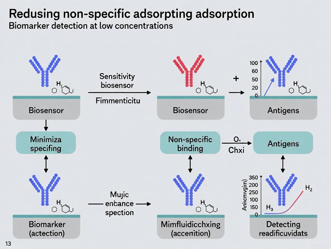
Visualization of NSA Mechanisms and Surface Modification
This diagram illustrates the fundamental difference between the desired specific binding and the problematic non-specific adsorption, highlighting the key intermolecular forces at play.
This diagram outlines a general workflow for developing a biosensor surface with reduced non-specific adsorption, incorporating both physical and chemical modification steps.
Troubleshooting Guides
Guide: Diagnosing and Remedying High Background Signal
Problem: Unusually high background signal is obscuring the specific detection of your target biomarker.
Explanation: A high background signal is a classic symptom of Non-Specific Adsorption (NSA), where proteins or other molecules in your sample matrix adhere to the biosensor surface through physisorption (hydrophobic forces, ionic interactions, van der Waals forces) rather than specific biorecognition [9] [3]. This fouling layer generates a signal that is often indistinguishable from your target's signal, leading to false positives and inaccurate quantification [3].
Solution Checklist:
| Step | Action | Rationale & Details |
|---|---|---|
| 1 | Verify Surface Passivation | Ensure your blocking step was performed correctly. If using Bovine Serum Albumin (BSA) or other protein blockers, confirm the solution was fresh and the incubation time was sufficient. Consider switching to or adding a chemical passivant like zwitterionic peptides [10]. |
| 2 | Analyze Sample Matrix | Complex samples like blood, serum, or cell lysates are prone to fouling. Implement or optimize sample pre-treatment steps such as centrifugation, dilution, or filtration to reduce the concentration of interfering substances [3]. |
| 3 | Incorporate Active Removal | For microfluidic biosensors, consider integrating active NSA removal methods. Apply acoustic waves or electromechanical transducers to generate surface shear forces that can physically shear away weakly adhered biomolecules [9]. |
| 4 | Check Bioreceptor Orientation | Mis-oriented immobilization of antibodies or aptamers can expose hydrophobic regions that promote NSA. Employ oriented immobilization strategies (e.g., using Protein A/G for antibodies, thiol-modified aptamers) to ensure the active binding site is fully available [9]. |
Guide: Addressing Signal Drift and Poor Reproducibility
Problem: The sensor's output signal drifts over time, or results are not reproducible across different sensor chips or assay runs.
Explanation: Signal drift and poor reproducibility are frequently caused by the progressive, non-specific accumulation of molecules on the sensing surface, which gradually degrades the interface [3]. This can lead to a continuous change in the baseline signal (drift) and inconsistent performance because the degree of fouling can vary between experiments [9] [11].
Solution Checklist:
| Step | Action | Rationale & Details |
|---|---|---|
| 1 | Evaluate Antifouling Coating Stability | The passive coating (e.g., Polyethylene Glycol - PEG) may be degrading. PEG is prone to oxidative degradation in biological media. Test more stable alternatives like zwitterionic polymers or peptides, which form a robust hydration layer [10]. |
| 2 | Standardize Regeneration Protocols | If re-using the sensor, a harsh regeneration step might not be fully removing the analyte and could be damaging the antifouling layer. Optimize the regeneration buffer (pH, ionic strength, surfactants) to gently elute the target without harming the surface chemistry [3]. |
| 3 | Control Microenvironment | Variations in pH, temperature, or ionic strength between runs can affect both the stability of the antifouling layer and the rate of NSA. Use buffered solutions consistently and control the assay temperature [3]. |
| 4 | Implement Real-time NSA Monitoring | For advanced setups like coupled Electrochemical-Surface Plasmon Resonance (EC-SPR) biosensors, use the dual-detection capability to monitor the formation of the fouling layer in real-time, allowing for more informed data correction [3]. |
Frequently Asked Questions (FAQs)
Q1: What is the fundamental mechanism behind NSA, and why is it such a persistent problem in biosensing?
A1: NSA occurs primarily through physisorption, driven by a combination of hydrophobic forces, ionic interactions, van der Waals forces, and hydrogen bonding between molecules in the sample and the biosensor surface [9] [3]. This is a persistent problem because most biosensor surfaces are inherently prone to these interactions. The issue is exacerbated when detecting low-concentration biomarkers in complex samples (e.g., blood, serum), where the number of interfering proteins can be billions of times higher than the target, making any small fraction of NSA significant enough to overwhelm the specific signal [3].
Q2: For electrochemical biosensors targeting low-concentration biomarkers in whole blood, what are the most promising antifouling strategies?
A2: For this challenging application, the most promising strategies involve a multi-pronged approach:
- Advanced Nanomaterial Coatings: The use of low-dimensional nanomaterials like graphene, carbon nanotubes, or MXenes can improve electron transfer and provide a tunable surface for functionalization [12] [13].
- Zwitterionic Chemistry: Modifying the electrode surface with zwitterionic peptides or polymers creates a strong hydration layer via electrostatic and hydrogen bonding. This layer acts as a highly effective physical and energetic barrier against protein adsorption, often outperforming traditional PEG coatings [10].
- Dual-Functionality Probes: Employing a dual-recognition strategy, such as combining an antibody with vancomycin for bacterial detection, can significantly enhance specificity and reduce false positives from complex samples without the need for extensive pre-treatment [13].
Q3: How can I quantitatively evaluate the effectiveness of a new antifouling coating in my biosensor?
A3: A robust evaluation requires a combination of methods:
- Quantitative Signal Change: Measure the signal response before and after exposing the coated sensor to a complex solution (e.g., 10% serum, GI fluid). Calculate the percentage of signal reduction compared to an uncoated sensor. A high-performance coating can reduce non-specific adsorption by more than 90% [10].
- Direct Comparison: Compare the signal generated by your target analyte in a clean buffer versus in the complex matrix. A good coating will show minimal difference in the signal, indicating resistance to matrix effects.
- Surface Characterization: Use techniques like X-ray Photoelectron Spectroscopy (XPS) or Ellipsometry to confirm the chemical composition and thickness of the coating, ensuring successful modification.
Table 1: Comparative Performance of Antifouling Materials for Biosensors
| Material/Strategy | Mechanism of Action | Key Performance Metrics | Ideal Use Case |
|---|---|---|---|
| Polyethylene Glycol (PEG) | Forms a hydrophilic, steric barrier that binds water via hydrogen bonding [10]. | Traditional "gold standard," but prone to oxidative degradation; can reduce ~80-90% NSA [10]. | General-purpose blocking for sensors used in buffered solutions or short-term assays. |
| Zwitterionic Peptides (e.g., EKEKEKEK) | Creates a net-neutral, super-hydrophilic surface that binds a tight hydration layer via electrostatic and hydrogen bonding [10]. | Superior to PEG; >90% reduction in protein adsorption; improves LOD by >10x vs. PEG in PSi sensors [10]. | Demanding applications in complex, undiluted biofluids (serum, GI fluid) and for long-term stability. |
| Bovine Serum Albumin (BSA) | Physically adsorbs to vacant surface sites, blocking them from further protein adsorption [3]. | Low-cost and easy to use; can be insufficient for very complex samples; effectiveness depends on surface coverage. | A quick, initial blocking step, often used in combination with other chemical passivants. |
| Dual-Target Recognition | Uses two distinct bioreceptors for the same target, requiring both to bind for a signal, minimizing false positives [13]. | Enabled specific detection of MRSA with LOD of 5.0 CFU mL⁻¹; high reproducibility (1.27% RSD) [13]. | Differentiating between closely related targets (e.g., antibiotic-resistant vs. susceptible strains) in complex matrices. |
Experimental Protocols
Protocol: Functionalizing a Porous Silicon (PSi) Biosensor with Zwitterionic Peptides
Purpose: This protocol details the covalent immobilization of an EK-based zwitterionic peptide onto a PSi surface to create a robust, antifouling layer for biomarker detection in complex fluids [10].
Materials:
- PSi chips (freshly prepared and thermally oxidized)
- Anhydrous toluene
- 3-aminopropyltriethoxysilane (APTES)
- Zwitterionic peptide (Sequence: EKEKEKEKEKGGC)
- N-Hydroxysuccinimide (NHS) and N-(3-Dimethylaminopropyl)-N'-ethylcarbodiimide (EDC)
- Phosphate Buffered Saline (PBS), pH 7.4
Procedure:
- Silane Functionalization: Hydrate the PSi chips in ethanol and then dry under a stream of nitrogen. Incubate the chips in a 2% (v/v) solution of APTES in anhydrous toluene for 4 hours at room temperature to form an amine-terminated monolayer. Rinse thoroughly with toluene and ethanol to remove unbound silane, and dry under nitrogen.
- Peptide Conjugation:
- Prepare a 1 mM solution of the zwitterionic peptide in PBS.
- Activate the terminal carboxylic acid group of the peptide by adding EDC and NHS to final concentrations of 5 mM and 2 mM, respectively. Allow the activation to proceed for 15-30 minutes.
- Incubate the amine-functionalized PSi chips in the activated peptide solution for 2-4 hours at room temperature.
- Washing and Storage: After incubation, rinse the chips extensively with PBS and deionized water to remove any physisorbed peptide. The functionalized chips can be stored in PBS at 4°C for short-term use.
Protocol: Building a Dual-Recognition Electrochemical Biosensor for Bacteria
Purpose: To construct an electrochemical biosensor that uses two distinct recognition elements (vancomycin and an antibody) for the highly specific and sensitive detection of Methicillin-resistant Staphylococcus aureus (MRSA) in a sample, minimizing false positives [13].
Materials:
- Screen-printed electrodes (SPE)
- Graphene (GR) dispersion
- Bovine Serum Albumin (BSA)
- Vancomycin (VAN)
- EDC/NHS crosslinking kit
- Anti-PBP2a antibody (specific to MRSA)
- AuNPs/MXene nanozyme composite
- o-Phenylenediamine (o-PD) and H₂O₂
Procedure:
- Electrode Modification: Drop-coat the graphene dispersion onto the SPE surface and allow it to dry to form a GR/SPE.
- First Recognition Layer: Adsorb BSA onto the GR/SPE. Then, chemically conjugate vancomycin to the amino groups of BSA using EDC/NHS chemistry, creating a VAN/BSA/GR/SPE.
- Second Recognition Probe: Decorate MXene nanosheets with AuNPs via in-situ reduction. Subsequently, immobilize the anti-PBP2a antibody onto the AuNPs/MXene via Au-S bonds to form the Anti-PBP2a/AuNPs/MXene detection probe.
- Assay Execution: Incubate the modified VAN/BSA/GR/SPE with the sample. If MRSA is present, it will be captured by the vancomycin anchor. Then, introduce the Anti-PBP2a/AuNPs/MXene probe, which will bind to the captured MRSA, bringing the MXene nanozyme to the electrode surface.
- Signal Measurement: Add a solution containing o-PD and H₂O₂. The MXene nanozyme will catalyze the oxidation of o-PD. Measure the reduction current of the oxidized product (o-PDox) using amperometry or differential pulse voltammetry. The current is proportional to the MRSA concentration [13].
Visualization: Systematic NSA Troubleshooting Workflow
Diagram: NSA Troubleshooting Workflow. This chart outlines a systematic approach to diagnosing and resolving two common NSA-related problems, guiding researchers from symptom identification to potential solutions and final validation.
The Scientist's Toolkit: Research Reagent Solutions
Table 2: Essential Materials for Advanced Antifouling Biosensor Research
| Reagent / Material | Function / Explanation | Key Considerations |
|---|---|---|
| Zwitterionic Peptides (EK repeats) | Covalently attached to sensor surfaces to form a net-neutral, highly hydrophilic layer that binds water strongly, creating a physical and energetic barrier against protein and cell adhesion [10]. | Superior stability and performance compared to PEG. Sequence (e.g., EKEKEKEKEKGGC) and length can be tuned for optimization [10]. |
| MXene-based Nanozymes | 2D nanomaterials (e.g., Ti₃C₂Tx) used for signal amplification. Possess peroxidase-like activity to catalyze substrate reactions, enhancing sensitivity. Also provide a large surface area for bioreceptor immobilization [13]. | Excellent hydrophilicity and conductivity. Can be composited with metal nanoparticles (e.g., AuNPs) for further functionalization [13]. |
| Dual-Recognition Probes | A pair of distinct bioreceptors (e.g., vancomycin + anti-PBP2a antibody) that bind to different sites on the same target. This strategy drastically improves specificity and reduces false positives in complex samples [13]. | Eliminates the need for complex sample pre-treatment to isolate the target from similar interferents. |
| Low-Dimensional Nanomaterials | Includes graphene, carbon nanotubes, and metal-organic frameworks (MOFs). Used to modify transducer surfaces to improve electron transfer, increase surface area, and enhance biocompatibility for electrochemical biosensors [12] [14]. | The structural diversity of these materials directly influences the ultimate sensitivity and specificity of the biosensor [12]. |
Non-specific adsorption (NSA) is a critical challenge in biosensing, particularly for the detection of low-concentration biomarkers in complex samples like blood, serum, and milk. NSA occurs when molecules undesirably adsorb to a biosensor's surface through physisorption, leading to elevated background signals, false positives, reduced sensitivity, and compromised reproducibility. The primary mechanisms driving this phenomenon are electrostatic interactions, hydrophobic forces, and van der Waals forces. Understanding and mitigating these interactions is fundamental to developing reliable biosensors for clinical diagnostics and drug development. This guide provides troubleshooting advice and methodologies to identify, understand, and counter these key mechanisms.
FAQs & Troubleshooting Guides
Q1: My biosensor shows high background signal in complex serum samples. Which NSA mechanism is most likely responsible and how can I confirm this?
A: Hydrophobic interactions are a common culprit with complex samples like serum. You can confirm this through a series of experimental tests:
- Change Ionic Strength: If increasing the salt concentration in your buffer increases NSA, it strongly indicates significant electrostatic interactions are present [3].
- Use Detergents: Introducing non-ionic detergents (e.g., Tween-20) can shield hydrophobic surfaces. A reduction in NSA suggests hydrophobic interactions are a major driver [3].
- Modify Surface Charge: If switching to a more negatively charged coating reduces NSA when testing positively charged proteins, it confirms the role of electrostatic interactions [1] [3].
Q2: I am getting false-positive responses in my electrochemical immunosensor. How can I determine if the issue is methodological rather than immunological?
A: Methodological NSA can arise from several factors related to surface physics and chemistry [1]. To troubleshoot, systematically check the following:
- Surface Passivation: Ensure all vacant spaces on your sensor substrate are effectively blocked with a reagent like BSA or casein to prevent molecules from adsorbing onto "sticky" spots [1].
- Bioreceptor Orientation: Mis-orientated antibodies can lead to non-specific binding. Use oriented immobilization strategies (e.g., using Protein A/G or Fc-specific tags) to ensure the antigen-binding sites are freely available [1].
- Substrate Stickiness: Test your bare sensor surface with a sample known to be free of your target analyte. A significant signal indicates an inherently sticky substrate that requires a better antifouling coating.
Q3: What are the most effective surface coatings to prevent NSA driven by these mechanisms?
A: The most effective coatings create a thin, hydrophilic, and neutrally charged boundary layer that minimizes all three intermolecular forces [1]. The optimal choice often depends on your transduction method (e.g., electrochemical vs. optical). The table below summarizes promising solutions.
| Material Type | Example Materials | Primary Mechanism Addressed | Key Feature |
|---|---|---|---|
| Polymer Brushes | Polyethylene glycol (PEG), Zwitterionic polymers | Hydrophobic interactions | Creates a hydrated, steric barrier |
| Self-Assembled Monolayers (SAMs) | Alkane thiols with terminal OH or EG groups | Electrostatic & van der Waals | Provides a dense, ordered, non-charged layer |
| Hydrogel Films | Cross-linked protein films, Peptide-based coatings | Hydrophobic & electrostatic interactions | 3D network that resists protein adsorption |
| Hybrid Materials | Conductive polymers with antifouling peptides | Combined mechanisms | Tunable conductivity and antifouling properties |
Experimental Protocols & Methodologies
Protocol: Evaluating NSA Contribution to Biosensor Signal
This protocol helps quantify the extent of NSA and its impact on your specific signal, which is vital for troubleshooting.
1. Principle: Compare the signal generated from a sample containing your target analyte to the signal from a control sample that is known to lack the analyte but is otherwise identical in matrix composition.
2. Reagents:
- Assay Buffer
- Sample containing target analyte (e.g., spiked serum)
- Control/Blank sample (e.g., plain serum, buffer)
- Blocking solution (e.g., 1% BSA)
3. Procedure:
- Step 1: Prepare the biosensor with its immobilized bioreceptor.
- Step 2: Expose the sensor to the control sample and record the signal over time. This signal is your NSA baseline.
- Step 3: Thoroughly wash the sensor with assay buffer to remove weakly adsorbed molecules.
- Step 4: Expose the sensor to the sample containing the target analyte and record the signal. This is your total signal.
- Step 5: The specific signal can be estimated as: Total Signal - NSA Baseline.
4. Data Analysis: A high NSA baseline relative to the specific signal indicates a poorly passivated surface. This protocol is applicable to various detection methods, including electrochemical (signal drift) and SPR (reflectivity change) [3].
Protocol: Systematic Troubleshooting for NSA
Follow this general troubleshooting process to logically identify the cause of NSA in your experiments [15].
1. Identify the Problem: Clearly define the symptom (e.g., "high background signal in negative controls"). 2. List All Possible Explanations: Brainstorm potential causes, including: * Ineffective blocking agent * Incorrect buffer ionic strength or pH * Sticky substrate material * Denatured or mis-oriented bioreceptors 3. Collect Data: Review your experimental notes. Check controls, reagent storage conditions, and procedure against manufacturer protocols. 4. Eliminate Explanations: Rule out causes that are not supported by your data (e.g., if positive controls worked, the core reagents are likely fine). 5. Check with Experimentation: Design targeted experiments to test remaining causes (e.g., test different blocking proteins or buffer additives). 6. Identify the Cause: Based on your experimentation, conclude the primary cause and implement a fix.
Research Reagent Solutions
The following table details essential materials used to mitigate NSA in biosensor research.
| Reagent/Solution | Function & Explanation |
|---|---|
| Bovine Serum Albumin (BSA) | A common blocking protein that passively adsorbs to vacant sites on the sensor surface, reducing NSA by providing a less sticky protein layer [1]. |
| Casein | A milk-derived protein used as a blocking agent, effective at reducing immunological and methodological NSA in assays like ELISA [1]. |
| Polyethylene Glycol (PEG) | Forms a hydrated, steric barrier on surfaces. Its high flexibility and hydrophilicity minimize hydrophobic and van der Waals interactions with approaching biomolecules [1]. |
| Zwitterionic Polymers | Materials like poly(carboxybetaine) create a super-hydrophilic surface through a strong water layer, effectively resisting protein adsorption via hydrogen bonding and ionic solvation [3]. |
| Tween 20 (Non-ionic Detergent) | Added to assay buffers to shield hydrophobic patches on surfaces and proteins, thereby reducing NSA driven by hydrophobic interactions [3]. |
| Self-Assembled Monolayers (SAMs) | Ordered molecular assemblies (e.g., of alkane thiols on gold) that create a dense, chemically defined surface which can be tailored with specific terminal groups (e.g., oligo-ethylene glycol) to resist fouling [1]. |
NSA Mechanisms and Mitigation Workflows
Experimental Workflow for NSA Evaluation
The Critical Need for NSA Reduction in Early Disease Diagnostics and Personalized Medicine
FAQs: Core Concepts for Researchers
What is non-specific adsorption (NSA) and why is it a critical problem in biomarker detection? Non-specific adsorption (NSA) refers to the unwanted binding of non-target molecules (like abundant proteins in serum) to detection surfaces such as immunoassay plates, sensors, or nanoparticles. This background noise severely obscures the signal from low-abundance target biomarkers, reducing assay sensitivity and specificity [16]. For context, a novel platform addressing NSA achieved a resolution of 50-60 picograms per milliliter, about 20 times more sensitive than traditional ELISA [16].
Which types of biomarkers are most affected by NSA? NSA is particularly detrimental when detecting low-abundance biomarkers, which are crucial for early disease diagnosis. Examples include:
- Neurological biomarkers in blood, such as Neurofilament Light Chain (NfL) or phosphorylated Tau for Alzheimer's disease, which exist at extremely low concentrations [17].
- Autoantibodies for early-stage autoimmune diseases [16].
- Volatile Organic Compounds (VOCs) in breath or other biofluids that act as metabolic signatures for cancers, COPD, and metabolic disorders [18].
What are the primary sources of NSA in a typical assay workflow? The main sources include:
- Solid Surfaces: The plastic of microtiter plates, sensor chips, and nanoparticles.
- Reagents: Enzymes, antibodies, and other detection molecules with inherent stickiness.
- Sample Matrix: High-abundance proteins (e.g., albumin, immunoglobulins), lipids, and other cellular debris in complex biofluids like blood, saliva, or urine [18] [17].
How does reducing NSA contribute to the goals of personalized medicine? Reducing NSA enhances the accuracy and reliability of diagnostic tests. This allows for:
- Earlier Diagnosis: Detecting diseases at lower biomarker concentrations, often before symptoms appear [16].
- Better Patient Stratification: More accurately identifying patient subgroups based on their molecular profiles for targeted therapies.
- Improved Treatment Monitoring: Precisely tracking minute changes in biomarker levels to assess drug efficacy [19] [20].
Troubleshooting Guides: Identifying and Resolving NSA Issues
Problem: High Background Signal
Symptoms:
- Elevated signal in negative controls and blank wells.
- Poor signal-to-noise ratio, making it difficult to distinguish true positive signals.
- High inter-assay and intra-assay variability.
Potential Causes and Solutions:
| Potential Cause | Recommended Solution | Principle |
|---|---|---|
| Inadequate Blocking | Use advanced blocking buffers containing engineered proteins or synthetic polymers. | Competitively occupies binding sites on the solid surface to prevent non-target adsorption [19]. |
| Inefficient Wash Stringency | Optimize wash buffer by adding mild detergents (e.g., Tween-20) or adjusting ionic strength. | Disrupts weak, non-specific ionic and hydrophobic interactions without eluting the specific immunocomplex [17]. |
| Antibody Cross-Reactivity | Re-validate antibody specificity using knockout controls or pre-absorb antibodies. | Ensures the primary and secondary antibodies bind only to the intended target epitope [18]. |
Problem: Inconsistent Results Between Replicates
Symptoms:
- High coefficient of variation (%CV) across replicate wells.
- Inability to reliably reproduce standard curves.
Potential Causes and Solutions:
| Potential Cause | Recommended Solution | Principle |
|---|---|---|
| Surface Heterogeneity | Source plates and sensors from a single, reputable supplier to ensure consistency. | Guarantees uniform binding chemistry and capacity across all reaction vessels [16]. |
| Variable Incubation Conditions | Standardize all incubation times, temperatures, and orbital shaking speeds. | Ensures consistent reaction kinetics and mass transfer for all samples and replicates [17]. |
| Sample Degradation | Establish standard operating procedures for sample collection, aliquoting, and freeze-thaw cycles. | Preserves biomarker integrity and prevents the generation of heterogeneous breakdown products that can bind non-specifically [18]. |
Problem: Failure to Detect Low-Abundance Targets
Symptoms:
- Signal below the limit of detection despite known presence of the biomarker.
- Assay fails to achieve the sensitivity reported in the literature.
Potential Causes and Solutions:
| Potential Cause | Recommended Solution | Principle |
|---|---|---|
| Signal Amplification Insufficiency | Implement Tyramide Signal Amplification (TSA) or switch to a digital ELISA platform. | TSA dramatically increases the number of reporter enzymes per binding event; digital ELISA allows for single-molecule counting [16] [17]. |
| Biomarker Loss to Vessels | Use low-bind tubes and plates made of polypropylene or specially coated polymers. | Minimizes passive adsorption of the target biomarker itself to container walls during sample preparation and storage [19]. |
| Matrix Interference | Dilute the sample or implement a pre-processing clean-up step (e.g., spin filtration, solid-phase extraction). | Reduces the concentration of interfering substances from the sample matrix that contribute to NSA [18]. |
Experimental Protocols for NSA Evaluation and Mitigation
Protocol 1: Systematic Evaluation of Blocking Agents Using a Model System
This protocol helps identify the optimal blocking agent for your specific assay system.
Research Reagent Solutions:
| Item | Function |
|---|---|
| Low-Bind Microtiter Plates | Minimizes passive adsorption of proteins to the plate surface. |
| Recombinant Target Biomarker | Provides a known positive control. |
| BSA, Casein, Fish Skin Gelatin | Traditional protein-based blocking agents. |
| SynBlock, PEI-based Polymers | Synthetic polymer-based blocking agents. |
| Fluorescently-Labeled Detection Antibody | Allows for quantitative signal measurement. |
| Plate Reader (Fluorescence) | For detecting and quantifying the assay signal. |
Methodology:
- Coat the plate with your capture antibody as per standard protocol.
- Block different wells with different candidate blocking buffers (e.g., 1% BSA, 2% Casein, a commercial synthetic blocker) for 1 hour at room temperature.
- Add a negative control (sample matrix without the biomarker) and a weak positive control (a low concentration of your recombinant biomarker).
- Proceed with your standard detection steps using a fluorescently-labeled antibody.
- Measure the fluorescence. Calculate the signal-to-noise ratio (Signal from Positive / Signal from Negative) for each blocking agent.
- Select the blocking agent that yields the highest signal-to-noise ratio for subsequent experiments.
Protocol 2: Incorporating Signal Amplification for Ultrasensitive Detection
This protocol outlines how to integrate Tyramide Signal Amplification (TSA) into a standard ELISA workflow, as demonstrated by Lei et al. [16].
Methodology:
- Perform the standard "sandwich" ELISA steps: capture antibody coating, blocking, sample incubation, and incubation with a biotinylated detection antibody.
- Incubate with Streptavidin-Conjugated Horseradish Peroxidase (SA-HRP).
- Amplify: Instead of adding the standard HRP substrate, incubate with a tyramide-biotin solution. HRX catalyzes the deposition of numerous biotin molecules onto the immunocomplexes nearby.
- Label: Introduce an Alkaline Phosphatase (ALP) enzyme conjugated to streptavidin, which will bind to the deposited biotins.
- Detect: Add the ELFP substrate. ALP breaks it down to form fluorescent microneedles. Use a microscope to capture images and count the fluorescent particles, which correspond to the amount of biomarker present [16].
TSA-Enhanced ELISA Workflow: Diagram illustrating the key steps in the Tyramide Signal Amplification process integrated into a standard ELISA, leading to a digital, countable output.
Research Reagent Solutions: Essential Materials for NSA Reduction
The following table details key reagents and materials critical for experiments focused on minimizing NSA.
| Item | Function/Benefit | Example Applications |
|---|---|---|
| Polymer-Based Blocking Agents (e.g., SynBlock, PVP) | Often more effective than proteins at passivating surfaces; less likely to create a sticky protein layer. | Reducing background in plate-based immunoassays and on biosensor surfaces [19]. |
| Low-Bind Tubes & Plates (e.g., polypropylene, COC polymer) | Surface treatment minimizes protein binding, preserving low-concentration analytes. | Sample storage and preparation for low-abundance biomarker assays to prevent analyte loss [17]. |
| Tyramide Signal Amplification (TSA) Kits | Enables significant signal amplification by depositing numerous reporter molecules per binding event. | Ultrasensitive detection of low-abundance biomarkers in ELISA or immunohistochemistry [16]. |
| Digital ELISA/Single Molecule Array (Simoa) | A revolutionary platform that isolates immunocomplexes in femtoliter wells for digital counting, drastically reducing the impact of background noise. | Detecting neurological biomarkers in blood at sub-picogram levels for research and clinical trials [17]. |
| Functionalized Nanoparticles & QDs | Can be engineered with specific surface chemistry to minimize NSA and serve as highly visible detection probes. | Used as labels in biosensors and assays for high-resolution, multiplexed biomarker detection [19] [20]. |
NSA Reduction Logic: A conceptual map showing how different strategies converge to solve the core problem of detecting low-abundance biomarkers amidst a complex sample matrix.
Practical Strategies: From Passive Coatings to Active Removal and Advanced Materials
Frequently Asked Questions
What is non-specific adsorption (NSA) and why is it a problem in biosensing? Non-specific adsorption (NSA) occurs when molecules other than your target analyte (such as proteins, DNA, or other biomolecules present in complex samples like serum or blood) adhere to the biosensor's surface [3]. This biofouling leads to false-positive signals, increased background noise, reduced sensitivity and specificity, and poor reproducibility, which can critically compromise the reliability of your assay, especially when detecting low-concentration biomarkers [9] [1] [21].
How do passive blocking methods work to reduce NSA? Passive blocking methods work by pre-coating the biosensor surface with a layer of molecules that occupy the binding sites that would otherwise be available for non-specific interactions. The goal is to create a thin, hydrophilic, and neutrally charged boundary layer that minimizes unwanted intermolecular forces (e.g., hydrophobic, electrostatic, van der Waals), making it difficult for foulants to adsorb [9] [1]. When a washing step is applied, these weakly adhered molecules are easily removed [9].
When should I choose a protein-based blocker over a chemical linker? The choice is often empirical and depends on your specific assay conditions and the nature of your sensor surface [21].
- Protein blockers like BSA and casein are a good first choice for many traditional immunoassays (e.g., ELISA) and are highly effective at blocking residual protein-binding sites on a variety of surfaces [1] [22].
- Chemical linkers like PEG or zwitterionic polymers are often preferred for modern biosensors where control over the surface chemistry is critical. They can form dense, well-hydrated layers that provide superior antifouling properties, particularly in complex biological fluids [9] [21]. They are also chosen when cross-reactivity from a protein blocker is a concern [21].
A common problem I face is that my blocking agent seems to be interfering with the specific signal from my bioreceptor. What can I do? This can occur if the blocking agent is not optimized for your system. We recommend:
- Titrate the concentration of your blocking agent. High concentrations can sometimes sterically hinder the bioreceptor [21].
- Try a different type of blocker. If using BSA causes issues, switch to gelatin or a polymer like PEG, which may have different steric and chemical properties [21].
- Include a surfactant. Adding a mild, non-ionic detergent like Tween 20 or Triton X-100 to your blocking buffer can help reduce hydrophobic interactions and improve washing efficiency without disrupting specific binding [21] [22].
- Re-evaluate your bioreceptor immobilization strategy. Ensure your capture probes (e.g., antibodies, DNA) are densely and correctly oriented on the surface to outcompete non-specific binding.
The performance of my biosensor degrades when I test in complex matrices like blood serum. How can I improve its robustness? This is a key challenge in translational research. Beyond optimizing a single blocking agent, consider a combined or layered approach:
- Use a chemical linker (e.g., a PEG-based SAM) to create a robust, non-fouling base layer.
- Immobilize your bioreceptor onto this layer.
- Follow with a secondary blocking step using a protein blocker like BSA or casein to seal any remaining vacant sites on the surface or on the bioreceptor itself [21].
- Always validate your biosensor's performance by spiking your target biomarker in the complex matrix (e.g., fetal bovine serum) and compare the signal to that in a clean buffer [21].
Experimental Protocols & Optimization Guides
Protocol 1: Optimizing a Blocking Agent for an Electrochemical DNA Biosensor
This detailed protocol, adapted from a study on a miRNA biosensor for ovarian cancer, provides a method to systematically compare blocking agents [21].
1. Sensor Surface Preparation:
- Functionalize carbon screen-printed electrodes (SPEs) with cysteamine hydrochloride.
- Decorate the surface with citrate-reduced gold nanoparticles (AuNPs).
- Immobilize a 5'-amine-modified ssDNA probe specific to your target (e.g., miRNA-204) onto the AuNP-functionalized surface.
2. Preparation of Blocking Buffers:
- Prepare a set of candidate blocking solutions. The cited study tested the following in 0.01 M PBS (pH 7.4) [21]:
- 1% Bovine Serum Albumin (BSA) in 0.5% Tween 20
- 1% Gelatin in 0.5% Tween 20
- 1% Polyethylene Glycol (PEG) 4000 in 0.5% Tween 20
- 1% PEG 6000 in 0.5% Tween 20
- Other surfactants like Triton X-100 or buffers like HEPES can also be evaluated.
3. Blocking and Washing:
- Incubate the fabricated biosensor (after probe immobilization) with your different blocking buffers for a set time (e.g., 1 hour at room temperature).
- Gently wash the sensor with a clean buffer (e.g., PBS or Tris-EDTA) to remove unbound blocking agents.
4. Performance Evaluation:
- Test the blocked biosensors by measuring the response to your target analyte (e.g., miRNA-204) spiked into both a simple buffer (0.01 M PBS) and a complex matrix (Fetal Bovine Serum, FBS).
- Use chronoamperometry to record the current response.
- Key Metric: The difference in the saturation current between the curves obtained in PBS and FBS should be minimal for an effective blocking agent, indicating negligible non-specific binding from the serum components [21].
5. Interference Analysis:
- To validate specificity, challenge the optimally blocked biosensor with a fixed concentration of the target analyte in the presence of potential interferents (e.g., other miRNAs, DNA, proteins) and measure the signal change.
Protocol 2: Standard Procedure for Blocking in Plate-Based Assays (e.g., ELISA)
This is a general protocol for reducing NSA in plate-based assays, which can be adapted for biosensor surfaces [22].
1. Surface Coating:
- Immobilize one member of the binding pair (e.g., a protein or antibody) onto the plate surface via passive adsorption or capture.
2. Blocking:
- Aspirate the coating solution.
- Add an excess volume (e.g., 200-300 µL for a 96-well plate) of your chosen blocking buffer to all wells.
- Common Blocking Buffers:
- 1-5% BSA in PBS-T (PBS with 0.05% Tween 20)
- 1-5% Casein in PBS
- 1% Gelatin in PBS-T
- Commercial protein-free blocking buffers
- Incubate for 1-2 hours at room temperature or overnight at 4°C with gentle shaking.
3. Washing:
- Aspirate the blocking buffer.
- Wash the plate 3-5 times with a wash buffer (e.g., PBS-T) to remove residual blocker.
4. Assay:
- Proceed with adding the sample and detection reagents as per your standard assay protocol.
Table 1: Comparison of Common Protein-Based Blocking Agents
| Blocking Agent | Molecular Weight | Key Mechanism | Advantages | Disadvantages & Considerations | Optimal Use Case |
|---|---|---|---|---|---|
| Bovine Serum Albumin (BSA) | ~66 kDa [21] | Adsorbs to surfaces, masking charged and hydrophobic sites [1]. | Widely used, effective, inexpensive [1]. | Can exhibit cross-reactivity with some targets; may bind some drug leads [21] [22]. | General purpose; immunoassays like ELISA. |
| Casein | ~20-25 kDa (subunits) | Forms a layer that sterically hinders NSA [1]. | Very effective, low cross-reactivity, inexpensive. | Can be less soluble and more viscous; source (e.g., from non-fat milk) can vary. | Immunoassays, Western blotting. |
| Gelatin | ~40-100 kDa (mixture) | Protein mixture that coats surfaces to prevent NSA. | Low cross-reactivity [21]. | Can be less effective alone; performance increases with surfactants [21]. | DNA biosensors (shown effective with Tween 20) [21]. |
Table 2: Comparison of Common Chemical Blocking Agents / Linkers
| Blocking Agent | Type / Structure | Key Mechanism | Advantages | Disadvantages & Considerations | Optimal Use Case |
|---|---|---|---|---|---|
| Polyethylene Glycol (PEG) | Polymer (various MW) | Forms a hydrated, steric barrier that repels biomolecules [9] [21]. | Tunable properties by MW; high antifouling efficiency; non-ionic. | Shorter chains form dense monolayers; longer chains can bend and be less effective [21]. | Coating hydrophobic surfaces; creating non-fouling base layers. |
| Zwitterionic Polymers | Polymers with mixed charges | Creates a strong hydration layer via electrostatic interactions [9]. | Extremely low fouling; very stable surface. | More complex surface chemistry for immobilization. | High-performance biosensors for complex media (serum, blood). |
| Self-Assembled Monolayers (SAMs) | Ordered molecular films | Creates a controlled, dense, and oriented surface that minimizes NSA [9]. | Highly reproducible and well-defined surface properties. | Limited to specific substrates (e.g., gold, silica). | Fundamental studies and advanced biosensor design. |
Table 3: Performance of Optimized Blocking Agents in a miRNA Biosensor Data derived from a study optimizing blocking for an electrochemical DNA biosensor in Fetal Bovine Serum (FBS) [21].
| Blocking Agent | Key Finding | Recommended Concentration | Note |
|---|---|---|---|
| Gelatin | Optimum blocking agent for this DNA biosensor, providing negligible nonspecific binding in FBS [21]. | 1% in Tween 20 [21] | Performance enhanced by the surfactant. |
| Bovine Serum Albumin (BSA) | Exhibited good blocking characteristics. | 1% in Tween 20 [21] | The conventional choice, but was outperformed by gelatin in this specific application. |
| Polyethylene Glycol (PEG) | Effective as an alternative blocking agent. | 1% of MW 4000 or 6000 in Tween 20 [21] | Shorter chains (PEG 4000) may form denser monolayers. |
The Scientist's Toolkit: Essential Research Reagents
Table 4: Key Reagents for Implementing Passive Blocking Methods
| Reagent / Material | Function in Blocking | Brief Explanation |
|---|---|---|
| Bovine Serum Albumin (BSA) | Protein Blocker | A versatile blocking protein that adsorbs to a wide range of surfaces, effectively passivating uncoated plastic, glass, or metal to prevent NSA of proteins [1] [21]. |
| Casein / Non-Fat Dry Milk | Protein Blocker | A mixture of phosphoproteins that forms a sticky, impermeable layer on surfaces, excellent for blocking in immunoassays like Western blots and ELISAs [1]. |
| Polyethylene Glycol (PEG) | Polymer Blocker | A hydrophilic polymer that, when grafted onto a surface, creates a hydrated "brush" or "monolayer" that sterically repels other biomolecules, reducing fouling [9] [21]. |
| Tween 20 | Non-ionic Surfactant | Added to blocking and wash buffers to reduce hydrophobic interactions and disrupt weak, non-specific binding, thereby lowering background signal [21] [22]. |
| Cysteamine / SAMs | Chemical Linker | A small molecule that forms a self-assembled monolayer on gold surfaces, providing a well-defined platform for further functionalization with bioreceptors and blocking agents [21]. |
| Gelatin | Protein Blocker | A mixture of peptides and proteins derived from collagen, useful for blocking in various assays, particularly when used in combination with surfactants [21]. |
Workflow Visualization
Diagram Title: Experimental Workflow for Blocking Agent Optimization
Diagram Title: Mechanism of Passive Blocking on a Biosensor Surface
Non-specific adsorption (NSA) is a persistent challenge that negatively affects biosensors by decreasing their sensitivity, specificity, and reproducibility. This is particularly problematic in low-concentration biomarker detection research, where distinguishing true signals from background noise is crucial. While passive methods, such as coating surfaces with blocker proteins like BSA or casein, have been used for decades, a significant shift toward active removal methods has emerged in the past decade. These techniques dynamically remove undesired molecules after they have adhered to the sensor surface, offering enhanced control and efficiency for demanding applications in diagnostic biomarker research and drug development [1] [9].
Active removal methods function by generating physical forces that overpower the adhesive forces binding non-specifically adsorbed molecules to the sensor surface. These techniques can be broadly categorized into transducer-based methods (electromechanical and acoustic) and fluid-based methods (hydrodynamic shear). This technical support article provides detailed troubleshooting guides, FAQs, and experimental protocols to help researchers effectively implement these advanced techniques in their experiments [1].
The following table summarizes the key characteristics of the three primary active removal techniques.
Table 1: Comparison of Active NSA Removal Techniques
| Technique | Fundamental Principle | Typical Force Generation Method | Key Advantages | Considerations for Low-Concentration Biomarkers |
|---|---|---|---|---|
| Electromechanical | Applies tunable alternating current electro-hydrodynamic (ac-EHD) forces to create localized "nano-shearing" fluid motion near the electrode surface [23]. | Application of an AC electric field across asymmetric planar and microtip electrode pairs [23]. | Externally tunable force; effective for displacing weakly bound cells; can be integrated into microfluidic devices [23]. | High specificity for removing nonspecific cellular analytes; demonstrated ~4-fold reduction in nonspecific blood cell adsorption [23]. |
| Acoustic | Generates mechanical surface waves (e.g., Love waves, thickness shear modes) that create surface forces to shear away weakly adhered biomolecules [1] [24]. | Input interdigitated transducers (IDTs) on a piezoelectric substrate (e.g., quartz) generate high-frequency acoustic waves [24]. | Sensitive to both mass adsorption and viscoelastic changes in adsorbed layers; can distinguish between different structural forms (e.g., vesicles vs. bilayers) [24]. | High operating frequency (100-500 MHz) provides high sensitivity to surface perturbations; probed layer depth of 25-56 nm minimizes bulk interference [24]. |
| Hydrodynamic Shear | Relies on pressure-driven fluid flow to generate shear forces at the sensor surface, physically detaching adsorbed molecules [1] [25]. | Controlled perfusion or flow through microfluidic channels or chambers [25]. | Conceptually simple; integrates seamlessly with microfluidic biosensors; force can be precisely controlled via flow rate [1] [25]. | Enhanced calcium deposition in tissue engineering was directly correlated with perfusion rate, demonstrating dose-dependent effect [25]. |
Troubleshooting Guides
Common Experimental Issues and Solutions
Problem: Low Specificity in Cell Capture
- Description: Despite using an active removal technique, non-specifically bound cells (e.g., blood cells) remain, obscuring the detection of rare target cells (e.g., circulating tumor cells).
- Potential Causes and Solutions:
- Insufficient Shear Force: The applied force is too weak to displace nonspecific adhesions. For electrohydrodynamic methods, try increasing the AC electric field amplitude or frequency in a controlled manner [23].
- Incorrect Buffer Composition: The ionic strength or pH of the buffer may be strengthening non-specific ionic interactions. Optimize the buffer conditions to weaken non-specific binding without affecting specific capture probe interactions.
- Fouled Electrode/Surface: Contamination on the electrode or sensor surface can promote stickiness. Implement a rigorous cleaning protocol before experiments (e.g., using a 2% Hellmanex detergent solution for silica surfaces) [24].
Problem: Damage to Sensitive Surface Layers or Captured Analytes
- Description: The active removal process is damaging fragile surface functionalizations (e.g., supported lipid bilayers - SLBs) or lysing captured target cells.
- Potential Causes and Solutions:
- Excessive Shear Force: The applied force is too strong. For hydrodynamic methods, systematically reduce the perfusion rate or flow velocity. For acoustic methods, reduce the power input to the transducer [25].
- Overly Long Application: Continuous application of high shear stress can cause cumulative damage. Consider using pulsed or oscillating flow/signals instead of continuous operation.
- Incompatible Substrate: The surface may not be adequately prepared to withstand the forces. Ensure the waveguide or sensor surface is smooth and properly functionalized to promote stability during shear [24].
Problem: Inconsistent or Unreproducible NSA Removal
- Description: The efficiency of non-specific adsorption removal varies significantly between experimental runs.
- Potential Causes and Solutions:
- Flow Instability: In hydrodynamic systems, transitioning to turbulent flow can create uneven shear forces. Ensure operating conditions maintain laminar flow (e.g., confirm Reynolds number is within laminar range) [25].
- Air Bubbles in Microfluidics: Bubbles can disrupt flow paths and shear profiles. Implement degassing protocols for buffers and include bubble traps in the fluidic setup.
- Electrode Degradation: In electrohydrodynamic devices, electrode fouling or passivation can alter the electric field over time. Clean electrodes regularly and validate their performance.
Frequently Asked Questions (FAQs)
Q1: Why should I use active removal methods instead of traditional passive blocking with BSA? Passive methods like BSA blocking are a good first line of defense but can be incomplete and may not be compatible with all sensor surfaces or miniaturized formats. Active methods provide a dynamic, physical means to remove adhered contaminants, offering a higher level of control and often greater effectiveness, especially in complex samples like blood [1] [23].
Q2: How do I choose the best active removal technique for my specific biosensor? The choice depends on your sensor platform and application.
- Electromechanical (ac-EHD): Ideal for microfluidic devices with integrated electrodes, especially when working with cellular analytes and needing tunable, nano-scale shearing [23].
- Acoustic: Best for label-free detection systems that are sensitive to mass and viscoelasticity. Excellent for studying the formation of soft films like supported lipid bilayers and their interactions with proteins [24].
- Hydrodynamic Shear: The most straightforward method to implement in any microfluidic-based biosensor. It is excellent for generating uniform, quantifiable shear forces across a surface by controlling flow rate [1] [25].
Q3: Can active removal techniques damage my specifically captured biomarkers? This is a valid concern. The goal is to tune the physical forces (shear, acoustic, electrohydrodynamic) to be strong enough to disrupt the weaker, non-specific physisorption (e.g., van der Waals, hydrophobic forces) but not the stronger, specific binding (e.g., antibody-antigen covalent-like binding). This requires careful optimization of parameters like flow rate, acoustic power, or electric field strength for each specific assay [1] [26].
Q4: What are the key parameters to optimize when setting up a hydrodynamic shear experiment? The most critical parameter is the flow rate, which directly determines the wall shear stress. Use computational fluid dynamics (CFD) simulations or established equations for your chamber geometry (e.g., parallel-plate) to relate flow rate to shear stress. Start with lower shear stresses and gradually increase until nonspecific adsorption is reduced without affecting specific binding [25] [27].
Detailed Experimental Protocols
Protocol 1: Implementing Hydrodynamic Shear in a Perfusion Chamber
This protocol is adapted from studies on tissue-engineered bone to illustrate the dose-dependent effect of shear stress on surface deposition [25].
Objective: To systematically evaluate the effect of hydrodynamic shear stress on the reduction of non-specific adsorption in a microfluidic biosensor channel.
Materials:
- Research Reagent Solutions:
- Titanium-fiber mesh scaffold or functionalized sensor surface: Provides a high-surface-area substrate for immobilization [25].
- Culture medium or sample buffer: The fluid carrying the analyte and potential interferents.
- Dextran molecules (optional): Used to increase medium viscosity without altering nutrient transfer, allowing for isolated study of shear stress effects [25].
- Syringe pump or precision perfusion system: To generate controlled, continuous flow.
- Parallel-plate flow chamber or microfluidic device: Designed to ensure laminar flow and uniform shear stress across the sensor surface.
- Tubing and connectors: Chemically inert and sized to prevent bubble formation.
Methodology:
- Surface Preparation: Functionalize the sensor surface inside the chamber or scaffold with your specific capture probe (e.g., antibody).
- System Setup: Connect the flow chamber to the perfusion system. Ensure all connections are secure and prime the entire system with buffer to remove air bubbles.
- Baseline Measurement (Static Condition): Expose the surface to a complex sample (e.g., serum-spiked buffer) under static conditions for a set time. Wash gently and measure the non-specific background signal.
- Perfusion Experiment:
- Set the syringe pump to the lowest desired flow rate. Calculate the corresponding wall shear stress. For example, superficial velocities in the range of 64 μm/s to 640 μm/s have been used to demonstrate a direct correlation with calcium deposition [25].
- Perfuse the sample through the chamber at this constant rate for the duration of the experiment.
- After perfusion, wash the chamber with buffer and measure the signal.
- Data Collection and Analysis: Repeat Step 4 at incrementally higher flow rates. Plot the measured non-specific adsorption (background signal) against the calculated wall shear stress to determine the optimal operating condition.
Protocol 2: Applying AC Electro-Hydrodynamic "Nano-Shearing"
This protocol is based on the method for tunable nanoshearing to displace nonspecific cell adhesion [23].
Objective: To capture rare cells (e.g., CTCs) from whole blood with high specificity by using ac-EHD forces to minimize nonspecific blood cell adsorption.
Materials:
- Research Reagent Solutions:
- Purpose-built microfluidic device with asymmetric electrode pairs: Contains planar and microtip electrodes to generate the non-uniform electric field required for ac-EHD flow [23].
- Phosphate Buffered Saline (PBS): For washing and dilution.
- Whole blood sample: Anticoagulated blood, potentially spiked with cultured target cells.
- Lysis buffer (optional): For lysing red blood cells to reduce sample complexity.
- Function generator or sourcemeter: To apply the AC electric field across the electrodes.
- Microscope: For visualizing cell capture and non-specific adhesion in real-time.
Methodology:
- Device Priming: Prime the microfluidic device with PBS to wet the channels and electrodes.
- Sample Introduction: Introduce the whole blood sample into the device under no electric field and allow it to incubate briefly for initial cell-surface contact.
- ac-EHD Application:
- Apply an AC electric field (e.g., ~10 kHz, ~5 Vpp) to the asymmetric electrodes. This generates lateral fluid motion (nanoshearing) within nanometers of the electrode surface.
- The tunable shear force will displace weakly bound, nonspecific cells while leaving strongly bound, specific captures intact.
- Washing and Analysis:
- Continue the ac-EHD application while washing the channel with buffer to remove displaced cells.
- Turn off the electric field and quantify the number of captured target cells and the number of remaining non-specifically adhered blood cells. The method has been shown to achieve a 4-fold reduction in nonspecific adsorption and ~87% capture efficiency of target cancer cells [23].
Signaling Pathways and Workflow Visualizations
Diagram 1: Logical workflow for applying active removal techniques to enhance biomarker detection specificity.
The Scientist's Toolkit: Essential Research Reagent Solutions
Table 2: Key Materials for Implementing Active Removal Techniques
| Item Name | Function/Brief Explanation | Example Application/Note |
|---|---|---|
| Asymmetric Electrode Pairs | Generates a non-uniform AC electric field to create tunable electro-hydrodynamic "nano-shearing" forces near the sensor surface [23]. | Critical for electromechanical (ac-EHD) removal of nonspecifically adsorbed cells [23]. |
| Love Wave Acoustic Device | A high-frequency surface acoustic wave (SAW) device that generates shear horizontal waves, sensitive to mass adsorption and viscoelastic changes on its surface [24]. | Operating frequency of 100-500 MHz probes a layer depth of 25-56 nm, ideal for studying soft films like lipid bilayers [24]. |
| Precision Syringe Pump | Provides accurate and steady pressure-driven flow in microfluidic channels, enabling controlled hydrodynamic shear experiments [25]. | Allows for systematic correlation between flow rate/shear stress and NSA reduction. |
| Dextran Molecules | Used to increase the viscosity of the perfusion medium without altering its chemical composition or nutrient transfer properties [25]. | Enables isolation of shear stress effects from mass transfer effects in hydrodynamic studies [25]. |
| Hellmanex Detergent | A potent cleaning agent used to create a clean, hydrophilic surface on silica waveguides, which is essential for consistent sensor performance and SLB formation [24]. | Ensures a reproducible starting surface, free of contaminants that promote NSA. |
Troubleshooting Guide: Addressing Common Experimental Challenges
This guide provides solutions to frequent issues encountered when working with Low-Dimensional Nanomaterials (LDNs) and Molecularly Imprinted Polymers (MIPs) for biosensing applications, specifically focused on reducing non-specific adsorption (NSA) in complex biological samples.
Table 1: Troubleshooting Common Issues with MIPs and Nanomaterials
| Problem | Possible Cause | Solution |
|---|---|---|
| High Background Signal/Noise | Non-specific adsorption (NSA) on non-imprinted sites or nanomaterial surface [28] [1]. | Implement electrostatic modification with surfactants like SDS or CTAB [28] [29]. Use blocking agents like BSA or casein on non-active areas [1]. |
| Poor Selectivity of MIPs | Incomplete template removal or non-specific binding sites [28]. | Optimize template extraction protocol. Apply surface imprinting techniques to create more accessible and specific cavities [30]. |
| Agglomeration of Nanomaterials | High surface energy and strong van der Waals forces in LDNs [31]. | Employ surface modification with suitable surfactants or polymers to create a physical barrier [31]. Utilize synergistic dispersion with a co-supporting nanomaterial [31]. |
| Low Sensitivity in Detection | Inefficient electron transfer or poor accessibility of binding sites [32] [29]. | Integrate conductive LDNs (e.g., graphene, MXene) into the sensor platform [33] [34]. Ensure MIP synthesis parameters (e.g., scan number in electropolymerization) are optimized to create a thin, porous polymer layer [29]. |
| Irreproducible Sensor Results | Inconsistent nanomaterial dispersion or uneven MIP film thickness [31] [29]. | Standardize synthesis protocols (e.g., monomer concentration, polymerization time/temperature) [28]. Use controlled electropolymerization for precise MIP film deposition [29]. |
Frequently Asked Questions (FAQs)
Q1: What are the most effective strategies to minimize non-specific adsorption in MIP-based sensors? There are two primary categories of strategies:
- Passive Methods: These aim to prevent NSA by coating the surface. Common approaches include using protein blockers like Bovine Serum Albumin (BSA) or casein, and chemical coatings that create a thin, hydrophilic, and non-charged boundary layer [1].
- Active Methods: These dynamically remove adsorbed molecules after functionalization. Techniques include generating surface shear forces using transducers (electromechanical or acoustic) or through hydrodynamic fluid flow in microfluidic devices [1]. A highly effective passive method is the electrostatic modification of MIPs with surfactants. For example, modifying a positively charged MIP with the anionic surfactant Sodium Dodecyl Sulfate (SDS) can effectively neutralize external functional groups responsible for NSA, thereby significantly enhancing selectivity [28] [29].
Q2: How can I improve the dispersion of low-dimensional nanomaterials in a polymer matrix for composite fabrication? Poor dispersion due to agglomeration is a major challenge. Key strategies include:
- Surface Modification: Introducing modifiers (e.g., surfactants, silanes) that adsorb or bond to the nanomaterial's surface, creating a physical or electrostatic barrier that prevents aggregation [31].
- Synergistic Dispersion: Constructing a co-dispersion system using two different nanomaterials (e.g., graphene oxide and silicon-doped hydroxyapatite). Their co-supporting structure increases spatial distance and weakens the interaction force between individual nanoparticles [31].
- In Situ Growth: Synthesizing one nanomaterial directly on the surface of another to form a steric hindrance effect that prevents aggregation [31].
Q3: Why are low-dimensional nanomaterials particularly advantageous for sensing low-concentration biomarkers? LDNs possess several critical properties that make them ideal for this task:
- High Surface-to-Volume Ratio: Their nanoscale dimensions provide an immense surface area per unit mass, allowing for a higher density of biorecognition elements (e.g., antibodies, MIPs) and maximizing the interaction with target biomarkers [33] [32].
- Unique Physicochemical Properties: Properties like excellent electrical conductivity (graphene, MXene), catalytic activity, and quantum confinement effects can be harnessed to create highly sensitive signal transduction mechanisms (electrochemical, optical) [33] [34].
- Tunable Surface Chemistry: Their surfaces can be readily functionalized with various groups to improve biocompatibility, enhance binding specificity, and reduce non-specific adsorption [31].
Q4: What are the key considerations when designing a MIP for a specific biomarker? The design process involves careful selection of several components:
- Template: The target biomarker itself or a fragment (epitope) of it.
- Functional Monomers: Chosen to interact (covalently or non-covalently) with the template during pre-polymerization. The monomer should provide complementary functional groups to the target [28] [30].
- Cross-linker: Creates a rigid polymer network that stabilizes the imprinted cavities after template removal. The cross-linking density affects the stability and accessibility of the binding sites [28].
- Polymerization Method: Common methods include bulk, precipitation, and electrochemical polymerization. Electro-polymerization is advantageous for sensor integration as it allows easy control of film thickness and morphology directly on the transducer surface [29].
Experimental Protocol: Surfactant Modification of MIPs to Suppress NSA
The following detailed protocol is adapted from recent studies for creating a MIP sensor with reduced NSA for biomarker detection [28] [29].
Objective: To synthesize a MIP for a target analyte (e.g., an amino acid like Tryptophan) and subsequently modify it with a surfactant to eliminate non-specific adsorption, thereby enhancing sensor selectivity.
Materials:
- Functional Monomers: Aniline (for conductive polymer), Methacrylic acid (for non-conductive polymer).
- Cross-linker: Ethylene glycol dimethacrylate (EGDMA).
- Template Molecule: L-Tryptophan (Trp).
- Surfactant: Sodium Dodecyl Sulfate (SDS).
- Initiator: Ammonium persulfate (APS).
- Solvent: Appropriate buffer or organic solvent (e.g., phosphate buffer, acetonitrile).
- Electrode: Glassy carbon electrode (GCE) or gold electrode.
Procedure:
Step 1: Synthesis of Molecularly Imprinted Polymer (MIP)
- Preparation of Pre-polymerization Complex: Dissolve the template molecule (Trp, 0.5 mmol) and functional monomer (Aniline, 2.0 mmol) in a suitable solvent (e.g., 10 mL of 0.1 M phosphate buffer, pH 7.0). Allow the mixture to stir for 30 minutes to form a pre-assembly complex via non-covalent interactions.
- Electropolymerization: Transfer the solution to an electrochemical cell containing the working electrode (GCE). Using cyclic voltammetry (CV), deposit the polymer film by scanning the potential between -0.2 V and +1.0 V (vs. Ag/AgCl) for 15-20 cycles. This process simultaneously forms and integrates the MIP with the electrode transducer [29].
- Template Removal: After polymerization, immerse the MIP-modified electrode in a washing solution (e.g., a mixture of acetic acid and methanol) and gently agitate to extract the template molecules. This leaves behind specific cavities complementary in size, shape, and functionality to the Trp molecule.
Step 2: Surfactant Modification for NSA Suppression
- Preparation of Surfactant Solution: Prepare a dilute aqueous solution of SDS (e.g., 0.1 mM).
- Immobilization of Surfactant: Immerse the template-removed MIP-film electrode into the SDS solution for a defined period (e.g., 30 minutes). The anionic SDS molecules will electrostatically bind to positively charged functional groups located outside the imprinted cavities on the MIP surface [28] [29].
- Rinsing and Drying: Gently rinse the modified electrode (now MIP-SDS) with deionized water to remove any physically adsorbed surfactant and air-dry at room temperature.
Workflow Diagram:
Signaling Pathways and Material Interactions
The following diagram illustrates the core logical relationship and mechanism by which surfactant modification reduces NSA in MIPs, a key concept for this research.
Diagram: Mechanism of Surfactant Suppression of Non-Specific Adsorption
The Scientist's Toolkit: Research Reagent Solutions
Table 2: Essential Materials for Developing MIP-LDN Based Biosensors
| Category | Item | Function/Benefit | Key Considerations |
|---|---|---|---|
| Polymers & Monomers | Aniline, Pyrrole, Dopamine | Functional monomers for constructing conductive or non-conductive MIP matrices via electropolymerization [29]. | Monomer choice affects conductivity, stability, and the type of interactions with the template. |
| o-Phenylenediamine (o-PD) | Used for forming non-conductive, highly selective MIP films; selectivity can be tuned by optimizing polymerization scan number [29]. | Creates a compact, insulating layer that can hinder electron transfer but offers excellent specificity. | |
| Surface Modifiers | Sodium Dodecyl Sulfate (SDS) | Anionic surfactant for electrostatic modification of positively charged MIPs to suppress NSA [28] [29]. | Concentration and incubation time are critical to avoid disrupting the imprinted cavities. |
| Cetyl Trimethyl Ammonium Bromide (CTAB) | Cationic surfactant for modifying negatively charged MIP surfaces to reduce NSA [28]. | ||
| Nanomaterials | Graphene Oxide / Graphene | 2D nanomaterial providing high surface area and excellent conductivity for enhancing sensor signal and bioreceptor loading [33] [34]. | Dispersion stability in aqueous solutions is key; may require sonication or chemical reduction. |
| MXene (e.g., Ti₃C₂Tₓ) | 2D transition metal carbide/nitride with high metallic conductivity and rich surface chemistry for electrochemical sensing [34]. | Susceptible to oxidation; storage in inert atmosphere or solvent is recommended. | |
| Cross-linkers | Ethylene Glycol Dimethacrylate (EGDMA) | Common cross-linker in free-radical polymerization to create a rigid 3D MIP network [28]. | High cross-linker ratio creates robust cavities but may limit template diffusion. |
| Blocking Agents | Bovine Serum Albumin (BSA), Casein | Proteins used to passivate non-imprinted surfaces and residual active sites to minimize NSA [1]. | A standard, well-established method, but may itself introduce background in some detection schemes. |
FAQs: Core Concepts and Material Selection
Q1: What is the fundamental cause of non-specific adsorption (NSA) on biosensor surfaces, and why is it particularly problematic for low-concentration biomarker detection?
Non-specific adsorption (NSA) occurs when biomolecules, such as proteins, physisorb onto a sensor's surface through intermolecular forces like hydrophobic interactions, ionic bonds, and van der Waals forces [1]. This phenomenon is particularly problematic for low-concentration biomarker detection because it leads to elevated background signals that are indistinguishable from specific binding events [1]. This obscures the true signal from the rare target biomarker, adversely affecting the sensor's dynamic range, limit of detection, reproducibility, selectivity, and sensitivity [1].
Q2: How do Self-Assembled Monolayers (SAMs) like Afficoat function to reduce NSA compared to traditional blockers like BSA?
Traditional blockers like Bovine Serum Albumin (BSA) work by passively adsorbing to vacant sites on the surface, physically blocking other proteins from adhering [1]. In contrast, advanced SAMs like Afficoat are designed to create a dense, hydrophilic, and neutrally charged boundary layer that minimizes the intermolecular forces responsible for physisorption [1] [8]. Afficoat, a zwitterionic peptide SAM, not only provides a highly effective non-fouling background but also includes functional carboxyl groups for the specific immobilization of capture molecules, thereby actively facilitating specific binding while passively resisting NSA [8].
Q3: In a complex biological sample like serum, what level of non-specific protein reduction can I expect from modern antifouling polymers?
The performance of antifouling surfaces is often quantified by their ability to resist adsorption from complex samples. The following table summarizes the non-specific adsorption levels of various surface coatings when exposed to crude bovine serum (76 mg/mL total protein) [8]:
| Surface Coating | Description | Non-Specific Adsorption (Approx.) |
|---|---|---|
| Afficoat | Zwitterionic peptide SAM | ~5 ng/cm² |
| PEG | Poly(ethylene glycol) based SAM | ~30 ng/cm² |
| CM-Dextran | Carboxymethylated dextran hydrogel | ~150 ng/cm² |
Q4: My immobilized capture molecule (e.g., an antibody) seems to have lost activity after surface functionalization. What could be the cause?
A loss of activity can occur due to surface-induced denaturation of the protein or improper orientation on the surface [1]. If the immobilization chemistry is non-specific, the antibody may attach via regions critical for its antigen-binding site, rendering it inactive. To mitigate this, use oriented immobilization strategies, such as binding via Fc regions using Protein A or G, or site-specific conjugation through engineered tags [8]. Ensuring the antifouling polymer is properly conditioned and that the immobilization is performed in a suitable buffer can also help maintain protein activity.
Troubleshooting Guides
Problem 1: High Background Signal in Complex Samples
Issue: Despite using a blocking agent, your sensor shows a high background response when analyzing complex samples like serum or cell lysate.
Possible Causes and Solutions:
- Cause 1: Inadequate Surface Passivation. The blocking step may not have been sufficient to cover all non-specific binding sites.
- Solution: Incorporate a dual-blocking strategy. After immobilizing your capture molecule, use a combination of blockers (e.g., 0.5% PVP with 0.02% gelatin) to saturate different types of adhesive sites [35]. Ensure the blocking solution is matched to the pH and ionic strength of your running buffer.
- Cause 2: Sub-Optimal Antifouling Coating.
- Solution: Transition from simple protein blockers (e.g., BSA) to a more robust synthetic polymer or SAM. Consider using a zwitterionic coating like Afficoat, which has demonstrated superior performance in crude cell lysate and serum [8].
- Cause 3: Sample Matrix Interference.
- Solution: If sample dilution is not an option, implement a reference flow cell or channel on your sensor that is functionalized with the antifouling coating but lacks the specific capture molecule. This allows for real-time subtraction of the NSA component from the signal [1].
Problem 2: Low Specific Signal or Poor Capture Efficiency
Issue: The background is low, but the specific signal from the target biomarker is also weak, leading to a poor signal-to-noise ratio.
Possible Causes and Solutions:
- Cause 1: Loss of Bioactivity of the Immobilized Receptor.
- Solution: Optimize the immobilization chemistry to ensure oriented coupling. For His-tagged proteins, use a NTA-based functionalized surface like Afficoat-NTA in conjunction with copper ions to stabilize the binding [8]. Avoid harsh conditions during immobilization that could denature proteins.
- Cause 2: The Antifouling Layer is Interfering with Specific Binding.
- Solution: Ensure the antifouling polymer is appropriately functionalized. The polymer should present the capture molecule at a density and conformation that is accessible to the analyte. A study on biotinylated polymers showed that surface hydration treatments can drive functional groups (biotin) to the surface, enhancing specific streptavidin binding while reducing NSA [35].
- Cause 3: Inefficient Surface Activation for Coupling.
- Solution: Follow a rigorous protocol for activating carboxyl-terminated SAMs to ensure a high yield of covalent attachment.
Experimental Protocol: Immobilization on a Carboxyl-Terminated SAM
- Surface Cleaning: If using a gold sensor chip, clean it with a piranha solution (3:1 H₂SO₄:H₂O₂) or oxygen plasma, followed by thorough rinsing with ethanol and water. (Caution: Piranha solution is extremely corrosive and must be handled with care.)
- SAM Formation: Incubate the clean gold surface with a 1 mM solution of the carboxyl-terminated thiol (e.g., for Afficoat, use the proprietary solution) in ethanol for 12-24 hours to form a dense, ordered monolayer [8].
- Surface Activation: Rinse the SAM with ethanol and water, then activate the carboxyl groups with a fresh mixture of 0.4 M EDC and 0.1 M NHS in water for 5-10 minutes. This forms amine-reactive NHS esters.
- Ligand Immobilization: Rinse off the EDC/NHS mixture with coupling buffer (e.g., 10 mM acetate buffer, pH 4.5-5.5) and immediately inject your amine-containing capture molecule (e.g., antibody, protein) in the same buffer. Allow the reaction to proceed for 10-30 minutes.
- Deactivation: Block any remaining activated esters by injecting 1 M ethanolamine hydrochloride (pH 8.5) for 5-10 minutes.
- Final Blocking: To passivate any remaining non-specific sites, inject a blocking solution such as 0.5% PVP and 0.02% gelatin in Tris-HCl buffered saline [35].
Problem 3: Poor Reproducibility Between Experimental Batches
Issue: The performance of the functionalized sensor surface varies significantly from one preparation to another.
Possible Causes and Solutions:
- Cause 1: Inconsistent SAM Quality.
- Solution: Standardize the SAM formation process. Use freshly prepared thiol solutions, control incubation time and temperature precisely, and ensure solvents are of high purity and anhydrous. Characterize the SAM with a technique like Surface Plasmon Resonance (SPR) or contact angle goniometry to verify consistency [35] [8].
- Cause 2: Variability in Immobilization Density.
- Solution: Monitor the immobilization process in real-time using a label-free technique like SPR or Quartz Crystal Microbalance (QCM). This allows you to standardize the surface density of the capture molecule across different batches by controlling the concentration and contact time [35].
- Cause 3: Contamination.
- Solution: Implement strict cleaning protocols for all fluidic components and use high-purity, filtered buffers to prevent particulate or microbial contamination.
Experimental Protocols & Data
Detailed Methodology: FT-SPR for Quantifying Specific vs. Non-Specific Adsorption
This protocol, adapted from a study on biotinylated polymer films, allows for the simultaneous quantification of specific and non-specific protein interactions on a functionalized surface [35].
1. Surface Preparation:
- Polymer Coating: A biotinylated copolymer (e.g., P(LA-co-DHC/biotin)) is spin-coated or drop-cast onto a clean SPR gold chip.
- Surface Treatments:
- Vacuum-Dried: The coated chip is dried under vacuum to create a baseline surface state.
- Hydrated: The film is hydrated at an elevated temperature (e.g., 70°C) to induce migration of biotin groups to the surface.
- Blocked: The film is treated with a blocking agent solution (e.g., 0.5% PVP and 0.02% gelatin in Tris-HCl buffer).
2. FT-SPR Measurement:
- Baseline: Establish a stable baseline with a running buffer (e.g., PBS or Tris-buffered saline).
- Non-Specific Adsorption Test: Expose the surface to a high-concentration solution of a non-specific protein (e.g., 1 mg/mL BSA) for a fixed period (e.g., 20 minutes). Monitor the frequency shift.
- Rinse: Rinse with buffer for 5 minutes to remove loosely bound proteins. The remaining signal corresponds to irreversibly adsorbed BSA.
- Specific Binding Test: On the same surface or a fresh one with the same treatment, expose it to the specific analyte (e.g., Streptavidin, Sav). Monitor the frequency shift and rinse.
3. Data Analysis:
- The frequency shift is proportional to the mass adsorbed on the surface.
- Compare the adsorbed mass of BSA (NSA) and Sav (specific binding) across the different surface treatments. An ideal surface will show high Sav binding with low BSA adsorption, a state often achieved on the "blocked" surface [35].
Quantitative Data: Performance of Surface Treatments
The following table summarizes the semi-quantified results from an FT-SPR study on a biotinylated polymer film, showing how different post-coating treatments affect protein interactions [35].
| Surface Treatment | Non-Specific Adsorption (BSA) | Specific Binding (Streptavidin) | Key Finding |
|---|---|---|---|
| Vacuum-Dried | High | High | Surface is sticky and non-selectively adsorbs protein. |
| Hydrated at 70°C | Medium | High | Hydration reduces NSA while promoting specific binding, likely by presenting biotin more effectively. |
| Blocked (PVP/Gelatin) | Lowest | Relatively Highest | Blocking agents saturate non-specific sites, minimizing NSA and revealing the highest specific binding efficiency. |
The Scientist's Toolkit: Research Reagent Solutions
| Item Name | Function/Brief Explanation | Example Use Case |
|---|---|---|
| Afficoat | A zwitterionic, peptide-based SAM that minimizes NSA via a hydrophilic, neutral boundary and provides carboxyl groups for ligand immobilization [8]. | Creating low-fouling SPR sensor chips for direct analysis in serum and cell lysate [8]. |
| PEG-based Thiols | SAMs of poly(ethylene glycol) create a hydrated, steric barrier that reduces protein adsorption [1]. A common choice for gold surface functionalization. | General purpose anti-fouling coating on gold surfaces in model studies. |
| Bovine Serum Albumin (BSA) | A protein blocker that passively adsorbs to vacant sites on a surface to prevent NSA from other proteins [1]. | Used as a cost-effective blocking agent in ELISA and other immunosensors after specific antibody immobilization. |
| Polyvinylpyrrolidone (PVP) | A non-ionic polymer blocker used in combination with other agents to saturate various non-specific binding sites on polymer surfaces [35]. | Component of a blocking cocktail for biotinylated polymer films in FT-SPR experiments [35]. |
| EDC / NHS Chemistry | Crosslinkers for activating carboxyl groups (-COOH) to form amine-reactive esters for covalent immobilization of proteins [8]. | Standard protocol for coupling antibodies to carboxyl-terminated SAMs like Afficoat. |
| His-Tag / NTA Chemistry | Provides oriented immobilization for recombinant proteins engineered with a polyhistidine tag, preserving activity [8]. | Capturing and studying His-tagged enzymes or binding proteins on SPR chips functionalized with NTA. |
Workflow and Conceptual Diagrams
Surface Functionalization Workflow
Specific vs. Non-Specific Binding
Coating Performance Comparison
Foundational Knowledge: NSA and its Impact on Biosensing
What is Non-Specific Adsorption (NSA) and why is it a critical issue in my biosensor research?
Non-Specific Adsorption (NSA), also known as non-specific binding or biofouling, refers to the unwanted adhesion of atoms, ions, or molecules (e.g., proteins, lipids) from a gas, liquid, or dissolved solid to your biosensor's surface. This occurs primarily through physisorption, driven by hydrophobic forces, ionic interactions, van der Waals forces, and hydrogen bonding, rather than specific chemical (covalent) bonds [1].
NSA is a paramount concern because it directly compromises key analytical performance metrics. It leads to elevated background signals that are often indistinguishable from the specific signal generated by your target biomarker, resulting in false positives. Conversely, it can also cause false negatives by passivating the sensor surface, sterically blocking the target analyte's access to the immobilized bioreceptor. The overall impacts are a reduced signal-to-noise ratio, decreased sensitivity and selectivity, poor reproducibility, and an unreliable limit of detection, which is particularly detrimental when detecting low-concentration biomarkers in complex clinical samples like blood, serum, or plasma [11] [1] [3].
In the context of a research thesis, what are the primary mechanisms I must address to counteract NSA?
A robust thesis investigation into NSA should consider a multi-layered approach that addresses the interplay between the sample, the interface, and the sensor surface itself. The core mechanisms to explore are [3]:
- Electrostatic Interactions: Modulating the surface charge of your sensor relative to the charge of potential interferents in your sample matrix.
- Hydrophobic Interactions: Rendering the sensor surface hydrophilic to minimize the adsorption of hydrophobic molecules.
- Hydrogen Bonding & Van der Waals Forces: Using surface coatings that minimize these weak but cumulative intermolecular forces.
The following diagram illustrates the logical workflow for diagnosing and troubleshooting NSA in a biosensing experiment.
Platform-Specific Troubleshooting FAQs
Electrochemical (EC) Biosensors
Q: My electrochemical biosensor for a inflammatory biomarker shows significant signal drift and poor reproducibility in 10% serum. What are the most promising antifouling strategies? [3] [36]
A: Recent advances in materials science have yielded several effective coatings. Your thesis should investigate the following, summarized in the table below:
| Strategy | Key Materials | Mechanism of Action | Recent Exemplar Performance (from literature) |
|---|---|---|---|
| Peptide-based Coatings | Zwitterionic peptides, cross-linked protein films | Form a hydrated layer that creates a physical and energetic barrier against protein adsorption. | High resistance to fouling from serum and blood; maintained sensor functionality. [3] |
| Hybrid Materials | Hydrogels, metal-organic frameworks (MOFs), carbon-based nanomaterials (e.g., graphene, CNTs) | Provide a large, tunable 3D structure that increases probe loading and can be engineered to be hydrophilic and non-fouling. | Mn-ZIF-67 MOF used in E. coli sensor; achieved LOD of 1 CFU mL–1 and high selectivity. [37] |
| Conductive Polymers | Poly(3,4-ethylenedioxythiophene) (PEDOT), Polypyrrole | Offer both electrical conductivity and a biocompatible, often hydrophilic, surface that can resist protein adsorption. | PEDOT film-based sensor for sweat lactate; excellent stability and low LOD. [36] |
Q: I am developing an electrochemical aptasensor. How does NSA specifically impact its function? [3]
A: For electrochemical aptamer-based (E-AB) biosensors, NSA can be particularly debilitating. Non-specifically adsorbed molecules can restrict the ability of the structure-switching aptamer to undergo the large conformational change required for target analyte binding and signal generation. Furthermore, fouling can cause progressive passivation and degradation of the biosensor coating, leading to significant signal drift over time, which complicates data interpretation and requires robust background correction algorithms.
Surface Plasmon Resonance (SPR) Biosensors
Q: My SPR sensograms for detecting anti-HLA antibodies in patient serum are unusable due to high NSB. What specific experimental adjustments can I make? [38] [39]
A: SPR is highly susceptible to NSA from complex matrices like serum. A seminal study successfully measured active antibody concentrations by using a robust reference surface method. The key was capturing a non-cognate target (structurally similar but not bound by the antibody) on the same flow cell in a different cycle to create a perfect blank for subtraction, finely tuning conditions to ensure NSB was identical on both surfaces [38]. For more general troubleshooting, the following buffer modifications are essential first steps:
| Strategy | Protocol / Solution | Rationale & Considerations |
|---|---|---|
| Adjust Buffer pH | Adjust running buffer pH to the isoelectric point (pI) of your analyte. | Neutralizes the overall charge of your analyte, minimizing charge-based interactions with the sensor chip. [39] |
| Add Protein Blockers | Add 1% Bovine Serum Albumin (BSA) to your buffer and sample solution. | BSA surrounds the analyte, shielding it from non-specific protein-protein interactions and interactions with charged surfaces/tubing. [39] |
| Add Non-ionic Surfactants | Add 0.005-0.05% Tween 20 to your buffers. | This mild detergent disrupts hydrophobic interactions between the analyte and the sensor surface. [39] |
| Increase Ionic Strength | Add 150-200 mM NaCl to your running buffer. | The ions produce a shielding effect, reducing electrostatic interactions between charged proteins and the sensor surface. [39] |
Q: How does sensor chip choice impact NSA for large analytes like nanotherapeutics or viruses in SPR? [40]
A: Chip selection is critical. Traditional carboxymethyl-dextran chips (e.g., CM5) create a 3D hydrogel layer that can reduce NSA but may sterically hinder large analytes from accessing immobilized ligands deep within the matrix. For large analytes, a flat, 2D-like surface (e.g., C1 chip) provides greater ligand accessibility. However, a key trade-off is that flat chips often exhibit higher non-specific binding compared to dextran-based chips, which inherently passivate the surface. Your thesis should include a comparison of ligand immobilization levels and NSB between different chip types for your specific system [40].
Combined EC-SPR Biosensors
Q: I am pioneering a combined EC-SPR platform. What unique challenges and solutions exist for minimizing NSA in this dual-transduction system? [3]
A: The primary challenge for combined EC-SPR is that the antifouling coating must simultaneously satisfy the requirements of both detection methods: it must be electrically conductive for EC and have an optimal, thin thickness to not dampen the surface plasmon wave for SPR. This eliminates many effective but thick or insulating coatings.
Promising Solutions to Investigate:
- Ultra-thin conductive monolayers: Such as self-assembled monolayers (SAMs) of specially designed molecules that provide both antifouling properties and facilitate electron transfer.
- Cross-linked polymer networks: Thin, robust networks that incorporate conductive elements and can be finely tuned for thickness and surface chemistry.
- 2D nanomaterials: Like graphene and its derivatives, which offer atomic-level thinness, excellent conductivity, and the potential for functionalization to resist fouling. The development and characterization of such dual-functional coatings represent a cutting-edge contribution to the field.
The diagram below illustrates how a combined EC-SPR biosensor operates and where NSA interferes with its dual detection mechanism.
Detailed Experimental Protocols
Objective: To accurately determine the active concentration and kinetics of serum antibodies (e.g., anti-HLA) by eliminating the contribution of NSA.
Materials:
- SPR instrument (e.g., Biacore series)
- Sensor chip (e.g., CMS series)
- Ligands: Cognate target (e.g., specific HLA molecule) and non-cognate target (structurally related HLA molecule not recognized by the antibody).
- Anchor molecule (e.g., anti-B2m mouse IgG for HLA capture).
- Patient serum samples.
- Running buffer (e.g., HBS-EP: 10 mM HEPES, 150 mM NaCl, 3 mM EDTA, 0.05% surfactant P20, pH 7.4).
- Regeneration solution (e.g., 10 mM Glycine-HCl, pH 2.0-3.0).
Step-by-Step Methodology:
- Immobilize Anchor: Use standard amine-coupling chemistry to immobilize the anchor molecule (e.g., anti-B2m) on all flow cells of the sensor chip.
- Capture Non-cognate Target: In the first binding cycle, capture the non-cognate target onto the anchor in the sample flow cell. The density of capture should be carefully controlled.
- Inject Serum Sample: Inject the diluted and pre-treated (e.g., dialyzed) patient serum over both the reference and sample flow cells. The signal from the non-cognate target surface provides a direct measurement of NSA.
- Regenerate: Strip the non-cognate target and antibodies from the surface using a regeneration solution, leaving the anchor intact.
- Capture Cognate Target: In a new binding cycle, capture the cognate target (the specific HLA of interest) onto the same anchor in the sample flow cell. Aim for a capture level similar to the non-cognate target in step 2.
- Inject Serum Sample Again: Re-inject the same patient serum sample. The signal now contains contributions from both specific binding and NSA.
- Data Analysis: Subtract the sensorgram from step 3 (NSA only) from the sensorgram from step 6 (Specific + NSA). The resulting double-subtracted sensorgram reflects the specific binding interaction, free from NSA, and is used for calculating active concentration and kinetics.
Objective: To synthesize and functionalize a Mn-doped ZIF-67 MOF on an electrode for sensitive and selective pathogen detection.
Materials:
- Electrode (e.g., Glassy Carbon Electrode, Screen-Printed Carbon Electrode)
- Cobalt nitrate hexahydrate (Co(NO₃)₂·6H₂O)
- Manganese chloride tetrahydrate (MnCl₂·4H₂O)
- 2-Methylimidazole
- Methanol
- Anti-target antibodies (e.g., anti-E. coli O antibody)
- Coupling agents (e.g., EDC/NHS).
Step-by-Step Methodology:
- Synthesis of Mn-ZIF-67: Dissolve Co(NO₃)₂·6H₂O and MnCl₂·4H₂O in methanol at a desired molar ratio (e.g., 5:1 Co:Mn). In a separate container, dissolve 2-methylimidazole in methanol. Rapidly mix the two solutions and stir for a defined period (e.g., 1 hour) at room temperature. Recover the resulting purple precipitate by centrifugation, and wash several times with methanol before drying.
- Electrode Modification: Prepare a dispersion of the synthesized Co/Mn ZIF material in a solvent (e.g., water/ethanol). Deposit a known volume onto the clean electrode surface and allow it to dry, forming a uniform film.
- Antibody Immobilization: Activate the MOF surface by applying a mixture of EDC and NHS to create reactive ester groups. Subsequently, incubate the electrode with a solution containing the anti-target antibody, allowing the antibodies to covalently attach to the MOF matrix via amine coupling.
- Blocking: To minimize remaining NSA sites, incubate the functionalized electrode with a blocking agent, such as 1% BSA, for at least 1 hour.
- Detection: The biosensor is now ready for use. The binding of the target pathogen (e.g., E. coli) to the antibody modulates the electrochemical properties (e.g., electron transfer resistance, current), which can be measured via techniques like Electrochemical Impedance Spectroscopy (EIS) or amperometry.
The Scientist's Toolkit: Key Research Reagent Solutions
The following table catalogs essential materials and their functions for developing biosensors with low NSA, as featured in recent research.
| Research Reagent / Material | Primary Function in NSA Reduction & Biosensing | Key Considerations for Use |
|---|---|---|
| Bovine Serum Albumin (BSA) | A ubiquitous protein blocker; adsorbs to vacant surface sites, preventing NSA of sample proteins. [1] [39] | Typically used at 1% concentration. Can be added to buffers and sample diluents. Ensure it does not interfere with the specific binding interaction. |
| Tween 20 | A non-ionic surfactant that disrupts hydrophobic interactions, a major driver of NSA. [39] | Use at low concentrations (0.005-0.05%). Higher concentrations may disrupt biological interactions or damage some sensor surfaces. |
| Zwitterionic Peptides & Polymers | Form highly hydrated surfaces via electrostatically-induced hydration, creating a physical and energetic barrier to protein adsorption. [3] | Superior antifouling performance compared to BSA. Requires chemical grafting to the sensor surface. Compatibility with transduction must be verified. |
| Metal-Organic Frameworks (MOFs) e.g., ZIF-67, Mn-ZIF-67 | Provide a high-surface-area 3D scaffold for probe immobilization. Enhances sensitivity and can be engineered with antifouling properties. [37] | Synthesis parameters (metal ratio, solvent, time) critically control structure and properties. Electrical conductivity can be tuned via metal doping. |
| Gold Nanoparticles (AuNPs) | Enhance electrical conductivity and provide a high-surface-area platform for functionalization with bioreceptors via Au-Thiol chemistry. [36] | Easy to synthesize and conjugate. The high surface energy can lead to aggregation; stability of the colloidal solution is key. |
| Carboxymethyl-Dextran SPR Chip (e.g., CM5) | The hydrogel matrix provides a low-fouling environment and offers a high capacity for ligand immobilization via amine coupling. [40] | The 3D structure may cause steric hindrance for very large analytes (e.g., nanoparticles, whole viruses). |
| C1 SPR Chip (Flat Surface) | A flat, 2D sensor chip that provides better access for large analytes to immobilized ligands. [40] | Generally exhibits higher NSA than dextran-based chips. Requires careful optimization of immobilization and blocking. |
Overcoming Practical Hurdles: Optimization Protocols and Emerging Technologies
Frequently Asked Questions (FAQs)
Q1: Why is the balance between conductivity, layer thickness, and bioreceptor loading so critical for my biosensor's performance? A balanced biosensor interface ensures that your signal is not compromised. The conductive properties are vital for electrochemical (EC) transduction, while the layer thickness is crucial for optical methods like Surface Plasmon Resonance (SPR), as it affects the evanescent field. Simultaneously, sufficient bioreceptor loading is necessary for high sensitivity. An imbalance can lead to high background noise (from non-specific adsorption), reduced signal strength, and a higher limit of detection [3].
Q2: What are the primary causes of non-specific adsorption (NSA) that interfere with my signal? NSA, or biofouling, is primarily caused by physisorption of unwanted molecules via:
- Hydrophobic interactions
- Electrostatic (ionic) interactions
- Hydrogen bonding
- van der Waals forces [1] [3] These interactions result in false-positive signals, reduced sensitivity, and poor reproducibility by generating background noise that is indistinguishable from specific binding events [1].
Q3: My biosensor shows significant signal drift. Could this be related to my surface modification? Yes. Signal drift is often a symptom of a poorly equilibrated sensor surface or ongoing non-specific adsorption. It can indicate that your antifouling layer is unstable, incomplete, or degrading over time. Ensuring a stable, well-passivated surface by thoroughly equilibrating with running buffer—sometimes overnight—can minimize this issue. Sudden spikes may also indicate sample carry-over, requiring additional wash steps [41].
Q4: How can I functionalize a silicon oxide (SiO₂) surface effectively for my immunoassay? An efficient protocol for SiO₂ involves creating a uniform, ordered monolayer using a silane like (3-Ethoxydimethylsilyl)propylamine (APDMS). This specific silane is less prone to polymerization than common alternatives (e.g., APTES), leading to more reproducible layers. The process involves:
- Surface Activation: Clean and plasma-treat the SiO₂ chip to create hydroxyl (-OH) groups.
- Silanization: Immerse the chip in a 1% (v/v) solution of APDMS in dry toluene under an argon atmosphere overnight.
- Probe Immobilization: Use a crosslinker to covalently attach your antibody (e.g., anti-MMP9) to the amine-terminated monolayer [42]. This method has been shown to minimize fouling and create a homogeneous surface for reliable detection.
Q5: What are my main options for creating an antifouling surface? You can generally choose between two strategies:
- Passive Methods: These aim to prevent NSA by coating the surface with a physical or chemical barrier. Examples include using blocker proteins like Bovine Serum Albumin (BSA) or casein, or engineering surfaces with hydrophilic polymers and zwitterionic materials [1].
- Active Methods: These dynamically remove adsorbed molecules after they have bound, often using transducers to generate surface shear forces (e.g., electromechanical or acoustic waves) or relying on hydrodynamic flow [1].
Q6: I am working with a CMOS-based biosensor. What functionalization methods are suitable? CMOS biosensors can be functionalized using several well-established techniques to immobilize bioreceptors:
- Physical Adsorption: Relies on ionic interactions; simple but may lack stability.
- Streptavidin-Biotin Interaction: Utilizes one of the strongest non-covalent bonds; excellent for oriented immobilization of probes.
- Covalent Immobilization: Provides the strongest and most stable binding. A common method involves using APTES and glutaraldehyde (GA) as linkers on oxide surfaces [43]. The choice depends on your required stability, orientation, and the nature of your transducer surface.
Troubleshooting Guide
Problem 1: High Background Signal or False Positives
| Possible Cause | Recommended Solution | Underlying Principle |
|---|---|---|
| Inadequate surface blocking [1] | Incorporate a blocking step with proteins like BSA (1-5% w/v) or casein. Alternatively, use synthetic blocking buffers. | Blockers adsorb to any remaining reactive sites on the sensor surface, preventing non-specific binding of sample components. |
| Substrate is inherently "sticky" [44] | Modify the surface with a dense, negatively charged polymer film. Example: Create a self-assembled layer of poly(styrene sulfonic acid) sodium salt (PSS) on a glass substrate. | The dense negative charge (e.g., from SO₃²⁻ groups) electrostatically repels negatively charged biomolecules, reducing physisorption. |
| Inhomogeneous or polymerized silane layer [42] | Switch from a trialkoxysilane (e.g., APTES) to a dialkoxysilane (e.g., APDMS) for monolayer formation. Use controlled reaction conditions (e.g., low water content, specific concentration). | Dialkoxysilanes like APDMS are less prone to uncontrolled vertical polymerization, leading to a more ordered and reproducible monolayer with better availability of functional groups. |
Problem 2: Low Specific Signal or Poor Sensitivity
| Possible Cause | Recommended Solution | Underlying Principle |
|---|---|---|
| Bioreceptors are denatured during immobilization | Ensure the pH and chemical environment during coupling are mild and preserve protein activity. Avoid harsh organic solvents. | Harsh conditions can disrupt the tertiary structure of antibodies or aptamers, destroying their binding pockets and reducing affinity. |
| Poor orientation of bioreceptors [45] | Use oriented immobilization strategies. For antibodies, bind via Fc regions using Protein A/G or specific crosslinkers. | Correct orientation ensures the antigen-binding sites (Fab regions) are exposed to the solution, maximizing the chance of capturing the analyte. |
| The functionalized layer is too thick, impairing transducer sensitivity [3] | Optimize the thickness of your surface chemistry. For SPR, keep modifications within the decay length of the evanescent wave (~200-300 nm). | In optical biosensors like SPR, the signal is generated from a limited region above the surface. A layer that is too thick places bioreceptors and analytes outside the detection zone. |
Problem 3: Signal Instability and Drift
| Possible Cause | Recommended Solution | Underlying Principle |
|---|---|---|
| Weak attachment of the bioreceptor layer [43] | Prefer covalent immobilization strategies (e.g., using EDC/NHS chemistry or silane-glutaraldehyde linkers) over physical adsorption. | Covalent bonds provide a stable, permanent linkage between the bioreceptor and the sensor surface, preventing leaching under flow or during long measurements. |
| Buffer mismatch between running buffer and sample [41] | Pre-dilute your sample in the running buffer or use a buffer exchange step. Ensure the ionic strength and composition are identical. | Differences in refractive index or conductivity between the sample zone and the running buffer cause a bulk shift, which manifests as a sudden signal step or drift. |
| Slow equilibration of the sensor surface [41] | Equilibrate the sensor surface with running buffer for an extended period (e.g., several hours or overnight) before starting experiments. | Newly modified surfaces can take time to reach a steady state in a liquid environment. Extended equilibration ensures a stable baseline. |
Experimental Protocols
Protocol 1: Reducing NSA on Glass with a Negatively Charged Polymer Film
This protocol details the creation of a low-fouling glass surface via layer-by-layer self-assembly, adapted from a study that achieved a 300-400 fold reduction in non-specific adsorption of quantum dots [44].
1. Materials
- Substrate: Soda-lime glass slides.
- Cleaning Agent: Piranha solution (3:1 mixture of concentrated sulfuric acid (H₂SO₄) and hydrogen peroxide (H₂O₂). *Handle with extreme care!*).
- Polyelectrolytes: Poly(diallyldimethylammonium chloride) (PDDA, cationic) and Poly(styrene sulfonic acid) sodium salt (PSS, anionic).
- Alternative Coating: Meso-tetra (4-sulfonatophenyl) porphine dihydrochloride (TSPP).
- Buffers: Phosphate Buffered Saline (PBS, pH 7.4).
2. Method 1. Surface Cleaning: Clean glass slides in freshly prepared piranha solution for 1 hour at 80°C. Rinse thoroughly with deionized water and dry under a stream of nitrogen. 2. Priming with Cationic Layer: Immerse the clean slides in a 2 mg/mL aqueous solution of PDDA for 20 minutes to adsorb a thin cationic layer. Rinse with water and dry. 3. Assembly of Anionic Layer: * Option A (PSS only): Immerse the PDDA-coated slide in a 2 mg/mL aqueous solution of PSS for 20 minutes. Rinse and dry. This forms one (PDDA/PSS) bilayer. * Option B (TSPP/PSS combo - recommended): To avoid fluorescence quenching, first adsorb 2 layers of TSPP, then follow with 4 layers of PSS using the same immersion process. This creates a dense, negatively charged surface while increasing the distance between potential quenchers (TSPP) and fluorescent labels. 4. Storage: The modified slides can be stored dry and in the dark before use.
3. Validation The success of the modification can be validated by testing non-specific adsorption. Expose the modified slide to a solution of your label (e.g., quantum dot-antibody probes) in the absence of the target analyte. A successfully modified surface will show a minimal fluorescence signal compared to an untreated glass slide [44].
Protocol 2: Optimized Silanization of SiO₂ with APDMS for Stable Monolayers
This protocol provides a reproducible method for creating an amine-functionalized SiO₂ surface for subsequent antibody immobilization, minimizing the polymerization issues common with APTES [42].
1. Materials
- Substrate: Silicon chips with a thermal oxide layer (e.g., ~660 nm SiO₂).
- Silane: (3-Ethoxydimethylsilyl)propylamine (APDMS).
- Solvents: Anhydrous toluene, acetone, ethanol, dichloromethane (DCM).
- Equipment: Plasma cleaner, argon gas supply, sonication bath.
2. Method 1. Surface Hydroxylation: * Sonicate the SiO₂ chips sequentially in acetone, ethanol, and DCM for 10 minutes each. * Dry with a stream of argon. * Place chips in a plasma cleaner and treat with oxygen plasma for 15 minutes (0.5 sccm O₂ flow, 29.6 W power, pressure ~0.2 mbar). This step generates a high density of surface hydroxyl (-OH) groups. 2. Silanization with APDMS: * Immediately after plasma treatment, place the chips in a reaction vessel with dry, anhydrous toluene. * Add APDMS to achieve a 1% (v/v) concentration in the toluene mixture. * Stir the solution under an argon atmosphere for 20 hours (overnight) at room temperature. 3. Post-treatment: * Sonicate the chips for 1 hour in fresh toluene to remove any physisorbed or polymerized silane. * Dry the chips under a stream of nitrogen. * Anneal the chips in an oven at 110°C for 1 hour to remove any residual solvent and strengthen the siloxane bonds.
3. Characterization
- Contact Angle (CA): Measures the change from a hydrophilic (after plasma) to a moderately hydrophobic surface (after APDMS), indicating successful monolayer formation.
- Ellipsometry: Measures the thickness of the APDMS layer, which should be consistent and on the molecular scale (~1-2 nm).
- X-ray Photoelectron Spectroscopy (XPS): Confirms the presence of nitrogen (N1s peak), verifying the introduction of amine groups onto the surface [42].
Table 1: Performance of Different Surface Modification Strategies for NSA Reduction
| Modification Strategy | Material/Coating Used | Reported Performance Metric | Result | Reference |
|---|---|---|---|---|
| Negatively Charged Polymer Film | PSS on glass | Reduction in QD adsorption vs. untreated glass | ~300-fold reduction | [44] |
| Negatively Charged Polymer Film | TSPP on glass | Reduction in QD adsorption vs. untreated glass | ~400-fold reduction | [44] |
| Optimized Silane Monolayer | APDMS on SiO₂ | Detection limit for MMP9 (as part of a full biosensor) | Successful biosensing demonstrated | [42] |
| Dual-layer Coating | TSPP + PSS on glass | Detection limit for C-Reactive Protein (CRP) | 0.69 ng/mL | [44] |
| Common Passive Blocking | BSA or Casein | - | Common method, but may not be sufficient for complex samples | [1] |
Table 2: Key Reagent Solutions for Surface Modification
| Research Reagent | Function in Experiment |
|---|---|
| Poly(styrene sulfonic acid) sodium salt (PSS) | Forms a dense, negatively charged polymer film to electrostatically repel non-specific adsorption [44]. |
| Bovine Serum Albumin (BSA) | A common blocking protein that passively adsorbs to uncovered surface sites to reduce fouling [1] [45]. |
| 3-Aminopropyltriethoxysilane (APTES) | A trialkoxysilane used to create an amine-functionalized surface on SiO₂ for covalent bioprobe immobilization [45] [43]. |
| 3-Glycidyloxypropyltrimethoxysilane (GOPS) | An epoxide-terminated silane used for surface functionalization, providing a different chemistry for bioreceptor attachment [45]. |
| Glutaraldehyde (GA) | A homobifunctional crosslinker used to covalently link amine-terminated surfaces (e.g., from APTES) to amine groups on biomolecules [45] [43]. |
Experimental Workflow and System Diagrams
Diagram 1: Surface Biofunctionalization Workflow. This diagram outlines the key steps for creating a biosensor surface, from substrate preparation to the final, ready-to-use functionalized state.
Diagram 2: Troubleshooting High Background Signal. A logical flow to diagnose and address the common issue of high background noise in biosensing experiments.
For researchers and scientists in drug development, accurate detection of low-concentration biomarkers in complex biological samples is a significant hurdle. The matrix effect—the alteration of an analyte's measurement due to all other components in the sample—presents a critical challenge for assay sensitivity, specificity, and accuracy. Matrix molecules present in clinical samples can interact with analytes or the sensor surface, causing nonspecific adsorption, signal suppression or enhancement, and ultimately, unreliable data [46]. This technical support center provides targeted strategies and troubleshooting guides to help you overcome these challenges, with a particular focus on reducing non-specific adsorption to enhance the reliability of your biomarker research.
FAQ: Core Concepts
What is the matrix effect and how does it impact biomarker detection? The matrix effect is defined as the combined effect of all components of the sample other than the analyte on the measurement of the quantity [47]. In practical terms, for biosensors targeting biomarkers in serum or whole blood, matrix components like proteins, lipids, and salts can foul sensor surfaces, non-specifically interact with detection elements, or alter ionization efficiency in MS-based methods. This can lead to suppressed or enhanced signals, high background noise, and reduced assay accuracy, making it difficult to distinguish true biomarker concentration from artifact [48] [46] [49].
Why is addressing non-specific adsorption so critical for low-concentration biomarkers? Non-specific adsorption (NSA) of matrix proteins or other molecules onto your sensor surface or assay components can completely obscure the signal from low-abundance biomarkers. This is because the number of non-specifically bound interfering molecules can far exceed the number of target biomarker molecules, leading to a high background that drowns out the specific signal. Effectively mitigating NSA is therefore a prerequisite for achieving the low limits of detection required for early disease diagnosis and therapeutic drug monitoring [50] [46].
What are the main strategies to minimize matrix effects? Strategies can be categorized into sample preparation, assay design, and data analysis:
- Sample Preparation: Dilution, robust extraction methods, and purification.
- Assay Design: Surface engineering (antifouling coatings), use of internal standards, optimizing biorecognition elements, and matrix-matching calibration standards.
- Data Analysis: Computational correction models and multivariate calibration techniques [48] [49] [47].
Troubleshooting Guides
High Background or Non-Specific Signal
| Possible Cause | Solution / Strategy |
|---|---|
| Insufficient Washing | Optimize washing procedure; increase soak steps and ensure plates are drained thoroughly [51]. |
| Surface Fouling | Implement antifouling coatings (e.g., PEDOT/alginate hydrogels, passivating proteins) on sensor surfaces to reduce non-specific adsorption [46] [52]. |
| Suboptimal Salt Conditions | Perform a series of test reactions to optimize salt concentrations (e.g., magnesium), which can stabilize primer-template binding and affect specificity [53]. |
| Sample Matrix Complexity | Dilute the sample to reduce matrix component concentration, provided method sensitivity is maintained [49]. |
| Cross-reactive Biorecognition Elements | Re-assess antibody/aptamer specificity under conditions that mimic the complex biological matrix [46]. |
Signal Suppression or Enhancement (e.g., in LC-MS/MS)
| Possible Cause | Solution / Strategy |
|---|---|
| Ion Suppression/Enhancement in MS | Use isotope-labeled internal standards to correct for variability in ionization efficiency [48] [49]. |
| Co-elution of Interferents | Optimize chromatographic conditions to separate the analyte from matrix components [48]. |
| Inefficient Sample Clean-up | Employ more rigorous pre-treatment or clarification methods (e.g., solid-phase extraction) to remove interfering substances [49]. |
| High Organic Matter Content | For complex matrices like sludge, adjust the injection volume and use a matrix-matching calibration strategy [49]. |
Poor Reproducibility Between Sample Lots
| Possible Cause | Solution / Strategy |
|---|---|
| Variable Matrix Composition | Use a matrix-matching strategy by selecting calibration sets that are spectrally and compositionally similar to the unknown sample [47]. |
| Inconsistent Incubation Temperature | Ensure consistent incubation temperatures across all runs as per optimized protocol [51]. |
| Improper Handling of Reagents | Avoid multiple freeze-thaw cycles of biological reagents; aliquot components to maintain stability [51] [53]. |
Detailed Experimental Protocols
Protocol: Systematic Assessment of Matrix Effect, Recovery, and Process Efficiency
This integrated protocol, adapted from LC-MS/MS bioanalytical method validation, provides a comprehensive framework for quantifying matrix effects in your assay system [48].
1. Principle: The method simultaneously evaluates the matrix effect (ME), recovery (RE), and process efficiency (PE) by comparing analyte responses in pre-spiked and post-spiked samples across different matrix lots.
2. Materials:
- Analyte(s) of interest and isotopically labeled Internal Standard (IS).
- At least 6 independent lots of the biological matrix (e.g., serum from different donors).
- Appropriate sample preparation equipment (pipettes, centrifuge, etc.).
- Neat solvent (e.g., mobile phase buffer).
3. Procedure:
- Step 1: Preparation of Sample Sets. For each of the 6+ matrix lots, prepare three sets at two concentration levels (e.g., low and high QC) in triplicate:
- Set 1 (Neat Solution): Spike analyte and IS into neat solvent. This represents the 100% baseline signal.
- Set 2 (Post-Extraction Spike): Spike analyte and IS into the supernatant of extracted blank matrix. This measures the Matrix Effect.
- Set 3 (Pre-Extraction Spike): Spike analyte into the blank matrix and then perform the entire extraction process with the IS added post-extraction. This measures the overall Process Efficiency.
- Step 2: Analysis. Run all prepared sets through your analytical platform (e.g., LC-MS/MS, electrochemical sensor).
- Step 3: Calculation.
- Matrix Effect (ME):
ME (%) = (Peak Area Set 2 / Peak Area Set 1) * 100 - Recovery (RE):
RE (%) = (Peak Area Set 3 / Peak Area Set 2) * 100 - Process Efficiency (PE):
PE (%) = (Peak Area Set 3 / Peak Area Set 1) * 100 = (ME * RE) / 100
- Matrix Effect (ME):
4. Interpretation: An ME > 100% indicates ion enhancement; < 100% indicates ion suppression. The IS-normalized ME (using the IS to correct for the analyte's ME) should have a CV < 15% across different matrix lots to be acceptable [48].
Protocol: Matrix-Matching Strategy Using MCR-ALS for Multivariate Calibration
This advanced protocol uses Multivariate Curve Resolution–Alternating Least Squares (MCR-ALS) to select optimal calibration sets that match the matrix of an unknown sample, thereby minimizing prediction errors [47].
1. Principle: The method assesses both spectral similarity and concentration profile alignment between an unknown sample and various potential calibration sets to identify the best-matched subset, thus mitigating matrix-induced inaccuracies.
2. Materials:
- A large and diverse calibration dataset spanning expected variations.
- Software capable of running MCR-ALS algorithms (e.g., MATLAB with MCR-ALS toolboxes).
3. Procedure:
- Step 1: Data Collection. Collect spectral or sensor response data for your comprehensive calibration set and the unknown sample.
- Step 2: MCR-ALS Decomposition. Apply MCR-ALS to resolve the data matrix (D) of each candidate calibration set into concentration (C) and spectral (S) profiles:
D = CS^T + E. - Step 3: Matrix Matching. Compare the unknown sample to each calibration set using two criteria:
- Spectral Matching: Assess the similarity of pure response profiles using net analyte signal projections and Euclidean distance.
- Concentration Matching: Evaluate the alignment of predicted concentration ranges.
- Step 4: Subset Selection. Identify the calibration subset that shows the best combined spectral and concentration match to the unknown sample.
- Step 5: Prediction. Use the selected, matrix-matched calibration subset to predict the analyte concentration in the unknown sample.
4. Interpretation: This procedure significantly enhances prediction accuracy and model robustness by proactively addressing matrix variability, outperforming conventional global calibration strategies [47].
Workflow and Strategy Visualization
Matrix Effect Mitigation Workflow
Matrix Effect Assessment
The Scientist's Toolkit: Key Research Reagent Solutions
| Item | Function / Explanation |
|---|---|
| Isotope-Labeled Internal Standards | Corrects for variability in sample preparation and ionization efficiency in mass spectrometry; essential for normalizing matrix effects [48] [49]. |
| Antifouling Polymers & Hydrogels | Coatings like PEDOT/alginate or zwitterionic polymers form a hydration layer that minimizes non-specific adsorption of proteins and other biomolecules onto sensor surfaces [46] [52]. |
| Molecularly Imprinted Polymers | Synthetic receptors with tailor-made cavities for specific analytes; offer an alternative to antibodies with potentially greater stability in complex matrices [46] [52]. |
| Solid-Phase Extraction Cartridges | Used for sample clean-up to remove interfering matrix components and pre-concentrate the analyte of interest, thereby reducing matrix effects [49]. |
| Engineered Nanopores | Biological or solid-state nanopores can be functionalized with aptamers or other receptors for label-free, single-molecule detection of biomarkers in complex fluids [54]. |
FAQs
What are the most effective strategies to reduce non-specific adsorption (NSA) in microfluidic biosensors used for high-throughput screening? NSA can be reduced via passive (blocking) or active (removal) methods. Passive methods involve coating surfaces with a thin, hydrophilic, and non-charged boundary layer to prevent protein adsorption. Common blocking agents include serum albumins (e.g., BSA), casein, and other milk proteins. Active methods use transducers (electromechanical or acoustic) or hydrodynamic fluid flow to generate surface shear forces that physically remove weakly adhered biomolecules after they have bound to the surface [1].
How can molecular dynamics (MD) simulations be utilized to improve the design of biosensor surfaces? MD simulation is a computational modeling technique that analyzes the physical movements of atoms and molecules over time. It can illustrate biomolecule and protein behavior in high resolution and with full atomic detail. In biosensor design, MD can be used to understand how minor conformational changes in a sensor's binding pocket affect interactions, thereby helping to design surfaces or ligands that stabilize specific conformational states and reduce non-specific adsorption [55].
Our HTS campaign yielded a high rate of false positives. What are the common causes and confirmation steps? False positives in HTS are frequently caused by fluorescent assay interference from coloured compounds, fluorescent quenchers, or compound aggregation. A common mechanism for false inhibition is the formation of compound aggregates onto which enzymes or substrates adhere. It is standard practice to run HTS assays in the presence of low concentrations of detergent (e.g., Triton X-100) to prevent aggregate formation. All initial "hits" should be rescreened using a secondary assay with a different substrate, inhibitor preparation, and readout format to minimize the risk of artefacts [56].
What are the key considerations for selecting a membrane in non-isotopic detection assays to minimize background noise? Positively charged nylon membrane is often the optimal choice. Gloves should always be worn when handling membranes to prevent smudge background from skin oils. Membranes should be handled by the edges with forceps and kept free of dust, debris, and gel fragments, which can cause speckled background. A quick rinse in a high salt buffer post-transfer can help eliminate any adhering gel fragments [57].
Troubleshooting Guides
Issue: High Background Signal in Biosensor Detection
| Potential Cause | Recommended Solution | Principle |
|---|---|---|
| Sticky Substrate | Apply a passive blocking agent like BSA (1-5% w/v) or casein to coat vacant surface sites [1]. | Blocks vacant spaces and non-immunological sites on the sensor surface from non-specifically interacting with biomolecules [1]. |
| Incomplete Washing | Implement a stringent post-hybridization wash protocol prior to detection washes. Ensure wash buffers are at the correct temperature and pH [57]. | Removes weakly adhered molecules and salts that can contribute to high background signals [1] [57]. |
| Membrane Contamination | Handle membrane with forceps only by the edges. Rinse membrane with 1X TBE buffer if dust or debris is suspected [57]. | Prevents speckled background caused by dust, gel fragments, or glove-prints on the detection membrane [57]. |
| Non-optimized Surface Chemistry | Functionalize the surface with linker molecules like Self-Assembled Monolayers (SAMs) to create a well-hydrated, neutral, or weakly negative anti-fouling layer [1]. | Minimizes intermolecular forces (ionic, van der Waals) between the adsorbing molecules and the substrate, allowing easy detachment under low shear stress [1]. |
Issue: Low Hit Validation Rate in HTS
| Potential Cause | Recommended Solution | Principle |
|---|---|---|
| Compound Aggregation | Include low concentrations of detergent (e.g., 0.01% Triton X-100) in the assay buffer [56]. | Prevents the formation of compound aggregates, a common cause of non-specific inhibition and false positives [56]. |
| Fluorescent Interference | Confirm all hits using a secondary, orthogonal assay with a different readout technology (e.g., colorimetric, radiometric) [56]. | Eliminates false positives caused by compounds that quench fluorescence or absorb at the detection wavelength [56]. |
| Substrate Concentration | Perform the screen under "balanced conditions" using a substrate concentration close to its Km value [56]. | Allows for the identification of multiple types of inhibitors (competitive, uncompetitive) rather than just one class [56]. |
| Pan-Assay Interference Compounds (PAINs) | Perform cheminformatic analysis to identify and filter out compounds with known PAINs substructures [56]. | Removes promiscuous compounds that tend to generate false-positive signals across multiple, unrelated assay types [56]. |
Experimental Protocols
Protocol 1: Passive Surface Blocking for a Microfluidic Immunosensor
Objective: To reduce NSA by coating a biosensor's surface with Bovine Serum Albumin (BSA).
- Surface Preparation: After immobilizing the primary bioreceptor (e.g., antibody), flush the microfluidic channel with a neutral pH phosphate-buffered saline (PBS).
- Blocking Solution Preparation: Prepare a 1% (w/v) solution of BSA in PBS. Filter the solution through a 0.22 µm filter to remove any particulates.
- Incubation: Introduce the BSA solution into the sensor channel, ensuring complete filling. Incubate for 1 hour at room temperature.
- Washing: Flush the channel thoroughly with PBS to remove any unbound BSA. The sensor is now ready for introduction of the analyte sample [1].
Protocol 2: Orthogonal Confirmation of HTS Hits
Objective: To validate initial hits from a fluorescence-based HTS screen using a colorimetric assay.
- Hit Compound Preparation: Prepare fresh solutions of the initial hit compounds in DMSO or an appropriate aqueous buffer.
- Secondary Assay Setup: Develop a colorimetric assay for the same target that uses a different substrate and reaction principle. For example, a colorimetric assay measuring the release of 2,4-dinitrophenolate at 354 nm can be used [56].
- Dose-Response Testing: Test each hit compound in the colorimetric assay across a range of concentrations (e.g., from 1 nM to 100 µM) to determine potency (IC50).
- Data Analysis: Compounds that show a dose-dependent response and confirmatory activity in the orthogonal assay are considered validated leads. Re-confirm the chemical structure of these validated hits via analytical methods like mass spectrometry [56].
Signaling Pathways & Workflows
Biosensor NSA Reduction Strategy
HTS Hit Identification & Validation
Research Reagent Solutions
| Item | Function | Application Note |
|---|---|---|
| BSA (Bovine Serum Albumin) | A common protein blocking agent that adsorbs to vacant sites on a surface, reducing non-specific binding of biomolecules [1]. | Typically used at 1-5% (w/v) concentration. Effective for ELISA, Western blotting, and biosensor surface preparation [1]. |
| Triton X-100 | A non-ionic detergent used in HTS assay buffers to prevent the formation of compound aggregates, a common source of false-positive results [56]. | Used at low concentrations (e.g., 0.01%). Helps identify specific inhibitors by reducing non-specific inhibition [56]. |
| Positively Charged Nylon Membrane | A membrane optimal for many non-isotopic detection methods due to its high binding capacity for biomolecules like nucleic acids and proteins [57]. | Handle with gloves and forceps to avoid smudge background. Rinse with buffer post-transfer to remove gel fragments [57]. |
| Self-Assembled Monolayers (SAMs) | Linker molecules that form well-ordered, dense layers on surfaces (e.g., gold). They can be functionalized with hydrophilic end groups to create anti-fouling surfaces [1]. | Used in biosensor design to improve surface immobilization of bioreceptors and create a hydration layer that resists protein adsorption [1]. |
| CDP-Star | A chemiluminescent substrate for alkaline phosphatase, used in non-isotopic detection methods for blots and assays [57]. | Emission peaks 2-4 hours after application. The membrane must remain damp for the reaction to proceed. Typical exposure time is 30-60 minutes [57]. |
Leveraging Machine Learning for Material Selection and Signal Deconvolution
Frequently Asked Questions (FAQs)
1. What is the primary cause of high background signals in low-concentration biomarker detection? High background signals are predominantly caused by non-specific adsorption (NSA), also known as biofouling. This occurs when proteins or other biomolecules physisorb to sensing surfaces through hydrophobic forces, ionic interactions, or van der Waals forces, creating a false-positive signal that is indistinguishable from specific target binding [1]. This phenomenon severely impacts the limit of detection, dynamic range, and reproducibility of assays [1].
2. How can machine learning (ML) improve biomarker discovery? ML algorithms can identify complex, non-linear patterns in high-dimensional omics data (e.g., metabolomics, transcriptomics) that traditional statistical methods might miss [58]. For instance, a study predicting Large-Artery Atherosclerosis (LAA) integrated clinical factors and metabolite profiles using multiple ML models. Their best model, using Logistic Regression, achieved an Area Under the Curve (AUC) of 0.92, significantly outperforming previous approaches [59]. ML is particularly valuable for finding robust biomarker signatures from large datasets [60] [58].
3. What is the purpose of signal deconvolution in this context?
Deconvolution is a computational method used to reverse optical distortion or instrumental broadening in collected signals [61]. In mass spectrometry, deconvolution algorithms are used to determine the true mass of an analyte from mass-to-charge (m/z) data and to sum the signal intensities of all charge states for a single analyte, thereby improving the signal-to-noise ratio, especially for low-concentration samples [62]. In microscopy, it sharpens images by reversing the blur caused by the instrument's point spread function [63] [61].
4. What are the main methods to reduce Non-Specific Adsorption (NSA)? Methods to reduce NSA can be broadly categorized into two groups [1]:
- Passive Methods: These aim to prevent adsorption by coating the surface with physical blockers (e.g., Bovine Serum Albumin - BSA, casein) or chemical layers (e.g., self-assembled monolayers) that create a hydrophilic, non-charged boundary [1].
- Active Methods: These dynamically remove adsorbed molecules after they have bound to the surface. Techniques include generating surface shear forces through fluid flow (hydrodynamic) or using transducers (acoustic, electromechanical) to shake off weakly adhered molecules [1].
5. How should I handle non-detectable or outlying values in my biomarker data? Non-detectable (ND) and outlying values (OV) should be treated as censored data (e.g., values outside a reliable measurement range). Simple methods like case-wise deletion or fixed-value imputation (e.g., substituting with zero or the limit of detection) are common but carry a high risk of biased parameter estimates [64]. More sophisticated methods are recommended, such as:
- Imputation using the censored intervals of a fitted distribution (e.g., lognormal) [64].
- Censored regression models (e.g., Tobit models) that directly model the data considering the known cutoffs [64].
Troubleshooting Guides
Problem: High Background Signal in Microfluidic Immunosensor
| Possible Cause | Solution | Underlying Principle |
|---|---|---|
| Insufficient Washing | Implement more stringent washing protocols. Increase soak time and ensure complete drainage by tapping the plate forcefully after each wash cycle [51]. | Removes physisorbed molecules through shear forces, reducing methodological NSA [1]. |
| Ineffective Surface Passivation | Apply a combination of passive and active methods. Use a PEG-based coating (passive) and integrate a piezoelectric transducer for active removal [1]. | Passive coatings create an energy barrier; active methods physically dislodge adsorbed biomolecules [1]. |
| Sub-optimal Bioreceptor Immobilization | Ensure proper orientation and density of capture antibodies. Use linker molecules with controlled chemistry to minimize surface denaturation and stickiness [1]. | Reduces methodological NSA by occupying free spaces and preventing mis-orientation that exposes hydrophobic patches [1]. |
Experimental Protocol for Active NSA Removal:
- Surface Functionalization: Coat the sensor surface with a thin layer of a non-fouling polymer (e.g., PEG-derivative).
- Bioreceptor Immobilization: Covalently attach capture antibodies to the functionalized surface at a density optimized for target capture and minimal steric hindrance.
- Sample Incubation: Introduce the complex sample (e.g., plasma) to the sensor.
- Active Removal: Activate an integrated piezoelectric transducer for a defined period (e.g., 60 seconds) to generate surface acoustic waves that shear away non-specifically bound molecules.
- Signal Detection: Measure the specific binding signal. The active removal step should significantly lower the background compared to a control sensor without transducer activation [1].
Problem: Weak or No Signal in Low-Concentration ELISA
| Possible Cause | Solution | Underlying Principle |
|---|---|---|
| Reagents at Wrong Temperature | Allow all reagents to equilibrate at room temperature for 15-20 minutes before starting the assay [51]. | Ensures optimal enzymatic reaction kinetics and consistent binding affinities. |
| Improper Storage of Components | Double-check storage conditions (typically 2-8°C) and confirm all reagents are within their expiration dates [51]. | Preserves the stability and activity of enzymes and antibodies. |
| Capture Antibody Not Binding | If coating your own plate, ensure you are using an ELISA plate (not tissue culture plate) and dilute the antibody in the correct buffer (e.g., PBS) with adequate incubation time [51]. | Maximizes the available binding sites for the target analyte on the solid phase. |
Problem: Poor Machine Learning Model Performance for Biomarker Classification
| Possible Cause | Solution | Underlying Principle |
|---|---|---|
| Overfitting on Training Data | Apply regularization techniques (e.g., LASSO, Ridge) and use hold-out validation or cross-validation. Perform feature selection to reduce dimensionality [60]. | Improves model generalization by penalizing complexity and ensuring it learns the true signal, not noise [59] [60]. |
| Inadequate Data Preprocessing | Properly handle missing and censored data (ND/OV) using robust imputation methods, not simple deletion [64]. Normalize the data to account for technical variance [58]. | Ensures the input data for the model accurately reflects the underlying biology and is not skewed by artifacts. |
| Uninformative Features | Use recursive feature elimination or analyze feature importance across multiple models (e.g., Random Forest, XGBoost) to identify the most predictive biomarkers [59]. | Identifies a robust, minimal set of features (e.g., clinical factors and metabolites) that are consistently important, improving model performance and interpretability [59]. |
Experimental Protocol for ML-Based Biomarker Discovery:
- Data Collection & Preprocessing: Collect data on clinical variables and metabolomic profiles. Preprocess the data by handling missing values (e.g., mean imputation), normalizing, and splitting into training/validation sets (e.g., 80/20 split) [59].
- Feature Selection: Apply a feature selection method like Recursive Feature Elimination with Cross-Validation (RFECV) on the training set to identify the most predictive features [59].
- Model Training & Validation: Train multiple ML models (e.g., Logistic Regression, Support Vector Machine, Random Forest, XGBoost) using the selected features. Optimize hyperparameters via cross-validation [59].
- Model Evaluation: Evaluate the best-performing model on the held-out external validation set. Use metrics such as AUC, accuracy, and F1-score [59] [60]. In the LAA study, this process improved the AUC from 0.89 to 0.92 [59].
Research Reagent Solutions
The following table details key materials used in experiments focused on reducing NSA and improving detection.
| Item | Function/Benefit |
|---|---|
| PEG (Polyethylene Glycol)-based Coatings | A widely used passive anti-fouling polymer. It creates a hydrated, steric barrier that reduces protein adsorption on sensor surfaces [1]. |
| Bovine Serum Albumin (BSA) | A common blocker protein used in assays like ELISA. It adsorbs to vacant sites on the surface, preventing non-specific binding of other proteins [1] [51]. |
| Targeted Metabolomics Kit (e.g., Biocrates p180) | Allows for the standardized quantification of a predefined set of metabolites from plasma or other samples, providing the high-dimensional data needed for ML-based biomarker discovery [59]. |
| Self-Assembled Monolayer (SAM) Linkers | Chemical layers that form organized structures on gold and other surfaces. They provide a well-defined platform for immobilizing bioreceptors while minimizing NSA [1]. |
| Piezoelectric Transducers | A key component for active NSA removal. When activated, it generates mechanical vibrations (e.g., surface acoustic waves) that create shear forces to dislodge weakly bound molecules [1]. |
Table 1. Performance Comparison of Machine Learning Models in Predicting Large-Artery Atherosclerosis (LAA) [59]
| Model | AUC (External Validation) | Key Findings |
|---|---|---|
| Logistic Regression (LR) | 0.92 | Best performance with 62 features; identified biomarkers in aminoacyl-tRNA biosynthesis and lipid metabolism. |
| Logistic Regression (with 27 shared features) | 0.93 | Using features common to five different models yielded even higher and more reliable performance. |
| Random Forest (RF) | -- | Achieved 91.41% accuracy in a separate study for LAA classification from MRI scans [59]. |
| Other Models (SVM, XGBoost, etc.) | -- | Performance varied; ensemble and feature selection methods were critical for optimal results. |
Table 2. Impact of Data Handling Methods on Parameter Estimates for Censored Biomarker Data [64]
| Handling Method | Risk of Bias | Risk of Pseudo-Precision | Recommended Use |
|---|---|---|---|
| Case-wise Deletion | High | High | Not recommended. |
| Fixed-value Imputation | High | High | Not recommended. |
| Single Imputation | Moderate | Moderate | Use with caution. |
| Multiple Imputation/Censored Regression | Low | Low | Recommended for robust results. |
Workflow and Relationship Diagrams
ML Biomarker Discovery Workflow
NSA Reduction Strategies
Signal Deconvolution Process
FAQs on Core Technical Challenges
This section addresses frequently asked questions on key technical issues in low-concentration biomarker detection research.
Q1: What is sensor drift and how does it affect the detection of low-concentration biomarkers? Sensor drift is a gradual, undesired change in a sensor's output over time, even when the measured input remains constant [65]. In the context of low-concentration biomarker detection, drift can cause significant inaccuracies, making it difficult to distinguish genuine, low-level signals from background noise and leading to false positives or an underestimation of biomarker levels [66] [65].
Q2: Why is template leakage a problem in Molecularly Imprinted Polymer (MIP)-based sensors? Template leakage occurs when not all template molecules are fully removed from the polymer after synthesis, or when residual templates slowly diffuse out during subsequent use [67]. For a sensor, this leaking template can be mistakenly detected as the target analyte, generating a false positive signal. This is particularly detrimental for trace analysis of low-concentration biomarkers, as the leaked template can constitute a significant portion of the measured signal, compromising quantitative accuracy [67].
Q3: What are the primary sources of signal degradation in high-speed electronic sensor systems? Signal degradation in electronic sensor systems, particularly on PCBs, manifests primarily as signal attenuation (loss of strength) and crosstalk (unwanted coupling) [68] [69]. Attenuation is caused by trace resistance ("conductor loss") and energy absorption by the dielectric material ("dielectric loss") [69]. Crosstalk occurs when electromagnetic interference from an "aggressor" trace induces noise in an adjacent "victim" trace, corrupting the signal integrity [68]. Both issues can distort sensor data and increase the error rate.
Troubleshooting Guides
Guide 1: Mitigating Sensor Drift
Sensor drift can be addressed through both physical hardening and signal processing.
Step 1: Identify the Type and Cause of Drift
- Zero Drift: Output shifts when the actual input is zero, often caused by temperature changes or aging components [70] [71].
- Thermal Drift: Output changes due to temperature variations, caused by differing thermal expansion coefficients of sensor materials [66] [70].
- Bias Instability: The sensor's inherent bias wanders over time due to temperature or aging [66].
Step 2: Implement Hardware-Based Compensation
- Temperature Control: Maintain a stable, controlled environment for the sensor. Use protective enclosures to shield from dust and contaminants [65].
- Temperature Compensation: Integrate temperature sensors and compensation algorithms to correct for thermal effects [66].
- Circuit Optimization: Use temperature compensation circuits, filtering, and amplification to correct output values and mitigate power supply variations [70] [71].
Step 3: Apply Software-Based Correction
Table 1: Sensor Drift Types, Causes, and Mitigation Strategies
| Drift Type | Primary Causes | Mitigation Strategies |
|---|---|---|
| Zero Drift | Temperature changes, aging of internal components, power supply variations [70] [65] [71] | Temperature stabilization, regular calibration, stable power supply [65] |
| Thermal Drift | Differing thermal expansion coefficients of sensor materials [66] [70] | On-board temperature sensors, temperature compensation algorithms [66] |
| Bias Instability | Intrinsic sensor noise, long-term aging of components [66] | Regular calibration, sensor fusion (e.g., with a GPS or camera) [66] |
Guide 2: Preventing Template Leakage in MIPs
Template leakage is a fundamental challenge in MIPs, but can be effectively addressed by design.
Step 1: Adopt a Dummy Template (DT) Strategy
- Use a structurally analogous but chemically distinct molecule as a template during polymerization [67]. This dummy template should be cheaper, less hazardous, and readily available than the target biomarker, and it should create binding cavities that are still complementary to the target.
- Example: Use cytochrome c (Cyt c) as a dummy template to create MIPs for the detection of cardiac troponin I (cTnI), an important but expensive cardiac biomarker [67].
Step 2: Optimize the MIP Synthesis Protocol
- Surface Imprinting: Confine the binding sites to the surface of the polymer matrix. This avoids trapping the template deep within a bulk polymer, facilitating more complete template removal and faster binding kinetics [72].
- Thorough Template Removal: After polymerization, use rigorous washing protocols (e.g., with suitable solvents or acidic/basic conditions) to ensure the complete extraction of the template/dummy template from the polymer cavities [72] [67].
Step 3: Validate with Computational Design
- Use molecular docking and molecular dynamics (MD) simulations to pre-screen potential dummy templates and functional monomers [67]. These computational tools can predict the stability of the template-monomer complex and the binding affinity of the resulting MIP for the target biomarker, saving time and resources in the lab.
The following diagram illustrates the key decision points and methods in the MIP development workflow, highlighting the dummy template approach to prevent leakage.
Guide 3: Minimizing Signal Degradation in PCB Designs
Ensuring signal integrity from the transducer is critical for accurate data acquisition.
Step 1: Minimize Signal Attenuation
- Material Selection: Choose PCB substrates with a low-loss tangent (a measure of dielectric loss) to reduce signal absorption by the dielectric material [69].
- Controlled Impedance: Design traces with controlled impedance to minimize reflections and signal degradation. Use trace width calculators based on your stack-up [68].
- Pre-emphasis/De-emphasis: On the transmitter side, use pre-emphasis (boosting high frequencies) or de-emphasis (attenuating low frequencies) to compensate for expected high-frequency losses in the channel [69] [73].
Step 2: Reduce Crosstalk and Coupling
- Increase Trace Spacing: A common rule is to space high-speed or sensitive traces at least 3 times the trace width apart from aggressor/victim traces [68].
- Optimize Layer Stack-up: Route sensitive signals on different layers, and use solid ground planes between signal layers to provide shielding and a low-impedance return path [68].
- Orthogonal Routing: Route traces on adjacent layers perpendicular to each other (orthogonally) to minimize overlapping and thus capacitive coupling between layers [68].
Step 3: Utilize Simulation and Analysis
- Signal Integrity Simulation: Use tools like Sigrity Aurora to analyze the PCB layout for crosstalk, impedance mismatches, and attenuation before fabrication. This allows for preemptive corrections [68].
- Vector Network Analyzer (VNA): Use a VNA to perform frequency-domain analysis on fabricated boards to characterize channel performance and identify specific points of signal loss [69].
Table 2: Signal Integrity Issues and Mitigation Techniques
| Problem | Root Cause | Mitigation Technique |
|---|---|---|
| Signal Attenuation | Conductor resistance (trace loss) and dielectric loss in the PCB substrate [69] | Use low-loss laminate materials; implement pre-emphasis/de-emphasis [69] [73] |
| Crosstalk | Unwanted electromagnetic coupling from an "aggressor" trace to a "victim" trace [68] | Increase trace spacing (3W rule); use guard traces; route adjacent layers orthogonally [68] |
| Impedance Mismatch | Variations in trace width, spacing, or proximity to planes, causing signal reflections [68] | Implement controlled impedance design rules; perform post-layout simulation [68] |
Experimental Protocols
Protocol 1: Developing a Dummy Template MIP Sensor for Protein Detection
This protocol outlines the synthesis of a MIP using a dummy template to avoid leakage, based on the work for cardiac troponin I (cTnI) detection [67].
- Objective: To create a selective electrochemical sensor for a target protein biomarker using a structurally similar, but cheaper and more stable, dummy template protein.
- Materials:
- Dummy Template: Cytochrome c (Cyt c) [67]
- Functional Monomer: o-Phenylenediamine (o-PD) [67]
- Electrode: Gold disk electrode or Screen-printed electrode
- Polymerization Solvent: Phosphate buffer saline (PBS), pH 7.4
- Target Analyte: Cardiac troponin I (cTnI)
- Redox Probe: Ferrocenecarboxylic acid (FcCOOH) [67]
- Methodology:
- Computational Pre-screening (Optional but Recommended): Perform molecular docking and molecular dynamics simulations to model the interactions between the dummy template (Cyt c) and the polymer (poly(o-PD)) to predict binding affinity [67].
- Electrode Preparation: Clean the working electrode surface according to standard protocols (e.g., polishing for gold disk electrodes).
- Electropolymerization: Immerse the electrode in a solution containing the dummy template (Cyt c) and the functional monomer (o-PD) in PBS. Perform electropolymerization via cyclic voltammetry (e.g., scanning between 0.0 V and 0.8 V for 15 cycles) to deposit a thin MIP film on the electrode surface [67].
- Template Removal: Wash the MIP-modified electrode with a suitable solution (e.g., a mixture of acetic acid and SDS) to completely remove the Cyt c dummy template from the polymer matrix, leaving behind specific cavities.
- Rebinding and Detection: Incubate the DT-MIP sensor in a solution containing the target analyte (cTnI). After washing, characterize the binding using electrochemical techniques like Differential Pulse Voltammetry (DPV) with FcCOOH as a redox probe. The binding of cTnI hinders the access of FcCOOH to the electrode surface, causing a measurable drop in current signal [67].
- Validation: Calculate the association constant from the DPV data using a Langmuir partition isotherm. Compare the experimental binding affinity with computationally predicted values for validation [67].
Protocol 2: A Combined Hardware-Software Approach for Sensor Drift Compensation
This protocol describes a method to correct for thermal drift in a sensor system.
- Objective: To characterize and compensate for the thermal drift of a sensor (e.g., a pressure sensor or IMU) to maintain accuracy over a range of operating temperatures.
- Materials:
- Sensor unit under test (e.g., with a Wheatstone bridge configuration)
- Precision temperature chamber
- Data acquisition system (DAQ)
- Reference standards (for calibration)
- Methodology:
- Data Collection: Place the sensor in the temperature chamber. Record the sensor's output signal across the entire operating temperature range while applying known, constant input conditions (including zero input).
- Characterization: Analyze the collected data to model the relationship between temperature and the sensor's zero-point output and sensitivity (scale factor). This often reveals a non-linear relationship [70].
- Implementation of Compensation:
- Hardware Compensation: Integrate a negative temperature coefficient (NTC) thermistor into the sensor's bridge circuit. The thermistor's resistance changes with temperature, counteracting the drift of the sensor's strain gauges [70] [71].
- Software Compensation: Fit a polynomial or train an RBF Neural Network to the characterization data. Embed this model in the sensor's firmware. During operation, the firmware reads an onboard temperature sensor and applies the model to correct the raw sensor output in real-time [70] [71].
- Validation: Verify the compensation by repeating the temperature sweep and confirming that the compensated sensor output remains within the specified accuracy limits across the temperature range.
The Scientist's Toolkit: Research Reagent Solutions
Table 3: Key Reagents and Materials for MIP-based Sensor Development
| Item | Function / Role in Experiment |
|---|---|
| o-Phenylenediamine (o-PD) | A functional monomer commonly used in electropolymerization to create a non-conductive polymer film for MIPs on electrode surfaces [67]. |
| Cytochrome c (Cyt c) | A dummy template protein; used as a cheaper and stable alternative to create imprinting cavities for the target protein cardiac troponin I (cTnI) [67]. |
| Ethylene Glycol Dimethacrylate (EGDMA) | A common crosslinker in MIP synthesis; it creates a rigid three-dimensional polymer network that stabilizes the binding cavities [72]. |
| Methacrylic Acid (MAA) | A common functional monomer for non-covalent bulk imprinting; it interacts with template molecules via hydrogen bonding and ionic interactions [72]. |
| Azobis(isobutyronitrile) (AIBN) | A thermal initiator used to start the polymerization reaction in bulk MIP synthesis [72]. |
| Ferrocenecarboxylic Acid (FcCOOH) | An electrochemical redox probe; its signal decreases upon the target analyte binding to the MIP, enabling indirect quantification of the analyte [67]. |
The following diagram summarizes the interconnected nature of the core challenges in sensor development and the primary strategies to mitigate them, providing a high-level overview for system design.
Evaluating Performance: Analytical Validation, Comparative Models, and Real-World Application
Standard Protocols for Quantifying NSA and Antifouling Efficacy
Frequently Asked Questions (FAQs)
Q1: What are the primary causes of non-specific adsorption (NSA) in biomarker detection assays? NSA is primarily caused by the nonspecific adsorption of non-target proteins, cells, or other biomolecules onto the surfaces of sensors or assay components. This is a significant issue in complex biological matrices like serum or saliva, where many interfering substances are present. NSA can decrease sensitivity, accuracy, and reliability by increasing background noise and obscuring the signal from the target analyte [74] [75].
Q2: What are zwitterionic polymer coacervates and how do they reduce NSA? Zwitterionic polymer coacervates are materials formed via liquid-liquid phase separation of polymers containing both positive and negative charges. They create a strong hydration shell via their charged groups, which exhibits ultralow nonspecific binding. They can be programmed to selectively recruit target analytes through antibody functionalization, providing a dynamic compartment for local target enrichment while effectively excluding most other molecules found in complex biological samples [74].
Q3: How can I functionalize a surface with cationic antimicrobial peptides (cAMPs) for antifouling purposes? A effective method involves creating fractional surface coatings using cAMPs tethered to gold nanoparticles (AuNPs) deposited on a substrate. The AuNPs are first functionalized with a heterobifunctional PEG-derived linker moiety. The alkyne terminus of this linker is then used for a copper(I)-catalyzed alkyne–azide cycloaddition (CuAAC "click" reaction) with an azido-functionalized cAMP. This approach allows for control over surface coverage and peptide density, which are critical for efficacy [76].
Q4: What is a key indicator of antifouling efficacy against organisms like mussels? For sessile organisms like the mussel Mytilus galloprovincialis, a key behavioral indicator is the number of byssus threads produced. A significant reduction in byssus thread count is correlated with effective antifouling properties, as it indicates the organism's inability to firmly attach to the treated surface [77].
Troubleshooting Guides
Issue 1: High Background Noise in Sandwich Immunoassays
| Symptom | Possible Cause | Solution / Validation Protocol |
|---|---|---|
| High fluorescence or signal in negative controls, obscuring target signal. | Nonspecific adsorption of proteins or detection antibodies to solid support or capture surface [75]. | Implement an antifouling coating. Use a oneSTEP immunoassay with zwitterionic polymer coacervates (e.g., ZW or ZWSucc polymers) to create a dynamic, highly selective compartment that excludes non-target molecules [74]. |
| Inadequate washing steps or buffer composition. | Optimize wash buffer stringency (e.g., adjust ionic strength, add mild detergents like Tween-20). Ensure sufficient number and volume of washes. | |
| Cross-reactivity of detection antibodies. | Validate antibody specificity. Include relevant isotype controls and pre-absorb antibodies if necessary. |
Issue 2: Inconsistent Antifouling Performance of Coated Surfaces
| Symptom | Possible Cause | Solution / Validation Protocol |
|---|---|---|
| Varying levels of fouling or bacterial colonization on a coated surface. | Inconsistent surface coverage of the antifouling agent [76]. | For AuNP-cAMP coatings, ensure a uniform deposition of nanoparticles. Use analytical techniques (e.g., ToF-SIMS, contact angle measurement) to quantify and verify surface coverage. |
| Loss of activity of the immobilized antifouling molecule during conjugation. | During conjugation, verify the activity of the peptide or polymer. For polymer-antibody conjugation, check conjugation efficiency via characterization methods like SDS-PAGE (Figure S2-S4 in [74]). | |
| Degradation or leaching of the coating in the operational environment. | Perform dynamic aging of test plates (e.g., rotating at 10 knots in a tank) to simulate real-world conditions before efficacy testing [77]. |
| Parameter | Value / Description | Experimental Context |
|---|---|---|
| Assay Type | One-pot sandwich immunoassay | Based on programmable zwitterionic polymer coacervates. |
| Key Innovation | Dynamic compartmentalization with local target enrichment. | Eliminates need for separation and washing steps. |
| Limit of Detection (LOD) | 300 pM | For Complement C5 in human serum and SARS-CoV-2 spike protein in artificial saliva. |
| Key Material | Zwitterionic copolymers (ZW, ZWSucc). | Composed of sulfabetaine (ZB) and sulfobetaine (SB) monomers. |
| Nonspecific Adsorption | Ultralow | Even in complex matrices like human serum and artificial saliva. |
| Readout Methods | Standard fluorescence microscopy, flow cytometry. |
| Parameter | Impact on Antifouling Efficacy | Experimental Context |
|---|---|---|
| Surface Coverage | Antifouling efficacy increases exponentially with 2D surface coverage of the coating. | Tested against Staphylococcus epidermidis using AuNPs with grafted cAMPs. |
| Peptide Cyclization | Cyclic cAMP (Peptide 2d) was much more potent after tethering than linear counterparts. | Despite similar MIC in solution, the conjugated cyclic peptide showed superior surface activity. |
| PEG Brush Shrinkage | ~50% shrinkage observed with cyclic cAMPs, increasing peptide closeness. | Suggests formation of nanosized peptide clusters that may enhance cooperative action and potency. |
| Test Organism | Staphylococcus epidermidis (Gram-positive bacterium, common in healthcare-associated infections). |
Experimental Protocols
Protocol 1: One-Step Immunoassay (oneSTEP) Using Zwitterionic Coacervates
Based on the oneSTEP method for detecting biomarkers in complex fluids [74].
Materials:
- Zwitterionic copolymer (e.g., ZW or ZWSucc).
- Capture antibody specific to your target analyte.
- N-(3-(Dimethylamino)propyl)-N'-ethylcarbodiimide hydrochloride (EDC), N-Hydroxysuccinimide (NHS).
- Detection antibody, fluorescently labeled.
- Complex sample (e.g., human serum, artificial saliva).
- MES buffer (0.1 M MES, 1 M NaCl, pH 6.0), Na2CO3 solution.
Method:
- Polymer Conjugation: Conjugate the capture antibody to the ZWSucc polymer via EDC/NHS amide coupling.
- Dissolve 10 mg ZWSucc, 3 mg EDC, and 5 mg NHS in 215 μL MES buffer. Stir for 15 minutes at room temperature.
- Raise pH to 8.2 using 1 M Na2CO3.
- Add 50 μL of capture antibody (1 mg/mL). Stir gently for 1 hour at room temperature.
- Purification: Dilute the mixture in 30 mL deionized water and store at 4°C for 2 hours to induce phase separation.
- Isolation: Centrifuge at 4600g for 30 minutes. Discard the supernatant and resuspend the polymer pellet in 600 mM NaCl to a final concentration of 40 mg/mL.
- Assay Execution: In a single pot, mix the sample containing the target analyte with the antibody-conjugated coacervates and the fluorescent detection antibody.
- Incubation and Readout: Allow the mixture to incubate. The coacervates will selectively enrich the target, forming the sandwich complex. Analyze using fluorescence microscopy or flow cytometry.
Protocol 2: Assessing Antifouling Efficacy of Coated Surfaces Against Mussels
Adapted from a laboratory flow-through system for evaluating antifouling paints [77].
Materials:
- Test plates coated with the antifouling material of interest.
- Flow-through tank system with temperature control.
- Young/adult mussels (Mytilus galloprovincialis or similar species).
- Saline water.
Method:
- Aging (Optional but Recommended): Age the coated test plates dynamically to simulate real-world conditions. For example, rotate them in a drum at a set speed (e.g., 10 knots) in a water tank for a defined period (e.g., 45 days).
- Behavioral Test: Paste five mussels onto the coated surface of each aged test plate.
- Exposure: Place the test plates in the flow-through system for a set period (e.g., 24 hours) with constant water flow and controlled temperature.
- Quantification: After the exposure period, count the number of byssus threads produced by each mussel. A lower number of threads indicates higher antifouling efficacy.
Experimental Workflow and Signaling Diagrams
oneSTEP Immunoassay Workflow
Nanoparticle Antifouling Coating
The Scientist's Toolkit: Research Reagent Solutions
Table 3: Essential Materials for NSA and Antifouling Experiments
| Reagent / Material | Function / Application |
|---|---|
| Zwitterionic Copolymers (e.g., ZW, ZWSucc) | Form antifouling coacervates for one-step immunoassays; ultralow NSA enables detection in complex media [74]. |
| Sulfabetaine Methacrylate (ZB) & Sulfobetaine Methacrylate (SB) | Monomers used to synthesize tunable zwitterionic polymers with stimulus-responsive phase separation [74]. |
| EDC & NHS Crosslinkers | Activate carboxylic acid groups on polymers or surfaces for covalent conjugation to antibodies or other biomolecules via amide bonds [74] [76]. |
| Cationic Antimicrobial Peptides (cAMPs) | Peptides (e.g., cyclic RBBRF) tethered to surfaces to disrupt bacterial membranes, providing non-leaching antifouling activity [76]. |
| Gold Nanoparticles (AuNPs) | Serve as a nano-scaffold for presenting cAMPs at controlled density and coverage on a surface [76]. |
| Heterobifunctional PEG Linkers | Tether molecules (e.g., cAMPs) to surfaces; provide flexibility and distance, influencing the activity of the immobilized molecule [76]. |
| Artificial Saliva / Human Serum | Complex biological matrices used to validate assay performance and antifouling efficacy under clinically relevant conditions [74]. |
Biological markers (biomarkers) are defined as measurable characteristics that provide indicators of normal biological processes, pathogenic processes, or responses to an exposure or intervention. These include molecular, histologic, radiographic, or physiologic characteristics [78]. In modern clinical practice and drug development, biomarkers serve critical functions across seven primary categories: susceptibility/risk, diagnostic, monitoring, prognostic, predictive, pharmacodynamic/response, and safety biomarkers [78].
The transition from single-biomarker to multi-biomarker approaches represents a significant evolution in diagnostic science. While single-biomarker tests have formed the foundation of diagnostic medicine for decades, emerging evidence suggests that multi-biomarker panels can offer superior diagnostic accuracy for complex diseases [79]. This comparative analysis examines the technical considerations, experimental protocols, and troubleshooting guidance for researchers developing both single and multi-biomarker diagnostic models, with particular attention to challenges in low-concentration biomarker detection.
Fundamental Differences: Single vs. Multi-Biomarker Approaches
Core Characteristics and Applications
Table 1: Comparison of Fundamental Characteristics Between Single and Multi-Biomarker Approaches
| Characteristic | Single-Biomarker Approach | Multi-Biomarker Approach |
|---|---|---|
| Complexity | Simple design and interpretation | Higher complexity in data integration |
| Specificity | May lack disease specificity | Improved specificity through biomarker combinations |
| Sensitivity | Potentially high for targeted conditions | Enhanced sensitivity for heterogeneous diseases |
| Cost | Generally lower per test | Higher initial development cost |
| Throughput | Typically higher | Variable depending on platform |
| Clinical Utility | Well-established for specific conditions | Emerging for complex disease stratification |
| Technical Challenges | Non-specific adsorption, hook effect | Panel optimization, data integration |
Selection Criteria for Biomarker Approaches
The decision to pursue single or multi-biomarker strategies depends on multiple factors, which can be visualized through the following diagnostic development workflow:
Technical Considerations and Methodologies
Detection Platforms and Biosensor Technologies
Biosensors represent the core technology enabling both single and multi-biomarker detection. Recent advances have focused on improving sensitivity, specificity, and multiplexing capabilities [79].
Table 2: Biosensor Platforms for Biomarker Detection
| Platform Type | Detection Mechanism | Sensitivity Range | Multiplexing Capacity | Best Application Context |
|---|---|---|---|---|
| Electrochemical | Measures electrical signals from bio-recognition events | 1-100 pg/mL | Low to moderate | Point-of-care testing |
| Optical | Detects light-based signals (fluorescence, SPR) | 0.1-10 pg/mL | High | Laboratory settings |
| Piezoelectric | Measures mass-based changes | 10-100 pg/mL | Low | Specific protein interactions |
| Microfluidic | Miniaturized fluid handling with integrated detection | 0.1-50 pg/mL | High | Complex biomarker panels |
Experimental Protocols for Biomarker Detection
Protocol 1: Single-Biomarker Detection Using Electrochemical Biosensors
Principle: This protocol outlines the procedure for detecting a single biomarker at low concentrations using electrochemical biosensors, with particular attention to minimizing non-specific adsorption.
Materials and Reagents:
- Functionalized electrode surface
- Target biomarker standard
- Blocking buffer (BSA, casein, or commercial blocking agents)
- Electrochemical detection solution (e.g., containing redox mediators)
- Washing buffers (PBS with varying concentrations of Tween-20)
- Reference and counter electrodes
Procedure:
- Surface Preparation: Clean electrode surface according to manufacturer protocols
- Receptor Immobilization: Apply capture molecules (antibodies, aptamers) to electrode surface using appropriate chemistry (e.g., EDC-NHS for antibodies)
- Blocking: Incubate with blocking buffer for 2 hours at room temperature to minimize non-specific binding
- Sample Incubation: Apply sample containing target biomarker for 30 minutes with gentle agitation
- Washing: Perform three wash cycles with optimized washing buffer
- Signal Detection: Apply electrochemical detection solution and measure signal response
- Data Analysis: Compare signals to standard curve prepared with known biomarker concentrations
Troubleshooting Notes:
- High background signal: Increase blocking time or try alternative blocking agents
- Poor sensitivity: Optimize receptor density on electrode surface
- Inconsistent results: Verify washing consistency and buffer composition
Protocol 2: Multi-Biomarker Detection Using Multiplexed Platforms
Principle: This protocol describes a multiplexed approach for simultaneous detection of multiple biomarkers, addressing challenges in cross-reactivity and data integration.
Materials and Reagents:
- Multiplex assay platform (Luminex, ELISA array, or custom microarray)
- Capture molecule library (antibodies, aptamers for each biomarker)
- Multiplex washing and blocking solutions
- Detection cocktails with spectrally distinct labels
- Calibration standards for all target biomarkers
- Data analysis software with deconvolution capabilities
Procedure:
- Array Fabrication: Precisely deposit capture molecules in defined array pattern
- Surface Passivation: Apply proprietary blocking solutions to minimize non-specific binding across all assay regions
- Sample Application: Incubate sample on array platform for predetermined optimal time
- Multiplex Detection: Apply detection solution containing multiple tagged detection molecules
- Signal Acquisition: Use appropriate scanner or reader to capture all signals simultaneously
- Data Deconvolution: Use computational methods to resolve individual biomarker signals from multiplex data
- Quality Control: Verify assay performance using internal controls and spiked standards
Troubleshooting Notes:
- Cross-reactivity issues: Re-evaluate capture molecule specificity and concentration
- Signal interference: Optimize detection molecule labels and concentrations
- Data interpretation challenges: Implement more sophisticated computational algorithms
Troubleshooting Guide: Frequently Encountered Challenges
FAQ 1: How can I reduce non-specific adsorption in low-concentration biomarker detection?
Issue: High background signal interfering with low-abundance biomarker detection.
Solutions:
- Surface Blocking Optimization: Systematically compare blocking agents including BSA, casein, fish gelatin, and commercial proprietary blockers
- Surface Chemistry Modification: Implement hydrophilic coatings or non-fouling polymers such as PEGylated surfaces
- Buffer Optimization: Include surfactants (Tween-20, Triton X-100) at optimal concentrations (0.01-0.1%)
- Sample Pre-treatment: Implement pre-clearing steps to remove interfering components
- Detection Strategy: Employ sandwich assays rather than direct detection to improve specificity
FAQ 2: What strategies can improve multi-biomarker panel performance?
Issue: Suboptimal performance when transitioning from single-plex to multiplex formats.
Solutions:
- Biomarker Selection: Prioritize biomarkers with orthogonal binding characteristics to minimize cross-reactivity [79]
- Concentration Balancing: Adjust capture molecule densities to account for different biomarker abundance levels
- Assay Condition Optimization: Implement factorial design experiments to identify optimal conditions for all biomarkers simultaneously
- Data Normalization: Develop robust normalization procedures accounting for technical variations across multiplex channels
- Validation Framework: Establish rigorous statistical methods to validate panel performance compared to individual biomarkers
FAQ 3: How do I determine the optimal number of biomarkers for my diagnostic panel?
Issue: Uncertainty in balancing complexity against performance gains.
Solutions:
- Statistical Power Analysis: Conduct power calculations to determine minimum biomarkers needed for target performance
- Stepwise Inclusion: Systematically add biomarkers while monitoring performance metrics
- Machine Learning Approaches: Implement feature selection algorithms to identify most informative biomarkers [79]
- Clinical Utility Assessment: Evaluate whether additional biomarkers provide clinically meaningful improvement
- Cost-Benefit Analysis: Balance performance gains against increased complexity and cost
The Scientist's Toolkit: Essential Research Reagents and Materials
Table 3: Essential Research Reagents for Biomarker Detection Development
| Reagent Category | Specific Examples | Primary Function | Key Considerations |
|---|---|---|---|
| Blocking Agents | BSA, casein, fish gelatin, commercial blockers | Reduce non-specific binding | Optimal concentration varies by surface; test multiple options |
| Surface Chemistry | PEG derivatives, zwitterionic polymers, SAMs | Create non-fouling surfaces | Stability and compatibility with detection method critical |
| Capture Molecules | Monoclonal antibodies, aptamers, affimers, MIPs | Specific biomarker recognition | Cross-reactivity assessment essential for multiplexing |
| Detection Labels | Enzymes, fluorophores, electroactive tags, nanoparticles | Generate measurable signal | Spectral overlap consideration in multiplex panels |
| Signal Amplification | Tyramide systems, rolling circle amplification, dendrimers | Enhance detection sensitivity | May increase background; requires optimization |
| Microfluidics | PDMS chips, paper-based devices, injection molding | Sample processing and manipulation | Integration with detection modality determines feasibility |
Data Analysis and Computational Approaches
The integration of machine learning and artificial intelligence represents a transformative development in multi-biomarker analysis. These computational approaches can recognize weak and complex signals that may not be apparent through traditional analytical methods, effectively improving the specificity, sensitivity, and accuracy of biosensors [79].
Workflow for Computational Analysis of Multi-Biomarker Data:
Regulatory and Validation Considerations
The biomarker qualification process involves a structured regulatory pathway to ensure reliability for specific contexts of use. The U.S. Food and Drug Administration's Biomarker Qualification Program follows a three-stage submission process: Letter of Intent, Qualification Plan, and Full Qualification Package [78]. Understanding this framework is essential for diagnostic developers, as regulatory requirements differ significantly between single and multi-biomarker tests.
Key Validation Parameters for Biomarker Assays:
- Analytical Sensitivity: Lower limit of detection for each biomarker
- Analytical Specificity: Interference from related biomarkers and matrix components
- Precision: Repeatability and reproducibility across operators and sites
- Linearity: Ability to obtain results proportional to biomarker concentration
- Reference Range: Establishment of normal versus pathological ranges
- Stability: Biomarker stability under storage and handling conditions
The comparative analysis of single versus multi-biomarker approaches reveals a complex landscape where neither strategy universally outperforms the other. Single-biomarker tests remain valuable for well-characterized conditions with established biomarker-disease relationships, while multi-biomarker panels show increasing promise for heterogeneous diseases and precision medicine applications [80] [79].
Future developments in this field will likely focus on integrating advanced computational methods with refined detection technologies, creating systems capable of leveraging complex biomarker patterns while managing technical challenges such as non-specific adsorption. The successful diagnostic developer must therefore maintain expertise across multiple domains, from surface chemistry and assay development to computational biology and regulatory science, to effectively navigate the transition from single to multi-biomarker diagnostic models.
Troubleshooting Guides and FAQs
Frequently Asked Questions
Q1: My biosensor signals are inconsistent between diluted and undiluted patient samples. What could be causing this? A1: You are likely experiencing non-linear dilution effects. This is a common phenomenon where the measured concentration of an analyte deviates from the expected value upon sample dilution. It occurs because dilution can alter the complex matrix of a biological sample, affecting protein interactions and binding behavior. One study observed that upon a 3-fold dilution, only 6% of biomarkers exhibited a proportional change in signal, with changes ranging from 0.61 to 5.45-fold, and some signals even increased upon dilution [81]. To troubleshoot, avoid differential dilution for multiplexed targets and consider methods that allow measurement from a single, undiluted sample.
Q2: How can I expand the dynamic range of my multiplexed assay to cover both high and low-abundance biomarkers simultaneously? A2: Simultaneous quantification of biomarkers with widely divergent concentrations requires decoupling the signal response curves for each analyte. The EVROS strategy employs two tuning mechanisms [81]:
- Probe Loading: Increasing the concentration of detection antibodies for low-abundance analytes shifts the binding curve upward, enhancing their signal.
- Epitope Depletion: For high-abundance analytes, adding unlabeled "depletant" antibodies competitively reduces the fraction of labeled antibodies that bind, attenuating the signal and preventing sensor saturation. By independently applying these two mechanisms, you can equalize the output signals for multiple analytes into the quantifiable dynamic range of your detector.
Q3: What are the most effective methods to reduce non-specific adsorption (NSA) when working with complex samples like serum? A3: Reducing NSA is critical for sensitivity and accuracy. Methods can be categorized as passive or active [1] [3]:
- Passive Methods: These aim to prevent adsorption by coating the surface.
- Physical Blocking: Using blocker proteins like Bovine Serum Albumin (BSA) or casein to occupy vacant surface sites [1].
- Chemical Coatings: Applying hydrophilic, non-charged boundary layers. Zwitterionic peptides (e.g., Afficoat) and self-assembled monolayers (SAMs) have shown excellent antifouling properties [8].
- Active Methods: These dynamically remove adsorbed molecules post-functionalization.
Q4: My sensor's Limit of Detection (LOD) is insufficient for detecting low femtomolar biomarkers. What strategies can improve sensitivity? A4: Improving LOD requires enhancing the signal-to-noise ratio. Consider these approaches:
- Advanced Signal Processing: Implement noise reduction and signal amplification algorithms to distinguish weak signals from background noise [82].
- Sensor Design & Materials: Utilize nanostructured materials (nanoparticles, nanowires) to increase the surface area for biomolecule interactions and amplify the sensing signal [82]. Employ optical resonance techniques like surface plasmon resonance (SPR) or Faraday cage-type sensing interfaces that create efficient electron transport pathways, significantly boosting sensitivity [83].
- Sample Handling: Integrate microfluidic systems for precise control over sample delivery, which can enhance analyte capture efficiency and reduce background interference [82].
Troubleshooting Common Experimental Issues
Table 1: Troubleshooting Sensor Performance Issues
| Problem | Potential Causes | Recommended Solutions |
|---|---|---|
| High Background Signal | High non-specific adsorption (NSA) from complex sample matrix [1] [3]. | Implement antifouling coatings (e.g., zwitterionic peptides, PEG) [8]. Use reversible blockers like amphiphilic sugars added to the analyte solution [7]. |
| Poor Reproducibility | Inconsistent surface functionalization; NSA fouling the sensor over time [1]. | Standardize surface preparation protocols. Use high-throughput screening to identify consistent antifouling materials [3]. |
| Limited Dynamic Range | High-abundance analytes saturate the signal; low-abundance analytes fall below the detection threshold [81]. | Apply signal equalization strategies like EVROS (probe loading and epitope depletion) to tune individual analyte responses [81]. |
| Inaccurate Quantification in Multiplex | Non-linear dilution effects from splitting samples into different panels [81]. | Develop a single-plex assay that uses probe loading and epitope depletion to avoid differential dilution [81]. |
| Signal Drift Over Time | Progressive fouling of the sensor surface, leading to passivation and degradation [3]. | Employ more robust antifouling coatings. For electrochemical sensors, use coatings that maintain conductivity while resisting fouling [3]. |
Performance Benchmarking and Protocols
Characterizing Limit of Detection (LOD) and Dynamic Range
Proper characterization of LOD and Dynamic Range is essential for evaluating biosensor performance [82].
- Limit of Detection (LOD): The lowest analyte concentration that can be reliably distinguished from a blank. It is typically determined using statistical methods on the background signal.
- Dynamic Range: The span of concentrations from the LOD to the highest concentration that can be measured with acceptable accuracy (Upper Limit of Quantification, ULOQ). It is assessed by plotting the sensor's response against the logarithm of the analyte concentration. The linear portion of the resulting sigmoidal curve defines the working range [82].
Table 2: Example Performance Metrics from Recent Biosensing Studies
| Sensor Technology / Strategy | Target(s) | Reported LOD | Reported Dynamic Range | Key Application Note |
|---|---|---|---|---|
| EVROS (spPLA) [81] | Panel of 4 proteins | Low femtomolar (fM) levels | 7 orders of magnitude (e.g., low fM to mid-nM) | Single 5 µL sample of undiluted human serum. |
| ECL Microfluidic Sensor [83] | E. coli, V. parahaemolyticus | 1.9 - 3.5 CFU/mL | 10¹ to 10⁸ CFU/mL | Utilized a Faraday cage-type interface for enhanced sensitivity. |
| P4SPR with Afficoat [8] | Various (in serum) | Not Specified | Demonstrated in 76 mg/mL protein serum | Effective reduction of NSA in crude bovine serum. |
Experimental Protocol: Evaluating Antifouling Coatings with SPR
This protocol is adapted from methods used to evaluate the peptide-based coating Afficoat [8].
- Surface Preparation: Functionalize a gold sensor chip with the antifouling coating of interest (e.g., via thiol-gold chemistry for SAMs).
- Baseline Stabilization: Mount the chip in the SPR instrument and flow a running buffer (e.g., PBS) until a stable baseline is achieved.
- Sample Exposure: Introduce the complex biological sample (e.g., undiluted bovine serum, 76 mg/mL total protein) over the sensor surface for a set period (e.g., 20 minutes).
- Rinsing: Flow running buffer again for a set period (e.g., 5 minutes) to remove loosely bound material.
- Quantification: Measure the change in the SPR signal (Response Units) after rinsing. This signal is directly proportional to the mass of non-specifically adsorbed material on the surface. Compare this value to those obtained from other coatings or unmodified surfaces.
Workflow for Sensor Optimization
The following diagram illustrates a logical workflow for developing and optimizing a biosensor, integrating key concepts from troubleshooting and benchmarking.
The Scientist's Toolkit: Research Reagent Solutions
Table 3: Essential Materials for Reducing Non-Specific Adsorption
| Research Reagent / Material | Function / Explanation | Example Use Case |
|---|---|---|
| Zwitterionic Peptides (e.g., Afficoat) | Forms a self-assembled monolayer (SAM) that is highly hydrophilic and electrically neutral, creating a hydration layer that resists protein adsorption [8]. | SPR sensor chips for analysis in crude cell lysate or serum [8]. |
| Amphiphilic Sugars (e.g., n-Dodecyl β-D-maltoside) | Acts as a reversible blocking agent. Its amphiphilic nature allows it to adsorb on hydrophobic surfaces, blocking NSA, and can be removed under specific conditions [7]. | Added to analyte solutions in label-free immunoassays to enable simple surface chemistry [7]. |
| Bovine Serum Albumin (BSA) | A traditional physical blocking agent that adsorbs to vacant sites on the sensor surface, reducing available area for non-specific binding [1]. | Commonly used in ELISA and other enzyme-based assays as a blocking buffer component [1]. |
| Polyethylene Glycol (PEG) | A polymer chain that forms a dense, hydrophilic brush-like structure on surfaces, sterically hindering the approach of foulant molecules [8]. | A common surface coating for various biosensors; often used as a benchmark for antifouling performance [8]. |
| Unlabeled Depletant Antibodies | Used in signal equalization strategies. They compete with labeled detection antibodies for epitope binding on high-abundance analytes, attenuating the signal to prevent saturation [81]. | EVROS method for multiplexed detection of proteins across a wide concentration range in a single sample [81]. |
Frequently Asked Questions (FAQs)
FAQ 1: What is non-specific adsorption (NSA) and why is it a critical problem in low-concentration biomarker detection?
Non-specific adsorption (NSA), or "fouling," refers to the accumulation of species other than the analyte of interest on the biosensing interface. It critically impacts biosensor performance by interfering with the specific biorecognition event, leading to false positives, or by passivating the sensor surface, restricting analyte access and causing false negatives. This is especially detrimental at low biomarker concentrations, where the specific signal can be easily masked or outweighed by fouling, compromising sensitivity, selectivity, and accuracy [3].
FAQ 2: Which complex samples are most challenging for biosensor fouling, and what are common preliminary sample preparation steps?
Liquid clinical samples such as blood, serum, and milk are particularly challenging due to their high complexity and content of interfering proteins, fats, and other biomolecules. Common preparation steps to reduce this complexity include centrifugation (e.g., to obtain serum from blood or reduce fat content), dilution, and filtration. The buffer used can also be enriched with surfactants, salts, or other proteins to help break interactions between the sample matrix and the biosensing interface [3].
FAQ 3: Beyond sample preparation, what are the primary strategic approaches to minimizing NSA?
The main strategies focus on engineering the biosensor surface itself. A primary approach is the application of antifouling coatings, which create a physical and chemical barrier. These coatings include new peptides, cross-linked protein films, and hybrid materials. Another key strategy involves tailoring the surface chemistry and functionalization to present a non-fouling, hydrophilic, and charge-neutral layer. The choice of strategy must also consider the requirements of the detection method, such as maintaining adequate conductivity for electrochemical (EC) sensors or controlling layer thickness for surface plasmon resonance (SPR) sensors [3].
FAQ 4: How is the performance of an antifouling strategy quantitatively evaluated?
The efficacy of antifouling coatings is evaluated using specific methods and quantitative metrics. Analytical techniques like SPR and EC can monitor adsorption in real-time. Key quantitative metrics include calculating the signal-to-noise ratio (SNR) and the limit of detection (LOD). A successful antifouling strategy should significantly improve the SNR and enable a lower LOD by reducing background noise. Furthermore, the perceived level of fouling is method-dependent, and a combination of analytical techniques often provides a more comprehensive assessment than a single method [3].
Troubleshooting Guides
Issue 1: High Background Signal in Complex Samples
Problem: Your biosensor exhibits an unacceptably high background signal when testing complex samples like undiluted serum or plasma, leading to poor signal-to-noise ratio.
Solutions:
- Apply an Antifouling Coating: Implement a dense, hydrophilic coating. Common choices include:
- Polyethylene glycol (PEG) and its derivatives: Form a hydrated brush-like layer that sterically hinders protein adsorption.
- Zwitterionic materials (e.g., carboxybetaine, sulfobetaine): Create a strong hydration layer via electrostatic interactions, providing excellent resistance to fouling.
- Hybrid materials and cross-linked protein films: Offer robust, tunable antifouling properties with enhanced stability.
- Optimize Surface Chemistry: Ensure your functionalization protocol results in a high-density, uniform coating. Incomplete surface coverage will leave sites available for NSA.
- Introduce a Blocking Step: After immobilizing your bioreceptor, incubate the sensor with an inert protein solution (e.g., bovine serum albumin (BSA) or casein) to block any remaining non-specific binding sites.
Recommended Experimental Protocol (Serum Testing):
- Coat the sensor surface with your chosen antifouling material (e.g., a zwitterionic polymer).
- Functionalize with the specific bioreceptor (e.g., an antibody or aptamer).
- Block with a 1% BSA solution for 30 minutes.
- Wash thoroughly with an appropriate buffer (e.g., PBS).
- Expose the sensor to a sample of 50-100% serum or plasma spiked with a known, low concentration of your target biomarker.
- Measure the signal and compare it to the signal from a blank (unspiked) sample from the same source to determine the background level and calculate SNR [3].
Issue 2: Loss of Sensitivity and Signal Drift Over Time
Problem: The biosensor's specific signal degrades over time, or a significant signal drift is observed during measurement, especially in flow-based systems.
Solutions:
- Improve Coating Stability: If the antifouling layer or bioreceptor degrades or desorbs, it exposes the underlying surface to fouling. Use cross-linked or covalently bound coatings for enhanced stability.
- Check for Biofouling: In long-term or in-situ applications, bacterial biofilm formation can cause severe fouling. Incorporate antimicrobial agents or highly hydrophilic non-fouling materials that resist initial protein adhesion, which is the first step in biofilm formation.
- Utilize Coupled EC-SPR Detection: Employ a coupled electrochemical-surface plasmon resonance (EC-SPR) setup. This allows you to simultaneously monitor the SPR signal (sensitive to mass changes, including fouling) and the EC signal (specific to the redox event). This can help distinguish signal drift due to fouling from the specific analytical signal [3].
Issue 3: Inconsistent Performance Between Calibrants and Real Samples
Problem: The biosensor performs excellently with biomarkers in simple buffer solutions but fails to accurately quantify the same biomarker in clinical samples like blood or serum.
Solutions:
- Validate with Realistic Samples: Always test and optimize your biosensor using samples that closely mimic the real matrix. Using only simple buffers for development is insufficient.
- Employ Standard Additions: For quantitative analysis, use the method of standard addition. This involves adding known quantities of the analyte to the actual sample matrix, which can help account for matrix effects and provide a more accurate calibration.
- Implement an Internal Reference: Use a dual-channel sensor where one channel is functionalized with a specific bioreceptor and the other with a non-specific or blocked receptor. The difference in signal between the two channels can correct for non-specific background and drift [3].
Summarized Experimental Data
Table 1: Antifouling Coatings for Biosensors in Complex Matrices
| Coating Material | Type | Key Characteristics | Demonstrated Performance (Sample) |
|---|---|---|---|
| Zwitterionic Polymers [3] | Synthetic | Highly hydrophilic, strong hydration layer, charge-neutral | Exceptional resistance to protein adsorption in serum. |
| Polyethylene Glycol (PEG) [3] | Polymer | Hydrophilic, forms steric brush layer | Industry standard; effective in reducing fouling in blood-based assays. |
| Peptide-based Films [3] | Biomaterial | Tunable sequence, biocompatible | New peptides show promise in resisting non-specific adsorption from serum and milk. |
| Cross-linked Protein Films [3] | Hybrid | Robust, high bioreceptor loading | Provides stable antifouling performance in complex samples. |
| Metal-Organic Frameworks (MOFs) [84] | Nanomaterial | Ultra-high surface area, tunable pores | Enhances conductivity and selectivity for exosome detection in biofluids. |
Table 2: Key Biomarkers and Advanced Detection Platforms
| Disease Area | Target Biomarker | Biosensor Platform | Key Performance Metric |
|---|---|---|---|
| Cardiovascular Disease (CVD) [85] | Metabolite Panel (e.g., Linoleic Acid, Phosphatidylcholine) | Machine Learning Model on UHPLC-MS/MS Data | Risk Assessment Accuracy: 0.91 (AUC) |
| Cancer [84] | Exosomes (from various biofluids) | Metal-Organic Framework (MOF)-based Electrochemical Sensor | High sensitivity and selectivity via enhanced loading and conductivity. |
| Critical Illnesses [86] | Autoantibodies, Therapeutic Antibodies | Electrochemical Biosensors | Rapid, sensitive point-of-care testing for autoimmune diseases and cancer. |
| General Biomarker Detection [87] | Proteins, miRNA, small molecules | Nanopore-based Sensing | Label-free, single-molecule sensitivity in complex biological matrices. |
Detailed Experimental Protocols
Protocol 1: Evaluating Antifouling Coatings with Coupled EC-SPR
This protocol is designed to quantitatively assess the effectiveness of an antifouling coating under conditions that simulate real-world use with complex samples [3].
Workflow:
Key Materials:
- Sensor Chip: Gold-coated glass slide for SPR, with integrated electrodes for EC.
- Antifouling Coating: e.g., Zwitterionic polymer solution or PEG-thiol.
- Bioreceptor: Target-specific antibody or DNA aptamer.
- Complex Sample: Undiluted human serum or plasma.
- Instrumentation: Coupled EC-SPR instrument with flow cell.
Procedure:
- Sensor Preparation: Clean the bare gold sensor chip thoroughly (e.g., with oxygen plasma or piranha solution) to ensure a pristine surface.
- Coating Application: Immerse the chip in a solution of your antifouling molecule (e.g., 1 mM zwitterionic thiol in ethanol) for a defined period (e.g., 24 hours) to form a self-assembled monolayer. Rinse and dry.
- Bioreceptor Immobilization: Activate the coated surface (if necessary) and incubate with your bioreceptor solution (e.g., 10 µg/mL antibody in PBS) to achieve covalent immobilization.
- Blocking: Incubate the functionalized sensor with a blocking agent (e.g., 1% BSA for 30 minutes) to passivate any remaining active sites.
- Baseline Acquisition: Mount the chip in the EC-SPR instrument and flow running buffer (e.g., PBS) until a stable baseline is established for both SPR angle and electrochemical current (if using a redox probe).
- NSA Challenge: Switch the flow to the challenging sample (e.g., 100% serum) for a set time (e.g., 30 minutes).
- Data Collection: Continuously monitor the SPR response (in Resonance Units, RU) and the electrochemical signal (e.g., via electrochemical impedance spectroscopy or amperometry).
- Analysis: The total change in SPR signal during sample injection is a direct measure of total mass adsorption (specific + non-specific). Compare this value to that of an uncoated or poorly coated control sensor. A significant reduction indicates effective antifouling performance.
Protocol 2: Untargeted Metabolomics for CVD Biomarker Discovery
This protocol outlines the process for identifying novel metabolite biomarkers for cardiovascular disease (CVD) risk assessment using high-resolution mass spectrometry [85].
Workflow:
Key Materials:
- Samples: Fasting venous blood collected in EDTA tubes (for plasma) and dry vacutainer tubes (for serum) from well-characterized CVD patient and control cohorts.
- Solvents: LC-MS grade methanol, water, formic acid, ammonium acetate.
- Instrumentation: Ultra-High Performance Liquid Chromatography system coupled to a tandem mass spectrometer (e.g., Orbitrap Q Exactive HF).
- Software: Data processing software (e.g., SIRIUS, XCMS) and statistical/ML software (e.g., R, Python).
Procedure:
- Sample Collection and Prep: Collect blood and immediately centrifuge (e.g., 1500 rpm for 15 min at 4°C) to separate plasma and serum. Aliquot and store at -80°C.
- Metabolite Extraction: Thaw plasma on ice. Mix 100 µL plasma with 400 µL ice-cold 80% methanol. Vortex, incubate on ice for 5 min, and centrifuge at high speed (15,000 g, 20 min, 4°C). Dilute the supernatant to 53% methanol and re-centrifuge.
- Chromatographic Separation: Inject the supernatant onto a UHPLC system equipped with a C18 column (e.g., Hypersil GOLD, 100mm x 2.1mm, 1.9µm). Use a gradient elution from 2% to 100% organic phase (methanol) over 12-14 minutes.
- Mass Spectrometry Detection: Analyze the eluent using a high-resolution mass spectrometer operating in both positive and negative electrospray ionization modes. Use data-dependent acquisition (DDA) to fragment the top ions for metabolite annotation.
- Data Processing: Process the raw data to perform peak picking, alignment, and integration. Annotate metabolites using accurate mass and MS/MS fragmentation spectra against databases.
- Biomarker Discovery & Modeling: Use univariate and multivariate statistics (e.g., t-tests, PCA, PLS-DA) to identify metabolites significantly altered in CVD groups. Feed these significant metabolites and clinical traits into a machine learning algorithm (e.g., random forest, logistic regression) to build a classification model for CVD risk assessment.
The Scientist's Toolkit: Research Reagent Solutions
Table 3: Essential Materials for Antifouling Biosensor Research
| Item | Function/Benefit | Example Use Case |
|---|---|---|
| Zwitterionic Monomers (e.g., carboxybetaine acrylamide) | Form ultra-low fouling polymer brushes via surface-initiated polymerization; create a strong hydration layer. | Coating SPR sensor chips for direct analysis in undiluted serum [3]. |
| PEG-Based Thiols (e.g., HS-C11-EG6) | Form self-assembled monolayers (SAMs) on gold surfaces providing a well-defined, protein-resistant layer. | Creating a baseline antifouling surface for electrode modification in electrochemical aptasensors [3]. |
| Metal-Organic Frameworks (MOFs) (e.g., ZIF-8) | Nanomaterial with high surface area and porosity for enhanced bioreceptor loading and signal amplification. | Immobilizing antibodies for ultrasensitive electrochemical detection of cancer-derived exosomes [84]. |
| Biological Nanopores (e.g., α-Hemolysin) | Protein channels for label-free, single-molecule sensing via ionic current modulation. | Detecting proteins, miRNAs, or small molecules in complex biofluids without amplification [87]. |
| Solid-State Nanopores (e.g., SiNx pores) | Synthetic nanopores with high mechanical/chemical stability for robust biosensing. | Long-term, multiplexed biomarker detection in harsh diagnostic environments [87]. |
Frequently Asked Questions (FAQs)
Q1: Our single-center biomarker study yielded promising results, but we are concerned about generalizability. What are the concrete advantages of transitioning to a multi-center clinical trial (MCCT)?
Multi-center clinical trials (MCCTs) significantly strengthen the validity and impact of your findings beyond what is achievable in a single-center setting [88].
- Enhanced Generalizability (External Validity): MCCTs enroll a more heterogeneous patient population from different geographic locations and diverse clinical practices. This ensures your biomarker's performance is robust across various demographic groups and real-world clinical environments, making the results more applicable to the broader population [88].
- Increased Statistical Power: By combining resources, MCCTs can achieve a larger sample size. This provides sufficient power to detect smaller, yet clinically significant, treatment effects or biomarker performance characteristics that a single-center study might miss [88].
- Collaborative Expertise: The involvement of multiple investigators from varying backgrounds and institutions creates a platform for thorough scrutiny of the study protocol, mitigating individual biases and methodological pitfalls [88].
Q2: We are planning a multi-center study for a novel nanopore-based biomarker sensor. What are the key considerations for ensuring consistent assay performance and data quality across all sites?
Consistency is the cornerstone of a successful MCCT. The following steps are critical [89]:
- Centralized Assay Validation: Perform rigorous, pre-trial validation of your analytical assay to establish its precision, accuracy, detection limit, and robustness. This validation data is essential for regulatory submissions [89].
- Utilize a Central Laboratory: For biomarker assays requiring specialized platforms (e.g., nanopore sensors), using a central lab for all sample analyses is the most effective way to minimize inter-laboratory variability and ensure consistency [89].
- Standardized Protocols and Training: Develop detailed, step-by-step Standard Operating Procedures (SOPs) for every stage, from patient sampling and sample processing to equipment calibration and data recording. Mandatory training for all site personnel is crucial to ensure uniform protocol adherence [88] [89].
- FAIR Data and EHR Integration: Implement an Electronic Data Capture (EDC) system that supports the FAIR principles (Findable, Accessible, Interoperable, and Reusable). Bi-directional integration with Electronic Health Records (EHRs) can reduce manual data entry errors and streamline the collection of structured clinical data [90].
Q3: In our low-concentration biomarker detection experiments, we are encountering high background noise and variable recovery rates. Could this be non-specific adsorption, and what strategies can we employ to mitigate it?
Yes, these symptoms are classic indicators of non-specific adsorption (NSA), where biomolecules adhere to unintended surfaces like container walls or sensor substrates. This is a major concern for low-concentration biomarkers. Mitigation strategies include:
- Surface Passivation: Treat surfaces with blocking agents that occupy non-specific binding sites. Common reagents include bovine serum albumin (BSA), casein, polyethylene glycol (PEG), or commercial blocking buffers.
- Optimized Diluent Composition: Modify your sample diluent or running buffer. Adding low concentrations of detergents (e.g., Tween-20), carrier proteins, or competitors can reduce NSA.
- Material Selection: Use low-protein-binding plastics (e.g., polypropylene instead of polystyrene) for tubes and tips. In sensor design, explore novel nanomaterials or surface coatings with anti-fouling properties [12] [87].
- Include Controls: Always run appropriate controls (e.g., blank sensors, sample-free buffers) to quantify the background signal and adjust your experimental results accordingly.
Q4: What regulatory and ethical aspects require special attention when designing a biomarker-guided clinical trial?
Biomarker-guided trials introduce specific regulatory and ethical dimensions that must be addressed proactively [89].
- Regulatory Issues: Requirements for biomarker-guided trials are not fully harmonized globally. Schedule early meetings with relevant regulatory agencies (e.g., FDA, EMA) to align on biomarker validation requirements, assay specifications, and clinical trial design. The regulatory landscape is evolving, so maintaining an open dialogue is key [89].
- Ethical Factors: Informed consent must clearly explain the purpose of biomarker testing, how the results will be used for treatment allocation, and the implications for the patient. Robust plans for protecting patient privacy and genetic data are paramount. Furthermore, consider the ethical balance of risks and benefits for patients whose biomarker profile may exclude them from the trial [89].
Troubleshooting Guides
Problem: Inconsistent Biomarker Readings Across Different Clinical Sites
| Potential Cause | Diagnostic Steps | Solution |
|---|---|---|
| Variable Sample Handling | Audit SOP adherence; review time-from-collection-to-processing logs at each site. | Retrain staff; implement a centralized monitoring system for sample logistics; use standardized collection kits with stabilizers. |
| Differences in Reagent Lots or Equipment | Cross-calibrate equipment; compare results using a common sample tested with different reagent lots. | Use a single, large lot of critical reagents for the entire trial; mandate equipment calibration schedules. |
| Insufficient Assay Validation | Review validation data for inter-operator and inter-day precision. | Conduct a more rigorous pre-trial assay validation that includes variability expected across multiple sites and operators [89]. |
| Data Heterogeneity from Disparate EHR Systems | Audit the data mapping and extraction process from each site's EHR to the central EDC. | Implement an EDC system with FAIR access to electronic Case Report Forms (eCRFs) and semantic annotation to ensure data interoperability [90]. |
Problem: High Background Signal in Low-Concentration Biomarker Detection
| Potential Cause | Diagnostic Steps | Solution |
|---|---|---|
| Non-Specific Adsorption | Run negative controls without the target biomarker; test different surface materials. | Implement surface passivation with BSA or PEG-based blockers; optimize buffer composition with mild detergents [12]. |
| Contaminated Reagents | Test reagents with a known negative sample. | Aliquot and filter reagents; use ultrapure water; establish strict reagent quality control procedures. |
| Sensor/Biosensor Fouling | Analyze sensor surface after exposure to complex matrices (e.g., blood, plasma) using microscopy. | Employ engineered pore chemistries or surface coatings designed for operation in complex biological matrices [87]. Use low-dimensional nanomaterials known to enhance specificity [12]. |
| Insufficient Washing | Review and standardize the washing protocol's volume, duration, and buffer composition. | Optimize and rigorously standardize the washing steps to remove unbound molecules effectively. |
Experimental Protocols & Methodologies
Protocol 1: Pilot Study for a Multi-Center Clinical Trial
A pilot study is a smaller-scale run of the planned larger study and is essential for troubleshooting.
- Objective: The primary objective of an external pilot study is to assess the feasibility of the main trial's protocols and identify potential problems [88].
- Key Methodological Steps:
- Feasibility Assessment: Test the recruitment rate, randomization method, and overall study protocol on a smaller scale [88].
- SOP Testing: Pilot the developed SOPs for sample collection, processing, and shipping at 2-3 potential trial sites.
- Data Collection Forms: Pilot-test the electronic Case Report Forms (eCRFs) to ensure they are user-friendly and capture data accurately [88].
- Sample Size Calculation: Use the data on variance and recruitment rates from the pilot to perform a more accurate sample size calculation for the definitive MCCT [88].
Protocol 2: Passivation of Surfaces to Minimize Non-Specific Adsorption
This protocol outlines a method to treat sensor surfaces or sample containers to reduce NSA.
- Preparation of Passivation Solution: Prepare a solution of 1% (w/v) Bovine Serum Albumin (BSA) in your assay buffer (e.g., phosphate-buffered saline). Alternatively, prepare a 0.1% (v/v) solution of Tween-20 or a 1 mM polyethylene glycol (PEG) solution.
- Surface Coating: Completely immerse the sensor surface or coat the container (e.g., microcentrifuge tube, microplate well) with the passivation solution.
- Incubation: Incubate for 1-2 hours at room temperature or overnight at 4°C with gentle agitation to ensure even coverage.
- Rinsing: Remove the passivation solution and rinse the surface thoroughly with assay buffer 3-5 times to remove unbound blocking agents.
- Validation: Test the passivated surface alongside a non-passivated control using a sample containing a known low concentration of your biomarker and a blank sample to quantify the reduction in background signal.
Research Reagent Solutions
The following table details key materials used in advanced biomarker detection and validation studies.
| Item | Function/Explanation |
|---|---|
| Low-Dimensional Nanomaterials | Used in electrochemical biosensors to enhance surface area and electron transfer, thereby improving sensitivity and specificity for detecting biomarkers at low concentrations in complex samples like whole blood [12]. |
| Biological & Solid-State Nanopores | Form the basis of label-free, single-molecule biosensors. Biological nanopores (e.g., aerolysin) offer precise molecular recognition, while solid-state nanopores (e.g., silicon nitride) provide superior mechanical and chemical stability for detecting proteins, DNA, and small metabolites [87]. |
| Electronic Data Capture (EDC) System | Regulatory-compliant software (e.g., OpenEDC based on CDISC standards) for collecting structured patient data in clinical trials. It provides a full audit trail and can be integrated with EHRs to improve data quality and efficiency [90]. |
| FAIR-Enabled Metadata Repository | A portal (e.g., MDM-Portal) providing access to thousands of semantically annotated electronic Case Report Forms (eCRFs) that can be reused and adapted for new studies, ensuring data compatibility and interoperability from the start [90]. |
| Centralized Laboratory Services | A single, CLIA-certified/CAP-accredited lab used across all trial sites to perform the biomarker assay. This is critical for minimizing inter-site variability and ensuring consistent, quality-controlled results [89]. |
Experimental Workflow and Signaling Diagrams
Conclusion
The effective suppression of non-specific adsorption is not merely an incremental improvement but a foundational requirement for the next generation of ultrasensitive biosensors. The synergy between advanced antifouling materials, innovative active removal methods, and intelligent data analysis is paving the way for devices capable of detecting biomarkers at ultralow concentrations in complex matrices. Future progress hinges on the continued development of universal functionalization strategies, the clinical validation of multi-biomarker models, and the integration of machine learning to create adaptive, robust sensing systems. By systematically addressing NSA, the scientific community can accelerate the translation of laboratory research into reliable point-of-care diagnostics, ultimately advancing personalized medicine and improving patient outcomes.
References
- 1. Non-Specific Adsorption Reduction Methods in Biosensing [https://pmc.ncbi.nlm.nih.gov/articles/PMC6603772/]
- 2. Non-Specific Adsorption Reduction Methods in Biosensing [https://pubmed.ncbi.nlm.nih.gov/31159167/]
- 3. Promising Solutions to Address the Non-Specific ... [https://www.mdpi.com/2227-9040/13/3/92]
- 4. Control of Specific/Nonspecific Protein Adsorption [https://pmc.ncbi.nlm.nih.gov/articles/PMC8706203/]
- 5. Protein adsorption measurements on low fouling and ... [https://www.sciencedirect.com/science/article/abs/pii/S1742706119307603]
- 6. A low-fouling electrochemical biosensor based on ... [https://www.sciencedirect.com/science/article/abs/pii/S0925400524000510]
- 7. Reduction of non-specific adsorption in label-free assays ... [https://www.sciencedirect.com/science/article/pii/S0925400522002994]
- 8. Afficoat™ for Reducing Non-Specific Adsorption in SPR ... [https://www.affiniteinstruments.com/post/afficoat-for-reducing-non-specific-adsorption]
- 9. Non-Specific Adsorption Reduction Methods in Biosensing [https://www.mdpi.com/1424-8220/19/11/2488]
- 10. Porous silicon biosensors meet zwitterionic peptides [https://pubs.rsc.org/en/content/articlehtml/2025/nh/d5nh00478k]
- 11. Challenges in the application of biosensor technology ... [https://www.frontiersin.org/journals/sensors/articles/10.3389/fsens.2025.1641221/full]
- 12. Low-dimensional nanomaterials for electrochemical ... [https://www.sciencedirect.com/science/article/pii/S2590006425006039]
- 13. Ultra-sensitive electrochemical biosensor integrated with ... [https://www.sciencedirect.com/science/article/abs/pii/S221334372503218X]
- 14. Advances in aptamer-based electrochemical biosensors for ... [https://pubs.rsc.org/en/content/articlehtml/2025/nh/d5nh00368g]
- 15. How to be a Better Troubleshooter in Your Laboratory [https://www.goldbio.com/blogs/articles/how-to-be-a-better-troubleshooter?srsltid=AfmBOooUMoPeFOaiKaocnFrZQCebxPOUACPuTLh4ghYMIiHrjkq9bPR6]
- 16. UConn Researcher Invents Low-Abundance Biomarker ... [https://today.uconn.edu/2022/07/uconn-researcher-invents-low-abundance-biomarker-detection-platform-for-early-diagnosis/]
- 17. Biomarker Detection Methods [https://www.quanterix.com/the-future-of-protein-biomarker-detection-methods-in-neuropathology/]
- 18. Cutting Edge Methods for Non-Invasive Disease Diagnosis ... [https://pmc.ncbi.nlm.nih.gov/articles/PMC5746782/]
- 19. Biomaterials for biomarker imaging and detection [https://pubmed.ncbi.nlm.nih.gov/40759350/]
- 20. Biomaterials for biomarker imaging and detection [https://www.sciencedirect.com/science/article/pii/S209012322500579X]
- 21. An optimization of blocking agents for designing reliable ... [https://www.sciencedirect.com/science/article/abs/pii/S2214785323023866]
- 22. Inhibition of Protein-Protein Interactions: Non-Cellular Assay ... [https://www.ncbi.nlm.nih.gov/books/NBK92000/]
- 23. Tunable "nano-shearing": a physical mechanism to ... [https://pubmed.ncbi.nlm.nih.gov/24446838/]
- 24. High Frequency Love Acoustic Waves and Their ... [https://www.sciencedirect.com/science/article/abs/pii/S1554451605010100]
- 25. Hydrodynamic Shear - an overview [https://www.sciencedirect.com/topics/engineering/hydrodynamic-shear]
- 26. Plasmonic ELISA for Biomarker Detection - PubMed Central [https://pmc.ncbi.nlm.nih.gov/articles/PMC12284857/]
- 27. Wall shear stress: the challenges of a rising imaging ... [https://pmc.ncbi.nlm.nih.gov/articles/PMC10267947/]
- 28. Innovative approaches to suppress non-specific adsorption ... [https://www.sciencedirect.com/science/article/abs/pii/S0956566324000563]
- 29. Improving MIP-based electrochemical sensors by ... [https://www.sciencedirect.com/science/article/abs/pii/S0026265X25024634]
- 30. Molecularly Imprinted Nanomaterials with Stimuli ... [https://pmc.ncbi.nlm.nih.gov/articles/PMC9919331/]
- 31. Dispersion strategies for low-dimensional nanomaterials ... [https://www.sciencedirect.com/science/article/abs/pii/S2588842021000195]
- 32. Challenges and Advances in Biomarker Detection for ... [https://pmc.ncbi.nlm.nih.gov/articles/PMC11202072/]
- 33. A Review on Low-Dimensional Nanomaterials - PubMed Central [https://pmc.ncbi.nlm.nih.gov/articles/PMC9824826/]
- 34. Tunable 2D Nanomaterials; Their Key Roles and ... [https://www.frontiersin.org/journals/environmental-science/articles/10.3389/fenvs.2022.766743/full]
- 35. Non-specific and specific interactions on functionalized ... [https://www.sciencedirect.com/science/article/abs/pii/S092777651000634X]
- 36. Research Progress of Electrochemical Biosensors for ... [https://pmc.ncbi.nlm.nih.gov/articles/PMC12024860/]
- 37. High-performance electrochemical biosensor comprising ... [https://www.nature.com/articles/s42004-025-01703-y]
- 38. Overcoming non-specific binding to measure the active ... [https://www.sciencedirect.com/science/article/abs/pii/S0956566318304433]
- 39. 4 Ways to Reduce Non-Specific Binding in SPR Experiments [https://nicoyalife.com/blog/4-ways-reduce-non-specific-binding-spr/]
- 40. Surface plasmon resonance as a high throughput method ... [https://pmc.ncbi.nlm.nih.gov/articles/PMC4656072/]
- 41. Troubleshooting [https://www.sprpages.nl/troubleshooting]
- 42. Efficient Chemical Surface Modification Protocol on SiO2 ... [https://www.mdpi.com/1424-8220/21/23/8156]
- 43. Surface-modified CMOS biosensors [https://www.frontiersin.org/journals/bioengineering-and-biotechnology/articles/10.3389/fbioe.2024.1441430/full]
- 44. Self-assembly strategy to reduce non-specific adsorption ... [https://www.sciencedirect.com/science/article/abs/pii/S0003267022009382]
- 45. Design and Optimization of a Biosensor Surface ... [https://pmc.ncbi.nlm.nih.gov/articles/PMC8399133/]
- 46. Transformative biomedical devices to overcome biomatrix ... [https://www.sciencedirect.com/science/article/abs/pii/S0956566325002477]
- 47. Matrix Effect Assessment via Matrix Matching Strategy ... [https://www.sciencedirect.com/science/article/abs/pii/S0003267025010864]
- 48. Systematic Assessment of Matrix Effect, Recovery, and ... [https://pmc.ncbi.nlm.nih.gov/articles/PMC12613129/]
- 49. Robust Extraction and Matrix Effect-Minimized method [https://www.sciencedirect.com/science/article/abs/pii/S0043135425015696]
- 50. Next-generation clinically relevant antibody detection [https://pmc.ncbi.nlm.nih.gov/articles/PMC12648048/]
- 51. ELISA Troubleshooting Guide [https://www.thermofisher.com/us/en/home/life-science/protein-biology/protein-biology-learning-center/protein-biology-resource-library/pierce-protein-methods/overview-elisa/elisa-troubleshooting-guide.html]
- 52. Advances in Electrochemical Nanobiosensors for ... [https://www.mdpi.com/1424-8220/25/23/7255]
- 53. PCR Basic Troubleshooting Guide [https://www.creative-biogene.com/blog/pcr-basic-troubleshooting-guide]
- 54. Nanopore-based sensing for biomarker detection [https://jnanobiotechnology.biomedcentral.com/articles/10.1186/s12951-025-03762-8]
- 55. Molecular Modeling: A Detailed Guide [https://www.verisimlife.com/publications-blog/molecular-modeling-a-detailed-guide]
- 56. High-throughput screening as a method for discovering ... [https://www.drugtargetreview.com/article/61883/high-throughput-screening-as-a-method-for-discovering-new-drugs/]
- 57. Practical Tips for Nonisotopic Detection [https://www.thermofisher.com/ht/en/home/references/ambion-tech-support/probe-labeling-systems/general-articles/practical-tips-for-nonisotopic-detection.html]
- 58. The benefits and pitfalls of machine learning for biomarker ... [https://pmc.ncbi.nlm.nih.gov/articles/PMC10558383/]
- 59. Machine learning approaches for biomarker discovery to ... [https://www.nature.com/articles/s41598-023-42338-0]
- 60. Applications of machine learning in drug discovery and ... [https://pmc.ncbi.nlm.nih.gov/articles/PMC6552674/]
- 61. Deconvolution [https://en.wikipedia.org/wiki/Deconvolution]
- 62. What is the purpose of deconvolution? [https://www.enovatia.com/promass-support/what-is-the-purpose-of-deconvolution/]
- 63. A convex 3D deconvolution algorithm for low photon count ... [https://www.nature.com/articles/s41598-018-29768-x]
- 64. How to deal with non-detectable and outlying values in ... [https://pmc.ncbi.nlm.nih.gov/articles/PMC9216349/]
- 65. Understanding Sensor Drift, What it is and How it Affects ... [https://sensor1stop.com/terminology/what-is-sensor-drift/]
- 66. The IMU Drift: Causes, Effects and Solutions - DaischSensor [https://daischsensor.com/the-imu-drift-causes-effects-and-solutions/]
- 67. Dummy Template Molecularly Imprinted Polymers for ... [https://www.mdpi.com/2227-9040/13/1/26]
- 68. Understanding Impacts and Effective Mitigation Strategies [https://www.ema-eda.com/ema-resources/blog/investigating-signal-coupling-understanding-impacts-and-effective-mitigation-strategies/]
- 69. How to Reduce Signal Attenuation in High-Speed PCBs [https://www.protoexpress.com/blog/how-to-reduce-signal-attenuation-high-speed-pcbs/]
- 70. What causes pressure sensor drift? [https://wfsensors.com/blog/what-causes-pressure-sensor-drift.html/]
- 71. The reasons for sensor drift and solutions - Just Measure it [https://zeroinstrument.com/the-reasons-for-sensor-drift-and-solutions/]
- 72. Molecularly Imprinted Polymer-Based Sensors for Protein ... [https://pmc.ncbi.nlm.nih.gov/articles/PMC9919373/]
- 73. What is Signal De-Emphasis and When is it Used? [https://www.totalphase.com/blog/2020/10/what-is-signal-de-emphasis-when-used/?srsltid=AfmBOorDdzUtFMwW-mbfj3YGHUbCKyrZJ2XiVmZNnWrVnxxQbKPj-t9a]
- 74. One-Step Immunoassay for Biomarker in Complex... Quantification [https://pmc.ncbi.nlm.nih.gov/articles/PMC11912134/]
- 75. technologies and future directions - Analyst... Biomarker detection [https://pubs.rsc.org/en/content/articlehtml/2016/an/c5an01790d]
- 76. Antifouling Efficacy on S. epidermidis of Nano-Au Surfaces ... [https://pmc.ncbi.nlm.nih.gov/articles/PMC12175129/]
- 77. A Method for Evaluating the Efficacy of Antifouling Paints ... [https://journals.plos.org/plosone/article?id=10.1371/journal.pone.0168172]
- 78. About Biomarkers and Qualification [https://www.fda.gov/drugs/biomarker-qualification-program/about-biomarkers-and-qualification]
- 79. Biosensors based on single or multiple biomarkers for ... [https://www.sciencedirect.com/science/article/pii/S2590137023001152]
- 80. Biomarker Testing for Cancer Treatment - NCI [https://www.cancer.gov/about-cancer/treatment/types/biomarker-testing-cancer-treatment]
- 81. Extending the dynamic range of biomarker quantification ... [https://pmc.ncbi.nlm.nih.gov/articles/PMC10344875/]
- 82. How to Characterize Limit of Detection (LOD) and Dynamic ... [https://eureka.patsnap.com/report-how-to-characterize-limit-of-detection-lod-and-dynamic-range-for-resonant-biosensors]
- 83. An electrochemiluminescence microfluidic sensor for rapid ... [https://www.sciencedirect.com/science/article/pii/S092540052501980X]
- 84. case study for the role of the MOFs - RSC Publishing [https://pubs.rsc.org/en/content/articlehtml/2025/tb/d4tb02465f]
- 85. Biomarker identification and risk assessment of ... [https://www.nature.com/articles/s41598-024-77352-3]
- 86. Next-generation clinically relevant antibody detection [https://www.sciencedirect.com/science/article/pii/S2211383525005672]
- 87. Nanopore-based sensing for biomarker detection [https://pmc.ncbi.nlm.nih.gov/articles/PMC12514832/]
- 88. A Guide on Organizing a Multicenter Clinical Trial: the WRIST ... [https://pmc.ncbi.nlm.nih.gov/articles/PMC2917608/]
- 89. 10 Essentials for Designing Biomarker-Guided Oncology ... [https://premier-research.com/perspectives/getting-it-right-from-the-start-10-essentials-for-designing-biomarker-guided-oncology-clinical-studies/]
- 90. Next-generation study databases require FAIR, EHR ... [https://www.nature.com/articles/s41746-023-00994-6]