Strategies for Reducing Non-Specific Binding in Optical Biosensors: From Antifouling Coatings to AI-Enhanced Detection
Non-specific adsorption (NSA) remains a critical challenge that compromises the sensitivity, specificity, and reliability of optical biosensors, particularly in complex matrices like blood and serum.
Strategies for Reducing Non-Specific Binding in Optical Biosensors: From Antifouling Coatings to AI-Enhanced Detection
Abstract
Non-specific adsorption (NSA) remains a critical challenge that compromises the sensitivity, specificity, and reliability of optical biosensors, particularly in complex matrices like blood and serum. This article provides a comprehensive overview for researchers and drug development professionals on the latest strategies to mitigate NSA. It explores the fundamental mechanisms of fouling, evaluates advanced antifouling materials and surface chemistries, and discusses practical optimization protocols. The content further covers innovative validation techniques, including the use of machine learning to distinguish specific from non-specific signals and a comparative analysis of bioreceptors. By synthesizing foundational knowledge with cutting-edge methodological and troubleshooting insights, this review serves as a strategic guide for developing robust, high-performance optical biosensing platforms for biomedical applications.
Understanding Non-Specific Binding: The Fundamental Challenge in Optical Biosensing
Defining Non-Specific Adsorption (NSA) and Its Impact on Sensor Performance
Frequently Asked Questions (FAQs)
What is Non-Specific Adsorption (NSA)?
Non-Specific Adsorption (NSA), also known as non-specific binding or biofouling, is the undesirable adhesion of atoms, ions, or molecules (such as proteins, cells, or other biomolecules) from a liquid or gas to a biosensor's surface [1] [2]. Unlike the specific, targeted binding between a bioreceptor and its analyte (e.g., an antibody and its antigen), NSA occurs through weaker, physical interactions known as physisorption [1]. This process is driven by intermolecular forces, including hydrophobic interactions, ionic or electrostatic attractions, van der Waals forces, and hydrogen bonding [1] [2].
Why is NSA a critical problem in optical biosensor research?
NSA is a persistent and critical challenge because it directly compromises key performance metrics of biosensors [1] [2]. Its negative impacts include:
- Decreased Sensitivity and Elevated Background: Non-specifically adsorbed molecules generate a background signal that is often indistinguishable from the specific binding signal, effectively masking the target analyte's signal, especially at low concentrations [1] [3].
- Reduced Selectivity and Specificity: NSA can lead to false-positive results, as the sensor may register a signal from interfering substances rather than the intended target [1].
- Poor Reproducibility: The random nature of NSA makes it difficult to obtain consistent results across different sensor batches or experimental runs [1] [4].
- Compromised Accuracy and Dynamic Range: Fouling can passivate the sensor surface, limiting the bioreceptor's ability to bind its target and potentially causing false negatives [2].
What are the primary mechanisms that cause NSA?
NSA is primarily caused by physisorption, which involves a combination of the following non-covalent interactions between the sensor surface and molecules in the sample matrix [1] [2]:
- Electrostatic Interactions: Attraction between oppositely charged groups on the surface and the biomolecule.
- Hydrophobic Interactions: Association of non-polar regions on the surface and the biomolecule in an aqueous environment.
- Hydrogen Bonding: Interactions between hydrogen donors and acceptors.
- Van der Waals Forces: Weak, short-range electrical forces between atoms and molecules.
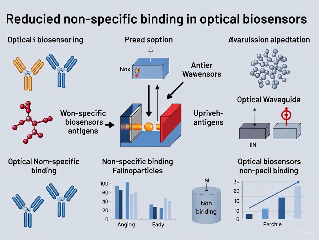
The following diagram illustrates the fundamental mechanisms behind NSA and the core strategies to counteract it.
Troubleshooting Guide: Addressing NSA in Your Experiments
My sensor signal is drifting upwards over time. Could this be NSA?
Yes, a drifting signal is a classic symptom of progressive biofouling. As non-target molecules accumulate on the sensing interface over time, they contribute an increasing background signal [2]. This drift can complicate data interpretation and is particularly problematic for measurements taken over long durations.
- Recommended Action: Implement a more robust surface passivation strategy (see Passive Methods below). For short-term measurements, signal drift correction algorithms can be applied, but these cannot compensate for severe, progressive fouling that degrades the sensor surface [2].
I am getting false-positive signals in complex samples like serum or blood. How can I mitigate this?
This is a common issue when biosensors are transitioned from clean buffer solutions to complex biological matrices, which contain a high concentration of interfering proteins and other biomolecules [2].
- Recommended Actions:
- Sample Pre-treatment: If possible, dilute the sample or use centrifugation and filtration to reduce its complexity before analysis [2].
- Optimize Your Blocking Step: Ensure you are using an effective blocking agent (e.g., BSA, casein, or synthetic peptides) to cover any remaining reactive sites on the sensor surface after functionalization [1] [3].
- Re-evaluate Your Surface Chemistry: Consider switching to a more effective antifouling coating, such as a zwitterionic material, which offers superior protection in complex fluids [2] [3].
My sensor's limit of detection (LOD) is worse than expected. Is NSA a factor?
Absolutely. The background noise generated by NSA directly raises the lowest detectable signal level, thereby worsening the LOD [3]. A high level of non-specific binding effectively buries the specific signal of a low-concentration analyte.
- Recommended Action: Focus on enhancing the signal-to-noise ratio by minimizing NSA. A combination of a high-quality antifouling coating and an optimized bioreceptor immobilization protocol to ensure proper orientation and activity can significantly improve the LOD [3].
Solutions for NSA Reduction
Passive Methods: Surface Coatings and Blocking Agents
Passive methods aim to prevent NSA by creating a physical or chemical barrier on the sensor surface. The goal is to create a thin, hydrophilic, and neutrally charged boundary layer that minimizes the intermolecular forces driving adsorption [1].
Table 1: Common Passive Methods for NSA Reduction
| Method Category | Examples | Mechanism of Action | Key Considerations |
|---|---|---|---|
| Protein Blockers [1] | Bovine Serum Albumin (BSA), Casein, milk proteins | Adsorbs to vacant surface sites, "blocking" them from future non-specific binding. | Inexpensive and easy to use; but can be unstable and might desorb over time. |
| Polymer Coatings [1] [3] | Polyethylene Glycol (PEG), Zwitterionic peptides/polymers | Forms a hydrated, steric, and energetic barrier that repels biomolecules. Zwitterionic materials bind water very tightly via electrostatic interactions. | PEG is a "gold standard" but can oxidize. Zwitterionic peptides are emerging as more stable and effective alternatives [3]. |
| Self-Assembled Monolayers (SAMs) [1] [5] | Alkanethiols on gold, Silanization on SiO₂ | Creates a dense, ordered, and customizable monolayer that can be engineered to be hydrophilic and non-charged. | Provides excellent control over surface properties; stability can vary with environment. |
| Chemical Functionalization [5] [2] | Ethanolamine, Tris, Hyperbranched polyglycerol (HPG) | Uses small molecules to covalently passivate reactive chemical groups on the surface. | Can be very stable; requires specific surface chemistry for covalent attachment. |
Active Methods: Physical Removal Techniques
Active methods dynamically remove adsorbed molecules after they have attached to the surface. They typically use a transducer to generate forces that shear away weakly adhered biomolecules [1].
- Electromechanical Removal: Uses transducers to create surface waves or vibrations that dislodge adsorbed molecules [1].
- Acoustic Removal: Applies sound waves (e.g., ultrasound) to agitate the surface and fluid interface [1].
- Hydrodynamic Removal: Relies solely on controlled fluid flow within microfluidic channels to generate shear forces that wash away non-specifically bound molecules [1].
Experimental Protocols and Material Selection
Protocol: Evaluating NSA on Different Substrate Materials
This protocol, adapted from a study on microfluidic materials, provides a method to quantitatively compare the NSA of proteins on different surfaces using fluorescence microscopy [6].
- Sample Preparation: Prepare clean substrates of the materials to be tested (e.g., SU-8, silica, various grades of fluoropolymers like CYTOP) on silicon wafers.
- Surface Cleaning: Clean all surfaces thoroughly with isopropanol (IPA) and deionized (DI) water, followed by a UV-Ozone treatment immediately before the experiment to ensure consistency [6].
- Protein Exposure: Introduce a solution of a fluorescently labeled protein (e.g., FITC-labeled BSA at 100 µg/mL in phosphate-buffered saline) to the surfaces and incubate for a set time.
- Washing: Gently rinse the surfaces with PBS buffer to remove any unbound or loosely adsorbed protein.
- Imaging and Analysis: Use fluorescence microscopy to image the surfaces. Measure the fluorescence intensity at multiple locations on each sample.
- Data Calculation: Calculate the averaged fluorescence intensity for each material. Subtract the averaged auto-fluorescence intensity from negative-control samples (not exposed to the protein) to obtain the relative fluorescence intensity, which is proportional to the amount of NSA [6].
Table 2: Example Results from Material NSA Evaluation (Based on [6])
| Material | Surface Characteristics | Relative Fluorescence Intensity (Approx.) | NSA Load |
|---|---|---|---|
| SU-8 | Hydrophilic (post-cleaning) | Lowest | Very Low |
| CYTOP S-grade (-CF₃ terminal) | Highly hydrophobic | Low | Low |
| CYTOP M-grade (-CONH-Si(OR)ₙ) | Intermediate hydrophobicity | Medium | Medium |
| Silica (SiO₂) | Hydrophilic but with fixed positive charge | High | High |
The Scientist's Toolkit: Key Research Reagent Solutions
Table 3: Essential Materials for NSA Reduction Experiments
| Reagent / Material | Function in NSA Reduction | Example Use Case |
|---|---|---|
| Bovine Serum Albumin (BSA) [1] | Protein-based blocking agent. Adsorbs to surface sites to prevent non-specific binding of other proteins. | Blocking step in immunosensors (e.g., ELISA-style assays on optical platforms). |
| Zwitterionic Peptide (e.g., EKEKEKEKEKGGC) [3] | Forms a stable, charge-neutral hydration layer that acts as a physical and energetic barrier to biomolecular adsorption. | Covalent passivation of biosensor surfaces (e.g., porous silicon, gold) for operation in complex fluids like blood serum or GI fluid. |
| Polyethylene Glycol (PEG) [1] [3] | Polymer coating that creates a steric and hydrated barrier to reduce fouling. | The "gold standard" against which new antifouling coatings are often compared. |
| Ethanolamine [3] | Small molecule used for chemical passivation. Blocks reactive groups (e.g., NHS-esters) on the surface after bioreceptor immobilization. | Quenching unreacted sites on a sensor surface after covalent immobilization of antibodies or aptamers. |
| CYTOP Fluoropolymer [6] | A low-refractive-index material used for microfluidic channels and optical cladding. Its inherent properties can influence NSA. | Used as a microfluidic channel material in surface plasmon resonance (SPR) biosensors due to its optical properties and lower NSA compared to some materials. |
Workflow for Systematic NSA Troubleshooting
The following diagram outlines a logical, step-by-step workflow to diagnose and address NSA issues in optical biosensor research.
Emerging Solutions and Future Perspectives
The field of NSA reduction is rapidly evolving. Key emerging areas include:
- AI-Enhanced Surface Design: Machine learning (ML) and computational modeling are being used to predict optimal surface architectures and antifouling material compositions, moving beyond traditional trial-and-error approaches [5].
- Advanced Zwitterionic Materials: Peptides and polymers with zwitterionic properties are showing broad-spectrum protection against proteins, bacteria, and mammalian cells, offering a universal strategy for enhancing sensor reliability [3].
- High-Throughput Screening: Combined with molecular simulations, this allows for the rapid evaluation of a wide range of new antifouling materials for specific applications [2].
Non-Specific Adsorption (NSA) is a prevalent challenge in optical biosensing that compromises data accuracy and reliability. It refers to the unintended adherence of biomolecules to sensor surfaces through non-covalent interactions, leading to false positive signals and reduced sensitivity. For researchers and drug development professionals, understanding and mitigating NSA is crucial for obtaining trustworthy kinetic and affinity data. The primary mechanisms driving NSA are physisorption, hydrophobic interactions, and electrostatic interactions. Physisorption involves weak van der Waals forces, hydrophobic interactions drive the association of non-polar surfaces in aqueous environments, and electrostatic interactions occur between charged residues on proteins and functional groups on the sensor surface. Controlling these interactions is fundamental to developing robust biosensing assays.
Troubleshooting Guides & FAQs
Frequently Asked Questions
What is the fundamental difference between specific and non-specific binding? Specific binding involves the selective recognition between a receptor and its target analyte, characterized by high affinity and saturability. In contrast, non-specific binding is the adsorption of molecules to solid surfaces or other areas through general physicochemical forces, is non-saturable, and leads to a high background signal [7] [8].
Why are cationic lipids and peptides particularly prone to NSA? Molecules like cationic lipids (e.g., DOTAP) and peptides often have amphiphilic properties. They possess localized charged groups (e.g., quaternary ammonium salts or basic amino acids) that create strong electrostatic effects, combined with long hydrocarbon chains or hydrophobic regions that contribute to hydrophobic interactions, making them highly adhesive to various surfaces [8].
How does the solution pH influence NSA? Solution pH directly affects the ionization state of both the analyte and the sensor surface. Operating at a pH that neutralizes the net charge of the interacting surfaces can minimize electrostatic NSA. Furthermore, adjusting pH can improve the solubility of the analyte, thereby reducing its driving force for NSA [8].
My analyte is a protein. Should I immobilize it or use it as the analyte to minimize NSA? If your protein is available only in small quantities, immobilizing it is more material-efficient. However, immobilizing a very small peptide can be difficult to control and may lead to excessively high surface density, exacerbating steric hindrance and mass transport limitations. As a general rule, immobilizing the larger binding partner can help minimize the chance of the immobilization chemistry affecting the binding epitope [9].
Troubleshooting Common NSA Problems
Problem: High background signal across all analyte concentrations, including blanks.
- Potential Cause: Physisorption of analyte or matrix components to the sensor surface.
- Solutions:
- Optimize Surface Passivation: Ensure the sensor surface is effectively blocked with a non-reactive protein (e.g., BSA) or polymer (e.g., PEG) after ligand immobilization.
- Increase Stringency of Wash Buffers: Incorporate mild detergents (e.g., Tween 20) or increase the ionic strength in the running and wash buffers to disrupt weak van der Waals forces.
- Reduce Contact Time: Minimize the time the sensor surface is exposed to the sample solution.
Problem: Irreproducible binding kinetics and poor data fitting.
- Potential Cause: Hydrophobic interactions, often exacerbated by exposed hydrophobic patches on the analyte or the sensor surface.
- Solutions:
- Add Surfactants: Include non-ionic surfactants in your sample and running buffers. These agents can uniformly disperse analytes, improving their dissolution state and shielding hydrophobic patches [8].
- Use Low-Adsorption Consumables: Employ surface-passivated tubes and plates specifically designed for proteins and nucleic acids to reduce losses during sample preparation [8].
- Modify Solvent Composition: For working solutions, adding a small percentage of a compatible organic solvent can enhance analyte solubility and reduce hydrophobic NSA [8].
Problem: Significant analyte loss or signal distortion, especially with charged molecules like peptides or nucleic acids.
- Potential Cause: Strong electrostatic interactions with charged surfaces of consumables or the liquid chromatography system.
- Solutions:
- Adjust Buffer pH and Ionic Strength: Modify the pH to neutralize the net charge of the analyte. Increasing the salt concentration can shield electrostatic attractions.
- Employ Chelating Agents: For nucleic acid drugs with phosphorothioate backbones, add chelators like EDTA to the mobile phase to prevent chelation with metal ions in the liquid chromatography path [8].
- Use Passivated Liquid Chromatography Systems: Utilize low-adsorption liquid phase systems and columns with passivated metal surfaces to minimize electrostatic binding and improve peak shape [8].
Problem: Mass transport limitation, where the binding rate is controlled by analyte diffusion to the surface rather than the interaction itself.
- Potential Cause: Excessively high density of immobilized ligand or a high chemical on-rate constant, leading to a depletion of analyte near the sensor surface [9].
- Solutions:
- Reduce Ligand Density: Optimize the immobilization protocol to achieve a lower density of active ligand on the sensor surface.
- Increase Flow Rate: If possible, increase the flow rate of the analyte solution to enhance mass transport to the surface.
- Verify with Control Experiments: A tell-tale sign of mass transport limitation is that the binding rate becomes dependent on flow rate or ligand density. Test for this by varying these parameters [9].
Quantitative Data on NSA Mechanisms
Table 1: Characteristics and Energetic Contributions of Primary NSA Mechanisms
| Mechanism | Interaction Force | Typical Energy Range | Key Influencing Factors | Effective Range |
|---|---|---|---|---|
| Electrostatic | Coulombic (charge-charge) | Variable; can be strong (~ several kcal/mol) [10] | pH, ionic strength, dielectric constant of medium [10] | Long-range (5–10 Å) [10] |
| Hydrophobic | Entropic (from water reorganization) | Not specified in results | Temperature, solvent polarity, surface hydrophobicity | Short-range |
| Physisorption | Van der Waals, Dipole-dipole | Weak (< 5 kcal/mol) | Surface energy, polarizability, distance | Very short-range |
Table 2: Common Desorption Agents and Their Applications
| Desorption Agent | Mechanism of Action | Typical Use Cases | Notes & Considerations |
|---|---|---|---|
| Surfactants (e.g., Tween 20) | Reduces surface tension; shields hydrophobic and electrostatic interactions [8] | General use in wash and running buffers | Non-ionic surfactants are preferred to avoid interference; can cause signal suppression in MS [8] |
| Bovine Serum Albumin (BSA) | Competes for binding sites on the surface | Surface passivation; blocking agents | Can bind to the analyte itself in some cases |
| Organic Solvents (e.g., Acetonitrile) | Alters solvent polarity; improves analyte solubility [8] | Small-volume matrix samples (e.g., CSF) | Compatibility with the biosensor and analyte must be verified |
| Chelators (e.g., EDTA) | Binds metal ions; prevents metal-ion bridging [8] | Nucleic acid analytes, phosphorothioate-modified drugs | Essential for passivating metal surfaces in LC systems |
Essential Research Reagent Solutions
Table 3: Key Reagents and Materials for Mitigating NSA
| Reagent / Material | Function | Specific Example & Application |
|---|---|---|
| Low-Adsorption Consumables | Tubes and plates with surface passivation to minimize binding of precious analytes [8] | Protein LoBind Tubes; nucleic acid-specific low-adsorption plates |
| Surface Passivation Reagents | Inert polymers or proteins used to block unused sites on the sensor surface | BSA, casein, PEG-based coatings |
| Blocking Buffers | Ready-to-use solutions containing agents to prevent NSA | Commercial BLI or SPR blocking buffers |
| High-Stringency Wash Buffers | Buffers with additives to disrupt non-covalent interactions without damaging the specific bond | Buffers containing mild detergents or elevated salt concentrations |
| Regeneration Solutions | Solutions that dissociate the specific analyte-ligand complex, preparing the surface for a new cycle [9] | Low pH (e.g., glycine-HCl), high pH, or high salt solutions; must be optimized for each specific interaction |
Experimental Protocols for Investigating NSA
Protocol 1: Systematic Evaluation of NSA Using Continuous Transfer and Gradient Dilution
Purpose: To qualitatively and quantitatively assess the propensity of an analyte for NSA under various conditions. Background: The degree of NSA is directly proportional to the contact surface area and the contact time between the analyte and the solid surface [8]. This protocol exploits this principle. Materials:
- Test analyte in the relevant buffer/biofluid
- Low-adsorption and standard microcentrifuge tubes
- HPLC system with a UV/VIS detector or plate reader
Procedure:
- Prepare a series of analyte solutions at the concentrations expected in your assay.
- Aliquot the same volume of solution into different containers:
- Test 1 (Surface Area): Place the same volume in containers of different sizes (e.g., a narrow PCR tube vs. a larger microcentrifuge tube).
- Test 2 (Contact Time): Place the solution in identical containers but subject them to different standing times before analysis (e.g., immediate, 30 min, 2 hours).
- For the "continuous transfer" method, repeatedly transfer the analyte solution between multiple new vials to continually expose it to fresh surfaces.
- Measure the recovered concentration of the analyte using an appropriate method (e.g., HPLC-UV, fluorescence).
- Data Analysis: A significant decrease in recovered concentration with increased surface area, contact time, or number of transfers indicates significant NSA. Compare results between low-adsorption and standard consumables to select the optimal labware.
Protocol 2: Identification and Optimization of Desorption Agents
Purpose: To screen and identify effective chemical additives that minimize NSA for a specific analyte. Background: Different analytes require tailored solutions. Surfactants, competing proteins, or solvents can be added to the matrix or running buffer to reduce NSA [8]. Materials:
- Test analyte
- Candidate desorption agents (see Table 2): surfactants (ionic/non-ionic), BSA, organic solvents, chelators
- Biosensor system (e.g., BLI, SPR) or analytical HPLC system
Procedure:
- Immobilize your ligand on the biosensor tip or use a bare surface to measure baseline NSA.
- Prepare a fixed concentration of your analyte in the running buffer supplemented with different candidate desorption agents at various concentrations.
- On the biosensor, expose the surface to the analyte solutions and monitor the binding response.
- Alternative HPLC Method: Incubate the analyte with the desorption agent in a vial, then inject and analyze the peak area and shape. Improved recovery and symmetric peaks indicate reduced NSA [8].
- Data Analysis: The condition that results in the lowest non-specific signal (in biosensing) or the highest peak area and best symmetry (in HPLC) identifies the most effective desorption agent. Always verify that the additive does not interfere with the specific binding interaction of interest.
NSA Mechanisms and Experimental Workflows
Diagram Title: Troubleshooting Logic for NSA Mechanisms
Diagram Title: Experimental Protocols for NSA Investigation
How NSA Compromises Sensitivity, Specificity, and Reproducibility
Non-specific adsorption (NSA), often referred to as non-specific binding (NSB), is a fundamental challenge that directly compromises the performance of optical biosensors. For researchers and scientists in drug development, understanding and mitigating NSA is crucial for generating reliable, high-quality data. This technical guide addresses how NSA negatively impacts key biosensor performance metrics—sensitivity, specificity, and reproducibility—and provides actionable troubleshooting strategies for experimental optimization.
Frequently Asked Questions (FAQs)
What is Non-Specific Adsorption (NSA) and how does it occur?
A: Non-specific adsorption is the physisorption of atoms, ions, or molecules (e.g., proteins, antibodies, or other interferents) to a biosensor's surface through weak intermolecular forces, rather than through specific, targeted binding [1]. This occurs due to:
- Hydrophobic interactions
- Electrostatic or charge-based interactions
- Van der Waals forces [11] [1] In a typical biosensor experiment, NSA happens when the analyte or other molecules in the sample interact with the sensor surface, the immobilized ligand, or other non-target sites, generating a background signal that is indistinguishable from the specific signal [1] [11].
How does NSA directly compromise sensitivity and specificity?
A: NSA affects sensitivity and specificity through distinct mechanisms:
- Reduced Sensitivity: The background signal generated by NSA obscures the specific signal from the target analyte, effectively increasing the noise floor. This elevates the Limit of Detection (LOD), making it difficult to detect low-abundance biomarkers [1] [12]. For instance, a study demonstrated that a novel single-molecule colocalization assay (SiMCA) could achieve a three-fold lower LOD than a conventional assay by eliminating NSA-derived background [12].
- Reduced Specificity: NSA leads to false-positive signals, as non-specifically bound molecules are misinterpreted as true binding events. This reduces the assay's ability to accurately discriminate the target from other components in a complex sample, such as serum or blood [1] [13].
Why does NSA lead to poor experimental reproducibility?
A: Reproducibility is undermined by the variable and unpredictable nature of NSA. Unlike specific binding, which is consistent and concentration-dependent, NSA can differ significantly between experiments due to:
- Surface Heterogeneity: Inconsistent coating or functionalization of sensor surfaces leads to stochastic NSA across different batches or even within different fields of view on the same sensor [12].
- Sample Complexity: Variations in the composition of complex biological matrices (e.g., differences between individual patient sera) can cause uncontrollable and heterogeneous levels of NSA, making it nearly impossible to replicate results using a standard blank subtraction [14].
The following diagram illustrates how NSA leads to inaccurate results across these three key metrics:
Troubleshooting Guides
Guide 1: Mitigating NSA in Surface Plasmon Resonance (SPR)
SPR is highly susceptible to NSA due to its sensitivity to mass changes on the sensor surface [11] [15].
Problem: Significant non-specific response is observed when injecting analyte over the immobilized ligand or a bare sensor surface.
Solution Steps:
- Characterize Your Molecules: Determine the isoelectric point (pI), charge, and hydrophobicity of your analyte and ligand. This informs the best mitigation strategy [11].
- Optimize Buffer Conditions: Screen the following buffer additives, which can be used individually or in combination:
- Add Surfactants: Incorporate a non-ionic detergent like Tween 20 (e.g., 0.005-0.05%) to disrupt hydrophobic interactions [11].
- Adjust pH: Modify the buffer pH to a value near the pI of your analyte to neutralize its charge and reduce electrostatic NSA [11].
- Increase Ionic Strength: Add salt (e.g., 150-200 mM NaCl) to shield charged molecules and prevent charge-based NSA [11].
- Use a Reference Surface: For complex samples like serum, use a "blank" injection over a captured non-cognate target (a structurally similar protein that does not specifically bind the analyte) on the same flow cell. The signal from this run is subtracted from the signal obtained with the target of interest to account for NSA [14].
Table 1: Common Buffer Additives to Reduce NSA in SPR
| Additive | Concentration Range | Primary Mechanism | Considerations |
|---|---|---|---|
| Tween 20 | 0.005% - 0.05% | Disrupts hydrophobic interactions | Mild, generally does not denature proteins. |
| BSA | 0.1% - 1% | Protein blocker, shields surfaces | Can interact with some analytes; a common blocker. |
| NaCl | 50 - 500 mM | Shields charged molecules to reduce electrostatic binding | High concentrations may affect specific binding. |
Guide 2: A Novel Experimental Approach to Discriminate Specific from Non-Specific Binding
Problem: Conventional single-signal biosensors cannot distinguish between specific binding and NSA, leading to inaccurate data.
Solution: Implement a Single-Molecule Colocalization Assay (SiMCA) [12].
Experimental Protocol:
- Sensor Functionalization:
- Passivate a coverslip with a mixture of PEG and PEG-biotin to minimize NSA.
- Immobilize biotinylated capture antibodies (cAb) via neutravidin binding. Label cAbs with a green fluorophore (e.g., Alexa-546).
- Sample Incubation:
- Incubate the functionalized sensor with a sample containing the target antigen.
- Use a detection antibody (dAb) labeled with a spectrally distinct red fluorophore (e.g., Alexa-647). The dAb concentration should be kept low (e.g., 50 nM) to minimize NSA.
- Data Acquisition & Analysis:
- Image the sensor surface using two-color Total Internal Reflection Fluorescence (TIRF) microscopy.
- Acquire images via sequential excitation of the two fluorophores.
- Use automated image analysis to count only the red dAb signals that are spatially colocalized with a green cAb signal. Signals from non-specifically bound dAbs (red only) are discarded.
Key Advantage: This method provides a powerful way to actively remove the effects of NSA from the final signal, leading to a lower LOD and higher reproducibility, even in complex samples like serum and blood [12].
The workflow and key advantage of this approach are summarized below:
The Scientist's Toolkit: Key Research Reagent Solutions
Table 2: Essential Reagents for NSA Reduction in Optical Biosensors
| Reagent / Material | Function | Example Use-Case |
|---|---|---|
| Bovine Serum Albumin (BSA) | Protein-based blocking agent that adsorbs to vacant sites on the sensor surface, preventing NSA of proteins from the sample. | Used as a standard blocker in ELISA and SPR assays; typically used at 0.1-1% concentration [11] [1]. |
| PEG-based Passivation | Creates a hydrophilic, non-charged boundary layer that resists protein adsorption via steric repulsion and hydration. | Used to coat sensor surfaces (e.g., coverslips in TIRF) to create a non-fouling background [12] [1]. |
| Tween 20 | Non-ionic surfactant that disrupts hydrophobic interactions between the analyte and the sensor surface. | Added to running buffers in SPR at low concentrations (e.g., 0.005-0.05%) to reduce NSA [11]. |
| Carboxymethylated Dextran Matrix | A hydrogel matrix used on SPR sensor chips. It provides a hydrophilic environment for ligand immobilization and can help reduce NSA. | The standard matrix in many commercial SPR systems (e.g., Biacore) for immobilizing ligands via amine coupling [15]. |
| High-Salt Buffers | High ionic strength shields charged molecules, reducing charge-based non-specific interactions. | Adding 150-200 mM NaCl to a buffer can eliminate NSA caused by a positively charged analyte interacting with a negatively charged surface [11]. |
The Unique NSA Challenges in Microfluidic and Label-Free Optical Systems
Troubleshooting Guides and FAQs
Frequently Asked Questions
Q1: What makes Non-Specific Adsorption (NSA) particularly challenging in microfluidic and label-free optical systems compared to other biosensors?
NSA is especially problematic in these integrated systems because it directly compromises the core advantages of the technology. In microfluidics, the high surface-to-volume ratio amplifies the impact of any surface fouling [16] [17]. For label-free optical biosensors that rely on refractometric principles—measuring changes in refractive index—any unwanted molecule adhering to the sensor surface generates a background signal that is indistinguishable from the specific binding of the target analyte [17] [18]. This combination can lead to false positives, reduced sensitivity, and poor reproducibility in quantitative measurements.
Q2: Our label-free SPR measurements in a microfluidic device show high background drift. Is this likely caused by NSA, and how can we confirm it?
Yes, a high background drift is a classic symptom of ongoing NSA. Label-free techniques like Surface Plasmon Resonance (SPR) are inherently sensitive to changes in mass on the sensor surface [19] [18]. To confirm NSA is the cause, you can perform a negative control experiment by running the same assay without the specific capture probe (e.g., the antibody) immobilized on the surface. If a signal or drift is still observed in the control channel, it is highly likely due to NSA from components in your sample matrix [17].
Q3: What are the most effective surface coatings to prevent NSA for protein-based assays?
The most effective passive coatings create a hydrophilic and non-charged boundary layer that minimizes intermolecular forces [17]. A widely used and effective strategy is the formation of self-assembled monolayers (SAMs) of poly(ethylene glycol) (PEG) or its derivatives, which create a molecular brush that resists protein adsorption [17]. Recent research also highlights advanced polymers like poly(oligo(ethylene glycol) methacrylate) (POEGMA) brushes, which exhibit excellent antifouling properties and can be grafted onto surfaces, sometimes eliminating the need for separate blocking steps [20].
Q4: Can the microfluidic flow itself be used to combat NSA?
Yes, active hydrodynamic removal is a viable method. By strategically increasing the shear force through pressure-driven flow, weakly adhered (physisorbed) molecules can be swept away from the sensor surface [17]. This is often implemented as a wash step in an assay protocol. Furthermore, proper microfluidic design, such as spatial hydrodynamic focusing, can confine cells or particles to the channel center, minimizing their contact with channel walls and thus reducing the surface area susceptible to fouling [21].
Troubleshooting Guide: Common NSA Problems and Solutions
| Problem Observed | Possible Cause | Recommended Solution |
|---|---|---|
| High background signal/drift in optical readout (SPR, etc.) | NSA of sample matrix components (e.g., proteins, lipids) to sensing surface. | Implement a rigorous surface passivation protocol (e.g., PEGylation) [17]. Include negative control channels without specific probes [17]. |
| Low signal-to-noise ratio, poor detection limit | NSA obscuring the specific signal from low-concentration targets. | Combine chemical passivation with optimized hydrodynamic wash steps to remove loosely bound molecules [17]. |
| Clogging in microfluidic channels | Aggregation and NSA of cells or proteins in narrow channels. | Optimize channel geometry and surface chemistry [22]. Use spatial hydrodynamic focusing to centralize particles [21]. |
| Irreproducible results between runs | Inconsistent surface modification or incomplete NSA removal. | Standardize surface preparation and functionalization workflows [16]. Automate fluidic handling to improve consistency [22]. |
Quantitative Data on NSA Reduction Methods
The following table summarizes the key characteristics of different NSA reduction strategies, helping you select the most appropriate one for your application.
| Method Category | Specific Technique | Key Mechanism | Advantages | Limitations / Considerations |
|---|---|---|---|---|
| Passive (Blocking) | Protein Blockers (e.g., BSA) | Coats surface vacant spaces to prevent NSA [17]. | Simple, widely used, cost-effective. | Can be unstable; may be incompatible with some transducers; potential for cross-reaction [17]. |
| Passive (Chemical) | PEG / POEGMA Brushes | Forms a hydrophilic, steric barrier that resists protein adsorption [17] [20]. | Highly effective, can be covalently bound, non-interfering. | Requires specific chemistry for surface grafting; quality is synthesis-dependent [17]. |
| Active (Hydrodynamic) | High-Shear Wash | Uses fluid flow to generate shear forces that overpower adhesive forces of NSA molecules [17]. | Can be applied post-functionalization, no chemicals needed. | May not remove strongly adhered molecules; requires careful optimization of flow rate [17]. |
| Active (Transducer-Based) | Acoustic | Generates surface forces (e.g., via surface acoustic waves) to shear away biomolecules [17]. | Highly effective localized removal, can be integrated on-chip. | Increased system complexity and cost; potential for heating or damaging delicate surfaces [17]. |
Experimental Protocols for NSA Reduction
Protocol 1: Establishing a Baseline with Spatial Hydrodynamic Focusing
This protocol, adapted from a recent Nature Communications paper, details how to set up a microfluidic system to minimize cell-wall interactions and improve single-cell analysis, thereby reducing NSA-related issues [21].
- Objective: To achieve precise 3D positioning of single cells in a microchannel for high-fidelity, label-free imaging while minimizing NSA on channel walls.
- Materials:
- PDMS-based microfluidic device with a five-inlet design for spatial focusing [21].
- Syringe pumps for precise control of sample and sheath flows.
- Custom-built Digital Holographic Microscopy (DHM) system or equivalent label-free imager [21].
- Cell culture sample and appropriate sheath fluid (e.g., PBS).
- Methodology:
- Device Priming: Thoroughly prime the microfluidic device with sheath fluid to remove all air bubbles.
- Flow Rate Calibration: Initiate the two vertical sheath flows to compress the sample flow. Adjust the flow rate ratio between sheath and sample flows to achieve the desired focused stream width. This ratio is critical and should be determined via CFD simulation or empirically [21].
- Spatial Focusing: Activate the two horizontal sheath flows, which enter at a 45° angle, to fully envelop the pre-focused stream. This completes the 3D spatial confinement, guiding cells to flow strictly along the channel's central axis.
- Optical Alignment: Meticulously align the focal plane of the DHM system with the spatially focused cell stream in the microchannel.
- Image Acquisition: Introduce the cell sample and capture in-focus holograms directly without the need for digital refocusing, thanks to the precise cell positioning.
- Expected Outcome: Reduced cell adhesion to channel walls, minimization of image blur and artifacts, and acquisition of clear, stable single-cell phase images for analysis [21].
Protocol 2: Implementing a POEGMA Brush Coating for antifouling
This protocol is based on methods described in recent research for reducing NSA in sensitive protein diagnostics [20].
- Objective: To graft a POEGMA polymer brush onto a biosensor surface to create an effective antifouling layer that minimizes the need for blocking and wash steps.
- Materials:
- Sensor substrate (e.g., gold chip for SPR, silicon oxide, or glass).
- POEGMA polymer or initiator for surface-initiated polymerization.
- An appropriate solvent (e.g., ethanol/water mixture).
- Vacuum chamber or system for vacuum-assisted entanglement, if applicable [20].
- Methodology:
- Surface Preparation: Clean and activate the sensor substrate using standard protocols (e.g., oxygen plasma for PDMS/glass, piranha solution for gold).
- Initiator Immobilization: If using atom transfer radical polymerization (ATRP), immobilize the ATRP initiator onto the activated surface.
- Polymer Grafting: Synthesize the POEGMA brushes directly on the surface via surface-initiated polymerization from the immobilized initiators, or graft pre-synthesized POEGMA chains using vacuum-assisted entanglement or covalent chemistry [20].
- Characterization: Validate the coating's presence and uniformity using techniques like Ellipsometry or Atomic Force Microscopy (AFM) to measure layer thickness and morphology.
- Expected Outcome: A robust, non-fouling surface that significantly reduces NSA from complex samples like blood plasma or serum, enabling more robust and sensitive detection of target analytes [20].
Research Reagent Solutions
Table: Essential Materials for NSA Reduction in Microfluidic and Label-Free Systems
| Reagent / Material | Function in NSA Reduction |
|---|---|
| Poly(ethylene glycol) (PEG) derivatives | Forms a dense, hydrophilic self-assembled monolayer (SAM) that creates a steric and thermodynamic barrier to protein adsorption [17]. |
| Poly(oligo(ethylene glycol) methacrylate) (POEGMA) | A advanced polymer brush coating that provides excellent antifouling properties, physically preventing non-specific binding [20]. |
| Bovine Serum Albumin (BSA) | A common protein blocker used to passively occupy vacant sites on a surface, preventing NSA of target analytes [17]. |
| Polydimethylsiloxane (PDMS) | The most common elastomer for rapid prototyping of microfluidic chips; its biocompatibility and optical clarity are ideal, though it can be prone to NSA without surface modification [16]. |
| Surface Plasmon Resonance (SPR) Chip | A sensor chip, typically with a gold coating, that serves as the transducer in SPR biosensing. Its surface is functionalized with biorecognition elements and antifouling layers [19] [18]. |
Integrated Experimental Workflow for NSA Management
The following diagram illustrates a logical workflow for setting up an experiment that integrates microfluidics and label-free detection while incorporating key steps to mitigate NSA.
Integrated NSA Management Workflow
Signaling and Interaction Logic in NSA
This diagram conceptualizes the molecular-level competition between specific binding and non-specific adsorption (NSA) on a functionalized sensor surface, which is central to the challenges in label-free biosensing.
Specific Binding vs. Non-Specific Adsorption
Advanced Antifouling Strategies and Surface Functionalization Methods
Non-specific adsorption (NSA) is a pervasive challenge in optical biosensing, resulting in decreased sensitivity, specificity, and reproducibility. NSA occurs when non-target molecules, such as proteins or other biomolecules, physisorb to the sensor surface through hydrophobic forces, ionic interactions, van der Waals forces, or hydrogen bonding. These false-positive signals are often indiscernible from specific binding events, compromising data accuracy [1].
Passive methods represent a fundamental approach to combating NSA by creating a permanent or semi-permanent barrier on the sensor surface. Unlike active methods that dynamically remove adsorbed molecules, passive techniques aim to prevent the initial adsorption by coating the surface with materials that resist fouling. The primary goal is to create a thin, hydrophilic, and non-charged boundary layer that minimizes intermolecular forces between adsorbing molecules and the substrate, allowing potential contaminants to be easily detached under low shear stresses like washing [1] [2]. This guide details the implementation, optimization, and troubleshooting of these critical surface modification strategies.
Chemical Coatings: Methodologies and Protocols
Chemical coatings prevent NSA by forming a physical and chemical barrier on the sensor surface. These coatings work by reducing available binding sites, neutralizing surface charge, and creating a hydrated layer that sterically hinders the approach of non-target molecules [1] [23].
Frequently Asked Questions: Chemical Coatings
Q1: What are the most effective chemical coatings for preventing NSA in optical biosensors?
The effectiveness of a chemical coating depends on your specific sample matrix and sensor platform. The table below summarizes the most common options:
Table: Common Chemical Coatings for NSA Reduction
| Coating Type | Example Materials | Mechanism of Action | Best For | Limitations |
|---|---|---|---|---|
| Protein Blockers | Bovine Serum Albumin (BSA), Casein, Milk proteins [1] | Adsorbs to vacant surface sites, shielding the analyte from non-specific interactions [11] [24]. | Routine immunoassays; a good first attempt for protein-based analytes. | Can introduce new organic contaminants; may block specific binding if not optimized. |
| Self-Assembled Monolayers (SAMs) | Alkanethiols on gold, Silanes on silica/glass [1] [23] | Forms a dense, ordered, hydrophilic monolayer that presents a non-fouling interface (e.g., with oligo- or poly(ethylene glycol) terminals) [23]. | Well-defined metal (Au, Ag) or oxide surfaces; requires controlled lab conditions. | Limited to specific substrates; sensitive to fabrication conditions. |
| Poly(Ethylene Glycol) (PEG) | OEG-terminated SAMs, PEG-polymers [23] | Creates a highly hydrated layer that generates a strong energy barrier, sterically repelling approaching biomolecules. | High-sensitivity applications in complex media (e.g., serum, blood). | Susceptible to oxidative degradation; activity can be time-limited. |
Q2: How do I choose the concentration for additives like BSA or Tween 20?
Start with standard concentrations and titrate for optimal performance. BSA is commonly used at 1% (w/v), while non-ionic surfactants like Tween 20 are typically used at 0.01-0.1% (v/v) [11] [24]. However, the optimal concentration depends on your specific surface and analyte. Begin with the standard value and perform a series of tests, adjusting the concentration up or down while monitoring the specific signal and background noise. Extreme conditions may deactivate or denature your biomolecules, so always consider their stability [11].
Q3: My buffer's pH and ionic strength are affecting NSA. How can I optimize them?
The pH dictates the overall charge of your biomolecules and the sensor surface. If your analyte is positively charged and the surface is negative, NSA will occur.
- Strategy: Adjust your running buffer to a pH close to the isoelectric point (pI) of your protein analyte, where its net charge is neutral. Alternatively, you can modify the surface charge. For charge-based NSA, increasing the salt concentration (e.g., 150-200 mM NaCl) can shield the interactions [11] [24].
Detailed Experimental Protocol: BSA Blocking for an Optical Immunosensor
Principle: BSA acts as a sacrificial protein, adsorbing to any remaining reactive sites on the sensor surface after immobilization of the capture probe (e.g., an antibody), thereby preventing non-specific adsorption of other proteins from the sample [1] [11].
Materials:
- Sensor chip with immobilized capture antibody
- Blocking Solution: 1% (w/v) BSA in PBS (Phosphate Buffered Saline), pH 7.4
- Washing Buffer: PBS, pH 7.4 (optionally with 0.05% Tween 20)
- Optical Biosensor System (e.g., SPR, Silicon Photonic)
Procedure:
- Preparation: After immobilizing your specific capture antibody on the sensor surface, equilibrate the system with PBS at a constant flow rate (e.g., 10-30 μL/min).
- Blocking: Inject the 1% BSA solution over the sensor surface for 15-30 minutes.
- Washing: Rinse the surface thoroughly with washing buffer for at least 10-15 minutes to remove any unbound or loosely adsorbed BSA.
- Validation: Test the efficacy of blocking by injecting a complex, analyte-free sample (e.g., serum or cell lysate) and measuring the response. A successfully blocked surface will show a minimal signal change compared to the baseline.
Polymer Brushes: Methodologies and Protocols
Polymer brushes are dense arrays of polymer chains tethered by one end to a surface. In a good solvent, these chains stretch away from the surface to maximize their interaction with the solvent, creating a physical and steric barrier that repels incoming biomolecules. This conformation is highly effective at resisting the adsorption of non-specific proteins, bacteria, and other microorganisms [25].
Frequently Asked Questions: Polymer Brushes
Q1: What is the difference between "grafting to" and "grafting from" methods?
The choice of grafting strategy is critical for brush performance.
- "Grafting To": Pre-synthesized polymer chains, often with a specific end-functional group (e.g., -NH₂, -COOH), are attached to a complementary functional group on the surface. This method is simpler but often results in lower grafting density because already-attached chains sterically hinder the approach of new chains [25].
- "Grafting From" (Surface-Initiated Polymerization, SIP): Initiator molecules are first attached to the surface. Polymerization is then initiated from these sites, growing the brushes directly from the substrate. This is the preferred method for achieving high grafting densities and thick brush layers, as monomer molecules can diffuse to the growing chain ends more easily than large polymers can [25].
Q2: Which polymerization techniques are used for "grafting from"?
Surface-Initiated Atom-Transfer Radical Polymerization (SI-ATRP) is one of the most popular methods due to its good control over polymer growth and "living" character, allowing for block copolymer grafting [25]. Other techniques include:
- Reversible Addition-Fragmentation Chain-Transfer (RAFT) Polymerization
- Electrochemically Mediated ATRP (SI-eATRP): Allows polymerization in the presence of air [25].
- UV-Induced SI-ATRP: Enables patterning of brushes [25].
Q3: How do I know if my polymer brush has a high enough grafting density?
A key parameter is the reduced tethered density, ∑ = σπRg² (where σ is grafting density and Rg is the radius of gyration of a free polymer chain). The brush is considered to be in a highly stretched, "brush" conformation when ∑ > 5. Below this, chains are in a more coiled "mushroom" state, which is less effective at preventing fouling. Characterization techniques like Ellipsometry (to measure brush thickness, h) and X-ray Photoelectron Spectroscopy (XPS) are needed to calculate σ [25].
Detailed Experimental Protocol: SI-ATRP of Antifouling Polymer Brushes
Principle: This protocol outlines the growth of a poly(ethylene glycol) methacrylate (PEGMA) brush from a gold sensor chip via SI-ATRP to create a highly effective antifouling layer.
Materials:
- Gold-coated sensor chip
- ATRP Initiator (e.g., thiol-containing bromoester for gold surfaces)
- Monomer: PEGMA
- Catalyst: Cu(I)Br complexed with a ligand (e.g., PMDETA)
- Solvent: Degassed Deionized Water or Methanol
- Reducing Agent (optional, for in situ Cu(I) generation)
Procedure:
- Surface Initiation:
- Clean the gold sensor chip in a UV-ozone cleaner or via plasma treatment.
- Immerse the chip in a 1 mM ethanolic solution of the thiol-initiator for 12-24 hours to form a self-assembled monolayer of initiators.
- Rinse thoroughly with ethanol and dry under a stream of nitrogen.
Polymerization:
- Prepare the polymerization mixture in a schlenk flask: Degas the monomer (PEGMA, 20% v/v) and solvent under an inert atmosphere (e.g., N₂).
- Add the Cu(I)Br/ligand complex. The solution will typically turn a turbid blue-green.
- Quickly transfer the solution to the reaction vessel containing your initiator-functionalized sensor chip.
- Allow the polymerization to proceed for a predetermined time (e.g., 1-4 hours) to control brush thickness.
- Terminate the reaction by exposing the mixture to air and diluting with solvent.
Post-Processing:
- Remove the chip and rinse it vigorously with solvent and water to remove physisorbed polymer and catalyst residues.
- Characterize the brush layer using ellipsometry and contact angle goniometry.
- Validate antifouling performance by exposing the chip to 100% blood serum or a concentrated protein solution (e.g., 1 mg/mL BSA) in your optical biosensor and measuring the non-specific adsorption, which should be negligible (< 5 ng/cm²) for a high-quality brush [25].
The Scientist's Toolkit: Essential Research Reagents
Table: Key Reagents for Implementing Passive NSA Reduction Methods
| Reagent/Material | Function | Key Considerations |
|---|---|---|
| Bovine Serum Albumin (BSA) | A protein blocker that adsorbs to vacant sites on the sensor surface. | Cheap and effective, but can be unstable and may block specific binding if not optimized. Standard concentration: ~1% [11] [24]. |
| Tween 20 | A non-ionic surfactant that disrupts hydrophobic interactions. | Effective at low concentrations (0.01-0.1%). It is mild and rarely denatures proteins [11] [24]. |
| Ethylene Glycol-based Compounds | Forms hydrated layers that sterically repel biomolecules. Includes OEG-alkanethiols and PEGMA polymers. | The gold standard for high-performance antifouling; PEGMA brushes via SI-ATRP are extremely effective [23] [25]. |
| ATRP Initiator | The molecular anchor that covalently attaches to the surface and initiates polymer brush growth. | Specific to the substrate material (e.g., thiols for gold, silanes for silicon/glass) [25]. |
| Cu(I)Br / Ligand Complex | Catalyst for the ATRP reaction. | Requires an oxygen-free environment. Can be toxic; must be thoroughly rinsed from the final product [25]. |
Decision and Troubleshooting Workflow
The following diagram illustrates the logical process for selecting and optimizing passive methods to tackle non-specific adsorption.
Troubleshooting Common Problems:
Persistent High Background After Chemical Coating: Your coating may be incomplete or inappropriate.
Polymer Brush is Ineffective (Low Grafting Density): The "grafting from" polymerization may have failed.
- Solution: Verify the quality and coverage of your initiator layer. Ensure your polymerization solution is thoroughly degassed to remove oxygen, which inhibits ATRP. Extend the polymerization time to grow thicker brushes [25].
Specific Binding Signal Decreased After Passivation: Your coating might be blocking access to the bioreceptor.
- Solution: For chemical coatings, try a shorter blocking time or a different blocker. For polymer brushes, use a shorter polymerization time to create a thinner brush, or employ a co-initiator strategy that allows for the simultaneous immobilization of the bioreceptor during brush synthesis [23] [25].
# Frequently Asked Questions (FAQs)
1. What is the fundamental difference between passive blocking and active removal methods for reducing non-specific adsorption (NSA)?
Passive methods aim to prevent NSA by coating the surface with a physical or chemical layer (e.g., blocker proteins like BSA or polymer-based coatings) that creates a hydrophilic, non-charged boundary to thwart protein adsorption. In contrast, active removal methods dynamically remove already adsorbed molecules post-functionalization by generating surface forces (e.g., through electromechanical or acoustic transducers) to shear away weakly adhered biomolecules [1].
2. Why are active removal methods particularly suited for micro/nano-scale biosensors?
As biosensor dimensions decrease to micro/nano-scale, the size of the molecules used for passivation and capture become comparable to the sensitive area of the sensor. This makes these sensors more susceptible to performance degradation from NSA. Active removal methods, which can generate localized forces to clear the sensitive area, are therefore increasingly favored for these advanced, small-scale sensing platforms [1].
3. How does acoustic shearing, specifically with Shear Horizontal Surface Acoustic Wave (SH-SAW) devices, remove non-specifically bound molecules?
SH-SAW devices generate mechanical vibrations (acoustic waves) that propagate along the sensor surface. When these waves interact with a liquid medium, they induce micro-streaming and generate surface shear forces. These hydrodynamic forces overpower the adhesive forces of the physisorbed, non-specifically bound molecules, effectively scrubbing them from the surface while leaving specifically bound analytes intact [1] [26].
4. My biosensor data shows a positive resistance change, but I expected a negative one. Could this indicate a problem with non-specific binding?
Yes, this could indicate nonspecific binding. Research on certain chemiresistive biosensors has demonstrated that specific binding events (e.g., Biotin/Avidin) can produce a negative ΔR (change in resistance), whereas nonspecific binding often results in a positive ΔR. Monitoring the direction of your signal response can help distinguish between specific and nonspecific interactions [13].
5. What are the key performance trade-offs between using Bulk Acoustic Wave (BAW) sensors and Surface Acoustic Wave (SAW) sensors in liquid environments?
BAW sensors, like Quartz Crystal Microbalances (QCM), are well-established but can have limitations in maximum frequency and sensitivity. Thin-film devices like Shear Mode Film Bulk Acoustic Resonators (FBARs) can achieve higher frequencies and better mass resolution. SAW sensors, particularly SH-SAW devices, are often more suitable for liquid applications because their energy is confined to the surface, and their design minimizes energy dissipation into the liquid, making them highly sensitive for surface-based binding detection in solutions [27] [26] [28].
# Troubleshooting Guide
Problem 1: High Background Signal in Liquid Phase Measurements
| Potential Cause | Diagnostic Steps | Recommended Solution |
|---|---|---|
| Insufficient surface shear forces | Verify transducer operation and input power; measure wave amplitude/phase changes. | Optimize the driving voltage and frequency of the acoustic transducer to enhance surface shear forces without damaging the biorecognition layer [1] [26]. |
| Sensor surface is overly hydrophobic | Measure contact angle of a water droplet on the sensor surface. | Implement a hydrophilic coating, such as chitosan or PEG-based polymers, on the sensing area to reduce passive protein adsorption [29]. |
| Non-optimal buffer conditions | Test different buffer ionic strengths and pH values. | Use a specialized kinetics buffer and employ a Design of Experiments (DOE) approach to systematically screen and optimize buffer composition, pH, and additives to minimize NSA [30]. |
Problem 2: Specific Signal is Removed Along with Non-Specific Binding
| Potential Cause | Diagnostic Steps | Recommended Solution |
|---|---|---|
| Excessive shear force | Conduct a calibration with known analyte concentrations while varying shear force. | Titrate the acoustic or electromechanical power to a level that removes weakly bound NSA but does not disrupt the stronger, specific covalent or affinity bonds (e.g., antibody-antigen interactions) [1]. |
| Poor immobilization of biorecognition element | Verify immobilization protocol and density through a control experiment. | Ensure robust covalent immobilization of the capture molecule (e.g., antibody, aptamer) using proven cross-linkers like glutaraldehyde or EDC/NHS chemistry to strengthen attachment to the surface [31] [29]. |
Problem 3: Signal Drift and Unstable Baseline After Active Cleaning
| Potential Cause | Diagnostic Steps | Recommended Solution |
|---|---|---|
| Incomplete removal of debris | Inspect for residual debris on the sensor surface visually or via microscopy. | Incorporate a pulsed or oscillating cleaning regimen instead of a continuous one to allow for dislodged molecules to be carried away by fluid flow, preventing re-attachment [1]. |
| Damage to transducer or sensing surface | Characterize the sensor surface post-cleaning (e.g., via SEM or AFM). | For acoustic sensors, ensure the use of a stable substrate like 36° Y-X quartz, which has a low temperature coefficient, and design protective features like air cavities to shield transducers from liquid [26] [32]. |
# Quantitative Comparison of Active NSA Reduction Methods
The following table summarizes key characteristics of different active removal modalities, helping researchers select the appropriate method.
| Method | Mechanism | Typical Platform | Key Performance Metrics | Advantages | Limitations |
|---|---|---|---|---|---|
| Acoustic Shearing (SH-SAW) | Generates shear horizontal surface waves to create hydrodynamic forces [1] [26]. | SH-SAW delay-line on piezoelectric quartz (e.g., 250 MHz) [26] [32]. | Mass resolution: < 3.53 ng/mL (for endotoxin) [29]; Assay time: < 3 min [26]. | Low-cost, portable, suitable for complex samples like whole blood [26] [32]. | Signal is sensitive to viscosity and temperature changes; requires stable electronics [26]. |
| Acoustic Shearing (FBAR) | Excites a shear mode bulk wave in a thin piezoelectric film [28]. | Thin-film ZnO resonator (e.g., ~2 GHz) [28]. | Mass sensitivity: 2.3 ng/cm² (surpasses QCM) [28]. | Very high frequency & sensitivity; CMOS-compatible fabrication [28]. | Complex fabrication; performance in liquids can be challenging to optimize [28]. |
| Electromechanical (Microcantilever - Dynamic) | Monitors shift in resonant frequency due to mass loading [27]. | Silicon microcantilevers in an array [27]. | Detection below 50 fg/mL [27]. | Unprecedented mass sensitivity [27]. | Sensitive to environmental noise; resonant frequency is also affected by flexural rigidity changes [27]. |
| Electromechanical (Microcantilever - Static) | Measures static deflection due to surface stress induced by adsorption [27]. | Piezoresistive silicon cantilevers [27]. | High stress sensitivity [27]. | Does not require an actuation source for resonance [27]. | Challenging to functionalize only one side; deflection detection can be complex [27]. |
| Hydrodynamic Removal | Relies solely on fluid flow to generate shear forces [1]. | Integrated microfluidic biosensors [1]. | Dependent on channel geometry and flow rate [1]. | Simple principle, no additional transducer needed [1]. | Less specific and controllable compared to transducer-based methods [1]. |
# Detailed Experimental Protocol: NSA Reduction via SH-SAW Acoustic Shearing
This protocol details the process of using a Shear Horizontal Surface Acoustic Wave (SH-SAW) biosensor to detect a target analyte while leveraging acoustic waves to mitigate non-specific binding.
Principle: The SH-SAW device propagates a wave confined to the sensor surface. Binding events (both specific and non-specific) cause changes in wave velocity and amplitude (mass and viscosity loading). A reference channel corrects for bulk effects, and the inherent shear forces of the wave help remove loosely bound NSA [26].
Materials and Reagents
| Item | Function/Description |
|---|---|
| SH-SAW Biosensor System | Includes a disposable cartridge with sensor chip and a palm-sized reader for measuring phase and amplitude changes in real-time [26] [32]. |
| 36° Y-X Quartz Substrate | Piezoelectric substrate for SH-SAW propagation; chosen for its low temperature coefficient [26]. |
| Bio-recognition Element | e.g., antibody, inactivated enzyme (Dsd), or aptamer; specifically binds the target analyte [31] [29]. |
| Cross-linker | e.g., Glutaraldehyde (GA) or EDC/NHS; for covalent immobilization of the bio-recognition element to the sensor surface [31] [29]. |
| Passivation Agent | e.g., Bovine Serum Albumin (BSA), Casein, or specialized blocking buffers; used to passivate unused gold surface areas to reduce NSA [1] [13]. |
| Octet Kinetics Buffer | A commercially available buffer optimized to minimize non-specific binding in biosensor assays [30]. |
Step-by-Step Procedure
Sensor Functionalization:
- Immobilize the bio-recognition element (e.g., aptamer) onto the gold sensing area of the SH-SAW chip. This can be done using a cross-linker chemistry. For example, use chitosan to improve hydrophilicity, followed by glutaraldehyde, to which the aptamer is attached [29].
- Passivate the surface by incubating with a solution of a protein blocker like BSA (1-2% w/v) or casein to cover any remaining reactive sites [1] [13].
Baseline Establishment:
Sample Introduction and Acoustic Shearing:
- Introduce the sample (e.g., serum, whole blood, or buffer spiked with analyte) onto the sensor.
- Keep the SH-SAW device active during sample incubation. The propagating shear wave will simultaneously allow specific binding to occur while generating hydrodynamic forces that shear away weakly adsorbed, non-specific molecules [1] [26].
Signal Acquisition and Analysis:
- Monitor the real-time phase and amplitude signals. A specific binding event will typically produce a stable change in phase (due to mass loading) once non-specific molecules are removed [26].
- Use the integrated reference channel (functionalized with a non-specific protein) to correct for bulk effects and non-specific signals that are not removed by shearing [26] [13].
- Quantify the analyte concentration by comparing the processed signal to a pre-loaded calibration curve (often a four-parameter logistic (4PL) curve) [26] [32].
# The Scientist's Toolkit: Research Reagent Solutions
| Research Reagent | Function in NSA Reduction |
|---|---|
| Bovine Serum Albumin (BSA) & Casein | Protein-based blockers used in passive methods to adsorb to uncovered surfaces, reducing NSA by creating a neutral, hydrophilic barrier [1] [13]. |
| Octet Kinetics Buffer | A specialized commercial buffer formulated to mitigate NSB in biosensor assays by optimizing pH, ionic strength, and containing additives that reduce hydrophobic and electrostatic interactions [30]. |
| Design of Experiments (DOE) Software (e.g., MODDE) | A systematic approach to efficiently screen multiple buffer conditions, additives, and mitigators simultaneously to identify the optimal formulation for minimizing NSA, saving time and resources [30]. |
| Chitosan | A natural polymer used to modify sensor surfaces (e.g., graphene) to increase hydrophilicity, thereby reducing passive NSA and providing functional groups for biomolecule immobilization [29]. |
| Cross-linkers (EDC/NHS, Glutaraldehyde) | Chemicals that create stable covalent bonds between the sensor surface (e.g., gold, graphene) and the biorecognition element (antibody, aptamer), ensuring robust attachment that can withstand active removal forces [31] [29]. |
FAQs and Troubleshooting Guides
FAQ 1: What are the most promising novel antifouling materials for preventing non-specific adsorption on optical biosensors?
Answer: Recent research highlights several classes of novel materials effective at reducing non-specific adsorption (NSA) on biosensor interfaces. The most promising include:
- Antifouling Peptides: These short amino acid sequences can be designed to form highly hydrated, neutral surfaces that resist protein adsorption. They are valued for their biocompatibility and the ability to be finely tuned for specific surface properties [33] [2].
- Hybrid and Composite Films: These materials combine different antifouling agents, such as polymers with nanomaterials, to create synergistic effects. They can offer improved stability, conductivity, and multifunctionality compared to single-component films [33] [2].
- Low-Dimensional Nanomaterials: This category includes two-dimensional (2D) materials like graphene derivatives and transition metal dichalcogenides (e.g., MoS₂), as well as one-dimensional (1D) materials like certain metal oxide nanowires. Their high surface area and unique physicochemical properties allow them to act as physical barriers or generate antimicrobial effects to prevent fouling [34] [35].
- Zwitterionic Polymers: Materials like poly(carboxybetaine) (pCB) and poly(sulfobetaine) (pSB) demonstrate superior antifouling performance by forming a strong hydration layer via electrostatic interactions. They are increasingly seen as alternatives to poly(ethylene glycol) (PEG) due to their high stability and effectiveness in complex media [33] [36].
FAQ 2: My biosensor's signal drifts significantly in complex samples like serum. Is this due to fouling and how can I mitigate it?
Answer: Yes, signal drift is a classic symptom of surface fouling. In complex matrices like serum, proteins and other biomolecules non-specifically adsorb to the sensing interface, which can gradually passivate the surface and lead to a drifting baseline or false signals [1] [2].
Troubleshooting Steps:
- Confirm Fouling: First, run a control experiment by exposing your sensor to a complex matrix (e.g., serum, plasma) without the target analyte. A significant signal change confirms NSA is occurring [2].
- Evaluate Coating Integrity: Ensure your antifouling coating is uniform and complete. Even small, uncoated areas can act as fouling sites. Techniques like electrochemical impedance spectroscopy or surface plasmon resonance (SPR) can help characterize coating quality [33] [2].
- Optimize Your Coating: If fouling is confirmed, consider the following mitigations:
- Increase Coating Density: For polymer brushes like PEG, a higher grafting density improves antifouling performance [33].
- Switch Materials: If using PEG, consider replacing it with a zwitterionic polymer, which offers better stability against oxidative damage [33] [36].
- Employ a Hybrid Layer: Incorporate nanomaterials into polymeric coatings to enhance the physical barrier effect and stability [34] [2].
FAQ 3: How do I choose between a passive antifouling coating and an active removal method?
Answer: The choice depends on your sensor's operational requirements and design constraints. The table below compares the two approaches:
| Feature | Passive Methods (Coatings) | Active Removal Methods |
|---|---|---|
| Mechanism | Prevents adsorption by creating a physical and chemical barrier [1]. | Dynamically removes adsorbed molecules post-functionalization using external energy [1]. |
| Typical Materials | PEG, zwitterionic polymers, peptides, hydrogels, self-assembled monolayers (SAMs) [1] [33]. | Applied shear forces (hydrodynamic), electromechanical transducers, acoustic devices (e.g., surface acoustic waves) [1]. |
| Best For | Single-use sensors, implantable devices, applications where simplicity is key [1]. | Reusable sensors, continuous monitoring systems, microfluidic platforms [1]. |
| Advantages | No external power required, can be highly effective with the right material [1]. | Can "clean" the sensor in situ, potentially offering longer operational life [1]. |
| Challenges | May reduce sensitivity if coating is too thick/insulating; long-term stability can be an issue [33] [2]. | Increases system complexity; may not be suitable for all sensor geometries or in vivo applications [1]. |
For optical biosensors, passive coatings are most common, but active methods are gaining traction for lab-on-a-chip and continuous monitoring applications [1] [2].
Troubleshooting Common Experimental Issues
Problem: Inconsistent Antifouling Performance Between Buffer and Complex Samples
Potential Causes and Solutions:
- Cause 1: Varying Ionic Strength/pH. The antifouling capacity of some materials, particularly charged or zwitterionic ones, can be sensitive to the ionic strength and pH of the sample solution, which differs between buffer and serum [33] [2].
- Solution: Test your antifouling coating's performance across a range of pH and ionic strength conditions that mimic your target sample. Consider using a pH-responsive coating if extreme conditions are encountered [33].
- Cause 2: Enzymatic Degradation. Complex biofluids contain proteolytic enzymes that can degrade peptide-based antifouling layers [33].
- Solution: Incorporate non-natural or D-amino acids into your peptide sequence to enhance stability against proteolysis [33].
- Cause 3: Lipid Interactions. Serum and other biofluids contain lipids that may foul surfaces resistant to proteins alone.
Problem: Low Sensor Sensitivity After Applying an Antifouling Coating
Potential Causes and Solutions:
- Cause 1: The coating is too thick or insulating. This creates a physical barrier that impedes electron transfer (in electrochemical sensors) or evanescent field penetration (in optical sensors like SPR) [33] [2].
- Cause 2: The coating interferes with bioreceptor activity. The antifouling layer may sterically hinder the binding of your capture probe (antibody, aptamer) to its target.
Standard Experimental Protocols
Protocol 1: Assessing Protein Fouling via Colorimetric Assay
This protocol provides a quantitative measure of protein adsorption on material surfaces [37].
Workflow: The process involves preparing test surfaces, exposing them to a protein solution, and then using a colorimetric assay to quantify the amount of adsorbed protein.
Key Reagent Solutions:
- Protein Solution: Bovine Serum Albumin (BSA) at 200 mg/mL in phosphate-buffered saline (PBS) is a standard model foulant [37].
- Colorimetric Assay Kit: Commercially available kits like Micro Bicinchoninic Acid (BCA) [37].
- Washing Buffer: PBS or Tris-buffered saline (TBS) to remove loosely bound proteins.
Protocol 2: Evaluating Antifouling Performance with Surface Plasmon Resonance (SPR)
SPR is a label-free technique to monitor biomolecular interactions in real-time, making it ideal for quantifying NSA [2].
Workflow: The protocol involves immobilizing a coating on an SPR chip and then flowing a complex sample to measure the non-specific adsorption.
Key Reagent Solutions:
- Running Buffer: HBS-EP (10 mM HEPES, 150 mM NaCl, 3 mM EDTA, 0.05% v/v Surfactant P20) at pH 7.4 is commonly used for stability and to minimize baseline drift [30].
- Complex Sample: Undiluted human serum or plasma is the gold standard for rigorous testing. Milk can be used for food safety application testing [2].
- Regeneration Solution: 10 mM Glycine-HCl buffer at pH 2.0 is often used to remove adsorbed proteins from the sensor surface without damaging it [30].
The Scientist's Toolkit: Research Reagent Solutions
| Reagent / Material | Function / Explanation | Example Use Case |
|---|---|---|
| Poly(ethylene glycol) (PEG) | The "gold standard" antifouling polymer; resists protein adsorption via hydration and steric repulsion [33]. | Grafting to sensor surfaces to create a protein-resistant monolayer. |
| Zwitterionic Polymers (e.g., pCB, pSB) | Forms a super-hydrophilic surface with a strong bound water layer; often more stable than PEG [33] [36]. | Coating nanoparticles or sensor chips for long-circulation and operation in blood/serum. |
| Antifouling Peptides | Short sequences that can be designed to self-assemble into non-fouling layers; offer high customizability [33] [2]. | Co-immunopobilization with antibodies on SPR chips to minimize background noise. |
| Low-Dimensional Materials (e.g., GO, MoS₂) | Provides a physical barrier and can possess intrinsic antimicrobial properties; high surface area [34] [35]. | Incorporating into composite films to enhance mechanical strength and fouling resistance. |
| Bovine Serum Albumin (BSA) | A model protein used to simulate and quantify biofouling in controlled experiments [37]. | Standardized testing of new antifouling coatings in the laboratory. |
| Silanization Reagents | Used to functionalize gold or glass surfaces with specific terminal groups for subsequent coating attachment [33]. | Creating a self-assembled monolayer (SAM) on an SPR gold chip as a foundation for polymer grafting. |
Universal Functionalization Strategies for SPR, LSPR, and Ring Resonator Biosensors
Frequently Asked Questions (FAQs)
Q1: What are the primary causes of non-specific binding in optical biosensors, and how can they be mitigated? Non-specific binding (NSB) occurs when analytes interact with the sensor surface through means other than the specific ligand-receptor interaction. This can be caused by electrostatic interactions, hydrophobic interactions, or insufficient blocking of the sensor surface. Mitigation strategies include:
- Surface Blocking: Using blocking agents like Bovine Serum Albumin (BSA), ethanolamine, or polyethylene glycol (PEG) to cover unused active sites on the sensor surface [38] [39].
- Buffer Optimization: Supplementing the running buffer with additives like surfactants (e.g., Tween 20) to reduce hydrophobic interactions [39].
- Advanced Coatings: Employing functionalization layers with innate antifouling properties, such as poly(oligo(ethylene glycol) methacrylate) (POEGMA) brushes or self-assembled monolayers (SAMs) with long alkyl chains, which physically prevent non-specific adsorption [20] [40].
Q2: How can I optimize the regeneration step to maintain sensor chip longevity? Regeneration removes bound analyte from the ligand without damaging it, allowing the sensor surface to be reused. Common problems include incomplete regeneration (carryover) or surface degradation.
- Solution Optimization: Systematically test different regeneration solutions. Common options include acidic solutions (e.g., 10 mM glycine pH 2.0), basic solutions (e.g., 10 mM NaOH), and high-salt solutions (e.g., 2 M NaCl). Adding 10% glycerol can help stabilize the immobilized ligand during this process [39].
- Protocol Adjustment: Increase the flow rate or regeneration time to ensure complete analyte removal. If the surface degrades, avoid overly harsh pH conditions and follow manufacturer guidelines for surface maintenance [38].
Q3: What should I do if I observe a weak or no binding signal? A weak signal can stem from issues with the ligand, analyte, or instrument.
- Check Ligand Activity: Ensure your immobilized protein or aptamer is functional. Consider using a different coupling strategy (e.g., capture assay or thiol coupling) to improve orientation and accessibility of the binding site [39].
- Increase Analyte Concentration: If feasible, use a higher analyte concentration [38].
- Optimize Immobilization Density: A low ligand density can result in a weak signal. Optimize the immobilization protocol to achieve a higher density, but avoid levels that cause steric hindrance [38].
Q4: Which surface functionalization strategy offers the best multiplexing potential? Strategies that allow for precise, spatially controlled immobilization of different probes are best for multiplexing.
- Aptamer-based immobilization is highly suitable because aptamers for different targets can be synthesized with specific functional groups and spotted in an array format on a suitably modified surface [41] [40].
- Self-Assembled Monolayers (SAMs) on gold surfaces, using molecules like 11-mercaptoundecanoic acid, provide a uniform platform where different probes (e.g., antibodies, DNA) can be immobilized in defined locations via EDC-NHS chemistry [40].
Troubleshooting Guides
Baseline and Signal Issues
| Issue | Possible Cause | Recommended Solution |
|---|---|---|
| Baseline Drift [38] [42] | Buffer not fully equilibrated; temperature fluctuations; system leaks. | Degas buffer thoroughly; run buffer overnight to equilibrate; ensure instrument is in a stable environment; check fluidic system for leaks. |
| No Signal Change [38] | Low analyte concentration; low ligand activity or density; incompatible interaction. | Verify analyte concentration and ligand functionality; optimize immobilization density; confirm the ligand and analyte are expected to bind. |
| High Non-Specific Binding [38] [39] | Inadequate surface blocking; hydrophobic or electrostatic interactions. | Implement a blocking step with BSA or ethanolamine; add surfactants to the running buffer; use a reference surface with a non-binding compound. |
| Signal Saturation [38] | Analyte concentration too high; ligand density too high. | Reduce analyte concentration or injection time; optimize ligand immobilization to a lower density. |
Surface Functionalization and Regeneration
| Issue | Possible Cause | Recommended Solution |
|---|---|---|
| Inconsistent Data Between Replicates [38] | Non-uniform ligand immobilization; unstable ligand; inconsistent sample handling. | Standardize the immobilization procedure; verify ligand stability; use consistent sample preparation techniques. |
| Carryover After Regeneration [38] | Regeneration conditions are too weak. | Optimize regeneration solution (pH, ionic strength); increase regeneration time or flow rate. |
| Surface Degradation [38] | Overly harsh regeneration conditions; physical damage. | Use milder regeneration buffers; follow manufacturer's guidelines for storage and handling. |
Optimized Functionalization Parameters for Ring Resonators
The following table summarizes key parameters from an optimized protocol for aptamer-based functionalization of microring resonators [41].
| Parameter | Optimized Condition | Function |
|---|---|---|
| Plasma Treatment | Argon Plasma | Cleans and activates the sensor surface for subsequent silanization. |
| Silanization | 1% v/v mercaptosilane | Forms a homogeneous layer for covalent aptamer conjugation. |
| Aptamer Concentration | 1 µM | Provides an optimal density of capture probes on the sensor surface. |
| Immobilization Time | 3 hours | Allows for sufficient covalent binding of aptamers to the silane layer. |
| Passivation | Mercaptohexanol | Blocks remaining reactive sites to reduce non-specific binding. |
Key Performance Metrics for Plasmonic Biosensors
The table below defines and compares key performance metrics for SPR and LSPR biosensors, which are critical for evaluating the success of functionalization and sensing experiments [43].
| Metric | Definition | Formula | SPR vs. LSPR Context |
|---|---|---|---|
| Sensitivity (S) | Resonance wavelength shift per unit change in refractive index. | ( S = \Delta \lambda / \Delta n ) | SPR generally has higher absolute sensitivity due to bulk effect detection. |
| Figure of Merit (FOM) | Ratio of sensitivity to resonance linewidth. | ( FOM = S / d ) | LSPR can have a higher FOM due to its narrower linewidth, indicating better sensing precision. |
| Limit of Detection (LOD) | Smallest detectable signal from a target analyte. | N/A (Determined by sensitivity and instrument resolution) | Functionalization quality directly impacts LOD by minimizing noise from NSB. |
Experimental Protocols
Detailed Protocol: Aptamer-Based Surface Functionalization for Ring Resonators
This protocol is adapted from a study focusing on the optimization of surface functionalizations for ring resonator biosensors [41].
Objective: To covalently immobilize DNA aptamers on a silica-based sensor surface (e.g., ring resonator, SPR chip) for specific biomarker detection.
Materials:
- Sensor chips (SiO₂ surface)
- Oxygen or Argon plasma cleaner
- Anhydrous ethanol
- 3-Mercaptopropyltrimethoxysilane (MPTMS)
- DNA aptamer with a 5' or 3' modification (e.g., thiol or amine group)
- Passivation agent (e.g., 6-mercapto-1-hexanol)
- Suitable buffers (e.g., phosphate-buffered saline)
Procedure:
- Surface Cleaning and Activation:
- Place the sensor chip in a plasma cleaner.
- Treat the surface with argon or oxygen plasma for several minutes. This step removes organic contaminants and activates the surface by increasing the density of silanol (Si-OH) groups.
Silanization:
- Immediately after plasma treatment, immerse the chip in a 1% (v/v) solution of MPTMS in anhydrous ethanol for a defined period (e.g., 1 hour) to form a mercaptosilane layer.
- Rinse thoroughly with ethanol and dry under a stream of nitrogen.
Aptamer Immobilization:
- Prepare a 1 µM solution of the thiol-modified aptamer in an appropriate buffer.
- Incubate the silanized chip with the aptamer solution for 3 hours at room temperature. This allows the thiolated aptamers to form covalent bonds with the mercaptosilane layer.
Surface Passivation:
- Rinse the chip with buffer to remove unbound aptamers.
- Incubate the surface with a solution of mercaptohexanol (e.g., 1 mM) for at least 30 minutes. This step passivates the remaining reactive thiol groups on the surface, significantly reducing non-specific binding in subsequent assays.
Workflow: Overcoming Diffusion Limits with Dielectrophoresis
The following diagram illustrates an advanced method to enhance biosensor sensitivity by actively transporting analytes to the sensor surface, overcoming the slow diffusion that limits the detection of low-concentration analytes [44].
Diagram: Enhancing Biosensor Sensitivity with Dielectrophoresis (DEP)
Research Reagent Solutions
The following table lists key materials and reagents essential for implementing the universal functionalization strategies discussed in this guide.
| Reagent | Function & Application | Key Consideration |
|---|---|---|
| Mercaptosilanes (e.g., MPTMS) | Forms a linker layer on silica and gold surfaces for covalent attachment of biomolecules via thiol groups [41] [40]. | Use anhydrous conditions for silanization to ensure reproducible monolayer formation. |
| Thiol-modified Aptamers | Synthetic DNA/RNA capture probes; covalently immobilize onto gold or mercaptosilane-modified surfaces [41] [40]. | Purification of aptamers after chemical modification ensures high coupling efficiency. |
| EDC/NHS Chemistry | Activates carboxyl groups on surfaces (e.g., SAMs, graphene) for covalent coupling to amine-containing ligands (antibodies, proteins) [40]. | Freshly prepared solutions are critical as EDC is unstable in aqueous environments. |
| Bovine Serum Albumin (BSA) | A common blocking agent used to passivate surfaces and minimize non-specific binding [39]. | Can sometimes interact with certain analytes; test for compatibility with your specific system. |
| Poly(ethylene glycol) (PEG) | Used as a blocking agent or as a component of polymer brushes (e.g., POEGMA) to create antifouling surfaces [20] [39]. | The molecular weight and chain density impact the effectiveness of the antifouling barrier. |
| 6-Mercapto-1-hexanol | A short-chain alkanethiol used to passivate gold surfaces after thiolated probe immobilization, creating a hydrophilic SAM that reduces NSB [41]. |
Troubleshooting and Optimization Protocols for Enhanced Biosensor Performance
Design of Experiments (DOE) for High-Throughput Condition Screening
Frequently Asked Questions (FAQs)
Q1: What is the primary advantage of using DOE over the one-variable-at-a-time (OVAT) approach for optimizing biosensor assays?
DOE is a powerful chemometric tool that enables the systematic, statistically reliable, and efficient optimization of multiple parameters simultaneously [45]. Unlike OVAT, which independently optimizes individual variables, DOE accounts for interactions between variables—a critical factor that OVAT methods often miss, potentially leading to incorrect optimal conditions [45]. Furthermore, DOE creates a data-driven model that predicts how input variables affect the biosensor's output, maximizing information gained from a minimal number of experiments and significantly reducing experimental time and resources [45] [30].
Q2: My biosensor data shows high background signal. How can I determine if this is caused by non-specific binding (NSB)?
A high background signal is a classic indicator of NSB [30]. NSB occurs when the analyte of interest binds non-specifically to the sensor surface or when other molecules in the sample bind non-specifically to the immobilized target [30]. To confirm and address this:
- Run Control Experiments: Include sensor channels without the immobilized ligand or with a non-relevant ligand. A significant signal in these controls confirms NSB.
- Analyze Sensorgram Shapes: Specific binding typically shows rapid association and dissociation kinetics. NSB can manifest as a signal that continues to rise non-specifically or fails to return to baseline during dissociation [30].
- Systematically Screen Conditions: Use a DOE approach to efficiently test various buffer additives, pH levels, and salt concentrations to identify conditions that suppress NSB [30].
Q3: Which DOE design should I start with for my biosensor development?
The choice of design depends on your goal and the number of factors to be investigated:
- Screening Designs (e.g., 2^k Factorial): Use these in early stages to identify which factors among many (k) have a significant effect on your response (e.g., signal-to-noise ratio). This design tests each factor at two levels (low: -1, high: +1) and requires 2^k experiments [45].
- Optimization Designs (e.g., Central Composite Design): Once key factors are identified, use these to model curvature in the response and find the true optimum. These designs are ideal for building a robust response surface model [45].
Troubleshooting Guides
Issue 1: High Non-Specific Binding (NSB)
Problem: Excessive background signal compromising data accuracy and leading to inaccurate kinetic parameter calculations [30].
Investigative Steps and Solutions:
| Investigative Step | Action | Reference Solution |
|---|---|---|
| Identify Contributing Factors | Analyze biophysical properties of your analyte (hydrophobicity, isoelectric point) and sample matrix. | [30] |
| Screen Buffer Compositions | Systematically test different buffers, pH, salt concentrations, and additives using a DOE approach. | [30] |
| Evaluate NSB Mitigators | Screen additives like detergents, proteins (BSA, casein), and polymers in your running buffer. | [30] |
| Optimize Surface Chemistry | Ensure proper surface activation and ligand immobilization. Consider different sensor chip chemistries. | [5] |
Recommended Experimental Protocol:
- Define Objective: Minimize NSB signal while preserving specific binding signal.
- Select Factors and Levels: Choose 3-4 factors (e.g., detergent concentration, pH, ionic strength, additive type) at 2 levels each.
- Choose DOE Design: A 2^3 or 2^4 full factorial design is ideal for initial screening [45].
- Run Experiments: Perform assays according to the experimental matrix in random order.
- Analyze Data: Use statistical software to identify factors and interactions that significantly reduce NSB.
- Validate Model: Run confirmation experiments at the predicted optimal conditions.
Issue 2: Low Signal-to-Noise Ratio
Problem: The specific biosensor signal is too weak to be reliably distinguished from background noise, affecting the limit of detection.
Investigative Steps and Solutions:
| Investigative Step | Action | Reference Solution |
|---|---|---|
| Optimize Bioreceptor Density | Immobilize the ligand at different densities to find the balance between high signal and minimal steric hindrance. | [46] [5] |
| Check Transducer Performance | Ensure the optical transducer (e.g., SPR, LSPR) is functioning optimally and is properly calibrated. | [15] |
| Amplify Signal | Employ signal amplification strategies, such as using enzyme labels or nanoparticles (e.g., gold nanoparticles). | [47] [5] |
| Systematic Optimization | Apply DOE to optimize parameters like ligand immobilization time, concentration, and surface chemistry formulation. | [45] |
Issue 3: Poor Reproducibility Between Experimental Replicates
Problem: Biosensor responses are inconsistent across replicates or between different sensor chips.
Investigative Steps and Solutions:
| Investigative Step | Action | Reference Solution |
|---|---|---|
| Standardize Immobilization | Ensure consistent ligand immobilization protocols, including activation chemistry, concentration, and reaction time. | [46] [5] |
| Control Fluidics | Check for bubbles, leaks, or inconsistencies in the fluidics system that delivers the analyte. | [15] |
| Monitor Surface Stability | Check for degradation of the bioreceptor or sensor surface over time. Implement regular cleaning and storage protocols. | [46] |
| Use DOE for Robustness Testing | Use a DOE to find operational conditions (e.g., flow rate, temperature) where performance is least variable. | [45] |
Key Experimental Protocols
Protocol 1: Full Factorial Design for Screening NSB Mitigation Conditions
This protocol uses a 2^k factorial design to efficiently screen multiple factors that influence NSB.
1. Objective: Identify which buffer components (Factor A: Detergent, Factor B: pH, Factor C: Ionic Strength) significantly reduce NSB in a label-free optical biosensor assay. 2. Experimental Design:
- Factors and Levels: Three factors, each at two levels.
- A: Detergent (0.01% Tween-20 [-1] vs. 0.05% Tween-20 [+1])
- B: pH (7.2 [-1] vs. 7.6 [+1])
- C: NaCl concentration (150 mM [-1] vs. 300 mM [+1])
- Design Matrix: The table below shows the 8 (2^3) required experiments.
| Experiment Order | A: Detergent | B: pH | C: NaCl |
|---|---|---|---|
| 1 | -1 | -1 | -1 |
| 2 | +1 | -1 | -1 |
| 3 | -1 | +1 | -1 |
| 4 | +1 | +1 | -1 |
| 5 | -1 | -1 | +1 |
| 6 | +1 | -1 | +1 |
| 7 | -1 | +1 | +1 |
| 8 | +1 | +1 | +1 |
3. Procedure:
- Prepare running buffers according to the eight conditions above.
- Run a standard sample containing both your target analyte and potential interferents on a ligand-immobilized sensor surface for each buffer condition.
- Measure the response unit (RU) signal from the specific binding region and the NSB control region for each run.
- Calculate the Signal-to-NSB ratio as the response for your DOE analysis.
4. Data Analysis:
- Input the experimental matrix and the corresponding Signal-to-NSB ratios into DOE software (e.g., MODDE, JMP, Minitab).
- The software will perform multiple linear regression and generate a model showing the main effects (A, B, C) and interaction effects (AB, AC, BC, ABC).
- Pareto charts and coefficient plots will visually indicate which factors have a statistically significant effect on reducing NSB.
Diagram 1: DOE screening workflow.
Protocol 2: Central Composite Design (CCD) for Response Surface Optimization
After screening, use CCD to model curvature and find the precise optimum.
1. Objective: Find the optimal concentration of a stabilizing additive and the ideal immobilization pH to maximize biosensor signal intensity. 2. Experimental Design:
- A Central Composite Design builds upon a 2^2 factorial design by adding center points and axial points, allowing for the estimation of quadratic terms. 3. Procedure:
- Immobilize the ligand at different pH levels and assay with analyte in buffers containing different additive concentrations as specified by the CCD matrix.
- Record the maximum biosensor response (e.g., RU for SPR, fluorescence intensity) for each experiment. 4. Data Analysis:
- Fit the data to a second-order polynomial model (e.g., Response = b0 + b1A + b2B + b12AB + b11A² + b22B²).
- The analysis will generate a contour plot or 3D response surface map, clearly showing the combination of factor levels that yields the maximum signal.
Research Reagent Solutions
The following table details key materials used in developing and optimizing optical biosensors to reduce NSB.
| Reagent / Material | Function in Biosensor Development | Key Considerations |
|---|---|---|
| Kinetics Buffer | A standardized running buffer designed to minimize NSB in affinity characterization assays. Often contains proprietary additives. | Provides a consistent baseline for kinetic experiments. Critical for achieving reliable, reproducible data [30]. |
| Blocking Agents (BSA, Casein) | Proteins added to buffer to occupy non-specific binding sites on the sensor surface and sample tubing. | Effective at reducing NSB caused by hydrophobic or charge-based interactions. Must not interfere with specific binding [30]. |
| Non-ionic Detergents (e.g., Tween-20) | Surfactants that reduce hydrophobic interactions between analytes/proteins and the sensor surface. | Concentration is critical; too little is ineffective, too much can disrupt specific biological interactions [30]. |
| Self-Assembled Monayers (SAMs) | Ordered molecular assemblies that form on surfaces (e.g., gold) and provide a well-defined platform for immobilizing bioreceptors. | Enable controlled orientation and density of ligands, which can enhance specificity and reduce NSB [5]. |
| Polyethylene Glycol (PEG) | A polymer used in surface coatings to create a hydrophilic, anti-fouling layer that resists non-specific protein adsorption. | "PEGylation" of surfaces is a common strategy to improve biocompatibility and reduce NSB from complex samples like serum [5]. |
| Carboxymethylated Dextran | A hydrogel matrix commonly used on SPR sensor chips. Provides a 3D structure for ligand immobilization, increasing capacity. | The hydrogel nature can sometimes lead to NSB or mass transport limitations; optimization of immobilization and buffer is key [15]. |
Optimizing Buffer Composition, pH, and Ionic Strength to Minimize NSA
What is Non-Specific Adsorption (NSA) and why is it a problem in biosensing?
Non-specific adsorption (NSA) refers to the unwanted accumulation of molecules (e.g., proteins, lipids) on the biosensor interface that are not the target analyte [2]. This "fouling" compromises assay accuracy by:
- Causing False Positives: The signal from non-specifically bound molecules can mask the specific signal from the target analyte, leading to overestimation of its concentration [2].
- Causing False Negatives: Fouling can block the bioreceptor or restrict its conformational changes, preventing the target analyte from binding, which results in an underestimated concentration [2].
- Degrading Sensor Performance: NSA can lead to signal drift and passivate the sensor surface, reducing its sensitivity and operational lifespan [13] [2].
NSA is primarily driven by a combination of electrostatic, hydrophobic, and van der Waals interactions between the sample matrix and the sensor surface [2].
How do pH and Ionic Strength Influence NSA and Specific Binding?
The pH and ionic strength of your buffer are critical environmental factors that control molecular interactions on the sensor surface.
- pH determines the net charge of proteins and the sensor surface based on their isoelectric points (pI). Optimizing pH can maximize repulsive electrostatic forces between the sensor surface and non-target molecules.
- Ionic Strength modulates the range of electrostatic interactions via the Debye length (λD)—the distance over which a charge is effectively screened by ions in the solution [48] [49]. A higher ionic strength shrinks the Debye length, which can help screen out nonspecific interactions. However, it can also weaken desired specific binding if set too high [48] [50].
The effect of pH and ionic strength is system-dependent, as illustrated by these research findings:
Table 1: Experimental Findings on Environmental Factor Effects
| Biomolecular Pair | pH Effect | Ionic Strength Effect | Key Finding | Source |
|---|---|---|---|---|
| CRP / anti-CRP antibody | Insensitive in pH 5.9-8.1 | Highly sensitive | Binding affinity decreased by 55% at very low ionic strength (1.6 mM) compared to physiological level (150 mM). | [48] |
| DNA / RNA (miRNA-21) | Not explicitly tested | Optimal balance required | For SiNW-FET detection, a 50 mM BTP buffer balanced hybridization efficiency (needs higher ionic strength) and FET sensitivity (needs longer Debye length at lower ionic strength). | [50] |
What is a Systematic Workflow for Optimizing Buffer Conditions?
A systematic approach is essential for finding the optimal buffer conditions for your specific assay. The following workflow, derived from experimental best practices, outlines this process.
1. Define Parameter Ranges: Start by identifying the isoelectric points (pI) of your target analyte, receptor, and common interferents. Test a pH range around these values (e.g., pI ± 1.5). For ionic strength, a range from 1 mM to 150 mM (physiological level) is a practical starting point [48] [50].
2. Select a Buffer System: Choose a buffer with a pKa within ±1 unit of your desired pH. Consider the buffer's ionic composition, as larger counterions (e.g., in BTP buffer) can improve sensitivity in field-effect biosensors by reducing ion accumulation on the sensor surface [50].
3. Design the Experiment: Use a Design of Experiments (DOE) approach to efficiently screen multiple factors and their interactions simultaneously, rather than testing one variable at a time. This method saves time and resources while providing a comprehensive view of the parameter space [30].
4. Run Binding Assays & Analyze: Perform your binding assays across the designed conditions. Include controls for non-specific binding (e.g., using a non-functionalized sensor or an irrelevant analyte) [13]. Calculate the signal-to-noise ratio for each condition to quantitatively identify the optimum.
What Detailed Protocols Can I Follow for Optimization?
Here are detailed methodologies adapted from recent research for testing the impact of ionic strength and pH.
Protocol 1: Ionic Strength Titration for Affinity Biosensors
This protocol is adapted from a fluorescence-based study on antibody-antigen binding [48].
- Objective: To determine the ionic strength that maximizes the specific binding affinity between a target protein and its immobilized capture reagent.
- Materials:
- Assay plates (e.g., 96-well polystyrene plates)
- Purified capture reagent (e.g., antibody)
- Fluorescently-labeled target analyte
- Blocking agent (e.g., 1% BSA)
- Wash buffer (e.g., 0.05% Tween 20 in PBS)
- Phosphate buffer stocks (e.g., 10 mM Na₂HPO₄/KH₂PO₄) for consistent buffering
- NaCl or KCl stock solution to adjust ionic strength
- Procedure:
- Immobilize the capture reagent onto the plate overnight at 4°C.
- Block the wells with a blocking agent for 1 hour to minimize free binding sites.
- Prepare a dilution series of the labeled analyte in buffers with varying ionic strength (e.g., 1.6 mM, 11 mM, 23 mM, 150 mM), adjusted by adding NaCl/KCl to the base phosphate buffer [48].
- Incubate the analyte solutions in the wells for 1 hour.
- Wash the wells thoroughly to remove unbound analyte.
- Measure the fluorescence intensity, which corresponds to the amount of bound analyte.
- Analyze the data by plotting the binding signal against ionic strength to identify the optimum.
Protocol 2: Signal Polarity Assessment for Conducting Polymer Sensors
This protocol is based on research using chemiresistive biosensors, where specific and non-specific binding can produce distinct electrical responses [13].
- Objective: To distinguish specific from non-specific binding events based on the direction of resistance change.
- Materials:
- Conducting polymer-based biosensor (e.g., PEDOT/P3TE network)
- Potentiostat for resistance measurement
- Specific analyte and non-specific interferent (e.g., Biotin vs. Gliadin)
- Procedure:
- Functionalize the sensor with the specific capture reagent (e.g., Avidin).
- Submerge the sensor in a standard buffer (e.g., PBS) and apply a constant current.
- Monitor the baseline resistance for 15 minutes to achieve stability.
- Introduce the specific target analyte (e.g., Biotin) and monitor the resistance for another 15 minutes.
- Repeat the experiment using a non-specific analyte (e.g., Gliadin).
- Calculate the percent change in resistance (ΔR%) using the formula:
ΔR% = [(R₀ - R₁) / R₁] × 100where R₁ is the initial resistance and R₀ is the final resistance. - Interpret the results: A negative ΔR% (resistance decrease) indicates specific binding, while a positive ΔR% (resistance increase) suggests non-specific adsorption [13].
Reagent Solutions and Tools for Effective NSA Reduction
Table 2: Key Reagents and Materials for NSA Optimization
| Reagent / Material | Function / Purpose | Example Use Case |
|---|---|---|
| Bis-Tris Propane (BTP) Buffer | A buffer with larger counterions that can reduce charge screening on sensor surfaces, improving detection sensitivity. | Enhanced signal in SiNW-FET detection of nucleic acids compared to PBS [50]. |
| Protein Blockers (e.g., BSA, Casein) | Inert proteins used to occupy uncovered surfaces on the sensor, preventing non-specific adsorption of sample proteins. | A standard step in immunoassays and surface functionalization to reduce background noise [48] [13]. |
| Detergent Blockers (e.g., Tween 20) | Surfactants that reduce hydrophobic interactions, a major driver of NSA, in wash buffers and sample diluents. | Commonly used in wash buffers (e.g., 0.05% Tween 20 in PBS) to minimize nonspecific binding [48]. |
| Octet Kinetics Buffer | A commercially available, optimized buffer designed to minimize NSA in biosensor assays like BLI. | Used as a starting point or benchmark buffer in the development of affinity binding assays [30]. |
| Design of Experiments (DOE) Software | Enables efficient screening of multiple buffer components and concentrations to find optimal conditions with fewer experiments. | Systematically evaluating the effects of pH, ionic strength, and blockers simultaneously [30]. |
What Are Final Best Practices for Troubleshooting NSA?
- Characterize Your Molecules: Know the isoelectric points (pI) of your target and interferents to make informed decisions about pH [48].
- Don't Forget the Sample Matrix: The optimal buffer for a purified analyte might not work in complex samples like serum or milk. Always perform final validation in a matrix that matches your real samples [2].
- Use a Combination of Strategies: Buffer optimization is powerful but often works best when combined with surface passivation (e.g., with PEG layers) and careful sample preparation (e.g., dilution, filtration) [2].
- Focus on Signal-to-Noise: The goal is not to eliminate the signal, but to maximize the specific signal relative to the non-specific background. Always run appropriate controls to measure both [13].
Troubleshooting Guides
Guide 1: Addressing Sensitivity Loss During Surface Regeneration
Problem: A noticeable decrease in the biosensor's detection signal occurs after multiple surface regeneration cycles.
- Potential Cause 1: Degradation of the transducer surface. Harsh chemical or physical treatments can gradually damage the sensitive optical transducer surface.
- Solution: Optimize regeneration conditions to be as mild as possible while still effective. For gold films used in SPR, ensure chemical reagents do not etch the surface. Consider implementing a protective buffering layer, such as a Nafion film on graphene-based sensors, which can be removed and reapplied without damaging the underlying transducer [51].
- Potential Cause 2: Incomplete removal of bioreceptors or target analytes, leading to a buildup that sterically hinders subsequent binding.
- Potential Cause 3: Irreversible damage to or loss of the immobilized bioreceptor (e.g., antibody denaturation).
- Solution: Consider switching to more robust bioreceptors, such as short DNA aptamers, which can withstand multiple regeneration cycles. Alternatively, use a re-functionalization approach where the old bioreceptor layer is completely stripped and a fresh one is immobilized before each new experiment [51].
Guide 2: Managing Incomplete Surface Regeneration
Problem: The sensor surface cannot be fully returned to its baseline state, causing signal drift and inaccurate readings in subsequent uses.
- Potential Cause 1: Re-adsorption of detached molecules or non-specific adsorption (NSA) of biomolecules onto vacant sites on the sensing surface.
- Solution: Use short-chain Self-Assembled Monolayers (SAMs) instead of long-chain ones. Short-chain SAMs are less prone to re-adsorption upon desorption. Additionally, control the surface roughness of the transducer (e.g., gold) to facilitate the formation of highly packed, high-quality SAMs that minimize empty spaces for NSA [53].
- Solution: Integrate antifouling coatings, such as polyethylene glycol (PEG) or zwitterionic materials, into your surface chemistry to create a barrier against NSA [2] [54].
- Potential Cause 2: The regeneration buffer is not strong enough to disrupt the specific ligand-analyte interaction.
- Solution: Systematically test a panel of regeneration buffers. Start with milder conditions (e.g., low/high pH glycine buffer) and progress to stronger ones (e.g., buffers containing chaotropic agents like urea, detergents like SDS, or competitive agents like imidazole for His-tagged systems) [52] [51].
Guide 3: Overcoming Persistent Non-Specific Adsorption
Problem: High background signal due to molecules adhering to the sensor surface non-specifically, even after regeneration.
- Potential Cause 1: Imperfect SAM formation or inadequate antifouling properties of the surface coating.
- Solution: Meticulously optimize SAM formation conditions, including incubation time, concentration, and solvent purity. For a short-chain alkanethiol SAM, ensure an incubation time of ~6 hours in a 2 mM ethanol solution [53]. After SAM formation, thoroughly rinse with ethanol and stabilization buffer [52].
- Potential Cause 2: The complex sample matrix (e.g., serum, milk) contains foulants like lipids or abundant proteins.
Frequently Asked Questions (FAQs)
FAQ 1: What are the most common and effective methods for regenerating optical biosensor surfaces?
The primary regeneration methods can be categorized as follows:
- Chemical Regeneration: This is the most widely used method. It involves flowing solutions that disrupt molecular interactions. Common reagents include:
- Electrochemical Regeneration: Applying a specific voltage to induce reductive or oxidative desorption of SAMs along with bound biomolecules. This is particularly useful in enclosed microfluidic devices [53].
- Physical Regeneration:
- Plasma Treatment: A dry-chemistry method using oxygen plasma to completely remove organic material from the sensor surface, effectively "resetting" it [55].
- Thermal or Light-Induced Regeneration: Using external energy sources like heat or light to break bonds between bioreceptors and analytes, especially effective with aptamer-based sensors [51].
FAQ 2: How can I quantify the success of my surface regeneration protocol?
A successful regeneration should return the sensor signal to the baseline level prior to analyte injection. Quantify this by calculating the Relative Standard Deviation (RSD) of the baseline signal after multiple regeneration cycles. An RSD of less than 1% over 50 cycles has been demonstrated as achievable with optimized protocols [53]. Consistently reproducible binding responses (in terms of response unit amplitude and kinetics) upon re-analyzation of a standard analyte concentration in later cycles is another key metric [52] [51].
FAQ 3: My biosensor uses Co(II)-NTA chemistry for His-tagged protein immobilization. How can I make it reusable?
A specific and effective regeneration protocol for Co(II)-NTA surfaces has been developed [52]:
- Regeneration Buffer: Treat the surface with a buffer containing 100 mM EDTA, 500 mM imidazole, and 0.5% SDS at pH 8.0 for 1 minute with shaking (150 rpm).
- Wash Step: Follow with a wash using 0.5 M NaOH for 3 minutes.
- Re-charging: After regeneration, the surface must be re-charged with Co(II) ions by immersion in 100 mM CoCl₂ solution for 5 minutes before immobilizing a fresh His-tagged bioreceptor. This protocol has been shown to work for various proteins, including antibody fragments and viral antigen fragments, for up to 10 regeneration cycles [52].
FAQ 4: What is the fundamental trade-off between reusability and sensitivity, and how can it be managed?
The trade-off exists because regeneration processes, by design, use disruptive forces to break specific bonds. These same forces can, over time, subtly damage the optical transducer, degrade the activity of immobilized bioreceptors, or alter the surface chemistry, leading to a gradual decline in sensitivity. This can be managed by:
- Preventing Over-Regeneration: Using the mildest possible regeneration conditions that still achieve complete analyte removal.
- Surface Engineering: Using a sacrificial layer or a buffering coating (like Nafion) that can be removed and replaced, shielding the core transducer from damage [51].
- Robust Bioreceptors: Choosing stable bioreceptors like peptides or specific DNA aptamers that are more resilient to regeneration conditions than conventional antibodies [53] [51].
Experimental Data & Protocols
Table 1: Comparison of Surface Regeneration Methods
| Method | Typical Cycles Demonstrated | Key Advantage | Key Limitation | Best For |
|---|---|---|---|---|
| Chemical (e.g., pH/SDS) [52] | 10+ | High effectiveness, wide applicability | Potential for bioreceptor denaturation | Most affinity biosensors, immunoassays |
| Electrochemical Desorption [53] | 50 | Excellent for enclosed microfluidics, precise control | Requires integrated electrodes | SAM-based biosensors with microfluidics |
| Plasma Treatment [55] | Not Specified | Complete surface cleaning, "resets" surface | May damage transducer over time | Substrate-based optical transducers |
| Re-functionalization [51] | 80 | Refreshes entire interface, high consistency | Time-consuming, requires fresh chemicals | Applications where cost of receptors is low |
Table 2: Optimized Regeneration Buffer Compositions
| Target / Chemistry | Regeneration Buffer Composition | Protocol / Contact Time | Efficacy |
|---|---|---|---|
| His-tagged Proteins (Co(II)-NTA) [52] | 100 mM EDTA, 500 mM Imidazole, 0.5% SDS, pH 8.0 | 1 min shaking @ 150 rpm, followed by 0.5 M NaOH wash for 3 min | Effective for 10 cycles with various proteins |
| General Protein A/G - Antibody | 10 mM Glycine-HCl, pH 2.0 [52] | Contact until signal returns to baseline (~30-60 sec) | Standard for many antibody-antigen pairs |
| SAM-based Surfaces [53] | Applied potential for reductive desorption (e.g., -0.8 to -1.2 V vs. Ag/AgCl) | In situ within microfluidic channel | ~50 cycles with <0.82% RSD |
This protocol is designed for regenerating biosensors within enclosed microfluidic devices using electrochemical desorption of short-chain SAMs.
Key Materials:
- Microfluidic Device: Integrated with Cr/Au electrodes and fluidic channels.
- Short-chain Thiols: e.g., 3-Mercaptopropionic acid (3-MPA).
- Electrochemical Workstation: To apply controlled potential.
Methodology:
- Surface Preparation: Clean glass substrates (e.g., BK7) and coat with a thin layer of Cr/Au (e.g., 2 nm/47 nm) via thermal evaporation. Control the gold roughness by adjusting the deposition rate.
- SAM Formation: Introduce a 2 mM ethanolic solution of the short-chain thiol (e.g., 3-MPA) into the microfluidic device and incubate for ~1 hour at room temperature to form a densely packed SAM.
- Bioreceptor Immobilization: Immobilize your chosen bioreceptor (e.g., antibody, aptamer) onto the functionalized SAM using standard coupling chemistry (e.g., EDC/NHS).
- Binding Experiment: Perform the analyte binding experiment while monitoring the signal (e.g., by SPR).
- Electrochemical Regeneration: To regenerate the surface, flush the microchannel with a suitable electrolyte and apply a negative voltage (e.g., -0.8 to -1.2 V vs. Ag/AgCl reference) for a short duration to induce reductive desorption of the SAM and all bound molecules.
- Re-immobilization: Reform the SAM by flowing the thiol solution again and immobilize a fresh batch of bioreceptors for the next experiment.
Workflow and Relationship Diagrams
Surface Regeneration Decision Workflow
Non-Specific Adsorption vs. Specific Signal
The Scientist's Toolkit: Research Reagent Solutions
Table 3: Essential Materials for Surface Regeneration Experiments
| Item | Function / Description | Example in Context |
|---|---|---|
| Short-chain Alkanethiols (e.g., 3-Mercaptopropionic acid, 3-MPA) | Form dense Self-Assembled Monolayers (SAMs) on gold that are less prone to re-adsorption during regeneration, improving reusability [53]. | Used as a linker layer for immobilizing bioreceptors; allows for cleaner electrochemical desorption [53]. |
| Co(II) Chloride | Source of cobalt ions for forming the metal-chelate complex on NTA-coated surfaces, enabling oriented immobilization of His-tagged proteins [52]. | Required for charging the NTA-SAM surface before capturing His-tagged antibodies or antigens [52]. |
| Complex Regeneration Buffers | Multi-component solutions designed to disrupt specific interactions. EDTA chelates metals, imidazole competes for His-tag binding, and SDS disrupts hydrophobic interactions [52]. | Critical for regenerating NTA surfaces without oxidizing to inert Co(III). Example: 100 mM EDTA, 500 mM imidazole, 0.5% SDS [52]. |
| Antifouling Polymers (e.g., Polyethylene Glycol (PEG), Zwitterionic materials) | Form a hydrophilic, bio-inert brush or layer on the sensor surface that resists the non-specific adsorption of proteins and other biomolecules, reducing background noise [2] [54]. | Co-immobilized with bioreceptors or used as a passivating layer after surface functionalization to minimize NSA in complex samples like serum [2]. |
| EDC / NHS Coupling Kit | A common carbodiimide crosslinking chemistry used to covalently immobilize biomolecules (with carboxyl or amine groups) onto the SAM-functionalized sensor surface [15] [51]. | Used to create a stable surface for bioreceptors like antibodies or aptamers that are not tag-specific. |
Addressing Bioreceptor Orientation and Density for Optimal Binding
A technical guide for minimizing non-specific binding in optical biosensors
This technical support center provides targeted troubleshooting guides and FAQs to help researchers address the critical challenges of bioreceptor orientation and density, which are fundamental to reducing non-specific binding and enhancing the performance of optical biosensors.
Troubleshooting Guides
Guide 1: Diagnosing and Correcting Suboptimal Bioreceptor Orientation
Poorly oriented bioreceptors, particularly antibodies, can lead to significantly reduced binding capacity for the target analyte and increased non-specific adsorption from the sample matrix. This guide helps you identify and correct this issue.
Symptoms: The biosensor exhibits low binding signal despite confirmed analyte presence, high background noise, poor reproducibility between sensor chips, and inconsistent data in kinetic assays [1] [56].
Diagnostic Steps:
- Perform a Binding Capacity Test: Immobilize your bioreceptor using your standard protocol. Then, inject a known concentration of analyte and quantify the bound signal. Compare this to the theoretical maximum binding capacity for a perfectly oriented layer. A significant shortfall suggests orientation problems [56].
- Use Advanced Characterization Techniques: Employ techniques like Atomic Force Microscopy (AFM) to obtain topological maps of your functionalized surface, which can reveal irregularities in the bioreceptor layer. X-ray Photoelectron Spectroscopy (XPS) can provide chemical composition data to confirm the success of specific surface modification steps [56].
Solutions:
- Utilize Site-Specific Immobilization Chemistries: Instead of random amine-coupling, use strategies that target specific sites on the bioreceptor.
- Immobilization via Oligohistidine Tags: Use bioreceptors engineered with a His-tag. Functionalize your sensor surface with nitrilotriacetic acid (NTA) groups, which capture the His-tag in a uniform, oriented manner [23].
- Immobilization via Carbohydrate Moieties (for antibodies): Oxidize the carbohydrate groups in the Fc region of antibodies and covalently couple them to a hydrazide-modified sensor surface. This ensures the antigen-binding Fab regions are pointed away from the surface [23].
- Employ Cross-Linker Spacers: Use heterobifunctional cross-linkers (e.g., SMCC) that present maleimide groups. These groups specifically react with thiol groups introduced or naturally present on the bioreceptor, promoting a more defined orientation [23].
- Utilize Site-Specific Immobilization Chemistries: Instead of random amine-coupling, use strategies that target specific sites on the bioreceptor.
Guide 2: Diagnosing and Correcting Inadequate Bioreceptor Density
Non-uniform or suboptimal density of bioreceptors on the sensor surface can create patches that are either too crowded for effective analyte binding or too sparse, leaving "naked" areas prone to non-specific adsorption [56].
Symptoms: Signal saturation at low analyte concentrations, slow binding kinetics, high non-specific adsorption from complex samples, and poor sensor-to-sensor reproducibility [23] [56].
Diagnostic Steps:
- Quantify Surface Coverage: Use a Quartz Crystal Microbalance (QCM) to measure the mass of bioreceptors immobilized during the functionalization process in real-time. This allows you to calculate the surface density [56].
- Visualize Distribution: Techniques like Fluorescence Microscopy (if your bioreceptor is fluorescently labeled) or AFM can help visualize the homogeneity of the bioreceptor layer across the sensor surface [56].
Solutions:
- Optimize Immobilization Conditions Systematically: Use a Design of Experiments (DOE) approach. Don't change one parameter at a time. Instead, create an experimental design that varies key factors like bioreceptor concentration, immobilization time, pH, and ionic strength simultaneously to find the optimal combination for achieving uniform, high-density coverage [30].
- Engineer a Biomimetic Surface: Create a hybrid surface that combines your bioreceptor with an antifouling background. For instance, immobilize antibodies within a supported lipid bilayer or a PEG-based hydrogel. These structures can present receptors at a controlled density while simultaneously resisting the non-specific adsorption of other proteins from the sample [23].
Frequently Asked Questions
Q1: What are the most effective surface chemistries for controlling antibody orientation on gold-based SPR sensors? Beyond simple physical adsorption, the most effective and controlled chemistries involve self-assembled monolayers (SAMs) of alkanethiols on the gold surface. These SAMs can be terminated with functional groups (e.g., carboxyl or maleimide) for subsequent covalent coupling. For superior orientation, use a cross-linker chemistry that targets specific antibody regions, such as hydrazide chemistry for Fc carbohydrates or maleimide-thiol chemistry [23].
Q2: How can I quickly determine if non-specific binding (NSB) in my assay is due to the sensor surface or from components in the sample buffer? Run a control experiment using a non-functionalized sensor (a sensor that has undergone all surface activation and blocking steps but without the specific bioreceptor immobilized). Inject your sample buffer or complex sample over this surface. Any significant signal increase indicates NSB from sample components to the sensor surface or blocking layer. If the signal is low on the control but high on your functionalized sensor, the issue may be specific to the bioreceptor-analyte interaction or its orientation [30].
Q3: Are there optimal blocking agents for optical biosensors intended for use in complex matrices like serum or blood? While traditional blockers like BSA and casein are common, they can sometimes introduce variability. For demanding applications in serum or blood, more robust antifouling coatings are recommended. These include:
- Poly(ethylene glycol) (PEG) and its derivatives [1].
- Zwitterionic polymers (e.g., based on carboxybetaine or sulfobetaine), which form a strong hydration layer that effectively resists protein adsorption [23] [1].
- Biomimetic lipid membranes, which are highly effective at mimicking a natural, non-fouling environment [23].
Q4: Can I re-use a biosensor chip if I suspect improper initial biofunctionalization? This depends heavily on the sensor platform and the nature of the surface chemistry. Some robust covalently bonded layers can be regenerated using harsh conditions (low pH, surfactants) to strip off the bioreceptor, allowing the surface to be refunctionalized. However, this process can be inconsistent and may damage the sensor surface. For critical quantitative work, it is often more reliable and reproducible to use a new sensor chip for each functionalization attempt [57].
Q5: My sensor shows good sensitivity but poor reproducibility. Could this be linked to bioreceptor density? Yes, this is a classic symptom of inconsistent bioreceptor density or orientation across different sensor spots or between different chips. Variability in the immobilization step leads to varying numbers of active binding sites, which directly impacts the magnitude of the signal obtained for the same analyte concentration, thus hurting reproducibility [56].
Experimental Data & Protocols
Quantitative Comparison of Immobilization Strategies
Table 1: Key parameters of different bioreceptor immobilization strategies for optical biosensors.
| Immobilization Strategy | Relative Binding Efficiency | Impact on Non-Specific Binding | Reproducibility | Best Use Cases |
|---|---|---|---|---|
| Physical Adsorption | Low-Moderate | High (leaves bare patches) | Low | Rapid prototyping, non-critical assays [23] |
| Random Covalent (e.g., Amine-coupling) | Moderate | Moderate | Moderate | Robust bioreceptors, well-understood systems [23] |
| Site-Specific Covalent (e.g., His-Tag/NTA) | High | Low | High | Recombinant proteins/antibodies, kinetic studies [23] |
| Site-Specific Covalent (e.g., Hydrazide Chemistry) | High | Low | High | Glycosylated antibodies, maximum sensitivity assays [23] |
Detailed Protocol: Site-Specific Immobilization of His-Tagged Bioreceptors
This protocol provides a methodology for achieving a uniformly oriented layer of His-tagged bioreceptors on an NTA-functionalized sensor surface, which minimizes non-specific binding by promoting correct orientation and creating a well-ordered surface.
Principle: Engineered oligohistidine (His-tag) on the bioreceptor specifically chelates with Ni²⁺ ions bound to NTA groups on the sensor surface, ensuring a uniform orientation.
Materials:
- Sensor surface functionalized with NTA groups.
- His-tagged bioreceptor (antibody, nanobody, recombinant protein).
- Running buffer (e.g., HBS-EP, PBS).
- Regeneration buffer (e.g., 350 mM EDTA, 10-100 mM HCl for harsh regeneration).
Procedure:
- Conditioning: If using a new NTA sensor, inject a solution of 0.5-1 mM NiCl₂ for 1-2 minutes to charge the surface with Ni²⁺ ions. Wash with running buffer.
- Baseline: Establish a stable baseline with running buffer.
- Immobilization: Dilute the His-tagged bioreceptor in running buffer. Inject over the NTA surface for 5-10 minutes, or until the desired immobilization level is achieved. The association will be visible in real-time.
- Washing: Wash with running buffer to remove loosely attached molecules. The signal should stabilize, indicating a stable layer.
- Blocking (Optional but Recommended): Inject a non-ionic surfactant solution (e.g., 0.1% Tween 20) or a short-chain alcohol to block any remaining non-specific sites on the sensor background.
- Assay: The sensor is now ready for your binding assay with the analyte.
- Regeneration: After the assay, the His-tagged layer can often be stripped off using a 1-2 minute injection of 350 mM EDTA, which chelates the Ni²⁺ ions, allowing the sensor to be recharged and reused.
Systematic Optimization Using Design of Experiments (DOE)
For optimizing complex multi-parameter processes like immobilization, a DOE approach is vastly superior to one-factor-at-a-time experiments [30].
- Step 1: Define Objectives - Clearly state your goal (e.g., "Maximize specific analyte signal while minimizing non-specific binding signal").
- Step 2: Identify Factors and Ranges - Select key factors to vary, such as:
- Bioreceptor concentration
- Immobilization time
- pH of immobilization buffer
- Ionic strength
- Type and concentration of blocking agent
- Step 3: Create Experimental Design - Use software like MODDE to generate a set of experiments that efficiently explores the factor space.
- Step 4: Execute and Analyze - Run the experiments according to the design and input the results (e.g., specific signal and NSB signal). The software will generate a model showing which factors have the greatest effect and where the optimal conditions lie [30].
Figure 1: A workflow for systematically optimizing bioreceptor immobilization using Design of Experiments (DOE) to efficiently find conditions that maximize specific binding and minimize non-specific adsorption.
The Scientist's Toolkit
Table 2: Essential reagents and materials for advanced surface biofunctionalization.
| Research Reagent / Material | Function / Application |
|---|---|
| Heterobifunctional Cross-linkers (e.g., SMCC, Sulfo-SMCC) | Enable site-specific covalent immobilization; one end reacts with the surface, the other (e.g., maleimide) with a thiol on the bioreceptor [23]. |
| Nitrilotriacetic Acid (NTA) | Functional group chelated with Ni²⁺ ions to capture His-tagged bioreceptors for oriented immobilization [23]. |
| Hydrazide Chemistry Kits | Surface chemistry for oriented immobilization of glycosylated antibodies via oxidized Fc carbohydrate moieties [23]. |
| Zwitterionic Polymers (e.g., PCB, SB) | Used to create highly effective antifouling background coatings that resist non-specific adsorption from complex samples [1]. |
| Octet Kinetics Buffer (or equivalent) | Specially formulated buffer designed to minimize non-specific interactions in biosensor assays, often containing surfactants and carrier proteins [30]. |
| Design of Experiments (DOE) Software (e.g., MODDE) | Enables efficient, systematic screening of multiple immobilization parameters (pH, time, concentration) to find optimal conditions faster [30]. |
Validation Techniques and Comparative Analysis of Platforms and Bioreceptors
Frequently Asked Questions (FAQs)
1. What is Non-Specific Adsorption (NSA) and why is it a critical issue in EC-SPR biosensors? Non-specific adsorption (NSA) refers to the unwanted accumulation of molecules (e.g., proteins, lipids) from the sample matrix on the biosensor surface, rather than the specific binding of the target analyte. In EC-SPR biosensors, NSA critically impacts performance by interfering with the analytical signal, leading to false positives or false negatives, reduced sensitivity, and inaccurate kinetic data. It can foul the electrode surface, affecting electron transfer in EC detection, and change the refractive index for SPR, both of which compromise data reliability [2].
2. How does the coupled EC-SPR approach provide a more comprehensive evaluation of NSA? Coupled EC-SPR provides a more detailed assessment of NSA by combining two complementary detection methods. SPR is highly sensitive to changes in mass on the sensor surface, effectively detecting the physical adsorption of foulants. Electrochemistry can probe the resulting changes in the electrochemical properties of the interface, such as electron transfer rates and surface passivation. This multi-modal approach offers a fuller picture of the extent and impact of fouling on the sensing interface than either method could alone [2].
3. What are the most effective surface coatings to prevent NSA in EC-SPR? Effective antifouling coatings must meet the dual requirements of EC (conductivity) and SPR (optimal thickness). Promising materials include:
- Hydrogel-based films: Such as cross-linked protein films and certain polymers, which create a hydrated physical barrier against fouling [2].
- Peptide-based monolayers: Specific short-chain peptides can form dense, non-fouling layers [2].
- Hybrid materials: Combinations of organic and inorganic nanomaterials can offer tunable conductivity, thickness, and functional groups for bioreceptor immobilization while resisting NSA [2].
4. What are the recommended experimental protocols for evaluating antifouling coatings? A robust protocol involves testing the coating's performance in conditions that mimic the real sample analysis [2]:
- Sample Exposure: Expose the coated sensor to a complex sample matrix (e.g., diluted serum, undiluted milk) for a defined period under static or hydrodynamic conditions.
- Real-Time Monitoring: Use the SPR channel to monitor the mass accumulation on the surface in real-time.
- Electrochemical Interrogation: Use electrochemical techniques (e.g., electrochemical impedance spectroscopy) to assess the degree of surface passivation.
- Quantitative Analysis: Calculate the percentage of signal reduction or the remaining signal-to-noise ratio after exposure to quantify the coating's efficacy.
5. My EC-SPR baseline is unstable. What could be the cause? Baseline drift or instability can stem from several sources [38] [58]:
- Improperly Degassed Buffer: Bubbles in the buffer can cause signal fluctuations.
- Buffer Incompatibility: Certain buffer components may be incompatible with the sensor surface chemistry.
- Surface Regeneration Issues: Inefficient cleaning between runs can lead to a buildup of residual material.
- Leaks in the Fluidic System: This can introduce air or cause pressure changes.
- Environmental Fluctuations: Uncontrolled temperature or vibrations can cause noise.
Troubleshooting Guide
| Problem | Possible Causes | Suggested Solutions |
|---|---|---|
| High NSA Signal | - Suboptimal surface chemistry- Hydrophobic or charged sensor surface- Inadequate blocking- Unsuitable buffer conditions | - Apply antifouling coatings (e.g., peptides, hydrogels) [2]- Optimize buffer pH to neutralize charges [11]- Use blocking agents (e.g., BSA, casein) or surfactants (e.g., Tween-20) [39] [11] [58]- Increase ionic strength with salts like NaCl to shield charges [11] |
| Low Signal-to-Noise Ratio | - High background NSA- Low ligand activity or density- Poor sample quality- Instrumental noise | - Implement strategies above to reduce NSA- Optimize ligand immobilization density and orientation [58]- Purify samples to remove aggregates and contaminants [58]- Ensure proper instrument grounding and a stable environment [38] |
| Poor Reproducibility | - Inconsistent surface regeneration- Variations in ligand immobilization- Fluctuations in sample composition or environment | - Standardize and optimize the regeneration protocol [39] [38]- Use consistent surface activation and coupling procedures [58]- Control temperature and use standardized sample preparation protocols [38] |
| Baseline Drift/Instability | - Buffer not degassed- Buffer incompatible with surface- Fluidic system leaks- Temperature fluctuations | - Degas all buffers before use [38]- Ensure buffer compatibility with the sensor chip [58]- Check the system for leaks and tighten connections [38]- Perform experiments in a temperature-stable environment [38] |
Experimental Protocols for NSA Evaluation
Protocol 1: Standardized NSA Challenge Test for Antifouling Coatings
This protocol is designed to quantitatively evaluate the performance of new antifouling coatings for EC-SPR biosensors in complex media [2].
- Surface Preparation: Functionalize the sensor surface with the candidate antifouling coating according to its established protocol.
- Baseline Establishment: Flow a standard running buffer (e.g., PBS, pH 7.4) over the surface until a stable SPR and EC baseline is achieved.
- Sample Injection: Switch the flow to the challenge solution (e.g., 10-50% blood serum, undiluted milk, or a solution of a known foulant like BSA at 1 mg/mL) for a set period (e.g., 30 minutes).
- Real-Time Monitoring: Continuously record both the SPR response (in Response Units, RU) and the electrochemical signal (e.g., impedance or amperometric current).
- Rinsing and Assessment: Switch back to the running buffer and monitor the signal. A significant residual signal indicates irreversible NSA.
- Data Analysis: Calculate the total SPR signal increase (in RU) during the challenge phase and the percentage of signal remaining after rinsing. For EC, calculate the percentage change in the signal parameter (e.g., % increase in charge transfer resistance).
Protocol 2: High-Throughput Screening of Buffer Additives
This protocol helps quickly identify buffer conditions that minimize NSA for a specific analyte-ligand system [11] [58].
- Prepare Additive Solutions: Prepare running buffers containing different additives:
- Surfactant: 0.005-0.05% Tween-20.
- Protein blocker: 1% Bovine Serum Albumin (BSA).
- Salt: 150-200 mM NaCl.
- Combinations: e.g., 0.01% Tween-20 + 1% BSA.
- Immobilize Ligand: Immobilize the ligand on one flow cell channel.
- Run Analyte: Inject a standardized concentration of the analyte over the ligand surface using each of the different running buffers.
- Reference Subtraction: Use a blank channel (with no ligand or an irrelevant ligand) to measure the NSA signal for each buffer condition.
- Evaluate Performance: The optimal buffer condition is the one that yields the highest specific binding signal (total signal minus NSA reference signal) and the lowest NSA on the reference channel.
Research Reagent Solutions
| Reagent | Function / Explanation | Example Use in EC-SPR |
|---|---|---|
| BSA (Bovine Serum Albumin) | A common protein-based blocking agent that occupies uncovered surface sites to prevent NSA [11] [58]. | Added to running buffer or used as a pre-treatment step to block the sensor surface before analysis. |
| Tween 20 | A non-ionic surfactant that disrupts hydrophobic interactions, a common cause of NSA [11] [58]. | Used at low concentrations (0.005-0.05%) in running buffers and sample solutions. |
| NaCl | A salt used to shield electrostatic interactions by increasing the ionic strength of the buffer [11]. | Added to running buffer at concentrations of 150-200 mM to reduce charge-based NSA. |
| Carboxymethylated Dextran Matrix | A hydrogel common in SPR sensor chips (e.g., CM5) that provides a hydrophilic environment and functional groups for ligand immobilization [58]. | Serves as the base surface chemistry; its hydrophilic nature inherently reduces some NSA. |
| Ethanolamine | A small molecule used to deactivate and block remaining active ester groups after covalent ligand immobilization via amine coupling [58]. | Injected immediately after ligand immobilization to block unreacted sites and minimize subsequent NSA. |
| PEG-based Coatings | Polyethylene glycol polymers create a hydrated, steric barrier that is highly resistant to protein adsorption [39] [2]. | Grafted onto the sensor surface as part of an antifouling coating strategy. |
EC-SPR NSA Evaluation Workflow
The diagram below illustrates the logical workflow for evaluating non-specific adsorption using the coupled EC-SPR method.
Troubleshooting Guide: FAQs on ML for Signal Decoupling
FAQ 1: Our machine learning model is achieving high accuracy on training data but performs poorly on new experimental biosensor data. What could be the cause and how can we fix it?
This is a classic case of overfitting, where the model learns the noise and specific characteristics of the training dataset instead of the underlying generalizable patterns [59] [60].
- Cause: The model has become too complex and specialized to the training data. This is often due to a training dataset that is too small, lacks diversity, or is not representative of real-world conditions [59].
- Solutions:
- Data Augmentation: Expand your training dataset to be more comprehensive and high-quality. Incorporate data from multiple sensor batches, different laboratory conditions, and a wide range of analyte concentrations [59].
- Model Simplification & Regularization: Employ regularization techniques such as LASSO (L1 regularization) or Elastic-Net, which penalize model complexity by forcing some feature coefficients to zero, effectively reducing overfitting and performing feature selection [61] [60].
- Cross-Validation: Use k-fold cross-validation during model training to ensure its performance is consistent across different subsets of your data [60].
FAQ 2: How can we trust the predictions of a complex "black box" deep learning model for critical applications like clinical diagnostics?
The interpretability of complex models is a significant challenge. The solution is to integrate Interpretable Artificial Intelligence (XAI) techniques [59].
- Cause: Deep learning models, such as Deep Neural Networks (DNNs), can have millions of parameters, making their decision-making process opaque [59].
- Solutions:
- Adopt XAI Tools: Implement XAI methods to make the model's decisions transparent. These tools help researchers understand not just the output, but the key features and logic that led to a specific prediction [59].
- Feature Importance Analysis: Use models that provide insight into which input features (e.g., specific signal frequencies, spatial patterns) were most influential in classifying a binding event as specific or non-specific [60].
FAQ 3: What are the most effective machine learning algorithms for decoupling specific binding from non-specific adsorption (NSA) in optical biosensors?
The choice of algorithm depends on your data type and the specific decoupling task. The table below summarizes suitable algorithms based on successful applications.
Table 1: Machine Learning Algorithms for Signal Decoupling in Biosensing
| Algorithm Category | Example Algorithms | Best Use Case for Signal Decoupling | Key Advantages |
|---|---|---|---|
| Regression Models | LASSO, Elastic-Net, Bayesian Ridge Regression [61] | Predicting sensor parameters (effective index, confinement loss) and quantifying analyte concentration. | High prediction accuracy (R²-score > 0.99 reported); LASSO and Elastic-Net automatically select relevant features, reducing model complexity [61]. |
| Supervised Classifiers | Support Vector Machine (SVM), Decision Trees, K-Nearest Neighbors (KNN) [60] | Classifying a binding event as "specific" or "non-specific" based on signal features. | Effective for nonlinear, high-dimensional data; can handle complex signal patterns from optical sensors [60]. |
| Deep Learning Models | Convolutional Neural Networks (CNN), Recurrent Neural Networks (RNN) [59] [62] | Processing complex data like luminescence spatiotemporal patterns [62] or microscopic images for real-time decoupling. | Superior at identifying hidden, nonlinear patterns in complex, high-dimensional data [59]. |
| Interpretable AI (XAI) | Model-specific interpretation tools [59] | Providing transparency for "black-box" models like deep learning, especially in sensitive diagnostic applications. | Builds trust and provides insight into the model's decision-making process [59]. |
FAQ 4: Our biosensor data has a high dimensionality (many features). How can we simplify the model without losing critical information?
Dimensionality reduction is a crucial pre-processing step in the machine learning workflow [60].
- Cause: High-dimensional data can lead to overfitting and increased computational cost [60].
- Solution: Apply Principal Component Analysis (PCA) or Linear Discriminant Analysis (LDA). These techniques project the data into a lower-dimensional subspace, removing redundancies and highlighting the most informative features, which simplifies the model and can improve its performance [60].
Experimental Protocols: Key Methodologies
Protocol 1: Real-Time Signal Decoupling with an Optical Tactile Sensor and ML
This protocol is based on a study using an Upconversion Nanocrystals (UCNs)-based optical tactile sensor to instantaneously decouple dynamic touch signals [62].
1. Sensor System Setup:
- Components: A tactile sensor pad, a Total Internal Reflection (TIR) dove prism, and a CCD camera.
- Sensor Pad Architecture: The pad is a multilayer structure:
- Top Layer: A stress concentration layer made of stiff thermoplastic polyurethane (TPU) with a micro-hemisphere array to mimic human skin and amplify forces.
- Middle Layer: A thin (20 nm) anti-reflective platinum layer to suppress diffuse reflection.
- Bottom Layer: A tactile layer of soft PDMS micro-hemispheres embedded with UCNs (β-NaYF4:Yb3+/Er3+/Gd3+).
2. Signal Generation and Data Acquisition:
- The TIR prism is irradiated with a 980 nm near-infrared (NIR) laser, creating evanescent waves.
- Application of force increases the contact area between the UMs and the prism, causing the UCNs to emit a luminescence signal.
- Key Differentiation: Static normal force produces an axisymmetric luminescence pattern. Dynamic shear force produces a non-axisymmetric pattern skewed in the direction of movement [62].
- The CCD camera captures these spatiotemporal luminescence patterns as single-image data points.
3. ML Model Implementation for Decoupling:
- Objective: Train a model to quantitatively decompose the applied force into normal and shear components from a single image.
- Process: A machine learning framework for dynamic force discrimination is trained on a dataset of these axisymmetric and non-axisymmetric luminescence patterns.
- Outcome: The trained model can analyze a new luminescence signal in real-time to independently quantify the magnitude of the vertical normal force and the velocity, direction, and magnitude of the lateral shear force [62].
ML Workflow for Real-Time Signal Decoupling
Protocol 2: Using ML to Predict Optical Biosensor Parameters and Reduce Development Time
This protocol outlines how to use ML regression models to predict the performance of optical biosensors, such as Photonic Crystal Fibers (PCF), drastically reducing design and simulation time [61].
1. Data Generation for Training:
- Use a simulation tool like COMSOL Multiphysics to build a balanced dataset of sensor design parameters and their corresponding optical properties.
- Input Parameters (Features): Core radius, cladding radius, pitch, analyte, and wavelength.
- Output Parameters (Labels): Effective index, core power, total power, effective area, sensitivity, and confinement loss.
2. Model Selection and Training:
- Algorithms: Apply and compare regression algorithms such as Least Squares (LS), LASSO, Elastic-Net (ENet), and Bayesian Ridge Regression (BRR) [61].
- Training: The models learn the complex, nonlinear relationships between the sensor's physical design (input) and its performance metrics (output).
3. Model Validation and Prediction:
- Validate model performance using metrics like R²-score, Mean Absolute Error (MAE), and Mean Squared Error (MSE).
- The study achieved an R²-score of >0.99 and a design error rate of <3% [61].
- Use the trained model to instantly predict the performance of new, untested sensor designs, bypassing the need for lengthy simulations.
The Scientist's Toolkit: Research Reagent Solutions
This table details key materials and their functions for developing ML-integrated optical biosensors focused on mitigating non-specific binding.
Table 2: Essential Research Reagents and Materials for ML-Enhanced Optical Biosensing
| Item Name | Function / Explanation | Relevance to NSA Reduction & ML |
|---|---|---|
| Blocking Proteins (BSA, Casein) | Passive method to coat biosensor surfaces and block sites for NSA [1]. | Reduces background noise, creating a cleaner signal for ML models to analyze and decouple. |
| Polyethylene Glycol (PEG) | A chemical used in surface functionalization to create a hydrophilic, non-charged boundary layer that resists protein adsorption [1]. | A primary method to physically prevent NSA, improving signal-to-noise ratio. |
| Octet Kinetics Buffer | A commercially available buffer optimized to minimize non-specific binding interactions in biosensor assays [30]. | A simple and effective solution to create assay conditions that inherently reduce NSA. |
| Upconversion Nanocrystals (UCNs) | Signal-generating material embedded in sensor layers; produces unique luminescence patterns for different force types [62]. | Provides the high-sensitivity, spatiotemporal signal data required for ML models to decouple complex binding events in real-time. |
| Design of Experiments (DOE) Software | A statistical tool (e.g., Sartorius MODDE) to systematically screen multiple buffer and surface conditions for NSA reduction [30]. | Efficiently generates optimal experimental conditions and high-quality, structured data for training robust ML models. |
Integrated Strategy to Overcome NSA with ML
For researchers developing optical biosensors, non-specific adsorption (NSA) or biofouling is a pervasive challenge that compromises sensor sensitivity, specificity, and reproducibility [1]. NSA occurs when non-target molecules physisorb to the sensing surface, generating background signals indistinguishable from specific binding events and increasing false-positive rates [1]. This interference is particularly problematic in complex matrices like serum, blood plasma, or food samples, where abundant proteins and other biomolecules can rapidly foul unprotected surfaces [63] [64].
The selection of appropriate biorecognition elements is fundamental to designing biosensors with inherent fouling resistance. While antibodies have long been the standard for molecular recognition, aptamers—short, single-stranded DNA or RNA oligonucleotides—have emerged as powerful alternatives with distinct advantages for reducing NSA [65] [66]. This technical resource provides a comparative analysis and practical guidance for researchers selecting between these recognition elements to optimize specificity and minimize fouling in optical biosensor applications.
Comparative Analysis: Aptamers vs. Antibodies
Fundamental Properties and Fouling Behavior
Table 1: Core Characteristics Comparison of Antibodies and Aptamers
| Property | Antibodies | Aptamers |
|---|---|---|
| Molecular Nature | Proteins (Immunoglobulins) [67] | Single-stranded DNA or RNA oligonucleotides [65] [66] |
| Production Process | Biological systems (in vivo); requires animals [67] | Chemical synthesis (in vitro) via SELEX [66] |
| Size (Molecular Weight) | ~150 kDa (large) [67] | ~10-30 kDa (small) [67] |
| Stability | Sensitive to temperature; denaturation risk [67] | Thermally stable; can tolerate re-folding [65] [67] |
| Modification | Limited sites; risk of affecting binding [67] | Easily modified with functional groups/spacers during synthesis [65] [66] |
| Key Fouling Advantage | - | Smaller size and negative charge contribute to reduced non-specific adsorption [67]; More compatible with dense antifouling layers like ternary SAMs [63]. |
Analytical Performance in Biosensing
Table 2: Performance Comparison in Sensor Applications
| Performance Metric | Antibodies (Immunosensors) | Aptamers (Aptasensors) |
|---|---|---|
| Typical Affinity (K_D) | nM – pM [67] | nM – pM (e.g., 1-100 nM) [67] |
| Specificity | High, but can suffer from immunological cross-reactivity [1] | High; can be selected to distinguish between closely related targets (e.g., chirality) [65] |
| Regeneration & Reusability | Poor; difficult to regenerate without activity loss (often disposable) [67] | Good; withstands harsh elution conditions for multiple use cycles [67] |
| Real-Time Monitoring | Possible, but stability can be limiting [57] | Excellent; enables real-time, label-free detection in complex environments [65] [66] |
| Demonstrated LOD (Example) | Varies by target and transducer | Lysozyme in milk: 2.95 nM [64]Arginine (L-Arg): 0.01 pM – 31 pM [65] |
Troubleshooting Guide: FAQs on Fouling and Specificity
FAQ 1: Why does my biosensor show a high background signal in complex samples like serum, and how can I mitigate this?
High background signal is typically caused by non-specific adsorption of proteins, lipids, or other components present in the sample matrix onto your sensor surface [1]. This fouling masks the specific signal from your target analyte.
Solutions:
- Implement an Antifouling Coating: Modify your sensor surface with a coating that resists protein adsorption. For aptasensors, a ternary self-assembled monolayer (SAM) is highly effective. This layer typically consists of your aptamer co-immobilized with a short-chain dithiol (e.g., 1,6-hexanedithiol, HDT) and backfilled with a mercaptanol (e.g., MCH) [63]. HDT forms horizontal bridges on the gold surface, creating a dense layer that significantly reduces NSA [63].
- Use Zwitterionic Polymers: Surfaces modified with charge-balanced zwitterionic polymers, such as poly-l-lysine (PLL) with anionic oligopeptide side chains, create a strong hydrated layer that effectively repels proteins [64].
- Employ Blocking Agents: As a simpler, post-instrumentation step, use blocker proteins like Bovine Serum Albumin (BSA) or casein to occupy any remaining non-specific binding sites on the surface [1].
FAQ 2: My sensor loses sensitivity after regeneration. Is this more common with antibodies or aptamers?
Sensor degradation upon regeneration is far more common with antibody-based (immuno)sensors [67]. Antibodies are proteins that are prone to denaturation when exposed to the harsh pH or low ionic strength solutions often required to break the antibody-antigen complex [67].
Solution:
- Switch to Aptamer-Based Detection. Aptamers, being nucleic acids, have superior chemical and thermal stability. They can typically withstand multiple regeneration cycles using conditions like low pH, high temperature, or denaturants (e.g., urea) without a significant loss of binding activity, making them ideal for reusable biosensors [67].
FAQ 3: Can I achieve the same specificity with aptamers as I can with high-quality monoclonal antibodies?
Yes, and in some cases, aptamers can achieve even higher specificity. Through the SELEX process, aptamers can be selected to distinguish between targets with minimal differences, such as a single methyl group or different chirality of a molecule [65].
Solution:
- Employ Counter-Selection During SELEX. To enhance aptamer specificity, include counter-selection steps against closely related non-target molecules during the selection process. This removes sequences that bind to non-desired epitopes, enriching the pool for highly specific binders [66].
- Utilize In-Silico Maturation. Computational tools and machine learning can now be used to optimize and refine selected aptamer sequences, improving their affinity and specificity post-SELEX [66].
FAQ 4: For small molecule targets (< 500 Da), which recognition element is preferable?
Aptamers generally hold a significant advantage for small molecule detection [68]. Small molecules are often not immunogenic, making it difficult to raise high-affinity antibodies against them. While hapten-carrier conjugates can be used, the resulting antibodies may recognize the conjugate rather than the free hapten [68].
Solution:
- Opt for Capture-SELEX. This variant of the SELEX process is particularly well-suited for selecting aptamers against small molecules. In Capture-SELEX, the oligonucleotide library is immobilized, and the small molecule target is free in solution, ensuring selection of aptamers that bind the target in its native, unmodified state [66].
Detailed Experimental Protocols
Protocol 1: Constructing a Low-Fouling Aptasensor with a Ternary SAM
This protocol details the creation of an electrochemical or optical aptasensor with enhanced antifouling properties, adapted from successful research for the detection of targets like thrombin [63].
Workflow Overview:
Materials:
- Gold substrate: Sensor chip or screen-printed gold electrode [63].
- Thiol-modified DNA aptamer: Specific to your target, with a C6-SH modification at the 5' or 3' end [63].
- 1,6-Hexanedithiol (HDT): Forms the bridging, antifouling matrix [63].
- 6-Mercapto-1-hexanol (MCH): Used for backfilling and further passivation [63].
- Ultra-pure water (18.2 MΩ·cm) and appropriate buffers (e.g., TE buffer, phosphate buffer) [63].
Step-by-Step Procedure:
- Surface Preparation: Clean the gold substrate using a standard piranha solution (3:1 H₂SO₄:H₂O₂) or via oxygen plasma treatment. Rinse thoroughly with ethanol and ultra-pure water, then dry under a stream of nitrogen.
- Form Ternary SAM: Prepare a mixed solution containing the thiolated aptamer and HDT in a specific molar ratio (e.g., 1:200 to 1:5000 aptamer:HDT, requires optimization) in a suitable buffer [63]. Incubate this solution on the clean gold surface for a set time (e.g., 16-24 hours) at room temperature in a humid chamber to allow for self-assembly.
- Backfill SAM: Rinse the surface with buffer to remove loosely adsorbed molecules. Incubate the sensor with a 1-5 mM solution of MCH for 30-60 minutes to displace non-specifically adsorbed aptamers and form a denser, more ordered monolayer.
- Validation with EIS: Characterize the modified sensor using Electrochemical Impedance Spectroscopy (EIS) in a solution containing [Fe(CN)₆]³⁻/⁴⁻. A successful modification will show a significant increase in charge transfer resistance (Rct). Expose the sensor to a high concentration of a non-specific protein (e.g., 1-2% BSA). A minimal change in Rct confirms excellent antifouling properties [63].
- Assay Performance: The sensor is now ready for use. Binding of the target analyte to the aptamer will cause a measurable change in the signal (e.g., increase in Rct for impedimetric sensors), allowing for quantification of the target in tested samples.
Protocol 2: Developing a Dual-Functional Poly-L-Lysine-Based Optical Aptasensor
This protocol is ideal for creating a low-fouling surface for direct detection in complex media like milk or serum, using a plasmonic setup like Surface Plasmon Resonance (SPR) [64].
Materials:
- SPR gold chip: or similar optical transducer.
- Poly-L-lysine (PLL) based copolymer: Functionalized with grafting sites (e.g., maleimide) and a high density of anionic oligopeptide side chains (e.g., glutamic acid) [64].
- Thiol-modified DNA aptamer: For your specific target.
- Sample matrix: e.g., milk, serum, diluted as required.
Step-by-Step Procedure:
- Surface Activation: Clean the SPR gold chip similarly to Protocol 1.
- Polymer & Aptamer Immobilization: The PLL-based polymer is first immobilized onto the gold surface. The key to its function is its dual-functional design:
- Antifouling Property: The polymer contains a high density of electrically neutral, charge-balanced anionic oligopeptide side chains that create a strong hydrated barrier, repelling non-specific proteins [64].
- Biorecognition Property: The polymer is sparsely functionalized with groups (e.g., maleimide) that allow for the covalent attachment of thiol-modified aptamer probes via Au-S chemistry [64].
- Direct Detection in Complex Media: The prepared sensor can be used to analyze complex samples with minimal pre-treatment. For example, to detect lysozyme in milk, simply dilute the milk sample and inject it over the sensor surface [64].
- Signal Measurement: The specific binding of the target (e.g., lysozyme) to the aptamer causes a change in the refractive index at the surface, which is detected in real-time by the SPR instrument. The antifouling background ensures that the signal originates almost exclusively from the specific binding event.
The Scientist's Toolkit: Essential Research Reagents
Table 3: Key Reagents for Developing Fouling-Resistant Biosensors
| Reagent / Material | Function / Description | Key Utility |
|---|---|---|
| Thiol-Modified Aptamers | Oligonucleotides with a terminal –SH group for covalent attachment to gold surfaces. | Enables stable, oriented immobilization on transducer surfaces [63]. |
| 1,6-Hexanedithiol (HDT) | A short-chain dithiol that forms horizontal bridges on gold surfaces. | Critical component of ternary SAMs; dramatically improves antifouling properties [63]. |
| Zwitterionic Polymers (e.g., PLL-based) | Polymers with a balanced mix of positive and negative charges that bind water strongly. | Creates a highly effective hydrated layer to repel non-specific protein adsorption in complex media [64]. |
| Magnetic Beads (Streptavidin-coated) | Microspheres used for separation in SELEX and sensor preparation. | Facilitates efficient selection of aptamers (Magnetic Bead-SELEX) and can be used as a solid support in assay development [66]. |
| Structure-Switching Aptamers | Aptamers that undergo a conformational change upon target binding. | Useful for designing label-free sensors; the conformational change can be directly transduced into a signal [66]. |
The selection between aptamers and antibodies is critical for optimizing biosensor performance. For applications demanding high specificity, low fouling, and robust reusability in complex matrices, aptamers offer distinct advantages. Their compatibility with advanced antifouling surface chemistries, such as ternary SAMs and zwitterionic polymers, makes them particularly suitable for next-generation optical biosensors intended for point-of-care diagnostics, environmental monitoring, and food safety [65] [64].
Future developments will be increasingly driven by the integration of machine learning and computational tools that accelerate the in-silico design and optimization of high-affinity aptamer sequences, further shortening development timelines and enhancing sensor performance [66]. By leveraging the protocols and troubleshooting guidance provided herein, researchers can effectively harness the properties of aptamers to create advanced biosensing platforms with minimized non-specific binding.
Optical biosensors are powerful tools for studying biomolecular interactions in real-time without labels. For researchers focused on reducing non-specific binding (NSB), understanding the core principles and comparative strengths of each platform is the first critical step. Surface Plasmon Resonance (SPR) and Bio-Layer Interferometry (BLI) are two well-established optical techniques, while chemiresistive sensors, such as those using magnetoresistive (MR) or field-effect transistor (FET) mechanisms, represent a different class of label-free detection [69] [70].
SPR operates by detecting changes in the refractive index near a thin gold film sensor surface when biomolecules bind. It utilizes a continuous flow microfluidics system to deliver analytes to the immobilized ligand [70]. BLI, in contrast, is a "dip-and-read" technology that measures the interference pattern of white light reflected from a biosensor tip. The shift in the interference pattern corresponds to a change in optical thickness at the biosensor surface as molecules bind [71] [70]. Chemiresistive sensors transduce a binding event into a measurable change in electrical resistance. For instance, MR-based biosensors detect the magnetic field from bound magnetic nanoparticle labels, while FET-based sensors detect changes in surface charge [69].
The choice between these platforms often involves a trade-off between data quality, throughput, and operational simplicity. A foundational study comparing biosensor platforms found that SPR systems like the Biacore T100 and Bio-Rad's ProteOn XPR36 generally provide excellent data quality and consistency, making them suitable for detailed kinetic analysis. BLI systems like the Octet RED384 and array-based systems like the IBIS MX96 offer higher throughput and greater flexibility for analyzing crude samples but may involve compromises in data accuracy and reproducibility [72]. The optimal platform selection should be guided by a "fit-for-purpose" approach, considering the specific requirements of the experiment, including the needed data reliability, sample throughput, and sample purity [72].
Platform Comparison Tables
The following tables summarize the key characteristics and a direct performance comparison of SPR, BLI, and representative chemiresistive biosensors to aid in platform selection.
Table 1: Key Characteristics of Biosensor Platforms
| Feature | SPR | BLI | Chemiresistive (e.g., MR, FET) |
|---|---|---|---|
| Detection Principle | Refractive index change (mass-based) [70] | Optical thickness change (interferometry) [70] | Resistance change (from magnetic fields or charge) [69] |
| Fluidics System | Continuous flow microfluidics [70] | Static "dip-and-read" in microplates [70] | Varies (flow cells or static incubation) |
| Label-Free | Yes | Yes | MR: No (requires magnetic labels)FET: Yes [69] |
| Sample Compatibility | Best with purified samples [70] | Tolerates unpurified samples (e.g., lysates, supernatants) [70] | Varies; can be engineered for complex samples |
| Throughput | Moderate to High (with automation) [72] | High (96- or 384-well format) [72] [70] | Potential for high-density arrays |
| Data Quality | Excellent data quality and consistency [72] | High throughput with potential compromises in data accuracy [72] | Highly dependent on sensor design and surface functionalization |
Table 2: Direct Comparison of Platform Performance in Published Studies
| Comparison Point | Finding | Reference |
|---|---|---|
| Data Quality vs. Throughput | Biacore T100 (SPR) and ProteOn XPR36 (SPR) showed excellent data quality and consistency. Octet RED384 (BLI) and IBIS MX96 (SPRi) demonstrated high throughput with compromises in data accuracy and reproducibility. | [72] |
| User and System Influence | A study comparing 6 label-free systems (including SPR, BLI, and others) found that kinetic constants and binder rankings depended significantly on the applied system and the user. | [73] |
| Kinetic Ranking | For high-affinity antibody-antigen interactions, the rank orders of association and dissociation rate constants were highly correlated across SPR (Biacore T100, ProteOn XPR36) and BLI (Octet RED384) platforms. | [72] |
| Small Molecule Sensitivity | SPR is recognized as a more sensitive technique for measuring interactions with small molecules and fragments. | [70] |
Troubleshooting Non-Specific Binding (NSB)
Non-specific binding (NSB) is a major challenge that can compromise data quality by making interactions appear stronger than they are or creating false-positive signals. The following FAQs address common NSB-related issues.
Frequently Asked Questions (FAQs)
What is non-specific binding and why is it a problem? NSB occurs when analytes interact with the biosensor surface itself, rather than specifically with the immobilized ligand. This interferes with the specific signal, leading to inaccurate kinetic parameters, overestimated affinity, and potentially false conclusions [39].
My sensorgram shows binding in the reference channel. What should I do? Binding in the reference channel is a clear indicator of NSB. To resolve this:
- Improve Surface Blocking: Use a suitable blocking agent like BSA or ethanolamine to passivate unreacted groups on the sensor surface [38].
- Optimize Running Buffer: Supplement your running buffer with additives such as surfactants (e.g., Tween 20), BSA, dextran, or polyethylene glycol (PEG) to shield the surface from non-specific interactions [39].
- Evaluate Reference Surface: Test a high concentration of your analyte over a native surface, a deactivated surface, and a surface coated with an irrelevant protein (e.g., BSA) to validate the suitability of your reference [39].
I see a negative binding signal. What does this mean? A negative binding signal can indicate that your analyte binds more strongly to the reference surface than to the target ligand. This is often caused by a buffer mismatch between your sample and the running buffer, or other non-specific interactions. Ensure your sample and running buffer are perfectly matched. The solutions for NSB, such as buffer additives and surface blocking, also apply here [39].
My regeneration step does not fully remove the analyte. How can I fix this? Incomplete regeneration leads to carryover effects and inaccurate data in subsequent cycles.
- Optimize Regeneration Solution: Systematically test different conditions, including acidic solutions (e.g., 10 mM glycine, pH 2.0), basic solutions (e.g., 10-50 mM NaOH), or high-salt solutions (e.g., 2 M NaCl) [38] [39].
- Add Stabilizers: Adding 10% glycerol to the regeneration solution can help maintain ligand stability during harsh regeneration conditions [39].
- Adjust Flow Parameters: Increase the flow rate or regeneration contact time to improve removal efficiency [38].
My baseline is unstable and drifting. Could this be related to NSB? While baseline drift can have multiple causes, it can be exacerbated by NSB or surface fouling.
- Degas Buffers: Ensure all buffers are properly degassed to eliminate microbubbles.
- Check for Leaks: Inspect the fluidic system for leaks that can introduce air.
- Clean the System: Contamination in the fluidics or on the sensor chip can cause drift. Perform instrument cleaning and sanitization procedures as recommended by the manufacturer [38].
Advanced NSB Troubleshooting Guide
Table 3: Advanced Troubleshooting Guide for Non-Specific Binding
| Problem | Potential Causes | Recommended Solutions |
|---|---|---|
| High NSB on Reference | Inadequate surface blocking; hydrophobic surfaces; charge interactions. | 1. Optimize blocking protocol with BSA or casein.2. Include a non-ionic surfactant (e.g., 0.05% Tween 20) in the running buffer.3. Adjust buffer pH or ionic strength to minimize electrostatic NSB. |
| NSB after Ligand Immobilization | Low ligand purity; inappropriate immobilization chemistry. | 1. Purify the ligand to remove contaminants.2. Switch immobilization strategy (e.g., from amine-coupling to capture-based or site-specific coupling via thiol groups) [39]. |
| Mass Transport Limitation | High ligand density; low flow rate. | 1. Reduce ligand immobilization level.2. Increase flow rate (e.g., ≥30 μL/min) to enhance analyte delivery [74]. |
| Inconsistent NSB Between Replicates | Inconsistent surface preparation; sample precipitation. | 1. Standardize immobilization and blocking procedures.2. Centrifuge samples before analysis to remove aggregates.3. Ensure uniform handling of sensor chips/cables [38] [73]. |
Experimental Protocols for NSB Evaluation
A systematic approach to evaluating and mitigating NSB is crucial for robust assay development. The following protocols provide a framework.
Protocol: Initial Assessment and Buffer Optimization for NSB Reduction
Objective: To establish a baseline and identify buffer conditions that minimize NSB. Materials: Biosensor system, sensor chips, running buffer, analyte, blocking agent (e.g., BSA), buffer additives (e.g., Tween 20, PEG).
- Prepare Sensor Surface: Immobilize your ligand on one flow cell/channel. Prepare a reference surface using the same chemistry but without the ligand, or with an irrelevant protein.
- Establish Baseline: Equilibrate the system with a standard running buffer (e.g., HBS-EP) until a stable baseline is achieved.
- Inject Analyte over Reference: Inject your highest concentration of analyte over the reference surface alone. A significant response indicates substantial NSB.
- Iterate Buffer Additives: Repeat step 3 using running buffers supplemented with different additives:
- Surfactant: Add 0.005-0.05% Tween 20 or CHAPS.
- Protein: Add 0.1-1 mg/mL BSA or casein.
- Polymer: Add 0.1-1% PEG or dextran.
- Evaluate Results: Select the buffer condition that yields the lowest response on the reference surface while preserving the specific signal on the ligand surface.
Protocol: Systematic Regeneration Scouting
Objective: To find a regeneration solution that completely removes bound analyte without damaging the immobilized ligand. Materials: Biosensor system, prepared ligand and reference surfaces, analyte, candidate regeneration solutions.
- Bind Analyte: Inject a high concentration of analyte over the ligand surface to achieve a robust binding signal.
- Test Regeneration Solutions: Inject a candidate regeneration solution for 15-60 seconds. Common candidates include:
- Acidic: 10 mM Glycine-HCl, pH 2.0 - 3.0
- Basic: 10-50 mM NaOH
- High Salt: 1-3 M MgCl₂ or NaCl
- Chaotropic: 1-3 M Guanidine-HCl
- Assess Regeneration: The ideal solution will return the signal to the pre-injection baseline.
- Check Ligand Activity: Inject a known concentration of analyte again. A binding response similar to the first injection confirms ligand activity was preserved. A decreased response indicates ligand damage.
- Repeat: Test all candidate solutions, selecting the mildest one that provides complete regeneration and stable ligand activity over multiple cycles.
Experimental Workflow Visualization
The following diagram illustrates a logical workflow for developing a biosensor assay with a focus on minimizing non-specific binding.
The Scientist's Toolkit: Essential Reagents for NSB Reduction
Table 4: Key Research Reagent Solutions for Minimizing Non-Specific Binding
| Reagent / Material | Function | Example Use Cases |
|---|---|---|
| BSA (Bovine Serum Albumin) | A blocking agent used to passivate unreacted groups on the sensor surface, reducing protein adsorption. | Standard blocking procedure after amine coupling; additive in running buffers for protein analytes [38] [39]. |
| Non-ionic Surfactants (e.g., Tween 20) | Disrupt hydrophobic and electrostatic interactions between the analyte and sensor surface. | Added to running buffer (0.005-0.05% v/v) to reduce NSB for a wide range of biomolecules [39]. |
| Ethanolamine | A small molecule used to deactivate and block unreacted NHS-ester groups after amine coupling immobilization. | Standard deactivation solution in amine coupling protocols [38]. |
| Carboxymethyl Dextran | A hydrogel that forms the basis of many sensor chips, providing a hydrophilic matrix that reduces NSB and offers a 3D structure for ligand immobilization. | The foundation of common sensor chips like Biacore CM5 series. |
| PEG (Polyethylene Glycol) | A polymer that creates a hydrophilic, non-adhesive barrier on surfaces. | Used as a buffer additive or as a linker for surface functionalization to resist protein adsorption [39]. |
| Regeneration Solutions (Glycine, NaOH) | Harsh solutions used to break specific and non-specific bonds, stripping the surface of analyte without (ideally) damaging the ligand. | Scouting for optimal regeneration conditions (e.g., 10 mM Glycine pH 2.0, 10-50 mM NaOH) [38] [39]. |
Conclusion
Effectively mitigating non-specific binding is paramount for unlocking the full potential of optical biosensors in clinical diagnostics and drug development. A multi-faceted approach is essential, combining tailored antifouling coatings, rigorous assay optimization, and advanced validation techniques. The future of the field points toward intelligent systems that leverage machine learning for real-time signal discrimination and the development of novel, stimulus-responsive materials. Furthermore, the strategic selection of bioreceptors, such as the growing use of aptamers, and the adoption of universal functionalization strategies will be crucial. Continued interdisciplinary collaboration is needed to translate these advanced solutions into robust, commercially viable, and clinically validated biosensing platforms that deliver reliable results in the most complex samples.
References
- 1. Non-Specific Adsorption Reduction Methods in Biosensing [https://pmc.ncbi.nlm.nih.gov/articles/PMC6603772/]
- 2. Promising Solutions to Address the Non-Specific ... [https://www.mdpi.com/2227-9040/13/3/92]
- 3. Porous silicon biosensors meet zwitterionic peptides [https://pubs.rsc.org/en/content/articlehtml/2025/nh/d5nh00478k]
- 4. Non-Specific Adsorption Reduction Methods in Biosensing [https://pubmed.ncbi.nlm.nih.gov/31159167/]
- 5. AI-Enhanced Surface Functionalization in Biosensors [https://www.sciencedirect.com/science/article/abs/pii/S0165993625003887]
- 6. Non-specific adsorption of protein to microfluidic materials [https://www.sciencedirect.com/science/article/abs/pii/S0927776521005828]
- 7. An Experimental Framework for Developing Point-of-Need ... [https://pmc.ncbi.nlm.nih.gov/articles/PMC9688365/]
- 8. Nonspecific Binding: Main Factors of Occurrence and ... [https://dmpkservice.wuxiapptec.com/articles/44-nonspecific-binding-main-factors-of-occurrence-and-strategies/]
- 9. Measuring Protein Interactions by Optical Biosensors - PMC [https://pmc.ncbi.nlm.nih.gov/articles/PMC5776739/]
- 10. Electrostatic Interactions in Protein Structure, Folding, Binding ... [https://pmc.ncbi.nlm.nih.gov/articles/PMC5831536/]
- 11. 4 Ways to Reduce Non-Specific Binding in SPR Experiments [https://nicoyalife.com/blog/4-ways-reduce-non-specific-binding-spr/]
- 12. Improved immunoassay sensitivity and specificity using ... [https://www.nature.com/articles/s41467-022-32796-x]
- 13. Isolating Specific vs. Non-Specific Binding Responses in ... [https://pmc.ncbi.nlm.nih.gov/articles/PMC8512428/]
- 14. Overcoming non-specific binding to measure the active ... [https://www.sciencedirect.com/science/article/abs/pii/S0956566318304433]
- 15. Optical biosensors - PMC [https://pmc.ncbi.nlm.nih.gov/articles/PMC4986466/]
- 16. How to Perform a Microfluidic Cultivation Experiment—A ... [https://pmc.ncbi.nlm.nih.gov/articles/PMC8699335/]
- 17. Non-Specific Adsorption Reduction Methods in Biosensing [https://www.mdpi.com/1424-8220/19/11/2488]
- 18. Label-free single-molecule optical detection | npj Biosensing [https://www.nature.com/articles/s44328-025-00048-9]
- 19. Recent advances in optical biosensing approaches for ... [https://www.sciencedirect.com/science/article/pii/S2590137022001625]
- 20. Insights in biosensors and biomolecular electronics 2024 ... [https://pmc.ncbi.nlm.nih.gov/articles/PMC12423070/]
- 21. Spatial microfluidic holographic integrated platform for ... [https://www.nature.com/articles/s41467-025-60897-w]
- 22. Microfluidics System Development: Challenges and ... [https://mantadesign.com/microfluidics-system-development/]
- 23. Biochemistry strategies for label-free optical sensor ... [https://pmc.ncbi.nlm.nih.gov/articles/PMC9242939/]
- 24. How to eliminate non-specific binding? [https://www.aatbio.com/resources/faq-frequently-asked-questions/how-to-eliminate-non-specific-binding]
- 25. Brushing up functional materials | NPG Asia Materials [https://www.nature.com/articles/s41427-019-0121-2]
- 26. Application of Shear Horizontal Surface Acoustic Wave (SH ... [https://pmc.ncbi.nlm.nih.gov/articles/PMC10296254/]
- 27. Biosensors Based on Mechanical and Electrical Detection ... [https://pmc.ncbi.nlm.nih.gov/articles/PMC7584018/]
- 28. Shear mode FBARs as highly sensitive liquid biosensors [https://www.sciencedirect.com/science/article/abs/pii/S0924424706000264]
- 29. An aptamer-based shear horizontal surface acoustic wave ... [https://www.nature.com/articles/s41378-019-0118-6]
- 30. Overcoming Non-Specific Binding in Biolayer ... [https://www.sartorius.com/en/products/biolayer-interferometry/bli-resources/overcoming-non-specific-binding-in-bli-article]
- 31. A Shear horizontal surface acoustic wave biosensor for ... [https://www.sciencedirect.com/science/article/abs/pii/S0925400515306869]
- 32. Application of Shear Horizontal Surface Acoustic Wave (SH ... [https://www.mdpi.com/2079-6374/13/6/605]
- 33. Antifouling (Bio)materials for Electrochemical (Bio)sensing [https://pmc.ncbi.nlm.nih.gov/articles/PMC6358752/]
- 34. Application of nanomaterials in antifouling: A review [https://www.sciencedirect.com/science/article/pii/S2589965124000096]
- 35. Antipathogenic properties and applications of low- ... [https://www.nature.com/articles/s41467-021-23278-7]
- 36. Current status, challenges and prospects of antifouling ... [https://pmc.ncbi.nlm.nih.gov/articles/PMC11109453/]
- 37. A Multidisciplinary Experiment to Characterize Antifouling ... [https://pmc.ncbi.nlm.nih.gov/articles/PMC10237151/]
- 38. Surface Plasmon Resonance (SPR) Troubleshooting Guide [https://knowledge.kactusbio.com/trouble-shooting-guide-for-surface-plasmon-resonance-spr]
- 39. Surface Plasmon Resonance Troubleshooting - SPR ... [https://bitesizebio.com/31665/spr-instrument-troubleshooting-surface-plasmon-resonance-assay-technology/]
- 40. Surface-Functionalizing Strategies for Multiplexed ... [https://www.mdpi.com/2079-4991/14/24/2014]
- 41. Optimization of Surface Functionalizations for Ring ... [https://pubmed.ncbi.nlm.nih.gov/38793970/]
- 42. Troubleshooting [https://www.sprpages.nl/troubleshooting]
- 43. Strategies to improve performances of LSPR biosensing [https://www.sciencedirect.com/science/article/abs/pii/S0956566320308368]
- 44. An Approach to Ring Resonator Biosensing Assisted by ... [https://www.mdpi.com/2072-666X/11/11/954]
- 45. Perspectives on systematic optimization of ultrasensitive ... [https://pmc.ncbi.nlm.nih.gov/articles/PMC11403992/]
- 46. Introduction to biosensors - PMC [https://pmc.ncbi.nlm.nih.gov/articles/PMC4986445/]
- 47. Tips and Methods to Enhance Biosensor Performance [https://www.linkedin.com/advice/0/how-can-you-enhance-biosensor-performance-experimental-xg3sc]
- 48. Ionic strength-sensitive and pH-insensitive interactions ... [https://pmc.ncbi.nlm.nih.gov/articles/PMC8926308/]
- 49. 6.2.6 Buffers and Ionic Strength [https://www.iue.tuwien.ac.at/phd/windbacher/node63.html]
- 50. The optimal ionic concentration of sensing buffer in ... [https://www.sciencedirect.com/science/article/abs/pii/S0039914025008653]
- 51. Recent progress in biosensor regeneration techniques [https://pubs.rsc.org/en/content/articlehtml/2024/nr/d3nr05456j]
- 52. Novel Regeneration Approach for Creating Reusable FO ... [https://pmc.ncbi.nlm.nih.gov/articles/PMC7828519/]
- 53. Reusable biosensors via in situ electrochemical surface ... [https://www.sciencedirect.com/science/article/abs/pii/S0956566309004242]
- 54. Non-Specific Adsorption Reduction Methods in Biosensing [https://www.semanticscholar.org/paper/Non-Specific-Adsorption-Reduction-Methods-in-Lichtenberg-Ling/498c19ce009e78ec3a6f75ba09e8b3c7be162139]
- 55. Recycling microcavity optical biosensors [https://opg.optica.org/ol/abstract.cfm?uri=ol-36-7-1092]
- 56. Techniques for characterizing biofunctionalized surfaces ... [https://www.sciencedirect.com/science/article/abs/pii/S0956566324006043]
- 57. A Closer Look at Biosensors: A Practical Tutorial for ... [https://www.spectroscopyonline.com/view/a-closer-look-at-biosensors-a-practical-tutorial-for-spectroscopists]
- 58. Troubleshooting and Optimization Tips for SPR Experiments [https://www.creative-proteomics.com/resource/troubleshooting-optimization-tips-for-spr-experiments.htm]
- 59. Influence of machine learning technology on the ... [https://www.sciencedirect.com/science/article/abs/pii/S0026265X25027559]
- 60. Machine learning assisted biosensing technology [https://pmc.ncbi.nlm.nih.gov/articles/PMC10831501/]
- 61. Machine Learning Algorithms for Prediction of Optical ... [https://pmc.ncbi.nlm.nih.gov/articles/PMC10302917/]
- 62. Behavioral biometric optical tactile sensor for ... [https://www.nature.com/articles/s41467-024-52331-4]
- 63. Optimisation and Characterisation of Anti-Fouling Ternary ... [https://pmc.ncbi.nlm.nih.gov/articles/PMC4634408/]
- 64. Plasmonic aptasensor with antifouling dual-functional ... [https://www.sciencedirect.com/science/article/pii/S000326702301200X]
- 65. A Brief Review of Aptamer-Based Biosensors in Recent Years [https://pmc.ncbi.nlm.nih.gov/articles/PMC11852377/]
- 66. Designing the Future of Biosensing: Advances in Aptamer ... [https://pmc.ncbi.nlm.nih.gov/articles/PMC12562710/]
- 67. Advances in Electrochemical and Acoustic Aptamer-Based ... [https://www.mdpi.com/2079-6374/11/6/177]
- 68. Optical Biosensors for Label-Free Detection of Small ... [https://pmc.ncbi.nlm.nih.gov/articles/PMC6308632/]
- 69. The Challenges of Developing Biosensors for Clinical ... [https://www.mdpi.com/2227-9040/9/11/299]
- 70. BLI vs. SPR: Choosing the Ideal Method for Analyzing ... [https://www.sartorius.hr/en/news/blog/bli-vs-spr-choosing-the-ideal-method-for-analyzing-biomolecular-interactions/]
- 71. Biolayer interferometry and its applications in drug ... [https://www.sciencedirect.com/science/article/pii/S0165993624002243]
- 72. Comparison of biosensor platforms in the evaluation ... [https://www.sciencedirect.com/science/article/pii/S0003269716301592]
- 73. Comparison of different label-free imaging high-throughput ... [https://www.sciencedirect.com/science/article/abs/pii/S0003269719302106]
- 74. The Current Status and Future Promise of SPR Biosensors [https://www.mdpi.com/2079-6374/12/11/933]