Systematic Optimization of Biosensors Using Design of Experiments (DoE): A Strategic Framework for Enhanced Performance and Reliability
This article provides a comprehensive guide for researchers and drug development professionals on implementing Design of Experiments (DoE) for the systematic optimization of biosensors.
Systematic Optimization of Biosensors Using Design of Experiments (DoE): A Strategic Framework for Enhanced Performance and Reliability
Abstract
This article provides a comprehensive guide for researchers and drug development professionals on implementing Design of Experiments (DoE) for the systematic optimization of biosensors. It covers foundational principles, contrasting DoE with traditional One-Variable-at-a-Time (OVAT) approaches to highlight advantages in experimental efficiency and insight. The guide details methodological applications across various biosensor types, including electrochemical, optical, and lateral flow immunoassays, and presents structured strategies for troubleshooting and optimization. Finally, it outlines robust validation and calibration protocols essential for regulatory compliance and clinical translation, synthesizing these intents into a actionable framework for developing reliable, high-performance diagnostic tools.
Laying the Groundwork: Why DoE is a Game-Changer for Biosensor Development
The optimization of biosensors has traditionally relied on the One-Variable-at-a-Time (OVAT) approach, a method characterized by its inefficiency and inability to detect factor interactions. Design of Experiments (DoE) represents a paradigm shift from OVAT, offering a systematic, statistical framework for efficiently exploring complex multivariable experimental spaces. Within biosensor research, DoE methodologies have demonstrated remarkable success in enhancing critical performance parameters including sensitivity, dynamic range, and signal-to-noise ratio. This technical guide explores the fundamental principles of DoE, provides detailed experimental protocols for its application in biosensor development, and synthesizes recent case studies and quantitative data, establishing DoE as an indispensable tool for researchers and drug development professionals seeking to accelerate the development of high-performance biosensing systems.
The Limitation of OVAT and the DoE Paradigm
The conventional OVAT approach involves varying a single experimental factor while holding all others constant. While intuitively simple, this method possesses critical flaws for optimizing complex systems like biosensors. Primarily, OVAT fails to detect factor interactions, which occur when the effect of one factor depends on the level of another. In biosensor fabrication, interactions between variables such as immobilization pH, biorecognition element concentration, and incubation temperature are common; these interactions consistently elude detection in OVAT approaches [1]. Furthermore, OVAT is highly inefficient, requiring a large number of experiments to explore the same experimental space compared to multivariate methods, and it often fails to identify true optimal conditions because it only provides localized knowledge of the response surface [1].
DoE overcomes these limitations by systematically varying multiple factors simultaneously according to a predetermined experimental plan. This allows for the efficient mapping of a system's response across a multidimensional domain. The core outcome of a DoE is a data-driven model, typically constructed via linear regression, that elucidates the quantitative relationship between experimental conditions (inputs) and the performance responses (outputs). This model enables the prediction of biosensor performance for any combination of factor levels within the studied range and provides a global understanding of the system, which is essential for robust optimization [1].
Core Principles and Methodologies of DoE
Fundamental Concepts and Workflow
The application of DoE follows a structured workflow that transforms experimental planning from an ad-hoc process into a rigorous, information-rich endeavor.
- Factors: These are the independent variables (inputs) to be studied (e.g., pH, concentration, temperature).
- Levels: These are the specific values or settings chosen for each factor.
- Response: This is the measured outcome or dependent variable (output) used to gauge performance (e.g., sensitivity, dynamic range).
- Experimental Domain: The multidimensional space defined by the ranges of all factors under investigation.
The DoE workflow is iterative. It begins with the identification of factors and their experimental ranges, followed by the selection of an appropriate experimental design. After conducting the planned experiments and measuring the responses, a mathematical model is built and statistically validated. If the model is inadequate, the process is repeated with refined factors or domains, ensuring continuous improvement toward the optimum [1].
Key Experimental Designs
The choice of experimental design is critical and depends on the objectives of the study and the presumed complexity of the response surface.
Factorial Designs: These are first-order designs used to screen factors and estimate main effects and interactions. The 2^k factorial design, where k is the number of factors, is the most common. Each factor is studied at two levels (coded as -1 and +1). For example, a 2^2 design with factors X1 and X2 requires 4 experiments, as shown in Table 1. This design efficiently reveals if the effect of X1 depends on the level of X2 (interaction effect) [1].
Definitive Screening Designs (DSD): DSDs are highly efficient designs that allow for the screening of a large number of factors with a minimal number of experimental runs. They require only one more than twice the number of factors (e.g., 7 experiments for 6 factors) and can identify important main effects and interactions while being robust to the presence of second-order effects [2] [3].
Response Surface Methodology (RSM): When the goal is to find the true optimum (e.g., maximum sensitivity), second-order models are often required. RSM designs, such as the Central Composite Design (CCD), are used for this purpose. A CCD builds upon a factorial design by adding axial points and center points, allowing for the estimation of quadratic effects and the modeling of curvature in the response surface [1].
Table 1: Experimental Matrix for a 2^2 Factorial Design
| Test Number | Factor X1 | Factor X2 |
|---|---|---|
| 1 | -1 | -1 |
| 2 | +1 | -1 |
| 3 | -1 | +1 |
| 4 | +1 | +1 |
DoE in Action: Optimizing Biosensor Performance
The application of DoE has led to significant performance enhancements across various biosensor types. The following case studies and synthesized data illustrate its impact.
Case Studies and Quantitative Outcomes
Iterative application of DoE has been successfully used to refine and improve biosensor systems, leading to orders-of-magnitude improvements in key metrics.
Table 2: Performance Enhancements Achieved via DoE in Biosensor Optimization
| Biosensor Type / Target | DoE Approach | Key Performance Improvement | Citation |
|---|---|---|---|
| Whole Cell / Protocatechuic Acid | Definitive Screening Design | 30-fold increase in max output; >500-fold wider dynamic range | [2] |
| Whole Cell / Ferulic Acid | Definitive Screening Design | >1500-fold increased sensitivity; sensing range expanded by ~4 orders of magnitude | [2] |
| In vitro RNA / RNA Integrity | Iterative Definitive Screening Design | 4.1-fold increase in dynamic range; RNA sample requirement reduced by one-third | [3] |
| TphR-based / Terephthalate (TPA) | DoE Framework | Tailored biosensors with enhanced dynamic range and sensitivity for enzyme screening | [4] |
Case Study: RNA Integrity Biosensor The need for rapid, high-throughput RNA quality control for mRNA vaccines and therapeutics prompted the optimization of an RNA integrity biosensor using an iterative DoE approach. Researchers systematically explored assay conditions through rounds of a Definitive Screening Design (DSD) and experimental validation. The optimization process identified that reducing the concentrations of the reporter protein and poly-dT oligonucleotide, while increasing the concentration of DTT, was key to performance gains. This resulted in a 4.1-fold increase in dynamic range and allowed the biosensor to function with one-third less RNA concentration, thereby improving its usability and cost-effectiveness without compromising its ability to discriminate between capped and uncapped RNA [3].
Detailed Experimental Protocol: Definitive Screening Design
The following protocol, adapted from successful applications in whole-cell and RNA biosensor optimization [2] [3], provides a actionable methodology for researchers.
Objective: To efficiently screen multiple factors and identify those with significant effects on biosensor performance (e.g., fluorescence output, dynamic range).
Step-by-Step Procedure:
- Factor Selection: Identify 4-6 critical factors for screening (e.g., inducer concentration, reporter protein concentration, incubation temperature, Mg²⁺ concentration, DTT concentration).
- Define Ranges: For each continuous factor, define a high (+1) and low (-1) level based on prior knowledge or literature.
- Generate DSD Matrix: Use statistical software (e.g., JMP, R, Minitab) to generate a DSD matrix. For k factors, the software will create a design with 2k+1 experimental runs.
- Randomize and Execute: Randomize the run order of the experiments to minimize the effect of confounding variables. Execute the experiments according to the matrix.
- Measure Response: For each run, measure the predefined response(s) of interest (e.g., maximum fluorescence, background signal, dynamic range calculated as Signalmax/Signalmin).
- Statistical Analysis:
- Input the response data into the software.
- Fit a model containing main effects and potential interactions.
- Use half-normal plots and Pareto charts to identify statistically significant factors (e.g., p-value < 0.05).
- Model Validation: Conduct 2-3 confirmation runs at the predicted optimal settings from the DSD model to verify the results.
The Scientist's Toolkit: Essential Reagents and Materials
The optimization of biosensors via DoE often involves a core set of reagents and materials that form the building blocks of the sensing system.
Table 3: Key Research Reagent Solutions for Biosensor Development and Optimization
| Reagent / Material | Function in Biosensor Development | Example Application |
|---|---|---|
| Allosteric Transcription Factors (TFs) | Bio-recognition element; binds a specific small molecule ligand, leading to a change in gene expression output. | Core component of TF-based whole-cell biosensors for metabolites like terephthalic acid [4] [5]. |
| Reporter Proteins (e.g., GFP) | Provides a measurable output (e.g., fluorescence) linked to the activation of the biosensor's genetic circuit. | Output signal for whole-cell biosensors; optimized concentration is often a key factor in DoE [3] [5]. |
| Glucose Oxidase (GOx) | Enzyme used as a bio-recognition element, particularly in electrochemical biosensors. | Critical component in electrochemical blood glucose monitors; catalyzes the oxidation of glucose, producing a measurable current [6]. |
| Smart Polymers / Hydrogels | Stimuli-responsive materials that undergo structural changes (e.g., swelling/shrinking) in response to a specific trigger (e.g., pH, glucose). | Acts as both sensor and actuator in closed-loop drug delivery systems, such as glucose-responsive insulin release [6]. |
| Plasmonic Materials (Gold, Silver) | Thin metal films used to generate the surface plasmon resonance (SPR) effect, which is highly sensitive to changes in refractive index. | Sensing layer in SPR and PCF-SPR biosensors for label-free detection of biomolecular interactions [7] [8]. |
Advanced Applications and Future Directions
The principles of DoE are now being integrated with cutting-edge computational approaches to further accelerate biosensor design. Machine Learning (ML) and Explainable AI (XAI) are emerging as powerful partners to traditional DoE.
In the development of Photonic Crystal Fiber-SPR (PCF-SPR) biosensors, ML regression models (Random Forest, Gradient Boosting) have been employed to predict key optical properties—such as wavelength sensitivity and confinement loss—based on design parameters like pitch and gold thickness. This ML-driven approach significantly reduces the reliance on computationally expensive simulations. Furthermore, Explainable AI (XAI) techniques, specifically SHapley Additive exPlanations (SHAP), are used to interpret the ML models. SHAP analysis quantifies the contribution of each input parameter to the model's output, providing crucial insights for optimization. For instance, SHAP can reveal that analyte refractive index and gold layer thickness are the most critical factors influencing sensitivity, thereby guiding researchers to focus their experimental efforts on these parameters [7]. The synergy between DoE, ML, and XAI represents the next frontier in the rational and efficient design of high-performance biosensors.
The Critical Limitations of One-Variable-at-a-Time (OVAT) Optimization
In the field of biosensor development and drug discovery, optimization is a critical step for enhancing performance metrics such as sensitivity, specificity, and reproducibility. Traditionally, this process has been dominated by the One-Variable-at-a-Time (OVAT) approach, a method where a single factor is varied while all others are held constant [9]. Despite its intuitive appeal and historical prevalence, this methodology contains fundamental flaws that systematically prevent researchers from achieving true optimal conditions, particularly in complex, multi-factorial systems like biosensors and pharmaceutical processes [10] [11].
The critical limitations of OVAT become profoundly evident when developing modern biosensors, where interactions between biological recognition elements, transducer surfaces, and detection conditions create a highly interdependent system. As the field moves toward increasingly sophisticated diagnostic tools—including wearable, implantable, and ultrasensitive platforms—the shortcomings of OVAT optimization become more pronounced and consequential [12] [1]. This technical analysis examines these limitations through both theoretical framework and experimental evidence, demonstrating how Design of Experiments (DoE) provides a statistically rigorous alternative that captures the complex interactions OVAT inevitably misses [10] [13].
Fundamental Limitations of the OVAT Approach
Systematic Failure to Detect Factor Interactions
The most significant limitation of OVAT is its inability to detect interactions between factors. Biosensor systems inherently involve complex interdependencies—for instance, between immobilization chemistry, surface topology, and electrochemical parameters [14] [1]. When using OVAT, these interaction effects remain hidden because only one factor changes while others remain fixed.
As noted in one study, "OVAT assumes that factors do not interact with each other, which is often an unrealistic assumption in complex systems. By varying one factor at a time, it fails to account for potential interactions between factors, which can lead to misleading conclusions" [9].
In practical terms, this means that the optimal level of one factor (e.g., antibody concentration) may shift depending on the level of another factor (e.g., incubation temperature). OVAT methodologies cannot detect these shifts, potentially leading researchers to select suboptimal operating conditions that fail to maximize the biosensor's performance [10].
Inefficiency in Resource Utilization and Experimental Effort
OVAT optimization demands a prohibitively large number of experiments as the number of variables increases, making it exceptionally resource-intensive for complex biosensor systems with multiple optimization parameters [10].
Table 1: Experimental Effort Comparison: OVAT vs. DoE
| Optimization Approach | Number of Variables | Experimental Runs Required | Resource Consumption |
|---|---|---|---|
| OVAT | 6 | 486 runs | High (time, reagents, cost) |
| DoE (D-optimal) | 6 | 30 runs | Low (94% reduction) |
| Full Factorial DoE | 6 | 64 runs | Moderate |
A compelling case study demonstrates this inefficiency: optimizing a hybridization-based paper electrochemical biosensor for miRNA-29c detection involved six variables. The OVAT approach would have required 486 experiments, while a D-optimal DoE achieved superior optimization with only 30 experiments—a 94% reduction in experimental effort [10]. This dramatic efficiency gain translates directly to reduced development time, lower reagent costs, and accelerated translation from research to application.
The sequential nature of OVAT optimization creates a significant risk of converging on local optima rather than identifying the true global optimum for the system. This occurs because the path of optimization becomes dependent on the arbitrary order in which variables are selected for modification [11].
A classic demonstration of this pitfall comes from bioreactor optimization, where researchers observed that changing temperature first, then substrate concentration, led to a different (and inferior) "optimum" compared to reversing the sequence [11]. This order-dependent outcome is scientifically unsatisfactory and highlights the methodological weakness of the OVAT approach.
Furthermore, OVAT provides only a fragmented understanding of the system, revealing effects along a single dimension while ignoring the multidimensional response surface that characterizes real biosensor behavior [9] [1]. Without comprehending this complete surface, researchers cannot reliably predict performance at untested conditions or understand the robustness of their optimized biosensor.
Case Study: Experimental Evidence from Biosensor Optimization
Direct Performance Comparison in MicroRNA Detection
A direct comparative study on a paper-based electrochemical biosensor for triple-negative breast cancer biomarker miRNA-29c provides quantitative evidence of OVAT's limitations [10]. Researchers optimized six variables related to both sensor manufacture (gold nanoparticles, DNA probe immobilization) and working conditions (ionic strength, hybridization parameters, electrochemical settings) using both approaches.
Table 2: Performance Outcomes: OVAT vs. DoE Optimization
| Performance Metric | OVAT Optimization | DoE (D-optimal) Optimization | Improvement |
|---|---|---|---|
| Limit of Detection (LOD) | Baseline (Reference) | 5-fold lower LOD | 500% improvement |
| Detection Repeatability | Lower consistency | Enhanced repeatability | Significant improvement |
| Experimental Runs | 486 (theoretical requirement) | 30 | 94% reduction |
The DoE-optimized biosensor achieved a 5-fold lower limit of detection compared to the OVAT-optimized version, demonstrating that the traditional approach had failed to identify conditions that maximized analytical sensitivity [10]. This enhancement is clinically significant, potentially enabling earlier disease detection with the same underlying technology.
Experimental Protocol: DoE Methodology for Biosensor Optimization
The superior outcomes achieved through DoE follow a systematic protocol that contrasts sharply with the unstructured nature of OVAT:
Problem Definition: Identify all factors potentially influencing biosensor performance (e.g., nanomaterial concentration, biological element density, incubation time, temperature, detection parameters) [10] [13].
Experimental Design Selection: Choose an appropriate experimental design based on the number of factors and suspected interactions. Common designs for biosensors include:
- D-optimal designs: Ideal for constrained experimental spaces and multiple factors [10]
- Factorial designs: Suitable for screening main effects and interactions [1]
- Response Surface Methodology (RSM): Effective for locating optima, using Central Composite Designs (CCD) or Box-Behnken Designs (BBD) [15] [13]
Experimental Execution: Conduct experiments in randomized order to minimize confounding from external variables [9].
Data Analysis and Modeling: Apply statistical analysis to develop mathematical models relating factors to responses and identify significant effects and interactions [1].
Optimization and Validation: Use prediction models to locate optimal factor settings and confirm through verification experiments [13].
This structured approach ensures efficient resource utilization while capturing the complex relationships that OVAT misses.
The DoE Alternative: A Structured Path to Superior Optimization
Fundamental Principles of Designed Experiments
DoE methodology rests on three statistical principles that address the core weaknesses of OVAT [9]:
- Randomization: Performing experimental trials in random order to minimize the effects of lurking variables and external influences.
- Replication: Repeating critical experimental points to estimate experimental error and assess significance.
- Blocking: Grouping experiments to account for known sources of variability (e.g., different reagent batches, equipment, operators).
These principles enable researchers to distinguish true factor effects from experimental noise, a capability largely absent in OVAT approaches [9].
Experimental Visualization: OVAT vs. DoE Workflow
The following diagram illustrates the fundamental conceptual differences between the OVAT and DoE approaches to experimental optimization:
Research Reagent Solutions for DoE Implementation
Successful implementation of DoE in biosensor optimization requires specific materials and statistical tools:
Table 3: Essential Research Reagents and Tools for DoE Implementation
| Reagent/Tool Category | Specific Examples | Function in Optimization |
|---|---|---|
| Nanomaterials | Multi-walled carbon nanotubes (MWCNT), Gold nanoparticles (AuNPs) [14] | Enhance electrode conductivity and surface area for improved signal transduction |
| Immobilization Matrices | Polyethylenimine (PEI) polymers [14] | Create homogeneous dispersions of nanomaterials and retain biological activity |
| Biological Elements | Antibodies, DNA probes, enzymes [10] [14] | Provide specific recognition capabilities for target analytes |
| Statistical Software | Various commercial and open-source DoE packages | Design experiments, analyze results, build predictive models |
| Electrochemical Platforms | Screen-printed carbon electrodes, potentiostats [10] [14] | Provide reproducible sensing platforms and precise measurement capabilities |
Implementation Pathway: Transitioning from OVAT to DoE
Strategic Adoption of DoE in Biosensor Development
Transitioning from OVAT to DoE requires both methodological and cultural shifts within research organizations. A phased implementation strategy proves most effective:
Initial Screening Designs: Begin with fractional factorial or Plackett-Burman designs to identify the most influential factors from a large set of potential variables [10] [13].
Response Surface Optimization: Apply central composite, Box-Behnken, or D-optimal designs to precisely model nonlinear relationships and locate optimal factor settings [15] [1].
Robustness Testing: Use DoE to establish operating ranges that ensure consistent biosensor performance despite minor variations in manufacturing or environmental conditions [13].
This systematic approach transforms biosensor development from an artisanal, trial-and-error process to an engineered, predictable methodology.
Factor Interaction Visualization in Biosensor Systems
The following diagram illustrates the critical concept of factor interactions that DoE can capture but OVAT misses:
The critical limitations of the OVAT approach—failure to detect factor interactions, experimental inefficiency, and high risk of suboptimal results—systematically prevent researchers from achieving the true performance potential of modern biosensors [10] [9] [11]. As biosensing technologies evolve toward greater complexity, with demands for ultra-sensitive detection, multiplex capability, and point-of-care applicability, these methodological shortcomings become increasingly consequential [12] [1] [16].
The alternative Design of Experiments framework provides a statistically rigorous methodology that captures the complex, interdependent nature of biosensor systems while dramatically reducing development resources [10] [13]. The experimental evidence clearly demonstrates that DoE-optimized biosensors achieve substantially better analytical performance, with documented cases of 5-fold improvements in detection limits alongside 94% reductions in experimental effort [10].
For researchers and drug development professionals, embracing DoE represents more than a methodological shift—it constitutes an essential evolution toward more predictive, efficient, and effective biosensor development. As the field advances toward increasingly sophisticated diagnostic platforms, including wearable, implantable, and ingestible devices, the systematic optimization approach offered by DoE will be indispensable for translating laboratory innovations into clinically viable solutions that address pressing healthcare challenges [12] [16].
The development and optimization of high-performance biosensors represent a critical challenge in fields ranging from medical diagnostics to environmental monitoring. Traditional optimization methods, such as the "one variable at a time" (OVAT) approach, are poorly suited to this multidimensional challenge, as they are resource-intensive, time-consuming, and incapable of detecting complex factor interactions [17]. In contrast, Design of Experiments (DoE) provides a structured, statistical framework for efficiently exploring complex experimental spaces and building predictive models that describe system behavior. DoE is a model-based optimization technique that develops a data-driven model connecting variations in input variables to sensor outputs, enabling researchers to systematically enhance biosensor performance characteristics such as dynamic range, sensitivity, and signal-to-noise ratio [1]. This methodology has demonstrated transformative potential in biosensor engineering, enabling achievements such as increasing dynamic range by >500-fold and improving sensitivity by more than 1500-fold in whole-cell biosensors for detecting lignin-derived compounds [18] [19].
The fundamental power of DoE lies in its ability to efficiently map multidimensional experimental space while simultaneously quantifying factor interactions—situations where one factor's impact on the response depends on the level of another factor [20]. This approach represents a significant departure from iterative optimization strategies, instead employing statistically designed experiments to build comprehensive mathematical models that predict system behavior across a defined experimental domain [18] [1]. For biosensor researchers, adopting DoE principles means moving beyond intuitive tuning of biosensor components toward a systematic methodology that can efficiently optimize complex genetic circuits, interface materials, and detection conditions.
Core Principles of DoE
Experimental Efficiency Through Structured Design
The principle of experimental efficiency distinguishes DoE from traditional OVAT approaches. Where OVAT requires numerous sequential experiments while holding all other variables constant, DoE employs predefined experimental matrices that vary multiple factors simultaneously according to statistical principles [17]. This structured approach allows researchers to extract maximum information from a minimal number of experimental runs. For example, a screening design with 13 runs can efficiently evaluate the effects of multiple genetic components on biosensor performance, a task that would require many more experiments using OVAT [18].
The efficiency gains emerge from DoE's ability to confound multiple factors in screening designs when full resolution is unnecessary, quickly identifying the most influential factors before investing in more detailed optimization [17]. This sequential approach—beginning with screening designs to identify critical factors followed by more detailed response surface studies—ensures that experimental resources are focused on the factors that truly impact biosensor performance [1]. The resulting experimental efficiency is particularly valuable in biosensor development, where testing often involves complex biological systems with long preparation times or expensive reagents.
Resolution of Factor Interactions
DoE's capacity to resolve factor interactions represents one of its most significant advantages over traditional optimization methods. Factor interactions occur when the effect of one independent variable on the response changes depending on the value of another independent variable [20]. These interactions consistently elude detection in OVAT approaches, which can lead to suboptimal biosensor performance and incomplete understanding of the underlying system [1].
In the context of biosensor optimization, interactions might occur between genetic components (e.g., promoters and ribosomal binding sites), between environmental factors (e.g., temperature and pH), or between material properties in the sensing interface [18] [1]. The statistical models generated through DoE can capture these interactions through cross terms in the model equation, such as β₃X₁X₂ in the linear model extension [20]. This capability provides biosensor researchers with critical insights into the complex relationships between multiple tuning parameters and performance metrics, enabling more rational design and optimization decisions.
Systematic Mapping of Process Behavior
DoE enables researchers to build predictive mathematical models that map the relationship between experimental factors and biosensor responses across the entire experimental domain. This systematic mapping transforms optimization from a discrete process of testing specific points to a continuous understanding of how the biosensor behaves across a range of conditions [1]. The general form of a linear model in DoE can be represented as:
Y = β₀ + β₁X₁ + β₂X₂ + ... + βₚXₚ + ɛ
Where Y represents the response variable, β₀ is the constant term, β₁, β₂, ..., βₚ are the coefficients associated with each input variable, and ɛ represents random error [20].
This model-based approach allows researchers to predict biosensor performance at any combination of factor settings within the experimental domain, including conditions not physically tested in the laboratory [1]. For biosensor development, this means that once an initial DoE is completed, researchers can use the resulting model to virtually explore the experimental space and identify optimal regions for further investigation, dramatically accelerating the optimization process.
DoE Methodologies and Workflows
Experimental Design Types
DoE encompasses a range of experimental designs suited to different optimization challenges. The selection of an appropriate design depends on the number of factors being investigated, the desired model resolution, and the available experimental resources.
Table 1: Common Experimental Designs in Biosensor Optimization
| Design Type | Key Characteristics | Common Applications in Biosensor Development |
|---|---|---|
| Full Factorial | Tests all possible combinations of factor levels; requires 2k experiments for k factors [1] | Initial screening when the number of factors is small (typically ≤4); can fit first-order models [1] |
| Fractional Factorial | Tests a fraction of all possible combinations; higher confounding but greater efficiency [17] | Screening larger numbers of factors (typically 5+); identifies critical factors with minimal runs [20] |
| Definitive Screening | Efficient design that can estimate main effects and some quadratic effects [18] | Mapping biosensor genetic circuits; efficient exploration of multidimensional space [18] [4] |
| Central Composite | Includes factorial points, center points, and axial points to estimate curvature [1] | Response surface optimization; building detailed quadratic models for critical factors [1] |
| Mixture Designs | Components must sum to 100%; changing one component proportionally changes others [1] | Optimizing formulation blends (e.g., reagent mixtures, material composites) in biosensor interfaces [1] |
Implementation Workflow
A typical DoE workflow for biosensor optimization follows a structured, sequential approach that maximizes learning while conserving resources:
Problem Formulation: Clearly define the optimization goals, identify potential factors that may influence biosensor performance, and select appropriate responses (e.g., dynamic range, sensitivity, specificity) [1].
Factor Screening: Use efficient screening designs (e.g., fractional factorial, definitive screening) to identify the few critical factors from the many potential factors that significantly impact biosensor performance [17]. This step typically eliminates non-significant factors, reducing experimental complexity.
Response Surface Optimization: Employ higher-resolution designs (e.g., central composite) with the reduced factor set to build detailed mathematical models that describe the relationship between factors and responses, including curvature and interactions [1].
Model Validation: Confirm the predictive capability of the developed model through additional confirmation experiments at optimal or challenging conditions [1].
Optimization and Robustness Testing: Utilize the validated model to identify optimal factor settings that achieve desired biosensor performance characteristics, then verify robustness to minor variations in manufacturing or operating conditions [1].
This workflow is inherently iterative, with insights from earlier stages informing the design of subsequent experiments. It is recommended not to allocate more than 40% of available resources to the initial experimental set, preserving budget for follow-up studies based on initial findings [1].
Case Study: DoE Optimization of Whole-Cell Biosensors
Experimental Setup and Factors
A compelling demonstration of DoE in biosensor optimization comes from the development of whole-cell biosensors for detecting catabolic breakdown products of lignin biomass, specifically protocatechuic acid (PCA) and ferulic acid [18]. Researchers applied a Definitive Screening Design to systematically modify biosensor dose-response behavior by engineering three key genetic components: the regulatory promoter (Preg) controlling transcription factor expression, the output promoter (Pout) controlling reporter gene expression, and the ribosomal binding site (RBSout) modulating translation efficiency [18].
The experimental design efficiently explored this three-dimensional genetic space with 13 variants, measuring responses including OFF-state expression (leakiness), ON-state expression (maximum output), and dynamic range (ON/OFF ratio) [18]. This approach demonstrates the efficient exploration of multidimensional space central to DoE principles, enabling comprehensive mapping with minimal experimental effort.
Table 2: Performance Outcomes from DoE-Optimized Whole-Cell Biosensors
| Performance Metric | Traditional Approach | DoE-Optimized Biosensor | Fold Improvement |
|---|---|---|---|
| Maximum Signal Output | Baseline | Up to 30-fold increase | 30x |
| Dynamic Range | Baseline | >500-fold improvement | >500x |
| Sensing Range | Baseline | ~4 orders of magnitude expansion | ~10,000x |
| Sensitivity | Baseline | >1500-fold increase | >1500x |
| Dose-Response Behavior | Single response mode | Modulated slope for both digital and analogue behavior | N/A |
Statistical Analysis and Modeling
The data collected from the designed experiments were analyzed using linear regression modeling to quantify the effects of each genetic component and their interactions on biosensor performance [18]. The resulting statistical models enabled researchers to predict how modifications to promoter strengths and RBS sequences would influence key biosensor characteristics, moving beyond intuitive design to predictive engineering.
The analysis included Parameter Estimates tables showing the estimated coefficients for each factor in the model, along with standard errors, t-values, p-values, and confidence intervals [20]. These coefficients indicate both the direction and magnitude of each factor's effect on biosensor performance. For example, a positive coefficient suggests that increasing the factor level tends to increase the response, while a negative coefficient indicates the opposite relationship [20].
Additionally, Analysis of Variance (ANOVA) was used to determine the statistical significance of each factor and their interactions, separating meaningful effects from random noise [20]. This rigorous statistical approach provides objective criteria for focusing optimization efforts on the factors that genuinely impact biosensor performance, rather than relying on subjective judgments.
Research Reagent Solutions
Table 3: Essential Research Reagents for DoE Biosensor Optimization
| Reagent / Material | Function in Biosensor Development |
|---|---|
| Allosteric Transcription Factors (aTFs) | Biological recognition elements that undergo conformational changes upon ligand binding, initiating signal transduction [18] [21] |
| Reporter Genes (e.g., GFP) | Encoded output signals that enable quantification of biosensor activation through fluorescence measurement [18] [19] |
| Promoter Libraries | Genetic parts with varying strengths to fine-tune transcription levels of regulatory and reporter components [18] [4] |
| RBS Libraries | Genetic sequences that modulate translation initiation rates, providing an additional layer of expression control [18] |
| Ligand/Analyte Standards | Pure chemical compounds used to characterize biosensor dose-response relationships and performance parameters [18] [4] |
Figure 1: DoE Optimization Workflow. This iterative process progresses from problem formulation through screening, modeling, optimization, and validation.
Fundamental DoE Models and Equations
Linear Model Foundations
The core mathematical framework underlying DoE is based on linear models that describe the relationship between experimental factors and responses. The general form of a linear model in DoE is represented as:
Y = β₀ + β₁X₁ + β₂X₂ + ... + βₚXₚ + ɛ [20]
Where:
- Y represents the response variable (e.g., biosensor dynamic range, sensitivity)
- β₀ is the intercept or constant term
- β₁, β₂, ..., βₚ are the coefficients representing the effects of each input variable
- X₁, X₂, ..., Xₚ are the input variables (factors) manipulated in the experiment
- ɛ represents random error or noise in the model [20]
The coefficients in this model are estimated using statistical techniques, primarily least squares regression, which minimizes the sum of squared differences between observed and predicted values. The magnitude of each coefficient indicates the strength of that factor's effect on the response, while the sign (positive or negative) indicates the direction of the relationship [20].
Modeling Factor Interactions
When factors interact, the linear model can be extended to include interaction terms:
Y = β₀ + β₁X₁ + β₂X₂ + β₃X₁X₂ + ... + βₚXₚ + ɛ [20]
In this extended equation, the term β₃X₁X₂ captures the interaction effect between factors X₁ and X₂. A significant interaction term indicates that the effect of one factor on the response depends on the level of another factor [20]. For example, in biosensor optimization, there might be an interaction between promoter strength and RBS sequence, where the optimal RBS depends on which promoter is used.
The ability to detect and quantify these interactions is a key advantage of DoE over traditional OVAT approaches, as it provides a more accurate model of complex biological systems and enables more effective optimization [1].
Experimental Design Matrices
The structure of DoE is defined by its design matrix, which specifies the factor settings for each experimental run. For a simple 2² factorial design with two factors, each tested at two levels, the design matrix would include four experimental runs:
Table 4: Experimental Matrix for 2² Factorial Design
| Test Number | X₁ | X₂ |
|---|---|---|
| 1 | -1 | -1 |
| 2 | +1 | -1 |
| 3 | -1 | +1 |
| 4 | +1 | +1 |
In this matrix, the coded levels (-1 and +1) represent the low and high settings for each factor, respectively [1]. The geometric representation of this design forms a square with points at each corner of the experimental domain [1]. This structured arrangement enables efficient estimation of both main effects and interaction effects with minimal experimental runs.
Figure 2: Factor Interaction Model. Interactions occur when the effect of one factor on the response depends on the level of another factor.
Design of Experiments provides biosensor researchers with a powerful systematic framework for overcoming the limitations of traditional optimization approaches. By embracing DoE's core principles of experimental efficiency, factor interaction resolution, and systematic process mapping, researchers can dramatically enhance biosensor performance while reducing development time and resource requirements. The demonstrated successes in optimizing whole-cell biosensors—achieving orders-of-magnitude improvements in dynamic range, sensitivity, and signal output—attest to the transformative potential of this methodology [18] [4].
As biosensor applications expand into increasingly complex diagnostic and monitoring scenarios, the ability to efficiently optimize multiple performance parameters becomes ever more critical. The structured, model-based approach of DoE offers a pathway to meeting these challenges, transforming biosensor development from an artisanal process to an engineering discipline. By adopting these principles and methodologies, researchers can accelerate the development of next-generation biosensors with enhanced capabilities for healthcare, environmental monitoring, and industrial biotechnology applications.
Biosensors have revolutionized diagnostic medicine, environmental monitoring, and food safety by providing rapid, precise detection of chemical and biological markers [22]. The systematic optimization of these analytical devices is paramount for enhancing their performance characteristics, including sensitivity, specificity, and reliability. Within a Design of Experiments (DoE) research framework, identifying and controlling key parameters becomes crucial for efficient biosensor development. This technical guide examines the three core optimization domains—biorecognition elements, transducers, and assay conditions—providing researchers with structured data and methodologies to advance biosensor technology through systematic investigation.
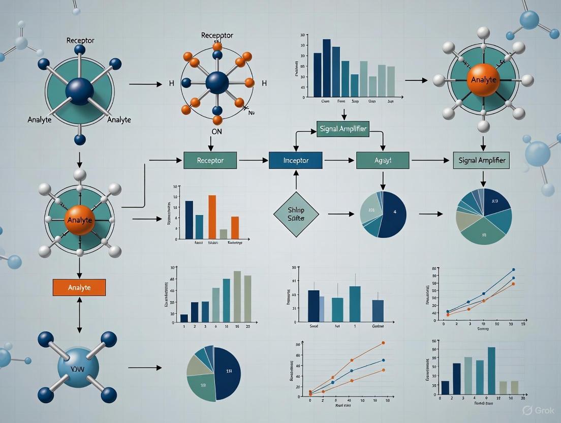
Core Biosensor Architecture and Function
A biosensor is defined as an analytical device that integrates a biological recognition element with a physicochemical transducer to convert a biological event into a measurable signal [23]. The fundamental operation involves five key components: the analyte (target substance), bioreceptor (biological recognition molecule), transducer (signal conversion element), electronics (signal processing unit), and display (user interface) [22].
The sequential process begins with the specific interaction between the bioreceptor and analyte, generating a biochemical signal. The transducer converts this signal into an electrical, optical, or other measurable output, which is then processed and displayed in a user-interpretable format [22]. Understanding this architecture is essential for identifying critical optimization parameters within each subsystem.
Figure 1: Core Biosensor Signal Pathway. This diagram illustrates the fundamental sequence of signal generation and processing in a typical biosensor system.
Optimization Domain I: Biorecognition Elements
Biorecognition elements constitute the primary source of biosensor specificity, as they determine the selective interaction with target analytes. The choice and immobilization of these biological components significantly influence analytical performance.
Types of Biorecognition Elements and Their Characteristics
Table 1: Comparative Analysis of Major Biorecognition Elements
| Bioreceptor Type | Key Advantages | Critical Limitations | Optimization Parameters |
|---|---|---|---|
| Antibodies | High specificity and sensitivity for antigens [24] | Resource-intensive production; batch-to-batch variability; stability concerns [24] | Epitope specificity, affinity constants, cross-reactivity, immobilization density |
| Enzymes | Catalytic amplification; high turnover number [24] | Stringent environmental requirements (pH, temperature); higher costs compared to synthetic elements [24] | Catalytic activity, substrate specificity, kinetic parameters (Km, Vmax), operational stability |
| DNA/Aptamers | Programmable structure; chemical stability; molecular recognition fidelity [24] | Sensitive to hybridization conditions (temperature, pH, ionic strength); nuclease degradation susceptibility [24] | Sequence design, hybridization efficiency, secondary structure stability, modification chemistry |
| Whole Cells | Complex response profiling; metabolic activity monitoring [23] | Viability maintenance requirements; slower response times | Membrane permeability, receptor expression, viability indicators, growth conditions |
Immobilization Techniques for Biorecognition Elements
The method of immobilizing biorecognition elements onto the transducer surface critically impacts biosensor performance by affecting orientation, stability, and accessibility.
- Cross-linking: Covalent binding using agents like glutaraldehyde creates robust enzyme-substrate interactions, reducing enzyme leaching and enhancing stability [25].
- Entrapment: Physical encapsulation within nanomaterial matrices (e.g., sol-gel or polymeric materials) protects enzymes from environmental fluctuations while preserving catalytic function [25].
- Physical Adsorption: Relies on non-covalent interactions (Van der Waals forces, electrostatic contacts); cost-effective but may result in enzyme desorption and reduced long-term stability [25].
- Covalent Bonding: Forms stable bonds between functional groups on enzymes and nanomaterials, providing permanent attachment and longer-term biosensor durability [25].
Optimization Domain II: Transduction Mechanisms
Transducers serve as the critical interface that converts biological recognition events into quantifiable signals, with each transduction modality offering distinct advantages and optimization requirements.
Classification and Performance Characteristics of Transducers
Table 2: Transducer Types and Their Performance Characteristics
| Transducer Type | Measurable Signal | Sensitivity Range | Key Advantages | Common Applications |
|---|---|---|---|---|
| Electrochemical | Current, potential, impedance changes [23] [25] | Varies by subtype; e.g., LOD to 0.027 mM for glucose [26] | Simplicity, portability, low power requirements [23] | Glucose monitoring, pathogen detection, cardiac biomarkers [23] [26] |
| Amperometric | Current from redox reactions [22] | ~0.027-0.034 mM (glucose) [26] | High sensitivity, enzymatic turnover quantification | Metabolic sensors, enzyme activity assays |
| Potentiometric | Potential difference [22] | Not specified in results | Simple instrumentation, ion concentration measurement | pH sensing, ion detection |
| Impedimetric | Impedance/Resistance to alternating current [23] [25] | Wide frequency range (Hz to MHz) [25] | Label-free detection, continuous monitoring capability [25] | Pathogen detection, antibody-antigen interactions [23] |
| Optical | Absorbance, fluorescence, luminescence, refractive index [23] | Single-molecule sensitivity possible [23] | Superior multiplexing capabilities, real-time kinetic monitoring [23] | Cellular response tracking, binding kinetics [23] |
| Piezoelectric | Mass changes via resonance frequency shifts [23] | Not specified in results | Label-free detection, real-time monitoring | Cancer biomarkers, pathogen detection [26] |
| Thermal | Heat exchange from reactions [23] | Not specified in results | Direct detection of enzymatic activity | Enzyme-substrate interactions |
Optimization Domain III: Assay and Environmental Conditions
Assay conditions and environmental factors constitute the third critical optimization domain, significantly influencing biosensor stability, reproducibility, and real-world applicability.
Critical Assay Parameters and Control Strategies
Temperature Effects: Biological elements exhibit significant temperature sensitivity; enzyme-based sensors require precise temperature regulation to maintain catalytic activity [24]. Implementation of temperature correction algorithms or engineered enzyme mutants can enhance robustness [23].
pH Optimization: DNA-based recognition systems require strict control of hybridization conditions as variations in pH reduce binding efficiency [24]. Buffer selection and capacity must match the operational requirements of the biological recognition element.
Matrix Interference Management: Complex samples (serum, wastewater, plant extracts) introduce nonspecific binding and fouling [23]. mitigation strategies include using blocking agents, antifouling coatings, or prefiltration to minimize false positives/negatives [23].
Calibration and Drift Control: Biological component degradation over time affects calibration curves, necessitating regular recalibration, reference standards, and proper storage conditions [23].
Integrated Experimental Framework for Biosensor Optimization
A systematic approach to biosensor optimization requires methodical investigation of parameters across all three domains. The following framework supports comprehensive characterization and performance enhancement.
Researcher's Toolkit: Essential Reagents and Materials
Table 3: Key Research Reagent Solutions for Biosensor Development
| Reagent/Material | Function/Purpose | Application Examples |
|---|---|---|
| Gold Nanoparticles | Enhance conductivity, increase surface area for bioreceptor immobilization [25] | Electrode modification for cancer antigen 125 detection [25] |
| Dendritic Gold Nanostructures | Create high-surface-area electrode platforms for enhanced sensitivity [26] | Glucose biosensor platforms with improved electron transfer [26] |
| Glutaraldehyde | Cross-linking agent for covalent enzyme immobilization [25] | Creating robust enzyme-substrate interactions in nanostructured biosensors |
| Streptavidin-coated Magnetic Nanoparticles | Separation and concentration of biotinylated DNA products [26] | PCR and LAMP product detection in nucleic acid amplification tests |
| PANI-AuNPs Nanocomposite | Conductive polymer-nanoparticle composite for enhanced electron transfer [26] | Glucose biosensor electrodes offering better stability and interference resistance |
| Aptamers (SELEX-derived) | Synthetic recognition elements with high stability and specificity [24] | Mycotoxin detection, small molecule sensing as antibody alternatives |
| Molecularly Imprinted Polymers | Synthetic receptors with tailored recognition sites [26] | Wearable cortisol sensors in sweat for stress monitoring |
Methodological Protocols for Key Characterization Experiments
Protocol 1: Immobilization Efficiency Assessment
- Surface Preparation: Modify electrode surfaces with dendritic gold nanostructures using electrochemical deposition [26].
- Bioreceptor Attachment: Apply biorecognition elements (enzymes, antibodies, aptamers) via selected immobilization method (cross-linking, entrapment, adsorption) [25].
- Activity Measurement: Quantify immobilized bioreceptor activity through enzymatic turnover rates or binding capacity assays.
- Stability Testing: Monitor signal retention over multiple operational cycles (e.g., 120 cycles for wearable sensors) and storage periods [26].
Protocol 2: Electrochemical Biosensor Calibration
- Electrode Preparation: Construct working electrodes using nanostructured materials (e.g., poly(o-phenylenediamine)/silver core-shell hybrids on GCE) [25].
- Standard Curve Generation: Measure response to analyte standards across operational concentration range.
- Signal Processing: Apply baseline correction, noise reduction, and drift compensation algorithms [23].
- Figure of Merit Calculation: Determine linear range, sensitivity (μA/mM), LOD (e.g., 0.027 mM for glucose), and reproducibility (% RSD) [26].
Protocol 3: Specificity and Interference Testing
- Sample Preparation: Spike target analyte into complex matrices (serum, food homogenates) alongside potential interferents.
- Response Measurement: Compare biosensor signals from target-specific samples versus interferent-containing samples.
- Selectivity Calculation: Quantify signal differences to establish selectivity coefficients (e.g., >100-fold against glucose, lactic acid) [26].
- Cross-reactivity Assessment: Test against structurally similar compounds to evaluate recognition specificity.
Figure 2: Biosensor Optimization Workflow. This diagram outlines the systematic approach to optimizing biosensor performance through parameter identification and experimental validation.
The systematic optimization of biosensors demands meticulous attention to parameters across three interconnected domains: biorecognition elements, transduction mechanisms, and assay conditions. Successful biosensor development requires leveraging structured experimental frameworks, such as Design of Experiments, to efficiently navigate this complex parameter space. The integration of advanced nanomaterials, sophisticated immobilization techniques, and robust signal processing algorithms will continue to push the boundaries of biosensor performance. Furthermore, the emerging incorporation of artificial intelligence promises enhanced data processing capabilities and predictive analytics for future biosensor platforms [22]. By applying the structured approaches and methodologies outlined in this technical guide, researchers can accelerate the development of next-generation biosensors with enhanced sensitivity, specificity, and reliability for diverse applications in diagnostics, environmental monitoring, and food safety.
Strategic Implementation: Applying DoE Frameworks to Diverse Biosensor Platforms
The systematic optimization of biosensors is a complex challenge, requiring the careful balancing of multiple interacting physical, chemical, and biological parameters. Sequential Design of Experiments (DoE) provides a structured, efficient framework for navigating this multi-faceted design space, enabling researchers to move rationally from initial screening to a robust, optimized final product. This methodology stands in stark contrast to the traditional "one-factor-at-a-time" (OFAT) approach, which is not only inefficient but also fails to capture critical factor interactions [27] [28]. Within the context of biosensor development, whether for enhancing the sensitivity of a lateral flow immunoassay (LFIA) for aflatoxin detection [27] or pushing the detection limits of a surface plasmon resonance (SPR) sensor to the single-molecule level [29], a phased experimental strategy is paramount.
This guide outlines the core trilogy of the sequential DoE workflow: Factor Screening to identify vital few factors from the trivial many; Response Surface Optimization to pinpoint the precise combination of factor levels that delivers optimal performance; and Robust Process Design to ensure the biosensor's performance remains reliable despite normal, expected variations in manufacturing and use conditions. By adopting this data-driven framework, researchers and development professionals can accelerate development timelines, reduce costs, and ultimately deliver more sensitive, reliable, and commercially viable biosensing platforms [28] [30].
Phase 1: Factor Screening with Definitive Screening Designs
The primary goal of the screening phase is to efficiently sift through a potentially large number of process parameters to identify which ones have a statistically significant and meaningful effect on the biosensor's critical quality attributes (CQAs). These CQAs may include the limit of detection (LOD), sensitivity, dynamic range, signal-to-noise ratio, and reproducibility.
Advanced Screening Designs
While traditional Plackett-Burman or Resolution IV fractional factorial designs have been widely used, Definitive Screening Designs (DSDs) represent a powerful modern alternative. DSDs are a class of three-level screening designs that require remarkably few experimental runs. For example, a DSD can evaluate six different input parameters with only 14 experimental runs [30]. Their three-level nature is a key advantage, as it allows for the initial assessment of potential curvature in the response. If curvature is detected for a factor, it indicates that the factor's optimum level is likely within the tested range, providing crucial early guidance for the optimization phase. Furthermore, DSDs can simultaneously evaluate main effects and quadratic relationships, offering a more informative screening step compared to two-level designs [30].
Practical Application in Biosensor Development
The screening phase typically involves the following steps:
- Define Inputs and Outputs: List all potential factors (e.g., concentrations, pH, temperatures, incubation times) and the key performance metrics (responses) to be measured.
- Select a Screening Design: Choose an appropriate design (e.g., a DSD) based on the number of factors.
- Execute Experiments: Run the experiments in a randomized order to avoid confounding with unknown nuisance variables.
- Analyze Data and Identify CPPs: Use statistical analysis (e.g., regression analysis, analysis of variance) to identify the Critical Process Parameters (CPPs) that significantly impact the CQAs.
Table 1: Example of a Definitive Screening Design Matrix for 6 Factors
| Experiment Run | Factor A (Antibody Conc.) | Factor B (pH) | Factor C (Incubation Time) | Factor D (Label Ratio) | Factor E (Blocking Agent) | Factor F (Substrate Type) |
|---|---|---|---|---|---|---|
| 1 | -1 (Low) | -1 (Low) | -1 (Low) | -1 (Low) | -1 (Low) | -1 (Low) |
| 2 | 1 (High) | -1 | -1 | 1 | 1 | -1 |
| 3 | -1 | 1 (High) | -1 | 1 | -1 | 1 |
| 4 | 1 | 1 | -1 | -1 | 1 | 1 |
| 5 | -1 | -1 | 1 (High) | 1 | 1 | 1 |
| 6 | 1 | -1 | 1 | -1 | -1 | 1 |
| 7 | -1 | 1 | 1 | -1 | 1 | -1 |
| 8 | 1 | 1 | 1 | 1 | -1 | -1 |
| 9 | 0 (Center) | 0 (Center) | 0 (Center) | 0 (Center) | 0 (Center) | 0 (Center) |
| 10 | 0 | 0 | 0 | 0 | 0 | 0 |
| 11 | 0 | 0 | 0 | 0 | 0 | 0 |
| 12 | 0 | 0 | 0 | 0 | 0 | 0 |
| 13 | 0 | 0 | 0 | 0 | 0 | 0 |
| 14 | 0 | 0 | 0 | 0 | 0 | 0 |
Figure 1: A sequential workflow for the factor screening phase, highlighting the iterative nature of identifying Critical Process Parameters.
Phase 2: Response Surface Optimization
Once the key factors are identified, the next phase is to model the relationship between these factors and the responses to find their optimal settings. Response Surface Methodology (RSM) is the premier technique for this purpose, creating a mathematical model that maps the experimental landscape.
Modeling with Central Composite and Box-Behnken Designs
The most common RSM designs are Central Composite Designs (CCD) and Box-Behnken Designs (BBD). A CCD, for instance, is built around a factorial or fractional factorial core, augmented with axial (star) points and center points. This structure allows for efficient estimation of a full quadratic (second-order) model, which is necessary to capture curvature and locate a maximum, minimum, or saddle point in the response surface [27].
Case Study: Optimizing a Lateral Flow Immunoassay
A study optimizing a competitive LFIA for aflatoxin B1 (AFB1) provides an excellent example. The researchers employed a sequential DoE strategy named the "4S" method (START, SHIFT, SHARPEN, STOP). In the optimization phase, they investigated four key variables: the concentration of the labeled antibody, the antibody-to-label ratio, the concentration of the competitor antigen spotted on the test line, and the hapten-to-protein substitution ratio of the competitor [27].
By generating and overlaying the response surfaces for the negative control signal (NEG) and the signal inhibition (IC%), the researchers identified a region of optimal compromise. The optimized LFIA-1 device achieved a limit of detection of 0.027 ng/mL, a significant enhancement over the original device's 0.1 ng/mL LOD. Furthermore, the amount of expensive antibody required was reduced by a factor of four, demonstrating how DoE can simultaneously improve performance and reduce cost [27].
Table 2: Experimental Factors and Ranges for an LFIA Optimization Study [27]
| Factor Name | Type | Low Level | High Level | Optimal Value (Found) |
|---|---|---|---|---|
| Labeled Antibody Concentration | Numerical | To be determined by design | To be determined by design | Optimized |
| Antibody-to-Label Ratio | Numerical | To be determined by design | To be determined by design | Optimized |
| Competitor Antigen Concentration | Numerical | To be determined by design | To be determined by design | Optimized |
| Hapten-to-Protein Ratio (Sr) | Categorical (e.g., 10, 40, 160) | Low (e.g., 10) | High (e.g., 160) | Optimized |
Detailed Protocol: LFIA Test Line Optimization
- Objective: To determine the optimal combination of competitor concentration (T) and hapten substitution ratio (Sr) that maximizes the signal-to-noise ratio (or minimizes IC%) for a target LOD.
- Materials:
- Nitrocellulose membrane strips
- Dispensing instrument for test line
- Labeled antibody-gold nanoparticle conjugate
- AFB1 standards (e.g., 0 ng/mL and 1 ng/mL)
- Running buffer
- Strip reader for quantitative signal measurement
- Method:
- Design: Set up a CCD or BBD with competitor concentration and substitution ratio as factors. Include center points to estimate pure error.
- Fabrication: Spot the test lines onto the nitrocellulose membrane according to the experimental design matrix, varying the concentration (T) and using the different synthesized conjugates (Sr).
- Testing: Run the LFIA strips with the NEG (0 ng/mL) and POS (1 ng/mL) samples. Measure the signal intensity at the test line for each strip.
- Analysis: For each experimental run, calculate the response IC% = (SignalPOS / SignalNEG) * 100%. Fit a quadratic model to the IC% data. Use the model's response surface and overlay plots with the Signal_NEG response to find a region that satisfies both a high negative control signal and a high level of inhibition at the target concentration.
Figure 2: The workflow for the response surface optimization phase, from experimental design to locating the optimum settings.
Phase 3: Robust Process Design and Verification
An optimized process is only valuable if it is consistently reproducible. The final phase focuses on ensuring the biosensor's performance is robust—that is, insensitive to small but inevitable variations in raw materials, environmental conditions, and operational parameters.
Principles of Robustness
Robustness in bioprocessing is defined as the ability of a process to deliver product quality within specified limits despite the inherent variability of biological systems and input materials [28]. Sources of variation can include fluctuations in substrate and medium compositions, phenotypic heterogeneity in microbial production cells, and minor deviations in operational parameters like temperature or pH [28]. A robust biosensor manufacturing process must account for these variances across the entire chain, from upstream reagent production to downstream assembly and testing.
Implementing Robust Design
The methodology for robust design involves:
- Identifying Noise Variables: These are factors that are difficult or expensive to control tightly in a manufacturing setting (e.g., ambient humidity, slight variations in buffer pH, different operator techniques).
- Designing the Experiment: A combined array design is used, where the controllable CPPs (from Phase 2) are set up in an inner array, and the noise variables are deliberately varied in an outer array.
- Analyzing for Robustness: The response is measured for all combinations. The goal is to find the settings of the controllable factors that minimize the variation in the response caused by the noise factors. This often involves analyzing the signal-to-noise ratio (S/N ratio) popularized by Taguchi methods.
For example, a critical parameter like the concentration of a capture antibody spotted on a membrane would be tested not only at its optimal mean level but also at levels slightly above and below, while simultaneously introducing small, controlled variations in other factors like incubation time or temperature. The combination that produces the most consistent LOD and signal intensity across these "noisy" conditions is deemed the most robust.
The Scientist's Toolkit: Research Reagent Solutions
Table 3: Key Reagents and Materials for Biosensor Optimization
| Item | Function in Biosensor Development | Example from Literature |
|---|---|---|
| Gold Nanoparticles (AuNPs) | Commonly used as plasmonic reporters in visual lateral flow immunoassays (LFIAs) due to their intense red color from localized surface plasmon resonance (LSPR). [27] | Used as the label in the optimized AFB LFIA. [27] |
| Hapten-Protein Conjugates | Act as the immobilized competitor antigen in competitive assay formats. The hapten-to-protein substitution ratio (Sr) is a critical optimization parameter. [27] | AFB1-CMO coupled to ovalbumin (OVA) at different molar excesses (10, 40, 160) was tested. [27] |
| Aptamers | Single-stranded oligonucleotides used as synthetic recognition elements. Offer advantages of thermal stability and flexibility of modification compared to antibodies. [31] | Selected via SELEX; can be integrated into electrochemical, optical, and lateral flow platforms. [31] |
| 2D Nanomaterials (Graphene, MoS₂) | Used to enhance sensor interfaces due to large specific surface area and strong analyte binding capabilities, improving sensitivity in SPR and other optical sensors. [29] [32] | A graphene-based biosensor used a multilayer architecture for breast cancer detection, achieving 1785 nm/RIU sensitivity. [32] |
| Organic Electrochemical Transistors (OECTs) | Used to dramatically amplify weak electrical signals from enzymatic or microbial fuel cells, enabling highly sensitive bioelectronic detection. [33] | Amplified signals from microbial fuel cells by 1,000-7,000x for sensitive arsenite and lactate detection. [33] |
Advanced & Emerging Methodologies
The field of biosensor optimization is being rapidly advanced by the integration of computational and machine learning (ML) tools.
- Machine Learning for Sensor Design: ML models are now used to predict the performance of complex biosensor designs, such as Photonic Crystal Fiber-SPR (PCF-SPR) sensors, based on their structural parameters. This approach can significantly accelerate the optimization process, reducing reliance on time-consuming and costly simulations and experiments. For instance, one study used ML regression and explainable AI (XAI) to optimize a PCF-SPR biosensor, achieving a wavelength sensitivity of 125,000 nm/RIU [7].
- Algorithm-Assisted Multi-Objective Optimization: For sophisticated sensor designs, balancing multiple, often competing, performance metrics is essential. Algorithms like Multi-Objective Particle Swarm Optimization (MOPSO) can be employed to concurrently optimize several parameters. This approach was used to enhance the sensitivity, figure of merit (FOM), and depth of resonant dip of an SPR biosensor, leading to a 230% improvement in sensitivity and a limit of detection of 54 ag/mL for mouse IgG [29].
- Automated and High-Throughput Workflows: The exploration of vast genetic design spaces for biological components, such as in transcription factor-based biosensors, is now being enabled by combining DoE with automation. This allows for the efficient fractional sampling of a combinatorial experimental space, drastically speeding up the identification of optimal biosensor configurations [34].
The development of high-performance biosensors and positron emission tomography (PET) tracers relies critically on the availability of optimally radiolabeled molecules. Copper-mediated radiofluorination (CMRF) has emerged as a transformative methodology for introducing fluorine-18 into complex molecules, enabling the creation of novel imaging agents and sensor tracers targeting specific biological processes. [35] [36] However, the optimization of CMRF reactions presents significant challenges due to the complex, multi-parameter nature of these processes and the unique constraints of radiochemistry, including limited reagent availability, short isotope half-life (110 minutes), and safety considerations when handling high radioactivity levels. [17]
Traditional optimization approaches in radiochemistry have predominantly utilized the "one variable at a time" (OVAT) method, which systematically varies individual factors while holding others constant. [17] While straightforward, OVAT is experimentally inefficient, time-consuming, and critically, unable to detect factor interactions—where the optimal level of one factor depends on the level of another. [17] This limitation often results in suboptimal reaction conditions and incomplete process understanding.
Design of Experiments (DoE) represents a powerful statistical approach that addresses these limitations by systematically varying multiple factors simultaneously according to a predefined experimental matrix. [17] [37] This perspective explores the application of DoE methodology to optimize CMRF processes for sensor tracer development, demonstrating how this approach accelerates optimization, enhances understanding of critical process parameters, and ultimately facilitates the development of more effective biosensing platforms.
Fundamental Principles of DoE in Radiochemistry
Core Concepts of Experimental Design
DoE is a model-based optimization approach that establishes mathematical relationships between input variables (factors) and output responses. [37] Unlike happenstance data collection or OVAT approaches, DoE employs causally-derived data from experiments distributed across the entire experimental domain to build predictive models that describe system behavior. [37] This methodology offers two primary advantages: (1) significantly reduced experimental effort compared to OVAT, and (2) the ability to detect and quantify factor interactions that would otherwise remain obscured. [17] [37]
The DoE workflow typically proceeds through sequential phases: [17]
- Factor Screening: Initial low-resolution designs (e.g., fractional factorial) identify which of many potential factors significantly influence key responses
- Response Surface Optimization: Higher-resolution designs (e.g., central composite) with reduced factor sets model complex behaviors, including curvature, to locate optimal conditions
- Model Validation: Verification of model adequacy and predictive capability through confirmation experiments
DoE Versus OVAT: A Comparative Analysis
Table 1: Comparison of DoE and OVAT Approaches for Reaction Optimization
| Characteristic | Traditional OVAT | DoE Approach |
|---|---|---|
| Experimental efficiency | Low - requires many sequential experiments | High - studies multiple factors simultaneously |
| Detection of factor interactions | No - cannot detect interactions | Yes - quantifies interaction effects |
| Risk of finding false optima | High - prone to local optima | Low - maps entire response space |
| Model building capability | Limited - empirical understanding | Comprehensive - mathematical models |
| Resource requirements | High time, materials, and radioactivity | Reduced experimental burden |
| Information quality | Limited understanding of process | Detailed process understanding |
The fundamental limitation of OVAT becomes particularly problematic in complex, multi-component systems like CMRF, where factors such as temperature, solvent composition, copper source, ligand, and precursor stoichiometry can exhibit significant interactions. [17] As demonstrated in one CMRF optimization study, DoE achieved equivalent process understanding with more than two-fold greater experimental efficiency compared to the OVAT approach. [17]
Copper-Mediated Radiofluorination: Methodology and Challenges
CMRF Reaction Mechanics
CMRF has revolutionized the synthesis of aromatic C-18F bonds, enabling radiolabeling of electron-rich and neutral aromatic rings that were previously inaccessible via conventional nucleophilic aromatic substitution. [35] [36] The methodology typically involves the reaction of an aryl precursor (boronic acid, boronic ester, stannane, or iodonium salt) with [18F]fluoride in the presence of a copper catalyst and suitable ligands. [17] [36]
The mechanism is believed to parallel the Chan-Lam cross-coupling, proceeding through: (1) transmetalation of the aryl nucleophile with a solvated copper(II)-ligand-[18F]fluoride complex, (2) oxidation to form an organoCu(III) species, and (3) C(sp2)-18F bond-forming reductive elimination to release the radiolabeled product. [36] This pathway enables efficient radiofluorination under relatively mild conditions with exceptional functional group tolerance.
Key Optimization Parameters in CMRF
Multiple factors influence the efficiency of CMRF reactions, creating an ideal application for DoE optimization: [17]
- Copper source and stoichiometry: Different copper salts (e.g., Cu(OTf)2, Cu(OTf)py4) exhibit varying reactivity
- Ligand system: Pyridine derivatives and other nitrogen-based ligands affect copper solubility and reactivity
- Solvent composition: Mixed solvent systems (e.g., DMF, DMSO, acetonitrile, t-butanol) influence reaction kinetics
- Temperature: Typically ranges from 80-110°C
- Reaction time: Generally 10-30 minutes
- Precursor stoichiometry and structure: Boron-based precursors (2-4 μmol) commonly used
- [18F]Fluoride processing method: Includes elution from ion exchange cartridges and azeotropic drying conditions
The complexity of these interacting factors, combined with the challenge of working with radioactive materials, makes CMRF optimization particularly suited to the DoE approach. [17]
DoE Implementation Strategy for CMRF Optimization
Experimental Workflow for DoE in CMRF
A systematic, phased approach to DoE implementation ensures efficient resource utilization and comprehensive process understanding. [17] [38]
Representative DoE Applications in CMRF
Case Study 1: Optimization of [18F]Olaparib Synthesis
A recent study demonstrated the application of DoE to optimize the CMRF synthesis of [18F]olaparib, a PARP inhibitor for cancer imaging. [38] Researchers implemented a scalable base-free method for processing [18F]fluoride as [18F]TBAF, enabling single production to be divided into aliquots for multiple small-scale DoE experiments. This approach facilitated efficient optimization, which was successfully translated to automated production, yielding [18F]olaparib in 78 ± 6% radiochemical yield (CMRF step only) and 41% activity yield in automated syntheses. [38]
Case Study 2: CMRF of Arylstannanes
A comprehensive DoE study addressed the optimization of copper-mediated 18F-fluorination reactions of arylstannanes. [17] Using factor screening and optimization designs, researchers identified critical factors and modeled their behavior with more than two-fold greater experimental efficiency than the traditional OVAT approach. The resulting models provided new insights into reaction behavior and guided the development of efficient reaction conditions suitable for 18F PET tracer synthesis. [17]
Table 2: Key Research Reagent Solutions for CMRF Optimization
| Reagent Category | Specific Examples | Function in CMRF |
|---|---|---|
| Copper Sources | Cu(OTf)2, Cu(OTf)py4 | Mediates fluoride transfer and reductive elimination |
| Ligands | Pyridine, 2,2'-bipyridine, Phenanthroline derivatives | Enhances copper solubility and stabilizes intermediates |
| Solvents | DMF, DMSO, MeCN, t-BuOH, H2O | Provides reaction medium; affects solubility and kinetics |
| Precursors | Arylboronic acids, Arylboronic esters, Arylstannanes | Source of aromatic ring for fluorination |
| Base/Additives | K2CO3, Cs2CO3, KOTf | Facilitates [18F]fluoride elution and reactivity |
| [18F]Fluoride Processing | "Minimalist" approach, [18F]TBAF method | Enables efficient fluoride recovery and reaction compatibility |
Advanced DoE Designs for CMRF Optimization
Factorial Designs for Initial Screening
Two-level factorial designs (2^k) serve as efficient first-order orthogonal designs for initial factor screening, requiring 2^k experiments where k represents the number of factors. [37] In these designs, each factor is studied at two levels (coded as -1 and +1), enabling estimation of main effects and two-factor interactions. [37]
For a CMRF study with three factors (e.g., temperature, copper amount, reaction time), a full factorial design would require 8 experiments (2^3). The experimental matrix and corresponding mathematical model would take the form: [37]
Y = b0 + b1X1 + b2X2 + b3X3 + b12X1X2 + b13X1X3 + b23X2X3 + b123X1X2X3
Where Y represents the response (e.g., radiochemical conversion), b0 is the constant term, b1-b3 are main effect coefficients, and b12-b123 represent interaction coefficients. [37]
Response Surface Methodologies
After identifying significant factors through screening designs, response surface methodologies (RSM) characterize complex nonlinear relationships between factors and responses. Central composite designs (CCD) represent the most common RSM approach, augmenting factorial designs with additional points to estimate curvature effects. [37]
These designs enable the construction of quadratic models of the form: Y = b0 + ΣbiXi + ΣbiiXi^2 + ΣbijXiXj
Such models can identify optimal conditions (maximum, minimum, or saddle points) within the experimental domain and generate response surface plots that visually represent the relationship between factors and responses. [17] [37]
DoE Implementation Protocol: Step-by-Step Guide
Preliminary Factor Screening Study
Objective: Identify factors with significant effects on radiochemical conversion (RCC) in CMRF of arylstannanes. [17]
Experimental Design: Resolution IV fractional factorial design for 5 factors (requiring 16 experiments plus center points)
Factors and Levels:
- Copper source (X1): Cu(OTf)2 (-1) vs. Cu(OTf)py4 (+1)
- Copper amount (X2): 5 μmol (-1) vs. 15 μmol (+1)
- Temperature (X3): 90°C (-1) vs. 110°C (+1)
- Reaction time (X4): 10 min (-1) vs. 20 min (+1)
- Precursor amount (X5): 2 μmol (-1) vs. 4 μmol (+1)
Execution:
- Prepare [18F]fluoride via standard processing methods [38]
- Set up reactions according to experimental matrix in randomized order
- Perform CMRF reactions in sealed vials with heating
- Analyze RCC via radio-TLC or radio-HPLC
- Perform statistical analysis to identify significant effects (p < 0.05)
Response Surface Optimization
Objective: Model nonlinear effects and identify optimal conditions for maximal RCC
Experimental Design: Central composite design for 3 critical factors identified in screening study (requiring 20 experiments including 6 center points)
Execution:
- Conduct experiments according to CCD matrix
- Measure multiple responses (RCC, specific activity, byproduct formation)
- Fit quadratic models using multiple linear regression
- Validate model adequacy through statistical and graphical analysis
- Generate response surface plots to visualize factor effects
- Identify optimal conditions using desirability functions
- Confirm predictions with confirmation experiments
Data Analysis and Interpretation
Statistical Analysis of DoE Results
Proper statistical analysis transforms experimental results into meaningful process understanding. Key analytical steps include: [17] [37]
- Model fitting: Calculate coefficient estimates using multiple linear regression
- Significance testing: Evaluate model terms using ANOVA (p-values) or Pareto charts
- Model adequacy checking: Examine residuals for patterns and outliers
- Model reduction: Eliminate non-significant terms to improve model robustness
- Lack-of-fit testing: Compare pure error (from replicates) to model error
Visualization of DoE Results
Graphical tools enhance interpretation of DoE results:
- Pareto charts: Display absolute magnitude of standardized effects, highlighting significant factors
- Interaction plots: Reveal how the effect of one factor changes across levels of another factor
- Contour plots: Show response values across two factors while holding others constant
- Response surface plots: Provide three-dimensional visualization of response behavior
- Optimization plots: Display simultaneous optimization of multiple responses using desirability functions
Translation to Automated Production
A critical advantage of the DoE approach is the reliable translation of optimized conditions from small-scale manual experiments to automated production modules. [17] [38] This translation requires consideration of several factors:
- Scale-up considerations: Differences in reactor geometry, mixing efficiency, and heating profiles
- Atmosphere control: Automated modules typically maintain inert atmosphere versus manual bench reactions
- [18F]Fluoride processing: Automated systems employ different drying and elution protocols
- Reagent addition sequences: Timing and method of reagent introduction may differ
- Purification requirements: Automated systems require efficient purification for clinical use
The implementation of scalable [18F]processing methods, such as the base-free [18F]TBAF approach, significantly enhances the reliability of this translation from DoE optimization to automated production. [38]
The application of Design of Experiments represents a paradigm shift in the optimization of copper-mediated radiofluorination for sensor tracer development. By enabling efficient, systematic exploration of complex factor spaces while quantifying interaction effects, DoE accelerates radiochemical optimization and enhances process understanding. The methodology has proven particularly valuable for CMRF, where multiple interacting factors and resource constraints create ideal conditions for implementation.
As the demand for novel biosensors and imaging agents continues to grow, the adoption of statistical optimization approaches like DoE will play an increasingly critical role in streamlining tracer development and facilitating the translation of promising candidates from bench to bedside. Future advances will likely incorporate emerging technologies such as high-throughput experimentation, automation, and machine learning to further enhance the efficiency and effectiveness of radiochemical optimization.
Optimizing Paper-Based Biosensors and Lateral Flow Immunoassays with High-Throughput DoE
The development of high-performance paper-based biosensors and lateral flow immunoassays (LFIAs) represents a critical frontier in point-of-care diagnostics, environmental monitoring, and food safety testing. These analytical devices leverage the unique properties of paper—including its high porosity, capillary action, and cost-effectiveness—to create portable, user-friendly diagnostic platforms [39] [40]. However, achieving optimal performance in terms of sensitivity, specificity, and reproducibility requires careful optimization of numerous interdependent parameters, a challenge that traditional one-variable-at-a-time (OVAT) approaches cannot effectively address [37] [41].
Design of Experiments (DoE) has emerged as a powerful statistical framework that enables researchers to systematically investigate multiple factors and their interactions while minimizing experimental effort [37] [42]. This methodology is particularly valuable for optimizing complex bioanalytical systems like LFIAs, where factors including membrane characteristics, reagent concentrations, conjugation chemistry, and flow dynamics interact in non-linear ways to determine overall assay performance [39] [43]. High-throughput DoE approaches further enhance this capability by enabling rapid screening of numerous parameter combinations, dramatically accelerating the development timeline while providing comprehensive understanding of factor interactions [39] [37].
This technical guide examines the application of high-throughput DoE methodologies to paper-based biosensor optimization, providing researchers with structured frameworks, experimental protocols, and practical insights to enhance the performance and reliability of these diagnostic platforms within the broader context of systematic biosensor optimization research.
Fundamental DoE Principles for Biosensor Development
Core Concepts and Terminology
Implementing DoE effectively requires understanding its fundamental principles and vocabulary. The methodology treats any biosensor development process as a "black box" system where controlled input variables (factors) influence measurable outputs (responses) [41]. Critical process parameters (CPPs) represent the controllable factors during development and fabrication, such as antibody concentration, nanoparticle size, or membrane pore size. Critical quality attributes (CQAs) are the measurable outputs that define biosensor performance, including limit of detection (LOD), signal intensity, specificity, and reproducibility [41].
The experimental domain encompasses the range of values being investigated for each factor, while the response surface represents the mathematical relationship between factors and responses [37]. DoE approaches differ fundamentally from OVAT methodology by simultaneously varying all factors according to predetermined experimental arrays, enabling researchers to not only determine individual factor effects but also to quantify factor interactions that would remain undetected in sequential approaches [37] [41].
DoE Workflow and Design Selection
A structured workflow is essential for successful DoE implementation in biosensor optimization. The process begins with clear definition of optimization objectives and careful selection of both factors to investigate and responses to measure [41]. Subsequent steps include selecting appropriate experimental designs, executing randomized experiments to minimize bias, collecting response data, performing statistical analysis to develop predictive models, and ultimately validating these models with confirmation experiments [37] [41].
The choice of experimental design depends on the specific optimization objective. Screening designs like full factorial or Plackett-Burman designs efficiently identify the most influential factors from a large set of potential variables [37] [43]. For response surface methodology (RSM) aimed at finding optimal factor settings, central composite designs (CCD) or Box-Behnken designs are typically employed [37] [42]. Specialized designs such as mixture designs are used when factors are proportionally constrained components of a formulation [37].
Figure 1: DoE Systematic Workflow. This diagram illustrates the iterative process for optimizing biosensors using Design of Experiments methodology.
DoE Applications in Lateral Flow Immunoassay Development
Case Studies in LFIA Optimization
Recent research demonstrates the successful application of DoE to overcome specific challenges in LFIA development. In one notable study focused on detecting foot-and-mouth disease virus serotypes, researchers employed full-factorial and optimal designs to optimize a multiplex sandwich-type LFIA [43]. The study revealed that positioning of the capture region along the LFIA strip emerged as the most influential variable for detectability, enabling a two-fold sensitivity improvement compared to previous implementations [43].
For competitive LFIAs—a format particularly challenging due to its inverse signal response—a sequential DoE approach has proven highly effective. Research on cortisol detection demonstrated a 500-fold sensitivity improvement after just 13 experiments, with further optimization achieving 5000-fold enhancement after 34 experiments [42]. The investigators implemented a structured 4S decision process (Start, Shift, Sharpen, Stop) for interpreting response surfaces, systematically navigating the complex relationship between antibody-gold nanoparticle ratios, probe quantities, and competitor concentrations on the test line [42].
Advanced DoE Implementations
Beyond basic optimization, DoE methodologies have been integrated with computational approaches to further enhance biosensor performance. In the development of a carbendazim fungicide LFIA, researchers combined computer-aided hapten design with experimental optimization to significantly improve assay specificity [44]. The cross-reactivity with structural analogs was reduced to less than 0.1%, addressing a critical limitation in environmental and food safety testing [44].
Similar advanced implementations have employed DoE to optimize paper-based electrochemical biosensors, focusing on parameters including ink formulation, electrode architecture, and surface modification techniques [40] [45]. The systematic approach has proven particularly valuable for balancing multiple competing objectives, such as simultaneously maximizing sensitivity while minimizing reagent consumption and production costs [42] [40].
Table 1: DoE Applications in Biosensor Optimization - Case Study Summary
| Analyte Target | DoE Design Type | Key Optimized Factors | Performance Improvement | Reference |
|---|---|---|---|---|
| Foot-and-mouth disease virus (SAT serotypes) | Full-factorial, Optimal design | Capture antibody position, probe concentration | 2x sensitivity increase, LOD: 103.7–104.0 TCID/mL | [43] |
| Cortisol (competitive assay) | Sequential DoE with 4S process | Ab:AuNP ratio, probe amount, competitor amount | 500–5000x sensitivity improvement, LOD: 0.07 ng/mL | [42] |
| Carbendazim fungicide | Computer-aided with experimental validation | Hapten design, antibody specificity | Cross-reactivity <0.1% with structural analogs | [44] |
| Paper-based electrochemical sensors | Response surface methodology | Ink formulation, electrode geometry | Enhanced sensitivity and reproducibility | [40] [45] |
Experimental Protocols for DoE Implementation
Screening Design Protocol for Preliminary Optimization
Initial screening studies efficiently identify influential factors from a broad set of potential variables. A practical protocol for implementing a fractional factorial design in LFIA development includes:
Factor Selection: Identify 5-7 potentially critical factors based on prior knowledge or literature, such as antibody concentration, gold nanoparticle size, conjugate pad treatment, membrane type, blocking buffer composition, sample volume, and flow time [39] [46].
Range Determination: Define appropriate low and high levels for each factor based on preliminary experiments. For example, antibody concentration might be tested at 0.5 mg/mL and 2.0 mg/mL, while nanoparticle size could be evaluated at 20 nm and 40 nm [37] [41].
Experimental Setup: Generate a fractional factorial design matrix using statistical software, requiring 16-32 experimental runs for 5-7 factors. Randomize the run order to minimize bias from external factors [37].
Response Measurement: Quantify key performance metrics for each experimental run, including signal intensity, background noise, limit of detection, and reproducibility. Both positive and negative controls should be included in each assessment [43] [42].
Statistical Analysis: Analyze results using analysis of variance (ANOVA) to identify statistically significant factors (p < 0.05) and quantify factor interactions. Pareto charts can visually represent the relative importance of each factor [37] [41].
Response Surface Methodology for Advanced Optimization
After identifying critical factors through screening designs, RSM precisely characterizes nonlinear relationships and identifies optimal factor settings:
Central Composite Design Implementation: For 3-4 critical factors identified from screening, create a CCD with 20-30 experimental runs, including center points to estimate curvature and experimental error [37] [42].
Model Development: Use multiple regression analysis to develop a quadratic model describing the relationship between factors and responses. The general form of the model for two factors (X₁, X₂) is: Y = b₀ + b₁X₁ + b₂X₂ + b₁₂X₁X₂ + b₁₁X₁² + b₂₂X₂² [37].
Response Surface Analysis: Visualize the fitted model using contour plots and three-dimensional surface plots to understand the relationship between factors and identify optimal regions [42].
Optimization and Validation: Utilize desirability functions to identify factor settings that simultaneously optimize multiple responses. Confirm predicted optima with 3-5 validation experiments [42] [41].
Table 2: Research Reagent Solutions for DoE Optimization Studies
| Reagent/Material | Function in Biosensor Development | Optimization Considerations |
|---|---|---|
| Nitrocellulose membranes | Platform for capillary flow and bioreceptor immobilization | Pore size (0.05-12 μm), capillary flow time, protein binding capacity [39] [46] |
| Gold nanoparticles | Visual signal generation in LFIAs | Size (20-60 nm), surface chemistry, conjugation efficiency [39] [46] |
| Monoclonal antibodies | Biorecognition elements for target capture | Specificity, affinity, concentration, orientation during immobilization [43] [44] |
| Blocking buffers (BSA, sucrose, surfactants) | Reduce non-specific binding and stabilize conjugates | Composition, concentration, pH, incubation time [39] [46] |
| Conductive inks (carbon, metal-based) | Electrode fabrication in paper-based electrochemical sensors | Conductivity, viscosity, biocompatibility, curing conditions [40] [45] |
Advanced DoE Applications and Future Directions
Integration with Complementary Technologies
The power of DoE methodology is amplified when integrated with other advanced optimization approaches. Computational modeling combined with DoE creates a powerful hybrid framework for biosensor development. Finite element analysis can model fluid dynamics and binding kinetics on paper substrates, while DoE provides empirical validation and refinement of computational predictions [39]. This integration enables researchers to simulate thousands of virtual experiments before conducting physical testing, dramatically accelerating the optimization process.
Molecular modeling coupled with DoE represents another advanced approach, particularly for enhancing antibody specificity. As demonstrated in carbendazim detection, computer-aided hapten design analyzing molecular conformations, charge distributions, and electrostatic properties can identify optimal antigen structures before synthetic and experimental validation [44]. This approach systematically addresses cross-reactivity challenges that have traditionally plagued immunoassay development.
Artificial intelligence and machine learning algorithms are increasingly being integrated with DoE frameworks to handle extremely complex, high-dimensional optimization spaces. These approaches can identify non-obvious factor interactions and predict optimal parameter combinations that might escape detection through traditional statistical analysis [39].
Figure 2: DoE Technology Integration. Advanced DoE implementations combine with computational approaches for enhanced optimization.
Emerging Trends and Implementation Guidelines
Future developments in DoE for biosensor optimization will likely focus on several key areas. Adaptive DoE methodologies that incorporate real-time feedback and redirection of experimental plans based on interim results promise to further enhance optimization efficiency [37] [41]. The integration of DoE with high-throughput robotic systems enables automated execution of complex experimental designs, substantially increasing throughput and reproducibility while minimizing human error [39].
For researchers implementing DoE in biosensor development, several practical recommendations emerge from recent studies. Allocate no more than 40% of total resources to initial screening designs, preserving flexibility for subsequent optimization rounds [37]. Prioritize factors based on potential impact and experimental controllability, focusing initially on parameters known to significantly influence biosensor performance [41]. Implement rigorous randomization and blocking strategies to account for potential batch effects in reagents and environmental conditions [37] [41].
As the field advances, standardization of DoE protocols and reporting standards will enhance reproducibility and enable more effective comparison across studies. The systematic application of these methodologies will play an increasingly critical role in accelerating the development of next-generation paper-based biosensors with enhanced sensitivity, specificity, and reliability for point-of-care applications [39] [40] [45].
High-throughput Design of Experiments provides a powerful, systematic framework for optimizing the complex, multifactorial systems inherent in paper-based biosensors and lateral flow immunoassays. By enabling efficient exploration of factor interactions and response surfaces, DoE methodologies dramatically accelerate development timelines while enhancing assay performance beyond what is achievable through traditional optimization approaches. The integration of DoE with computational modeling, molecular design, and artificial intelligence represents the cutting edge of biosensor development, promising to deliver increasingly sophisticated diagnostic platforms capable of meeting diverse challenges in healthcare, environmental monitoring, and food safety. As these methodologies continue to evolve and become more accessible, they will play an indispensable role in advancing point-of-care diagnostics and enabling rapid, reliable detection of analytes across diverse applications.
Leveraging Machine Learning and Hybrid Models with DoE for Enhanced Predictive Power
The systematic optimization of biosensors is a primary obstacle limiting their widespread adoption as dependable point-of-care tests [1]. Traditional one-variable-at-a-time (OVAT) approaches remain problematic, particularly when dealing with interacting variables, often resulting in conditions that do not represent true optima [1]. Experimental design (DoE) has emerged as a powerful chemometric tool that provides a systematic, statistically reliable methodology for optimizing biosensor fabrication by accounting for both individual variable effects and their interactions [1]. While DoE offers a structured approach to experimentation, its integration with machine learning (ML) creates a transformative paradigm for enhancing predictive power in biosensor development.
The combination of mechanistic and machine learning models enables robust genotype-to-phenotype predictions in metabolic engineering applications [47]. This hybrid approach unites the causal understanding from mechanistic models with the pattern recognition capabilities of ML, creating a powerful framework for predictive engineering [47]. For complex biological systems like biosensors, this integration is particularly valuable as it leverages prior knowledge while efficiently extracting insights from multivariate experimental data.
Foundational DoE Frameworks for Biosensor Development
Core Experimental Design Modalities
DoE methodologies provide structured approaches for exploring complex experimental spaces. The fundamental principle involves strategically planning experiments to maximize information gain while minimizing resource expenditure [1]. Several core designs form the foundation of effective biosensor optimization:
Factorial Designs: 2^k factorial designs are first-order orthogonal designs requiring 2^k experiments, where k represents the number of variables being studied [1]. In these models, each factor is assigned two levels coded as -1 and +1, corresponding to the variable's selected range [1]. The experimental matrix comprises 2^k rows (individual experiments) and k columns (variables), systematically alternating levels to test all possible combinations [1].
Central Composite Designs: When response functions exhibit curvature, second-order models become essential [1]. Central composite designs augment initial factorial designs to estimate quadratic terms, enhancing the predictive capacity of the model [1]. These designs are particularly valuable for mapping response surfaces near optimal regions where linear approximations prove inadequate.
Mixture Designs: These specialized designs follow the inherent rule that the combined total of all components must equal 100% [1]. Consequently, mixture components cannot be altered independently, as changing one component's proportion necessitates adjustments to others [1]. This approach is particularly relevant for formulating detection interfaces or immobilization matrices with specific compositional constraints.
DoE Implementation Workflow
The DoE workflow follows a systematic, iterative process [1]:
- Factor Identification: Determine all factors that may exhibit a causality relationship with the targeted output signal (response)
- Experimental Domain Definition: Establish experimental ranges and distribution of experiments within the domain
- Model Construction: Use responses gathered from predetermined experimental points to build a mathematical model through linear regression
- Validation: Assess model adequacy by inspecting residuals (discrepancy between measured and predicted responses)
This approach provides comprehensive, global knowledge of the optimization space, offering maximum information for optimization purposes [1]. Importantly, DoE considers potential interactions among variables that consistently elude detection in OVAT approaches [1].
Hybrid Machine Learning Approaches for Predictive Modeling
Integration Frameworks
Hybrid machine learning approaches combine multiple algorithmic strategies to leverage their complementary strengths. These frameworks have demonstrated significant improvements in predictive accuracy across various biological applications:
Global-Local Modeling Integration: A particularly effective hybrid framework combines Ordinary Least Squares (OLS) for global surface estimation with Gaussian Process (GP) regression for uncertainty modeling [48]. OLS modeling provides a computationally inexpensive, interpretable method for capturing global trends within experimental data, while GP regression addresses its limitations by modeling data through flexible, probabilistic functions that quantify uncertainty explicitly [48]. This combination enables both broad pattern recognition and nuanced local exploration.
Mechanistic-ML Fusion: Combining mechanistic models with machine learning creates a powerful framework for predictive engineering [47]. Mechanistic models provide causal understanding based on prior knowledge, while ML algorithms learn complex patterns from experimental data [47]. This approach is particularly valuable for metabolic engineering applications where pathway regulation occurs at multiple levels [47].
Surrogate Modeling: Hybrid methodologies can explicitly correlate control strategies with operational parameters, formulating multiple strategies as design variables within statistical multi-objective optimization [49]. The forecasting ML model serves as a data-driven surrogate for optimal strategy selection, allowing for robust learning of complex control interactions [49].
Bio-Inspired Optimization Algorithms
Nature-inspired optimization algorithms offer powerful approaches for navigating complex parameter spaces:
- Ropalidia Marginata Optimization (RMO): This bio-inspired algorithm simulates the decentralized leadership and task allocation behaviors of Ropalidia marginata wasps, where any individual can temporarily assume leadership without centralized control [50]. Unlike particle swarm optimization (PSO) which models particle movement based on velocity and global/local best positions, RMO's flexible leader transition mechanism offers higher adaptability to dynamic optimization environments and reduces premature convergence risk [50]. When hybridized with neural networks, RMO enhances learning performance by efficiently navigating high-dimensional weight spaces, avoiding local minima, and improving convergence speed [50].
Table 1: Performance Comparison of Bio-Inspired Optimization Algorithms
| Algorithm | Mechanism Inspiration | Exploration-Exploitation Balance | Convergence Behavior |
|---|---|---|---|
| Ropalidia Marginata Optimization (RMO) | Wasp dominance hierarchies | Dynamic role shifting promotes diversity | Reduced premature convergence |
| Particle Swarm Optimization (PSO) | Bird flocking | Often favors exploitation once good solutions found | Susceptible to stagnation in local optima |
| Artificial Bee Colony (ABC) | Honey bee foraging | Balanced through employed, onlooker, and scout bees | Good but slower convergence |
| Genetic Algorithms (GA) | Natural evolution | Crossover and mutation maintain diversity | Can converge prematurely without parameter tuning |
Implementation Protocols for Biosensor Optimization
DoE-Guided Biosensor Development
The systematic optimization of ultrasensitive biosensors through experimental design involves carefully orchestrated protocols:
Protocol 1: Factorial Screening for Critical Parameters
- Identify potential factors influencing biosensor performance (e.g., immobilization strategy, detection interface formulation, detection conditions)
- Select appropriate ranges for each factor based on preliminary experiments
- Construct a 2^k factorial design matrix where k is the number of factors
- Execute experiments in randomized order to minimize confounding effects
- Analyze results to identify significant main effects and two-factor interactions
- Use analysis to eliminate non-significant variables before proceeding to more detailed optimization [1]
Protocol 2: Response Surface Methodology for Optimization
- Select 3-5 most critical factors identified from factorial screening
- Implement a Central Composite Design (CCD) to model curvature in the response surface
- Include center points to estimate pure error and check for curvature
- Execute experimental runs and measure response variables (e.g., sensitivity, limit of detection, signal-to-noise ratio)
- Fit a quadratic model to the experimental data
- Validate model adequacy through residual analysis and diagnostic plots
- Use response surface plots to identify optimal operating conditions [1]
ML-Enhanced DoE Workflows
Protocol 3: Hybrid Experimental Optimization with Active Learning
- Begin with an initial set of experiments selected through DoE principles
- Measure responses and train a hybrid ML model combining OLS for global trends and GP for uncertainty
- Use Expected Improvement (EI) as an acquisition function to identify promising untested conditions
- Apply K-means clustering to select diverse representative points from top candidates
- Conduct new experiments at selected conditions
- Retrain models with expanded dataset
- Iterate until convergence to optimal conditions [48]
Protocol 4: Biology-Guided Machine Learning for Biosensor Design
- Build a library of genetic parts and select relevant environmental factors
- Plan initial experiments using D-optimal design of experiments
- Characterize biosensor dynamic responses under varied conditions
- Calibrate an ensemble of mechanistic models by optimally fitting parameters
- Build a predictive ensemble of models using deep learning
- Validate predictions with additional experiments
- Use model to determine optimal condition combinations for desired biosensor specifications [51]
Case Studies in Biosensor Optimization
Whole-Cell Biosensor Optimization with DBTL Cycles
A compelling case study demonstrates the application of a Design-Build-Test-Learn (DBTL) pipeline for optimizing whole-cell biosensors based on allosteric transcription factors [51]. Researchers assembled a library of FdeR biosensors and characterized their performance under different conditions. They developed a mechanistic model to describe dynamic behavior under reference conditions, which guided a machine learning-based predictive model accounting for context-dependent dynamic parameters [51].
The implementation involved:
- Constructing a combinatorial library of biosensors in Escherichia coli with two modules: a naringenin-responsive transcription factor FdeR and a reporter module containing the FdeR operator region and GFP reporter gene [51]
- Testing 17 constructs under the same conditions (M9, 0.4% glucose, 400 μM naringenin), identifying promoters P1 and P3 as producing highest fluorescence values [51]
- Selecting an initial set of 32 experiments through D-optimal design of experiments to systematically explore interactions among factors [51]
- Observing that promoter P3 consistently exhibited higher fluorescence values across various RBSs, media, and supplements [51]
Cell-Free Biosensor Development for Environmental Monitoring
Cell-free biosensors represent another successful application area, particularly for environmental monitoring [52]. These systems leverage the flexibility of cell-free protein synthesis (CFPS) to operate in environments that would otherwise be toxic to living cells and can be designed for field deployment through preservation methods [52].
Notable implementations include:
- A paper-based system for detecting mercury using a dual-filter approach with smartphone readout, achieving detection limits as low as 6 μg/L [52]
- The ROSALIND platform capable of detecting copper, lead, and fluoride in water samples [52]
- A cell-free optical biosensor for mercury detection utilizing the merR gene incorporated into plasmid DNA constructs, achieving a detection limit of 1 ppb [52]
- A cell-free paper-based biosensor dependent on allosteric transcription factors for on-site detection of Hg²⁺ and Pb²⁺ in water, achieving impressive detection limits of 0.5 nM for Hg²⁺ and 0.1 nM for Pb²⁺ [52]
Table 2: Performance Characteristics of Cell-Free Biosensors for Environmental Monitoring
| Target Analyte | Detection Method/System | Limit of Detection | Sample Matrix |
|---|---|---|---|
| Mercury | Paper-based, dual-filter, smartphone readout | 6 μg/L | Water |
| Mercury | merR gene, plasmid DNA, firefly luciferase/eGFP | 1 ppb | Water |
| Mercury | Allosteric transcription factors (aTFs) | 0.5 nM | Water |
| Lead | aTFs | 0.1 nM | Water |
| Lead | Engineered PbrR mutants | 50 nM | Water |
| Tetracyclines | Riboswitch-based, RNA aptamers | 0.4 μM | Milk samples |
Computational Framework and Workflow Visualization
Integrated DoE-ML Optimization Workflow
The following diagram illustrates the comprehensive integration of Design of Experiments with Machine Learning for biosensor optimization:
DBTL Cycle for Biosensor Engineering
The Design-Build-Test-Learn cycle represents a foundational framework for systematic biosensor optimization:
Essential Research Reagents and Materials
Successful implementation of DoE-ML frameworks for biosensor optimization requires specific research reagents and computational tools:
Table 3: Essential Research Reagent Solutions for Biosensor Optimization
| Reagent/Material | Function | Example Applications |
|---|---|---|
| Allosteric Transcription Factors (aTFs) | Biological recognition elements for specific analyte detection | Naringenin biosensors using FdeR transcription factor [51] |
| Cell-Free Protein Synthesis (CFPS) Systems | Enable protein production without cell viability constraints | Portable environment-signal detection biosensors [52] |
| Plasmid DNA Constructs with Reporter Genes | Provide detectable signals upon analyte recognition | merR-based mercury detection with firefly luciferase/eGFP [52] |
| Promoter and RBS Libraries | Enable tuning of gene expression levels | Combinatorial optimization of biosensor genetic circuits [51] [47] |
| Paper-Based Detection Platforms | Facilitate field deployment and preservation | Lyophilized cell-free biosensors for water quality monitoring [52] |
| Supported Lipid Bilayers & Hydrogels | Create artificial microenvironments for biosensor stabilization | Enhanced biosensor longevity and performance [52] |
The integration of machine learning with design of experiments represents a paradigm shift in biosensor optimization, moving from traditional trial-and-error approaches to systematic, data-driven methodologies. By combining the structured exploration of DoE with the predictive power of ML, researchers can navigate complex parameter spaces more efficiently, account for variable interactions, and accelerate the development of high-performance biosensing systems.
Future advancements will likely focus on increasing automation throughout the DBTL cycle, developing more sophisticated hybrid modeling approaches, and creating standardized frameworks for biosensor characterization. As these methodologies mature, they will play an increasingly critical role in addressing global challenges in healthcare diagnostics, environmental monitoring, and biosecurity through the development of robust, reliable biosensing technologies.
Overcoming Hurdles: A DoE-Driven Guide to Troubleshooting and Sensitivity Enhancement
Systematic Troubleshooting of Common Biosensor Issues Using DoE
The development of reliable biosensors for point-of-care diagnostics and drug development is frequently hampered by complex optimization challenges that traditional methods struggle to address efficiently. The conventional "one variable at a time" (OVAT) approach to optimization remains prevalent but possesses significant limitations, including inability to detect factor interactions, propensity to find local optima rather than global optima, and poor experimental efficiency [17]. These limitations are particularly problematic in biosensor development, where multiple variables affecting the biosensor's performance must be optimized simultaneously.
Design of Experiments (DoE) provides a powerful statistical framework for systematically troubleshooting and optimizing biosensor performance. DoE is a model-based optimization approach that develops a data-driven model connecting variations in input variables to sensor outputs, enabling researchers to understand both individual variable effects and their interactions [37]. This methodology allows for comprehensive mapping of a biosensor's behavior across the entire experimental domain with significantly greater efficiency than OVAT approaches. For biosensor developers, this translates to reduced development time, lower resource consumption, and more robust final products capable of performing reliably in real-world applications.
The systematic nature of DoE is particularly valuable for addressing the multifaceted challenges in biosensor development, where optimal performance depends on the careful balancing of numerous parameters including biorecognition element immobilization, transducer interface design, and detection conditions [37]. By adopting DoE, researchers can transform the often-empirical process of biosensor optimization into a structured, data-driven endeavor that maximizes information gain while minimizing experimental effort.
Fundamentals of Design of Experiments (DoE) for Biosensors
Core Principles and Terminology
DoE operates on the fundamental principle of causal data collection across a predetermined grid of experiments that cover the entire experimental domain. The approach involves identifying all factors that may exhibit a causality relationship with the targeted output signal (response), establishing their experimental ranges, and determining the distribution of experiments within the experimental domain [37]. The responses gathered from these predetermined points are used to construct a mathematical model through linear regression that elucidates the relationship between outcomes and experimental conditions.
Key terminology in DoE includes:
- Factors: Input variables that can be controlled and may influence the response (e.g., temperature, pH, concentration)
- Levels: Specific values at which factors are set during experiments
- Response: Measurable output that reflects biosensor performance (e.g., sensitivity, limit of detection, signal-to-noise ratio)
- Experimental domain: Multi-dimensional space defined by the ranges of all factors being studied
- Interactions: Occur when the effect of one factor depends on the level of another factor
Unlike OVAT approaches, DoE varies all factors simultaneously according to a predefined experimental matrix, enabling researchers to detect interactions between variables that would otherwise remain hidden [17]. This capability is particularly valuable in biosensor systems where factors such as immobilization chemistry, buffer composition, and surface properties often interact in complex ways.
Common DoE Designs and Their Applications
Several DoE designs are particularly relevant to biosensor troubleshooting and optimization:
Factorial designs are first-order orthogonal designs that require 2^k experiments, where k represents the number of variables being studied. In these designs, each factor is assigned two levels (coded as -1 and +1) corresponding to the variable's selected range [37]. For example, a 2^2 factorial design for optimizing immobilization pH and antibody concentration would consist of four experiments covering all combinations of low and high values for both factors. These designs are ideal for initial screening to identify significant factors with minimal experimental effort.
Central composite designs build upon factorial designs by adding center points and axial points, enabling estimation of quadratic terms and modeling of curvature in responses [37]. These response surface designs are particularly valuable when optimizing biosensor performance, as they can identify optimal conditions even when the response follows a non-linear relationship with the experimental factors.
Mixture designs are specialized for situations where the total proportion of components must equal 100%, such as when formulating buffer solutions or reagent mixtures [37]. In these designs, changing the proportion of one component necessitates proportional changes to others, requiring specialized experimental arrangements.
The sequential application of these designs—typically beginning with screening designs to identify critical factors followed by optimization designs to model their behavior—represents a powerful strategy for efficient biosensor troubleshooting [37].
A Structured DoE Framework for Biosensor Troubleshooting
Systematic Workflow for Problem Resolution
Implementing DoE for biosensor troubleshooting follows a logical sequence that maximizes learning while conserving resources. The process begins with precise problem definition, where the specific biosensor performance issue is clearly identified and quantified. This is followed by factor identification, where all potential variables that might influence the problem are listed based on theoretical knowledge and practical experience.
The next stage involves experimental design selection based on the number of factors to be investigated and the desired resolution of the model. For initial investigations with many potential factors, fractional factorial designs provide efficient screening capabilities. Once significant factors are identified, more comprehensive response surface designs can be employed to model complex behaviors and identify optimal conditions [37].
After model development through regression analysis of the experimental data, the model must be verified and validated through confirmation experiments. The entire process is iterative, with initial designs often informing the need for follow-up studies focusing on a refined set of factors within adjusted experimental ranges [37]. This structured approach ensures that resources are allocated efficiently throughout the troubleshooting process.
Connecting Molecular Interactions to Biosensor Performance
A particularly valuable application of DoE involves connecting the outcomes of molecular interaction studies with key performance indicators in biosensor development. Research has demonstrated frameworks that link parameters such as binding affinity (KD), association rate (kon), and dissociation rate (k_off) with critical biosensor performance metrics including sensitivity, selectivity, response time, and operating range [53].
This approach enables more rational biosensor design by establishing quantitative relationships between molecular-level interactions and device-level performance. For example, studying the interaction between a biorecognition element and its target using techniques like bio-layer interferometry (BLI) can provide kinetic and affinity data that inform the selection of optimal receptor-target pairs and immobilization strategies before moving to full biosensor fabrication [53].
Figure 1: Systematic DoE workflow for biosensor troubleshooting, highlighting the iterative nature of the optimization process.
Common Biosensor Issues and DoE-Based Solutions
Problem 1: Poor Sensitivity and Limit of Detection
Inadequate sensitivity remains one of the most frequent challenges in biosensor development, particularly for applications requiring detection of low-abundance biomarkers. While the drive for increasingly lower limits of detection (LOD) has dominated biosensor research, it is essential to align sensitivity targets with clinical relevance rather than pursuing arbitrary benchmarks [54]. DoE provides a systematic approach to optimizing sensitivity while maintaining awareness of practical requirements.
A representative case study involved optimizing a recombinant TtgR-based whole-cell biosensor for monitoring bioactive compounds. Researchers employed DoE to systematically engineer TtgR-binding pockets, altering sensing profiles and developing biosensors with tailored ligand responses [55]. Computational structural analysis and ligand docking provided insights into interaction mechanisms between TtgR variants and flavonoids, enabling the development of biosensors capable of quantifying resveratrol and quercetin at 0.01 mM with >90% accuracy [55].
Table 1: Factors and Responses for Sensitivity Optimization
| Factor | Low Level | High Level | Response |
|---|---|---|---|
| Bioreceptor density | 0.1 mg/mL | 0.5 mg/mL | Signal amplitude |
| Incubation time | 5 min | 30 min | Signal-to-noise ratio |
| Transducer gain | 1X | 10X | Limit of detection |
| Buffer ionic strength | 10 mM | 100 mM | Non-specific binding |
Problem 2: Specificity and Selectivity Issues
Specificity problems, including cross-reactivity and interference from matrix components, can severely compromise biosensor reliability. DoE approaches enable systematic investigation of factors influencing specificity, leading to optimized conditions that maximize target recognition while minimizing off-target interactions.
In lateral flow immunoassays (LFAs), membrane selection represents a critical factor influencing specificity. DoE can systematically evaluate how membrane properties such as pore size, protein holding capacity, and wicking rate affect assay specificity and sensitivity [39]. Additionally, buffer composition—including blocking agents, detergents, and preservatives—can be optimized using mixture designs to minimize non-specific binding while maintaining robust target recognition [39].
Research on biosensors for bacterial detection has demonstrated how DoE can optimize culture medium composition and detection conditions to enhance specificity. By measuring optical transmittance through mannitol salt agar at specific wavelengths, researchers developed a biosensor capable of detecting Staphylococcus aureus growth in approximately 90-120 minutes, significantly faster than traditional incubation methods while maintaining high specificity [55].
Problem 3: Signal Instability and Reproducibility
Signal drift and poor reproducibility between production batches represent significant barriers to biosensor commercialization. These issues often stem from complex interactions between biorecognition element stability, transducer performance, and environmental conditions—precisely the type of multifactorial problems that DoE is designed to address.
The development of dissolvable microneedles (LH-DMNs) for transdermal lidocaine delivery illustrates how DoE can enhance reproducibility. Researchers systematically optimized the polyvinyl alcohol (PVA) matrix composition and fabrication parameters to create microneedles with high mechanical strength, uniform drug loading (24.0 ± 2.84 mg per patch), and excellent biocompatibility [55]. This systematic approach resulted in a robust manufacturing process capable of producing consistent products batch-to-batch.
Similarly, in electrochemical biosensors, DoE has been employed to optimize the electrode modification process, including deposition time, potential, and precursor concentration, to create stable, reproducible transducer surfaces [37]. By explicitly modeling factor interactions, DoE can identify processing windows that maximize reproducibility while maintaining other performance metrics.
Problem 4: Non-specific Binding and Matrix Effects
Non-specific binding (NSB) represents a pervasive challenge in biosensing, particularly when analyzing complex samples such as blood, urine, or environmental samples. DoE provides powerful tools for identifying the root causes of NSB and developing effective mitigation strategies.
A demonstrated approach involves using DoE to optimize surface passivation strategies. By systematically varying the concentration of blocking agents (e.g., BSA, casein, synthetic blockers), incubation time, and washing stringency, researchers can develop effective protocols for minimizing NSB while maintaining specific signal [39]. Response surface methodologies are particularly valuable for identifying optimal passivation conditions that may represent compromises between competing objectives.
In the development of capacitive biosensors for SARS-CoV-2 detection, researchers employed BLI as a screening tool to evaluate non-specific binding and selectivity before moving to full sensor fabrication [53]. This approach allowed for efficient screening of receptor candidates and binding conditions, with the most promising combinations then optimized using DoE for integration into the final biosensor platform.
Table 2: DoE Applications for Common Biosensor Issues
| Biosensor Issue | Critical Factors to Investigate | Recommended DoE Design |
|---|---|---|
| Poor sensitivity | Bioreceptor density, incubation time, transducer settings, temperature | Central composite design |
| Specificity problems | Buffer composition, membrane selection, washing stringency, pH | Full factorial design |
| Signal instability | Storage conditions, stabilizer concentration, manufacturing parameters | Response surface methodology |
| Non-specific binding | Blocking agents, surface chemistry, sample dilution, detergent type | Fractional factorial followed by central composite |
Implementation Guide: DoE Protocols for Biosensor Optimization
Preliminary Factor Screening Protocol
Before embarking on comprehensive optimization, it is essential to identify which factors significantly influence biosensor performance. This screening phase maximizes resource efficiency by focusing subsequent optimization efforts on the most influential variables.
Protocol Steps:
- Define scope: List all potential factors that might influence the critical responses (typically 5-10 factors)
- Select ranges: Establish appropriate low and high levels for each factor based on theoretical knowledge and practical constraints
- Choose design: Select a fractional factorial design (e.g., Resolution IV) that provides adequate screening capability with minimal experimental runs
- Randomize order: Execute experiments in randomized order to minimize confounding from external variables
- Analyze results: Use statistical analysis to identify significant main effects and potential interactions
- Refine factor set: Eliminate non-significant factors for subsequent optimization studies
This approach was successfully demonstrated in the optimization of copper-mediated fluorination reactions, where initial screening designs identified critical factors with more than two-fold greater experimental efficiency than traditional OVAT approaches [17].
Response Surface Optimization Protocol
Once critical factors have been identified through screening, response surface methodologies provide detailed models of system behavior, enabling identification of optimal conditions and comprehensive understanding of factor interactions.
Protocol Steps:
- Select factors: Choose 2-4 most significant factors identified during screening
- Define domain: Establish appropriate experimental ranges based on screening results
- Choose design: Central composite designs are typically recommended for estimating quadratic responses
- Include centerpoints: Incorporate 3-5 replicate centerpoints to estimate pure error
- Execute experiments: Perform all experimental runs in randomized order
- Model development: Use multiple linear regression to develop mathematical models
- Model validation: Confirm model adequacy through statistical tests and confirmation runs
This methodology enables researchers to not only find optimal conditions but also understand the shape of the response surface, identifying regions of robust performance and potential trade-offs between multiple responses [37] [17].
Figure 2: Common DoE designs arranged by application sequence and purpose.
The Scientist's Toolkit: Essential Research Reagents and Materials
Table 3: Key Research Reagent Solutions for Biosensor Development and DoE Optimization
| Reagent/Material | Function in Biosensor Development | DoE Optimization Parameters |
|---|---|---|
| Biorecognition elements (antibodies, aptamers, enzymes) | Target-specific binding and signal generation | Concentration, immobilization density, orientation |
| Blocking agents (BSA, casein, synthetic blockers) | Minimize non-specific binding | Concentration, incubation time, composition mixtures |
| Membrane materials (nitrocellulose, PVDF) | Platform for reagent immobilization and fluid flow | Pore size, protein binding capacity, wicking rate |
| Signal labels (enzymes, nanoparticles, fluorescent dyes) | Generate detectable signal from binding events | Concentration, size, conjugation chemistry |
| Buffer components (salts, detergents, stabilizers) | Maintain optimal assay conditions and stability | pH, ionic strength, detergent concentration, additives |
The systematic application of DoE represents a paradigm shift in biosensor troubleshooting and optimization, moving beyond traditional empirical approaches to a more rigorous, data-driven methodology. The demonstrated efficiency gains—more than two-fold improvement in experimental efficiency compared to OVAT approaches [17]—coupled with enhanced understanding of factor interactions make DoE an indispensable tool for researchers developing next-generation biosensors.
Future developments in this field will likely involve greater integration of DoE with high-throughput automation and artificial intelligence approaches. The combination of automated experimental systems with DoE principles enables rapid exploration of complex experimental spaces, accelerating the optimization process [39]. Additionally, the growing emphasis on real-world applicability rather than purely technical metrics [54] underscores the importance of DoE in developing biosensors that not only perform well under controlled laboratory conditions but also deliver reliable performance in practical applications.
As biosensor technology continues to evolve toward more complex multiplexed detection systems and point-of-care applications, the systematic troubleshooting approach enabled by DoE will become increasingly critical. By adopting these methodologies, researchers and drug development professionals can overcome common biosensor challenges more efficiently, bringing robust, reliable diagnostic tools to market faster and with greater confidence in their performance.
The development of high-performance biosensors is a complex, multidisciplinary endeavor whose success hinges on the meticulous optimization of its core components. Among these, bioconjugation strategies, the use of blocking agents, and the selection of appropriate biomembranes are particularly critical, as they directly govern the biosensor's specificity, sensitivity, and stability. In the context of modern analytical science, optimization can no longer rely on inefficient one-variable-at-a-time (OVAT) approaches, which often miss interactions between factors and can lead to suboptimal performance [37]. The systematic application of Design of Experiments (DoE) provides a powerful, statistically sound framework for navigating this multi-parameter space efficiently. This whitepaper serves as a technical guide for researchers and drug development professionals, detailing how to apply DoE principles to optimize these critical reagents, thereby accelerating the development of robust and reliable biosensing platforms for point-of-care diagnostics and other applications [56].
Foundational Concepts and Components
Bioconjugation: Engineering the Interface
Bioconjugation involves creating stable covalent links between biological molecules (e.g., antibodies, enzymes, DNA) and transducer surfaces. This process is fundamental to immobilizing the biorecognition element that confers specificity to the biosensor.
- Strategic Importance: The method of conjugation determines the orientation, activity, and stability of the immobilized biomolecule. Random conjugation can lead to heterogeneous populations with inconsistent performance, while site-specific techniques yield uniform assemblies with improved binding efficiency and reproducibility [57].
- Key Techniques:
- Cysteine Engineering: Introduces specific cysteine residues into the antibody structure, providing predictable and uniform sites for conjugation [57].
- Enzymatic Conjugation: Utilizes enzymes like transglutaminase to attach payloads with high specificity under mild conditions [57].
- Click Chemistry: Employs bioorthogonal, rapid, and high-yielding reactions (e.g., strain-promoted azide-alkyne cycloaddition) for stable linkage [57].
Blocking Agents: Maximizing Signal-to-Noise
Blocking agents are used to passivate any remaining reactive sites on the sensor surface after the immobilization of the biorecognition element. This step is crucial for minimizing non-specific adsorption (NSA) of interfering molecules, which is a major contributor to background noise and false positives.
- Mechanism of Action: They work by adsorbing to all potential non-specific binding sites, thereby "blocking" other proteins or molecules from sticking to the surface.
- Performance Enhancement: Research on graphene oxide DNA biosensors has demonstrated that the strategic use of blocking agents, particularly specific DNA oligonucleotides, can enhance sensor sensitivity by nearly 10-fold. This is achieved by minimizing non-specific target adsorption and increasing the efficiency of the desired hybridization event [58].
Biomembrane Selection: Recapitulating the Native Environment
Biomembrane-based sensors harness the functionality of biological membranes for sensing applications. They are highly versatile and can be designed for either bulk (3D) detection using lipid vesicles or surface-based (2D) detection using Supported Lipid Bilayers (SLBs) [59].
- Lipid Composition: The choice of phospholipids (e.g., phosphatidylcholine, phosphatidylserine) determines the membrane's physical and chemical properties, affecting molecular permeation, protein insertion, and overall stability [59].
- Functional Incorporation: Membrane proteins can be incorporated to act as receptors, signal transducers, or molecular transporters. For instance:
- Pore Proteins (e.g., OmpF): Allow passive transport of ions and small molecules.
- Gated Channels (e.g., TREK proteins): Facilitate selective, triggered transport of specific analytes like potassium ions [59].
The following diagram illustrates the core workflow and logical relationships involved in systematically developing a biosensor, integrating these critical components with the DoE optimization process.
Figure 1: Systematic Biosensor Development Workflow. This diagram outlines the logical flow from defining the biosensor's goal to achieving an optimized system, highlighting the parallel optimization of critical reagents within a DoE framework.
Systematic Optimization Using Design of Experiments (DoE)
The Principles of DoE in Biosensor Development
DoE is a chemometric approach that enables the systematic and statistically reliable optimization of multiple variables simultaneously. It moves beyond OVAT by proactively planning experiments to build a data-driven model that connects input variables to the output response, all while accounting for potential interactions between factors [37].
- Key Advantages:
- Efficiency: Reveals the global optimum with significantly fewer experiments than OVAT.
- Interaction Detection: Identifies when the effect of one variable (e.g., blocking agent concentration) depends on the level of another (e.g., incubation temperature), which OVAT methods invariably miss [37].
- Model Building: Generates a mathematical model (e.g., a linear or quadratic function) that predicts performance across the entire experimental domain [37].
Common Experimental Designs
The choice of experimental design depends on the objective (screening or optimization) and the number of variables being studied.
- Full Factorial Designs (2^k): These are first-order designs used to screen the main effects of
kfactors and their interactions. They require2^kexperiments and are highly efficient for initial screening. For example, a2^3factorial design investigating three factors (e.g., Bioconjugation pH, Blocking Time, and Lipid Ratio) would require only 8 experiments to estimate all main effects and two- and three-way interactions [37]. - Central Composite Designs (CCD): These are second-order designs used for response surface modeling and optimization. They build upon factorial designs by adding axial and center points, allowing for the estimation of quadratic effects and the location of a true maximum or minimum [37].
Table 1: Summary of Common Experimental Designs for Biosensor Optimization
| Design Type | Primary Use | Key Features | Model Equation | Example Application |
|---|---|---|---|---|
| Full Factorial (2^k) | Screening main effects and interactions | Efficiently estimates the influence of k factors and their interactions with 2^k runs. |
Y = b₀ + b₁X₁ + b₂X₂ + b₁₂X₁X₂ |
Screening the impact of pH, temperature, and ionic strength on antibody immobilization efficiency. |
| Central Composite (CCD) | Response surface modeling and optimization | Adds axial points to a factorial design to fit a quadratic model and locate an optimum. | Y = b₀ + b₁X₁ + b₂X₂ + b₁₂X₁X₂ + b₁₁X₁² + b₂₂X₂² |
Finding the optimal values for conjugation density and blocking concentration to maximize signal-to-noise ratio. |
| Mixture Design | Optimizing component proportions | Factors are components of a mixture, and the total sum must be constant (100%). | Specialized polynomials (e.g., Scheffé) | Optimizing the percentage of different lipids (PC, PE, PS) in a biomembrane for maximum stability and protein function. |
Detailed Experimental Protocols and Methodologies
Protocol: DoE-Optimized Bioconjugation for an Immunosensor
This protocol outlines the steps for immobilizing an antibody onto a gold electrode surface, using a DoE-optimized site-specific conjugation strategy.
Materials:
- Gold disk electrode or screen-printed gold electrode.
- Thiolated Protein G or Cysteine-engineered antibody.
- Crosslinker: Sulfo-SMCC (succinimidyl-4-(N-maleimidomethyl)cyclohexane-1-carboxylate).
- Blocking Solution: 1-3% Bovine Serum Albumin (BSA) or casein in PBS.
- DoE Variables: Crosslinker concentration (X1), incubation time (X2), pH (X3).
Methodology:
- Electrode Pretreatment: Clean the gold electrode by cycling in 0.5 M H₂SO₄ or by polishing with alumina slurry, followed by sonication in ethanol and water [56].
- Self-Assembled Monolayer (SAM) Formation: Incubate the clean electrode with a solution of thiolated Protein G (e.g., 50 µM) for a defined period (e.g., 2 hours) to form a SAM.
- Crosslinking: React the NHS-ester end of the SAM with the primary amines of the antibody using the Sulfo-SMCC crosslinker. The concentration, time, and pH for this step are determined by the pre-defined DoE matrix (e.g., a 2^3 full factorial design) [57].
- Response Measurement: The response (Y) for each experimental run is the measured signal output (e.g., current in amperometry) for a fixed concentration of target antigen. This data is used to build the model and identify optimal conditions.
Protocol: Systematic Screening of Blocking Agents for a DNA Biosensor
This protocol is adapted from research on graphene oxide (GO)-based DNA biosensors, where blocking agents were shown to dramatically increase sensitivity [58].
Materials:
- Graphene Oxide solution.
- Fluorescently-labeled ssDNA probe.
- Target DNA.
- Candidate Blocking Agents: Polymers (e.g., PEG), surfactants (e.g., Tween-20), and DNA oligonucleotides (e.g., random sequences, salmon sperm DNA).
Methodology:
- Probe Adsorption: Adsorb the fluorescent ssDNA probe onto the GO surface.
- Blocking Step: Incubate the GO-probe complex with different blocking agents. The type and concentration of the blocking agent are the primary factors in the DoE.
- Target Introduction: Add the complementary target DNA.
- Response Measurement: Measure the fluorescence intensity recovered due to probe desorption and hybridization. The response (Y) is the signal-to-noise ratio or the calculated limit of detection (LOD).
- Optimization: A screening design (e.g., a factorial design) can identify the most effective class of blocking agent. A subsequent Mixture Design could be used to optimize the ratio of components in a blended blocking buffer.
Table 2: Experimentally-Determined Efficacy of Blocking Agents for a GO-DNA Biosensor [58]
| Blocking Agent Category | Example Agents | Impact on Probe Adsorption | Impact on Target-Induced Desorption | Relative Sensitivity Enhancement |
|---|---|---|---|---|
| Polymers | Polyvinylpyrrolidone (PVP), PEG | Moderate reduction | Moderate improvement | ~2-3 fold |
| Surfactants | Tween-20, SDS | Can cause significant desorption | Variable, can be inhibitory | ~1-4 fold |
| DNA Oligonucleotides | Random sequences, non-complementary DNA | Minimal impact | Significant improvement | Up to 10 fold |
| Proteins | BSA, Casein | Can occur, may compete | Can sterically hinder access | ~2-5 fold |
Protocol: Formulating a Biomembrane Sensor using a Mixture Design
This protocol describes the creation of a vesicle-based sensor for detecting an ion channel modulator, where the lipid composition is critical.
Materials:
- Lipids: Phosphatidylcholine (PC), Phosphatidylethanolamine (PE), Phosphatidylserine (PS), Cholesterol.
- Ion Channel Protein: e.g., TREK-1.
- Fluorescent Dye: A membrane-potential sensitive dye.
Methodology:
- Lipid Mixture Preparation: Prepare lipid mixtures according to a Mixture DoE, where the proportions of PC, PE, PS, and Cholesterol must sum to 100%.
- Vesicle Reconstitution: Form liposomes using the lipid mixtures and incorporate the TREK-1 ion channel protein using detergent dialysis or other reconstitution methods [59].
- Assay Execution: Add the stimulus (e.g., a mechanical or chemical trigger) and the analyte to the vesicles in a plate reader format. Monitor the fluorescence change as the response.
- Optimization: The model will reveal the ideal lipid composition that maximizes the fluorescence response (signal amplitude) and stability (low drift), which is often a balance between membrane fluidity, charge, and ability to support protein function.
The Scientist's Toolkit: Essential Research Reagent Solutions
A well-stocked toolkit is essential for the experimental execution of the protocols described above.
Table 3: Key Reagents for Optimizing Critical Biosensor Components
| Reagent Category | Specific Examples | Primary Function in Biosensor Development |
|---|---|---|
| Bioconjugation Tools | Sulfo-SMCC, DBCO-PEG4-NHS Ester, Maleimide-PEG-NHS | Enable stable, covalent attachment of biomolecules (antibodies, enzymes) to sensor surfaces, often in a site-specific manner. |
| Site-Specific Enzymes | Microbial Transglutaminase (MTG), Sortase A | Facilitate precise, reproducible conjugation of payloads to antibodies or other proteins at specific amino acid sequences. |
| Blocking Agents | BSA, Casein, Salmon Sperm DNA, Tween-20 | Passivate sensor surfaces to minimize non-specific binding, thereby reducing background noise and improving the signal-to-noise ratio. |
| Membrane Lipids | DOPC, DOPE, DOPS, Cholesterol | Form the structural basis of biomimetic membranes (vesicles or SLBs), providing a native-like environment for incorporating membrane proteins. |
| Membrane Proteins | OmpF, TREK-1, P2X2 | Act as functional elements within the membrane, facilitating selective analyte transport or serving as gated receptors for signal transduction. |
The path to a high-performance biosensor is paved with critical decisions regarding its biochemical interface. The integration of advanced bioconjugation techniques, strategic use of blocking agents, and rational design of biomembranes are non-negotiable for achieving the requisite specificity, sensitivity, and stability. However, the true catalyst for efficient and robust development is the adoption of a systematic DoE framework. By simultaneously optimizing these critical reagents through statistically designed experiments, researchers can not only save valuable time and resources but also gain deeper insights into the interactions that govern biosensor performance. This structured approach is indispensable for translating innovative biosensing concepts into reliable tools for drug development and clinical diagnostics.
Strategies for Enhancing Sensitivity and Lowering Limits of Detection (LOD)
In biosensor research and development, the Limit of Detection (LOD) serves as a fundamental gauge of a biosensor's sensitivity, often acting as the cornerstone upon which the success of biosensor technologies is assessed [54]. Achieving lower LODs has been a primary driver in the field, enabling the detection of increasingly minute concentrations of analytes, which is particularly crucial for early disease diagnosis, environmental monitoring, and food safety applications [54]. The development of ultra-sensitive biosensors has been driven by several critical factors, primarily stemming from the growing need for precise and early detection of biomarkers in various fields [54]. However, an intense focus on achieving ultra-low LODs can sometimes overshadow other crucial performance metrics, such as detection range, robustness, cost-effectiveness, and real-world applicability [54]. This technical guide explores systematic strategies for enhancing biosensor sensitivity and lowering LOD while maintaining a balanced approach to overall sensor optimization, with particular emphasis on Design of Experiments (DoE) methodology for efficient exploration of complex parameter spaces.
Strategic Framework: When Lower LOD Matters
The pursuit of lower LOD must be guided by clinical and practical relevance rather than technical achievement alone. Table 1 outlines scenarios where ultra-low LOD provides significant value versus situations where it may offer diminishing returns.
Table 1: Analytical Scenarios for LOD Optimization
| Low LOD is Critical | Moderate LOD May Suffice |
|---|---|
| Early-stage disease biomarkers present at trace concentrations [54] | Analytics with high physiological concentrations (e.g., glucose in diabetes management) [54] |
| Pathogen detection in early infection stages [60] | Semi-quantitative diagnostic tests (e.g., yes/no detection of biomarkers above clinical threshold) [54] |
| Single-molecule detection for fundamental studies [61] | Point-of-care tests requiring robustness and cost-effectiveness over extreme sensitivity [54] |
| Environmental pollutants at regulatory limits [62] | High-abundance biomarkers for disease monitoring |
| Illicit drug detection in forensic applications [54] | Quality control applications with established concentration thresholds |
The "LOD paradox" acknowledges that while lower LODs represent significant technical achievements, they do not always translate to improved practical utility [54]. Successful biosensor development must therefore begin with a clear clinical or analytical objective that defines the required sensitivity, ensuring that LOD optimization efforts align with real-world needs.
Technological Approaches for Enhanced Sensitivity
Advanced Transduction Mechanisms
Multiple sensing modalities have demonstrated exceptional capabilities for low-LOD detection. Surface plasmon resonance (SPR) and localized SPR (LSPR) sensors detect minute interactions between sensing materials and chemicals through changes in absorbance and refractive index, enabling accurate detection of minimal changes [62]. Photonic crystal fiber-based SPR (PCF-SPR) biosensors represent a sophisticated evolution, with recent designs achieving remarkable sensitivity metrics, including wavelength sensitivity of 125,000 nm/RIU and resolution of 8×10⁻⁷ RIU [7]. Electrochemical biosensors employing three-dimensional (3D) structured materials enhance performance by expanding the binding surface area for biorecognition probes and optimizing signal transduction mechanisms [60]. For optical platforms, surface-enhanced Raman spectroscopy (SERS) using nanostructured substrates like Au-Ag nanostars offers intense plasmonic enhancement due to sharp-tipped morphology, enabling powerful signal amplification for biomarker detection [63].
Signal Amplification Strategies
Isothermal amplification techniques provide powerful alternatives to PCR for nucleic acid detection, particularly in point-of-care settings. Rolling circle amplification (RCA) enables localization of amplified signals, eliminating the need for compartmentalization and increasing multiplex capability while achieving femtomolar sensitivity [64]. CRISPR-based systems offer both amplification and specific recognition capabilities, with immobilized CRISPR/Cas13a assays in chitosan hydrogel-coated platforms enabling unamplified quantification of distinct miRNAs simultaneously at femtomolar sensitivity (LOD of 0.1 fM) [64]. Nanomaterial-enhanced signaling represents another powerful approach, where materials such as metal nanoparticles, carbon-based structures, and metal-organic frameworks (MOFs) provide high surface-to-volume ratios and unique electronic, optical, and catalytic properties that significantly enhance detection signals [65] [60].
Material Innovations
The integration of flexible materials with optical sensing technologies has advanced wearable optical biosensors, offering significant potential for personalized medicine [65]. Polymer substrates like PDMS, polyimide, and PET provide excellent design flexibility, optical transparency, and biocompatibility [65]. Two-dimensional materials such as MXenes and graphene-based composites enhance electron transfer and provide abundant functionalization sites [65]. Nanostructured materials, including zero-dimensional quantum dots and one-dimensional nanotubes, offer unique size effects and surface characteristics that play a critical role in enhancing sensitivity and response speed [65]. Core-shell structures and carefully engineered heterostructures can further optimize charge transfer and binding kinetics, ultimately leading to improved LOD.
DoE-Driven Optimization Frameworks
Fundamental DoE Methodology
Design of Experiments (DoE) provides an efficient, statistically-based framework for structured mapping and fractional sampling of complex combinatorial design spaces in biosensor development [34]. This approach is particularly valuable for optimizing biosensor performance traits, such as tunability, which require effector titration analysis under monoclonal screening conditions [34]. The fundamental workflow begins with the creation and automated selection of component libraries (e.g., promoters, ribosome binding sites), which are transformed into structured dimensionless inputs to enable computational mapping of the full experimental design space [34]. Fractional sampling is then performed using a DoE algorithm coupled with high-throughput automation, significantly reducing the number of experimental runs required to identify optimal configurations compared to one-factor-at-a-time approaches.
Diagram 1: DoE Optimization Workflow - Systematic approach for biosensor optimization using Design of Experiments methodology [34].
Machine Learning and Explainable AI Integration
Machine learning (ML) regression techniques can predict key optical properties of biosensors, while explainable AI (XAI) methods, particularly Shapley Additive exPlanations (SHAP), analyze model outputs to identify the most influential design parameters [7]. This hybrid approach significantly accelerates sensor optimization, reduces computational costs, and improves design efficiency compared to conventional methods [7]. In PCF-SPR biosensor optimization, ML models demonstrated high predictive accuracy for effective index, confinement loss, and amplitude sensitivity, with SHAP analysis revealing that wavelength, analyte refractive index, gold thickness, and pitch are the most critical factors influencing sensor performance [7]. For complex multi-objective optimization, algorithms like multi-objective particle swarm optimization can simultaneously enhance multiple sensing metrics, including sensitivity, figure of merit, and depth of resonant dip, leading to significant improvements in single-molecule detection capabilities [61].
Experimental Protocol: DoE-Mediated Biosensor Optimization
Objective: Systematically optimize biosensor configuration for enhanced sensitivity and lowered LOD using DoE methodology.
Materials and Equipment:
- High-throughput liquid handling system
- Microplate readers (fluorescence, absorbance, luminescence)
- Component libraries (promoter variants, RBS sequences, transducer elements)
- Cell-free expression system or appropriate biological chassis
- Target analytes at varying concentrations
Procedure:
- Define Critical Parameters: Identify key factors (e.g., component stoichiometry, transporter expression levels, host-biosensor interactions) and their potential ranges based on prior knowledge [34].
- Experimental Design: Implement a fractional factorial design (e.g., Plackett-Burman for screening or Central Composite Design for response surface methodology) to select representative combinations from the full parameter space [34].
- Library Assembly: Automate construction of biosensor variants using robotic liquid handling systems to ensure precision and reproducibility [34].
- High-Throughput Characterization: Conduct effector titration analyses across all biosensor variants under controlled conditions, measuring output signals (e.g., fluorescence, electrochemical current) across a range of target analyte concentrations [34].
- Data Transformation: Convert raw expression data into structured dimensionless inputs to enable comparative analysis across different configurations [34].
- Computational Mapping: Employ DoE algorithms to map the full experimental design space and identify regions yielding optimal performance characteristics [34].
- Model Validation: Select promising configurations for detailed validation, including determination of LOD, dynamic range, specificity, and temporal response characteristics.
Expected Outcomes: This protocol enables efficient identification of biosensor configurations with digital and analogue dose-response curves, maximizing sensitivity while minimizing experimental runs [34].
Implementation Tools and Reagent Solutions
Table 2: Essential Research Reagent Solutions for LOD Optimization
| Reagent/Material | Function in LOD Optimization | Example Applications |
|---|---|---|
| Au-Ag Nanostars | Plasmonic enhancement for optical signal amplification [63] | SERS-based immunoassays for cancer biomarker detection [63] |
| 3D Graphene Oxide | Enhanced surface area for probe immobilization and electron transfer [60] | Electrochemical biosensors for influenza virus detection [60] |
| CRISPR/Cas Systems | Specific target recognition with collateral cleavage activity for signal amplification [64] | Multiplexed miRNA detection for Alzheimer's disease [64] |
| Polydopamine Coatings | Versatile surface functionalization via simple oxidative polymerization [63] | Biosensor interface engineering for improved probe density [63] |
| MXene Nanosheets | High conductivity and rich surface chemistry for enhanced signal transduction [65] | Flexible electrochemical and optical biosensors [65] |
| Molecularly Imprinted Polymers (MIPs) | Synthetic recognition elements with high stability [64] | Detection of small toxic molecules in environmental and food samples [64] |
Decision Framework for Technology Selection
Choosing the appropriate sensitivity enhancement strategy depends on multiple factors, including the target analyte, required detection limit, sample matrix, and intended application setting. Diagram 2 provides a systematic approach for selecting optimal technologies based on analytical requirements.
Diagram 2: Technology Selection Framework - Decision process for selecting appropriate sensitivity enhancement strategies based on detection requirements and application context.
Enhancing biosensor sensitivity and lowering LOD requires a multifaceted approach that combines advanced materials, innovative signal transduction mechanisms, and systematic optimization methodologies. The integration of DoE and machine learning frameworks provides a powerful strategy for efficiently navigating complex parameter spaces, enabling researchers to identify optimal biosensor configurations with unprecedented efficiency. Future developments will likely focus on the convergence of multiple enhancement strategies, such as combining 3D nanostructures with isothermal amplification techniques or integrating machine learning algorithms directly into sensor systems for adaptive optimization. As these technologies mature, the focus must remain on developing biosensors that not only achieve impressive LOD metrics but also deliver robust, cost-effective, and practical solutions for real-world diagnostic and monitoring applications.
Addressing Non-Specific Binding and Improving Assay Kinetics through Factorial Design
Non-specific adsorption (NSA) represents a fundamental barrier in the development of robust biosensors, compromising analytical accuracy through false positives, signal drift, and reduced sensitivity. For researchers and drug development professionals, these artifacts directly impact assay reliability, particularly when analyzing complex matrices like serum, blood, or milk [66]. Simultaneously, the kinetic parameters of biomolecular interactions—association rate (kₐ), dissociation rate (kd), and equilibrium dissociation constant (KD)—are critical predictive metrics for therapeutic efficacy and specificity, especially in emerging modalities like CAR-T cell therapy and targeted protein degradation [67].
Traditional one-factor-at-a-time (OFAT) experimental approaches often fail to capture the complex, multifactorial nature of these challenges. This technical guide outlines a systematic framework employing Design of Experiments (DoE) to efficiently decouple NSA from specific binding events and optimize assay kinetics. By framing this within a broader thesis on biosensor optimization, we demonstrate how factorial design moves beyond troubleshooting to become a strategic tool for developing predictive, reproducible, and high-quality biosensor assays.
Theoretical Foundation: The Interplay of NSA and Binding Kinetics
The Impact and Mechanisms of Non-Specific Binding
NSA refers to the undesirable accumulation of non-target molecules (foulants) on the biosensor interface. Its impact is twofold: it can mask the specific signal by adding a non-correlated background, or it can sterically hinder access to the bioreceptor, leading to false negatives at low analyte concentrations [66]. The mechanisms driving NSA are primarily governed by interfacial interactions:
- Electrostatic interactions between charged residues on proteins and the functionalized sensor surface.
- Hydrophobic interactions, which are a major driver for the adsorption of many serum proteins.
- Hydrogen bonding and other dipole-dipole interactions.
- van der Waals forces [66].
In real-time biosensing platforms like Surface Plasmon Resonance (SPR) or Biolayer Interferometry (BLI), NSA manifests as a baseline drift or a signal that cannot be fully regenerated, complicating the accurate extraction of kinetic parameters [68].
The Critical Role of Binding Kinetics in Biosensing
Binding kinetics provide a dynamic perspective on molecular interactions, moving beyond the static snapshot offered by equilibrium affinity (KD). The dissociation rate (kd), which defines the complex's half-life (t₁/₂), is particularly crucial. For instance, in therapeutic antibody development, an excessively slow kd might hinder the efficient turnover of a biosensor in a continuous monitoring context, while a very fast kd can lead to false negatives in endpoint assays, as bound complexes may dissociate during wash steps [67] [69].
Engineering bioreceptors with tuned kinetics is an emerging strategy. For example, pH-sensitive anti-insulin single-chain variable fragments (scFvs) have been developed, showing an 8.4-fold difference in K_D between pH 7.4 and 6.0. This property can be leveraged for improved biosensor regeneration in continuous monitoring applications [69].
DoE as a Systematic Optimization Framework
A DoE approach is paramount for navigating the complex interplay of factors influencing NSA and kinetics. It allows for the efficient, simultaneous investigation of multiple variables and their interactions, which are often missed in OFAT experiments.
A documented case study using a DoE approach with Sartorius MODDE software for BLI assays systematically screened buffer composition and additives to mitigate NSA. This method identified optimal conditions that reduced NSB by evaluating various mitigators efficiently, saving significant time and resources [68].
Table 1: Key Factors to Investigate in a DoE for Biosensor Optimization
| Factor Category | Specific Factors | Potential Impact on NSA & Kinetics |
|---|---|---|
| Surface Chemistry | Immobilization chemistry (e.g., covalent, non-covalent), surface density, linker type, antifouling coatings (e.g., PEG, zwitterions). | Determines bioreceptor orientation/activity and baseline NSA levels. |
| Buffer Conditions | pH, ionic strength, detergent type/concentration (e.g., Tween-20), blocking agents. | Modulates electrostatic/hydrophobic interactions; critical for kinetic accuracy. |
| Analyte & Sample | Concentration, purity, injection time/flow rate, matrix complexity. | High concentration/purity reduces NSA; flow affects mass transport. |
| Regeneration | Buffer composition, contact time, number of cycles. | Must dissociate specific analyte without damaging the bioreceptor layer. |
Experimental Workflow for a DoE Study
The following diagram illustrates a generalized workflow for applying DoE to biosensor development, from problem definition to validated assay implementation.
Detailed Experimental Protocols
This section provides actionable methodologies for key experiments cited in the literature.
Objective: To systematically identify buffer conditions that minimize NSA of a therapeutic monoclonal antibody (mAb) analyte onto a biosensor functionalized with a target protein.
Materials:
- BLI instrument (e.g., Sartorius Octet)
- Anti-human Fc (AHQ) biosensors
- Purified target protein and mAb analyte
- DoE software (e.g., Sartorius MODDE)
- Key Reagent Solutions:
- Kinetics Buffer: The base buffer for dilution and running.
- Detergents: Polysorbate 20 (Tween-20), Polysorbate 80.
- Salts: NaCl, MgCl₂, to modulate ionic strength.
- Blocking Agents: BSA, casein, or proprietary commercial blockers.
- Charge Modifiers: Charged amino acids (e.g., Lysine, Glutamic acid).
Method:
- Factor Selection: Choose 4-5 critical factors (e.g., pH, ionic strength, % surfactant, type of blocking agent, concentration of a charged additive).
- Experimental Design: Generate a fractional factorial or response surface design (e.g., Central Composite Design) using DoE software. This may involve 16-30 individual assay runs.
- Assay Execution:
- Hydrate biosensors in kinetics buffer for at least 10 minutes.
- Load the target protein onto AHQ biosensors according to manufacturer's protocol.
- Baseline biosensors in the unique buffer condition for each run as defined by the DoE matrix.
- Expose biosensors to the mAb analyte diluted in the same unique buffer condition.
- Monitor the association and dissociation phases.
- Regenerate biosensors and confirm surface stability.
- Response Measurement: The primary response variable is the NSA Signal, quantified as the response amplitude (nm) at the end of the dissociation phase in a blank buffer containing no specific analyte.
Objective: To quantify the change in binding kinetics of an engineered anti-insulin scFv under different pH conditions to assess its utility in a continuous biosensor.
Materials:
- BLI instrument
- Amine Reactive 2nd Generation (AR2G) biosensors
- Recombinant scFv (wild-type and T32H mutant)
- Human insulin
- Buffers: HBS-EP (pH 7.4) and a suitable buffer at pH 6.0.
Method:
- Biosensor Functionalization:
- Activate AR2G biosensors in a mixture of EDC and NHS for 300 seconds.
- Immobilize the scFv (in 10 mM acetate buffer, pH 4.5) to a target level of 1-2 nm.
- Quench remaining active esters with 1M ethanolamine for 300 seconds.
- Kinetic Assay:
- For each pH condition (7.4 and 6.0), perform a multi-concentration kinetic experiment.
- Dilute human insulin in the respective running buffer (pH 7.4 or 6.0) to a concentration series (e.g., 0, 25, 50, 100, 200 nM).
- Baseline the scFv-loaded biosensor in the appropriate running buffer.
- Load the biosensor with an insulin sample for 300 seconds (association).
- Transfer the biosensor to running buffer for 600 seconds (dissociation).
- Regenerate the surface with a mild acidic glycine buffer (pH 2.0) between cycles.
- Data Analysis:
- Subtract the signal from a reference biosensor.
- Fit the processed data to a 1:1 binding model using the instrument's software.
- Extract kₐ, kd, and KD (KD = kd/kₐ) for both pH conditions.
Table 2: Exemplar Kinetic Data for pH-Sensitive scFv (T32H Mutant) [69]
| Variant | pH | kₐ (1/Ms) | k_d (1/s) | K_D (nM) | Fold-Change in K_D |
|---|---|---|---|---|---|
| Wild-Type | 7.4 | ( 1.21 \times 10^5 ) | ( 1.02 \times 10^{-2} ) | 84.3 | - |
| Wild-Type | 6.0 | ( 1.35 \times 10^5 ) | ( 3.15 \times 10^{-3} ) | 23.3 | 3.6x |
| T32H Mutant | 7.4 | ( 1.45 \times 10^5 ) | ( 2.11 \times 10^{-2} ) | 145.5 | - |
| T32H Mutant | 6.0 | ( 1.67 \times 10^5 ) | ( 2.90 \times 10^{-3} ) | 17.4 | 8.4x |
Data Analysis and Implementation
Interpreting DoE Results and Model Validation
After executing the DoE, data analysis focuses on identifying which factors have a statistically significant effect on the responses (e.g., NSA signal, kd, KD). The output is often summarized in a Pareto chart or coefficient plot.
- Main Effects: The individual impact of a single factor.
- Interaction Effects: When the effect of one factor depends on the level of another (e.g., a specific detergent may work best only at a certain pH).
A powerful visualization is the interaction plot. The model's adequacy is then verified by running 2-3 confirmation experiments under the predicted optimal conditions. A close match between predicted and observed results validates the model.
The Scientist's Toolkit: Essential Reagent Solutions
Table 3: Key Research Reagent Solutions for NSA and Kinetics Optimization
| Reagent Category | Specific Examples | Function & Mechanism |
|---|---|---|
| Surface Coatings | Polyethylene Glycol (PEG), Zwitterionic polymers (e.g., PSB, CBMA), Bovine Serum Albumin (BSA) | Form a hydrated, energetically unfavorable barrier that reduces protein adsorption via steric repulsion and/or forming a non-fouling surface. |
| Blocking Agents | BSA, Casein, SuperBlock, Synperonic F108 | Saturate unused binding sites on the sensor surface and the immobilized bioreceptor to prevent non-specific attachment. |
| Detergents | Polysorbate 20 (Tween-20), Triton X-100 | Disrupt hydrophobic interactions, a primary driver of NSA, by solubilizing hydrophobic residues. |
| Charge Modifiers | Lysine, Glutamic Acid, Controlled Ionic Strength | Neutralize charge-based interactions between the analyte/sample matrix and the sensor surface. |
| Specialized Buffers | Octet Kinetics Buffer, HBS-EP (HEPES + EDTA + Surfactant) | Proprietary or optimized formulations that provide a consistent, low-noise baseline for kinetic measurements while minimizing NSA. |
Addressing the dual challenges of non-specific binding and suboptimal assay kinetics requires a move from ad hoc troubleshooting to a systematic engineering mindset. The integration of a rigorous factorial design approach empowers researchers to not only solve these problems but to build robustness and predictability directly into their biosensor assays. By comprehensively understanding the interaction of surface chemistry, buffer conditions, and bioreceptor properties, scientists can develop assays that deliver reliable, kinetically characterized data. This is paramount for critical applications in drug discovery, diagnostic development, and basic research, ultimately accelerating the translation of biosensor technologies from the laboratory to the clinic. The future of biosensor optimization lies in the continued integration of these systematic approaches with emerging technologies like AI-driven material design [70] and engineered bioreceptors [69], paving the way for a new generation of precise and reliable analytical tools.
Ensuring Efficacy: Validation, Calibration, and Comparative Analysis of Optimized Biosensors
Defining Analytical and Clinical Validation Criteria for DoE-Optimized Biosensors
The systematic optimization of biosensors using Design of Experiments (DoE) represents a paradigm shift from traditional one-variable-at-a-time approaches. DoE provides a structured, statistical framework for optimizing biosensor fabrication and operation parameters while accounting for complex factor interactions [37]. This methodology is particularly crucial for ultrasensitive biosensing platforms where challenges like enhancing signal-to-noise ratio, improving selectivity, and ensuring reproducibility are most pronounced [37]. The powerful chemometric tool of experimental design effectively guides the development and refinement of biosensors by establishing data-driven models that connect variations in input variables to sensor outputs, enabling researchers to navigate complex optimization landscapes efficiently [37].
Within a broader thesis on systematic biosensor optimization, establishing robust validation criteria is fundamental to translating DoE-optimized prototypes into reliable analytical tools. This technical guide provides comprehensive frameworks for defining analytical and clinical validation criteria specifically for DoE-optimized biosensors, addressing the critical need for standardized validation protocols in the biosensor research community.
DoE Methodologies for Biosensor Optimization
Fundamental DoE Frameworks
The application of DoE in biosensor development hinges on creating data-driven models from causal data collected across a comprehensive experimental grid. Several specific DoE frameworks have demonstrated particular utility in biosensor optimization:
Full Factorial Designs: These first-order orthogonal designs require 2k experiments (where k represents the number of variables) with each factor assigned two levels coded as -1 and +1 [37]. For example, a 22 factorial design investigating two variables (X1 and X2) would require four experiments covering all possible combinations of the factor levels [37]. These designs efficiently fit first-order approximating models but may fail to account for response curvature.
Central Composite Designs: When biosensor responses follow quadratic functions with respect to experimental variables, second-order models become essential [37]. Central composite designs augment initial factorial designs to estimate quadratic terms, thereby enhancing the predictive capacity of the model to handle curvature in the response surface.
Mixture Designs: These designs follow the inherent rule that the combined total of all components must equal 100% [37]. In biosensor development, this is particularly relevant when optimizing formulation components where changing the proportion of one component necessitates proportional changes to others.
DoE Implementation Case Studies
Table 1: DoE Applications in Biosensor Optimization
| Biosensor Type | DoE Approach | Optimized Parameters | Performance Improvement | Reference |
|---|---|---|---|---|
| SPR Biosensor | Multi-objective Particle Swarm Optimization | Incident angle, adhesive layer thickness, metal layer thickness | 230.22% sensitivity increase, LOD: 54 ag/mL (0.36 aM) | [29] |
| TPA Biosensor | Factorial Design | Core promoter and operator regions of responsive promoter | Enhanced dynamic range, diverse signal output, and sensitivity | [4] |
| Electrochemical Biosensor | Systematic parameter screening | Electrode modification, enzyme immobilization, buffer conditions | Sensitivity: 1.02 mA µM−1, LOD: 0.21 µM for methylglyoxal | [71] |
The power of DoE is exemplified in the optimization of surface plasmon resonance (SPR) biosensors, where a multi-objective optimization strategy simultaneously enhanced sensitivity (S), figure of merit (FOM), and depth of resonant dip (DRD) [29]. This approach optimized three design parameters—incident angle, chromium film thickness, and gold film thickness—achieving a 230.22% increase in bulk refractive index sensitivity, 110.94% improvement in FOM, and 90.85% enhancement in DFOM compared to conventional designs [29]. The resulting biosensor demonstrated a detection limit of 54 ag/mL (0.36 aM) for mouse IgG, enabling effective identification of low-abundance single molecules.
Similarly, in the development of transcriptional biosensors for terephthalate (TPA) detection, a DoE approach was employed to build a framework for efficiently engineering activator-based biosensors with tailored performances [4]. By simultaneously engineering the core promoter and operator regions of the responsive promoter, researchers explored an enhanced biosensor design space and assigned their causative performance effects, enabling development of tailored biosensors with enhanced dynamic range and diverse signal output, sensitivity, and steepness [4].
Analytical Validation Frameworks and Protocols
The V3 Framework for Analytical Validation
Analytical validation provides rigorous evidence that a biosensor consistently produces accurate and reliable results under specified conditions. For DoE-optimized biosensors, the V3 framework (Verification, Analytical Validation, Clinical Validation) offers a structured approach to establishing analytical performance [72] [73].
Verification constitutes the technical foundation, involving engineering tests to confirm that the biosensor meets predefined specifications [73]. This process focuses on the quality of the sample-level data generated by the sensor, ensuring accuracy, reliability, and consistency through defined metrics including accuracy (±5% acceptable range), reliability (<0.1% failure rate), and consistency (low variability) [73].
Analytical validation assesses the precision and accuracy of algorithms that transform raw data into meaningful biological metrics [72]. This process encompasses several critical steps:
- Algorithm Comparison: The biosensor's algorithm outputs are compared with gold-standard reference measures to ensure accurate capture of intended data [73].
- Data Quality Assurance: Rigorous testing confirms the quality of captured data, focusing on eliminating or understanding error sources [73].
- Statistical Validation: Statistical methods quantify the variability and reliability of biosensor measurements [73].
- Clinical Relevance Confirmation: Establishing that the measurements are clinically meaningful and can be reliably used in research or patient care settings [73].
Key Analytical Performance Parameters
Table 2: Analytical Validation Parameters for DoE-Optimized Biosensors
| Performance Parameter | Definition | Experimental Protocol | Acceptance Criteria |
|---|---|---|---|
| Limit of Detection (LOD) | Lowest analyte concentration detectable | Serial dilution of standard analyte in matrix; measurement of response vs. blank | Signal-to-noise ratio ≥ 3:1 [29] |
| Sensitivity | Change in signal per unit change in analyte concentration | Calibration curve with minimum 6 concentrations across claimed range | Linear response with R² ≥ 0.99 [71] |
| Dynamic Range | Concentration interval where response is linear | Measure sensor response across analyte concentrations from low to saturation | Cover clinically relevant concentrations [71] |
| Selectivity/Specificity | Ability to detect target without interference from substances | Challenge with structurally similar compounds, metabolites, matrix components | <10% signal change vs. target response [39] |
| Reproducibility | Precision under same conditions over time/inter-day | Repeated measurements (n≥5) of QC samples at low, medium, high concentrations | CV ≤ 15% [71] |
For DoE-optimized biosensors, the analytical validation process must specifically evaluate how the optimized parameters affect these performance characteristics. For instance, in the clinical validation of an electrochemical biosensor for methylglyoxal detection in type-2 diabetes mellitus, researchers established a linear range of 1.0-7.5 μM with a sensitivity of 1.02 mA μM⁻¹ and LOD of 0.21 μM [71]. The biosensor responses for 350 human blood plasma samples were recorded and cross-validated with ELISA technique, showing 90% correlation [71].
Clinical Validation Criteria and Implementation
Clinical Validation Framework
Clinical validation confirms that a biosensor accurately reflects the intended biological or functional states in real-world contexts [72]. For DoE-optimized biosensors, this process establishes that the performance enhancements achieved through systematic optimization translate to meaningful clinical utility. The clinical validation framework encompasses several key components:
Context of Use Definition: Delineating how the biosensor will be used in regulated environments and for product development review purposes [73]. This includes specifying the intended medical application, sample matrix, and operational conditions.
Target Population Identification: Specifying which patient groups the biosensor is intended for and ensuring appropriate inclusion/exclusion criteria for validation studies [73]. This requires understanding biological variables that might affect performance.
Clinical Study Protocol Development: Crafting well-structured study protocols with suitable inclusion/exclusion criteria, measurements, and outcomes to validate content [73]. Protocols must account for the optimized parameters identified through DoE.
Outcome Measures Assessment: Ensuring the biosensor can reliably measure or predict meaningful clinical states or experiences [73]. This involves establishing correlation with established clinical endpoints.
Clinical Data Evaluation: Analyzing data gathered from the biosensor in the context of its intended use to confirm clinical relevance [73].
Clinical Validation Protocols and Metrics
Table 3: Clinical Validation Study Parameters for DoE-Optimized Biosensors
| Validation Parameter | Study Design | Sample Considerations | Statistical Analysis |
|---|---|---|---|
| Accuracy vs. Reference Standard | Comparison with gold-standard method using paired measurements | Minimum 100 samples covering clinical range; matrix-matched | Passing-Bablok regression, Bland-Altman analysis [71] |
| Precision (Repeatability & Reproducibility) | Within-run: n≥20 replicates at 3 concentrations; Between-run: different days, operators, instruments | Clinical samples representing intended matrix | CV ≤ 15% for precision; ≤20% at LLOQ [71] |
| Clinical Sensitivity/Specificity | Case-control study with confirmed positive and negative samples | Population-representative sample size; power calculation | ROC curve analysis; AUC ≥0.90 [74] |
| Reportable Range | Multiple samples across measuring interval; demonstration of dilution linearity | Samples beyond upper limit with serial dilution | Linearity with R² ≥ 0.95 [71] |
| Reference Interval Verification | Testing 20 samples from healthy population to verify stated reference intervals | Appropriately selected healthy donors | Non-parametric 95% interval estimation |
In the clinical validation of an electrochemical biosensor for methylglyoxal detection, researchers recruited 350 human subjects (185 with diabetes and 165 with normal glucose tolerance) with age ranges of 20-70 years [71]. The study collected human blood plasma samples from males (39%) and females (61%) along with fasting glucose and HbA1c data, enabling comprehensive correlation analysis [71]. The biosensor demonstrated a significant correlation with HbA1c and fasting plasma glucose, suggesting its utility as a point-of-care device to screen for diabetes [71].
Integrated Workflow: From DoE Optimization to Clinical Validation
The pathway from systematic optimization to validated biosensor implementation requires a structured workflow that integrates DoE methodologies with comprehensive validation frameworks. The following diagram illustrates this integrated approach:
Figure 1: Integrated Workflow for DoE-Optimized Biosensor Validation
This integrated workflow emphasizes the sequential yet iterative nature of biosensor validation, where insights from clinical validation may inform refinements in DoE optimization parameters. The process begins with systematic DoE optimization, proceeds through rigorous analytical validation, and culminates in comprehensive clinical validation establishing real-world utility.
The Scientist's Toolkit: Essential Research Reagents and Materials
Table 4: Essential Research Reagent Solutions for Biosensor Validation
| Reagent/Material | Function in Validation | Application Examples | Considerations |
|---|---|---|---|
| Biorecognition Elements | Target capture and specificity | Enzymes (GLO1), antibodies, aptamers, whole cells [75] | Specificity, stability, immobilization method [39] |
| Signal Transduction Materials | Converting biological event to measurable signal | Metal nanoparticles, quantum dots, redox mediators, fluorescent dyes [39] | Signal amplification, background noise, compatibility [29] |
| Membrane/Matrix Components | Providing support for biorecognition elements | Nitrocellulose, PVDF, cellulose, specialized polymers [39] | Pore size, protein holding capacity, wicking rate [39] |
| Reference Standard Materials | Establishing accuracy and calibration | Certified reference materials, purified analytes, spiked samples [71] | Purity, stability, matrix matching, concentration verification |
| Blocking and Stabilization Agents | Reducing non-specific binding and enhancing stability | BSA, casein, sucrose, trehalose, surfactants [39] | Compatibility with biorecognition elements, matrix effects |
The selection of appropriate research reagents is critical for both DoE optimization and subsequent validation studies. For example, in the development of electrochemical biosensors for methylglyoxal detection, cerium oxide (CeO₂) nanoparticles served as an effective nanointerface due to their multiple oxidation states and high isoelectric point (7.6 ± 0.2), which enhanced electrostatic attraction with glyoxalase I (GLO1) enzyme (isoelectric point 6.0) [71]. This strategic selection of transducer material contributed to the biosensor's high sensitivity (1.02 mA µM⁻¹) and low detection limit (0.21 µM) [71].
Similarly, in lateral flow immunoassays, membrane selection represents a critical foundation, with fluid dynamics influenced by factors such as pore size, protein holding capacity, and wicking rate [39]. The optimal combination of membrane characteristics with specific reagents and buffer compositions directly determines the biosensor's limit of detection and overall performance [39].
The systematic optimization of biosensors using Design of Experiments provides a powerful foundation for developing high-performance sensing platforms, but requires equally systematic approaches to analytical and clinical validation. By implementing structured frameworks such as the V3 framework and designing comprehensive validation studies that address both analytical performance and clinical utility, researchers can effectively translate DoE-optimized biosensors from research prototypes to clinically valuable tools. The integrated workflow and validation criteria outlined in this technical guide provide a roadmap for establishing robust validation protocols that reflect the systematic optimization approaches employed in modern biosensor development.
As biosensor technologies continue to evolve toward greater sensitivity, specificity, and point-of-care applicability, the rigorous validation approaches described here will be essential for ensuring reliability, building clinical confidence, and ultimately achieving widespread adoption in both diagnostic and research settings. The strategic implementation of these validation criteria will support the broader translation of systematically optimized biosensors into tools that genuinely advance biomedical research and clinical practice.
The systematic optimization of biosensors is paramount for developing reliable analytical tools for environmental monitoring and diagnostic applications. This guide details the calibration and specificity testing of a novel Genetically Engineered Microbial (GEM) biosensor for detecting heavy metal ions, using Design of Experiments (DoE) principles to structure the validation process. The biosensor, E. coli-BL21:pJET1.2-CadA/CadR-eGFP, was designed to specifically detect bioavailable Cd²⁺, Zn²⁺, and Pb²⁺ in water samples [76] [77]. We demonstrate how a structured experimental approach ensures the generation of robust, reproducible, and quantitative data, transforming a biological construct into a validated analytical device.
Biosensor Design and Core Mechanism
The biosensor is constructed around a synthetic genetic circuit modeled on the CadA/CadR operon system from Pseudomonas aeruginosa [76]. This system functions as a NOT logic gate, where the presence of the target heavy metal ions triggers the expression of a reporter gene.
Genetic Circuit Architecture
The core DNA motifs of the natural operon were reconfigured and coupled with the coding sequence for enhanced Green Fluorescent Protein (eGFP) [76]. The circuit is cloned into a pJET1.2 plasmid and transformed into E. coli-BL21 host cells [76]. The key components are:
- CadR Gene: Codes for a regulatory protein that binds specific metal ions.
- CadA Promoter: A promoter region regulated by the metal-CadR complex.
- eGFP Reporter Gene: Leads to the production of a fluorescent signal upon activation.
Signaling Pathway Logic
The following diagram illustrates the core "NOT gate" logic of the biosensor's genetic circuit, showing how the presence of heavy metal ions de-represses the system to trigger eGFP production.
Experimental Protocol for Biosensor Validation
A multi-stage experimental protocol was employed to validate the biosensor's functionality, specificity, and quantitative performance.
Biosensor Preparation and Molecular Validation
- Strain Construction: The chemically synthesized CadA/CadR-eGFP gene circuit was cloned into the pJET1.2 plasmid and transformed into E. coli-BL21 competent cells [76].
- Molecular Confirmation: Successful integration of the genetic construct was initially verified via Polymerase Chain Reaction (PCR) to confirm the presence of the insert [76].
- Culture Conditions: Transformed biosensor cells were cultured in standard media, such as Lysogeny Broth (LB), supplemented with appropriate antibiotics for plasmid maintenance. Optimal growth and fluorescence were observed at 37°C and pH 7.0 [76].
Specificity and Cross-Reactivity Testing
To establish specificity, the biosensor's response to target metals (Cd²⁺, Zn²⁺, Pb²⁺) was compared against its response to non-target metals.
- Preparation of Metal Solutions: Stock solutions (100 ppm) of target and non-target metals (e.g., Fe³⁺, AsO₄³⁻, Ni²⁺) were prepared in deionized water using high-purity salts [76]. Concentrations were verified using Microwave Plasma-Atomic Emission Spectrometry (MP-AES) [76].
- Experimental Procedure:
- Biosensor cells were exposed to a series of metal solutions at various concentrations.
- Fluorescence intensity was measured after a specified incubation period using a fluorometer or microplate reader.
- The fluorescence signal was correlated with metal concentration to generate calibration curves.
Calibration and Quantitative Analysis
The biosensor was calibrated by measuring the fluorescent intensity output against a range of known heavy metal concentrations.
- Data Analysis: The fluorescence intensity data for each target metal was plotted against its concentration. Linear regression analysis was performed to obtain the calibration equation and the coefficient of determination (R²) [76] [77].
Table 1: Calibration Data and Specificity Profile of the GEM Biosensor
| Metal Ion | Linear Range (ppb) | Coefficient of Determination (R²) | Remarks |
|---|---|---|---|
| Cd²⁺ | 1–6 | 0.9809 | High sensitivity and linearity |
| Zn²⁺ | 1–6 | 0.9761 | High sensitivity and linearity |
| Pb²⁺ | 1–6 | 0.9758 | High sensitivity and linearity |
| Ni²⁺ | - | 0.8498 | Low specificity, significant cross-reactivity |
| AsO₄³⁻ | - | 0.3825 | Negligible response |
| Fe³⁺ | - | 0.0373 | Negligible response |
Workflow for Calibration and Specificity Testing
The overall validation process, from biosensor preparation to data analysis, follows the workflow below.
Application of Design of Experiments (DoE) in Biosensor Optimization
The optimization of biosensor performance, particularly for complex systems with interacting variables, is ideally suited for a DoE approach. While the featured GEM biosensor study established core functionality, a DoE framework can be applied to further optimize critical parameters.
A study on an electrochemical biosensor for heavy metals exemplifies this approach, using a Response Surface Methodology (RSM) based on a Central Composite Design (CCD) [78]. The factors investigated were enzyme concentration (U/mL), number of electrosynthesis cycles, and flow rate (mL/min), with biosensor sensitivity as the response [78]. This multivariate approach allows for the identification of optimal conditions while understanding interaction effects between factors, which is not possible with a traditional "one-factor-at-a-time" approach [78].
Table 2: Key Research Reagent Solutions for GEM Biosensor Development
| Reagent / Material | Function in the Experiment | Example / Specification |
|---|---|---|
| Plasmid Vector | Cloning and maintenance of the genetic circuit. | pJET1.2 blunt-end cloning vector [76]. |
| Host Organism | Provides cellular machinery for gene expression. | Escherichia coli BL21 strain [76]. |
| Metal Salts | Source of target and non-target analytes for testing. | CdCl₂, Pb(NO₃)₂, Zn(CH₃COO)₂, Ni(NO₃)₂·6H₂O, etc. (Sigma-Aldrich) [76]. |
| Culture Media | Supports growth and maintenance of biosensor cells. | Lysogeny Broth (LB), optimized for 37°C and pH 7.0 [76]. |
| Fluorescence Reporter | Provides the measurable signal output. | enhanced Green Fluorescent Protein (eGF) [76]. |
The systematic calibration and specificity testing of the GEM biosensor for Cd²⁺, Zn²⁺, and Pb²⁺ demonstrates the successful translation of a genetic design into a functional analytical tool. The data confirms the biosensor's high specificity for its target metals and its ability to produce a quantitative, linear response in environmentally relevant low-concentration ranges (1-6 ppb). Integrating these validation protocols with a structured DoE framework, as illustrated in the optimization of electrochemical biosensors, provides a powerful methodology for maximizing biosensor performance. This end-to-end approach, from genetic construction to statistical optimization, is critical for developing robust biosensing platforms suitable for real-world environmental monitoring and diagnostic applications.
The development of high-performance biosensors is critical for advancements in medical diagnostics, environmental monitoring, and drug development. A fundamental challenge in this field lies in the optimization process, where multiple interacting parameters—including biological recognition elements, transducer materials, and detection conditions—must be finely tuned to achieve superior sensitivity, specificity, and reliability. Traditional optimization, often referred to as the one-variable-at-a-time (OVAT) approach, has significant limitations, potentially missing optimal conditions and failing to account for synergistic or antagonistic effects between variables [1] [10].
In response to these challenges, the systematic framework of Design of Experiments (DoE) has emerged as a powerful chemometric tool for biosensor development. DoE employs statistical principles to efficiently explore complex experimental landscapes, model variable interactions, and identify true optimum conditions with minimal experimental effort [1]. This whitepaper provides a comparative analysis of biosensors optimized through DoE methodologies against those developed via conventional OVAT approaches. Framed within a broader thesis on systematic optimization, this analysis demonstrates how DoE not only accelerates development cycles but also significantly enhances key biosensor performance metrics, paving the way for more robust and commercially viable sensing platforms.
## 2 Theoretical Foundation: DoE vs. OVAT
### 2.1 The One-Variable-at-a-Time (OVAT) Approach
The conventional OVAT strategy involves varying a single factor while holding all others constant to observe its effect on the output response. While straightforward, this method is inherently flawed for complex systems. Its primary weakness is the inability to detect interactions between variables [10]. For instance, the ideal concentration of a capture probe might depend on the temperature of the hybridization step, an interplay that OVAT cannot capture. Consequently, the process can converge on a suboptimal "false peak," neglecting a better combination of parameters. Furthermore, OVAT is notoriously inefficient, often requiring a large number of experiments, which is both time-consuming and resource-intensive [1].
### 2.2 The Design of Experiments (DoE) Methodology
DoE is a model-based optimization approach that strategically varies all relevant factors simultaneously across a predefined experimental domain. This allows for the construction of a mathematical model that describes the relationship between the input variables and the sensor's performance (the response) [1]. The typical DoE workflow involves:
- Screening Design: Identifying the most influential factors from a large set of potential variables using designs like Plackett-Burman.
- Optimization Design: Using designs such as Full Factorial, Central Composite (CCD), or D-Optimal (DO) to model the response surface and pinpoint the true optimum, accounting for variable interactions [1] [10].
- Model Validation: Confirming the predictive power of the model with verification experiments.
The key advantage of DoE is its efficiency and comprehensiveness. It provides a global understanding of the experimental domain, ensuring that the identified optimum is robust and accounts for the complex interplay between variables [1].
### 2.3 Comparative Workflow
The diagram below illustrates the fundamental differences in the logical flow between the OVAT and DoE approaches.
## 3 Comparative Performance Data
Empirical evidence consistently demonstrates that DoE-optimized biosensors outperform their OVAT-developed counterparts across a range of metrics, including sensitivity, detection limit, and dynamic range. The following table synthesizes quantitative performance data from recent studies.
Table 1: Performance Comparison of DoE-Optimized vs. Conventionally Developed Biosensors
| Biosensor Type / Target | Optimization Method | Key Performance Metrics | Experimental Efficiency | Source |
|---|---|---|---|---|
| Paper-based Electrochemical / miRNA-29c | D-Optimal DoE | 5-fold lower LOD vs. OVAT | 30 experiments vs. 486 required for OVAT | [10] |
| TphR-based Transcriptional / Terephthalate (TPA) | DoE Framework | Tailored dynamic range & sensitivity; application in PET hydrolase screening | Efficient sampling of complex sequence-function relationships | [4] |
| PCF-SPR Optical / Refractive Index | ML & Explainable AI (DoE-inspired) | Max sensitivity: 125,000 nm/RIU; FOM: 2112.15 | ML models accelerated sensor optimization, reducing computational costs | [7] |
| Electrochemical / Heavy Metals | Response Surface Methodology (RSM) | LOD improved from 12 nM to 1 nM | Optimized with only 13 experiments | [10] |
| Electrochemical Glucose / Glucose | Full Factorial Design | Achieved similar current density using 93% less nanoconjugate; improved operational stability (75% vs 50% current retained) | Optimized with 17 experiments | [10] |
The data unequivocally shows that DoE is not merely an alternative but a superior strategy. The most striking evidence comes from a direct comparison on a paper-based electrochemical biosensor, where the use of a D-optimal DoE led to a five-fold improvement in the limit of detection (LOD) for miRNA-29c, a cancer biomarker, compared to the OVAT-optimized version of the same sensor [10]. This profound enhancement in sensitivity was achieved with a drastic 94% reduction in experimental workload.
## 4 Detailed Experimental Protocols
### 4.1 Case Study: DoE Optimization of a Paper-based Electrochemical Biosensor
This protocol details the methodology from the comparative study that demonstrated a 5-fold LOD improvement [10].
Objective: To optimize a hybridization-based electrochemical biosensor for the detection of miRNA-29c by systematically investigating six key variables.
Materials and Reagents: Table 2: Research Reagent Solutions Toolkit
| Reagent/Material | Function in the Experiment |
|---|---|
| Gold Nanoparticles (AuNPs) | Transducer material to enhance electrode conductivity and signal. |
| Thiolated DNA Probe | Biological recognition element that immobilizes on AuNPs and hybridizes with the target miRNA. |
| miRNA-29c Target | The target analyte, a microRNA biomarker for triple-negative breast cancer. |
| Potassium Ferricyanide | Redox mediator used in the electrochemical detection system. |
| Buffer Solutions | To control ionic strength and pH, critical for hybridization efficiency and electrochemical stability. |
Variables and Experimental Design:
- Selected Variables: The study identified six critical factors: two related to sensor manufacture (e.g., concentration of gold nanoparticles, concentration of the immobilized DNA probe) and four related to working conditions (e.g., ionic strength, probe-target hybridization time, electrochemical parameters) [10].
- DoE Selection: A D-optimal (DO) design was selected. This design is ideal for situations with a large number of variables and levels, as it maximizes the information gained while minimizing the number of experimental runs.
- Execution: The DO design specified a set of 30 unique experimental conditions. For each condition, the biosensor was fabricated and its electrochemical response (e.g., peak current) was measured.
Data Analysis and Model Fitting:
- The responses from the 30 experiments were used to build a statistical model.
- The model quantified the individual effect of each variable and, crucially, the interaction effects between them.
- The model's predictions were used to identify the specific combination of the six variables that would yield the highest sensitivity and lowest LOD.
- The final step was a verification experiment at the predicted optimum to confirm the model's validity.
### 4.2 Case Study: DoE for Terephthalate (TPA) Biosensor Engineering
This protocol highlights the application of DoE for tuning the performance of a genetically encoded biosensor [4].
Objective: To engineer TphR-based transcriptional biosensors with tailored dynamic range, sensitivity, and steepness for screening PET hydrolase enzymes.
Methods:
- Design Space: The researchers simultaneously engineered the core promoter and operator regions of the responsive promoter controlling the reporter gene output.
- DoE Implementation: A DoE approach was used to efficiently sample this complex, multi-dimensional sequence-function landscape. This involved creating a library of genetic variants based on the experimental design rather than random mutation.
- Analysis: The performance of each variant (e.g., fluorescence output in response to TPA) was measured. Statistical modeling was then applied to the dataset to understand how specific sequence changes in the promoter and operator regions causatively affected biosensor performance characteristics [4].
Outcome: The framework successfully generated a suite of tailored biosensors with enhanced dynamic range and diverse operational profiles, which were directly applied to screen for enzyme activity under different conditions.
## 5 Implementation Guide
Integrating DoE into a biosensor development project requires a structured workflow. The following diagram outlines the key stages from problem definition to a finalized, optimized sensor.
Selecting the Appropriate DoE Design:
- Screening Designs (e.g., Plackett-Burman): Ideal for the initial phase when facing a large number (e.g., >5) of potential variables. They efficiently identify the "vital few" factors that have the most significant impact with minimal experiments [10].
- Full Factorial Designs: Used to comprehensively study a smaller set of factors (typically 2-4). They evaluate all possible combinations of factor levels, providing full information on main effects and interactions but becoming prohibitively large with many factors [1].
- Optimization Designs (e.g., Central Composite, Box-Behnken): These are response surface methodologies (RSM) used after screening to model curvature and pinpoint the precise optimum settings for the critical variables [1] [10].
- D-Optimal Designs: A highly efficient choice for constrained or complex situations, such as when the number of experiments must be strictly limited or when factors have an uneven number of levels. It selects the set of experiments that provides the most information for model fitting [10].
The transition from One-Variable-at-a-Time to Design of Experiments represents a paradigm shift in biosensor development. As the comparative data and protocols in this whitepaper illustrate, DoE is not merely a statistical tool but a critical enabling framework for systematic optimization. It directly addresses the core limitations of conventional methods by efficiently uncovering complex variable interactions, leading to quantitatively superior biosensor performance in terms of sensitivity, detection limit, and robustness. Furthermore, it achieves this enhancement while significantly reducing the time and resource expenditure required for development.
The implications for researchers, scientists, and drug development professionals are profound. Adopting a DoE methodology leads to more reliable and commercially viable diagnostic tools, accelerates R&D cycles, and provides a deeper, data-driven understanding of the biosensor system. For the broader thesis on systematic optimization, this analysis firmly establishes DoE as an indispensable component in the development of next-generation biosensors for advanced medical and analytical applications.
Navigating Regulatory Landscapes and Preparing for Technology Transfer
The transition of a biosensor from a research prototype to a commercially viable medical device is a complex process, requiring a meticulous balance between analytical performance, manufacturing reproducibility, and regulatory compliance. This guide details how a systematic Design of Experiments (DoE) approach serves as a foundational strategy throughout this journey. By enabling efficient, data-driven optimization of sensor parameters, DoE provides the rigorous documentation and process understanding essential for navigating regulatory landscapes and ensuring a successful transfer to manufacturing. The following sections provide a detailed technical roadmap for researchers and scientists to integrate these principles into their development workflow.
The Foundation: Systematic Optimization of Biosensors Using DoE
Traditional univariate (one-variable-at-a-time) optimization approaches are inefficient and often fail to detect critical interactions between factors. A DoE methodology overcomes these limitations by systematically varying multiple input parameters simultaneously to build a predictive model of the biosensor's performance.
Core DoE Methodologies in Biosensor Development
Several DoE designs are applicable at different stages of biosensor optimization.
Full Factorial Designs: These are first-order orthogonal designs used to screen for significant factors and identify interactions. A
2^kdesign, wherekis the number of factors each tested at two levels (-1and+1), requires2^kexperiments. For example, a2^3design exploring three factors (e.g., enzyme concentration, immobilization time, pH) requires 8 experiments. The mathematical model fitted is:Y = b0 + b1X1 + b2X2 + b3X3 + b12X1X2 + b13X1X3 + b23X2X3 + b123X1X2X3whereYis the response (e.g., sensitivity),b0is the constant,b1-b3are main effects, andb12-b123are interaction coefficients [37] [1].Response Surface Methodology (RSM): Once critical factors are identified, RSM is used to find their optimal levels, especially when the response is non-linear. A common design is the Central Composite Design (CCD), which augments a factorial design with axial and center points. A CCD for
k=3factors typically involves 20 experiments (8 factorial points, 6 axial points, and 6 center points). This allows for fitting a second-order polynomial model:y = β0 + Σβixi + Σβiixi² + ΣΣβijxixj + εThis model can accurately map the response surface to locate a maximum or minimum [78].
Table 1: Key Experimental Designs for Biosensor Optimization
| Design Type | Primary Purpose | Key Advantages | Typical Use Case |
|---|---|---|---|
| Full Factorial | Factor Screening | Identifies all main effects and interaction effects; relatively simple to execute. | Initial assessment of the impact of 3-4 fabrication parameters on signal-to-noise ratio. |
| Response Surface (CCD) | Optimization | Models curvature in responses; accurately pinpoints optimum factor settings. | Fine-tuning the enzyme concentration, deposition cycles, and flow rate for maximum sensitivity [78]. |
| Mixture Design | Formulation | Handles constrained factors where the total mixture must sum to 100%. | Optimizing the composition of a polymer blend or ink used in the sensor's biorecognition layer [37] [1]. |
Detailed Experimental Protocol: Optimizing an Electrochemical Biosensor
The following protocol, adapted from a study optimizing a heavy metal biosensor, illustrates the application of a CCD [78].
- Biosensor System: Pt/PPD/GOx (Platinum electrode modified with poly(o-phenylenediamine) and Glucose Oxidase) for the inhibitive detection of
Bi^(3+)andAl^(3+)ions. - Response Variable: Sensitivity (
S,μA·mM⁻¹) toward the target metal ions. - Independent Factors & Ranges:
X1: Enzyme (GOx) Concentration (50 - 800 U·mL⁻¹)X2: Number of Electropolymerization Cycles (10 - 30)X3: Flow Rate in Flow Injection Analysis (0.3 - 1.0 mL·min⁻¹)
- Experimental Matrix:
- The CCD consisted of 20 experimental runs performed in random order.
- The model was fitted using least squares regression, and its adequacy was validated by inspecting residuals and the coefficient of determination (
R²).
- Outcome: The model predicted optimal conditions (50
U·mL⁻¹enzyme, 30 cycles, 0.3mL·min⁻¹flow rate) which, when validated experimentally, yielded biosensor responses that agreed closely with predictions, demonstrating high reproducibility (RSD = 0.72%).
Navigating the Regulatory Landscape
Regulatory approval is not an endpoint but a parallel process that should be integrated into the development lifecycle. A DoE-driven approach inherently supports this by generating the required data and evidence.
Key Regulatory Considerations and DoE Alignment
- Design History File (DHF) and Traceability: The DoE protocol, raw data, statistical analysis, and model conclusions form a critical part of the DHF. This provides a clear, auditable trail demonstrating how and why specific process parameters were set.
- Process Validation: Regulators require evidence that the manufacturing process consistently produces devices meeting specifications. The predictive models developed through DoE directly define the "design space" or proven acceptable ranges for your critical process parameters (CPPs), which is a core component of process validation [79].
- Analytical Performance Verification: DoE is ideally suited for establishing the analytical validity of the biosensor—its sensitivity, specificity, accuracy, and reproducibility—as required by agencies like the FDA and Notified Bodies under MDR/IVDR.
Table 2: Biosensor Market Forces and Regulatory Implications
| Market Aspect | Impact & Driver | Regulatory Consideration |
|---|---|---|
| Chronic Disease Management | Rising prevalence of diabetes and cardiovascular diseases fuels demand for real-time monitoring devices like glucose biosensors [80]. | Clinical validation must demonstrate improved patient outcomes compared to standard of care. |
| Technology Maturity | Electrochemical biosensors for applications like leukemia detection are reaching an early maturity stage, indicating a crowded and competitive IP landscape [81]. | 510(k) clearance may require substantial equivalence data against multiple predicates. De novo pathways may be necessary for novel devices. |
| Regional Growth | High demand for portable biosensors in Asia-Pacific and Europe necessitates a global regulatory strategy from the outset [80]. | Requirements of agencies like EMA (Europe), PMDA (Japan), and NMPA (China) must be planned for, as they can differ from FDA requirements. |
The Technology Transfer Pathway
Technology transfer is the formal process of transferring a product and its manufacturing process from development to commercial production. A DoE-optimized process significantly de-risks this phase.
Stages of Technology Transfer
- Process Definition: The DoE models provide a precise definition of the optimized process, specifying the target values and acceptable ranges for all CPPs.
- Development of Control Strategies: Understanding which factors have the greatest impact on performance (via Pareto charts from DoE analysis) allows manufacturers to implement focused controls on raw materials and in-process checks.
- Manufacturing Process Qualification: During this stage, the DoE model predictions are verified at the manufacturing scale. The high-throughput testing capabilities, such as automated electrochemical sensor testing platforms, can be used to generate the large datasets needed for statistical confidence with minimal manual effort [79].
- Continuous Improvement: Post-launch, the models can be used to troubleshoot process drift and evaluate the impact of potential process improvements without costly and time-consuming trial-and-error.
The Scientist's Toolkit: Research Reagent Solutions
The following table details essential materials and their functions in the development and optimization of electrochemical biosensors, as cited in the experimental protocol [78].
Table 3: Key Research Reagents for Biosensor Fabrication and Testing
| Reagent / Material | Specification / Example | Critical Function in Development |
|---|---|---|
| Glucose Oxidase (GOx) | From Aspergillus niger, 248,073 U/g [78] | Model enzyme for biorecognition; used in inhibition-based biosensors for heavy metals. |
| o-Phenylenediamine (oPD) | 5 mmol/L in electropolymerization [78] | Monomer for forming a selective polymer (PPD) membrane to entrap enzyme and reject interferents. |
| Screen-Printed Electrodes (SPE) | Disposable Pt working, Ag/AgCl reference, Pt counter electrode [78] | Low-cost, reproducible transducer platform ideal for portable point-of-care device development. |
| Target Analytes | Bi^(3+), Al^(3+), Ni^(2+), Ag⁺ standard solutions [78] |
Used to challenge the biosensor and characterize its analytical performance (sensitivity, LOD, LOQ). |
| Buffer Systems | Acetate buffer (50 mM, pH 5.2) [78] | Maintains consistent pH, which is critical for enzyme activity and electrochemical measurements. |
Workflow Visualization
The following diagram illustrates the integrated, iterative process of developing a biosensor using DoE, with parallel tracks for regulatory strategy and technology transfer preparation.
Conclusion
The systematic application of Design of Experiments provides a powerful, efficient, and data-driven framework for biosensor optimization, fundamentally superior to traditional OVAT methods. By integrating foundational understanding, strategic methodology, proactive troubleshooting, and rigorous validation, DoE enables the development of biosensors with enhanced sensitivity, specificity, and reliability. Future directions point toward deeper integration with machine learning and AI for autonomous process control, the development of universal validation standards for broad-spectrum biosensors, and the accelerated translation of robust, high-performance diagnostic tools from the lab to clinical and point-of-care settings, ultimately advancing personalized medicine and global health outcomes.
References
- 1. Perspectives on systematic optimization of ultrasensitive ... [https://pubs.rsc.org/en/content/articlehtml/2024/tc/d4tc02131b]
- 2. Development of High-Performance Whole Cell Biosensors ... [https://pubmed.ncbi.nlm.nih.gov/32023410/]
- 3. Enhancing the performance of an in vitro RNA biosensor ... [https://pubmed.ncbi.nlm.nih.gov/40071731/]
- 4. Tuning the performance of a TphR-based terephthalate ... [https://www.sciencedirect.com/science/article/pii/S2214030124000191]
- 5. Design, Optimization and Application of Small Molecule ... [https://www.frontiersin.org/journals/microbiology/articles/10.3389/fmicb.2017.02012/full]
- 6. Biosensor-Integrated Drug Delivery Systems as New ... [https://pmc.ncbi.nlm.nih.gov/articles/PMC9496590/]
- 7. Design optimization of high-sensitivity PCF-SPR biosensor ... [https://journals.plos.org/plosone/article?id=10.1371/journal.pone.0330944]
- 8. Optimizing drug discovery: Surface plasmon resonance ... [https://www.sciencedirect.com/science/article/pii/S2667022423002530]
- 9. OFAT (One-Factor-at-a-Time). All You Need to Know [https://www.6sigma.us/six-sigma-in-focus/ofat-one-factor-at-a-time/]
- 10. Chemometrics-Assisted Enhancement of Electrochemical ... [https://pmc.ncbi.nlm.nih.gov/articles/PMC12019774/]
- 11. 5.7. Changing one single variable at a time (COST) [https://learnche.org/pid/design-analysis-experiments/changing-one-single-variable-at-a-time]
- 12. Grand Challenges in Biosensors and Biomolecular ... [https://pmc.ncbi.nlm.nih.gov/articles/PMC8377753/]
- 13. Design of Experiments for Process Optimization of the ... [https://www.mdpi.com/2073-4344/14/6/360]
- 14. Optimization of a nanostructured surface for the ... [https://www.sciencedirect.com/science/article/abs/pii/S1572665719300578]
- 15. Unlocking DOE potential by selecting the most appropriate ... [https://www.sciencedirect.com/science/article/pii/S2329050124001451]
- 16. Specialty Grand Challenges in Biosensors [https://www.frontiersin.org/journals/sensors/articles/10.3389/fsens.2020.00003/full]
- 17. A Design of Experiments (DoE) Approach Accelerates the ... [https://www.nature.com/articles/s41598-019-47846-6]
- 18. Development of High-Performance Whole Cell Biosensors ... [https://pmc.ncbi.nlm.nih.gov/articles/PMC7146887/]
- 19. Biosensing and regulation - dixon lab [https://dixonlab.org/research/wcb/]
- 20. DoE Models for Bioreactors [https://help.idbs.com/Polar/Latest/en/doe-models-for-bioreactors.html]
- 21. Blueprints for Biosensors: Design, Limitations, and ... [https://pmc.ncbi.nlm.nih.gov/articles/PMC6115959/]
- 22. Biosensors, Artificial Intelligence Biosensors, False Results ... [https://pmc.ncbi.nlm.nih.gov/articles/PMC12025796/]
- 23. A Closer Look at Biosensors: A Practical Tutorial for ... [https://www.spectroscopyonline.com/view/a-closer-look-at-biosensors-a-practical-tutorial-for-spectroscopists]
- 24. Advanced biosensor technology for mycotoxin detection [https://www.frontiersin.org/journals/nutrition/articles/10.3389/fnut.2025.1596690/full]
- 25. Current Developments in Biosensor Applications and ... [https://www.intechopen.com/chapters/1210649]
- 26. Biosensors, Volume 15, Issue 3 (March 2025) – 72 articles [https://www.mdpi.com/2079-6374/15/3]
- 27. Optimization by the 4S Sequential Experimental Design ... [https://www.mdpi.com/2072-6651/17/11/557]
- 28. Analyzing and understanding the robustness of bioprocesses [https://www.sciencedirect.com/science/article/abs/pii/S0167779923000641]
- 29. Algorithm assisted comprehensive optimization of SPR ... [https://www.sciencedirect.com/science/article/abs/pii/S0956566325007225]
- 30. How Definitive Screening Designs Enable Faster, Cheaper ... [https://bioprocessingjournal.com/breaking-the-doe-bottleneck-how-definitive-screening-designs-enable-faster-cheaper-biologics-process-development/]
- 31. Designing the Future of Biosensing: Advances in Aptamer ... [https://pmc.ncbi.nlm.nih.gov/articles/PMC12562710/]
- 32. Enhancement and optimization of a graphene-based ... [https://www.nature.com/articles/s41598-025-20570-0]
- 33. New method developed to dramatically enhance ... [https://www.sciencedaily.com/releases/2025/02/250226125012.htm]
- 34. Efficient Sampling of Genetically Encoded Biosensor ... [https://pubmed.ncbi.nlm.nih.gov/41182995/]
- 35. Copper-Mediated Late-stage Radiofluorination: Five Years ... [https://pmc.ncbi.nlm.nih.gov/articles/PMC7968072/]
- 36. Copper-mediated radiochemistry: historical impact, current ... [https://pmc.ncbi.nlm.nih.gov/articles/PMC12151854/]
- 37. Perspectives on systematic optimization of ultrasensitive ... [https://pmc.ncbi.nlm.nih.gov/articles/PMC11403992/]
- 38. Scalable 18 F processing conditions for copper-mediated ... [https://pubs.rsc.org/en/content/articlelanding/2021/ob/d1ob00903f]
- 39. Review Biosensors and lateral flow immunoassays [https://www.sciencedirect.com/science/article/abs/pii/S0009898125001512]
- 40. Emerging Trends in Paper-Based Electrochemical ... [https://www.mdpi.com/2673-4591/106/1/8]
- 41. Design of experiments in the optimization of nanoparticle- ... [https://www.sciencedirect.com/science/article/abs/pii/S0168365923003073]
- 42. Improving the sensitivity and the cost-effectiveness of a ... [https://www.sciencedirect.com/science/article/abs/pii/S0026265X24025633]
- 43. Experimental design for the development of a multiplex ... [https://pubmed.ncbi.nlm.nih.gov/38052755/]
- 44. Haptens Optimization Using Molecular Modeling and ... [https://pmc.ncbi.nlm.nih.gov/articles/PMC12467735/]
- 45. Recent advancements of paper-Based electrochemical ... [https://www.sciencedirect.com/science/article/abs/pii/S1369800125005773]
- 46. Lateral flow assays - PMC [https://pmc.ncbi.nlm.nih.gov/articles/PMC4986465/]
- 47. Combining mechanistic and machine learning models for ... [https://www.nature.com/articles/s41467-020-17910-1]
- 48. A Simple Yet Powerful Hybrid Machine Learning Approach ... [https://www.mdpi.com/2504-4990/7/3/60]
- 49. A hybrid machine learning framework for optimizing heat ... [https://www.sciencedirect.com/science/article/pii/S1359431125022562]
- 50. A hybrid bio inspired neural model based on Ropalidia ... [https://www.nature.com/articles/s41598-025-26030-z]
- 51. Context-Aware Biosensor Design Through Biology-Guided ... [https://pmc.ncbi.nlm.nih.gov/articles/PMC12186671/]
- 52. Recent Advances in Developing Cell-Free Protein ... [https://pmc.ncbi.nlm.nih.gov/articles/PMC12385134/]
- 53. An Experimental Framework for Developing Point-of-Need ... [https://www.mdpi.com/2079-6374/12/11/938]
- 54. When lower isn't always better in biosensor research and ... [https://www.sciencedirect.com/science/article/abs/pii/S0956566324006766]
- 55. Biosensors, Volume 15, Issue 8 (August 2025) – 85 articles [https://www.mdpi.com/2079-6374/15/8]
- 56. Multivariate Optimization of Electrochemical Biosensors for ... [https://www.mdpi.com/2079-6374/13/7/694]
- 57. The Future of Antibody-Drug Conjugates: Advances in ... [https://blog.crownbio.com/cancer-research/adc-development-advances-bioconjugation-linkers-payload-optimization]
- 58. Graphene oxide surface blocking agents can increase ... [https://pubmed.ncbi.nlm.nih.gov/26773450/]
- 59. Emerging Designs and Applications for Biomembrane ... [https://pmc.ncbi.nlm.nih.gov/articles/PMC11913122/]
- 60. Electrochemical biosensors based on the 3D immobilization of ... [https://pubs.rsc.org/en/content/articlehtml/2025/ra/d5ra03744a]
- 61. Algorithm assisted comprehensive optimization of SPR ... [https://pubmed.ncbi.nlm.nih.gov/40812130]
- 62. Advancements in surface plasmon resonance sensors for real ... [https://pubs.rsc.org/en/content/articlehtml/2025/tc/d4tc04890c]
- 63. Biosensors, Volume 15, Issue 9 (September 2025) [https://www.mdpi.com/2079-6374/15/9]
- 64. Biosensors, Volume 15, Issue 6 (June 2025) – 62 articles [https://www.mdpi.com/2079-6374/15/6]
- 65. Recent Advances in Flexible Materials for Wearable ... [https://pmc.ncbi.nlm.nih.gov/articles/PMC12467128/]
- 66. Promising Solutions to Address the Non-Specific ... [https://www.mdpi.com/2227-9040/13/3/92]
- 67. Real-Time SPR Biosensing to Detect and Characterize ... [https://pmc.ncbi.nlm.nih.gov/articles/PMC12190930/]
- 68. Overcoming Non-Specific Binding in Biolayer ... [https://www.sartorius.com/en/products/biolayer-interferometry/bli-resources/overcoming-non-specific-binding-in-bli-article]
- 69. Engineered single chain variable fragments (scFvs) with ... [https://pubs.rsc.org/en/content/articlelanding/2025/ra/d5ra02051d]
- 70. AI-Enhanced Surface Functionalization in Biosensors [https://www.sciencedirect.com/science/article/abs/pii/S0165993625003887]
- 71. Clinical validation of electrochemical biosensor for the ... [https://www.sciencedirect.com/science/article/abs/pii/S1567539423002384]
- 72. Validation framework for in vivo digital measures - PMC [https://pmc.ncbi.nlm.nih.gov/articles/PMC11751004/]
- 73. Your New Guide to Validate Clinical Trial DHTs [https://www.clinicaltrialvanguard.com/article/your-new-guide-to-validate-clinical-trial-dhts/]
- 74. Leveraging biosensors in clinical and research settings [https://www.nature.com/articles/s44277-025-00037-w]
- 75. A Review on the Application of Biosensors for Monitoring ... [https://pmc.ncbi.nlm.nih.gov/articles/PMC12390133/]
- 76. Validation and calibration of a novel GEM biosensor for ... [https://pmc.ncbi.nlm.nih.gov/articles/PMC10709830/]
- 77. Validation and calibration of a novel GEM biosensor for ... [https://pubmed.ncbi.nlm.nih.gov/38066557/]
- 78. Response Surface Methodology for the Optimisation of ... [https://www.mdpi.com/2079-6374/9/1/26]
- 79. Biosensing Technology Development [https://www.ttp.com/industries/biosensor-device-development]
- 80. Biosensors Industry Forecast Report 2025-2034 - The U.S. [https://www.globenewswire.com/news-release/2025/06/17/3100408/28124/en/Biosensors-Industry-Forecast-Report-2025-2034-The-U-S-Biosensors-Market-Thrives-Amid-Regulatory-Challenges-with-Innovation-and-Partnerships-Fuelling-Expansion.html]
- 81. Technological and patent landscape of biosensors for ... [https://www.sciencedirect.com/science/article/pii/S0172219025000729]