Unraveling Signal Drift in Electrochemical Biosensors: From Fundamental Origins to Advanced Mitigation Strategies
Signal drift presents a significant challenge to the reliability and long-term stability of electrochemical biosensors, hindering their translation from research to clinical and point-of-care applications.
Unraveling Signal Drift in Electrochemical Biosensors: From Fundamental Origins to Advanced Mitigation Strategies
Abstract
Signal drift presents a significant challenge to the reliability and long-term stability of electrochemical biosensors, hindering their translation from research to clinical and point-of-care applications. This article provides a comprehensive analysis of the origins and mechanisms of signal drift, exploring fundamental causes such as electrode fouling, monolayer desorption, and environmental fluctuations. It systematically reviews current methodological approaches for drift suppression, from material innovations to algorithmic corrections, and offers practical troubleshooting and optimization guidelines. Furthermore, it critically evaluates validation frameworks and comparative performance of different strategies, providing researchers and drug development professionals with a holistic resource to design robust, drift-resilient biosensing platforms for accurate in vivo and in vitro diagnostics.
The Core Mechanisms: Deconstructing the Fundamental Origins of Signal Drift
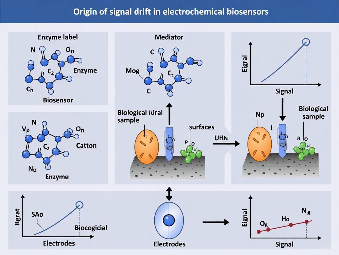
Electrochemical biosensors are powerful tools for therapeutic drug monitoring, in vivo sensing, and diagnostic applications. However, their deployment, particularly in complex biological environments, is hampered by signal drift, a phenomenon characterized by a gradual decrease in sensor signal over time. This instability primarily originates from two fundamental mechanisms: the desorption of self-assembled monolayers (SAMs) from electrode surfaces and the degradation of redox reporters. These processes constitute a significant challenge for the development of robust, long-term sensing platforms, especially for continuous monitoring applications in drug development and clinical settings. Research by [1] has systematically demonstrated that when challenged in biologically relevant conditions such as whole blood at 37°C, electrochemical biosensors exhibit biphasic signal loss. The initial, rapid exponential phase is dominated by biofouling, while the subsequent linear phase is primarily driven by electrochemical instabilities. Understanding and mitigating these specific degradation pathways is therefore critical for advancing the reliability and commercial viability of electrochemical biosensors.
Core Mechanisms of Instability
Self-Assembled Monolayer (SAM) Desorption
The SAM serves as the foundational layer that tethers biorecognition elements (e.g., aptamers, antibodies) to the electrode surface. Its instability directly compromises the sensor's integrity and function.
- Electrochemically Driven Desorption: The gold-thiolate bond, most commonly used for SAM formation, is susceptible to both reductive and oxidative desorption under applied potentials. Studies show that thiol-on-gold monolayers undergo reductive desorption at potentials below -0.5 V and oxidative desorption at potentials above ~1.0 V [1]. The stability of the SAM is therefore highly dependent on the electrochemical interrogation protocol. Research confirms that signal loss is minimal when the potential window is restricted to a narrow, stable region (e.g., -0.4 V to -0.2 V), whereas it increases significantly when the window encroaches on these desorption potentials [1].
- Impact of Molecular Structure on SAM Stability: The chemical structure of the thiol anchor profoundly influences packing density and monolayer stability.
- Anchor Geometry: Flexible trihexylthiol anchors (e.g., Letsinger-type) demonstrate superior stability compared to rigid adamantane-based trithiols or conventional monothiols. Sensors with flexible trithiol anchors retained 75% of their original signal after 50 days of storage in aqueous buffer, whereas those with monothiols or rigid trithiols suffered significant signal loss (>60%) under the same conditions [2].
- Chain Length: Longer alkane thiol chains (e.g., C11) form more stable and densely packed SAMs due to enhanced van der Waals interactions. However, this increased stability comes at the cost of higher electron transfer resistance, which can degrade sensor performance. Shorter chains (e.g., C6) offer a compromise between stability and electron transfer efficiency [3] [2].
Table 1: Factors Influencing SAM Stability and Their Impact on Sensor Performance
| Factor | Effect on Stability | Impact on Sensor Performance |
|---|---|---|
| Applied Potential | Outside stable window (-0.5 V to 1.0 V) causes rapid desorption [1] | Severe signal drift; dictates usable electrochemical techniques |
| Anchor Geometry | Flexible multidentate anchors (e.g., trithiols) enhance stability [2] | Greatly improved long-term and operational stability |
| Chain Length | Longer chains increase stability but impede electron transfer [3] [2] | Trade-off between sensor lifetime and signal strength/sensitivity |
| Surface Crystallinity | Pure gold surfaces ([111] orientation) promote denser SAM formation [3] | Improved reproducibility and reduced non-specific adsorption |
Redox Reporter Degradation
The redox reporter (e.g., Methylene Blue, ferrocene) is responsible for generating the electrochemical signal. Its degradation directly diminishes the sensor's output.
- Irreversible Redox Reactions: The reporter molecule can undergo irreversible side reactions during repeated electrochemical cycling, leading to its decomposition and a permanent loss of signal. The susceptibility to degradation is directly linked to its redox potential [1].
- Superior Stability of Methylene Blue: Among common reporters, Methylene Blue (MB) exhibits exceptional stability. This is attributed to its favorable redox potential (E⁰ = -0.25 V at pH 7.5), which falls within the narrow potential window where alkane-thiol-on-gold SAMs are also stable [1]. This synergy minimizes simultaneous SAM desorption and reporter degradation. Comparative studies have confirmed that MB-tagged peptides provide a much more stable background signal than those tagged with ferrocene, making MB the optimal choice for many biosensing applications [4].
- Reporter Position and Fouling: The placement of the redox reporter on the DNA or peptide backbone influences its vulnerability to signal loss from biofouling. When fouling occurs, it reduces the rate of electron transfer by physically impeding the reporter's approach to the electrode. Studies show that the rate and magnitude of this fouling-induced signal drift are strongly and monotonically dependent on the reporter's position [1].
Table 2: Comparison of Common Redox Reporters in Electrochemical Biosensors
| Redox Reporter | Redox Potential (Approx.) | Stability | Key Advantages / Disadvantages |
|---|---|---|---|
| Methylene Blue (MB) | -0.25 V (vs. ref.) [1] | High | Stable within SAM-stable potential window; optimal for biological media [1] [4] |
| Ferrocene | > +0.3 V (vs. ref.) | Moderate/Low | Requires potentials that can promote SAM desorption [1] [4] |
| Inorganic Complexes | Variable | Variable | (e.g., ([Fe(CN)_6]^{3-/4-})) often used in solution-phase, can be sensitive to environment |
Experimental Analysis of Instability
To effectively study and quantify these instability mechanisms, researchers employ a suite of well-defined experimental protocols and analytical techniques.
Protocol for Quantifying SAM Desorption
This protocol is designed to isolate and measure signal loss originating from SAM desorption under electrochemical interrogation.
- Sensor Fabrication: Create a simplified, EAB-like proxy sensor by immobilizing a thiolated, single-stranded DNA sequence (lacking significant secondary structure to avoid confounding effects) modified with a redox reporter (e.g., Methylene Blue) onto a gold electrode. Backfill with 6-mercapto-1-hexanol (MCH) to form a mixed SAM [1] [2].
- Stability Testing in Benign Medium: Place the fabricated sensor in a controlled, non-biological medium such as phosphate-buffered saline (PBS) at 37°C. The absence of biological components helps isolate electrochemical degradation mechanisms from biofouling [1].
- Electrochemical Interrogation: Subject the sensor to repeated square-wave voltammetry (SWV) scans over an extended period (e.g., 10+ hours). To test the impact of potential window, systematically vary the positive and negative limits of the SWV scan while monitoring the faradaic peak current [1].
- Data Analysis: The signal loss observed as a linear decrease over time in PBS is primarily attributed to electrochemically driven SAM desorption. The strong dependence of this loss on the potential window, particularly when exceeding the stable region, confirms the mechanism [1].
Protocol for Differentiating Fouling from Reporter Degradation
This methodology distinguishes signal loss from surface fouling versus the irreversible degradation of the redox reporter.
- Stability Testing in Complex Media: Challenge the sensor from Step 3.1 in a biologically relevant matrix such as undiluted whole blood at 37°C, using a narrow potential window to minimize electrochemical SAM desorption [1].
- Signal Analysis: Observe the signal drift profile. A biphasic loss—an initial exponential decay followed by a linear decrease—is typically observed. The exponential phase is attributed to blood-specific mechanisms [1].
- Fouling Reversibility Test: After a period of interrogation in blood (e.g., 2.5 hours), wash the electrode with a solubilizing agent like concentrated urea. Recovery of at least 80% of the initial signal indicates that the exponential drift phase is dominated by reversible fouling (e.g., protein adsorption) rather than irreversible enzymatic degradation of the DNA or reporter [1].
- Electron Transfer Kinetics Analysis: Monitor the square-wave voltammetry frequency at which maximum charge transfer occurs. A significant decrease in this frequency during the exponential drift phase indicates that fouling is reducing the electron transfer rate, consistent with a physical barrier on the surface [1].
Diagram 1: Experimental workflow for deconvoluting the sources of signal drift, showing the decision pathway for identifying fouling versus degradation mechanisms.
The Scientist's Toolkit: Research Reagent Solutions
Selecting appropriate materials is paramount for constructing stable and reliable electrochemical biosensors. The table below details key reagents and their optimal use cases for mitigating instabilities.
Table 3: Essential Research Reagents for Mitigating Electrochemical Instabilities
| Reagent / Material | Function / Description | Rationale for Stability Enhancement |
|---|---|---|
| Flexible Trihexylthiol Anchor (e.g., Letsinger-type) [2] | Multidentate anchor for immobilizing DNA probes on gold surfaces. | Provides superior stability in aqueous storage and against thermal cycling compared to monothiols, due to multiple attachment points. |
| Methylene Blue (MB) [1] [4] | Redox reporter attached to the terminus of DNA or peptides. | Its redox potential lies within the SAM-stable window, minimizing simultaneous reporter degradation and SAM desorption during interrogation. |
| 6-Mercapto-1-hexanol (MCH) [3] [2] | Backfilling / blocking agent in mixed SAMs. | Dilutes the probe layer, reduces non-specific adsorption, and helps the biorecognition element adopt a functional conformation. |
| 2'O-methyl RNA / Spiegelmers [1] | Nuclease-resistant oligonucleotide backbone. | Used to confirm that signal loss is not primarily due to enzymatic degradation, helping to isolate fouling as the dominant mechanism. |
| PEG-based Dithiol [4] | Component of a Ternary SAM (T-SAM). | Improves analytical performance and minimizes non-specific protein adsorption, thereby reducing fouling-induced drift. |
| Pure [111] Gold Electrode [3] | Solid support with defined crystallinity. | Promotes denser and more uniform SAM formation compared to nanoparticle-coated or polycrystalline surfaces, enhancing baseline stability. |
The journey toward truly stable and long-lasting electrochemical biosensors requires a fundamental and mechanistic understanding of signal drift. This review has delineated the two primary culprits: the desorption of self-assembled monolayers and the degradation of redox reporters. The experimental evidence confirms that SAM desorption is an electrochemically driven process that can be managed by carefully selecting the interrogation potential and employing advanced anchoring chemistries like flexible trithiols. Concurrently, the choice of redox reporter is critical, with Methylene Blue emerging as the optimal candidate due to its favorable redox potential that aligns with SAM stability. Moving forward, rational design strategies that co-optimize the SAM anchor, the redox reporter, and the surface chemistry will be essential. The integration of novel, fouling-resistant monolayers, the exploration of gold-alkyne bonds as alternatives to thiols [3], and the development of even more robust reporter molecules represent the forefront of research aimed at suppressing these electrochemical instabilities. By systematically addressing these core issues, the path is cleared for the development of highly reliable biosensors capable of long-term, in vivo monitoring, thereby unlocking their full potential in drug development and personalized medicine.
Biological fouling, the non-specific adsorption of proteins and adhesion of cells to sensor surfaces, is a fundamental challenge that compromises the long-term stability and accuracy of electrochemical biosensors in complex biological environments. This phenomenon is a primary origin of signal drift, a key obstacle for in vivo monitoring and reliable in vitro diagnostics [1] [5]. Fouling occurs immediately upon exposure of a sensor to biological fluids such as blood or serum, leading to the formation of an impermeable layer on the electrode surface. This layer can increase background noise, screen the analyte's signal, and significantly degrade the sensor's sensitivity and reproducibility [6] [1]. For applications such as real-time, in vivo monitoring of drugs and biomarkers, even minor surface fouling can be disastrous, as it can completely obscure the already low signal of the target analyte [6] [7]. Understanding the mechanisms of protein adsorption and cell adhesion is therefore not merely a surface chemistry problem, but a critical requirement for designing next-generation robust biosensing platforms.
Fundamental Mechanisms of Biofouling
The Dynamics of Protein Adsorption
Protein adsorption is a dynamic, multi-step process initiating the fouling cascade. It involves reversible attachment, irreversible adsorption, and often subsequent conformational changes and denaturation of the protein on the surface [8]. This process can be simply described by the following kinetic model:
Paq + S ⇄ kads/kdes Ps → kd Pad
Where Paq is protein in the aqueous phase, S is a surface site, Ps is a reversibly adsorbed protein, and Pad is an irreversibly adsorbed, often denatured, protein [8]. The rate coefficients kads, kdes, and kd govern the kinetics of adsorption, desorption, and denaturation, respectively.
The interaction of proteins with a sensor surface is driven by non-covalent forces, including Van der Waals interactions, hydrogen bonding, electrostatics, and hydrophobic interactions [5]. A critical phenomenon in complex media is the Vroman effect, which describes the competitive displacement of abundant, high-mobility proteins (like albumin) over time by proteins that have higher surface affinity but lower mobility (such as fibrinogen) [5]. The final adsorbed protein layer is thus a result of a dynamic interplay of concentration, affinity, and mobility.
From Protein Adsorption to Cell Adhesion
The layer of adsorbed proteins directly dictates subsequent cell adhesion. Mammalian cell adhesion is controlled by the identity, density, conformation, and orientation of the adsorbed proteins [5]. For instance, as little as ~10 ng cm⁻² of adsorbed fibrinogen is sufficient for most mammalian cells to adhere [5]. Denaturation of non-adhesive proteins can increase surface hydrophobicity, promoting bacterial adhesion. Beyond the protein layer, material properties such as surface stiffness, roughness, and topography independently influence cell adhesion, signaling, and differentiation [5]. Bacterial adhesion, if left unchecked, can lead to biofilm formation—structured communities of bacterial cells enclosed in a self-produced extracellular polymeric substance (EPS) matrix. Biofilms are markedly more resistant to cleaning and antimicrobial agents than individual planktonic cells, making their prevention a paramount goal [9].
The Direct Link to Sensor Signal Drift
The fouling layer contributes to signal drift through several physical and electrochemical mechanisms [1]:
- Physical Barrier Effect: The accumulation of proteins, cells, and EPS creates a diffusion barrier, physically impeding the access of the target analyte to the electrode surface and the return of the redox reporter (e.g., methylene blue) to the electrode for electron transfer [1].
- Electron Transfer Inhibition: Fouling can drastically reduce the rate of electron transfer. Studies have shown that the electron transfer rate can decrease by a factor of three during the initial exponential drift phase in whole blood, directly reducing the Faradaic current [1].
- Electrochemical Instability: Electrochemical interrogation itself can accelerate fouling-induced degradation. The applied potential can drive the desorption of the self-assembled monolayers (SAMs) commonly used to functionalize gold electrodes, leading to a linear, continuous signal loss over time [1] [7]. This is particularly severe when potential windows encroach on the regions of reductive (below -0.5 V) or oxidative (above ~1 V) desorption of thiol-on-gold bonds [1].
The following diagram illustrates the sequential mechanisms leading from initial exposure to signal loss.
Quantitative Analysis of Fouling and its Impact
The impact of fouling is quantifiable, providing critical data for evaluating antifouling strategies. The table below summarizes key quantitative findings from fouling studies.
Table 1: Quantitative Impacts of Biofouling on Surfaces and Systems
| System / Surface | Fouling Condition | Quantitative Impact | Reference |
|---|---|---|---|
| Ship Hulls | Heavy marine fouling | Up to 40% increase in hydrodynamic resistance; 62.5% spike in fuel consumption. | [10] |
| Ship Hulls | Moderate fouling | 10-20% higher annual fuel costs; 2 knots loss in speed. | [10] |
| Electrochemical Sensor | Whole blood at 37°C | Biphasic signal loss: exponential drop over ~1.5h, followed by a linear decrease. | [1] |
| PEG Brush Surfaces | Protein adsorption | Defined as "ultralow fouling" at < 5 ng cm⁻² of irreversibly adsorbed protein. | [8] |
| Cell Adhesion | Fibrinogen-coated surface | As little as ~10 ng cm⁻² required for most mammalian cells to adhere. | [5] |
The effectiveness of antifouling strategies is also measured quantitatively, often by the reduction in adhesion forces.
Table 2: Measured Effectiveness of Antifouling Strategies
| Antifouling Strategy | Measurement Technique | Quantitative Outcome | Reference |
|---|---|---|---|
| Vanillin-modified PES Membrane | FluidFM Force Spectroscopy | Significant decrease in biofilm adhesion forces, work, and binding events. | [9] |
| Narrow Potential Window (-0.4V to -0.2V) | Electrochemical Interrogation in PBS | Only 5% signal loss after 1500 scans, vs. major loss with wider windows. | [1] |
| High-Performance Ship Coating | Fuel consumption analysis | Reduced fouling-related fuel consumption increase to ~5% per year, vs. 20% for standard coatings. | [10] |
Experimental Protocols for Fouling Analysis
Protocol: Single-Molecule TIRF Microscopy for Protein Adsorption Kinetics
This technique provides high spatiotemporal resolution of protein-surface interactions, revealing dynamics obscured by ensemble-averaging methods [8].
- Objective: To probe the dynamics of individual protein molecules (reversible/irreversible adsorption, desorption rates) on low-fouling surfaces in real time.
- Materials:
- Total Internal Reflection Fluorescence (TIRF) Microscope.
- Low-fouling surface sample (e.g., PEG brush, polyelectrolyte multilayer).
- Fluorescently labeled protein of interest (e.g., BSA, fibrinogen).
- Relevant buffer for the experiment.
- Procedure:
- Sample Mounting: Secure the surface sample in a flow cell on the microscope stage.
- Baseline Imaging: Introduce buffer alone and focus the objective to excite only the evanescent field (typically < 150 nm from the surface).
- Protein Introduction: Flush in a solution of fluorescently labeled protein at a very low concentration (to enable single-molecule detection).
- Real-Time Data Acquisition: Record video-rate microscopy of the surface. Individual protein molecules interacting with the surface will appear as discrete, diffraction-limited spots.
- Data Analysis: Use single-molecule localization and tracking software to:
- Count the number of adsorption events per unit area over time.
- Track the residence time of individual molecules on the surface.
- Calculate the adsorption rate coefficient (kads) and desorption rate coefficient (kdes).
- Key Consideration: The requirement for fluorescent labeling and low protein concentrations means this method is conducted under non-physiological conditions, complementing rather than replacing other techniques [8].
Protocol: FluidFM for Biofilm-Surface Adhesion Force Measurement
This novel method quantifies the adhesion forces of entire biofilms, offering more realistic data than single-cell probes [9].
- Objective: To directly measure the adhesion forces between a bacterial biofilm and a modified membrane surface.
- Materials:
- FluidFM instrument (combination of AFM and microfluidics).
- Microfluidic cantilevers with an aperture.
- COOH-functionalized polystyrene beads (~
- Procedure:
- Biofilm Probe Fabrication:
- Incubate COOH-functionalized beads with bacteria for 3 hours to allow biofilm growth.
- Aspirate a single biofilm-covered bead onto the aperture of a microfluidic cantilever by applying a negative pressure.
- Surface Preparation: Mount the test membrane (e.g., vanillin-modified PES) in the FluidFM liquid cell.
- Force Spectroscopy:
- Approach the biofilm probe to the membrane surface.
- Retract the probe at a constant speed while recording the cantilever deflection.
- Repeat the measurement across multiple locations on the surface.
- Data Analysis:
- Analyze the retraction force curves to extract:
- Adhesion Force: The maximum force required to detach the biofilm.
- Adhesion Work: The area under the retraction curve.
- Number of Adhesion Events: The number of discrete "jump-off" events in the curve, indicating bond ruptures.
- Analyze the retraction force curves to extract:
- Biofilm Probe Fabrication:
- Key Advantage: This method captures the complex contribution of the EPS matrix, showing that biofilm adhesion behavior is fundamentally different from that of single cells [9].
The workflow for this advanced technique is outlined below.
The Scientist's Toolkit: Key Research Reagents and Materials
Table 3: Essential Reagents and Materials for Fouling Research
| Item | Function in Fouling Research | Key Considerations |
|---|---|---|
| Poly(ethylene glycol) (PEG) | Gold-standard polymer for creating low-fouling surfaces and brushes; resists protein adsorption via steric repulsion and hydration [8] [6]. | Grafting density and chain length are critical. Low-density brushes can be less effective. |
| Self-Assembled Monolayers (SAMs) | Well-defined organic surfaces formed on gold (e.g., from alkanethiols) to study fundamental interactions or as a platform for attaching antifouling molecules [1]. | Stability can be a limitation; susceptible to electrochemical desorption depending on applied potential. |
| Vanillin | A natural phenolic aldehyde used as an anti-biofouling coating; acts as a quorum-sensing inhibitor, reducing EPS production and biofilm formation [9]. | Offers a non-biocidal, "anti-virulence" mechanism of action. |
| Sol-Gel Silicate Layers | Porous, mechanically stable coatings for electrochemical sensors; act as a physical diffusion barrier to protect the electrode from foulants [6]. | Showed remarkable long-term stability, sustaining signal for 6 weeks in cell culture medium. |
| Syringaldazine | A redox mediator adsorbed onto electrode surfaces; used as a model catalyst to evaluate the protective effect of antifouling layers without adding external probes [6]. | Its rapid deterioration in complex media makes it an excellent indicator of coating efficacy. |
| COOH-functionalized Beads | Serve as carriers for growing biofilms for use in FluidFM adhesion force measurements [9]. | Provide a suitable surface for bacterial growth and biofilm formation. |
Biological fouling, initiated by protein adsorption and amplified by cell adhesion, remains a central problem in the development of stable, reliable electrochemical biosensors. It is a direct and major contributor to signal drift, limiting the in vivo lifespan and in vitro reproducibility of these devices. Combating this issue requires a multi-faceted approach: a deep understanding of the fundamental interaction mechanisms, the application of sophisticated characterization techniques like single-molecule TIRF and FluidFM that go beyond simple ensemble averages, and the rational design of advanced antifouling materials such as high-density polymer brushes, zwitterionic coatings, and non-fouling hydrogels. Future progress will hinge on the development of standardized testing protocols and the creation of robust, surface-modification strategies that can withstand the complex, dynamic, and harsh environment of real-world biological applications.
Electrochemical biosensors are powerful analytical tools that convert biological recognition events into quantifiable electrical signals, finding extensive applications in healthcare diagnostics, environmental monitoring, and food safety [11] [12]. A significant challenge in their practical deployment, however, lies in their susceptibility to signal drift induced by fluctuations in environmental and operational parameters such as temperature, pH, and ionic strength [13] [14]. This drift originates from the profound influence these stressors exert on both the biological recognition elements and the underlying physico-chemical transduction processes [15] [16].
The stability of a biosensor is critical for its commercial success and reliable operation, as it directly translates to longevity and measurement accuracy [15]. Biological elements like enzymes, antibodies, and aptamers are highly sensitive to their immediate environment. Temperature shifts can alter their conformational structure and reaction kinetics, pH variations can affect their charge state and catalytic activity, and changes in ionic strength can modulate binding affinities and electron transfer rates [13] [12] [14]. Simultaneously, these parameters directly affect electrochemical properties, including the conductivity of the solution, the double-layer capacitance at the electrode-solution interface, and the kinetics of Faradaic reactions [14]. Isolating the specific signal originating from the target analyte from the noise and drift caused by these extrinsic variables is therefore a fundamental pursuit in biosensor research and development [14]. This guide provides a technical examination of these stressors, detailing their mechanisms of action, methodologies for their systematic study, and strategies for their mitigation.
Mechanisms of Signal Interference
Environmental stressors induce signal drift through multiple, often interconnected, mechanisms that impact the biorecognition element, the transducer interface, and the sample matrix itself.
Impact of Temperature Fluctuations
Temperature is one of the most critical factors affecting biosensor performance. Its influence is multifaceted, altering the properties of the biological layer, the electrode kinetics, and the bulk solution.
- Biorecognition Element Dynamics: The activity of biological receptors, particularly enzymes, is intrinsically temperature-dependent. According to the Arrhenius equation, reaction rates typically increase with temperature, potentially enhancing sensitivity within an optimal range. However, excessive temperatures induce denaturation, an irreversible loss of tertiary structure and function, leading to permanent sensor degradation [15]. For instance, in hydrogel-based ionic strength sensors, temperature changes directly impact sensor characteristics like sensitivity, response time, and stability [13].
- Electrode and Solution Properties: Temperature changes affect the electrochemical cell's physics. A rise in temperature generally decreases the viscosity of the solution, increasing the diffusion coefficient of ions and redox species. This leads to higher mass transport rates and larger Faradaic currents. Furthermore, the standard rate constant of electron transfer for electrochemical reactions is itself temperature-dependent, following an Arrhenius-type relationship [14]. Studies on interdigitated microelectrodes (IDEs) have quantified this, showing a significant negative coefficient for temperature, where impedance decreases as temperature rises [14].
Impact of pH Shifts
The pH of the sample medium can drastically alter the charge state and functionality of the biological components and influence the electrochemical environment.
- Charge State of Biomolecules: Enzymes, antibodies, and aptamers possess specific ionic groups that can protonate or deprotonate. The isoelectric point (pI) of a protein defines the pH at which it has no net charge. Deviations from this pH can alter the molecule's three-dimensional structure, its binding affinity for the target analyte, and its catalytic efficiency. A shift in pH can render an aptamer's folded structure unstable, compromising its specificity and affinity [12].
- Electrochemical Reaction Kinetics: The pH directly governs the availability of protons in redox reactions. For many common redox couples (e.g., ferrocene/ferrocenium or reactions involving hydroquinone), the electron transfer kinetics are pH-sensitive. A classic example is in fermentation monitoring, where CO2 production lowers the pH, which in turn affects the impedance measurement independently of the chemical changes being tracked [14]. Research on IDE sensors has demonstrated a strong positive correlation between pH and impedance, with impedance increasing as the solution becomes more basic [14].
Impact of Ionic Strength Variations
The concentration of ions in a solution defines its ionic strength, which shapes the electrostatic environment around the biosensor interface.
- Shielding of Electrostatic Interactions: High ionic strength solutions contain a high concentration of counter-ions that can screen electrostatic charges. This shielding can disrupt the binding between a charged bioreceptor (e.g., a DNA aptamer) and its target, which often relies on complementary charge interactions [12].
- Modification of the Electrical Double Layer: At the electrode-electrolyte interface, a structured layer of ions forms, known as the electrical double layer (EDL). Its thickness, governed by the Debye length, is inversely proportional to the square root of the ionic strength. In high ionic strength solutions, the EDL is compressed, which can significantly alter the measured capacitance and electron transfer resistance, key parameters in electrochemical impedance spectroscopy (EIS) [14]. Hydrogel-based sensors are explicitly designed to swell or shrink in response to changes in ionic strength, converting this physical change into a measurable pressure signal [13].
Quantitative Analysis of Stressor Effects
A systematic, quantitative understanding of how each stressor impacts sensor output is essential for developing robust sensing platforms and correction algorithms. The following table summarizes key quantitative findings from experimental studies on these stressors.
Table 1: Quantitative Effects of Environmental Stressors on Biosensor Performance
| Stressor | Sensor Type / Application | Observed Impact | Quantified Effect / Model Coefficient | Reference |
|---|---|---|---|---|
| Temperature | Interdigitated Microelectrodes (IDEs) in wine | Impedance decreases with temperature increase. | Coefficient (β): -195.5 Ω/°C(in a multi-parameter model: Z(Ω) = ... -1.955·10²·T(°C) ...) | [14] |
| pH | Interdigitated Microelectrodes (IDEs) in wine | Impedance increases with pH increase. | Coefficient (β): +897.8 Ω/pH unit(in a multi-parameter model: Z(Ω) = ... +8.978·10²·pH ...) | [14] |
| Temperature | Hydrogel-based Ionic Strength Sensor | Affects sensor sensitivity, response time, and stability. | Sensor characteristics investigated as a function of temperature in vitro. | [13] |
| Ionic Strength | Hydrogel-based Biosensor | Hydrogel volume changes with ionic strength. | Volume change captured as a pressure signal in a confined cavity. | [13] |
The mathematical model developed for IDE sensors, which incorporates sensor geometry and operational frequency alongside environmental parameters, provides a powerful tool for quantifying the significance of each variable [14]. The general form of the model is:
Z(Ω) = Constant + β₁·dₑ + β₂·sₑ + β₃·Area + β₄·f(Hz) + β₅·T(°C) + β₆·pH
Where dₑ is electrode width, sₑ is electrode spacing, Area is the sensing area, and f is the measurement frequency. The magnitude and sign of the coefficients (β) directly quantify the effect of each parameter on the impedance. For example, the negative coefficient for temperature (β₅ = -195.5) confirms that impedance decreases with rising temperature, while the positive coefficient for pH (β₆ = +897.8) shows that impedance increases with pH [14].
Experimental Protocols for Stressor Analysis
To isolate and analyze the effects of environmental stressors, controlled experimental protocols are required. The following workflow provides a generalized methodology that can be adapted for specific biosensor platforms.
Diagram 1: Experimental workflow for analyzing environmental stressors.
Protocol for Isolating Temperature Effects
This protocol outlines the steps to characterize the effect of temperature on a biosensor's signal independently of other variables.
Materials:
- Electrochemical Cell with Temperature Control: A jacketed cell connected to a thermostated water bath or a Peltier-controlled cell holder is essential for precise temperature regulation.
- Potentiostat/Galvanostat: An instrument capable of performing the intended electrochemical technique (e.g., amperometry, EIS).
- Buffer Solution: A well-buffered solution (e.g., 0.1 M phosphate buffer, pH 7.4) to ensure pH remains stable over the temperature range.
- Thermometer or Temperature Probe: For accurate and independent monitoring of the solution temperature.
Procedure:
- Sensor Preparation: Functionalize the electrode surface and immobilize the bioreceptor (e.g., enzyme, aptamer) according to the established protocol.
- Baseline Measurement: Place the sensor in the electrochemical cell with the buffer solution and a fixed concentration of the target analyte. Set the temperature to a reference point (e.g., 25°C) and allow the system to equilibrate for 10-15 minutes. Record the baseline signal (e.g., steady-state current or impedance spectrum).
- Temperature Variation: Systematically increase or decrease the temperature in increments (e.g., 5°C). At each new temperature, allow sufficient time for thermal equilibration before recording the signal.
- Data Analysis: Plot the sensor signal (e.g., current, charge transfer resistance Rct) against temperature. The data can be fitted to an appropriate model (e.g., Arrhenius plot for reaction rates, or a linear/polynomial model for impedance) to derive a temperature coefficient [14].
Protocol for Isolating pH Effects
This protocol describes how to evaluate the impact of pH shifts on biosensor performance.
Materials:
- Buffer Series: A set of buffers with identical ionic strength but different pH values (e.g., 0.1 M phosphate buffers for pH 6-8, 0.1 M acetate for lower pH, 0.1 M Tris for higher pH). The use of a background electrolyte like KNO₃ or NaCl can help maintain constant ionic strength across different buffers.
- pH Meter: A calibrated, high-precision pH meter is mandatory for verifying the actual pH of the solution in the cell.
Procedure:
- Sensor Preparation: As in the previous protocol.
- Buffer Introduction: Introduce the first buffer into the electrochemical cell. Add a fixed concentration of the target analyte.
- Signal Measurement: Measure the sensor signal at a constant temperature and stirring rate. Precisely record the pH of the solution.
- pH Cycling: Rinse the sensor and cell thoroughly with deionized water. Introduce the next buffer in the series with the same concentration of analyte and repeat the measurement. Continue this process across the desired pH range.
- Data Analysis: Plot the sensor signal versus pH to determine the optimal pH for maximum activity and the range over which the sensor operates reliably. This profile is crucial for understanding the sensor's limitations in samples with variable pH [14].
Protocol for Isolating Ionic Strength Effects
This protocol is designed to assess sensor performance against changes in background ionic strength.
Materials:
- Concentrated Salt Solution: A high-purity stock solution of an inert salt such as NaCl or KCl.
- Concentrated Buffer Solution: A buffer concentrate that, when diluted, will give the desired pH.
Procedure:
- Sensor Preparation: As above.
- Baseline in Low Ionic Strength: Prepare a solution with the target analyte in a low-ionic-strength buffer.
- Titration of Salt: While continuously measuring the sensor signal (e.g., via amperometry or EIS), add small, known volumes of the concentrated salt stock solution to the cell. Ensure thorough mixing after each addition.
- Data Analysis: Calculate the ionic strength for each addition. Plot the sensor signal (e.g., Rct from EIS, or current) against the square root of the ionic strength. A sharp change may indicate EDL compression or disruption of biorecognition binding events [13] [12].
The Scientist's Toolkit: Research Reagent Solutions
Selecting appropriate materials and reagents is fundamental to constructing stable biosensor interfaces and conducting reliable stressor analysis.
Table 2: Essential Research Reagents and Materials for Biosensor R&D
| Reagent / Material | Function / Application | Key Characteristics |
|---|---|---|
| Gold Nanoparticles (AuNPs) | Electrode nanomodification; signal amplification carrier. | Excellent biocompatibility, high conductivity, large surface-to-volume ratio, facile functionalization with thiolated biomolecules [16] [17]. |
| Reduced Graphene Oxide (rGO) | Carbon nanomaterial for electrode modification. | High electrical conductivity, large specific surface area, good mechanical strength, enhances electron transfer [15] [17]. |
| Chitosan (CS) | Biopolymer for constructing biocompatible interfaces. | Excellent film-forming ability, biodegradability, biocompatibility, non-toxicity; often used with other nanomaterials (e.g., GO-CS) [16]. |
| Interdigitated Microelectrodes (IDEs) | Transducer platform for impedimetric sensing. | Enhanced signal sensitivity, suitable for monitoring adhesion, biofouling, and chemical changes in liquids [14]. |
| Poly(o-phenylenediamine) | Electropolymerized membrane for enzyme entrapment. | Used to create selective, low-fouling permselective membranes on electrode surfaces [15]. |
| Systematic Evolution of Ligands by Exponential Enrichment (SELEX) | Technology for generating specific DNA/RNA aptamers. | Produces stable nucleic acid bioreceptors with high affinity and specificity for targets, from ions to cells [12] [17]. |
Mitigation Strategies and Future Directions
Addressing the challenge of signal drift requires a multi-faceted approach that combines interface engineering, signal processing, and smart material design.
Interface Engineering with Advanced Materials: Using composite materials can significantly enhance interface stability. Nanomaterials like gold nanoparticles (AuNPs) and graphene oxide improve conductivity and provide a stable microenvironment for biomolecules [16] [17]. Polymers like chitosan offer biocompatible matrices that protect biological elements from harsh environmental conditions [16]. Bimetallic core-shell nanostructures have also shown improved stability and catalytic activity compared to their single-metal counterparts [16].
Mathematical Modeling and Signal Correction: As demonstrated with the IDE sensor model, developing a quantitative understanding of the influence of temperature and pH allows for software-based correction of the acquired signal [14]. The general form of the model is: Zcorrected = Zmeasured - [βT · (T - Tref) + βpH · (pH - pHref)] This approach effectively isolates the impedance change due to the target analyte from the drift caused by the extrinsic variables [14].
Microfluidic Integration and Automated Systems: Integrating biosensors into microfluidic platforms enables precise control over the sample environment, including temperature and flow conditions. Furthermore, the application of vibration and hydrodynamic flow in such systems has been shown to enhance sensor performance, lower the limit of detection, and pave the way for automated, high-throughput analysis, reducing environmental variability [11].
Exploration of Robust Bioreceptors: The search for more stable recognition elements is ongoing. Aptamers are generally more stable than antibodies over a range of temperatures and can be regenerated more easily [12] [17]. The development of thermostable enzymes or biomimetic catalysts (e.g., nanozymes) also offers a path toward sensors capable of operating in demanding environments [16].
The following diagram illustrates the logical relationship between the core environmental stressors, their mechanisms of action, and the corresponding mitigation strategies discussed.
Diagram 2: Stressor mechanisms and mitigation strategies.
The Impact of Electrode Potential and Scanning Parameters on Drift Kinetics
Signal drift, the undesired change in sensor output over time under constant conditions, presents a major obstacle to the long-term stability and reliability of electrochemical biosensors. This phenomenon is particularly critical in applications such as continuous molecular monitoring in vivo or in complex biological fluids, where sensor stability over many hours or days is required for effective patient management or drug development studies [1]. The drift kinetics are not merely a function of time but are intimately governed by the electrochemical interrogation parameters used during sensor operation. Electrode potential and scanning protocols directly influence the fundamental processes occurring at the electrode-electrolyte interface, either accelerating or mitigating the mechanisms that lead to signal degradation [1]. Understanding these relationships is paramount for designing robust biosensing systems with predictable longevity. This technical guide examines the origin of signal drift within the context of how operational electrochemical parameters impact the underlying degradation mechanisms, providing researchers with a framework for optimizing biosensor performance through controlled electrochemical protocols.
Fundamental Mechanisms of Signal Drift
Signal drift in electrochemical biosensors originates from multiple physical and chemical processes that can be categorized into two primary classes: electrochemically-driven degradation and biology-driven fouling.
Electrochemically-Driven Degradation
This category encompasses processes directly instigated by the electrical signals used to operate the sensor. A primary mechanism is the electrochemically driven desorption of the self-assembled monolayer (SAM) that typically anchors biorecognition elements (such as DNA aptamers or enzymes) to the electrode surface, often gold [1]. The stability of the gold-thiol bond, fundamental to these SAMs, is highly dependent on the applied electrode potential. Both reductive desorption at potentials below approximately -0.5 V and oxidative desorption at potentials above ~1.0 V can break this bond, leading to a loss of the sensing layer and a corresponding signal decrease [1]. A secondary electrochemical mechanism is the irreversible degradation of the redox reporter molecule (e.g., methylene blue) through side reactions that occur during its repeated cycling between oxidized and reduced states [1].
Biology-Driven Fouling and Degradation
When deployed in biological matrices like blood or interstitial fluid, sensors face additional challenges. Surface fouling involves the non-specific adsorption of proteins, cells, and other biomolecules to the electrode surface [1]. This fouling layer can hinder the diffusion of the redox reporter to the electrode surface, thereby reducing the electron transfer rate and the observed signal [1]. Furthermore, enzymatic degradation of biological recognition elements (e.g., nucleases cleaving DNA or RNA aptamers) contributes to the exponential signal loss phase often observed in complex media [1]. The interplay between these mechanisms dictates the overall drift profile, which often manifests as a biphasic signal loss: an initial rapid exponential decay followed by a slower, more linear decrease [1].
Impact of Electrode Potential on Drift Kinetics
The applied electrode potential is a critical parameter controlling the rate of signal drift, primarily through its effect on the stability of the electrode-sensing layer interface.
Potential Window and SAM Desorption
The voltage range, or potential window, scanned during electrochemical measurements is a major determinant of the sensor's operational lifespan. Research has demonstrated that the stability of thiol-on-gold monolayers is highly susceptible to extreme potentials. The table below summarizes the effect of the applied potential window on the observed signal drift, highlighting the existence of a "stability window" [1].
Table 1: Impact of Electrode Potential Window on Signal Drift
| Negative Potential Limit (V) | Positive Potential Limit (V) | Observed Drift Over 1500 Scans | Primary Mechanism |
|---|---|---|---|
| -0.4 V | -0.2 V | ~5% signal loss | Minimal desorption |
| -0.4 V | 0.0 V | Low degradation rate | Onset of oxidative processes |
| -0.4 V | > +0.2 V | Significant signal loss | Oxidative desorption of SAM |
| < -0.4 V | -0.2 V | Significant signal loss | Reductive desorption of SAM |
Experiments reveal that a narrow potential window of -0.4 V to -0.2 V results in only 5% signal loss after 1500 scans, whereas expanding the window to include more extreme potentials dramatically increases the degradation rate [1]. This is because potentials beyond the threshold for reductive or oxidative desorption directly break the gold-thiol bonds [1].
Redox Reporter Stability
The choice and positioning of the redox reporter are also influenced by potential. Methylene blue (MB), with a formal potential (E⁰) of approximately -0.25 V vs. Ag/AgCl at pH 7.5, is notably stable because its redox activity falls within the narrow potential window where alkane-thiol-on-gold monolayers are most stable [1]. In contrast, reporters with redox potentials outside this stable window necessitate the use of destabilizing potentials, accelerating drift. Furthermore, the reporter's position within a DNA or protein scaffold influences its susceptibility to fouling, which is itself modulated by the applied electric fields [1].
Impact of Scanning Parameters on Drift Kinetics
Beyond the static potential limits, the dynamic parameters of the electrochemical scanning technique itself contribute to drift kinetics.
Scan Rate and Frequency
The scan rate in voltammetric techniques or the frequency in impedance spectroscopy determines how rapidly the interfacial structure is perturbed. While not explicitly quantified in the provided research, faster scanning generally subjects the SAM to more frequent structural stress and redox cycling, which can potentially accelerate fatigue and desorption over prolonged operation. However, higher frequencies in alternating current (AC) techniques like EIS can sometimes help isolate the faradaic process from slow fouling effects.
Continuous vs. Intermittent Interrogation
The duty cycle of electrochemical measurement is a significant factor. Studies show that pausing the electrochemical interrogation in a controlled environment (e.g., PBS buffer) can halt the signal degradation associated with the linear drift phase [1]. This indicates that the electrochemically-driven desorption is active only when a potential is being applied, providing a strategy to extend total sensor lifetime through intermittent measurement protocols rather than continuous operation.
Experimental Protocols for Drift Kinetics Characterization
A systematic approach is required to deconvolute the various contributions to signal drift. The following protocol offers a methodology for evaluating the impact of electrode potential and scanning parameters.
Protocol: Quantifying Potential-Dependent Drift
This experiment is designed to isolate the effect of the electrochemical potential window on SAM stability.
- Objective: To determine the rate of signal loss due to electrochemical desorption as a function of the applied potential window.
- Materials:
- Phosphate Buffered Saline (PBS), pH 7.4
- Custom-fabricated gold electrode sensor with a redox-labeled DNA SAM
- Potentiostat
Method:
- Sensor Preparation: Immobilize a thiolated DNA probe labeled with Methylene Blue (MB) onto a clean gold electrode to form a SAM. Rinse and stabilize in PBS.
- Baseline Measurement: Record a square-wave voltammogram (SWV) in PBS using a very stable, narrow potential window (e.g., -0.4 V to -0.2 V) to establish the initial signal.
- Stress Testing: Subject the sensor to a continuous sequence of SWV scans (e.g., 1000 scans) using the test potential window.
- Signal Monitoring: Periodically (e.g., every 100 scans) record a "probe" SWV scan using the stable baseline window to track the decay of the peak current without contributing significantly to the degradation.
- Data Analysis: Plot the normalized peak current from the probe scans versus the cumulative number of stress scans. Fit the data to determine the degradation rate constant for each tested potential window.
Key Analysis: Compare the degradation rates across different potential windows. A sharp increase in the degradation rate as the window expands beyond the stability thresholds provides a quantitative basis for selecting optimal operating parameters [1].
Protocol: Deconvoluting Fouling from Electrochemical Drift
This protocol characterizes the biological contribution to drift using blood as a challenging matrix.
- Objective: To distinguish signal loss from surface fouling and enzymatic degradation from purely electrochemical drift.
- Materials:
- Undiluted whole blood, heparinized
- PBS, pH 7.4
- Urea solution (concentrated, e.g., 6-8 M)
- Gold electrode sensors with MB-labeled DNA or enzyme-resistant RNA
- Method:
- Sensor Interrogation in Blood: Place the sensor in whole blood at 37°C. Interrogate continuously with SWV using a narrow, stable potential window to minimize electrochemical degradation.
- Signal Recording: Monitor the SWV peak current over time (e.g., 2-3 hours). Observe the characteristic biphasic drift.
- Fouling Recovery: After a significant exponential signal drop (e.g., 2.5 hours), wash the sensor with concentrated urea to solubilize and remove non-specifically adsorbed biomolecules.
- Signal Recovery Measurement: Re-measure the SWV signal in PBS. Recovery of >80% of the initial signal indicates that fouling, not permanent degradation, was the primary cause of the initial exponential drift [1].
- Control with Enzyme-Resistant Oligonucleotides: Repeat the experiment using a sensor fabricated with a nuclease-resistant backbone (e.g., 2'-O-methyl RNA). A persistent exponential drift phase suggests that fouling, rather than enzymatic cleavage, is the dominant biological mechanism [1].
Visualization of Drift Mechanisms and Experimental Workflows
The following diagrams illustrate the core concepts and experimental workflows discussed in this guide.
Diagram 1: Signal drift mechanisms and controlling factors.
Diagram 2: Experimental workflow for quantifying potential-dependent drift.
The Scientist's Toolkit: Essential Research Reagents and Materials
The following table details key materials and their specific functions in experiments focused on drift kinetics.
Table 2: Essential Research Reagents and Materials for Drift Kinetics Studies
| Reagent / Material | Function / Rationale | Key Consideration |
|---|---|---|
| Gold Electrode | Standard substrate for forming stable thiol-based Self-Assembled Monolayers (SAMs). | Purity and surface roughness affect SAM uniformity and stability [18] [19]. |
| Alkane-Thiol SAM | Creates an ordered monolayer that minimizes non-specific adsorption and provides a scaffold for bioreceptors. | Chain length and terminal functional group influence packing density and stability [1]. |
| Methylene Blue-labeled DNA | Acts as a model EAB sensor; MB's redox potential falls within the stable window for gold-thiol SAMs [1]. | Reporter position on the DNA strand affects susceptibility to fouling-induced signal loss [1]. |
| 2'-O-Methyl RNA | Enzyme-resistant nucleic acid analog used to decouple enzymatic degradation from fouling effects [1]. | Confirms that exponential drift in blood is primarily due to fouling, not nuclease activity. |
| Ultra-Pure Buffer (PBS) | Provides a controlled, biologically inert medium for isolating electrochemical drift mechanisms [1]. | Absence of proteins and cells allows study of SAM desorption and reporter degradation alone. |
| Whole Blood | Complex biological matrix used as a proxy for in vivo conditions to study fouling and biological degradation [1]. | Contains proteins, cells, and enzymes that collectively contribute to the exponential drift phase. |
| Concentrated Urea | A chemical denaturant used to wash sensors; it solubilizes proteins, reversing fouling-based signal loss [1]. | Useful for quantifying the recoverable portion of signal drift, confirming fouling's role. |
The kinetics of signal drift in electrochemical biosensors are not an immutable property but a controllable variable dictated by the operational electrochemical parameters. This guide has established that the electrode potential window is a primary lever, with a clearly defined stability zone for thiol-on-gold chemistry that, when respected, can minimize electrochemically driven SAM desorption. Furthermore, the duty cycle of interrogation and the choice of redox reporter are critical secondary parameters. Mitigating drift requires a multi-pronged strategy: employing the narrowest possible potential window that encompasses the redox reaction of interest, using stable reporters like methylene blue, and considering intermittent measurement schemes for long-term monitoring. Future research will likely focus on engineering even more robust surface architectures, such as using non-thiol anchor chemistries with wider electrochemical stability windows, and developing advanced drift-correction algorithms that can dynamically adapt to changing sensor performance. A fundamental understanding of the impact of electrode potential and scanning parameters on drift kinetics, as detailed herein, is essential for transforming electrochemical biosensors from research tools into reliable, long-term monitoring solutions in biomedicine and drug development.
Electrochemical biosensors synergistically integrate the molecular recognition capabilities of biological elements with the sensitivity of electrochemical transducers, offering a powerful platform for detecting targets ranging from small molecules to whole cells [20]. A critical challenge impeding the reliable deployment of these biosensors, particularly for long-term monitoring in complex biological environments, is signal drift—the undesirable decrease in sensor signal over time. A primary origin of this drift is the degradation of the immobilized biorecognition elements, such as nucleic acids (e.g., aptamers) and peptides, by nucleases and proteases present in biological fluids [1]. This degradation compromises the structural integrity and function of the sensing interface, leading to a loss of signal fidelity. This whitepaper provides an in-depth technical analysis of the mechanisms by which nuclease and protease activity induces signal drift, summarizes quantitative studies on degradation kinetics, outlines detailed experimental protocols for investigating these phenomena, and presents advanced strategies to engineer stable, degradation-resistant biosensing interfaces.
Mechanisms of Biomolecule Degradation and Signal Drift
Nuclease Degradation of DNA-Based Biorecognition Elements
Electrochemical aptamer-based (EAB) sensors, which utilize a redox-tagged DNA aptamer immobilized on a gold electrode, are highly susceptible to nuclease degradation. When deployed in biologically relevant conditions like whole blood at 37°C, the sensor signal exhibits a biphasic drift profile [1].
- Exponential Drift Phase: The initial, rapid signal loss occurs over approximately 1.5 hours and is primarily driven by biological mechanisms. Studies using enzyme-resistant oligonucleotides (2'-O-methyl RNA) have demonstrated that this phase persists despite nuclease resistance, pointing to biofouling as a dominant mechanism. Fouling by blood cells and proteins alters the dynamics of the redox reporter (e.g., methylene blue), reducing its electron transfer rate to the electrode by a factor of three [1].
- Linear Drift Phase: The subsequent, slower signal loss is electrochemically driven. Systematic investigation has linked this phase to the reductive and oxidative desorption of the alkane-thiolate self-assembled monolayer (SAM) from the gold electrode surface, a process triggered by the applied electrochemical potentials. By narrowing the potential window to -0.4 V to -0.2 V (vs. Ag/AgCl), this degradation phase was nearly eliminated, with only 5% signal loss observed after 1500 scans [1].
Protease Degradation of Peptide-Based Biorecognition Elements
Peptide-based biosensors detect protease activity by monitoring the cleavage of an electrode-bound, redox-tagged peptide substrate. Protease-induced cleavage severs the redox reporter from the electrode surface, causing a measurable drop in current [4] [21]. This same principle, while useful for detection, becomes a source of signal drift when non-specific proteolysis degrades the peptide biorecognition layer. The degradation kinetics can often be modeled using a heterogeneous Michaelis-Menten model, allowing for the extraction of kinetic parameters like kcat and KM [4] [22]. The stability of the peptide layer is influenced by its structure; for instance, designed arched peptides can exhibit enhanced resistance to proteolytic hydrolysis compared to linear peptides [23].
Quantitative Data on Degradation Kinetics
The following tables summarize key quantitative findings from research on the degradation of biorecognition elements and its impact on biosensor performance.
Table 1: Quantifying Signal Drift in Electrochemical Biosensors
| Drift Phase | Proposed Primary Mechanism | Experimental Evidence | Impact on Signal |
|---|---|---|---|
| Exponential (Initial 1.5 hrs) | Biofouling from blood components [1] | Signal loss persists with nuclease-resistant oligonucleotides; ~80% signal recovery after urea wash [1] | Rapid signal decrease; Electron transfer rate reduced 3-fold [1] |
| Linear (Long-term) | Electrochemically driven SAM desorption [1] | Drift rate highly dependent on applied potential window; minimized at -0.4 V to -0.2 V [1] | Slow, continuous signal decrease; Little change in electron transfer rate [1] |
Table 2: Enzymatic Degradation Kinetics of Biorecognition Elements
| Enzyme Target | Biorecognition Element | Redox Reporter | Kinetic Parameter (kcat/Km) | Reference |
|---|---|---|---|---|
| Trypsin | Methylene blue-tagged peptide, T-SAM | Methylene Blue | Not specified (LOD: 250 pM) [4] | [4] |
| Cathepsin B | Ferrocene-appended tetrapeptide on VACNF NEA | Ferrocene | (4.3 ± 0.8) × 10⁴ M⁻¹s⁻¹ [22] | [22] |
| Legumain | Ferrocene-appended tetrapeptide on VACNF NEA | Ferrocene | (1.13 ± 0.38) × 10⁴ M⁻¹s⁻¹ [22] | [22] |
Experimental Protocols for Investigating Degradation
Protocol: Investigating Nuclease-Driven Signal Drift
This protocol is adapted from studies elucidating the mechanisms of EAB sensor drift [1].
- Sensor Fabrication:
- Substrate: Use gold disk working electrodes (e.g., 2 mm diameter).
- SAM Formation: Incubate electrodes in a 1 µM solution of alkane-thiolated DNA (e.g., a 37-base sequence without significant secondary structure) modified with a methylene blue (MB) redox reporter at the 3'-end. Incubate for 1 hour.
- Rinsing & Storage: Rinse thoroughly with deionized water and store in PBS buffer until use.
- Experimental Deployment & Drift Measurement:
- Electrochemical Setup: Use a standard three-electrode system (Ag/AgCl reference, Pt counter) and square wave voltammetry (SWV).
- Conditions: Challenge the sensors in two environments at 37°C:
- Complex Biofluid: Undiluted whole blood.
- Control: Phosphate Buffered Saline (PBS).
- Data Acquisition: Record successive SWV scans over several hours (e.g., 10+ hours). The peak current of the MB signal is the primary metric.
- Mechanistic Interrogation:
- Fouling Test: After 2.5 hours in blood, wash sensors with concentrated urea (e.g., 6 M) and re-measure signal in PBS to assess recoverable signal loss.
- Nuclease Degradation Test: Repeat the deployment experiment using a sensor fabricated with a nuclease-resistant oligonucleotide (e.g., 2'-O-methyl RNA).
- Electrochemical Stress Test: In PBS, systematically vary the SWV potential window to identify regimes that minimize the linear drift phase.
Protocol: Measuring Protease Activity and Peptide Stability
This protocol is derived from electrochemical protease biosensor studies [4] [22].
- Sensor Fabrication:
- Electrode Platform: Use a gold electrode or a nanoelectrode array (e.g., Vertically Aligned Carbon Nanofiber NEA).
- Peptide Immobilization:
- Form a ternary self-assembled monolayer (T-SAM) on gold using a mixture of a thiolated peptide substrate (e.g., specific to trypsin, cathepsin B, or legumain) and a spacer molecule (e.g., a PEG-based dithiol) to minimize non-specific adsorption [4].
- For carbon surfaces, use covalent chemistry (e.g., EDC/sulfo-NHS) to attach the peptide.
- Redox Tagging: Ensure the immobilized peptide is labeled with a redox reporter (e.g., Methylene Blue or Ferrocene) at the distal end.
- Proteolysis Measurement:
- Baseline Acquisition: In the appropriate reaction buffer, perform square wave voltammetry (SWV) or AC voltammetry (ACV) to record the initial peak current.
- Enzyme Kinetics: Add a known concentration of the target protease to the electrochemical cell.
- Real-time Monitoring: Continuously record SWV/ACV scans over time (e.g., 30-60 minutes). The proteolytic cleavage and release of the redox reporter will manifest as a decrease in peak current.
- Data Analysis:
- Plot the normalized peak current versus time.
- Fit the kinetic data to a heterogeneous Michaelis-Menten model to extract the catalytic efficiency (kcat/KM) for the surface-attached peptide substrate [22].
The Scientist's Toolkit: Research Reagent Solutions
Table 3: Essential Reagents for Investigating and Mitigating Biomolecule Degradation
| Reagent / Material | Function / Application | Key Feature / Rationale |
|---|---|---|
| Methylene Blue (MB) | Redox reporter for DNA and peptide-based sensors [4] [1]. | Its redox potential falls within the stable window of thiol-on-gold SAMs, minimizing electrochemical desorption [1]. |
| 2'-O-Methyl RNA | Nuclease-resistant oligonucleotide backbone [1]. | Used to isolate the contribution of nuclease degradation from fouling in drift studies [1]. |
| Phosphorothioate Aptamer (PS-Apt) | Nuclease-resistant biorecognition element [23]. | Replacement of non-bridging oxygen with sulfur in the phosphate backbone confers enhanced stability against nucleases [23]. |
| Arched Peptide (APEP) | Antifouling and protease-resistant peptide layer [23]. | An arched structure formed by immobilization at both ends enhances stability against proteolytic hydrolysis [23]. |
| Ternary SAM (T-SAM) | Mixed self-assembled monolayer on gold [4]. | Incorporates a PEG-based diluent (e.g., dithiol) to reduce steric hindrance, improve enzyme access, and minimize non-specific adsorption [4]. |
| Vertically Aligned Carbon Nanofiber (VACNF) NEA | Nanostructured electrode platform [22]. | High current density and fast electron transfer kinetics enable sensitive detection of low-activity proteases [22]. |
Visualization: Experimental Workflows and Mechanisms
Workflow for Deconvoluting Signal Drift Mechanisms
Mechanisms of Biomolecule Degradation on Sensor Surface
Understanding and mitigating biomolecule degradation is paramount for advancing the field of electrochemical biosensors, particularly for applications requiring long-term stability in vivo or in complex biological samples. The research demonstrates that signal drift originates from a complex interplay of electrochemical desorption, enzymatic degradation, and biofouling. Future research directions should focus on the synergistic integration of multiple stabilization strategies. This includes developing novel biorecognition elements with inherent stability (e.g., phosphorothioate aptamers, D-peptides), engineering robust antifouling matrices (e.g., arched peptides, hydrogels), and optimizing electrochemical protocols to minimize interfacial stress. Furthermore, the application of artificial intelligence for data interpretation and the development of wearable biosensing systems will demand even greater emphasis on interface stability. By systematically addressing the degradation pathways outlined in this guide, researchers can design next-generation biosensors with the reliability required for transformative impact in biomedical research, drug development, and clinical diagnostics.
Counteracting Drift: Methodological Innovations and Practical Applications
Signal drift, the undesirable degradation of sensor signal over time, presents a fundamental obstacle to the long-term, reliable operation of electrochemical biosensors in real-world applications. This phenomenon is particularly debilitating in contexts such as continuous therapeutic drug monitoring and in vivo biomarker sensing, where measurement stability over hours or days is essential [24] [1]. The origins of signal drift are multifaceted, primarily stemming from biofouling in complex biological environments, electrode passivation, desorption of molecular layers, and degradation of sensing elements [24] [1]. This technical guide examines three cornerstone material strategies—anti-fouling polymers, advanced nanocomposites, and stable self-assembled monolayers (SAMs)—developed to mitigate these mechanisms at their source. By enhancing the interfacial stability between the biosensor and its operational environment, these material solutions directly address the physicochemical origins of signal drift, thereby paving the way for robust, continuous sensing platforms suitable for clinical and point-of-care diagnostics.
Table 1: Core Mechanisms of Signal Drift and Corresponding Material Solutions
| Drift Mechanism | Impact on Sensor Performance | Proposed Material Solution |
|---|---|---|
| Biofouling [24] [1] | Non-specific adsorption of proteins, cells, or other biomolecules onto the electrode surface, causing signal suppression and noise. | Anti-fouling Polymer Coatings (e.g., Zwitterionic polymers, PEG) |
| SAM Desorption [2] [1] | Loss of the bioreceptor anchor layer from the electrode surface, leading to a continuous decrease in signal amplitude. | Stable Monolayer Architectures (e.g., Tri-thiol anchors, optimized potential windows) |
| Insufficient Signal & Poor Stability [25] | Low signal-to-noise ratio and inherent instability of nanostructured interfaces limit sensitivity and operational lifespan. | Conductive Nanocomposites (e.g., AuNPs@MXene, Nanoclay composites) |
| Enzymatic Degradation [1] | Cleavage of DNA or protein-based recognition elements in biological fluids, resulting in permanent signal loss. | Enzyme-Resistant Oligonucleotides (e.g., 2'O-methyl RNA) |
Anti-fouling Polymers: Creating a Bio-inert Interface
Biofouling from blood components and other biological matrices is a dominant source of the initial, rapid signal decay observed in electrochemical biosensors [1]. Anti-fouling polymers form a physical and chemical barrier that minimizes non-specific adsorption, thereby preserving the sensor's signal integrity.
Zwitterionic Poly(SBMA) Coating
Poly(sulfobetaine methacrylate) (poly(SBMA)) is a zwitterionic polymer that creates a super-hydrophilic surface through a tightly bound water layer. This layer forms a physical and thermodynamic barrier that energetically discourages the adhesion of biomolecules [24].
Experimental Protocol for SBMA@PDA Coating:
- Surface Priming: A polydopamine (PDA) layer is first deposited on the cleaned electrode surface. This is achieved by immersing the electrode in a weak alkaline solution (e.g., 10 mM Tris-HCl, pH 8.5) containing dopamine hydrochloride (2 mg/mL) for 30-60 minutes. The PDA layer adheres to virtually any substrate and provides a versatile platform for subsequent grafting.
- Polymer Grafting: The PDA-coated electrode is then immersed in an aqueous solution of sulfobetaine methacrylate (SBMA) monomer (e.g., 0.5 - 1.0 M).
- Radical Polymerization: Polymerization is initiated, typically by adding ammonium persulfate (APS) as an initiator and heating the solution to 60-70°C for several hours. This process grafts the poly-SBMA brush onto the PDA-primed surface.
- Rinsing and Storage: The resulting SBMA@PDA-coated sensor is thoroughly rinsed with deionized water and stored in a buffer until use [24].
Performance Data: Sensors coated with this SBMA@PDA antifouling layer demonstrated high robustness to variations in pH, temperature, and mechanical stress. When integrated into a wearable microneedle patch for monitoring vancomycin in artificial interstitial fluid, the coating enabled stable and continuous detection, showcasing its potential for in vivo applications [24].
Poly(OEGMA) for Extended Debye Length
In transistor-based biosensors (BioFETs), the Debye screening effect in high-ionic-strength physiological fluids severely limits the detection of charged biomarkers. Poly(oligo(ethylene glycol) methyl ether methacrylate) (POEGMA) is a polymer brush that acts as a "Debye length extender."
- Mechanism: When grafted onto the sensor surface, the POEGMA brush establishes a Donnan equilibrium potential, which effectively pushes the electrical double layer further out from the sensor surface. This extends the sensing distance beyond the typical sub-nanometer range in 1X PBS, allowing for the detection of larger biomolecules like antibodies [26].
- Experimental Protocol:
- The sensor surface (e.g., a CNT-based transistor) is functionalized with an initiator for atom transfer radical polymerization (ATRP).
- The surface is then immersed in a solution containing the OEGMA monomer, and polymerization is carried out via surface-initiated ATRP to grow dense POEGMA brushes.
- Capture antibodies are subsequently printed into this polymer brush matrix, creating a sensing interface that operates effectively in undiluted biological fluids [26].
Diagram 1: SBMA@PDA coating workflow.
Nanocomposites: Enhancing Signal and Stability
Nanocomposites enhance the electrochemical properties of biosensor electrodes, providing a larger active surface area and improved conductivity, which directly translates to higher sensitivity and better signal stability.
AuNPs@MXene Nanocomposite
The combination of gold nanoparticles (AuNPs) with MXene (Ti₃C₂) creates a synergistic nanocomposite that significantly boosts sensor performance.
Experimental Protocol for AuNPs@MXene-Modified Electrode:
- MXene Preparation: MXene Ti₃C₂ is typically prepared by etching the aluminum layer from the MAX phase (Ti₃AlC₂) using hydrofluoric acid or a fluoride-containing salt and HCl, followed by delamination via sonication.
- Nanocomposite Synthesis: AuNPs are synthesized in situ on the MXene sheets. This can be done by mixing an aqueous dispersion of MXene with a solution of chloroauric acid (HAuCl₄). A reducing agent, such as sodium citrate or sodium borohydride, is added to reduce the Au³⁺ ions to metallic gold, nucleating AuNPs on the MXene surface.
- Electrode Modification: A drop-casting method is used where a controlled volume (e.g., 5-10 µL) of the AuNPs@MXene nanocomposite dispersion is deposited onto a polished gold electrode surface and allowed to dry, often under mild heating or in a vacuum desiccator [25].
Performance Data: This nanocomposite achieved an over thirty-fold increase in electroactive surface area compared to a bare gold electrode and a half-fold increase compared to an AuNPs-modified electrode. This massive increase directly enhances the analytical capability and signal stability of the sensor during continuous operation [25].
Functionalized Nanoclay Composites
Nanoclays, such as montmorillonite (MMT), are layered silicate materials. While they offer high ion exchange capacity and a modifiable layered structure, their poor conductivity is a limitation. Functionalization with conductive nanoparticles creates highly effective nanocomposites for electrode modification.
- Functionalization Modalities:
- Metal/Metal Oxide Incorporation: Silver (Ag), zinc oxide (ZnO), or bimetallic (Ag-Au) nanoparticles can be intercalated into the clay structure via ion exchange or mechanochemical methods [27].
- Organic Molecule Modification: Incorporation of organic compounds like carbon paste, polymers, or biomolecules such as Human Serum Albumin (HSA) can improve biocompatibility and binding capacity. For instance, HSA-modified nanoclay composites showed improved conductivity and were successfully applied in the detection of pharmaceuticals like efavirenz and zidovudine [27].
Table 2: Quantitative Performance Comparison of Nanocomposites
| Nanocomposite | Target Analyte | Key Performance Metric | Reported Enhancement |
|---|---|---|---|
| AuNPs@MXene [25] | Vascular Endothelial Growth Factor (VEGF) | Active Surface Area | >30x vs. bare Au electrode; 0.5x vs. AuNPs-only electrode |
| HSA-Modified Nanoclay/Ag-Au [27] | Efavirenz, Nevirapine, Zidovudine | Conductivity & Analytical Performance | Improved current response, good linearity, and acceptable detection limits |
| POEGMA-based D4-TFT [26] | General Biomarkers (Immunoassay) | Sensitivity in 1X PBS | Sub-femtomolar (aM) detection in undiluted ionic solution |
Stable Monolayers: Anchoring the Sensing Element
The self-assembled monolayer (SAM) is the foundational layer that anchors bioreceptors (e.g., DNA aptamers) to the gold electrode surface. Instability of this monolayer, leading to desorption, is a major source of signal drift [2] [1].
Trithiol versus Monothiol Anchors
Research has systematically compared the stability of different thiol-based anchors.
- Flexible Trihexylthiol Anchor (Letsinger-type): This anchor uses three thiol groups connected by a flexible backbone, providing multiple attachment points to the gold surface. This multi-point attachment dramatically improves stability compared to monothiols [2].
- Experimental Comparison: In a landmark study, E-DNA sensors fabricated with the flexible trithiol anchor retained 75% of their original signal after 50 days of storage in aqueous buffer. In contrast, sensors made with a conventional six-carbon monothiol or a rigid adamantane-based trithiol suffered significant signal loss (>60%) under the same conditions. The flexible trithiol anchors also exhibited excellent stability against temperature cycling and repeated electrochemical interrogation [2].
Optimizing Electrochemical Interrogation
The stability of thiol-on-gold monolayers is highly dependent on the electrochemical potential window applied during sensor operation.
- Mechanism Investigation: Studies have shown that signal drift in simpler, EAB-like proxies in PBS is primarily caused by electrochemically driven desorption of the SAM. This occurs when the applied potential triggers reductive desorption (at potentials below -0.4 V vs. Ag/AgCl) or oxidative desorption (at potentials above 0.0 V) [1].
- Protocol for Stability: By using a narrow potential window (e.g., from -0.4 V to -0.2 V), which avoids these desorption thresholds, researchers observed minimal signal loss—only 5% after 1500 sequential square-wave voltammetry scans [1]. This finding is critical for designing stable continuous sensing protocols.
Diagram 2: Signal drift mechanisms and solutions.
The Scientist's Toolkit: Essential Research Reagents
Table 3: Key Reagents for Implementing Material Solutions
| Reagent / Material | Function / Role | Example Application / Note |
|---|---|---|
| Sulfobetaine Methacrylate (SBMA) [24] | Zwitterionic monomer for creating ultra-low-fouling polymer brush coatings. | Grafted with a polydopamine (PDA) primer for durability. |
| Poly(OEGMA) [26] | Polymer brush that extends the Debye screening length in ionic solutions. | Critical for BioFET sensors operating in physiological buffers (e.g., 1X PBS). |
| MXene (Ti₃C₂) [25] | 2D conductive nanomaterial that serves as a high-surface-area support for nanoparticles. | Often combined with AuNPs to form a high-performance nanocomposite. |
| Gold Nanoparticles (AuNPs) [25] | Nanoparticles that enhance conductivity and electroactive surface area. | Can be used alone or supported on MXene/nanoclay. |
| Montmorillonite (MMT) Nanoclay [27] | Layered silicate with high ion-exchange capacity; base for composites. | Functionalized with metals, polymers, or HSA to improve conductivity. |
| Flexible Trihexylthiol Anchor [2] | Multi-point anchor for stabilizing DNA probes on gold electrodes. | Superior to monothiols for long-term storage and operational stability. |
| 2'O-methyl RNA [1] | Enzyme-resistant oligonucleotide backbone for bioreceptors. | Mitigates signal loss from nuclease degradation in biological fluids. |
The combination of anti-fouling polymers, conductive nanocomposites, and stable anchoring chemistries provides a powerful, multi-faceted toolkit for combating the various physical and chemical mechanisms that cause signal drift in electrochemical biosensors. The strategic implementation of these material solutions—such as an SBMA polymer brush over an AuNPs@MXene-modified electrode using trithiol-anchored, enzyme-resistant aptamers operated within a carefully chosen potential window—enables the design of sensors with the robustness required for long-term deployment in complex biological environments. This material-centric approach is fundamental to realizing the full potential of electrochemical biosensors in transformative applications like continuous health monitoring and personalized medicine.
In the pursuit of reliable electrochemical biosensors for real-time, in-vivo monitoring of biomarkers, therapeutics, and metabolites, signal drift presents a fundamental obstacle. This gradual decay of sensor signal over time limits measurement duration and ultimately curtails the practical deployment of these promising technologies in clinical and research settings [1]. Signal drift is particularly pronounced in complex biological environments like whole blood, where sensor surfaces face a barrage of challenges including fouling from proteins and cells, enzymatic degradation, and electrochemical instability [1]. Understanding the origin of this drift is therefore not merely an academic exercise but a critical prerequisite for engineering robust, long-lasting biosensors. This guide details two primary strategic approaches to counteracting signal drift: the optimization of electrochemical potential windows to minimize inherent sensor degradation, and the implementation of intelligent signal normalization methods to correct for unavoidable signal loss.
Unraveling the Origins of Signal Drift
Research demonstrates that signal drift in complex biological fluids arises from multiple, concurrent mechanisms. A systematic investigation of Electrochemical Aptamer-Based (EAB) sensors in whole blood at 37°C revealed a characteristic biphasic signal loss, suggesting the involvement of at least two distinct processes operating on different timescales [1].
Primary Mechanisms of Signal Loss
The table below summarizes the key mechanisms contributing to signal drift and their characteristics.
Table 1: Primary Mechanisms Underlying Signal Drift in Electrochemical Biosensors
| Mechanism | Primary Driver | Temporal Phase | Key Evidence |
|---|---|---|---|
| Fouling by Blood Components | Biological (Proteins >100 kDa) | Initial exponential phase (~1.5 hours) | Signal loss is blood-specific; >80% signal recovery after urea wash [1]. |
| Electrochemically Driven SAM Desorption | Electrochemical (Potential-dependent) | Subsequent linear phase (long-term) | Drift persists in PBS; rate highly dependent on applied potential window [1]. |
| Enzymatic Degradation of DNA | Biological (Nucleases) | Minor contributor under tested conditions | Enzyme-resistant 2'O-methyl RNA constructs still show significant exponential drift phase [1]. |
Experimental Workflow for Drift Mechanism Investigation
The following diagram illustrates a generalized experimental approach for deconvoluting the sources of signal drift, as exemplified by the studies cited.
Diagram: Experimental workflow for investigating signal drift mechanisms.
Strategy 1: Optimizing the Electrochemical Potential Window
A key finding from mechanistic studies is that a significant portion of long-term signal drift is not biological but electrochemical in origin, specifically the desorption of the self-assembled monolayer (SAM) from the gold electrode surface. This process is strongly dependent on the applied electrode potential [1].
The Mechanism of Potential-Dependent Desorption
The stability of the gold-thiol bond, which anchors the sensing monolayer to the electrode, is highly sensitive to the applied potential. Reductive desorption occurs at potentials below approximately -0.5 V (vs. a common reference), while oxidative desorption occurs at potentials above ~1.0 V [1]. When a redox reporter like methylene blue (MB) is used, the potential must be scanned to a point that includes its formal potential (E⁰ = -0.25 V vs. Ag/AgCl at pH 7.5). The art of optimization lies in using a window that is wide enough to efficiently measure the redox reporter but narrow enough to avoid the potentials that catalyze SAM desorption.
Experimental Protocol: Determining the Optimal Window
Objective: To empirically determine the electrochemical potential window that minimizes signal drift while maintaining a robust signal from the redox reporter.
Materials:
- Potentiostat and compatible software.
- Functionalized biosensors (e.g., thiolated DNA with MB reporter on gold electrodes).
- Relevant electrolyte (e.g., phosphate buffered saline (PBS) for baseline stability tests).
Method:
- Sensor Preparation: Functionalize gold electrodes using standard thiol-gold chemistry to form a SAM with the redox-labeled biorecognition element.
- Define Test Windows: Program a series of square-wave voltammetry (SWV) methods with varying potential windows. Key is to systematically change one limit while holding the other constant.
- Example 1: Fix the lower potential limit at -0.4 V and vary the upper limit from -0.2 V to +0.2 V.
- Example 2: Fix the upper potential limit at -0.2 V and vary the lower limit from -0.4 V to -0.6 V.
- Stability Testing: For each defined potential window, continuously scan the sensor in the chosen electrolyte (e.g., PBS at 37°C) for a prolonged period (e.g., 1500 scans).
- Data Analysis: For each window, plot the peak signal (e.g., MB current) versus scan number. Calculate the rate of signal loss (%/scan or %/hour) from the linear portion of the decay curve.
Key Quantitative Findings
The following table synthesizes experimental data on how potential window affects sensor stability.
Table 2: Impact of Potential Window on Sensor Signal Stability
| Fixed Potential (V) | Varied Potential (V) | Observed Drift Rate | Inference |
|---|---|---|---|
| Lower = -0.4 V | Upper = -0.2 V | Very Low (5% loss after 1500 scans) | Window avoids oxidative/reductive desorption thresholds [1]. |
| Lower = -0.4 V | Upper = 0.0 V | Low | Window remains relatively stable. |
| Lower = -0.4 V | Upper = +0.2 V | High | Positive limit is too anodic, inducing oxidative desorption [1]. |
| Upper = -0.2 V | Lower = -0.4 V | Low | Window remains relatively stable. |
| Upper = -0.2 V | Lower = -0.6 V | High | Negative limit is too cathodic, inducing reductive desorption [1]. |
Conclusion: The data demonstrates that a narrow potential window of -0.4 V to -0.2 V provides exceptional stability for a MB-based sensor, with only 5% signal loss after 1500 scans. This window successfully encompasses the formal potential of MB while avoiding the destructive desorption potentials.
Strategy 2: Signal Normalization and Drift Correction
Even with an optimized potential window, some drift—particularly from biological fouling—may be unavoidable. Signal normalization techniques are therefore essential to achieve accurate, multi-hour measurements.
Dual-Frequency Normalization Method
This method leverages the fact that the sensor signal comprises multiple components that drift at different rates.
Principle: The method involves measuring the sensor's response at two different square-wave frequencies. The signal at the primary, high frequency is sensitive to both the binding-induced conformational change of the aptamer and drift-related factors like surface fouling. The signal at a secondary, lower frequency is used as an internal standard, as it is primarily sensitive only to the drift factors (e.g., the number of intact, redox-active molecules on the surface) and largely independent of the binding event [1]. Normalizing the primary signal by the secondary signal corrects for the common-mode drift.
Protocol:
- Sensor Interrogation: Perform square-wave voltammetry using a waveform that incorporates two super-imposed frequencies (e.g., a high frequency of 100 Hz and a low frequency of 10 Hz) in a single scan.
- Signal Extraction: Measure the peak current (Iₚ) for both the high-frequency (IHF) and low-frequency (ILF) signals.
- Data Processing: For each measurement time point, calculate the normalized signal (Snorm) as the ratio: Snorm(t) = IHF(t) / ILF(t) This ratio cancels out the fraction of signal loss common to both frequencies.
- Calibration: Relate the normalized signal (S_norm) to the target analyte concentration using a pre-established calibration curve.
Normalization Workflow
The following diagram outlines the logical flow and underlying principle of the dual-frequency normalization method.
Diagram: Logic of dual-frequency signal normalization.
The Scientist's Toolkit: Essential Research Reagents and Materials
The development and implementation of drift-resistant electrochemical biosensors rely on a suite of critical materials and reagents.
Table 3: Essential Research Reagents and Materials for Drift-Optimized Biosensors
| Category / Item | Function / Rationale | Specific Examples & Notes |
|---|---|---|
| Electrode Substrates | ||
| High-Purity Gold Leaf Electrodes | Provides a crystalline, dopant-free surface for forming stable, high-quality SAMs, reducing inherent heterogeneity and drift [28]. | Preferred over doped screen-printed gold electrodes (SPEs) for superior SAM formation and biosensor performance [28]. |
| SAM Components | ||
| Alkanethiolates (e.g., C6-OH) | Form the insulating monolayer; backbone length and terminal group impact packing density and stability [1]. | Often used in mixed monolayers with thiolated DNA. |
| Phosphatidylcholine (PC)-Terminated Thiols | Creates a biomimetic surface that reduces nonspecific protein adsorption, mitigating fouling-driven drift [29]. | |
| Biorecognition Elements | ||
| Thiolated DNA or RNA Aptamers | Provides target specificity; attached to electrode via gold-thiol bond. Engineered sequences can minimize secondary structure that influences drift [1]. | |
| Enzyme-Resistant Oligonucleotides (e.g., 2'O-methyl RNA) | Backbone modifications reduce degradation by nucleases, addressing a potential source of signal loss [1]. | |
| Redox Reporters | ||
| Methylene Blue (MB) | A redox reporter whose formal potential (-0.25 V) falls within the stable potential window for gold-thiol SAMs, enabling low-drift operation [1]. | Superior stability compared to reporters with potentials outside the -0.4V to -0.2V window [1]. |
| Surface Regeneration | ||
| Concentrated Urea Solution | Used to wash sensors and remove reversibly adsorbed foulants, helping to confirm the role of fouling in signal drift [1]. | A 6-8 M solution is typical; does not disrupt properly formed DNA SAMs. |
The path to realizing the full potential of in-vivo electrochemical biosensors necessitates a fundamental and mechanistic confrontation with the challenge of signal drift. As this guide has detailed, drift is not an monolithic problem but arises from distinct biological and electrochemical sources. The synergistic application of optimized electrochemical potential windows to curtail SAM desorption and intelligent signal normalization methods to correct for residual drift provides a powerful, two-pronged strategy. By grounding sensor design and data processing in a rigorous understanding of these underlying mechanisms, researchers can significantly enhance the stability, reliability, and operational lifetime of biosensors, thereby accelerating their translation into transformative tools for therapeutic drug monitoring, diagnostic testing, and biomedical research.
Signal drift, the deviation of a sensor's output from the true value despite a constant input, presents a fundamental challenge in electrochemical biosensing, severely limiting long-term measurement accuracy and reliability [30]. In the context of biomedical research and drug development, this drift can obscure critical data on drug pharmacokinetics, metabolite concentrations, and biomarker levels, ultimately compromising experimental validity and diagnostic outcomes. The origins of drift are multifaceted, stemming from complex interactions at the interface between the sensor and the biological environment. For electrochemical aptamer-based (EAB) sensors, key drift mechanisms have been systematically identified as electrochemically driven desorption of self-assembled monolayers (SAMs) from electrode surfaces and fouling by blood components such as proteins and cells [1]. Additional contributing factors include irreversible redox reactions degrading the electrochemical reporter and, to a lesser extent, enzymatic degradation of biorecognition elements like DNA [1].
Addressing these challenges requires more than simple hardware improvements; it demands sophisticated algorithmic correction techniques. This guide explores the integration of adaptive Kalman filtering theory with signal processing to construct core algorithms that can dynamically identify and correct for signal drift, thereby enhancing the robustness of electrochemical biosensors in complex, real-world applications.
Theoretical Foundation of Kalman Filtering
The Kalman filter is a recursive, linear, and minimum-variance state estimation algorithm for dynamic systems. Its power lies in its ability to fuse noisy measurement data with a predictive model of the system's behavior to produce an optimal estimate of the system's internal state, which cannot be directly observed.
Core Mathematical Formulation
A linear discrete-time dynamic system is described using a state-space representation, consisting of two primary equations [31]:
State (Evolution) Equation:
X_k = A * X_{k-1} + B * U_k + V_kThis equation predicts the next state (X_k) based on the previous state (X_{k-1}), a control input (U_k), and process noise (V_k). The matrixAis the state transition matrix, andBis the control input matrix.Measurement (Observation) Equation:
Y_k = C * X_k + W_kThis equation describes how the measurements (Y_k) relate to the current state (X_k) and measurement noise (W_k). The matrixCis the observation matrix.
The noises V_k and W_k are assumed to be zero-mean, Gaussian white noise sequences with known covariance matrices Q and R, respectively [31].
The Kalman Filter Recursive Cycle
The filter operates in a two-step recursive process: prediction and correction, as detailed in Table 1.
Table 1: The Kalman Filter Recursive Process [31]
| Step | Description | Key Equations |
|---|---|---|
| 1. Prediction (A Priori) | Projects the current state and error covariance forward in time. | X̃_k = A * X̂_{k-1} + B * U_k P̃_k = A * P̂_{k-1} * A^T + Q |
| 2. Correction (A Posteriori) | Updates the prediction using the latest measurement to obtain an improved state estimate. | K_k = P̃_k * C^T * (C * P̃_k * C^T + R)^{-1} X̂_k = X̃_k + K_k * (Y_k - C * X̃_k) P̂_k = (I - K_k * C) * P̃_k |
In this process, X̃_k and P̃_k are the predicted state and error covariance, K_k is the optimal Kalman gain, and X̂_k and P̂_k are the corrected state and error covariance estimates.
The following diagram illustrates this recursive workflow and the interaction between its core components.
Experimental Protocols for Drift Characterization and Correction
Validating algorithmic corrections requires robust experimental protocols to characterize sensor drift and quantify correction performance.
Protocol for Characterizing Drift Mechanisms
This protocol is designed to identify the primary sources of signal loss in electrochemical biosensors, as investigated in [1].
- Objective: To systematically distinguish between biological (fouling, enzymatic degradation) and electrochemical (SAM desorption, reporter degradation) drift mechanisms.
- Materials: EAB-like proxy (e.g., methylene-blue-modified, single-stranded DNA), gold working electrode, potentiostat, phosphate-buffered saline (PBS), undiluted whole blood.
- Methodology:
- Sensor Fabrication: Immobilize the MB-modified DNA sequence on a gold electrode via thiol-on-gold self-assembled monolayer chemistry.
- Blood Challenge Test: Place the functionalized sensor in undiluted whole blood at 37°C. Continuously interrogate using square-wave voltammetry (SWV) and record the MB signal amplitude over several hours.
- PBS Control Test: Repeat the interrogation protocol in PBS at 37°C to establish a baseline in a non-biological, low-fouling environment.
- Potential Window Analysis: In PBS, vary the positive and negative limits of the SWV potential window to probe the potential dependence of signal degradation.
- Fouling Recovery Test: After signal stabilization in blood, wash the sensor with a concentrated urea solution and re-measure the signal in PBS to assess recoverable signal loss.
- Expected Outcomes: The experiment typically reveals a biphasic signal loss in blood: a rapid, exponential phase (dominated by biological fouling) followed by a slow, linear phase (dominated by electrochemical SAM desorption). The PBS control will show a significantly attenuated exponential phase, confirming its biological origin [1].
Protocol for Implementing an Adaptive Kalman Filter for QRS Detection
This protocol outlines the application of an adaptive Kalman filter for a specific biomedical signal processing task, based on the work in [32].
- Objective: To detect QRS complexes in an electrocardiogram (ECG) signal with high sensitivity and positive prediction, using an adaptive threshold controlled by a Kalman filter.
- Materials: MIT-BIH arrhythmia database, MATLAB for simulation, FPGA (e.g., Altera CYCLONE IV) for implementation.
- Methodology:
- Preprocessing: The raw ECG signal is first passed through a band-pass filter (3–30 Hz) to remove baseline wander and high-frequency noise. This is followed by dual-slope processing, low-pass filtering, and sliding window integration to enhance the QRS complex [32].
- Kalman Filter Initialization: Define the state vector to represent the signal and threshold components. Initialize the state transition matrix
A, observation matrixC, and process and measurement noise covariance matricesQandRbased on empirical data. - Adaptive Thresholding: The Kalman filter is used to recursively estimate and update two sets of adaptive thresholds (
th1andth2). These thresholds track the peak and noise levels of the preprocessed signal. The filter's state equation allows the threshold to rapidly follow ECG signal variations [32]. - Peak Detection & Search Back: A candidate peak is identified if it crosses the primary threshold (
th1). A double-threshold rule is applied to minimize false detections. A search-back procedure is employed if no peak is detected within a certain interval, using the secondary threshold (th2) to find potentially missed peaks.
- Performance Metrics: The algorithm's success is evaluated using detection sensitivity (Se) and positive predictivity (+P) on standard databases like MIT-BIH. The cited implementation achieved an average of
Se = 99.30%and+P = 99.31%[32].
Data Presentation and Analysis
Quantitative Performance of Drift Correction Algorithms
Table 2 summarizes the performance improvements reported by various algorithmic approaches for sensor data correction.
Table 2: Performance Comparison of Sensor Correction Algorithms
| Algorithm / Method | Application Context | Reported Performance Improvement | Source |
|---|---|---|---|
| Adaptive Kalman Filter | QRS Complex Detection in ECG | 99.30% detection sensitivity, 99.31% positive predictivity. | [32] |
| Gaussian Process Regression | Dissolved Oxygen Sensor Drift Correction | Mean Squared Error (MSE) reduced by up to 90% (avg. >20%). | [33] |
| Double-Threshold Peak Detection | QRS Complex Detection | Dramatic decrease in false detection conditions caused by noise. | [32] |
Key Reagents and Materials for Experimental Research
Table 3 lists essential reagents and materials used in the featured experiments on drift characterization and biosensor development.
Table 3: Research Reagent Solutions for Drift Characterization
| Research Reagent / Material | Function in Experiment | Application Context |
|---|---|---|
| Methylene Blue (MB)-modified DNA | Acts as an EAB-sensor proxy; the MB redox reporter provides the electrochemical signal. | Drift mechanism studies [1] |
| Thiol-on-Gold SAM Chemistry | Forms the foundational monolayer for anchoring DNA probes to the electrode surface. | General EAB sensor fabrication [1] |
| Poly(oligo(ethylene glycol) methacrylate) (POEGMA) | A non-fouling polymer brush that extends the Debye length and reduces biofouling. | BioFET stability enhancement [26] |
| Urea Solution | A denaturant used to wash sensors post-experiment to solubilize and remove fouling proteins. | Fouling recovery tests [1] |
| Screen-Printed Electrodes (SPEs) | Disposable, cost-effective electrodes with integrated working, counter, and reference elements. | Electrochemical sensing validation [34] |
Advanced Signal Processing: Workflow for Drift-Resilient Sensing
Building upon the core Kalman filter, advanced workflows integrate multiple signal processing and sensing strategies to achieve drift resilience. The following diagram maps the comprehensive workflow from sensor design to final drift-corrected measurement, integrating strategies like polymer brushes and adaptive algorithms.
The integration of adaptive Kalman filters and sophisticated signal processing protocols provides a powerful, algorithmic framework to combat the pervasive challenge of signal drift in electrochemical biosensors. By moving beyond static calibrations and embracing dynamic, model-based estimation, these techniques allow sensors to maintain accuracy over extended durations in complex biological milieus. This capability is paramount for realizing the full potential of biosensors in long-term therapeutic drug monitoring, continuous biomarker tracking, and reliable point-of-care diagnostics. As sensor technology continues to evolve towards greater miniaturization and integration, the role of intelligent, embedded algorithmic corrections will only grow in importance, ensuring that the data driving scientific and clinical decisions is both precise and trustworthy.
The pursuit of reliable, sensitive electrochemical biosensors for in vivo and point-of-care diagnostics is fundamentally constrained by two intertwined physical phenomena: the Debye screening effect and signal drift. In physiological environments, the high ionic strength of biological fluids causes dissolved ions to form an Electrical Double Layer (EDL), screening the charge of target analytes and exponentially decaying the detectable electric potential within a distance of less than 1 nm, known as the Debye length (λD) [35] [36]. Since most clinically relevant biomarkers (e.g., antibodies, nucleic acids) are significantly larger than this length scale, this screening effect severely limits the sensitivity of traditional electronic biosensors. Compounding this challenge is signal drift, a temporal decay in sensor signal caused by factors such as electrolytic ion diffusion into the sensing region, biofouling, and degradation of sensor components, which can obscure accurate measurements over time [26] [1]. This technical guide explores advanced design strategies to overcome these limitations, framing them within the broader context of mitigating the origin of signal drift in electrochemical biosensors.
Overcoming the Debye Length: From Theory to Practical Solutions
The Debye length is a fundamental barrier defined by the Poisson-Boltzmann equation for decaying potentials in ionic solutions. Conventional wisdom holds that direct electronic detection of biomolecules is virtually impossible beyond this nanometer-scale distance under physiological conditions [35]. However, recent innovations have successfully circumvented this limitation through material science and interfacial engineering.
Table 1: Strategies for Overcoming Debye Length Screening
| Strategy | Underlying Principle | Key Materials/Examples | Reported Performance |
|---|---|---|---|
| Polymer Brush Interface | Establishes a Donnan equilibrium potential, creating an ion-depleted region that extends the sensing distance [26] [35]. | Poly(oligo(ethylene glycol) methyl ether methacrylate) (POEGMA), Polyethylene glycol (PEG) [26]. | Detection of sub-femtomolar concentrations in 1X PBS [26]. |
| Supported Lipid Bilayer (SLB) | An ion-impermeable membrane preserves an ion-free water layer, enabling potentiometric sensing irrespective of bulk ionic strength [36]. | 1,2-dioleoyl-sn-glycero-3-phosphocholine (DOPC) bilayers with embedded bioreceptors [36]. | World-record sensitivity, detection of avidin at 100 pM in 1X PBS [36]. |
| Nanogeometry & Debye Volume | Concave nanostructures restrict the volume available for EDL formation, reducing charge screening [35] [37]. | Nanogaps, nanopores, nanowire FETs [35]. | Improved sensitivity predicted by simulation; experimental validation in complex structures [35]. |
| High-Frequency Operation | Using AC fields at frequencies faster than the Debye time (ion relaxation time) prevents the EDL from reaching equilibrium, reducing screening [35] [37]. | Nanoelectronic heterodyne sensors, high-frequency impedance spectroscopy [37]. | Demonstrated direct detection in high ionic strength solutions [37]. |
The following diagram illustrates the core mechanisms of two primary strategies for overcoming Debye screening:
Experimental Protocol: Implementing a POEGMA Polymer Brush Interface
The following detailed methodology is adapted from the fabrication of the D4-TFT (an ultrasensitive CNT-based BioFET) [26].
Substrate Functionalization:
- Begin with a cleaned sensor substrate (e.g., a CNT thin-film transistor on a Si/SiO₂ wafer).
- Activate the surface for polymer grafting. This may involve oxygen plasma treatment to generate hydroxyl groups or the use of a silane-based coupling agent.
- Immerse the substrate in a solution containing a polymerization initiator, such as an azo compound, suited for surface-initiated polymerization.
Surface-Initiated Polymerization:
- Prepare a deoxygenated solution of the monomer, oligo(ethylene glycol) methyl ether methacrylate (OEGMA), in a suitable solvent (e.g., water/ethanol mixture).
- Transfer the initiator-functionalized substrate to the monomer solution.
- Conduct the polymerization reaction at a controlled temperature (e.g., 60-70°C) for 2-4 hours to grow a dense, brush-like layer of POEGMA.
Antibody Immobilization:
- The POEGMA layer presents a high density of functional groups (e.g., hydroxyls) for subsequent bioconjugation.
- Use a non-contact printer (e.g., inkjet) to spot and covalently immobilize capture antibodies (cAb) directly into the POEGMA matrix above the CNT channel.
- Include a control region on the same device where no antibodies are printed to account for non-specific binding and drift.
Biosensing Assay (D4 Protocol):
- Dispense: A sample containing the target analyte is dispensed onto the sensor.
- Dissolve: A dissolvable trehalose layer, pre-printed with detection antibodies (dAb), dissolves upon contact with the sample, releasing the dAbs.
- Diffuse: The target analyte and dAbs diffuse to the sensor surface.
- Detect: A sandwich complex (cAb-analyte-dAb) forms within the POEGMA brush, inducing a measurable shift in the transistor's drain current.
Signal Drift: Origins and Mitigation Strategies
Signal drift poses a major threat to the reliability and long-term stability of electrochemical biosensors. Understanding its origins is the first step toward developing effective mitigation strategies.
Table 2: Primary Mechanisms of Signal Drift and Corresponding Solutions
| Drift Mechanism | Physical Origin | Corrective Strategies |
|---|---|---|
| Electrochemical Desorption | Redox-driven breakage of the gold-thiol bond that anchors recognition elements (e.g., DNA aptamers) to the electrode [1]. | Use a narrow electrochemical potential window that avoids reductive (< -0.5 V) and oxidative (> ~1 V) desorption limits [1]. |
| Biofouling | Non-specific adsorption of proteins, cells, or other biomolecules to the sensor surface, which can block electron transfer and alter interfacial properties [1]. | Implement non-fouling surface coatings (e.g., POEGMA [26], SLBs [36]); use empirical drift correction algorithms [1]. |
| Ion Diffusion & Gate Capacitance Change | Slow diffusion of electrolytic ions into the sensitive region of the device, altering gate capacitance and threshold voltage over time [26]. | Use stable electrical testing configurations (e.g., infrequent DC sweeps instead of continuous static measurements); employ robust device passivation [26]. |
| Enzymatic Degradation | Cleavage of biological recognition elements (e.g., DNA) by nucleases present in biological fluids [1]. | Utilize enzyme-resistant nucleic acid analogs (e.g., 2'-O-methyl RNA) [1]. |
The relationship between drift mechanisms and mitigation techniques is visualized below:
Experimental Protocol: Interrogating Drift Mechanisms in EAB Sensors
This protocol, derived from foundational research, outlines how to systematically investigate the sources of signal drift in Electrochemical Aptamer-Based (EAB) sensors [1].
Sensor Fabrication:
- Clean gold working electrodes (e.g., 1 mm diameter) using standard piranha solution and electrochemical cycling.
- Immerse the electrodes in a solution of thiol-modified DNA or RNA sequences (e.g., 1 µM in PBS) for 1-2 hours to form a self-assembled monolayer (SAM).
- Backfill with a short-chain mercaptan (e.g., 6-mercapto-1-hexanol) for 30 minutes to passivate unbound gold surfaces and create a well-ordered SAM.
- Rinse and store in buffer.
Experimental Setup for In Vitro Drift Analysis:
- Use a three-electrode system (fabricated sensor as working electrode, Pt counter electrode, Ag/AgCl reference electrode) in a temperature-controlled cell at 37°C.
- Challenge the sensor with a complex biological matrix, such as undiluted whole blood, as a proxy for in vivo conditions.
Signal Interrogation and Mechanism Testing:
- Drift Kinetics: Continuously interrogate the sensor using Square-Wave Voltammetry (SWV) over several hours. A biphasic signal loss (exponential decay followed by a linear decrease) indicates multiple concurrent drift mechanisms [1].
- Electrochemical Mechanism Test: Repeat the experiment in a controlled buffer (e.g., PBS at 37°C). The abolition of the exponential phase suggests it is biologically driven (fouling/enzymatic), while the persistence of the linear phase indicates an electrochemical origin (e.g., SAM desorption) [1].
- Potential Window Test: To confirm SAM desorption, vary the SWV potential window. A strong dependence of the degradation rate on the scan window (e.g., rapid loss when scanning beyond -0.4 V to 0.0 V) confirms that redox-driven desorption of the thiol-on-gold bond is a primary contributor [1].
- Fouling vs. Enzymatic Test: To differentiate fouling from enzymatic degradation, expose sensors fabricated with enzyme-resistant oligonucleotides (e.g., 2'-O-methyl RNA) to whole blood. The persistence of an exponential drift phase strongly implicates fouling as the dominant biological mechanism [1]. A post-experiment wash with a denaturant like urea can partially recover the signal, confirming reversible fouling.
The Scientist's Toolkit: Essential Reagents and Materials
Table 3: Key Research Reagent Solutions for Advanced Biosensor Development
| Reagent/Material | Function in Sensor Design | Specific Example |
|---|---|---|
| POEGMA Polymer Brush | Debye length extension via Donnan potential; reduction of non-specific binding (biofouling) [26]. | Poly(oligo(ethylene glycol) methyl ether methacrylate) grafted from sensor surface [26]. |
| Supported Lipid Bilayer (SLB) | Creates an ion-impermeable membrane for potentiometric sensing in high ionic strength solutions; provides a biomimetic surface [36]. | 95% DOPC / 5% Biotinyl-PE bilayer formed by vesicle rupture on SiO₂ [36]. |
| Palladium (Pd) Pseudo-Reference Electrode | Enables miniaturized, point-of-care device form factor by replacing bulky, conventional Ag/AgCl reference electrodes [26]. | Thin-film Pd electrode deposited via sputtering or evaporation [26]. |
| 2'-O-methyl RNA Oligonucleotides | Enzyme-resistant biorecognition element (aptamer) that reduces signal drift caused by nuclease degradation in biological fluids [1]. | Synthetically modified nucleic acid aptamer used in EAB sensors for in vivo stability [1]. |
| PEG-based Coatings | Hydrated polymer layer used to modulate the interface, extend Debye volume, and reduce fouling [35]. | High molecular weight Poly(ethylene glycol) co-immobilized with aptamers on electrode surfaces [35]. |
The path toward robust, field-deployed electrochemical biosensors necessitates a co-design approach that simultaneously addresses the fundamental limitations of charge screening and signal drift. Strategies like polymer brushes and supported lipid bilayers effectively overcome the Debye length barrier by engineering the electrochemical interface. Concurrently, a rigorous understanding of drift mechanisms—including electrochemical desorption, biofouling, and enzymatic degradation—enables the development of stable sensing platforms through material selection, optimized measurement protocols, and intelligent data processing. The integration of these advanced sensor design strategies is critical for realizing the full potential of biosensors in demanding applications such as continuous health monitoring and precision drug development.
The convergence of microfluidic technologies, wearable form factors, and advanced electrochemical sensing is driving a transformative shift in point-of-care (POC) diagnostic systems. These integrated platforms enable decentralized, accessible, and rapid analytical tools that reduce healthcare costs associated with traditional laboratory-based analyses and frequent patient hospitalizations [38]. The fundamental architecture of these systems combines miniaturized fluid management with sophisticated biosensing capabilities to enable continuous, real-time physiological monitoring through non-invasive or minimally invasive methods [39] [40]. This technical guide examines the core considerations for developing such integrated systems, with particular emphasis on the origins and mitigation of signal drift in electrochemical biosensors—a critical challenge that impacts measurement accuracy and reliability in both research and clinical applications.
These platforms are particularly valuable for monitoring dynamic biochemical processes through biofluids such as sweat, tears, saliva, and interstitial fluid, providing insights into health status, chronic disease management, and early detection of abnormal conditions [39] [41]. The integration of microfluidics enables low sample consumption, cost-effective analysis, and multiplexed detection through custom-designed microchannel architectures that support passive fluid motion and wearable integration [38] [42]. However, achieving reliable performance requires careful attention to signal stability, especially when deploying these systems in complex biological environments where factors like biofouling, monolayer desorption, and foreign body responses can significantly impact sensor performance over time [43] [44].
Signal Drift Origins in Electrochemical Biosensors
Signal drift—the gradual deviation from baseline signal response—represents a fundamental challenge in electrochemical biosensing platforms, particularly for continuous monitoring applications. Understanding the multiple origins of this phenomenon is essential for developing robust, reliable biosensing systems.
Biofouling and Surface Contamination
When biosensors are exposed to biological fluids (sweat, blood, interstitial fluid), nonspecific adsorption of proteins, cells, and other biomolecules occurs on the electrode surface, forming an insulating layer that hinders electron transfer kinetics [43] [44]. This biofouling phenomenon simultaneously restricts the conformational dynamics of electrode-attached recognition elements and reduces electron transfer efficiency between the electrode and redox reporter [44]. The resulting signal degradation manifests as both decreased sensitivity and baseline drift, compromising measurement accuracy. This challenge is particularly pronounced in implantable applications where microelectrodes induce foreign body reactions due to innate immunity after implantation, triggering acute neuroinflammatory responses including glial cell activation and aggregation, eventually leading to fibrotic encapsulation of the electrode [43].
Monolayer Instability and Desorption
Most nucleic acid-based electrochemical sensors (NBEs) and many affinity-based biosensors leverage thiol-on-gold self-assembled monolayers (SAMs) containing thiol- and redox reporter-modified nucleic acids for target binding alongside short-chain alkylthiols for electrode surface passivation [44]. Unfortunately, these chemistries desorb over time when exposed to environmental or experimental factors including dry air, elevated temperatures, voltage pulsing during electrochemical interrogation, and biological fluids [44]. This desorption process simultaneously removes sensing moieties and passivating thiols from the electrode surface, fundamentally changing the electrode interface and leading to significant signal drift. This limitation is particularly critical for applications requiring extended monitoring, as traditional monolayer chemistries typically prohibit deployment for more than a few hours [44].
Redox Reporter Limitations and Interferences
The methylene blue redox reporter dominates NBE applications due to its superior stability compared to alternatives, but it presents significant limitations that contribute to signal drift [44]. Its negative reduction potential overlaps with the reduction of molecular oxygen, which includes the formation of highly reactive radical superoxide that likely accelerates degradation of the underlying monolayer [44]. Additionally, because methylene blue undergoes a proton transfer during reduction, its signaling is pH-dependent, creating vulnerability to pH fluctuations in biological environments [44]. The scarcity of alternative redox reporters with more positive reduction potentials and insensitivity to sample pH remains a significant challenge in the field. Furthermore, the square-wave voltammetry (SWV) electrochemical interrogation method, while offering high signal-to-noise ratios, employs voltage pulsing that can accelerate monolayer desorption, creating a fundamental trade-off between signal quality and sensor lifetime [44].
Physiological and Environmental Variability
Biosensors operating in real-world conditions face constantly changing physiological environments that introduce signal variations easily mistaken for drift. Fluctuations in pH, temperature, ionic strength, and flow rates in biofluids can significantly impact sensor response [43] [42]. For wearable sweat sensors, variable sweat secretion rates directly affect analyte concentrations, while contamination from the skin and environment introduces additional confounding factors [41]. The complex brain environment presents particular challenges for in vivo neurosensing, where various neurochemicals coexist and electroactive substances with similar formal potentials interfere with each other, complicating accurate identification of trace target molecules [43].
Table 1: Primary Sources of Signal Drift in Electrochemical Biosensors
| Drift Category | Specific Mechanisms | Impact on Signal |
|---|---|---|
| Biofouling | Protein adsorption, cellular adhesion, fibrotic encapsulation | Reduced electron transfer, decreased sensitivity, baseline drift |
| Monolayer Instability | Thiol desorption, oxidation of anchoring groups | Changed electrode interface, signal loss over time |
| Redox System Limitations | Methylene blue degradation, oxygen interference, pH dependence | Signal attenuation, measurement inaccuracy |
| Physiological Variability | pH fluctuations, temperature changes, flow rate variations | Signal variations mimicking drift |
Material and Fabrication Strategies for Enhanced Stability
Advanced materials and fabrication approaches offer promising pathways to address the fundamental challenges of signal drift in electrochemical biosensors.
Advanced Monolayer Stabilization Strategies
Significant research efforts have focused on enhancing the stability of thiol-based monolayers to extend sensor operational lifetime. Three primary approaches have demonstrated particular promise:
Multidentate Anchoring Groups: Increasing the number of attachment points to the electrode surface enhances binding energy due to the chelate effect. Studies have demonstrated that sensors using DNA immobilized via flexible trithiols retained 94% of their original signal after 50 scans of alternating current voltammetry, compared to only ~75% for traditional monothiols [44]. These multidentate monolayers enable prolonged shelf-life and increased thermal stability, critical for real-world applications.
Crosslinking Strategies: Post-deposition crosslinking of thiol moieties significantly enhances monolayer stability. Ultraviolet light crosslinking of diacetylenic thiols creates monolayers with superior resistance to electrochemically induced desorption [44]. Alternative approaches using BM(PEG)₃ to crosslink surface-deposited PMPMS layers have demonstrated remarkable stability, with crosslinked sensors retaining >90% hybridization capacity after 3 hours at 90°C, compared to undetectable hybridization for monothiol sensors [44].
Hydrophobic Thiols: Replacing traditional hydrophilic thiols like 6-mercapto-hexanol with their methyl-terminated hydrophobic analogs (e.g., hexanethiol) enhances monolayer packing density and reduces solubility, significantly extending operational stability [44]. This approach has enabled E-AB sensors that retained signal for more than 80 hours in buffered solutions, though deployment in biological fluids remains challenging due to immediate fouling concerns [44].
Anti-fouling Materials and Surface Modifications
Preventing biofouling represents a critical strategy for maintaining signal stability in complex biological environments. Several innovative approaches have demonstrated significant promise:
Zwitterionic Polymers: These materials contain highly hydrophilic groups with both positive and negative charges that create a hydration layer resistant to protein adsorption [43]. Their exceptional antifouling properties make them particularly valuable for implantable sensors and those operating in protein-rich environments like serum or whole blood.
Nanoporous Materials: Nanoporous gold electrodes exclude proteins and cells based on size while allowing small molecules like target analytes to diffuse into pores and interact with immobilized capture strands [44]. This approach has demonstrated only 10% signal suppression in fetal bovine serum compared to conventional electrodes, significantly improving performance in fouling-prone environments.
Hydrogel Coatings: Soft, hydrated hydrogels such as polyethylene glycol (PEG), alginate, and polyacrylamide provide tissue-like properties that minimize foreign body response while creating a physical barrier to fouling agents [44] [42]. Their tunable permeability can be optimized to allow analyte diffusion while blocking larger proteins and cells.
Natural Cell Membranes: Biomimetic approaches using natural cell membranes provide exceptional biocompatibility and inherent resistance to biofouling through mechanisms evolved in biological systems [43].
Table 2: Material Strategies for Signal Stabilization
| Material Category | Representative Examples | Stabilization Mechanism | Limitations |
|---|---|---|---|
| Multidentate Thiols | Flexible trithiols, rigid trithiols | Multiple attachment points enhance binding energy | Complex synthesis, potential reduced accessibility |
| Hydrophobic Thiols | Hexanethiol, alkanethiols | Increased packing density, reduced solubility | Limited efficacy in biological fluids |
| Zwitterionic Polymers | Poly(carboxybetaine), poly(sulfobetaine) | Hydration layer resistant to protein adsorption | Potential interference with electron transfer |
| Nanoporous Materials | Nanoporous gold, mesoporous membranes | Size exclusion of fouling agents | Complex fabrication, potential clogging |
| Hydrogel Coatings | PEG, alginate, PVA, HA | Physical barrier, tissue-like interface | Swelling behavior, diffusion limitations |
Microfluidic Integration for Enhanced Sampling
Strategic integration of microfluidics addresses several fundamental sources of signal drift by enabling precise control over the sensor environment. Soft microfluidic systems fabricated from materials like polydimethylsiloxane (PDMS), adhesive tapes, and paper support custom-designed microchannel architectures that enable passive fluid motion, minimal sample consumption, and wearable integration [38] [41]. These systems facilitate continuous sampling of biofluids like sweat while excluding contaminants and air bubbles that contribute to signal noise [41] [42]. Advanced designs incorporate capillary-driven flows and smart microfluidic designs to enable continuous and autonomous monitoring of biomarkers while maintaining consistent sample delivery to the sensor interface [38]. For sweat sensing in particular, microfluidic channels transport sweat across electrodes in a controlled manner, enabling registration of pH and mineral contents while minimizing evaporation and contamination concerns [45].
Experimental Protocols for Signal Stability Assessment
Rigorous experimental validation is essential for characterizing signal stability and identifying drift sources in integrated biosensing platforms. The following protocols provide standardized methodologies for assessing key stability parameters.
Continuous Operational Stability Testing
Purpose: Evaluate sensor signal stability under continuous operation in relevant biological matrices. Materials: Phosphate-buffered saline (PBS), artificial sweat, diluted serum, target analytes at physiological concentrations. Procedure:
- Initialize sensors in target matrix and acquire baseline measurements every 15 minutes for first 2 hours, then hourly for 24-72 hours.
- Maintain constant temperature (37°C) with agitation at 200 rpm to simulate physiological conditions.
- Introduce target analytes at predetermined intervals (e.g., every 8 hours) to distinguish between signal drift and loss of biorecognition element activity.
- Calculate coefficient of variation (CV) for baseline signals and response amplitudes across measurement intervals. Acceptance Criterion: <15% CV in baseline signal over 24-hour period in biological matrix [44].
Accelerated Shelf-Life Testing Protocol
Purpose: Predict long-term storage stability through elevated temperature exposure. Materials: Freshly fabricated sensors, sterile packaging materials, temperature-controlled storage. Procedure:
- Divide sensors into four groups stored at -20°C (control), 4°C, 25°C, and 37°C.
- At predetermined intervals (1, 2, 4, 8, 12 weeks), retrieve sensors from each condition and test performance in standardized assay.
- Compare response amplitude, sensitivity, and response time to initial measurements.
- Apply Arrhenius kinetics to extrapolate stability under recommended storage conditions. Acceptance Criterion: >80% retained signal response after 12 weeks at 4°C [44].
Anti-fouling Efficacy Assessment
Purpose: Quantify resistance to biofouling in protein-rich environments. Materials: Fetal bovine serum (FBS), artificial sweat with added proteins, fluorescently-labeled albumin. Procedure:
- Incubate functionalized sensors in 100% FBS at 37°C for 1, 6, and 24 hours.
- For fluorescent assessment, incubate in FITC-labeled albumin (1 mg/mL) for 1 hour, wash thoroughly, and quantify fluorescence intensity.
- Compare sensor performance (sensitivity, LOD, response time) before and after fouling challenges.
- For imaging, fix samples with 4% paraformaldehyde and examine with SEM or AFM. Acceptance Criterion: <30% signal attenuation after 24-hour FBS exposure [43] [44].
Diagram 1: Signal stability assessment workflow for electrochemical biosensors
The Scientist's Toolkit: Essential Research Reagents and Materials
Successful development of stable, integrated biosensing platforms requires careful selection of research reagents and materials. The following table details essential components and their functions for investigating and mitigating signal drift.
Table 3: Essential Research Reagents and Materials for Signal Stability Research
| Category | Specific Examples | Function in Stability Research | Key Considerations |
|---|---|---|---|
| Monolayer Components | 6-mercapto-1-hexanol (MCH), hexanethiol, dithiothreitol (DTT) | Electrode passivation, stability comparison | Purity >95%, fresh preparation recommended |
| Redox Reporters | Methylene blue, ferrocene derivatives, ruthenium hexamine | Electron transfer mediation, stability assessment | Potential-dependent degradation, pH sensitivity |
| Anti-fouling Agents | Zwitterionic polymers (e.g., PSB, PCB), PEG-thiols, Tween-20 | Surface modification to reduce nonspecific adsorption | Compatibility with biorecognition elements |
| Biorecognition Elements | Nucleic acid aptamers, enzymes, molecularly imprinted polymers | Target-specific recognition, stability benchmarking | Temperature sensitivity, protease susceptibility |
| Microfluidic Substrates | PDMS, PMMA, pressure-sensitive adhesives, filter paper | Fluid handling, sample delivery control | Biocompatibility, gas permeability, adhesion |
| Electrode Materials | Gold, carbon nanotubes, graphene, MXene | Signal transduction, surface functionalization | Roughness, conductivity, functionalization density |
Integrated System Implementation and Validation
Successful implementation of drift-resistant biosensing platforms requires systematic validation across multiple dimensions, from laboratory characterization to real-world performance assessment.
Multimodal Sensing and Data Correlation
Integrating multiple sensing modalities and biomarker measurements provides internal validation and enhances measurement reliability. Multiplexed detection of related biomarkers (e.g., cortisol and glucose; multiple electrolytes) enables cross-verification of signal patterns and identification of potential drift artifacts [41] [42]. Simultaneous monitoring of physiological parameters (temperature, pH) provides reference points for signal normalization, helping distinguish true analyte concentration changes from drift-induced artifacts [41]. Additionally, correlation with gold standard analytical methods (LC-MS, ELISA) during validation establishes measurement credibility and quantifies any systematic drift [41].
The integration of graphene-based nanomaterials has shown particular promise for enhancing sensor stability while enabling multimodal detection. Graphene's exceptional electrical properties, mechanical flexibility, and biocompatibility make it ideal for wearable applications [46]. Its large surface area facilitates high-density functionalization of recognition elements, while its excellent electron transfer capabilities enhance signal-to-noise ratios, indirectly mitigating drift impacts through improved measurement quality [46].
In Vivo Validation Considerations
For implantable and wearable applications, in vivo validation presents unique challenges for signal stability assessment. The foreign body response triggered by microelectrode implantation produces complex biological reactions including protein adsorption, glial cell activation, and eventual fibrotic encapsulation—all contributing to signal degradation [43]. Validating sensor performance in realistic biological environments requires:
- Establishing baseline neurochemical levels using microdialysis coupled with LC or electrochemical detectors as reference [43].
- Implementing appropriate surface modification strategies including Nafion coatings, zwitterionic polymers, silica nanoporous membranes, and natural cell membranes to enhance in vivo stability [43].
- Monitoring reference electrode stability during long-term implantation, as reference electrode drift directly impacts working electrode measurements [43].
- Utilizing amperometric waveform techniques specifically designed for detecting low basal concentrations of neurochemicals in vivo [43].
Diagram 2: Information flow in an integrated biosensing platform
The integration of microfluidics, wearable platforms, and electrochemical sensing represents a powerful approach for advancing point-of-care diagnostic systems. However, signal drift remains a significant challenge that must be addressed through multidisciplinary strategies encompassing materials science, surface chemistry, microengineering, and data analytics. The most promising approaches include developing multidentate anchoring chemistries with enhanced stability, implementing advanced anti-fouling surface modifications, and designing intelligent microfluidic systems that provide consistent sample delivery while excluding interferents.
Future advancements will likely focus on bioinspired materials that mimic natural systems' ability to maintain function in complex biological environments, autonomous calibration systems that continuously correct for drift, and machine learning algorithms that distinguish true analyte signals from drift artifacts [40] [46]. Additionally, the development of standardized testing protocols and validation frameworks will be essential for translating laboratory demonstrations into clinically viable solutions [45]. As these technologies mature, integrated microfluidic wearable systems will play an increasingly important role in decentralized healthcare, enabling continuous, real-time monitoring of health status while providing fundamental insights into physiological processes through stable, reliable biosensing platforms.
Optimizing Performance: A Practical Guide to Troubleshooting and Drift Suppression
Signal drift presents a fundamental challenge in electrochemical biosensor research, significantly impacting measurement accuracy, reliability, and the potential for long-term or continuous monitoring applications. Within the broader context of thesis research on the origins of signal drift, this technical guide establishes a systematic experimental framework for diagnosing specific drift sources. As electrochemical biosensors transition from laboratory settings to real-world applications in clinical diagnostics and therapeutic drug monitoring, understanding and mitigating signal drift becomes increasingly critical [1] [26]. This guide provides researchers and drug development professionals with targeted methodologies to identify predominant drift mechanisms, distinguish between their unique signal signatures, and implement appropriate countermeasures.
Fundamental Drift Mechanisms and Their Signatures
Electrochemical biosensors exhibit multiple, concurrent drift mechanisms whose relative contributions vary significantly based on sensor design, operational parameters, and the biological matrix. Understanding these primary mechanisms is essential for selecting appropriate diagnostic experiments.
Table 1: Primary Drift Mechanisms in Electrochemical Biosensors
| Mechanism | Underlying Cause | Temporal Signature | Key Influencing Factors |
|---|---|---|---|
| Interfacial Degradation | Desorption of self-assembled monolayers (SAMs) from electrode surface [1] | Linear, persistent signal loss over time [1] | Applied potential window, temperature, SAM composition [1] |
| Surface Fouling | Non-specific adsorption of proteins, cells, or other biomolecules to sensor surface [1] [26] | Rapid, exponential signal loss, often saturating [1] | Biological matrix complexity, surface anti-fouling coatings [1] [26] |
| Charge Trapping | Electron trapping at insulator defects (e.g., silicon oxide) modulating channel capacitance [47] | Complex, history-dependent translation of transfer curves [47] | Gate voltage, measurement duration, resting time, temperature [47] |
| Enzymatic Degradation | Nuclease-driven cleavage of biological recognition elements (DNA, RNA) [1] | Irreversible, saturating signal loss [1] | Oligonucleotide backbone (DNA vs. enzyme-resistant analogs) [1] |
Experimental Diagnostic Framework
A systematic, hierarchical approach isolates individual drift contributions by controlling experimental variables. The following step-by-step protocol begins with simplified systems and progressively introduces complexity.
Step 1: Isolate Electrochemical from Biological Contributions
Objective: Determine whether signal drift originates primarily from electrochemical instability or biological fouling/degradation.
Protocol:
- Prepare sensors using standard fabrication protocols (e.g., thiol-on-gold SAM with redox-labeled DNA sequences) [1].
- Acquire baseline stability in an inert, simplified medium such as phosphate buffered saline (PBS) at 37°C using continuous square-wave voltammetry (SWV) or chronoamperometry.
- Transfer sensors to a complex biological matrix (e.g., undiluted whole blood, serum) at 37°C while maintaining identical electrochemical interrogation parameters.
- Compare signal trajectories between the two environments over a minimum of 2-4 hours.
Interpretation: A stable signal in PBS with rapid, exponential decay in biological matrix indicates dominant biological fouling. A similar, persistent linear decay in both environments suggests inherent electrochemical instability is the primary contributor [1].
Step 2: Characterize Electrochemical Instability
Objective: Differentiate between interfacial degradation (SAM desorption) and redox reporter degradation.
Protocol:
- Systematically vary the electrochemical potential window applied during square-wave voltammetry scans.
- Fix the negative potential limit (e.g., at -0.4 V) while incrementally increasing the positive limit from -0.2 V to +0.2 V.
- In a separate experiment, fix the positive limit (e.g., at -0.2 V) while incrementally lowering the negative limit from -0.4 V to -0.6 V.
- Quantify degradation rate for each potential window as the slope of signal loss over time.
Interpretation: A strong dependence of degradation rate on the applied potential window, particularly significant increase when scanning to positive potentials >0.0 V or negative potentials <-0.4 V, indicates SAM desorption is the dominant electrochemical mechanism. The narrow potential window of -0.4 V to -0.2 V typically yields the highest stability [1].
Step 3: Differentiate Biological Fouling from Enzymatic Degradation
Objective: Determine whether signal loss in biological matrices stems from physical fouling or enzymatic destruction of biorecognition elements.
Protocol:
- Fabricate parallel sensors using standard DNA sequences and enzyme-resistant analogs (e.g., 2'-O-methyl RNA, spiegelmers) [1].
- Challenge both sensor types in the target biological matrix (e.g., whole blood) using a narrow potential window to minimize concurrent electrochemical drift.
- After signal stabilization, wash sensors with a solubilizing agent (e.g., 6-8 M urea) known to remove proteins without damaging stable molecular layers.
- Measure signal recovery post-wash and compare to initial signal.
Interpretation: Significant signal recovery (>80%) after washing indicates fouling is the dominant mechanism. Minimal signal recovery, coupled with similar drift profiles for both standard and enzyme-resistant DNA, further confirms fouling dominance. Significantly reduced drift in enzyme-resistant constructs alone would point to enzymatic degradation as a major factor [1].
Step 4: Multivariate Diagnostics via In Situ Impedance Spectroscopy
Objective: Correlate faradaic signal drift with non-destructive, label-free measurements of interfacial properties.
Protocol:
- Implement combined EIS/CV tracking during sensor operation, acquiring periodic electrochemical impedance spectra alongside standard voltammetric scans [48].
- Extract key parameters from equivalent circuit fitting, including polarization resistance (Rp) and effective capacitance (Ceff), and calculate net charge transfer (Qₙ) from CV data [48].
- Apply Principal Component Analysis (PCA) to model the co-variance of parameters (Rp, Ceff, Qₙ) over time.
- Track the trajectory of the primary principal components to visualize sensor health evolution.
Interpretation: Smooth, directional evolution of PCA scores suggests systematic drift (e.g., monolayer desorption), while disordered, non-monotonic trajectories indicate more complex degradation patterns, often seen in modified electrodes with multiple failure modes [48].
The following workflow diagram illustrates the sequential decision-making process for diagnosing drift sources:
The Scientist's Toolkit: Research Reagent Solutions
Table 2: Essential Reagents and Materials for Drift Diagnosis Experiments
| Category | Specific Reagent/Model | Function in Drift Diagnosis | Key Considerations |
|---|---|---|---|
| Electrochemical Platforms | Screen-printed electrodes (SPE) [48]; Potentiostat with EIS capability | Provides standardized, disposable sensing substrates; Enables acquisition of voltammetric and impedimetric data | Ensure compatibility with fluidic cells or droplet setups for biological matrices |
| Biorecognition Elements | Thiolated DNA sequences; 2'-O-methyl RNA analogs [1] | Forms self-assembled monolayers for baseline studies; Controls for nuclease-driven degradation | Use HPLC-purified sequences; Store in reducing environment to protect thiols |
| Biological Matrix | Undiluted whole blood [1]; Synthetic bio-fluids | Provides realistic fouling and enzymatic challenge | Maintain at 37°C during experiments; Consider anticoagulants for blood |
| Polymer Coatings | Poly(oligo(ethylene glycol) methyl ether methacrylate) (POEGMA) [26] | Extends Debye length, reduces fouling, and modulates drift via Donnan potential | Optimize grafting density and thickness for specific biomarkers |
| Diagnostic Analytes | Benzenediol isomers (catechol, resorcinol, hydroquinone) [48] | Model redox analytes for validating sensor function and tracking performance drift | Use in standardized concentrations for reproducible baseline metrics |
| Model Systems | Electrolyte-gated graphene FETs (EG-gFETs) [47] | Well-characterized platform for studying charge trapping mechanisms | Fabrication quality critically influences defect density and drift behavior |
Advanced Techniques and Emerging Solutions
Signal Processing and Drift Compensation
For applications where physical drift elimination is insufficient, computational approaches provide supplemental mitigation:
Adaptive Kalman Filtering: This method employs an Auto Regressive Moving Average (ARMA) model to characterize random drift as a state-space model. By continuously adjusting the filter gain based on signal stability detection and innovation sequences, it effectively suppresses low-frequency drift components in real-time sensor outputs [49].
Principal Component Analysis (PCA): As implemented in multivariate diagnostic frameworks, PCA synthesizes trends across multiple parameters (polarization resistance, effective capacitance, net charge transfer) to distinguish systematic drift evolution from random signal fluctuations, providing a quantitative metric of sensor health [48].
Material and Design Solutions
Nanomaterial Integration: Carbon nanotubes (CNTs) and other nanomaterials, when functionalized with appropriate polymer brushes like POEGMA, can simultaneously address charge screening and drift challenges. The POEGMA layer extends the Debye length via the Donnan potential effect while providing a non-fouling surface that improves stability in biological fluids [26].
Stabilized Electrochemical Interfaces: Optimizing the potential window to avoid SAM desorption thresholds (-0.5 V for reductive desorption, ~+1.0 V for oxidative desorption) significantly enhances operational stability. Additionally, using methylene blue as a redox reporter—with its favorable potential (-0.25 V vs. Ag/AgCl) within the stable window—minimizes electrochemical degradation [1].
This experimental framework provides a systematic methodology for diagnosing the origins of signal drift in electrochemical biosensors. By progressing from simplified controls to complex biological challenges and integrating multivariate analysis, researchers can precisely identify dominant drift mechanisms specific to their sensor designs and operational environments. The consistent application of this diagnostic approach, coupled with the targeted use of reagent solutions and analytical techniques outlined herein, will accelerate the development of stable, reliable biosensors capable of meeting the rigorous demands of clinical diagnostics and therapeutic drug monitoring. As the field advances, the integration of machine learning with real-time diagnostic data promises to further enhance drift prediction and compensation, ultimately enabling the next generation of high-precision, long-duration biosensing platforms.
Electrochemical biosensors synergistically integrate the high molecular recognition specificity of bioreceptors, such as aptamers and enzymes, with sensitive signal transduction capabilities, offering a promising platform for advanced bioanalytical applications [50]. However, their deployment in real-world Scenarios—particularly for in vivo monitoring, point-of-care diagnostics, and environmental sensing—is severely hampered by signal drift, a phenomenon where the sensor signal decreases over time [1]. This drift primarily originates from the inherent instability of the bioreceptor layer under complex operational conditions. In biological fluids, for instance, electrochemical biosensors face a hostile environment containing nucleases, fouling agents, and variable physicochemical parameters that degrade performance [1] [51].
Addressing signal drift is not merely an engineering challenge but a fundamental requirement for the clinical translation and commercial viability of these devices. This guide provides an in-depth examination of the sources of bioreceptor instability and systematically outlines stabilization strategies for the two primary classes of bioreceptors: aptamers and enzymes. By framing this discussion within the context of signal drift origins, we aim to equip researchers with the methodologies needed to develop robust, reliable, and long-lasting electrochemical biosensors.
Fundamental Mechanisms of Signal Drift
Understanding signal drift begins with dissecting its root causes. For electrochemical DNA-based (eDNA) sensors deployed in biological environments, signal loss typically follows a biphasic pattern: an initial rapid, exponential decrease followed by a slower, linear decline [1].
Research has elucidated four primary mechanisms contributing to this degradation:
- Electrochemically Driven Desorption: The self-assembled monolayer (SAM), often an alkane-thiolate, that anchors bioreceptors to the gold electrode surface can desorb under applied electrochemical potentials. This process is strongly dependent on the potential window used during interrogation, with significant desorption occurring at potentials outside the -0.4 V to 0.0 V range (vs. a common reference) [1].
- Surface Fouling: Proteins, cells, and other biomolecules in complex samples like whole blood adsorb non-specifically to the sensor surface. This fouling layer creates a physical barrier, reducing the electron transfer rate between the redox reporter and the electrode. Studies show this fouling can decrease the electron transfer rate by a factor of three and is a major contributor to the initial exponential drift phase [1].
- Bioreceptor Degradation: Aptamers, being nucleic acids, are susceptible to enzymatic cleavage by nucleases present in biological fluids [51]. Similarly, enzymes used as bioreceptors or labels can denature or lose activity under non-physiological conditions of temperature or pH.
- Redox Reporter Degradation: The redox-active molecules (e.g., methylene blue) used to generate the electrochemical signal can undergo irreversible side reactions, rendering them electro-inactive [1].
Table 1: Primary Mechanisms Underlying Signal Drift in Electrochemical Biosensors
| Mechanism | Impact Phase | Key Characteristics | Primary Contributing Factors |
|---|---|---|---|
| Electrochemical Desorption | Linear Phase | Loss of bioreceptor attachment to electrode surface | Applied potential window exceeding stability of SAM (e.g., >0.0 V or <-0.4 V) [1] |
| Surface Fouling | Exponential Phase | Reduced electron transfer rate; physical blockage | Non-specific adsorption of proteins, cells in blood/serum [1] |
| Bioreceptor Degradation | Exponential/Linear | Loss of molecular recognition and binding function | Nuclease cleavage (aptamers); denaturation (enzymes) [1] [51] |
| Redox Reporter Degradation | Linear Phase | Decrease in faradaic current signal | Irreversible redox reactions during electrochemical scanning [1] |
The following diagram illustrates the relationship between these mechanisms and the observed biphasic signal drift.
Stabilization Strategies for Aptamer-Based Bioreceptors
Aptamers, single-stranded DNA or RNA oligonucleotides, are prized for their high specificity, affinity, and ease of synthesis. However, their practical use is limited by nuclease degradation and fouling-induced drift. The following strategies have been developed to enhance their stability.
Chemical Modifications to the Nucleic Acid Backbone
Chemical modification of the aptamer structure is the most direct approach to impart nuclease resistance and enhance binding affinity.
- 2'-Sugar Position Modifications: Substitution at the 2'-position of the ribose sugar with fluoro, amino, or O-methyl groups significantly increases resistance to nucleases. One study demonstrated that a 2'O-methyl RNA analog of a DNA construct exhibited markedly improved stability in whole blood, maintaining signal integrity for a longer duration [1].
- Locked Nucleic Acids (LNA): LNAs contain a methylene bridge that "locks" the ribose ring in a specific conformation. This modification dramatically increases thermal stability, nuclease resistance, and improves target binding affinity [51].
- Polyethylene Glycol (PEG) Conjugation: Attaching PEG polymers to the aptamer terminus can shield the oligonucleotide from enzymatic attack and reduce non-specific interactions with proteins and surfaces, thereby mitigating fouling [51].
- Phosphodiester Backbone Alterations: Replacing the standard phosphodiester linkage with phosphorothioates, where a sulfur atom substitutes a non-bridging oxygen, creates a nuclease-resistant backbone [51].
Table 2: Common Chemical Modifications for Enhancing Aptamer Stability
| Modification Type | Mechanism of Stabilization | Key Advantage | Example/Note |
|---|---|---|---|
| 2'-O-Methyl (2'-OMe) | Steric hindrance prevents nuclease binding and cleavage. | High nuclease resistance; commercially accessible. | Used in sensor constructs challenged in whole blood [1]. |
| Locked Nucleic Acid (LNA) | Ribose conformation is locked, enhancing structural rigidity. | Superior affinity & stability; reduces off-target folding. | Employed in sensors requiring high thermal stability [51]. |
| Phosphorothioate (PS) | Alters backbone chemistry, making it resistant to nucleases. | Easy to incorporate during synthesis; broad nuclease protection. | A common first-choice modification for in vivo applications [51]. |
| PEG Conjugation | Creates a hydrophilic, steric shield around the aptamer. | Reduces non-specific protein adsorption (fouling). | Effective in complex media like serum and blood [51]. |
Experimental Protocol: Evaluating Aptamer Stability and Fouling Resistance
Objective: To quantify the stability and fouling resistance of modified and unmodified aptamers under biologically relevant conditions.
Materials:
- Biosensor Constructs: Gold electrodes functionalized with thiol-modified aptamers (both unmodified DNA and chemically modified versions).
- Challenge Media: Undiluted whole blood or 10% serum in phosphate-buffered saline (PBS), maintained at 37°C. PBS alone is used as a control.
- Instrumentation: Potentiostat for electrochemical interrogation.
Methodology:
- Sensor Preparation: Fabricate sensors using standard thiol-gold self-assembly chemistry. Incubate thiolated aptamers on cleaned gold electrodes overnight, followed by a passivation step with 1 mM mercaptohexanol to create a mixed SAM [1] [52].
- Baseline Measurement: Record square-wave voltammetry (SWV) scans for all sensors in a clean buffer to establish the initial signal magnitude (peak current).
- Stability Challenge:
- Immerse the sensors in the challenge media (whole blood/serum and PBS control) at 37°C.
- Perform continuous or intermittent electrochemical interrogation (e.g., SWV scans every 15 minutes for 6-10 hours) [1].
- Fouling Assessment (Recovery Test):
- After a set period (e.g., 2.5 hours) in whole blood, remove a subset of sensors.
- Wash them with a stringent washing buffer (e.g., containing 4-6 M urea or 0.1% SDS) to remove reversibly adsorbed foulants [1].
- Measure the SWV signal again in clean buffer to determine the percentage of signal recovery. High recovery indicates fouling was the primary cause of initial signal loss.
- Data Analysis:
- Plot normalized signal (%) versus time for each aptamer variant.
- Fit the data to a biphasic decay model to determine the rate constants for the exponential and linear drift phases.
- Compare the signal recovery post-wash for different modifications to assess their anti-fouling efficacy.
Stabilization Strategies for Enzyme-Based Bioreceptors
Enzymes are powerful biorecognition elements due to their catalytic activity, which provides inherent signal amplification. Their instability, however, is a major limitation.
Enzyme Immobilization Techniques
The method of attaching an enzyme to the transducer surface is critical for retaining its native conformation and activity.
- Covalent Immobilization: Enzymes are tethered to functionalized electrode surfaces (e.g., with carboxyl, amine, or epoxy groups) via stable covalent bonds. This method prevents enzyme leaching and can enhance stability, though it requires careful optimization to avoid inactivation at the active site [53].
- Cross-Linking: Enzymes can be cross-linked into a stable matrix using bifunctional agents like glutaraldehyde, often forming Cross-Linked Enzyme Aggregates (CLEAs) or Cross-Linked Enzyme Crystals (CLECs). This creates a robust, insoluble biocatalytic layer [53].
- Entrapment within Matrices: Enzymes can be physically encapsulated within porous inorganic (e.g., sol-gel silica) or organic (e.g., polymer hydrogels) matrices. These matrices protect the enzyme from denaturation and proteolysis while allowing substrate and product diffusion [53].
- Affinity Immobilization: Utilizing specific, non-covalent interactions (e.g., streptavidin-biotin, His-tag chelation) allows for oriented and gentle immobilization, which often results in higher retained activity.
Strategic Use of Nanomaterials and Nanozymes
Nanomaterials can significantly improve the stability and performance of enzyme-based sensors.
- Nanomaterial-Enzyme Conjugates: Immobilizing enzymes onto nanomaterials like gold nanoparticles (AuNPs), graphene oxide (GO), or metal-organic frameworks (MOFs) can stabilize the enzyme structure. For instance, a Mn-doped ZIF-67 MOF was used to create a high-performance biosensor, where the framework enhanced electron transfer and provided a stable microenvironment [54].
- Nanozymes as Stable Alternatives: Nanozymes are nanomaterials with enzyme-like catalytic properties. They offer superior stability over biological enzymes, as they are resistant to harsh conditions of temperature and pH. Materials like MOFs, metallic oxides, and carbon-based nanostructures have been used as peroxidases, oxidases, and more, providing a robust signal-generating element [55].
The Scientist's Toolkit: Essential Reagents and Materials
Table 3: Key Research Reagents for Bioreceptor Stabilization
| Reagent/Material | Function in Stabilization | Example Application |
|---|---|---|
| 2'-O-Methyl RNA Nucleotides | Confers high nuclease resistance to aptamers. | Synthesis of nuclease-stable aptamer sequences for sensing in serum [1]. |
| Locked Nucleic Acid (LNA) Monomers | Enhances aptamer binding affinity and thermal stability. | Improving performance of aptasensors for chemotherapeutic drug detection [51] [52]. |
| Phosphorothioate Linkages | Replaces standard phosphate backbone to resist nuclease degradation. | Stabilizing aptamer termini against exonucleases [51]. |
| Mercaptohexanol (MCH) | Forms a self-assembled monolayer to passivate gold surfaces and reduce non-specific binding. | Backfilling aptamer-modified gold electrodes to create a well-ordered interface [1] [52]. |
| Metal-Organic Frameworks (MOFs) | Provides a high-surface-area, porous scaffold for enzyme immobilization and protection. | Mn-ZIF-67 used to develop a highly sensitive E. coli biosensor [54]. |
| Gold Nanoparticles (AuNPs) | Enhances electron transfer, provides large surface area for bioreceptor immobilization. | Used in nanocomposites to amplify signals and stabilize bioreceptors [56] [55]. |
| Glutaraldehyde | A cross-linking agent for creating stable enzyme aggregates or enzyme-polymer networks. | Immobilizing glucose oxidase on electrodes for amperometric glucose sensing [53]. |
The path to reliable, commercially successful electrochemical biosensors hinges on overcoming the fundamental challenge of signal drift. As detailed in this guide, this requires a multi-faceted approach targeting the stability of the bioreceptor layer. For aptamers, chemical modifications such as 2'-O-methyl and LNA incorporation are essential for nuclease resistance, while strategic surface passivation is key to mitigating fouling. For enzymes, advanced immobilization techniques and the use of stable nanozymes offer a path to enhanced operational lifetime. By systematically applying these strategies—informed by a clear understanding of drift mechanisms and validated through rigorous experimental protocols—researchers can design next-generation biosensors capable of precise, long-term monitoring in real-world environments, thereby unlocking their full potential in healthcare, environmental monitoring, and food safety.
Signal drift presents a fundamental challenge in the application of electrochemical biosensors for long-term or real-time monitoring in complex biological environments. This technical guide examines the origin of signal drift through the lens of interface engineering, focusing specifically on the stability of self-assembled monolayers (SAMs) and electrode biocompatibility. SAMs provide a critical interface between electrode surfaces and biological recognition elements, yet their susceptibility to degradation under physiological conditions significantly limits the translational potential of numerous biosensing platforms [57] [16]. Understanding and mitigating the mechanisms underlying SAM instability is therefore essential for advancing electrochemical biosensor technology, particularly for applications in therapeutic drug monitoring, diagnostic testing, and biomedical research [1] [58].
This guide systematically analyzes the principal failure mechanisms of SAM-based interfaces, evaluates current strategies to enhance their operational stability, and provides detailed experimental protocols for validating interface performance. By framing these engineering considerations within the broader context of signal drift origins, we aim to equip researchers with practical methodologies for developing robust, reliable biosensing interfaces capable of functioning in demanding biological environments.
Core Mechanisms of Signal Drift at SAM-Based Interfaces
The degradation of signal in electrochemical biosensors originates from multiple, often simultaneous, processes occurring at the biointerface. Research by Arroyo-Currás et al. systematically dissected these mechanisms, identifying four primary contributors to signal loss in SAM-based electrochemical aptamer-based (EAB) sensors deployed in biological fluids [1].
Principal Degradation Pathways
Electrochemically Driven SAM Desorption: The application of electrical potentials during electrochemical interrogation can directly damage the gold-thiol bond. Specifically, reductive desorption occurs at potentials below -0.5 V (vs. Ag/AgCl), while oxidative desorption occurs above approximately 1.0 V [1]. This phenomenon explains the observed "linear drift" phase during continuous sensor operation, where signal loss correlates directly with the number of electrochemical scans and the width of the potential window applied [1].
Surface Fouling by Biofouling Agents: Proteins, lipids, cells, and other components in blood or serum rapidly adsorb to electrode surfaces, forming an insulating layer that physically blocks electron transfer and reduces the efficiency with which redox reporters (e.g., methylene blue) approach the electrode surface [1] [16]. This manifests as an initial "exponential drift" phase and can reduce electron transfer rates by a factor of three within hours [1].
Enzymatic Degradation of Biomolecular Probes: Nucleases present in biological fluids progressively cleave DNA or RNA-based recognition elements immobilized on the sensor surface [1]. While this contributes to signal decay, studies comparing DNA to nuclease-resistant analogs (e.g., 2'-O-methyl RNA) suggest fouling often plays a more dominant role in the initial rapid signal loss [1].
Irreversible Redox Reporter Degradation: Certain redox reporters undergo irreversible chemical reactions during repeated cycling, diminishing signal over time [1]. The stability of methylene blue, in particular, is attributed to its favorable redox potential that falls within the stable window of alkane-thiol-on-gold monolayers [1].
The diagram below illustrates the interplay of these primary degradation mechanisms.
Figure 1: Primary mechanisms contributing to signal drift in SAM-based biosensors. Key drivers include electrochemical scanning parameters and exposure to biological matrix components.
Quantitative Analysis of SAM Anchor Stability
The molecular architecture of the thiol-based anchor group fundamentally determines the packing density and stability of the resulting monolayer. Research comparing different anchor chemistries reveals significant variations in sensor lifetime and performance.
Comparative Performance of Thiol Anchors
Table 1: Stability and performance characteristics of monothiol versus trithiol anchors
| Anchor Type | Structure | Electron Transfer Rate | Signal Retention After Storage | Key Stability Findings |
|---|---|---|---|---|
| C6-Monothiol | Single six-carbon chain | ~40-70 s⁻¹ | <40% after 50 days in buffer [2] | Significant signal loss (>60%) upon wet storage or thermocycling [2] |
| Flexible Trithiol | Three hexyl chains via Letsinger-type scaffold | ~40-70 s⁻¹ [2] | 75% after 50 days in buffer [2] | Enhanced stability; retains excellent signaling properties after extended storage [2] |
| Rigid Trithiol | Adamantane core with three thiol groups | ~40-70 s⁻¹ [2] | Similar to C6-Monothiol [2] | Limited stability improvement despite multiple attachment points [2] |
The data indicates that while all three anchors support similarly efficient electron transfer, their stability profiles differ dramatically. The flexible trithiol anchor's superior performance is attributed to its multiple attachment points to the gold surface, which provide redundancy against desorption, combined with molecular flexibility that enables optimal packing with co-adsorbed alkanethiols [2]. This combination enhances stability without sacrificing the electron transfer efficiency crucial for sensitive biosensing.
Impact of Transducer Surface Quality
The physical and chemical properties of the underlying gold electrode significantly influence SAM quality and biosensor performance. Studies comparing different fabrication methods reveal that screen-printed electrodes often exhibit surface heterogeneity that leads to inconsistent SAM formation, non-specific binding, and ultimately, signal instability [59]. In contrast, physical vapor deposition (PVD) produces high-quality, uniform gold films that support dense, well-ordered monolayers, resulting in improved signal-to-noise ratios and measurement reproducibility [59]. Surface characterization techniques such as scanning electron microscopy (SEM), atomic force microscopy (AFM), and cyclic voltammetry in sulfuric acid are essential for verifying electrode surface quality prior to SAM formation [59].
Experimental Protocols for Stability Assessment
Rigorous evaluation of SAM stability under biologically relevant conditions is essential for developing robust biosensors. The following protocols provide standardized methods for quantifying interface degradation.
Solution-Phase Stability Testing
Objective: Quantify SAM stability during extended immersion in aqueous buffers or complex biological media [2] [1].
Procedure:
- Fabricate sensors according to standard protocols (see Section 6).
- Immerse functionalized sensors in phosphate-buffered saline (PBS), serum, or whole blood at 37°C.
- Periodically interrogate sensors using square wave voltammetry (SWV) or alternating current voltammetry (ACV).
- For EAB-type sensors, measure both the background current and specific signal suppression upon target binding.
- Regenerate surfaces as needed (e.g., with 30-second distilled water washes for DNA-based sensors [2]).
- Continue testing for predetermined durations (e.g., 50 days [2]).
Data Analysis: Plot normalized signal current versus time. Biphasic decay (exponential followed by linear) indicates multiple degradation mechanisms [1]. Compare decay rates between experimental and control SAM formulations.
Electrochemical Interrogation Robustness Testing
Objective: Evaluate stability against potential-induced desorption during repeated measurement cycles [2] [1].
Procedure:
- Subject freshly fabricated sensors to continuous square wave voltammetry in buffer.
- Vary potential windows systematically to identify stability thresholds.
- Fix the negative potential limit while expanding the positive limit, or vice versa.
- Monitor signal degradation relative to the number of scans.
Data Analysis: Determine the critical potentials where degradation accelerates dramatically (typically below -0.4V or above 0.0V for alkanethiol SAMs [1]). This identifies the "electrochemical stability window" for the SAM architecture.
Thermal Stability Assessment
Objective: Quantify SAM resilience under thermal stress relevant to diagnostic applications or accelerated aging [2].
Procedure:
- Subject sensors to thermal cycling mimicking polymerase chain reaction conditions (e.g., 95°C for 25s, 55°C for 30s, 75°C for 55s) [2].
- Alternatively, incubate sensors at constant elevated temperatures (e.g., 37-45°C).
- Measure signal retention after defined cycle numbers or time intervals.
Data Analysis: Compare signal retention rates across different SAM architectures. Flexible trithiol anchors demonstrate superior stability under thermal stress compared to conventional monothiols [2].
The experimental workflow for a comprehensive stability assessment integrates these individual protocols as shown below.
Figure 2: Experimental workflow for comprehensive SAM stability assessment, integrating solution-phase, electrochemical, and thermal testing protocols.
Advanced Interface Engineering Strategies
Nanocomposite and Antifouling Materials
Incorporating nanomaterials and specialized polymers into the biointerface can significantly enhance stability and resistance to fouling:
Gold Nanoparticles (AuNPs): Provide high surface-area-to-volume ratios, improved electrochemical properties, and enhanced biomolecule immobilization stability. AuNP-modified interfaces demonstrate less than 4% current increase over one month compared to 8-14% for other methods [16].
Carbon Nanomaterials: Graphene oxide and carbon nanotubes offer large surface areas, excellent conductivity, and numerous functionalization sites. When composited with polymers like chitosan, they create stable, biocompatible interfaces with low relative standard deviations (0.21-1.95%) in detection signals [16].
Zwitterionic Polymers: Materials with balanced positive and negative charges create a hydration layer that resists protein adsorption and cell attachment, reducing biofouling-induced signal drift [16].
Molecular Engineering Approaches
Backbone Modification: Replacing DNA recognition elements with nuclease-resistant analogs (2'-O-methyl RNA, spiegelmers) reduces enzymatic degradation [1].
Redox Reporter Positioning: Strategic placement of redox reporters (e.g., methylene blue) further from the DNA terminus reduces sensitivity to fouling-induced electron transfer suppression [1].
Ternary SAM Formulations: Optimized mixtures of probe-linked thiols, shorter spacer thiols, and antifouling molecules can simultaneously enhance probe accessibility, electron transfer efficiency, and fouling resistance [2] [59].
The Scientist's Toolkit: Essential Reagents and Materials
Table 2: Key research reagents and materials for stable SAM-based biosensor development
| Reagent/Material | Function/Application | Specific Examples | Performance Considerations |
|---|---|---|---|
| Thiol Anchors | Molecular attachment to gold surfaces | C6-monothiol; Flexible Letsinger-type trithiol; Rigid adamantane trithiol [2] | Trithiol anchors provide enhanced stability with equivalent electron transfer rates compared to monothiols [2] |
| Backfilling Agents | SAM completion & passivation | 6-Mercapto-1-hexanol (MCH) [2] [59] | Creates well-ordered, dense monolayer; displaces non-specifically adsorbed DNA [2] |
| Redox Reporters | Electron transfer signaling | Methylene blue [2] [1] [59] | Favorable redox potential within SAM stability window; relatively stable to irreversible reactions [1] |
| Electrode Substrates | Sensor transducer foundation | PVD-fabricated gold films; Screen-printed gold (limited) [59] | High-quality PVD gold enables superior SAM formation and reproducibility compared to screen-printed alternatives [59] |
| Nuclease-Resistant Oligos | Biostable recognition elements | 2'-O-methyl RNA analogs; Spiegelmers [1] | Resist enzymatic degradation in biological fluids; reduce signal drift from probe cleavage [1] |
| Surface Regeneration Agents | Sensor refresh between measurements | Deionized water; Urea solutions (2-4M) [2] [1] | Urea effectively removes fouling proteins without damaging properly formed SAMs [1] |
Interface engineering represents the frontline defense against signal drift in electrochemical biosensors. Through strategic selection of SAM anchors—with flexible trithiols offering particular promise for stability—combined with high-quality electrode substrates, optimized electrochemical protocols, and advanced antifouling strategies, researchers can significantly enhance biosensor reliability and operational lifetime. The experimental frameworks and technical considerations outlined in this guide provide a pathway for developing robust sensing interfaces capable of functioning in the challenging environments required for pharmaceutical applications, clinical diagnostics, and continuous biomonitoring. As the field advances, the integration of these interface stabilization strategies with emerging materials and fabrication technologies will be crucial for translating laboratory biosensing innovations into real-world analytical solutions.
Electrochemical biosensors represent a transformative technology for real-time, in vivo monitoring of drugs, metabolites, and biomarkers. However, their operational lifespan is fundamentally constrained by signal drift—a progressive decrease in signal response over time. This technical guide examines the origin of signal drift in electrochemical biosensors and establishes evidence-based operational protocols that balance measurement frequency against sensor longevity. By elucidating the mechanisms underlying performance degradation and providing quantitative frameworks for parameter optimization, this review empowers researchers to design experimental protocols that maximize data quality while extending functional sensor lifetime in biological environments.
Electrochemical biosensors convert biological recognition events into quantifiable electrical signals through integrated bioreceptors and transducer elements [18]. Despite their transformative potential for therapeutic drug monitoring and personalized medicine, these sensors exhibit progressive signal degradation during continuous operation, particularly within the challenging environment of the living body [1] [60]. This signal drift manifests as a decreasing sensor response over time, ultimately limiting measurement duration and reliability.
The operational deployment of electrochemical biosensors requires careful consideration of the inherent trade-offs between measurement frequency and operational longevity. While frequent electrochemical interrogation provides high-temporal-resolution data, it simultaneously accelerates sensor degradation through multiple mechanisms [1]. Understanding these fundamental drift mechanisms is prerequisite to developing optimized operational protocols that balance these competing priorities within the context of specific research or clinical applications.
Fundamental Mechanisms of Signal Drift
Research demonstrates that signal drift in electrochemical biosensors deployed in biological environments primarily originates from two distinct mechanisms: electrochemically driven desorption of sensor components and biofouling by biological constituents [1] [7]. These processes occur on different timescales and respond to different operational parameters, necessitating tailored mitigation strategies.
Electrochemically Driven Desorption
The electrochemical interrogation process itself contributes significantly to sensor degradation through desorption of the self-assembled monolayer (SAM) from the electrode surface. Studies employing simpler, EAB-like proxy sensors in phosphate-buffered saline (PBS) at 37°C have revealed a linear signal loss phase directly attributable to redox-driven breakage of gold-thiol bonds [1].
Critical Finding: The stability of the gold-thiol bond is strongly dependent on the applied electrochemical potential window. Thiol-on-gold monolayers undergo reductive desorption at potentials below -0.5 V and oxidative desorption at potentials above approximately +1.0 V [1]. When the potential window was limited to -0.4 V to -0.2 V, sensors exhibited only 5% signal loss after 1500 scans, demonstrating the profound impact of potential window selection on sensor longevity [1].
Biofouling in Biological Environments
When sensors are deployed in whole blood at 37°C, they exhibit biphasic signal loss characterized by an initial exponential decrease over approximately 1.5 hours followed by a linear phase [1]. This exponential phase is abolished when sensors are tested in PBS, confirming its origin in blood-specific biological mechanisms [1].
Fouling by blood components (proteins, cells) reduces electron transfer rates by physically impeding the approach of redox reporters to the electrode surface [1]. Evidence supporting this mechanism includes:
- Signal Recovery: Washing fouled electrodes with concentrated urea recovers approximately 80% of the initial signal [1]
- Electron Transfer Reduction: The square-wave voltammetry frequency for maximum charge transfer decreases threefold during the exponential drift phase in whole blood [1]
- Positional Dependence: Fouling-induced drift is strongly dependent on the position of the redox reporter along the DNA chain, with reporters positioned closer to the electrode exhibiting different drift profiles [1]
Table 1: Primary Signal Drift Mechanisms in Electrochemical Biosensors
| Drift Mechanism | Temporal Profile | Primary Cause | Experimental Evidence |
|---|---|---|---|
| Electrochemical Desorption | Linear phase | Potential-dependent breakage of gold-thiol bonds | 5% signal loss after 1500 scans in PBS with optimized potential window [1] |
| Biofouling | Exponential phase (≈1.5 hours) | Adsorption of proteins/cells reducing electron transfer | 80% signal recovery with urea wash; Electron transfer rate decreased 3-fold [1] |
| Enzymatic Degradation | Minimal contribution | Nucleases cleaving DNA recognition elements | Enzyme-resistant 2'O-methyl RNA constructs still exhibit significant exponential drift phase [1] |
Experimental Protocols for Investigating Signal Drift
In Vitro Drift Characterization in Whole Blood
Objective: To quantify and characterize signal drift mechanisms using a proxy sensor platform in undiluted whole blood at 37°C as a mimic of in vivo conditions.
Materials:
- MB-modified, single-stranded DNA sequences (lacking significant internal complementarity)
- Gold electrodes with thiol-on-gold monolayer chemistry
- Undiluted whole blood maintained at 37°C
- Potentiostat for electrochemical interrogation
- Square-wave voltammetry parameters: Variable potential windows and frequencies
Methodology:
- Functionalize gold electrodes with thiolated MB-modified DNA sequences via SAM formation
- Immerse functionalized electrodes in undiluted whole blood at 37°C
- Perform continuous square-wave voltammetry measurements at defined intervals
- Quantify signal loss by tracking methylene blue (MB) redox current over time
- Analyze signal decay profile to distinguish exponential (fouling) and linear (electrochemical) phases
Key Parameters:
- Duration: 10+ hours to capture both exponential and linear phases
- Temperature: Maintained at 37°C to simulate physiological conditions
- Biological Matrix: Undiluted whole blood as a proxy for in vivo environment [1]
Potential Window Optimization Protocol
Objective: To determine the effect of electrochemical potential window on sensor degradation rate.
Materials:
- EAB-like proxy sensors (MB37 construct)
- Phosphate buffered saline (PBS) at 37°C
- Potentiostat with customizable potential window settings
Methodology:
- Configure square-wave voltammetry with systematically varied potential windows:
- Fix negative potential at -0.4 V while varying positive potential from -0.2 V to +0.2 V
- Fix positive potential at -0.2 V while varying negative potential from -0.4 V to -0.6 V
- Perform continuous scanning (1500+ cycles) in PBS at 37°C
- Quantify signal loss as a function of potential window parameters
- Identify optimal potential window that minimizes degradation while maintaining sufficient signal amplitude
Validation: Sensors tested with an optimized window of -0.4 V to -0.2 V showed only 5% signal loss after 1500 scans [1]
Fouling Mechanism Investigation
Objective: To distinguish between enzymatic degradation and biofouling as contributors to the exponential drift phase.
Materials:
- Traditional DNA-based sensors
- Enzyme-resistant 2'O-methyl RNA analog sensors
- Undiluted whole blood at 37°C
- Concentrated urea solution (for reversibility testing)
Methodology:
- Compare drift profiles of DNA-based sensors versus enzyme-resistant analogs in whole blood
- After 2.5 hours of operation in whole blood, wash sensors with concentrated urea
- Measure signal recovery post-wash to assess fouling reversibility
- Determine electron transfer rate changes using square-wave frequency optimization
Expected Outcomes: Enzyme-resistant constructs still exhibit significant exponential drift phase, confirming fouling (not enzymatic degradation) as the primary mechanism [1]
Quantitative Framework: Measurement Frequency vs. Sensor Longevity
The relationship between measurement frequency and sensor longevity is governed by the cumulative exposure to electrochemical stress and biological fouling. The following quantitative models enable prediction of sensor lifetime under different operational protocols.
Electrochemical Stress Model
The electrochemical stress component follows a predominantly linear degradation profile described by:
Degradation Rate (electrochemical) = k₁ × F × W
Where:
- k₁ = Electrochemical degradation constant (sensor-dependent)
- F = Measurement frequency (scans/hour)
- W = Potential window stress factor (derived from potential window experiments)
Table 2: Impact of Potential Window on Degradation Rate in PBS at 37°C
| Potential Window (V) | Degradation Rate (% signal loss/1000 scans) | Recommended Usage |
|---|---|---|
| -0.4 to -0.2 | 3.3% | Long-term monitoring |
| -0.4 to 0.0 | 15.2% | Medium-term studies |
| -0.4 to +0.2 | 38.7% | Short-term experiments only |
| -0.6 to -0.2 | 42.1% | Avoid for extended operations |
Biological Fouling Model
The biological fouling component follows an exponential decay profile described by:
Signal Loss (fouling) = A × (1 - e^(-k₂ × t))
Where:
- A = Maximum fouling-related signal loss (approximately 40-50% in whole blood)
- k₂ = Fouling rate constant (environment-dependent)
- t = Deployment time in biological environment
Comprehensive Lifetime Prediction
Combining both models yields the complete signal degradation profile:
Total Signal (t) = Initial Signal × [1 - (k₁ × F × W × t) - A × (1 - e^(-k₂ × t))]
This equation enables researchers to optimize measurement frequency (F) and potential window (W) to achieve target operational lifetimes while maintaining sufficient signal-to-noise ratio.
Research Reagent Solutions for Enhanced Stability
Table 3: Essential Research Reagents for Signal Drift Investigation and Mitigation
| Reagent / Material | Function in Drift Studies | Experimental Utility |
|---|---|---|
| Thiolated DNA/RNA Sequences | Formation of self-assembled monolayers on gold electrodes | Fundamental sensor construct; Enzyme-resistant variants distinguish degradation mechanisms [1] |
| Methylene Blue (MB) Reporter | Redox reporter for electron transfer monitoring | Preferred reporter due to optimal potential window matching SAM stability [1] |
| 2'O-methyl RNA Analogs | Enzyme-resistant recognition elements | Isolate fouling contribution by eliminating enzymatic degradation [1] |
| Urea Solution (concentrated) | Solubilizes adsorbed biomolecules | Reversibility testing to confirm fouling mechanisms [1] |
| Prussian Blue Nanoparticles | Signal amplification for hydrogen peroxide detection | Enhances sensitivity in catalytic biosensors, improving signal-to-noise ratio [61] |
| Poly(dimethylamino)ethyl methacrylate | Enzyme immobilization matrix | Extends functional enzyme lifetime (>520 days for glucose oxidase) [62] |
Operational Protocol Recommendations
Measurement Frequency Guidelines
Based on the quantitative degradation models, the following measurement frequency protocols are recommended:
For Short-Term Experiments (<6 hours):
- Measurement Frequency: 1-2 scans/minute
- Rationale: High temporal resolution achievable before significant fouling accumulation
- Applications: Pharmacokinetic profiling, acute intervention studies
For Medium-Term Monitoring (6-24 hours):
- Measurement Frequency: 1 scan/2-5 minutes
- Rationale: Balances temporal resolution with extended functionality through fouling phase
- Applications: Therapeutic drug monitoring, circadian rhythm studies
For Long-Term Deployment (>24 hours):
- Measurement Frequency: 1 scan/10-15 minutes
- Rationale: Minimizes electrochemical stress for maximum operational lifetime
- Applications: Chronic disease monitoring, extended pharmacokinetic studies
Potential Window Optimization
The applied potential window should be optimized for specific redox reporter systems:
Methylene Blue-Based Sensors:
- Optimal Window: -0.4 V to -0.2 V vs. Ag/AgCl
- Signal Retention: >95% after 1500 scans [1]
- Compromise: Potential trade-off in signal amplitude versus longevity
Alternative Redox Reporters:
- Selection Criteria: Match redox potential to stable SAM window (-0.4 V to 0.0 V)
- Avoidance: Reporters requiring potentials outside SAM stability region
Data Normalization Strategies
To compensate for inevitable signal drift, implement these normalization approaches:
Dual-Frequency Normalization:
- Method: Normalize changing electrochemical signal to a standardizing signal generated at a second square-wave frequency [1]
- Effectiveness: Enables good measurement precision over multihour in vivo deployments
Reference Electrode Validation:
- Method: Continuous monitoring of reference electrode potential stability
- Critical for: Potentiometric sensors where potential drift directly impacts measurement accuracy
Visualization of Drift Mechanisms and Experimental Workflows
Drift Mechanisms and Experimental Workflow
Trade-offs in Operational Parameter Selection
The operational longevity of electrochemical biosensors is fundamentally linked to measurement protocols through discrete degradation mechanisms. By understanding the potential-dependent nature of SAM desorption and the time-dependent characteristics of biofouling, researchers can implement optimized operational protocols that balance temporal resolution with sensor lifetime. The quantitative frameworks presented herein enable predictive modeling of sensor performance under various operational conditions, facilitating protocol design tailored to specific experimental timelines and data quality requirements. As electrochemical biosensors continue to evolve toward longer-term in vivo monitoring, these principles for balancing measurement frequency with sensor longevity will remain foundational to both research and clinical translation.
Signal drift represents a fundamental obstacle in electrochemical biosensing, particularly for applications requiring long-term, real-time monitoring in complex biological environments such as the living body. This gradual change in sensor signal over time, unrelated to the target analyte concentration, ultimately limits measurement duration and reliability as the signal-to-noise ratio falls to unusable levels [1]. For electrochemical aptamer-based (EAB) sensors—a prominent class of biosensors that enable real-time, in-situ monitoring of drugs, metabolites, and biomarkers—signal drift manifests as a biphasic signal loss when deployed in biological media like whole blood at body temperature [1]. Understanding the origins of this drift is essential for developing effective correction workflows. This technical guide examines the mechanistic origins of signal drift and presents integrated algorithmic solutions for robust real-time correction, providing researchers with practical frameworks for improving measurement accuracy in demanding applications from therapeutic drug monitoring to closed-loop drug delivery systems.
Fundamental Origins of Signal Drift in Electrochemical Biosensors
Research has elucidated that signal drift in electrochemical biosensors arises from distinct mechanistic pathways, primarily through electrochemical degradation and biological fouling processes. A systematic investigation of EAB sensor degradation revealed a biphasic signal loss pattern when sensors were challenged in undiluted whole blood at 37°C [1]. The initial exponential phase occurs over approximately 1.5 hours, followed by a prolonged linear decrease phase [1]. Controlled experiments isolating these mechanisms demonstrate that:
- Exponential Phase: Driven primarily by biological mechanisms, specifically fouling from blood components such as proteins and cells adsorbing to the sensor surface [1].
- Linear Phase: Caused by electrochemical mechanisms, predominantly electrochemically driven desorption of the self-assembled monolayer (SAM) from the gold electrode surface [1].
Notably, enzymatic degradation of DNA plays a minimal role, as demonstrated by the persistent exponential drift phase even when using enzyme-resistant 2′O-methyl RNA analogs [1]. Fouling reduces signal by altering the dynamics of electron transfer, decreasing the rate at which the redox reporter approaches the electrode surface [1]. The position of the methylene blue redox reporter along the DNA chain significantly influences fouling susceptibility, with reporters placed closer to the electrode experiencing less signal degradation [1].
Table 1: Primary Mechanisms of Signal Drift in Electrochemical Biosensors
| Mechanism | Temporal Phase | Primary Driver | Experimental Evidence |
|---|---|---|---|
| Biofouling | Exponential (first 1.5 hours) | Adsorption of proteins/cells to sensor surface | 80% signal recovery after urea wash [1] |
| SAM Desorption | Linear (prolonged) | Electrochemical potential cycling | Strong dependence on potential window; minimal degradation at -0.4V to -0.2V [1] |
| Redox Reporter Degradation | Minimal contribution | Irreversible redox reactions | MB stable due to favorable potential window [1] |
| Enzymatic DNA Degradation | Negligible | Nucleases | Enzyme-resistant constructs show similar drift [1] |
Drift Correction Algorithms and Methodologies
Signal Processing-Based Correction Approaches
Kinetic Differential Measurement (KDM) KDM represents a well-established drift correction method for EAB sensors that utilizes signal acquisition at multiple square-wave frequencies [63]. This approach generates normalized signals by combining peak currents from both "signal-on" and "signal-off" frequencies, effectively correcting for signal drift while enhancing measurement gain [63]. The KDM value is calculated as:
These KDM values are then fitted to a Hill-Langmuir isotherm for concentration quantification [63]. This method successfully enables precise (±10%) measurement of vancomycin in whole blood at body temperature [63].
Dual-Chronoamperometry with Faradaic Current Extraction A recent innovation in drift correction employs dual-chronoamperometry pulses—a reference pulse at -500 mV and a test pulse at +500 mV—to capture both capacitive and faradaic currents [64]. In the absence of the target, the drift in reference and test currents exhibits a multilinear relationship, which can be used to predict and correct for the target current contribution [64]. This approach has demonstrated effectiveness across multiple sensing platforms, including monolayer transporter sensors and electrochemical DNA sensors [64]. Machine learning integration, specifically linear regression models, further enhances quantification accuracy using the data generated by this method [64].
Data-Driven and Multi-Sensor Approaches
Maximum Likelihood Estimation (MLE) with Sensor Redundancy Inspired by data transmission redundancy in communication theory and truth discovery algorithms in social sensing, this innovative approach employs multiple low-cost sensors measuring the same analyte [65]. The MLE framework estimates the true signal and a time-varying credibility index for each sensor, weighting their contributions accordingly [65]. This system dynamically adapts to sensor degradation, enabling reliable measurement even when the majority (~80%) of sensors become unreliable [65]. The estimated signal further enables "on-the-fly" drift correction during field operation [65].
Nearest Paired Cloud (NP-Cloud) for Localization Data Although developed for single-molecule localization microscopy (SMLM), the NP-Cloud algorithm offers a robust framework for drift correction in sequential data acquisition systems [66]. By pairing nearest molecules within a small search radius and calculating displacements between data segments, NP-Cloud efficiently utilizes precise localization information while minimizing computational cost [66]. The iterative algorithm converges to accurate drift estimates even with significant uncorrelated background signals, achieving speeds >100-fold faster than traditional cross-correlation methods [66].
Experimental Protocols for Drift Mechanism Analysis
Isulating Drift Mechanisms in EAB Sensors
Protocol: Electrochemical vs. Biological Drift Contribution Objective: Determine the relative contributions of electrochemical and biological mechanisms to observed signal drift.
- Sensor Fabrication: Prepare EAB-like proxies using methylene blue-modified single-stranded DNA (37-base sequence lacking significant internal complementarity) attached via thiol-on-gold chemistry to gold electrodes [1].
- Experimental Conditions:
- Challenge sensors in undiluted whole blood at 37°C as biological matrix
- Parallel control in phosphate buffered saline (PBS) at 37°C as non-biological control
- Interrogate using square-wave voltammetry with appropriate potential windows
- Signal Analysis:
- Quantify signal loss over time in both conditions
- Compare exponential and linear drift phases between media
- Pause electrochemical interrogation intermittently to isolate contribution of continuous measurement to drift [1]
Protocol: Potential Window Optimization for SAM Stability Objective: Identify electrochemical potential windows that minimize SAM desorption.
- Sensor Preparation: Fabricate EAB sensors as described above
- Systematic Potential Scanning:
- Fix negative potential at -0.4V while varying positive potential from -0.2V to +0.2V
- Fix positive potential at -0.2V while varying negative potential from -0.4V to -0.6V
- Measure degradation rate for each potential window combination
- Optimal Window Identification: Determine the potential range showing minimal signal loss (e.g., -0.4V to -0.2V demonstrated <5% loss after 1500 scans) [1]
Validation Protocols for Drift Correction Algorithms
Protocol: KDM Performance Validation Objective: Validate Kinetic Differential Measurement drift correction accuracy in biologically relevant conditions.
- Calibration Curve Generation:
- Perform titrations in freshly collected whole blood at 37°C
- Collect voltammograms at multiple square-wave frequencies (e.g., 25 Hz and 300 Hz)
- Convert to KDM values and fit to Hill-Langmuir isotherm [63]
- Accuracy Assessment:
- Challenge sensors with known target concentrations in fresh whole blood at 37°C
- Apply calibration parameters to estimate concentrations
- Calculate accuracy as mean relative difference between estimated and applied concentrations [63]
Protocol: Multi-Sensor MLE Implementation Objective: Implement and validate maximum likelihood estimation with sensor redundancy.
- Sensor Network Deployment: Deploy multiple (≥3) sensors measuring the same analyte
- Data Segmentation: Divide continuous sensor output into discrete time windows
- MLE Implementation:
- Initialize credibility indices based on historical performance (if available)
- For each time window, estimate true signal using credibility-weighted sensor outputs
- Update credibility indices based on agreement with estimated truth
- Iterate until convergence [65]
- Performance Metrics: Calculate root mean square error between estimated signal and ground truth measurements [65]
Signal Drift Origins and Correction Workflows
Implementation Framework and Research Toolkit
Research Reagent Solutions for Drift-Resistant Sensing
Table 2: Essential Materials for Drift Mechanism Investigation and Correction
| Reagent/Material | Function/Application | Specification Notes |
|---|---|---|
| Thiol-modified DNA | EAB sensor fabrication | 37-base sequences lacking internal complementarity recommended for mechanistic studies [1] |
| 2'O-methyl RNA | Enzyme-resistant control | Demonstrates enzymatic degradation contribution to drift [1] |
| Methylene Blue | Redox reporter | Favorable potential window enhances stability vs. other reporters [1] |
| Alkane-thiolates | SAM formation | C6-C12 chain lengths recommended for stable monolayer formation [1] |
| Fresh whole blood | Biologically relevant calibration | Species-matched, freshly collected blood essential for accurate calibration [63] |
| Urea solution | Fouling assessment | Concentrated urea (e.g., 6-8M) for reversible fouling evaluation [1] |
Integrated Workflow for Real-Time Drift Management
Integrated Drift Correction Workflow
Effective management of signal drift in electrochemical biosensors requires an integrated approach that addresses both the fundamental origins of drift and implements appropriate correction algorithms. The biphasic nature of signal loss—with initial exponential decay from biofouling followed by linear decrease from electrochemical degradation—demands tailored correction strategies [1]. Current methodologies, including KDM, dual-chronoamperometry, and data-driven MLE approaches, provide powerful tools for maintaining measurement accuracy in complex biological environments [64] [65] [63]. Optimal performance requires careful attention to calibration conditions, particularly matching temperature and media composition between calibration and measurement environments [63]. As these correction workflows continue to evolve, integration with artificial intelligence and machine learning platforms promises to further enhance the reliability of electrochemical biosensors for long-term monitoring applications in biomedical research and clinical medicine.
Benchmarking Success: Validation Frameworks and Comparative Analysis of Drift Mitigation Strategies
Signal drift presents a fundamental obstacle in electrochemical biosensing, profoundly impacting the reliability, accuracy, and long-term viability of measurements, particularly for extended in vivo monitoring and environmental sensing. Drift refers to the non-random, time-dependent change in a sensor's output signal that occurs independently of variations in the target analyte concentration. Within the context of a broader thesis on the origin of signal drift in electrochemical biosensors, standardizing its assessment is a critical prerequisite for developing effective mitigation strategies. The absence of standardized metrics complicates direct comparison between sensor technologies, obscures the true performance limits of emerging platforms, and ultimately hinders translational progress from laboratory research to real-world applications. This guide establishes a structured framework for quantifying drift, detailing the core experimental protocols for its characterization, and defining the key performance indicators essential for cross-study evaluation, thereby providing a common language and methodology for researchers and drug development professionals engaged in combating this pervasive issue.
Core Drift Mechanisms and Origins
Understanding the origin of signal drift is essential for developing targeted assessment and correction strategies. The mechanisms are often material- and application-dependent but can be broadly categorized. A synthesis of recent research reveals several primary sources.
Interfacial Degradation: A dominant mechanism involves the deterioration of the electrode-electrolyte interface. Studies on electrochemical aptamer-based (EAB) sensors identify electrochemically driven desorption of self-assembled monolayers (SAMs) from gold electrodes as a key source of signal loss. This process is potential-dependent, with reductive desorption occurring at potentials below -0.5 V and oxidative desorption above ~1 V [1]. Furthermore, the physical fouling of the electrode surface by proteins, cells, and other biological components in complex fluids like blood leads to a rapid, exponential signal decay by reducing the rate of electron transfer from the redox reporter to the electrode surface [1].
Material Instability and Ion Diffusion: In devices like organic electrochemical transistors (OECTs), drift is attributed to the slow diffusion and accumulation of ions (e.g., Na+, Cl-) within the gate material or bioreceptor layer. This process can be modeled using first-order kinetics, where ions move between the solution and the material at characteristic rates (k+ and k-), until reaching an equilibrium partition governed by the electrochemical potential [67].
Physical and Chemical Sensor Aging: For metal-oxide gas sensors, long-term physical and chemical alterations of the sensor material, often described as aging or poisoning, lead to first-order drift. Uncontrollable variations in environmental conditions, such as temperature and humidity, contribute to second-order drift effects [68].
The manifestation of drift is often biphasic, as observed in EAB sensors deployed in whole blood, featuring an initial exponential decay phase driven largely by biological fouling, followed by a slower linear decay phase dominated by electrochemical desorption [1]. The following diagram illustrates the primary drift mechanisms and their interrelationships.
Key Metrics for Quantifying Drift
Standardized assessment requires a set of quantitative metrics that capture the magnitude, kinetics, and impact of drift. The table below summarizes the core Key Performance Indicators (KPIs) for drift assessment.
Table 1: Key Metrics and Performance Indicators for Sensor Drift Assessment
| Metric Category | Specific Metric | Definition & Calculation | Interpretation & Significance |
|---|---|---|---|
| Signal Stability | Baseline Drift (ΔB) | Change in sensor output under zero-analyte conditions over time (ΔB = Bₜ - B₀). | Quantifies inherent signal instability. Stable baselines (e.g., ±5 ppb over 6 months for NO₂) enable longer calibration cycles [69]. |
| Sensitivity Drift (ΔS) | Change in sensor response per unit analyte concentration over time (ΔS = Sₜ - S₀). | Reflects loss of sensor responsiveness. Clustered sensitivity values (CV < 20%) allow for universal parameterization [69]. | |
| Temporal Drift | Signal Loss Rate | The rate of signal decrease, often modeled as a percentage per hour or per measurement cycle. | Differentiates between rapid (e.g., fouling) and slow (e.g., SAM desorption) degradation mechanisms [1]. |
| Drift Kinetics Model | Parameters from fitting signal decay to models (e.g., exponential + linear). | Identifies dominant drift mechanisms; exponential phase suggests fouling, linear phase suggests electrochemical desorption [1]. | |
| Multivariate Drift | Principal Component (PC) Trajectory | The evolution of sensor state in a PCA score plot defined by parameters like polarization resistance (Rₚ) and capacitance (C_eff) [48]. | Visualizes complex, multi-parameter drift. Smooth, directional evolution indicates predictable drift; disordered trajectories indicate instability [48]. |
| System Performance | Recalibration Frequency | The time or number of measurement cycles before recalibration is required to maintain accuracy. | A practical outcome of drift rate. Informed by long-term baseline stability studies [69]. |
| Functional Lifetime | The total operational duration before signal-to-noise ratio falls below a usable threshold. | The ultimate indicator of sensor longevity, determined by the combined effect of all drift mechanisms [1]. |
Experimental Protocols for Drift Characterization
A standardized approach to experimental design is crucial for generating comparable and meaningful drift data. The following protocols are adapted from seminal studies in the field.
In-Situ Electrochemical Diagnostics Protocol
This protocol, derived from the multivariate diagnostics framework for electrochemical sensors, uses EIS and CV to track performance drift in real-time [48].
- Sensor Preparation: Utilize the sensor in its standard configuration (e.g., screen-printed electrodes, unmodified or modified with catalytic layers like Pt/C).
- Continuous Electrochemical Interrogation: Subject the sensor to repeated measurement cycles in a relevant analyte solution (e.g., benzenediols in acidic media) and a controlled environment (e.g., 37°C in whole blood for in vivo proxies).
- EIS Data Acquisition: At defined intervals, perform EIS measurements over a specified frequency range (e.g., 100 kHz to 0.1 Hz) at the open-circuit potential.
- CV Data Acquisition: Simultaneously, run cyclic voltammetry scans at regular intervals to monitor changes in redox peak currents and potentials, and calculate the net charge transfer (Qₙ).
- Parameter Extraction: Fit the EIS data to an appropriate equivalent circuit model to extract key parameters such as polarization resistance (Rₚ) and effective capacitance (C_eff).
- Multivariate Analysis: Perform Principal Component Analysis (PCA) on the dataset comprising Rₚ, C_eff, and Qₙ. The trajectory of the scores in the principal component space reveals the nature and extent of sensor drift.
The workflow for this comprehensive diagnostic protocol is outlined below.
Drift Mechanism Deconvolution Protocol
This protocol is designed to systematically isolate the contributions of different drift mechanisms, such as SAM desorption and biofouling, as demonstrated in EAB sensor studies [1].
- Control Environment Testing: Place the sensor in a simplified, non-biological buffer (e.g., PBS at 37°C) and perform continuous electrochemical interrogation (e.g., square-wave voltammetry). The observed signal loss in this system is primarily attributed to electrochemical mechanisms like SAM desorption.
- Complex Environment Testing: Challenge a separate, identical sensor in a complex biological medium (e.g., undiluted whole blood at 37°C). The signal loss here is a combination of electrochemical and biological mechanisms.
- Potential Window Variation: In the control environment (PBS), systematically vary the electrochemical potential window. Monitor the signal loss rate as a function of the positive and negative potential limits. A strong dependence on the potential window (e.g., increased degradation when scanning beyond -0.4 V or above 0.0 V) confirms SAM desorption as a key mechanism.
- Fouling Recovery Test: For sensors tested in complex media, apply a chemical wash (e.g., concentrated urea) after a period of signal decay. A significant recovery of the signal (e.g., >80%) indicates that reversible biofouling is a major contributor to the initial exponential drift phase.
- Electron Transfer Rate Analysis: Monitor the square-wave voltammetry frequency at which maximum charge transfer occurs during the experiment. A significant decrease in this frequency during the exponential drift phase in blood confirms that fouling impedes electron transfer kinetics.
The Scientist's Toolkit: Essential Research Reagents and Materials
The experimental investigation of drift relies on a specific set of materials and reagents, each serving a critical function in mimicking operational environments or probing specific mechanisms.
Table 2: Key Research Reagents and Materials for Drift Studies
| Item | Function in Drift Studies | Example from Literature |
|---|---|---|
| Screen-Printed Electrodes (SPEs) | Low-cost, disposable sensor platforms for high-throughput testing of drift under various modifications and conditions. | Used as the base platform (unmodified and Pt/C-modified) for tracking drift via EIS/CV in benzenediol sensing [48]. |
| Benzenediols (Catechol, Hydroquinone) | Model redox-active analytes in electrochemical drift studies; their well-defined electrochemistry helps isolate sensor performance from analyte reactivity. | Served as the model system for validating a multivariate diagnostic framework for sensor drift [48]. |
| Self-Assembled Monolayer (SAM) Reagents | Form the foundational layer on gold electrodes; their stability is critical, and their desorption is a primary drift mechanism studied. | Alkane-thiolate SAM desorption was identified as a key source of signal loss in electrochemical aptamer-based sensors [1]. |
| Whole Blood (Undiluted) | A biologically complex medium used as an in vitro proxy for in vivo conditions to study biofouling and enzymatic degradation. | Used at 37°C to elucidate the mechanisms (fouling vs. enzymatic degradation) underlying the signal drift of EAB sensors [1]. |
| Enzyme-Resistant Oligonucleotides | Probes (e.g., 2'O-methyl RNA) used to deconvolute the contribution of nuclease degradation from other drift mechanisms like fouling. | Their use demonstrated that significant exponential drift persists despite nuclease resistance, pointing to fouling as the dominant mechanism [1]. |
| Human Serum | A complex biological fluid for testing sensor stability and drift in a clinically relevant matrix. | Used to investigate the drift performance of organic electrochemical transistor (OECT) biosensors in real human fluid [67]. |
| Dual-Gate OECT Architecture | A specific sensor design used as a tool to actively compensate for and mitigate the temporal current drift observed in single-gate devices. | The dual-gate setup was shown to largely cancel drift, increasing accuracy and sensitivity in human serum [67]. |
The path to overcoming the challenge of signal drift in electrochemical biosensors begins with its rigorous and standardized characterization. This guide has outlined the core mechanistic origins of drift, proposed a standardized set of quantitative metrics for its assessment, and detailed experimental protocols for its diagnosis. By adopting this common framework—incorporating multivariate electrochemical diagnostics, systematic mechanism deconvolution, and clear KPIs—researchers can move beyond qualitative descriptions of sensor stability. This will enable the direct comparison of technologies, guide the rational design of more robust sensors, and accelerate the development of effective drift correction algorithms. Ultimately, such standardization is not merely an academic exercise but a critical step in translating innovative biosensing technologies from the laboratory bench to reliable, long-term operation in real-world biological and environmental settings.
Comparative Analysis of Material-Based vs. Algorithm-Based Drift Correction
Signal drift, the undesirable change in sensor output over time under constant conditions, presents a fundamental challenge in electrochemical biosensing, directly impacting measurement accuracy, reliability, and operational lifespan. This phenomenon is particularly critical in applications demanding high precision, such as real-time therapeutic drug monitoring, continuous health diagnostics, and in vivo biomarker sensing [1] [70]. The origins of drift are multifaceted, stemming from complex and often interrelated physicochemical processes at the sensor interface. Addressing drift is therefore paramount for the deployment of robust electrochemical sensors in both research and clinical settings. Correction strategies have evolved along two distinct, yet sometimes complementary, philosophical pathways: material-based approaches, which seek to engineer the sensor's physical and chemical structure to inherently resist degradation; and algorithm-based approaches, which use computational models to identify and mathematically correct for drift patterns in the collected signal. This review provides a comparative analysis of these two paradigms, examining their underlying mechanisms, experimental implementations, and performance characteristics within the context of a broader thesis on the origins of signal drift in electrochemical biosensors.
The Origins of Signal Drift in Electrochemical Biosensors
A targeted approach to drift correction begins with a deep understanding of its root causes. Mechanistic studies reveal that drift primarily originates from physical and chemical instabilities at the sensor-electrolyte interface.
- Electrochemical Desorption: The self-assembled monolayers (SAMs), often used to tether recognition elements like DNA aptamers to gold electrodes, can desorb from the surface under applied electrochemical potentials. This process is potential-dependent, with significant desorption occurring when potentials exceed the stability window of the gold-thiolate bond, leading to a progressive loss of signal [1].
- Surface Fouling: The non-specific adsorption of proteins, cells, and other biomolecules from complex samples like whole blood onto the electrode surface can block electron transfer and hinder the conformational dynamics of surface-bound probes. This fouling dramatically reduces the electron transfer rate, causing a rapid, often exponential signal loss in the initial phases of deployment [1].
- Charge Trapping in Transistors: For field-effect transistor (FET) based biosensors, charge trapping at defects within the gate oxide substrate (e.g., silicon oxide) induces a doping effect on the channel material (e.g., graphene). The emission of these trapped charges over a broad time distribution results in a continuous drift of the transfer curve, manifesting as a shift in key parameters like the Dirac point voltage [47].
- Environmental Factor Interference: In environmental gas sensing, variations in temperature and humidity significantly alter sensor response, creating drift that confounds accurate concentration measurements of target analytes like nitrogen dioxide (NO₂) [71] [72].
- Biomolecule Degradation: The enzymatic degradation of biological recognition elements (e.g., DNA aptamers) by nucleases in biological fluids can contribute to long-term signal decay, although studies suggest this may be a secondary factor compared to fouling and desorption when using stable oligonucleotide analogs [1].
The following diagram illustrates the primary mechanisms and their interrelationships, leading to the observed signal drift.
Material-Based Drift Correction Strategies
Material-based strategies adopt a "design-for-stability" approach, focusing on modifying the sensor's physical composition and structure to mitigate the root causes of drift.
Core Mechanisms and Methodologies
This paradigm aims to enhance the intrinsic stability of the sensor by engineering its components.
- Stabilizing Electrode Interfaces: This involves developing fouling-resistant coatings and more robust attachment chemistries. For example, poly(oligo(ethylene glycol) methacrylate) (POEGMA) brushes grafted onto surfaces exhibit exceptional antifouling properties by physically preventing non-specific binding [73]. Furthermore, optimizing the electrochemical potential window during sensor operation to remain within the stability region of the SAM (e.g., between -0.4 V and -0.2 V for thiol-on-gold) can drastically reduce desorption-driven drift [1].
- Engineering Robust Biorecognition Elements: Replacing natural DNA with enzyme-resistant analogs, such as 2'-O-methyl RNA or spiegelmers, can minimize signal loss from nuclease degradation, ensuring the recognition element remains functional for longer durations in biological fluids [1].
- Innovative Probe Design: The dual-reporter approach is a seminal example of material-based drift correction. In this design, an aptamer probe is labeled with two distinct redox reporters (e.g., Methylene Blue and Ferrocene). One reporter is sensitive to target binding, while the other serves as an invariant internal reference. The differential signal between the two corrects for common-mode drift, effectively canceling it out [70].
Experimental Protocol: Evaluating SAM Stability and Fouling
Objective: To quantify the contributions of electrochemical desorption and biological fouling to signal drift in an EAB sensor.
- Sensor Fabrication: Immobilize a thiolated, methylene-blue-modified DNA aptamer onto a gold electrode to form a SAM.
- In Vitro Drift Challenge: Challenge the sensor in undiluted whole blood at 37 °C under continuous electrochemical interrogation (e.g., square-wave voltammetry every 30 seconds).
- Signal Analysis: Observe the signal decay, which typically shows a biphasic loss: an initial exponential phase (over ~1.5 hours) followed by a slower linear phase.
- Mechanism Isolation:
- To isolate electrochemical desorption, repeat the experiment in a controlled phosphate buffered saline (PBS) solution at 37 °C. The absence of the exponential phase confirms its origin is blood-specific.
- To probe the exponential phase, pause the electrochemical interrogation. If the signal decay halts, the drift is electrochemically driven. If it continues, biological fouling or enzymatic degradation is dominant.
- To confirm fouling, after signal decay in blood, wash the electrode with a solubilizing agent like concentrated urea. Significant signal recovery implicates fouling as the primary mechanism [1].
Performance and Applications
Material-based corrections are highly effective in specific scenarios. The dual-reporter method, for instance, has been shown to reduce drift from over 50% to less than 2% during continuous operation in whole blood for many hours [70]. These strategies are particularly well-suited for in vivo monitoring and point-of-care diagnostics, where sensors must operate autonomously in complex, harsh environments and recalibration is impossible. The main advantage is that once engineered, the correction is passive and requires no additional computational power from the host instrument.
Algorithm-Based Drift Correction Strategies
Algorithm-based strategies tackle drift post-hoc, using computational models to process the raw sensor signal and mathematically separate the drift component from the true analyte response.
Core Mechanisms and Methodologies
This paradigm treats drift as a mathematically modelable phenomenon.
- Unsupervised Drift Modeling: This approach uses algorithms to model the drift function without requiring continuous reference measurements ("labeled data"). For example, an empirical linear drift model can be defined, and its parameters (e.g., slope and intercept representing changing sensitivity and baseline) can be identified using optimization algorithms like Particle Swarm Optimization (PSO). This allows the calibration model to remain accurate for extended periods (e.g., 3 months) without manual recalibration [71].
- AI and Machine Learning: Artificial intelligence (AI), particularly machine learning (ML) and deep learning, can model highly nonlinear and complex drift patterns. Artificial Neural Networks (ANNs) can be trained on large datasets to perform background correction, anomaly detection, and signal denoising. AI is also being used to enhance other algorithms, such as by providing AI-assisted spectral analysis to compensate for signal drift in optical-fiber neuro-biosensors [74] [75].
- Multi-Algorithm Frameworks: Comprehensive studies have evaluated suites of algorithms for specific applications. For electrochemical gas sensors, eight different data correction schemes were benchmarked for monitoring CO, NO₂, and O₃. These algorithms typically use inputs from the working and auxiliary electrodes, along with temperature and humidity data, to correct the signal and investigate bias dependence on environmental factors [72].
Experimental Protocol: Implementing an Unsupervised PSO-Based Correction
Objective: To correct for long-term drift in a nitrogen dioxide (NO₂) electrochemical sensor deployed in an ambient air monitoring station.
- Data Collection: Deploy the sensor collocated with a reference analyzer (e.g., a chemiluminescence-based NO₂ analyzer). Collect continuous data over several months, including sensor outputs (working and auxiliary electrode currents), temperature (T), and relative humidity (RH).
- Initial Calibration Model: Using an initial period of data (e.g., one month), establish a multiple linear regression model:
[NO₂] = (WE * a) - (AE * b) + (T * c) + (RH * d) + e, wherea-eare regression coefficients. This model accounts for baseline sensitivity and environmental interference. - Drift Correction Model: Define a linear drift correction model that updates the coefficients over time:
[NO₂]_corrected = (WE * a(t)) - (AE * b(t)) + c(t), wherea(t) = a₀ + m_a * t,b(t) = b₀ + m_b * t, andc(t) = c₀ + m_c * t. The parametersm_a,m_b, andm_crepresent the drift rates. - Parameter Identification with PSO: Use the Particle Swarm Optimization algorithm to identify the optimal drift rates (
m_a, m_b, m_c) that minimize the difference between the sensor output (using the drifting model) and the reference data over a subsequent validation period. No labeled data is used during this optimization. - Validation: Apply the identified drift model to correct sensor data from future, unseen time periods and validate its performance against the reference analyzer [71].
Performance and Applications
Algorithm-based methods offer a powerful software-based solution to a hardware problem. The PSO-based unsupervised method demonstrated the ability to maintain adequate [NO₂] estimation accuracy for at least three consecutive months without any labeled data for recalibration [71]. These methods are ideal for environmental sensor networks and wearable consumer devices, where sensors are deployed at scale and physical maintenance or replacement is costly and impractical. Their strength lies in their adaptability, but they depend on the quality and quantity of data used for training and are susceptible to failure if the drift pattern changes in an unmodeled way.
Comparative Analysis: Material vs. Algorithm
The following table provides a structured, quantitative comparison of the two drift correction paradigms, summarizing their key characteristics and performance metrics.
Table 1: Comparative analysis of material-based and algorithm-based drift correction strategies
| Feature | Material-Based Correction | Algorithm-Based Correction |
|---|---|---|
| Corrected Drift Mechanisms | SAM desorption, biological fouling, bioreceptor degradation [1] [70] | Long-term aging, environmental interference (T, RH), cross-sensitivity [71] [72] |
| Typical Efficacy | Reduces drift from >50% to <2% in whole blood over hours [70] | Maintains accuracy for >3 months in field deployments [71] |
| Implementation Complexity | High (requires sophisticated chemical synthesis/engineering) | Moderate to High (requires data pipelines and computational resources) |
| Resource Requirements | Specialized materials and fabrication facilities | Processing power, data storage, and reference data for training |
| Advantages | Inherent correction, no power requirement, ideal for single-use or implantable sensors | Highly adaptable, can be updated post-deployment, cost-effective for large networks |
| Limitations | May reduce signal-to-noise ratio, limited by material science, design is application-specific | Risk of over-fitting, depends on data quality, can be a "black box" |
| Ideal Application Context | In vivo biosensing, point-of-care diagnostics, single-use sensors [1] [70] | Environmental air quality networks, wearable health monitors, IoT-based monitoring [74] [71] |
The workflow for selecting and implementing a drift correction strategy, based on the sensor's application and operational context, can be visualized as follows.
The Scientist's Toolkit: Essential Reagents and Materials
Table 2: Key research reagents and materials for drift correction studies
| Item | Function in Drift Correction Research |
|---|---|
| Thiolated DNA Aptamers | Forms the self-assembled monolayer (SAM) on gold electrodes; the primary biorecognition element whose stability is under investigation [1]. |
| 2'-O-methyl RNA / Spiegelmers | Enzyme-resistant oligonucleotide analogs used to isolate and minimize the contribution of nuclease degradation to overall signal drift [1]. |
| Methylene Blue & Ferrocene | Redox reporters used in electrochemical aptamer-based sensors. Methylene Blue is often the sensing reporter, and its stability under different potentials is studied. Both are used in dual-reporter designs [1] [70]. |
| Poly(oligo(ethylene glycol) methacrylate) (POEGMA) | A polymer brush coating grafted onto sensor surfaces to confer antifouling properties, preventing non-specific protein adsorption and thus mitigating a major source of drift [73]. |
| Urea Solution | A solubilizing and denaturing agent used in experiments to wash sensor surfaces. Significant signal recovery after urea wash is a key indicator that fouling is a primary drift mechanism [1]. |
| Gold Electrodes | The most common substrate for SAM-based biosensors; the stability of the thiol-gold bond is central to material-based correction strategies [1] [70]. |
| Electrolyte-Gated Graphene FETs (EG-gFETs) | Platform for studying charge-trapping-induced drift in 2D material-based sensors, crucial for understanding and modeling this specific drift mechanism [47]. |
The fight against signal drift in electrochemical biosensors is waged on two fronts: the physical, through material-based engineering, and the virtual, through algorithm-based computation. Material-based strategies offer an elegant, fundamental solution by building more resilient sensors from the ground up, making them indispensable for challenging environments like the inside of a living body. Algorithm-based strategies provide a flexible and powerful software-layer solution, enabling the long-term deployment of sensor networks in the field. The choice between them is not a matter of superiority but of context, dictated by the application's specific requirements and constraints. The future of drift correction lies in the intelligent fusion of these paradigms—designing sensors with materials that are inherently stable, while using advanced AI-powered algorithms to correct for any residual, unpredictable drift, thereby paving the way for the next generation of precise, reliable, and long-lasting electrochemical biosensors.
Electrochemical biosensors represent a powerful platform for real-time molecular monitoring in biomedical research and clinical practice. A significant obstacle confounding their deployment in real-world applications is signal drift—the gradual decrease in sensor signal over time—when transitioning from simple buffer solutions to complex biological environments [1]. This drift limits measurement duration and reliability, particularly for in vivo applications. Understanding the origin of this drift is essential for developing robust sensors capable of accurate long-term operation in the challenging conditions found in the living body.
The matrix effect describes how complex biological samples can interfere with sensor performance. While sensors may function optimally in phosphate buffered saline (PBS), their performance often degrades dramatically in whole blood, serum, and other bodily fluids due to factors including fouling from proteins and cells, enzymatic degradation, and variations in ionic strength, pH, and temperature [76]. This guide examines the mechanisms underlying signal drift and provides methodologies for validating sensor performance across this transition.
Mechanisms of Signal Drift and Performance Degradation
Research reveals that signal drift in complex matrices follows a biphasic pattern, suggesting multiple contributing mechanisms operating on different timescales [1]:
- Exponential Drift Phase: Rapid signal decrease occurring over approximately 1.5 hours, primarily driven by biological mechanisms specific to blood components.
- Linear Drift Phase: Gradual, approximately linear signal decrease persisting over many hours, primarily driven by electrochemical mechanisms.
The relative contributions of proposed degradation mechanisms have been systematically investigated:
Table 1: Mechanisms Contributing to Sensor Signal Drift
| Mechanism | Primary Phase | Contributing Factors | Evidence |
|---|---|---|---|
| SAM Desorption | Linear | Applied potential outside stable window (-0.4V to 0.0V) [1] | Strong potential dependence; <5% signal loss in stable window [1] |
| Surface Fouling | Exponential | Protein adsorption, cell adhesion [1] | ~80% signal recovery after urea wash [1]; decreased electron transfer rate [1] |
| Enzymatic Degradation | Minimal | Nucleases in biological fluids [1] | Enzyme-resistant constructs show similar exponential phase [1] |
| Redox Reporter Degradation | Minimal | Irreversible reactions of reporter molecule [1] | Minimal contribution when using stable reporters like methylene blue [1] |
Matrix Effects on Sensor Performance
Complex matrices present multiple challenges beyond direct sensor degradation:
- Charge-Based Interference: Varying ionic strength causes Debye screening, shielding sensors from detecting target binding events [76].
- Optical Interference: Autofluorescence and optical absorption affect fluorescence-based detection platforms [76].
- pH and Temperature Fluctuations: Biological systems exhibit natural variations that impact sensor performance [76].
Experimental Approaches for Validation
Protocol: Systematic Assessment of Signal Drift Mechanisms
Objective: Characterize contributions of different drift mechanisms in whole blood at 37°C.
Materials:
- EAB-like proxy sensor (MB-modified single-stranded DNA on gold electrode) [1]
- Undiluted whole blood, anticoagulated
- PBS control (pH 7.4)
- Electrochemical workstation with temperature control
- Urea solution (concentrated)
Methodology:
Sensor Preparation: Fabricate EAB-like proxies using thiol-on-gold chemistry with MB redox reporter [1].
Baseline Measurement: Record square-wave voltammetry (SWV) signals in PBS at 37°C to establish baseline stability.
Whole Blood Challenge:
- Transfer sensors to fresh whole blood maintained at 37°C
- Perform continuous SWV measurement with narrow potential window (-0.4V to -0.2V) to minimize electrochemical degradation [1]
- Monitor signal for 2.5 hours to capture both exponential and linear phases
Fouling Assessment:
- After 2.5 hours in blood, wash sensors with concentrated urea [1]
- Measure signal recovery in PBS
- Calculate percentage recovery: (Post-wash signal / Initial signal) × 100
Electrochemical Mechanism Testing:
- Test sensors in PBS at 37°C with varying potential windows
- Monitor signal loss over 1500 scans for each window [1]
Electron Transfer Rate Analysis:
- Determine SWV frequency for maximum charge transfer at different time points [1]
- Calculate rate change: (Initial frequency / Final frequency)
Protocol: Validation Across Multiple Biological Matrices
Objective: Evaluate sensor performance across diverse biological fluids to assess matrix independence.
Materials:
- Functionalized sensors (appropriate for target analyte)
- Collection of biological matrices: PBS, mouse serum, human serum, urine, saliva, cell lysates [76]
- Target analytes at known concentrations
- Appropriate detection platform (electrochemical, magnetic, or optical)
Methodology:
Sensor Functionalization: Prepare identical sensors with appropriate capture elements.
Matrix Comparison:
- Spike each matrix with identical concentration series of target analyte
- Measure sensor response in each matrix
- Generate calibration curves for each matrix
Background Assessment:
- Measure sensor response in each matrix without added analyte
- Compare background signals across matrices
Performance Metrics Calculation:
- Calculate limit of detection (LOD) for each matrix
- Determine linear dynamic range for each matrix
- Assess reproducibility through coefficient of variation
Table 2: Validation Metrics Across Biological Matrices
| Matrix | LOD (Comparative) | Linear Range | Background Signal | Key Interferences |
|---|---|---|---|---|
| PBS | Baseline | Reference | Minimal | None significant |
| Serum | Similar to PBS [76] | Maintained [76] | Minimal increase [76] | Proteins, lipids |
| Whole Blood | Often elevated | Potentially compressed | Significant | Cells, proteins, metabolites |
| Urine | Matrix-dependent [76] | Matrix-dependent [76] | Variable | Metabolites, salts, pH variations |
| Saliva | Matrix-dependent | Matrix-dependent | Variable | Bacteria, enzymes, mucins |
| Cell Lysates | Often elevated | Potentially compressed | Significant | Cellular components, enzymes |
Visualization of Drift Mechanisms and Experimental Workflow
Signaling Pathways and Drift Mechanisms
Experimental Validation Workflow
The Scientist's Toolkit: Research Reagent Solutions
Table 3: Essential Materials for Sensor Validation in Complex Matrices
| Reagent/Material | Function | Application Notes |
|---|---|---|
| EAB-like Proxy Sensors | Model system for drift studies [1] | Use MB-modified DNA lacking secondary structure to minimize confounding factors [1] |
| Undiluted Whole Blood | Physiologically relevant challenge medium [1] | Use fresh, anticoagulated blood; maintain at 37°C during experiments [1] |
| Urea Solution | Reversibly disrupts non-covalent fouling [1] | Concentrated solution recovers ~80% of signal after blood exposure [1] |
| 2'O-methyl RNA | Enzyme-resistant oligonucleotide backbone [1] | Controls for enzymatic degradation contribution to drift [1] |
| Magnetic Nanosensors | Matrix-insensitive alternative platform [76] | Useful for comparison; minimal magnetic background in biological samples [76] |
| SAM-forming Thiols | Electrode passivation and biomolecule attachment [1] | Stability dependent on applied potential window [1] |
| Multiple Biological Matrices | Comprehensive validation [76] | Include serum, urine, saliva, cell lysates for thorough assessment [76] |
The validation of electrochemical biosensors from buffer to whole blood and in vivo environments requires systematic investigation of multiple drift mechanisms. The biphasic nature of signal loss reveals distinct biological and electrochemical components, each requiring specific remediation strategies. By implementing the protocols outlined in this guide—including controlled potential windows, fouling reversal tests, and multi-matrix validation—researchers can precisely characterize sensor performance and develop targeted approaches to enhance stability. This mechanistic understanding enables the rational design of robust biosensors capable of reliable operation in complex biological environments, advancing their application in clinical diagnostics and biomedical research.
Long-term stability is a pivotal factor determining the commercial success and practical utility of electrochemical biosensors. These devices are prone to signal drift and sensitivity loss over time, phenomena collectively termed "aging" [77]. For researchers and drug development professionals, understanding and quantifying this drift is not merely a regulatory hurdle but a fundamental scientific challenge essential for validating sensor reliability in clinical, environmental, and pharmaceutical applications. Aging manifests as a decrease in the output signal for a fixed analyte concentration and stems from complex degradation mechanisms affecting every component of the biosensor, including the biological recognition element (e.g., enzyme, antibody), the electrochemical mediator, and the protective membranes [77]. This technical guide outlines rigorous, scientifically-grounded protocols for conducting accelerated and real-time aging studies, framed within a broader research context focused on elucidating the origin of signal drift in electrochemical biosensors.
Biosensor instability is not a singular phenomenon but the net result of multiple, often simultaneous, failure mechanisms. A comprehensive understanding of these pathways is a prerequisite for designing meaningful stability tests. The primary sources of drift can be categorized as follows:
Electrode and Interface Degradation
The integrity of the self-assembled monolayer (SAM) on gold electrode surfaces is critical for stable sensor function. However, the gold-thiol bonds in traditional alkane-thiol SAMs are susceptible to electrochemically driven desorption. Studies have demonstrated that this desorption is strongly dependent on the applied potential window, with significant signal loss occurring when potentials venture outside the -0.4 V to 0.0 V range (vs. Ag/AgCl) where the SAM is stable [1]. This degradation presents as a slow, linear signal loss over time. Innovative anchor molecules, such as flexible trihexylthiol anchors, have been shown to enhance stability, retaining 75% of their initial signal after 50 days of storage in buffer, a significant improvement over conventional monolothiols [2]. Furthermore, N-heterocyclic carbene (NHC) linkers form bonds with gold that have higher dissociation energy (67 kcal/mol for Au-C vs. 45 kcal/mol for Au-S), resulting in functionalized gates for organic electrochemical transistors (OECTs) that remain stable for 24 months at room temperature [78].
Biofouling and Surface Contamination
When deployed in complex biological fluids like whole blood, biosensors experience rapid signal loss due to the non-specific adsorption of proteins, cells, and other biomolecules. This fouling layer physically blocks the electrode surface and reduces the rate of electron transfer, leading to a significant, rapid (exponential) signal drop within the first few hours of exposure [1]. This fouling is sensitive to the physical architecture of the sensor; for instance, the drift rate is strongly dependent on the position of a redox reporter along a DNA strand, with reporters placed closer to the electrode surface experiencing less fouling-induced signal attenuation [1].
Biological Component Inactivation
The immobilized biological recognition element, such as an enzyme, is a primary locus of degradation. Denaturation, hydrolysis, and oxidative damage over time lead to a loss of catalytic activity or binding affinity [77]. This is a temperature-dependent process, and the use of stabilizers like polyethylenimine (PEI) and crosslinkers such as glutaraldehyde (GA) or polyethylene glycol diglycidyl ether (PEGDE) is a common strategy to mitigate this inactivation [79]. For example, a glutamate oxidase biosensor crosslinked with PEGDE displayed stable performance over a 90-day period [79].
Table 1: Primary Mechanisms of Signal Drift in Electrochemical Biosensors
| Drift Mechanism | Primary Cause | Manifestation in Signal | Key Influencing Factors |
|---|---|---|---|
| SAM Desorption | Electrochemically driven breakage of electrode-anchor bonds [1] | Slow, linear signal decay [1] | Applied potential window, anchor chemistry (monothiol vs. trithiol) [2] |
| Biofouling | Non-specific adsorption of proteins and cells [1] | Rapid, exponential signal loss [1] | Biological matrix, surface chemistry, reporter position [1] |
| Biological Inactivation | Denaturation and degradation of enzymes/antibodies [77] | Gradual loss of sensitivity [77] | Temperature, stabilizers (PEI), crosslinkers (PEGDE, GA) [79] |
| Mediator Degradation | Irreversible redox reactions of the reporter molecule [1] | Signal loss and changed redox kinetics | Redox potential of the reporter, interrogation frequency [1] |
Diagram 1: Signal drift mechanisms and their manifestations.
Accelerated Aging Protocols
Accelerated aging studies use elevated stress conditions, most commonly temperature, to predict long-term stability in a compressed timeframe. The core principle is that the chemical and biochemical reactions responsible for degradation proceed faster at higher temperatures.
Thermally Accelerated Shelf-Life Estimation
This protocol is designed to determine the potential storage lifetime of a biosensor before its initial use.
- Procedure: Prepare multiple identical biosensors (a minimum of n=3 per condition is recommended). Divide them into groups and store each group at different elevated temperatures (e.g., 4°C, 25°C, 37°C, 45°C, 60°C). At predetermined time intervals, remove sensors from each storage condition and calibrate them by measuring the signal response to a standard analyte solution at a known concentration. Monitor the signal decrease over time at each temperature [77].
- Data Modeling: Plot the remaining signal (%) versus time for each temperature. Research indicates that a linear degradation model often provides a better fit for biosensor aging than the traditional Arrhenius (exponential) model [77]. The degradation rate (slope) is determined for each temperature. The rate is then plotted against temperature, and the linear relationship is used to extrapolate the degradation rate at the intended storage temperature (e.g., 4°C). The shelf-life is calculated as the time required for the signal to drop below a pre-defined threshold (e.g., 80% of initial signal) at the storage temperature [77].
- Outcome: This method can determine a biosensor's long-term shelf-life in a matter of days [77].
In-Situ Accelerated Aging for Continuous Use Stability
This protocol assesses stability under conditions of continuous operation, which is critical for sensors used in implantable or online monitoring systems.
- Procedure: The biosensor is continuously operated in a relevant matrix (e.g., buffer, serum, whole blood) maintained at an elevated temperature (e.g., 37°C). The analyte concentration can be held constant or varied in a controlled manner. The sensor's output is recorded continuously or at high frequency [77] [1].
- Data Analysis: The signal output is analyzed for both rapid exponential drift (typically from biofouling) and slower linear drift (from SAM desorption or enzyme inactivation) [1]. The rate of signal decay under accelerated conditions provides an estimate of the operational lifespan at lower, physiological temperatures.
- Outcome: This method can predict continuous use stability in less than 24 hours [77].
Table 2: Comparison of Accelerated Aging Models and Protocols
| Protocol Type | Objective | Standard Conditions | Data Modeling | Key Advantage |
|---|---|---|---|---|
| Thermally Accelerated Shelf-Life | Predict storage lifetime [77] | Storage at elevated temperatures (e.g., 4°C to 60°C) [77] | Linear regression of degradation rate vs. temperature [77] | Determines long-term shelf-life in ~4 days [77] |
| In-Situ Accelerated Aging | Predict continuous use stability [77] [1] | Operation in matrix (e.g., whole blood) at 37°C [1] | Biphasic analysis: exponential (fouling) and linear (desorption) drift [1] | Estimates operational stability in <24 hours [77] |
| Maximum Likelihood Estimation (MLE) | On-the-fly drift correction in multi-sensor arrays [65] | Field deployment with redundant sensors [65] | Estimates true signal and sensor credibility from redundant data [65] | Enables high-precision sensing with low-cost, drifting sensors [65] |
Diagram 2: Accelerated aging workflow.
Real-Time Aging and Reliability Testing
While accelerated studies are invaluable for prediction, real-time aging studies under actual or simulated use conditions provide the most definitive evidence of long-term stability.
Real-Time Shelf-Life Testing
Sensors are stored under their recommended storage conditions (e.g., dry, at 4°C). At regular, extended intervals (e.g., monthly), sensors are removed, calibrated, and their sensitivity, selectivity, and response time are measured and compared to baseline values. This provides a ground-truth dataset for validating the predictions made by accelerated models [79].
Continuous Operational Stability Testing
Sensors are operated continuously in a simulated or real application environment. For implantable sensors, this involves in-vivo studies in animal models, where the sensor's ability to accurately track analyte concentrations is validated against gold-standard methods (e.g., microdialysis or blood draws) over weeks or months [80]. Key challenges addressed in these studies include the foreign body response (FBR), biofouling, and the long-term sterility and biocompatibility of the sensor materials [80].
Advanced Strategies for Enhanced Stability and Data Reliability
Material and Design Solutions for Stability
- Advanced Anchor Chemistry: Replacing traditional monothiols with flexible trihexylthiol anchors or N-heterocyclic carbenes (NHCs) can dramatically improve SAM stability on gold surfaces [2] [78].
- Stabilizers and Crosslinkers: Using polymers like polyethylenimine (PEI) and crosslinkers like polyethylene glycol diglycidyl ether (PEGDE) within the enzyme layer protects biological components from inactivation, significantly extending functional life [79].
- Protective Membranes: Applying permselective membranes (e.g., poly-(ortho-phenylenediamine) or Nafion) can reduce interferent access and mitigate fouling [77] [79].
Data-Centric Solutions for Reliability
When hardware-based stability is insufficient, algorithmic correction can maintain data integrity.
- Multi-Sensor Arrays with Truth Discovery: Deploying an array of redundant, low-cost sensors to measure the same analyte and using a Maximum Likelihood Estimation (MLE) framework can yield a highly reliable aggregate signal. This approach estimates a "credibility index" for each sensor based on its historical and current performance, dynamically weighting its contribution to the final output. This system can estimate the true analyte concentration within 0.06 mM of a high-precision laboratory sensor over 22 days in the field, even when the majority (~80%) of the individual sensors are unreliable [65].
The Scientist's Toolkit: Essential Reagents for Stability Research
Table 3: Key Research Reagents and Materials for Stability Studies
| Reagent/Material | Function in Stability Research | Example Application |
|---|---|---|
| N-Heterocyclic Carbenes (NHCs) | Ultra-stable anchor for gold surfaces; resistant to oxidation and degradation [78] | Functionalizing gate electrodes in OECTs for 24-month stable biosensors [78] |
| Polyethyleneimine (PEI) | Polycationic enzyme stabilizer; electrostatic stabilization of biological components [79] | Enhancing initial sensitivity and decay half-life in glutamate biosensors [79] |
| Polyethylene Glycol Diglycidyl Ether (PEGDE) | A gentle, effective crosslinker for biomolecules; retains high enzyme activity [79] | Crosslinking glutamate oxidase in biosensors for 90-day stable performance [79] |
| Parylene-C | A biocompatible polymer used for thin-film encapsulation and insulation of implantable devices [81] | Protective coating for chronic implants to prevent water ingress and biofouling [81] |
| Glutaraldehyde (GA) | A traditional, strong crosslinking agent for proteins and other biomolecules [79] | Immobilizing enzymes in biosensor architectures; can be harsher than PEGDE [79] |
| Screen-Printed Electrodes (SPEs) | Low-cost, disposable, and mass-producible sensor platforms for accelerated testing [77] | Used as a model biosensor platform in thermally accelerated aging studies [77] |
Signal drift presents a fundamental challenge in biosensing, undermining measurement accuracy and long-term reliability, particularly for in vivo and point-of-care applications. This whitepaper provides a critical evaluation of drift performance across diverse biosensor platforms, framing the issue within a broader thesis on the origin of signal drift in electrochemical biosensors. For researchers and drug development professionals, understanding these drift mechanisms is essential for developing robust biosensing technologies. We define "drift" as the temporal deviation in sensor output under constant input conditions, stemming from complex interactions between the sensor's physical components and its operational environment [30]. The following analysis synthesizes findings from recent peer-reviewed studies to compare drift origins, quantitative performance, and mitigation strategies across platform types.
Comparative Analysis of Drift Mechanisms Across Biosensor Platforms
The origins and magnitudes of signal drift vary significantly depending on the biosensor platform and its operational context. The table below provides a comparative summary of drift characteristics in key biosensor technologies.
Table 1: Drift Mechanisms and Performance in Biosensor Platforms
| Biosensor Platform | Primary Drift Mechanisms | Magnitude & Time Scale | Key Influencing Factors |
|---|---|---|---|
| Electrochemical Aptamer-Based (EAB) Sensors [1] | Biphasic drift: 1. Exponential phase: Biofouling from blood components.2. Linear phase: Electrochemically-driven desorption of self-assembled monolayers (SAMs). | - Rapid exponential loss (~1.5 hours).- Subsequent slow linear decrease. | - Electrochemical potential window.- Reporter position on DNA chain.- Surface fouling from proteins/cells. |
| CNT-Based BioFETs [26] | - Electrolytic ion diffusion into sensing region.- Changes in gate capacitance and threshold voltage. | - Unspecified magnitude; obscures target detection.- Temporal effects convolute results. | - Ionic strength of solution.- Gate electrode stability.- Passivation quality. |
| General Sensor Drift [30] | - Temperature fluctuations: Thermal expansion mismatch.- Aging: Mechanical stress, corrosion, material fatigue.- Power supply fluctuations. | - Varies with sensor design and environment.- Long-term degradation. | - Environmental conditions (humidity, pressure).- Component quality and design. |
| Implantable Electrochemical Biosensors [80] | - Foreign Body Response (FBR).- Biofouling.- Enzyme degradation.- Energy source depletion. | - Lifetime often limited to ~3 weeks in vivo.- Progressive signal attenuation. | - Biocompatibility of materials.- Smart coating effectiveness.- Sterilization methods. |
Critical Insights from Platform Comparison
The data reveals that drift is rarely monolithic. The EAB sensor study [1] demonstrates a clear biphasic degradation, indicating that different mechanisms dominate at different time scales. This is critical for designing mitigation strategies; a solution effective against the initial biofouling may not address the subsequent electrochemical desorption.
A key trade-off exists between data reliability and sample throughput. A comparative study of biosensor platforms for antibody-antigen kinetics found that systems like the Biacore T100 offered excellent data quality and consistency, while higher-throughput platforms like the Octet RED384 made compromises in data accuracy and reproducibility [82]. This underscores the "fit-for-purpose" imperative in instrument selection.
For implantable sensors, the Foreign Body Response (FBR) is a dominant long-term drift factor. Advances in smart biodegradable materials and coatings aim to extend functional sensor life beyond three weeks by mitigating FBR [80].
Experimental Protocols for Drift Analysis
To ensure reproducible evaluation of biosensor drift, standardized experimental protocols are essential. The following section details methodologies from key studies.
Protocol 1: Mechanistic Drift Analysis in EAB Sensors
This protocol, derived from Arroyo-Currás et al., is designed to deconvolute the individual contributions of different drift mechanisms in electrochemical biosensors [1].
- Sensor Fabrication: Immobilize methylene-blue-modified, single-stranded DNA sequences (e.g., a 37-base proxy, MB37) onto a gold electrode via thiol-on-gold self-assembled monolayer (SAM) chemistry. Use DNA sequences lacking significant internal complementarity to simplify mechanistic studies.
- Experimental Conditions:
- Test Condition: Challenge sensors in undiluted whole blood at 37°C to mimic the in vivo environment.
- Control Condition 1: Challenge sensors in phosphate-buffered saline (PBS) at 37°C to isolate electrochemical from biological mechanisms.
- Control Condition 2: Pause electrochemical interrogation during testing in PBS to confirm the electrochemical nature of the linear drift phase.
- Electrochemical Interrogation: Use square-wave voltammetry (SWV). Systematically vary the applied potential window to probe its effect on the gold-thiol bond stability.
- Fouling & Degradation Analysis:
- Fouling Test: After 2.5 hours in whole blood, wash the electrode with concentrated urea to solubilize and remove adsorbed biomolecules. Measure signal recovery.
- Nuclease Degradation Test: Repeat the experiment using an enzyme-resistant 2'O-methyl RNA analog of the DNA construct and compare the drift profile.
- Data Analysis: Plot signal loss over time and fit the curve to identify exponential and linear phases. Calculate electron transfer rates by determining the square-wave voltammetry frequency at which the greatest charge transfer occurs.
Protocol 2: Drift-Stable Operation of CNT-Based BioFETs
This protocol, based on the D4-TFT platform, outlines a methodology to achieve stable BioFET operation in high-ionic-strength solutions [26].
- Device Fabrication:
- Fabricate a carbon nanotube (CNT) thin-film transistor (TFT).
- Functionalize the channel with a poly(oligo(ethylene glycol) methyl ether methacrylate) (POEGMA) polymer brush to act as a non-fouling interface and Debye length extender.
- Inkjet-print capture antibodies into the POEGMA layer.
- Implement a stable Pd pseudo-reference electrode and encapsulate the device to mitigate leakage currents.
- Testing Methodology (D4 Assay):
- Dispense: Dispense a sample containing the target analyte onto the device.
- Dissolve: Dissolve a trehalose layer containing detection antibodies.
- Diffuse: Allow the detection antibodies to diffuse and form a sandwich complex with the analyte and capture antibodies.
- Detect: Perform electrical detection.
- Drift-Mitigating Electrical Measurement:
- Use a stable electrical testing configuration with a regulated power supply.
- Rely on infrequent DC current-voltage (I-V) sweeps rather than continuous static measurements or complex AC measurements to minimize electrochemical stress on the system.
- Include a control device on the same chip with no antibodies printed over the CNT channel to distinguish drift from true binding signals.
- Data Analysis: The specific on-current shift (e.g., in the transfer curve) between the active and control devices confirms target detection, with the control validating that observed shifts are not due to baseline drift.
Visualization of Drift Mechanisms and Analysis Workflows
Understanding the logical flow of drift mechanisms and their analysis is crucial. The following diagrams, generated with Graphviz, map these processes.
Signaling Pathways in Biosensor Drift
This diagram illustrates the primary causal pathways that lead to signal drift in biosensors, integrating mechanisms from multiple platforms.
Diagram 1: Causal Pathways of Biosensor Drift. This map shows how environmental stresses trigger physical/chemical events that manifest as distinct drift mechanisms, ultimately leading to signal degradation. SAM: Self-Assembled Monolayer; Vth: Threshold Voltage.
Experimental Workflow for Drift Mechanism Deconvolution
This workflow outlines the step-by-step experimental procedure for deconvoluting complex drift mechanisms, as detailed in Section 3.1.
Diagram 2: Experimental Workflow for Drift Deconvolution. This flowchart outlines the protocol for isolating specific drift mechanisms through parallel testing, controlled interrogation, and post-hoc analysis. SAM: Self-Assembled Monolayer; MB: Methylene Blue; SWV: Square-Wave Voltammetry.
The Scientist's Toolkit: Key Research Reagent Solutions
The following table catalogues essential materials and reagents identified in the cited studies for fabricating drift-resilient biosensors and conducting drift analysis.
Table 2: Key Research Reagents for Drift Analysis and Mitigation
| Reagent / Material | Function / Application | Key Drift-Related Insight |
|---|---|---|
| Thiolated DNA/RNA Aptamers [1] [51] | Biorecognition element immobilized on gold electrodes via Au-S bond. | Drift Link: 2'O-methyl RNA analogs resist nuclease degradation, reducing one source of signal loss in biological fluids [1]. |
| Methylene Blue (MB) Redox Reporter [1] | Electron transfer tag for electrochemical aptamer-based (EAB) sensors. | Drift Link: Its redox potential falls within a narrow window that minimizes reductive/oxidative desorption of thiol-on-gold SAMs, enhancing stability [1]. |
| POEGMA Polymer Brush [26] | Non-fouling interface coated on BioFET channels. | Drift Link: Extends Debye length via Donnan potential, enables detection in physiological PBS, and mitigates biofouling-induced drift [26]. |
| Polyethylene Glycol & Derivatives [80] [51] | Surface passivant and aptamer stabilizer. | Drift Link: Conjugating PEG to aptamers enhances robustness against nuclease degradation and non-specific binding in complex matrices [51]. |
| Urea Solution [1] | Post-experiment washing agent. | Drift Link: Used to solubilize and remove fouling agents (proteins, cells) from sensor surface, helping quantify fouling's contribution to drift [1]. |
| Smart Biodegradable Coatings [80] | Surface modification for implantable sensors. | Drift Link: Reduces the Foreign Body Response (FBR) and biofouling, extending functional sensor lifetime beyond 3 weeks in vivo [80]. |
This critical evaluation demonstrates that signal drift is not a singular problem but a collection of distinct, co-occurring phenomena. The most effective strategies for mitigating drift involve a multi-pronged approach that addresses both the electrochemical stability of the transducer interface (e.g., optimizing potential windows, using stable SAMs) and the biological compatibility of the sensor surface (e.g., using polymer brushes like POEGMA, smart coatings). The emergence of sophisticated data analysis platforms, such as AIDA (Adaptive Interaction Distribution Algorithm) and other AI-driven tools, provides powerful means to deconvolute complex sensorgram data and account for system drift, thereby improving the reliability of extracted kinetic parameters [83] [84]. For the field to advance, the adoption of standardized benchmarking and rigorous testing methodologies—such as the use of internal controls and infrequent DC sweeps for BioFETs—is paramount. By moving beyond simply observing drift to actively understanding and engineering against its specific origins, researchers can develop the next generation of robust, reliable biosensors capable of precise long-term monitoring in real-world biological environments.
Conclusion
Addressing signal drift is paramount for advancing electrochemical biosensors from laboratory prototypes to reliable real-world diagnostic tools. A holistic approach that integrates fundamental understanding of drift mechanisms with innovative material design, intelligent signal processing, and rigorous validation is essential. Future progress will hinge on the development of standardized benchmarking protocols, the creation of novel anti-fouling and stable interface materials, and the seamless integration of adaptive correction algorithms into miniaturized systems. By systematically tackling the multifaceted challenge of signal drift, the next generation of biosensors will achieve the stability required for long-term implantable monitoring, precise therapeutic drug monitoring, and robust point-of-care diagnostics, ultimately bridging the critical gap between biosensor research and clinical application.
References
- 1. Elucidating the Mechanisms Underlying the Signal Drift of ... [https://pmc.ncbi.nlm.nih.gov/articles/PMC12038169/]
- 2. Improving the Stability and Sensing of Electrochemical ... [https://pmc.ncbi.nlm.nih.gov/articles/PMC3956047/]
- 3. Optimizing self-assembled monolayer construction for ... [https://www.sciencedirect.com/science/article/abs/pii/S0013468625004591]
- 4. Optimal reporters for electrochemical detection of protease ... [https://www.sciencedirect.com/science/article/pii/S0956566315306382]
- 5. Controlling Experimental Parameters to Improve ... [https://pmc.ncbi.nlm.nih.gov/articles/PMC7759637/]
- 6. Methods of Protection of Electrochemical Sensors against ... [https://pmc.ncbi.nlm.nih.gov/articles/PMC10831843/]
- 7. Elucidating the Mechanisms Underlying the Signal Drift of ... [https://pubmed.ncbi.nlm.nih.gov/34491055/]
- 8. Protein adsorption measurements on low fouling and ... [https://www.sciencedirect.com/science/article/abs/pii/S1742706119307603]
- 9. Probing the reduction of adhesion forces between biofilms ... [https://www.sciencedirect.com/science/article/abs/pii/S0927776523005957]
- 10. AI Takes on Biofouling Prevention and Removal in 2025 [https://maritime-innovations.com/ai-takes-on-biofouling-prevention-and-removal-in-2025/]
- 11. Electrochemical sensors: Types, applications, and the ... [https://www.sciencedirect.com/science/article/pii/S0956566324011060]
- 12. A Review on the Application of Biosensors for Monitoring ... [https://pmc.ncbi.nlm.nih.gov/articles/PMC12390133/]
- 13. Effect of temperature changes on the performance of ionic ... [https://pubmed.ncbi.nlm.nih.gov/22254691/]
- 14. Analysis of temperature and pH shifts on the impedance ... [https://www.sciencedirect.com/science/article/abs/pii/S0924424718301869]
- 15. Stability in biosensors derived from domain map analysis of ... [https://pmc.ncbi.nlm.nih.gov/articles/PMC11077358/]
- 16. Materials and Methods of Biosensor Interfaces With Stability [https://www.frontiersin.org/journals/materials/articles/10.3389/fmats.2020.583739/full]
- 17. Research Progress on Aptamer Electrochemical ... [https://www.mdpi.com/1424-8220/25/14/4367]
- 18. Electrochemical Biosensors - Sensor Principles and ... [https://pmc.ncbi.nlm.nih.gov/articles/PMC3663003/]
- 19. Controlling parameters and characteristics of ... [https://www.nature.com/articles/s41598-019-43680-y]
- 20. Biomolecule sensors based on organic electrochemical ... [https://www.nature.com/articles/s41528-025-00383-x]
- 21. Protease detection in the biosensor era: A review [https://www.sciencedirect.com/science/article/abs/pii/S0956566323007303]
- 22. Electrochemical Protease Biosensor Based on Enhanced ... [https://pmc.ncbi.nlm.nih.gov/articles/PMC3694732/]
- 23. A Non-fouling and Highly Stable Electrochemical ... [https://www.sciencedirect.com/science/article/abs/pii/S0925400525011608]
- 24. Antifouling zwitterionic coating enhances electrochemical ... [https://www.sciencedirect.com/science/article/pii/S1748013225002646]
- 25. Enhancing the sensitivity and stability of electrochemical ... [https://www.sciencedirect.com/science/article/pii/S0956566323008606]
- 26. Addressing Signal Drift and Screening for Detection of ... [https://pmc.ncbi.nlm.nih.gov/articles/PMC12327215/]
- 27. Nanoclay composites in electrochemical sensors [https://www.frontiersin.org/journals/sensors/articles/10.3389/fsens.2024.1395853/full]
- 28. Surface Requirements for Optimal Biosensing with ... [https://pmc.ncbi.nlm.nih.gov/articles/PMC9026247/]
- 29. Elucidating the Mechanisms Underlying the Signal Drift of ... [https://www.semanticscholar.org/paper/Elucidating-the-Mechanisms-Underlying-the-Signal-of-Leung-Downs/af79e6354f13498395ea94731aeb12390da17dbc]
- 30. Sensor Drift: Causes, Mechanisms, and Effective ... [https://zeroinstrument.com/sensor-drift-causes-mechanisms-and-effective-compensation-methods/]
- 31. A Study about Kalman Filters Applied to Embedded Sensors [https://pmc.ncbi.nlm.nih.gov/articles/PMC5751614/]
- 32. A Kalman filtering based adaptive threshold algorithm for ... [https://www.sciencedirect.com/science/article/abs/pii/S1746809419304082]
- 33. Drift correction and calibration scheduling for the IoT [https://arxiv.org/abs/2506.09186]
- 34. Artificial intelligence-assisted electrochemical sensors for ... [https://pubs.rsc.org/en/content/articlehtml/2025/dd/d4dd00318g]
- 35. Going beyond the Debye Length: Overcoming Charge ... [https://pmc.ncbi.nlm.nih.gov/articles/PMC7761593/]
- 36. Ionic contrast across a lipid membrane for Debye length ... [https://www.nature.com/articles/s41467-021-24122-8]
- 37. Going beyond the Debye Length: Overcoming Charge ... [https://www.semanticscholar.org/paper/8b67eecc7de5ac420f85bc481f22b80c06dd7791]
- 38. Smart microfluidic devices integrated in electrochemical... [https://pubmed.ncbi.nlm.nih.gov/41087747/]
- 39. Wearable biosensors for healthcare monitoring - PMC [https://pmc.ncbi.nlm.nih.gov/articles/PMC8183422/]
- 40. Smart flexible wearables for non-invasive body fluid ... [https://www.sciencedirect.com/science/article/pii/S2666053925000980]
- 41. Biomedical sensing applications of soft, wearable microfluidic systems [https://www.oaepublish.com/articles/ss.2025.45]
- 42. technologies for Microfluidic and implantable biomedical... wearable [https://pubs.rsc.org/en/content/articlehtml/2025/lc/d5lc00499c]
- 43. Challenges and strategies faced in the electrochemical ... [https://www.sciencedirect.com/science/article/abs/pii/S0039914023006847]
- 44. The challenge of long-term stability for nucleic acid-based ... [https://pmc.ncbi.nlm.nih.gov/articles/PMC9455832/]
- 45. Establishing a Global Standard for Wearable Devices in Sport ... [https://pmc.ncbi.nlm.nih.gov/articles/PMC8666971/]
- 46. Wearable biosensors for health monitoring: advances in ... [https://pubs.rsc.org/en/content/articlehtml/2025/nh/d5nh00141b]
- 47. Analytical modeling and experimental characterization of ... [https://www.nature.com/articles/s41699-025-00547-3]
- 48. Multivariate diagnostics of electrochemical sensor drift by ... [https://www.sciencedirect.com/science/article/pii/S2214180425001370]
- 49. Drift suppression method based on signal stability ... [https://www.sciencedirect.com/science/article/abs/pii/S1051200424004378]
- 50. Aptamer-based electrochemical biosensors: Signal ... [https://www.sciencedirect.com/science/article/pii/S2666053925000840]
- 51. Advances in aptamer-based electrochemical biosensors for ... [https://pubs.rsc.org/en/content/articlehtml/2025/nh/d5nh00368g?page=search]
- 52. Design of electrochemical aptameric biosensor for the ... [https://www.nature.com/articles/s41598-025-16913-6]
- 53. Recent Trends in Enzyme‐Based Biosensors for On‐Site ... [https://pmc.ncbi.nlm.nih.gov/articles/PMC12498194/]
- 54. High-performance electrochemical biosensor comprising ... [https://www.nature.com/articles/s42004-025-01703-y]
- 55. Electrochemical Biosensors Driving Model Transformation ... [https://www.mdpi.com/2304-8158/14/15/2669]
- 56. Research Progress on Aptamer Electrochemical ... [https://pmc.ncbi.nlm.nih.gov/articles/PMC12300297/]
- 57. Self-assembled monolayers as a tunable platform for ... [https://www.sciencedirect.com/science/article/abs/pii/S0956566301002779]
- 58. Maximising the translation potential of electrochemical ... [https://pubs.rsc.org/en/content/articlelanding/2025/cc/d5cc02322j]
- 59. Practical aspects of SAM-based electrochemical DNA ... [https://www.sciencedirect.com/science/article/abs/pii/S0263224125005718]
- 60. Continuous monitoring of biomolecular targets in vivo ... [https://www.sciencedirect.com/science/article/pii/S2451910325001243]
- 61. Recent Advances in Electrochemical Biosensors [https://www.mdpi.com/2079-6374/11/9/336]
- 62. Application of Electrochemical Biosensors in Clinical ... [https://pmc.ncbi.nlm.nih.gov/articles/PMC6807684/]
- 63. Improved calibration of electrochemical aptamer-based ... [https://www.nature.com/articles/s41598-022-09070-7]
- 64. Dual-Chronoamperometry Drift Correction for ... [https://pubmed.ncbi.nlm.nih.gov/40957077/]
- 65. A new paradigm of reliable sensing with field-deployed ... [https://www.nature.com/articles/s41598-022-25920-w]
- 66. Fast and robust drift correction for single-molecule ... [https://www.nature.com/articles/s41467-025-64085-8]
- 67. A Study of the Drift Phenomena of Gate-Functionalized ... [https://www.mdpi.com/1420-3049/29/7/1459]
- 68. Long-term drift behavior in metal oxide gas sensor arrays [https://www.nature.com/articles/s41597-025-05993-8]
- 69. In-situ baseline calibration approach for enhanced data ... [https://www.nature.com/articles/s41612-025-01184-9]
- 70. Dual-Reporter Drift Correction To Enhance the Performance of ... [https://pubmed.ncbi.nlm.nih.gov/27960346/]
- 71. Empiric Unsupervised Drifts Correction Method of ... [https://pmc.ncbi.nlm.nih.gov/articles/PMC8196723/]
- 72. Field comparison of electrochemical gas sensor data ... [https://www.sciencedirect.com/science/article/pii/S0925400520312442]
- 73. Insights in biosensors and biomolecular electronics 2024 ... [https://pmc.ncbi.nlm.nih.gov/articles/PMC12423070/]
- 74. AI-Enhanced Electrochemical Sensing Systems: A Paradigm ... [https://pmc.ncbi.nlm.nih.gov/articles/PMC12467342/]
- 75. Biosensors Market Size, Trends & Growth Drivers 2030 [https://www.mordorintelligence.com/industry-reports/biosensors-market]
- 76. Matrix-insensitive protein assays push the limits of ... [https://pmc.ncbi.nlm.nih.gov/articles/PMC4165514/]
- 77. Determination of stability characteristics for electrochemical ... [https://www.sciencedirect.com/science/article/abs/pii/S0039914017304253]
- 78. Long-term stability of N-heterocyclic carbene (NHC ... [https://www.frontiersin.org/journals/sensors/articles/10.3389/fsens.2024.1430958/full]
- 79. Characterization of Biosensors Based on Recombinant ... [https://www.mdpi.com/1424-8220/16/10/1565]
- 80. Implantable electrochemical biosensors: Challenges, ... [https://www.sciencedirect.com/science/article/pii/S2451910325001048]
- 81. Evaluation methods for long-term reliability of polymer ... [https://pmc.ncbi.nlm.nih.gov/articles/PMC8155150/]
- 82. Comparison of biosensor platforms in the evaluation ... [https://www.sciencedirect.com/science/article/pii/S0003269716301592]
- 83. Reliable Strategy for Analysis of Complex Biosensor Data [https://pmc.ncbi.nlm.nih.gov/articles/PMC6150654/]
- 84. Integration of Artificial Intelligence in Biosensors for Enhanced ... [https://pmc.ncbi.nlm.nih.gov/articles/PMC12564411/]