Wearable Biosensors in Health Monitoring: A Comprehensive Review of Technologies, Clinical Applications, and Validation
This article provides a comprehensive analysis of wearable biosensors, a rapidly evolving field at the intersection of digital health and personalized medicine.
Wearable Biosensors in Health Monitoring: A Comprehensive Review of Technologies, Clinical Applications, and Validation
Abstract
This article provides a comprehensive analysis of wearable biosensors, a rapidly evolving field at the intersection of digital health and personalized medicine. Tailored for researchers, scientists, and drug development professionals, it explores the foundational principles and emerging trends of these devices, which enable real-time, non-invasive monitoring of physiological and biochemical parameters. The review delves into the diverse methodologies and specific applications in chronic disease management, clinical trials, and remote patient monitoring. It critically addresses key challenges such as data accuracy, sensor biocompatibility, and regulatory hurdles, while also examining rigorous validation protocols and comparative performance against gold-standard clinical equipment. By synthesizing current advancements and future trajectories, including the integration of artificial intelligence and novel materials, this article serves as a vital resource for understanding how wearable biosensors are transforming healthcare paradigms and enabling proactive, data-driven health interventions.
The Foundation of Wearable Biosensors: Principles, Trends, and Biofluid Analysis
Wearable biosensor systems represent a significant breakthrough in the life sciences, offering real-time monitoring and quantitative assessment of various human health parameters [1]. The escalating demand for continuous and immediate surveillance of both acute and chronic conditions, compounded by constraints in clinical infrastructure, has propelled extensive investigation into wearable biosensors [1]. These devices have evolved from simple fitness trackers to sophisticated medical devices capable of continuous health monitoring, transforming the landscape of modern healthcare by enabling continuous, non-invasive monitoring and real-time diagnostics across a myriad of medical applications [2]. This evolution reflects a paradigm shift towards active, personalized health care, leading to improved health outcomes and enhanced quality of life [1].
Fundamental Definitions and Classification Framework
Wearable biosensors are non-invasive devices embedded in smartwatches, patches, and other accessories designed to monitor vital physiological and biochemical signals in real time [3]. They function by combining biological recognition elements with physiochemical transducers, enabling them to specifically recognize biomolecules and convert their interactions into detectable signals [4].
Table 1: Classification of Wearable Biosensors by Transduction Mechanism
| Transduction Type | Operating Principle | Measurable Analytes/Parameters | Common Applications |
|---|---|---|---|
| Electrochemical [3] | Measures electrochemical reactions (e.g., current, potential) from biochemical substances | Glucose, lactate, electrolytes [3] | Continuous glucose monitoring, sweat analysis [3] |
| Optical [4] [3] | Detects changes in light properties (absorption, fluorescence, scattering) | Blood oxygen, humidity, analytes via structural color [5] [3] | Pulse oximetry, passive environmental sensing [5] [3] |
| Piezoelectric [3] | Converts changes in mass, pressure, or mechanical stress into electrical signals | Physical movement, breathing patterns, heart rate [3] | Activity tracking, respiratory monitoring [3] |
These devices monitor various analytes through the analysis of diverse biological matrices, including sweat, tears, epidermal fluids, interstitial fluid, and exhaled breath [1]. They are easily incorporated into flexible substrates, ensuring long-term wear and continuous monitoring, which is particularly useful for individuals with metabolic disorders [1].
Core Technologies and Material Innovations
The performance of wearable biosensors is critically dependent on advancements in materials science and engineering technologies. These innovations enhance device sensitivity, flexibility, and overall functionality.
Key Enabling Technologies
- Microfluidics: This technology allows for the secure manipulation of minuscule amounts of fluids on a chip, enabling non-invasive sampling of biofluids like sweat or interstitial fluid for real-time analyte measurement [3].
- Flexible and Stretchable Electronics: These materials are thin, lightweight, and can twist or stretch without losing functionality, making them ideal for comfortable, long-term health monitoring [3].
- Wireless Communication: Technologies such as Bluetooth, Near Field Communication (NFC), and Wi-Fi enable seamless data transmission from biosensors to mobile devices or cloud platforms, facilitating real-time monitoring and remote data access [3].
- Energy Harvesting: This technology aims to power devices using the user's own actions, such as body movements or body heat, thereby improving battery life and reducing charging frequency [3].
Advanced Material Substrates
- Polymers: Biocompatible polymers like polydimethylsiloxane (PDMS), polyimide (PI), and polyethylene terephthalate (PET) are favored for flexible substrates due to their excellent design flexibility, physical properties, optical transparency, and thermal stability [4].
- Nanomaterials: Nanomaterials such as graphene, carbon nanotubes, and metal nanoparticles greatly enhance the sensitivity and specificity of biosensors. Their large surface area enables effective interactions with biomolecules, allowing for highly sensitive detection even at low concentrations [6] [4] [3].
- Hydrogels: These water-based polymers, which harmonize well with biology, can act as a mediator between the sensor and the target analytes, making them particularly useful in applications like sweat analysis [3].
- Bioinspired Materials: Biological mechanisms observed in nature—such as the adhesive structures on insect tarsi, the humidity-responsive photonic structures in beetle exoskeletons, and the self-cleaning properties of lotus leaves—are being systematically translated into engineering designs to solve challenges in skin conformity, passive sensing, and antifouling [5].
Experimental Protocols for Biosensor Development and Validation
This section outlines standardized protocols for the fabrication, functionalization, and analytical validation of wearable biosensors, with a focus on graphene-based electrochemical platforms and optical sensing systems.
Protocol 1: Fabrication of a Graphene-Based Flexible Electrochemical Sensor
Application: Continuous monitoring of biomarkers in sweat (e.g., glucose, lactate). Objective: To create a highly sensitive, flexible biosensor for real-time, non-invasive analyte detection.
Table 2: Research Reagent Solutions for Graphene-Based Sensor Fabrication
| Reagent/Material | Function/Description | Specifications & Notes |
|---|---|---|
| Graphene Oxide (GO) Dispersion [6] | Primary nanomaterial providing high surface area and tunable surface chemistry. | Aqueous dispersion, single-layer GO sheets. Serves as the precursor for reduced Graphene (rGO). |
| Flexible Polymer Substrate [4] | Base material for the sensor, providing mechanical flexibility and skin conformity. | PDMS, PI, or PET film. Thickness: 100-200 µm. |
| Biorecognition Element [6] [3] | Imparts specificity to the target analyte. | Enzyme (e.g., Glucose Oxidase), antibody, or aptamer. Must be immobilized on the graphene surface. |
| Crosslinking Agent | Facilitates covalent immobilization of the biorecognition element onto the sensor surface. | Glutaraldehyde or 1-Ethyl-3-(3-dimethylaminopropyl)carbodiimide (EDC). |
| Phosphate Buffered Saline (PBS) | Buffer for washing and electrochemical testing. | 0.01 M, pH 7.4. Provides a physiologically relevant ionic environment. |
Step-by-Step Procedure:
- Substrate Preparation: Clean a flexible PDMS substrate (e.g., 1 cm x 1 cm) with ethanol and deionized water, then dry under a nitrogen stream [4].
- Electrode Patterning: Pattern microelectrodes (working, counter, reference) onto the substrate using techniques such as screen printing with conductive inks or photolithography [3].
- Graphene Modification: Drop-cast or spin-coat the GO dispersion onto the working electrode area. Thermally or chemically reduce GO to conductive rGO (e.g., by heating at 200°C under vacuum or using hydrazine vapor) [6].
- Biofunctionalization: Immobilize the biorecognition element (e.g., Glucose Oxidase) onto the rGO surface. This can be achieved via drop-casting followed by crosslinking with a crosslinking agent like glutaraldehyde to ensure stable binding [6].
- Encapsulation: Apply a thin layer of Nafion or a biocompatible hydrogel to encapsulate the sensing area. This step minimizes biofouling and protects the biorecognition element from the external environment [3].
- Curing and Storage: Cure the assembled sensor at room temperature for 12 hours and store at 4°C in a desiccator until use.
Diagram 1: Graphene-based biosensor fabrication workflow.
Protocol 2: Validation of a Power-Free Optical Humidity Sensor
Application: Passive monitoring of local humidity or microclimate conditions at the skin-device interface. Objective: To validate a biomimetic, power-free optical sensor that changes color in response to humidity, inspired by natural systems.
Step-by-Step Procedure:
- Sensor Fabrication: Fabricate a photonic-hydrogel film. This typically involves synthesizing a flexible polymer hydrogel (e.g., polyacrylamide) embedded with a periodic nanostructure (e.g., colloidal crystals) that gives rise to structural color [5].
- Calibration Setup: Place the sensor in a controlled humidity chamber. Use commercial hygrometers for reference measurements.
- Optical Stimulation and Imaging: Expose the sensor to a range of relative humidity (RH) levels (e.g., 20% to 90% RH in 10% increments). Allow equilibration for 10 minutes at each step.
- Data Acquisition: Capture high-resolution images of the sensor surface under standardized white light illumination at each RH level using a digital microscope or a smartphone camera.
- Colorimetric Analysis: Analyze the acquired images using image processing software (e.g., ImageJ) to extract quantitative color values (e.g., Red-Green-Blue (RGB) intensity or Hue-Saturation-Value (HSV) coordinates).
- Data Correlation: Plot the extracted color values (e.g., Hue) against the reference RH values to generate a calibration curve. Calculate the sensor's sensitivity (e.g., shift in nm/%RH or change in Hue/%RH) and limit of detection.
Regulatory and Commercialization Framework
The transition from a consumer step-counter to a clinical-grade monitor necessitates navigating a complex global regulatory landscape to ensure safety, efficacy, and data integrity.
Table 3: Key Regulatory Requirements for Clinical-Grade Wearable Biosensors
| Regulatory Area | Core Requirements | Governing Standards/Bodies |
|---|---|---|
| Device Classification & Approval [7] [8] | Risk-based classification (FDA Class I, II, III); Clinical validation; Quality Management Systems (ISO 13485). | FDA (USA), MDR/IVDR (EU), NMPA (China), PMDA (Japan). |
| Technical Performance & Accuracy [7] | Validation of measurement precision, reliability, sensitivity, and specificity across different environmental conditions and user populations. | Standardized testing protocols against reference methods. |
| Data Privacy & Security [7] | Data encryption; Secure transmission protocols; User consent mechanisms; Compliance with health data regulations. | HIPAA (US), GDPR (Europe), CCPA (California). |
| Biocompatibility & Safety [7] | Testing for skin irritation, sensitization, cytotoxicity, and long-term wear effects; Electrical safety. | ISO 10993 series. |
The global market for regulated wearable biosensors is experiencing unprecedented growth, with the sector valued at approximately USD 15 billion in 2023 and a projected compound annual growth rate of 18-20% through 2028 [7]. This growth is driven by aging populations, the rising prevalence of chronic diseases, and the expansion of telehealth [7]. Regulatory compliance, while a significant barrier to entry, also serves as a competitive advantage, with approved products commanding price premiums of 30-45% over unregulated devices [7].
Diagram 2: Clinical-grade biosensor regulatory pathway.
Future Perspectives and Emerging Trends
The future of wearable biosensors is oriented toward greater integration, intelligence, and biological synergy. Key emerging trends include:
- Multimodal Sensing and Closed-Loop Systems: The next generation of devices will move beyond monitoring to enable closed-loop therapeutic systems. These systems can monitor physiological data and automatically trigger interventions, such as insulin delivery in response to glucose levels, pushing the boundaries of regulatory oversight [7].
- Intelligent Data Analytics: The voluminous health data generated by these devices, when linked with artificial intelligence (AI) and machine learning, enables predictive healthcare by identifying potential health risks well before symptoms manifest [3].
- Advanced Bioinspired and Nanomaterial Integration: The field will continue to leverage insights from nature and the unique properties of nanomaterials like graphene and MXenes to overcome current limitations in power consumption, sensor stability, and biocompatibility [5] [6] [4]. The convergence of these approaches holds the promise of transforming healthcare practice by providing continuous observance and remote monitoring [1].
In conclusion, wearable biosensors have fundamentally expanded from simple step-counters to sophisticated clinical-grade tools. This evolution, driven by cross-disciplinary innovations in materials science, bioengineering, and data analytics, is paving the way for a new era of predictive, personalized, and participatory healthcare.
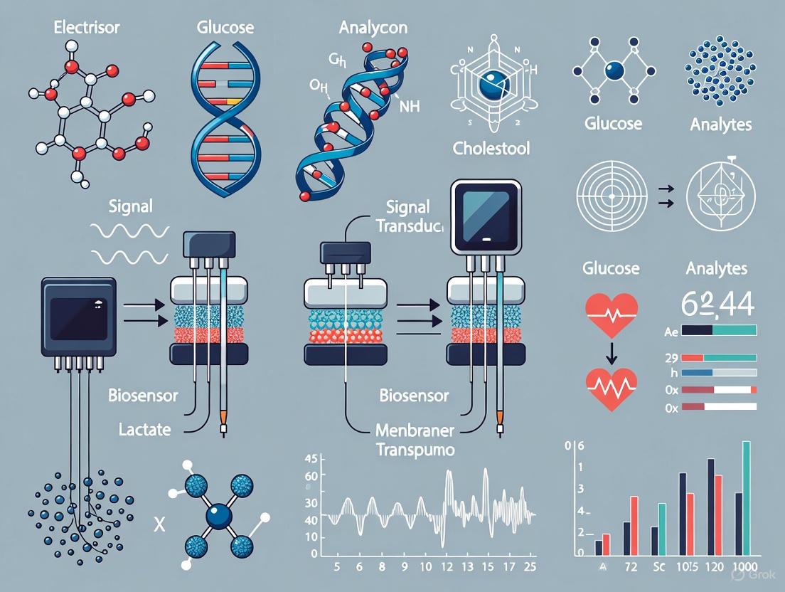
Wearable biosensors have revolutionized modern healthcare by enabling real-time, continuous monitoring of physiological parameters, facilitating a paradigm shift towards proactive and personalized medicine [9]. These devices empower users—from patients to athletes—to dynamically assess their well-being and make informed healthcare decisions [1]. The fundamental operation of any wearable biosensor hinges on the seamless integration of three core components: a recognition element that selectively interacts with the target analyte, a transducer that converts this biological interaction into a quantifiable signal, and electronic systems that process, transmit, and present the data [10] [11]. This application note provides a detailed technical examination of these components, their operational principles, and standardized protocols for their implementation, framed within health monitoring research.
Core Component 1: Recognition Elements
Recognition elements are the biologically active part of a biosensor, responsible for the selective binding or catalytic transformation of a specific target analyte. Their specificity and affinity directly determine the sensor's analytical performance.
Table 1: Common Bio-Recognition Elements and Their Characteristics
| Recognition Element | Mechanism of Action | Target Analytes | Stability | Example Application |
|---|---|---|---|---|
| Enzymes | Catalytic transformation of substrate | Metabolites (e.g., Glucose, Lactate) [12] | Moderate | Glucose oxidase in diabetes monitoring patches [11] |
| Antibodies/Antibody Fragments | High-affinity, selective binding to antigen | Proteins, Hormones, Pathogens [12] | High to Moderate | Immunosensors for cortisol detection [12] |
| Aptamers | Folding into 3D structures for target binding | Ions, Small molecules, Proteins [12] | High | Aptamer-based FET sensors for cortisol in sweat [12] |
| Nucleic Acids | Hybridization with complementary sequence | DNA, RNA [10] | High | Pathogen detection [10] |
| Ion-Selective Membranes | Selective partitioning of ions | Electrolytes (e.g., K⁺, Na⁺) [12] | High | Potentiometric sensors for sweat electrolyte analysis [12] |
Experimental Protocol: Immobilization of Recognition Elements on Transducer Surfaces
Objective: To covalently immobilize enzyme-based recognition elements (e.g., Glucose Oxidase) onto a flexible graphene-based electrode for a wearable sweat sensor [6].
Materials:
- Graphene-based working electrode: Fabricated via screen-printing or laser ablation on a flexible polyimide substrate.
- Glucose Oxidase (GOx): From Aspergillus niger, lyophilized powder.
- Cross-linker: N-(3-Dimethylaminopropyl)-N'-ethylcarbodiimide (EDC) and N-Hydroxysuccinimide (NHS).
- Buffers: 0.1 M Phosphate Buffered Saline (PBS), pH 7.4.
- Blocking agent: Bovine Serum Albumin (BSA).
Procedure:
- Electrode Pre-treatment: Clean the graphene electrode surface via voltammetric cycling in 0.5 M H₂SO₄ from -0.3 V to +0.8 V (vs. Ag/AgCl) for 10 cycles to activate and introduce oxygen-containing functional groups [6].
- Cross-linker Activation: Prepare a fresh solution of 2 mM EDC and 5 mM NHS in 0.1 M PBS. Pipette 50 µL onto the active area of the graphene electrode and incubate for 1 hour at room temperature to activate carboxyl groups on the graphene surface.
- Enzyme Immobilization: Prepare a 10 mg/mL solution of GOx in 0.1 M PBS (pH 7.4). Rinse the electrode with deionized water to remove excess EDC/NHS. Apply 50 µL of the GOx solution and incubate for 12 hours at 4°C to allow covalent amide bond formation.
- Surface Blocking: Rinse the electrode gently with PBS to remove physically adsorbed enzyme. Apply a 1% (w/v) BSA solution for 1 hour to block any remaining non-specific binding sites.
- Storage: Store the functionalized biosensor in 0.1 M PBS (pH 7.4) at 4°C until use.
Validation: Confirm successful immobilization via a colorimetric assay to detect residual amine groups in the supernatant, indicating unbound enzyme. The biosensor's performance should be calibrated against standard glucose solutions.
Core Component 2: Transducers
Transducers are the critical interface that transform the biological recognition event into a measurable electrical signal. The choice of transducer dictates key sensor specifications like sensitivity, detection limit, and form factor [10].
Table 2: Classification and Performance of Biosensor Transducers
| Transducer Type | Principle | Measured Signal | Detection Limit | Wearable Form Factor |
|---|---|---|---|---|
| Electrochemical | Redox reactions at electrode interface [10] | Current, Potential, Impedance | µM - fM [12] | Skin patch, Tattoo [10] |
| Field-Effect Transistor (FET) | Gating effect from charged biomolecules [12] | Drain-Source Current (IDS) | fM range [12] | Smart contact lens [12] |
| Optical | Light absorption, emission, interference | Fluorescence, Absorbance, Refractive Index | Varies | Ring oximeter [11] |
| Thermal | Reaction enthalpy from catalytic event [10] | Temperature Change | - | Skin patch |
| Gravimetric | Mass change on piezoelectric crystal [10] | Frequency Shift | - | - |
Operational Workflow of a Wearable Biosensor
The following diagram illustrates the logical sequence of signal conversion in a typical wearable biosensor, from biological event to user-interpretable data.
Experimental Protocol: Characterization of a Field-Effect Transistor (FET) Biosensor
Objective: To measure the transfer characteristic (IDS vs. VGS) of a MoS₂-based FET biosensor and determine its sensitivity to a target protein in a liquid-gate configuration [12].
Materials:
- Liquid-gate FET biosensor: e.g., MoS₂ channel on a flexible substrate with source/drain electrodes and a liquid-gate port.
- SourceMeter/Parameter Analyzer: Keithley 2600B series or equivalent.
- Phosphate Buffered Saline (PBS): 0.01 M, pH 7.4.
- Analyte: Purified target protein (e.g., Cortisol) at known concentrations in PBS.
Procedure:
- Sensor Setup: Place a 50 µL droplet of 0.01 M PBS on the active channel area of the FET. Insert a Ag/AgCl reference electrode into the droplet to act as the liquid gate [12].
- Electrical Connection: Connect the source and drain terminals of the FET to the SourceMeter. Connect the reference electrode to the gate terminal of the SourceMeter.
- Transfer Curve Measurement:
- Set the drain-source voltage (VDS) to a constant value (e.g., 0.1 V or 0.5 V).
- Sweep the liquid-gate voltage (VGS) from a negative to a positive potential (e.g., -1 V to +1 V) while measuring the resulting drain-source current (IDS).
- Record the IDS - VGS curve. The threshold voltage (Vth) is identified as the gate voltage where the current begins to sharply increase.
- Sensitivity Measurement:
- Gently rinse the sensor channel with PBS.
- Introduce the target analyte solution at a specific concentration (e.g., 1 fM) and incubate for 15 minutes to allow binding to the immobilized probes on the gate surface.
- Repeat step 3 to obtain a new IDS - VGS curve.
- Note the shift in the threshold voltage (ΔV).
- Data Analysis: Plot ΔVth as a function of the logarithm of analyte concentration. The sensitivity of the biosensor is given by the slope of this calibration curve (in mV/decade).
Critical Consideration: Account for the Debye screening effect in high ionic strength solutions like sweat or blood. The sensitivity is limited by the Debye length (λD), which is typically 1-3 nm in physiological fluids, making it challenging to detect biomolecules larger than this length [12].
Core Component 3: Electronics and Data Processing
The electronic subsystem is the "brain" of the wearable biosensor, responsible for signal conditioning, data processing, power management, and communication.
Key Electronic Subsystems and Materials
Table 3: Research Reagent Solutions for Biosensor Electronics
| Component / Material | Function | Example & Notes |
|---|---|---|
| Analog Front-End (AFE) IC | Conditions the weak analog signal from the transducer. | Texas Instruments LMP91000 or MAX30001. Provides amplification and filtering. |
| Microcontroller Unit (MCU) | Processes digitized data and manages sensor operations. | ARM Cortex-M series (low power). Often integrates ADC and memory [13]. |
| Analog-to-Digital Converter (ADC) | Converts the conditioned analog signal to a digital value. | Resolution: 16-bit (e.g., in ECG biosensors [14]). Sampling: 244-976 Hz [14]. |
| Wireless Communication Module | Transmits data to external devices (e.g., smartphone). | Bluetooth Low Energy (BLE) [13], WiFi (IEEE 802.11b) [14]. |
| Flexible/Stretchable Substrate | Provides mechanical compatibility with skin. | Polyimide, Polydimethylsiloxane (PDMS), Ecoflex [15]. |
| Graphene-based Nanomaterials | Enhances electrode conductivity and flexibility. | Used in channels of FETs or as working electrodes [6]. |
Standardized Protocol for System Integration and Data Acquisition
Objective: To implement a standardized data acquisition system for a multi-sensor wearable platform integrating both an electrophysiological sensor (sEMG) and a lactate biosensor, compliant with the ISO/IEC/IEEE 21451 framework for interoperability [13].
Materials:
- Transducer Interface Module (TIM): Custom PCB housing the analog front-end for the biosensor and sEMG electrodes.
- Network-Capable Application Processor (NCAP): A smartphone or a dedicated gateway device (e.g., Raspberry Pi).
- Transducer Electronic Data Sheet (TEDS): A standardized digital document describing the sensor characteristics, stored in the TIM's memory [13].
Procedure:
- System Architecture:
- Design the TIM to include separate transducer channels (TCh) for each physical sensor (e.g., one for lactate, eight for sEMG). Each TCh includes the sensor, conditioning electronics, and an ADC [13].
- The MCU on the TIM acts as a local processor, reading data from all ADCs via a Serial Peripheral Interface (SPI) bus.
- TEDS Configuration:
- Program the TEDS for each transducer channel. The TEDS should contain:
- Meta-TEDS: General information about the TIM itself (e.g., UUID, communication protocol).
- Channel-TEDS: Detailed description for each sensor (e.g., sensor type, units, calibration data, operational range).
- Calibration-TEDS: For biosensors, store multiple calibration points (concentration vs. signal output) instead of a single calibration equation for greater flexibility [13].
- Program the TEDS for each transducer channel. The TEDS should contain:
- Data Acquisition and Communication:
- The NCAP (smartphone) discovers the TIM via a wireless protocol like BLE (standardized by ISO/IEC/IEEE 21451-5).
- Upon connection, the NCAP reads the TEDS from the TIM, automatically understanding the capabilities and data structure of the connected sensors.
- The NCAP can then send commands to setup the acquisition (e.g., select active channels, set sampling rate) based on the application needs.
- The TIM streams the acquired data to the NCAP, which forwards it to the cloud for further analysis and display to healthcare professionals [13] [14].
Advantages: This standardized approach ensures plug-and-play interoperability, simplifies application development, and allows for the seamless integration of biosensors from different manufacturers into a unified health monitoring system [13].
The Expanding Market and Global Research Landscape
The global biosensors market is experiencing robust growth, propelled by technological advancements and rising demand for personalized healthcare monitoring. The market is poised to expand significantly from 2025 to 2030, with projections from leading market research firms detailed in Table 1.
Table 1: Global Biosensors Market Size Projections
| Source | Market Size 2024/2025 | Projected Market Size 2030/2034 | Compound Annual Growth Rate (CAGR) |
|---|---|---|---|
| MarketsandMarkets [16] | USD 34.5 billion (2025) | USD 54.4 billion (2030) | 9.5% (2025-2030) |
| GM Insights [17] | USD 32.3 billion (2024) | USD 68.5 billion (2034) | 7.9% (2025-2034) |
| Persistence Market Research [18] | USD 32.5 billion (2025) | USD 56.4 billion (2032) | 8.2% (2025-2032) |
This growth is primarily fueled by the increasing prevalence of chronic diseases such as diabetes, the growing demand for point-of-care (POC) testing, and continuous technological innovations in the field of wearable biosensors [16] [17] [19].
The market segmentation reveals key areas of dominance and high-growth sectors. Table 2 summarizes the market share and growth trends by product, technology, and application.
Table 2: Biosensors Market Segmentation and Growth Trends
| Segment | Dominant/Largest Category | Highest Growth Category | Key Insights |
|---|---|---|---|
| Product [16] [17] | Non-wearable (POC) Devices (~60% share) | Wearable Biosensors (CAGR up to 14.1%) | Wearables are transformative for continuous health monitoring. |
| Technology [16] [20] | Electrochemical (~41-72% share) | Optical Biosensors | Electrochemical is favored for mass production (e.g., glucose strips). |
| Application [17] [20] | Blood Glucose Testing | Home Diagnostics / Chronic Disease Management | Driven by the global diabetes epidemic and shift to home-based care. |
| Region [16] [17] | North America (~41% share) | Asia-Pacific | Large population base and rising lifestyle diseases fuel APAC growth. |
Application Notes: Key Areas and Experimental Focus
Wearable Biosensors for Non-Invasive Monitoring
Wearable biosensor systems represent a significant breakthrough, offering real-time monitoring and quantitative assessment of various human health parameters [1]. The escalating demand for continuous surveillance of acute and chronic conditions has propelled extensive investigation into these devices, which are pivotal for providing physiological insights and facilitating non-invasive monitoring [1]. They function by analyzing biomarkers in easily accessible biofluids such as sweat, tears, saliva, and interstitial fluid [1] [2] [21].
A major research and development focus is shifting from traditional protein-based biorecognition elements (enzymes, antibodies) to nucleic acid-based assays (oligonucleotides, aptamers, CRISPR-Cas) [22]. Nucleic acids offer advantages in stability, scalability through chemical synthesis, and programmability, making them suitable for continuous monitoring applications [22].
Protocol: Fabrication of a Nucleic Acid-Based Wearable Biosensor
The fabrication of a modern, nucleic acid-integrated wearable biosensor typically involves a multi-layered approach, as illustrated in the workflow below.
Title: Wearable Biosensor Fabrication Workflow
Detailed Methodology:
Microfluidics/Reagent Layer Fabrication:
- Master Mold Creation: Use soft lithography. Spin-coat a silicon wafer with a photoresist (e.g., SU-8). Apply a photomask with the desired microfluidic channel design and expose it to UV light in a mask aligner to create the pattern [22].
- Channel Replication: Pour a mixture of Polydimethylsiloxane (PDMS) pre-polymer and curing agent (typically 10:1 ratio) onto the master mold. Cure at 65-80°C for 1-2 hours [22].
- Bonding and Sealing: Treat the PDMS layer and a covering PDMS layer with oxygen plasma to activate surfaces. Bring them into contact to form an irreversible, covalent seal, encapsulating the microfluidic channels [22]. Alternatively, use uncured PDMS as an adhesive for a reversible seal.
Sensing Layer Fabrication:
- Substrate and Electrode Patterning: Use flexible polymer substrates like Polyethylene Terephthalate (PET). Electrode patterns (working, reference, counter) can be defined via photolithography or xurography. Metals like Gold (Au) and Silver/Silver Chloride (Ag/AgCl) are deposited via sputtering or thermal evaporation [22].
- Nanomaterial Functionalization (for enhanced sensitivity): For graphene-based electrodes, synthesize graphene nanostructures (e.g., via chemical vapor deposition). Modify the surface chemistry using linkers like 1-pyrenebutanoic acid succinimidyl ester (PBASE) to facilitate the immobilization of biomolecules [21].
- Biorecognition Element Immobilization: Spot and covalently immobilize amino-modified nucleic acid probes (e.g., aptamers) onto the functionalized electrode surface. Block non-specific binding sites with reagents like Bovine Serum Albumin (BSA) or ethanolamine [22].
Readout/Packaging Layer Integration:
- Integrate the fabricated sensor with a miniaturized potentiostat for electrochemical measurements (e.g., Amperometric, Impedimetric).
- Package the system with a printed circuit board (PCB) containing a microcontroller for signal processing and a Bluetooth Low Energy (BLE) module for wireless data transmission to a smartphone or cloud server [22] [18].
Protocol: Electrochemical Detection of Analyte
This protocol follows the successful fabrication and integration of the biosensor layers.
Workflow Overview:
Title: Analyte Detection and Signaling Workflow
Detailed Methodology:
- Sample Handling: The microfluidic layer collects the biofluid (e.g., sweat via capillary action) and transports it to the sensing chamber where the nucleic acid probes are immobilized [22].
- Target Binding and Signal Transduction:
- Introduce the sample to the sensing surface.
- Upon binding of the target analyte to the specific nucleic acid aptamer, a conformational change or binding event occurs.
- This biochemical event is transduced into an electrical signal. For electrochemical sensors, this may result in a change in current (amperometry), potential (potentiometry), or impedance (impedimetry) that is proportional to the analyte concentration [22] [21].
- Data Acquisition and Analysis:
- The generated electrical signal is captured by the integrated electronics.
- The signal is amplified, filtered, and converted from analog to digital.
- Calibration curves (pre-established) are used to convert the signal magnitude into a quantitative concentration value.
- Processed data is transmitted wirelessly via BLE to a paired smartphone application for real-time visualization and long-term trend analysis [18].
The Scientist's Toolkit: Research Reagent Solutions
Table 3: Essential Materials for Wearable Biosensor Development
| Item | Function/Application | Key Characteristics |
|---|---|---|
| Nucleic Acid Aptamers [22] | Biorecognition element; binds specific biomarkers (ions, small molecules, proteins). | High stability, synthetic production, programmable, can exhibit catalytic activity (DNAzymes). |
| Graphene Nanostructures [21] | Sensing electrode material; enhances signal sensitivity and provides flexible substrate. | Exceptional electrical conductivity, high surface area, mechanical flexibility, biocompatibility. |
| Polydimethylsiloxane (PDMS) [22] | Microfluidic layer material; transports and handles biofluid samples. | Optical clarity, flexibility, gas permeability, biocompatibility, easy replication. |
| SU-8 Photoresist [22] | Master mold fabrication for microfluidics; creates high-resolution patterns via photolithography. | Negative-tone, epoxy-based, forms thick, stable structures. |
| 1-Pyrenebutanoic acid succinimidyl ester (PBASE) [21] | Surface chemistry linker; facilitates covalent immobilization of biomolecules on graphene. | Pyrenyl group π-stacks on graphene; NHS ester reacts with amine groups on biomolecules. |
| Ag/AgCl Ink [22] | Reference electrode fabrication; provides a stable, reproducible reference potential. | Essential for accurate electrochemical measurements in three-electrode systems. |
The advancement of wearable biosensors has catalyzed a shift towards non-invasive, continuous health monitoring, moving diagnostics from clinical settings to the point-of-care. Central to this paradigm are biofluids that can be sampled with minimal invasiveness yet provide a rich source of physiological information. This application note provides a structured analysis of four key biofluids—sweat, saliva, tears, and interstitial fluid (ISF)—evaluating their diagnostic potential, inherent challenges, and practical methodologies for biosensing applications. Framed within broader research on wearable biosensors, this document equips researchers and drug development professionals with the comparative data and protocols necessary to inform experimental design and technology development.
Comparative Analysis of Biofluids
The table below summarizes the core characteristics, advantages, and limitations of each biofluid for biosensing applications.
Table 1: Comparative Analysis of Key Biofluids for Wearable Biosensing
| Feature | Sweat | Saliva | Tears | Interstitial Fluid (ISF) |
|---|---|---|---|---|
| Primary Collection Method | Patches, bands, microfluidics [23] | Passive drool, salivettes, swabs [24] [25] | Schirmer's strip, microcapillary [26] | Microneedles, reverse iontophoresis [27] [28] |
| Invasiveness | Non-invasive | Non-invasive | Minimally invasive | Minimally invasive [27] |
| Key Biomarkers | Electrolytes (Na+, K+, Cl-), metabolites (lactate, glucose, urea), drugs [23] | Hormones (cortisol), enzymes (alpha-amylase), antibodies, metabolites [24] [29] [25] | Proteins (lysozyme), lipids, electrolytes (Ca2+), metabolites (glucose, vitamin C) [26] [30] | Glucose, lactate, drugs, hormones, electrolytes (concentrations often similar to plasma) [27] [28] |
| Volume & Sampling Rate | Can be copious; rate varies with activity | 1-1.5 L/day; continuous but variable flow [25] | Low volume (3-10 µL); slow rate (1-2 µL/min) [26] | Reservoir in skin; continuous but access can be rate-limiting [28] |
| Major Strength | Rich in electrolytes & metabolites; good for fitness/dehydration monitoring | Easiest to collect non-invasively; rich in hormones & infection markers [25] | Direct correlation to ocular diseases; less complex than blood [26] | High clinical relevance; composition closely mirrors blood plasma [27] [31] |
| Major Limitation | Analyte concentration can be diluted and varies with sweat rate | Predictive value for complex performance outcomes can be limited; subject to contamination [24] [25] | Extremely low volume makes collection and analysis challenging [26] | Requires minimally invasive technology (e.g., microneedles) for access [27] |
| Correlation with Blood | Varies by analyte; reported for lactate, ethanol, glucose [23] | Many compounds filtered from blood; concentration often lower [25] | Biomarkers can passively leak from blood; concentration often lower [26] | High correlation for small molecules (< 3 kDa); larger molecules may have reduced concentration [27] |
Experimental Protocols for Biofluid Analysis
Protocol: Saliva Collection and Analysis for Stress Biomarkers
This protocol details the methodology for collecting saliva and analyzing key biomarkers like cortisol and alpha-amylase, which are indicative of stress and sympathetic nervous system activity [24] [29] [25].
1. Reagents and Equipment:
- Sterile salivettes or cryovials for passive drool
- Cold storage (-20°C for short-term, -80°C for long-term)
- Centrifuge
- Commercial ELISA kits for cortisol and alpha-amylase
- Microplate reader
2. Procedure: Step 1: Pre-collection Instructions. Instruct participants to avoid eating, drinking (except water), and brushing teeth for at least 60 minutes prior to collection. Rinse mouth with water 10 minutes before sampling [25]. Step 2: Sample Collection. - Unstimulated Passive Drool: Have the participant tilt their head forward and allow saliva to pool in the mouth for 60-90 seconds before passively drooling into a pre-weighed cryovial. Alternatively, use a Salivette tube, where the participant chews on a synthetic swab for 1-2 minutes, which is then placed back into the tube [24]. Step 3: Sample Processing. Centrifuge Salivette tubes at a high speed (e.g., 3000 rpm) for 5-15 minutes to extract saliva into the base tube. For passive drool, centrifuge at 2000-3000 x g for 15 minutes to separate debris. Aliquot the clear supernatant for analysis. Step 4: Storage. Immediately freeze samples at -20°C if analysis occurs within a day. For long-term storage (months), keep at -80°C [24]. Step 5: Analysis. Perform biomarker quantification using commercially available ELISA kits according to the manufacturer's instructions, using a microplate reader for detection.
3. Data Interpretation:
- Cortisol levels typically exhibit a diurnal rhythm, peaking in the morning.
- Alpha-amylase is a more acute marker of sympathetic activity.
- Values should be interpreted against established normative ranges and study-specific baselines.
Protocol: Tear Collection and Colorimetric Multi-Biomarker Sensing
This protocol describes the use of a microfluidic patch and an AI-assisted system for the simultaneous, colorimetric detection of key tear biomarkers [30].
1. Reagents and Equipment:
- Flexible PDMS microfluidic patch with colorimetric paper chips
- Chromogenic reagents: e.g., Xylidyl Blue for Ca2+, Bromocresol Green for protein
- Smartphone with a dedicated app and cloud server data analysis system (CSDAS)
- Artificial intelligence model (e.g., CNN-GRU neural network) for data correction
2. Procedure: Step 1: Sensor Preparation. Fabricate a crescent-shaped, multi-layered PDMS microfluidic patch containing separate micro-reservoirs. Pre-load each reservoir with a filter paper chip treated with a specific chromogenic reagent for Vitamin C, H+ (pH), Ca2+, and protein [30]. Step 2: Tear Collection and Sensing. Adhere the flexible patch to the skin underneath the eye using medical-grade adhesive. Tear fluid is collected via the patch's inlets and routed by microchannels (800 µm x 800 µm) to the four sensing reservoirs. A volume of ~20 µL is sufficient. The colorimetric reaction occurs upon contact with the analyte [30]. Step 3: Data Acquisition. Use a smartphone to capture an image of the sensor patch. The embedded app uploads the color data (RGB values) from each reservoir to the CSDAS. Step 4: AI-Assisted Data Analysis. The CSDAS, powered by a pre-trained multi-channel Convolutional Neural Network-Gated Recurrent Unit (CNN-GRU) model, processes the RGB data. The model corrects for interfering variables such as ambient light color temperature and cross-reactivity from varying tear pH, outputting the calibrated concentration for each biomarker [30].
3. Data Interpretation:
- The coefficient of determination (R2) for the AI-predicted concentrations can be as high as 0.994-0.998 with a well-trained model [30].
- Abnormal levels can indicate conditions such as dry eyes (elevated Ca2+) or rosacea (altered pH/protein) [30].
Protocol: Dermal Interstitial Fluid (ISF) Sampling with Microneedle Sensors
This protocol outlines the use of solid microneedle arrays for minimally invasive access to ISF for biomarker monitoring [27] [28].
1. Reagents and Equipment:
- Solid microneedle array (e.g., polymer, metal; length: < 1 mm)
- Functionalized microneedle tips (e.g., with enzymes or antibodies)
- Potentiostat or optical reader for signal transduction
- Integrated wearable monitoring electronics (optional for continuous sensing)
2. Procedure: Step 1: Skin Site Preparation. Clean and disinfect the application site (e.g., forearm, upper arm) with a 70% alcohol swab and allow to dry. Step 2: Microneedle Application. Apply the microneedle array to the skin using a custom applicator that provides consistent velocity and force to pierce the stratum corneum, creating microchannels. The microneedles should penetrate to a depth of 50-1000 µm, reaching the ISF-rich dermis without contacting nerves or blood vessels [27] [28]. Step 3: ISF Sensing. - Extraction Method: Microneedles create transient microchannels, allowing ISF to flow to the skin surface via capillary action or osmosis. The fluid can then be collected and analyzed externally [27]. - In-Situ Sensing Method: For real-time monitoring, microneedles with functionalized tips (e.g., with carbon paste and glucose oxidase for glucose sensing) are indwelled in the dermis. The sensing element directly interacts with ISF biomarkers, transducing a signal (electrochemical or optical) that is read by an integrated monitor [27] [28]. Step 4: Signal Measurement and Monitoring. Connect the microneedle sensor to a potentiostat for electrochemical measurements (e.g., amperometry for glucose) or an optical reader for colorimetric/fluorescent signals. For wearable applications, this system is integrated with a compact potentiostat, power source, and wireless data transmission module for continuous, on-body monitoring [28].
3. Data Interpretation:
- Correlate ISF analyte levels with blood concentrations, noting that small molecules (< 3 kDa) like glucose show a strong correlation, while larger molecules like proteins may have a time-lag or concentration gradient [27].
Visualized Workflows and Pathways
Tear Biomarker Sensing Workflow
Tear Biosensing Workflow
Biomarker Transport to Interstitial Fluid
ISF Biomarker Transport
The Scientist's Toolkit: Research Reagent Solutions
Table 2: Essential Materials for Wearable Biofluid Research
| Category | Item | Primary Function | Example Application |
|---|---|---|---|
| Sample Collection | Salivette (synthetic swab) | Non-invasive saliva collection | Hormonal, immunologic studies [24] [25] |
| Schirmer's Test Strip | Tear volume measurement & collection | Dry eye diagnosis, general tear analysis [26] | |
| Solid Microneedle Array (e.g., polymer) | Creates microchannels in stratum corneum | Minimally invasive access to dermal ISF [27] [28] | |
| Sensing Elements | Enzyme-Linked Immunosorbent Assay (ELISA) Kit | Quantitative protein/hormone detection | Measuring cortisol, cytokines in saliva/ISF [24] [29] |
| Chromogenic Reagent (e.g., Xylidyl Blue) | Produces color change upon analyte binding | Colorimetric detection of Ca2+ in tears [30] | |
| Ion-Selective Membrane (e.g., for Na+, K+) | Potentiometric ion detection | Electrolyte monitoring in sweat [23] | |
| Data Acquisition & Analysis | Potentiostat | Applies potential & measures current | Electrochemical biosensing (e.g., glucose, lactate) [23] [28] |
| Smartphone with Custom App | Image capture & data upload | Point-of-care colorimetric readout [30] | |
| Cloud Server & AI Model (e.g., CNN-GRU) | Processes & corrects complex sensor data | Improving accuracy of multi-analyte tear sensors [30] |
Wearable biosensors are revolutionizing the landscape of modern healthcare by enabling continuous, non-invasive monitoring and real-time diagnostics [2]. The convergence of several key engineering and materials science disciplines has propelled this transformation. This document details the core emerging trends of miniaturization, the use of flexible substrates, and advancements in non-invasive sampling protocols, which collectively address the critical need for patient-friendly, accurate, and continuous health monitoring tools for research and drug development [32] [3]. These trends are foundational to the development of next-generation wearable devices that seamlessly integrate with the human body, providing unprecedented access to physiological data.
Emerging Trends and Key Technologies
The advancement of wearable biosensors is underpinned by progress in three interconnected technological domains.
Miniaturization and System Integration
Miniaturization is crucial for developing compact, unobtrusive, and user-compliant devices. This trend extends beyond mere size reduction to encompass the integration of multiple functionalities into a single, miniaturized platform, often described as a "wearable lab on the body" [33].
Key Advances:
- Microfluidics: The use of chips to control and manipulate minuscule amounts of fluids is a breakthrough for secure, non-invasive sampling of biofluids like sweat and saliva, enabling real-time measurement of analytes such as glucose and electrolytes [3].
- Flexible Printed Circuit Boards (FPCBs): The advent of soft, FPCBs allows for the embedding of integrated circuits, sensors, and antennas into thin, contour-friendly substrates, which is essential for maintaining device performance under mechanical deformation [32].
- Nanomaterial-Enhanced Sensitivity: The use of nanomaterials like graphene, carbon nanotubes, and metallic nanoparticles provides a very large surface area for effective interaction with biomolecules. This allows for highly sensitive detection even at low concentrations, which is critical as sensor sizes shrink [3] [34].
Flexible and Stretchable Substrates
The mechanical mismatch between conventional rigid electronics and soft, dynamic human tissues is a significant challenge. Flexible substrates are engineered to overcome this, ensuring device comfort, stability, and signal accuracy during prolonged use [35] [34].
Material Innovations:
- Polymer Substrates: Materials such as polydimethylsiloxane (PDMS), polyimide (PI), and polyethylene terephthalate (PET) are widely used due to their excellent flexibility, optical transparency, biocompatibility, and thermal stability [32] [4].
- Conductive Nanocomposites and Hydrogels: These materials combine conductivity with stretchability and biocompatibility. Conductive hydrogels, in particular, can mimic real body tissue, making them ideal mediators between the sensor and target analytes in applications like sweat analysis [3] [33] [36].
- Carbon-Based Nanomaterials: Graphene and its derivatives (graphene oxide, reduced graphene oxide) are prized for their exceptional electrical conductivity, mechanical strength, and flexibility. These properties enable the fabrication of sensors with intimate and conformal contact with biological tissues like skin and eyes [4] [34].
Table 1: Properties of Key Flexible Substrate Materials
| Material | Key Properties | Advantages in Biosensing | Example Applications |
|---|---|---|---|
| PDMS | Flexible, optically transparent, biocompatible, low surface energy [4] | Enhances comfort and stability; suitable for optical sensing platforms [4] | Skin patches, microfluidic channels [32] |
| Polyimide (PI) | Excellent thermal stability, insulation, film-forming ability [4] | Withstands fabrication processes; reliable functional substrate [4] | Flexible electrodes, implantable sensors |
| Graphene | High conductivity, exceptional mechanical strength, atomic-scale thickness, flexibility [34] | Maximizes analyte interaction; enhances sensitivity; conformal contact with skin [34] | Electrochemical sensors, electrophysiology monitoring [34] |
| Conductive Hydrogels | High water content, biocompatible, tissue-like mechanical properties [33] [36] | Excellent tissue-sensor interface; mediates analyte sensing [3] [33] | Sweat analysis, strain sensors [33] |
Non-Invasive Sampling and Multi-Biofluid Analysis
The shift from invasive blood draws to the analysis of readily available biofluids is a cornerstone of modern wearable biosensors. Each biofluid offers a unique window into physiological and pathological states [32].
Biofluid Applications and Device Embodiments:
- Sweat: The most intensively studied biofluid, sweat contains metabolites (glucose, lactate), electrolytes, and hormones. Wearable patches are the primary form factor, leveraging advancements in microfluidics and epidermal adhesion for continuous monitoring [32] [37].
- Saliva: Rich in electrolytes, hormones, drugs, and viruses, saliva is analyzed using wearable mouthguards, dental patches, or pacifier sensors. These devices allow for continuous capture of biomarkers through constant contact with oral fluid [32] [2].
- Tears: Tears contain proteins, lipids, and metabolites associated with ocular and systemic diseases. Smart contact lenses are the dominant platform, though they require extreme measures for mechanical matching to avoid corneal irritation [32] [2].
- Exhaled Breath: The COVID-19 pandemic intensified the demand for smart masks that can monitor respiratory health and detect infectious pathogens by analyzing exhaled breath condensate and volatile organic compounds (VOCs) [32].
Table 2: Non-Invasive Biofluids and Their Target Analytics
| Biofluid | Primary Wearable Form Factor | Key Example Biomarkers | Research/Clinical Utility |
|---|---|---|---|
| Sweat | Epidermal patches [32] | Glucose, Lactate, Cortisol, Na+, K+ ions [32] [37] | Fitness monitoring, cystic fibrosis diagnosis, stress monitoring [32] |
| Saliva | Mouthguards, dental patches [32] | Cortisol, Glucose, Uric Acid, Viruses [32] [2] | Oral disease monitoring, systemic biomarker detection, drug development [2] |
| Tears | Smart contact lenses [32] [2] | Glucose, Proteins, Ascorbic Acid [32] | Intraocular pressure monitoring, diabetes management [2] |
| Exhaled Breath | Smart masks [32] | Volatile Organic Compounds (VOCs), Humidity, Cytokines [32] | Early detection of respiratory infections, environmental exposure studies [32] |
Experimental Protocol: FRET-Based Lactate Aptasensor for Sweat Analysis
The following protocol details the development and application of a highly sensitive, non-invasive biosensor for lactate detection in sweat, illustrating the integration of miniaturization principles and advanced materials [37].
Principle and Workflow
This protocol utilizes a Fluorescence Resonance Energy Transfer (FRET)-based aptasensor. The core mechanism involves Aptamer-Functionalized Core-Shell Upconversion Nanoparticles (APT-CS-UCNPs) as energy donors and Fe₃O₄-decorated Molybdenum Disulfide (Fe₃O₄-MoS₂) nanosheets as quenchers.
- In the absence of lactate, the aptamer adsorbs onto the MoS₂ nanosheet, bringing the donor and acceptor into close proximity (<10 nm), which quenches the donor's fluorescence.
- Upon lactate binding, the aptamer undergoes a conformational change, detaching from the MoS₂ surface. This increases the donor-acceptor distance, restoring fluorescence intensity at 545 nm, which is proportional to the lactate concentration [37].
The experimental workflow is summarized in the following diagram:
Detailed Methodology
Synthesis of Core-Shell Upconversion Nanoparticles (CS-UCNPs)
- Prepare Rare-Earth Solution: Dissolve YCl₃·6H₂O (78 mol%), YbCl₃·6H₂O (20 mol%), and ErCl₃·6H₂O (2 mol%) in a mixture of oleic acid (6 mL) and 1-octadecene (15 mL) in a 100 mL three-neck flask [37].
- Form Nanoparticle Core: Heat the solution to 160°C under argon protection and stir for 30 minutes to form a clear solution. Cool to room temperature.
- Precipitate and Wash: Add an ammonium fluoride/methanol solution and stir. Subsequently, add a sodium hydroxide/methanol solution and heat to 100°C for 1 hour. Then, heat to 300°C and maintain for 1.5 hours under an argon atmosphere. Cool, precipitate with ethanol, and collect the core nanoparticles via centrifugation at 12,000 rpm for 10 minutes. Wash twice with cyclohexane/ethanol.
- Grow Shell Layer: Re-disperse the core nanoparticles in cyclohexane. Use a standard shell-growth protocol (typically involving additional layers of inert host materials) to form the core-shell structure to enhance fluorescence efficiency [37].
Synthesis of Fe₃O₄-MoS₂ Nanosheets
- Synthesize MoS₂ Nanosheets: Hydrothermally synthesize MoS₂ nanosheets using sodium molybdate and thiourea as precursors [37].
- Decorate with Fe₃O₄: Synthesize Fe₃O₄ nanoparticles in situ on the MoS₂ nanosheets using a co-precipitation method with ferrous and ferric salts in an alkaline solution. This imparts superparamagnetism to the composite [37].
- Characterize: Confirm successful synthesis using Transmission Electron Microscopy (TEM), X-ray Diffraction (XRD), and X-ray Photoelectron Spectroscopy (XPS).
Aptamer Functionalization and Sensor Assembly
- Functionalize UCNPs: Incubate the synthesized CS-UCNPs with the biotinylated L-lactate aptamer. Use streptavidin-biotin chemistry to immobilize the aptamer onto the nanoparticle surface, creating the APT-CS-UCNP probe [37].
- Assemble FRET Pair: Mix the APT-CS-UCNP probes with the Fe₃O₄-MoS₂ nanosheets in Tris-HCl buffer. The aptamer will adsorb onto the MoS₂, quenching the fluorescence.
Lactate Detection in Sweat Samples
- Sample Collection: Collect human sweat samples using a standardized protocol (e.g., via exercise induction and absorption with a sterile pad). Filter and dilute if necessary.
- Incubation: Incubate the sweat sample with the assembled APT-CS-UCNP/Fe₃O₄-MoS₂ complex for a defined period (e.g., 15-20 minutes) to allow lactate binding.
- Magnetic Separation: Place the reaction tube on a magnetic rack for <1 minute to separate the Fe₃O₄-MoS2/aptamer complex from the supernatant containing the unbound, fluorescence-restored APT-CS-UCNPs [37].
- Fluorescence Measurement: Transfer the supernatant to a cuvette. Using a fluorescence spectrophotometer equipped with a 980 nm laser, measure the fluorescence emission spectrum and record the intensity at 545 nm.
- Quantification: Generate a calibration curve using lactate standards of known concentration (0–30 mM). Calculate the lactate concentration in the unknown sweat sample from the calibration curve [37].
The Scientist's Toolkit: Research Reagent Solutions
Table 3: Essential Reagents and Materials for the FRET-Based Lactate Aptasensor
| Item Name | Function/Description | Critical Parameters/Notes |
|---|---|---|
| L-lactate Aptamer | Synthetic biological recognition element; binds specifically to L-lactate [37] | Sequence: 5'-Biotin-TEG-GACGACGAGTAGCGCGTATGAATGCTTTTCTATGGAGTCGTC-3'; HPLC purified. |
| Rare-Earth Salts (Y, Yb, Er chlorides) | Core components of the UCNPs; enable upconversion of NIR light to visible light [37] | Purity >99.9%; molar ratios are critical for optimal fluorescence (e.g., 78% Y, 20% Yb, 2% Er). |
| Fe₃O₄-MoS₂ Nanosheets | Function as the energy acceptor/quencher and provide a platform for aptamer adsorption; Fe₃O₄ enables magnetic separation [37] | Superparamagnetic property is essential for clean separation; high surface area of MoS₂ enhances quenching efficiency. |
| 980 nm NIR Laser | Excitation source for the UCNPs [37] | Minimizes autofluorescence from biological samples, leading to a higher signal-to-noise ratio. |
| Fluorescence Spectrophotometer | Instrument for detecting and quantifying the fluorescence signal output [37] | Must be capable of detecting emission at 545 nm following NIR excitation. |
| Tris-HCl Buffer | Provides a stable pH environment for the biochemical reaction [37] | Typical concentration: 10 mM, pH 7.4. |
The synergistic advancement of miniaturization, flexible substrate engineering, and non-invasive sampling protocols is fundamentally accelerating the capabilities of wearable biosensors. These technologies enable the development of devices that are not only highly sensitive and specific but also comfortable and practical for long-term use. For researchers and drug development professionals, this translates to powerful new tools for continuous biomarker monitoring, objective endpoint assessment in clinical trials, and the development of personalized medicine strategies. The detailed protocol for lactate sensing serves as a representative example of how these trends converge into a functional, cutting-edge research application. Future directions will involve deeper integration of artificial intelligence for data analytics, further material innovations for long-term stability, and rigorous clinical validation to translate these technological promises into tangible healthcare solutions [32] [4] [33].
Sensing Modalities and Clinical Applications in Disease Management and Drug Development
The advancement of biosensor technologies is revolutionizing personalized healthcare by enabling real-time, non-invasive, and continuous monitoring of physiological parameters. Wearable biosensors represent a paradigm shift from conventional diagnostic tools, offering unprecedented opportunities for preventive medicine, chronic disease management, and personalized treatment strategies. These devices are increasingly critical for researchers and drug development professionals who require precise, continuous biomarker data for therapeutic monitoring and clinical trials. The integration of biosensors into wearable platforms facilitates the collection of rich, dynamic datasets that were previously inaccessible through sporadic clinical measurements.
This document provides a comprehensive technical analysis of three principal biosensing modalities—electrochemical, optical, and piezoelectric—within the context of advanced healthcare monitoring systems. Each technology offers distinct operational principles, advantages, and implementation challenges. Electrochemical biosensors currently dominate the wearable market, particularly for metabolic monitoring, while optical biosensors are gaining traction for their sensitivity and multiplexing capabilities. Piezoelectric biosensors represent an emerging technology with unique potential for self-powered sensing and mechanical biomarker detection. The following sections present detailed application notes, experimental protocols, performance comparisons, and implementation frameworks to guide researchers in selecting, developing, and deploying these technologies for specific research and clinical applications.
Table 1: Comparative Analysis of Biosensor Technologies for Healthcare Monitoring
| Parameter | Electrochemical Biosensors | Optical Biosensors | Piezoelectric Biosensors |
|---|---|---|---|
| Primary Transduction Mechanism | Measurement of electrical signals (current, potential, impedance) from biochemical reactions [38] [39] | Detection of optical property changes (absorption, fluorescence, SPR, SERS) [40] [4] | Conversion of mechanical energy to electrical signals via piezoelectric effect [41] [42] |
| Key Measurables | Current (Amperometry), Potential (Potentiometry), Impedance (EIS) [38] | Fluorescence intensity, refractive index (SPR), wavelength shift, Raman spectra [4] | Voltage, frequency shift, charge accumulation [41] |
| Detection Limits | Ultra-low (fM-nM range for various biomarkers) [38] [43] | High (single molecule detection possible with SERS) [44] | Varies with material and design [42] |
| Response Time | Seconds to minutes [38] | Milliseconds to seconds (real-time monitoring) [40] | Milliseconds (rapid mechanical response) [41] |
| Multi-analyte Capability | Yes (with electrode arrays) [39] [43] | Excellent (wavelength multiplexing) [40] [4] | Limited (typically single analyte) [41] |
| Power Requirements | Low to moderate [38] | Moderate to high (light sources required) [4] | Very low (self-powering capability) [41] |
| Market Share (2024) | Dominant (80.6% share) [45] | Growing rapidly (fastest CAGR) [45] | Niche segment [45] |
| Primary Applications in Healthcare | Continuous glucose monitoring, metabolite detection, hormone sensing [39] [45] | Cancer biomarker detection, vital signs monitoring, infectious disease screening [40] [4] [44] | Vital signs monitoring (heart rate, respiration), tissue regeneration, neuromodulation [41] [42] |
Electrochemical Biosensors: Application Notes and Protocols
Technology Fundamentals
Electrochemical biosensors represent the most established and commercially successful category of biosensing platforms, with the global market valued at USD 27.40 billion in 2024 and projected to reach USD 55.78 billion by 2032 [45]. These sensors operate on the principle of detecting electrical signals generated from biochemical reactions occurring at the electrode-solution interface. The fundamental components include a biorecognition element (enzymes, antibodies, aptamers) that specifically interacts with the target analyte, and a transducer that converts this biological interaction into a quantifiable electrical signal [38] [39]. Recent advancements have focused on enhancing sensor performance through nanotechnology integration, improved electrode designs, and miniaturization for wearable applications.
The operational mechanisms of electrochemical biosensors can be categorized into three primary modalities: amperometry (current measurement at fixed potential), potentiometry (potential measurement at zero current), and impedance spectroscopy (interface resistance measurement) [38]. Each approach offers distinct advantages for specific applications; for instance, amperometric sensors provide high sensitivity for continuous metabolite monitoring, while impedance-based sensors are particularly suited for label-free detection of binding events. The evolution of these technologies has enabled the development of wearable platforms for non-invasive monitoring of biomarkers in sweat, interstitial fluid, saliva, and tears [39].
Experimental Protocol: Multiplexed Electrochemical Detection of Cancer Biomarkers
Table 2: Research Reagent Solutions for Multiplexed Electrochemical Biosensing
| Reagent/Material | Function | Specifications |
|---|---|---|
| Y-shaped DNA scaffold | Molecular framework for nanoparticle attachment | Designed with specific aptamer sequences for target cancer cell recognition [43] |
| Metal Nanoparticles (Ag, PtFe, Au) | Electrochemical labels for signal generation | 10-20 nm diameter, functionalized with thiol groups for DNA conjugation [43] |
| Screen-printed carbon electrode array | Transduction platform | 8-electrode configuration, carbon working electrodes, Ag/AgCl reference, carbon counter electrode [38] |
| Phosphate Buffered Saline (PBS) | Electrochemical measurement buffer | 0.1 M, pH 7.4, containing 5 mM Fe(CN)₆³⁻/⁴⁻ as redox mediator [43] |
| Aptamer sequences | Biorecognition elements | Specific to MCF-7, HeLa, and A549 cancer cell surface markers [43] |
Procedure:
- Electrode Pretreatment: Clean the screen-printed electrode array by cycling in 0.5 M H₂SO₄ from 0 to +1.2 V for 20 cycles at 100 mV/s, followed by rinsing with deionized water [38].
- DNA-Nanoparticle Conjugate Preparation:
- Incubate Y-shaped DNA (100 nM in Tris-EDTA buffer) with respective metal nanoparticles (Ag, PtFe, Au) at 4°C for 16 hours with gentle agitation [43].
- Purify conjugates using centrifugal filtration (100 kDa MWCO) at 10,000 × g for 15 minutes.
- Sensor Assembly:
- Immobilize aptamer-functionalized DNA-nanoparticle conjugates on carbon working electrodes via π-π stacking by applying 5 μL droplet to each electrode and incubating at room temperature for 2 hours [43].
- Rinse gently with PBS to remove unbound conjugates.
- Sample Incubation:
- Apply 10 μL of cell suspension (10³-10⁶ cells/mL in PBS) to the electrode surface.
- Incubate for 30 minutes at 37°C to facilitate specific binding between cancer cells and aptamer sequences.
- Rinse with PBS to remove unbound cells.
- Electrochemical Measurement:
- Perform square wave voltammetry from -0.2 to +0.6 V with amplitude of 25 mV and frequency of 15 Hz in PBS containing 0.1 M KCl [43].
- Record oxidation peaks for AgNPs (+0.32 V), PtFeNPs (+0.54 V), and AuNPs (+0.78 V) for simultaneous detection of multiple cancer cell types.
- Data Analysis:
- Quantify cell concentration based on peak current intensity using calibration curves established with known cell concentrations.
- Perform statistical analysis with n=3 replicates for each measurement.
Diagram 1: Workflow for multiplexed electrochemical detection of cancer cells using nanoparticle labeling.
Implementation Considerations for Wearable Applications
The transition from laboratory electrochemical sensors to wearable devices necessitates addressing several critical challenges. Biocompatibility remains paramount for sensors intended for prolonged skin contact or implantation, requiring careful selection of encapsulation materials that prevent biofouling while maintaining analyte permeability [39]. Sensor degradation over time affects measurement stability, particularly for enzyme-based sensors where protein denaturation diminishes performance. Recent approaches incorporate nanostructured materials like porous gold-polyaniline-platinum composites to enhance stability in interstitial fluid while maintaining high sensitivity (95.12 ± 2.54 µA mM⁻¹ cm⁻² for glucose detection) [44].
Power management represents another significant consideration for wearable electrochemical systems. While traditional biosensors require external power sources, integration with energy harvesting technologies such as nanogenerators enables self-powered operation. Triboelectric and piezoelectric nanogenerators can harness biomechanical energy from body movements, converting it to electrical energy for sensor operation [41]. This approach aligns with the growing emphasis on sustainable and autonomous wearable devices for continuous health monitoring.
Optical Biosensors: Application Notes and Protocols
Technology Fundamentals
Optical biosensors constitute a rapidly advancing field with the highest projected growth rate in the biosensors market [45]. These devices operate by detecting changes in optical properties—including absorbance, fluorescence, luminescence, refractive index, or spectral characteristics—resulting from the interaction between target analytes and biorecognition elements. The integration of artificial intelligence with optical biosensing has created new paradigms for point-of-care diagnostics by enabling enhanced signal processing, pattern recognition, and automated decision-making [40]. Major optical sensing modalities include surface plasmon resonance (SPR), fluorescence, colorimetric, and surface-enhanced Raman spectroscopy (SERS), each offering unique advantages for specific healthcare applications.
The emergence of flexible optical biosensors represents a significant milestone in wearable health monitoring technology. By combining polymer substrates, nanostructured materials, MXenes, hydrogels, and textile-based platforms, researchers have developed sensors with enhanced functionality, sensitivity, and adaptability [4]. These flexible configurations enable conformal contact with skin, improving measurement accuracy while maintaining user comfort during extended wear. Recent innovations in material science have addressed traditional limitations of optical systems regarding miniaturization and mechanical robustness, expanding their potential for continuous physiological parameter monitoring outside clinical settings.
Experimental Protocol: SERS-Based Immunoassay for Cancer Biomarker Detection
Table 3: Research Reagent Solutions for SERS-Based Biosensing
| Reagent/Material | Function | Specifications |
|---|---|---|
| Au-Ag nanostars | SERS substrate providing plasmonic enhancement | Sharp-tipped morphology (50-100 nm), surfactant-free, tunable concentration via centrifugation [44] |
| Mercaptopropionic acid (MPA) | Raman probe and linker molecule | Forms self-assembled monolayer on nanostar surface [44] |
| EDC/NHS mixture | Carboxyl group activation | 1:1 molar ratio in deionized water, freshly prepared [44] |
| Anti-α-fetoprotein antibodies (AFP-Ab) | Biorecognition element | Monoclonal, specific to AFP antigen [44] |
| Phosphate buffer | Assay medium | 10 mM, pH 7.4 for biological compatibility [44] |
Procedure:
- Nanostar Substrate Preparation:
- Concentrate Au-Ag nanostar solution by centrifugation at 10,000 × g for 30 minutes [44].
- Deposit 5 μL of concentrated nanostars on clean silicon wafer and dry under nitrogen flow.
- Characterize nanostar morphology using SEM to ensure sharp-tipped morphology for optimal SERS enhancement.
Surface Functionalization:
- Immerse nanostar substrate in 10 mM MPA solution in ethanol for 12 hours at room temperature to form self-assembled monolayer [44].
- Rinse thoroughly with ethanol and deionized water to remove physically adsorbed MPA.
- Activate carboxyl groups by incubating with EDC/NHS mixture (400 mM/100 mM) for 1 hour at room temperature.
- Rinse with deionized water to remove excess crosslinkers.
Antibody Immobilization:
- Incubate functionalized substrate with anti-AFP antibody solution (50 μg/mL in 10 mM phosphate buffer, pH 7.4) for 2 hours at 37°C [44].
- Rinse with phosphate buffer containing 0.05% Tween 20 to remove unbound antibodies.
- Block non-specific sites with 1% BSA in phosphate buffer for 1 hour at room temperature.
Antigen Detection:
- Apply 10 μL of clinical sample or AFP standard (0-500 ng/mL) to functionalized substrate.
- Incubate for 1 hour at 37°C in humidified chamber to facilitate antigen-antibody binding.
- Rinse with phosphate buffer containing 0.05% Tween 20 to remove unbound antigens.
SERS Measurement:
- Acquire SERS spectra using Raman spectrometer with 785 nm excitation laser, 10 mW power, and 10-second integration time [44].
- Focus laser beam on nanostar substrate using 20× objective lens.
- Collect spectra from 10 random spots per sample to account for substrate heterogeneity.
Data Analysis:
- Measure intrinsic Raman signal of AFP antigen at characteristic vibrational modes.
- Construct calibration curve by plotting SERS intensity at specific Raman shift (e.g., 1650 cm⁻¹ amide I band) against AFP concentration.
- Calculate limit of detection (LOD) based on 3σ method, typically achieving 16.73 ng/mL for AFP [44].
Diagram 2: Workflow for SERS-based immunoassay for α-fetoprotein biomarker detection.
Implementation Considerations for Wearable Applications
The translation of optical biosensors to wearable platforms requires careful consideration of several technological challenges. Signal stability during movement remains a significant hurdle, as motion artifacts can introduce substantial noise in optical measurements. Advanced signal processing algorithms, including machine learning approaches, have shown promise in distinguishing physiological signals from motion-induced artifacts [40]. Miniaturization of optical components without compromising performance represents another critical challenge, with recent innovations in photonic integrated circuits and waveguide structures offering potential solutions for compact wearable designs.
The integration of optical biosensors with wireless communication systems enables real-time data transmission to smartphones or cloud platforms, facilitating continuous health monitoring and remote patient assessment. This connectivity, combined with AI-powered analytics, transforms raw optical signals into clinically actionable information [40]. Additionally, the development of textile-integrated optical sensors creates opportunities for seamless health monitoring through clothing, with optical fibers woven into fabrics to measure parameters like heart rate, respiration, and blood oxygen saturation during daily activities [4].
Piezoelectric Biosensors: Application Notes and Protocols
Technology Fundamentals
Piezoelectric biosensors operate on the principle of converting mechanical energy into electrical signals through the piezoelectric effect, which was first discovered in 1880 by Pierre and Jacques Curie [42]. These materials generate surface charges when subjected to mechanical stress, enabling the detection of mass changes, pressure variations, and mechanical forces. The fundamental relationship governing piezoelectric behavior follows the equation: D = d·T + ε·E, where D is charge density, d is the piezoelectric coefficient, T is applied stress, ε is permittivity, and E is electric field [42]. This direct piezoelectric effect enables self-powered sensing capabilities, particularly valuable for wearable and implantable devices where battery replacement is impractical.
Recent advances in piezoelectric biomaterials have expanded application possibilities in biomedical fields. Unlike conventional piezoelectric ceramics like lead zirconate titanate (PZT) which contain toxic elements and exhibit rigidity, natural piezoelectric biomaterials such as collagen, chitin, and cellulose offer biocompatibility, biodegradability, and environmental sustainability [42]. The 2021 Nobel Prize-winning discovery of Piezo1 and Piezo2 proteins further illuminated the biological significance of piezoelectric mechanisms in human physiology, including touch sensation, pain perception, and vascular regulation [42]. These developments have stimulated research into bio-inspired piezoelectric materials for health monitoring applications.
Experimental Protocol: Self-Powered Cardiac Monitoring Using Piezoelectric Nanogenerators
Table 4: Research Reagent Solutions for Piezoelectric Biosensing
| Reagent/Material | Function | Specifications |
|---|---|---|
| PVDF-based nanogenerator | Energy harvesting and sensing element | Flexible film (50-100 μm thickness), corona-poled for enhanced β-phase content [41] [42] |
| Laser-ablated electrodes | Conductive interfaces | Interdigitated pattern, gold coating (100 nm thickness) on polyimide substrate [41] |
| Medical-grade elastomer | Encapsulation layer | PDMS or Ecoflex, 200 μm thickness, provides biocompatibility and mechanical protection [41] |
| Signal conditioning circuit | Signal processing | Custom-designed for impedance matching and noise filtering, ultra-low power consumption [41] |
Procedure:
- Piezoelectric Material Preparation:
- Prepare PVDF solution (15% w/v in DMF/acetone mixture) and stir at 60°C for 4 hours until complete dissolution [42].
- Cast solution on clean glass substrate using doctor blade technique to achieve uniform thickness.
- Evaporate solvent sequentially at 60°C (2 hours), 80°C (1 hour), and 110°C (30 minutes).
- Corona pole the film at 50 kV for 30 minutes at 100°C to enhance piezoelectric β-phase content.
Device Fabrication:
- Deposit gold electrodes (100 nm thickness) on both sides of PVDF film using thermal evaporation through shadow mask [41].
- For wearable patch configuration, encapsulate piezoelectric element between medical-grade elastomer layers (200 μm thickness each) using spin coating.
- Cure encapsulation layers at 80°C for 2 hours to achieve mechanical stability.
System Integration:
- Connect piezoelectric sensor to signal conditioning circuit optimized for high impedance matching.
- Integrate wireless transmission module (Bluetooth Low Energy) for data transmission to mobile device.
- Calibrate system using mechanical shaker with known displacement amplitudes (0.1-1 mm) and frequencies (0.5-5 Hz) simulating cardiac vibrations.
On-Body Deployment:
- Clean skin surface with alcohol swab and allow to dry completely.
- Apply piezoelectric sensor to chest wall at cardiac apex position using medical-grade adhesive.
- Ensure firm contact without constraining natural chest movement during respiration.
Data Acquisition:
- Record piezoelectric output voltage continuously for desired monitoring period (typically 24-72 hours) [41].
- Simultaneously record reference ECG signals for validation purposes in clinical setting.
- Store data locally on microSD card or transmit wirelessly to mobile device.
Signal Analysis:
- Apply bandpass filter (0.5-20 Hz) to remove baseline drift and high-frequency noise.
- Detect heartbeats from peak-to-peak voltage excursions exceeding threshold (typically 0.1-0.5 V).
- Calculate heart rate variability parameters from interbeat intervals using standard algorithms.
Diagram 3: Workflow for self-powered cardiac monitoring using piezoelectric nanogenerators.
Implementation Considerations for Wearable Applications
The implementation of piezoelectric biosensors in wearable health monitoring systems faces several unique challenges. Energy output optimization remains critical, as the electrical signals generated from physiological movements are typically in the micro- to milliwatt range [41]. Recent approaches focus on material engineering to enhance piezoelectric coefficients through crystallographic alignment, nanostructuring, and composite formation. Additionally, device architecture optimization through multilayer stacking and interdigitated electrodes maximizes energy harvesting efficiency from subtle body movements such as arterial pulsations and respiratory motions.
The integration of machine learning with piezoelectric sensing systems addresses limitations in signal interpretation, particularly for distinguishing pathological patterns from normal physiological variations [41]. These intelligent systems can learn from training data to refine algorithmic models, enabling appropriate responses and predictions based on acquired insights. For chronic conditions such as cardiovascular diseases, such intelligent piezoelectric systems contribute significantly to diagnosis and treatment processes by providing continuous, real-time mechanical biomarker data that complements traditional electrical measurements.
The evolving landscape of biosensor technologies presents unprecedented opportunities for advancing wearable health monitoring systems. Electrochemical biosensors continue to dominate commercial applications, particularly for metabolic monitoring, while optical biosensors offer expanding capabilities for multiplexed biomarker detection with high sensitivity. Piezoelectric biosensors represent an emerging paradigm with unique potential for self-powered operation and mechanical biomarker sensing. Each technology presents distinct advantages that can be leveraged for specific healthcare monitoring applications, with selection criteria depending on target analytes, required detection limits, power constraints, and wearability requirements.
Future advancements in biosensor technology will likely focus on several key areas. Multi-modal sensor integration combines complementary sensing approaches to provide comprehensive physiological profiling, such as simultaneous electrochemical metabolite detection and optical vital signs monitoring. Advanced power solutions, including biomechanical energy harvesting through nanogenerators, will enable extended operation without battery replacement [41]. Artificial intelligence integration will enhance data analysis, enabling pattern recognition, anomaly detection, and predictive analytics from complex biosensor datasets [40]. Additionally, novel material systems including piezoelectric biomaterials, 2D nanomaterials, and stimulus-responsive hydrogels will address current limitations in stability, biocompatibility, and sensing performance [42] [4].
The translation of these technologies from research laboratories to clinical practice requires addressing remaining challenges in long-term stability, regulatory approval, and clinical validation. As these hurdles are overcome, biosensor technologies will increasingly transform healthcare from episodic assessment to continuous monitoring, enabling truly personalized medicine through real-time physiological insight and early pathological detection. For researchers and drug development professionals, these advancements provide powerful tools for therapeutic monitoring, clinical trial optimization, and fundamental physiological investigation.
The advancement of wearable biosensors for health monitoring is fundamentally driven by progress in three key technological domains: microfluidics, flexible electronics, and nanomaterials. These fields converge to create devices that are not only highly sensitive and specific but also comfortable for long-term, continuous use. Microfluidics enables the precise handling and analysis of minute volumes of biofluids like sweat, saliva, or interstitial fluid. Flexible electronics allow these devices to conform to the soft, dynamic surfaces of the human body, maintaining reliable contact and function during movement. Nanomaterials dramatically enhance performance by providing exceptional electrical conductivity, catalytic activity, and a high surface-to-volume ratio for biomarker interaction. This integration is pivotal for developing the next generation of non-invasive, real-time health monitoring systems for research and clinical applications [32] [46].
Quantitative Analysis of Key Technologies
The performance of modern wearable biosensors is quantified through key metrics across the three enabling technologies, as summarized in the table below.
Table 1: Performance and Characteristics of Key Enabling Technologies
| Technology Domain | Key Function | Common Materials/Components | Key Performance Metrics | Reported Values / Ranges |
|---|---|---|---|---|
| Microfluidics | Controlled biofluid transport and analysis [47] | PDMS, TPU, PMMA, Hydrogels [48] [47] | Flow Rate, Channel Dimensions, Sample Volume | ~nL/min to µL/min [47], Microscale channels (e.g., 240 µm tall) [48] |
| Flexible Electronics | Conformable circuits and sensors [49] [50] | PEDOT:PSS, Ag/AgCl Inks, PI, PET substrates [48] [50] [51] | Sheet Resistance, Stretchability, Flexibility | Sheet resistance: <1 Ω/sq to kΩ/sq range [50]; Stretchability: >20% strain [50] |
| Nanomaterials | Enhanced signal transduction and sensitivity [52] [53] | Graphene, MXenes, AgNWs, Quantum Dots [52] [50] [53] | Sensitivity, Limit of Detection (LOD), Surface Area | LOD: sub-nM to µM range for biomarkers [53] [32] |
Interrelationship of Technologies
The synergy between these technologies is critical for system functionality. Flexible electronics and nanomaterials are often combined to create highly sensitive, stretchable electrodes. For instance, nanomaterials like silver nanowires (AgNWs) or graphene can be integrated into flexible polymer matrices to form conductive traces that maintain performance under strain [50]. These electrodes are then incorporated into microfluidic systems, where the microchannels direct biofluids to the nanomaterial-enhanced sensing sites, enabling precise and sensitive electrochemical or optical detection [47] [46]. This integrated approach allows for the creation of compact, self-contained lab-on-chip (LoC) devices that can be worn on the skin for continuous monitoring [46].
Experimental Protocols
Protocol 1: Fabrication of a Microfluidic Sweat Sensor Patch
This protocol details the construction of a wearable patch for continuous electrochemical monitoring of sweat biomarkers, such as sodium and sweat loss, integrating microfluidics, flexible electrodes, and nanomaterials [48].
1. Objectives and Applications
- Primary Objective: To fabricate a disposable microfluidic sensor substrate that interfaces with a reusable electronic module for real-time, in-situ analysis of sweat.
- Research Application: Continuous, non-invasive hydration and electrolyte monitoring in field studies involving physically demanding activities [48].
2. Materials and Reagents
- Substrate & Structural Materials: Thermoplastic Polyurethane (TPU) film, Patterned skin adhesive (e.g., medical-grade acrylic).
- Electrode Fabrication: Ag/AgCl ink (21 µm thick), Flexible polyester or polyimide substrate for printed circuit.
- Microfluidic Layer: TPU layers for channel patterning.
- Other Components: Nylon clip, Cotton/adhesive skirt, Lamination adhesives.
3. Step-by-Step Procedures
- Step 1: Fabricate the Electrode Array
Step 2: Fabricate the Multi-Layer Microfluidic System
- Laser cut or micromold a microfluidic channel (e.g., 240 µm tall, 1.5 mm wide) into a TPU layer [48].
- Create inlet and exit ports (radius ~1 mm) for sweat entry and venting.
- Laminate the microfluidic layer, the electrode array, and the patterned skin adhesive layer together using intermediate adhesive layers. The adhesive creates a defined sweat collection zone (~115 mm²) and a water-tight seal.
Step 3: Assemble the Complete Patch
- Align and laminate the assembled layers, ensuring the electrode array is precisely positioned under the microchannel.
- Attach the nylon clip to the top surface to secure the reusable electronic module.
- The final substrate thickness is approximately 1.14 mm [48].
4. Critical Experimental Parameters
- Alignment: Precise alignment between the microfluidic channel and the electrode array is crucial for accurate impedance and conductivity measurements.
- Adhesion: The reinforcing adhesive layer must provide robust skin adhesion for extreme environments and prevent contamination from external fluids.
- Curing: Optimal curing of conductive inks is essential for electrical performance and stability.
Protocol 2: Functionalization of Flexible Electrodes with Nanomaterials
This protocol describes the enhancement of flexible electrochemical sensors using single-atom catalysts (SACs) to boost sensitivity and selectivity for specific biomarkers like glucose or cortisol [52].
1. Objectives and Applications
- Primary Objective: To anchor single-atom catalytic sites on a flexible carbon-based electrode to create highly active sensing interfaces.
- Research Application: Developing wearable biosensors with superior performance for monitoring low-abundance biomarkers in complex biofluids like sweat [52] [32].
2. Materials and Reagents
- Substrate: Flexible carbon electrode (e.g., screen-printed carbon on PET or PI).
- Nanomaterial Precursors: Metal-organic framework (e.g., ZIF-8) or metal salt (e.g., FeCl₃).
- Chemical Reagents: Dopant source (e.g., nitrogen precursor like urea or melamine), 2-methylimidazole, Methanol.
- Equipment: Tube furnace, Centrifuge, Ultrasonicator, Glove box.
3. Step-by-Step Procedures
- Step 1: Synthesize the SAC Precursor
- For a Pt-SAC, mix a carbon/nitrogen substrate (e.g., derived from a MOF) with a Pt salt in solution.
- Alternatively, adsorb metal ions onto a high-surface-area support like graphene oxide through stirring.
Step 2: Perform High-Temperature Pyrolysis
- Transfer the precursor to a quartz boat and place it in a tube furnace.
- Anneal under an inert atmosphere (Argon or Nitrogen) at 800-900°C for 1-2 hours to form atomically dispersed metal sites anchored in the N-doped carbon matrix [52].
Step 3: Prepare the Sensor Ink and Modify the Electrode
- Grind the pyrolyzed powder and disperse it in a mixture of water and isopropanol (e.g., 1:1 v/v) with a small amount of Nafion as a binder.
- Sonicate the mixture for 30-60 minutes to form a homogeneous ink.
- Drop-cast or spray-coat the ink onto the working area of the flexible carbon electrode [50].
- Air-dry the modified electrode at room temperature.
4. Critical Experimental Parameters
- Pyrolysis Temperature: This is critical for reducing metal ions to a zero-valence state without forming nanoparticles. Temperature must be tightly controlled [52].
- Metal Loading: Typically kept below 2 wt% to prevent aggregation and ensure atomic dispersion.
- Ink Dispersion: Proper sonication is required to exfoliate nanosheets and achieve a stable, uniform suspension for consistent sensor performance.
Data Analysis and Technical Visualization
Technology Integration and Workflow
The following diagram illustrates the functional relationship and data workflow between microfluidics, flexible electronics, and nanomaterials within a typical wearable biosensor.
Wearable Biosensor Data Workflow
Device Fabrication Process
This diagram outlines the key stages in fabricating a multi-layered flexible microfluidic biosensor.
Flexible Biosensor Fabrication Steps
The Scientist's Toolkit: Research Reagent Solutions
This table catalogues essential materials and their functions for developing and fabricating advanced wearable biosensors.
Table 2: Essential Research Reagents and Materials for Wearable Biosensor Development
| Item Name | Function/Application | Key Characteristics |
|---|---|---|
| Polydimethylsiloxane (PDMS) [47] | Elastomer for soft microfluidics and flexible substrates | High flexibility, transparent, biocompatible, gas permeable |
| Ag/AgCl Ink [48] [50] | Printed electrodes for electrochemical sensing | High conductivity, stable reference electrode potential |
| Single-Atom Catalysts (SACs) [52] | Signal amplification on sensor surfaces | Maximum atom efficiency, high catalytic activity, superior selectivity |
| MXenes (e.g., Ti₃C₂Tₓ) [52] [50] | Conductive nanomaterial for sensors and electronics | High conductivity, hydrophilic surface, tunable chemistry |
| PEDOT:PSS [51] | Conductive polymer for flexible transparent electrodes | High conductivity, mechanical flexibility, solution-processable |
| Thermoplastic Polyurethane (TPU) [48] [47] | Polymer for flexible and stretchable microfluidic channels | Excellent mechanical durability, flexibility, chemical resistance |
| Screen Printer [50] [51] | Equipment for patterning electrodes on flexible substrates | High-throughput, scalable for R2R manufacturing |
| Laser Micromachining System [47] | Equipment for precision cutting of microfluidic channels | High-resolution patterning, suitable for various polymers |
The management of chronic diseases, particularly diabetes, has been revolutionized by the advent of wearable biosensors. Continuous Glucose Monitoring (CGM) systems represent a paradigm shift from intermittent blood glucose snapshots to a comprehensive, real-time understanding of glucose dynamics. A growing body of evidence now positions CGM not only as a tool for glycemic control but also as a significant technology for improving cardiovascular health in people with diabetes. Individuals with Type 1 and Type 2 diabetes are 2-4 times more likely to develop heart disease, a major cause of death for both groups [54]. Recent real-world studies reveal that the use of CGM technology is associated with a statistically significant reduction in the risk of hospitalization for cardiovascular complications, forging a critical link between glucose monitoring and cardiovascular outcome improvement [54]. This document provides detailed application notes and experimental protocols for researchers and drug development professionals exploring the intersection of CGM data and cardiovascular health tracking.
Recent large-scale studies provide robust quantitative evidence supporting the role of CGM in reducing cardiovascular risk. The data summarized below highlight the significant clinical impact of CGM technology.
Table 1: Cardiovascular Outcomes Associated with CGM Use from the REFLECT Studies
| Population | Intervention | Comparator | Key Outcome | Risk Reduction |
|---|---|---|---|---|
| Type 1 Diabetes (with prior severe hypoglycemia) [54] | Libre CGM | Traditional Blood Glucose Monitor | Cardiovascular Disease Hospitalizations | 78% |
| Type 1 Diabetes (no prior CVD history) [54] | Libre CGM | Traditional Blood Glucose Monitor | Cardiovascular Disease Hospitalizations | 80% |
| Type 1 Diabetes (with prior CVD history) [54] | Libre CGM | Traditional Blood Glucose Monitor | Cardiovascular Disease Hospitalizations | 49% |
| Type 2 Diabetes (on insulin) [54] | Libre CGM | Traditional Blood Glucose Monitor | Heart-related Hospitalizations | Significant Reduction* |
*The study reported a significant reduction; specific percentage not provided in source.
Table 2: Cardiometabolic Outcomes from a Meta-Analysis of RCTs on CGM in Type 2 Diabetes
| Cardiometabolic Parameter | Change with CGM Use (Mean Difference) | P-value |
|---|---|---|
| HbA1c [55] | -0.48% (95% CI: -0.68 to -0.29) | < 0.001 |
| Mean Glucose [55] | -14.72 mg/dL | < 0.001 |
| Time in Range (TIR) [55] | +10.76% | < 0.001 |
| Non-HDL Cholesterol [55] | -9.31 mg/dL | Not specified |
| Triglycerides [55] | -32.17 mg/dL | Not specified |
| Systolic Blood Pressure [55] | -3.47 mmHg | Not specified |
| Weight [55] | -3.26 kg | Not specified |
CGMs are a prominent category of wearable biosensors that measure glucose levels in the interstitial fluid every few minutes, providing up to 288 readings daily [56]. These devices typically consist of a subcutaneous sensor, a transmitter, and a receiver or smart device for data display. The core functionality relies on electrochemical biosensors that use an enzymatic reaction (typically with glucose oxidase) to generate an electrical signal proportional to glucose concentration [3].
Modern devices incorporate advanced materials such as biocompatible polymers (e.g., polydimethylsiloxane or PDMS) and nanomaterials (e.g., graphene, carbon nanotubes) to enhance sensitivity, flexibility, and long-term wearability [1] [3]. Key enabling technologies include:
- Microfluidics: For manipulation of interstitial fluid [3].
- Flexible Electronics: Allow the sensor to conform to the skin without losing functionality [3].
- Wireless Communication: Bluetooth and NFC for real-time data transmission to mobile devices [3].
The following diagram illustrates the functional workflow of a CGM system from glucose sensing to clinical insight.
Detailed Experimental Protocols
Protocol: Real-World Observational Study on CGM and Hospitalization Rates
This protocol is based on the REFLECT study methodology, which utilized a national diabetes registry to assess the impact of CGM on cardiovascular hospitalizations [54].
4.1.1. Objective: To determine the association between the use of intermittently scanned Continuous Glucose Monitoring (isCGM) and the risk of hospitalization for vascular complications in adults with Type 1 and insulin-treated Type 2 diabetes, compared to those using self-monitoring of blood glucose (SMBG).
4.1.2. Materials and Reagents:
- Data Source: Swedish National Diabetes Register (NDR) or equivalent comprehensive registry data.
- Study Populations: Cohorts of patients with Type 1 and insulin-treated Type 2 diabetes.
- Cohort Classification: Define exposed (CGM users) and control (SMBG users) groups. Apply propensity score matching for confounding factors (e.g., age, sex, diabetes duration, prior cardiovascular history, HbA1c).
- Statistical Analysis Software: R, SAS, or Stata.
4.1.3. Procedure:
- Ethics and Data Access: Secure ethical approval and data access agreements from the relevant registry governing body.
- Cohort Identification:
- Identify all patients with a diagnosis of Type 1 or Type 2 diabetes within the registry during the defined study period.
- Classify patients into CGM users and SMBG users based on prescription records or device registration data.
- Propensity Score Matching:
- For each CGM user, select one or more SMBG users with a similar propensity score, calculated based on a wide range of clinical and demographic variables.
- Ensure balance between the groups after matching by comparing standardized differences for all covariates.
- Outcome Assessment:
- The primary outcome is the first event of hospitalization for a composite of cardiovascular diseases (e.g., non-fatal heart attack, non-fatal stroke, coronary artery disease, heart failure, atrial fibrillation) [54].
- Identify outcome events through linkage with national hospital discharge registries using relevant ICD codes.
- Statistical Analysis:
- Calculate incidence rates for cardiovascular hospitalizations in both groups.
- Use Cox proportional hazards regression models to estimate hazard ratios (HR) and 95% confidence intervals (CI) for the association between CGM use and the outcome, adjusting for any residual confounding.
- Perform subgroup analyses based on prior history of cardiovascular disease and history of severe hypoglycemic events.
Protocol: RCT to Assess Cardiometabolic Outcomes in Type 2 Diabetes
This protocol outlines the methodology for a randomized controlled trial (RCT) to evaluate the effect of patient-accessible CGM on cardiometabolic risk factors, as synthesized in recent meta-analyses [55].
4.2.1. Objective: To evaluate the efficacy of real-time CGM (rtCGM) or intermittently scanned CGM (isCGM) versus self-monitoring of blood glucose (SMBG) on cardiometabolic risk factors in adults with Type 2 diabetes.
4.2.2. Materials and Reagents:
- Study Devices: rtCGM or isCGM systems (e.g., FreeStyle Libre, Dexcom G-series) for the intervention arm. Blood glucose meters and test strips for the control arm.
- Laboratory Reagents: Kits for measuring HbA1c, non-HDL cholesterol, and triglycerides.
- Clinical Equipment: Automated blood pressure monitor, calibrated weighing scale, stadiometer for height.
- Data Collection Tools: Electronic Case Report Form (eCRF) system.
4.2.3. Procedure:
- Participant Recruitment:
- Recruit adults with Type 2 diabetes (e.g., baseline HbA1c ≥ 7.5%) from clinical practices.
- Obtain written informed consent.
- Randomization and Blinding:
- Randomize eligible participants in a 1:1 ratio to the CGM group or the SMBG group. Stratify randomization by insulin use and baseline HbA1c.
- Blinding of participants and clinicians to the intervention is challenging due to the nature of the device, but outcome assessors and data analysts should be blinded.
- Intervention:
- CGM Group: Participants are trained to use the CGM device and are instructed to wear it continuously for the study duration (e.g., 6 months). They are encouraged to use the glucose data to make behavioral and therapeutic decisions.
- Control Group: Participants continue with their usual SMBG practice (e.g., 4-6 fingerstick tests per day) or a structured SMBG protocol.
- Outcome Measurement (at Baseline and 6 months):
- Primary Outcome: Change in HbA1c from baseline.
- Secondary Outcomes:
- Glycemic: Change in mean glucose, Time-in-Range (TIR), glycemic variability.
- Cardiometabolic: Change in non-HDL cholesterol, triglycerides, systolic and diastolic blood pressure, weight, and BMI.
- Patient-Reported: Treatment satisfaction (e.g., using DTSQ survey), physical activity levels.
- Data Analysis:
- Analyze data on an intention-to-treat basis.
- For continuous outcomes (HbA1c, lipids, etc.), use an inverse variance random effects model to compute mean differences (MD) between groups [55].
- For dichotomous outcomes, compute odds ratios (OR).
Advanced Data Analysis and Signal Processing
Moving beyond traditional summary statistics (CGM Data Analysis 1.0) is critical for unlocking deeper physiological insights. The field is now advancing towards CGM Data Analysis 2.0, which employs Functional Data Analysis (FDA) and Artificial Intelligence (AI) or Machine Learning (ML) [57].
Table 3: Comparison of CGM Data Analysis Methods
| Feature | Traditional Statistics (CGM 1.0) | Functional Data Analysis (FDA) | Machine Learning (ML) |
|---|---|---|---|
| Core Approach | Summary statistics (Mean glucose, TIR, CV) [57] | Treats the CGM trajectory as a mathematical function [57] | Predictive modeling using algorithms [57] |
| Data Utilized | Aggregated metrics [57] | Entire time-series data as a continuous curve [57] | Large CGM datasets, often combined with other data [57] |
| Primary Purpose | Identify obvious trends; clinical practicality [57] | Quantify and model complex temporal dynamics and patterns [57] | Predict future glucose levels; classify metabolic subphenotypes [57] |
| Key Insight | Oversimplifies dynamic fluctuations [57] | Reveals nuanced patterns (e.g., weekday vs. weekend differences) [57] | Uncovers non-linear, hidden patterns for personalized forecasting [57] |
| Example Methods | Ambulatory Glucose Profile (AGP), GMI, CV [57] | Functional Principal Components Analysis (FPCA) [57] | Deep Belief Networks (DBNs), pattern recognition for activity classification [58] |
The following diagram illustrates the workflow for processing and analyzing multimodal biosensor data, incorporating advanced ML techniques.
The Scientist's Toolkit: Research Reagent Solutions
Table 4: Essential Materials and Reagents for CGM and Cardiovascular Health Research
| Item | Function/Application | Examples / Notes |
|---|---|---|
| CGM Systems | Continuous measurement of interstitial glucose levels for glycemic variability and pattern analysis. | FreeStyle Libre (isCGM), Dexcom G-series (rtCGM). Selection depends on study design (blinded vs. patient-accessible) [54] [56]. |
| Electrochemical Biosensor Components | Fabrication and testing of novel sensor platforms. Includes enzymes, mediators, and nanomaterials. | Glucose oxidase enzyme; Mediators like ferrocene; Nanomaterials (graphene, carbon nanotubes) to enhance sensitivity [1] [3]. |
| Biocompatible Polymers | Substrate for wearable sensors, ensuring comfort and long-term skin adhesion. | Polydimethylsiloxane (PDMS), Polyethylene terephthalate (PET), hydrogels [3]. |
| Laboratory Assay Kits | Validation of cardiometabolic biomarkers from blood samples. | HbA1c, non-HDL cholesterol, triglyceride assay kits. Used as gold-standard references in RCTs [55]. |
| Data Processing Software | For advanced analysis of time-series CGM data, including FDA and ML. | R with fda package for Functional Data Analysis; Python with Scikit-learn, TensorFlow, or PyTorch for ML model development [57]. |
| Multimodal Biosignal Datasets | For training and validating ML models that integrate glucose with other physiological signals. | mHealth dataset; ScientISST MOVE dataset. Contain synchronous ECG, EMG, and accelerometer data [58]. |
Applications in Remote Patient Monitoring (RPM) and Decentralized Clinical Trials
Wearable biosensors are revolutionizing both clinical care and research by enabling the continuous, real-time collection of physiological data outside traditional settings. These devices, which include electrochemical, optical, and piezoelectric sensors, facilitate non-invasive monitoring of biomarkers through sweat, epidermal fluids, interstitial fluid, breath, and tears [1] [3]. Their integration into Remote Patient Monitoring (RPM) and Decentralized Clinical Trials (DCTs) represents a paradigm shift toward patient-centered, data-driven health models. In RPM, this technology supports proactive chronic disease management, while in DCTs, it helps decentralize trial activities, reducing participant burden and expanding access to diverse populations [59] [60] [61]. This convergence is powered by advances in flexible electronics, microfluidics, nanomaterials, and wireless communication technologies [3].
Remote Patient Monitoring (RPM): Applications and Quantitative Outcomes
RPM uses connected digital technologies to collect health data from individuals in non-clinical settings and transmit it to providers for assessment and intervention [62] [63]. The RPM process typically involves data collection via patient-owned or provided devices, secure data transmission to a cloud platform, clinical review and data analysis, and subsequent intervention by healthcare staff [63]. Driven by the rising burden of chronic illness, an aging population, and a shift toward value-based care, the U.S. RPM market is projected to grow from approximately $14–15 billion in 2024 to over $29 billion by 2030 [62]. The following table summarizes high-impact RPM applications and their documented outcomes.
Table 1: Key RPM Applications and Associated Clinical Outcomes
| Application Area | Monitored Parameters & Devices | Documented Clinical Outcomes |
|---|---|---|
| Hypertension Management [59] | Blood pressure (cellular-enabled cuffs) | Reductions in systolic BP: 11.9 mmHg (uncontrolled), up to 16.7 mmHg (Stage 2) [59]. |
| Congestive Heart Failure (CHF) [59] | Daily body weight (connected scales) | 45% reduction in 30-day readmissions with RPM and nurse-led follow-up [59]. |
| Diabetes Mellitus [59] | Glucose levels (Continuous Glucose Monitors - CGM) | HbA1c reduction: 1.8% at 6 months, 1.3% at 12 months in a statewide initiative [59]. |
| Chronic Obstructive Pulmonary Disease (COPD) [59] | Oxygen saturation (pulse oximeters) | Decrease in all-cause hospitalizations from 1.09 to 0.38 per patient-year; 44.3% reduction in ED visits [59]. |
| Post-Surgical Recovery [59] | Vital signs (BP, heart rate, O₂), pain levels, mobility (wearables, apps) | 25% reduction in 90-day readmissions in liver transplant patients [59]. |
| Obesity Management [59] | Weight, body composition (smart scales), physical activity (trackers) | Average weight loss of 5% over six months, a threshold for significant health benefits [59]. |
RPM Protocol: Chronic Disease Management (e.g., Hypertension, CHF, Diabetes)
Objective: To proactively manage patients with chronic conditions through continuous physiological monitoring, enabling early intervention to prevent complications and hospitalizations [59] [63].
Materials and Reagents:
- FDA-Cleared Medical Devices: Cellular or Bluetooth-enabled blood pressure cuffs, connected weight scales, continuous glucose monitors (CGMs), or pulse oximeters [59] [62] [63].
- Patient Mobile Application: A secure app for viewing data and communicating with the care team (e.g., MyTrials app in research contexts) [60].
- HIPAA-Compliant Cloud Platform: A secure provider portal or dashboard for data aggregation, visualization, and review (e.g., DrKumo platform) [63]. The platform should integrate with Electronic Health Records (EHRs) [64] [63].
- Clinical Support Infrastructure: Defined staff (e.g., nurses, care coordinators) and protocols for responding to automated alerts [59].
Procedure:
- Patient Onboarding and Education:
- Data Collection and Transmission:
- Patients perform prescribed daily or symptom-driven measurements (e.g., daily weight for CHF, BP for hypertension) [59].
- Devices automatically and securely transmit data (e.g., via cellular networks) to the cloud platform. A minimum of 16 days of data transmission in a 30-day period is required for Medicare reimbursement (CPT 99454) [59] [62].
- Data Analysis and Clinical Review:
- Intervention and Care Plan Adjustment:
Decentralized Clinical Trials (DCTs): Frameworks and Implementation
DCTs are an operational model where some or all trial-related activities occur at a participant's home or local facility instead of a traditional research site [61]. They leverage telemedicine, wearable sensors, eConsent, and direct-to-patient services to reduce participant burden, accelerate recruitment, and enhance trial diversity [60] [65]. The FDA's 2024 guidance formalizes the integration of decentralized elements, with most trials existing on a hybrid spectrum [64]. Key challenges include ensuring data integrity, preventing fraud, and managing the complexity of integrated technology stacks [60] [64].
DCT Protocol: Hybrid Trial with Remote Biomarker Collection
Objective: To evaluate a therapeutic intervention in a participant's home environment by remotely collecting biomarker and patient-reported outcome data, thereby reducing site visit frequency and expanding the geographic and demographic reach of the trial [60] [65].
Materials and Reagents:
- Central DCT Platform: An integrated software platform (e.g., Castor, MyTrials) combining Electronic Data Capture (EDC), electronic Clinical Outcome Assessments (eCOA), and eConsent functionalities to avoid data silos [60] [64].
- Validated Wearable Biosensors: FDA-cleared or CE-marked devices for collecting primary endpoint data (e.g., Bluetooth glucometers, activity trackers, smartwatches) [65].
- Identity Verification and Fraud Detection Tools: Software tools (e.g., CheatBlocker) to screen for duplicate or fraudulent entries during online screening [60].
- Home Health Kits: Kits shipped directly to participants containing all necessary collection equipment (e.g., saliva containers, capillary blood sampling devices) with clear instructions [64].
- Telemedicine Platform: A secure, compliant video conferencing system for remote visits and consent discussions [64].
Procedure:
- Remote Recruitment and eConsent:
- Potential participants are prescreened through online questionnaires integrated with the EDC system [64].
- Eligible participants undergo a remote informed consent process via an eConsent platform, which includes identity verification, video explanation of the study, and comprehension assessment [64].
- Participant Onboarding and Device Provisioning:
- Decentralized Data Acquisition:
- Participants wear biosensors and perform measurements as per the protocol (e.g., continuous heart rate monitoring, daily glucose readings) [65].
- Sensor data is automatically streamed via the participant's smartphone to the central EDC system via integrated eCOA/ePRO platforms [64].
- Patient-reported outcomes (PROs) are collected directly through the study app [65].
- Data Integrity and Monitoring:
- Hybrid Follow-up:
The Scientist's Toolkit: Essential Research Reagents and Solutions
The successful implementation of RPM and DCTs relies on a suite of technological and methodological solutions.
Table 2: Essential Reagents and Solutions for RPM and DCT Research
| Item | Function/Application | Examples & Key Features |
|---|---|---|
| Continuous Glucose Monitor (CGM) [59] [62] | Provides real-time, continuous interstitial fluid glucose measurements for diabetes management and metabolic trials. | Dexcom G7, Abbott Freestyle Libre; measures glucose every few minutes [59] [62]. |
| Connected Blood Pressure Monitor [59] [63] | Enables remote monitoring of hypertension in clinical care and cardiovascular trials. | Cellular-enabled BP cuffs; automatically transmits systolic/diastolic pressure and heart rate [59]. |
| Integrated DCT Platform [64] | Unifies EDC, eCOA, eConsent, and device data into a single system to simplify trial operations and ensure data integrity. | Castor, Medable; provides a single source of truth, reducing integration complexity [64]. |
| AI-Powered Analytics Platform [62] [66] [63] | Analyzes continuous sensor data streams to identify trends, predict adverse events (e.g., heart failure exacerbation), and reduce alert fatigue for clinicians. | Biofourmis, DrKumo; uses machine learning (e.g., Multi-Instance Ensemble Perceptron Learning) on historical and real-time data [62] [66] [63]. |
| Fraud & Bias Mitigation Software [60] | Ensures data integrity and sample representativeness in remote, unsupervised DCTs. | CheatBlocker (detects duplicate screenings), QuotaConfig (ensures representative enrollment) [60]. |
Workflow Visualization
The following diagrams illustrate the core operational workflows for RPM and data flow in a hybrid DCT.
Figure 1: The RPM process forms a continuous feedback loop between patients and clinicians.
Figure 2: In a DCT, data from multiple sources flows into a central platform for monitoring.
Wearable biosensors represent a paradigm shift in health monitoring, moving beyond traditional physical vital signs to enable the continuous, non-invasive tracking of dynamic biochemical markers such as metabolites, hormones, and infectious pathogens [21] [67]. These devices leverage easily accessible biofluids—including sweat, tears, saliva, and interstitial fluid (ISF)—to provide real-time, in-situ physiological insights, thereby facilitating proactive personal healthcare and refined clinical research [21] [68]. The integration of advanced nanomaterials, sophisticated transduction mechanisms, and microfluidic systems has dramatically enhanced the sensitivity, specificity, and operational stability of these platforms, making them indispensable tools for researchers and drug development professionals [21] [67]. This document outlines detailed application notes and experimental protocols for leveraging these advanced biosensing technologies in research settings, framed within the broader thesis that wearable biosensors are pivotal for the future of predictive, personalized, and participatory medicine.
Biosensor Technologies and Performance Metrics
The effective monitoring of target analytes relies on selecting the appropriate biosensor technology based on the biofluid, analyte of interest, and required performance characteristics. The following tables summarize the core technologies and their reported performance for key biomarker classes.
Table 1: Wearable Biosensor Technologies for Target Analytic Classes
| Target Analytic Class | Example Biomarkers | Primary Biofluids | Common Transduction Methods | Key Advantages |
|---|---|---|---|---|
| Metabolites | Glucose, Lactate, Cortisol [67] [68] | Sweat, ISF, Tears, Saliva [67] | Electrochemical (Amperometric) [67] | Continuous, real-time metabolic monitoring [68] |
| Hormones | Cortisol [68] | Sweat, Saliva, Tears [68] | Electrochemical, Optical [67] | Non-invasive stress & endocrine profiling [69] |
| Infectious Pathogens | Bacteria (e.g., S. aureus), Viruses [67] | Saliva, Sweat [67] | Electrochemical, Bioaffinity Assays [67] | Potential for early outbreak detection |
Table 2: Quantitative Performance of Select Wearable Biosensors
| Target Analyte | Biosensor Platform | Biofluid | Linear Range | Sensitivity | Limit of Detection (LOD) |
|---|---|---|---|---|---|
| Glucose | Graphene-based Electrochemical Patch [21] | ISF, Sweat [67] | 0.1-1.0 mM [67] | — | < 0.1 μM [21] |
| Lactate | Enzymatic Electrochemical Sensor [68] | Sweat [68] | 5-20 mM [68] | — | — |
| Cortisol | Aptamer-based Sensor [67] | Sweat [68] | 0.1-100 nM [67] | — | ~0.5 nM [67] |
| Sodium (Na⁺) | Ion-Selective Electrode (ISE) [68] | Sweat [68] | 10-100 mM [68] | — | — |
Experimental Protocol: Development of a Graphene-Based Sweat Biosensor for Metabolic Monitoring
This protocol details the fabrication, functionalization, and validation of a flexible, graphene-based electrochemical biosensor for simultaneous monitoring of metabolites (e.g., glucose and lactate) in human sweat.
Materials and Reagents
Table 3: Research Reagent Solutions for Biosensor Fabrication
| Item Name | Function/Application | Specifications/Notes |
|---|---|---|
| Graphene Ink | Conductive electrode material [21] | High electrical conductivity, mechanical flexibility [21] |
| Polyurethane Substrate | Flexible, skin-conformal base [21] | Biocompatible, stretchable, ~100-200 μm thickness [21] |
| Glucose Oxidase (GOx) | Biorecognition element for glucose [67] | Enzyme; immobilization required [67] |
| Lactate Oxidase (LOx) | Biorecognition element for lactate [67] | Enzyme; immobilization required [67] |
| Nafion Solution | Permselective membrane [67] | Reduces interferent effects (e.g., ascorbic acid, uric acid) [67] |
| Phosphate Buffered Saline (PBS) | Electrolyte for testing & calibration [67] | 0.01 M, pH 7.4 |
| Artificial Eccrine Sweat | Solution for in vitro sensor validation [67] | Contains NaCl, KCl, Lactate, Urea, Glucose at physiological levels [67] |
Step-by-Step Procedure
Sensor Fabrication
- Substrate Preparation: Clean a flexible polyurethane substrate (2 cm x 2 cm) sequentially with acetone, isopropanol, and deionized water in an ultrasonic bath for 10 minutes each. Dry under a stream of nitrogen gas.
- Electrode Patterning: Using a screen-printing stencil or inkjet printer, deposit graphene ink onto the substrate to pattern a three-electrode system: Working Electrode (WE), Counter Electrode (CE), and Reference Electrode (RE). Cure at 80°C for 60 minutes.
- Enzyme Immobilization:
- Prepare separate solutions of 10 mg/mL Glucose Oxidase (GOx) and Lactate Oxidase (LOx) in 0.01 M PBS.
- Spot 5 μL of the GOx solution onto one WE and 5 μL of the LOx solution onto a second WE.
- Allow the enzymes to cross-link and immobilize for 12 hours at 4°C in a humidified chamber.
- Membrane Coating: To minimize biofouling and interference, dip-coat the entire sensor array in a 0.5% Nafion solution for 30 seconds and allow it to dry at room temperature for 1 hour.
Calibration andIn VitroValidation
- Electrochemical Setup: Connect the fabricated sensor to a potentiostat. For amperometric detection of glucose and lactate, apply a constant potential of +0.6 V vs. the on-chip RE for GOx-based detection and +0.4 V for LOx-based detection.
- Calibration Curve:
- Prepare standard solutions of D-glucose and L-lactate in artificial sweat at concentrations of 0, 0.1, 0.2, 0.5, 1.0, and 2.0 mM.
- Immerse the sensor in each standard solution and record the steady-state current.
- Plot the current response (μA) against analyte concentration (mM) to generate a calibration curve for each sensor. Calculate sensitivity from the slope of the linear fit.
- Stability and Selectivity Test:
- Continuously monitor the sensor response in a 0.5 mM analyte solution for 24 hours to assess operational stability.
- Introduce common interferents (e.g., 0.1 mM ascorbic acid) into the analyte solution to confirm the Nafion membrane effectively blocks non-target signals.
On-Body Validation and Data Acquisition
- Ethics and Recruitment: Obtain approval from the institutional review board (IRB). Recruit healthy human volunteers and obtain informed consent.
- Sensor Deployment: Clean the volunteer's ventral forearm with an alcohol swab. Adhere the biosensor to the skin using a medical-grade adhesive film.
- Sweat Induction: Induce sweating locally via pilocarpine iontophoresis or have the subject perform moderate exercise on a stationary bicycle.
- Real-Time Monitoring: Activate the sensor and a connected wireless data acquisition module. Simultaneously collect sweat samples at timed intervals using absorbent patches for subsequent validation analysis via standard laboratory methods (e.g., LC-MS).
- Data Correlation: Correlate the real-time sensor data with the results from the lab-based analysis to validate accuracy and establish the relationship between sweat and blood analyte concentrations.
Data Analysis and Interpretation
- Signal Processing: Smooth raw current data using a moving average filter to reduce high-frequency noise.
- Concentration Calculation: Convert the processed current values to analyte concentrations using the pre-established calibration curves.
- Temporal Profiling: Plot analyte concentrations against time to visualize metabolic dynamics during the monitoring period. For example, a steady rise in lactate concentration correlates with increased exercise intensity.
Visualization of Biosensor Workflow and Signaling
The following diagrams, generated with Graphviz DOT language, illustrate the core workflow of a wearable biosensor and the signaling pathway of a key hormone, cortisol, that can be monitored.
Biosensor Operational Workflow
Cortisol Biosensor Recognition Pathway
Overcoming Implementation Hurdles: Accuracy, Power, and Data Integration Challenges
Addressing Biofluid Variability and Dynamic Physiological Conditions
Wearable biosensors have revolutionized healthcare monitoring by enabling the non-invasive, continuous analysis of physiological and biochemical markers from biofluids like sweat, saliva, and tears [32]. A significant challenge in utilizing these biofluids for reliable data collection is their inherent variability. Biofluid composition and secretion rates are not constant; they are dynamically influenced by a multitude of physiological and environmental factors, such as the wearer's physical activity, hydration status, and circadian rhythms [32]. This application note details the primary sources of biofluid variability and provides standardized protocols to mitigate their impact, ensuring the generation of robust and clinically relevant data from wearable biosensor research.
Quantitative Data on Biofluid Variability
The table below summarizes the key variable factors across different biofluids relevant to wearable biosensing.
Table 1: Key Variable Factors in Major Biofluids for Wearable Sensing
| Biofluid | Variable Factors | Impact on Sensing | Representative Biomarkers |
|---|---|---|---|
| Sweat | Sweat rate, skin pH, skin surface contamination, ambient temperature [32] | Analyte dilution, sample integrity, sensor fouling, signal drift [32] | Glucose, Lactate, Electrolytes (Na+, K+), Cortisol [70] [32] |
| Saliva | Viscosity, pH, food intake, oral hygiene, hormonal cycles [32] | Biomarker concentration, enzyme activity, sensor biofouling | Glucose, Cortisol, Drugs of abuse, Viruses [32] |
| Tears | Tear film breakup rate, blink rate, eye irritation, evaporation [32] | Sample volume, biomarker stability, sensor contact | Glucose, Electrolytes, Proteins (e.g., Lysozyme) [32] [71] |
| Exhaled Breath | Respiratory rate, humidity, volatile organic compound (VOC) origin (oral vs. pulmonary) [32] | Condensate volume, analyte concentration, sensor selectivity | Acetone (for glucose monitoring) [71] |
Experimental Protocols for Mitigating Variability
Protocol for Dynamic Sweat Analysis and Calibration
This protocol is designed for the real-time monitoring of metabolites like glucose and lactate in sweat while accounting for dynamic changes in sweat rate and composition.
- Objective: To continuously measure analyte concentrations in sweat with correction for sweat rate-induced variability.
- Materials:
- Flexible electrochemical sensor patch (e.g., enzyme-based for glucose/lactate) [70]
- Integrated microfluidic system for sweat collection and routing [32]
- Conductivity sensor for real-time sweat rate estimation [9]
- Potentiostat/readout circuit with wireless data transmission (e.g., Bluetooth) [32]
- pH sensor strip or integrated pH electrode [70]
- Procedure:
- Sensor Calibration: Prior to deployment, calibrate the biosensor in artificial sweat solutions at known pH levels (e.g., pH 4, 6, 8) and across a range of target analyte concentrations.
- Subject Preparation: Clean and dry the skin area (e.g., forearm) with 70% isopropyl alcohol and deionized water.
- Sensor Deployment: Adhere the sensor patch firmly to the skin to ensure proper inlet alignment with sweat glands.
- Sweat Induction: Initiate sweat secretion through exercise (e.g., stationary cycling) or pilocarpine iontophoresis.
- Data Acquisition:
- Record amperometric/potentiometric signals from the metabolite sensor at 1-minute intervals.
- Simultaneously, record impedance or conductivity data to correlate with sweat rate.
- Record pH sensor data periodically.
- Data Processing: Normalize the analyte concentration signal against the sweat rate signal. Apply pH-correction algorithms based on pre-calibration data to account for its effect on enzymatic activity [70] [71].
Protocol for Multiplexed Saliva Sensing with Internal Control
This protocol guides the use of a mouthguard-based sensor for salivary biomarkers, using an internal standard to control for viscosity and sampling variability.
- Objective: To accurately measure salivary glucose or cortisol levels while correcting for changes in saliva viscosity.
- Materials:
- Procedure:
- Pre-experiment Baseline: Collect a baseline saliva sample from the subject after a 1-hour fasting period (no food or drink except water).
- Sensor Integration: Secure the sterilized biosensor mouthguard.
- Stimulated Sampling: The subject performs a standardized chewing motion to stimulate saliva flow, ensuring consistent sample volume across the sensor.
- Multiplexed Measurement:
- The primary sensor measures the target analyte (e.g., glucose).
- A reference sensor measures a stable endogenous compound (e.g., uric acid) or added inert electrolyte to serve as an internal viscosity/flow control.
- Data Analysis: Calculate the ratio of the target analyte signal to the internal control signal. This ratio corrects for dilution effects and variations in salivary flow rate, providing a more reliable measure of analyte concentration.
Visualization of a Multimodal Sensing Strategy
The following diagram illustrates the logical workflow of an integrated system designed to address biofluid variability through multimodal sensing and AI-driven data fusion.
The Scientist's Toolkit: Research Reagent Solutions
Table 2: Essential Reagents and Materials for Wearable Biosensor Research
| Item | Function/Application | Specific Example(s) |
|---|---|---|
| Enzyme Solutions | Biocatalytic element for selective analyte recognition [71] | Glucose Oxidase (GOx) for glucose sensing; Lactate Oxidase for lactate sensing [71] |
| Ion-Selective Membranes | Potentiometric detection of specific ions [70] | Valinomycin-based membranes for potassium (K+) ion sensing [70] |
| Antifouling Coatings | Prevent non-specific adsorption of proteins and other biomolecules on the sensor surface, maintaining sensitivity [32] | Zwitterionic polymers, PEG-based hydrogels [32] |
| Self-Healing Polymers | Enhance device durability and longevity by autonomously repairing mechanical damage [32] | Polymers based on dynamic covalent bonds or hydrogen bonding networks [32] |
| Flexible Substrate Materials | Provide mechanical compliance for comfortable, long-term skin contact [32] [72] | Polydimethylsiloxane (PDMS), Polyimide, Polyethylene terephthalate (PET) [32] |
| Liquid Metal Inks | Create highly conductive, stretchable electrodes and interconnects for robust signal acquisition [72] | Eutectic Gallium-Indium (EGaIn) [72] |
Combating Sensor Fouling, Environmental Interference, and Motion Artifacts
Wearable biosensors face significant challenges that can compromise data reliability in real-world settings. Sensor fouling refers to the accumulation of biological material on the sensor surface, impairing its function. Environmental interference involves external factors like humidity and temperature affecting sensor performance. Motion artifacts introduce noise during physical activity, particularly problematic for physiological monitoring [67] [73] [74]. These issues are critical barriers to the adoption of wearable biosensors in clinical research and drug development, where data accuracy directly impacts study validity and therapeutic outcomes. This document provides detailed protocols and analytical frameworks to mitigate these challenges, enabling more robust health monitoring research.
Sensor Fouling: Mechanisms and Mitigation Strategies
Sensor fouling occurs through the nonspecific adsorption of proteins, cells, and other biological constituents onto sensor surfaces, leading to signal drift, reduced sensitivity, and inaccurate measurements [67] [73].
Bioinspired Antifouling Surface Designs
Nature offers sophisticated solutions to fouling resistance. The following table summarizes bioinspired mechanisms and their engineering applications for antifouling wearable sensors.
Table 1: Bioinspired Antifouling Strategies for Wearable Biosensors
| Biological Model | Antifouling Mechanism | Engineering Application | Key Performance Metrics |
|---|---|---|---|
| Lotus Leaf | Micro/nano-structured surface creating superhydrophobicity; low adhesion [73] | Textured sensor coatings with high contact angles | ~90% reduction in bacterial adhesion; self-cleaning with fluid flow |
| Dragonfly Wing | Nanoscale pillar structures that mechanically rupture bacterial cells [73] | Biomimetic nanopillar arrays on sensor surfaces | >80% reduction in bacterial viability on surface |
| Marine Mussels | Polydopamine-based surface priming for controlled antifouling coatings [44] | Versatile bio-adhesive priming layer for hydrogel coatings | >90% reduction in protein adsorption |
Experimental Protocol: Evaluating Antifouling Coatings
Objective: To quantitatively assess the efficacy of antifouling surface modifications on wearable biosensors in a simulated wear environment.
Materials:
- Test Sensors: Functionalized biosensors with and without antifouling coatings.
- Artificial Sweat/Saline Solution: Prepared according to standard formulations (e.g., ISO 3160-2).
- Biofouling Agents: Bovine Serum Albumin (BSA, 1 mg/mL) for protein fouling; Staphylococcus epidermidis suspension (~10⁵ CFU/mL) for bacterial fouling.
- Analytical Setup: Flow cell system or immersion setup; Quartz Crystal Microbalance (QCM) or Surface Plasmon Resonance (SPR) sensor for real-time adsorption monitoring [73] [44].
Procedure:
- Baseline Measurement: Place the coated sensor in the analytical setup with sterile PBS buffer. Record the baseline signal for 30 minutes.
- Protein Fouling Challenge: Replace PBS with the BSA solution. Monitor the signal change (e.g., frequency shift in QCM, resonance angle shift in SPR) for 2 hours.
- Bacterial Adhesion Challenge: Incubate another set of sensors in the bacterial suspension for 4-6 hours at 37°C with gentle agitation.
- Post-Test Analysis: Gently rinse sensors with PBS to remove loosely adhered material.
- Quantification: For bacterial adhesion, fix the biofilm with glutaraldehyde (2.5%), stain with SYTO 9, and quantify via fluorescence microscopy or colony counting.
- Data Analysis: Calculate the percentage reduction in adsorbed mass or bacterial adhesion for coated sensors compared to uncoated controls.
Research Reagent Solutions for Antifouling
Table 2: Essential Reagents for Antifouling Research
| Reagent/Material | Function | Example Application |
|---|---|---|
| Polydopamine Coating | Forms a versatile, adherent priming layer for further functionalization [44] | Surface pretreatment for hydrogel attachment |
| PEGylated Silanes | Creates a hydrophilic, protein-repellent surface brush layer | Covalent modification of sensor substrates |
| Zwitterionic Polymers | Forms superhydrophilic surface with neutral charge, minimizing bio-adhesion [73] | Final antifouling coating on biosensors |
| Nanostructured Hydrogels | Provides a hydrated, low-fouling physical barrier | 3D matrix for sensor encapsulation |
Environmental Interference: Compensation and Control
Environmental factors such as humidity, temperature, and ambient light can significantly alter sensor response, leading to inaccurate readings [73] [74].
Humidity and Temperature Compensation
Fluctuations in ambient humidity and user perspiration can affect both electrochemical and optical biosensors. The following table outlines major interference types and countermeasures.
Table 3: Environmental Interference Sources and Mitigation Strategies
| Interference Type | Impact on Sensor | Compensation Strategy |
|---|---|---|
| High Humidity | Alters electrolyte concentration; causes swelling in polymeric sensors [73] | Integrated humidity sensors for real-time signal correction; hydrophobic membranes |
| Temperature Variation | Affects enzyme kinetics in biocatalytic sensors; alters fluid viscosity [73] | On-board temperature sensors with temperature-calibrated lookup tables |
| Ambient Light Noise | Introduces stray light in optical sensors (e.g., PPG) [75] | Physical shielding; optical filtering; modulated light sources with lock-in amplification |
Protocol: Characterizing Environmental Robustness
Objective: To systematically evaluate a wearable sensor's performance across a range of environmental conditions.
Materials:
- Environmental Chamber: Capable of controlling temperature (e.g., 15°C to 40°C) and relative humidity (e.g., 20% to 90% RH).
- Reference Sensors: Calibrated temperature and humidity sensors.
- Test Solutions: Standard analyte solutions at known concentrations (e.g., 0-10 mM glucose in artificial sweat).
Procedure:
- Sensor Calibration at Standard Conditions: Calibrate the biosensor at 25°C and 50% RH using standard solutions. Establish a baseline dose-response curve.
- Temperature Variation Test: Place the sensor in the environmental chamber. Maintain a constant humidity (50% RH). Cycle the temperature from 15°C to 40°C in 5°C increments. At each temperature plateau, measure the sensor response to a standard analyte solution.
- Humidity Variation Test: Set the chamber to a constant temperature (25°C). Cycle the humidity from 20% to 90% RH. At each humidity level, measure the sensor response.
- Data Analysis: Plot sensor response versus temperature and humidity. Develop a multivariate correction algorithm that uses the data from the integrated temperature and humidity sensors to normalize the primary sensor's signal.
Motion Artifacts: Detection and Signal Recovery
Motion artifacts are a predominant source of error in ambulatory monitoring, particularly for photoplethysmography (PPG)-based heart rate sensors and electrochemical sensors in dynamic environments [74] [75].
Multi-Modal Sensing for Artifact Rejection
The most effective approach combines the primary biosensor with inertial measurement units (IMUs) to detect and correct for motion.
Table 4: Motion Artifact Mitigation Techniques in Wearable Biosensors
| Technique | Principle | Implementation Example |
|---|---|---|
| Adaptive Filtering | Uses accelerometer/gyroscope data as a noise reference to clean the primary signal [74] [75] | LMS/NLMS filters to remove motion components from PPG |
| Signal Quality Indices (SQI) | Algorithms to assess data quality in real-time and flag/reject corrupted segments [74] | Thresholding on accelerometer magnitude and signal variance |
| Multi-Channel Fusion | Combines data from multiple identical or different sensors to improve robustness | Using multiple PPG wavelengths or combining ECG with PPG |
Protocol: Motion Artifact Correction in PPG Signals
Objective: To implement and validate a signal processing pipeline for mitigating motion artifacts in wrist-worn PPG data during physical activity.
Materials:
- Wearable Device: A research-grade sensor suite with synchronized PPG (e.g., 64 Hz) and a 3-axis accelerometer (e.g., 32 Hz) [74].
- Reference System: A certified chest-strap ECG/HR monitor (e.g., Polar H10) [75].
- Data Processing Environment: Python/MATLAB with signal processing toolboxes.
Procedure:
- Data Collection: Collect synchronized data from the PPG sensor, accelerometer, and the reference chest strap during a controlled activity protocol (rest, walking, running).
- Preprocessing: Bandpass filter the PPG signal (e.g., 0.5-5 Hz) to retain physiological components. Normalize accelerometer axes and calculate the magnitude vector.
- Artifact Detection: Identify periods of high motion by applying a threshold to the accelerometer magnitude. Flag these segments for correction [74].
- Adaptive Filtering: Implement a Normalized Least Mean Squares (NLMS) adaptive filter. Use the accelerometer magnitude as the reference noise input to the filter and the raw PPG as the primary input. The filter output will be a cleaned PPG signal.
- Validation: Compare the Heart Rate (HR) derived from the cleaned PPG signal with the gold-standard reference from the chest strap. Calculate performance metrics like Mean Absolute Error (MAE), Mean Absolute Percentage Error (MAPE), and Pearson's correlation coefficient [75].
Research Toolkit for Motion Artifact Studies
Table 5: Essential Tools for Motion Artifact Research
| Tool/Software | Application | Research Utility |
|---|---|---|
| Empatica E4/Other Research-Grade Sensors | Provides synchronized PPG, accelerometry, and other physiological data streams [74] | Gold-standard data acquisition for algorithm development |
| tsflex & Plotly-Resampler (Python) | High-performance processing and visualization of wearable time-series data [74] | Enables handling of large, real-world datasets and pipeline validation |
| Non-Wear Detection Algorithm | Algorithm to identify and flag periods when the device is not being worn [74] | Crucial for pre-processing to avoid analyzing invalid data |
Integrated Experimental Design for Real-World Validation
A comprehensive validation strategy must address all three challenges simultaneously to demonstrate utility in ambulatory monitoring and clinical trials [74].
Protocol: Ambulatory Field Study
Objective: To assess the long-term stability and reliability of a wearable biosensor prototype in an unsupervised, real-world setting.
Materials: Biosensor prototype with integrated motion and environmental sensors; companion mobile app for data logging; cloud infrastructure for data storage.
Procedure:
- Participant Recruitment and Training: Recruit participants representing the target population. Provide clear, standardized instructions on device use, placement, and charging.
- Data Collection: Conduct a multi-day/weeks monitoring period. Collect continuous sensor data and prompt participants for intermittent ground-truth entries (e.g., symptom logs, activity labels) [74].
- Data Quality Monitoring: Implement near real-time data quality checks:
- Compliance Visualization: Monitor average hours of daily wear time.
- Signal Quality Indices (SQI): Automatically flag periods of high noise, non-wear, or sensor disconnection.
- Post-Hoc Analysis:
- Non-Wear Detection: Apply an optimized pipeline to accurately identify and exclude non-wear periods from analysis [74].
- Bootstrapping for Robustness: Use a bootstrapping technique to evaluate the variability of derived features in the presence of partially missing data, ensuring statistical conclusions are reliable even with imperfect data [74].
Mitigating sensor fouling, environmental interference, and motion artifacts is not a one-size-fits-all endeavor but requires a multi-pronged, systematic approach. The protocols and strategies outlined here—from bioinspired antifouling coatings and environmental compensation algorithms to advanced motion artifact correction techniques—provide a robust toolkit for researchers. By rigorously implementing these methods, the research community can enhance the reliability and validity of data generated by wearable biosensors, accelerating their adoption in critical areas like clinical trials, drug development, and personalized medicine. Future work should focus on the seamless integration of these solutions into low-power, miniaturized systems and the application of AI-driven analytics for predictive maintenance and automated quality control [76].
Power Supply Limitations and the Rise of Battery-Free and Energy-Harvesting Solutions
The advancement of wearable biosensors for continuous health monitoring is critically constrained by a fundamental challenge: power supply limitations. Traditional batteries significantly restrict the long-term, maintenance-free operation required for these devices due to their finite energy capacity, considerable size and weight, and the need for regular recharging or replacement [32]. These limitations present a major obstacle for applications in chronic disease management, such as the continuous monitoring of biomarkers like glucose and lactate, and for enabling the vision of deployable, large-scale health monitoring systems [3].
In response to these challenges, the field is increasingly turning towards battery-free and energy-harvesting solutions. These technologies promise to power sensor systems by scavenging ambient energy from the user's environment or body, thereby enabling self-powered, autonomous operation [77]. The transition to these solutions is a critical enabler for the next generation of wearable biosensors, facilitating their miniaturization, improving user compliance through reduced maintenance, and supporting sustainable, long-term deployment [78] [79].
Quantitative Analysis of Powering Approaches and Market Trends
The shift towards energy-autonomous sensing is supported by strong market growth and technological diversification. The table below summarizes the key quantitative data and characteristics of different powering modalities for wearable biosensors.
Table 1: Comparative Analysis of Biosensor Power Supply Technologies
| Power Supply Technology | Key Principles / Market Data | Advantages | Limitations / Challenges |
|---|---|---|---|
| Traditional Batteries | N/A (Legacy approach) | High, readily available power; simple integration | Finite energy; large size/weight; requires recharging/replacement; environmental concerns [32] |
| RF Energy Harvesting | Converts ambient radio waves (e.g., Wi-Fi, NFC) to power [78] | Enables fully battery-free sensors; leverages existing RF infrastructure | Limited range and power output; susceptible to signal interference [78] |
| Piezoelectric Harvesting | Converts mechanical stress/body movement into electrical energy [77] | High power density for biomechanical energy; self-replenishing source | Intermittent power generation; complex integration into flexible substrates [77] |
| Thermoelectric Harvesting | Converts body heat to electricity using temperature gradient [78] | Continuous power from body heat; highly reliable with no moving parts | Low power conversion efficiency; requires large surface area for usable power [78] |
| Battery-Free Sensors Market | 2024 Size: USD 51.91M [79]2025 Forecast: USD 73.2M [78]2032 Forecast: USD 313.64M [79]CAGR (2025-2035): 21.5% [78] | Maintenance-free operation; reduced environmental impact; lower total cost of ownership | Limited energy availability; high initial costs; need for standardization [78] [79] |
Experimental Protocols for Energy-Harvesting Biosensor Systems
Developing a self-powered biosensor requires a methodical approach to integration and validation. The following protocols outline the key steps for creating and evaluating such systems.
Protocol: Fabrication of an NFC-Powered Electrochemical Biosensor
This protocol details the construction of a wearable, battery-free patch for metabolite monitoring (e.g., glucose, lactate) powered via Near-Field Communication (NFC).
- Objective: To fabricate and characterize a disposable, NFC-powered biosensor for the amperometric detection of biomarkers in sweat.
- Materials:
- Flexible Substrate: Polyimide or Polyethylene terephthalate (PET) sheet.
- Electrode Materials: Conductive inks (Silver/Silver Chloride, Carbon) and screen-printing apparatus.
- Biorecognition Layer: Target-specific enzyme (e.g., Glucose Oxidase), Bovine Serum Albumin (BSA), and glutaraldehyde for cross-linking.
- NFC IC: Ultra-low-power NFC chip (e.g., Texas Instruments RF430FRL152H).
- Microfluidic Layer: Polydimethylsiloxane (PDMS) film with microchannels for sweat transport.
- Readout Device: Smartphone with NFC capability or dedicated NFC reader.
- Procedure:
- Electrode Patterning: Screen-print a three-electrode system (Working, Reference, Counter) onto the flexible substrate.
- Enzyme Immobilization: On the working electrode, deposit a mixture of the specific enzyme, BSA, and glutaraldehyde. Allow to cross-link and dry at 4°C.
- NFC Integration: Physically and electrically connect the printed electrodes to the terminals of the NFC Integrated Circuit (IC). The NFC chip will harvest energy from the reader and perform potentiostatic amperometry.
- Microfluidic Assembly: Bond the fabricated PDMS layer, which contains the sweat collection inlet and channels, onto the sensor substrate, ensuring alignment between the channel outlet and the electrode array.
- Calibration: Use standard solutions with known analyte concentrations. Bring the NFC reader close to the sensor to power it and record the resulting amperometric current. Plot current vs. concentration to establish a calibration curve.
- Validation: Test the sensor against gold-standard methods (e.g., ELISA, clinical analyzer) using artificial sweat or real human sweat samples.
Diagram: NFC-Powered Biosensor System Workflow
Protocol: Performance Evaluation of a Piezoelectric-Powered Sweat Sensor
This protocol describes the methodology for assessing a self-powered system that uses a piezoelectric nanogenerator (PENG) to harvest energy from body movement for sweat analysis.
- Objective: To evaluate the energy output and sensing stability of a PENG-driven biosensor under simulated physiological conditions.
- Materials:
- Piezoelectric Material: Lead Zirconate Titanate (PZT) nanofibers or Polyvinylidene Fluoride (PVDF) film.
- Rectifier Circuit: Miniaturized full-wave bridge rectifier.
- Energy Storage: Small capacitor (e.g., 10 µF).
- Biosensor Unit: Electrochemical sensor (as described in Protocol 3.1).
- Testing Equipment: Mechanical shaker, electrochemical workstation, source meter, artificial sweat solution.
- Procedure:
- System Integration: Connect the PENG output to the rectifier circuit, then to the storage capacitor, and finally to the biosensor unit.
- Energy Characterization: Mount the integrated system on a mechanical shaker. Apply controlled mechanical vibrations to simulate body movement. Measure the open-circuit voltage, short-circuit current, and the rate at which the storage capacitor charges using a source meter.
- Power Requirement Matching: Determine the minimum power and voltage required for the biosensor's operation (e.g., for potentiostatic measurement). Verify that the PENG's output under simulated movement meets or exceeds these requirements.
- Stability Testing: Submerge the sensor unit in a continuous flow of artificial sweat at body temperature (37°C). Periodically activate the system via mechanical stimulation to perform analyte detection. Monitor the consistency of the sensor's output signal over time (e.g., 24-72 hours).
- Data Recording: Record the generated electrical parameters (voltage, current, power) and the corresponding sensor signal (e.g., amperometric current) for each test cycle.
Diagram: Piezoelectric Energy Harvesting Biosensor Architecture
The Scientist's Toolkit: Key Research Reagents and Materials
The development of advanced energy-harvesting biosensors relies on a specific set of materials and reagents. The following table catalogs essential components for constructing and testing these systems.
Table 2: Essential Research Reagents and Materials for Self-Powered Biosensor Development
| Item Name | Function / Application | Key Considerations |
|---|---|---|
| Polydimethylsiloxane (PDMS) | A biocompatible polymer used for flexible substrates and microfluidic layers for sweat sampling [32]. | Gas permeability, optical clarity, and ease of fabrication. |
| Conductive Inks (Ag/AgCl, Carbon) | Formulation for screen-printing flexible and robust working, reference, and counter electrodes [3]. | Conductivity, stability in aqueous environments, and adhesion to flexible substrates. |
| NFC Integrated Circuits (e.g., TI RF430 Series) | Ultra-low-power chip that harvests RF energy from a reader to power the sensor and enable wireless data transmission [32]. | Power consumption, compatibility with electrochemical methods, and communication protocol. |
| Piezoelectric Polymers (e.g., PVDF) | Flexible films or nanofibers that generate electrical charge in response to mechanical deformation (e.g., from body motion) [77]. | Piezoelectric coefficient, flexibility, and biocompatibility. |
| Enzymes (e.g., Glucose Oxidase, Lactate Oxidase) | Biorecognition elements that provide high specificity for the target analyte in electrochemical biosensors [32]. | Specific activity, stability, and optimal immobilization method. |
| Graphene & Carbon Nanotubes | Nanomaterials used to modify electrodes, enhancing surface area, electron transfer kinetics, and overall sensor sensitivity [3]. | Purity, dispersion quality, and functionalization for biomolecule attachment. |
| Glutaraldehyde | A cross-linking agent used to create stable covalent bonds for immobilizing enzymes on sensor surfaces [80]. | Cross-linking efficiency and potential impact on enzyme activity. |
| Artificial Sweat Solution | Standardized solution for in-vitro calibration and testing of wearable sweat sensors, containing key electrolytes and metabolites [32]. | Ionic composition, pH, and stability should mimic real human sweat. |
Wearable biosensors have emerged as a transformative technology in healthcare research, enabling continuous, real-time monitoring of physiological parameters such as heart rate, glucose levels, and hydration status [81] [3]. These devices generate vast volumes of high-frequency physiological data that must be efficiently transmitted, processed, and stored to support clinical research and drug development [66]. The integration of Internet of Things (IoT) architecture in healthcare typically comprises three layers: the perception/physical layer (sensors), network layer (transmission), and application layer (data analysis and services) [81]. This infrastructure facilitates cost-effective interactions, early detection, and prevention of illnesses while potentially reducing healthcare costs [81]. However, researchers face significant challenges in managing the wireless transmission, cloud connectivity, and security of biosensor data, which must be addressed to ensure data integrity, reliability, and compliance with regulatory standards.
Wireless Transmission Challenges and Protocols
Technical Hurdles in Data Transmission
Wireless transmission from wearable biosensors presents multiple technical challenges that impact data quality and reliability. Data heterogeneity arises from collecting information from diverse sensors including electrocardiogram (ECG), photoplethysmography (PPG), electrodermal activity (EDA), and accelerometers, resulting in complex, multimodal datasets [82]. Signal variability occurs due to sensor oscillations caused by subject mobility, environmental conditions, or sensor positioning, which can inject noise and unpredictability into the data streams [66]. Battery drainage remains a persistent issue, as continuous data transmission imposes significant power demands that limit device operational lifetime [82]. Additionally, data loss during transmission creates gaps in monitoring sequences that complicate analysis and interpretation [66].
Wireless Communication Technologies
Wearable biosensors employ various communication protocols, each with distinct advantages and limitations for research applications. Bluetooth Low Energy (BLE) dominates consumer wearables due to its optimized power consumption, while Zigbee offers advantages for research applications requiring multi-node sensor networks. Wi-Fi provides higher data rates suitable for bandwidth-intensive applications but at the cost of increased power consumption. Near Field Communication (NFC) enables efficient pairing and data transfer in patch-style sensors, and emerging protocols like ANT+ provide alternatives specifically designed for sensor networks with low overhead [3].
Table 1: Comparison of Wireless Communication Technologies for Wearable Biosensors
| Technology | Data Rate | Range | Power Consumption | Primary Research Applications |
|---|---|---|---|---|
| Bluetooth Low Energy (BLE) | 1-2 Mbps | Short (10-100m) | Very Low | Consumer wearables, continuous monitoring |
| Zigbee | 250 kbps | Medium (10-100m) | Low | Multi-node research sensor networks |
| Wi-Fi | 10-100 Mbps | Long (50-100m) | High | Bandwidth-intensive applications, video data |
| NFC | 100-400 kbps | Very Short (<0.2m) | Very Low | Patch sensors, intermittent data transfer |
| ANT+ | 1 Mbps | Short (10-30m) | Very Low | Sports medicine, multi-parameter monitoring |
Experimental Protocol: Validating Wireless Transmission Reliability
Objective: To assess the reliability and integrity of data transmission from wearable biosensors under various environmental conditions and usage scenarios.
Materials:
- Multi-parameter wearable biosensors (ECG, EDA, accelerometer)
- Reference data acquisition system for validation
- Electromagnetic shielding materials
- Variable resistor load for simulated signal testing
- Protocol compliance verification software
Methodology:
- Setup: Deploy multiple wearable biosensors on simulated motion platforms alongside a wired reference monitoring system as ground truth.
- Transmission Testing: Subject devices to controlled environmental variables including electromagnetic interference, physical obstructions, and distance variations from receivers.
- Data Integrity Assessment: Calculate transmission success rates using the formula:
Packet Success Rate = (Successfully Received Packets / Total Transmitted Packets) × 100 - Latency Measurement: Quantify end-to-end transmission delay from sensor acquisition to cloud reception under different network conditions.
- Statistical Analysis: Perform ANOVA testing to determine significance of environmental factors on transmission quality.
Validation Metrics:
- Quantitative: Packet loss rate, bit error rate, transmission latency, power consumption
- Qualitative: Signal-to-noise ratio comparison with ground truth, data continuity assessment
Cloud Connectivity and Data Integration
Cloud Architecture for Research-Grade Data
Cloud-based architectures for wearable biosensor data must support the unique requirements of clinical research while ensuring scalability and interoperability. The application layer in IoT healthcare systems provides the interface for researchers to access processed data and analytical tools [81]. Modern implementations increasingly leverage edge computing to perform preliminary data processing on local devices before transmitting to cloud infrastructure, reducing bandwidth requirements and enabling real-time alerts [66]. Multi-instance ensemble perceptron learning and other advanced analytical techniques can be deployed in cloud environments to identify disparities in data sequences and harmonize them with clinical benchmarks [66].
Data Integration Challenges
Integrating wearable biosensor data with clinical research systems presents several substantive challenges. Data heterogeneity from various sensor types and manufacturers creates interoperability issues that complicate aggregation and analysis [82] [66]. Ecological validity concerns arise when data collected in controlled environments fails to represent real-world conditions, limiting the translational potential of research findings [82]. The volume and velocity of continuous monitoring data can overwhelm traditional research infrastructure, requiring specialized data lakes and streaming analytics platforms [81]. Additionally, temporal alignment of asynchronous data streams from multiple sensors remains technically challenging without sophisticated synchronization protocols.
Table 2: Cloud Data Management Solutions for Wearable Biosensor Research
| Challenge | Current Solutions | Performance Metrics | Limitations |
|---|---|---|---|
| Data Heterogeneity | Standardized APIs (HL7 FHIR), Common Data Models | Reduces integration time by 30-40% | Legacy system incompatibility, proprietary formats |
| Ecological Validity | Hybrid deep learning methods | 93.5% accuracy in controlled studies [81] | Limited real-world validation, small sample sizes |
| Data Volume & Velocity | Edge computing preprocessing, Data lake architectures | Enables analysis of 100GB+ daily datasets | High infrastructure costs, specialized expertise required |
| Temporal Alignment | Hardware synchronization, Precision Time Protocol | Sub-millisecond synchronization achievable | Increased system complexity, power consumption |
Experimental Protocol: Cloud Integration Validation
Objective: To evaluate the performance and reliability of cloud-based data integration pipelines for multi-modal wearable biosensor data.
Materials:
- Cloud computing infrastructure (AWS, Azure, or GCP)
- Data integration platform (e.g., Apache NiFi, StreamSets)
- Reference datasets with known quality metrics
- Data validation and profiling tools
- Network condition emulation software
Methodology:
- Pipeline Configuration: Implement parallel data ingestion pipelines for structured (device metadata) and unstructured (sensor readings) data.
- Load Testing: Subject the cloud infrastructure to simulated data loads representing small (N=50), medium (N=500), and large-scale (N=5000) research cohorts.
- Integration Validation: Introduce datasets with known anomalies to assess error detection and handling capabilities.
- Performance Benchmarking: Measure throughput, latency, and computational costs across different cloud configurations.
- Quality Assessment: Verify data integrity, completeness, and temporal alignment throughout the processing pipeline.
Validation Metrics:
- System throughput (records processed/second)
- Data completeness index
- Integration error rate
- End-to-end latency from ingestion to availability
Security Frameworks and Data Protection
Security Challenges in Biosensor Data
Wearable biosensors present unique security challenges that must be addressed to protect sensitive health information and ensure regulatory compliance. Data sensitivity concerns are paramount as biosensor data may reveal intimate details about individuals' health status, behaviors, and locations [82]. Regulatory compliance requires adherence to standards such as HIPAA, GDPR, and FDA guidelines for medical device data protection [81]. The expanded attack surface created by distributed IoT architectures increases vulnerability to security breaches, particularly during wireless transmission [81]. Additionally, device resource constraints limit the implementation of computationally intensive security protocols, creating potential vulnerabilities.
Security Implementation Framework
A comprehensive security framework for wearable biosensor research must address protection at multiple levels. Data encryption should be implemented end-to-end using AES-256 for data at rest and TLS 1.3 for data in transit [81]. Access control systems must provide role-based permissions aligned with research protocols and ethical approvals. Authentication mechanisms should combine device-level certificates with user authentication to prevent unauthorized data access [66]. Blockchain-based integrity verification can provide tamper-evident audit trails for regulatory compliance and data quality assurance.
Table 3: Security Framework Components for Wearable Biosensor Research
| Security Layer | Implementation Methods | Research Applications | Compliance Alignment |
|---|---|---|---|
| Data Encryption | AES-256 (at rest), TLS 1.3 (in transit) | Protection of clinical trial data, subject privacy | HIPAA, GDPR, FDA submissions |
| Access Control | Role-Based Access Control (RBAC), Attribute-Based Encryption | Multi-center trial data sharing, PI oversight | IRB requirements, Data Use Agreements |
| Authentication | Multi-factor authentication, Device certificates | Principal investigator access, Monitoring staff | FDA 21 CFR Part 11, GCP guidelines |
| Integrity Verification | Blockchain-based audit trails, Digital signatures | Clinical trial data integrity, Regulatory submissions | ALCOA+ principles, Clinical trial protocols |
Experimental Protocol: Security Framework Assessment
Objective: To evaluate the effectiveness and performance impact of security frameworks for protecting wearable biosensor data in research environments.
Materials:
- Wearable biosensors with configurable security settings
- Security testing tools (vulnerability scanners, penetration testing frameworks)
- Performance monitoring infrastructure
- Compliance assessment checklist (HIPAA, GDPR)
- Data breach simulation environment
Methodology:
- Baseline Assessment: Document existing security posture and potential vulnerabilities in the research data ecosystem.
- Implementation: Deploy security framework including encryption, access controls, and authentication mechanisms.
- Penetration Testing: Conduct controlled attacks including eavesdropping, man-in-the-middle, and privilege escalation attempts.
- Performance Impact Assessment: Measure system latency, power consumption, and computational overhead with security enabled versus baseline.
- Compliance Verification: Validate against regulatory requirements using standardized assessment tools.
Validation Metrics:
- Security incident frequency and severity
- Encryption/decryption throughput
- Authentication success rates
- Compliance assessment scores
Visualization of Data Management Architecture
Wearable Biosensor Data Flow Architecture
Security Implementation Workflow
The Scientist's Toolkit: Research Reagent Solutions
Table 4: Essential Research Tools for Wearable Biosensor Data Management
| Tool/Category | Function | Example Products/Platforms | Research Application |
|---|---|---|---|
| Wireless Protocol Analyzers | Monitor and debug wireless communication | Nordic nRF Connect, Ubertooth, Wireshark | Troubleshooting BLE connectivity issues, range testing |
| Data Integration Platforms | Harmonize multi-modal sensor data | Apache NiFi, StreamSets, TensorFlow IO | Creating unified datasets from heterogeneous sources |
| Security Assessment Tools | Evaluate system vulnerabilities | Nessus, Metasploit, OWASP ZAP | Pre-deployment security validation, compliance auditing |
| Cloud Analytics Services | Process and analyze large-scale biosensor data | AWS HealthLake, Google Cloud Healthcare API, Azure Health Data Services | Multi-center clinical trial data analysis, longitudinal studies |
| Data Anonymization Tools | Protect subject privacy in research datasets | ARX, Amnesia, MIT OpenPDS | Preparing data for secondary analysis, public datasets |
| Validation Reference Systems | Ground truth for biosensor validation | BIOPAC Systems, ADInstruments, Shimmer Sensing | Protocol development, device accuracy assessment |
Navigating Regulatory Pathways and Standardization for Clinical Adoption
The integration of wearable biosensors into clinical research and healthcare represents a paradigm shift towards personalized, data-driven medicine. These devices, which range from smartwatches to epidermal patches, enable real-time monitoring of physiological and biochemical parameters such as heart rate, glucose levels, and electrodermal activity [3]. However, their path from research prototypes to clinically adopted tools is governed by a complex regulatory framework designed to ensure safety, efficacy, and data integrity. For researchers and drug development professionals, navigating this landscape is crucial for the successful translation and acceptance of their innovations [83] [84].
The regulatory pathway a device must follow is primarily determined by its intended use, technological characteristics, and risk profile. In the United States, the Food and Drug Administration (FDA) serves as the primary regulatory body for medical devices, employing a risk-based classification system [83]. Concurrently, issues of data privacy, cybersecurity, and standardization present additional challenges that must be addressed throughout the development lifecycle [84]. This document provides a structured overview of these regulatory pathways, standardization requirements, and practical protocols to guide the clinical adoption of wearable biosensors.
Regulatory Classification and Pathways
Definition of a Medical Device
A wearable biosensor becomes an FDA-regulated medical device when its intended use meets the definition under Section 201(h) of the Federal Food, Drug, and Cosmetic Act. This applies if the device is intended for use in the "diagnosis, cure, mitigation, treatment, or prevention of disease" or to affect the structure or function of the body [84]. The distinction often hinges on marketing claims and functionality. For example, a biosensor that merely records and displays heart rate for general wellness purposes may not be regulated, whereas one that detects atrial fibrillation for clinical diagnosis would be subject to FDA oversight [83] [84].
Enforcement Discretion and Exclusions: The FDA exercises "enforcement discretion" for some products that technically meet the device definition but pose a low risk to users (e.g., tools for general wellness or self-management) [83]. Furthermore, the 21st Century Cures Act of 2016 excluded certain Clinical Decision Support (CDS) software from the device definition if it is intended to support (not replace) clinical decision-making and allows providers to independently review the basis for its recommendations [83].
Risk-Based Classification and Premarket Pathways
The FDA classifies medical devices into three categories based on risk, which dictates the premarket submission pathway [83].
Table 1: FDA Medical Device Classifications and Common Premarket Pathways for Wearable Biosensors
| Device Class | Risk Level | Description & Examples | Premarket Pathway | Key Requirements |
|---|---|---|---|---|
| Class I | Low to Moderate | Devices with minimal potential for harm. Some wearable biosensors may fall here if they pose low risk. | Mostly exempt from premarket notification [510(k)]. | General Controls (e.g., registration, labeling, Good Manufacturing Practices). |
| Class II | Moderate to High | Devices where General Controls alone are insufficient to ensure safety and effectiveness. Examples: Wearable ECG monitors for arrhythmia detection, glucose monitors. | 510(k) Clearance (most common) or De Novo classification. | Requires demonstration of "substantial equivalence" to a legally marketed predicate device. Special Controls may apply. |
| Class III | High | Devices that sustain or support life, are implanted, or present potential unreasonable risk of illness or injury. | Premarket Approval (PMA). | Most stringent pathway. Requires scientific evidence from valid clinical investigations to demonstrate safety and effectiveness. |
The Total Product Life Cycle (TPLC) approach guides FDA oversight, assessing a device from design and development through post-market monitoring. This is particularly important for AI/ML-enabled biosensors that may evolve after authorization [83]. Furthermore, developers are encouraged to adhere to Good Machine Learning Practice (GMLP) principles, which emphasize transparency, data quality, and ongoing model maintenance [83].
The following workflow outlines the key decision points for determining the regulatory status and pathway for a wearable biosensor.
Standardization, Validation, and Cybersecurity
Analytical and Clinical Validation
For a wearable biosensor to be credible in clinical research and acceptable for regulatory approval, it must undergo rigorous validation. This process occurs in stages [69]:
- Verification: Does the sensor hardware and software capture raw data accurately within a physiologically plausible range?
- Analytical Validation: Do the algorithms for processing raw signals (e.g., noise filtering, artifact correction, feature extraction like HRV from PPG) function properly? Are the resulting metrics stable and accurate compared to a gold-standard method?
- Clinical Validation: Does the device's output reliably correlate with or predict the clinical condition or endpoint of interest? For instance, does a specific HRV pattern measured by a wearable accurately stratify patients with depression?
Table 2: Key Considerations for Biosensor Validation in Clinical Research
| Validation Stage | Key Questions | Example Methods & Metrics |
|---|---|---|
| Verification | - Is the signal within a plausible range?- Is the sampling rate sufficient?- Is data being recorded without loss? | - Bench testing against calibrated simulators.- Comparison with gold-standard lab equipment (e.g., clinical-grade ECG). |
| Analytical Validation | - Do algorithms correctly filter noise and artifacts?- Is the derived metric (e.g., HRV) accurate and repeatable? | - Bland-Altman plots against reference standard.- Intra-class correlation coefficients (ICC) for test-retest reliability.- Coefficient of variation for repeatability. |
| Clinical Validation | - Does the biosensor metric predict or correlate with a clinical state?- Does it perform equally well across diverse patient populations? | - Sensitivity, Specificity, AUC-ROC for diagnostic accuracy.- Correlation with clinical severity scores (e.g., PHQ-9 for depression).- Assessment of bias across sex, race, and ethnicity. |
Cybersecurity and Data Privacy Mandates
With the rise of connected "cyber devices," cybersecurity has become a critical component of regulatory submissions. The Consolidated Appropriations Act of 2023 established mandatory cybersecurity requirements for medical devices with software or connectivity [84].
Premarket Cybersecurity Requirements: As per FDA guidance, submissions for cyber devices must include [84]:
- A cybersecurity management plan.
- Documentation of processes to ensure reasonable assurance of cybersecurity (e.g., security controls, testing results).
- A Software Bill of Materials (SBOM) that lists all software components, including open-source and off-the-shelf elements.
Data Privacy and Breach Notification: Regulatory oversight for data privacy is multi-faceted:
- HIPAA (Health Insurance Portability and Accountability Act): Applies primarily to "covered entities" (healthcare providers, plans) and their "business associates." A device manufacturer is typically not subject to HIPAA unless it is functioning as a business associate for a covered entity [84].
- FTC Health Breach Notification Rule (HBNR): This is a key regulation for many wearable companies. It applies to vendors of personal health records (PHR) not covered by HIPAA and requires them to notify users and the FTC in case of a data breach [84]. The FTC has actively enforced this rule against health apps and wearable tech companies.
Experimental Protocols for Regulatory-Grade Data
This section provides a detailed protocol for a validation study, a common requirement for regulatory submissions (e.g., for a 510(k)) and high-impact clinical research.
Protocol: Validation of a Wearable PPG Sensor for Heart Rate Variability Measurement
1.0 Objective: To analytically and clinically validate the HRV metrics derived from a novel wrist-worn PPG biosensor against gold-standard ECG-derived HRV in a target patient population (e.g., individuals with generalized anxiety disorder).
2.0 Materials and Reagents
Table 3: Research Reagent Solutions and Essential Materials
| Item | Function/Description | Example/Specification |
|---|---|---|
| Test Device | The wearable biosensor under investigation. | Wrist-worn PPG sensor with accelerometer. |
| Reference Device | Gold-standard equipment for benchmark measurements. | Clinical-grade 12-lead ECG monitor with HRV analysis software. |
| Data Acquisition System | Hardware/software for synchronous data recording. | System capable of simultaneous timestamping from both test and reference devices. |
| Signal Processing Tools | Software for data analysis and comparison. | MATLAB, Python (with SciPy/NumPy), or Kubios HRV Premium. |
| Skin Preparation | Ensures good sensor-skin contact and signal quality. | 70% Isopropyl alcohol wipes. |
| Calibration Solutions | Verifies reference sensor function (if applicable). | ECG simulator with known output. |
3.0 Experimental Workflow
4.0 Step-by-Step Methodology
- Step 1: Participant Recruitment & Screening. Recruit a cohort of approximately 50-100 participants that reflects the intended-use population, including relevant demographics and clinical characteristics. Obtain informed consent as approved by an Institutional Review Board (IRB).
- Step 2: Simultaneous Data Collection.
- Prepare the skin at electrode and sensor sites per standard protocols.
- Fit the participant with both the reference ECG and the test PPG device.
- In a controlled lab setting, collect synchronous data under multiple conditions:
- Resting State (10 mins): Seated, quiet rest.
- Controlled Breathing (5 mins): Paced at 6 breaths per minute.
- Postural Change (5 mins): From sitting to standing.
- Light Physical Activity (5 mins): e.g., walking on a treadmill at 2 mph.
- Step 3: Data Pre-processing & Synchronization.
- Export raw signal data from both devices.
- Align data streams using a synchronous trigger or timestamp.
- Apply standard filters to both signals (e.g., bandpass filter for ECG, motion artifact correction for PPG using accelerometer data).
- Step 4: Analytical Validation.
- For each condition, extract HRV metrics (e.g., RMSSD, SDNN, HF power) from both the PPG and ECG signals using identical algorithmic windows (e.g., 5-minute segments).
- Perform statistical analysis to compare PPG-HRV to ECG-HRV using:
- Bland-Altman plots to assess bias and limits of agreement.
- Intra-class correlation coefficients (ICC) for reliability.
- Pearson's correlation coefficient (r) for strength of linear association.
- Step 5: Clinical Validation.
- In the patient cohort, correlate the validated HRV metrics from the wearable with clinical anxiety scores (e.g., GAD-7) collected during the study visit.
- Analyze the data to determine if the biosensor metric can significantly differentiate between severity subgroups (e.g., mild vs. moderate anxiety).
- Step 6: Data Analysis & Reporting.
- Compile all results, including participant demographics, raw data summaries, statistical comparisons, and clinical correlation analyses.
- Prepare a final report suitable for inclusion in a regulatory submission or scientific publication.
The successful clinical adoption of wearable biosensors hinges on a meticulous and proactive approach to regulatory strategy and standardization. By understanding the risk-based classification system, engaging with regulators early, and designing robust validation studies that address analytical performance, clinical utility, and cybersecurity, researchers and developers can effectively navigate the pathway from innovation to impact. Adherence to these frameworks not only facilitates regulatory approval but also builds the foundational trust required for the integration of these powerful tools into mainstream clinical research and patient care.
Ensuring Reliability: Validation Frameworks and Comparative Device Analysis
The integration of wearable biosensors into health monitoring research represents a paradigm shift towards personalized, proactive healthcare. These devices, which are frequently integrated into clothes, accessories, or directly applied to the skin, provide continuous, real-time monitoring of physiological and biochemical parameters such as heart rate, glucose levels, and hydration status [3] [85]. For researchers and drug development professionals, establishing the credibility of data generated by these novel sensors is paramount. This necessitates robust validation methodologies to demonstrate that these new measurement techniques agree sufficiently with established standards or other methods measuring the same biological variables. Within this context, the Bland-Altman analysis has emerged as a standard statistical approach for assessing agreement between two quantitative measurement methods, moving beyond the limitations of correlation analysis [86] [87]. Furthermore, the integration of these sensors into clinical research requires meticulously developed clinical trial protocols guided by standards such as the SPIRIT 2025 Statement to ensure scientific rigor and reproducible outcomes [88]. This application note provides detailed methodologies for employing these critical validation frameworks within health monitoring research.
Bland-Altman Analysis: A Framework for Method Agreement
Core Principles and Applications
In method comparison studies, it is common to assess the agreement between two quantitative methods of measurement. While correlation analysis is frequently misused for this purpose, it is inadequate because it measures the strength of linear relationship, not the actual agreement between methods [86] [87]. Two methods can be perfectly correlated yet show consistent differences, making them non-interchangeable. Introduced in 1983 by Altman and Bland, the Bland-Altman analysis quantifies agreement by studying the mean difference (bias) between two methods and constructing limits of agreement (LoA) [86]. This analysis is typically visualized through a Bland-Altman plot, which is a scatter plot where the Y-axis represents the difference between the two paired measurements (A-B) and the X-axis represents the average of these two measurements ((A+B)/2) [86]. The plot includes a horizontal line for the mean difference (bias), and upper and lower limits of agreement, defined as bias ± 1.96 × standard deviation of the differences [86]. It is critical to note that the Bland-Altman method only defines the intervals of agreements; it does not determine whether those limits are acceptable. Acceptable limits must be defined a priori, based on clinical necessity, biological considerations, or other scientific goals [86].
Detailed Experimental Protocol for Bland-Altman Analysis
The following protocol provides a step-by-step methodology for conducting a Bland-Altman analysis in a study comparing a novel wearable biosensor to an established reference method.
Table 1: Key Steps in Bland-Altman Analysis Protocol
| Step | Action | Description & Considerations |
|---|---|---|
| 1. Study Design | Collect paired measurements. | For each subject or sample, obtain two measurements: one from the new method (e.g., wearable sensor) and one from the reference method. The sample should cover the entire expected measurement range [86]. |
| 2. Data Preparation | Calculate means and differences. | For each pair (A, B), compute the mean of the two values, (A+B)/2, and their difference, A-B [86]. |
| 3. Assumption Checking | Assess distribution and relationship. | Create a Bland-Altman plot (difference vs. mean). Visually assess if the differences are (a) normally distributed and (b) show no systematic pattern (e.g., funnel shape) indicating that the variability is consistent across the measurement range [86] [89]. If assumptions are violated, consider log-transformation [87]. |
| 4. Statistical Analysis | Calculate bias and limits of agreement. | Compute the mean difference (bias) and the standard deviation (SD) of the differences. The LoA are calculated as bias ± 1.96 × SD [86]. Calculate 95% confidence intervals for both the bias and the LoA to quantify their precision [89]. |
| 5. Interpretation | Compare to clinical acceptability. | Compare the calculated bias and LoA to the pre-defined, clinically acceptable limits. Agreement is concluded if the LoA fall within the acceptable range [86]. |
Workflow Diagram and Reporting Standards
The following diagram illustrates the logical workflow and decision-making process for conducting a Bland-Altman analysis.
To ensure transparency and quality, researchers should adhere to established reporting standards. A comprehensive proposal by Abu-Arafeh et al. outlines 13 key items for reporting a Bland-Altman analysis [89]. Essential items include:
- The a priori establishment of acceptability benchmarks for the limits of agreement.
- Estimation of the repeatability of measurements (if replicates are available).
- A visual assessment of the normality of differences and homogeneity of variance.
- Numerical reporting of the bias, limits of agreement, and their respective 95% confidence intervals [89].
Failure to report confidence intervals for the LoA is a common shortcoming, even though they are crucial for understanding the precision of these estimates, especially in studies with small to moderate sample sizes [89].
Clinical Trial Protocols for Wearable Biosensor Evaluation
The SPIRIT 2025 Guideline Framework
The rigorous evaluation of wearable biosensors in human subjects requires robust clinical trial protocols. The SPIRIT 2025 Statement provides an evidence-based framework for defining standard protocol items for clinical trials, ensuring that all critical elements are addressed to minimize research waste and enhance reproducibility [88]. A well-structured protocol is essential for the successful design, conduct, and reporting of trials involving wearable biosensors. The SPIRIT guidelines cover the entire protocol, including administrative information, introduction, methods, ethics, and dissemination plans. Furthermore, extensions like SPIRIT-AI are particularly relevant for trials involving interventions with artificial intelligence, a common component in modern wearable biosensor data analysis [88]. Adherence to these guidelines assures reviewers, regulators, and the scientific community that the trial has been carefully planned, with a clear rationale, predefined objectives, and meticulous methodology.
Core Protocol Components for Biosensor Trials
Trials incorporating wearable biosensors must integrate traditional clinical trial design with unique considerations for device validation and data management. Key elements of a specialized protocol include:
- Objective and Hypotheses: Clearly state the primary objective, which is often to evaluate the agreement, accuracy, or clinical validity of the wearable biosensor against a reference standard. Define the primary and secondary endpoints precisely.
- Trial Population and Recruitment: Define inclusion/exclusion criteria, specifying the health status, age range, and any relevant physiological characteristics of the participant population. Detail recruitment strategies.
- Device Description and Data Acquisition: Provide a detailed description of the wearable biosensor (make, model, version), its technical specifications, and the physiological parameters it measures. Specify the wearing location, duration, and conditions (e.g., during rest, activity, sleep).
- Reference Method and Measurement Schedule: Identify the validated reference method (the "gold standard") for comparison. Create a detailed schedule of enrollment, interventions (device wearing), and assessments for both the biosensor and reference method, as recommended by SPIRIT [88].
- Data Management and Analysis Plan: Describe procedures for data handling, transfer, security, and storage. Predefine the statistical analysis plan, including the method for agreement assessment (e.g., Bland-Altman analysis) and the pre-defined clinical acceptability limits.
Table 2: Essential Research Reagents and Materials for Wearable Biosensor Validation
| Item / Solution | Function in Validation Research |
|---|---|
| Validated Reference Device | Serves as the benchmark ("gold standard") against which the new wearable biosensor is compared for accuracy and agreement. |
| Bland-Altman Analysis Software | Statistical software (e.g., R, Python, SPSS) used to calculate bias, limits of agreement, confidence intervals, and generate plots. |
| Flexible/Stretchable Substrates | Polymers like PDMS or conductive textiles that form the base of the sensor, providing comfort and consistent skin contact for reliable data [3]. |
| Electrochemical Reagents | Enzymes (e.g., glucose oxidase) or antibodies immobilized on the sensor to enable specific detection of target biomarkers in biofluids like sweat [9] [3]. |
| Signal Processing Algorithms | Algorithms, including those based on Artificial Neural Networks (ANN), to filter noise and extract meaningful physiological signals from raw sensor data [9]. |
| Wireless Communication Module | Components (e.g., Bluetooth, NFC) that enable real-time data transmission from the wearable sensor to a data processing unit (e.g., smartphone, cloud) [3]. |
Clinical Trial Development Workflow
The development of a clinical trial protocol for validating a wearable biosensor is a multi-stage process. The following diagram outlines the key phases from concept to dissemination.
Integrated Application in Health Monitoring Research
The convergence of robust statistical analysis and rigorous trial design is fundamental for advancing the field of wearable health monitoring. For instance, a clinical trial protocol designed to validate a novel electrochemical sweat biosensor for monitoring lactate levels in athletes would explicitly predefine, in its statistical analysis section, the use of Bland-Altman analysis to assess agreement with venous blood lactate measurements. The protocol would specify the clinically acceptable limit for bias and LoA based on physiological knowledge. Following data collection, the analysis would not only report the correlation between the two methods but, more importantly, would present the BA plot with the bias and LoA, including their confidence intervals, allowing for a direct assessment of whether the sensor's performance is sufficient for its intended use [86] [87] [89]. This integrated approach ensures that the data generated by wearable biosensors is reliable, interpretable, and ultimately fit-for-purpose in both clinical research and personalized healthcare applications.
Wearable biosensors represent a transformative shift in health monitoring, moving diagnostics from clinical settings to continuous, real-time assessment in naturalistic environments. For researchers and drug development professionals, validating the data from these novel devices against established clinical gold standards is not merely a regulatory formality but a critical scientific practice. It ensures the reliability of the data used for making clinical decisions, evaluating therapeutic efficacy, and advancing digital biomarkers. This Application Note provides a structured framework and detailed protocols for benchmarking wearable biosensor performance against the accepted gold standards in cardiac monitoring, vital signs measurement, and laboratory assays, specifically tailored for a research context.
Performance Benchmarking of Wearable Cardiac Monitors
The diagnostic accuracy of wearable cardiac monitors is typically assessed through metrics of sensitivity and specificity when compared to gold-standard reference devices.
Table 1: Diagnostic Performance of Cardiac Monitoring Technologies
| Device Category | Target Arrhythmia | Sensitivity (%) | Specificity (%) | Gold Standard Comparator |
|---|---|---|---|---|
| PPG-based Smartwatches | Atrial Fibrillation | 97.4 [90] | 96.6 [90] | 12-lead ECG / Clinical ECG [90] |
| ECG Smart Chest Patches | Atrial Fibrillation | 96.1 [90] | 97.5 [90] | 12-lead ECG / Clinical ECG [90] |
| In-hospital Bedside Monitors (Best Performance) | Ventricular Tachycardia | 98.0 [91] | (See False Alarm Data) | Annotated ECG Recordings [91] |
Key Findings: A 2024 meta-analysis found that both PPG-based smartwatches and ECG smart chest patches exhibit excellent and comparable performance in detecting atrial fibrillation, with smartwatches showing a marginally higher sensitivity and chest patches a slightly higher specificity [90]. For in-hospital settings, a 2024 study highlighted significant performance variations among six common bedside monitors in detecting ventricular tachycardia, with the best-performing system (GE HealthCare's B125M) achieving 98% sensitivity and a significantly lower false-positive rate, which is crucial for reducing alarm fatigue [91].
Validation of Vital Signs Monitoring
For parameters like heart rate, blood pressure, and oxygen saturation, statistical agreement with gold-standard devices is the primary validation method.
Table 2: Agreement Analysis for Vital Signs Monitoring
| Vital Sign | Wearable Device | Gold Standard | Statistical Method | Key Finding |
|---|---|---|---|---|
| Oxygen Saturation (SpO₂), Diastolic BP, Pulse Rate | mWear Wearable System [92] | BeneVision N15 Bedside Monitor [92] | Bland-Altman Analysis [92] | 94.2% of data points within limits of agreement [92] |
| Systolic Blood Pressure | mWear Wearable System [92] | BeneVision N15 Bedside Monitor [92] | Bland-Altman Analysis [92] | 92.3% of data points within limits of agreement [92] |
| Heart Rate, Respiratory Rate | mWear Wearable System [92] | BeneVision N15 Bedside Monitor [92] | Bland-Altman Analysis [92] | 94.7% of data points within limits of agreement [92] |
| QT/QTc Intervals | Bedside Monitor (4-lead) [93] | Standard 12-Lead ECG [93] | Bland-Altman Analysis [93] | Good agreement; mean bias not statistically significant [93] |
Key Findings: Studies demonstrate strong agreement between wearable monitoring systems and traditional bedside monitors across multiple vital signs, supporting their use in clinical research settings [92]. Furthermore, specialized cardiac measurements like QT/QTc intervals, critical for drug safety studies, also show good agreement between reduced-lead bedside monitors and standard 12-lead ECGs, enabling continuous monitoring in intensive care units [93].
Experimental Protocols
Protocol 1: Benchmarking AFib Detection Accuracy
This protocol outlines the procedure for validating the arrhythmia detection performance of a wearable biosensor against a simultaneous 12-lead ECG.
1. Principle: A wearable biosensor (e.g., PPG smartwatch or ECG patch) and a gold-standard 12-lead ECG are used simultaneously on a subject cohort. The sensitivity and specificity of the wearable device for detecting atrial fibrillation are calculated by comparing its automated rhythm classification to the cardiologist-adjudicated rhythm from the 12-lead ECG [90].
2. Materials:
- Device Under Test (DUT): Wearable biosensor (e.g., single-lead ECG chest patch, PPG smartwatch).
- Gold Standard Device: 12-lead ECG machine.
- Data recording system.
- Subject cohort with and without the target condition (e.g., AFib).
3. Procedure: 1. Subject Preparation: Recruit a cohort of subjects, ensuring a mix of healthy individuals and those with paroxysmal or persistent atrial fibrillation. Obtain informed consent. 2. Device Setup: Apply the gold-standard 12-lead ECG according to clinical standard procedures. Simultaneously, fit the subject with the wearable DUT as per the manufacturer's instructions (e.g., on the chest for a patch, on the wrist for a smartwatch). 3. Simultaneous Data Recording: Initiate simultaneous data recording on both the 12-lead ECG and the wearable DUT for a predefined period (e.g., 24-48 hours). 4. Data Extraction and Adjudication: Export all ECG recordings from the 12-lead system. Have a minimum of two certified cardiologists, blinded to the DUT's results, independently adjudicate the rhythm for every epoch (e.g., 30-second segments). This adjudicated result serves as the "truth." 5. Data Analysis: Compare the automated rhythm classification output from the DUT for each corresponding epoch against the adjudicated gold standard. 6. Statistical Calculation: Calculate sensitivity, specificity, positive predictive value (PPV), and negative predictive value (NPV) with 95% confidence intervals using a 2x2 contingency table.
Protocol 2: Assessing Continuous Vital Signs Agreement
This protocol describes the methodology for assessing the agreement between a continuous wearable monitor and a traditional bedside patient monitoring system.
1. Principle: Paired measurements for vital signs (e.g., heart rate, SpO₂, blood pressure, respiratory rate) are collected from a wearable device and a gold-standard bedside monitor. The agreement between the two devices is statistically evaluated using Bland-Altman analysis [92].
2. Materials:
- Device Under Test (DUT): Continuous wearable vital signs monitor (e.g., Mindray mWear) [92].
- Gold Standard Device: Traditional bedside multiparameter monitor (e.g., Philips IntelliVue, Mindray BeneVision) [92] [91].
- Controlled clinical setting or simulation lab.
3. Procedure: 1. Calibration: Ensure both the DUT and the gold-standard monitor are calibrated according to manufacturer specifications. 2. Concurrent Monitoring: Attach the sensors from both devices to the subject (or patient simulator). For parameters like blood pressure, cycle the measurements as closely together as possible. 3. Paired Data Collection: Collect a large number of paired data points (e.g., >200 pairs) for each vital sign of interest across a range of physiological states (rest, post-exercise, etc.). 4. Data Extraction: Log the timestamped measurements from both devices. 5. Statistical Analysis - Bland-Altman Plot: a. For each paired measurement, calculate the mean of the two values ( (DUT + Gold Standard)/2 ) and the difference between them (DUT - Gold Standard). b. Plot the differences against the means. c. Calculate the mean difference (bias) and the 95% limits of agreement (LoA): Bias ± 1.96 * standard deviation of the differences. d. A predefined clinical agreement threshold (e.g., ±5% for SpO₂) is used to determine acceptability.
Protocol 3: Evaluating Heart Rate Variability (HRV) Measurement Accuracy
This protocol provides a method for evaluating the accuracy of wearable devices in measuring Heart Rate Variability, a critical biomarker for autonomic nervous system function.
1. Principle: HRV is highly sensitive to subtle errors in R-R interval detection. This method evaluates the accuracy of a wearable device by comparing its derived HRV metrics against those from a concurrent, high-fidelity ECG recording, which serves as the gold standard [94].
2. Materials:
- Device Under Test (DUT): Wearable device capable of HRV output (e.g., ECG patch, PPG watch).
- Gold Standard Device: Research-grade ECG system with high sampling rate (>500 Hz).
- HRV analysis software (e.g., Kubios, ARTiiFACT).
3. Procedure: 1. Simultaneous Recording: Fit the subject with both the research-grade ECG and the wearable DUT. Record data during both resting state and controlled physiological maneuvers (e.g., paced breathing, postural changes) to induce HRV modulation. 2. Signal Processing: Extract the R-R interval time series from both the gold-standard ECG and the DUT. For the gold standard, use automated detection with manual correction for artifacts. 3. Metric Calculation: Calculate standard HRV metrics from both time series, including: * Time-domain: SDNN (Standard Deviation of NN intervals), RMSSD (Root Mean Square of Successive Differences). * Frequency-domain: Power in Low-Frequency (LF) and High-Frequency (HF) bands. 4. Error Quantification: For each HRV metric, calculate the absolute error and relative error between the DUT and the gold standard. The distribution of these errors across the dataset quantifies the DUT's performance [94].
Workflow and Signaling Pathways
Wearable Biosensor Validation Workflow
Diagram 1: A generalized workflow for benchmarking wearable biosensors against clinical gold standards, covering study design, data collection, and statistical analysis.
PPG & ECG Signal Acquisition Pathways
Diagram 2: Parallel signaling pathways for PPG-based (e.g., smartwatch) and ECG-based (e.g., patch, bedside monitor) biosensors, culminating in algorithmic output that is benchmarked against a gold standard.
The Scientist's Toolkit: Research Reagent Solutions
Table 3: Essential Materials for Wearable Biosensor Validation
| Item Category | Specific Examples | Research Function & Application |
|---|---|---|
| Gold Standard Reference Devices | 12-Lead ECG Machine; Bedside Multiparameter Monitor (e.g., Philips IntelliVue, GE B125M); BeneVision N15 Monitor [92] [91] [93] | Provides the clinical-grade reference signal for validating the accuracy of wearable biosensor data for ECG, SpO₂, NIBP, and RR [90] [92] [91]. |
| Device Under Test (DUT) | ECG Smart Chest Patches; PPG-based Smartwatches (e.g., Apple Watch); Integrated Wearable Systems (e.g., Mindray mWear) [90] [92] [95] | The novel technology being validated. Enables continuous, remote monitoring of physiological parameters outside the lab. |
| Data Analysis Software | Statistical Software (e.g., R, Jamovi, Python); Bland-Altman Plot Tools; HRV Analysis Suite (e.g., Kubios) [90] [94] | Performs critical statistical analyses including sensitivity/specificity calculations, agreement analysis, and HRV metric extraction. |
| Simulation & Calibration Tools | Patient Simulators (e.g., for ECG, NIBP); Motion Platforms [92] | Provides controlled, reproducible signals for initial device calibration and performance testing under idealized and artifact-laden conditions. |
| Biomarker Detection Materials | Enzyme-based Biosensors; Immunosensors; Nanomaterial-based Electrodes (e.g., for sweat analysis) [1] [96] | Enables the detection of biochemical markers (e.g., glucose, lactate, hormones) in biofluids like sweat, moving beyond biophysical sensing. |
Comparative Analysis of Commercial Devices (e.g., Apple Watch, KardiaMobile) and Research Prototypes
Wearable biosensors represent a significant breakthrough in the life sciences, offering real-time monitoring and quantitative assessment of various human health parameters [1]. These systems have emerged as pivotal tools for furnishing physiological insights and facilitating non-invasive monitoring, with applications spanning from clinical diagnostics to chronic disease management and fitness tracking [3]. This review provides a comparative analysis of commercially available wearable biosensors and advanced research prototypes, focusing on their technological foundations, performance characteristics, and applications in health monitoring research. The escalating demand for continuous and immediate surveillance of both acute and chronic conditions, compounded by constraints in clinical infrastructure, has propelled extensive investigation into wearable biosensors [1].
Wearable biosensor systems can be broadly categorized into consumer-grade commercial devices and research-stage prototypes. Commercial devices such as the Apple Watch and KardiaMobile primarily focus on biophysical monitoring and heart rhythm analysis, while research prototypes explore novel sensing modalities, materials, and biomarkers.
Table 1: Classification of Wearable Biosensor Technologies
| Category | Examples | Primary Sensing Modalities | Key Analytes/Parameters | Technology Readiness Level |
|---|---|---|---|---|
| Commercial Devices | Apple Watch, KardiaMobile [97] [98] | Electrocardiography (ECG), Photoplethysmography (PPG) [99] | Heart rate, rhythm (AFib, Bradycardia, Tachycardia) [97] | FDA-cleared, commercially available |
| Research Prototypes | Graphene-based sensors [21], Sweat biosensors [9], Microfluidic patches [3] | Electrochemical, Optical, Piezoelectric [3] | Metabolites (glucose, lactate), electrolytes, hormones [1] [21] | Proof-of-concept, laboratory validation |
| Emerging Platforms | Smart textiles [9], Epidermal patches [1], Microneedle-based sensors [21] | Multiple integrated modalities | Multi-analyte panels, interstitial fluid biomarkers [21] | Early prototyping, preclinical testing |
Fundamental Engineering Principles
Wearable biosensors operate on several transduction principles. Electrochemical biosensors detect biochemical substances such as glucose or lactate using electrochemical reactions, converting the concentration of the target molecule into electrical impulses [3]. Optical biosensors utilize light to detect the presence of biomolecules by detecting changes in light absorption, fluorescence, or scattering, typically used in pulse oximeters for measuring blood oxygen levels [3]. Piezoelectric biosensors are based on the piezoelectric effect that shows changes in mass, pressure, or mechanical stress and convert them into electrical signals, widely used in tracking physical activities and breathing [3].
Comparative Performance Analysis of Commercial Devices
Commercial wearable devices have undergone extensive clinical validation, particularly in cardiovascular monitoring applications. The following comparative analysis highlights the performance characteristics of two leading commercial platforms: Apple Watch and KardiaMobile.
Table 2: Performance Comparison of Apple Watch Series 4 vs. KardiaMobile [98]
| Parameter | Apple Watch Series 4 | KardiaMobile | Gold Standard |
|---|---|---|---|
| Sinus Rhythm Detection Accuracy | 100% | 99.03% | 12-lead ECG |
| Atrial Fibrillation Detection Accuracy | 90.48% | 100% | 12-lead ECG |
| Sinus Rhythm Heart Rate Accuracy (ECG) | 96.26% | 94.39% | 12-lead ECG |
| Atrial Fibrillation Heart Rate Accuracy (ECG) | 86.96% | 91.30% | 12-lead ECG |
| PPG Heart Rate Accuracy (Sinus Rhythm) | 90.65% | N/A | 12-lead ECG |
| Inconclusive Readings (AFib) | 19 | 0 | N/A |
| Rhythm Detection Method | Single-lead ECG (Lead I equivalent) [99] | Single bipolar lead (Lead I) [98] | 12-lead ECG |
QT Interval Measurement Capabilities
Beyond basic rhythm detection, some commercial platforms are developing capabilities for more advanced interval measurements. A 2022 prospective observational study demonstrated that QT and corrected QT (QTc) intervals obtained by an Apple Watch coupled with the AccurBeat platform were comparable to those from a 12-lead ECG [100]. The Bland-Altman plot results found that 96% of the average QTc interval measurements between the platform and QTc intervals from the 12-lead ECG were within the 95% confidence limit of the average difference between the two measurements, with a mean difference of -10.5 (95% limits of agreement -71.43, 50.43) [100]. This capability is particularly important for patients treated with cardiac medications that may prolong the QT interval and increase arrhythmia risk [100].
Advanced Research Prototypes and Emerging Technologies
Research prototypes are pushing the boundaries of wearable biosensing through innovations in materials science, microfluidics, and biomarker discovery.
Graphene-Based Biosensors
Graphene has emerged as a revolutionary material for wearable biosensors due to its exceptional electrical properties, mechanical flexibility, and biocompatibility [21]. The two-dimensional single layer of carbon atoms arranged in a perfect honeycomb-like hexagonal lattice provides unique characteristics for biomedical applications, including superior conductivity, electrochemical stability, large surface area, and mechanical durability [21]. These properties make graphene ideal for a wide range of medical devices, including optical, electrochemical, and field-effect transistor (FET) biosensors [21].
Graphene-based wearable devices can achieve seamless integration with the human body, accommodating the body's conformational changes while enabling precise monitoring of parameters of interest in real-time [21]. The detection of relevant parameters is facilitated by graphene's extended surface, excellent electron mobility, and ability to detect minute changes in its environment [21].
Sweat Biosensing Platforms
Non-invasive sweat biosensors represent a promising frontier in continuous health monitoring. These devices monitor various analytes through the analysis of sweat, including metabolites (lactate, urea), electrolytes, and hormones [1] [9]. Sweat glands are distributed throughout our bodies and represent the most readily available biological fluid for chemical sensing applications [9].
Epidermal biosensors for sweat analysis face several technical challenges, including establishing a clear correlation with blood concentrations and accounting for dilution effects influenced by sweat rate [9]. Advanced platforms address these challenges through simultaneous monitoring of analytes and skin impedance measurement of concentration distribution independent of sweat rate [9].
Multiparametric Sensing Systems
Next-generation research prototypes increasingly focus on multi-analyte detection through integrated sensing platforms. These systems combine physical parameters (heart rate, activity) with biochemical markers (glucose, lactate, electrolytes) to provide comprehensive health assessment [3]. The convergence of these approaches in wearable biosensors highlights a comprehensive approach to understanding individual health, holding the promise of transforming healthcare practice by providing continuous observance and remote monitoring [1].
Experimental Protocols and Methodologies
Clinical Validation Protocol for ECG Wearables
Based on the comparative study of Apple Watch and KardiaMobile [98], the following protocol outlines a standardized methodology for validating the accuracy of wearable ECG devices:
Objective: To determine the rhythm-detection and heart rate accuracy of commercial wearable ECG devices compared to 12-lead ECG.
Materials:
- 12-lead ECG machine (e.g., GE MAC 5500HD)
- Test wearable devices (Apple Watch, KardiaMobile)
- Data collection forms
- Disinfecting supplies for device cleaning between uses
Procedure:
- Recruit participants from appropriate clinical settings (e.g., cardiology clinics)
- Obtain informed consent and document demographic information, medical history, and risk factors
- Perform a standard 10-second 12-lead ECG with participants in a supine position
- Within 5 minutes of the 12-lead ECG:
- Fit the Apple Watch on the participant's non-dominant wrist
- Instruct participants to use the ECG function, taking 30-second readings
- Have participants hold the KardiaMobile with both hands, thumbs on sensors, for 30-second readings
- Clean and disinfect devices between each use
- Have a cardiologist blinded to device interpretations analyze the 12-lead ECG recordings
- Compare device readings to cardiologist-annotated 12-lead ECG for rhythm and heart rate
Statistical Analysis:
- Calculate sensitivity, specificity, and accuracy for rhythm detection
- Consider heart rate readings accurate if within ±5 beats per minute of 12-lead ECG
- Use Cohen's kappa coefficients for agreement in rhythm detection
- Employ Bland-Altman analysis for QT interval measurement studies [100]
Development Workflow for Research-Grade Biosensors
Sweat Biosensor Characterization Protocol
For research-grade sweat biosensors [9], comprehensive characterization involves multiple analytical steps:
Sensor Calibration:
- Prepare standard solutions of target analytes at physiological concentrations
- Measure sensor response using electrochemical techniques (amperometry, potentiometry)
- Establish calibration curve and determine limit of detection, sensitivity, and dynamic range
Selectivity Assessment:
- Test sensor response against potential interferents present in sweat
- Evaluate performance with synthetic sweat containing multiple analytes
- Validate with real sweat samples when possible
Stability Testing:
- Monitor sensor response over extended periods (days to weeks)
- Assess performance under different environmental conditions
- Evaluate shelf life and storage requirements
On-Body Validation:
- Recruit human subjects for wearability studies
- Collect sweat through exercise or iontophoretic stimulation
- Compare sensor readings with reference analytical methods
- Assess comfort, skin irritation, and practical wearability
The Scientist's Toolkit: Essential Research Reagents and Materials
Table 3: Essential Research Reagents and Materials for Wearable Biosensor Development
| Category | Specific Examples | Function/Application | Key Characteristics |
|---|---|---|---|
| Nanomaterials | Graphene, Carbon Nanotubes, Metal Nanoparticles [21] | Signal enhancement, electrode modification | High surface area, excellent conductivity, mechanical flexibility [21] |
| Flexible Substrates | Polydimethylsiloxane (PDMS), Polyethylene, Polyimide [3] | Sensor foundation, skin interface | Biocompatibility, flexibility, conformability [3] |
| Biorecognition Elements | Enzymes, Antibodies, Aptamers [21] | Target analyte recognition | Specificity, stability, immobilization compatibility [21] |
| Conductive Inks | Silver/silver chloride, Carbon-based inks [3] | Electrode fabrication, circuit printing | Conductivity, stretchability, adhesion to substrates [3] |
| Microfluidic Components | Microchannels, Membranes, Pumps [3] | Biofluid handling, sampling | Precise fluid control, minimal sample requirements [3] |
| Energy Materials | Flexible batteries, Supercapacitors, Piezoelectric materials [3] | Power supply, energy harvesting | Miniaturization, flexibility, sustainable operation [3] |
Signaling Pathways and Sensing Mechanisms
The comparative analysis of commercial devices and research prototypes reveals a rapidly evolving landscape in wearable biosensors. Commercial devices like Apple Watch and KardiaMobile provide clinically validated solutions for cardiovascular monitoring, with high accuracy in detecting common arrhythmias [98]. Meanwhile, research prototypes are exploring novel sensing paradigms through advanced materials like graphene [21] and expanding the range of detectable biomarkers in biofluids such as sweat [9].
Future development will likely focus on increasing integration of artificial intelligence for data analysis [9], enhancing multi-analyte detection capabilities [3], and improving the seamless integration of sensors into daily life through advanced materials and miniaturization [21]. These advancements hold the promise of transforming healthcare practice by providing continuous monitoring and remote patient assessment, ultimately leading to more personalized and proactive healthcare solutions [1].
The advent of wearable biosensors has revolutionized the monitoring of vital signs, shifting paradigms from intermittent checks in clinical settings to continuous, real-time assessment in free-living conditions. These devices represent a significant breakthrough in the life sciences, offering real-time monitoring and quantitative assessment of various human health parameters [1]. For researchers and drug development professionals, this capability is transformative, enabling the collection of rich, longitudinal datasets that provide deeper insights into disease progression, therapeutic efficacy, and patient physiology in natural environments. The escalating demand for continuous and immediate surveillance of both acute and chronic conditions, compounded by constraints in clinical infrastructure, has propelled extensive investigation into wearable biosensors, establishing them as pivotal tools for furnishing physiological insights and facilitating non-invasive monitoring [1]. This document provides detailed application notes and experimental protocols for assessing the performance of wearable biosensors in tracking four critical vital signs: heart rate, blood pressure, oxygen saturation, and respiratory rate, framed within rigorous research contexts.
Quantitative Reference Data for Vital Signs
Accurate performance assessment of biosensors requires benchmarking against established normative values. The following tables summarize the standard resting vital sign ranges across different age groups, serving as a critical reference for evaluating sensor data in clinical and research settings [101] [102].
Table 1: Normal Resting Heart Rate and Respiratory Rate by Age Group
| Age Group | Heart Rate (beats per minute) | Respiratory Rate (breaths per minute) |
|---|---|---|
| Newborn (0-3 mos) | 100 - 160 [102] | 30 - 60 [102] |
| 3-11 months | 70 - 170* [102] | 25 - 60 [102] |
| 1-3 years | 80 - 130 [102] | 20 - 40 [102] |
| 3-5 years | 80 - 120 [102] | 20 - 40 [102] |
| 6-10 years | 70 - 110 [102] | 14 - 30 [102] |
| 11-14 years | 60 - 105 [102] | 12 - 20 [102] |
| Adults (15+ years) | 60 - 100 [101] [102] | 12 - 18 [101] [102] |
Note: The 70-170 bpm range for 3-11 months is aggregated from available data [102].
Table 2: Normal Blood Pressure and Body Temperature Ranges
| Parameter | Age Group | Normal Range |
|---|---|---|
| Blood Pressure [102] | 0-3 months | Systolic: 65-104 mmHg; Diastolic: 37-65 mmHg |
| 3-11 months | Systolic: 70-105 mmHg; Diastolic: 41-65 mmHg | |
| 1-3 years | Systolic: 86-107 mmHg; Diastolic: 41-78 mmHg | |
| 3-6 years | Systolic: 90-110 mmHg; Diastolic: 47-75 mmHg | |
| 6-12 years | Systolic: 90-121 mmHg; Diastolic: 59-80 mmHg | |
| 13+ years | Systolic: 102-124 mmHg; Diastolic: 64-80 mmHg | |
| Adults | <120/80 mmHg [101] | |
| Body Temperature [101] [102] | All Ages | 97.8°F - 99.1°F (36.5°C - 37.3°C) |
The Researcher's Toolkit: Essential Materials and Reagents
The development and validation of wearable biosensors for vital signs monitoring rely on a specialized set of materials and reagents that ensure functionality, biocompatibility, and sensitivity.
Table 3: Key Research Reagent Solutions for Wearable Biosensor Development
| Category | Specific Examples | Research Function |
|---|---|---|
| Flexible Substrate Materials | Polydimethylsiloxane (PDMS), Polyethylene [103] | Provides a flexible, stretchable, and biocompatible base for sensor construction, enabling conformal contact with skin and long-term wearability. |
| Conductive Nanomaterials | Graphene, Carbon Nanotubes, Metal Nanoparticles [6] [103] | Enhances electrical conductivity, mechanical flexibility, and sensing sensitivity. Their high surface area facilitates efficient biomolecule interactions. |
| Conductive Inks/Polymers | Silver-based inks, Carbon-based inks, Conductive polymers [103] | Enables the printing of lightweight, flexible electrical circuits and electrodes on various substrates for mass production of low-cost sensors. |
| Biorecognition Elements | Glucose Oxidase, Lactate Oxidase, other enzymes/antibodies [6] | Provides specificity for detecting target biochemical analytes (e.g., in sweat) through selective binding or catalytic reactions. |
| Hydrogels | Polyvinyl alcohol, Polyethylene glycol-based hydrogels [103] | Acts as an interface medium between the sensor and the skin, facilitating the uptake and transport of biofluids (e.g., sweat) for analysis. |
| Microfluidic Components | Soft lithography chips, capillary channels [103] | Allows for the precise manipulation and routing of minute volumes of biofluids (e.g., sweat, interstitial fluid) to different sensor domains. |
Experimental Protocols for Biosensor Validation
Protocol for Multi-Parameter Sensor Benchmarking
Objective: To validate the accuracy and reliability of a novel wearable biosensor against gold-standard clinical instruments for monitoring heart rate, blood pressure, oxygen saturation, and respiratory rate.
Materials:
- Test wearable biosensor system (e.g., smart patch or wristband)
- Gold-standard reference devices: ECG for heart rate, auscultatory sphygmomanometer for blood pressure, clinical pulse oximeter for SpO₂, impedance pneumography or capnography for respiratory rate [101]
- Data acquisition system for synchronized recording
- Participant consent forms and study protocol approved by an Institutional Review Board (IRB)
Procedure:
- Participant Preparation: Recruit a cohort of healthy adult volunteers (n≥30). Exclude individuals with known cardiovascular or respiratory conditions. Participants should abstain from caffeine, tobacco, and strenuous exercise for at least 2 hours prior to testing.
- Device Placement: Fit the wearable biosensor according to the manufacturer's instructions. Simultaneously, attach all reference clinical sensors.
- Resting Baseline Measurement: With the participant in a seated position with back support and feet flat on the floor, record all parameters simultaneously for a 10-minute period in a climate-controlled environment (22-24°C) [101].
- Controlled Maneuvers:
- Tilt-Table Test: Measure vitals in supine, 70° head-up tilt, and supine again positions (3 minutes each) to assess dynamic cardiovascular response.
- Controlled Breathing: Guide the participant through paced breathing at 6, 12, and 18 breaths per minute to validate respiratory rate tracking.
- Light Exercise: Conduct 5 minutes of stationary cycling at a low workload (50W) to assess response to physiological stress.
- Data Analysis: Synchronize data streams from all devices. Calculate the mean absolute error (MAE), root mean square error (RMSE), and Pearson correlation coefficient (r) for each vital sign from the wearable biosensor compared to the gold-standard references. Bland-Altman analysis should be performed to assess limits of agreement.
Protocol for Continuous Sweat-Based Biomarker and Vital Sign Correlation
Objective: To investigate the correlation between dynamic changes in vital signs and biochemical markers in sweat using a flexible, microfluidic epidermal sensor.
Materials:
- Graphene-based electrochemical biosensor patch for sweat analysis (lactate, electrolytes) [6] [103]
- Integrated optical/photoplethysmography (PPG) sensors for heart rate and oxygen saturation
- Wireless data transmitter and receiver
- Sweat induction system (e.g., pilocarpine iontophoresis or exercise regimen)
Procedure:
- Sensor Functionalization: Immobilize specific enzymes (e.g., Lactate Oxidase) onto graphene-based working electrodes via cross-linking chemistry (e.g., glutaraldehyde vapor) [6].
- Calibration: Calibrate the electrochemical sensors in vitro using artificial sweat solutions with known concentrations of analytes (e.g., 5, 10, 20 mM lactate).
- On-Body Deployment: Clean the ventral forearm site with isopropanol and deionized water. Adhere the sensor patch securely to the skin.
- Study Protocol: Induce sweat via controlled exercise on a treadmill. Simultaneously monitor vital signs (heart rate, SpO₂) via the integrated PPG and sweat analytes (lactate, sodium) via the electrochemical sensors throughout a 30-minute protocol (10-min rest, 15-min exercise, 5-min recovery).
- Data Processing: Transmit data wirelessly to a base station. Align the temporal profiles of vital signs and sweat analyte concentrations. Perform cross-correlation analysis to identify lag times and correlation strengths between physiological shifts (e.g., increased heart rate) and biochemical responses (e.g., lactate surge).
System Architecture and Experimental Workflow Visualization
Diagram 1: Wearable Biosensor System Architecture for Vital Signs Monitoring
Diagram 2: Experimental Workflow for Biosensor Validation
The integration of advanced materials like graphene and flexible polymers with sophisticated transduction mechanisms has positioned wearable biosensors as indispensable tools in modern health monitoring research [6] [103]. The protocols and frameworks outlined in this document provide a rigorous methodology for assessing the performance of these devices in tracking critical vital signs, thereby enabling robust and reproducible research. For scientists in drug development, these technologies offer unprecedented opportunities to gather continuous, real-world physiological data in clinical trials, potentially revealing novel endpoints and enhancing understanding of therapeutic impacts. As the field evolves, the convergence of wearable biosensors with artificial intelligence and telemedicine platforms holds the promise of transforming healthcare practice by providing continuous observance and remote monitoring, ultimately leading to a paradigm shift towards active, personalized healthcare and improved health outcomes [1] [103].
Gaps in Current Validation Literature and the Need for Real-World Performance Data
Wearable biosensors represent a significant breakthrough in the life sciences, offering real-time monitoring and quantitative assessment of various human health parameters [1]. These systems have emerged as pivotal tools for furnishing physiological insights and facilitating non-invasive monitoring through the analysis of biofluids such as sweat, tears, saliva, and interstitial fluid [1] [67]. Despite rapid technological advancements and substantial market growth, widespread clinical adoption remains hampered by significant gaps in validation literature and insufficient real-world performance data [67] [104] [105].
The transition from laboratory prototypes to clinically accepted tools requires rigorous validation across diverse populations and conditions [105]. Current literature demonstrates a disproportionate focus on controlled laboratory settings rather than real-world environments where variables such as motion, environmental conditions, and user behavior introduce substantial complexity [66] [104]. This validation gap is particularly problematic for researchers and drug development professionals who require reliable, continuous physiological data for clinical trials and therapeutic monitoring [67] [105].
A primary obstacle lies in our limited understanding of the correlations between analyte concentrations in blood and non-invasive biofluids [67]. Without established physiological relationships, determining clinical accuracy remains challenging. Furthermore, performance validation across diverse skin types, age groups, and clinical conditions is often inadequate [105]. Recent reviews highlight that many studies suffer from limited sample sizes, lack of statistical power, and insufficient representation of real-world usage scenarios [104] [105].
Critical Gaps in Current Validation Approaches
Limited Real-World Performance Data
Most validation studies for wearable biosensors occur in highly controlled laboratory environments, failing to account for real-world variables that significantly impact sensor performance [66] [104]. Factors such as user motion, environmental temperature and humidity, sensor placement variations, and skin characteristics introduce substantial noise and artifacts that are rarely addressed in current literature [66]. This limitation is particularly problematic for drug development professionals who require reliable data collected in ambulatory settings over extended periods [105].
The absence of standardized real-world testing protocols compounds this issue, making cross-study comparisons difficult and potentially misleading [104]. Research by Shoaib et al. highlights that sensor performance can degrade significantly under real-world conditions compared to laboratory settings, with motion artifacts causing particularly substantial data quality issues [66]. This validation gap undermines confidence in the physiological data collected during clinical trials and longitudinal studies [105].
Insufficient Diversity in Validation Cohorts
Current validation studies frequently lack appropriate demographic and physiological diversity, potentially overlooking performance variations across different population subgroups [104] [105]. This insufficiency is particularly evident in limited testing across varying skin tones, ages, body mass indices, and clinical conditions [105]. Recent investigations have demonstrated that optical biosensors, for instance, may exhibit performance variations across different skin pigmentation levels, highlighting the critical need for inclusive validation practices [105].
This diversity gap presents significant challenges for drug development applications, where reliable performance across the entire target population is essential [105]. Without comprehensive subgroup analyses, uncertainties remain regarding measurement accuracy for specific patient groups, potentially compromising the validity of clinical trial results and therapeutic monitoring applications [104].
Inadequate Analytical Framework Standardization
The absence of standardized analytical frameworks for validating wearable biosensor data presents a substantial barrier to clinical acceptance [66] [105]. Current literature demonstrates considerable heterogeneity in statistical methods, performance metrics, and reference standards, complicating cross-study comparisons and meta-analyses [105]. This methodological inconsistency extends to data processing algorithms, artifact handling techniques, and missing data protocols, further undermining result reliability [66].
Table 1: Current Gaps in Biosensor Validation Literature
| Validation Gap | Impact on Research & Drug Development | Current Evidence Status |
|---|---|---|
| Limited real-world performance data | Compromises reliability of ambulatory data collection in clinical trials | Few studies validate under realistic conditions with appropriate sample sizes [104] |
| Insufficient population diversity | Uncertain accuracy across patient subgroups in multi-center trials | Limited testing across skin tones, BMI ranges, and clinical conditions [105] |
| Non-standardized analytical methods | Hinders cross-study comparison and meta-analysis | Heterogeneous statistical approaches and performance metrics [66] [105] |
| Inadequate biomarker correlation | Uncertain relationship between measured analytes and clinical gold standards | Limited understanding of blood-to-biofluid analyte relationships [67] |
| Minimal clinical utility validation | Unclear impact on healthcare decisions and patient outcomes | Few studies demonstrate improved decision-making or health economics [105] |
Quantitative Framework for Validation Assessment
Performance Metrics and Statistical Considerations
Comprehensive validation of wearable biosensors requires a structured quantitative framework encompassing specific performance metrics and appropriate statistical methods [105]. Investors and regulatory bodies expect rigorous analytical validation including sensitivity, specificity, accuracy, and reliability measurements under varied conditions [105]. This framework must account for both technical performance and clinical utility to ensure generated data meets research and drug development requirements.
Statistical planning should include pre-specified primary endpoints, sample size justifications, and analysis methods capable of handling the unique characteristics of continuous sensor data [105]. Bland-Altman analysis with mean bias and limits of agreement, intra-class correlation coefficients, and error grid analysis for clinically significant variations represent essential components of this framework [105]. Additionally, prospective definition of clinical agreement thresholds ensures meaningful interpretation of statistical results [105].
Table 2: Essential Validation Metrics for Wearable Biosensors
| Validation Domain | Key Metrics | Target Thresholds | Application in Research |
|---|---|---|---|
| Analytical Performance | Limit of detection (LOD), linearity, drift, repeatability | LOD below physiological range; CV < 5-10% | Ensures detection of physiologically relevant biomarker levels [105] |
| Clinical Accuracy | Sensitivity, specificity, MAE, RMSE vs. gold standard | MAE ≤5 bpm for heart rate; sensitivity ≥0.95 for AFib detection | Validates clinical decision-making capability [105] |
| Real-World Reliability | Failure rate, data loss incidence, time-to-first-valid-reading | Failure rate <5%; connectivity success >95% | Determines practicality for longitudinal studies [104] [105] |
| Inter-individual Variation | Performance across skin tones, BMI, age brackets | No statistically significant degradation | Ensures equitable performance across study populations [105] |
| Clinical Utility | Diagnostic impact, decision change rate, health outcomes | Demonstrated improvement in relevant clinical endpoints | Establishes value for therapeutic development and monitoring [105] |
Sample Size Considerations for Validation Studies
Appropriate sample size calculation is a critical yet frequently overlooked component of wearable biosensor validation [105]. Underpowered studies lack statistical robustness and fail to detect clinically relevant performance differences across population subgroups. Sample size determination should account for primary endpoint variability, desired precision, and expected dropout rates to ensure adequate statistical power [105].
For binary classification endpoints such as arrhythmia detection, sample size calculations should ensure sufficient positive cases to estimate sensitivity with appropriate confidence intervals [105]. As demonstrated in investor-expected protocols, a target sensitivity of 0.95 with a 95% CI half-width of 0.03 requires approximately 203 positive cases, potentially necessitating thousands of participants for conditions with low prevalence [105]. These considerations are particularly important for multi-center clinical trials where consistent device performance across sites is essential [105].
Experimental Protocols for Comprehensive Validation
Staged Validation Framework
A comprehensive validation strategy should follow a staged evidence ladder progressing from analytical to real-world utility studies [105]. This structured approach systematically addresses validation gaps while building compelling evidence for clinical and research applications.
Stage 1: Analytical Validation (Bench Testing)
- Duration: 2-8 weeks of controlled laboratory testing
- Key Protocols:
- Determine limit of detection (LOD) and limit of quantification (LOQ) for target analytes
- Assess linearity across physiological concentration ranges
- Evaluate sensor drift and calibration stability over time
- Conduct repeatability and reproducibility studies with multiple device lots
- Perform interference testing with common confounding substances
- Outcome Measures: LOD/LOQ values, linear regression coefficients, coefficient of variation (CV) for repeatability [105]
Stage 2: Technical/Engineering Verification
- Duration: 4-12 weeks of stress testing
- Key Protocols:
- Hardware/software stress tests under extreme environmental conditions
- Electromagnetic compatibility and electrical safety testing (IEC 60601 family)
- Battery life assessment under typical usage patterns
- Thermal testing to ensure safe skin contact temperatures
- Mechanical durability testing for shock, vibration, and water resistance
- Outcome Measures: Compliance with safety standards, battery longevity, failure rates under stress conditions [105]
Diagram 1: Staged validation framework for wearable biosensors
Stage 3: Controlled Clinical Accuracy
- Duration: 4-16 weeks depending on recruitment
- Key Protocols:
- Compare biosensor measurements against gold standard references under ideal conditions
- For metabolic sensors: use venous blood samples with laboratory analysis as reference
- For physical sensors: use clinical-grade ECG, approved blood pressure monitors, or laboratory oximetry
- Collect paired measurements across physiological ranges (rest, exercise, recovery)
- Ensure proper time synchronization between test device and reference standard
- Outcome Measures: Mean absolute error (MAE), sensitivity/specificity, Bland-Altman limits of agreement [105]
Stage 4: Prospective Clinical Validation
- Duration: 12-52 weeks to assess real-world performance
- Key Protocols:
- Enroll consecutive participants from intended use population
- Collect data under real-world conditions with minimal intervention
- Pre-specified primary endpoints and statistical analysis plan
- Document environmental conditions, user activities, and any protocol deviations
- Assess performance across subgroups (skin tone, age, BMI, clinical condition)
- Outcome Measures: Pre-specified primary endpoint results, subgroup performance, adverse event rates [105]
Stage 5: Real-World Utility Assessment
- Duration: 6-24 months to establish clinical value
- Key Protocols:
- Deploy devices in intended clinical or research setting
- Evaluate impact on clinical decisions, patient outcomes, or research efficiency
- Assess health economic metrics including workflow integration and cost-effectiveness
- Measure long-term adherence and user satisfaction
- Document any changes to clinical pathways or research protocols
- Outcome Measures: Clinical decision impact, health economic metrics, long-term adherence rates [105]
Specialized Protocol for Real-World Variability Testing
Addressing critical gaps in real-world performance data requires specialized testing protocols that explicitly introduce and measure variability [66] [104]. This protocol systematically assesses performance across conditions researchers encounter in actual studies.
Objective: Evaluate biosensor performance across real-world variables including motion, environmental conditions, and user factors.
Duration: 2-6 weeks depending on number of test conditions.
Participant Selection:
- Include minimum of 30 participants per major subgroup (skin tone, age decade, BMI category)
- Recruit participants with relevant clinical conditions when appropriate
- Ensure representation of both sexes and varied fitness levels
Test Conditions Protocol:
- Motion Artifact Assessment
- Collect simultaneous sensor data and motion capture during standardized activities
- Include sitting, walking, running, typing, and simulated daily tasks
- Quantify signal-to-noise ratio and data loss during each activity type
Environmental Stress Testing
- Expose sensors to controlled temperature (15-35°C) and humidity (20-80% RH) variations
- Assess performance during transitions between environments
- Evaluate condensation effects and sweat interference
User Application Variability
- Train novice users to apply sensors according to instructions
- Measure performance variation with intentional placement errors
- Assess impact of hairy skin, tattoos, and other skin variations
Cross-Sensitivity Testing
- Expose sensors to common interferents (sunscreen, moisturizers, alcohol swabs)
- Assess performance during concomitant use with other electronic devices
- Evaluate electromagnetic interference in typical research environments
Data Analysis:
- Calculate performance metrics stratified by test condition
- Compare real-world performance to ideal laboratory baselines
- Identify failure modes and performance boundary conditions
- Develop artifact detection algorithms for problematic conditions
The Scientist's Toolkit: Research Reagent Solutions
Successful validation of wearable biosensors requires specialized reagents, reference materials, and methodological approaches. The following toolkit outlines essential components for comprehensive validation studies.
Table 3: Research Reagent Solutions for Biosensor Validation
| Tool Category | Specific Examples | Research Application | Validation Role |
|---|---|---|---|
| Reference Measurement Systems | Clinical-grade ECG machines, laboratory hematology analyzers, mass spectrometry | Provide gold-standard comparison measurements | Establish ground truth for accuracy assessment [105] |
| Calibration Standards | Certified reference materials for target analytes, buffer solutions with known pH and conductivity | Sensor calibration and accuracy verification | Ensure measurement traceability to reference standards [105] |
| Biofluid Collection Devices | Standardized sweat collection systems, saliva collection kits, tear collection strips | Controlled biofluid sampling for correlation studies | Enable comparison between biosensor readings and biofluid concentrations [67] |
| Motion Simulation Platforms | Limb motion simulators, calibrated shaker tables, standardized activity protocols | Reproducible motion artifact assessment | Quantify performance degradation during physical activity [66] |
| Skin Simulants & Phantoms | Synthetic skin with calibrated electrical properties, optical phantoms with specified scattering | Controlled testing without human subjects | Enable preliminary testing of sensor-skin interface [105] |
| Data Analysis Tools | Statistical packages for Bland-Altman analysis, custom algorithms for signal processing, machine learning for artifact detection | Processing and interpretation of sensor data | Standardize analytical approaches and enable cross-study comparisons [66] [105] |
Diagram 2: Biosensor validation toolkit components
Addressing the substantial gaps in current validation literature for wearable biosensors requires methodical, comprehensive approaches that prioritize real-world performance assessment [104] [105]. The protocols and frameworks outlined provide structured pathways for generating robust evidence regarding biosensor performance across diverse conditions and populations. For researchers and drug development professionals, implementing these comprehensive validation strategies is essential for establishing the reliability of wearable-derived endpoints in clinical trials and therapeutic monitoring applications [105].
Future validation efforts must prioritize real-world testing environments, diverse participant recruitment, standardized analytical methods, and clinical utility assessment [66] [104] [105]. Only through such rigorous approaches can wearable biosensors fulfill their potential to transform health monitoring and generate reliable data for research and therapeutic development [67] [105].
Conclusion
Wearable biosensors represent a paradigm shift towards proactive, personalized, and decentralized healthcare. This review synthesizes key findings, confirming that the convergence of advanced materials, sophisticated sensing modalities, and AI-driven data analytics has positioned these devices as powerful tools for continuous health monitoring. Their validated accuracy in measuring vital signs and biomarkers underscores significant potential for revolutionizing chronic disease management, enhancing drug development through rich, real-world data from decentralized trials, and reducing the overall burden on healthcare systems. Future progress hinges on overcoming persistent challenges in biofluid calibration, long-term sensor stability, and seamless integration into clinical workflows. The trajectory points toward multifunctional, intelligent sensor arrays capable of providing a holistic view of patient health, ultimately paving the way for predictive diagnostics and closed-loop therapeutic systems. For researchers and drug development professionals, engaging with this dynamic field is crucial to shaping the next generation of digital health solutions.
References
- 1. Wearable biosensors in modern healthcare: Emerging ... [https://www.sciencedirect.com/science/article/pii/S2666831925000888]
- 2. Wearable biosensors: A comprehensive overview [https://pubmed.ncbi.nlm.nih.gov/40683741/]
- 3. Advances in Wearable Biosensors for Healthcare [https://www.mdpi.com/2079-6374/14/11/560]
- 4. Recent Advances in Flexible Materials for Wearable ... [https://pmc.ncbi.nlm.nih.gov/articles/PMC12467128/]
- 5. Bioinspired Functional Design for Wearable Environmental ... [https://www.mdpi.com/2313-7673/10/10/698]
- 6. Wearable biosensors for health monitoring: advances in ... [https://pubs.rsc.org/en/content/articlelanding/2025/nh/d5nh00141b]
- 7. What Are the Latest Standards in Wearable Biosensor ... [https://eureka.patsnap.com/report-latest-standards-in-wearable-biosensor-regulation]
- 8. Biosensors in wearable medical devices: Regulatory ... [https://pubmed.ncbi.nlm.nih.gov/40020872/]
- 9. Reshaping healthcare with wearable biosensors [https://www.nature.com/articles/s41598-022-26951-z]
- 10. Transducer Technologies for Biosensors and Their Wearable ... [https://pmc.ncbi.nlm.nih.gov/articles/PMC9221076/]
- 11. Wearable Biosensors and Their Applications | The Scientist [https://www.the-scientist.com/wearable-biosensors-and-their-applications-71360]
- 12. Field effect transistor based wearable biosensors for ... [https://pmc.ncbi.nlm.nih.gov/articles/PMC10629051/]
- 13. Wearable Biosensor Standardization: How to Make Them ... [https://www.mdpi.com/2305-6703/2/3/25]
- 14. 1AX Wearable Biosensor – Continuous Remote ECG ... [https://www.lifesignals.com/wearable-biosensors/1ax-biosensor/]
- 15. Biomedical applications of wearable biosensors [https://www.sciencedirect.com/science/article/pii/S2949822823000849]
- 16. Biosensors Market Size, Share & Trends Report 2030 [https://www.marketsandmarkets.com/Market-Reports/biosensors-market-798.html]
- 17. Biosensors Market Size & Share Report, 2025 – 2034 [https://www.gminsights.com/industry-analysis/biosensors-market]
- 18. The global wearable biosensors [https://hackmd.io/@marketresearchpmr/Bk52mtZZ-e]
- 19. Top 10 Biosensors Manufacturers 2025: Key Drivers & ... [https://www.polarismarketresearch.com/blog/top-10-biosensors-manufacturers-in-global-market-2025:-key-driving-factors-futuristic-sense]
- 20. Biosensors Market Size, Trends & Growth Drivers 2030 [https://www.mordorintelligence.com/industry-reports/biosensors-market]
- 21. Wearable biosensors for health monitoring: advances in ... [https://pubs.rsc.org/en/content/articlehtml/2025/nh/d5nh00141b]
- 22. Recent Advances, Opportunities, and Challenges in ... [https://pmc.ncbi.nlm.nih.gov/articles/PMC9688223/]
- 23. Wearable and flexible electrochemical sensors for sweat ... [https://www.nature.com/articles/s41378-022-00443-6]
- 24. Salivary Biosensing Opportunities for Predicting Cognitive ... [https://www.mdpi.com/2079-6374/15/7/418]
- 25. Saliva: An emerging biofluid for early detection of diseases [https://pmc.ncbi.nlm.nih.gov/articles/PMC2860957/]
- 26. Tear-Based Ocular Wearable Biosensors for Human ... [https://pmc.ncbi.nlm.nih.gov/articles/PMC11506517/]
- 27. Interstitial fluid-based wearable biosensors for minimally ... [https://www.nature.com/articles/s43246-024-00468-6]
- 28. Microneedle sensors for dermal interstitial fluid analysis [https://pmc.ncbi.nlm.nih.gov/articles/PMC11445365/]
- 29. Biosensors for saliva biomarkers [https://www.sciencedirect.com/science/article/abs/pii/S006524232200097X]
- 30. An artificial intelligence-assisted microfluidic colorimetric ... [https://www.nature.com/articles/s41528-024-00321-3]
- 31. Interstitial fluid-based optical biosensors [https://www.sciencedirect.com/science/article/pii/S2949771X25000258]
- 32. A comprehensive review of wearable devices for non- ... [https://www.sciencedirect.com/science/article/abs/pii/S0165993625002936]
- 33. Advances in materials for wearable biosensors [https://www.sciencedirect.com/science/article/abs/pii/S1877117325000754]
- 34. A Review of Graphene-Integrated Biosensors for Non-Invasive ... [https://pmc.ncbi.nlm.nih.gov/articles/PMC12608150/]
- 35. Materials strategy and device fabrication for stable closed ... [https://www.nature.com/articles/s44328-025-00045-y]
- 36. Wet chemically produced nanomaterials for soft wearable ... [https://pubs.rsc.org/en/content/articlehtml/2025/nh/d5nh00048c]
- 37. A novel non-invasive sweat lactate biosensor and its ... [https://www.frontiersin.org/journals/bioengineering-and-biotechnology/articles/10.3389/fbioe.2025.1661224/full]
- 38. Electrochemical Biosensors: A Prospective Insight to ... [https://www.sciencedirect.com/science/article/abs/pii/S2451910325001358]
- 39. Wearable Electrochemical Biosensors for Advanced Healthcare Monitoring - PubMed [https://pubmed.ncbi.nlm.nih.gov/39588557/]
- 40. Emerging trends in AI-integrated optical biosensors for point ... [https://pubs.rsc.org/en/content/articlehtml/2025/cc/d5cc04899k]
- 41. Nanogenerator-induced personalized wearable health ... [https://www.sciencedirect.com/science/article/abs/pii/S2211285525002563]
- 42. Design and Manufacturing of Piezoelectric Biomaterials for ... [https://pmc.ncbi.nlm.nih.gov/articles/PMC12550821/]
- 43. A multiplexed electrochemical biosensor for simultaneous ... [https://pubs.rsc.org/en/content/articlehtml/2025/cc/d5cc04129e?page=search]
- 44. Biosensors, Volume 15, Issue 9 (September 2025) [https://www.mdpi.com/2079-6374/15/9]
- 45. Biosensors Market Size, Share & Analysis | Growth Report ... [https://www.fortunebusinessinsights.com/industry-reports/bio-sensors-market-100146]
- 46. Biochips on the Move: Emerging Trends in Wearable and ... [https://www.mdpi.com/2079-9292/14/16/3224]
- 47. Microfluidic technologies for wearable and implantable ... [https://pmc.ncbi.nlm.nih.gov/articles/PMC12368579/]
- 48. Wearable microfluidic biosensors with haptic feedback for ... [https://www.nature.com/articles/s41746-025-01466-9]
- 49. Towards robust flexible electronics: Fabrication ... [https://www.sciencedirect.com/science/article/pii/S0924424725008593]
- 50. advanced fabrication techniques for body-conformable ... [https://www.nature.com/articles/s41528-024-00370-8]
- 51. A Review of Manufacturing Methods for Flexible Devices ... [https://pmc.ncbi.nlm.nih.gov/articles/PMC10526154/]
- 52. Advanced nanomaterials for health monitoring and ... [https://www.sciencedirect.com/science/article/abs/pii/S0927796X25000920]
- 53. Nanosensors in healthcare: transforming real-time monitoring ... [https://pubs.rsc.org/en/content/articlehtml/2025/pm/d5pm00125k]
- 54. Abbott's Libre® Technology is First Continuous Glucose ... [https://abbott.mediaroom.com/2025-05-15-Abbotts-Libre-R-Technology-is-First-Continuous-Glucose-Monitor-Associated-with-Reduced-Hospitalizations-for-Heart-Complications-in-People-with-Diabetes]
- 55. Patient-accessible continuous glucose monitoring for ... [https://pubmed.ncbi.nlm.nih.gov/41309353/]
- 56. Continuous Glucose Monitoring in 2025: Complete Guide ... [https://alternativemedicalcareofarizona.com/2025/11/05/continuous-glucose-monitoring-in-2025-complete-guide-to-cgm-technology-accuracy-and-integration-with-holistic-diabetes-care/]
- 57. CGM Data Analysis 2.0: Functional Data Pattern Recognition ... [https://pmc.ncbi.nlm.nih.gov/articles/PMC12356821/]
- 58. Wearable sensors-based assistive technologies for patient ... [https://www.frontiersin.org/journals/bioengineering-and-biotechnology/articles/10.3389/fbioe.2025.1437877/full]
- 59. Remote Patient Monitoring: 10 Key Applications for 2025 [https://www.healtharc.io/blogs/remote-patient-monitoring-10-game-changing-applications-transforming-us-healthcare-in-2025/]
- 60. Decentralized Clinical Trials | MUSC [https://www.musc.edu/content-hub/News/2025/10/29/decentralized-clinical-trials]
- 61. The status quo of the development of decentralized clinical ... [https://www.frontiersin.org/journals/medicine/articles/10.3389/fmed.2025.1664648/full]
- 62. Remote Patient Monitoring in the United States: 2025 ... [https://intuitionlabs.ai/articles/remote-patient-monitoring-united-states-2025-landscape]
- 63. How Remote Patient Monitoring Works (2025 Guide) [https://drkumo.com/how-remote-patient-monitoring-works/]
- 64. Decentralized Clinical Trial Platforms in 2025 - Castor EDC [https://www.castoredc.com/insight-briefs/decentralized-clinical-trial-platforms-in-2025-a-practical-guide-for-clinical-operations/]
- 65. Operationalizing Decentralized Clinical Trials - PubMed Central [https://pmc.ncbi.nlm.nih.gov/articles/PMC12598119/]
- 66. Enhancing wearable sensor data analysis for patient ... [https://www.nature.com/articles/s41598-025-08051-w]
- 67. Wearable biosensors for healthcare monitoring - PMC [https://pmc.ncbi.nlm.nih.gov/articles/PMC8183422/]
- 68. Frontiers | Sweat, tears, and beyond: advanced wearable sensors for... [https://www.frontiersin.org/journals/bioengineering-and-biotechnology/articles/10.3389/fbioe.2025.1684674/full]
- 69. Leveraging biosensors in clinical and research settings [https://www.nature.com/articles/s44277-025-00037-w]
- 70. Sweat-based wearable biosensors: a new era of ... [https://www.sciencedirect.com/science/article/pii/S2950235725000253]
- 71. Recent Advancement in Biofluid-Based Glucose Sensors ... [https://pmc.ncbi.nlm.nih.gov/articles/PMC9000490/]
- 72. Seeing inside the Body Using Wearable Sensing and Imaging ... [https://pmc.ncbi.nlm.nih.gov/articles/PMC12645078/]
- 73. Bioinspired Functional Design for Wearable Environmental ... [https://pmc.ncbi.nlm.nih.gov/articles/PMC12562074/]
- 74. Mitigating data quality challenges in ambulatory wrist-worn ... [https://www.nature.com/articles/s41598-024-67767-3]
- 75. Validation of a Wearable Sensor Prototype for Measuring ... [https://biomedeng.jmir.org/2024/1/e57373]
- 76. AI-driven multimodal colorimetric analytics for biomedical and ... [https://pmc.ncbi.nlm.nih.gov/articles/PMC12166737/]
- 77. All-in-one self-powered wearable biosensors systems [https://www.sciencedirect.com/science/article/pii/S0927796X25000117]
- 78. Battery-Free Sensors Market Size & Trends 2025-2035 [https://www.futuremarketinsights.com/reports/battery-free-sensors-market]
- 79. Battery-free Sensors Market Size & Trends Report 2025-2032 [https://www.snsinsider.com/reports/battery-free-sensors-market-2225]
- 80. Biosensors, Volume 15, Issue 4 (April 2025) – 66 articles [https://www.mdpi.com/2079-6374/15/4]
- 81. IoT-enabled biosensors for real-time monitoring and early ... [https://pmc.ncbi.nlm.nih.gov/articles/PMC11811615/]
- 82. Fusing Wearable Biosensors with Artificial Intelligence for ... [https://pmc.ncbi.nlm.nih.gov/articles/PMC12025234/]
- 83. FDA Oversight: Understanding the Regulation of Health AI ... [https://bipartisanpolicy.org/issue-brief/fda-oversight-understanding-the-regulation-of-health-ai-tools/]
- 84. Medical wearables under the microscope: U.S. regulatory, ... [https://www.aoshearman.com/en/insights/life-sciences-and-healthcare-insights/medical-wearables-under-the-microscope-us-regulatory-data-privacy-and-cybersecurity-perspectives]
- 85. Advances in Wearable Biosensors for Healthcare [https://pubmed.ncbi.nlm.nih.gov/39590019/]
- 86. Understanding Bland Altman analysis - PMC [https://pmc.ncbi.nlm.nih.gov/articles/PMC4470095/]
- 87. Bland-Altman methods for comparing ... [https://pmc.ncbi.nlm.nih.gov/articles/PMC10446118/]
- 88. SPIRIT 2025 Statement: Defining standard protocol items ... [https://www.equator-network.org/reporting-guidelines/spirit-2013-statement-defining-standard-protocol-items-for-clinical-trials/]
- 89. Reporting Standards for a Bland–Altman Agreement Analysis [https://www.mdpi.com/2075-4418/10/5/334]
- 90. Diagnostic accuracy of ECG smart chest patches versus ... [https://pmc.ncbi.nlm.nih.gov/articles/PMC11853970/]
- 91. Performance comparison of 6 in-hospital patient monitoring ... [https://www.sciencedirect.com/science/article/pii/S2666693624000094]
- 92. Validation of wearable vital signs monitoring - PubMed Central [https://pmc.ncbi.nlm.nih.gov/articles/PMC12583877/]
- 93. Computerized QT and QTc Measurements From Bedside ... [https://pmc.ncbi.nlm.nih.gov/articles/PMC11625382/]
- 94. Evaluation of measurement accuracy of wearable devices ... [https://www.sciencedirect.com/science/article/pii/S2589004223022058]
- 95. Smart Wearable ECG Monitors Market Latest Published ... [https://www.insightaceanalytic.com/report/smart-wearable-ecg-monitors-market/3128]
- 96. Wearable Biosensors: An Alternative and Practical ... [https://pmc.ncbi.nlm.nih.gov/articles/PMC7867046/]
- 97. KardiaMobile EKG Monitor - Instant EKG on Your Phone [https://kardia.com/products/kardiamobile?srsltid=AfmBOoqqyaIXWDlyIU_3jI9OUkWrI97WDFTSBa7QdSnb3siJJGZyCoPu]
- 98. Comparison of Apple Watch vs KardiaMobile [https://pmc.ncbi.nlm.nih.gov/articles/PMC9700214/]
- 99. Smart wearable devices in cardiovascular care: where we are ... [https://pmc.ncbi.nlm.nih.gov/articles/PMC7931503/]
- 100. Comparison Between QT and Corrected QT Interval ... [https://formative.jmir.org/2022/9/e41241]
- 101. Vital Signs (Body Temperature, Pulse Rate, Respiration ... [https://www.hopkinsmedicine.org/health/conditions-and-diseases/vital-signs-body-temperature-pulse-rate-respiration-rate-blood-pressure]
- 102. Normal Vital Signs [https://emedicine.medscape.com/article/2172054-overview]
- 103. Advances in Wearable Biosensors for Healthcare [https://pmc.ncbi.nlm.nih.gov/articles/PMC11592256/]
- 104. Recent Advancements in Wearable Hydration-Monitoring ... [https://mhealth.jmir.org/2025/1/e60569]
- 105. Clinical Validation of Biosensors: Protocols That Investors ... [https://medstartups.org/clinical-validation-biosensors-protocols-investors-expect]